


Topic
- Abducens nerve (Cranial nerve VI)
- Abducens nerve (orbital part )
- Accessory Nerve (Cranial nerve XI)
- Alveolar arch of maxilla
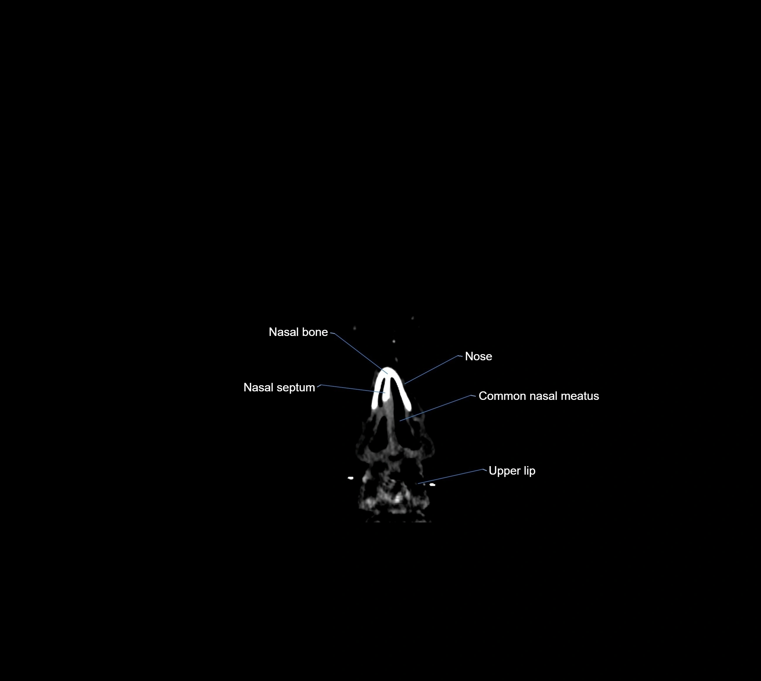
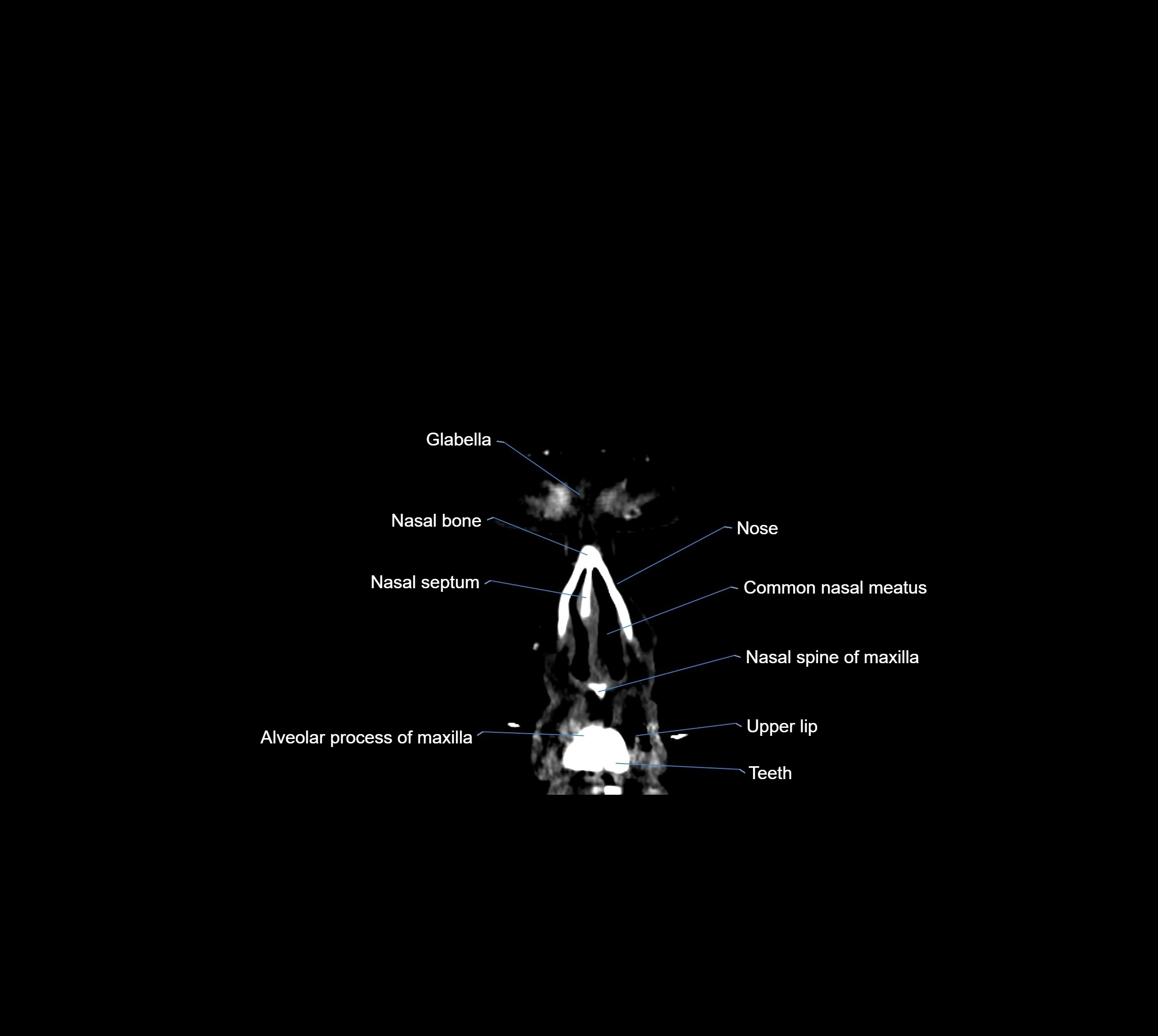
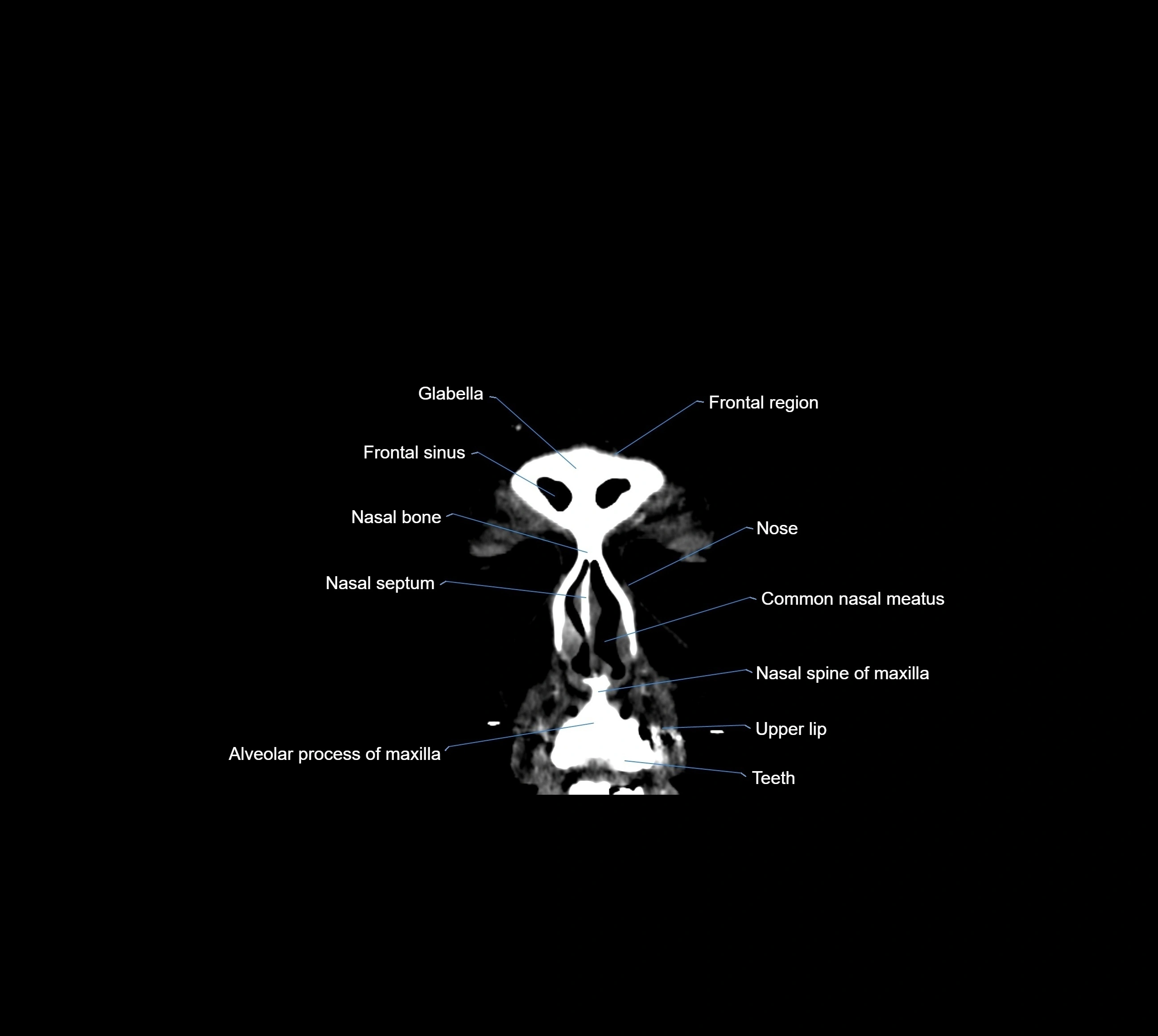
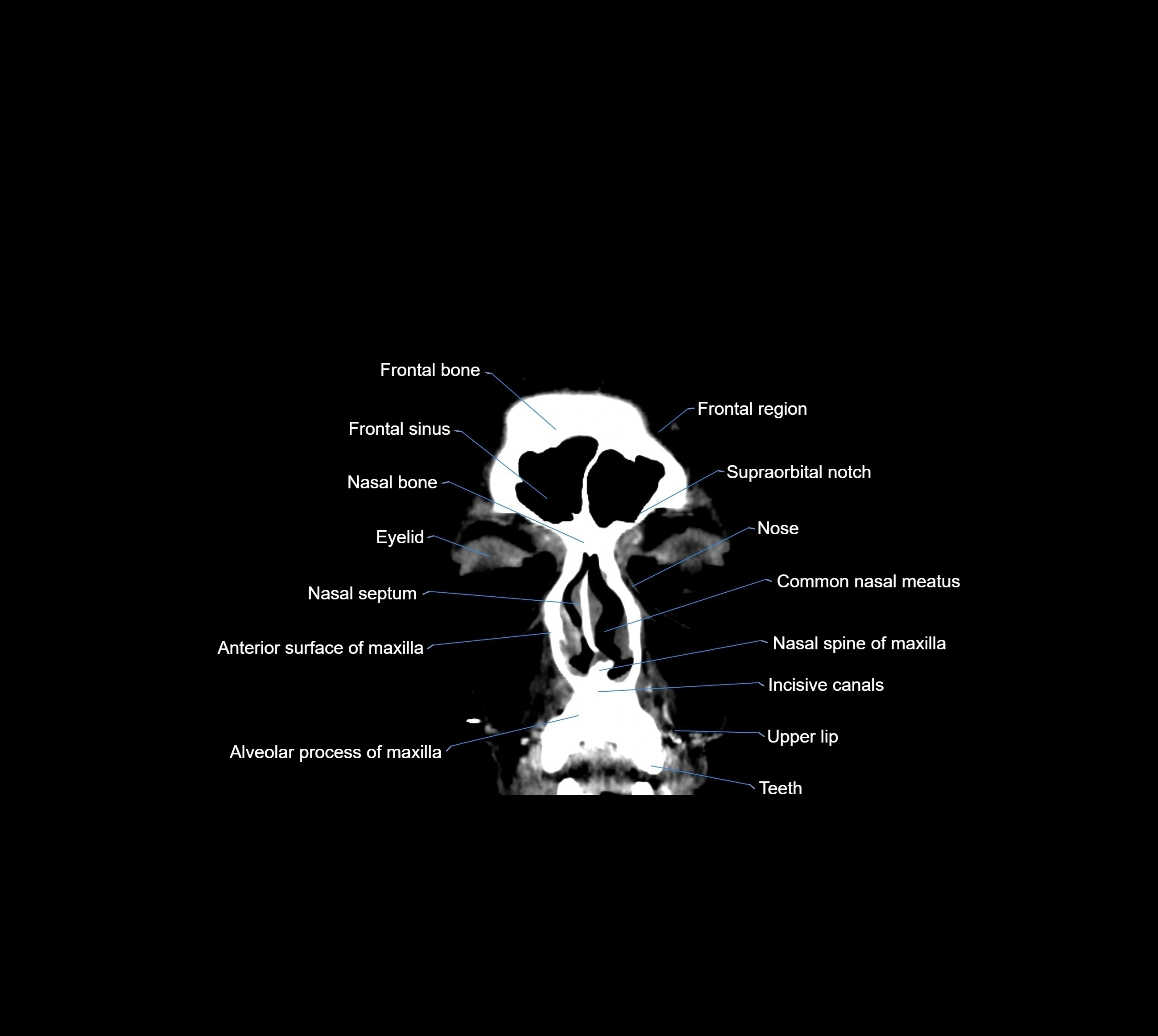
- Alveolar process of maxilla
- Alveolar ridge
- Alveus
- Ambient cistern
- Amygdala
- Amygdaloclaustral area
- Amygdalohippocampal area
- Angular gyrus
- Angular vein
- Anterior Band of Articular Disc TMJ
- Anterior Choroidal Artery
- Anterior Communicating Artery
- Anterior ascending ramus of Sylvian fissure
- Anterior calcarine sulcus
- Anterior cerebral artery
- Anterior cerebral artery (A1 Segment)
- Anterior cerebral artery (A2 Segment)
- Anterior cerebral artery (A3 Segment)
- Anterior cerebral veins
- Anterior chamber of eyeball
- Anterior clinoid process
- Anterior cochlear nucleus
- Anterior commissure
- Anterior ethmoidal air cells
- Anterior external vertebral venous plexuses
- Anterior hippocampal veins
- Anterior horizontal ramus of Sylvian fissure
- Anterior intercavernous sinus
- Anterior internal vertebral venous plexus
- Anterior limb of internal capsule
- Anterior lobe of cerebellum
- Anterior lobe of pituitary gland
- Anterior long insular gyrus
- Anterior median fissure of medulla oblongata
- Anterior nasal spine
- Anterior orbital gyrus
- Anterior parietal artery
- Anterior quadrangular lobule
- Anterior quadrangular lobule (HV) of cerebellum
- Anterior quadrangular lobule (HlV) of cerebellum
- Anterior short insular gyrus
- Anterior superior alveolar nerve
- Anterior temporal artery anatomy
- Anterior vein of caudate nucleus
- Anterolateral central (lenticulostriate) arteries anatomy
- Anterolateral medullary vein
- Anterolateral pontine vein
- Anteromedial central (perforating) arteries anatomy
- Anteromedian medullary vein
- Anteromedian pontine vein
- Apex of insula
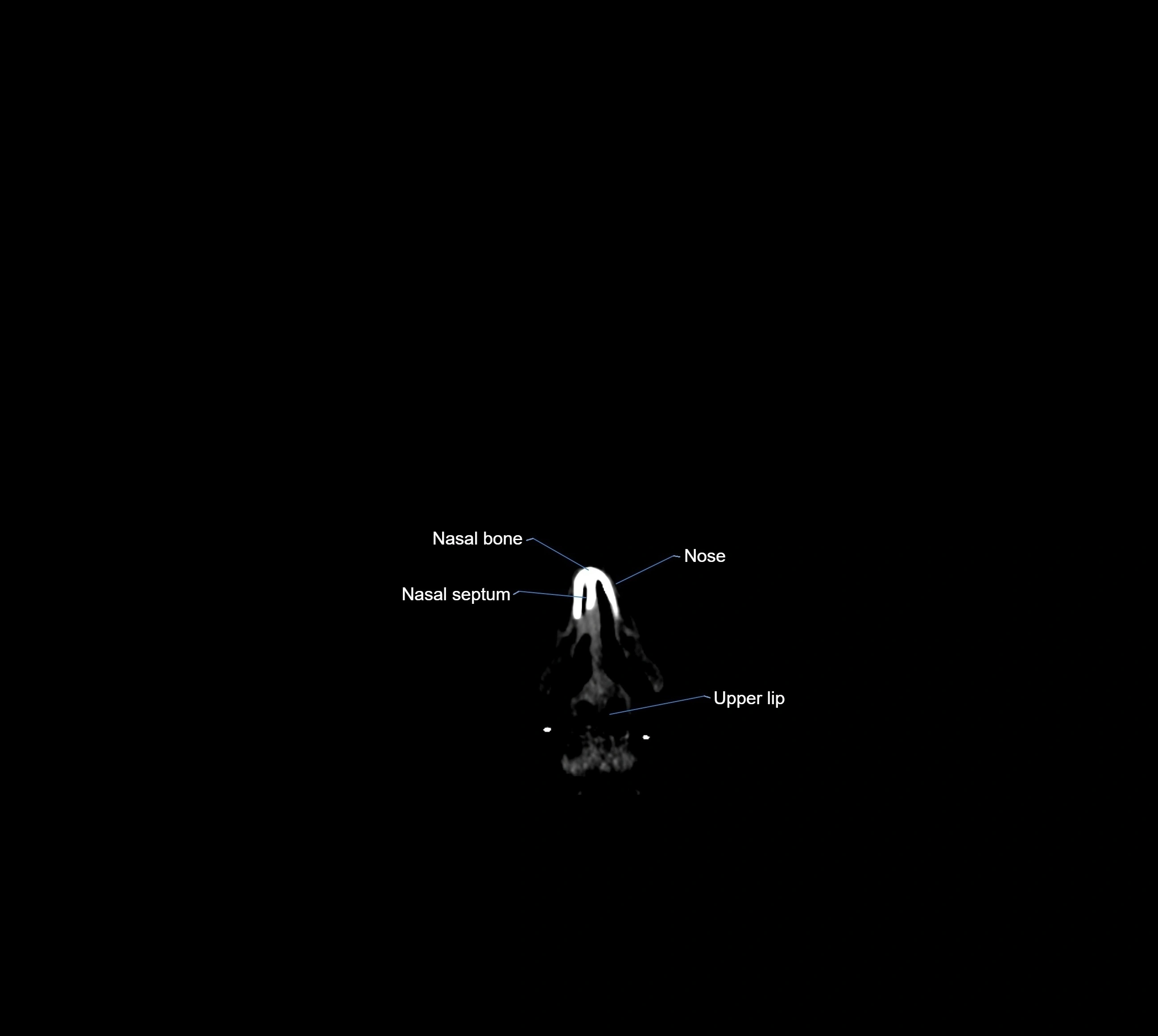
- Apex of nose
- Apical foramen
- Aqueduct of midbrain (Sylvian Aqueduct)
- Arbor Vitae (Cerebellar White Matter)
- Artery of central sulcus
- Artery of postcentral sulcus
- Artery of precentral sulcus
- Artery to angular gyrus anatomy
- Articular capsule of temporomandibular joint
- Articular disc of temporomandibular joint
- Articular eminence
- Articular surface of mandibular fossa
- Articular tubercle
- Atrium of lateral ventricle
- Attachment of inferior head of lateral pterygoid muscle
- Attachment of superior head of lateral pterygoid muscle
- Auricle (Pinna)
- Auriculotemporal nerve
- Basal vein of rosenthal
- Basilar artery
- Basilar part of pons
- Basilar plexus
- Basilar sulcus
- Basilar venous plexus
- Biventral lobule (HVIII) of cerebellum
- Body of caudate nucleus
- Body of corpus callosum
- Body of fornix
- Body of hippocampus
- Body of lateral ventricle
- Body of mandible
- Body of tongue
- Body of vertebra
- Brachium of inferior colliculus
- Brachium of superior colliculus
- Calcarine Sulcus
- Calcarine artery
- Calcarine branch of posterior cerebral artery
- Calcarine spur
- Callosomarginal artery
- Carotid cistern
- Carotid siphon
- Cartilaginous part of nasal septum
- Caudato-lenticular bridges
- Caudatolenticular gray bridges
- Cave of septum pellucidum
- Central canal
- Central lobule
- Central lobule (II & III) of Cerebellum
- Central part of lateral ventricle
- Central sulcus
- Central sulcus of insula
- Cerebellar commissure
- Cerebellar falx
- Cerebellar tentorium
- Cerebellar tonsil (H IX)
- Cerebellopontine angle
- Cerebellum
- Cerebral crus
- Cerebral falx
- Chiasmatic cistern
- Choroid
- Choroid plexus
- Choroid plexus of fourth ventricle
- Choroid plexus of lateral ventricle
- Choroid plexus of the lateral ventricle
- Cingulate gyrus
- Cingulate sulcus
- Circular sulcus of insula
- Cistern of central sulcus
- Cistern of lamina terminalis
- Cistern of lateral cerebral fossa
- Cistern of transverse fissure
- Cisterna magna
- Claustrum
- Cochlea
- Cochlear Canaliculus
- Cochlear nerve (Cranial nerve VIII)
- Collateral sulcus
- Column of fornix
- Common facial vein
- Common tendinous ring (Annulus of zinn)
- Confluence of sinuses
- Cornea
- Cornu Ammonis 1 (CA1)
- Cornu ammonis
- Cornu ammonis 2 (CA2)
- Cornu ammonis 3 (CA3)
- Cornu ammonis 4 (CA4)
- Corona radiata
- Coronal suture
- Coronoid process of mandible
- Corticospinal tract
- Costotransverse joint
- Crista galli
- Crural cistern
- Crus I of ansiform lobule of cerebellum
- Crus II of ansiform lobule of cerebellum
- Crus cerebri
- Crus helix of ear
- Crus of fornix
- Culmen
- Culmen (IV, V) of Cerebellum
- Cuneate fasciculus
- Cuneus
- Declive
- Declive (VI) of Cerebellum
- Decussation of the superior cerebellar peduncles
- Deep mddle cerebral veins
- Deep middle cerebral vein
- Deep part of masseter muscle
- Dental branches of inferior alveolar artery, vein, & nerve
- Dental pulp of upper molar tooth
- Dental pulp of upper premolar tooth
- Dentate gyrus
- Dentate nucleus
- Descending occipital gyrus
- Diploic veins
- Dorsal cochlear nucleus
- Dorsal nucleus of vagus nerve
- Dorsal vein of corpus callosum
- Dorsum of nose
- Dorsum of tongue
- Enamel of lower molar tooth
- Enamel of upper molar tooth
- Enamel of canines tooth
- Enamel of lower incisor tooth
- Enamel of lower canines tooth
- Enamel of lower premolar tooth
- Enamel of upper incisor tooth
- Epiglottis
- Ethmoid sinus
- Ethmoidal air cells
- External acoustic meatus
- External capsule
- Extreme capsule
- Eyeball
- Facial Nerve (Cranial nerve VII)
- Fibers of trigeminal nerve
- Fimbria of hippocampus
- Flocculonodular lobe
- Flocculus
- Floor of orbit
- Floor of the maxillary sinus
- Folium (VII) of Cerebellum
- Folium of Vermis
- Foramen caecum of medulla oblongata
- Foramen cecum of tongue
- Fourth ventricle
- Frenulum veli
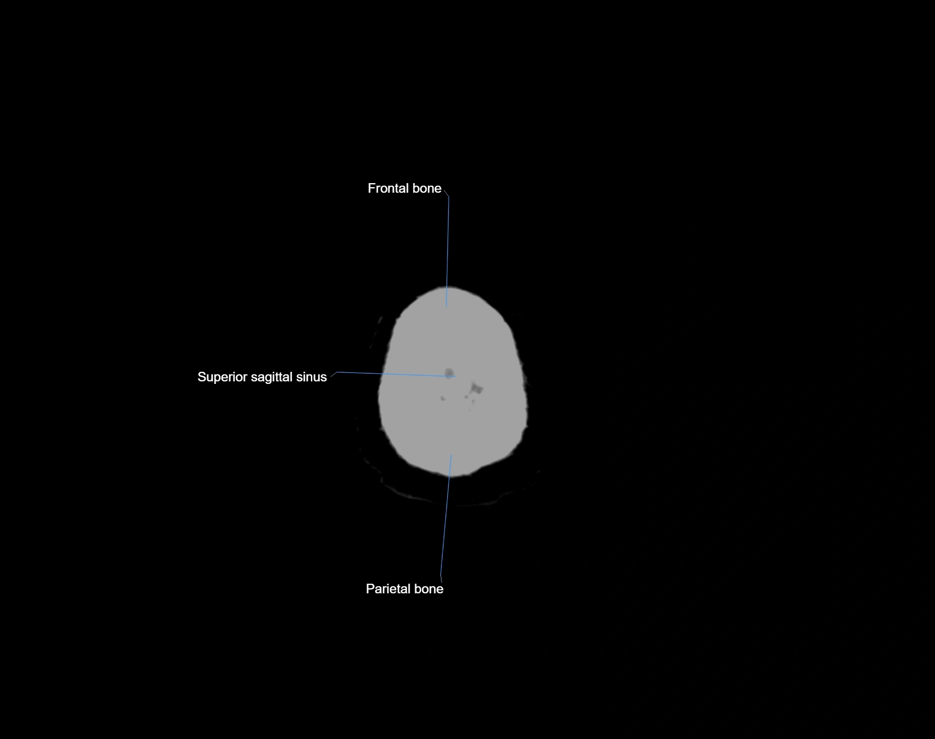
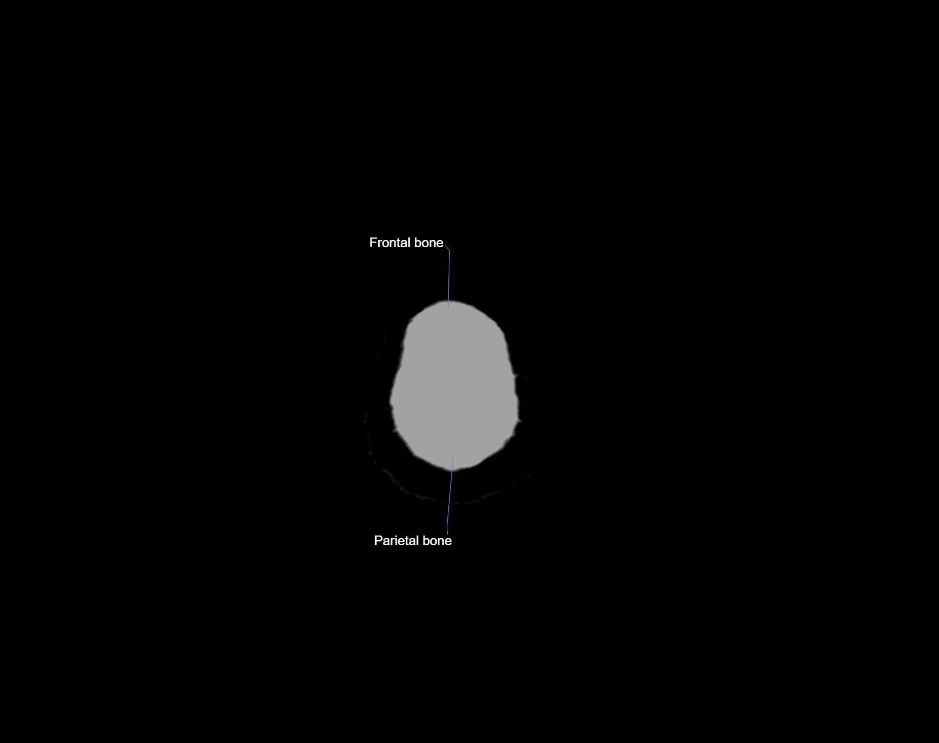
- Frontal bone
- Frontal horn of lateral ventricle
- Frontal nerve
- Frontal operculum
- Frontal pole
- Frontal process of maxilla
- Frontal process of zygomatic bone
- Frontal sinus
- Frontal suture
- Frontal veins
- Frontobasal artery
- Frontonasal suture
- Frontopolar artery
- Frontopolar vein
- Genioglossus muscle
- Genu of corpus callosum
- Glabella
- Glenoid process of parotid gland
- Globus pallidus external segment
- Globus pallidus internal segment
- Glossopharyngeal nerve (Cranial nerve IX)
- Glottis
- Gracile fasciculus
- Great cerebral vein
- Greater wing of sphenoid bone
- Gum (gingiva)
- Habenula
- Habenular commissure
- Hard palate
- Head of caudate nucleus
- Head of hippocampus
- Hippocampal body
- Hippocampal head
- Hippocampal sulcus
- Hippocampal tail
- Hippocampus
- Horizontal fissure (cerebellum)
- Hyoepiglottic ligament
- Hypoglossal Nerve (Cranial nerve XII)
- Hypoglossal nucleus
- Hypophysial cistern
- Hypothalamic sulcus
- Hypothalamus
- Incisive duct
- Incisive foramen
- Inferior anastomotic vein (Vein of Labbé)
- Inferior articular process of vertebra
- Inferior branch vestibular nerve
- Inferior cerebellar peduncle
- Inferior cerebellar veins
- Inferior choroidal vein
- Inferior colliculus
- Inferior eyelid
- Inferior frontal gyrus
- Inferior frontal sulcus
- Inferior head of lateral pterygoid muscle
- Inferior hemispheric cerebellar veins
- Inferior hemispheric veins of the cerebellum
- Inferior hypophyseal artery anatomy
- Inferior nasal concha
- Inferior nasal meatus
- Inferior oblique muscle
- Inferior occipital gyrus
- Inferior olive
- Inferior opening of cerebral aqueduct
- Inferior ophthalmic vein
- Inferior orbital fissure
- Inferior parietal lobule
- Inferior petrosal sinus
- Inferior rectus muscle
- Inferior sagittal sinus
- Inferior salivatory nucleus
- Inferior semilunar lobule
- Inferior tarsus
- Inferior temporal gyrus
- Inferior temporal sulcus
- Inferior vein of vermis
- Inferior vermian vein
- Inferior vestibular nucleus
- Infraglottic cavity
- Infraorbital canal
- Infraorbital margin
- Infraorbital nerve
- Infundibular recess
- Infundibulum
- Innominate line (skull)
- Insular cortex
- Insular lobe
- Insular threshold
- Intermediate zone of articular disc
- Internal carotid artery
- Internal carotid artery (cervical part)
- Internal carotid artery (petrous part)
- Internal cerebral vein
- Internal jugular vein
- Internal medullary lamina
- Internal occipital crest
- Interpeduncular Cistern
- Interpeduncular fossa
- Interthalamic adhesion
- Interventricular foramen
- Intrabiventral Fissure of Biventral Lobule
- Intracanalicular part of optic nerve
- Intracranial part of optic nerve
- Intraculminate fissure
- Intraparietal sulcus
- Iris
- Isthmus of cingulate gyrus
- Jugular foramen
- Jugular foramen pars vascularis
- Jugular tubercle
- Labyrinthine artery
- Lacrimal artery
- Lacrimal bone
- Lacrimal canaliculi
- Lacrimal gland
- Lacrimal nerve
- Lacrimal nucleus
- Lacrimal vein
- Lambdoid suture
- Lamina of vertebra
- Lamina terminalis
- Lateral aperture of the fourth ventricle
- Lateral canthal ligament
- Lateral cerebellomedullary cistern
- Lateral frontobasal artery
- Lateral geniculate body
- Lateral groove of midbrain
- Lateral lemniscus
- Lateral longitudinal fasciculus
- Lateral mass of atlas
- Lateral mesencephalic vein
- Lateral nasal cartilage
- Lateral occipital artery
- Lateral occipitotemporal gyrus
- Lateral orbital gyrus
- Lateral orbitofrontal artery
- Lateral pallidum
- Lateral part of biventeral lobule
- Lateral pontine vein
- Lateral pterygoid muscle
- Lateral recess fourth ventricle
- Lateral rectus muscle
- Lateral sulcus (Sylvian fissure)
- Lateral temporomandibular ligament
- Lateral vein of lateral ventricle
- Lateral vestibular nucleus
- Lateral wall of the orbit
- Left vertebral artery
- Lesser wing of sphenoid
- Levator palpebrae superioris muscle
- Limbic lobe
- Lingual gyrus
- Lingual vein
- Lingula of cerebellum
- Lingula of cerebellum (I)
- Lockwood’s ligament
- Locus ceruleus
- Long gyri of insula
- Long medial striate artery
- Longitudinal cerebral fissure
- Lower molar apical foramen
- Lower premolar apical foramen
- Lunogracle fissure
- Lunogranicile fissure of cerebellum
- Major alar cartilage
- Mammillary body
- Mandible
- Mandibular condyle
- Mandibular foramen
- Mandibular fossa
- Mandibular nerve
- Mandibular notch
- Marginal branch of cingulate sulcus
- Marginal sinus
- Marginal sulcus
- Masseter muscle
- Mastoid air cells
- Mastoid emissary vein
- Mastoid process
- Maxillary bone
- Maxillary nerve
- Maxillary sinus
- Maxillary veins
- Meckel’s cave (Trigeminal cave)
- Medial canthal ligament
- Medial frontal gyrus
- Medial frontobasal artery
- Medial geniculate body
- Medial lemniscus
- Medial longitudinal fasciculus
- Medial occipital artery
- Medial occipitotemporal gyrus
- Medial orbital gyrus
- Medial orbitofrontal artery
- Medial pallidum
- Medial part of biventeral lobule
- Medial pterygoid muscle
- Medial rectus muscle
- Medial vein of lateral ventricle
- Medial vestibular nucleus
- Medial wall of orbit
- Medial wall of the maxillary sinus
- Median aperture of the fourth ventricle
- Median sulcus of the 4th ventricle
- Medulla oblongata
- Medullopontine sulcus
- Meninges
- Mental protuberance
- Mesencephalic nucleus of trigeminal nerve
- Middle Nasal Meatus
- Middle cerebellar peduncle
- Middle cerebral artery
- Middle cerebral artery cortical segment (M4)
- Middle cerebral artery horizontal segment (M1)
- Middle cerebral artery insular segment (M2)
- Middle cerebral artery opercular segment (M3)
- Middle frontal gyrus
- Middle meningeal artery
- Middle nasal concha
- Middle occipital gyrus
- Middle short insular gyrus
- Middle superior alveolar nerve
- Middle temporal artery
- Middle temporal gyrus
- Minor alar cartilage
- Motor nucleus of facial nerve
- Motor nucleus of trigeminal nerve
- Nasal bones
- Nasal cavity
- Nasal part of frontal bone
- Nasal septum
- Nasal spine of frontal bone
- Nasal vestibule
- Nasofrontal vein
- Nasolacrimal duct (Tear duct)
- Neck of mandible
- Nodule of vermis
- Nodule of vermis (X)
- Nucleus of abducens nerve
- Nucleus of hypoglossal nerve
- Nucleus of oculomotor nerve
- Nucleus of solitary tract
- Nucleus of the solitary tract
- Nucleus of trochlear nerve
- Nucleus reticularis tegmenti pontis
- Nucleus supraspinalis
- Obex
- Occipital bone
- Occipital condyle
- Occipital emissary vein
- Occipital horn of lateral ventricle
- Occipital pole
- Occipital sinus
- Occipital veins
- Occipitotemporal sulcus
- Oculomotor Nerve (Cranial Nerve III)
- Oculomotor cistern
- Oculomotor nerve (Superior branch)
- Oculomotor nerve (inferior branch)
- Odontoid process
- Olfactory Nerve (Cranial Nerve I)
- Olfactory bulb
- Olfactory cistern
- Olfactory sulcus
- Olfactory tract
- Olfactory trigone
- Olfactory tubercle
- Ophthalmic artery
- Ophthalmic nerve
- Optic Nerve (Cranial Nerve II)
- Optic canal
- Optic chiasm
- Optic disc
- Optic nerve sheath
- Optic tract
- Orbicularis oculi muscle
- Orbital Sulci
- Orbital cavity
- Orbital gyri
- Orbital part of optic nerve
- Orbital plate
- Orbital surface of maxilla
- Oropharynx
- Palatine process of maxilla
- Para-brachial recess
- Parabrachial recess
- Paracentral artery
- Paracentral gyrus
- Paracentral lobule
- Paracentral sulcus
- Parahippocampal gyrus
- Paramedian lobule
- Paramedian lobule (HVII) of cerebellum
- Paraolfactory sulci
- Parasubiculum (Presubiculum)
- Paraterminal gyrus
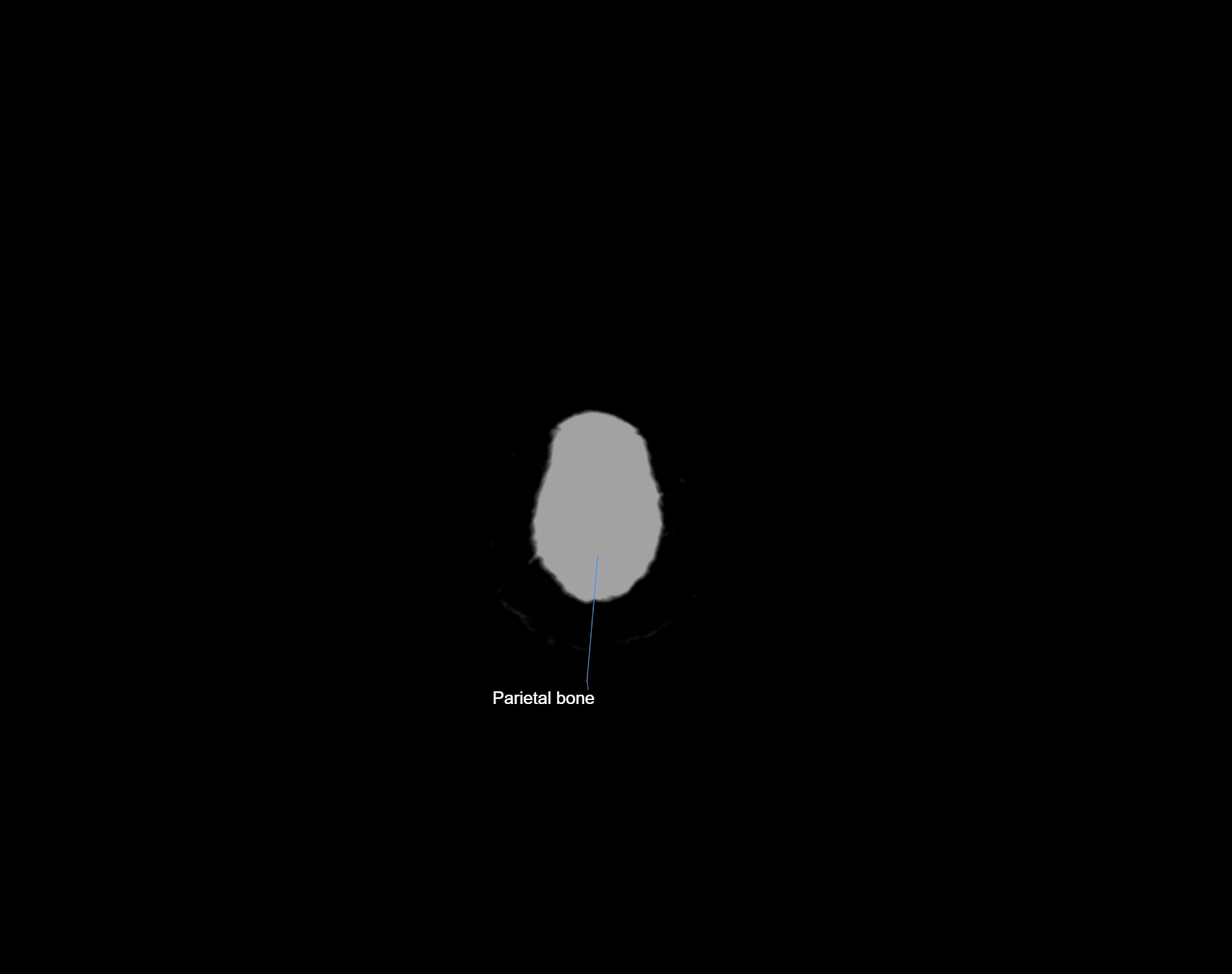
- Parietal bone
- Parietal veins
- Parieto-occipital artery
- Parietooccipital sulcus
- Parotid gland
- Pars opercularis of inferior frontal gyrus
- Pars orbitalis of inferior frontal gyrus
- Pars triangularis of inferior frontal gyrus
- Pedicle of vertebra
- Peduncle of flocculus
- Pericallosal artery
- Pericallosal cistern
- Perivascular spaces
- Pes hippocampi
- Petrosal vein
- Petrous part of temporal bone
- Pineal gland
- Pineal recess
- Pituitary fossa
- Pituitary gland
- Pituitary stalk
- Polar frontal artery
- Polar temporal artery
- Pons
- Pons (basilar portion)
- Pontine arteries
- Pontine artery
- Pontine nucleus
- Pontocerebellar cistern
- Pontocerebellar fibers
- Pontomedullary junction
- Pontomedullary sulcus
- Post-olivary sulcus
- Postcentral gyrus
- Postcentral sulcus
- Posterior auricular vein
- Posterior band of articular disc, TMJ
- Posterior border of maxilla
- Posterior cerebellomedullary cistern (cisterna magna)
- Posterior cerebral artery
- Posterior cerebral artery (P1 Segment)
- Posterior cerebral artery (P2 Segment)
- Posterior cerebral artery (P3 Segment)
- Posterior cerebral artery (P4 Segment)
- Posterior chamber of eyeball
- Posterior clinoid process
- Posterior cochlear nucleus
- Posterior commissure
- Posterior communicating artery
- Posterior external vertebral venous plexus
- Posterior hippocampal artery
- Posterior inferior cerebellar artery
- Posterior intercavernous sinus
- Posterior lateral choroidal artery
- Posterior limb of internal capsule
- Posterior lobe of cerebellum
- Posterior lobe pituitary gland
- Posterior long insular gyrus
- Posterior medial choroidal artery
- Posterior median sulcus of medulla oblongata
- Posterior mesencephalic vein
- Posterior nasal spine
- Posterior orbital gyrus
- Posterior orbital sulcus
- Posterior parietal artery
- Posterior part of tongue
- Posterior quadrangular lobule
- Posterior ramus of lateral sulcus
- Posterior short insular gyrus
- Posterior superior alveolar nerve
- Posterior superior fissure
- Posterior vein of caudate nucleus
- Posterior vein of septum pellucidum
- Posterior veins of septum pellucidum
- Posterolateral fissure
- Posteromedial central (perforating) arteries
- Posteromedian medullary vein
- Pre-Rolandic artery
- Pre-olivary sulcus
- Prebiventral fissure
- Precentral cerebellar vein
- Precentral fissure
- Precentral gyrus
- Precentral sulcus
- Precommunicating part of posterior cerebral artery
- Preculminate fissure
- Precuneal artery
- Precuneus
- Preolivary groove
- Preoptic area
- Prepontine cistern
- Primary fissure
- Principal sensory nucleus of the trigeminal nerve
- Principal sensory nucleus of trigeminal nerve
- Prosubiculum
- Pterygoid venous plexus
- Pterygomaxillary fissure
- Pulvinar
- Pupil
- Putamen
- Pyramid of medulla oblongata
- Pyramid of vermis (VIII)
- Pyramids of the medulla oblongata
- Pyramis of vermis
- Quadrigeminal cistern
- Ramus of mandible
- Raphe of pons
- Red nucleus
- Retina
- Retrobulbar fat
- Retromandibular vein
- Retroolivary groove
- Rhinal sulcus
- Right vertebral artery
- Rolandiс artery
- Root canal of lower canines tooth
- Root canal of lower premolar tooth
- Root canal of upper canines tooth
- Root canal of upper molar tooth
- Root canal of upper premolar tooth
- Root of lower canines tooth
- Root of lower molar tooth
- Root of upper molar tooth
- Rostral gyrus
- Rostral sulcus
- Rostrum of corpus callosum
- Sagittal suture
- Sclera
- Secondary fissure
- Sella turcica
- Semicircular Canals
- Septum of sphenoid sinus
- Septum pellucidum
- Sheath of styloid process
- Short gyri of insula
- Sigmoid sinus
- Simple lobule
- Simple lobule (HVI) of cerebellum
- Soft palate
- Sphenoid sinus
- Sphenoparietal sinus
- Spinal cord
- Spinal lemniscus
- Spinal nucleus of trigeminal nerve
- Spinal trigeminal nucleus
- Spinal trigeminal nucleus (pars caudalis)
- Spinal trigeminal nucleus (pars oralis)
- Spinothalamic tract
- Splenium of corpus callosum
- Squamous part of temporal bone
- Straight gyrus
- Straight sinus
- Stria medullaris thalami
- Styloid process
- Subarachnoid space of optic nerve
- Subiculum
- Submandibular lymph nodes
- Subparietal sulcus
- Substantia nigra
- Sulcus of corpus callosum
- Superciliary arch
- Superficial cerebral veins
- Superficial middle cerebral vein
- Superficial part of masseter
- Superficial temporal artery
- Superficial temporal vein
- Superior anastomotic vein
- Superior articular process of vertebra
- Superior branch of vestibular nerve
- Superior cerebellar artery
- Superior cerebellar cistern
- Superior cerebellar peduncle
- Superior cerebellar vein
- Superior colliculus
- Superior cornu of thyroid cartilage
- Superior cortical veins
- Superior dental plexus
- Superior eyelid
- Superior frontal gyrus
- Superior frontal sulcus
- Superior head of lateral pterygoid muscle
- Superior hemispheric cerebellar veins
- Superior hemispheric veins of the cerebellum
- Superior hypophyseal artery
- Superior medullary velum
- Superior nasal concha
- Superior nasal meatus
- Superior oblique muscle
- Superior occipital gyrus
- Superior opening of the cerebral aqueduct
- Superior ophthalmic vein
- Superior parietal lobule
- Superior petrosal sinus
- Superior rectus muscle
- Superior retrodiscal layer
- Superior sagittal sinus
- Superior salivatory nucleus
- Superior semilunar lobule of cerebellum
- Superior synovial membrane of temporomandibular joint
- Superior tarsus
- Superior temporal gyrus
- Superior temporal sulcus
- Superior thalamic veins
- Superior thalamostriate vein
- Superior vein of vermis
- Superior vermian vein
- Superior vestibular nucleus
- Superior wall of orbit
- Supramarginal gyrus
- Supraoptic recess
- Supraorbital artery
- Supraorbital margin
- Supraorbital nerve
- Supraorbital vein
- Suprapineal recess
- Supratrochlear artery
- Supratrochlear nerve
- Supratrochlear vein
- Supratrochlear veins
- Tail of caudate nucleus
- Tail of hippocampus
- Tectal plate
- Tegmentum of midbrain
- Tegmentum of pons
- Tela choroidea of third ventricle
- Temporal bone
- Temporal horn of lateral ventricle
- Temporal lobe
- Temporal pole
- Temporalis muscle
- Temporomandibular joint
- Temporopolar artery
- Tenia of fornix
- Thalamus
- Third ventricle
- Thyroid cartilage
- Tonsil of cerebellum
- Tragus of ear
- Transverse facial artery
- Transverse facial vein
- Transverse occipital sulcus
- Transverse pontine fiber
- Transverse pontine vein
- Transverse sinus
- Transverse temporal gyri
- Transverse temporal sulcus
- Transverse veins of caudate nucleus
- Trigeminal cave
- Trigeminal ganglion
- Trigeminal nerve (Cranial nerve V)
- Trochlear nerve (Cranial nerve IV)
- Tuber cinereum
- Tuber of vermis
- Tuber of vermis (VII)
- Tympanic part of temporal bone
- Uncus
- Upper cervical spinal cord
- Upper premolar apical foramen
- Uvula of vermis
- Uvula of vermis (IX)
- Vagus nerve (Cranial nerve X)
- Vein of cerebellomedullary cistern
- Vein of lateral recess of fourth ventricle
- Vein of septum pellucidum
- Venous plexus of foramen ovale
- Venous plexus of hypoglossal canal
- Ventral cochlear nucleus
- Ventrolateral sulcus of medulla oblongata
- Vermis of cerebellum
- Vertebral artery
- Vertex
- Vestibular ganglion
- Vestibule
- Vestibulocochlear nerve (Cranial nerve VIII)
- Vestigial hippocampal sulcus
- Vitreous chamber of eyeball
- Vomer
- White matter of cerebellum (Arbor vitae)
- White substance of cerebellum
- Whitnall's ligament
- Wing of central lobule
- Zygomatic arch
- Zygomatic bone
- Zygomatic nerve
- Zygomatic process of frontal bone
- Zygomatic process of temporal bone
- cavernous sinus
- foramen of Monro
- genu of internal capsule
- interpeduncular veins
- jugular foramen pars nervosa
- lens of the eye
The Abducens nerve (Cranial nerve VI) is a purely motor cranial nerve responsible for innervating the lateral rectus muscle of the eye, which is crucial for lateral movement (abduction) of the eyeball. It arises from the abducens nucleus in the dorsal pons, emerges at the pontomedullary junction, and travels a long intracranial course before entering the orbit via the superior orbital fissure. Because of its long path and proximity to the clivus, it is particularly susceptible to injury from increased intracranial pressure or trauma.
Synonyms
-
Sixth cranial nerve
-
CN VI
-
N. abducens (Latin)
-
Nervus abducens
Function
-
Innervates the lateral rectus muscle of the eye
-
Responsible for abduction of the eyeball (moving the eye outward, away from the midline)
-
Is a purely motor nerve (no sensory or autonomic fibers)
-
Lesion results in inability to abduct the affected eye, leading to horizontal diplopia (double vision)
MRI Appearance
-
The abducens nerve is a small, thin, linear structure
-
Best visualized on high-resolution T2-weighted 3D MRI sequences (e.g., FIESTA or CISS)
-
Seen as a hypointense (dark) line running from the brainstem at the pontomedullary junction, traversing the prepontine cistern, and entering Dorello’s canal under the petrosphenoidal ligament, then into the cavernous sinus, and finally the orbit
-
May be challenging to visualize in standard MRI due to its small size
-
Pathology may be inferred by absence, displacement, or enhancement of the nerve
CT Appearance
-
The nerve itself is not directly visualized on conventional CT due to its small size and soft tissue density
-
Indirect signs: assessment of the bony course, such as the Dorello’s canal, superior orbital fissure, or adjacent pathologies (fractures, masses, or inflammation) that could impinge the nerve
-
CT is mainly used to exclude structural lesions or fractures that might affect the course of CN VI
MRI images

MRI images

MRI images
