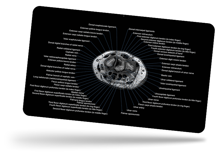
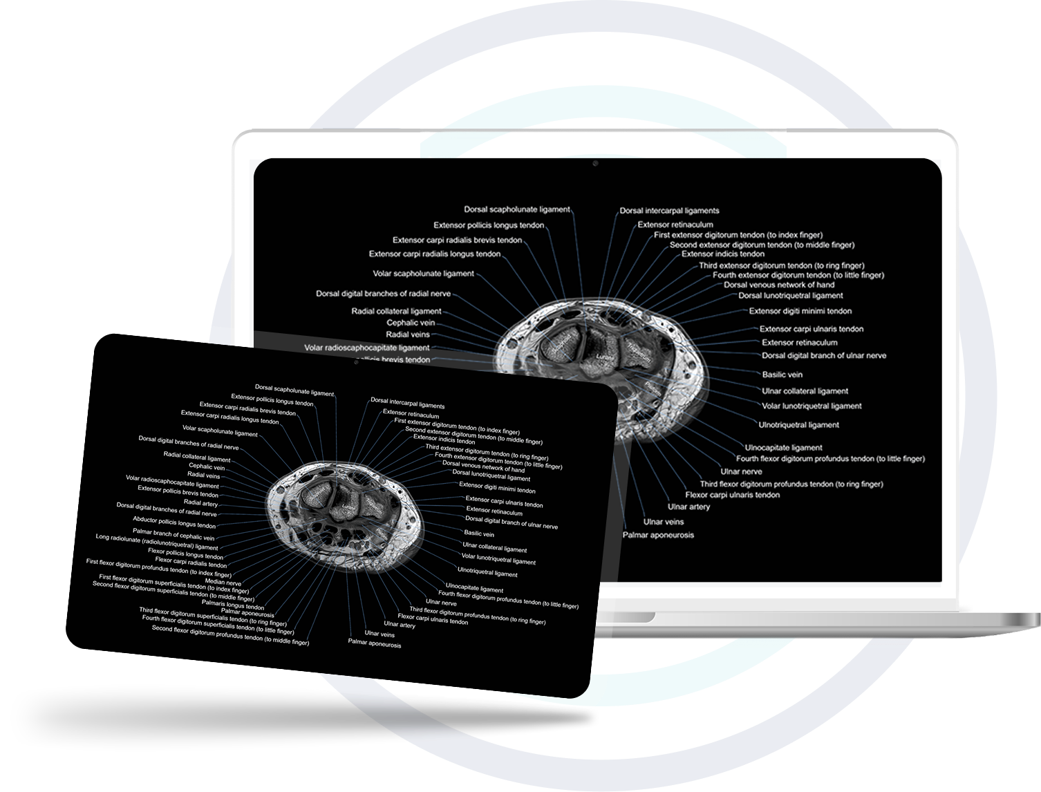
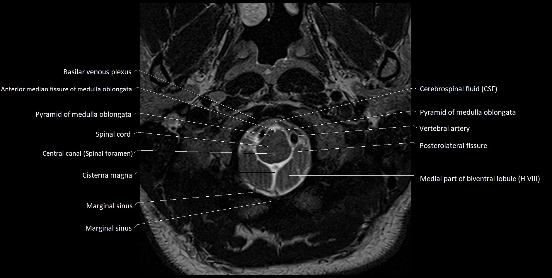
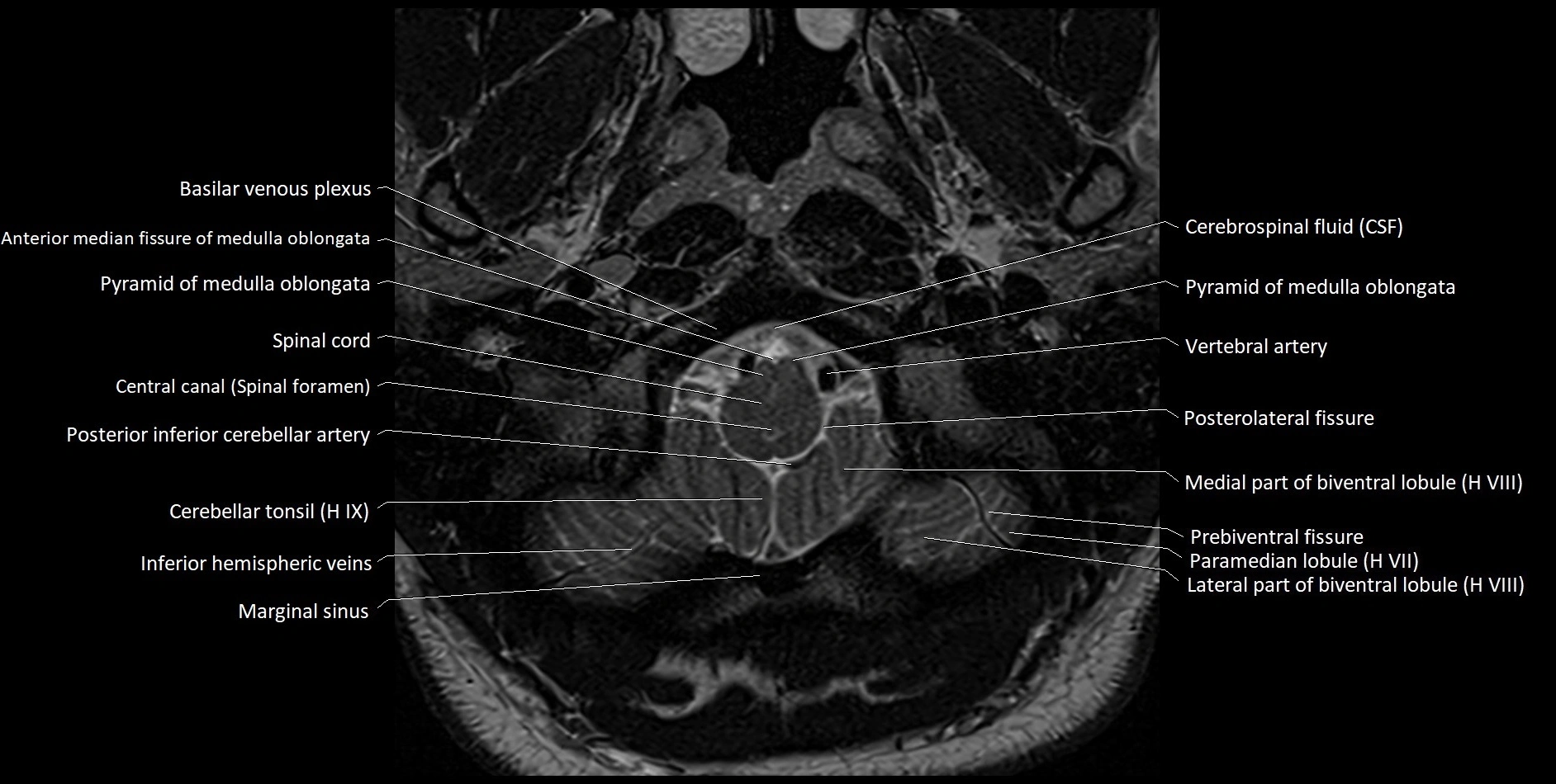
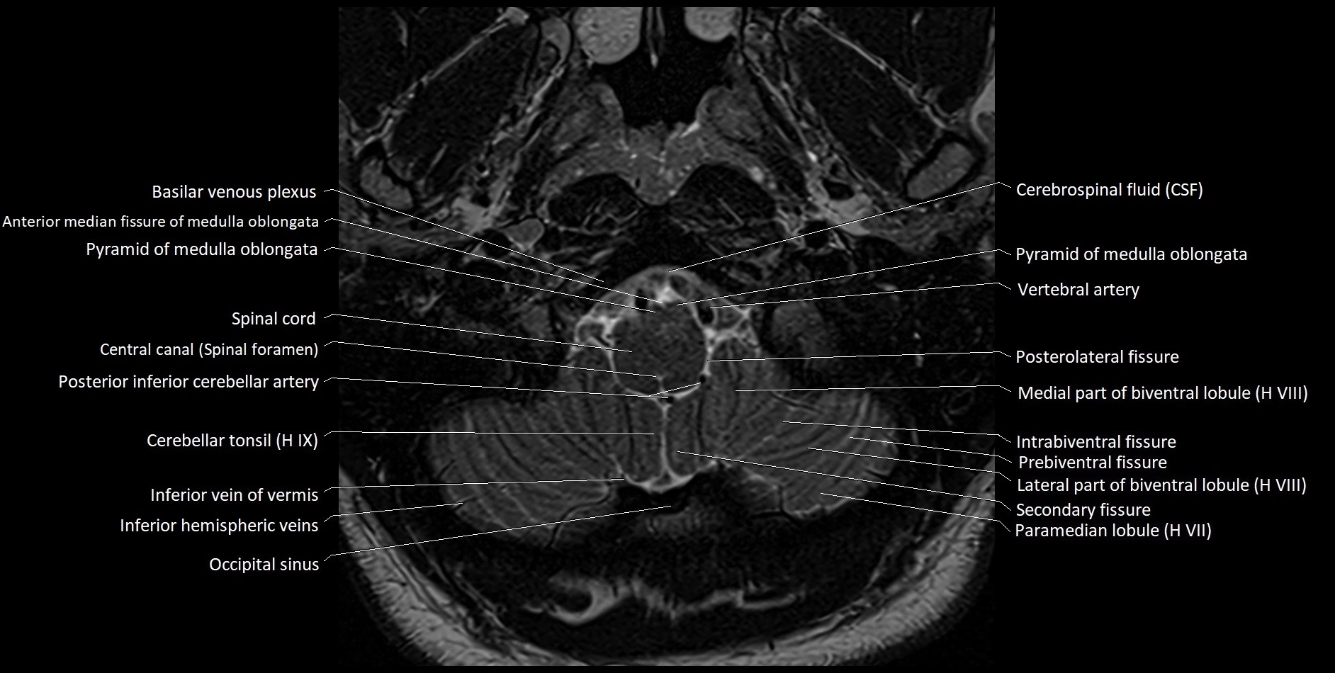
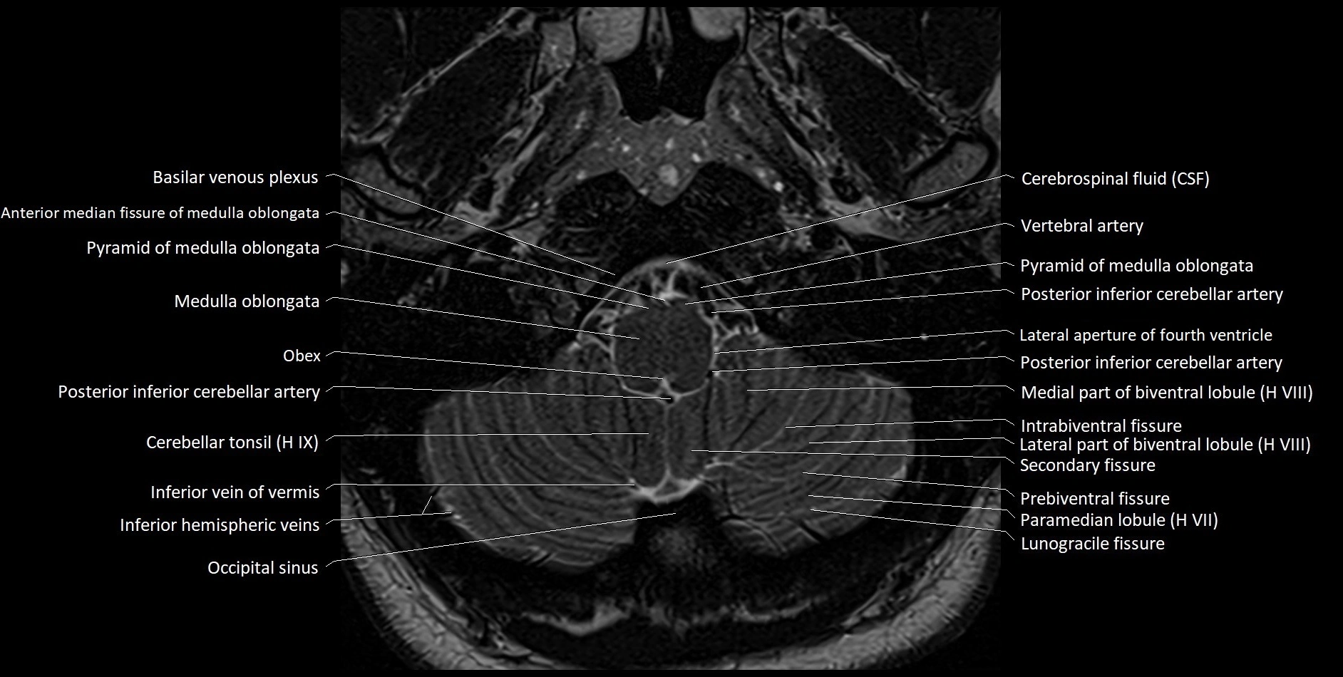
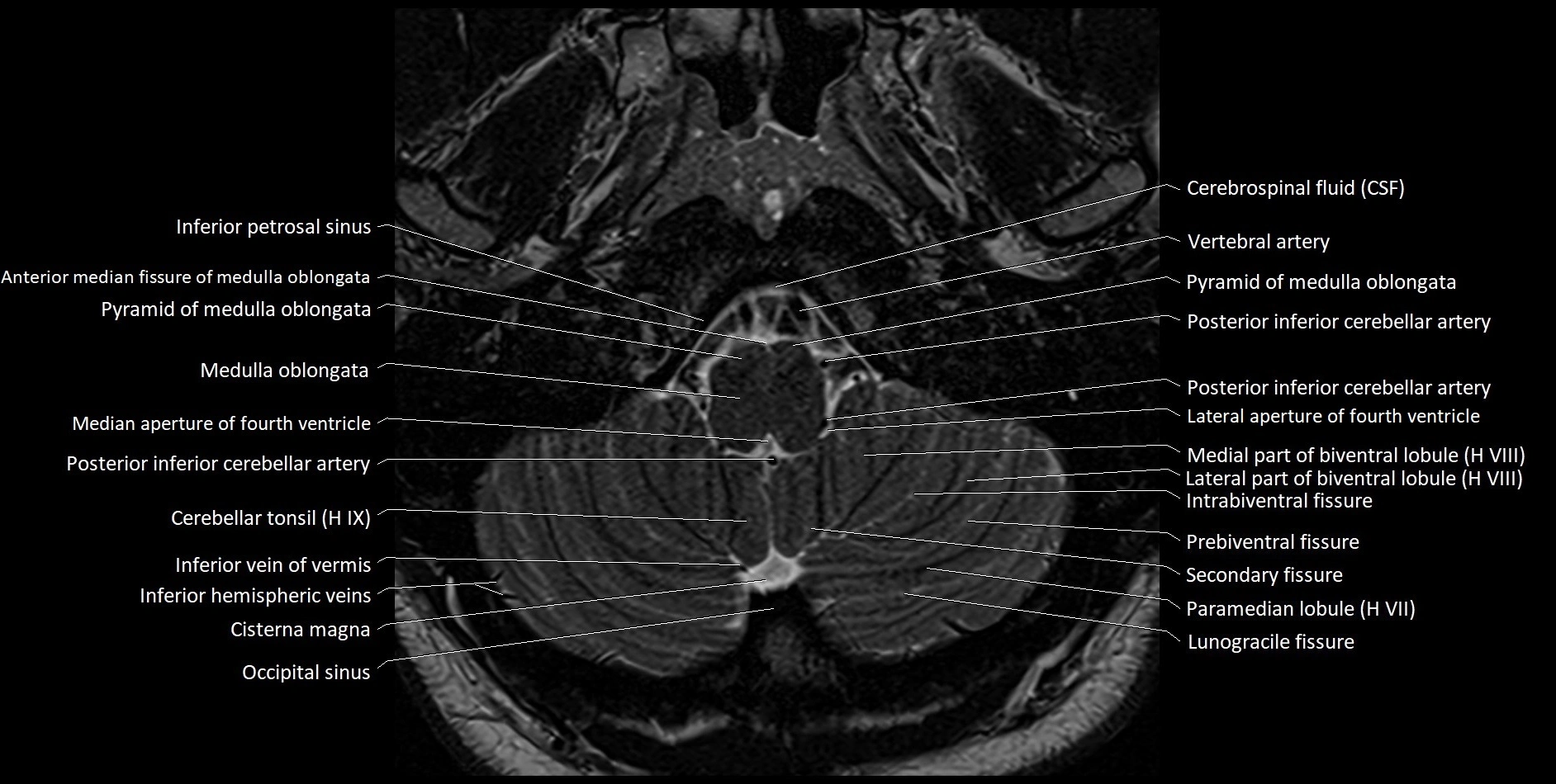
Experience the next generation of cross-sectional anatomy with our cutting-edge 3T MRI images, enhanced by AI-powered Deep Resolve technology. Our ultra-high resolution (1.5mm) and ultra-small FOV images unveil anatomical details like never before, transforming the way anatomy is visualized and understood.
Explore our unique collection of high-resolution cross-sectional MRI images, created from scanning hundreds of volunteers thousands of times over three years using advanced 3T scanners, dedicated multi-channel coils, and cutting-edge AI enhancement. Our library showcases real, detailed anatomy—highlighting even the finest microanatomical structures—without the heavy use of virtual labels found in other resources, allowing you to study authentic, uncompromised images as seen in the human body.
MRI, CT, X-ray & Visible Human Project anatomy—all in one place
Learn MRI anatomy like never before—48 body parts, 144 planes
Crystal-clear, high-res images for effective large-screen teaching
Clear structure descriptions with CT/MRI appearances
1000s of quizzes to sharpen your anatomy knowledge
Budget-friendly plans, perfect for students
All our cross-sectional images are meticulously hand-labeled by professionals with over 20 years of experience in cross-sectional imaging. This is the result of over six years of dedicated work, precision, and passion. Every image is individually checked and verified by senior consultant radiologists from major London NHS trusts, each with relevant subspecialty expertise—ensuring the highest level of accuracy and clinical relevance.