


Topic
- Abdominal aorta
- Abdominal part of esophagus
- Acromial end of clavicle
- Acromial part of deltoid muscle
- Acromioclavicular joint
- Acromion process of scapula
- Acute marginal artery (AM)
- Adipose tissue (Shoulder)
- Adrenal glands
- Anatomical neck of humerus
- Annular epiphysis
- Anterior basal segmental artery of left lung
- Anterior basal segmental artery of right lung
- Anterior basal segmental bronchus of right lung (B7+8, B8)
- Anterior basal vein of left lung
- Anterior interventricular sulcus
- Anterior jugular vein
- Anterior leaflet of left atrioventricular valve
- Anterior longitudinal ligament
- Anterior papillary muscle
- Anterior segmental artery of left lung
- Anterior segmental artery of right lung
- Anterior segmental bronchus of right lung
- Anterior sternoclavicular ligament
- Anterior vein of left lung
- Anterior vein of right lung
- Anteromedial basal bronchus of left lung (B7+8)
- Anulus fibrosus of intervertebral disc
- Aortic arch
- Aortic knob
- Aortic root
- Aortic sinus
- Aortic valve
- Aortic vestibule
- Apex of left lung
- Apex of right lung
- Apex of the heart
- Apical segmental artery of left lung
- Apical segmental artery of right lung
- Apical segmental bronchus of right lung
- Apicoposterior segmental bronchus of left lung
- Arch of aorta
- Articular cartilage of glenoid fossa
- Ascending aorta
- Ascending colon
- Atrioventricular Node (AV Node)
- Atrioventricular bundle (bundle of His)
- Axilla
- Axillary artery
- Axillary lymph nodes
- Axillary nerve
- Axillary veins
- Azygos vein
- Base of right lung
- Basivertebral veins
- Biceps brachii muscle
- Bicipital groove
- Body of humerus
- Body of rib
- Body of scapula
- Body of sternum
- Body of vertebra
- Brachial artery
- Brachial plexus
- Brachial veins
- Brachiocephalic trunk
- Cardia of stomach
- Carina of trachea
- Cecum
- Celiac trunk
- Central axillary lymph nodes
- Cephalic vein
- Circumflex artery (LCx)
- Circumflex scapular artery
- Circumflex subscapular artery
- Clavicle
- Clavicular part of deltoid muscle
- Common basal vein of right lung
- Common hepatic artery
- Conoid tubercle
- Conus arteriosus
- Conus artery
- Coracobrachialis muscle
- Coracoid process of scapula
- Coronary sulcus
- Costal cartilages
- Costal notches
- Costal part of diaphragm
- Costochondral joints
- Costotransverse joint
- Costotransverse joint of first rib
- Costotransverse ligament
- Costovertebral joint
- Costovertebral joint of first rib (rib head joint)
- Costoxiphoid ligaments
- Crista terminalis
- Crura of diaphragm
- Crural part of diaphragm
- Deltoid Tendon (Proximal)
- Deltoid muscle
- Deltoid tendon (Distal)
- Descending colon
- Descending thoracic aorta
- Diaphragm
- Distal left anterior descending artery (dLAD)
- Dorsal scapular artery
- Dorsal scapular nerve
- Dorsal scapular vein
- Duodenum – Superior part (D1)
- Endocardium
- Epicardium
- Erector spinae muscles
- External oblique muscle
- Facet joint of vertebra (Zygapophyseal joints)
- Falciform ligament (liver)
- First diagonal branch (D1) of LAD
- First obtuse marginal branch
- Fissure for ligamentum teres
- Fissure for ligamentum venosum
- Gallbladder
- Gastroduodenal artery
- Glenohumeral joint
- Glenoid fossa
- Glenoid process of scapula
- Great cardiac vein
- Greater tubercle of humerus
- Head of humerus
- Head of rib
- Heart
- Hemiazygos vein
- Hepatic portal vein
- Hilum of lung
- Horizontal fissure of right lung
- Humerus
- Iliocostalis cervicis muscle
- Inferior belly of omohyoid muscle
- Inferior lingular bronchus of left lung (B5)
- Inferior lingular vein of left lung
- Inferior lobar artery of right lung
- Inferior lobe of left lung
- Inferior lobe of right lung
- Inferior phrenic artery
- Inferior phrenic vein
- Inferior thyroid vein
- Inferior tracheobronchial lymph nodes
- Inferior vena cava
- Infraclavicular lymph nodes
- Infraspinatus muscle
- Infraspinatus tendon
- Infraspinous fossa
- Interatrial septum
- Intercostal muscles
- Intercostal space
- Interlobar lymph nodes
- Intermediate hepatic vein
- Internal oblique muscle
- Internal thoracic artery
- Internal thoracic veins
- Interspinales muscles
- Interspinous ligament
- Intertransversarii muscle
- Intertransverse ligament
- Interventricular Septum
- Interventricular septal branch S1
- Interventricular septal branch S2
- Intervertebral Disc
- Intervertebral foramen
- Intervertebral foramina
- Intra-articular ligament of head of rib
- Jugular notch
- Jugular venous arch
- L (Lumbar spine)
- Lamina of vertebra
- Lateral basal segmental artery of left lung
- Lateral basal segmental artery of right lung
- Lateral basal segmental bronchus of left lung (B9)
- Lateral basal segmental bronchus of right lung (B9)
- Lateral basal vein of right lung
- Lateral head of triceps brachii muscle
- Lateral segmental artery of right Lung
- Lateral segmental bronchus of right lung
- Lateral thoracic artery
- Lateral vein of right lung
- Latissimus dorsi muscle
- Latissimus dorsi tendon
- Left Lung (Superior Lobe)
- Left adrenal gland
- Left anterior descending artery (LAD)
- Left anterior segmental bronchus (B3a, B3b, B3c)
- Left apicoposterior bronchus (B1+2a, B1+2b)
- Left atrial branch (coronary artery)
- Left atrioventricular valve (mitral or bicuspid valve)
- Left atrium
- Left auricle
- Left brachiocephalic vein
- Left branch of atrioventricular bundle
- Left cardiophrenic angle
- Left common carotid artery
- Left coronary aortic sinus
- Left costophrenic angle
- Left gastric artery
- Left gastro-omental (gastroepiploic) vein
- Left hemidiaphragm
- Left hepatic artery
- Left hepatic vein
- Left inferior lobar bronchus
- Left inferior pulmonary vein
- Left interlobar artery
- Left internal thoracic artery
- Left internal thoracic veins
- Left lobe of liver
- Left lobe of thyroid gland
- Left lung
- Left lung (inferior lobe)
- Left main bronchus
- Left main coronary artery (LMCA)
- Left marginal vein
- Left phrenic nerve
- Left pulmonary artery
- Left recurrent laryngeal nerve
- Left subclavian vein
- Left superior lobar bronchus
- Left superior pulmonary vein
- Left ventricle
- Lesser tubercle of humerus
- Ligamenta flava (Ligamentum flavum)
- Ligamentum teres (round ligament of the liver)
- Ligamentum venosum
- Lingular bronchus of left lung
- Lingular vein of left lung
- Liver
- Long head of biceps brachii muscle
- Long head of biceps tendon
- Long head of triceps brachii muscle
- Long thoracic nerve
- Longus colli muscle
- Lumbar part of diaphragm
- Manubrium of sternum
- Medial cutaneous nerve of forearm
- Medial head of triceps brachii muscle
- Medial segmental artery of right lung
- Medial segmental bronchus of right lung (B5)
- Medial vein of right lung
- Median arcuate ligament
- Median nerve
- Mediastinum
- Middle cardiac vein
- Middle lobar artery of right lung
- Middle lobe of right lung
- Multifidus muscles
- Myocardium
- Neck of rib
- Neck of scapula
- Noncoronary aortic sinus
- Oblique fissure of left lung
- Oblique fissure of right lung
- Oblique pericardial sinus
- Oblique vein of left atrium
- Opening of inferior vena cava
- Opening of superior vena cava
- Openings of pulmonary veins
- Oval fossa of right atrium
- Pancreas
- Pectinate muscles
- Pectoralis major muscle
- Pectoralis minor muscle
- Pedicle of vertebra
- Pericardial cavity
- Pericardium
- Phrenicomediastinal recess
- Phrenoesophageal ligament
- Platysma muscle
- Pleura
- Posterior basal segmental artery of left lung
- Posterior basal segmental artery of right lung
- Posterior basal segmental bronchus of left lung (B10)
- Posterior basal segmental bronchus of right lung (B10)
- Posterior basal vein of left lung
- Posterior basal vein of right lung
- Posterior circumflex humeral artery
- Posterior circumflex humeral vein
- Posterior cutaneous nerve of arm
- Posterior intercostal arteries
- Posterior intercostal veins
- Posterior interventricular sulcus
- Posterior leaflet of left atrioventricular valve
- Posterior longitudinal ligament
- Posterior papillary muscle
- Posterior segmental artery of left lung
- Posterior segmental artery right lung
- Posterior segmental bronchus of right lung
- Posterior sternoclavicular ligament
- Posterior vein of left ventricle
- Posterior vein of right lung
- Proper hepatic artery
- Pulmonary trunk
- Pulmonary valve
- Radiate sternocostal ligament
- Rectus abdominis muscle
- Retrosternal space
- Rhomboid major muscle
- Rhomboid minor muscle
- Ribs
- Right adrenal gland
- Right anterior segmental bronchus (B3)
- Right apical segmental bronchus (B1)
- Right atrioventricular valve (tricuspid valve)
- Right atrium
- Right auricle of heart
- Right brachiocephalic vein
- Right branch of atrioventricular bundle
- Right cardiophrenic angle
- Right coronary aortic sinus
- Right coronary artery (RCA)
- Right costophrenic angle
- Right crus of diaphragm
- Right fibrous trigone
- Right hemidiaphragm
- Right hepatic artery
- Right hepatic vein
- Right inferior lobar bronchus
- Right inferior pulmonary vein
- Right interlobar artery
- Right intermediate bronchus
- Right internal thoracic artery
- Right internal thoracic veins
- Right lobe of liver
- Right lobe of thyroid gland
- Right lung
- Right lung (inferior lobe)
- Right lung (middle lobe)
- Right lung (superior lobe)
- Right main bronchus
- Right marginal vein of heart
- Right middle lobar bronchus
- Right middle lobe bronchus
- Right middle lobe vein
- Right posterior descending coronary artery (Right PDA)
- Right posterior segmental bronchus (B2, B2a, B2b)
- Right pulmonary artery
- Right subclavian vein
- Right superior lobar bronchus
- Right superior pulmonary vein
- Right ventricle
- Risorius muscle
- Rotatores cervicis muscle
- Rotatores muscle
- Scalenus anterior muscle (Anterior scalene muscle)
- Scalenus medius muscle (middle scalene muscle)
- Scalenus posterior muscle (Posterior scalene muscle)
- Scapula
- Scapular body
- Scapular spinal part of deltoid muscle
- Second diagonal branch (D2) of LAD
- Semispinalis capitis muscle
- Semispinalis cervicis muscle
- Serratus anterior muscle
- Serratus posterior inferior muscle
- Serratus posterior superior muscle
- Shaft (body) of clavicle
- Shaft of humerus
- Short gastric arteries
- Short gastric veins
- Short head of biceps brachii muscle
- Short head of the biceps brachii tendon
- Shoulder joint (glenohumeral joint)
- Sinoatrial nodal artery
- Sinoatrial node (SA node)
- Sinus of venae cavae (sinus venarum)
- Small cardiac vein
- Spinal nerves
- Spinalis cervicis muscle
- Spine of scapula
- Spinous process of vertebra
- Spleen
- Splenic artery
- Splenic branches of splenic artery
- Splenic vein
- Splenius capitis muscle
- Splenius cervicis muscle
- Sternal angle
- Sternal end of the clavicle
- Sternal part of diaphragm
- Sternoclavicular joint
- Sternocleidomastoid muscle
- Sternocostal joint
- Sternocostal synchondrosis of first rib
- Sternohyoid muscle
- Sternothyroid muscle
- Sternum
- Subacromial space
- Subcarinal lymph nodes
- Subclavius muscle
- Subscapular artery
- Subscapular fossa
- Subscapularis muscle
- Subscapularis tendon
- Subsuperior segmental artery of right lung
- Sulcus terminalis of heart
- Superior articular process of vertebra
- Superior epigastric artery
- Superior epigastric veins
- Superior intercostal artery
- Superior lingular artery of left lung
- Superior lingular bronchus of left lung (B4)
- Superior lingular vein of left lung
- Superior lobar artery left lung
- Superior lobar artery of right lung
- Superior lobe of left lung
- Superior lobe of right lung
- Superior mesenteric artery (SMA)
- Superior phrenic artery
- Superior segmental artery of left lung
- Superior segmental artery of right lung
- Superior segmental bronchus of right lung (B6)
- Superior tracheobronchial lymph nodes
- Superior vein of left lung
- Superior vein of right lung
- Superior vena cava
- Supraclavicular fossa
- Suprascapular artery
- Suprascapular vein
- Supraspinatus muscle
- Supraspinatus tendon
- Surgical neck of humerus
- T (Thoracic spine)
- Teres major muscle
- Teres major tendon (Distal)
- Teres minor muscle
- Teres minor tendon (Distal)
- Third diagonal branch (D3) of LAD
- Thoracic duct
- Thoracic part of esophagus
- Thoracodorsal artery
- Thoracodorsal vein
- Thyrocervical trunk
- Thyroid gland
- Trabeculae carneae
- Trachea
- Tracheal bifurcation
- Transverse abdominal muscle
- Transverse cervical artery
- Transverse colon
- Transverse pericardial sinus
- Transverse process of vertebra
- Transverse processes
- Transversus thoracis muscle
- Trapezius muscle
- Triceps brachii muscle
- Tubercle of rib
- Umbilical vein
- Valve of coronary sinus
- Vertebral vein
- Xiphisternal joint
- Xiphoid process of sternum
- left gastro-omental artery (left gastroepiploic artery)
- supraspinous fossa of scapula
The abdominal aorta is the continuation of the thoracic aorta, beginning at the level of the aortic hiatus of the diaphragm (T12 vertebra) and terminating at the level of the L4 vertebra where it bifurcates into the right and left common iliac arteries. It lies slightly to the left of the midline and courses anterior to the vertebral bodies, surrounded by the retroperitoneal structures of the abdomen.
The abdominal aorta gives off numerous visceral and parietal branches, supplying the abdominal organs, pelvic structures, and lower limbs. It is the main conduit of oxygenated blood from the heart to the abdomen and lower body. The aorta is clinically significant as the common site of aneurysm, dissection, atherosclerosis, and traumatic injury.
Synonyms
-
Aorta abdominalis
-
Infradiaphragmatic aorta
-
Abdominal portion of aorta
Function
-
Conducts oxygenated blood from the thoracic aorta to abdominal, pelvic, and lower limb structures
-
Provides direct arterial supply to major abdominal organs (liver, spleen, kidneys, intestines)
-
Maintains systemic blood flow and hemodynamic regulation
-
Plays a central role in surgical and interventional procedures (aneurysm repair, stent grafts)
Branches
-
Unpaired visceral branches: celiac trunk, superior mesenteric artery (SMA), inferior mesenteric artery (IMA)
-
Paired visceral branches: middle suprarenal arteries, renal arteries, gonadal arteries (testicular or ovarian)
-
Parietal branches: inferior phrenic arteries, lumbar arteries, median sacral artery
-
Terminal branches: right and left common iliac arteries
MRI Appearance
T1-weighted images:
-
Flowing blood appears as a signal void (black lumen)
-
Vessel wall appears as a thin hypointense rim; retroperitoneal fat enhances contrast
T2-weighted images:
-
Lumen remains a signal void due to flow
-
Adjacent edema, hematoma, or aneurysm wall thrombus may appear hyperintense
STIR (Short Tau Inversion Recovery):
-
Fat suppression improves visualization of the aortic wall and periaortic tissues
-
Wall edema, inflammation, or periaortic hematoma appears hyperintense
-
Useful in vasculitis, dissection, or trauma
T1 Post-Contrast (Gadolinium-enhanced):
-
Aortic lumen enhances brightly and homogeneously
-
Clearly demonstrates aneurysm, stenosis, dissection, mural thrombus, or aortic wall enhancement in vasculitis
MRA (Magnetic Resonance Angiography):
-
Contrast-enhanced MRA provides high-resolution imaging of the aorta and its branches
-
Allows 3D reconstruction of visceral, parietal, and terminal branches
-
Excellent for evaluating aneurysm size, dissection flap, stenosis, or preoperative planning
-
Non-invasive alternative to conventional angiography
CT Appearance
Non-contrast CT:
-
Appears as a tubular soft tissue structure anterior to vertebral bodies
-
Calcified atherosclerotic plaques appear as hyperdense foci along the wall
-
Useful for screening abdominal aortic aneurysm (AAA) size and mural calcification
Contrast-enhanced CT (CTA):
-
Gold standard for abdominal aortic imaging
-
Provides excellent detail of lumen, wall, aneurysm, thrombus, and branch vessels
-
Multiplanar and 3D reconstructions help in aneurysm measurement, stent graft planning, and dissection evaluation
-
Detects acute rupture, traumatic injury, or occlusion with high sensitivity
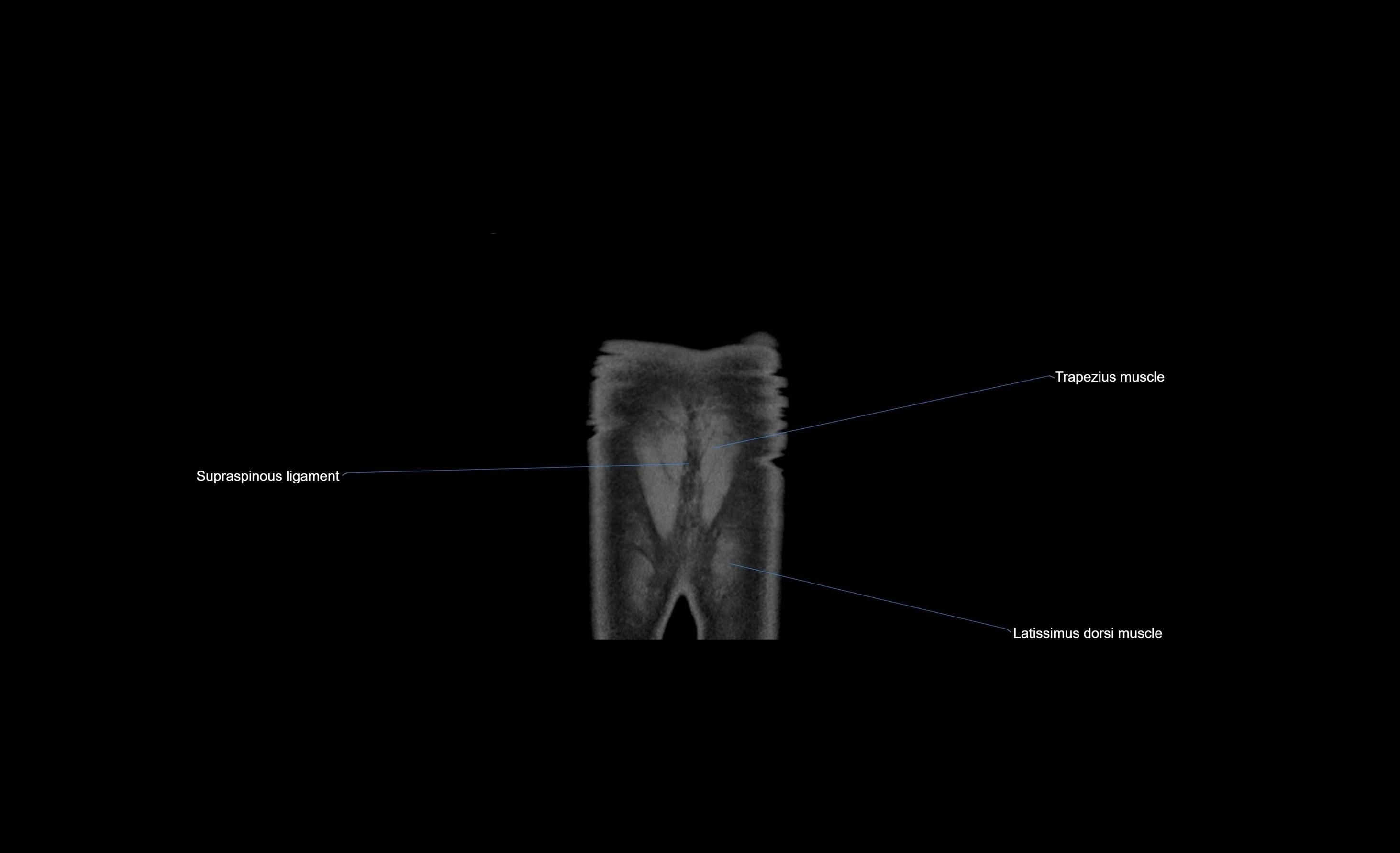
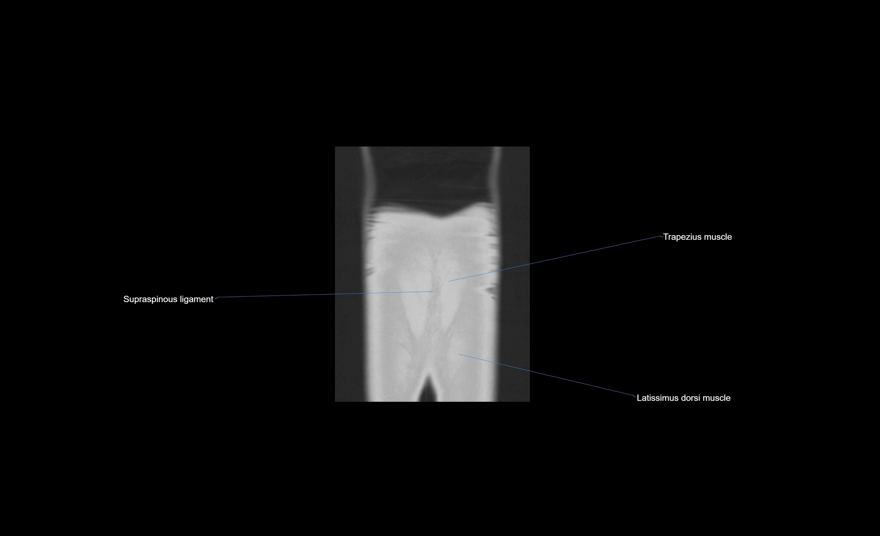
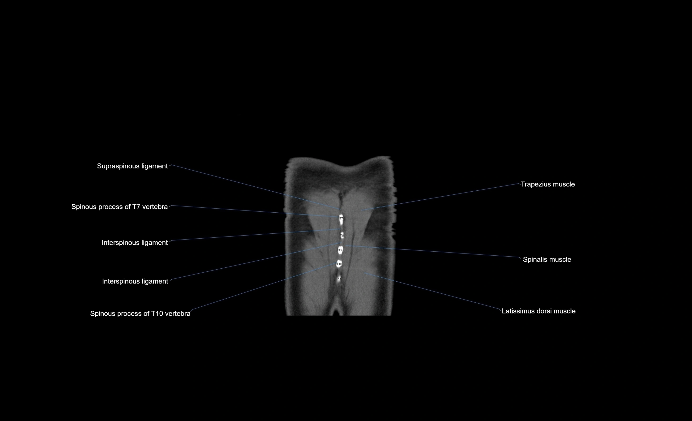
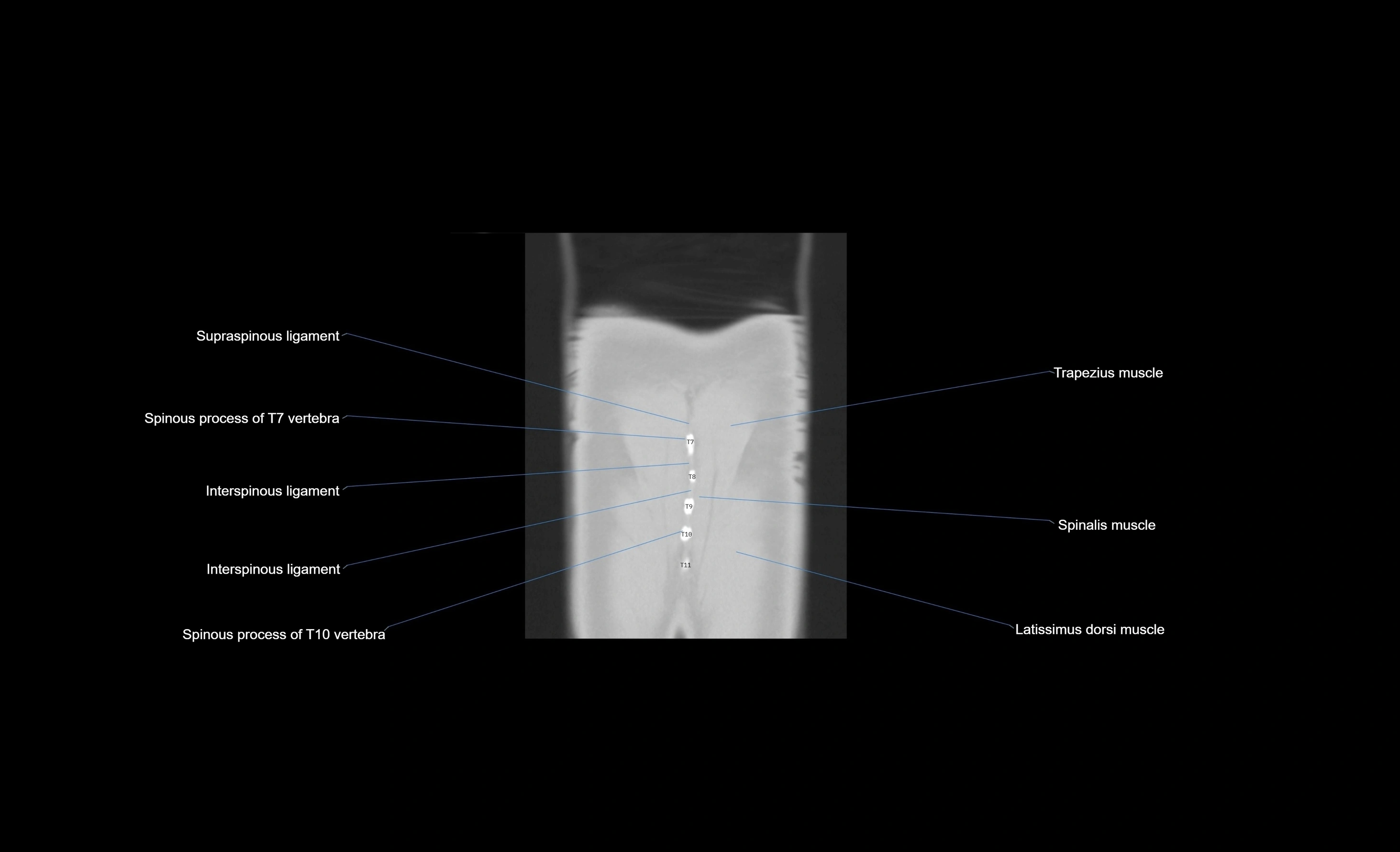
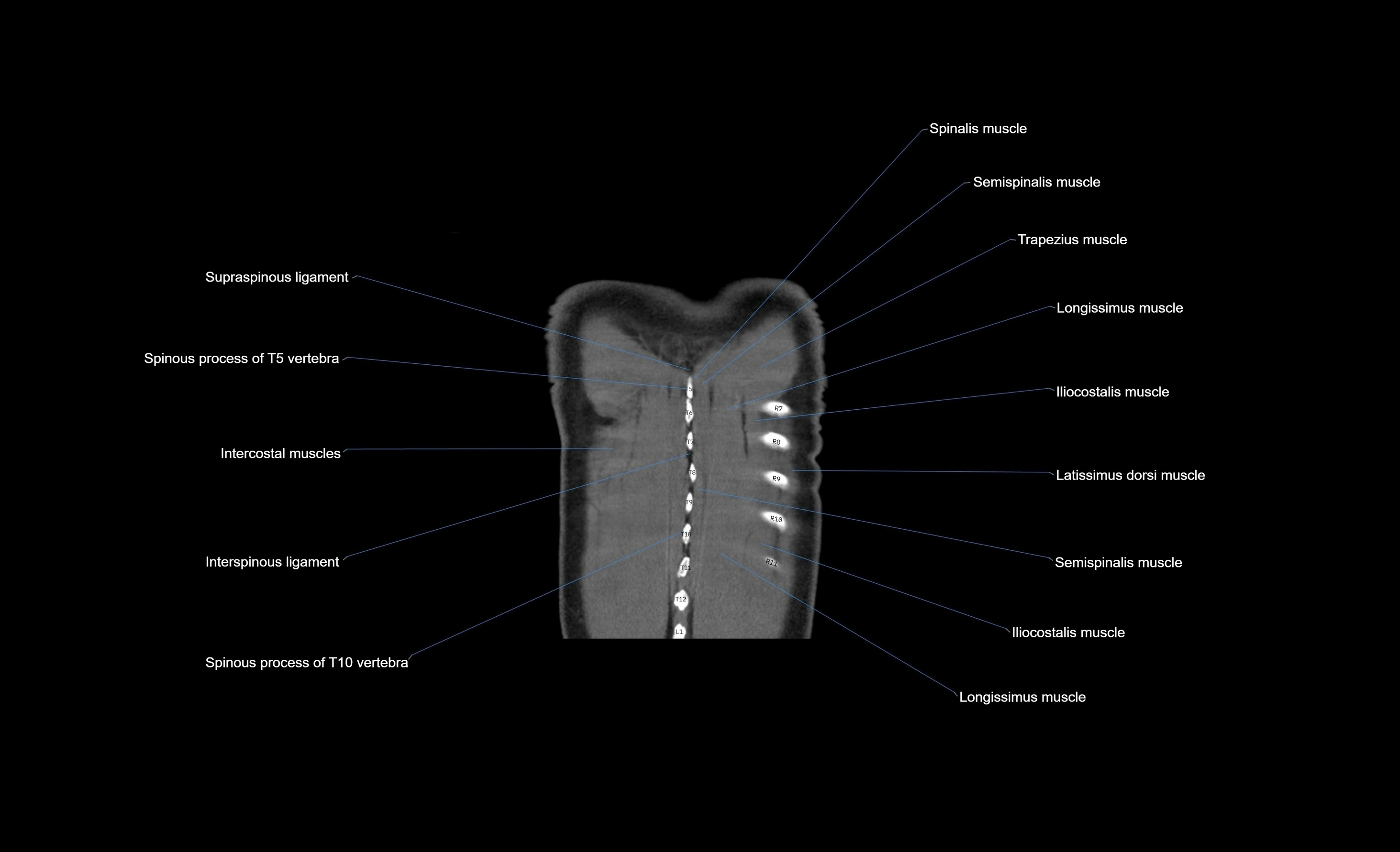
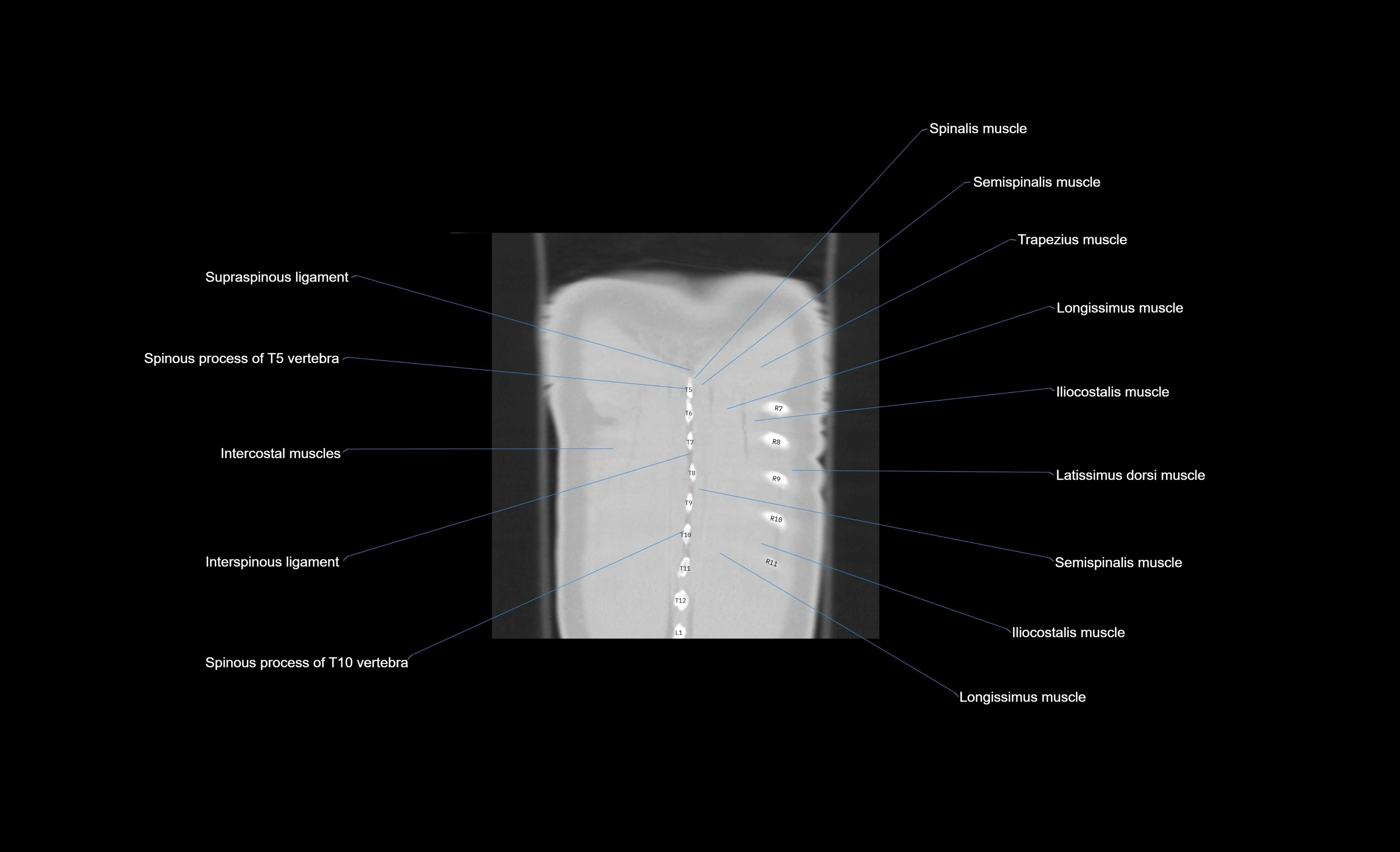
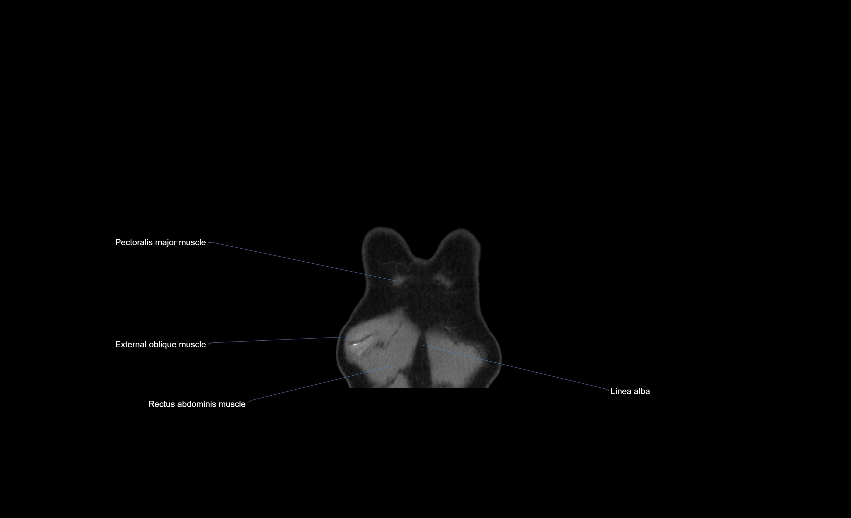
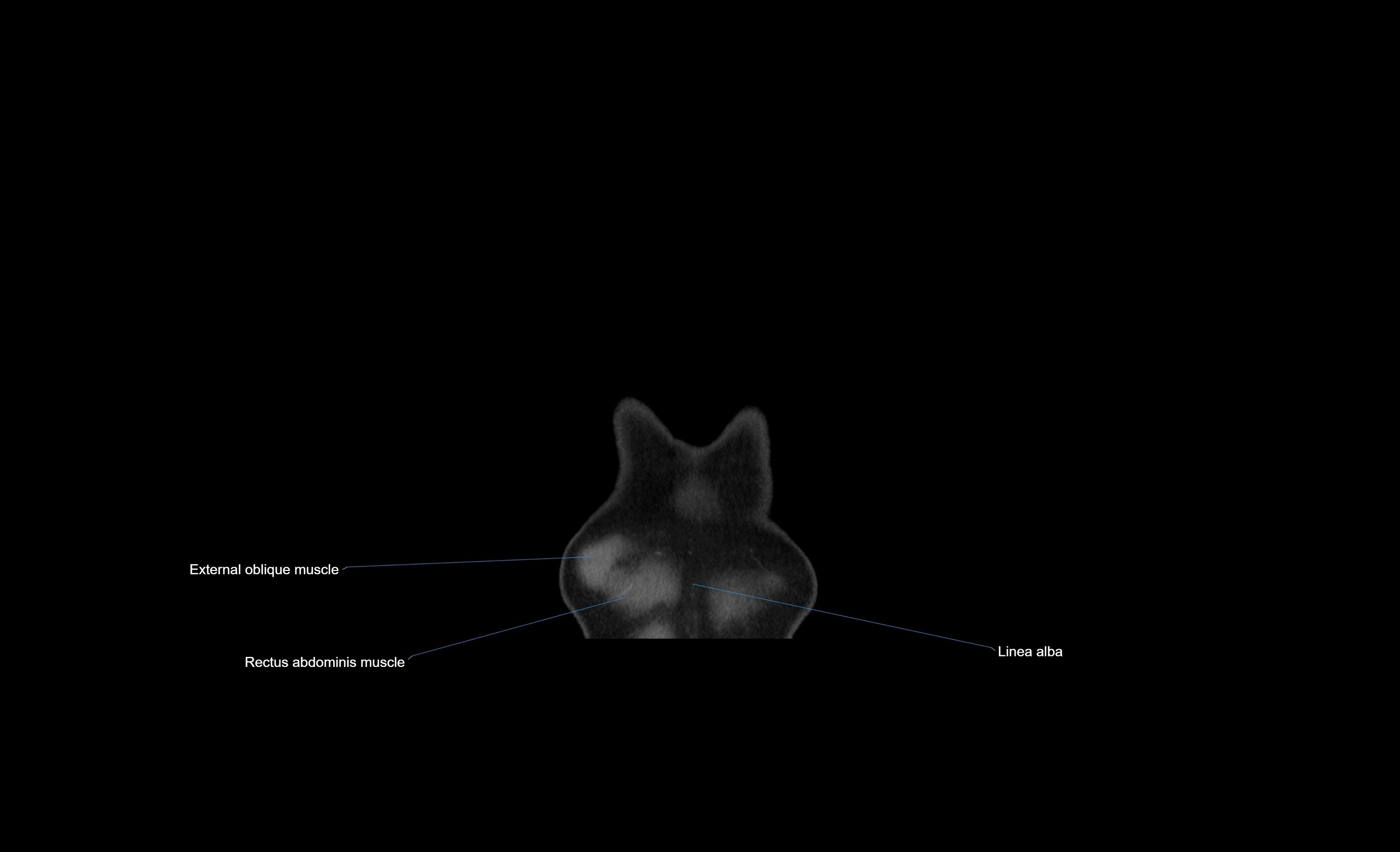
MRI images

MRI images

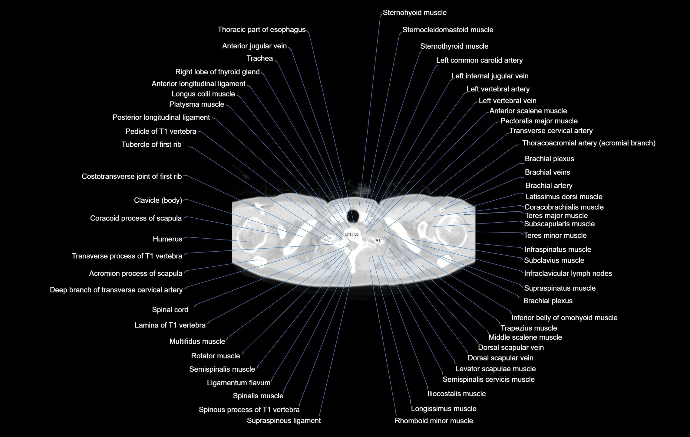
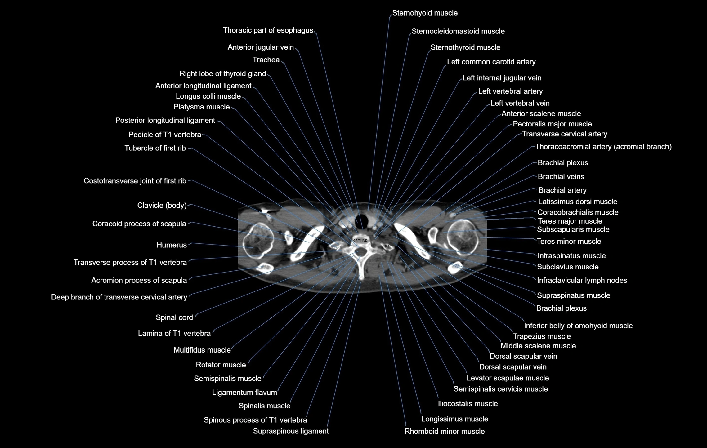
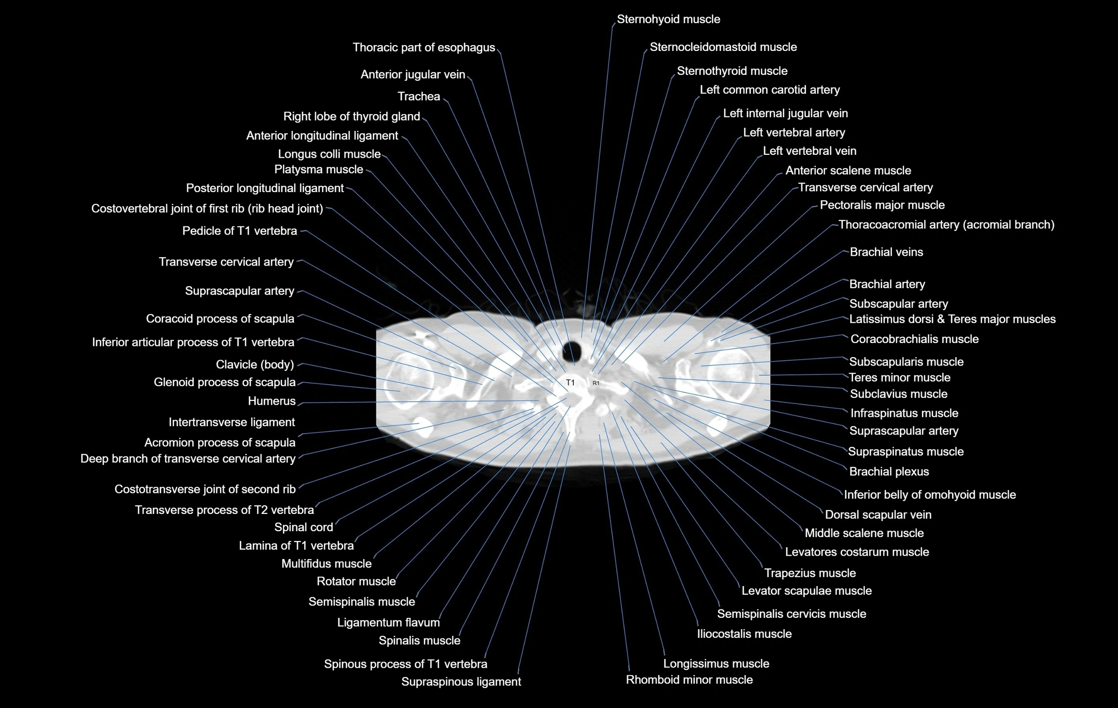
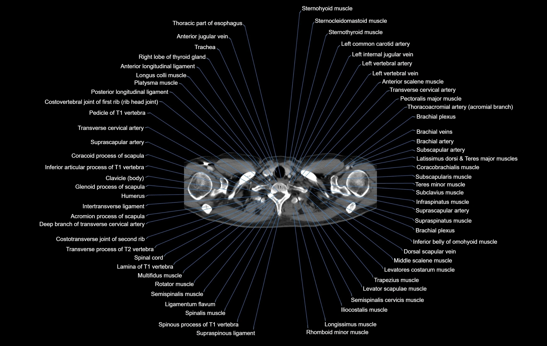
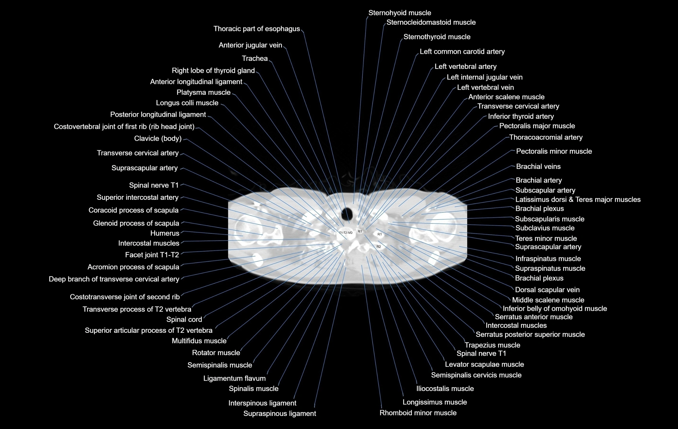
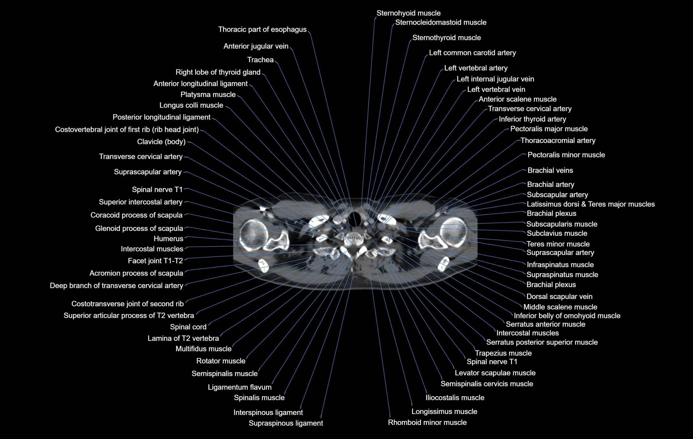
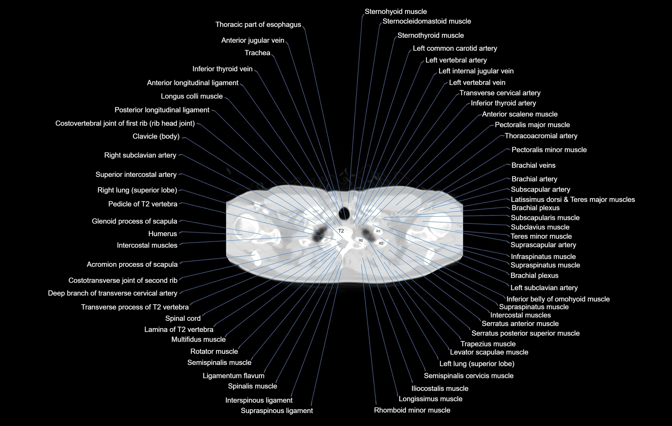
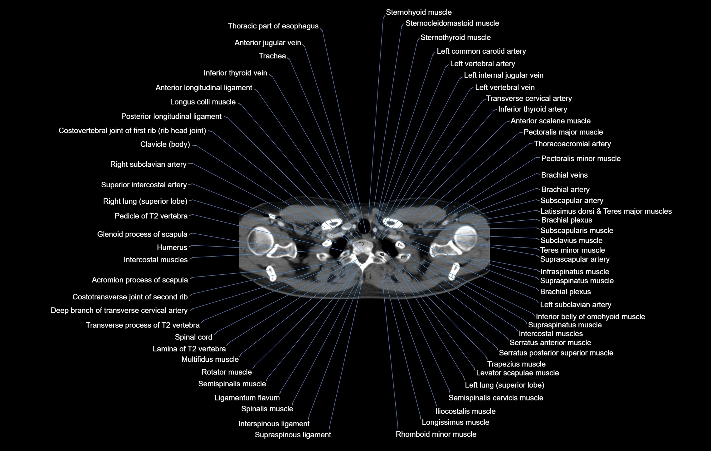
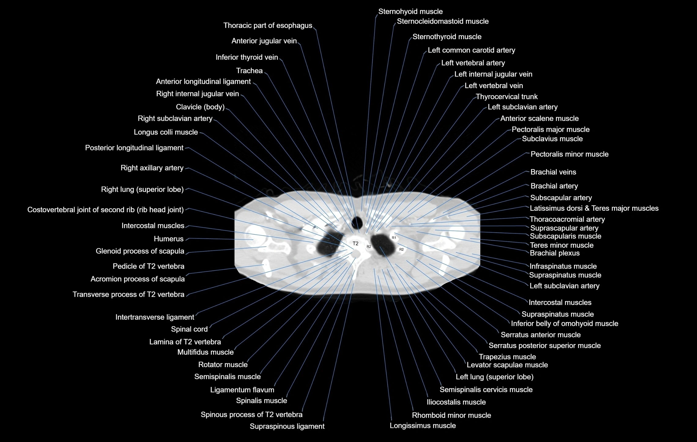
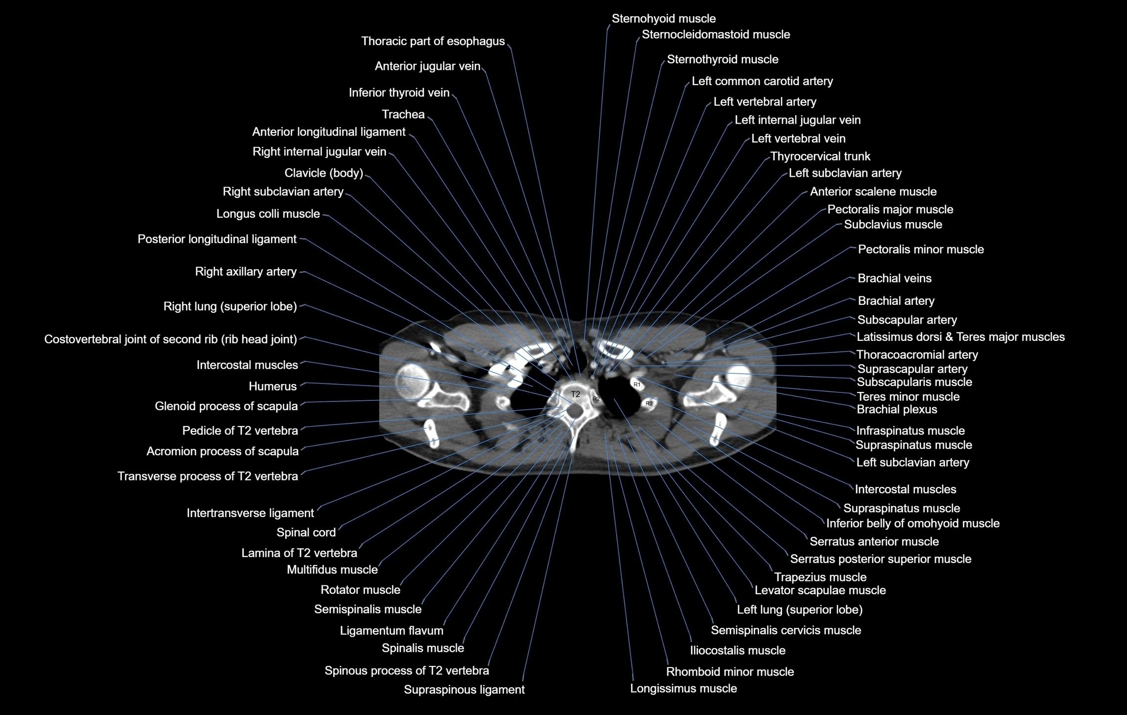
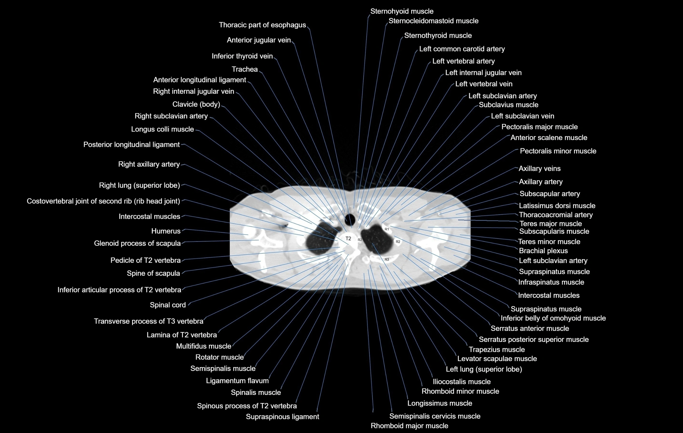
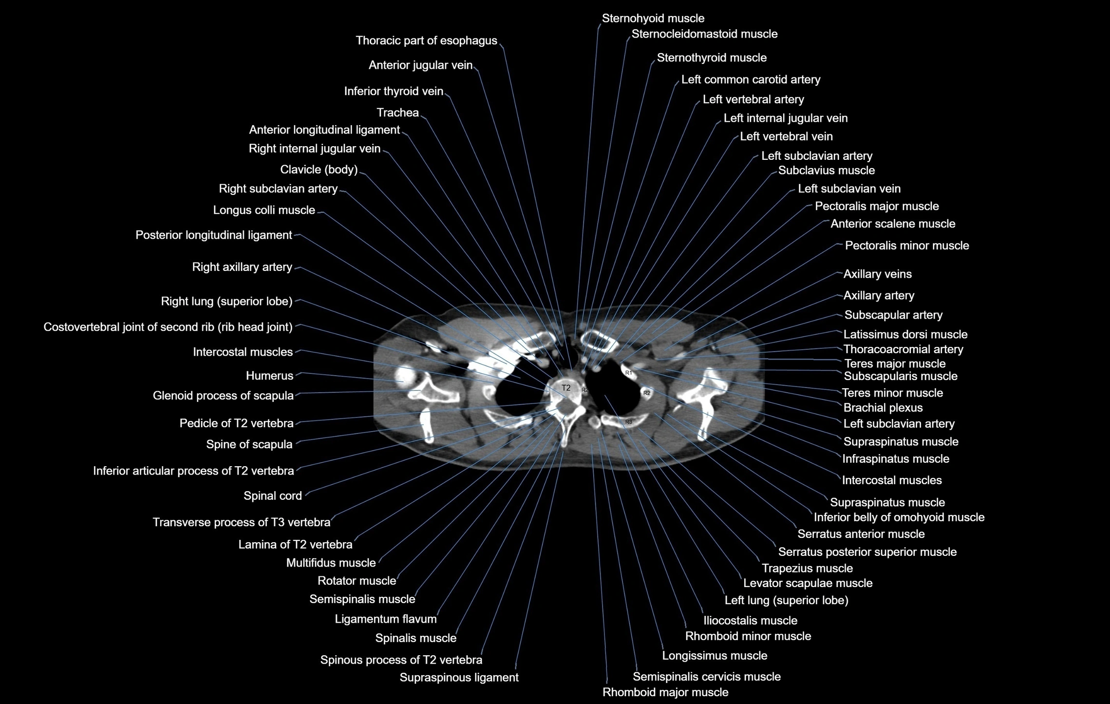
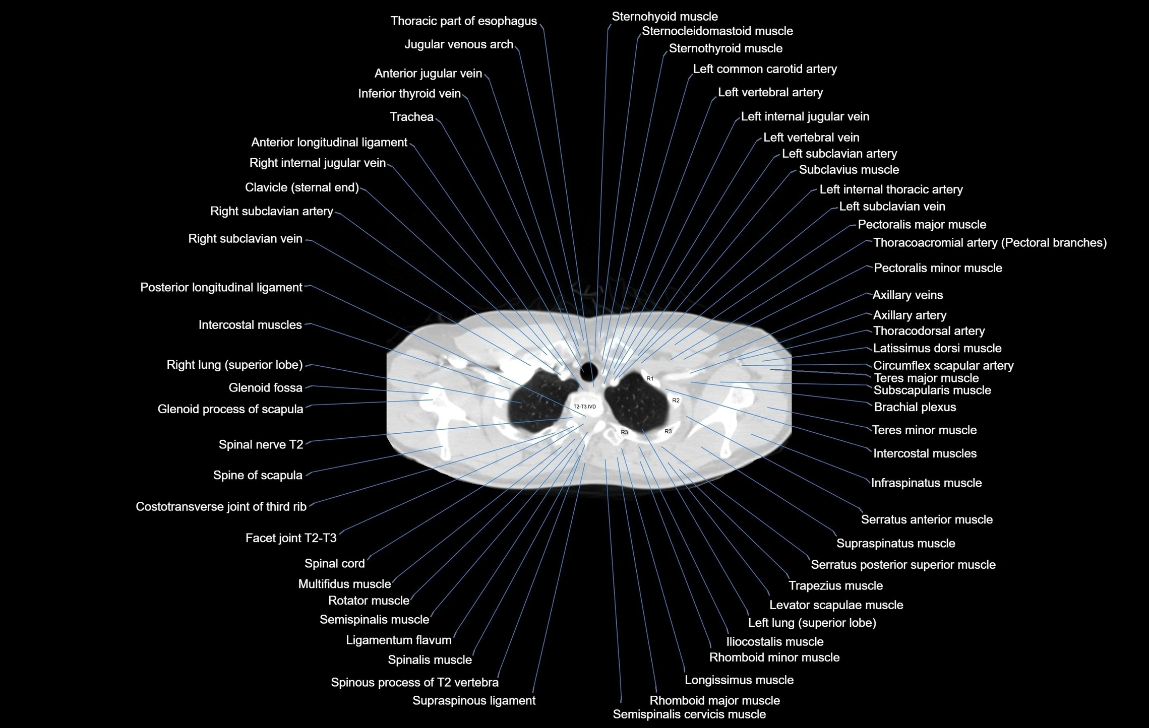
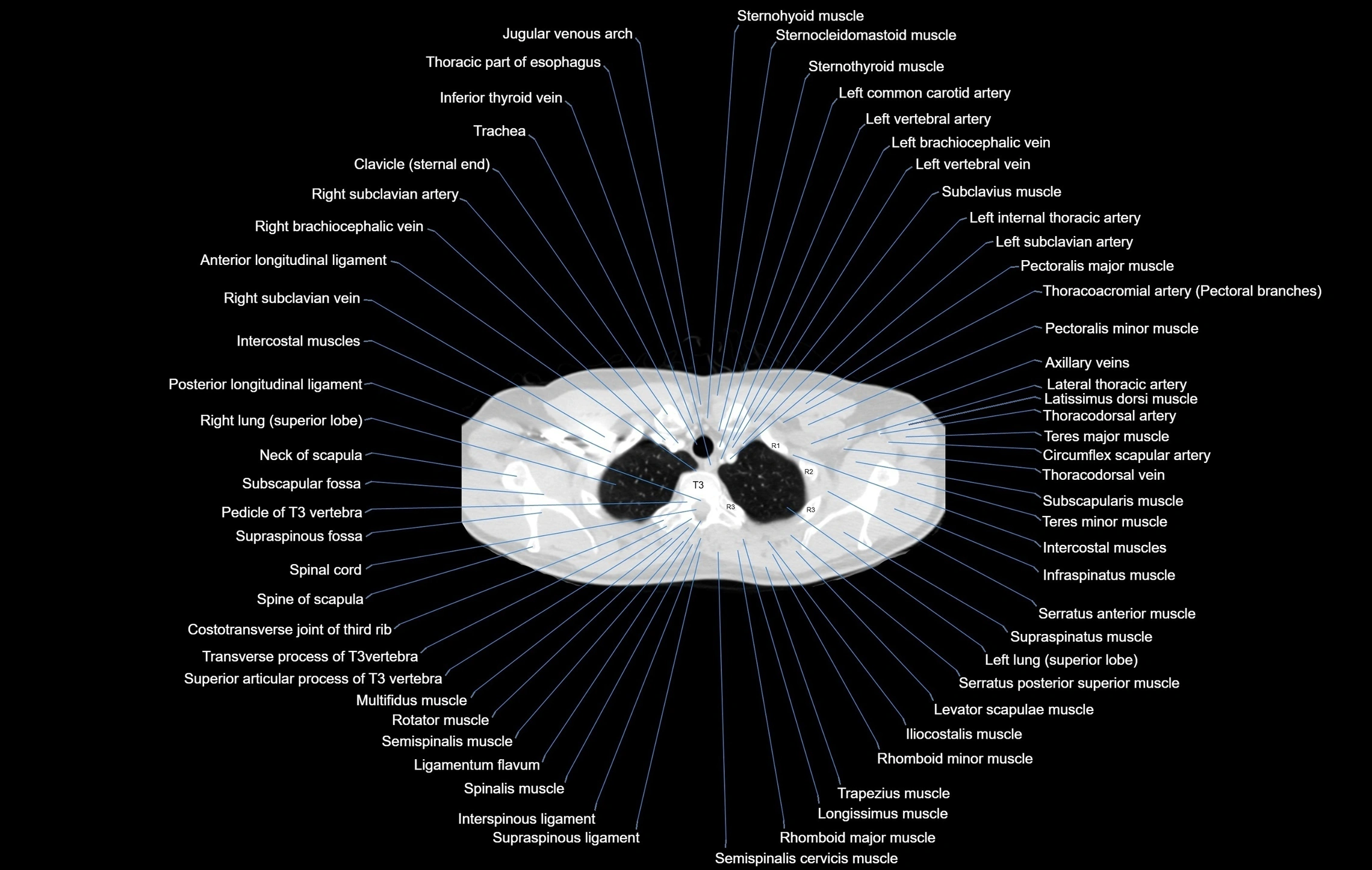
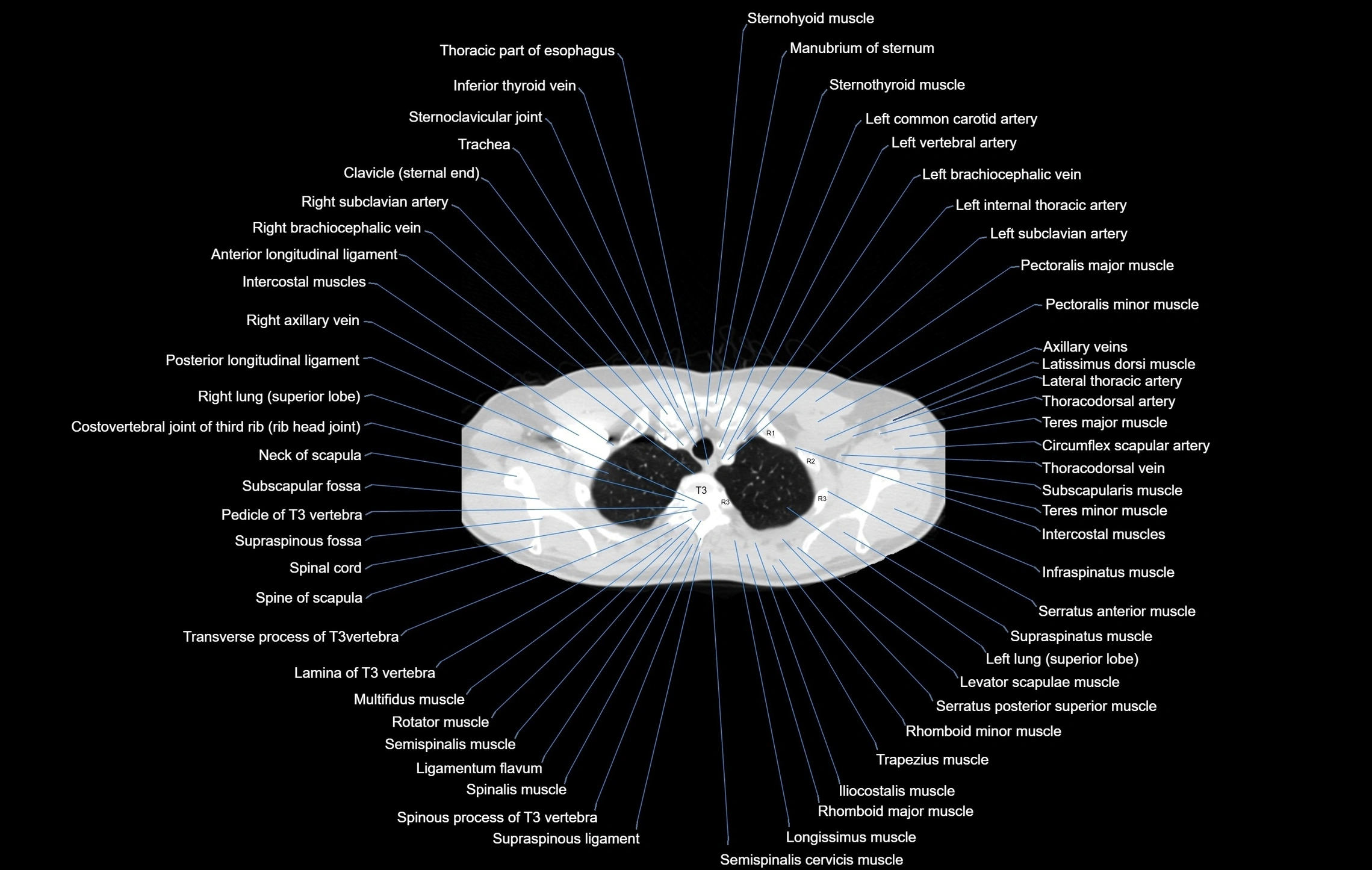
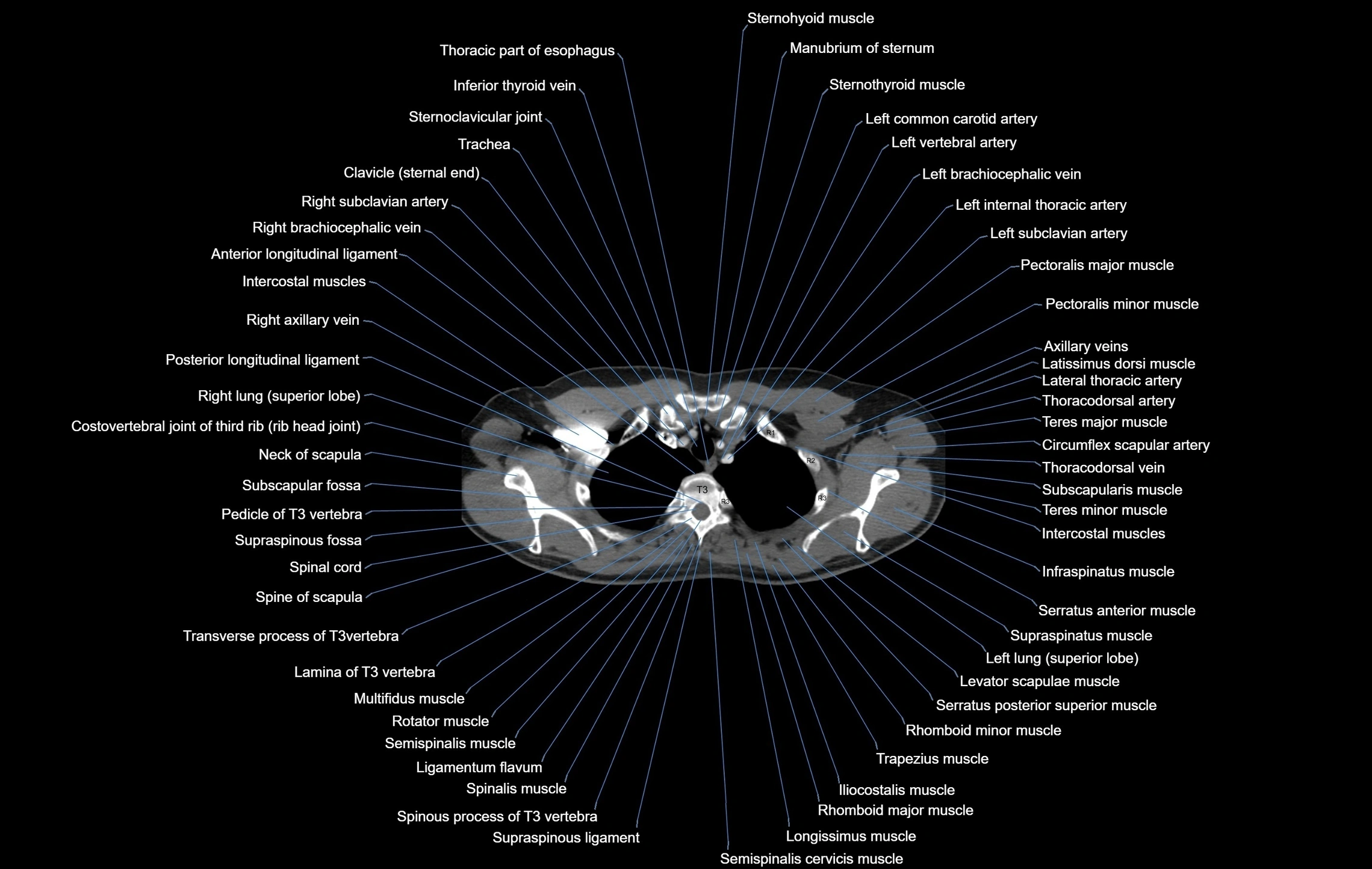
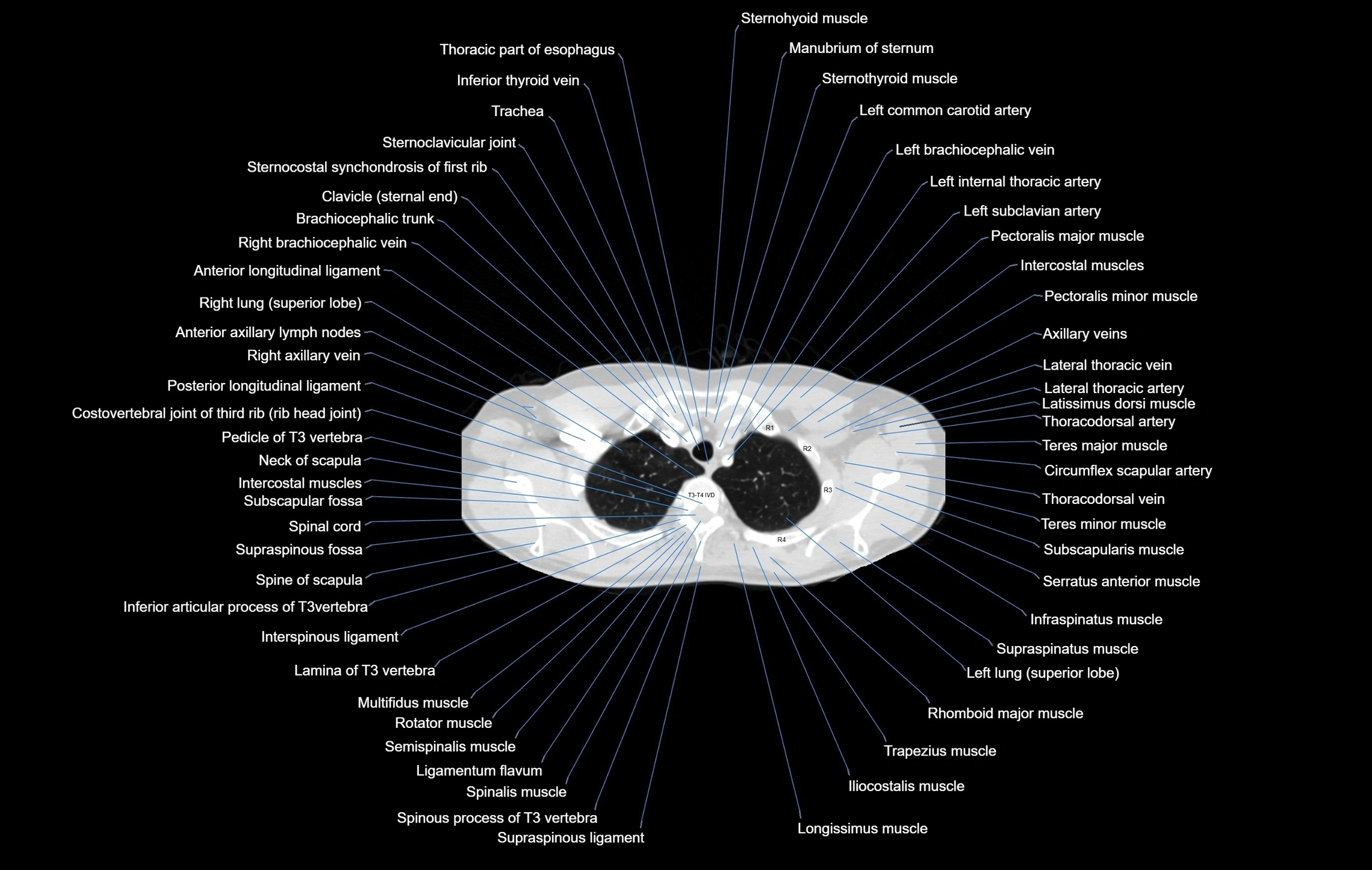
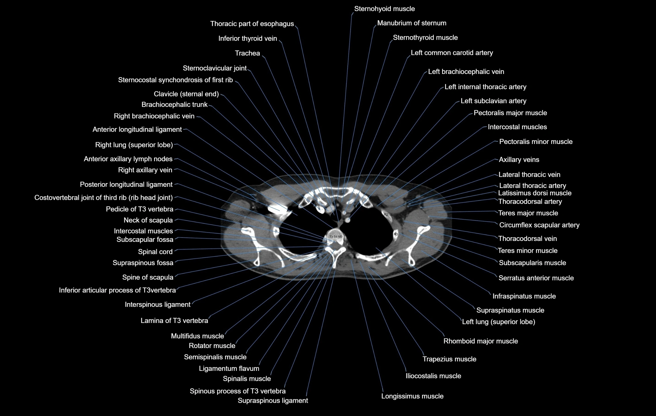
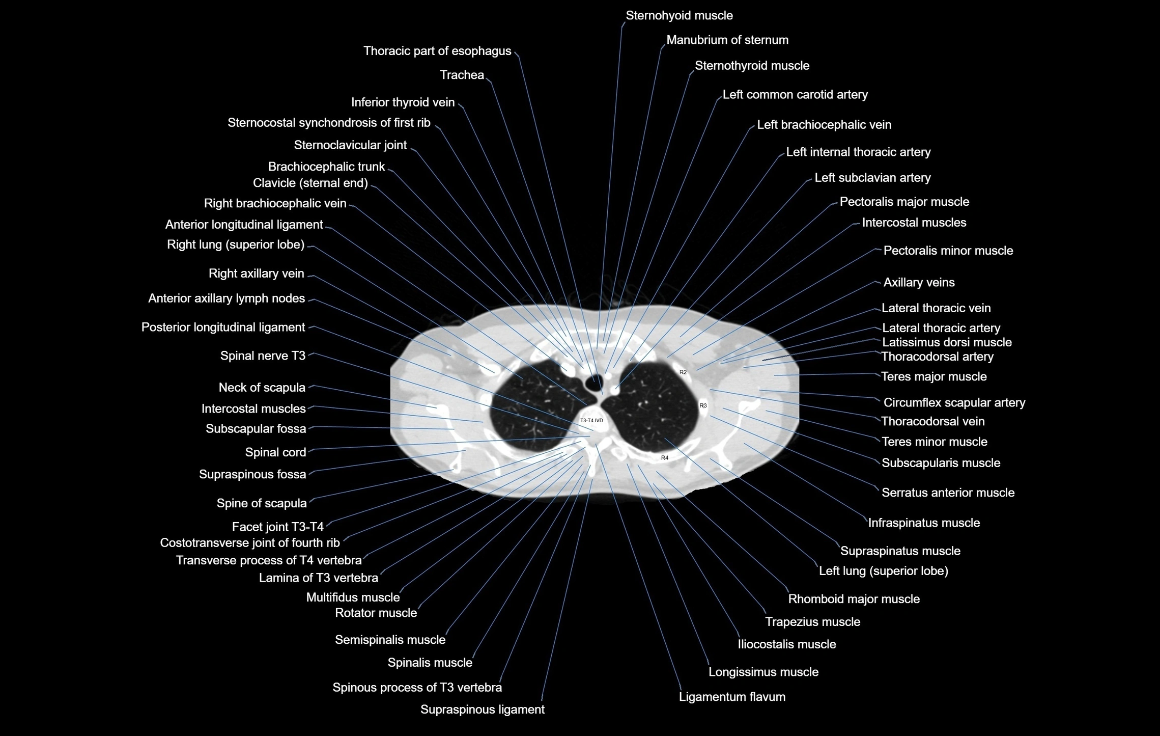
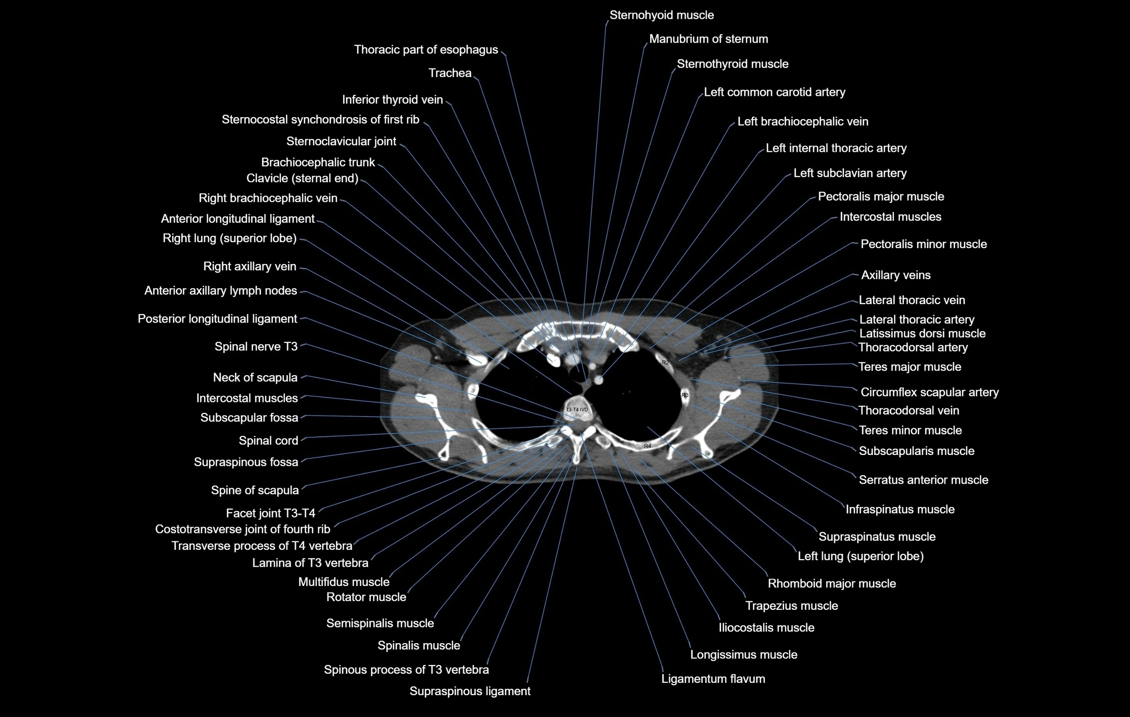
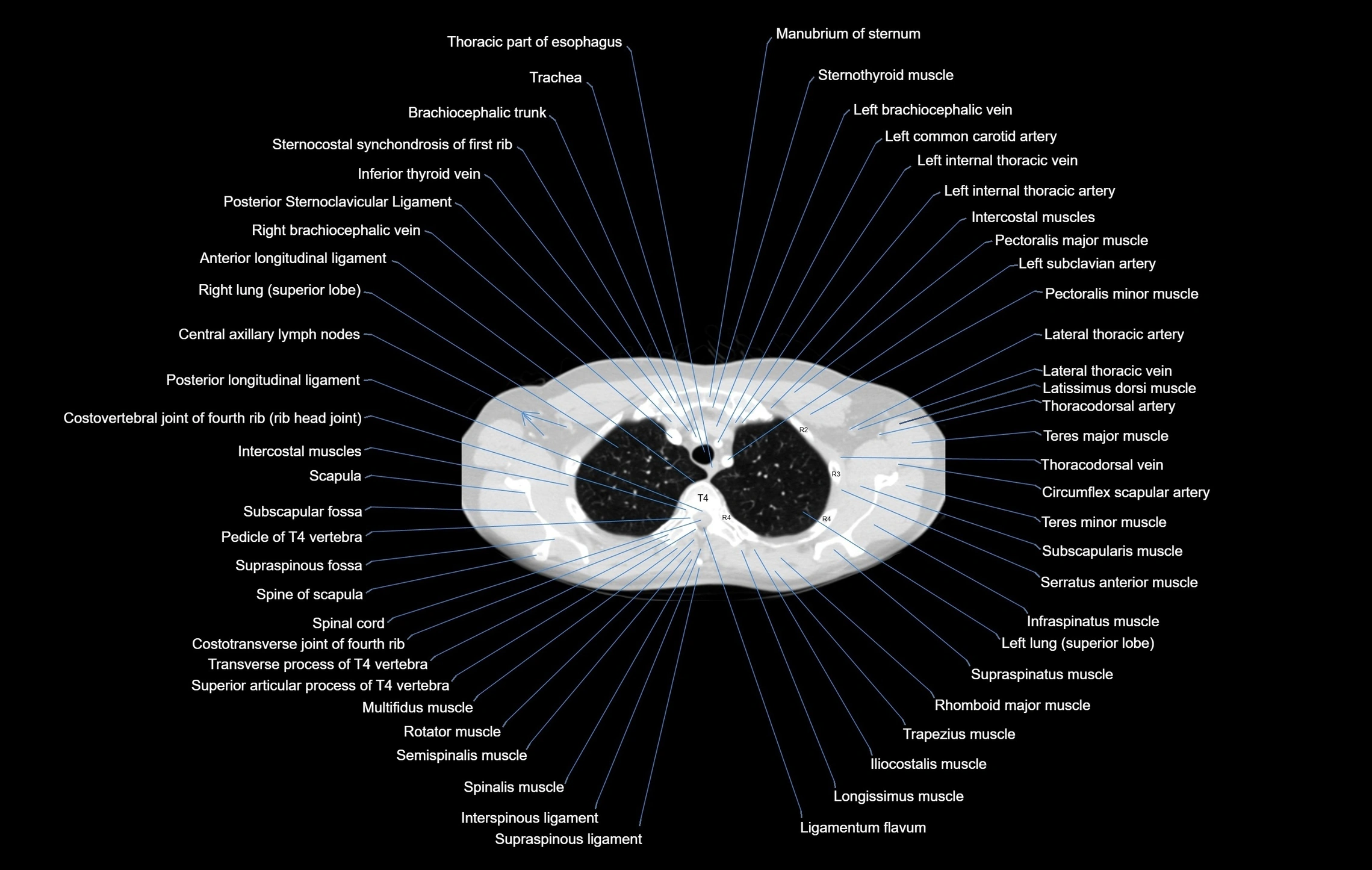
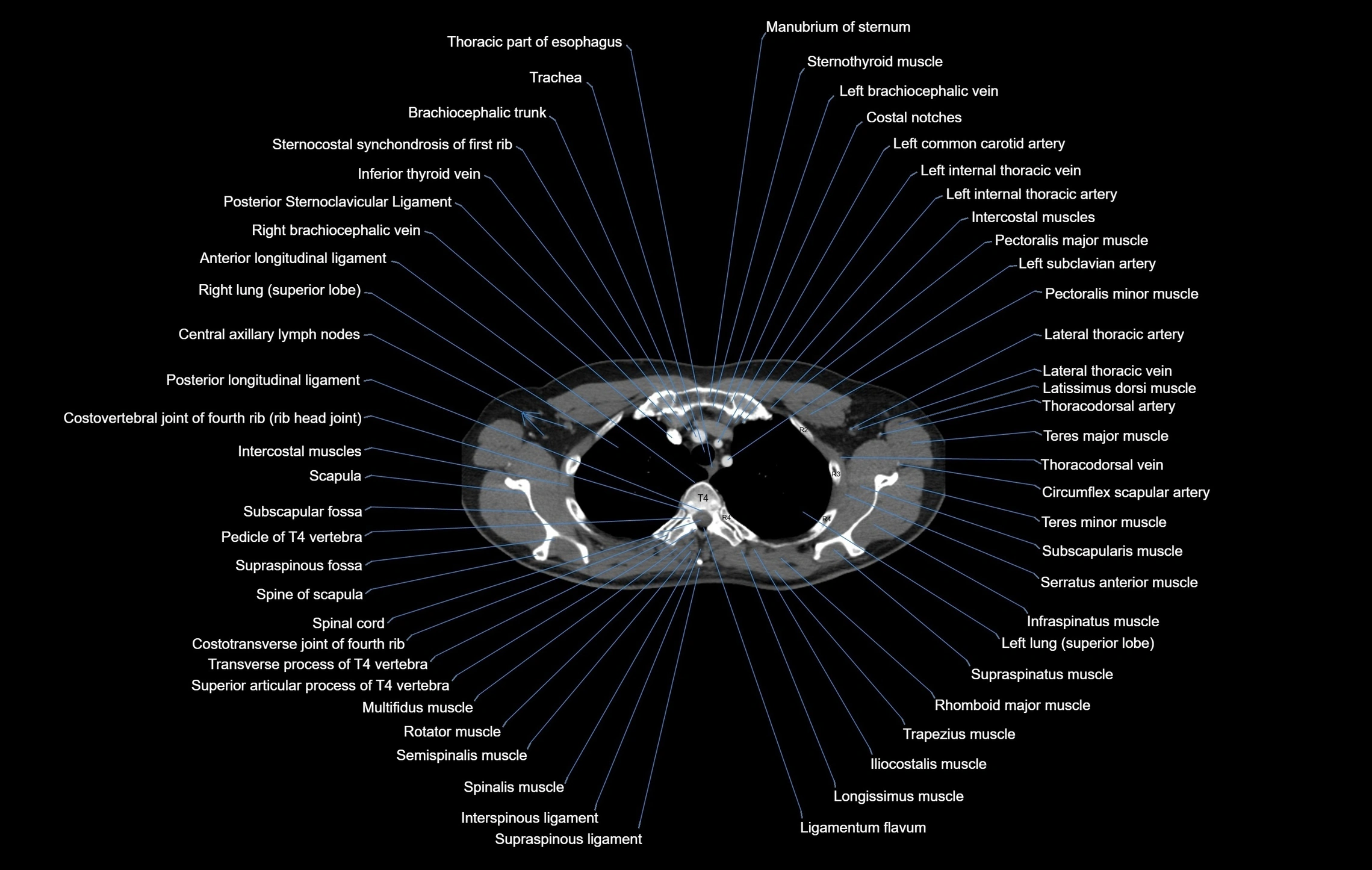
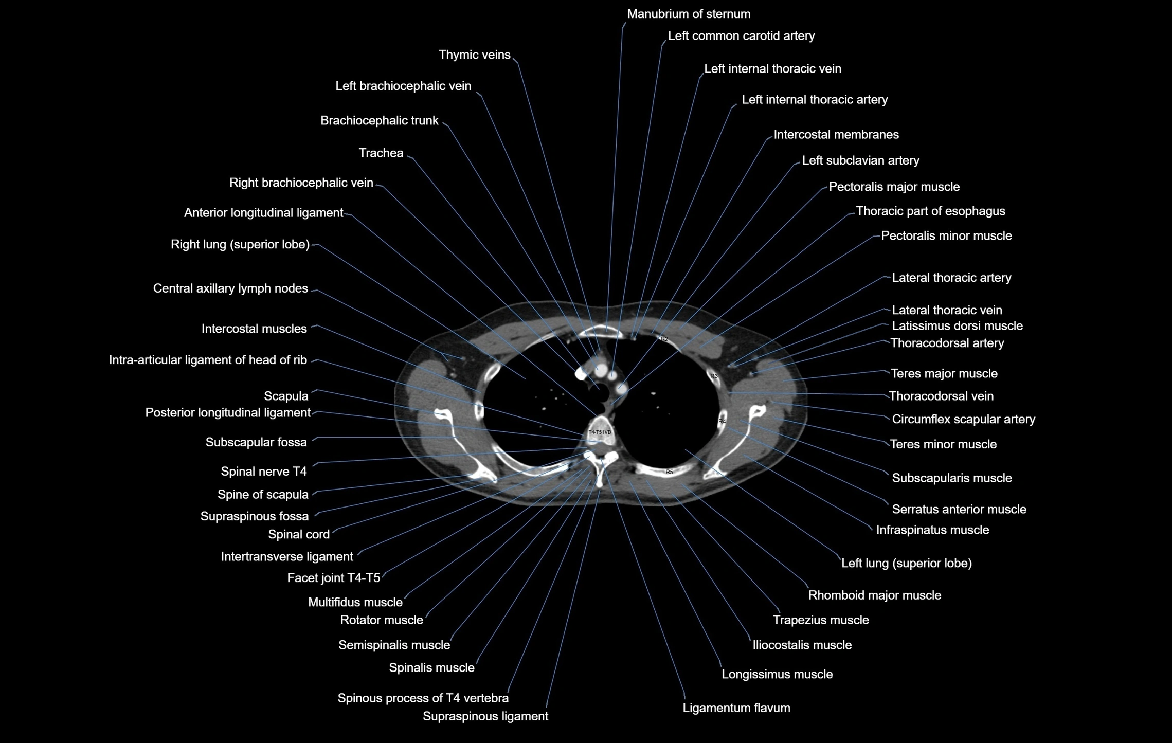
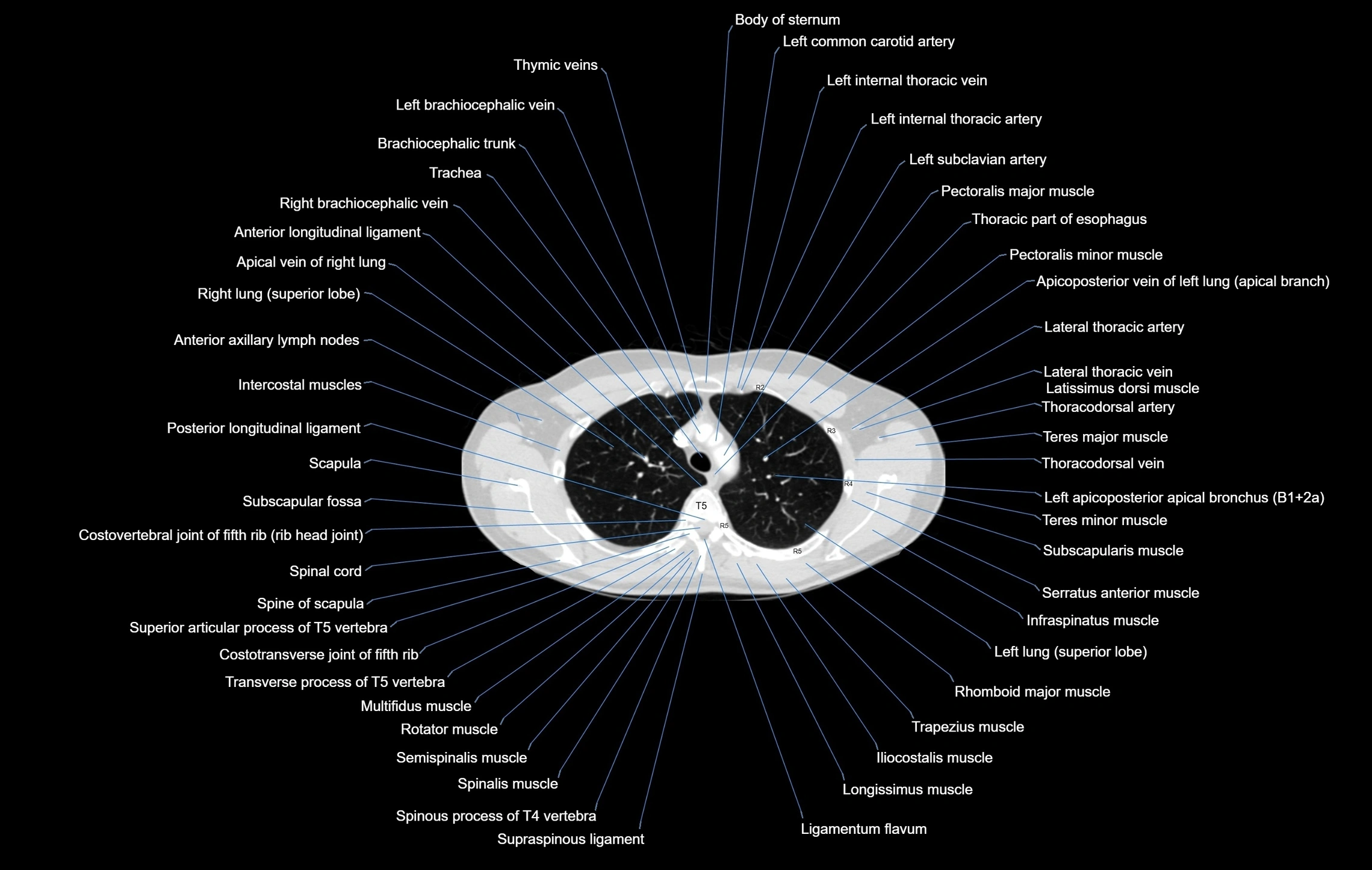
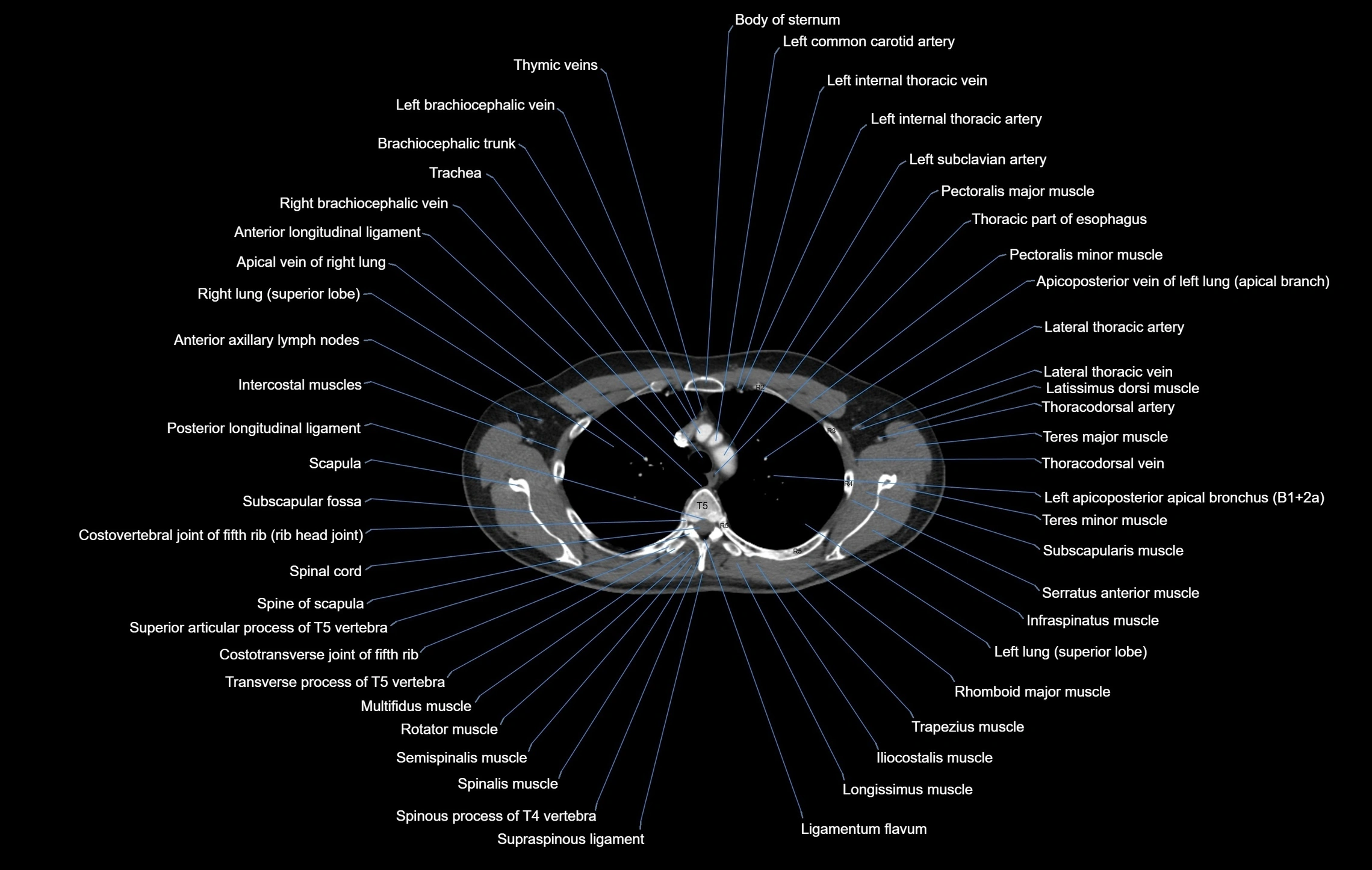
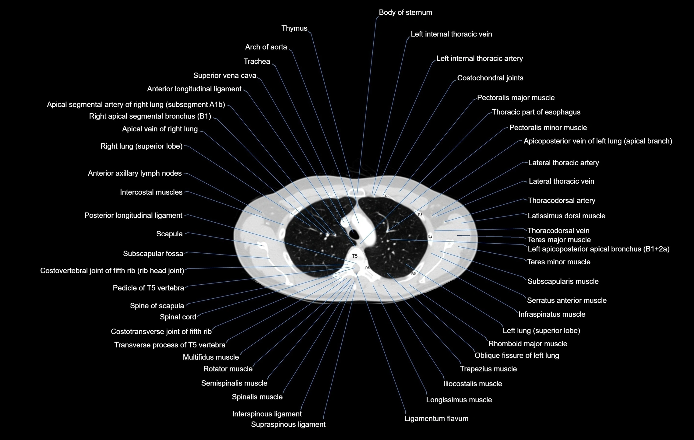
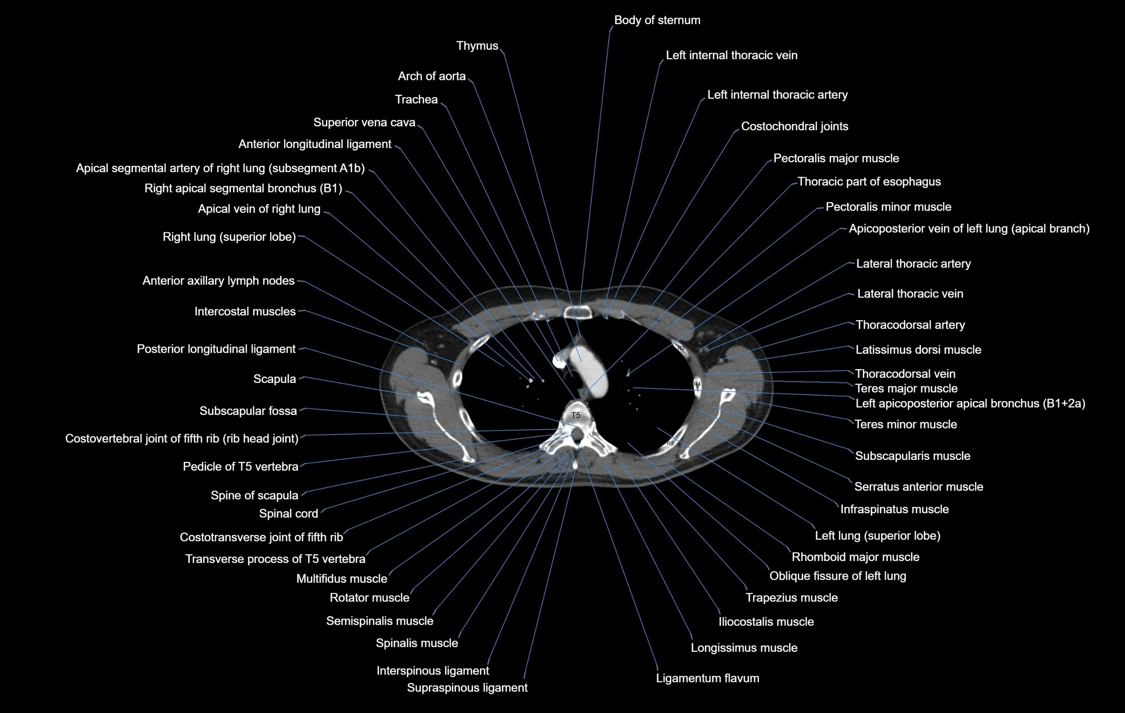
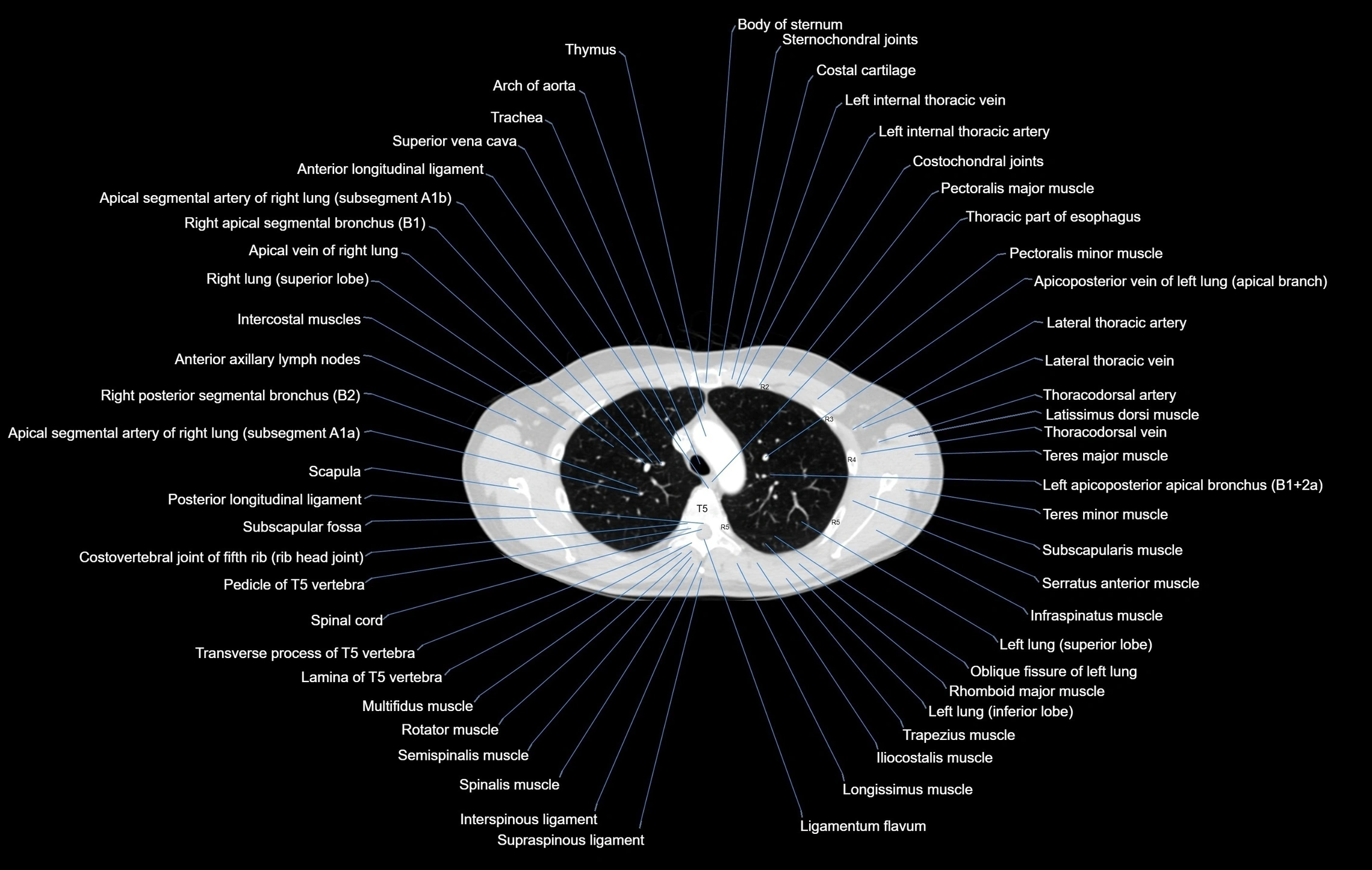
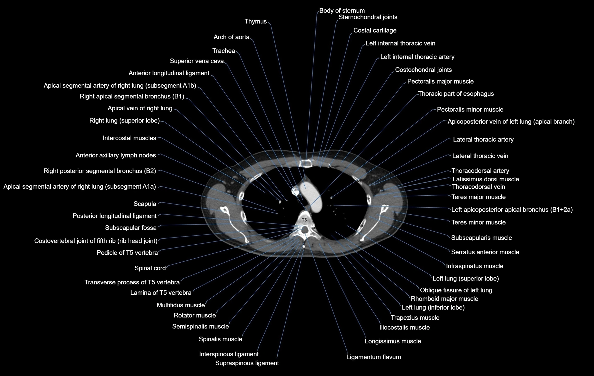
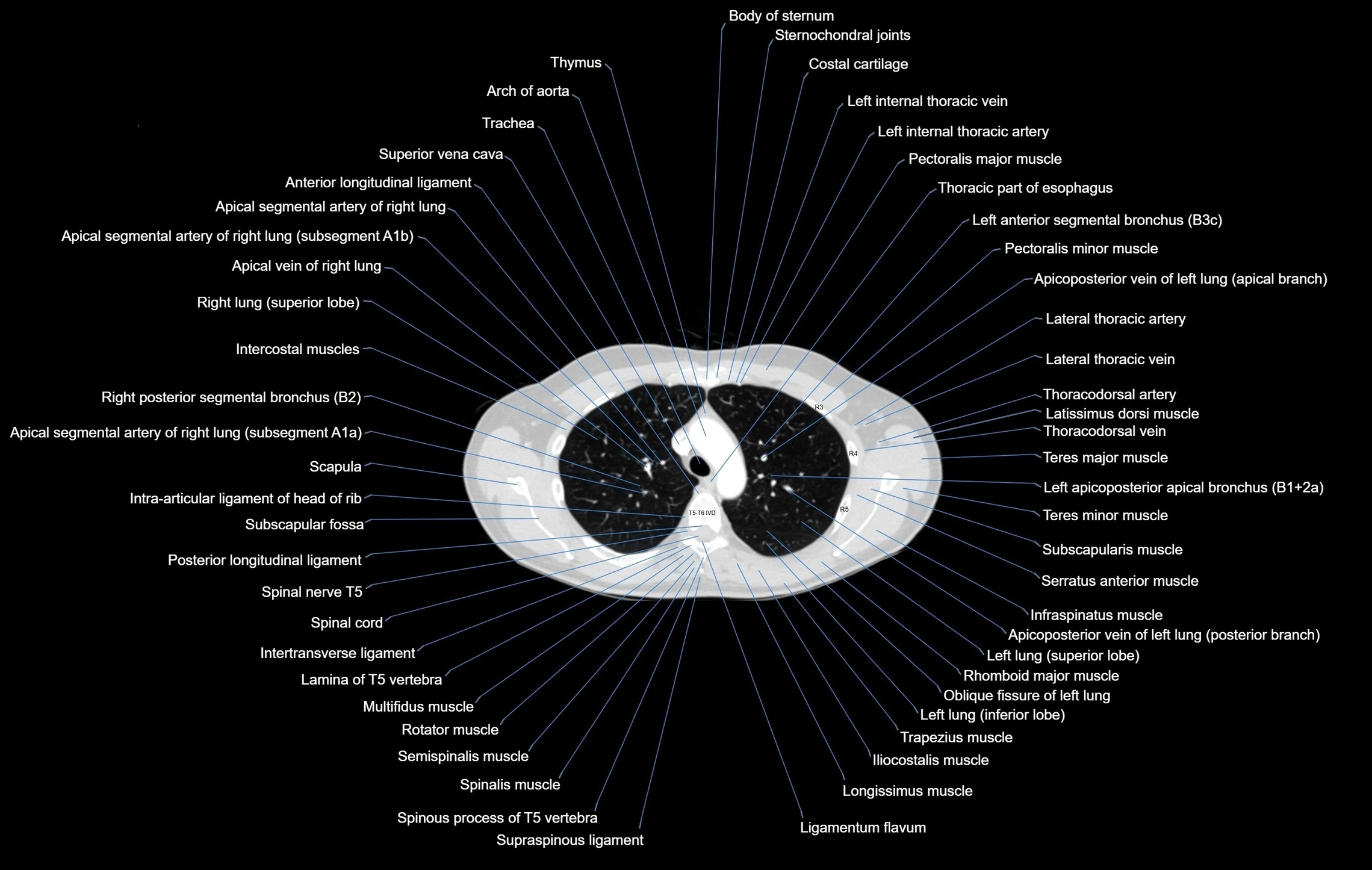
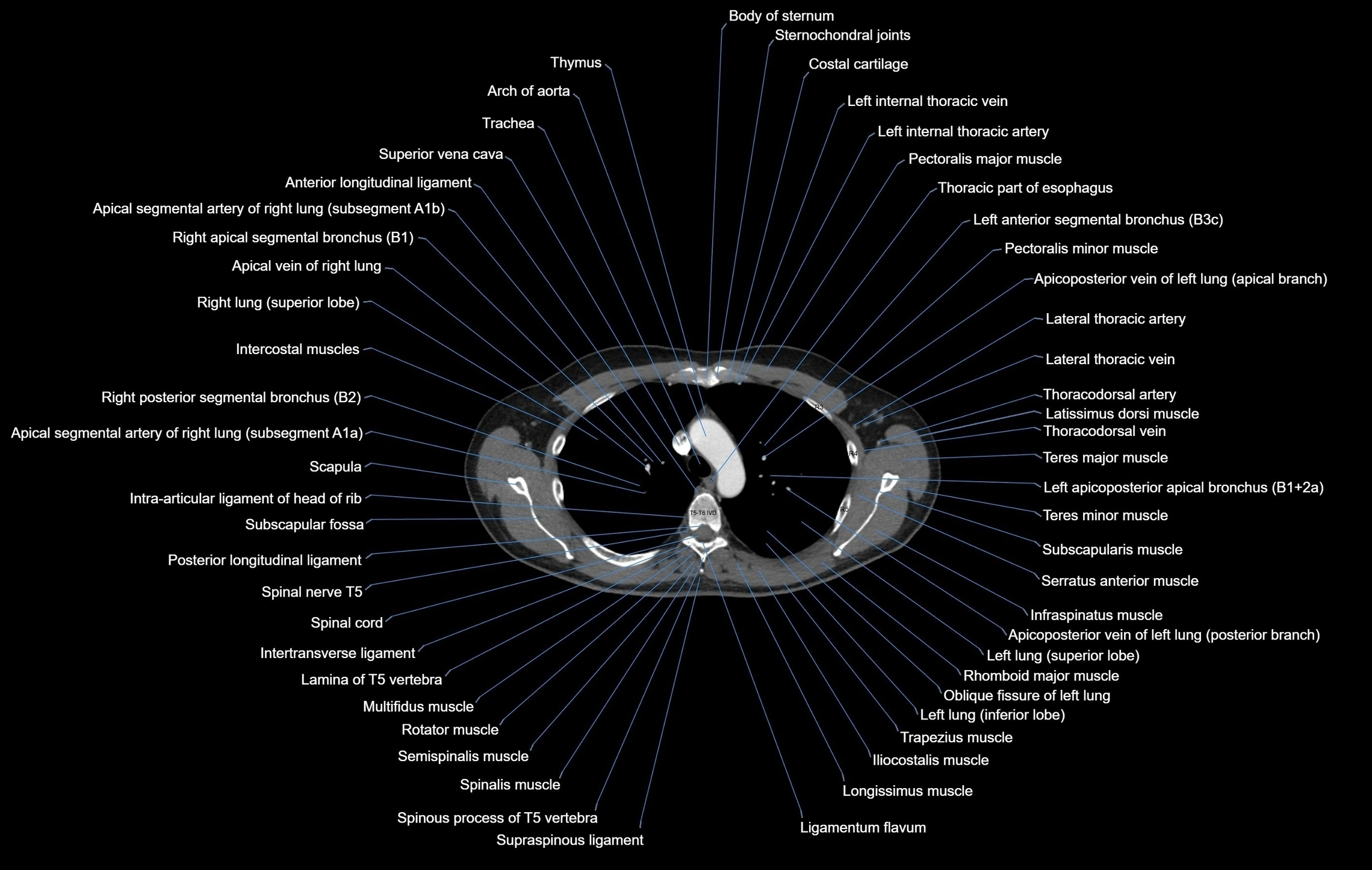
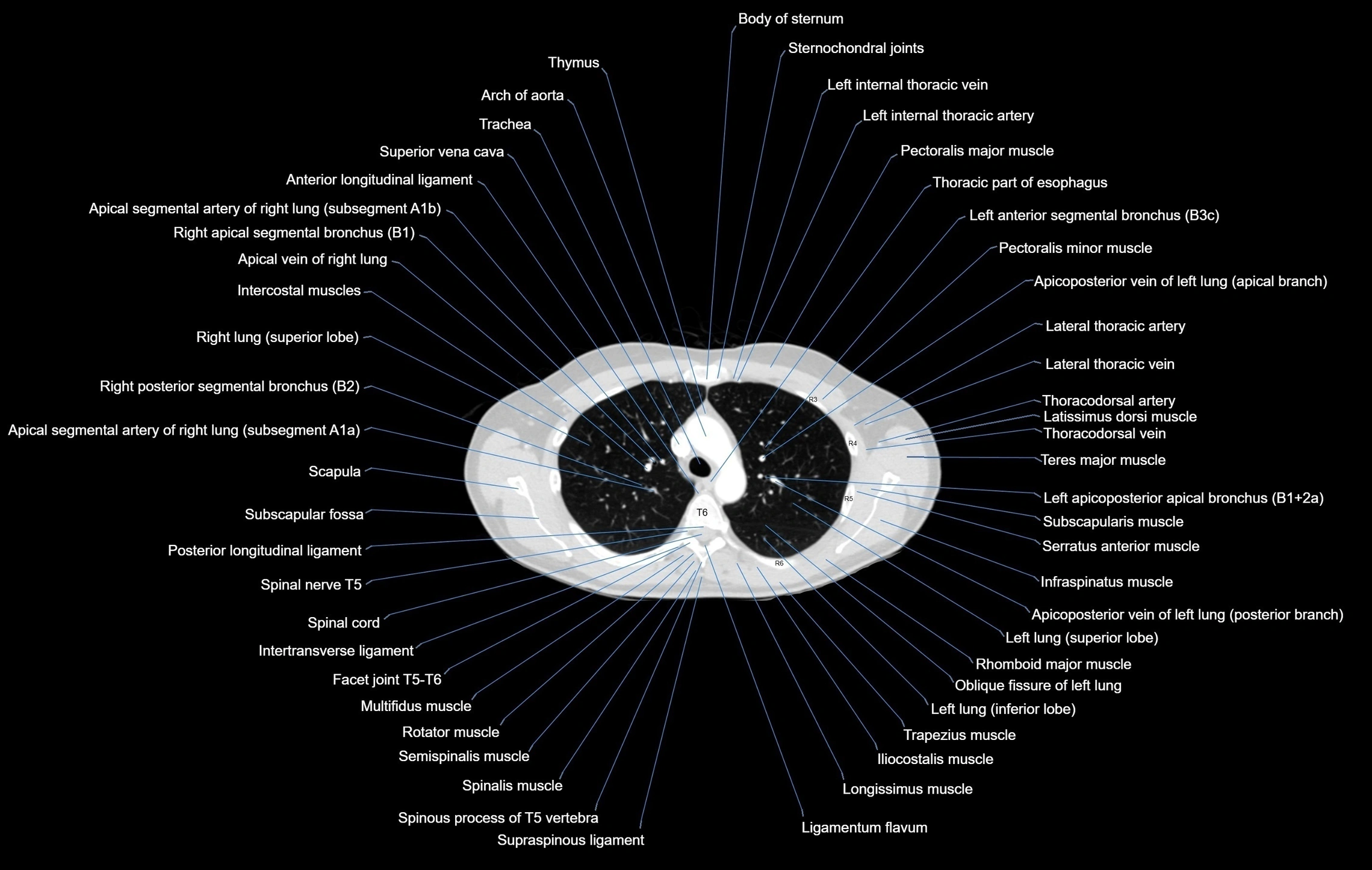
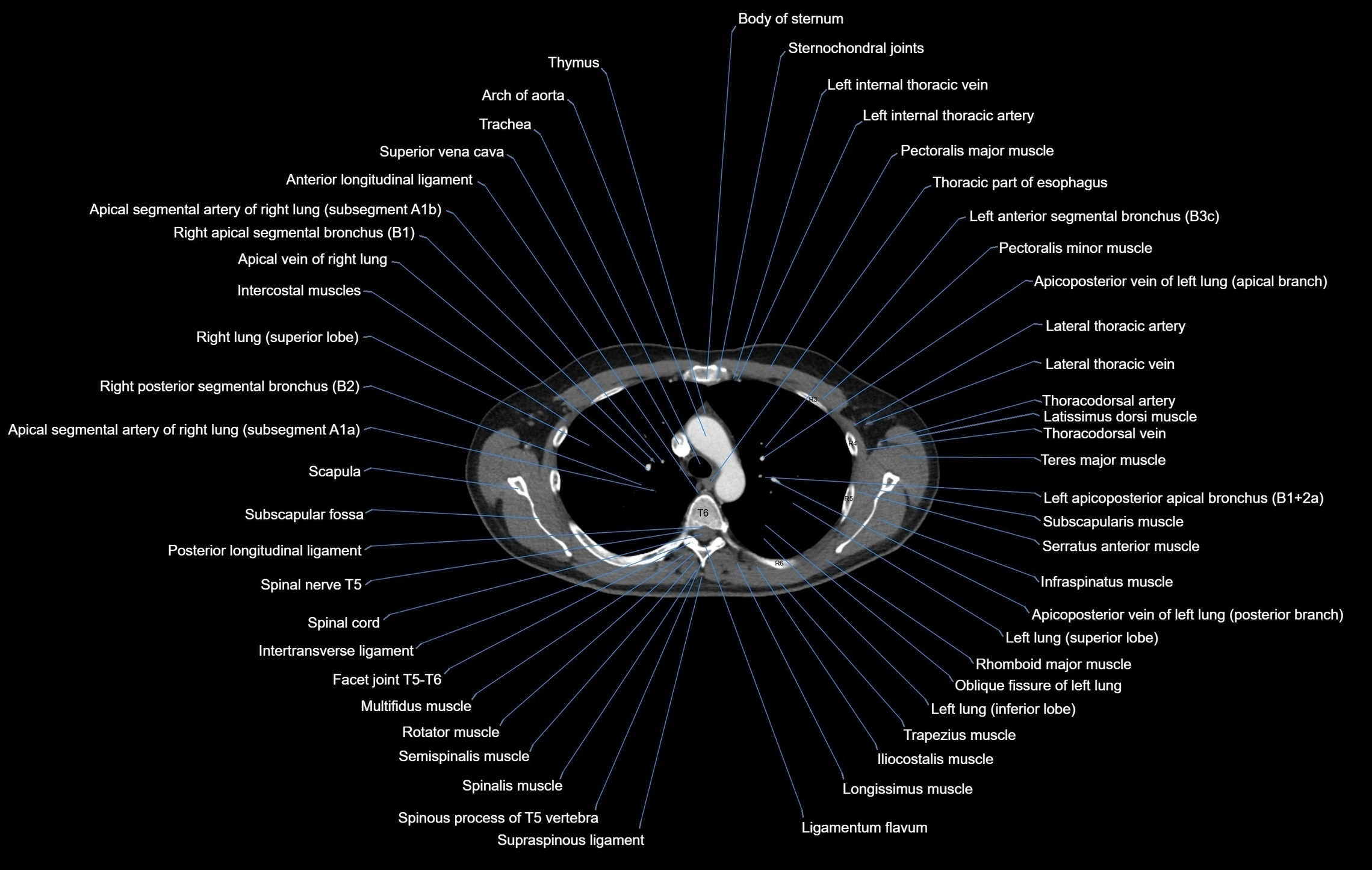
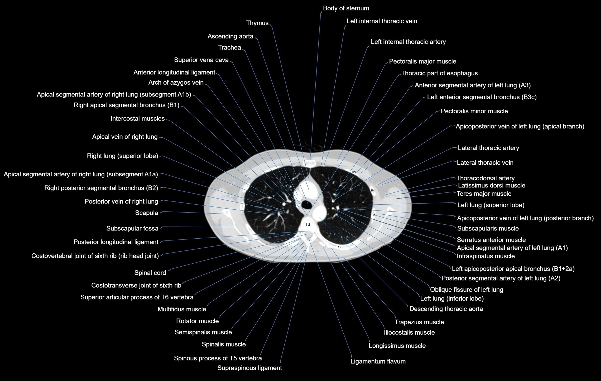
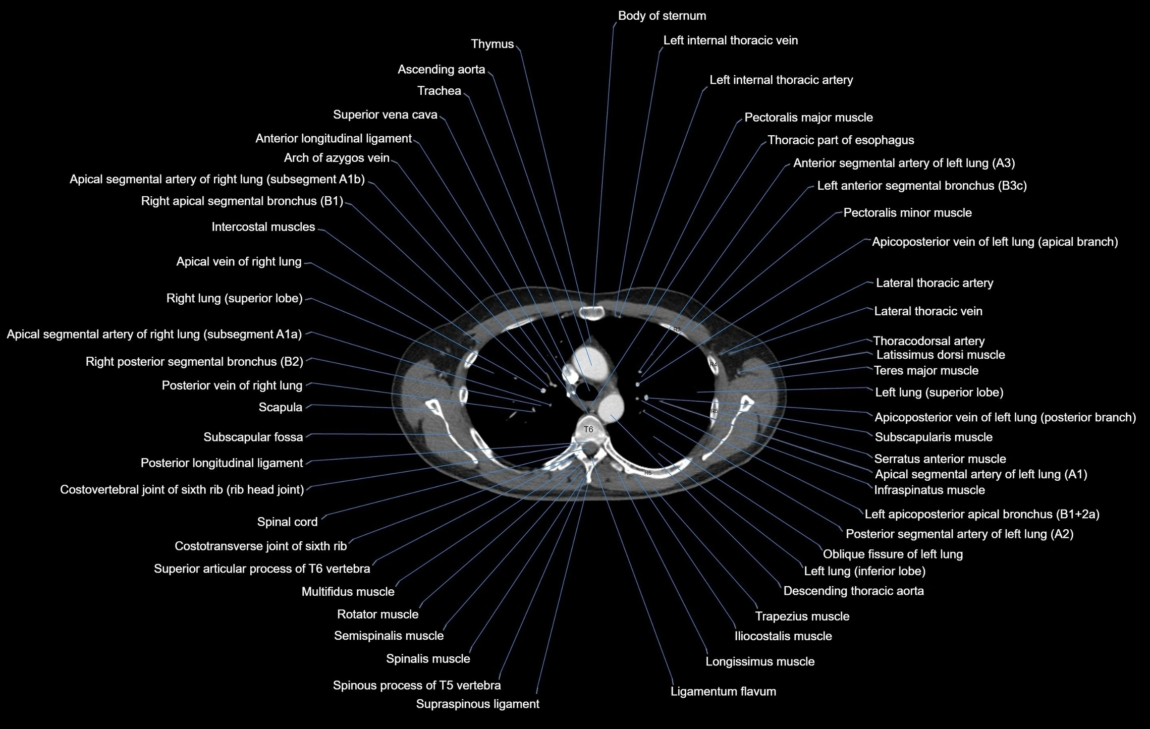
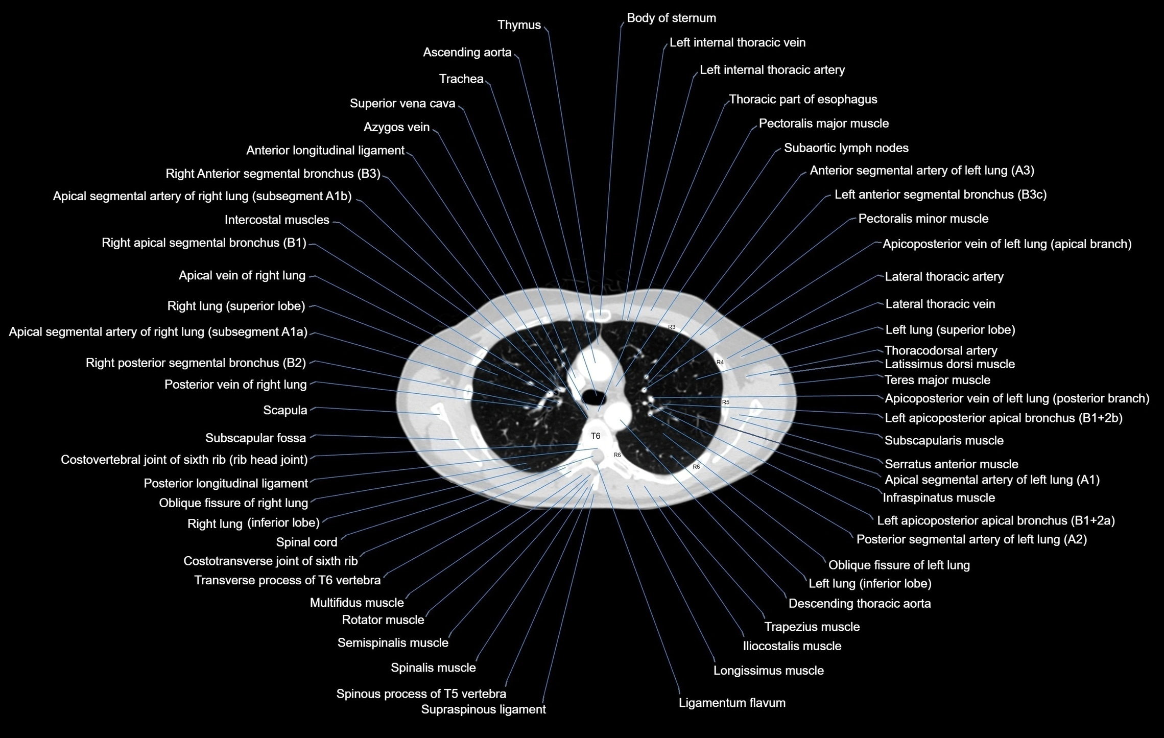
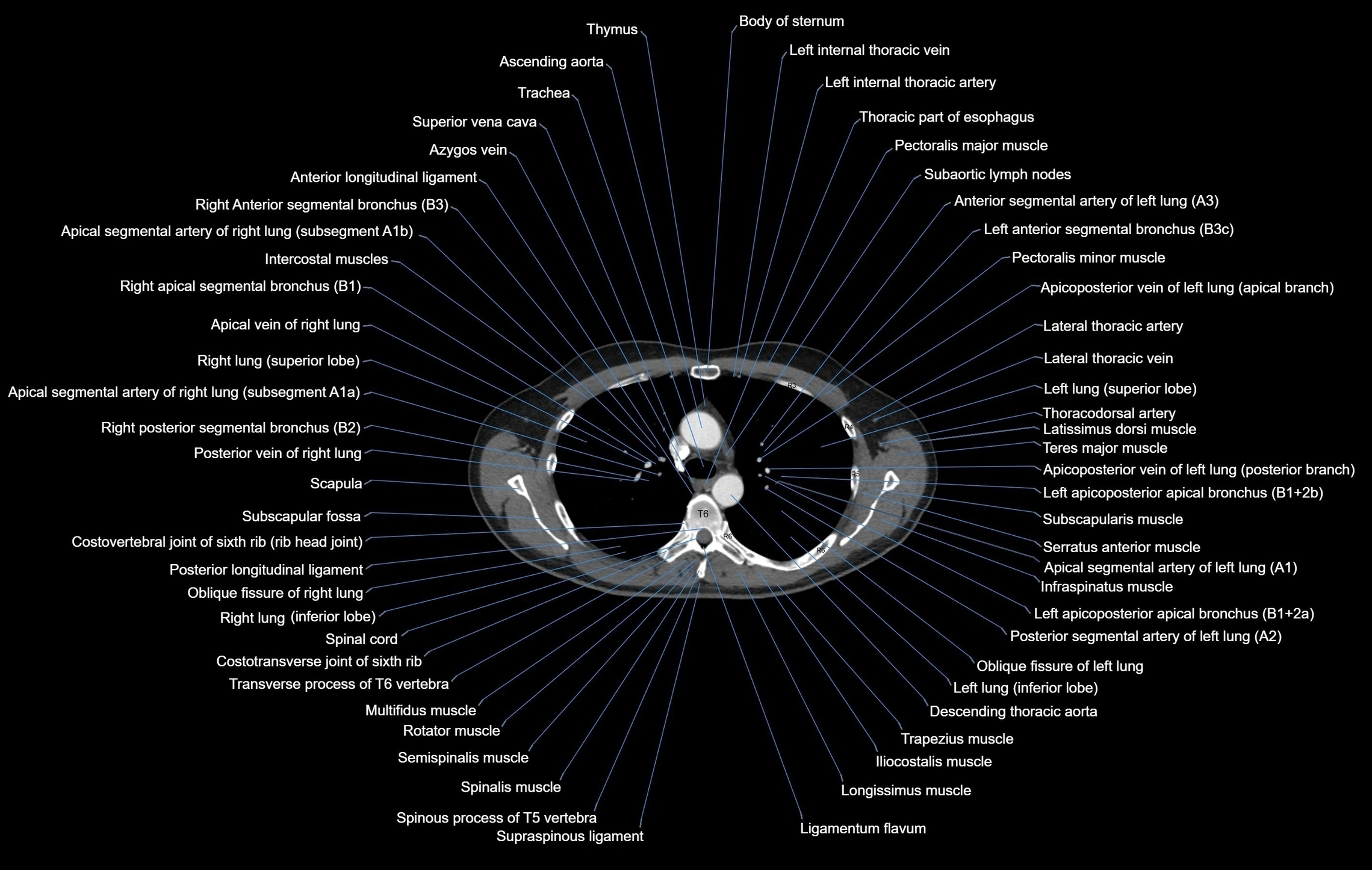
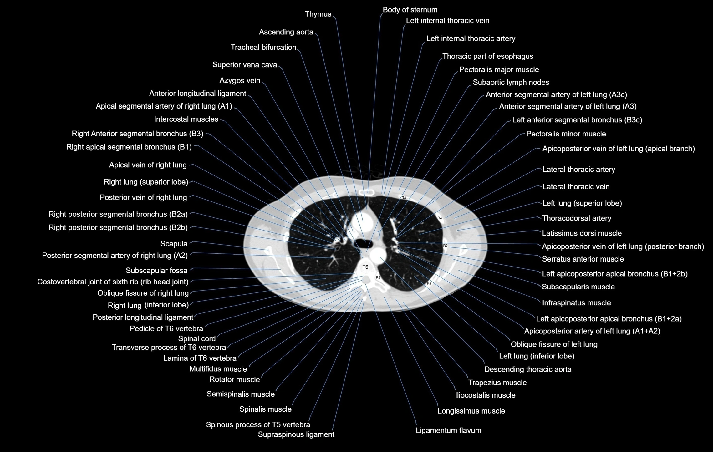
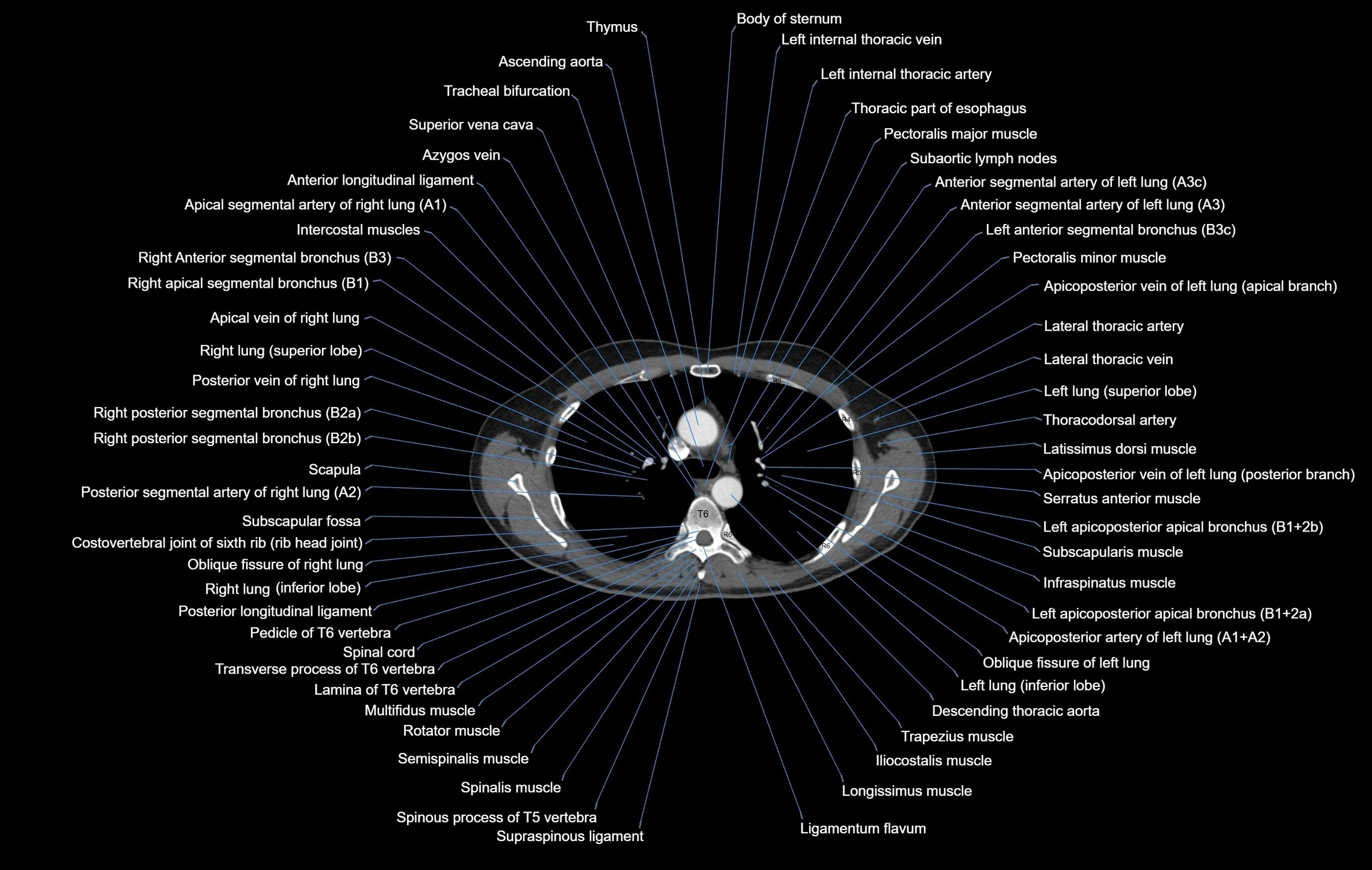
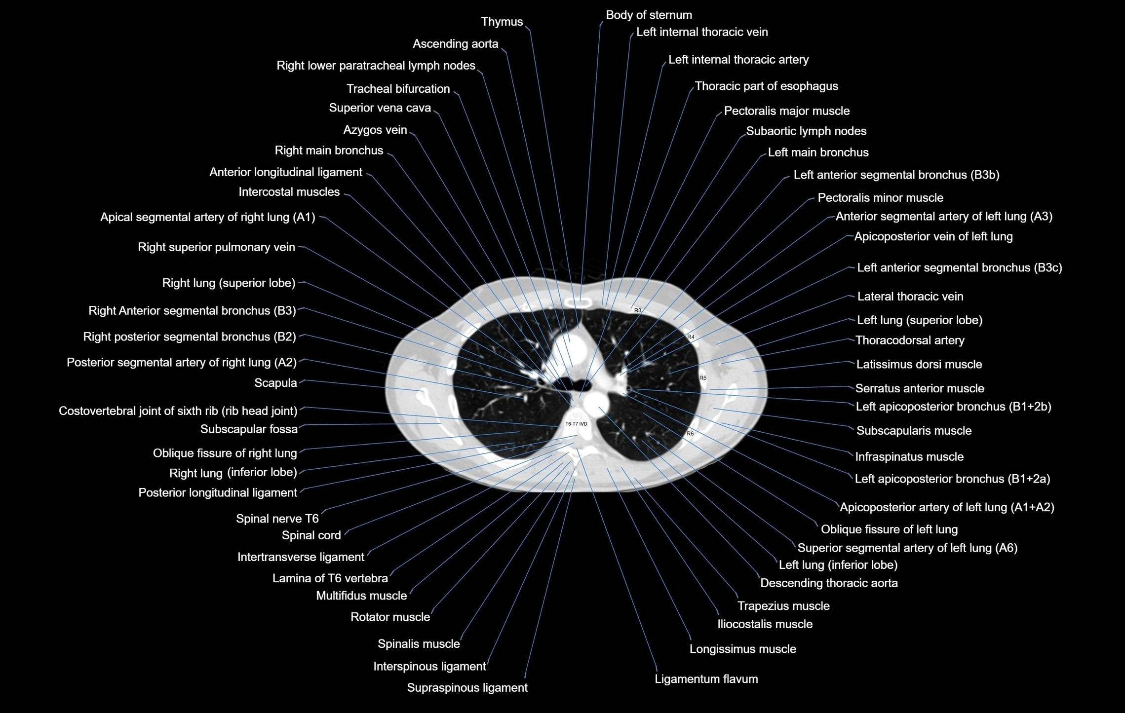
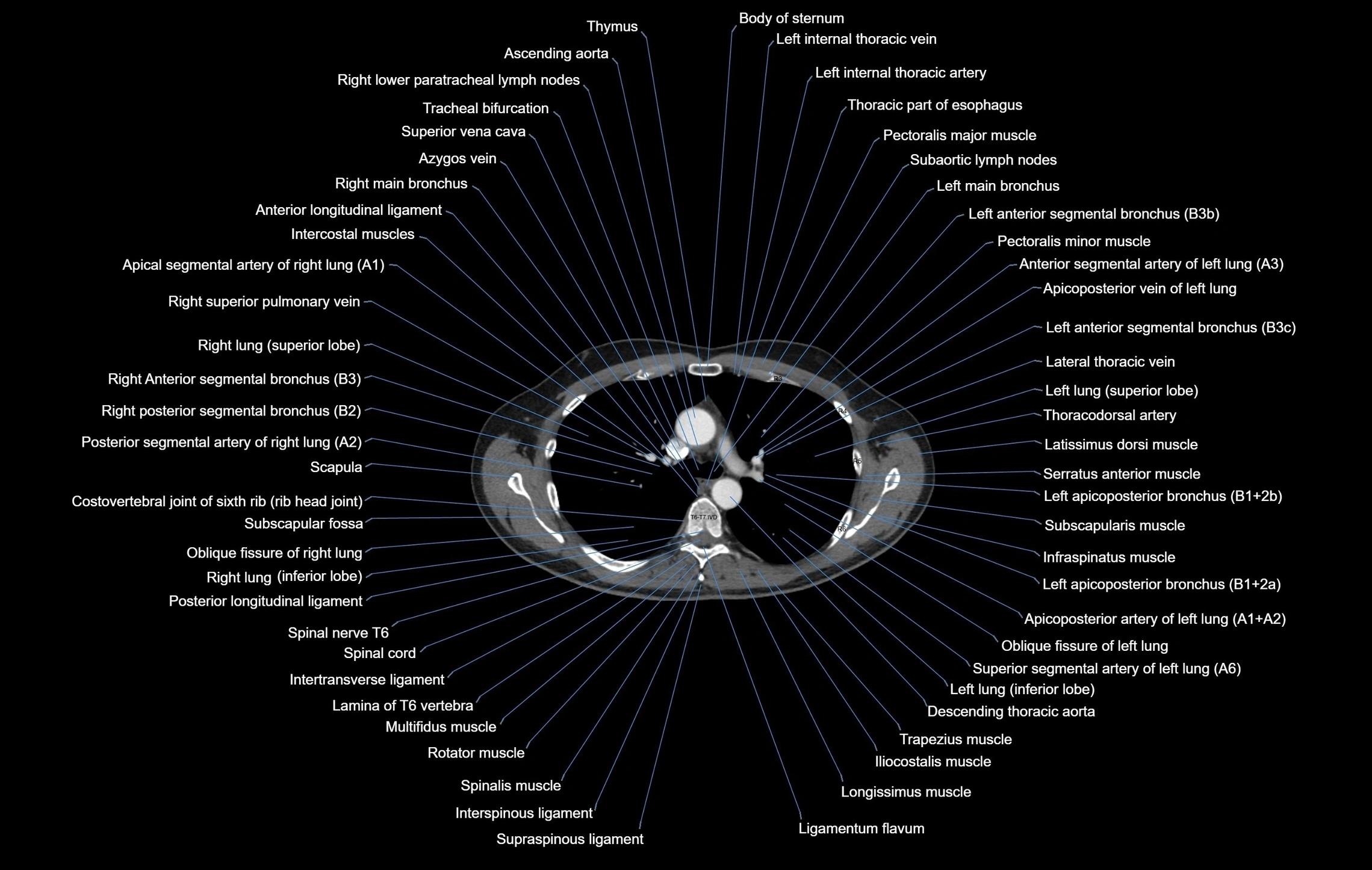
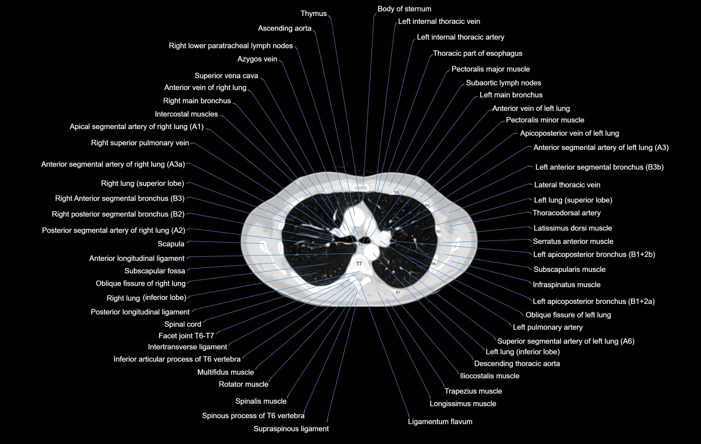
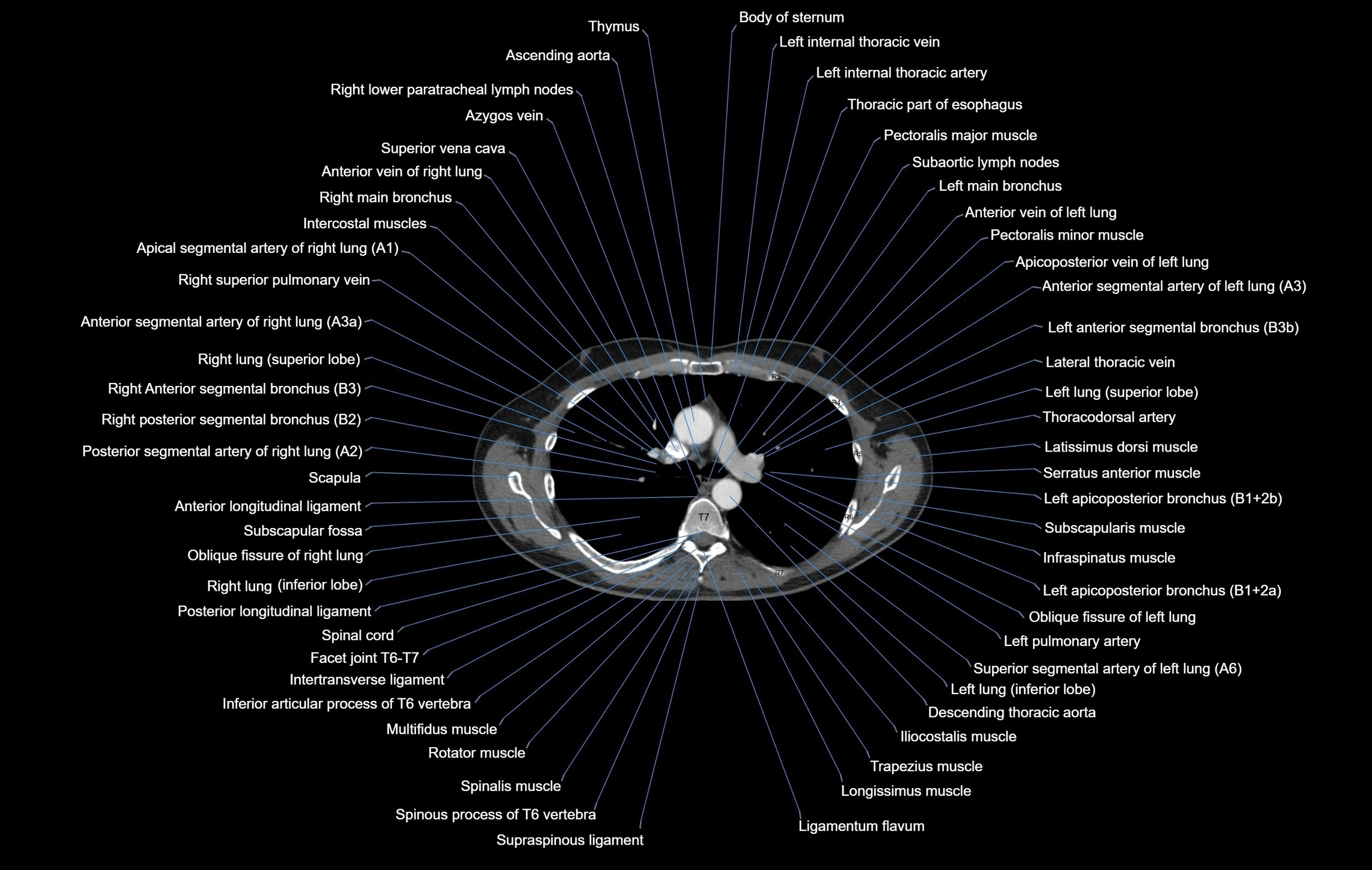
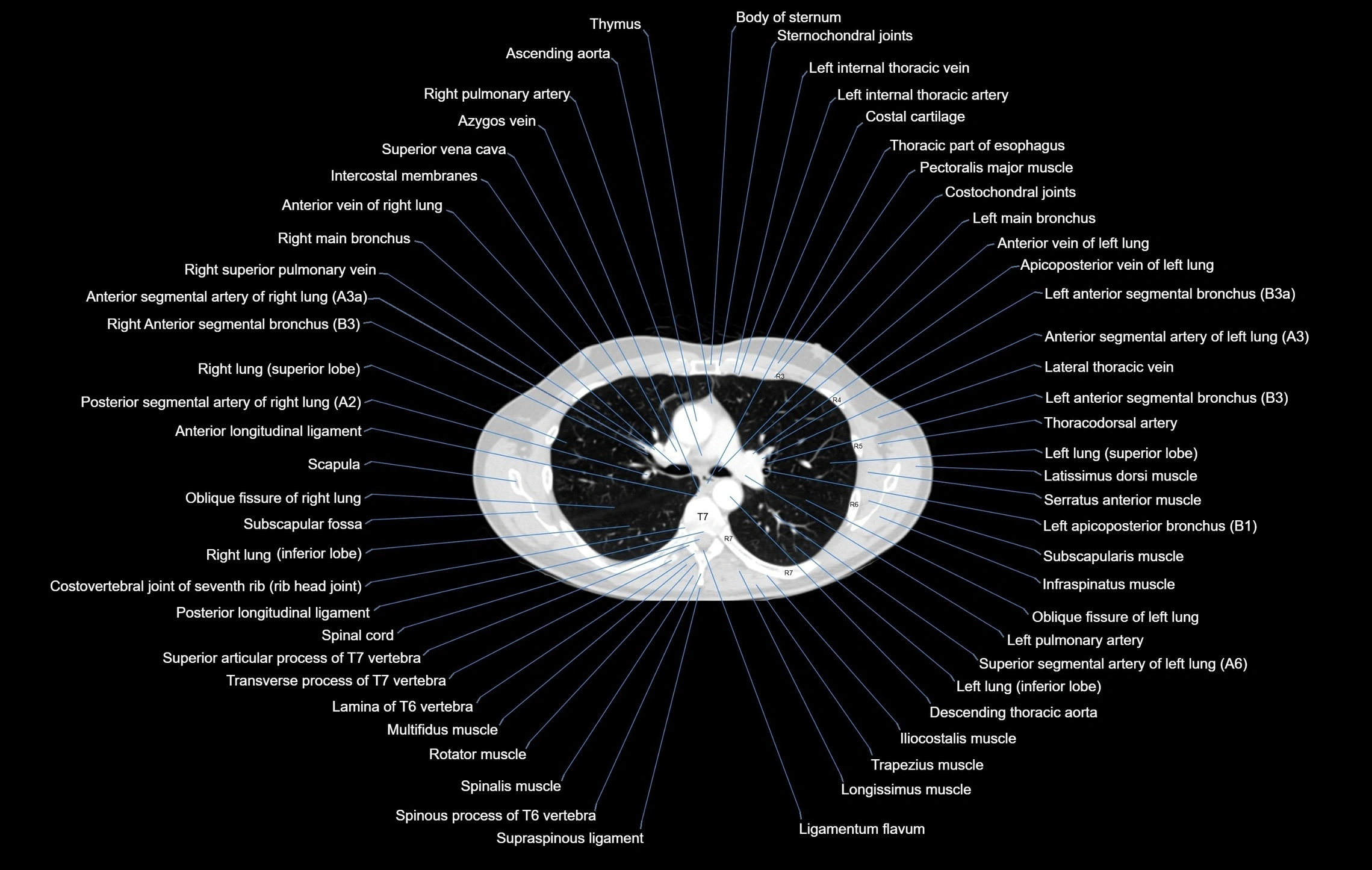
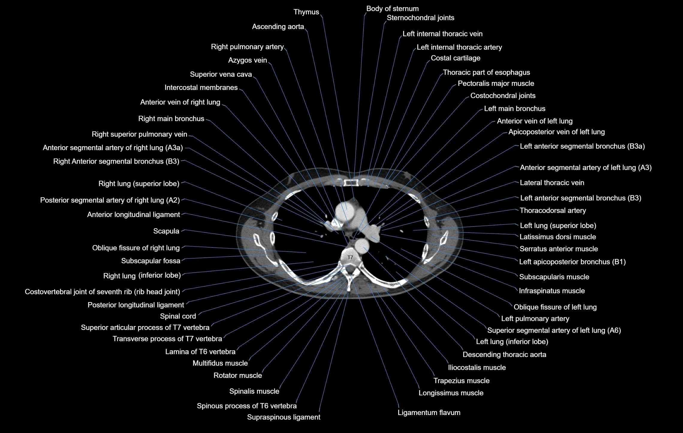
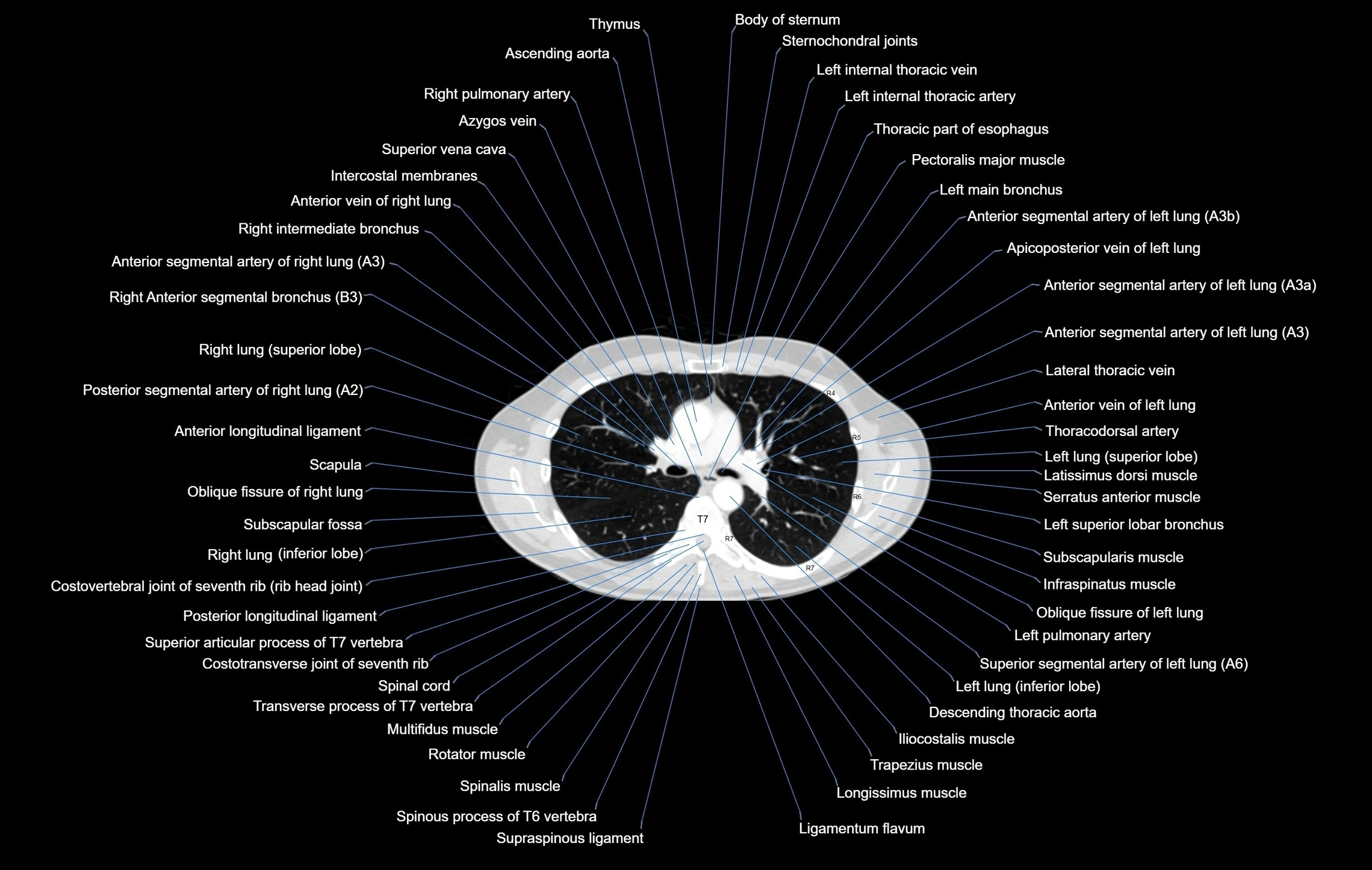
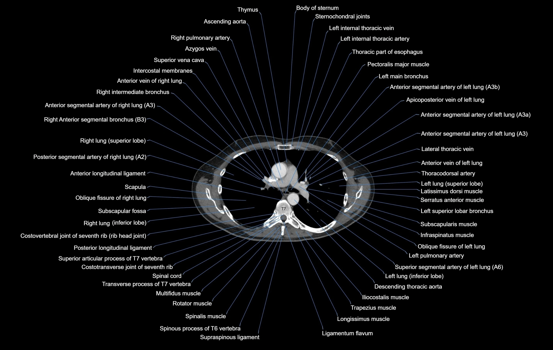
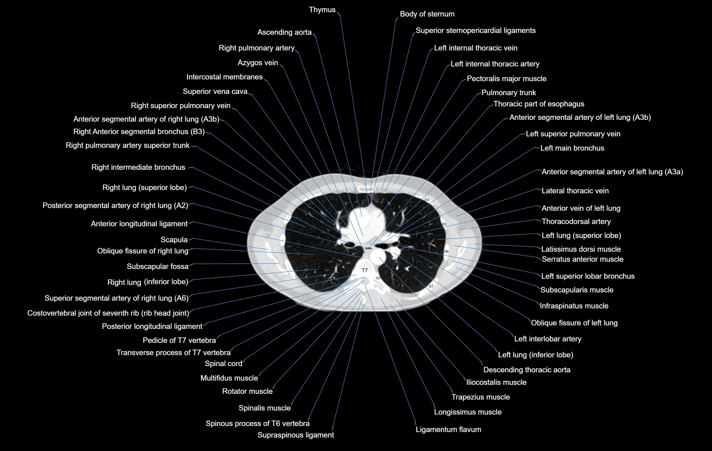
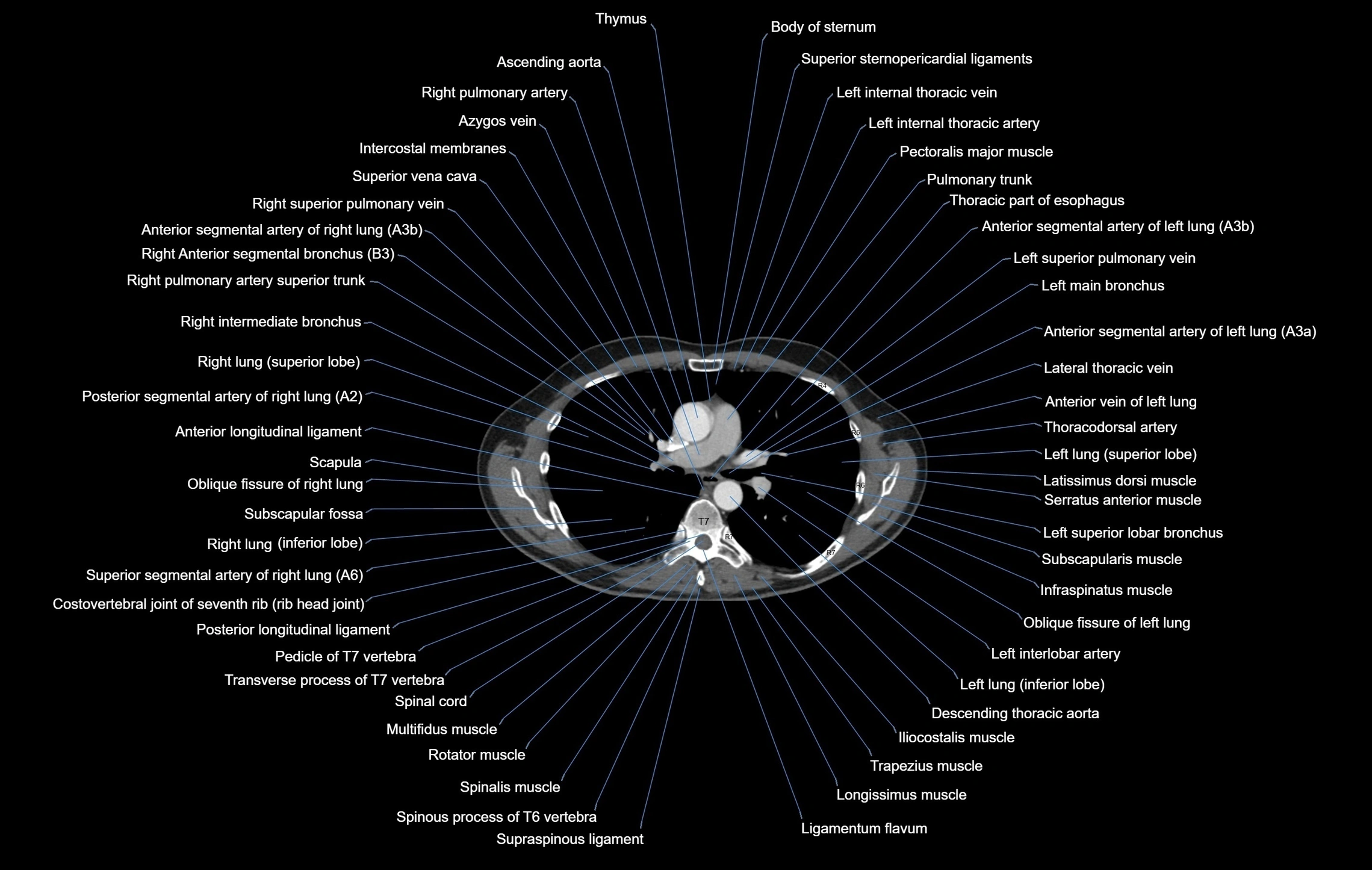
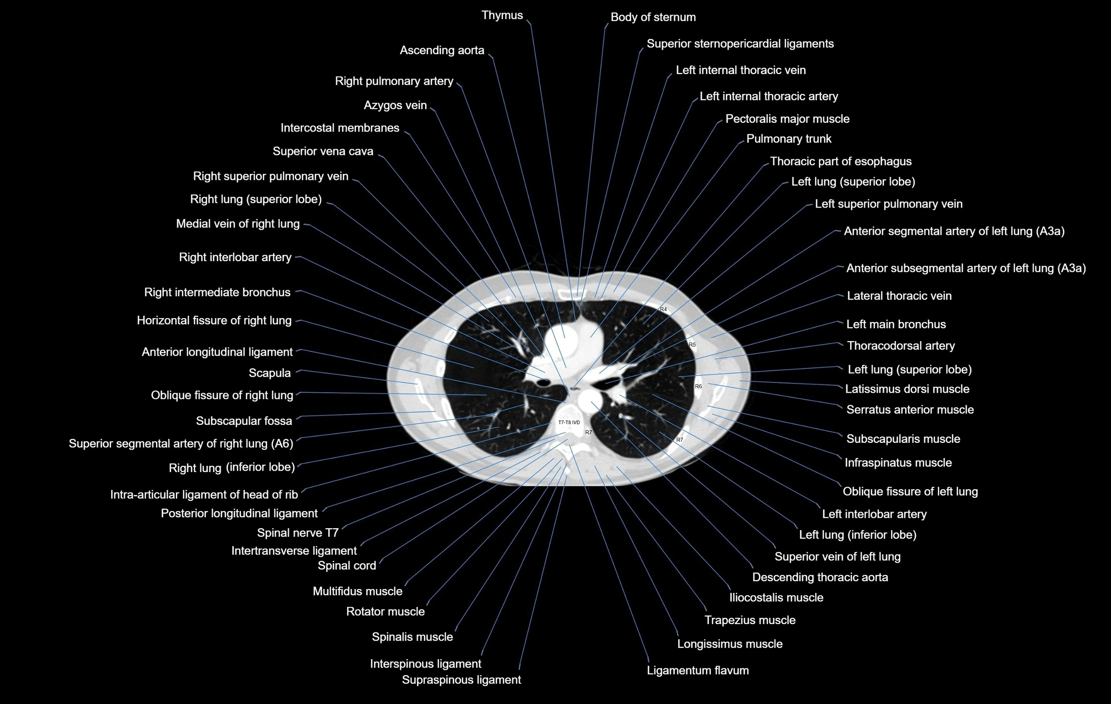
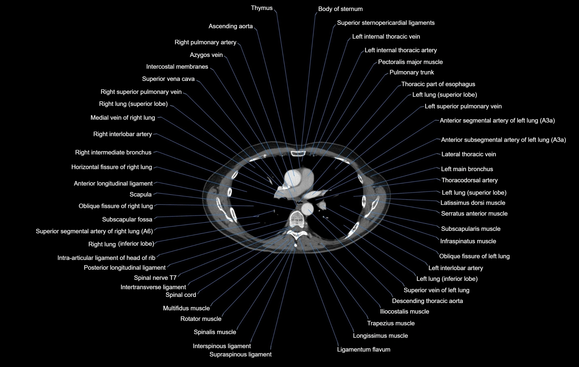
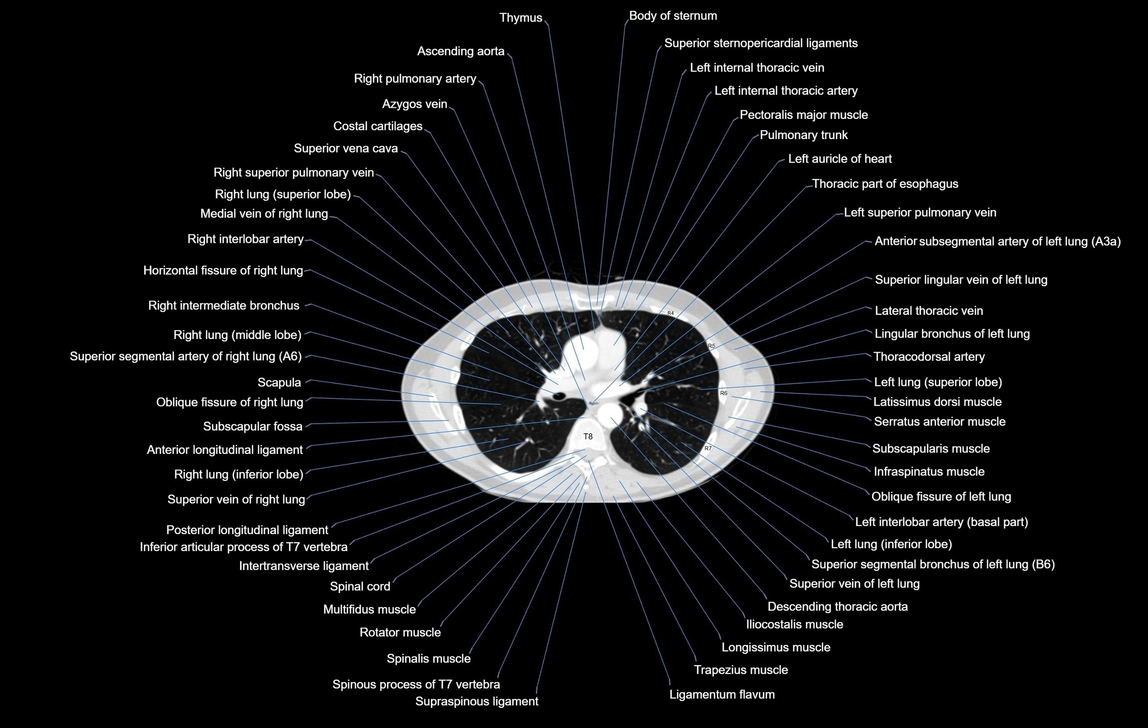
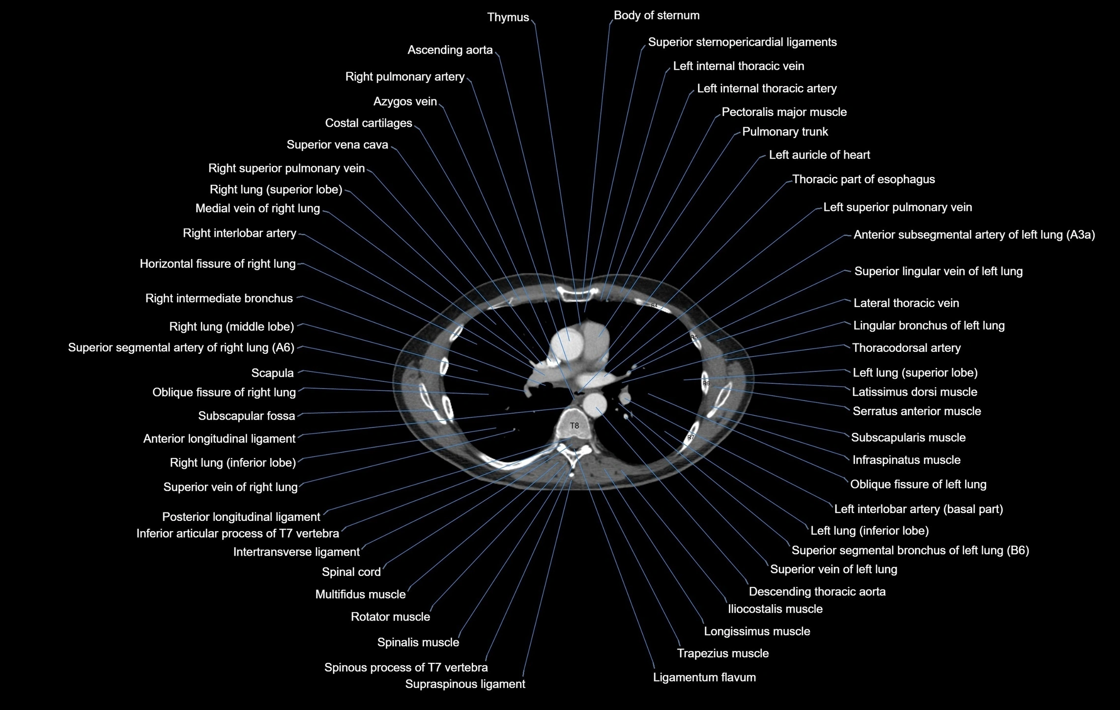
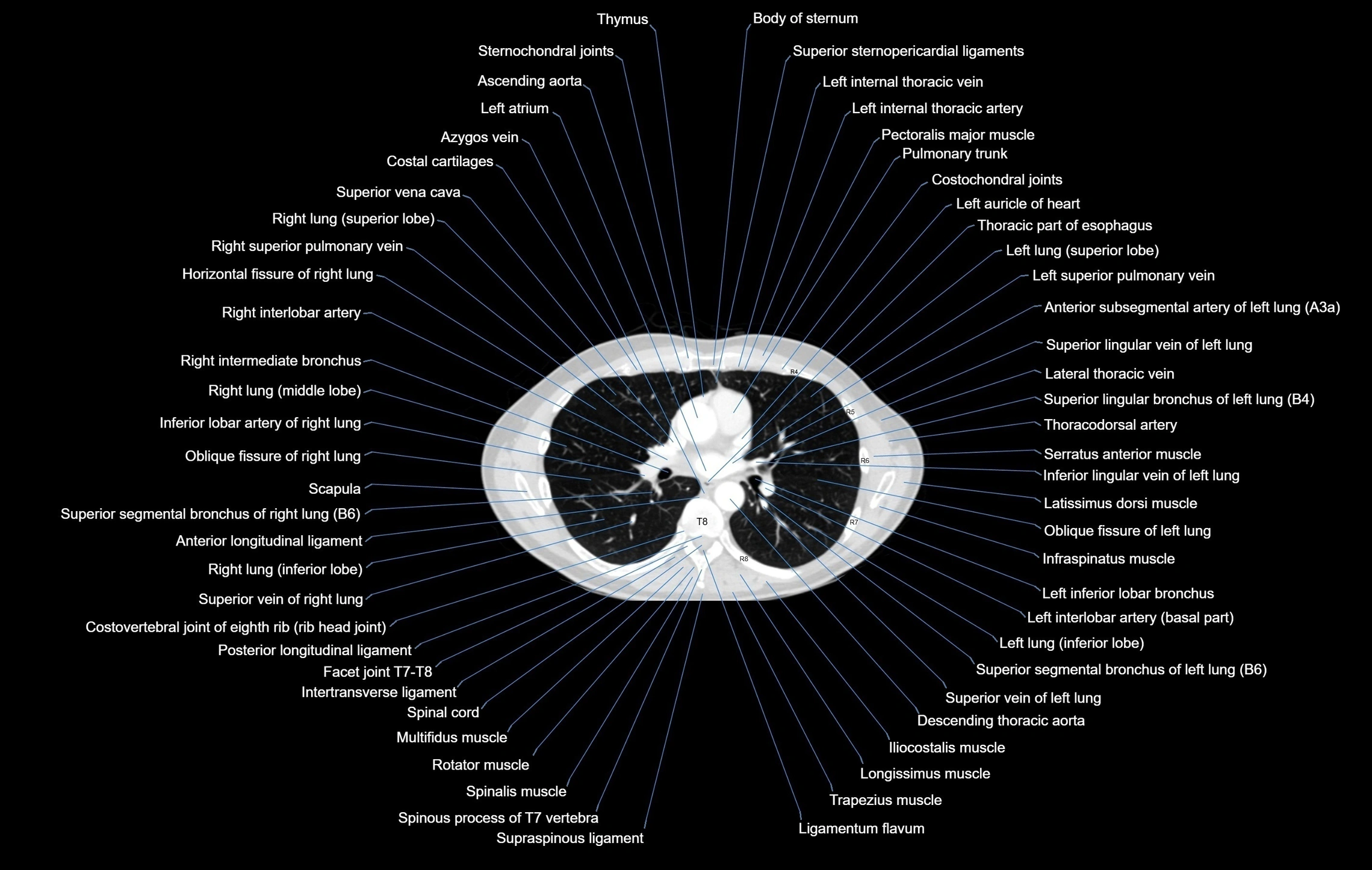
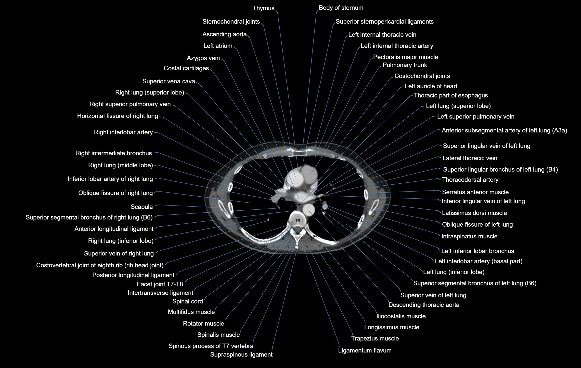
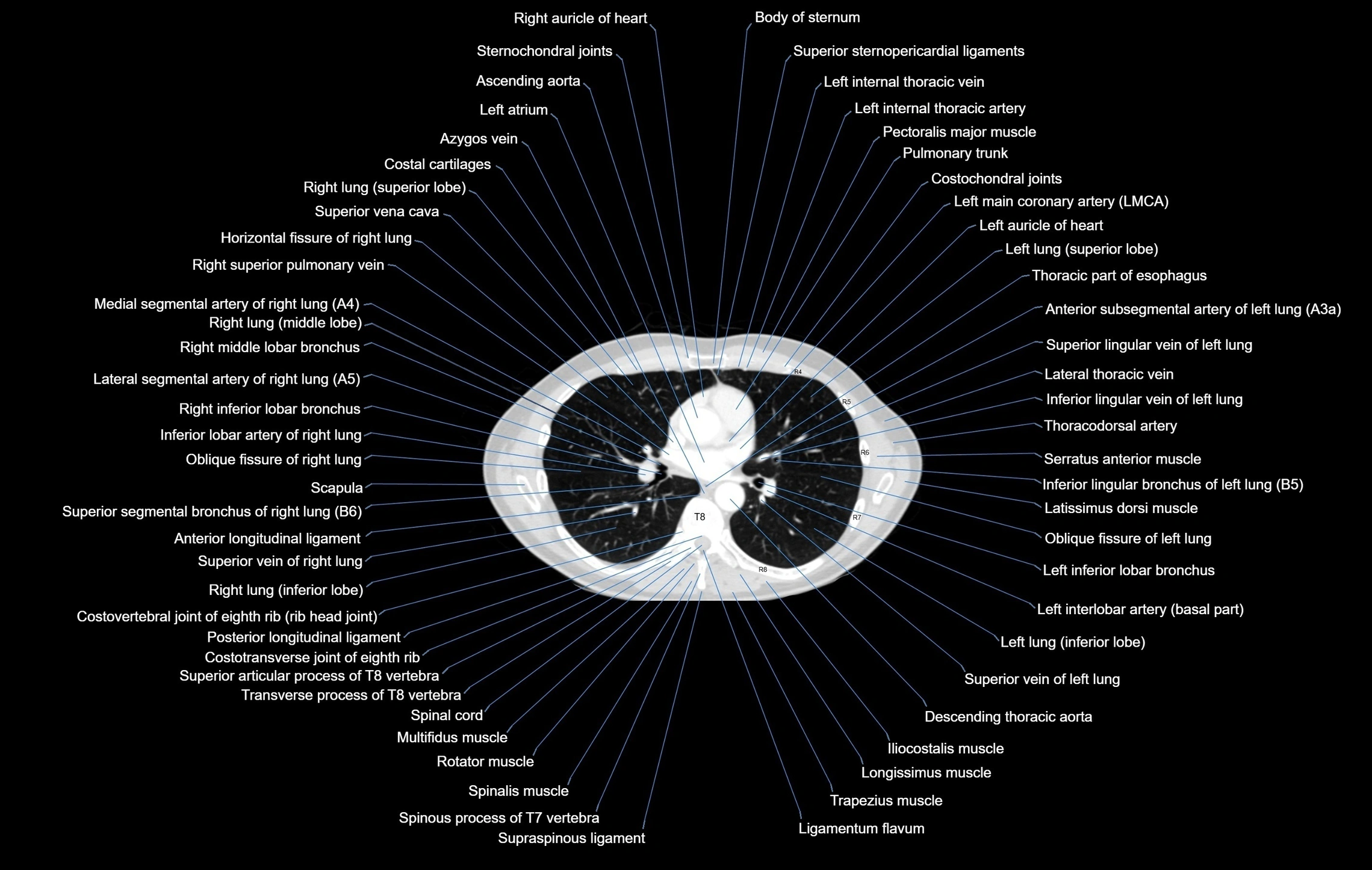
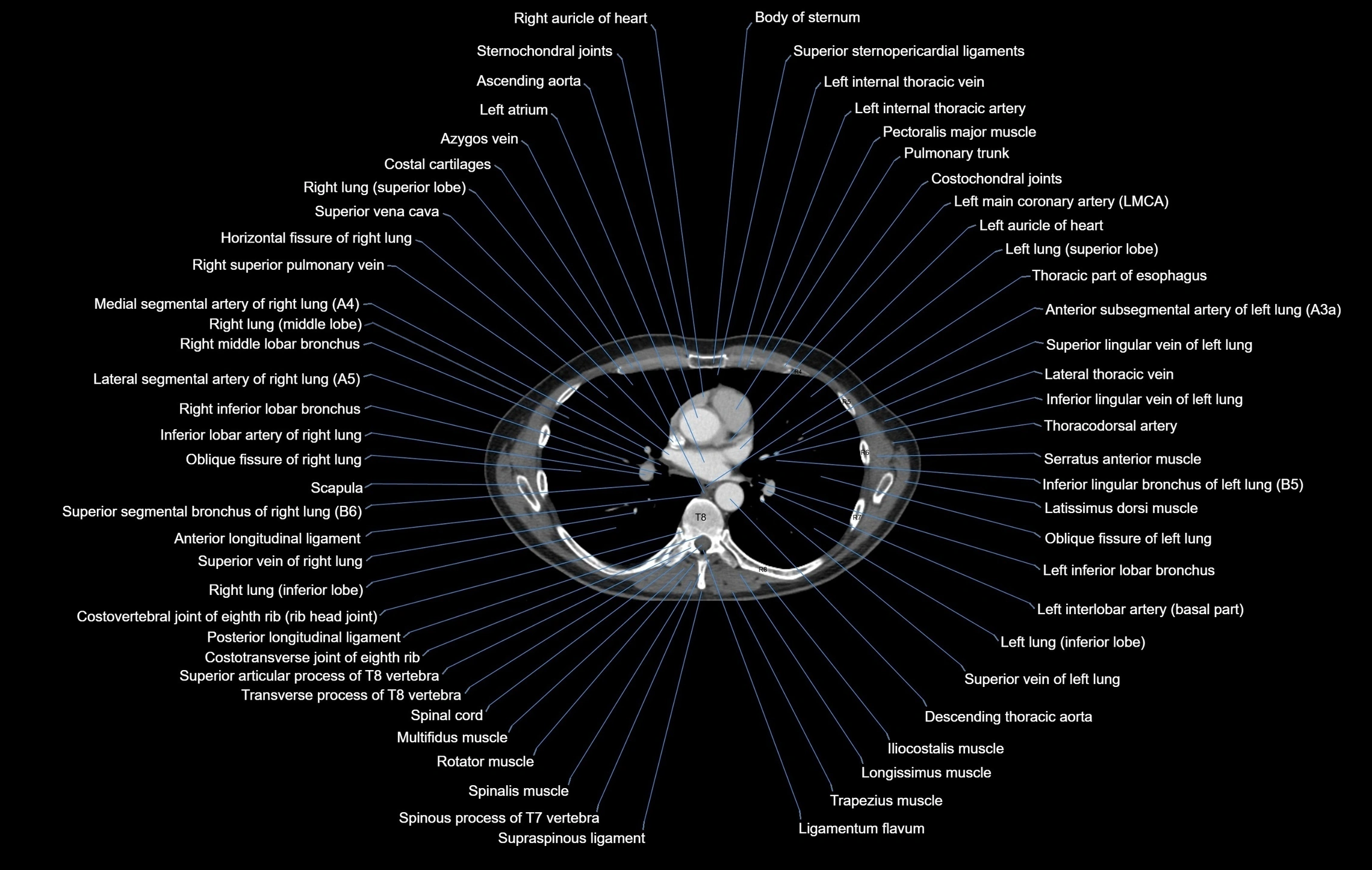
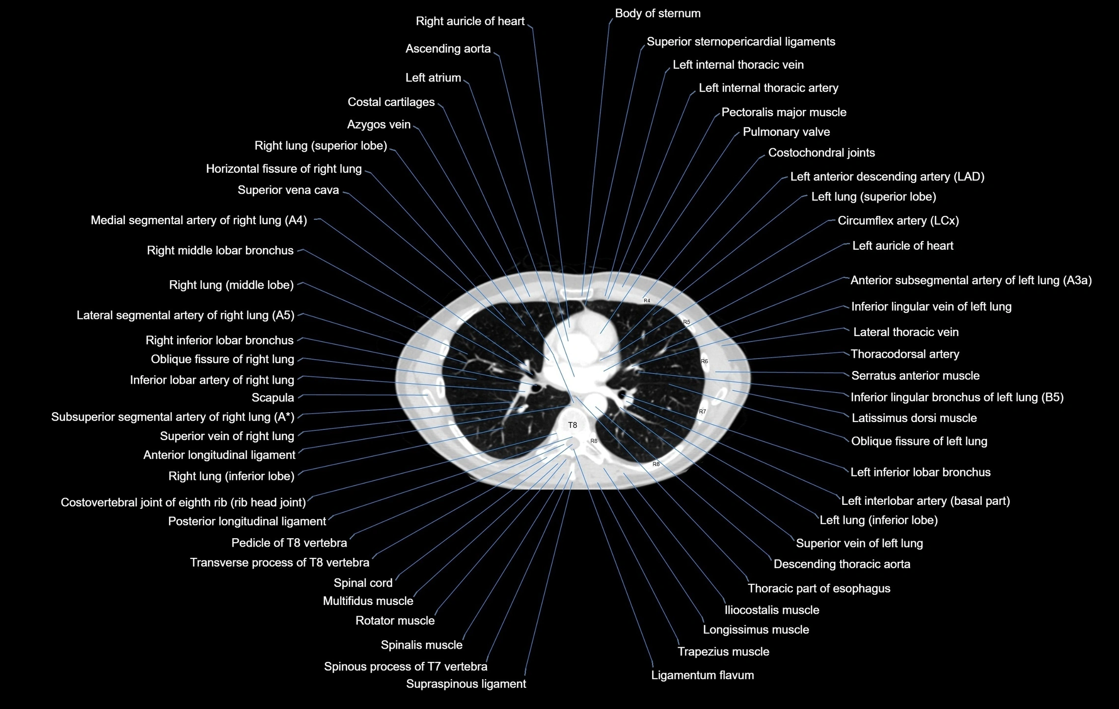
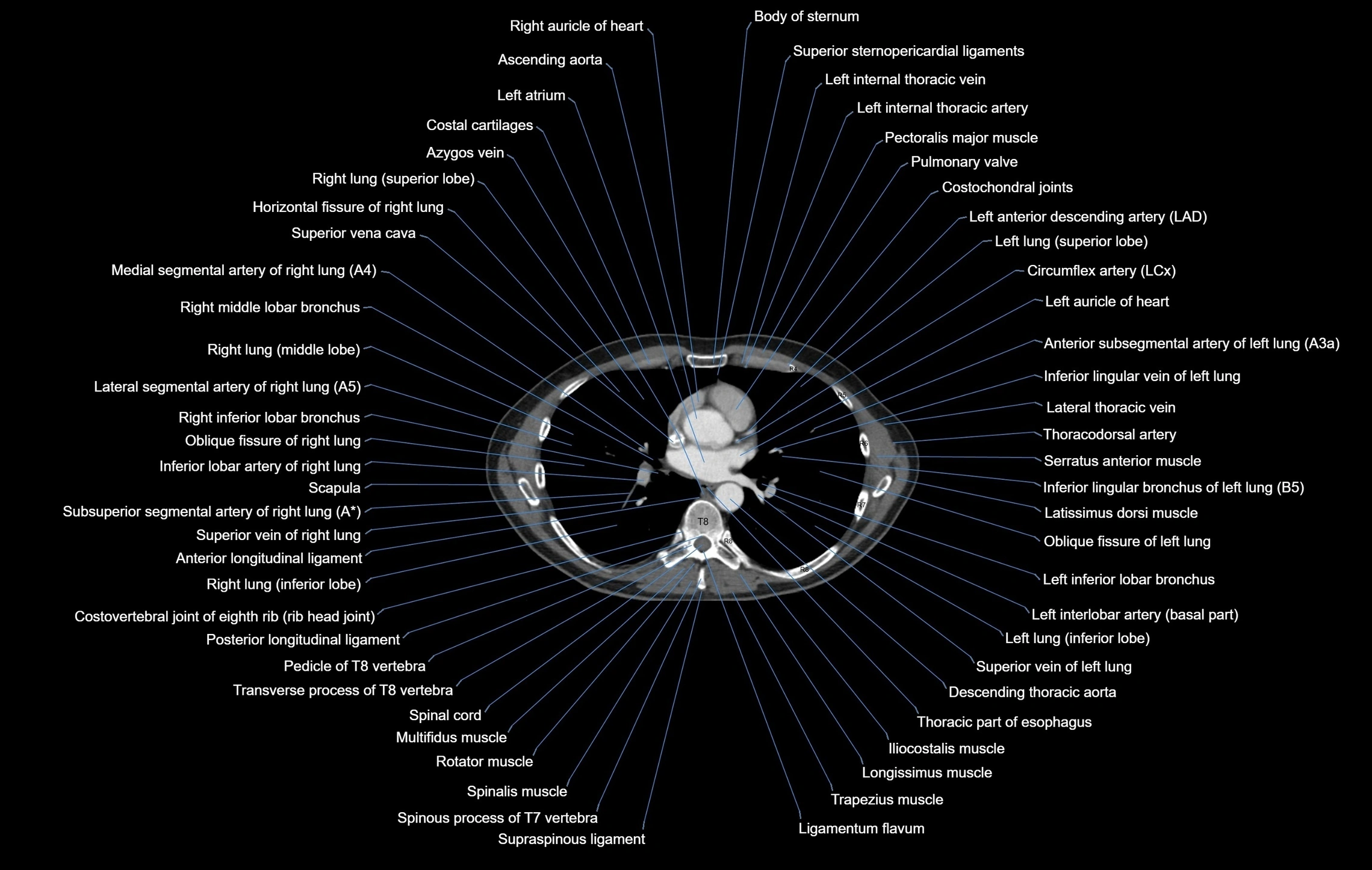
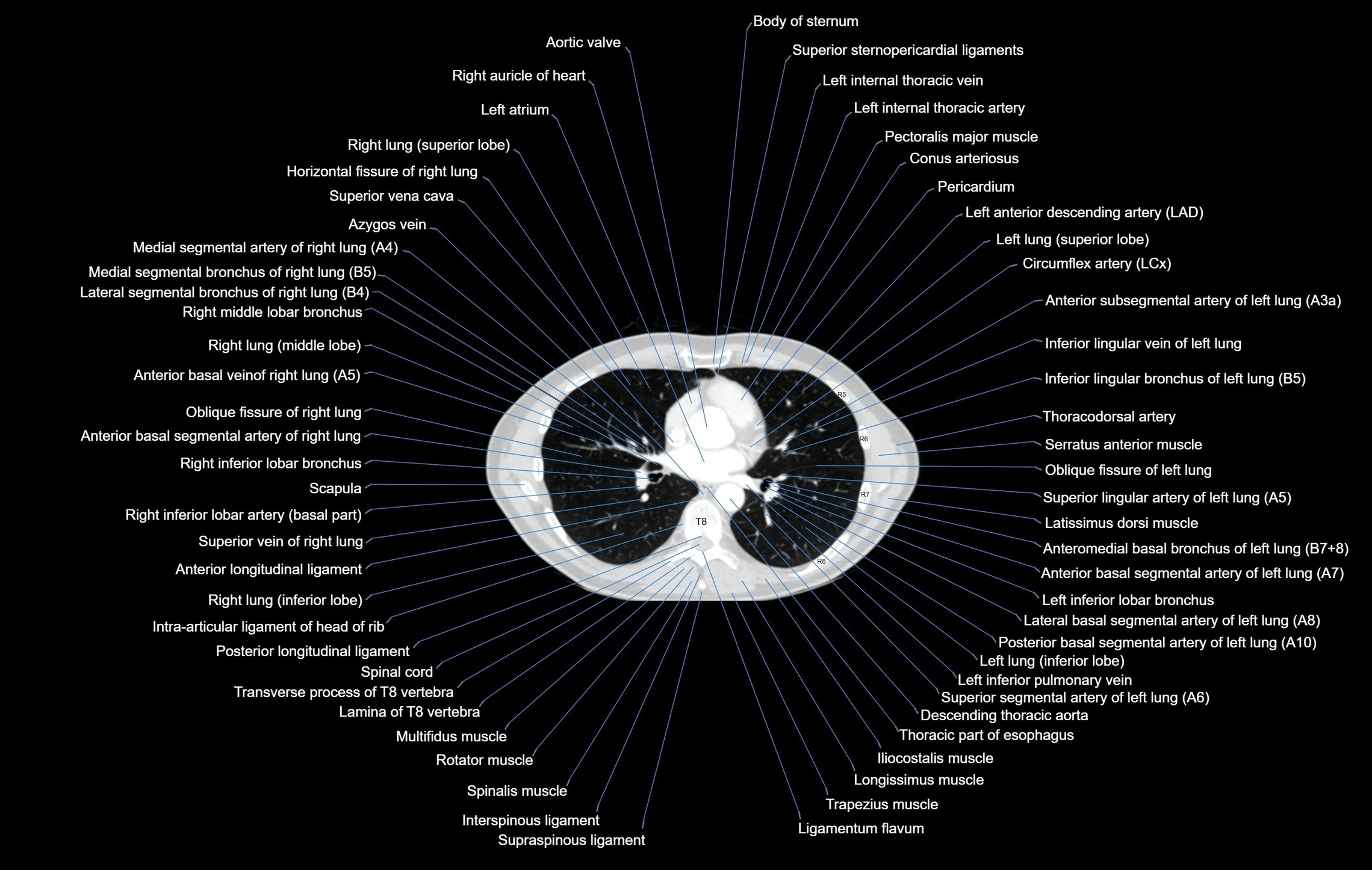
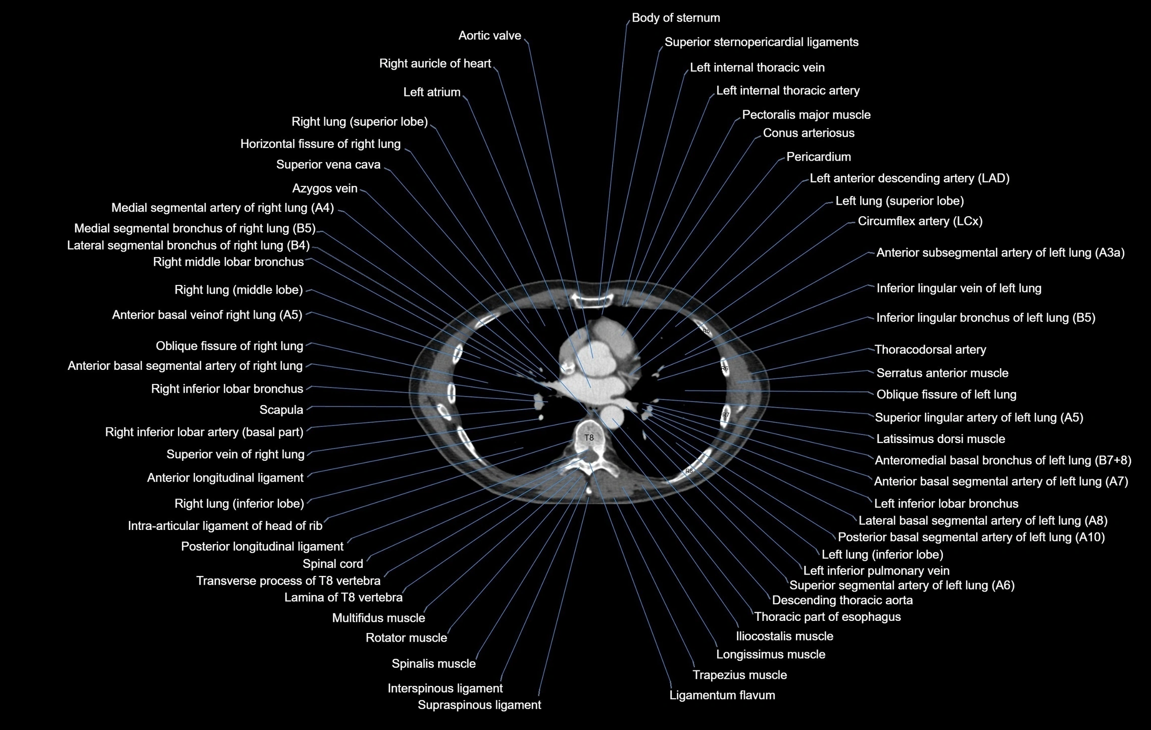
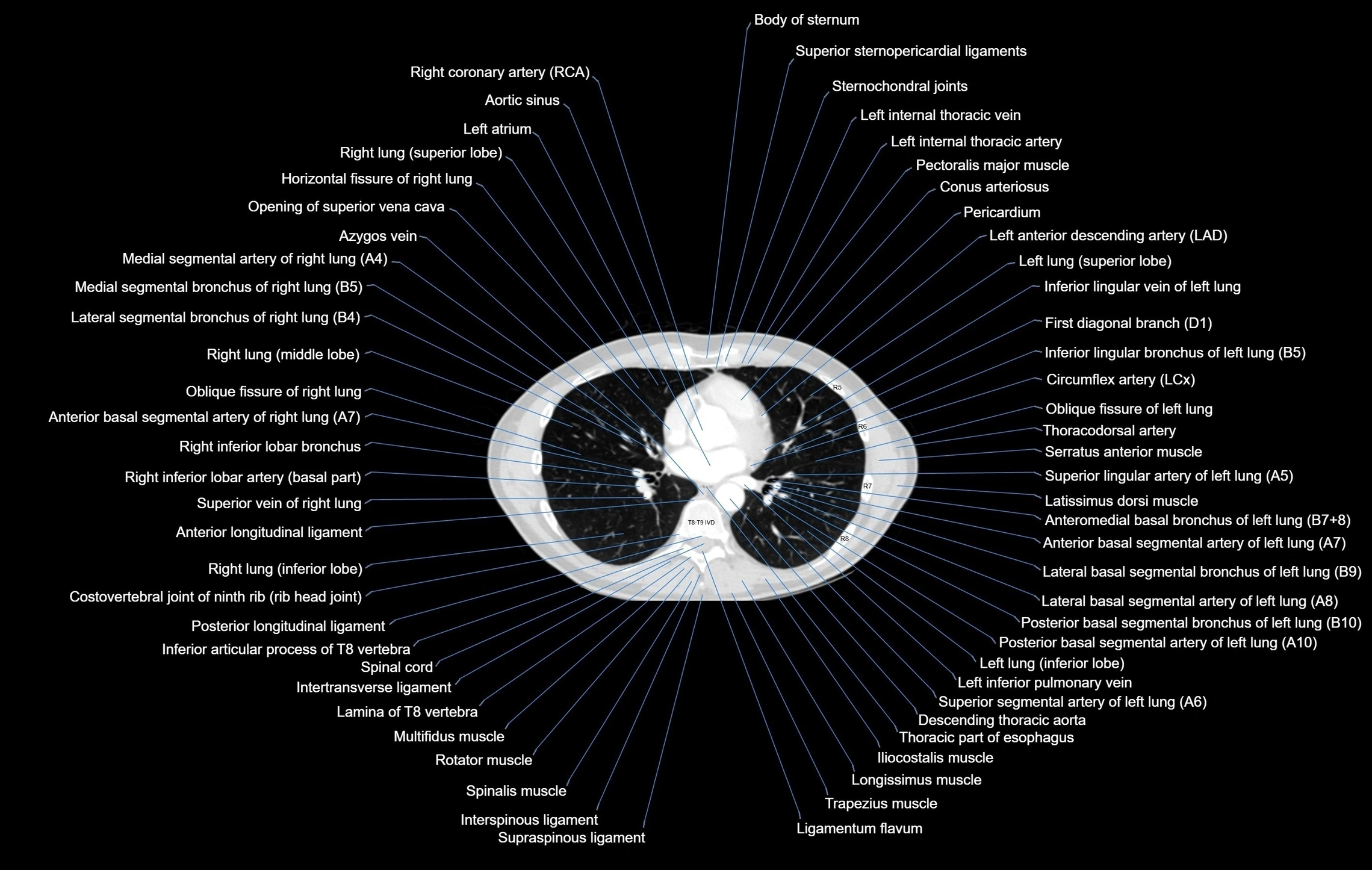
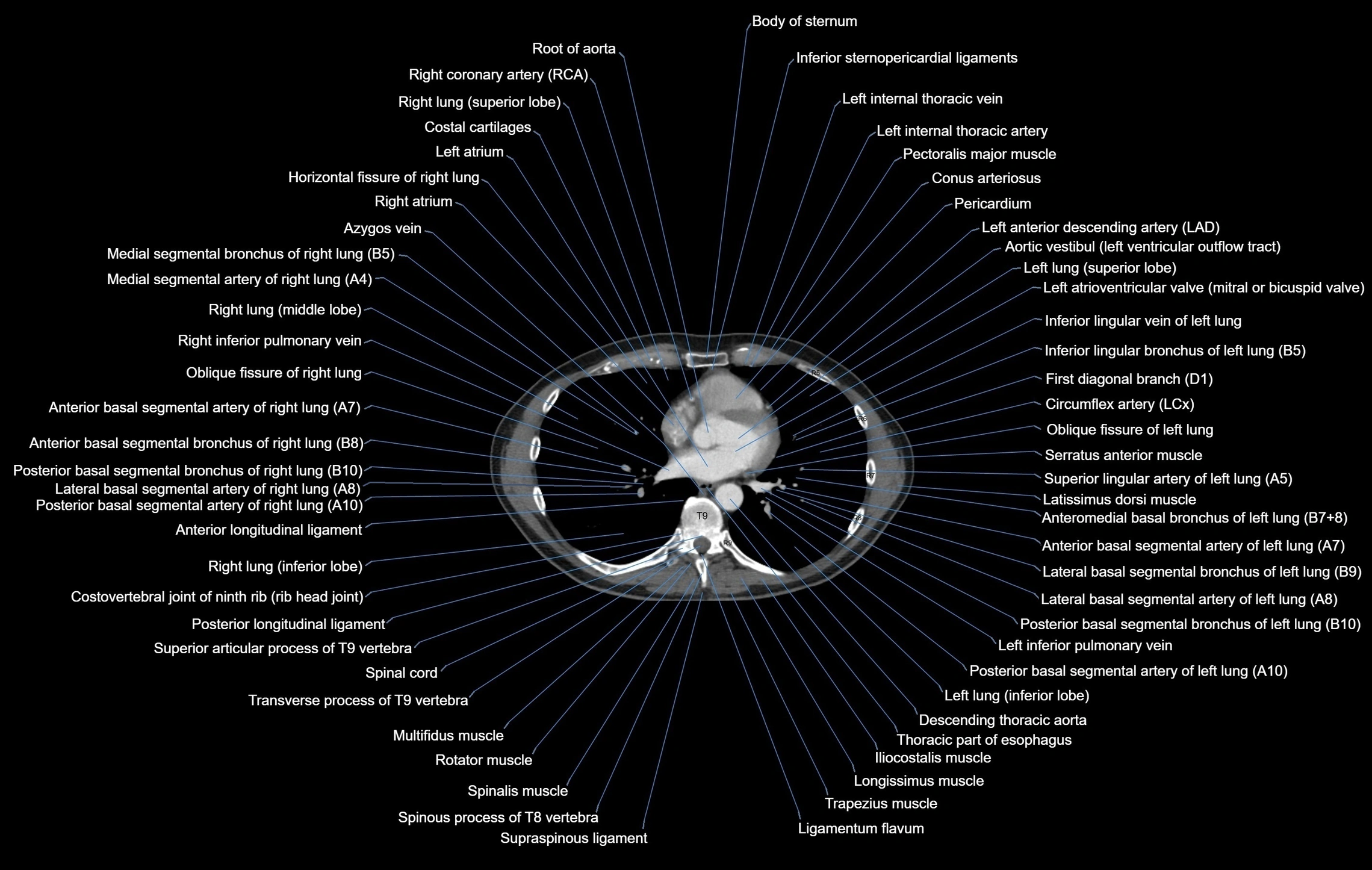
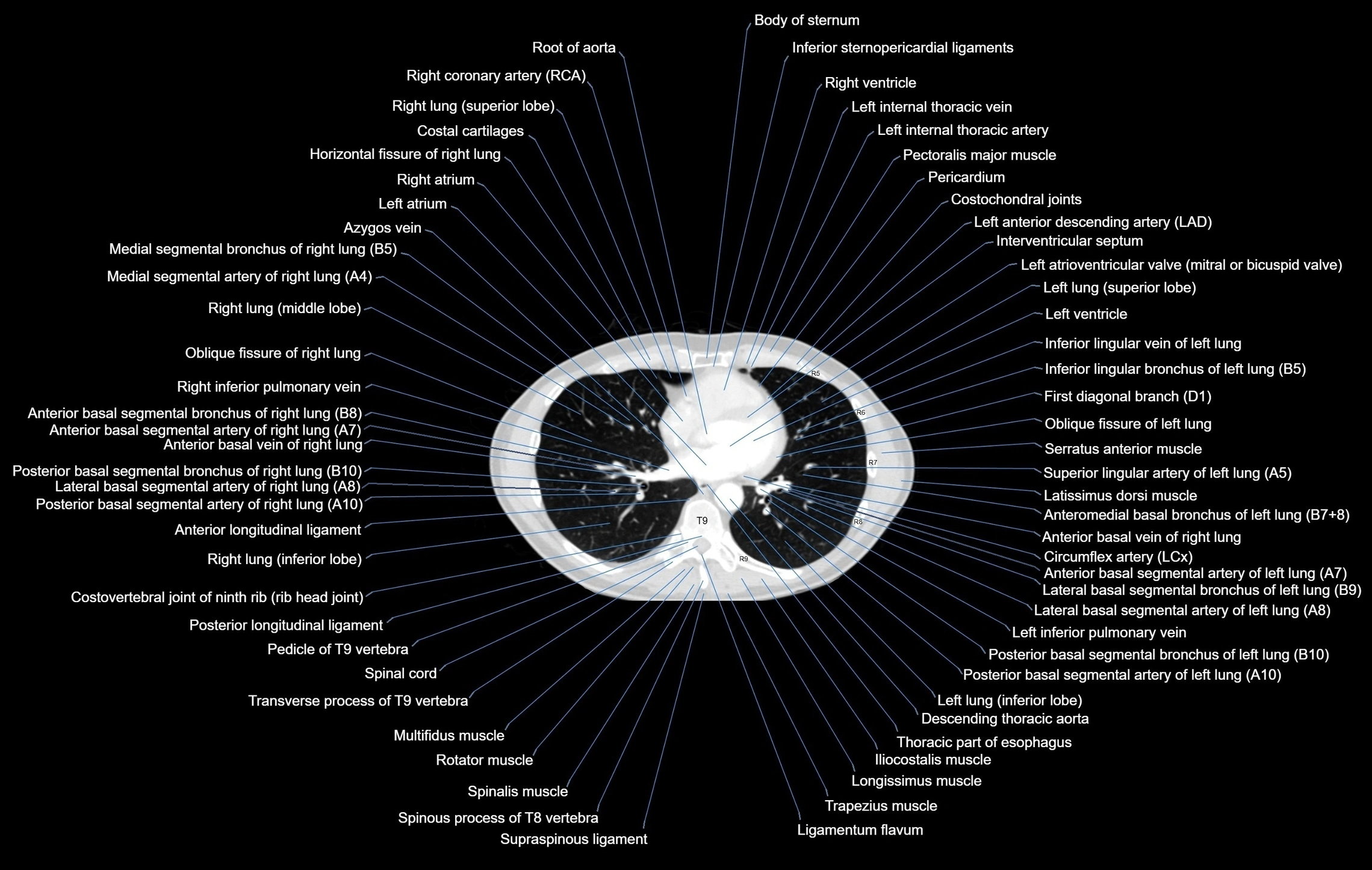
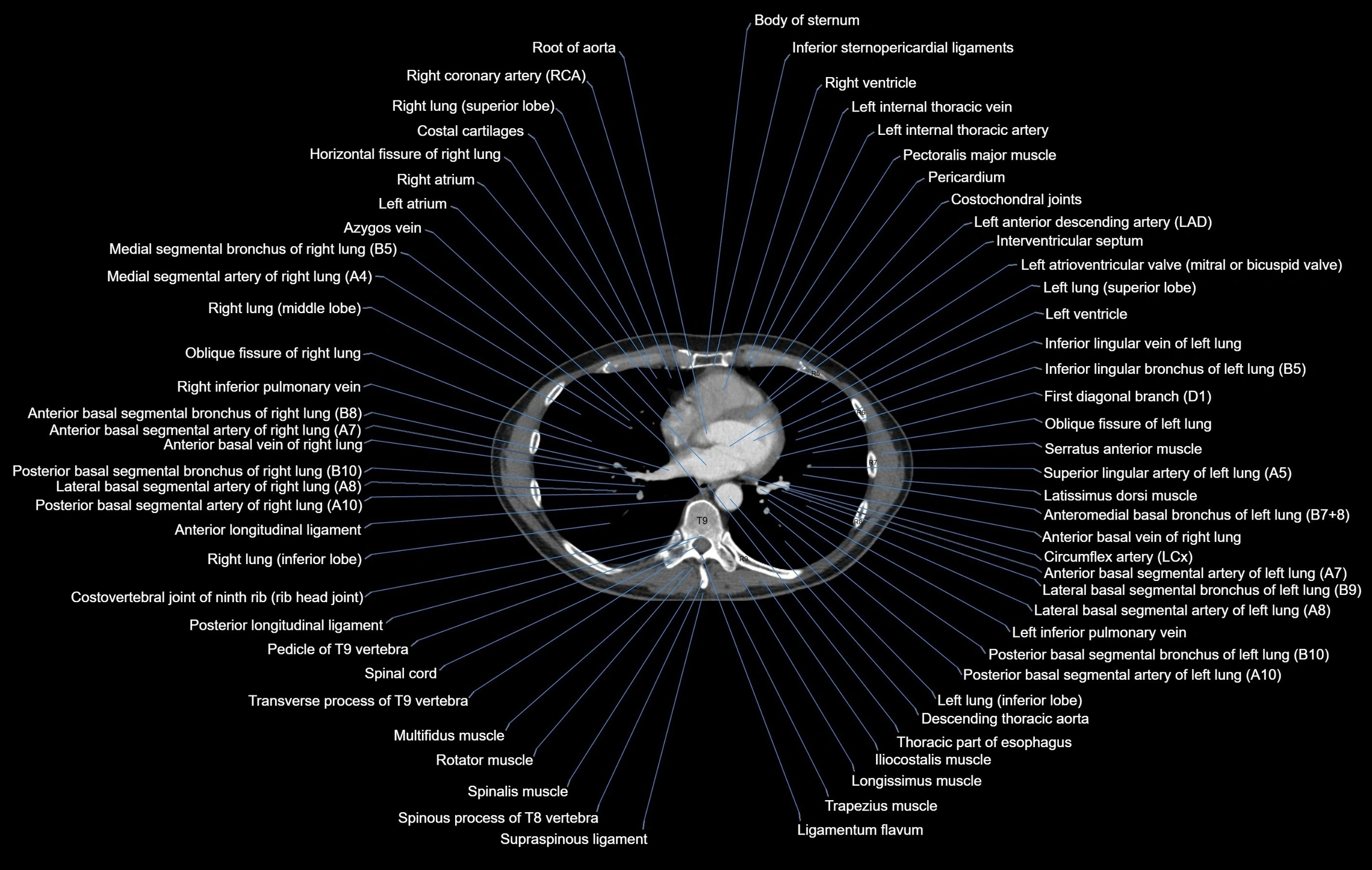
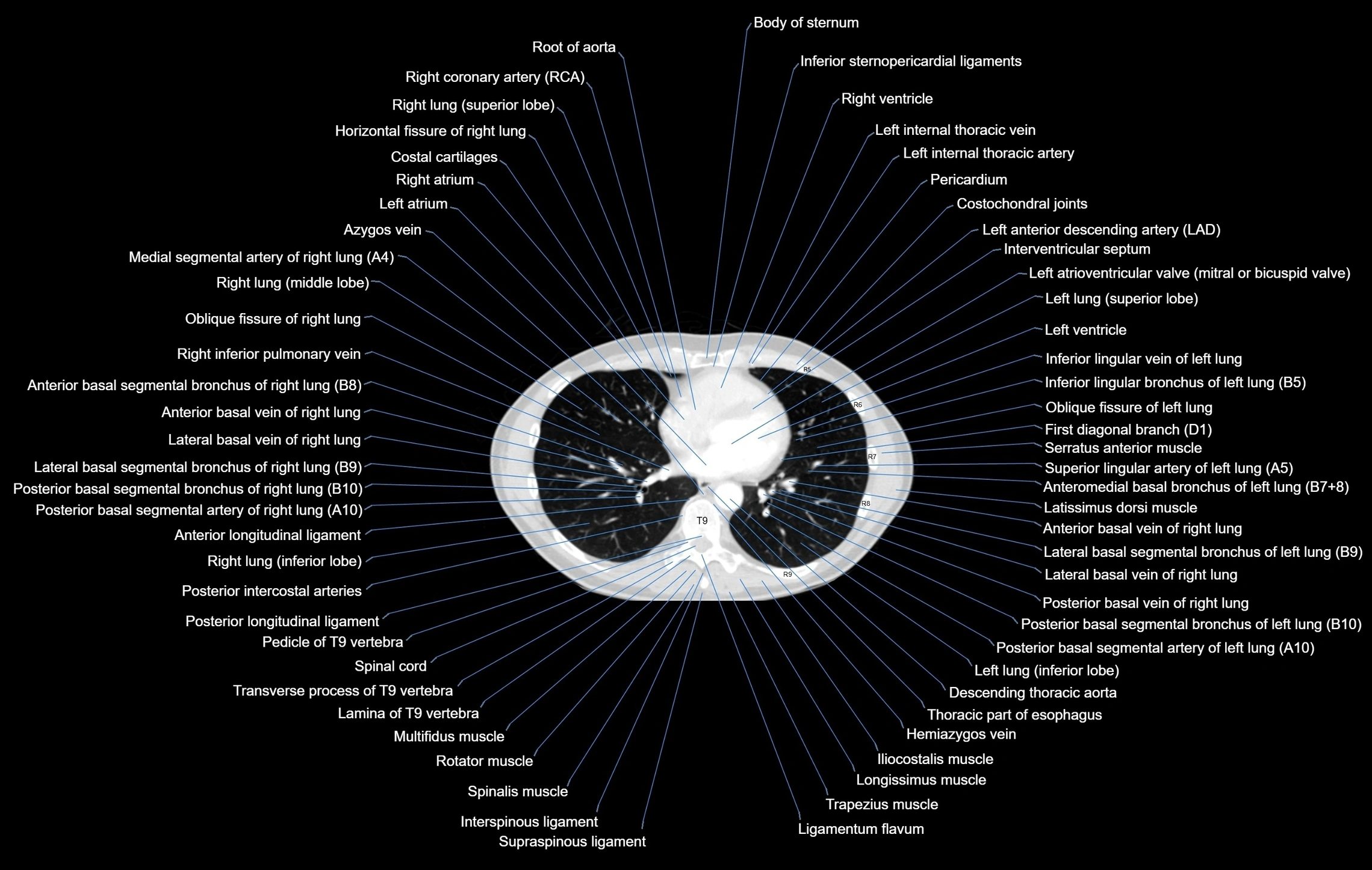
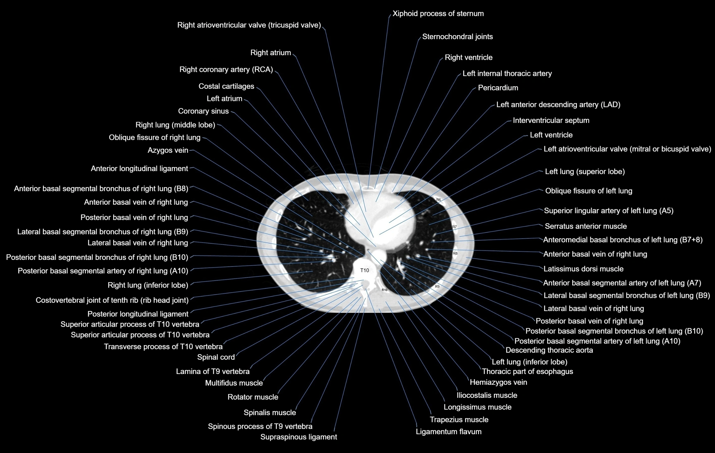
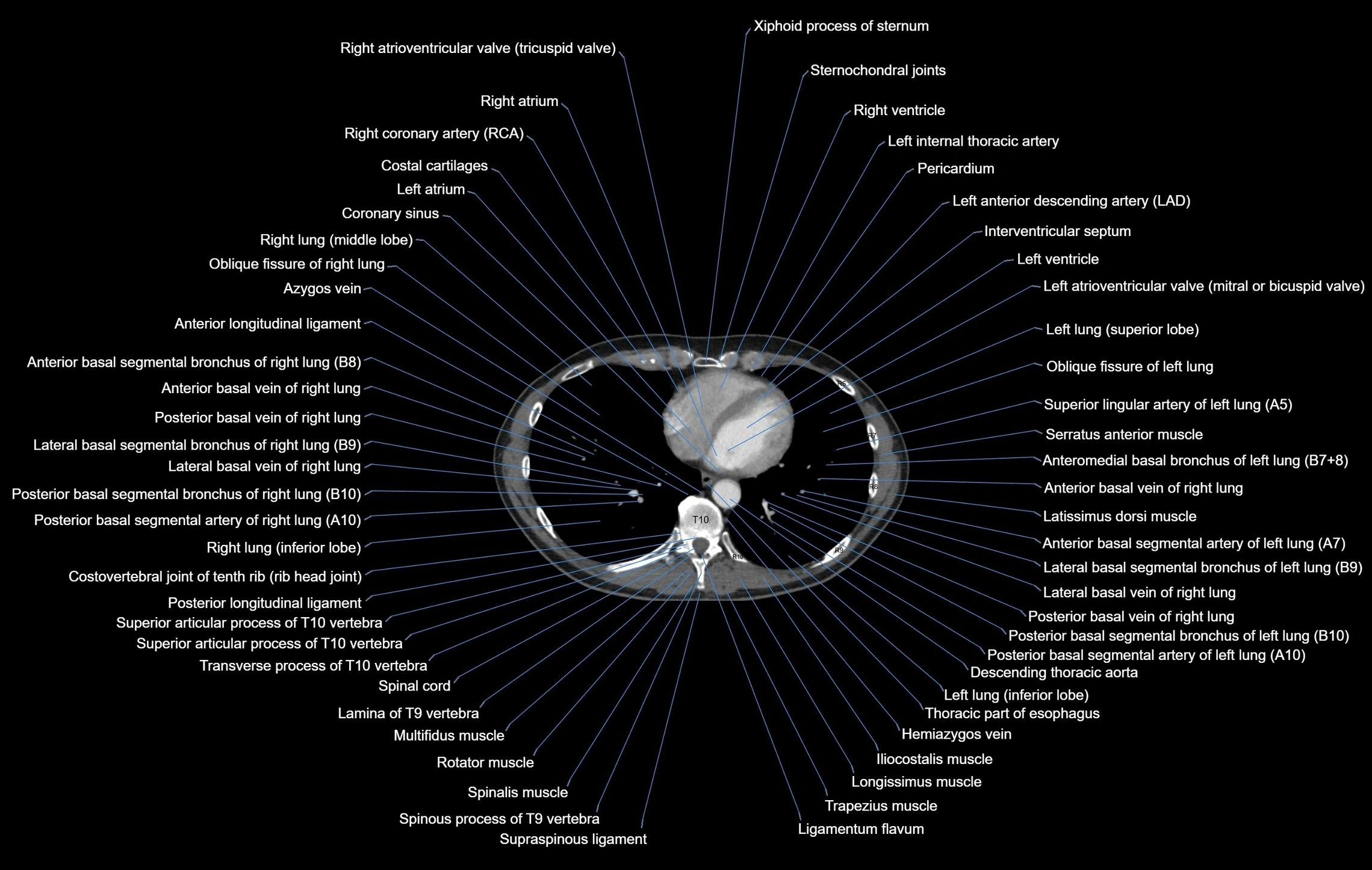
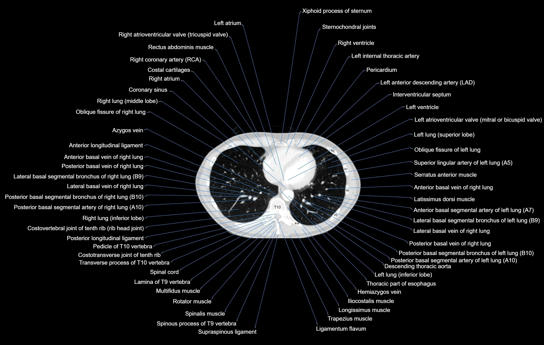
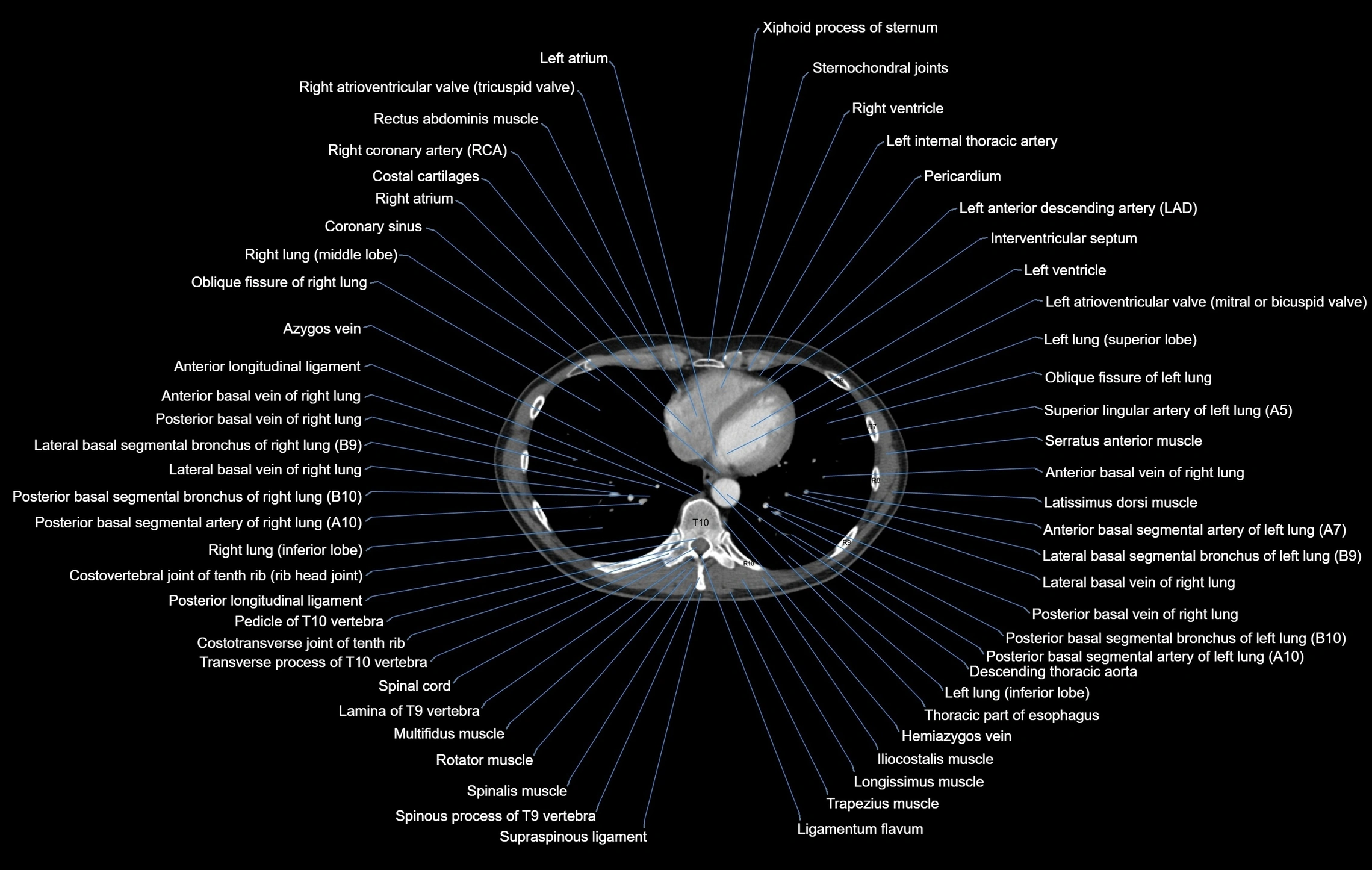
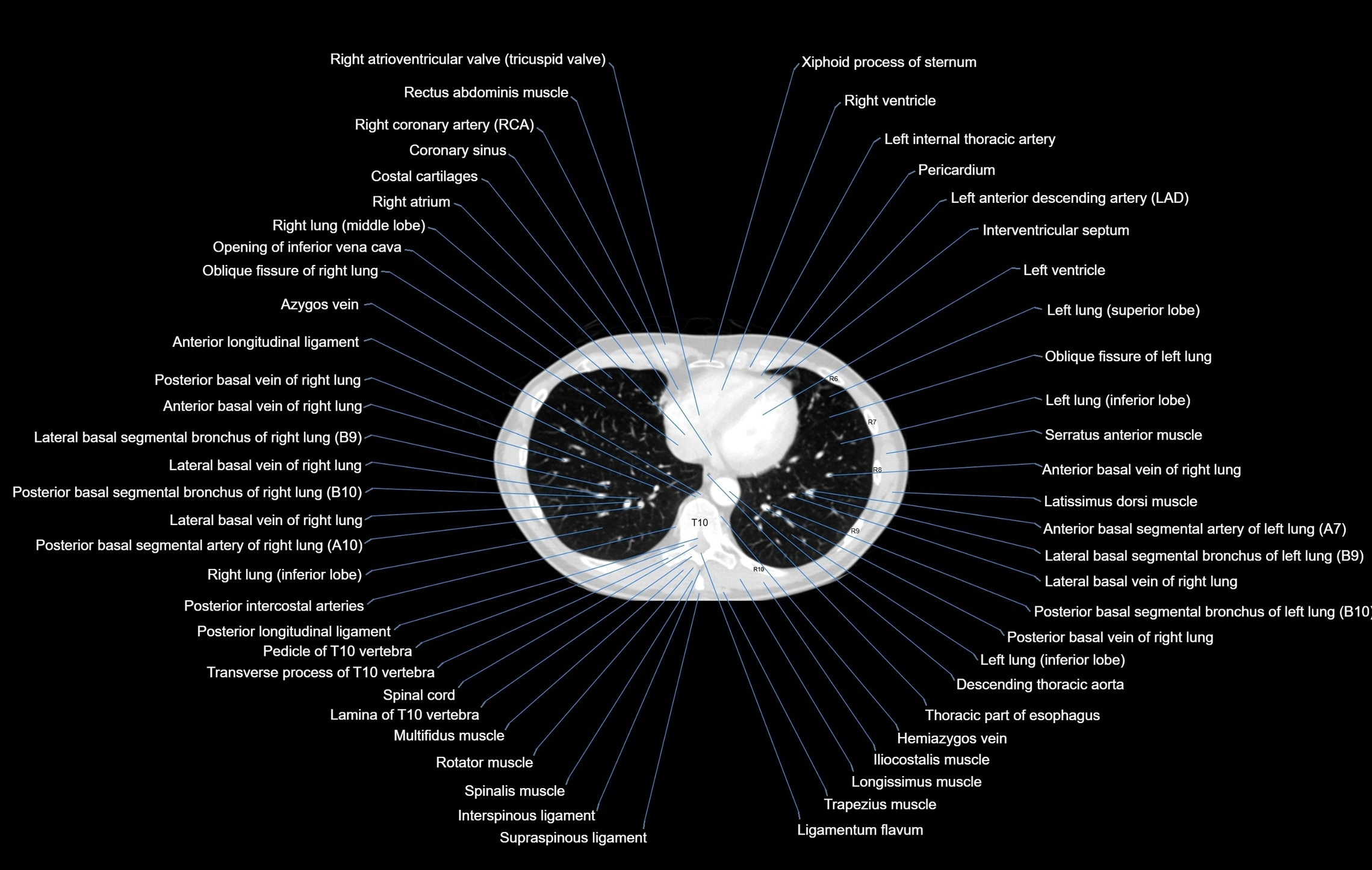
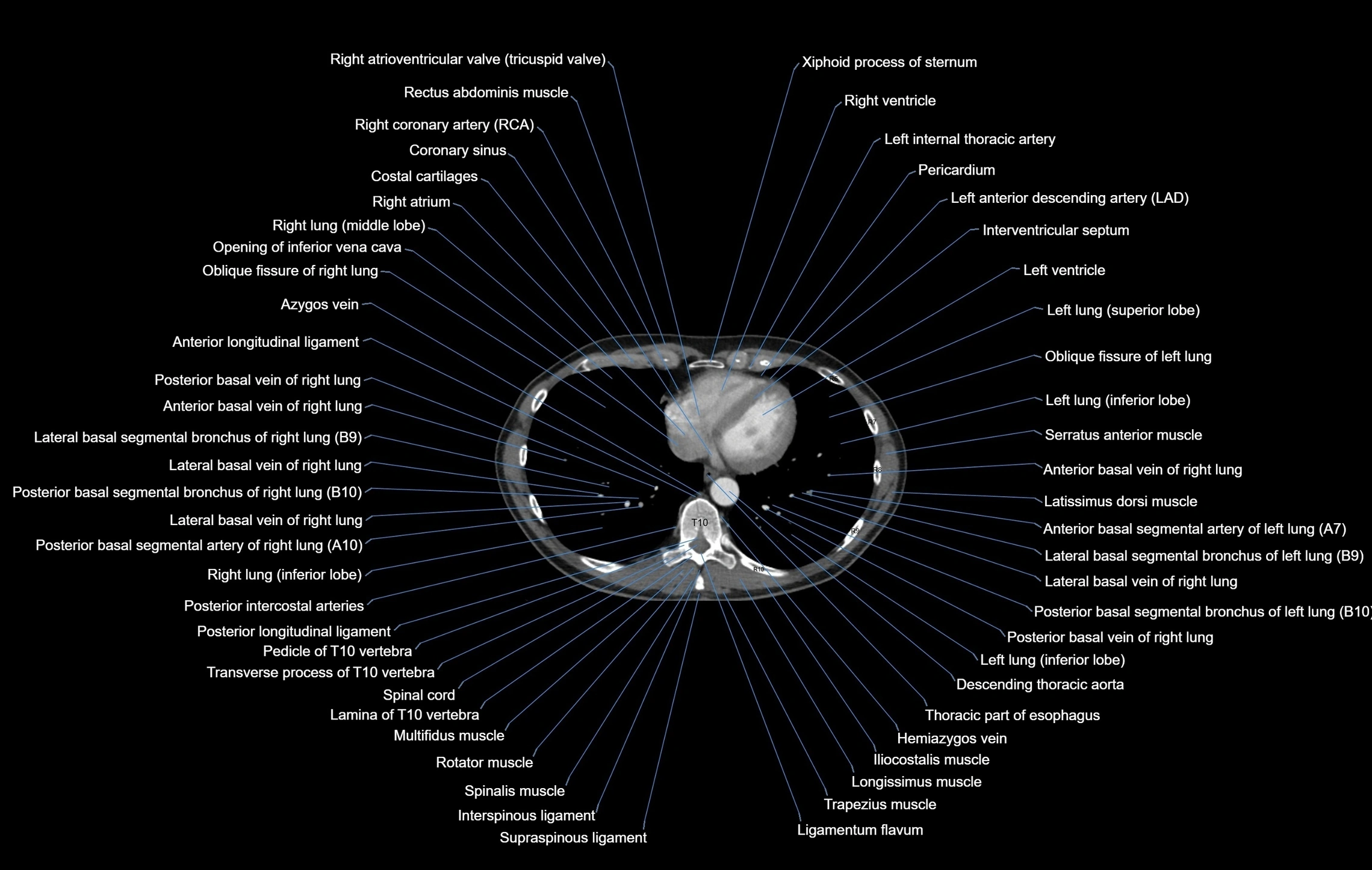
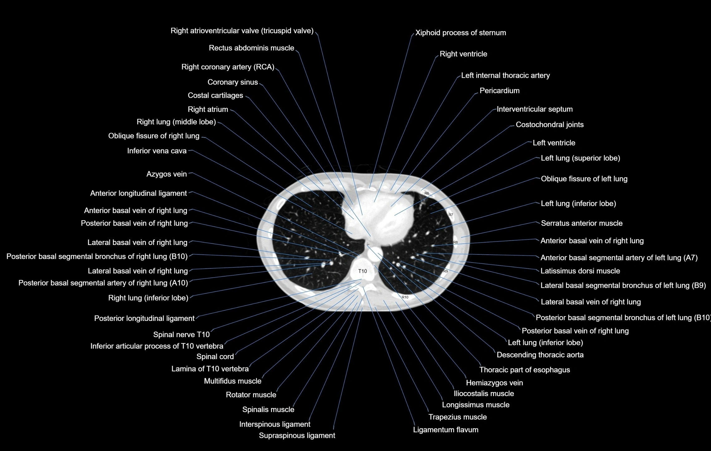
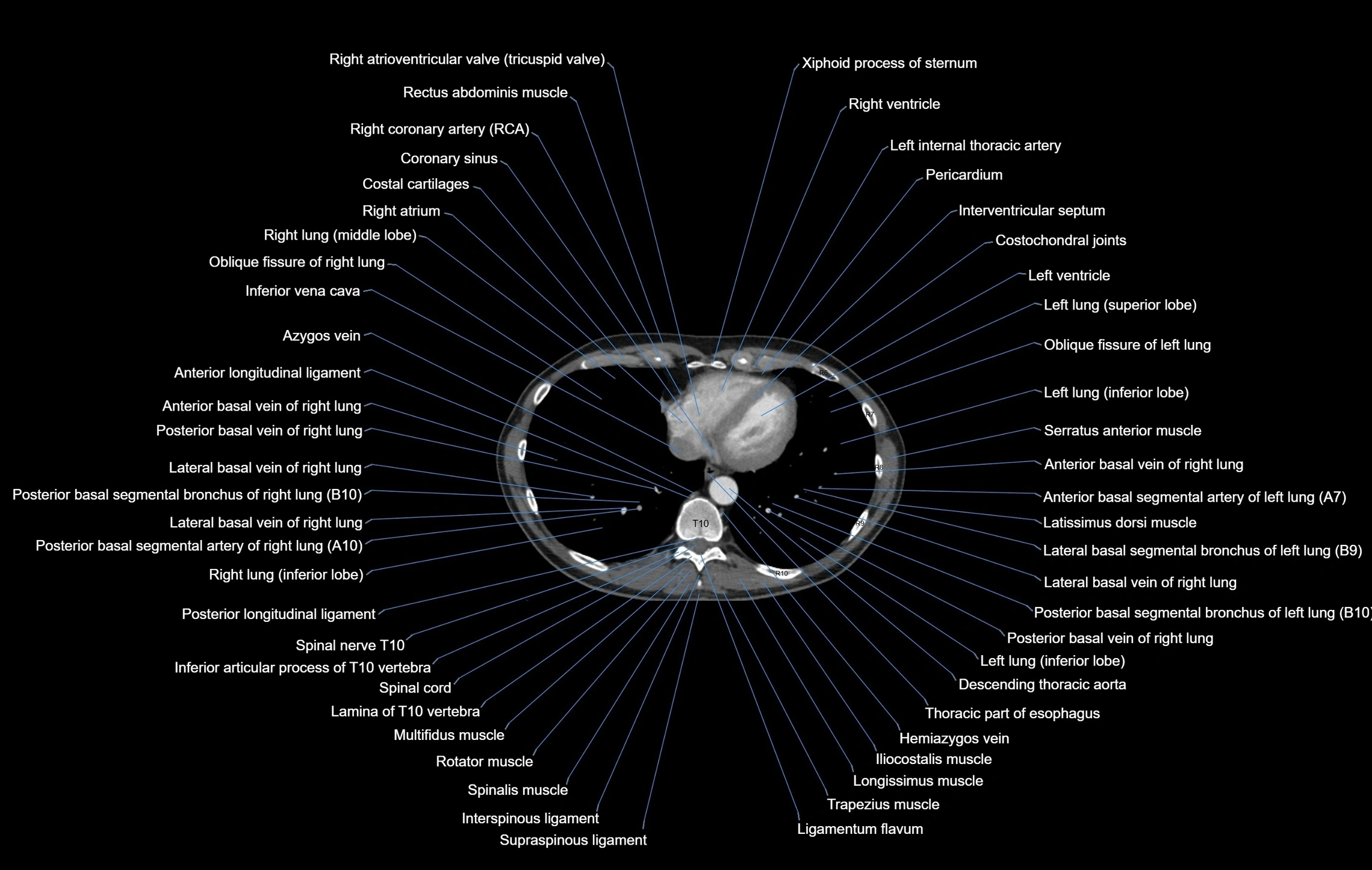
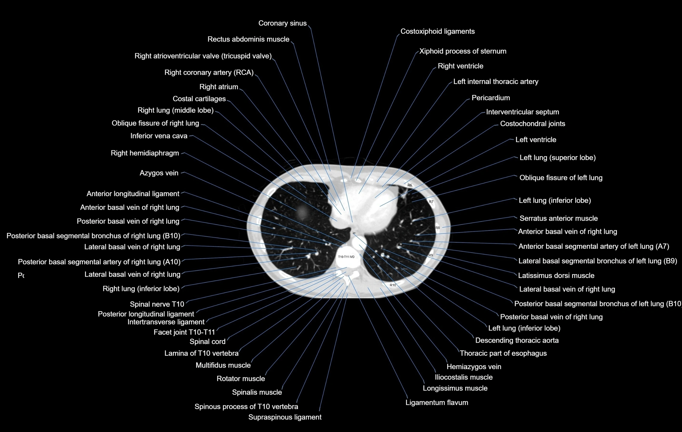
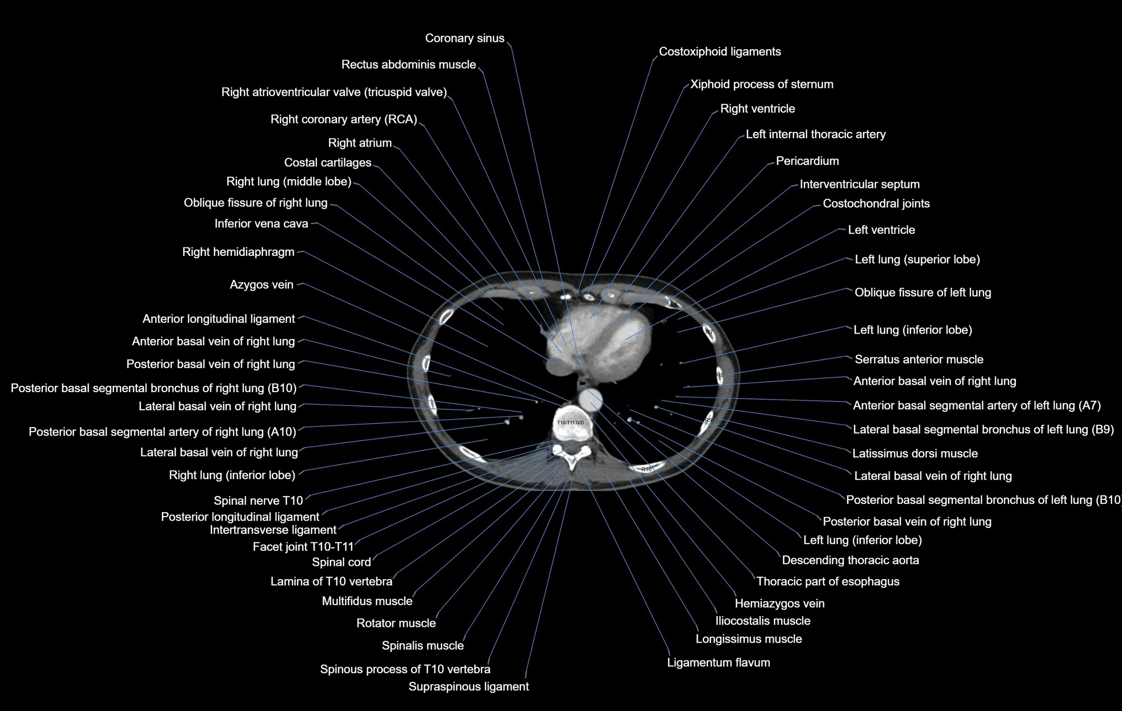
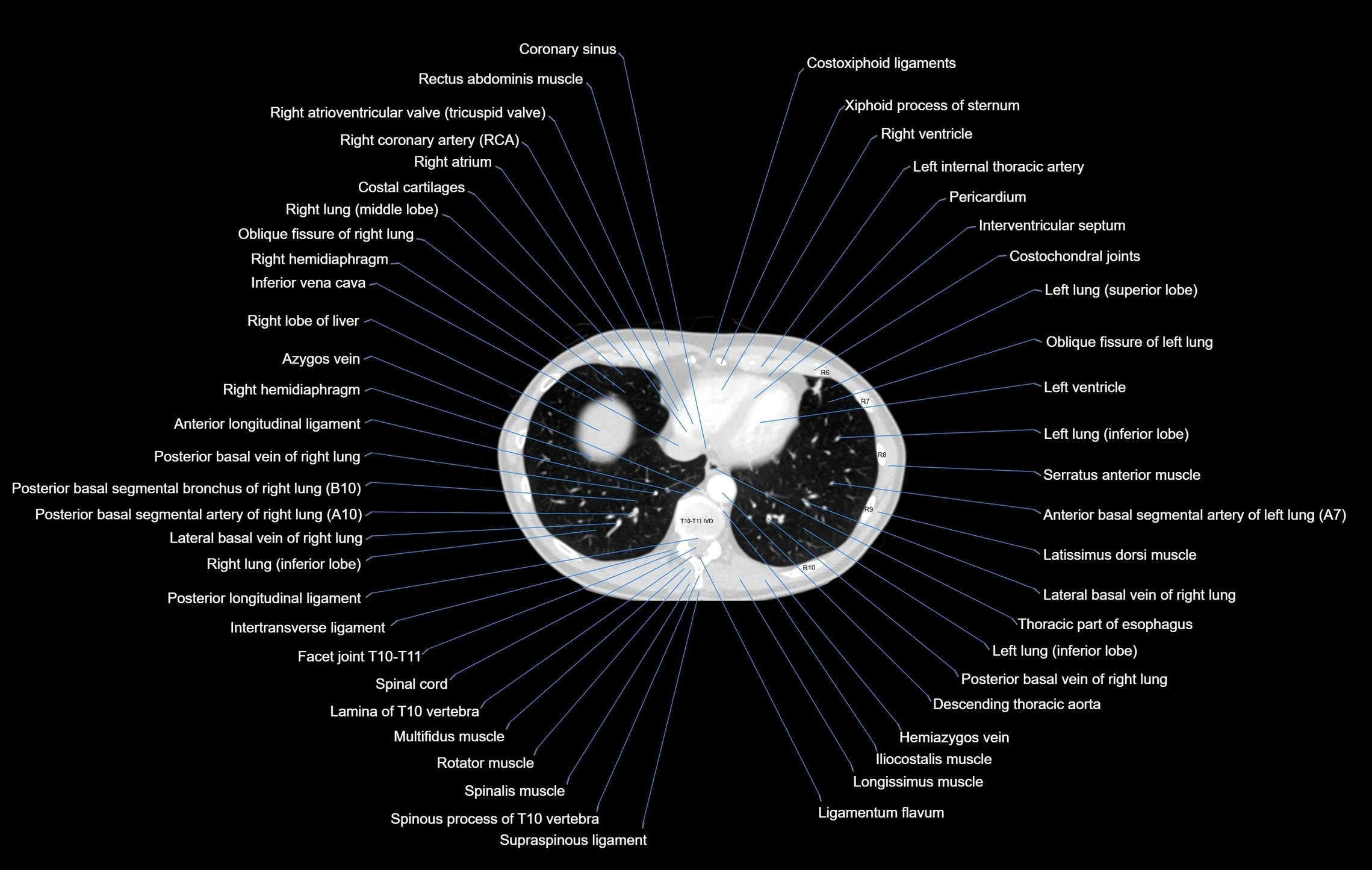
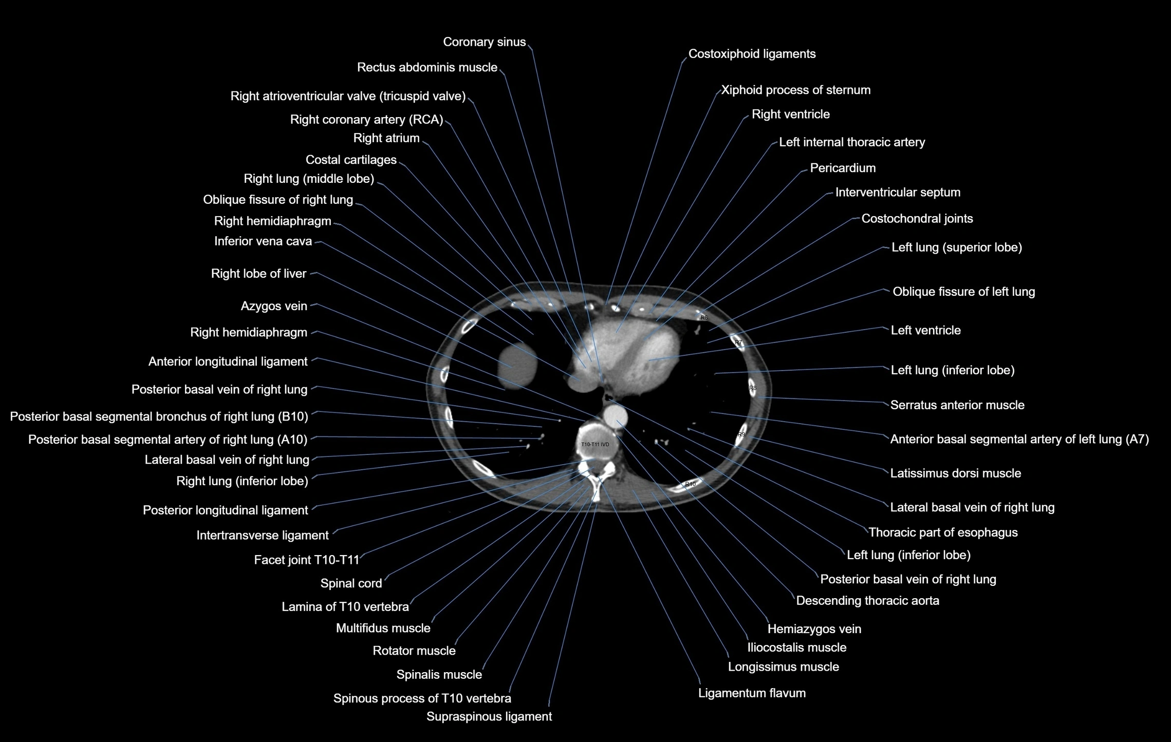
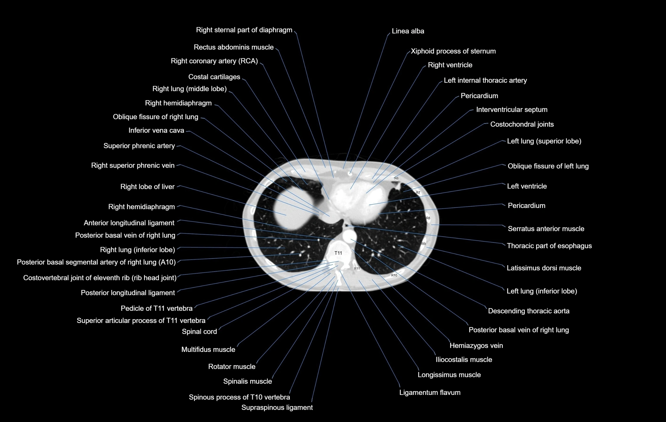
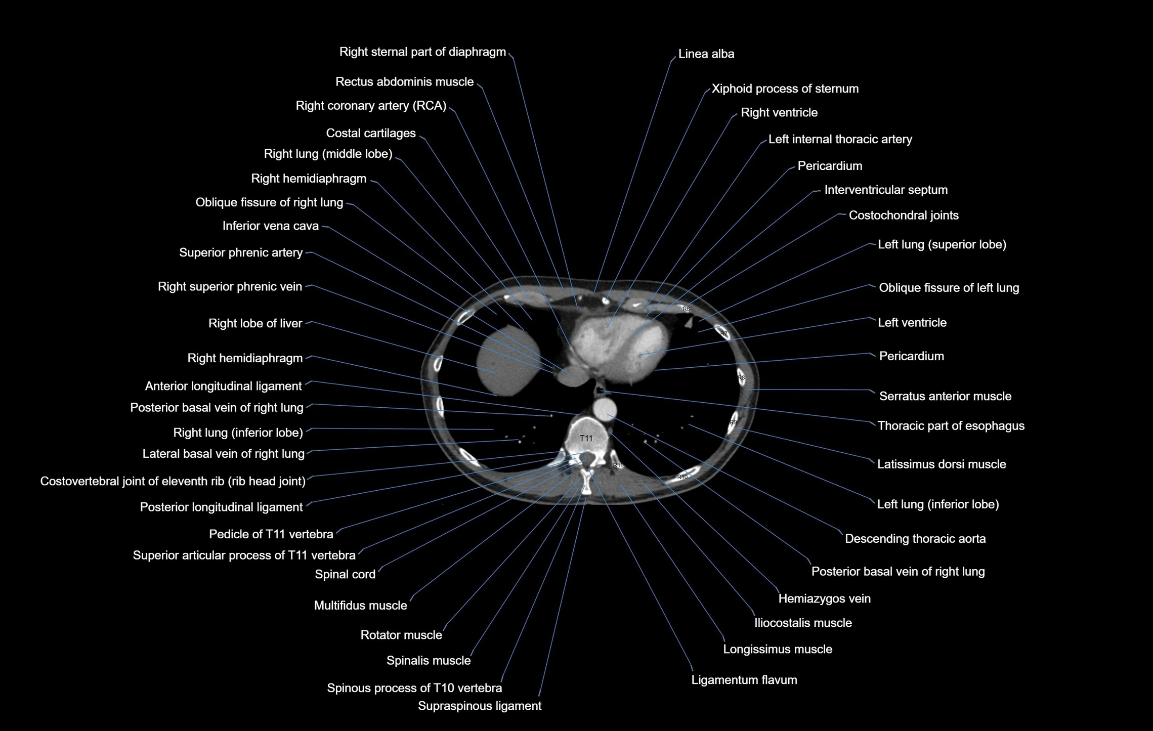
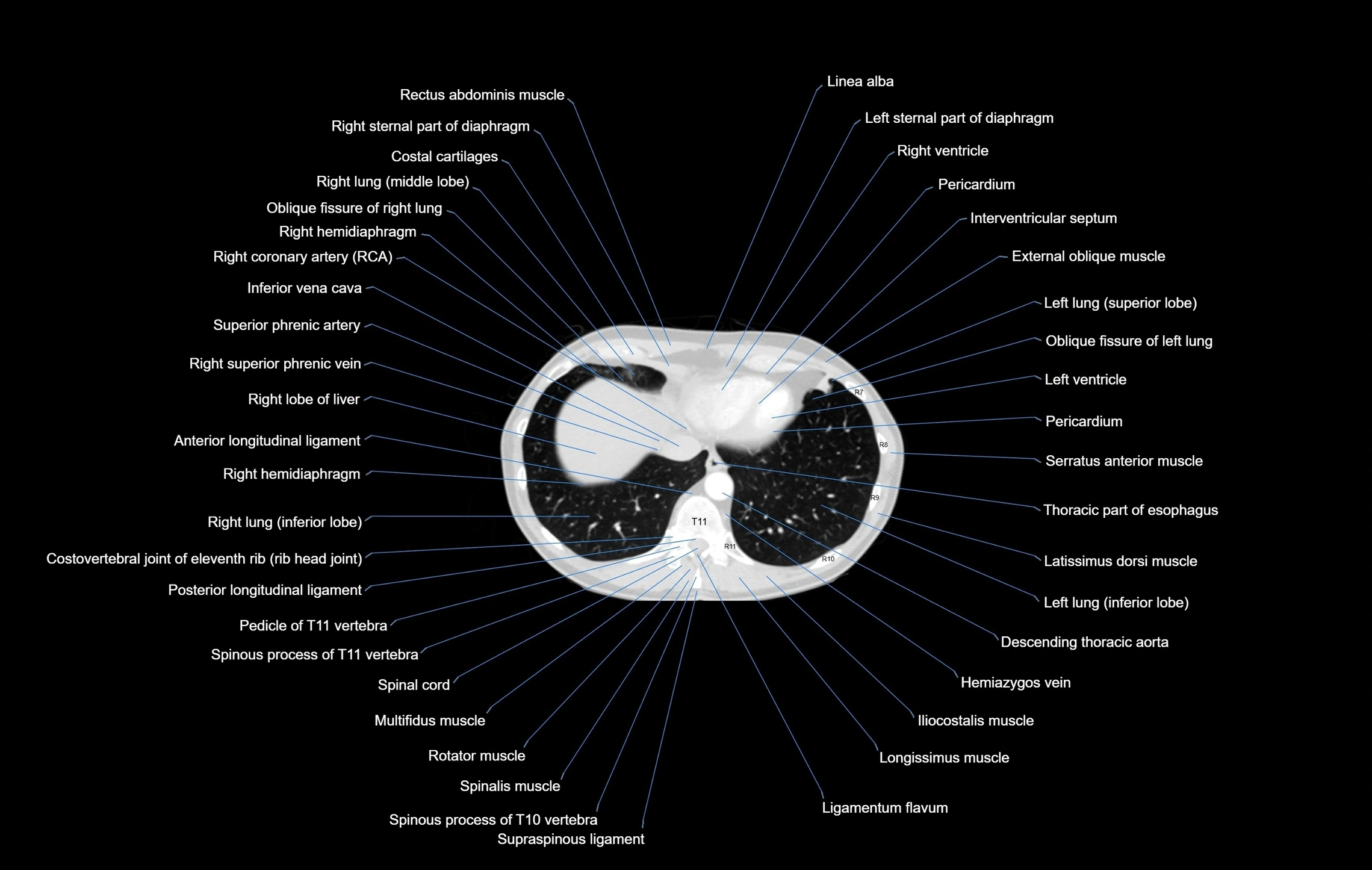
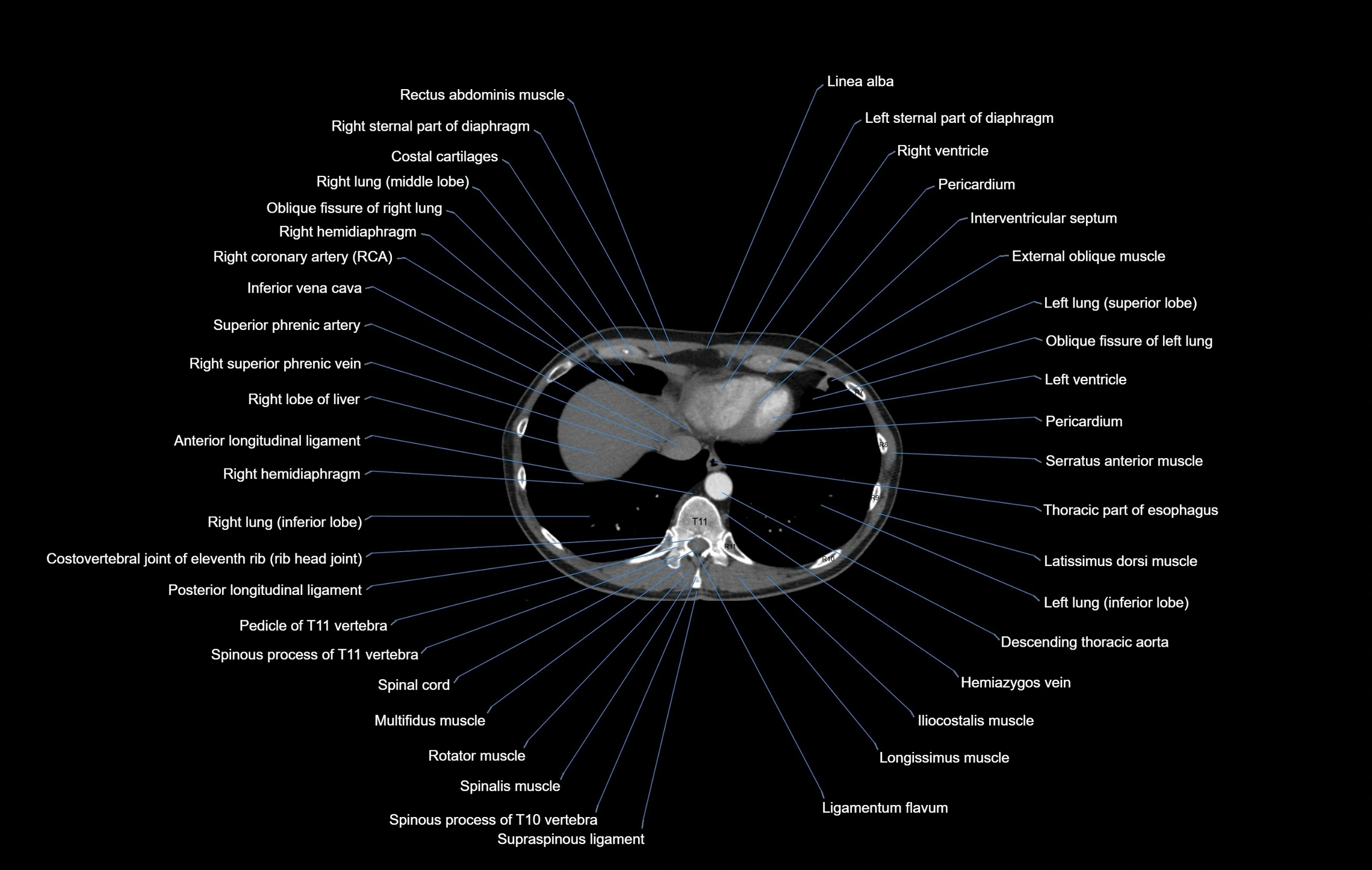
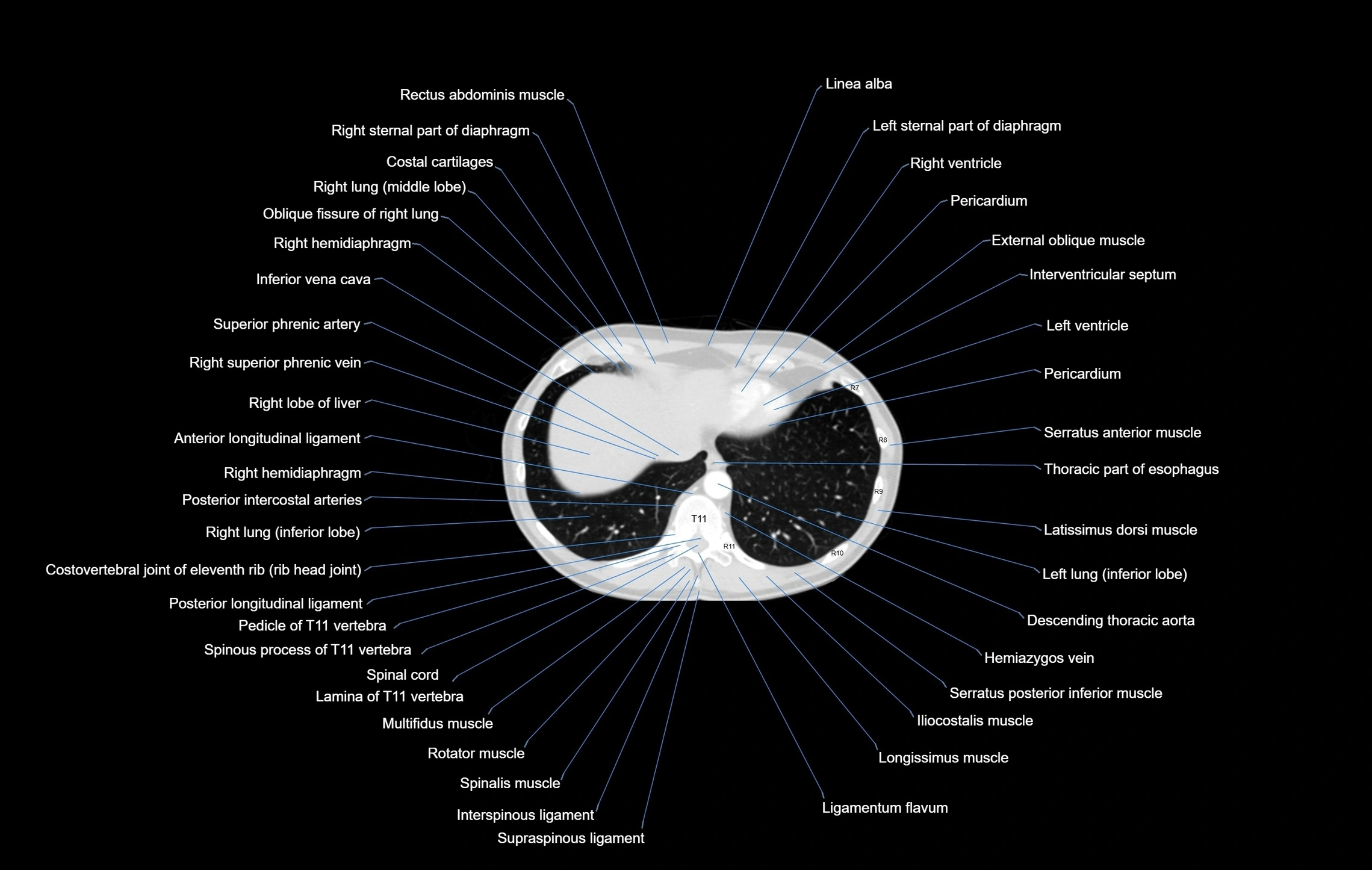
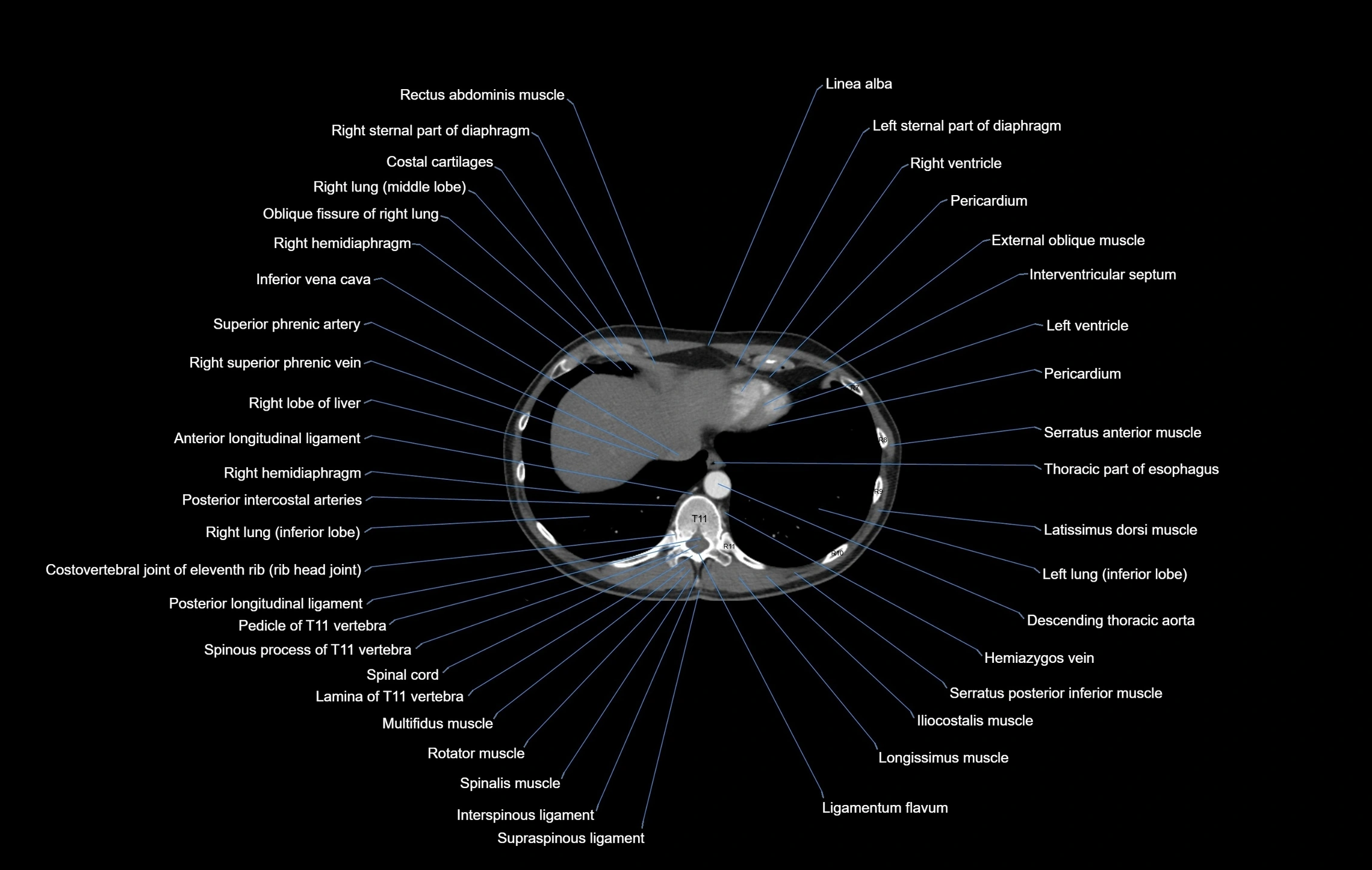
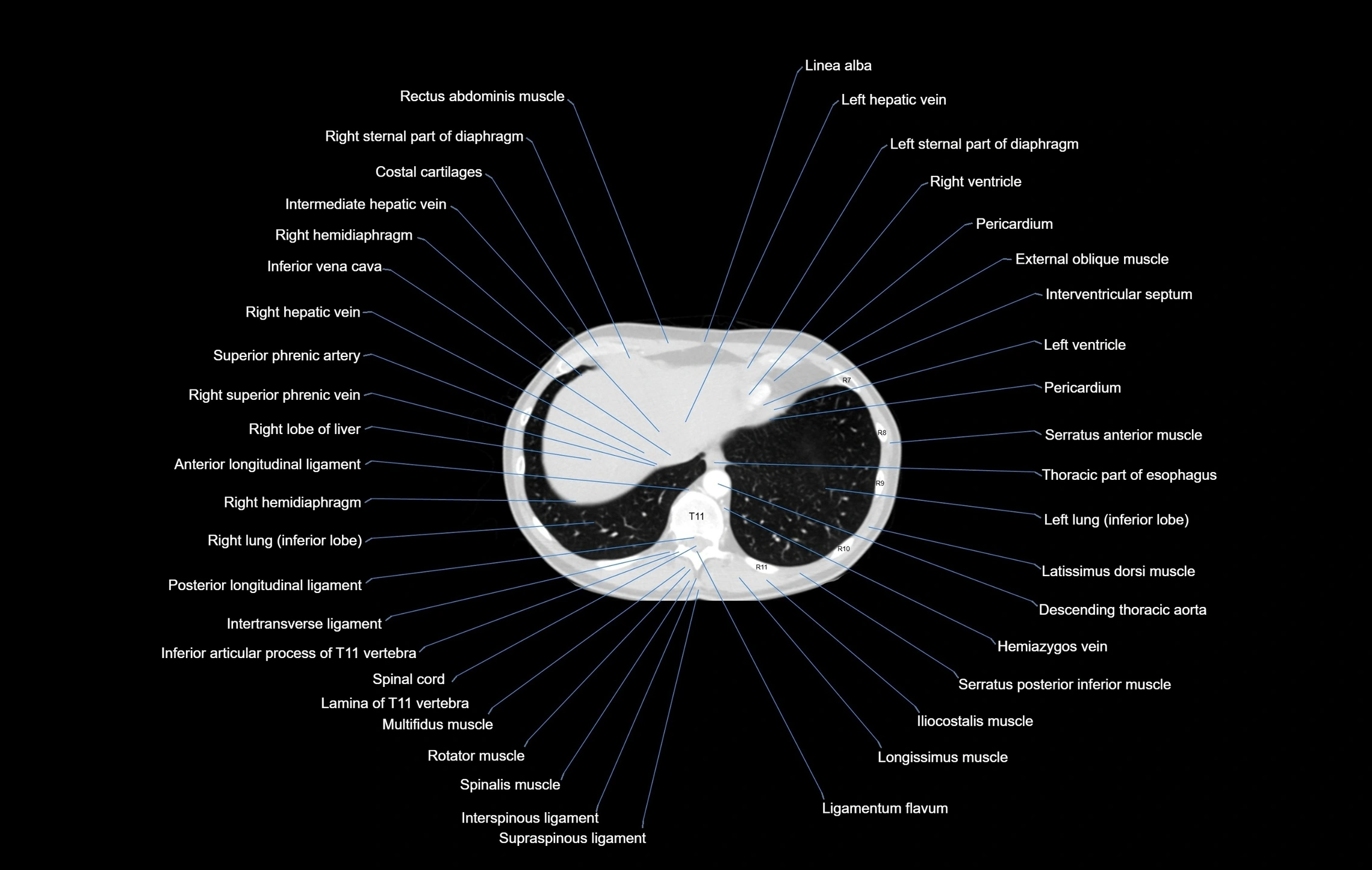
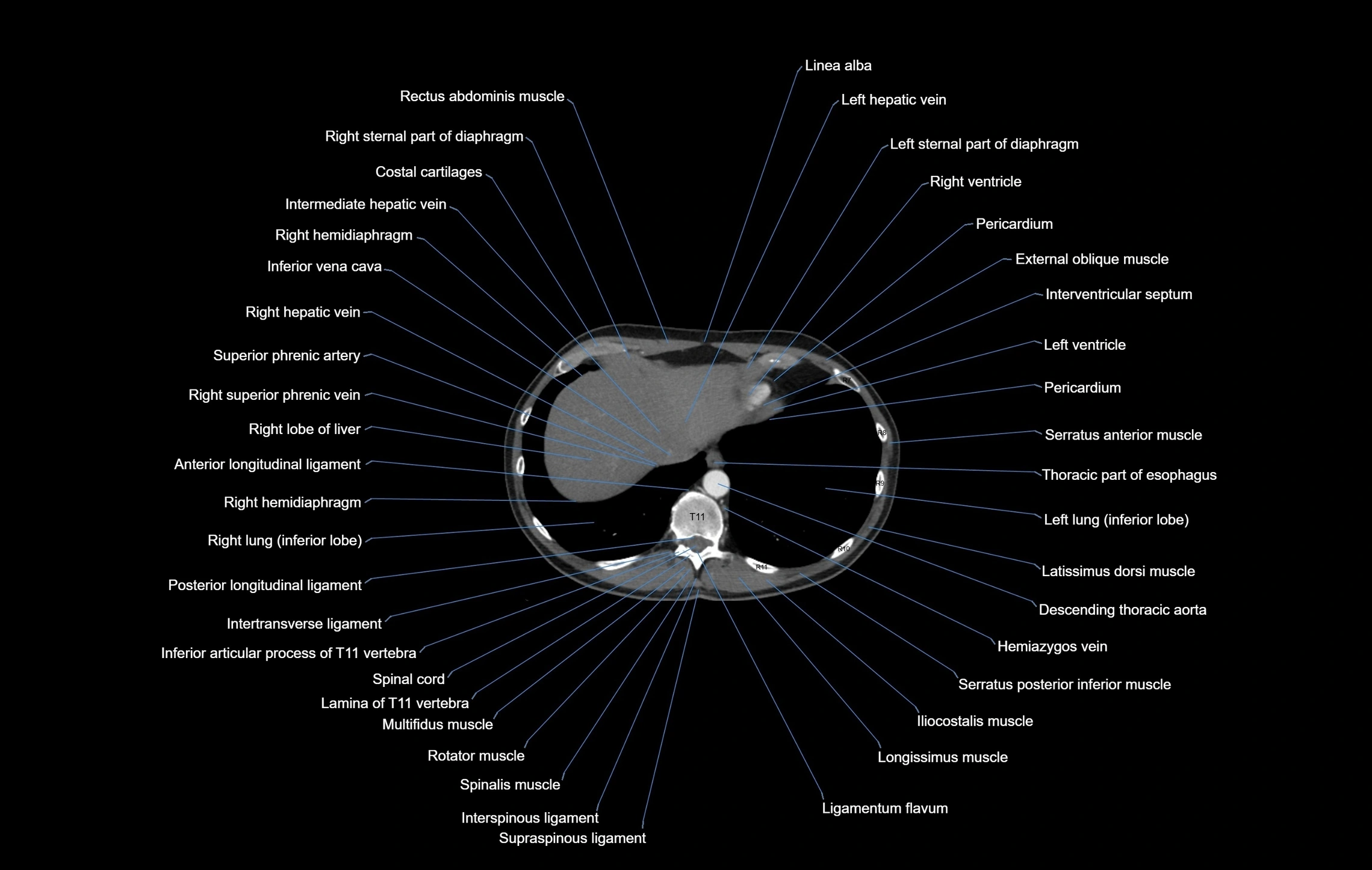
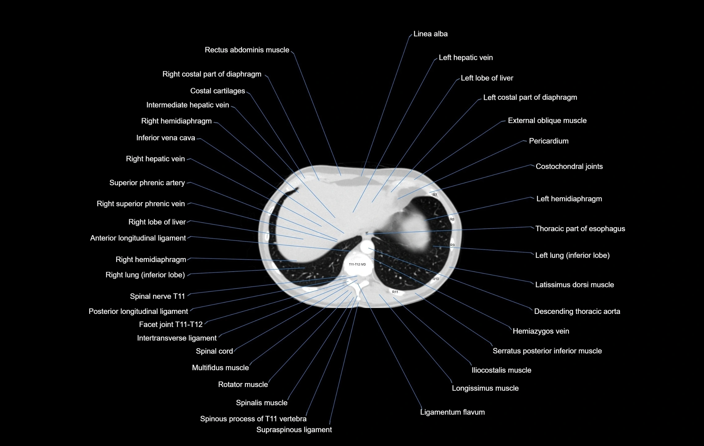
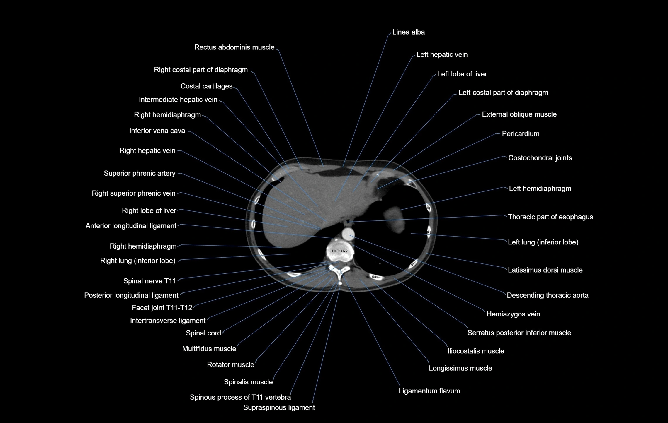
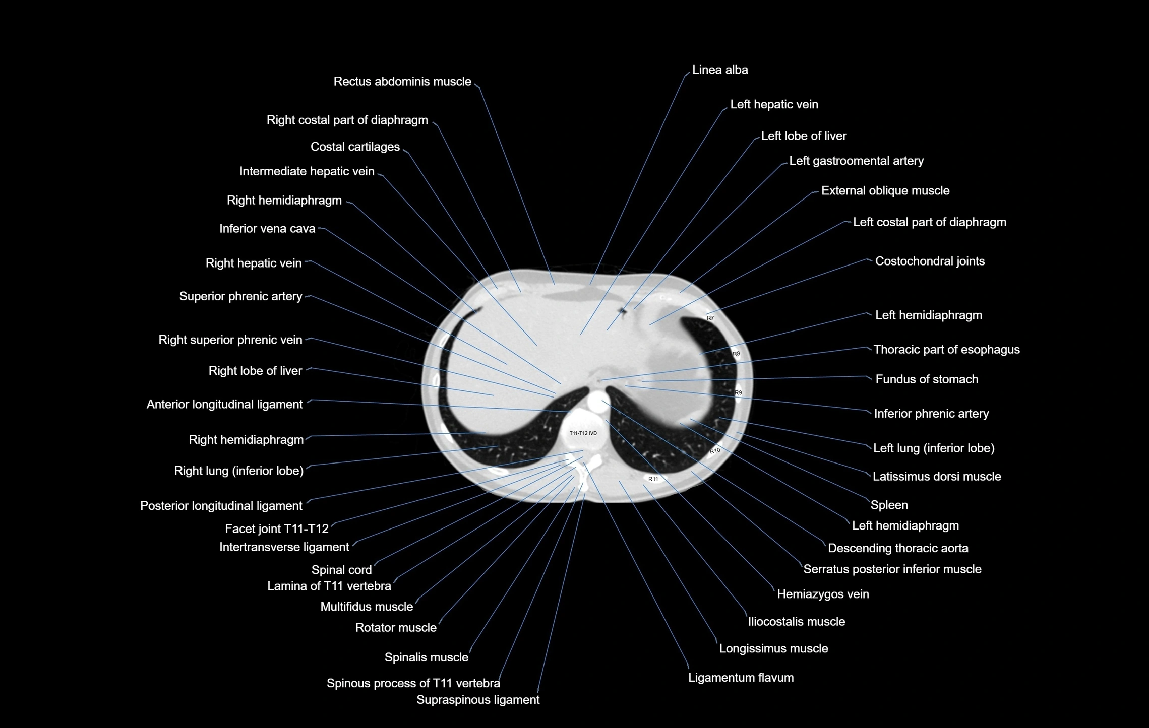
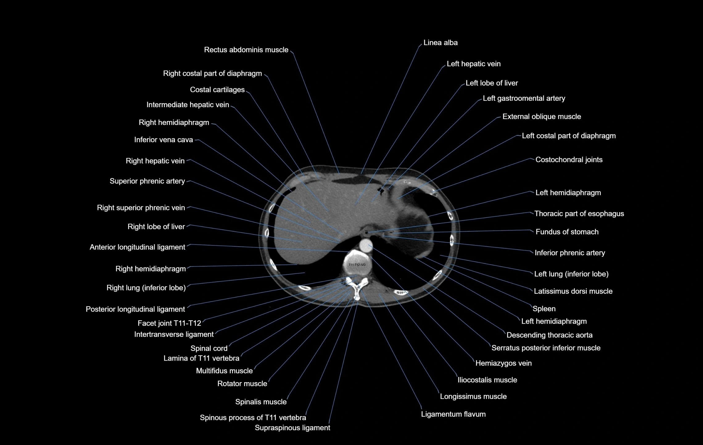
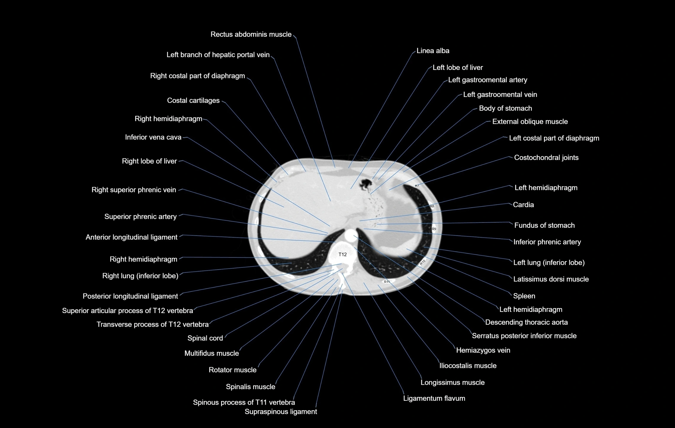
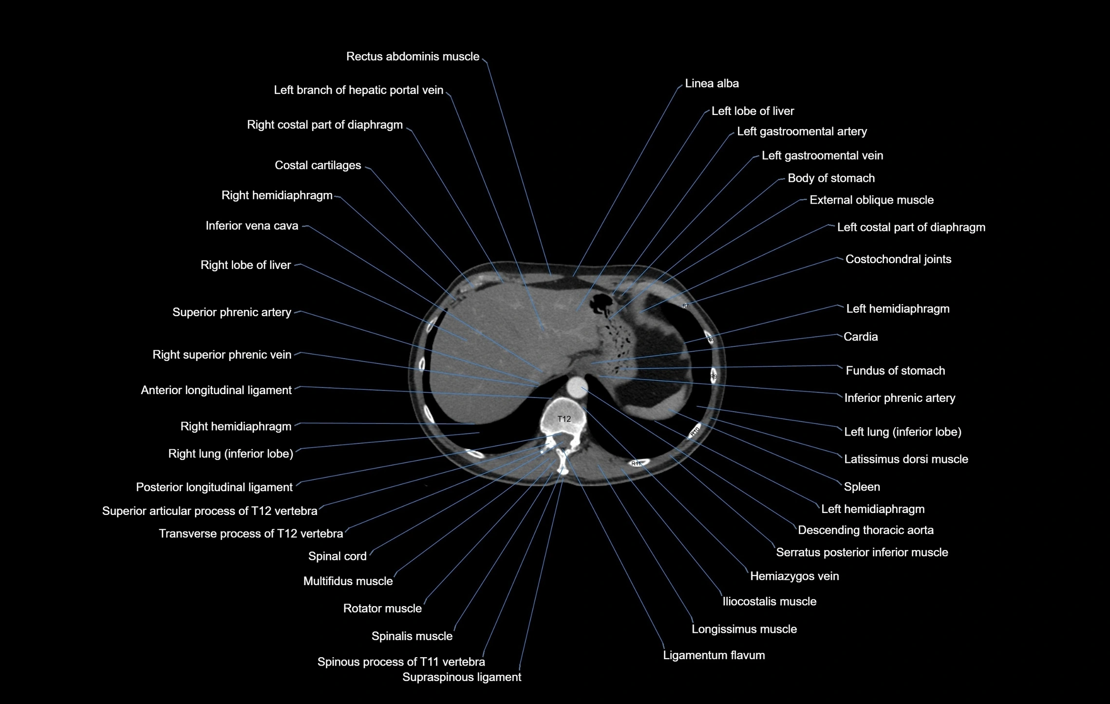
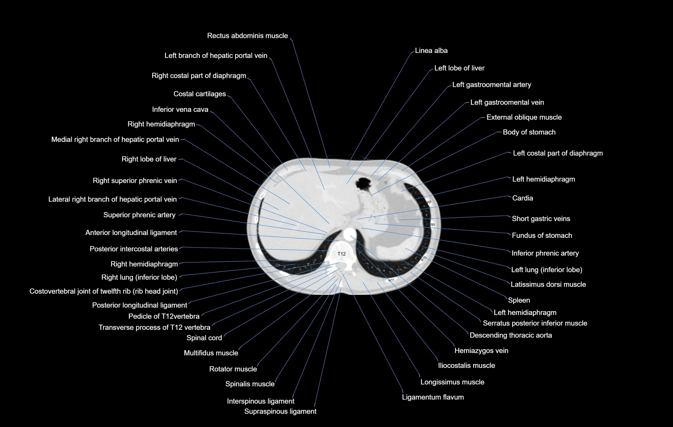
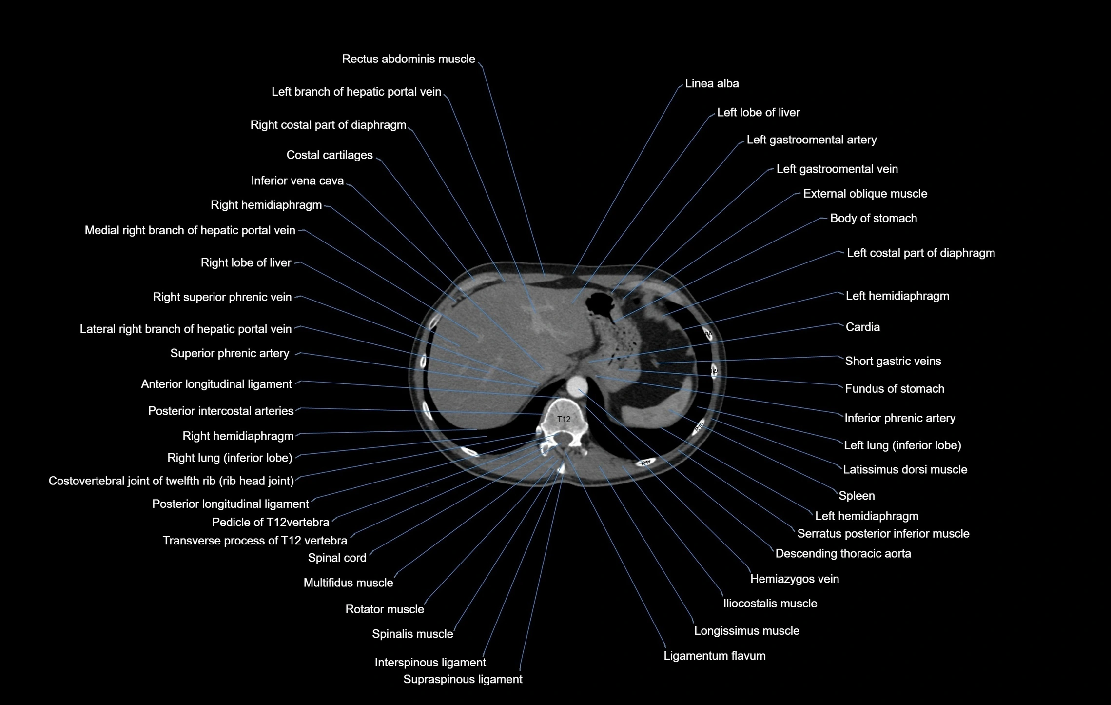
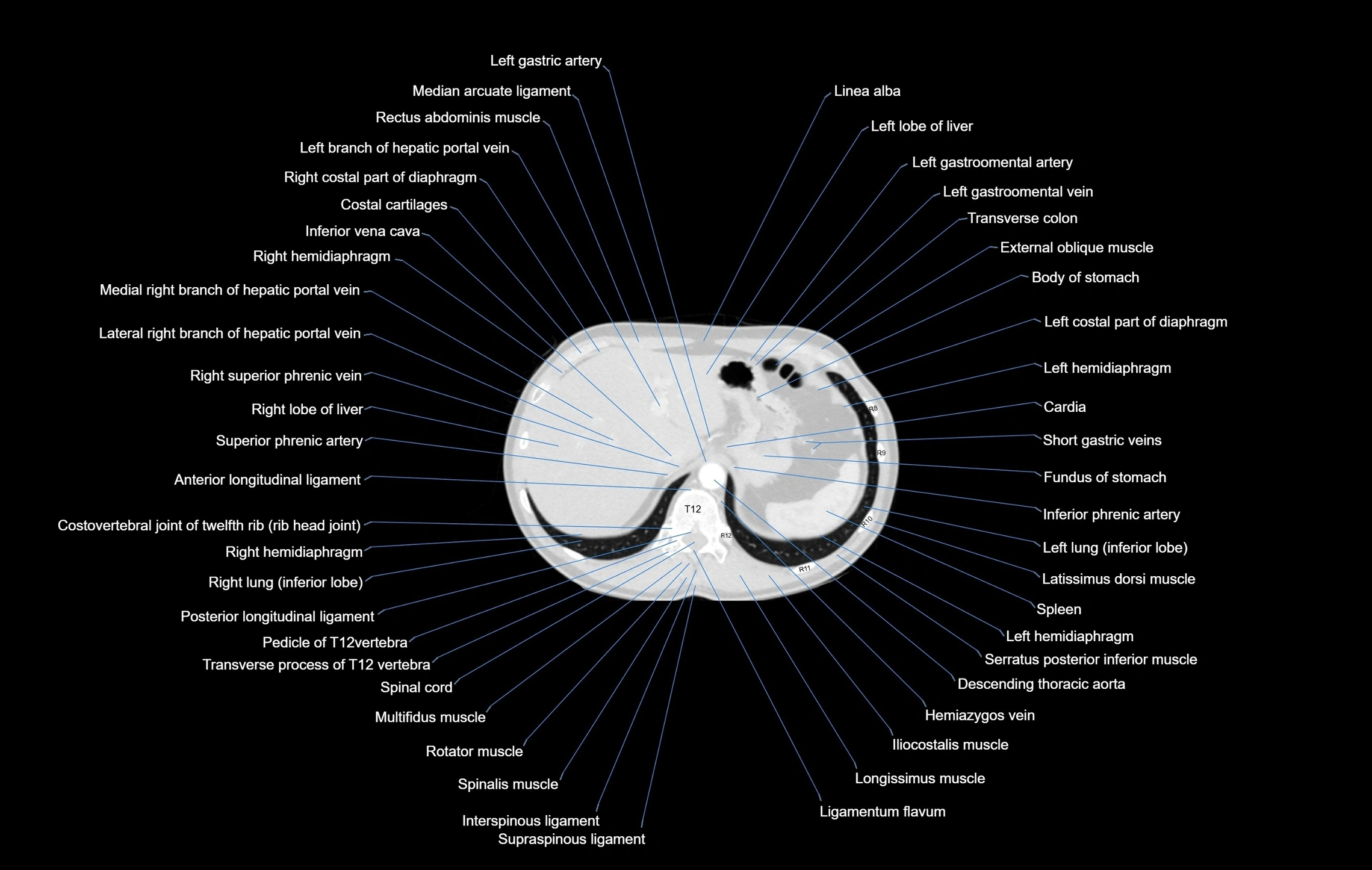
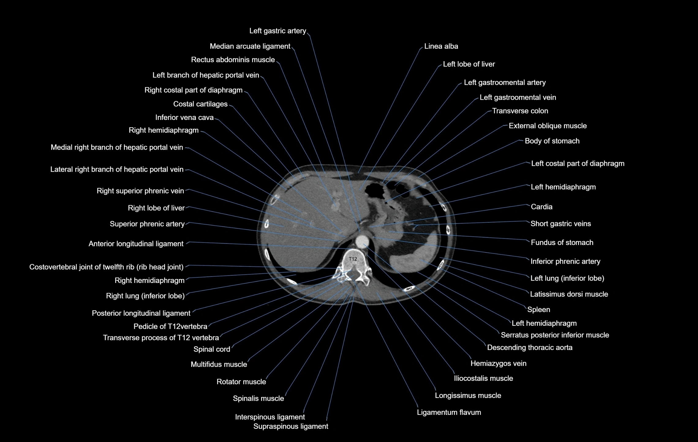
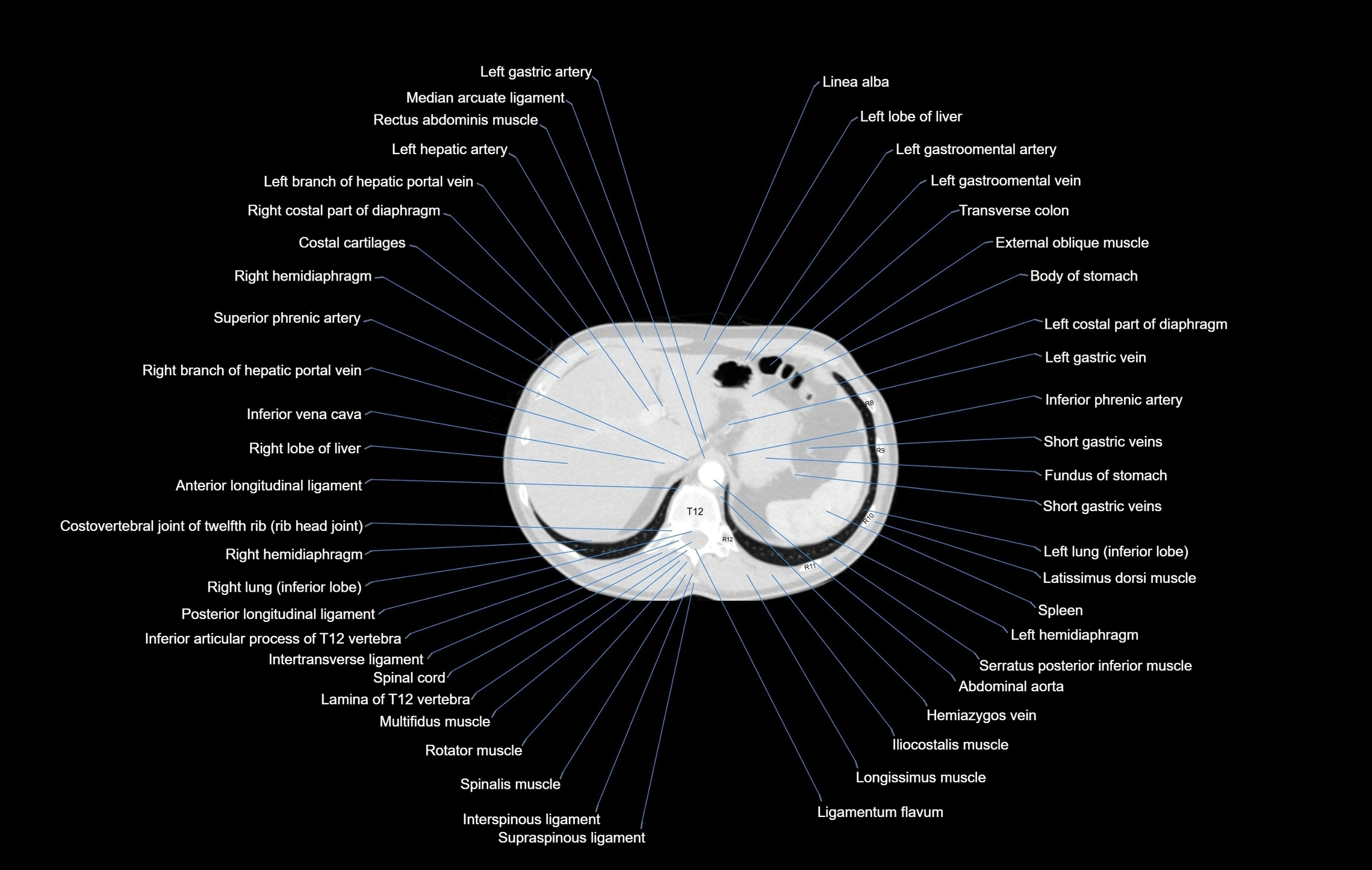
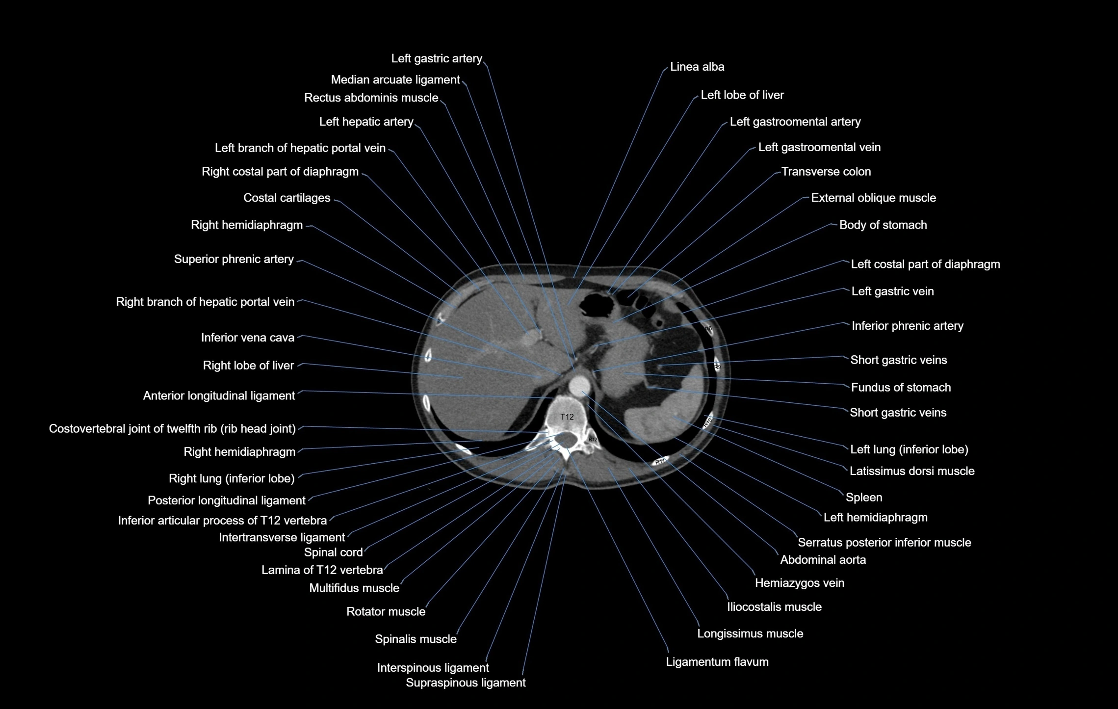
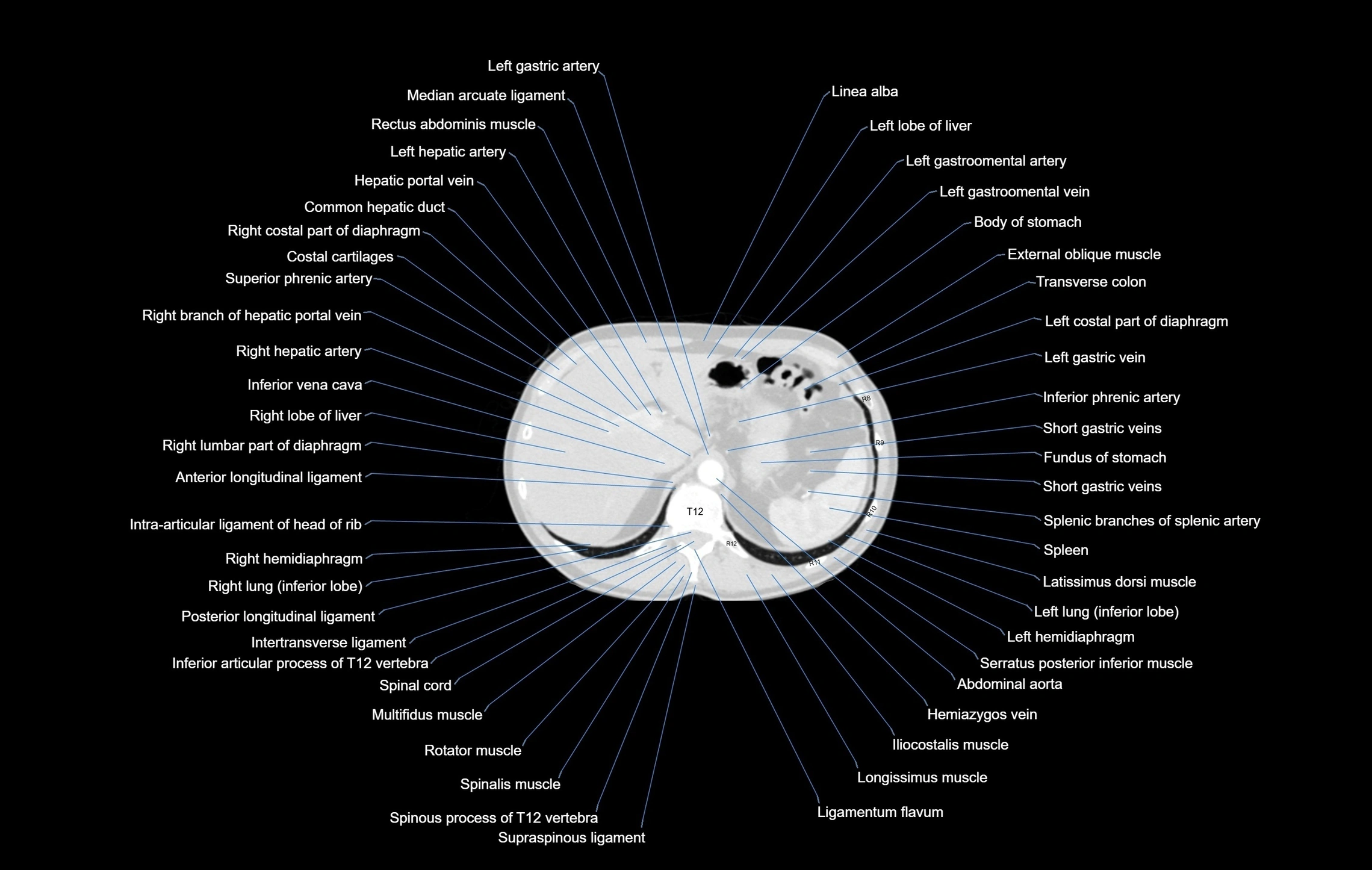
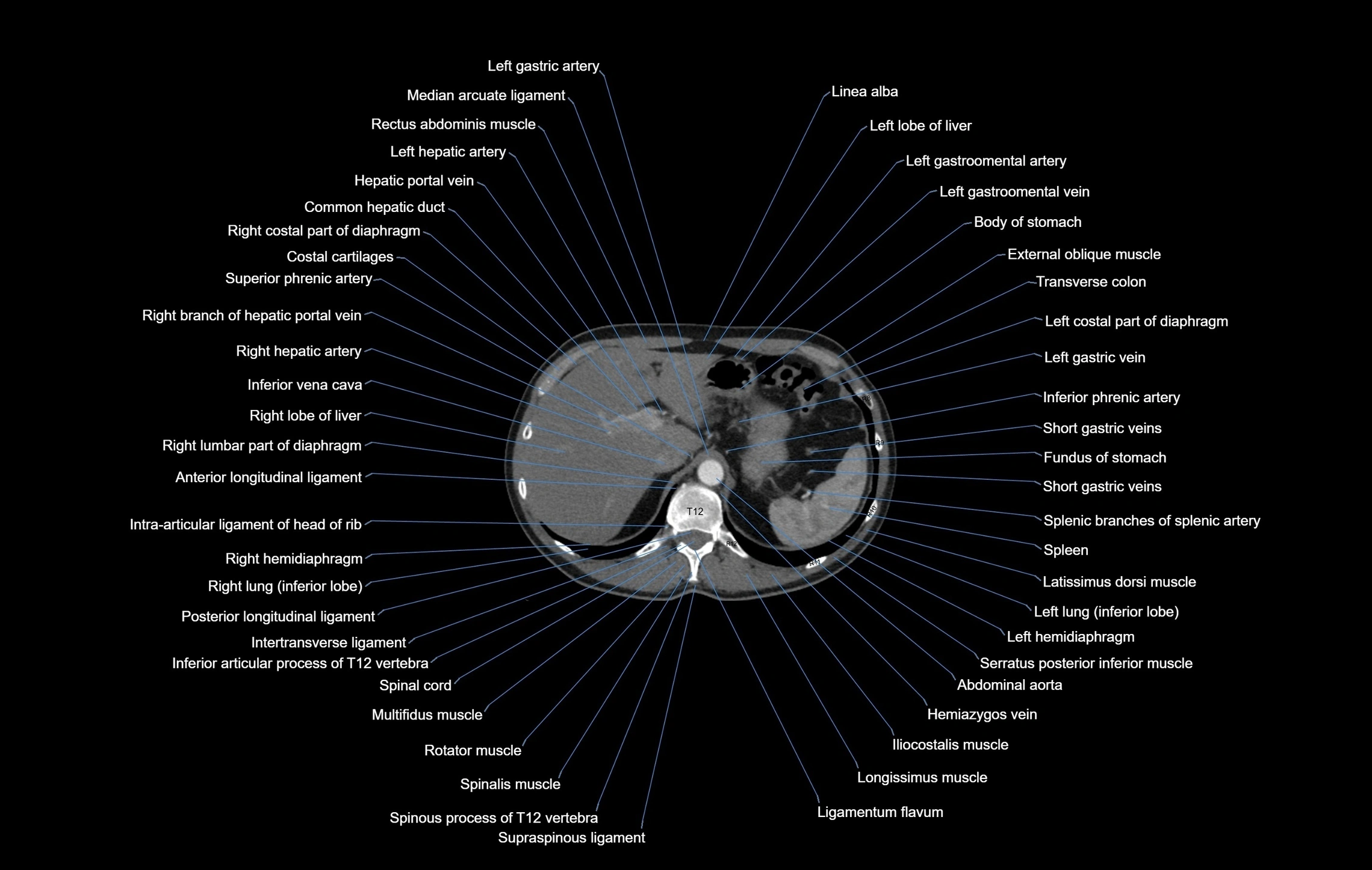
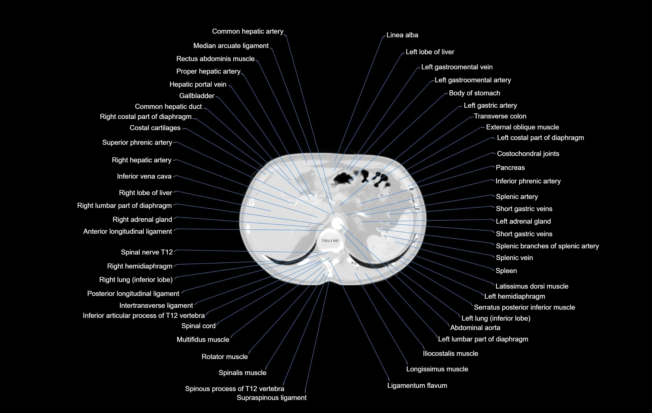
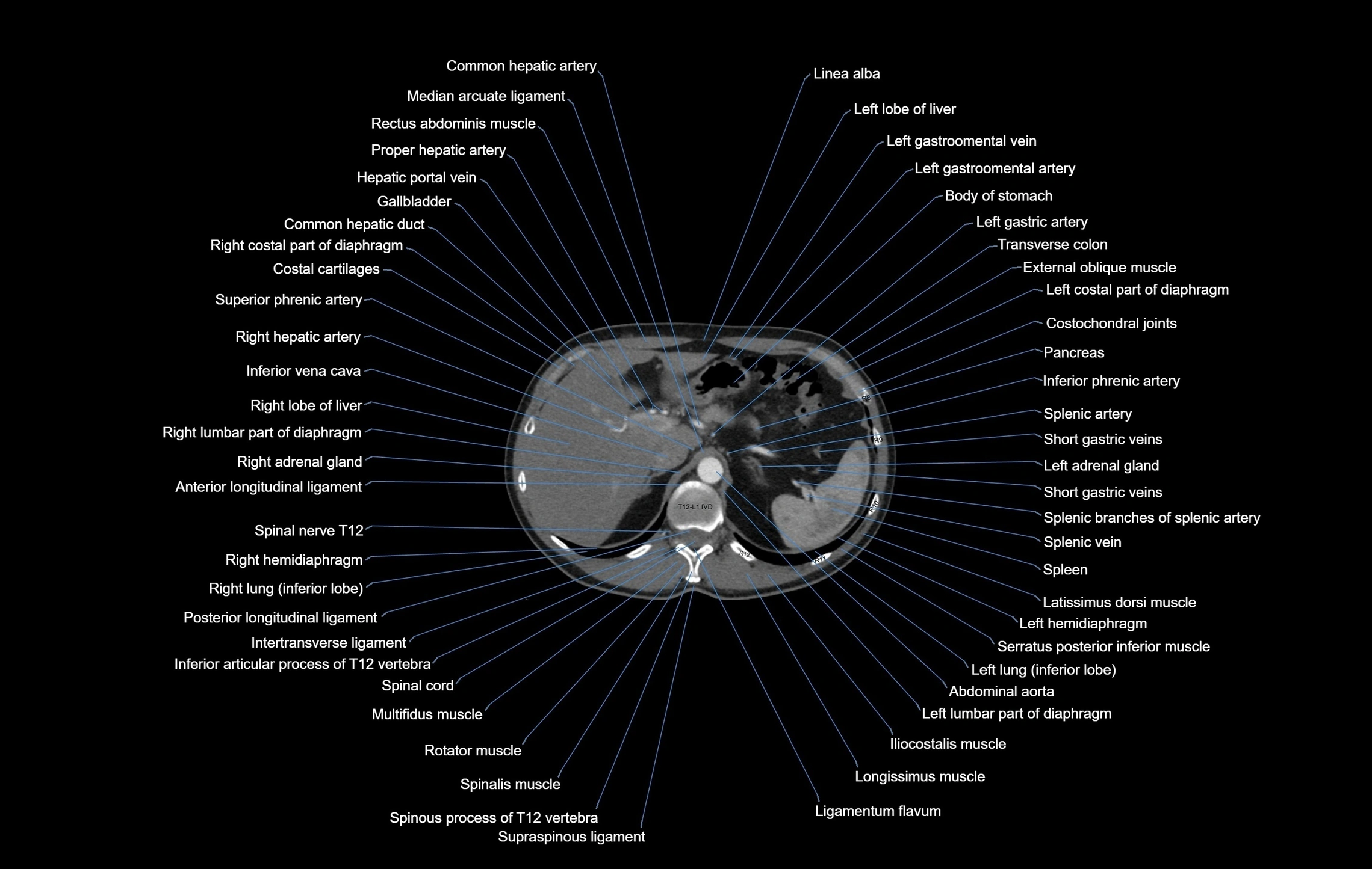
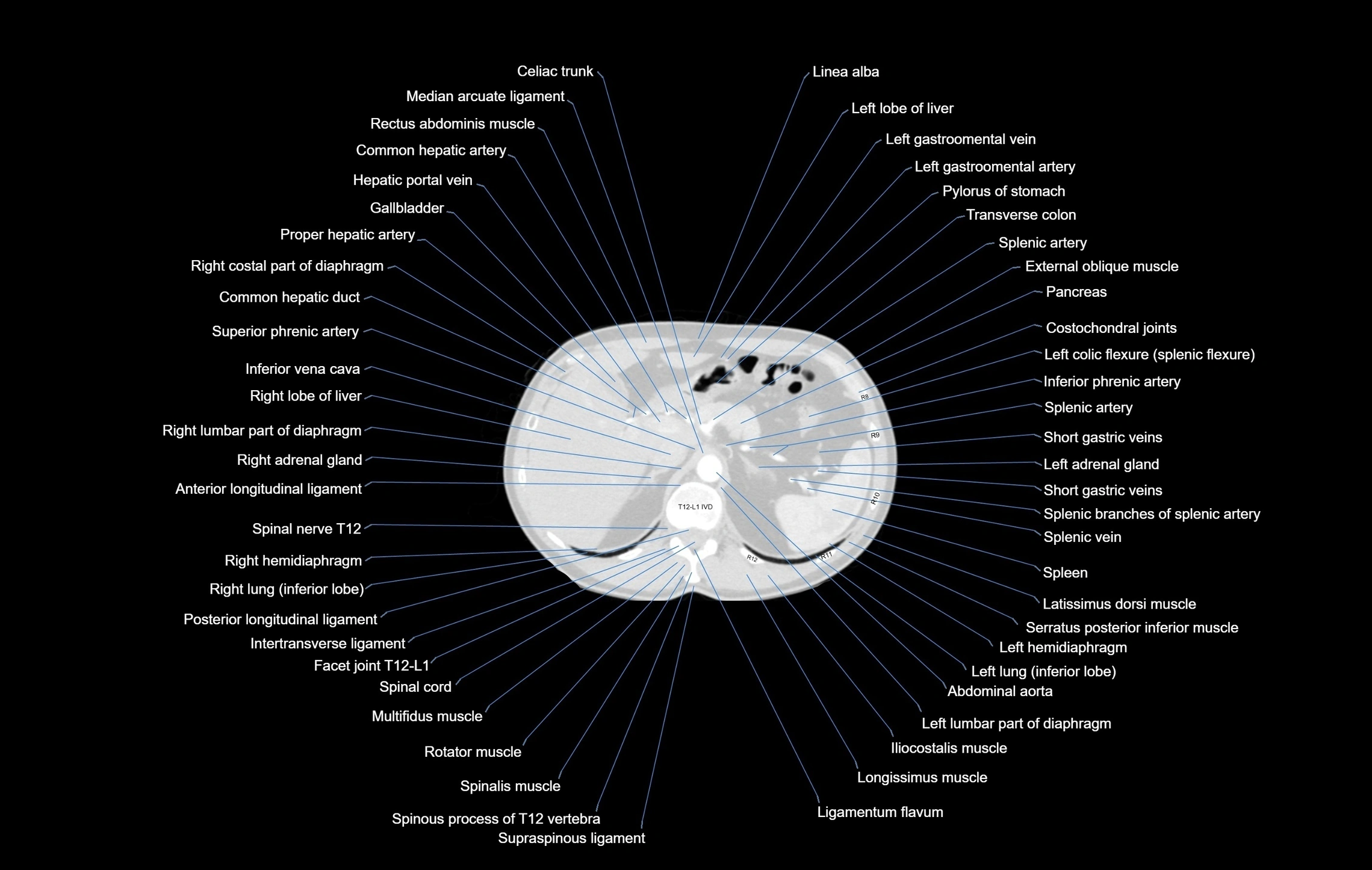
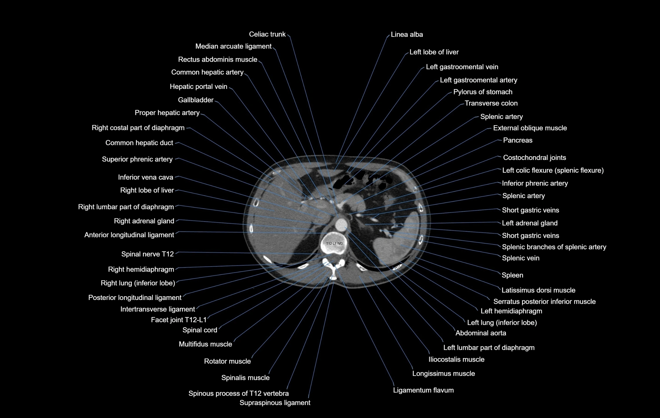
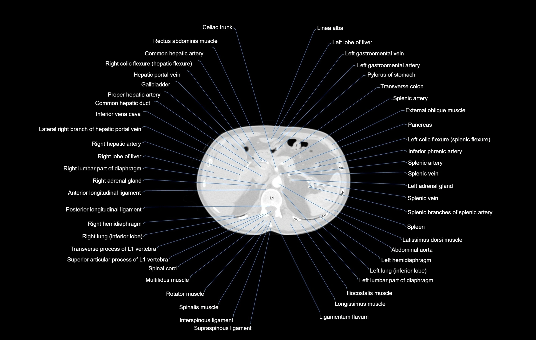
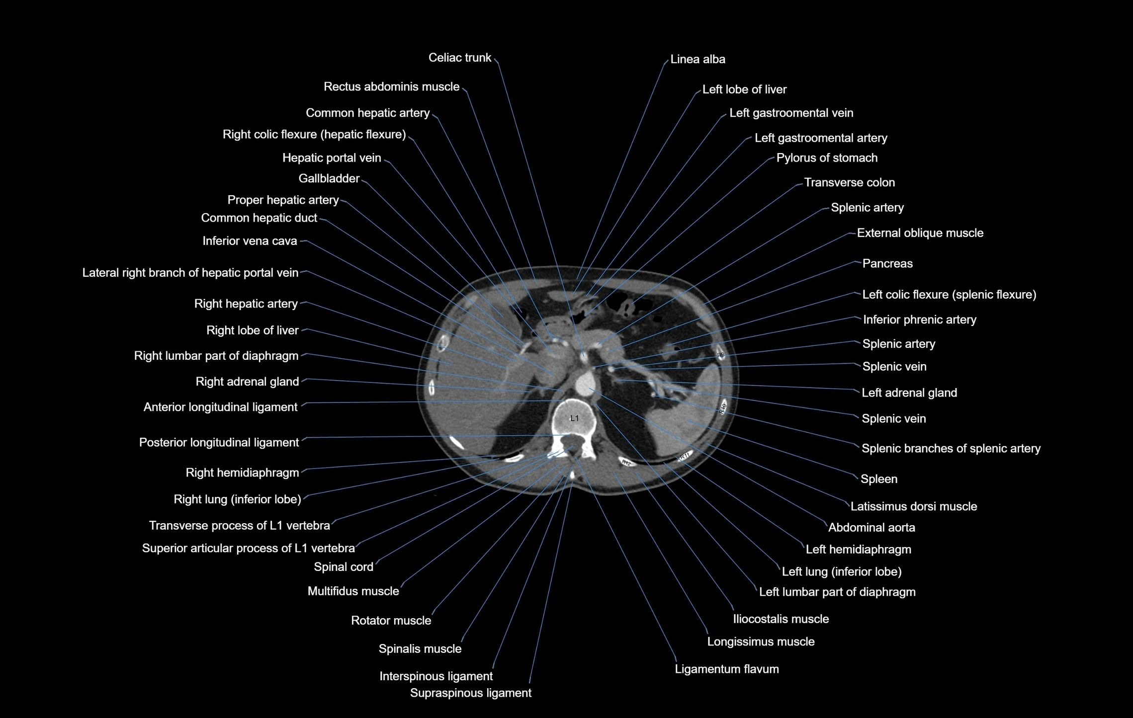
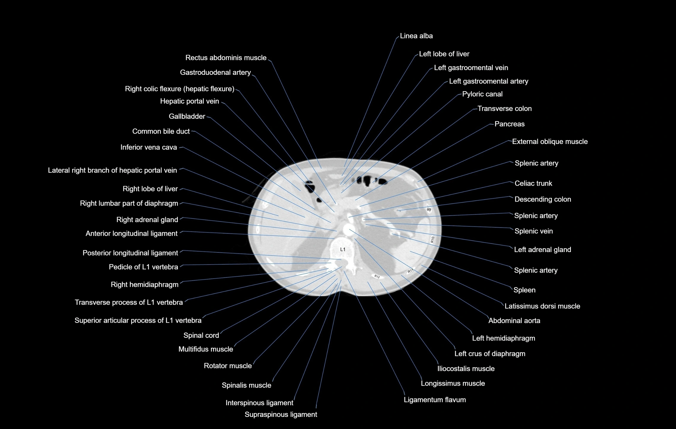
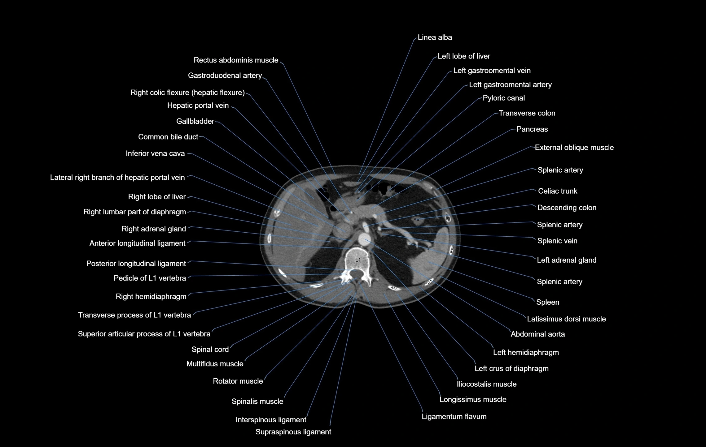
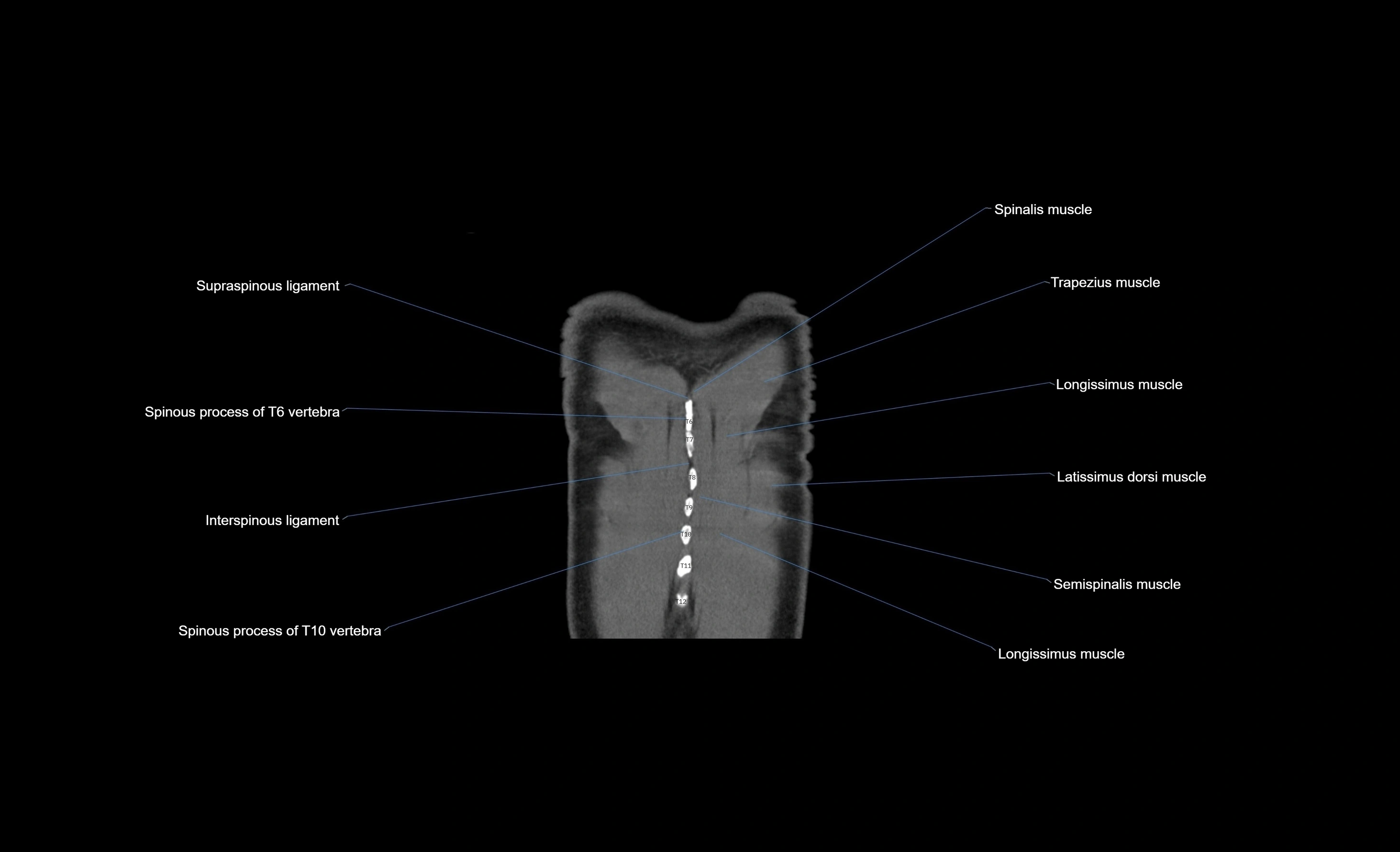
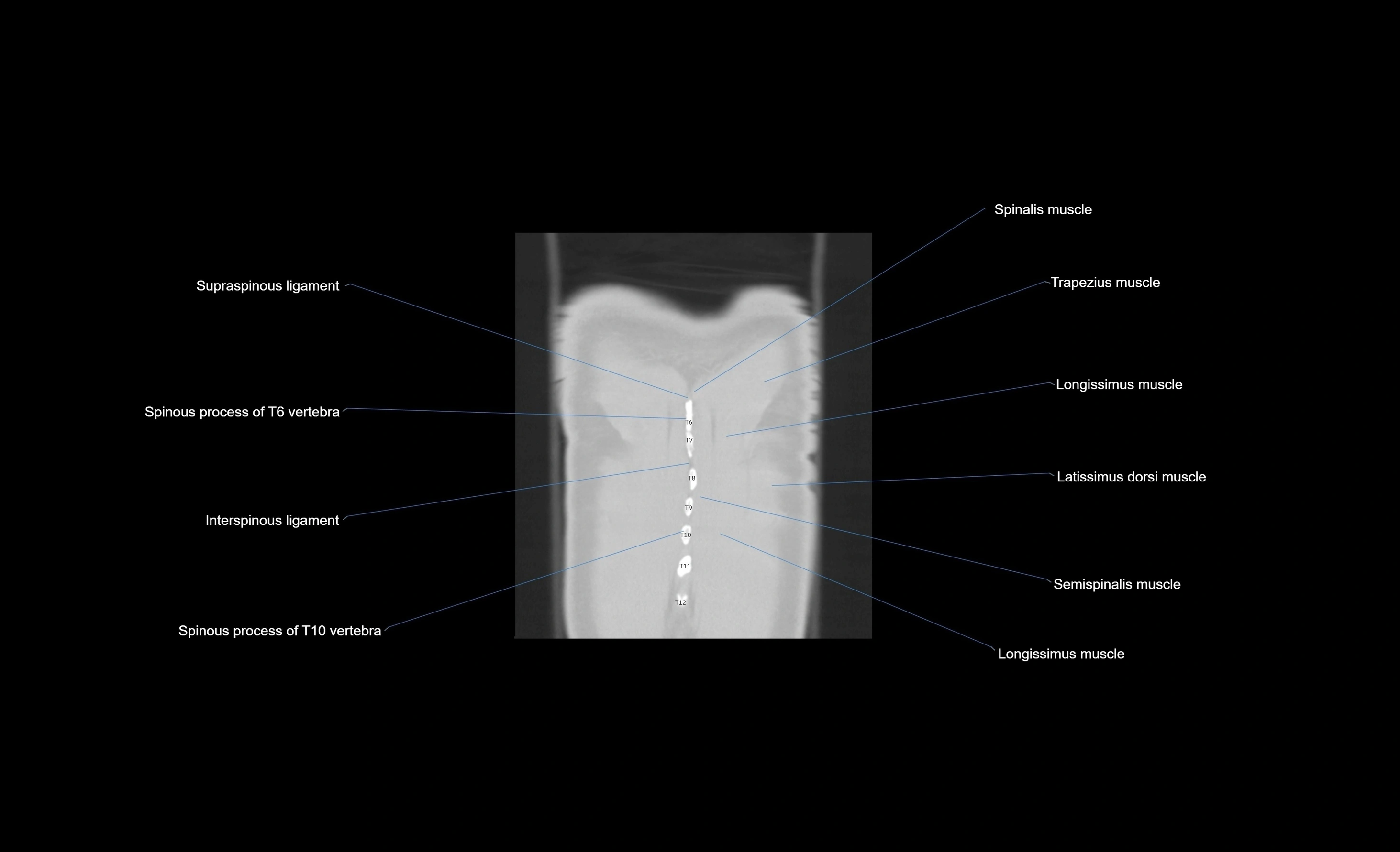
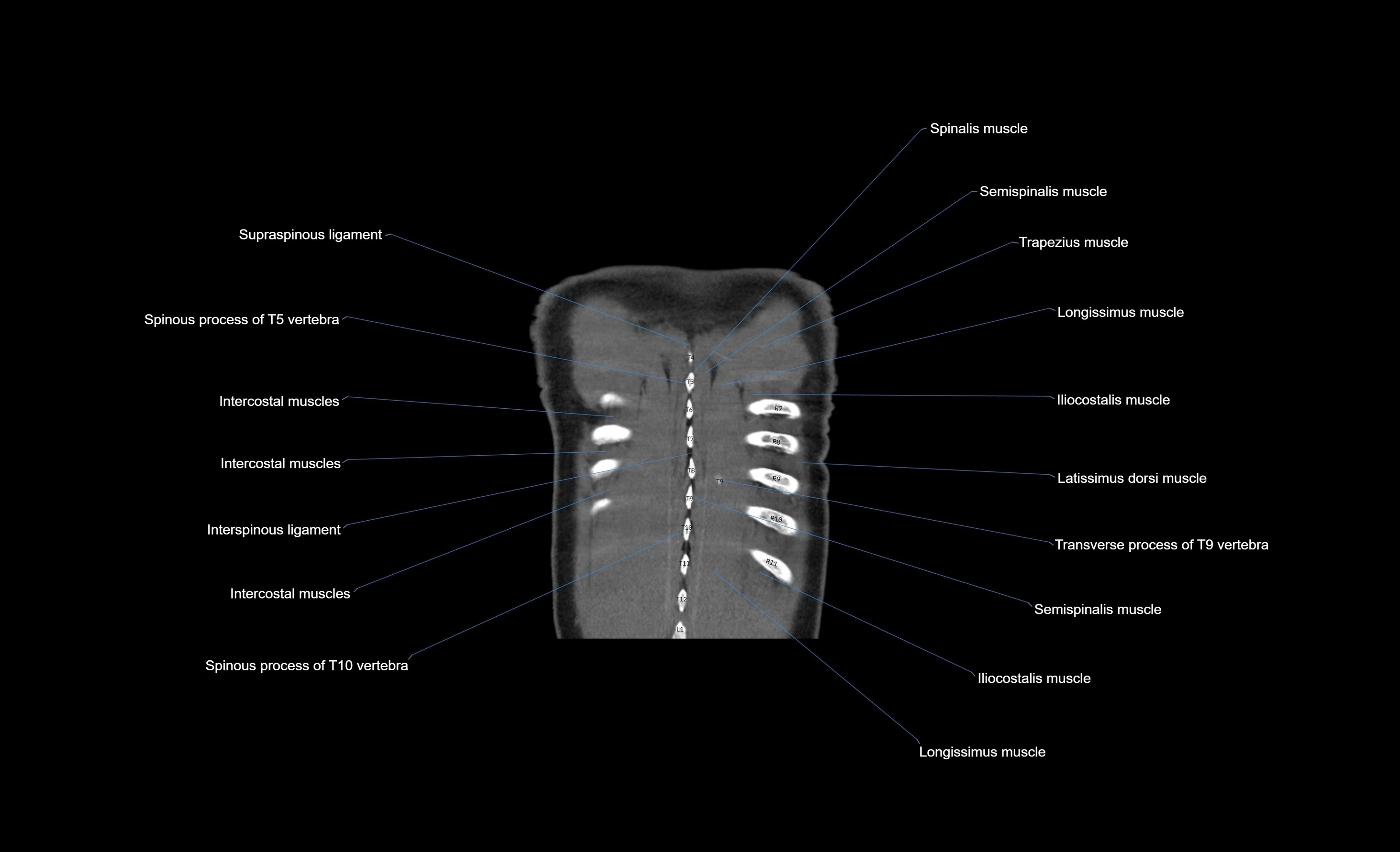
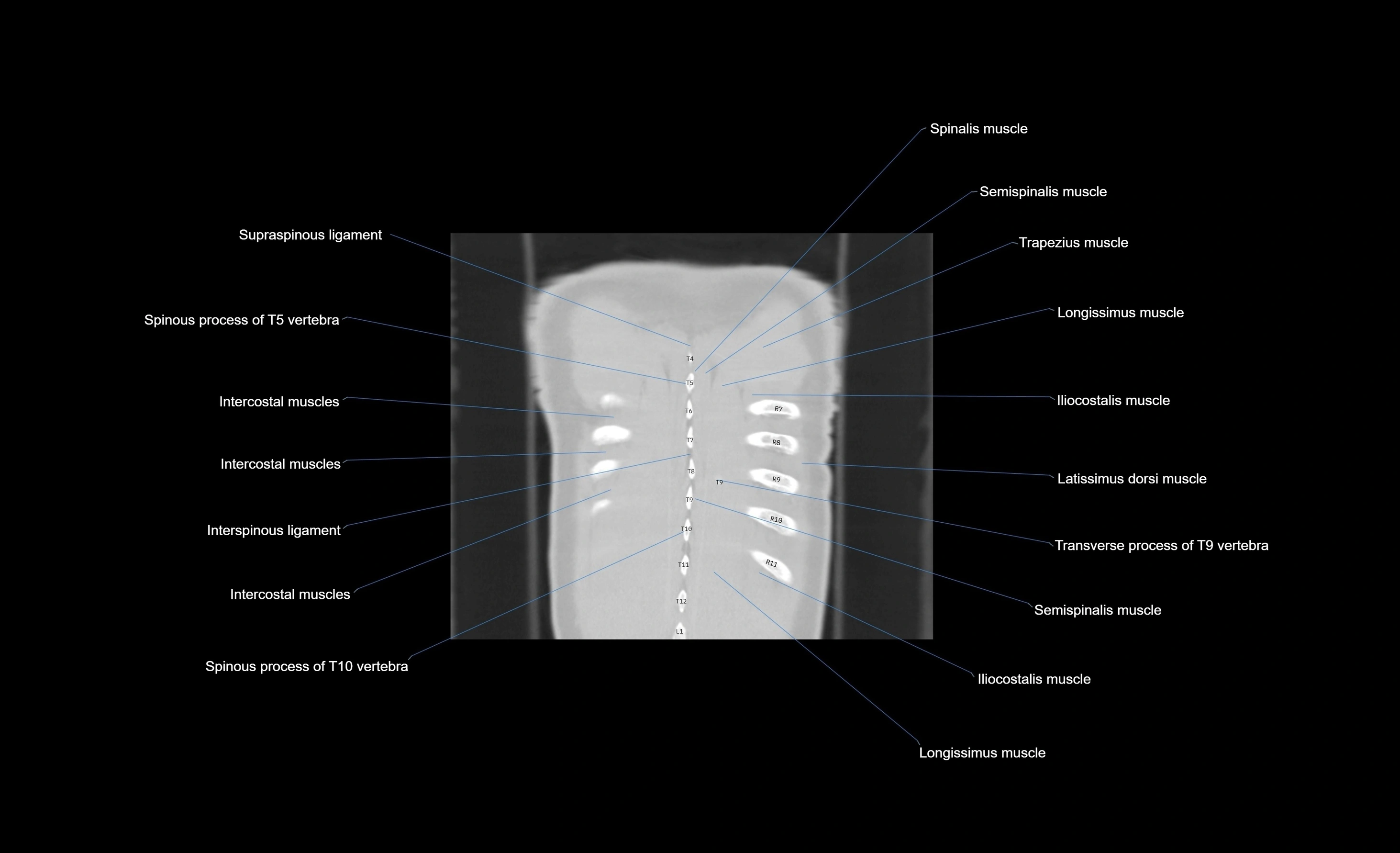
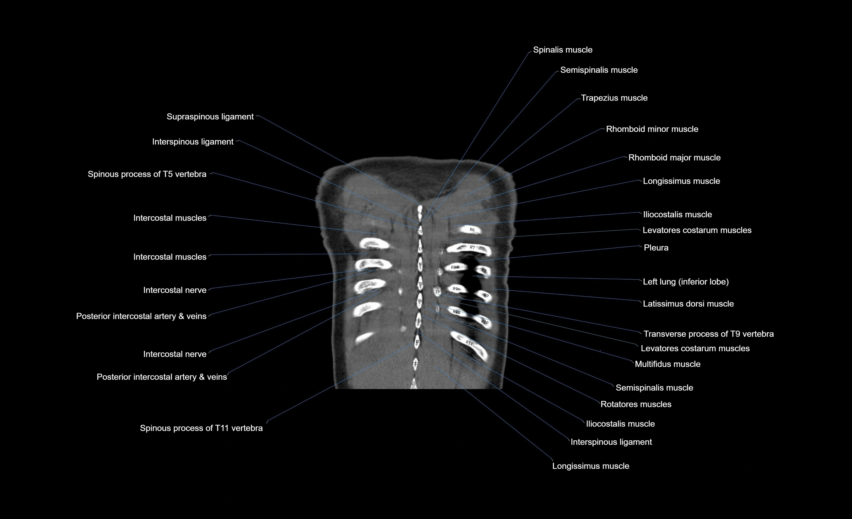
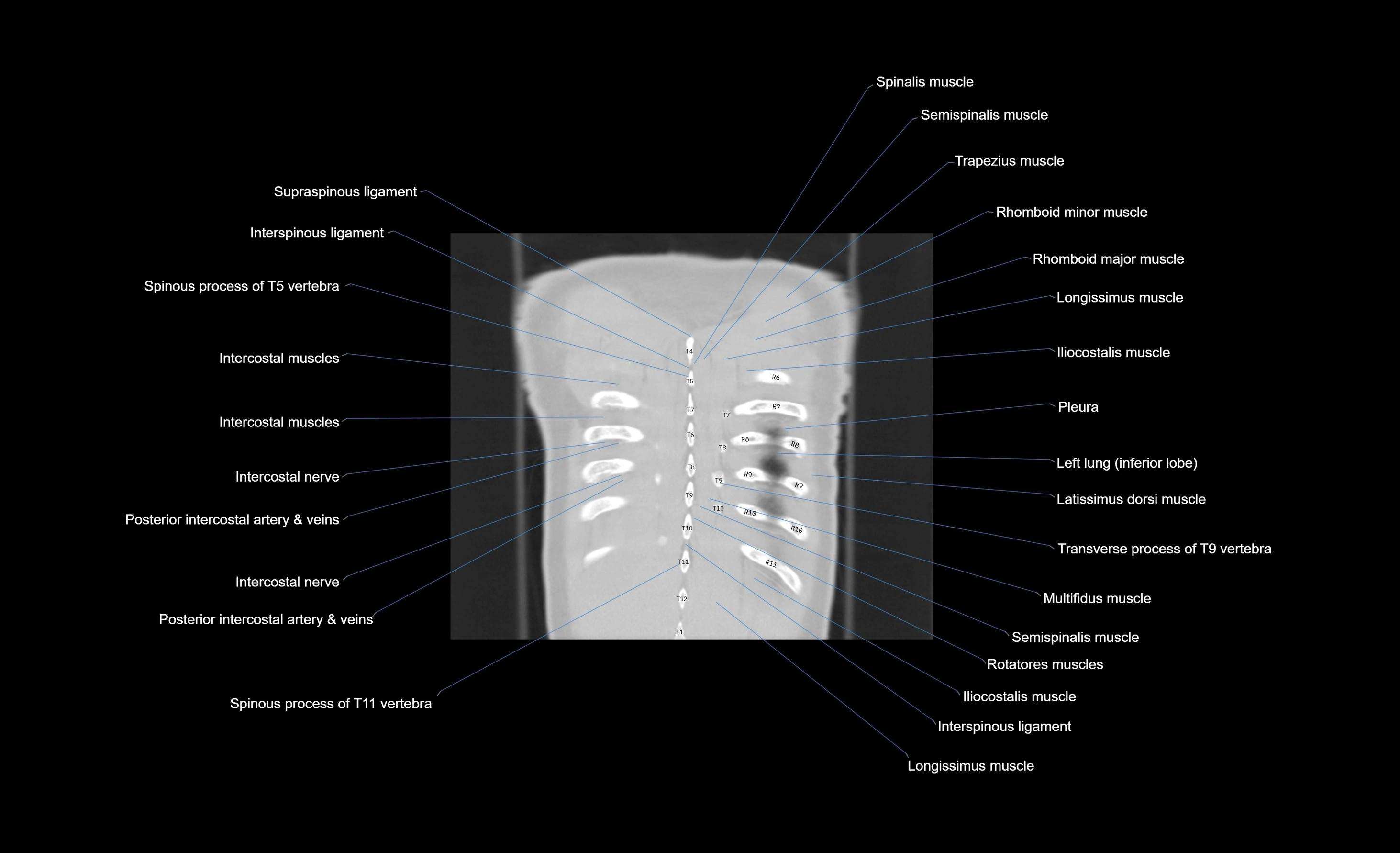
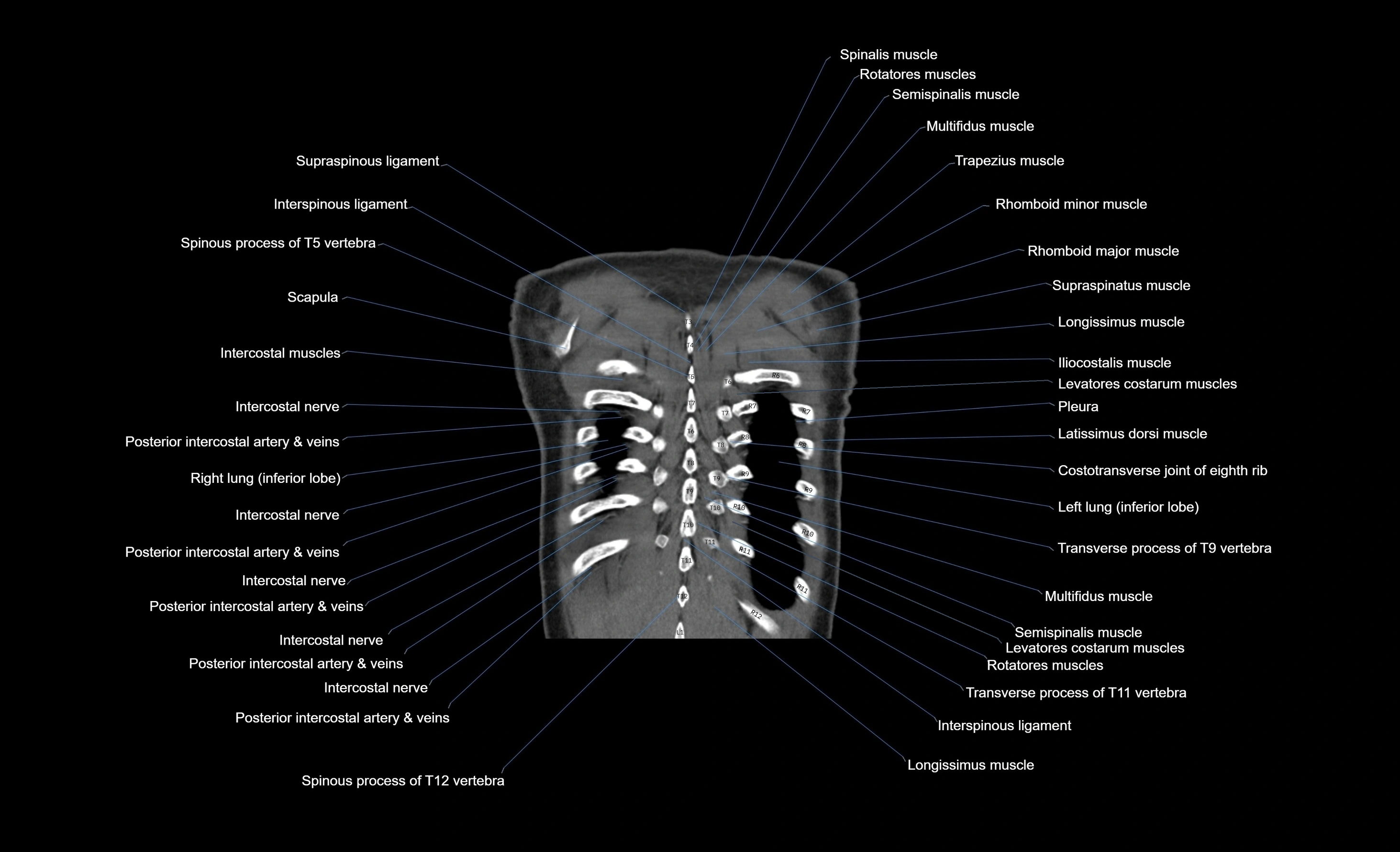
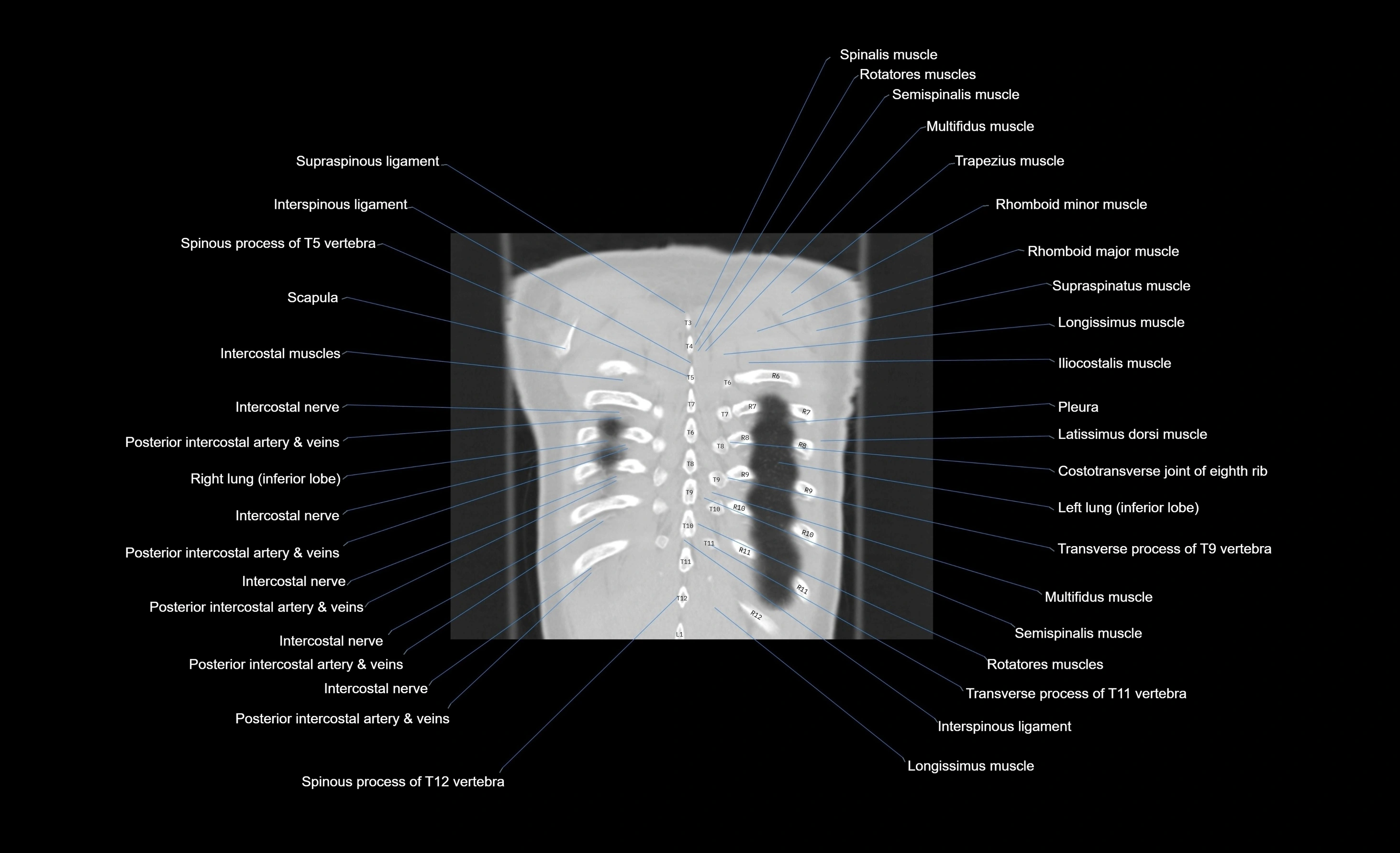
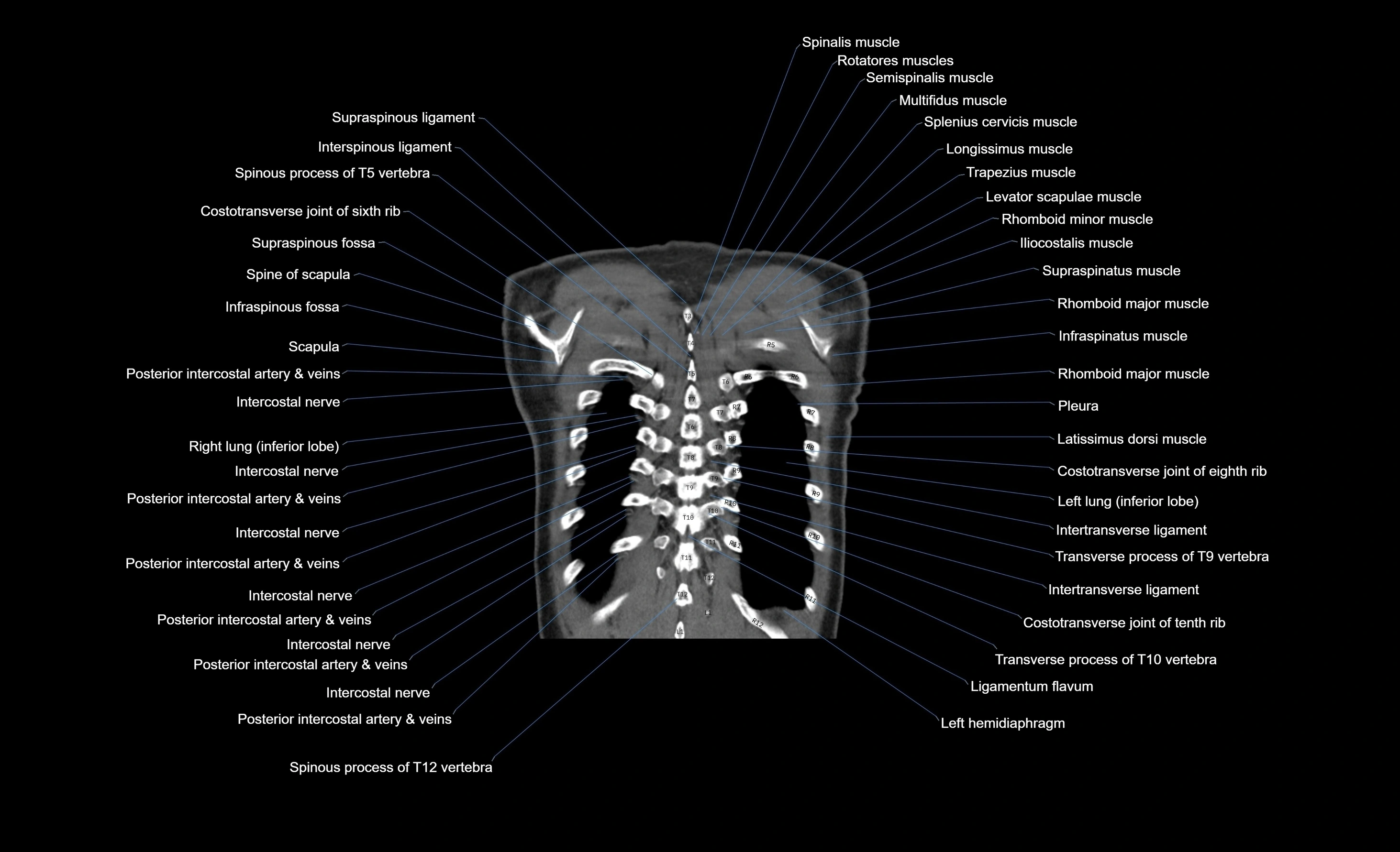
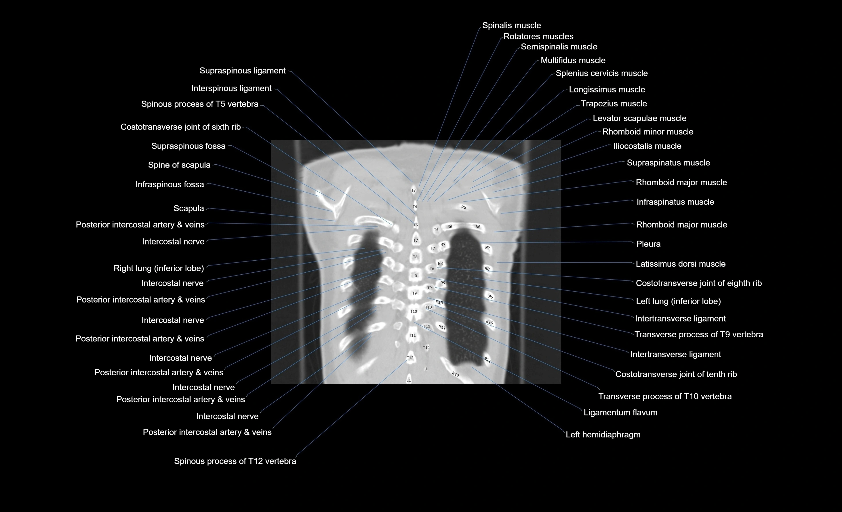
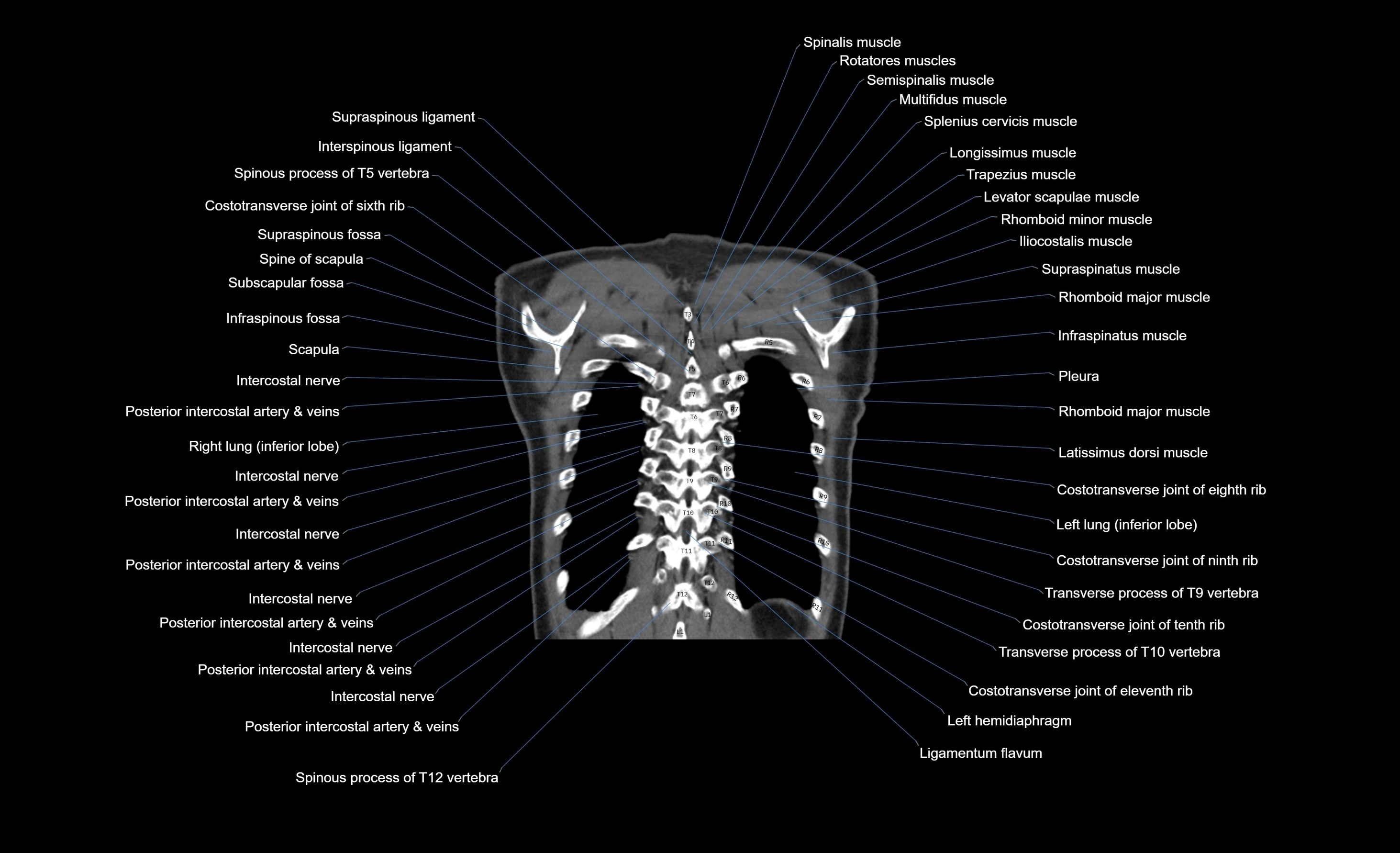
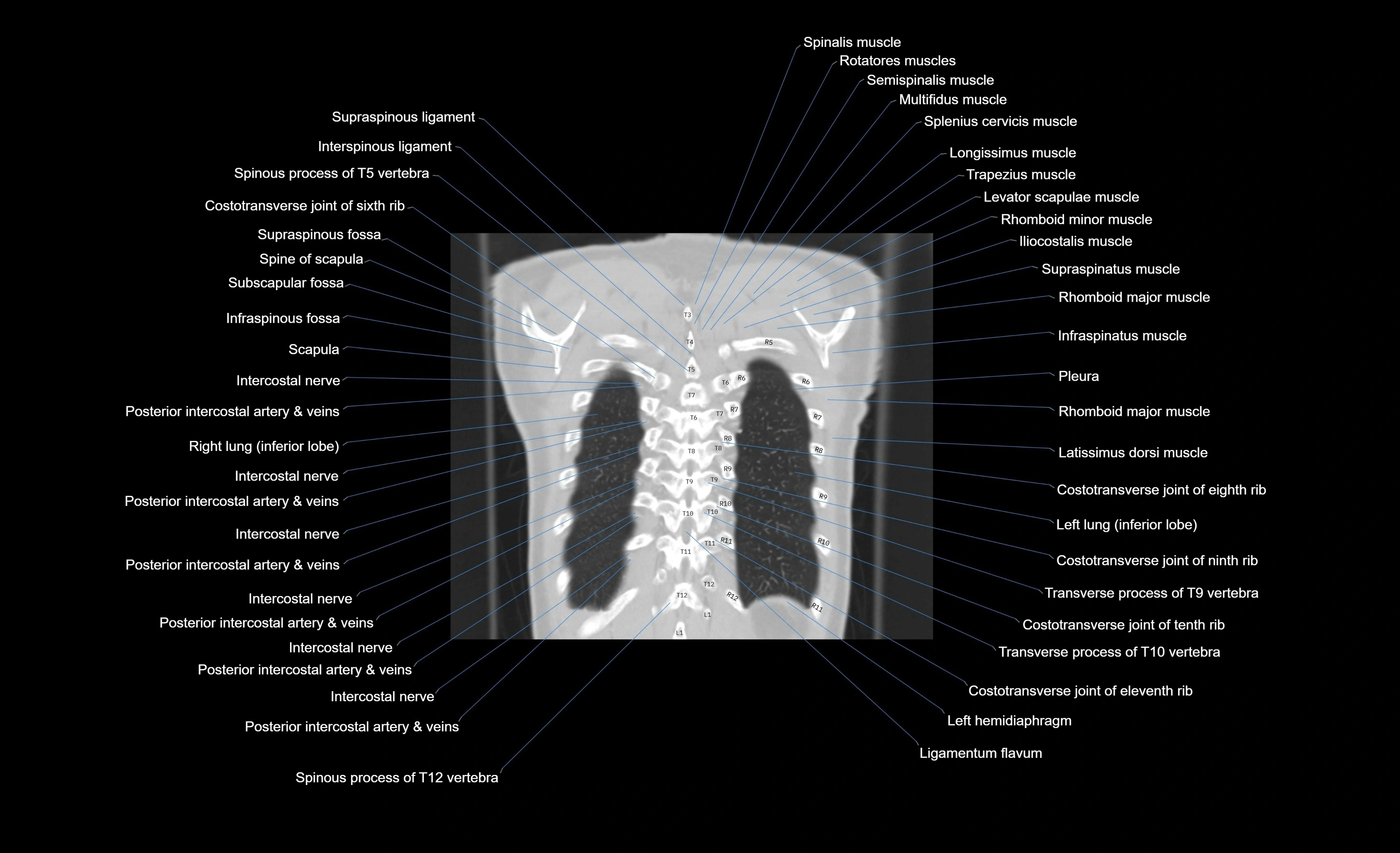
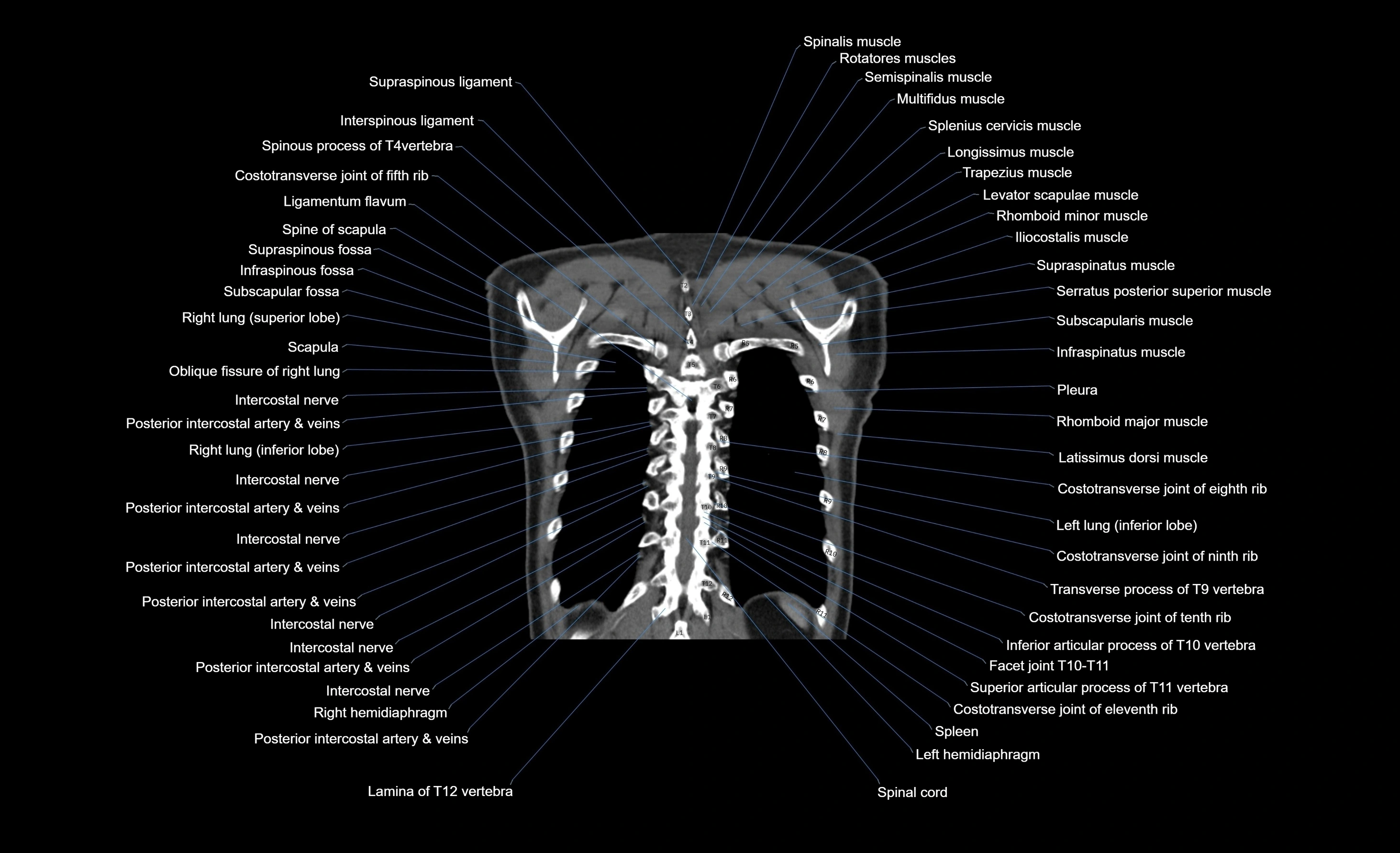
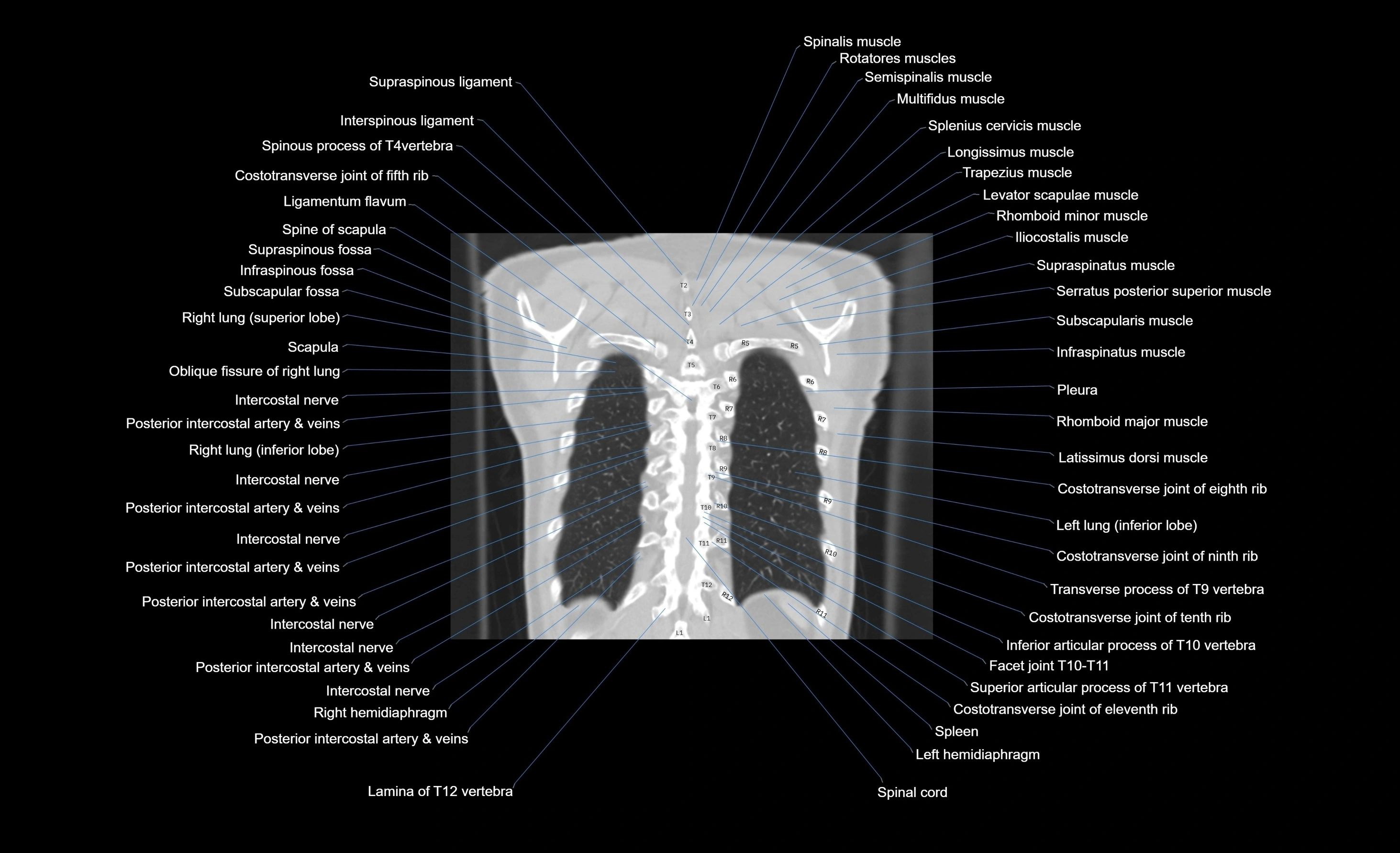
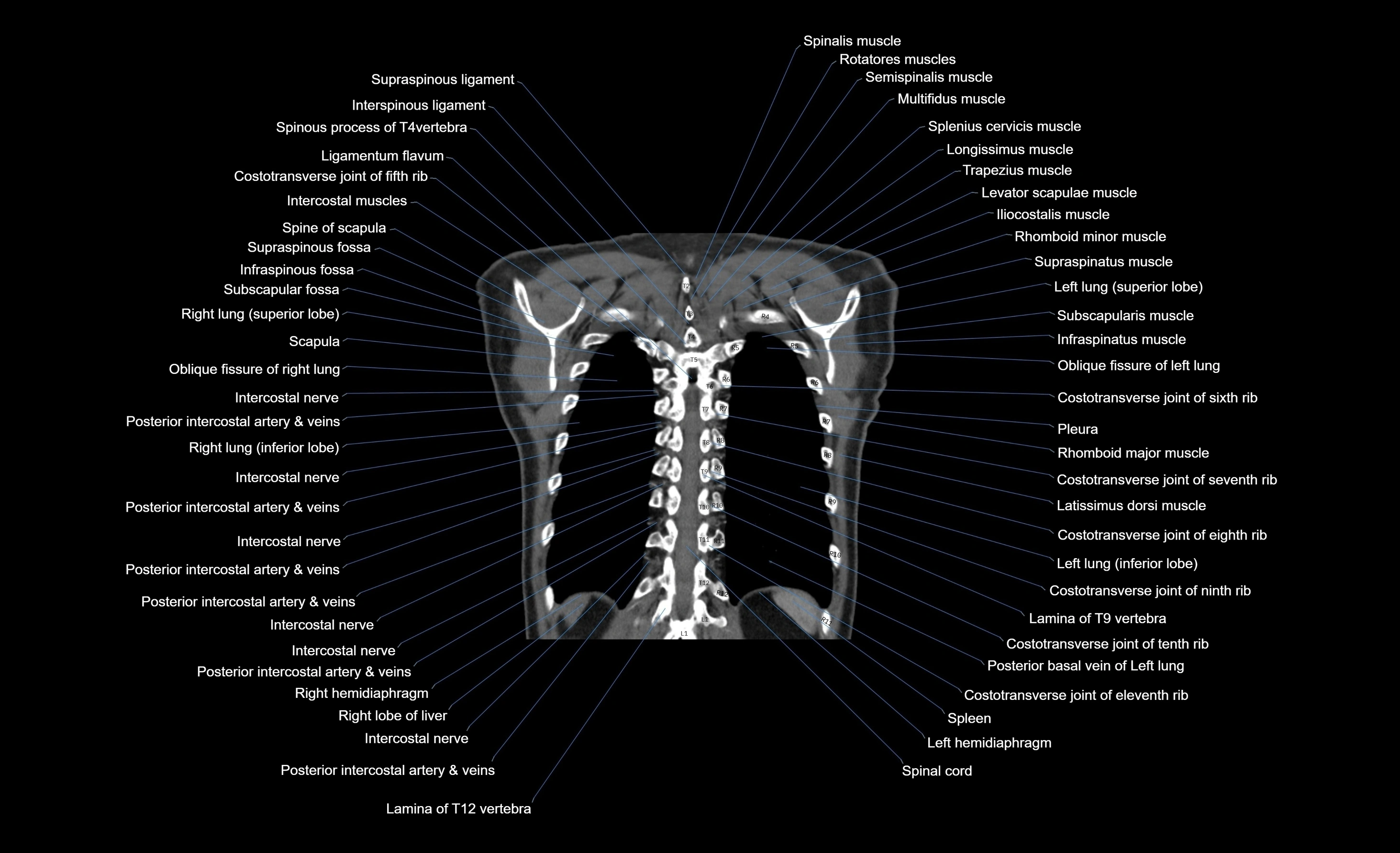
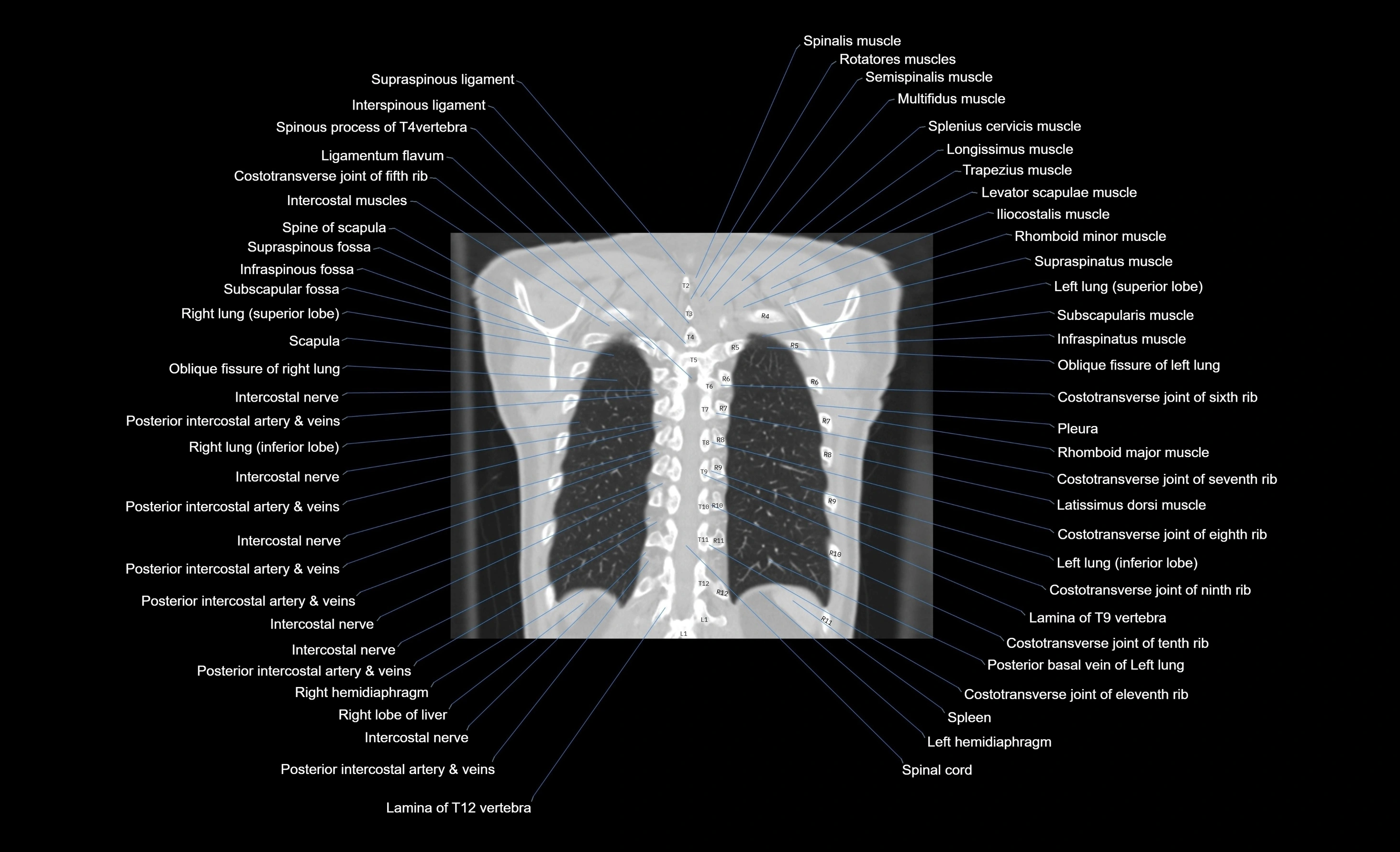
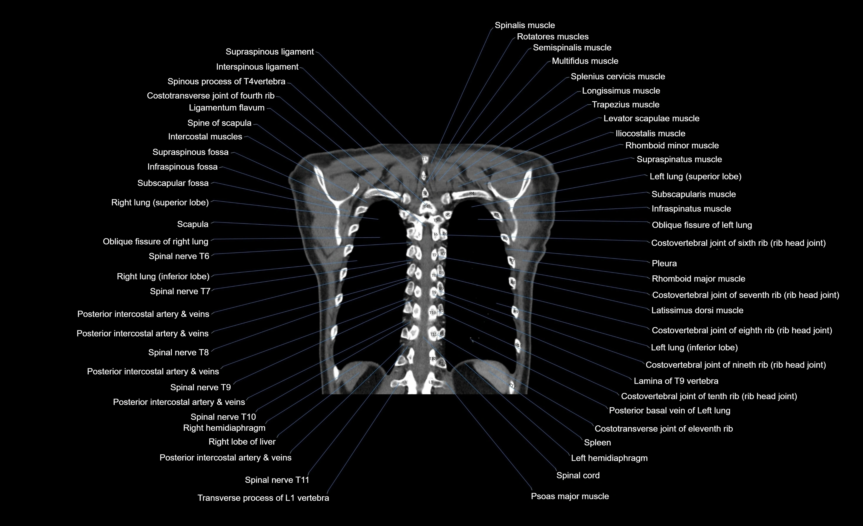
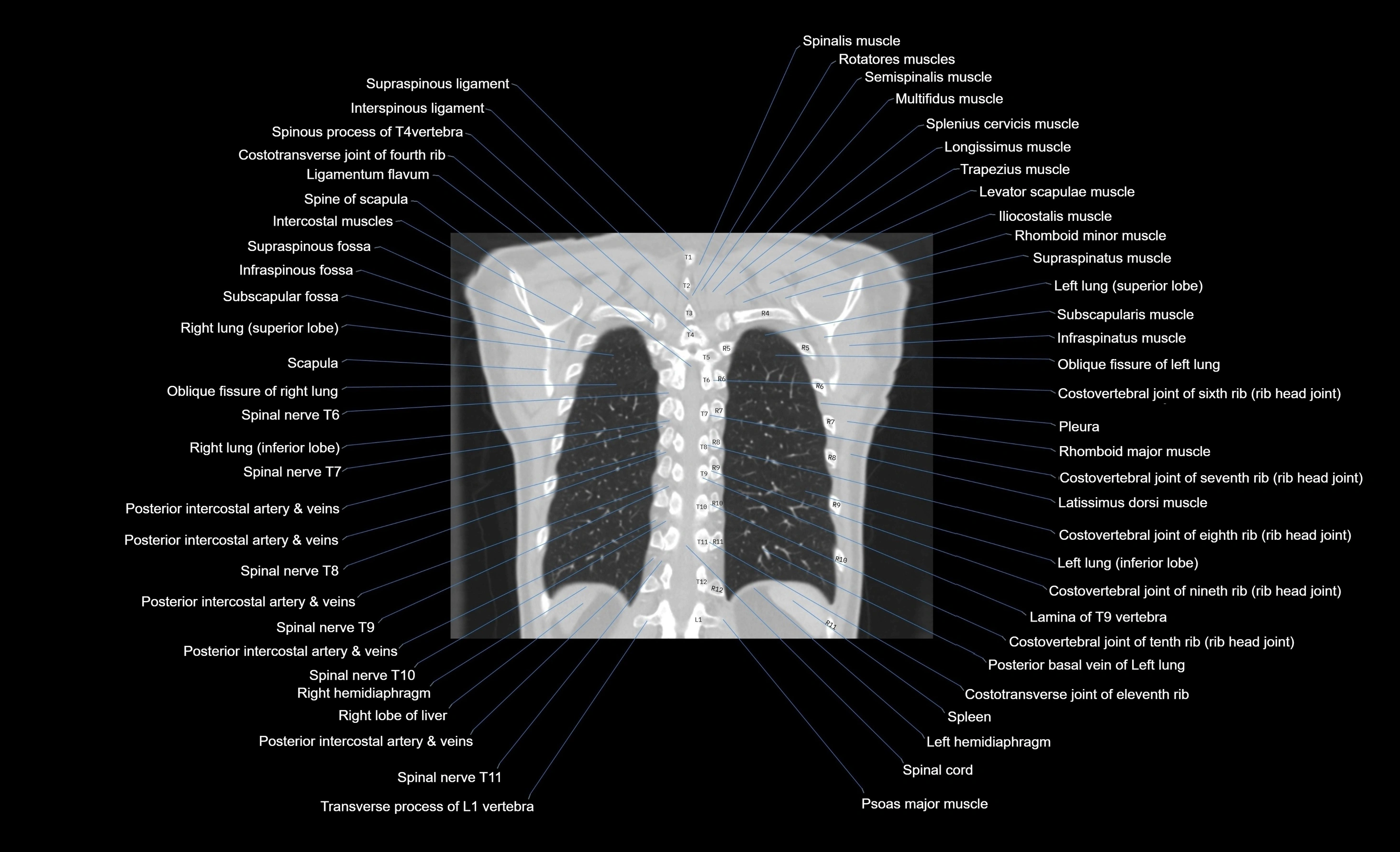
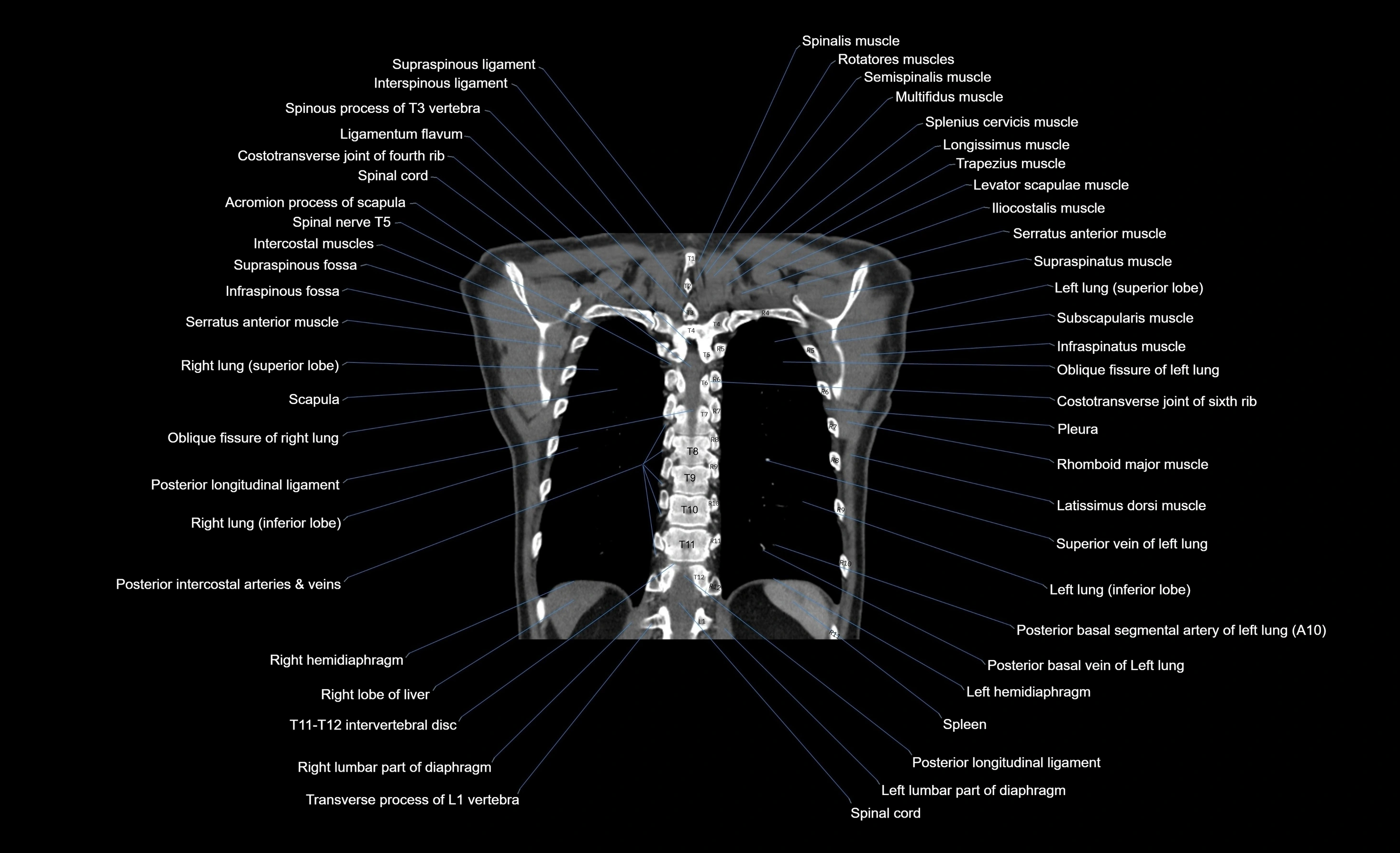
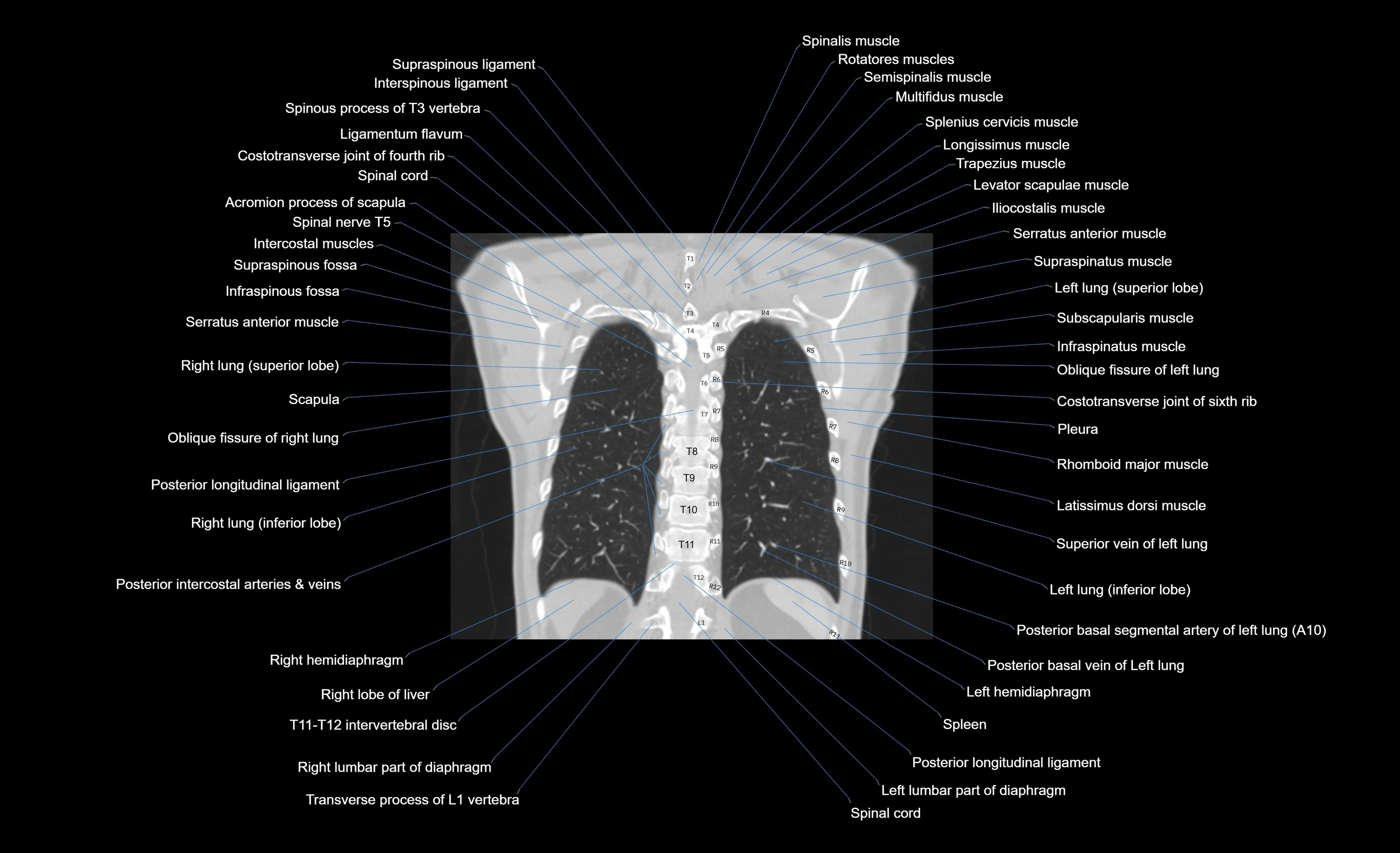
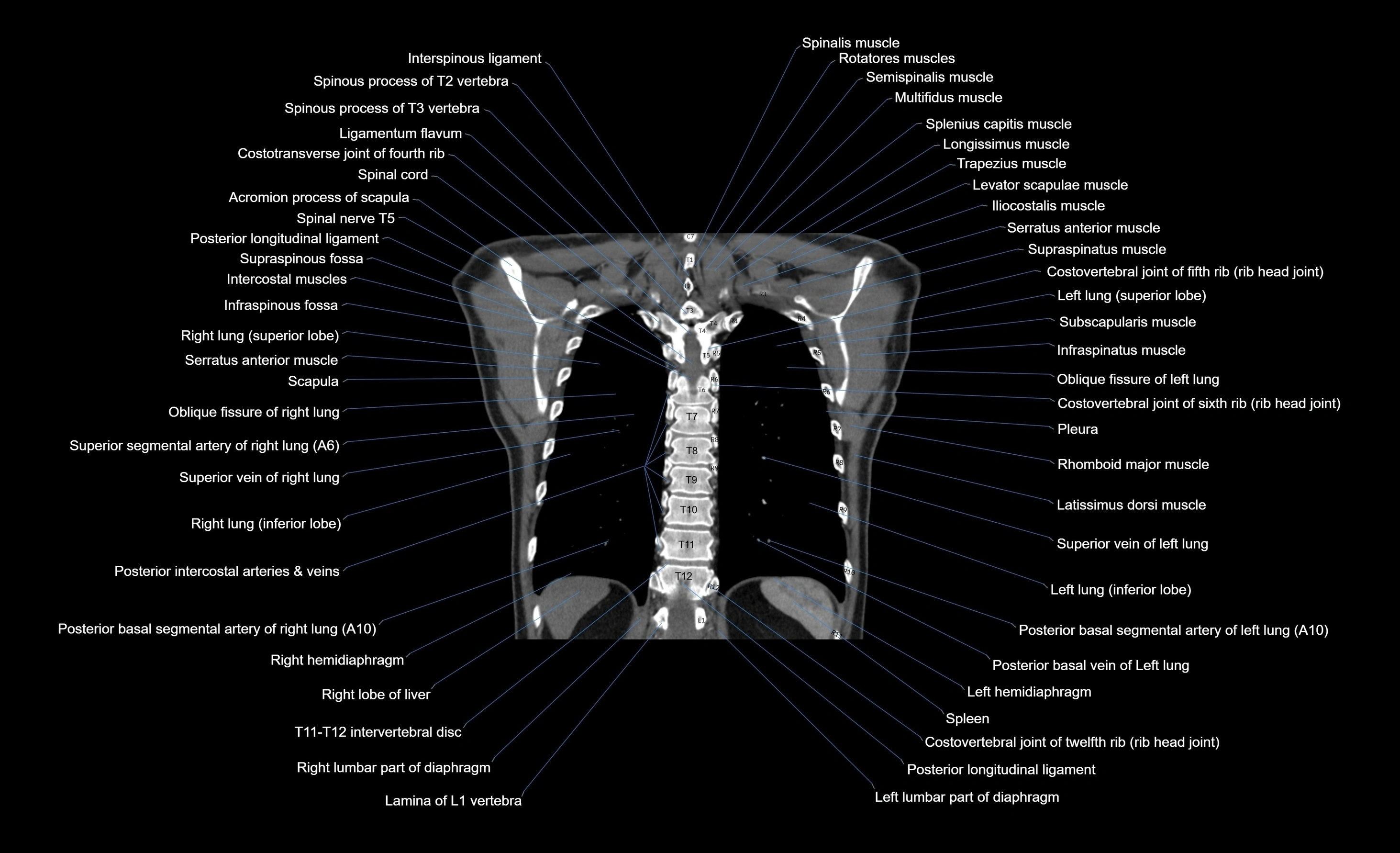
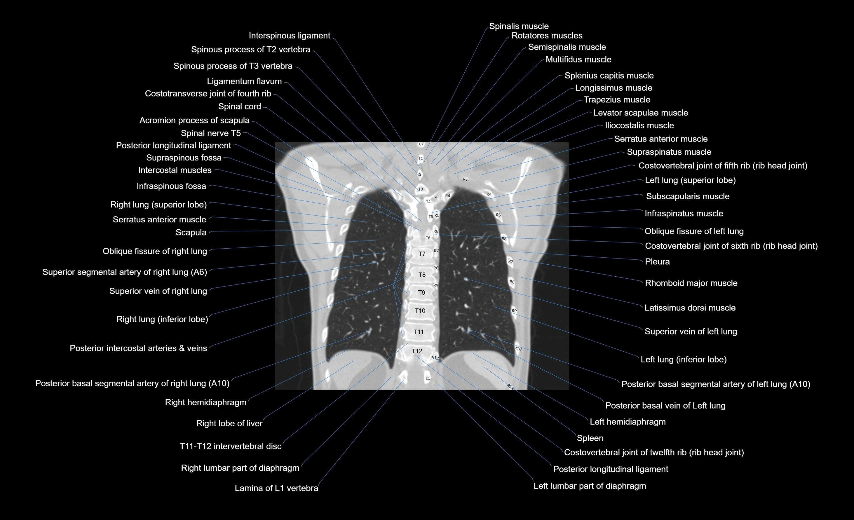
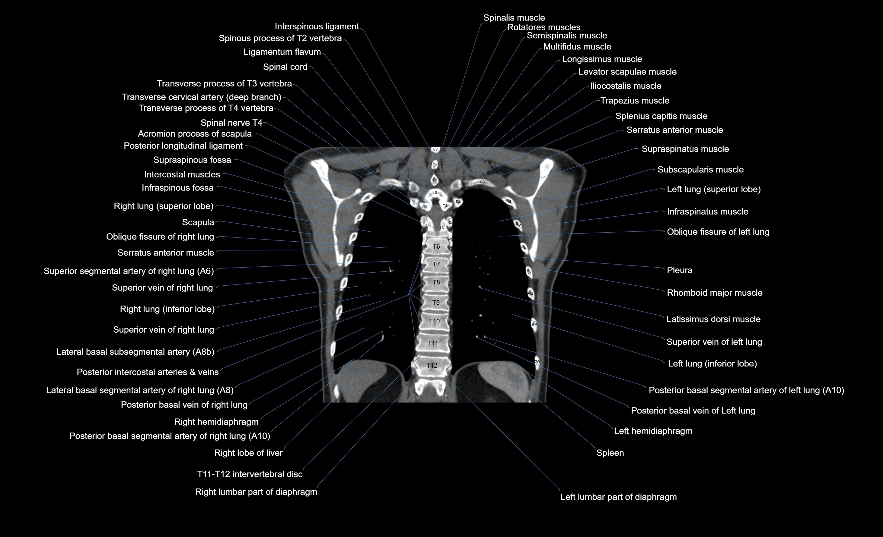
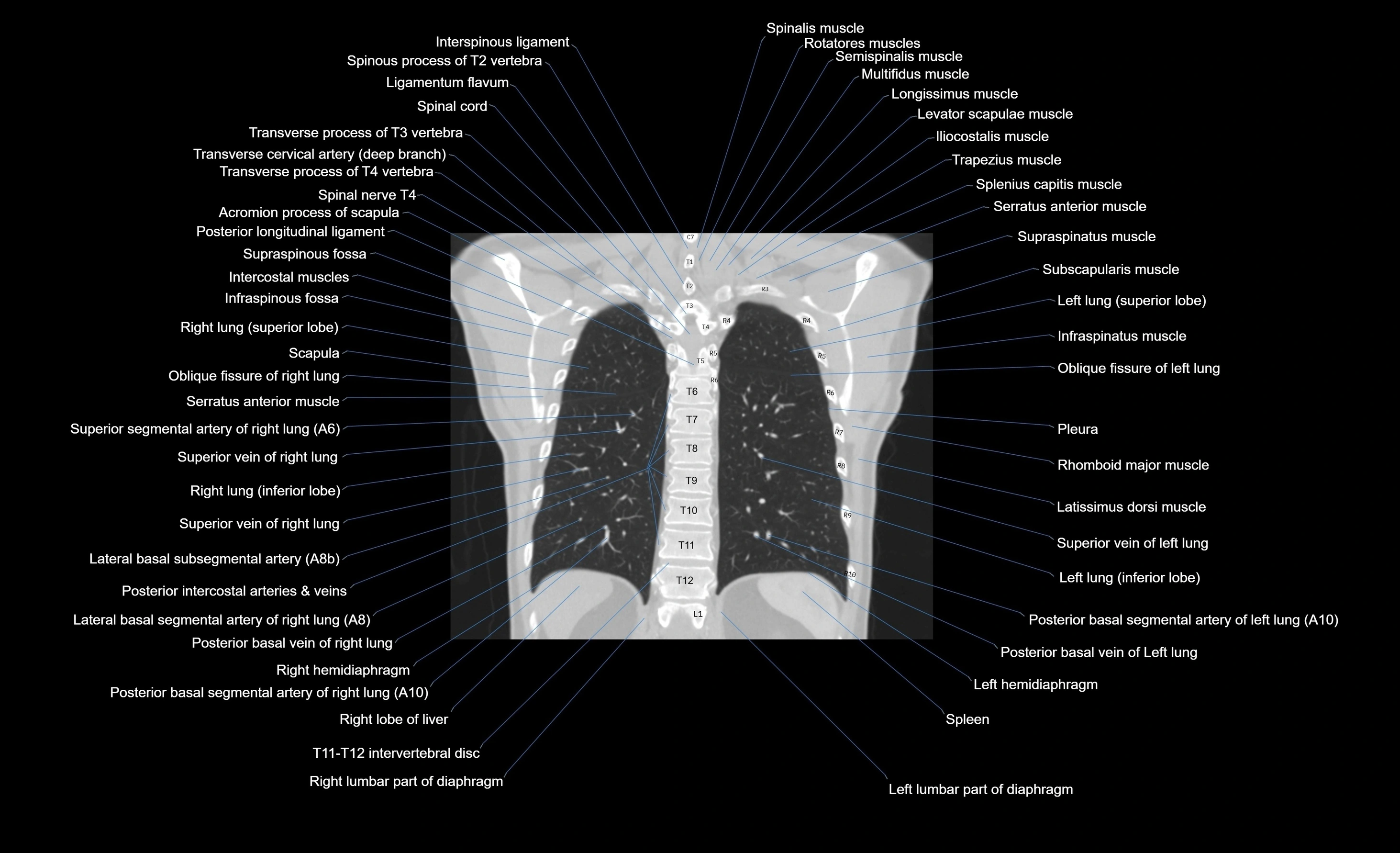
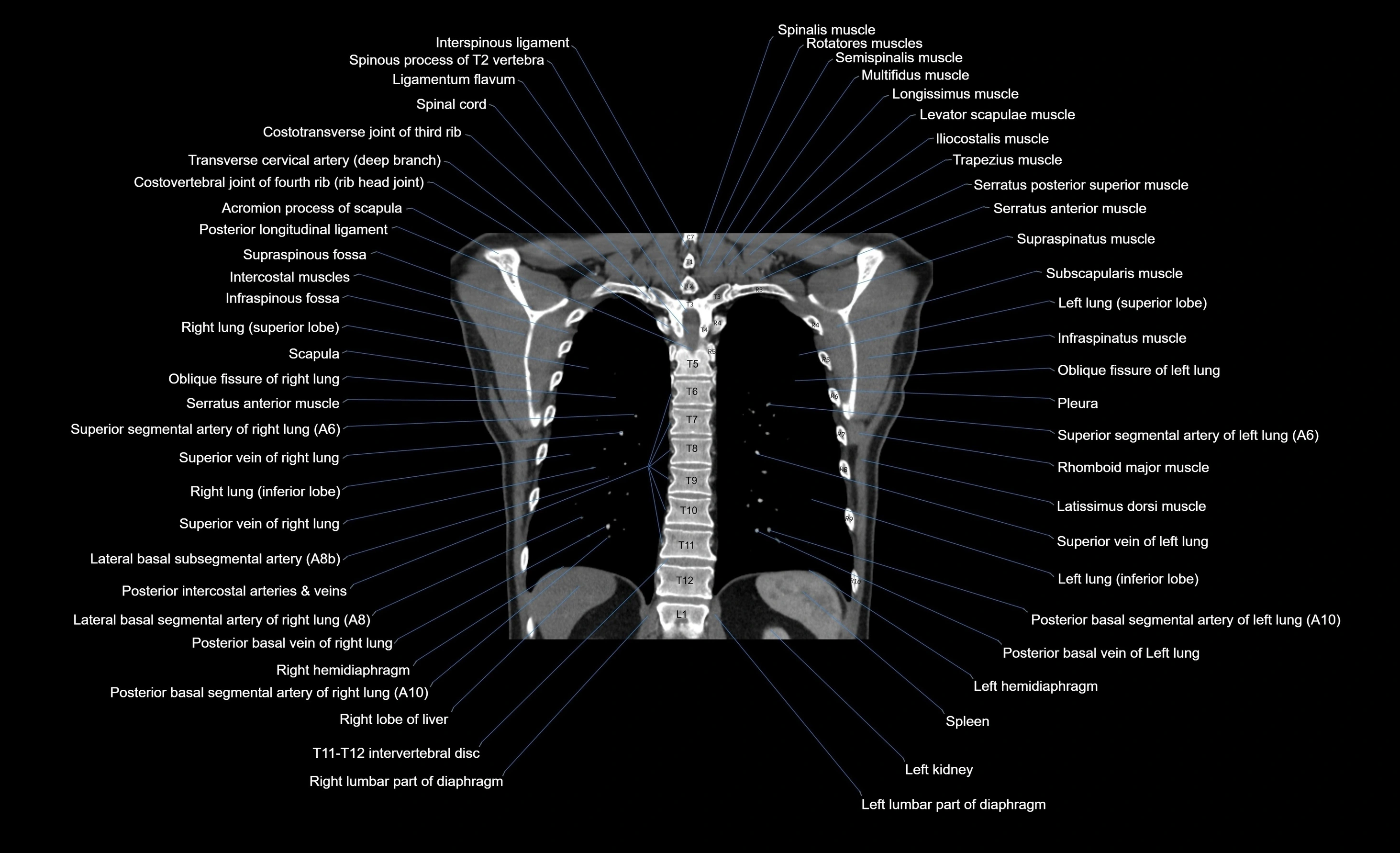
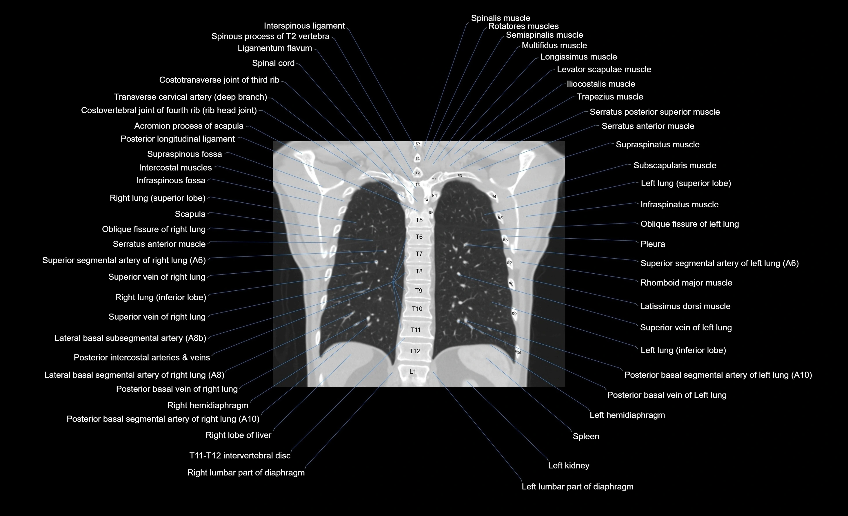
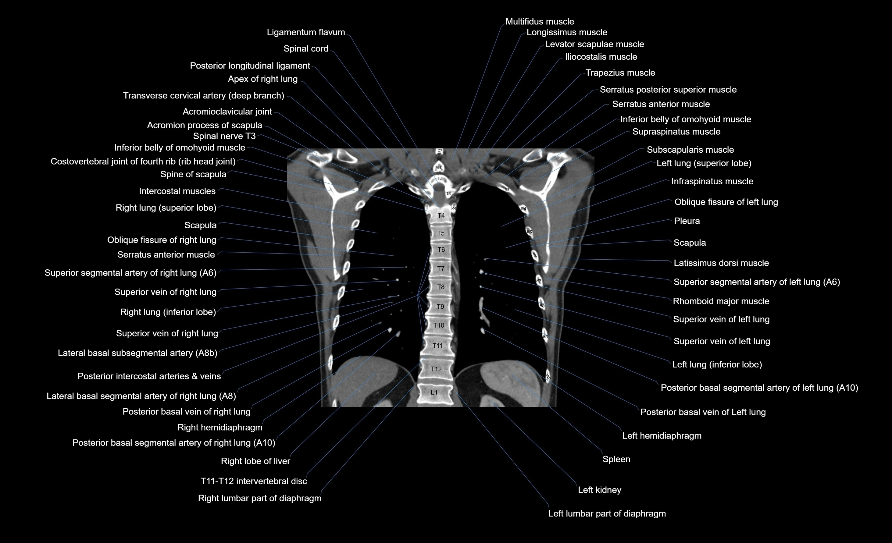
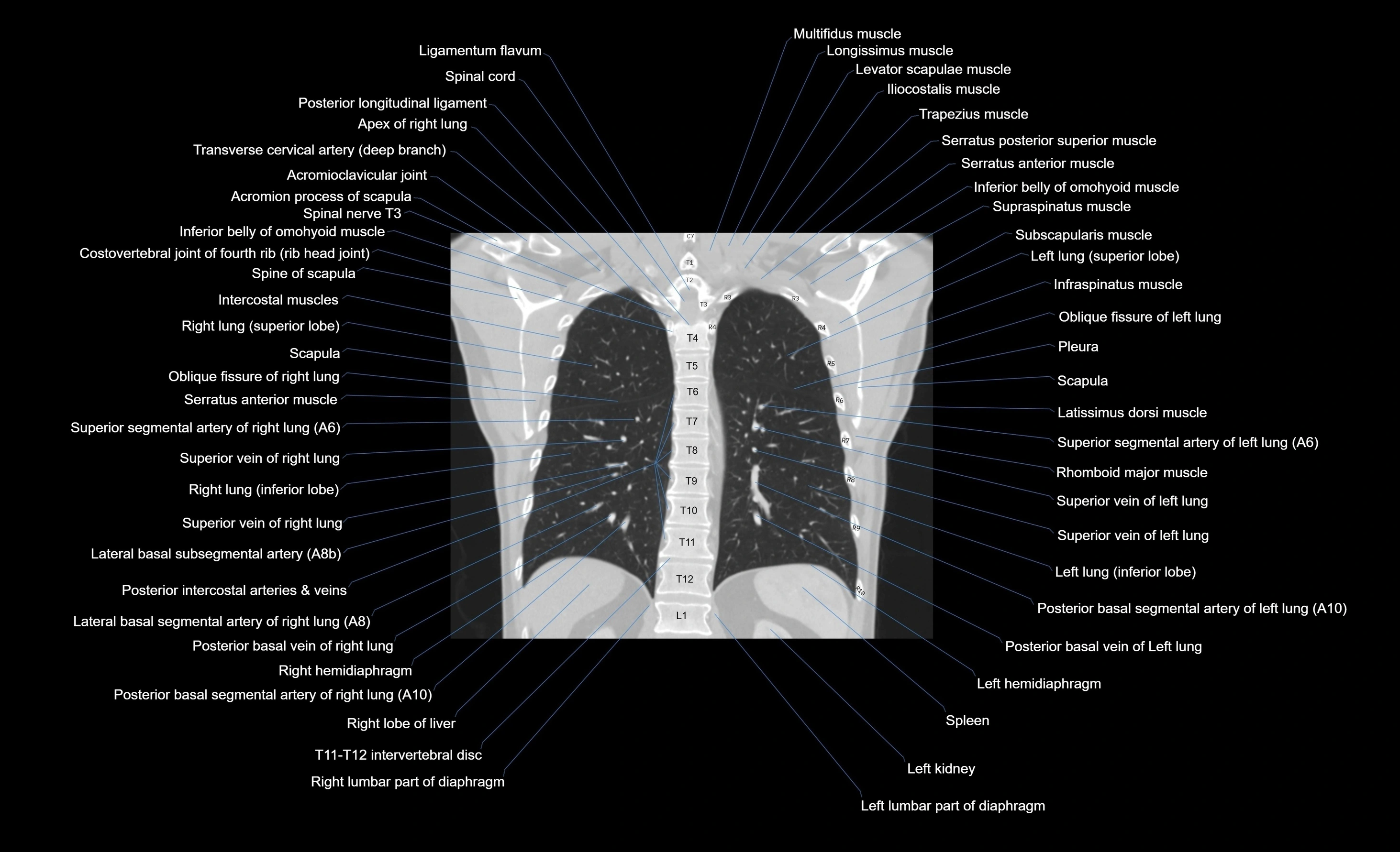
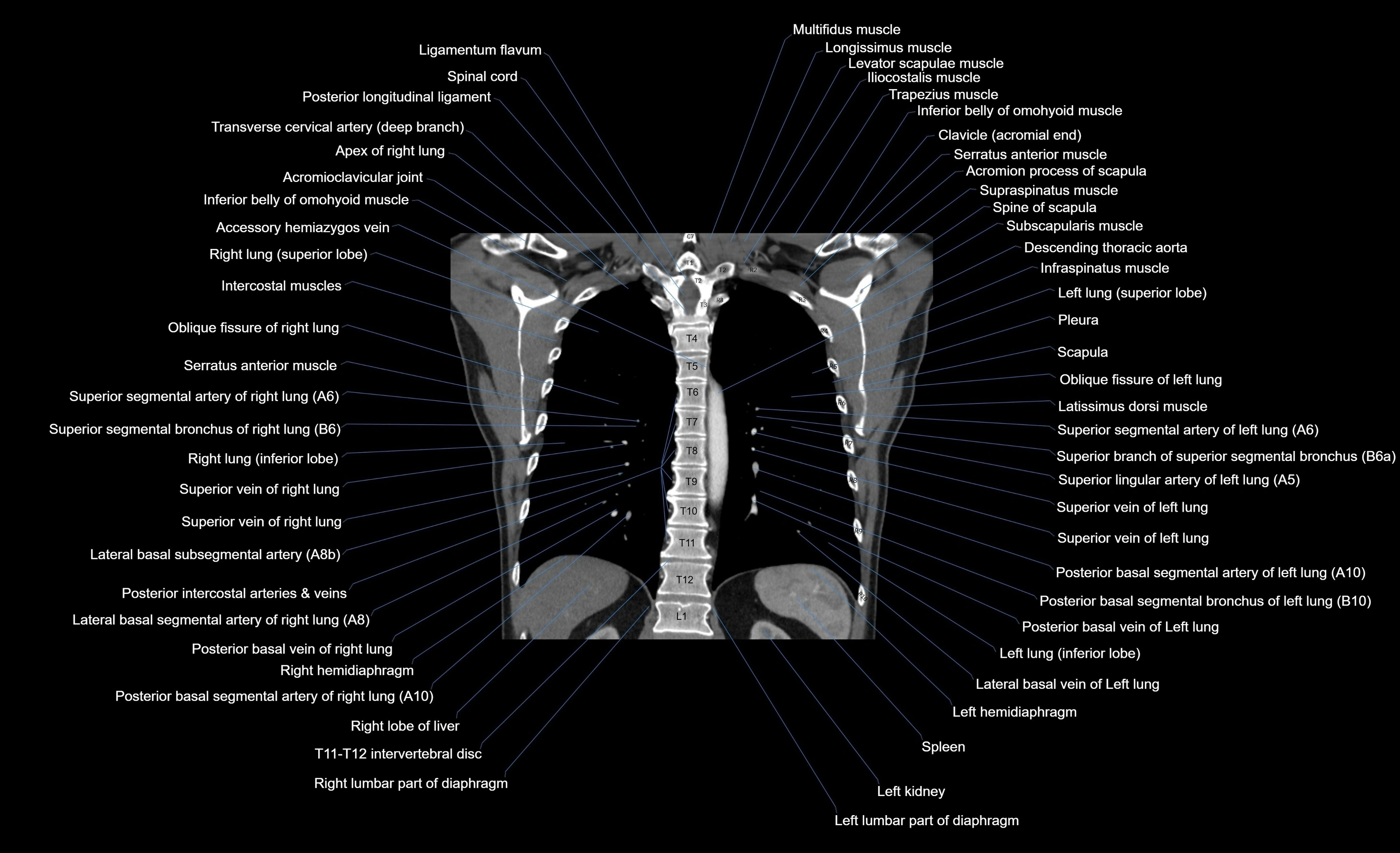
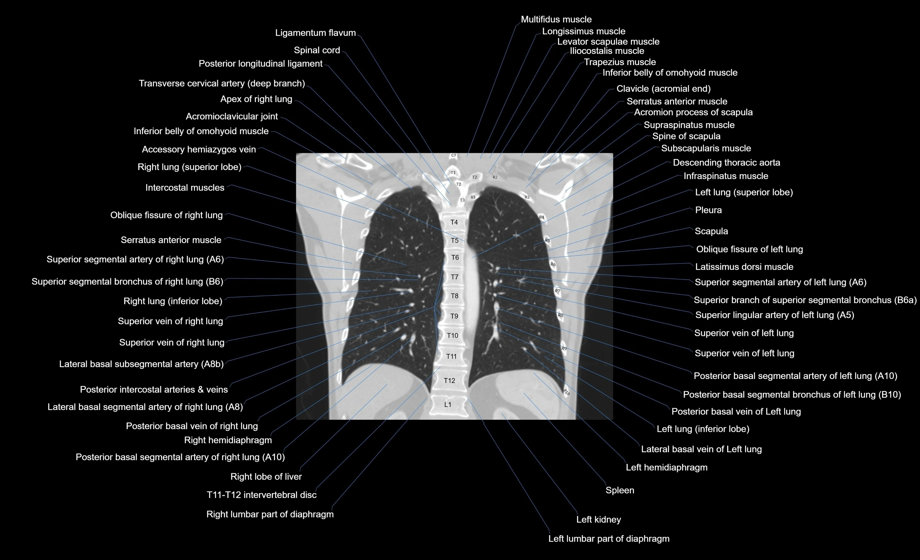
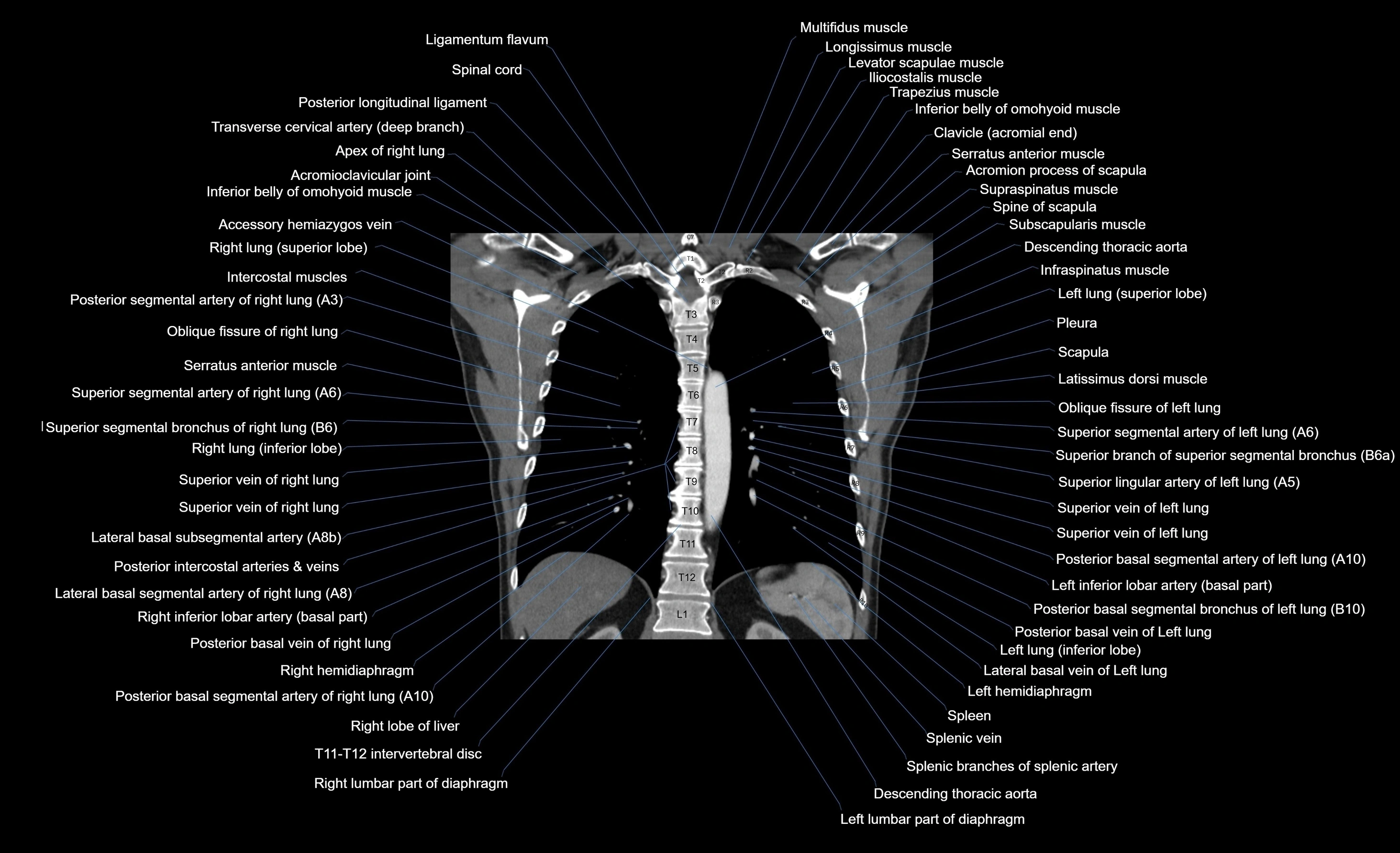
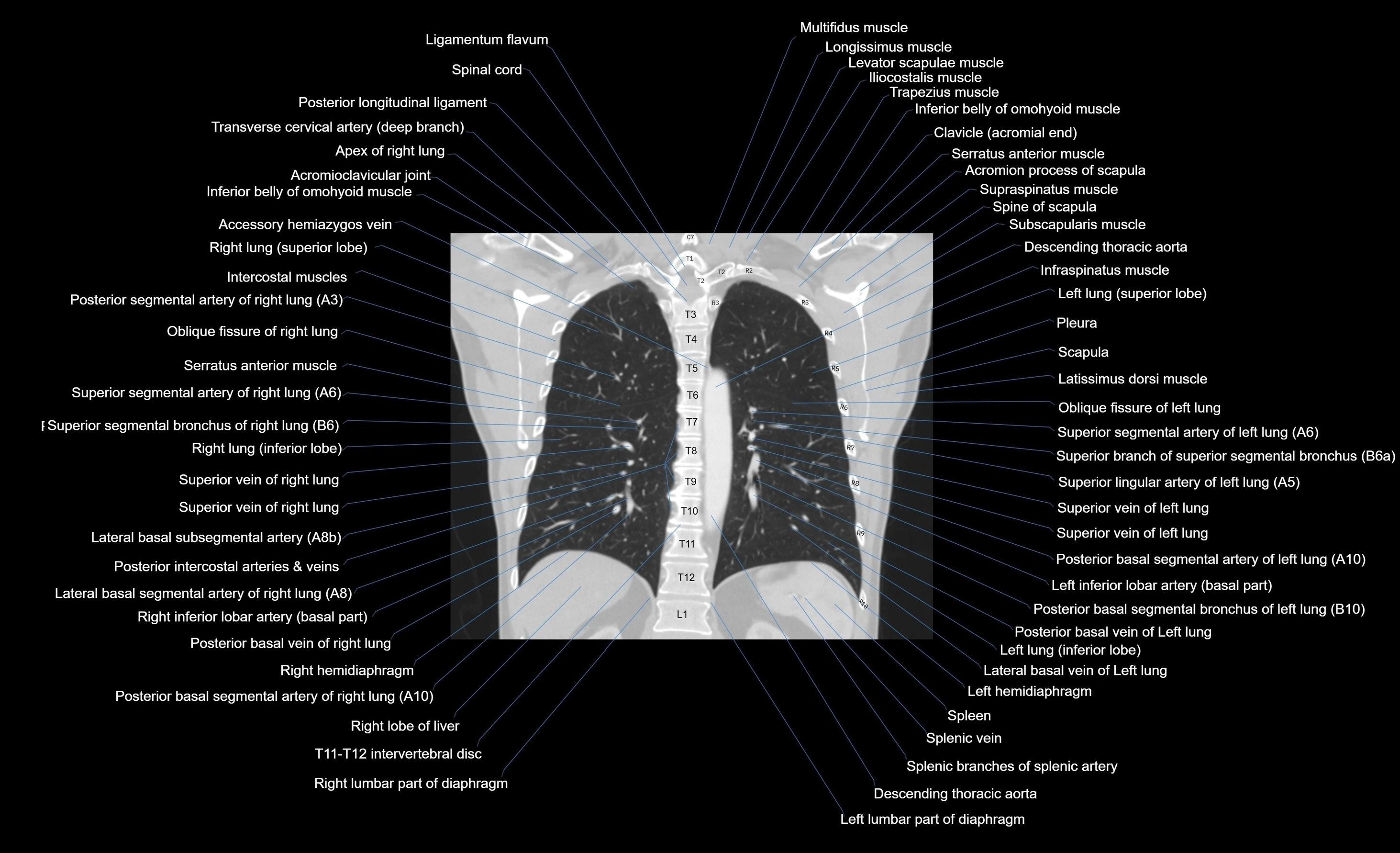
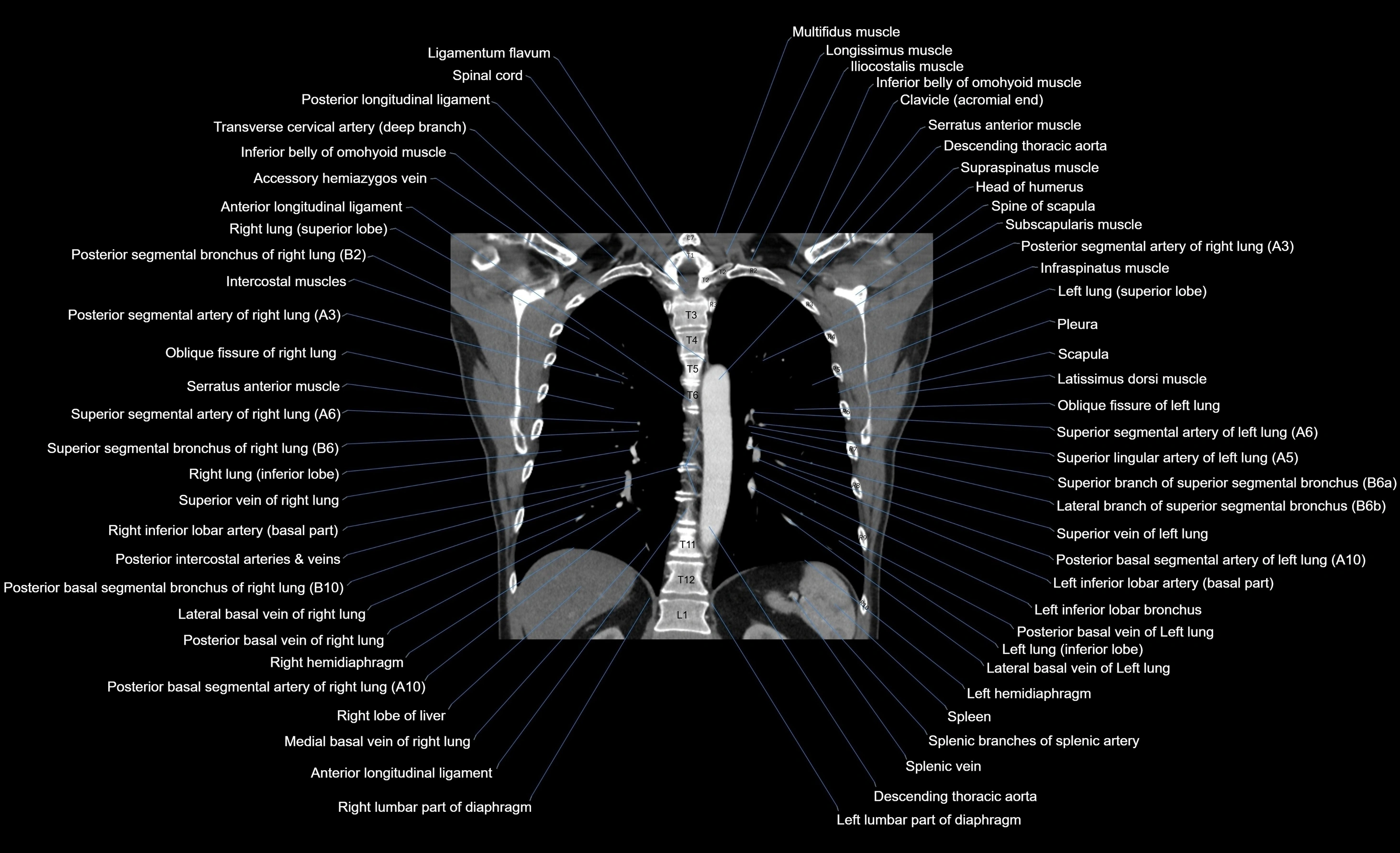
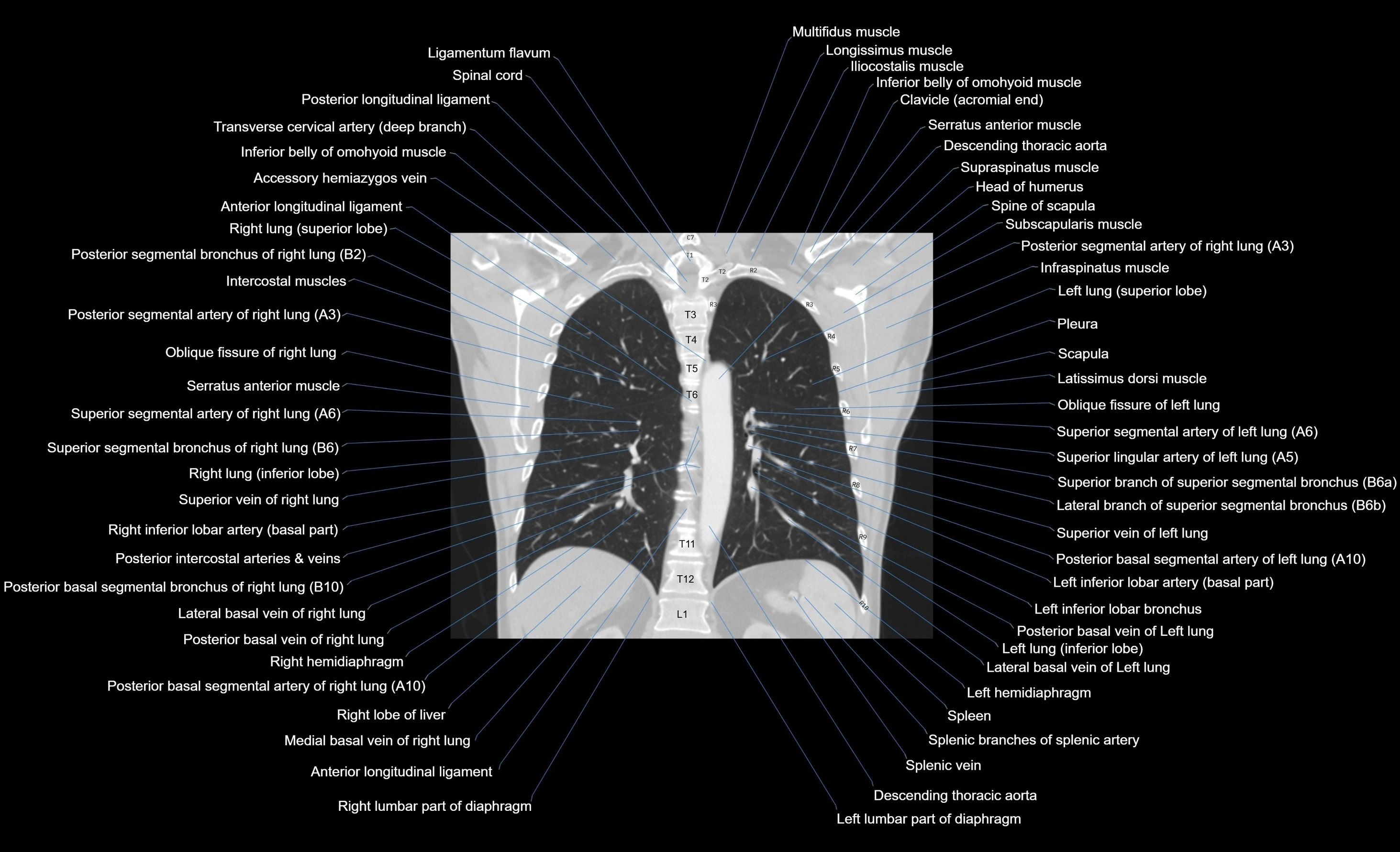
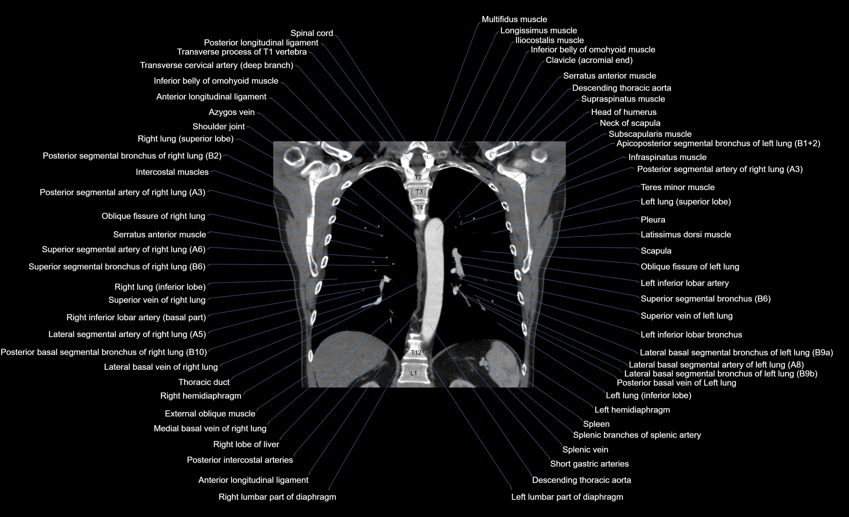
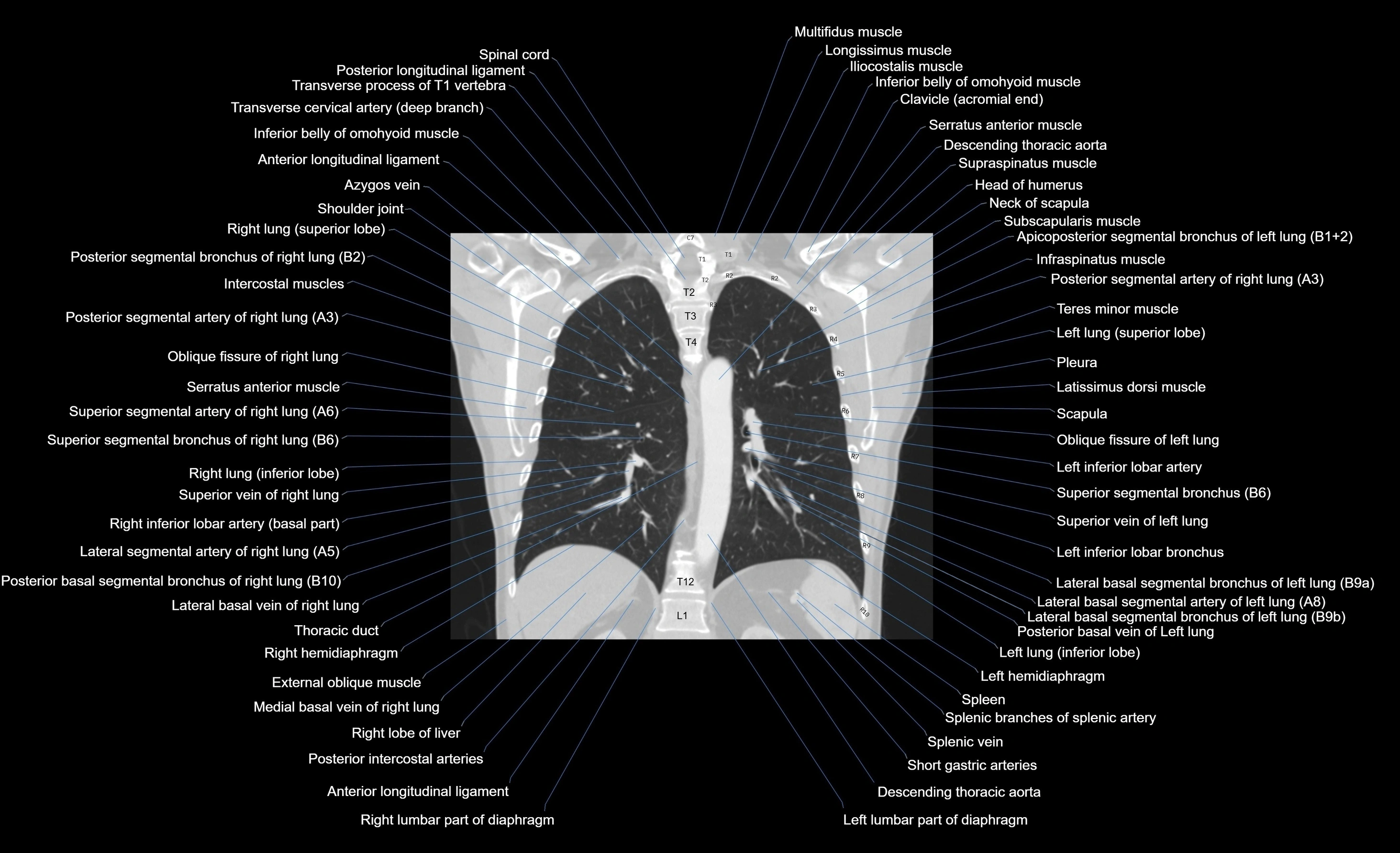
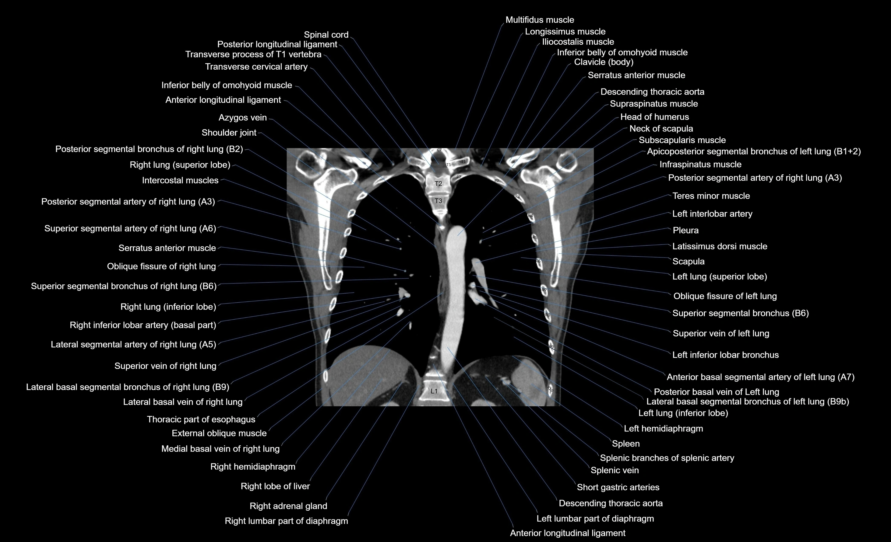
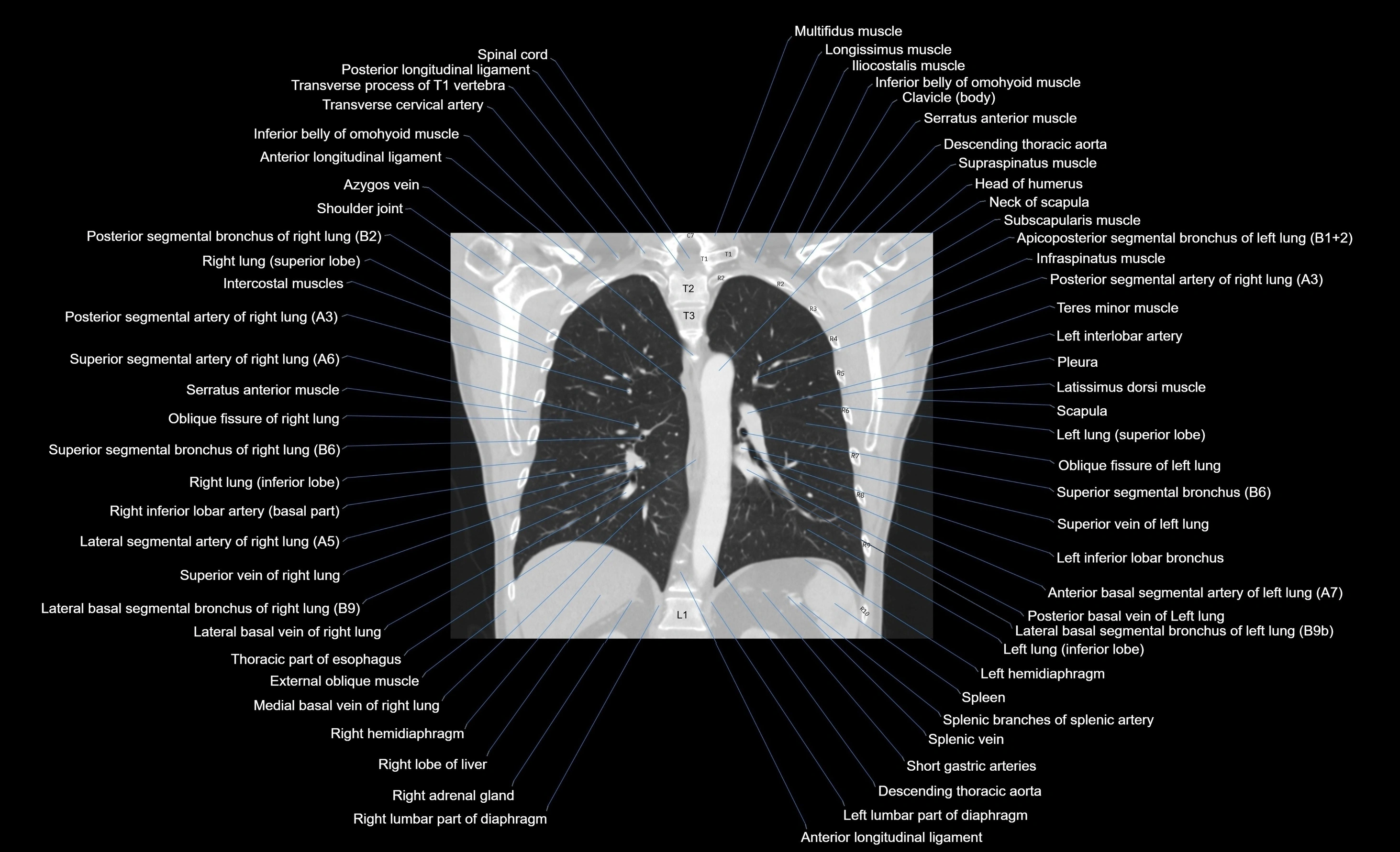
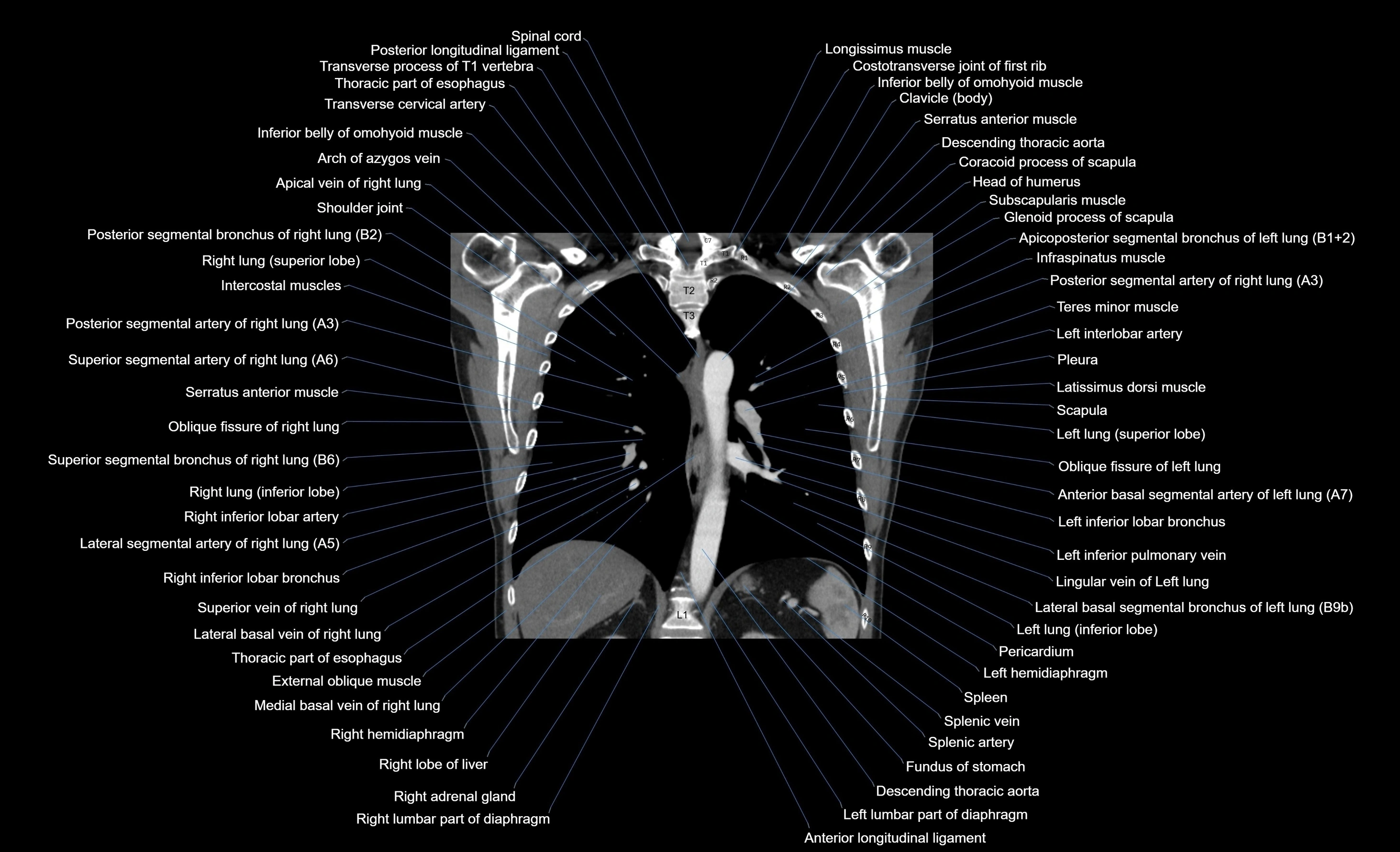
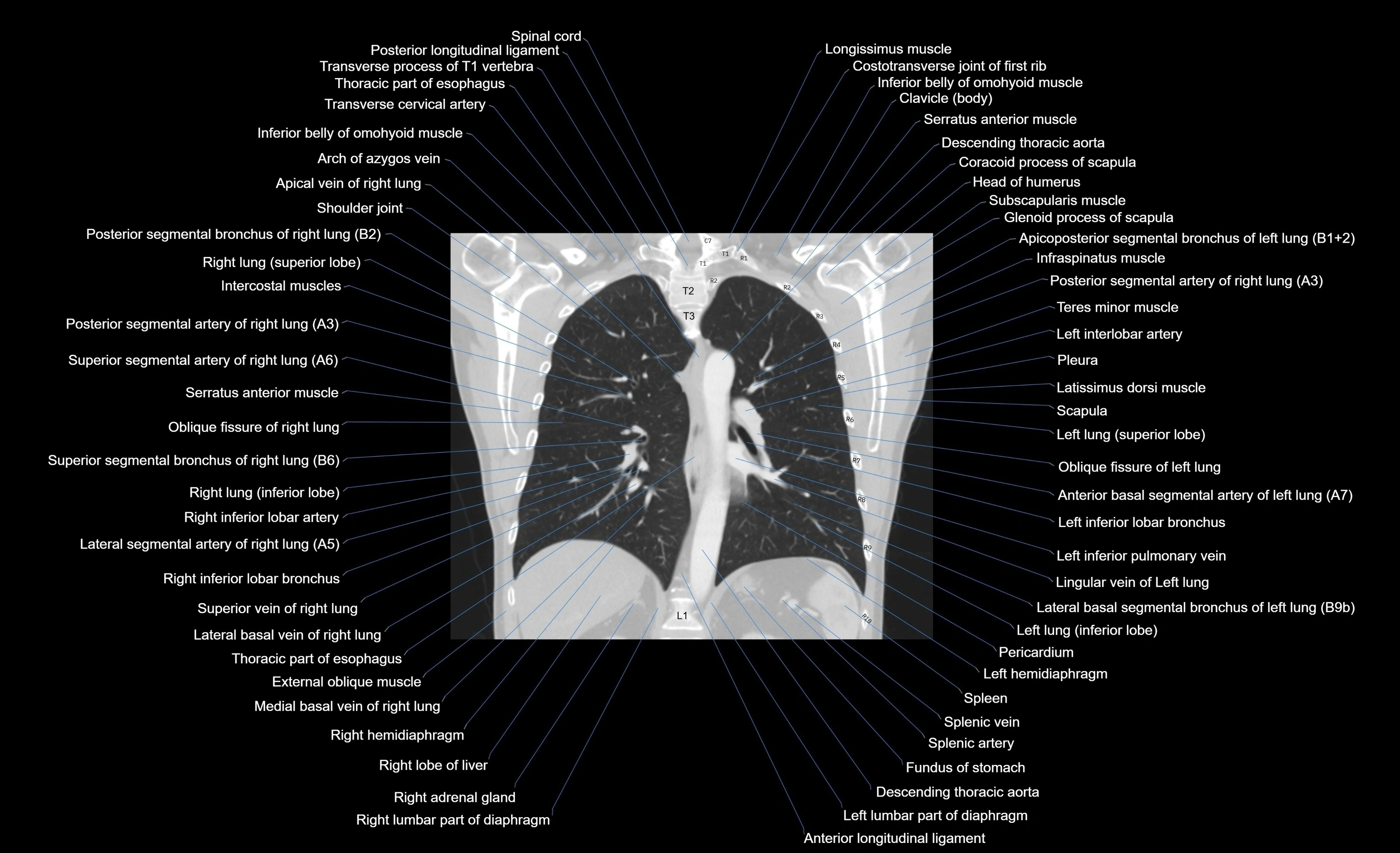
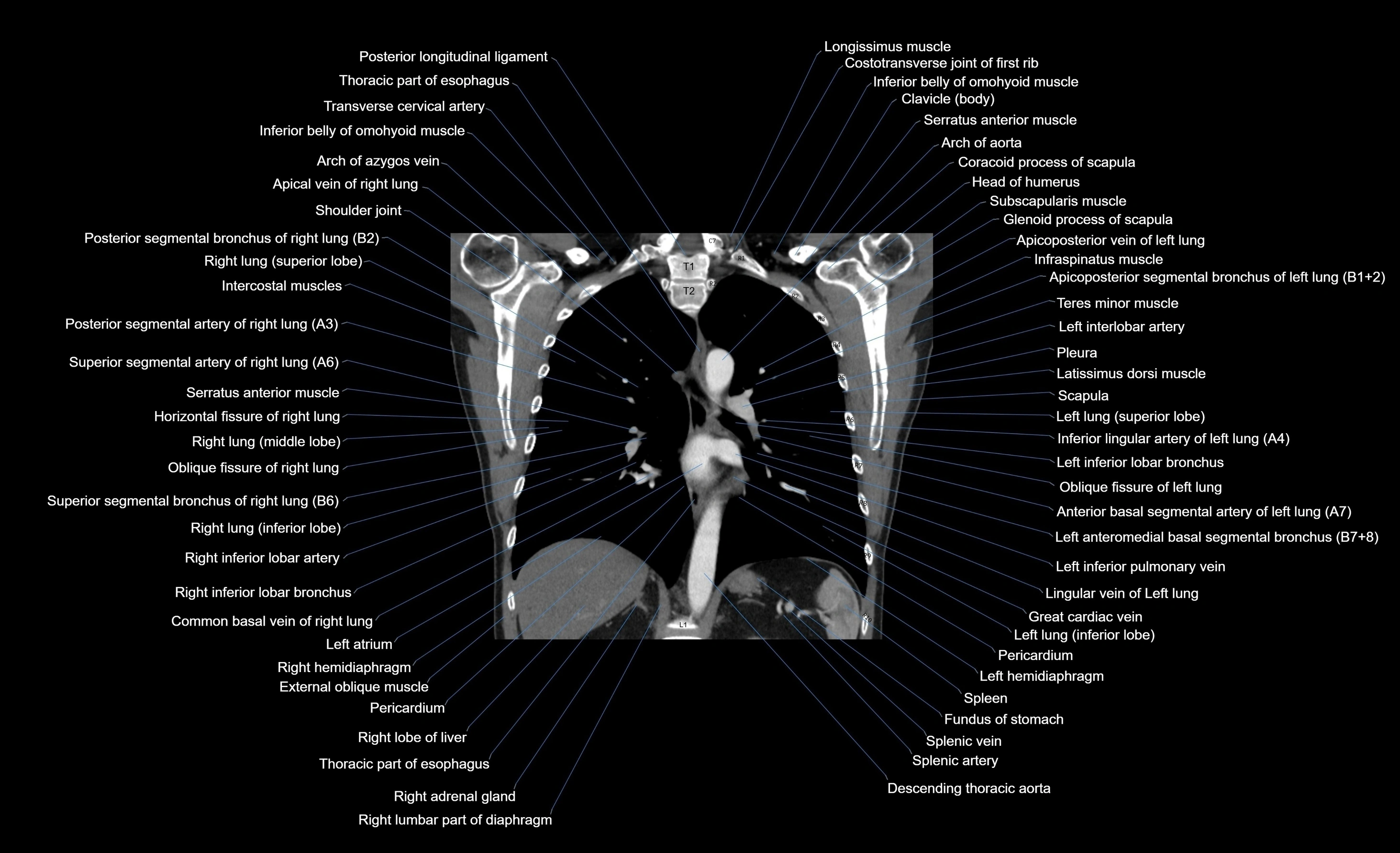
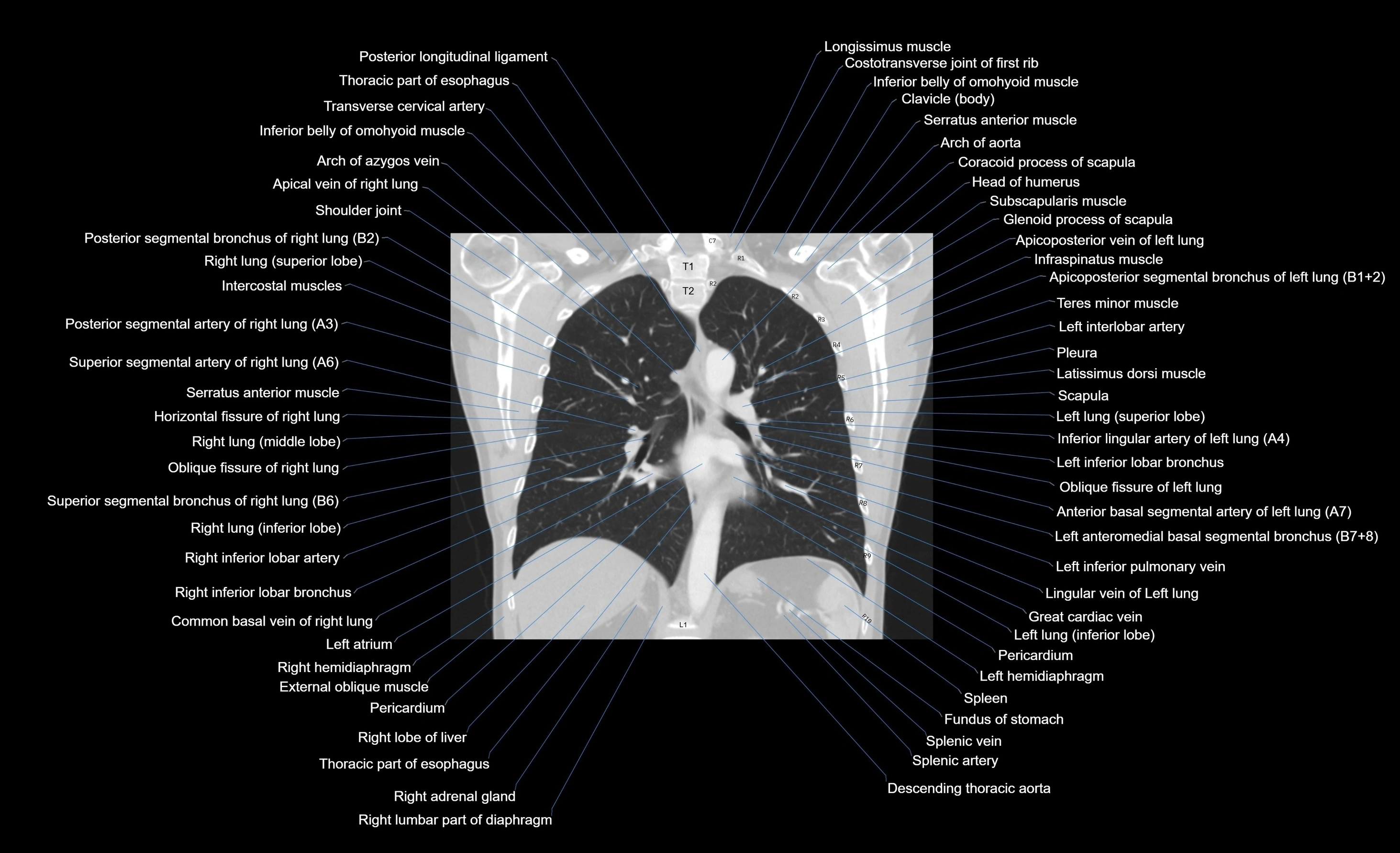
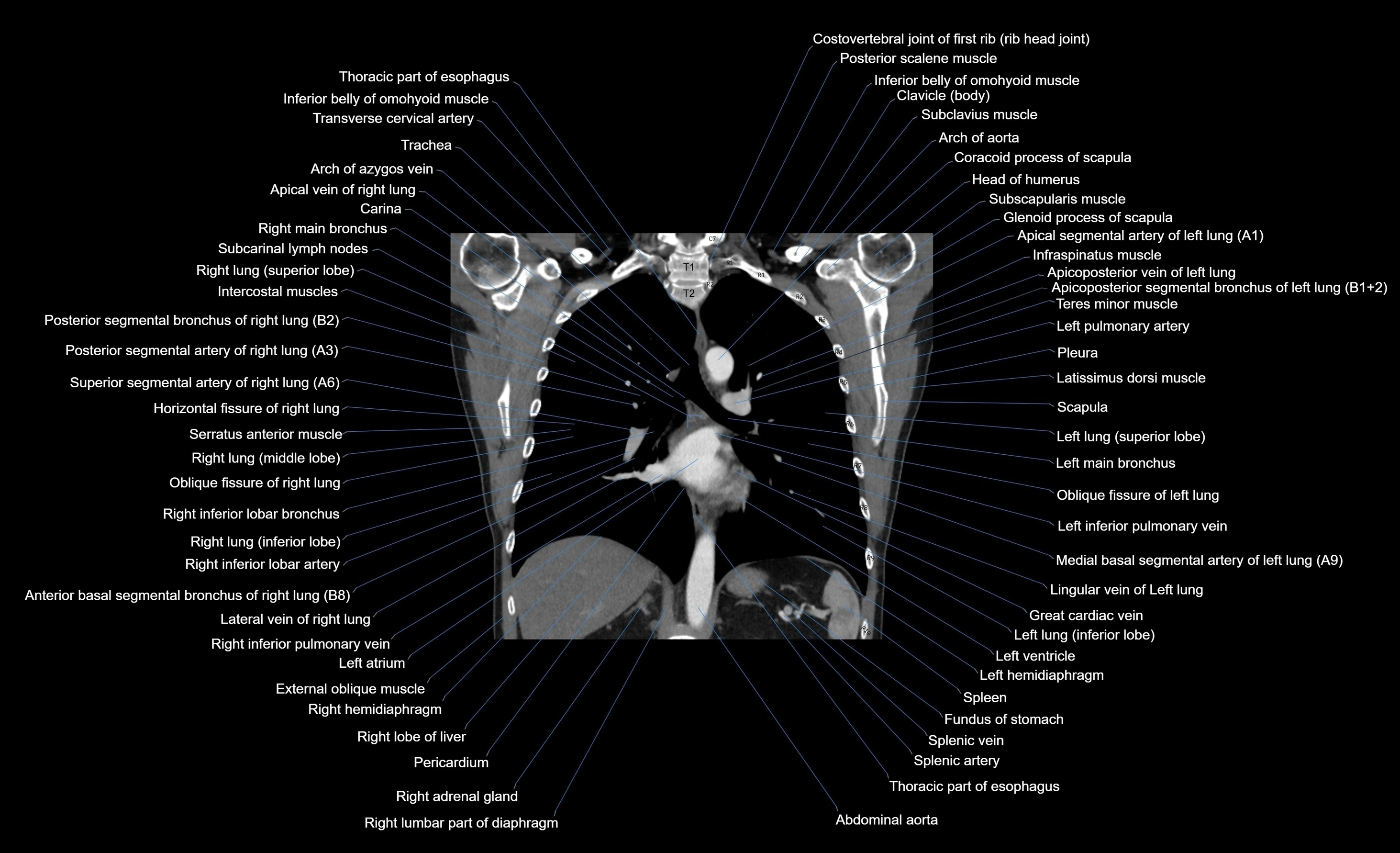
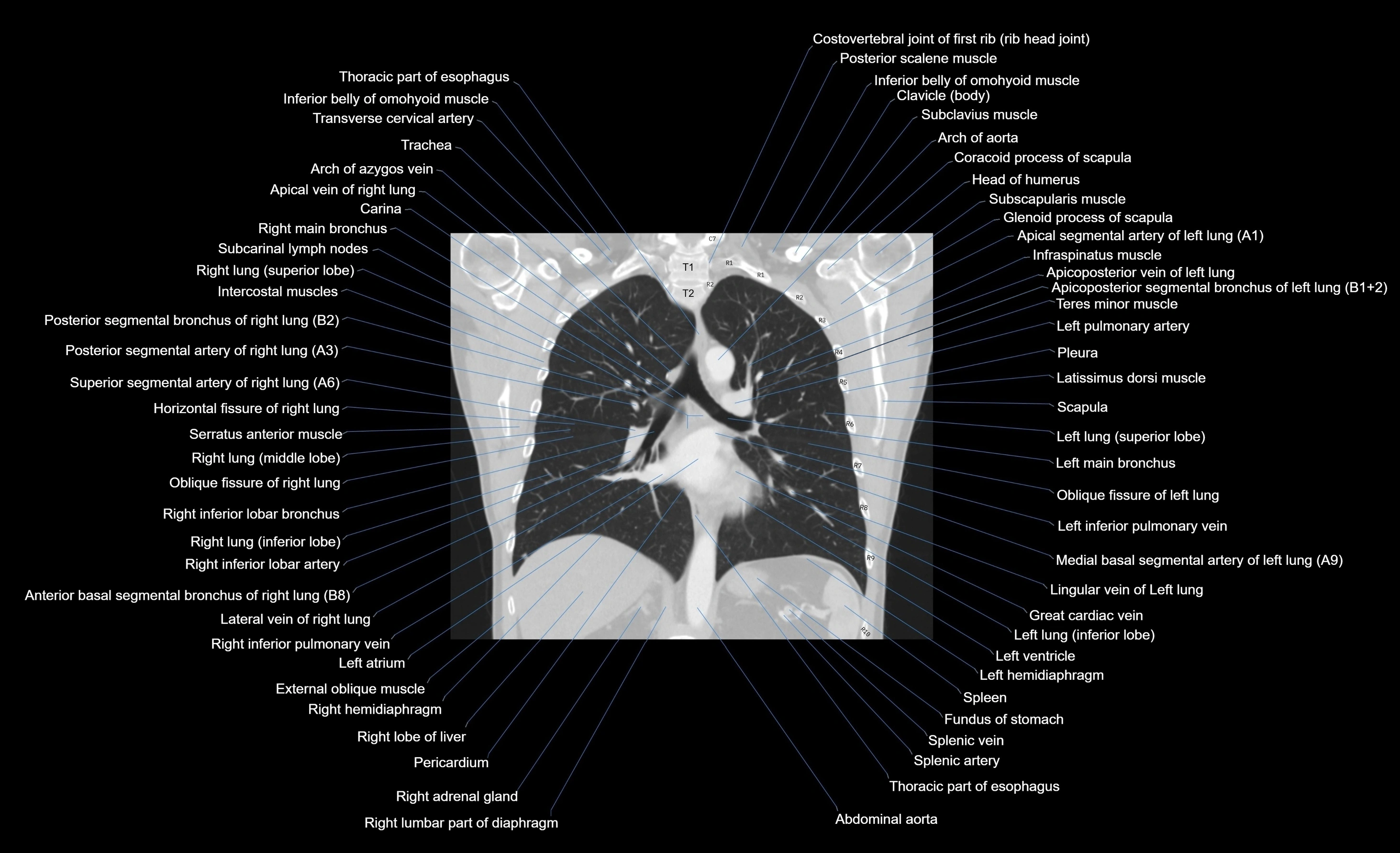
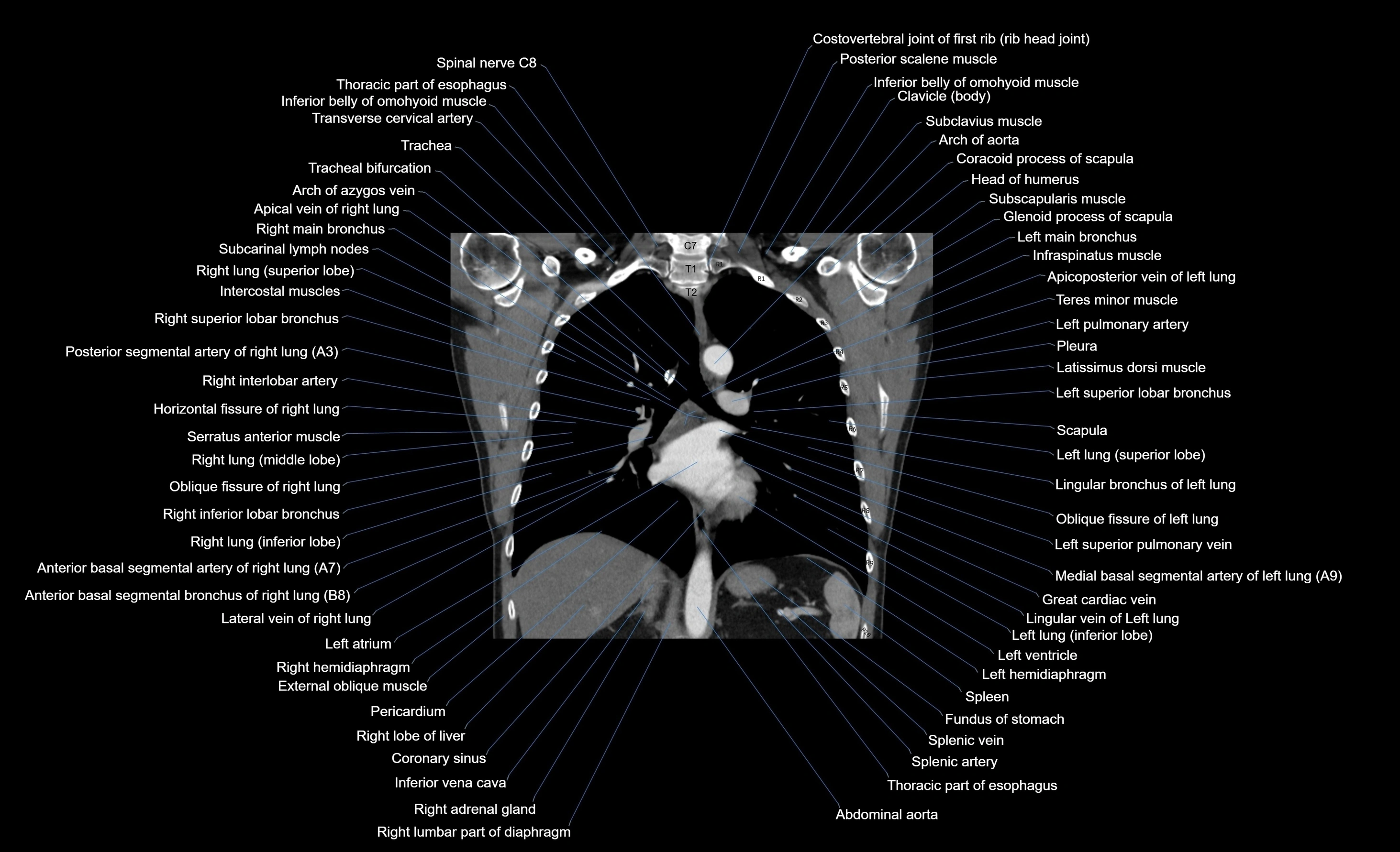
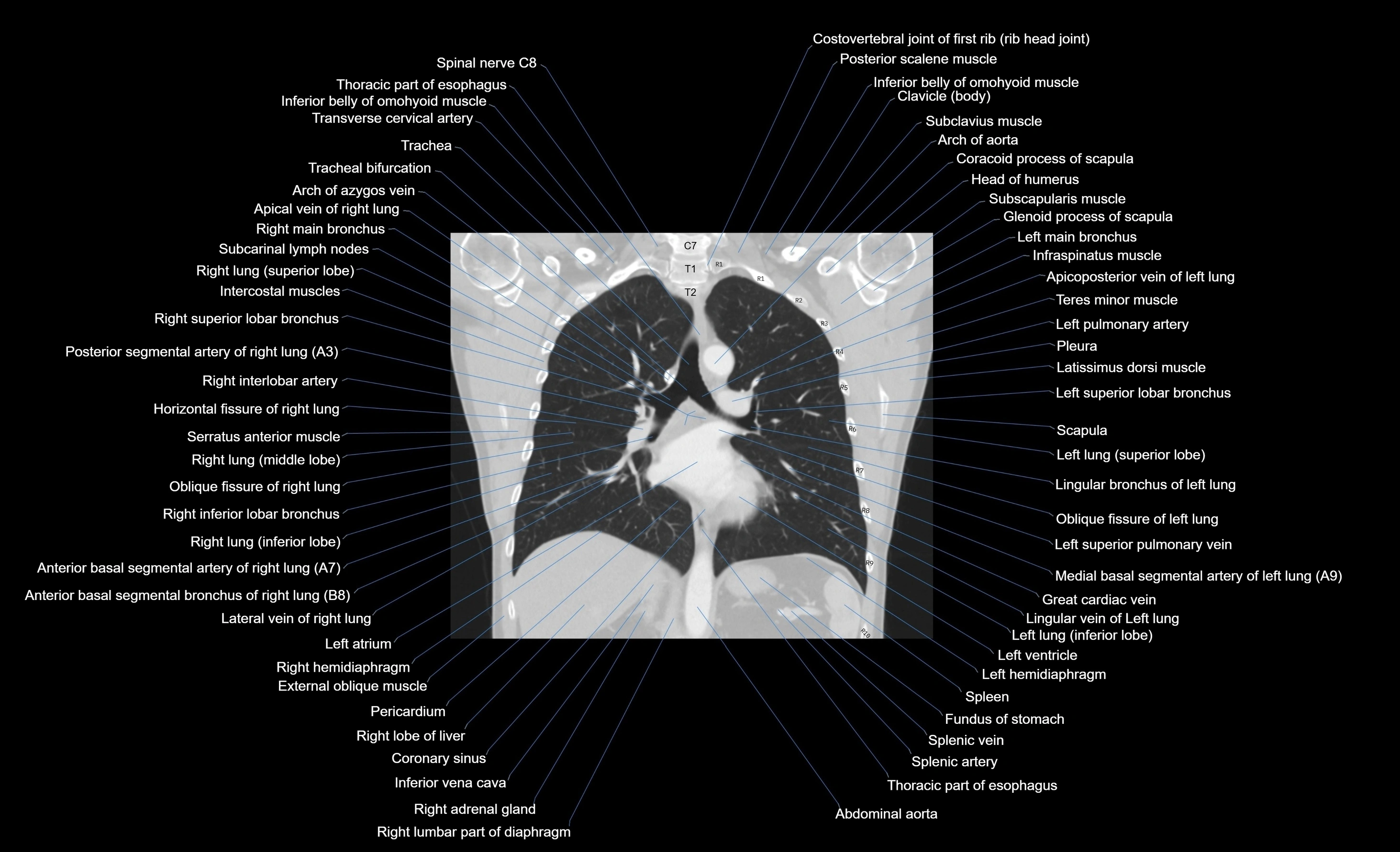
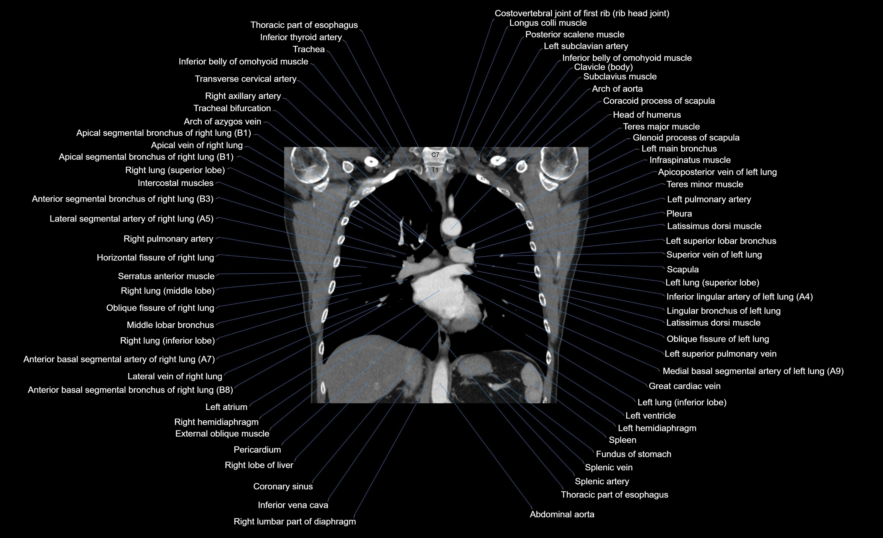
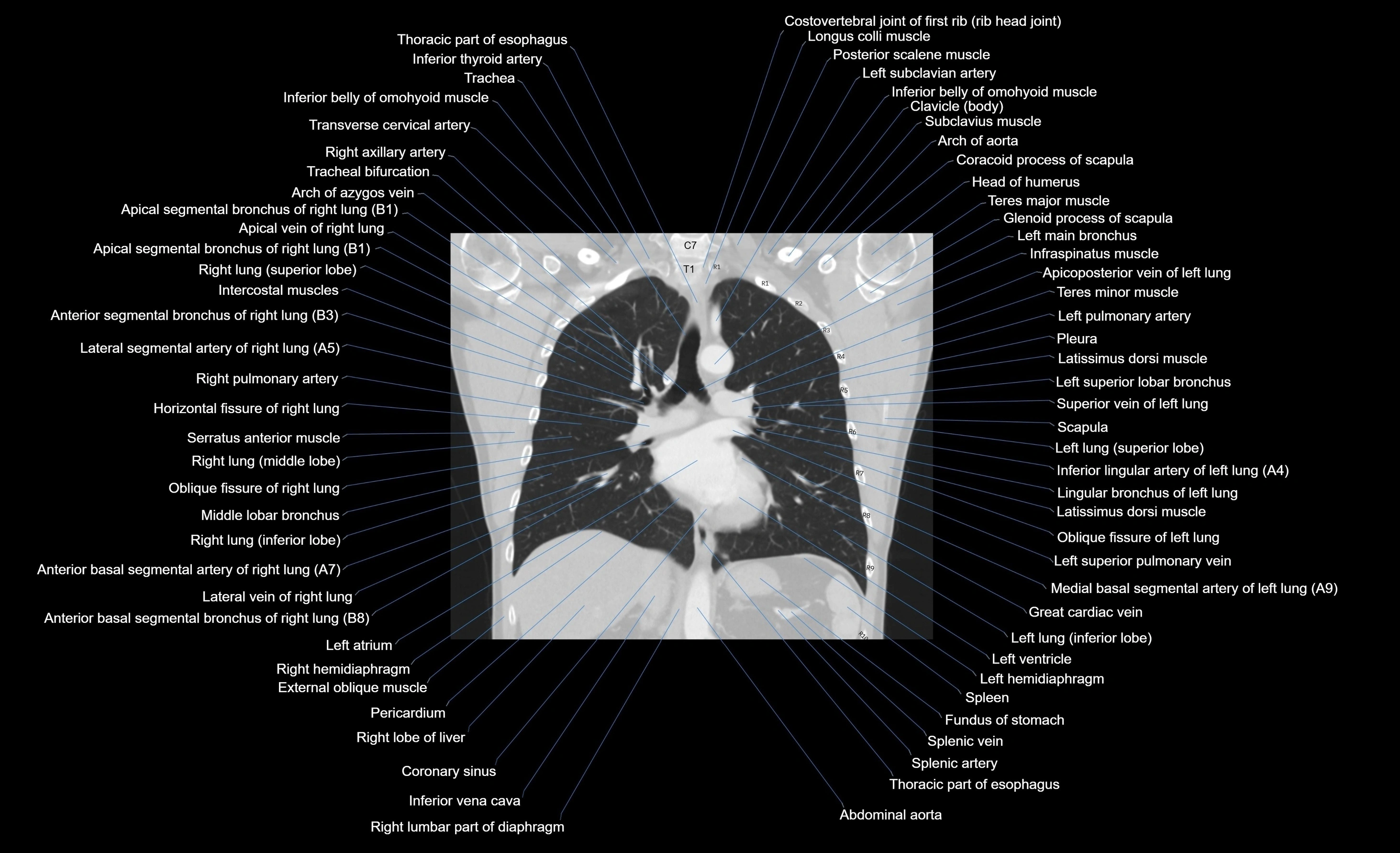
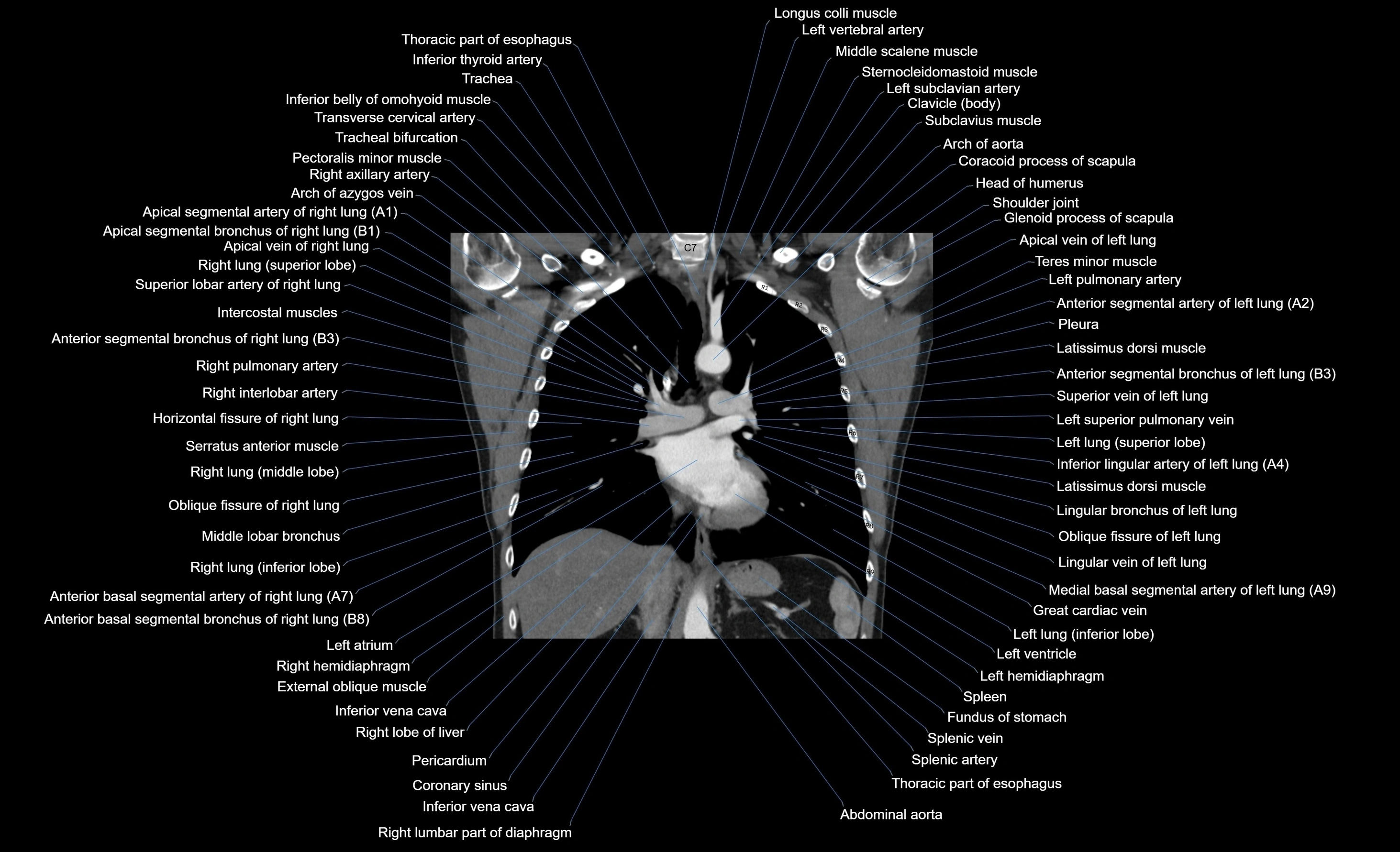
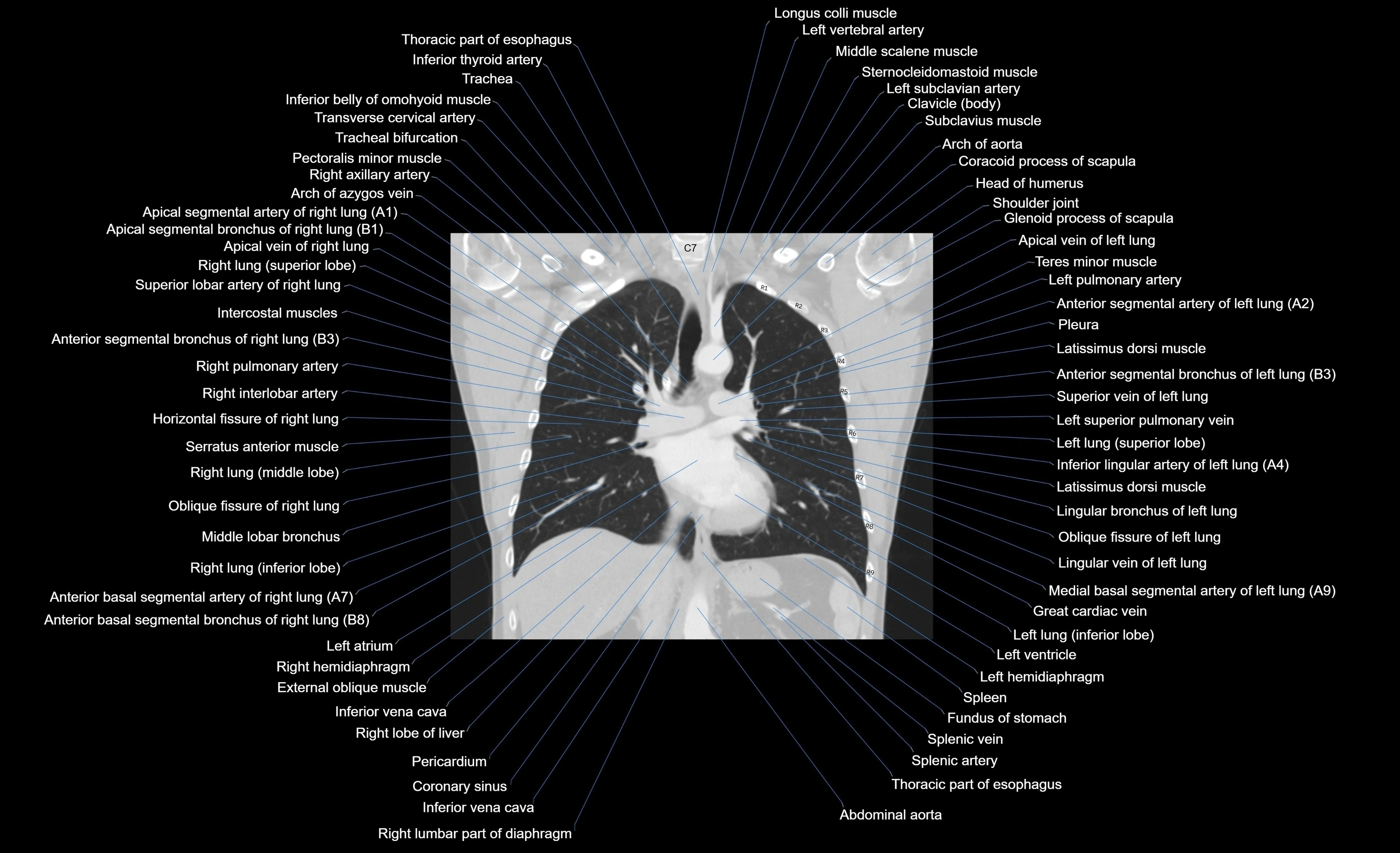
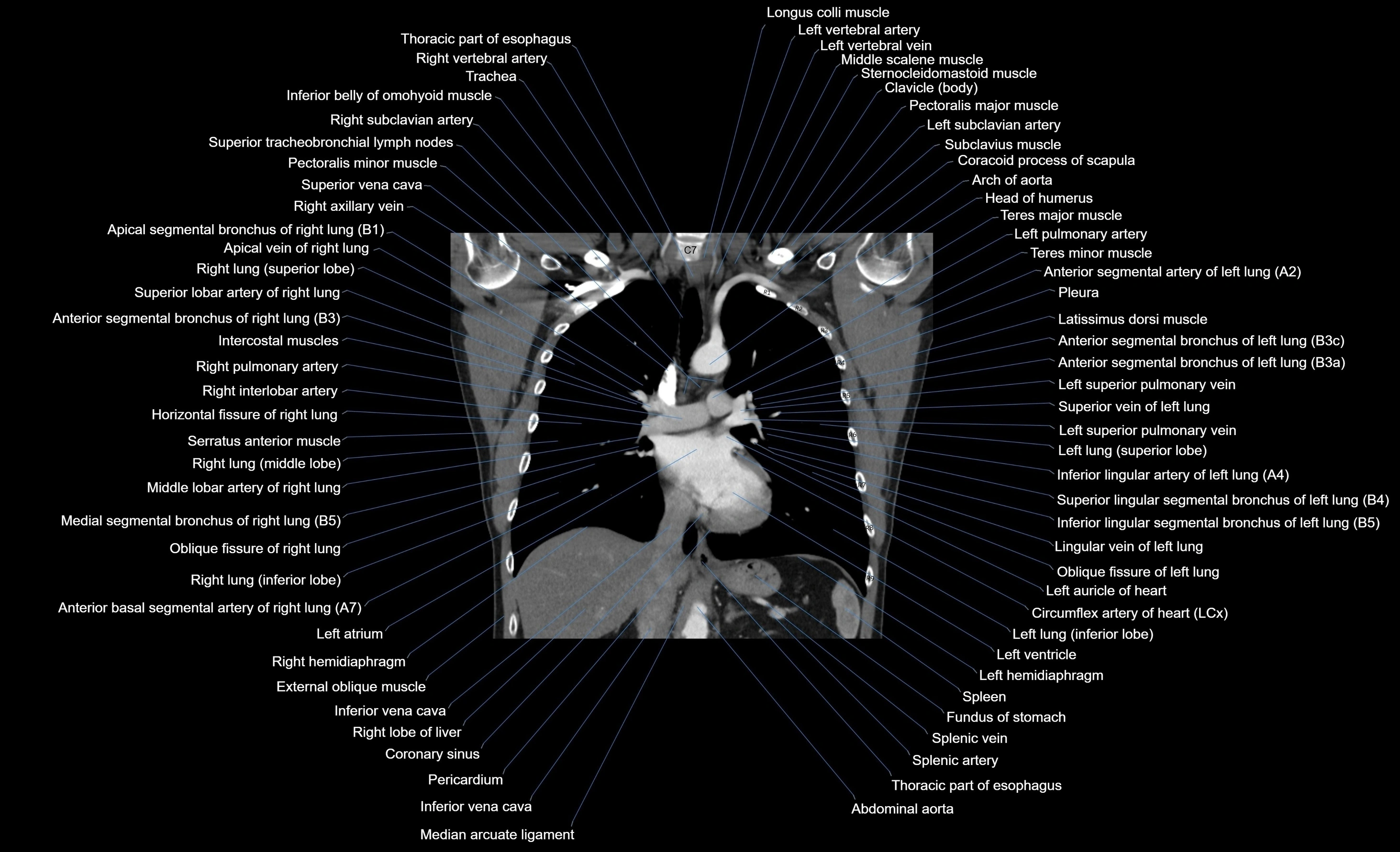
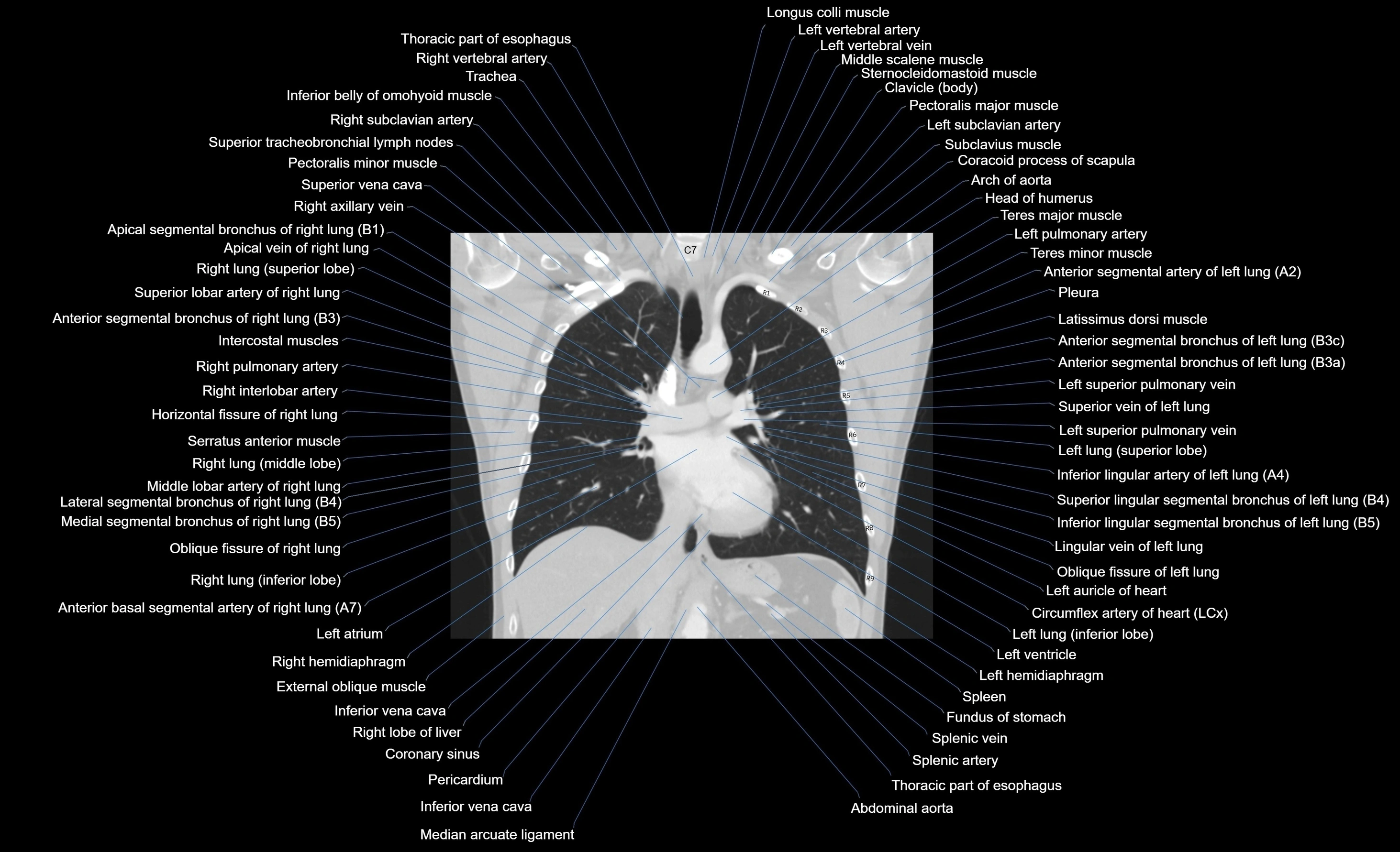
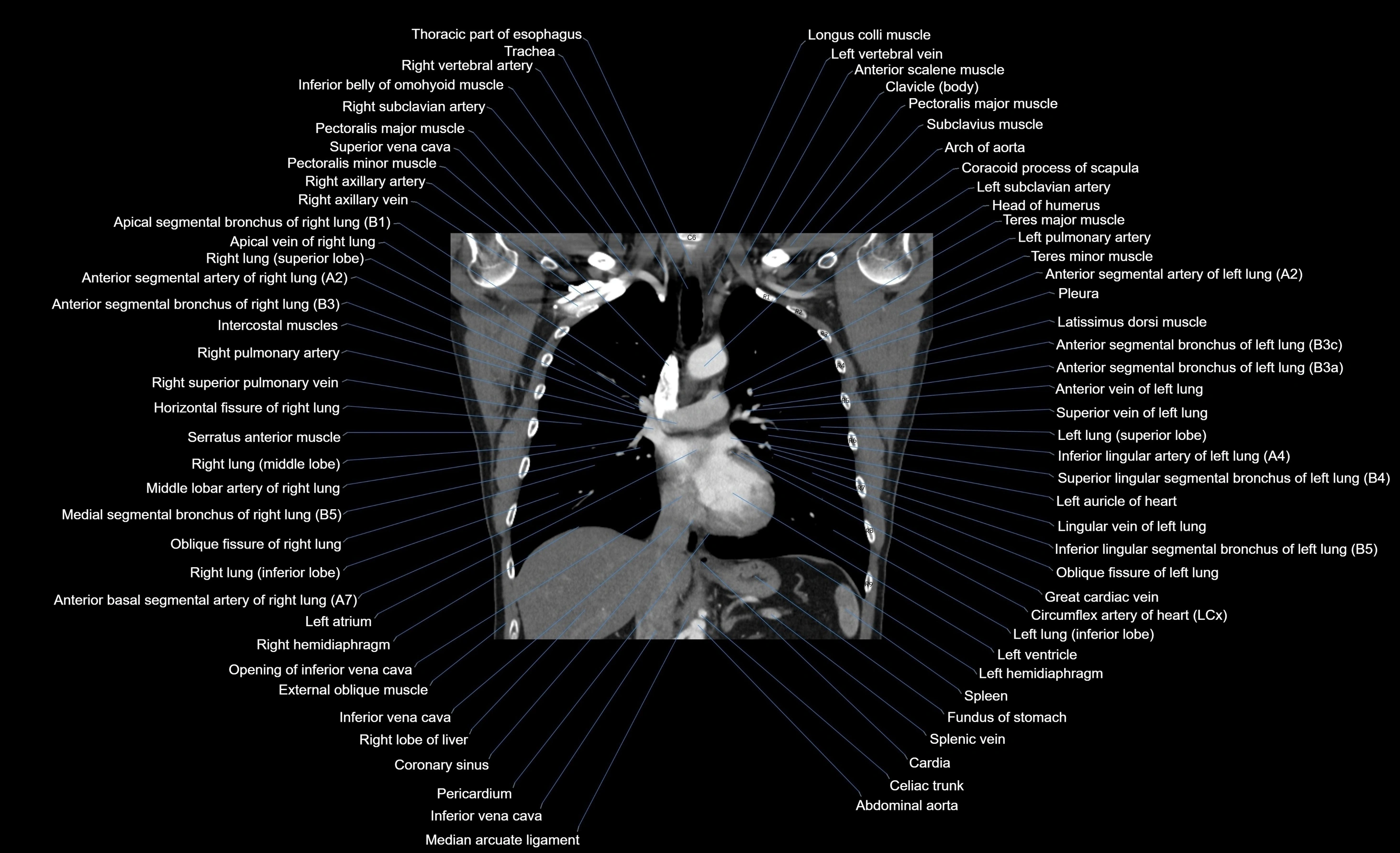
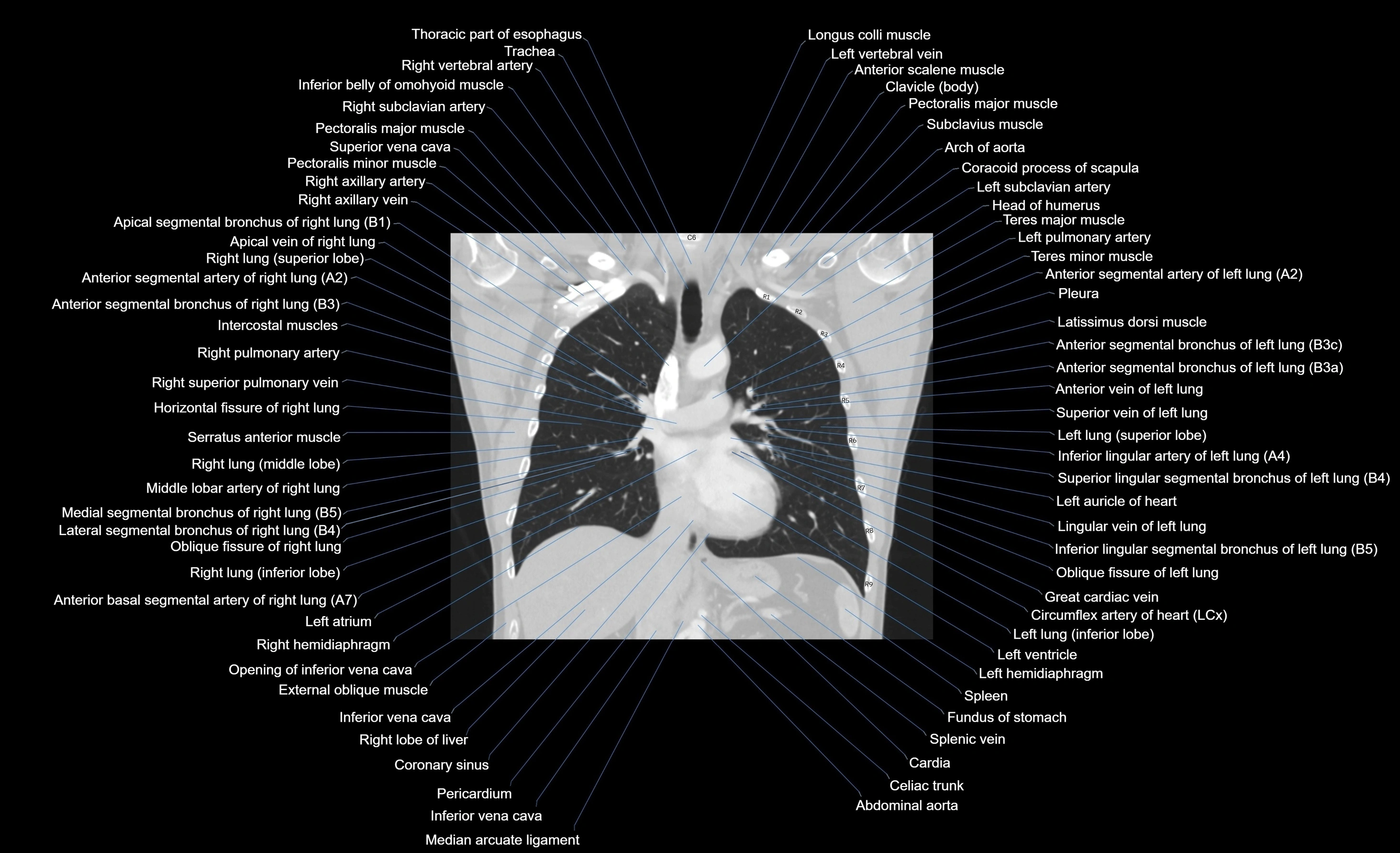
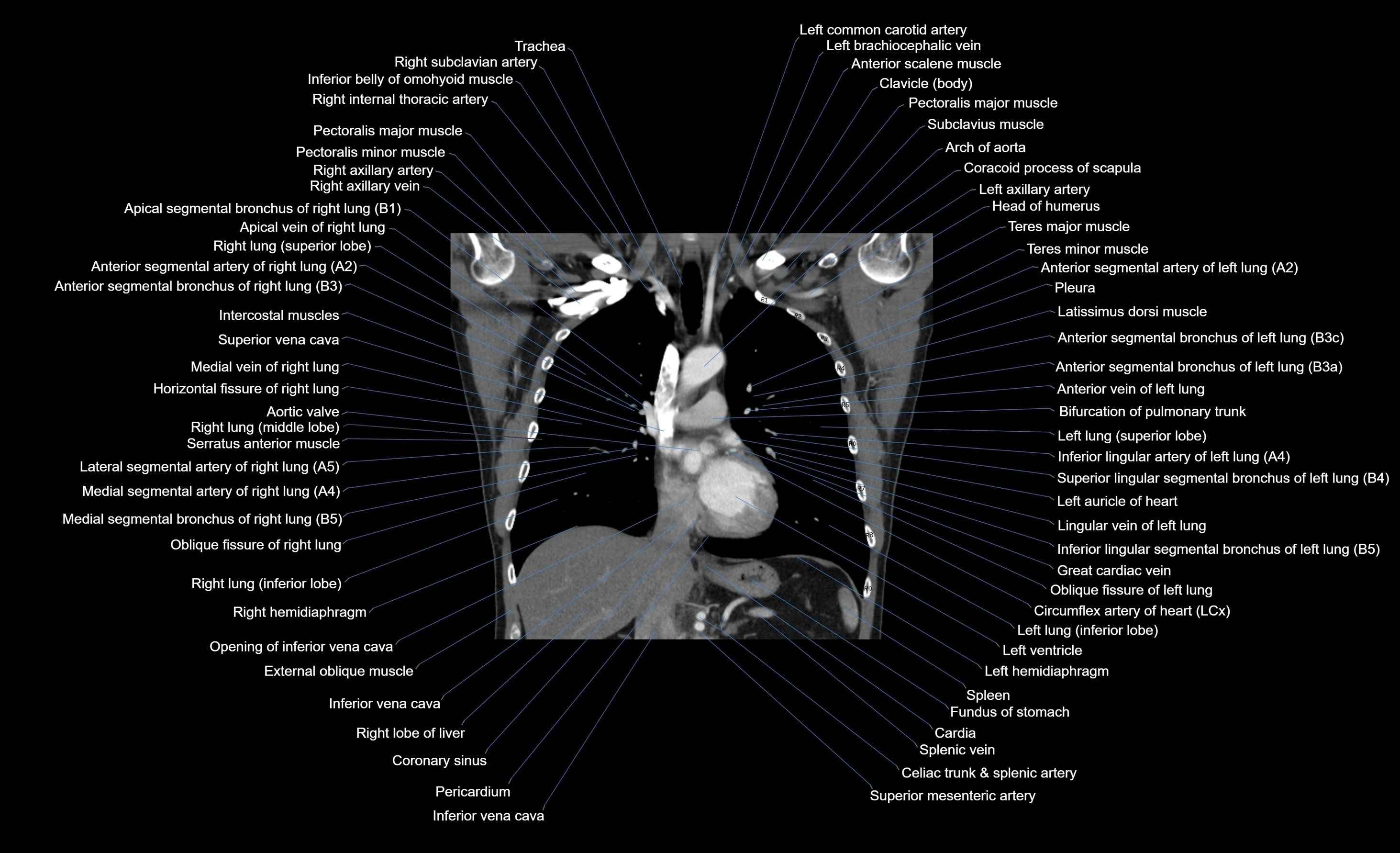
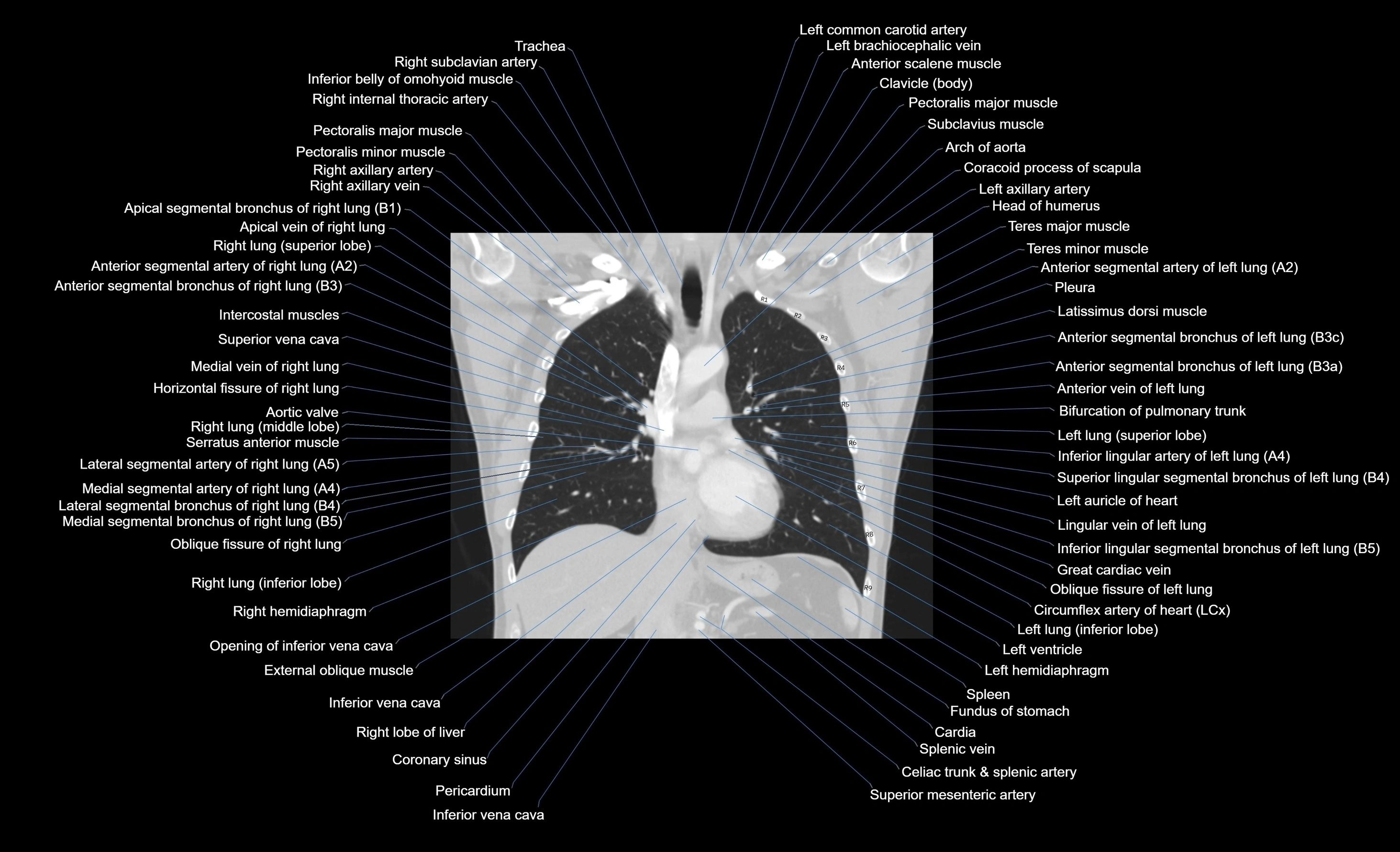
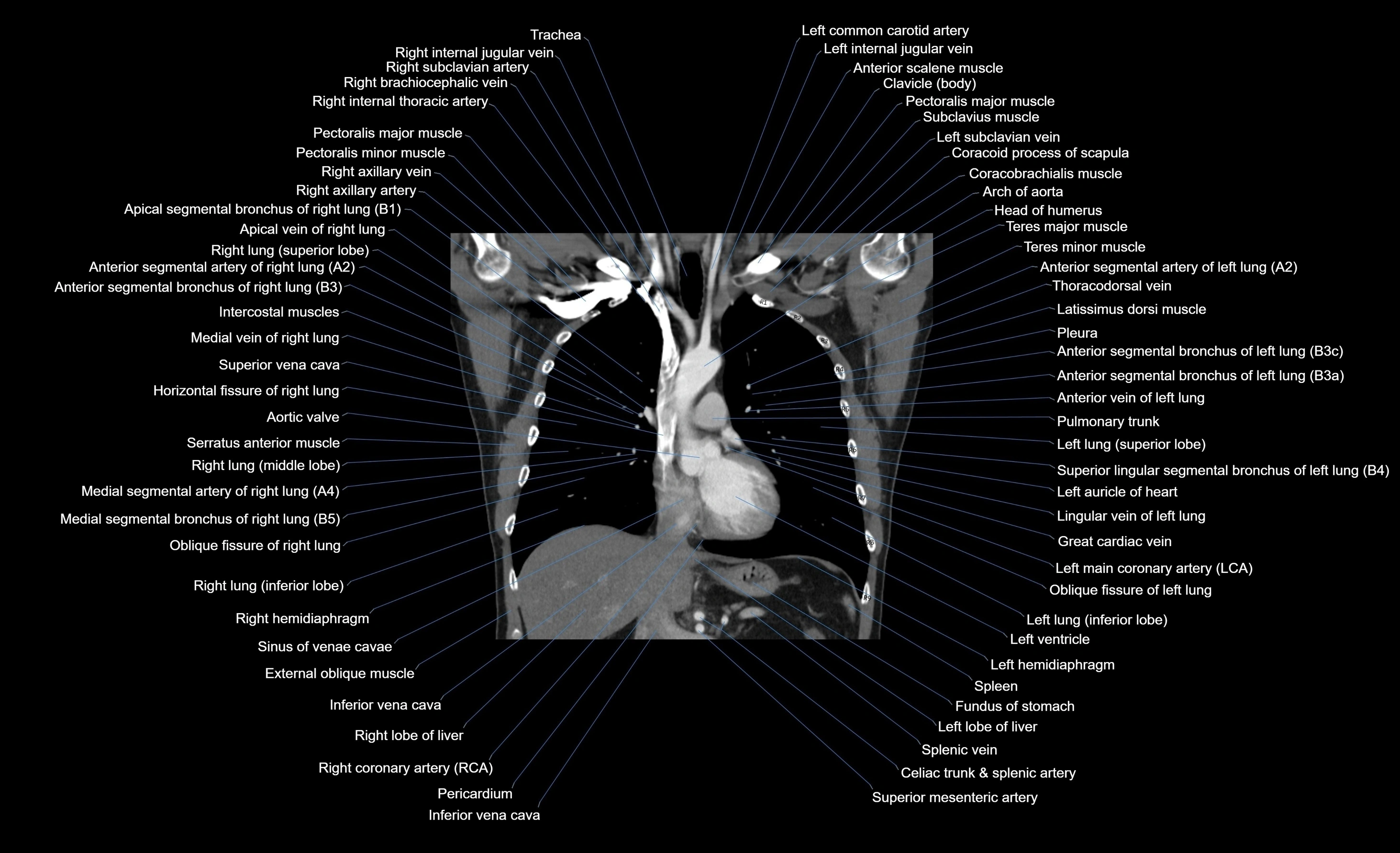
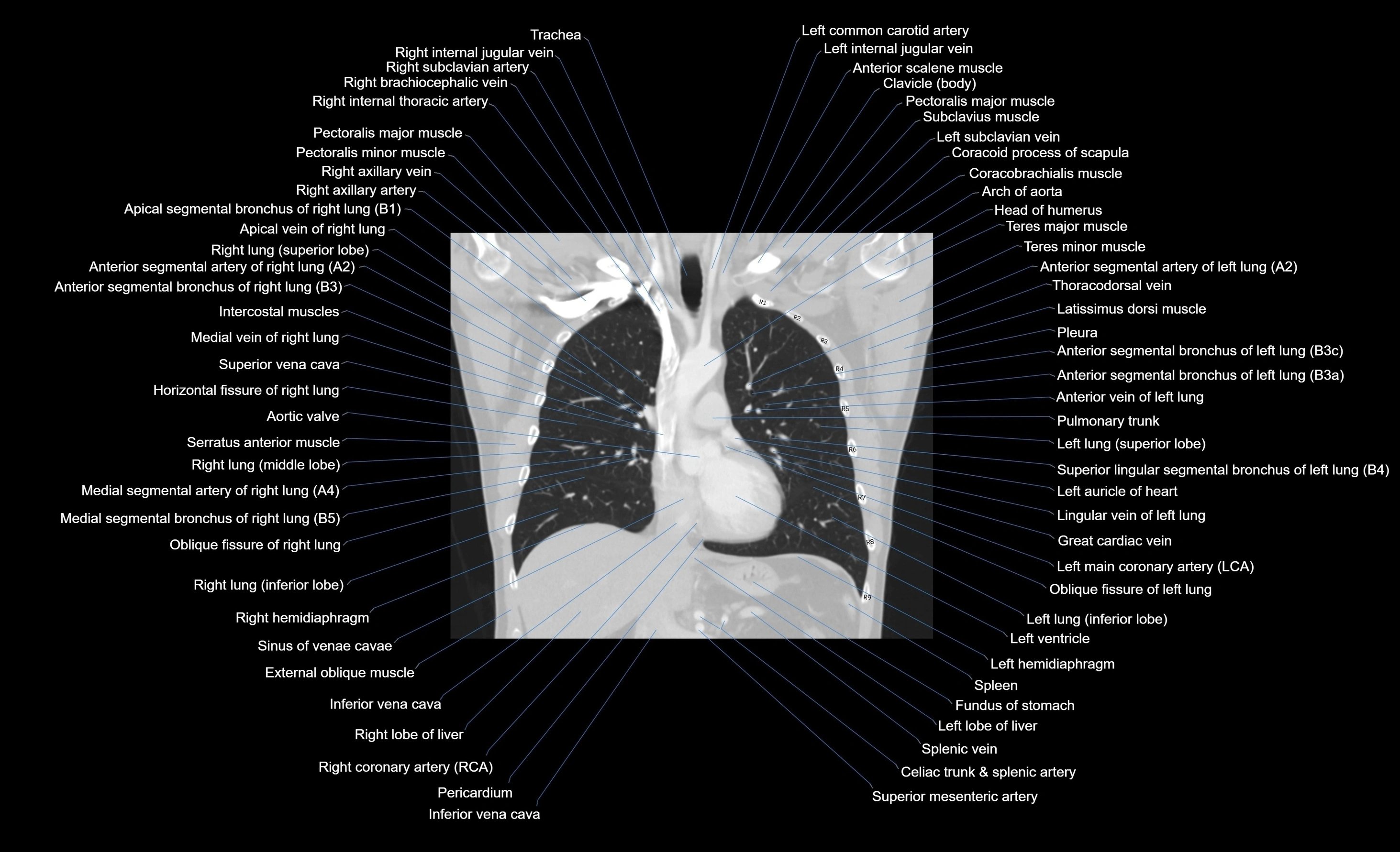
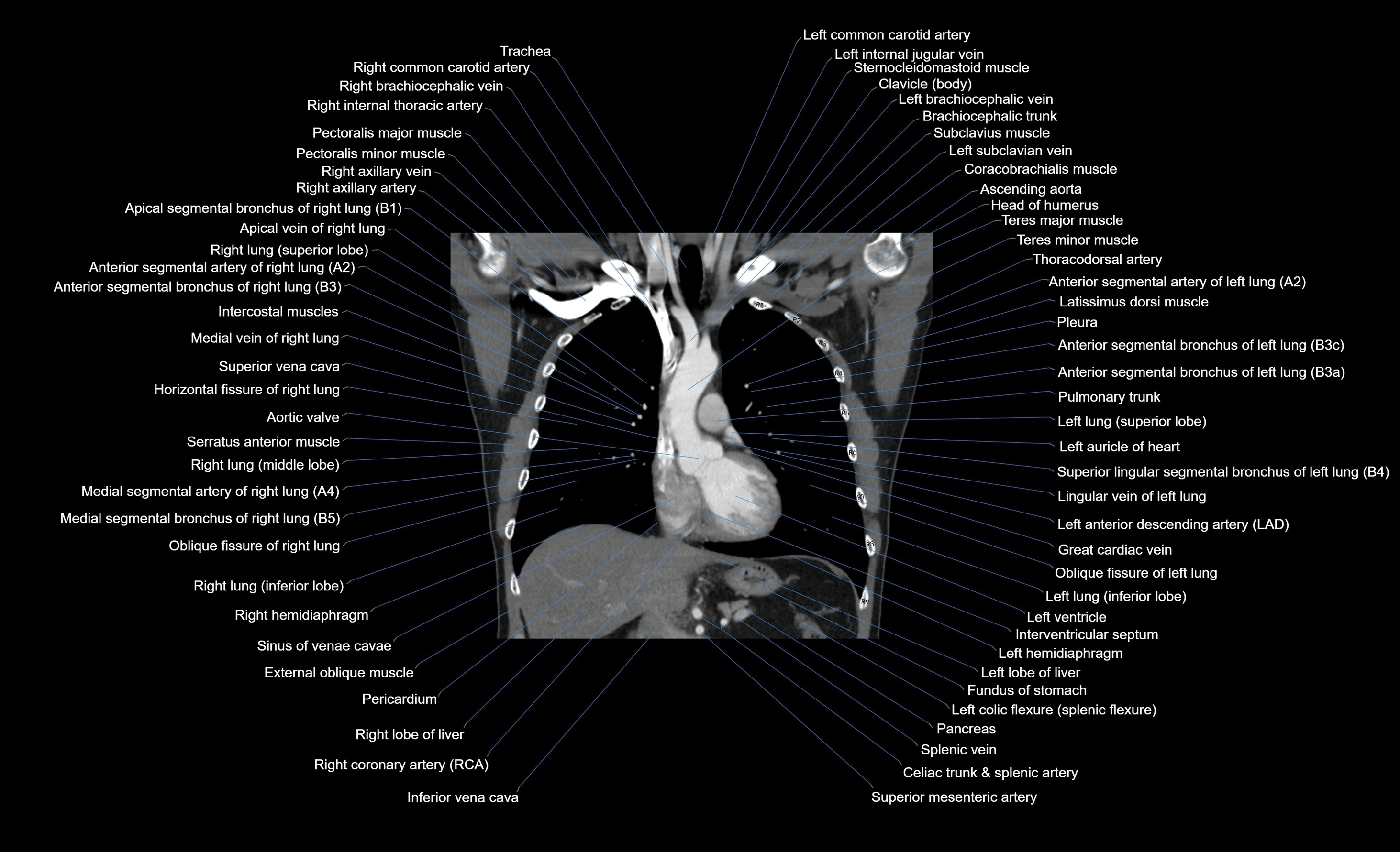
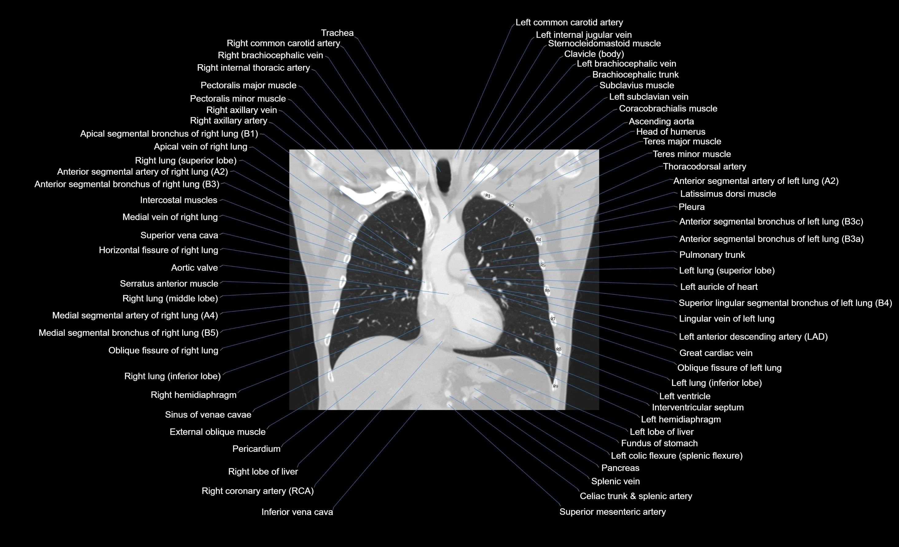
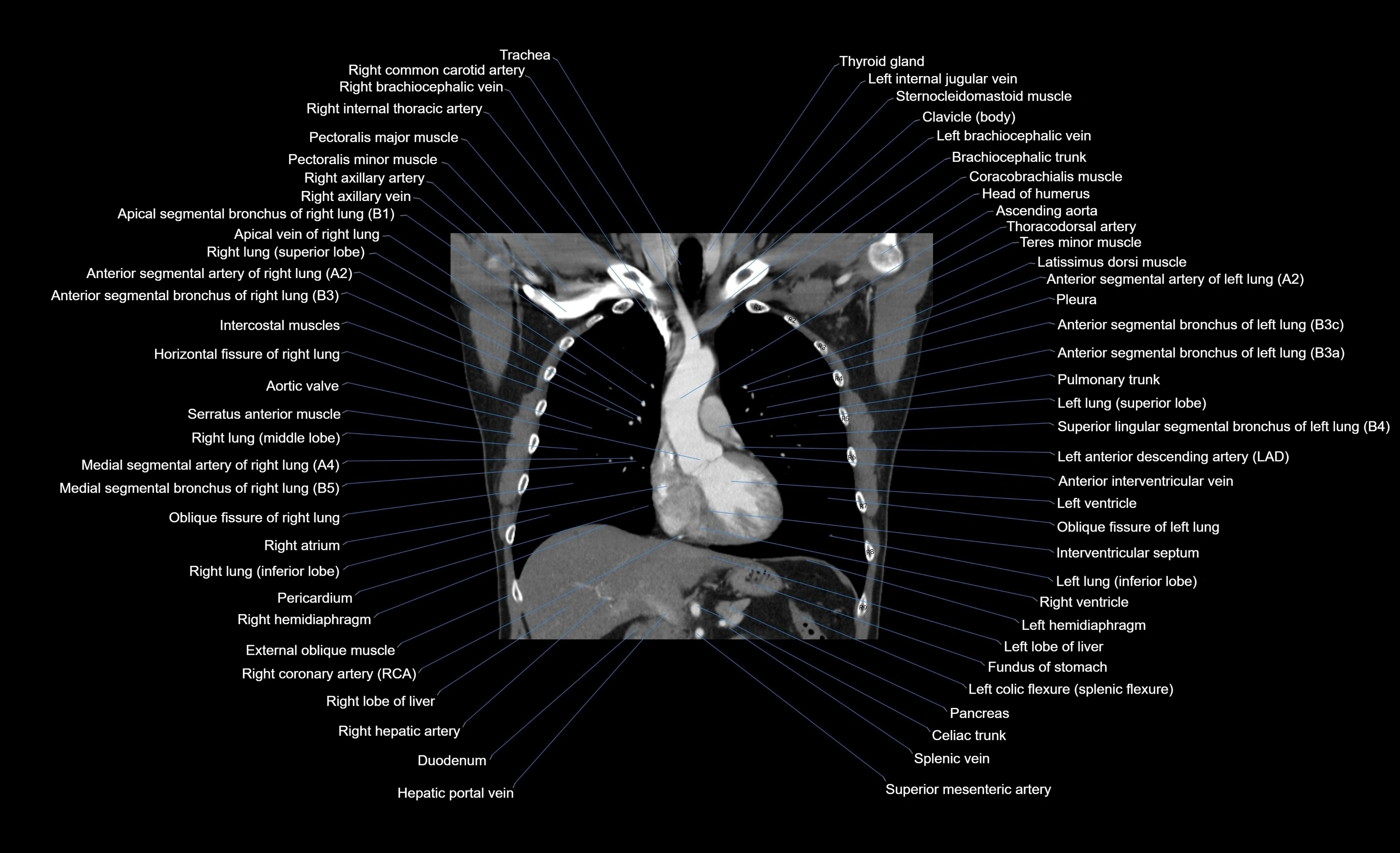
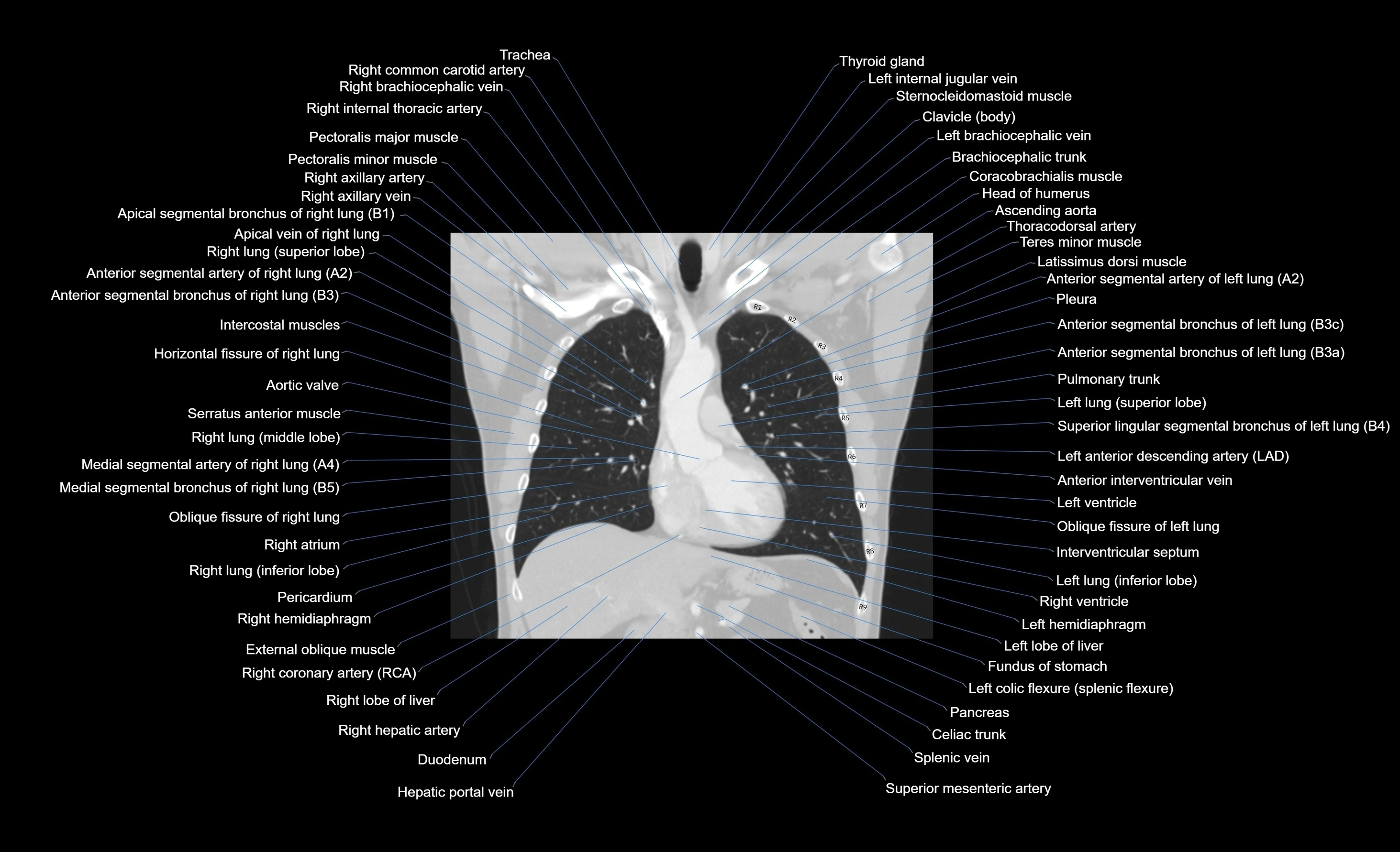
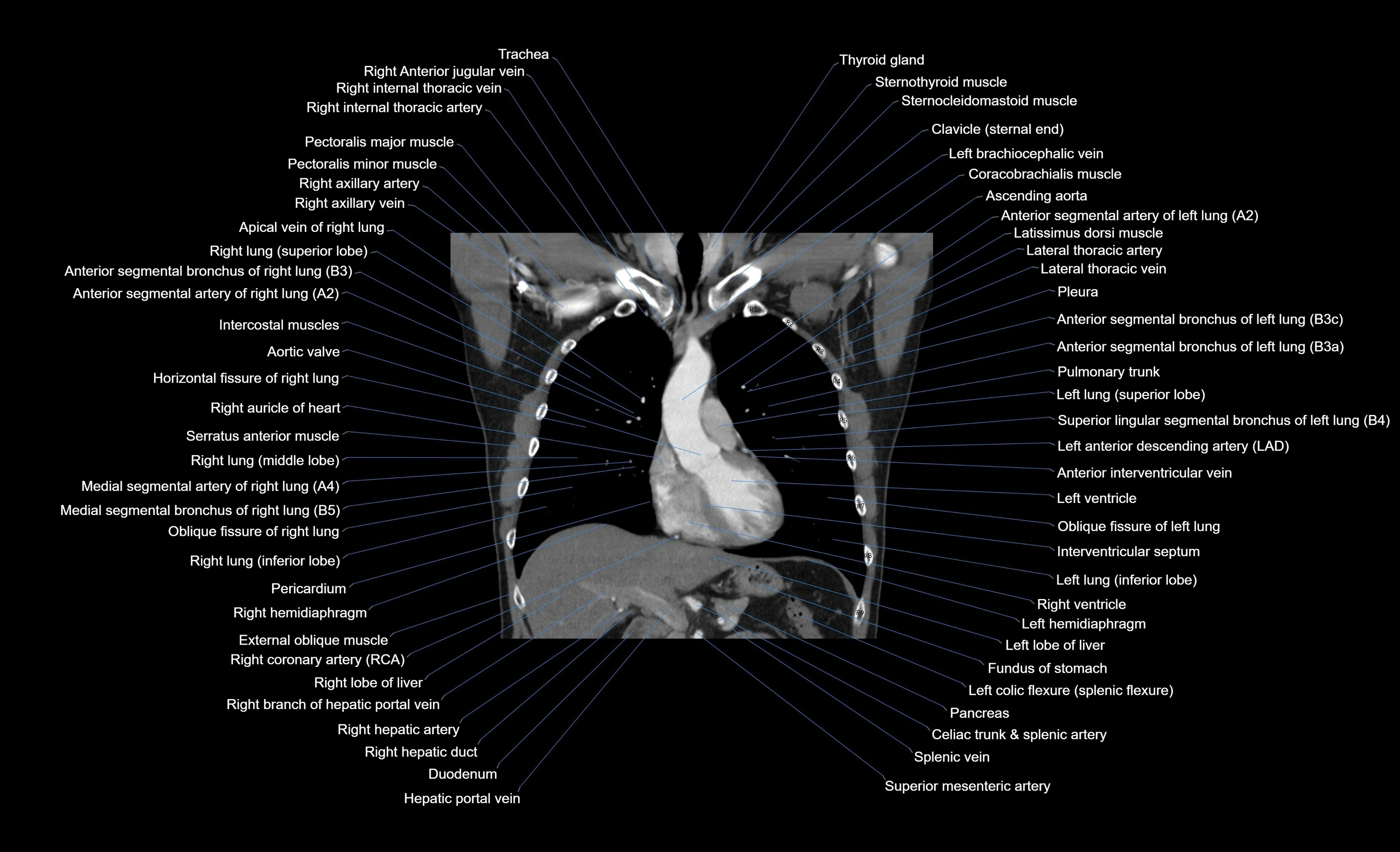
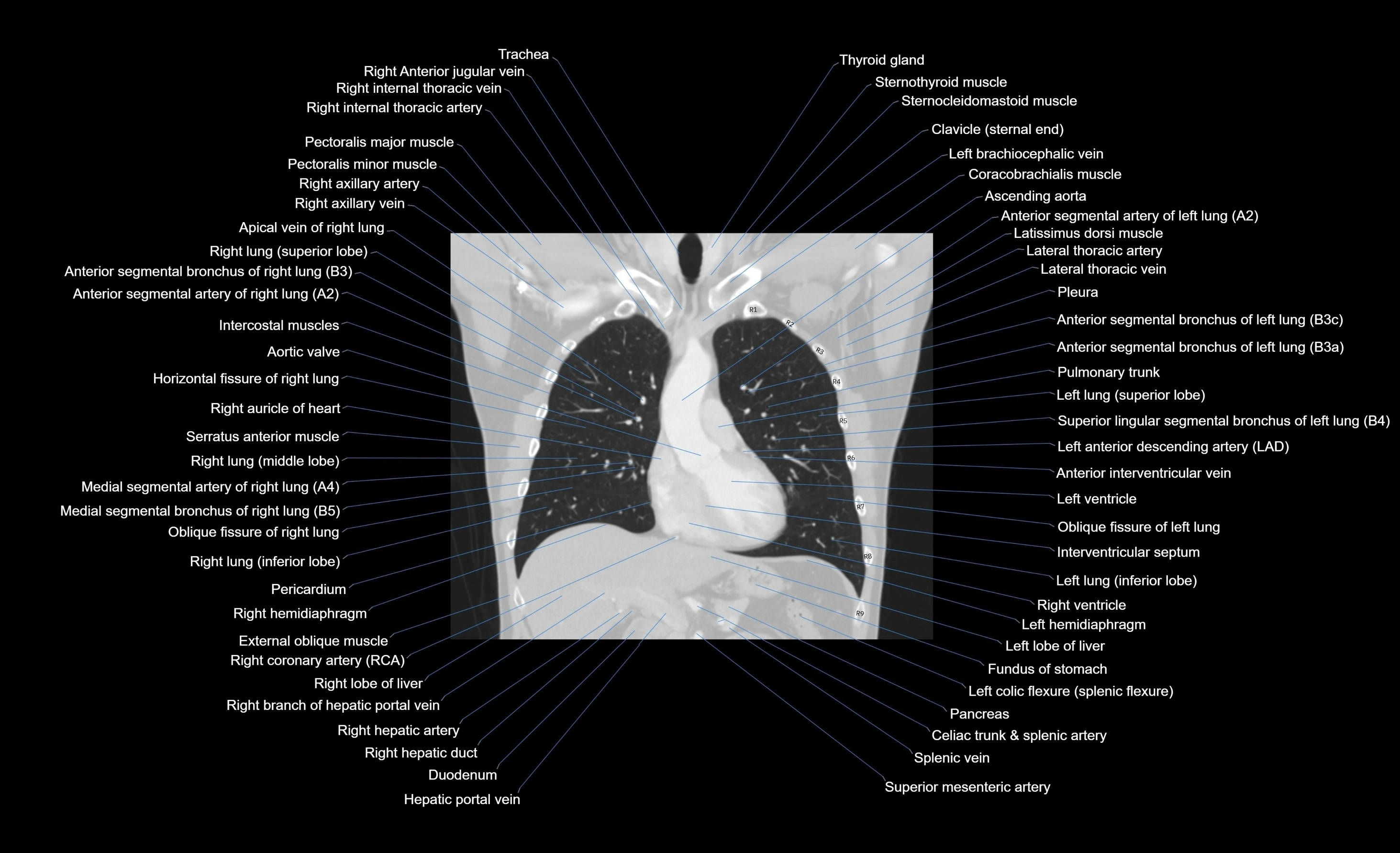
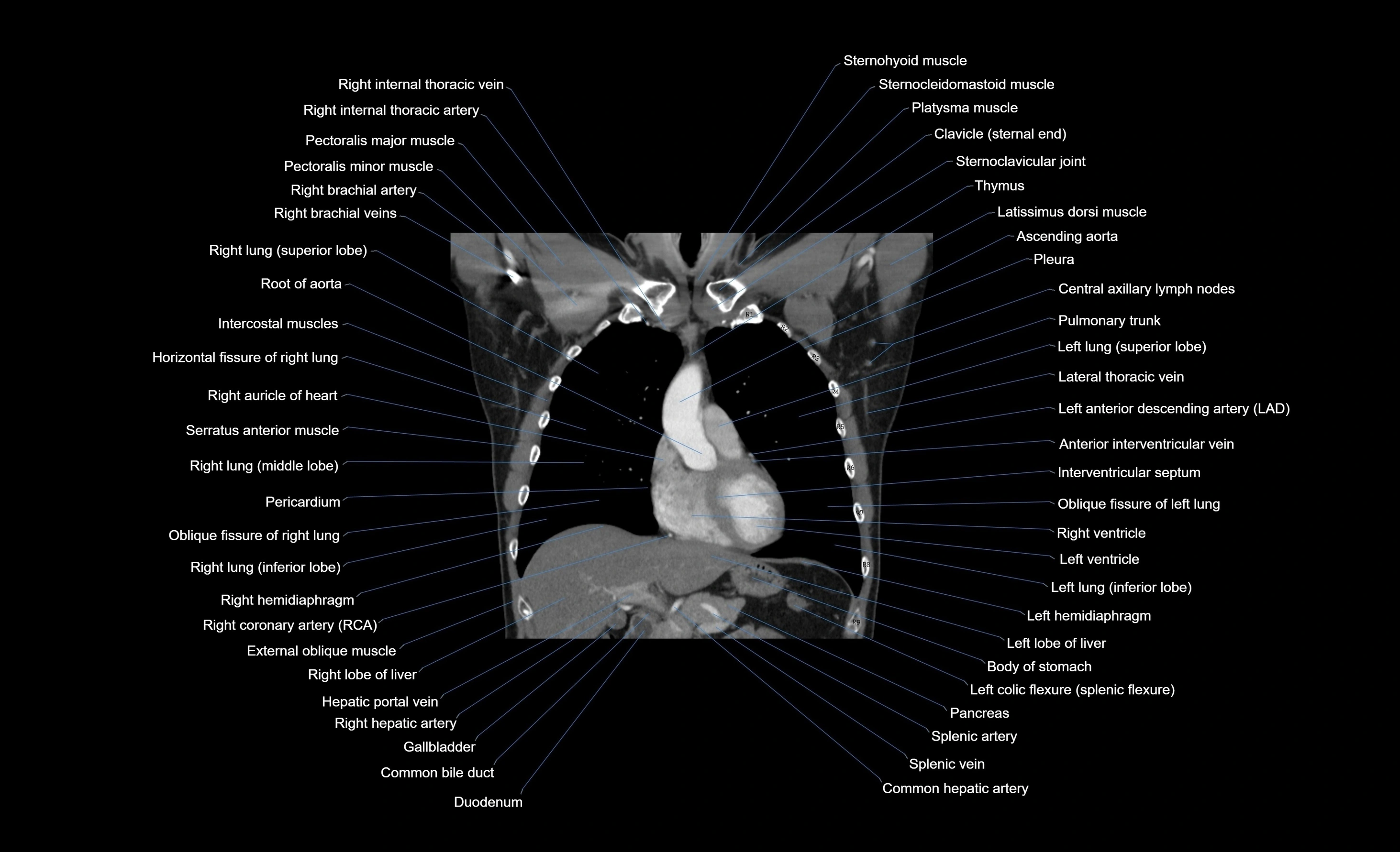
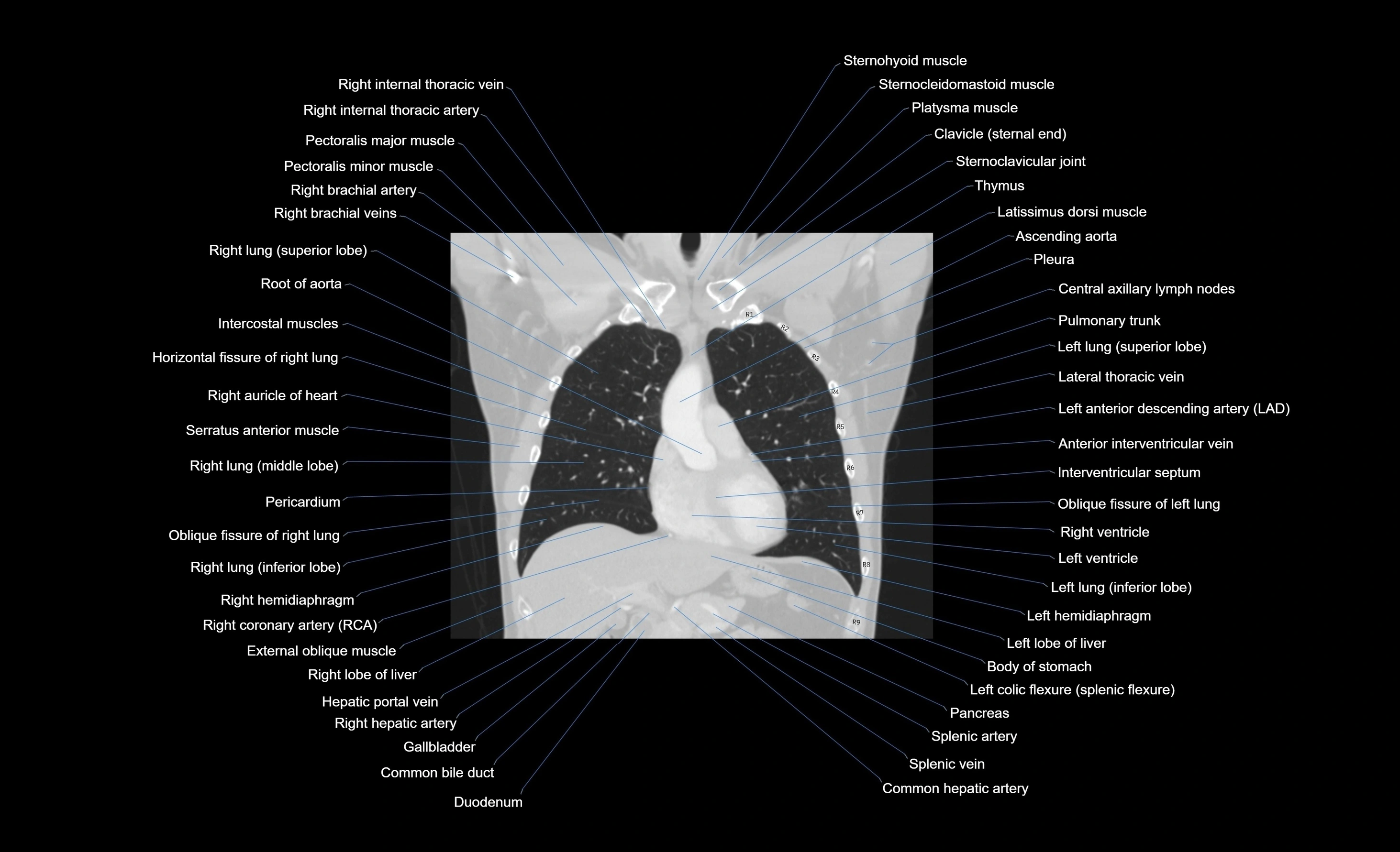
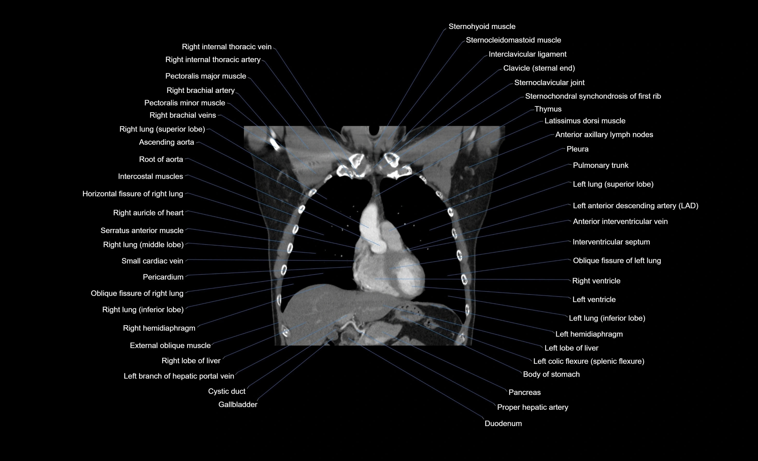
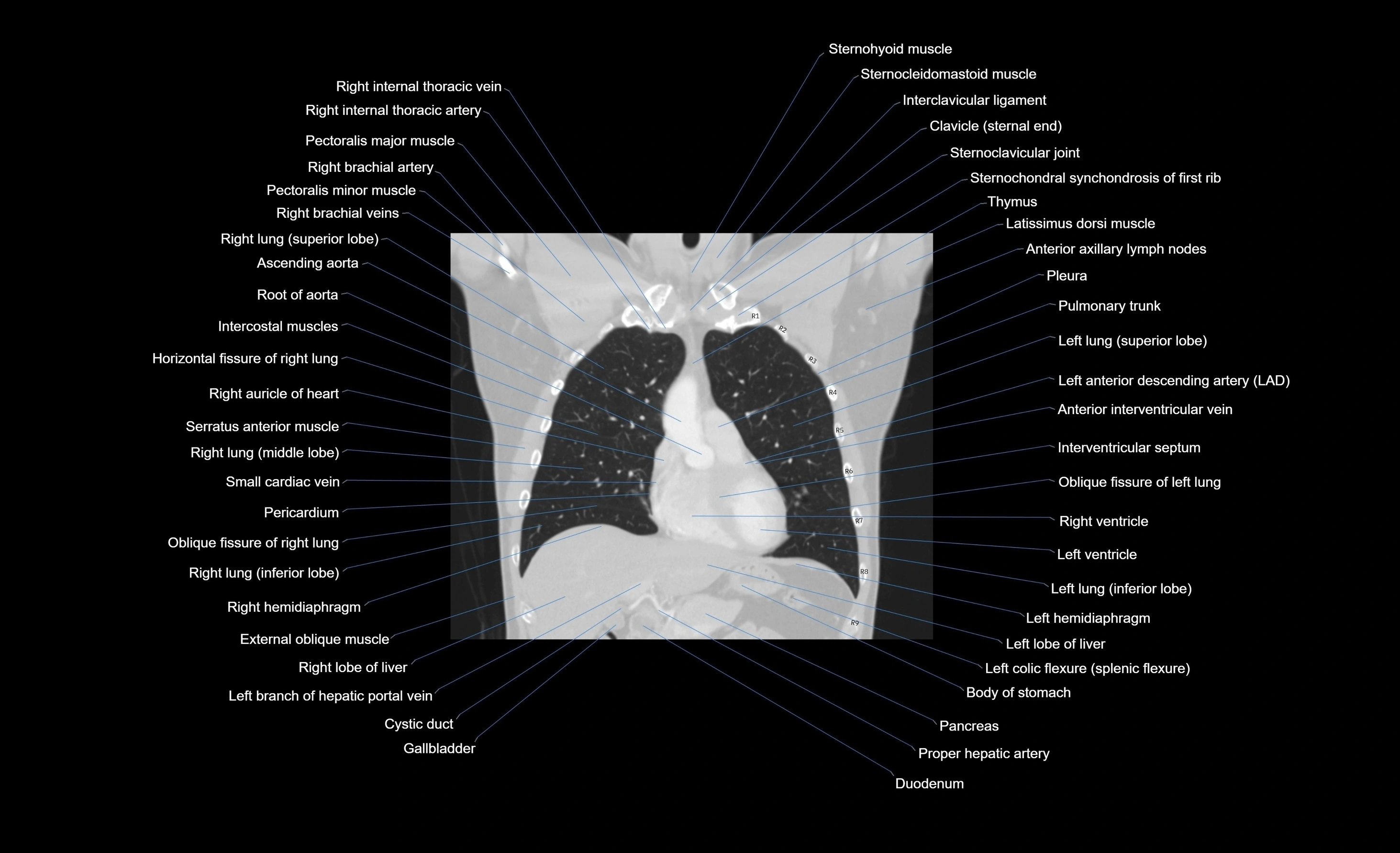
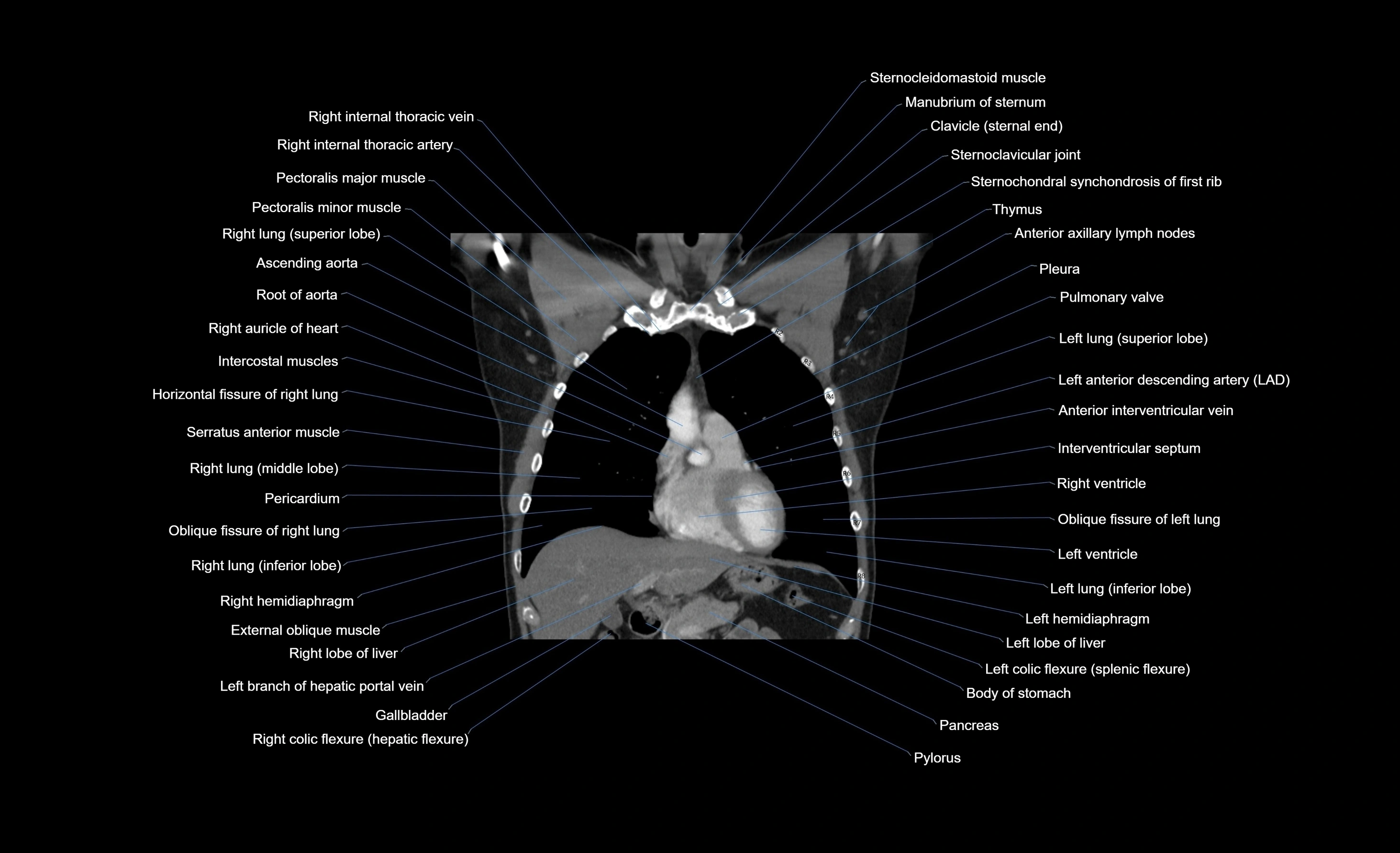
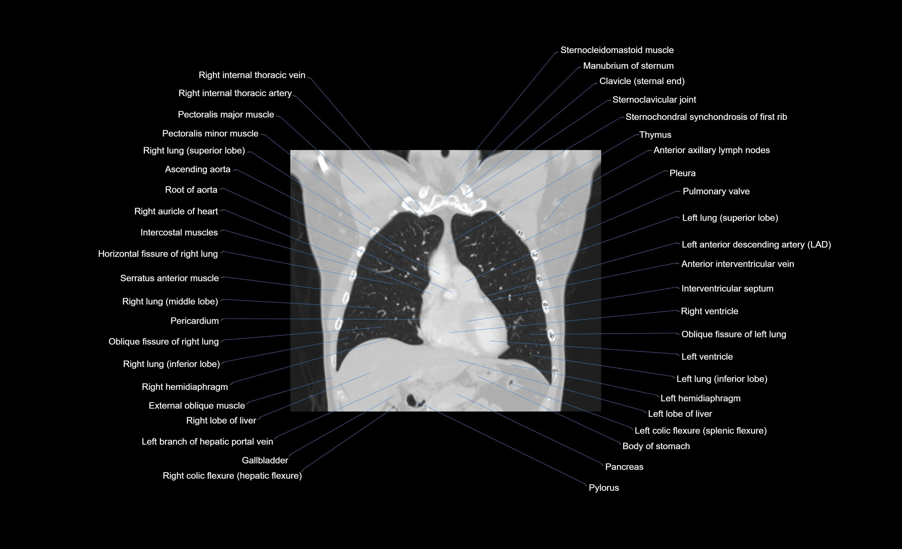
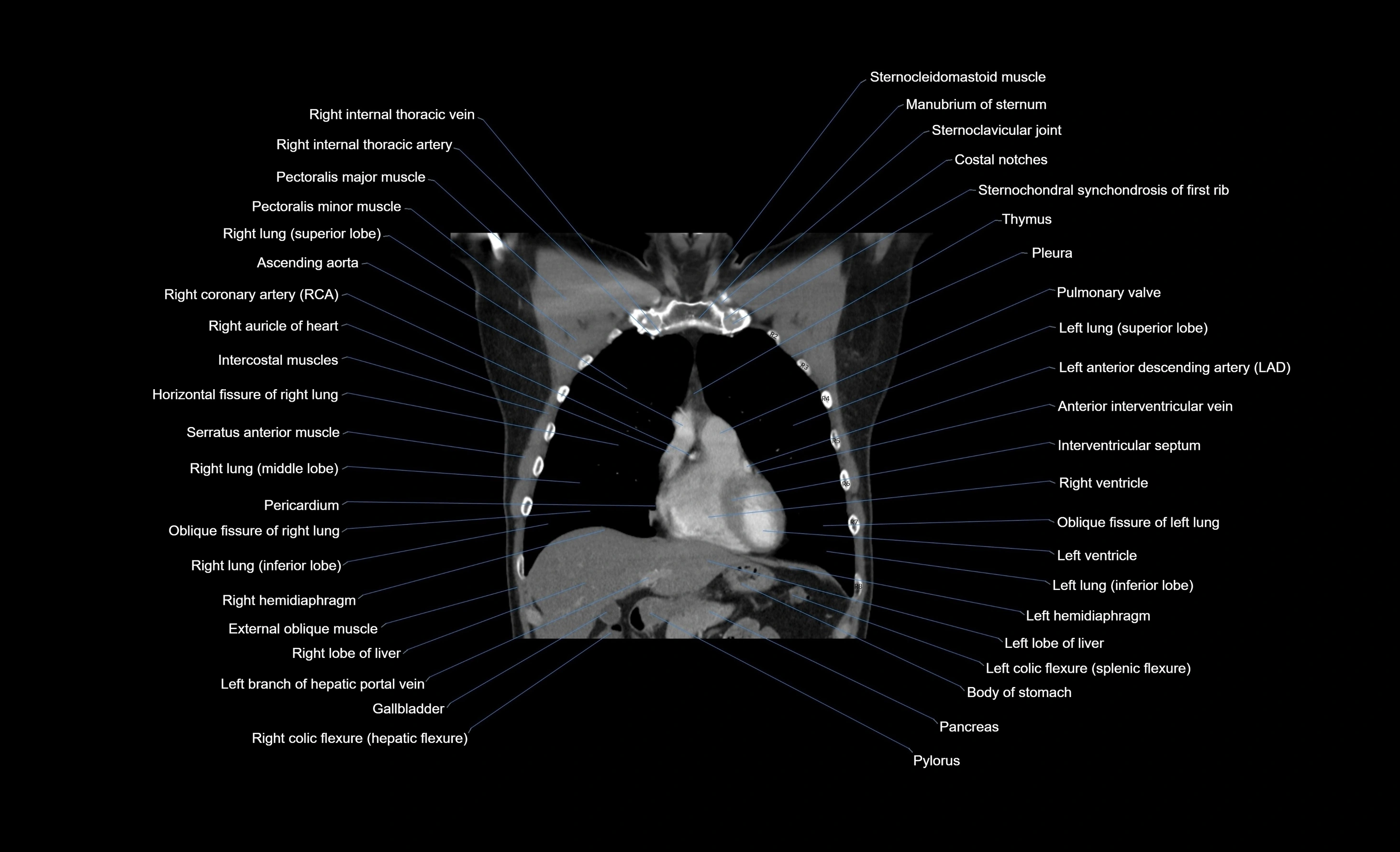
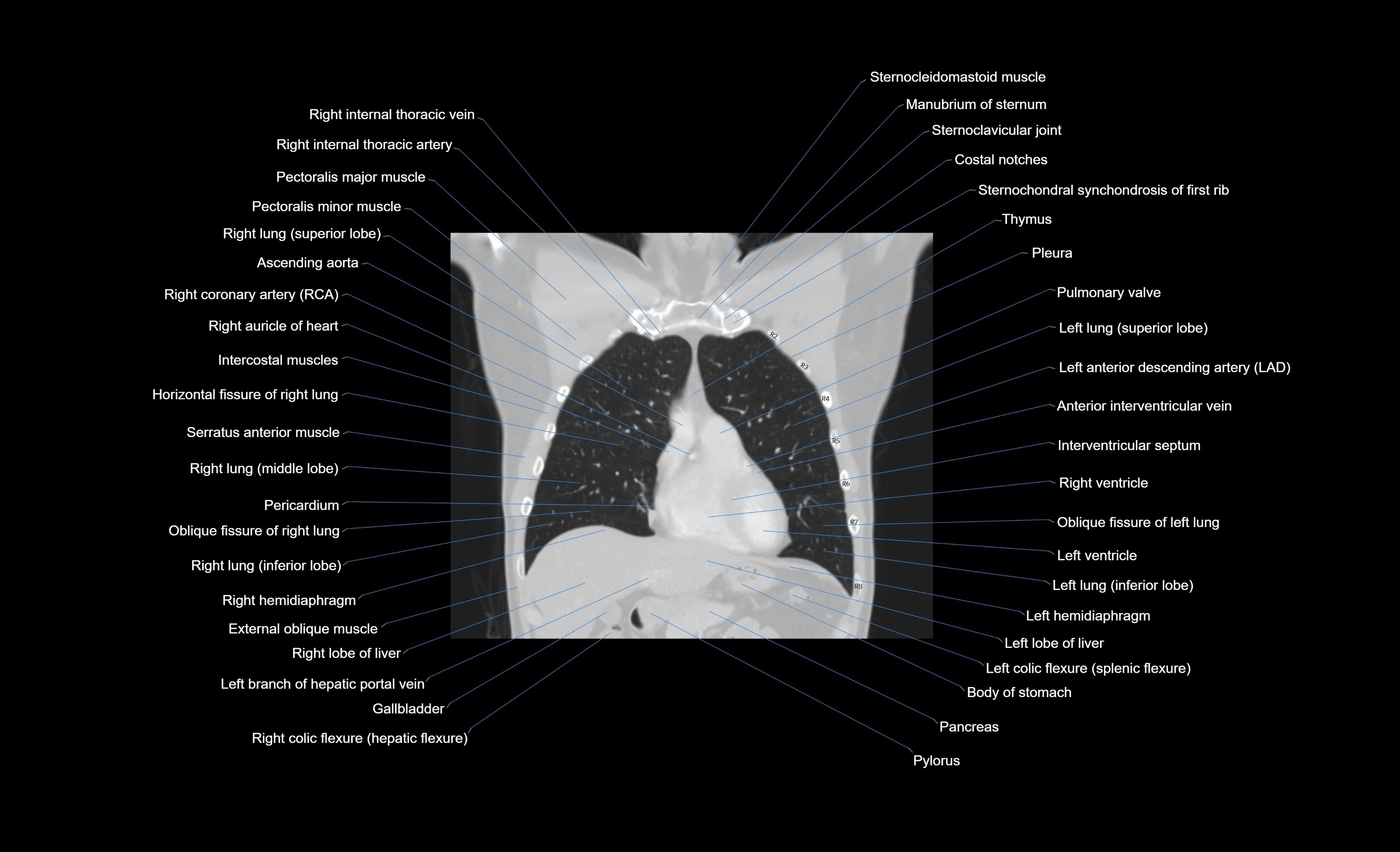
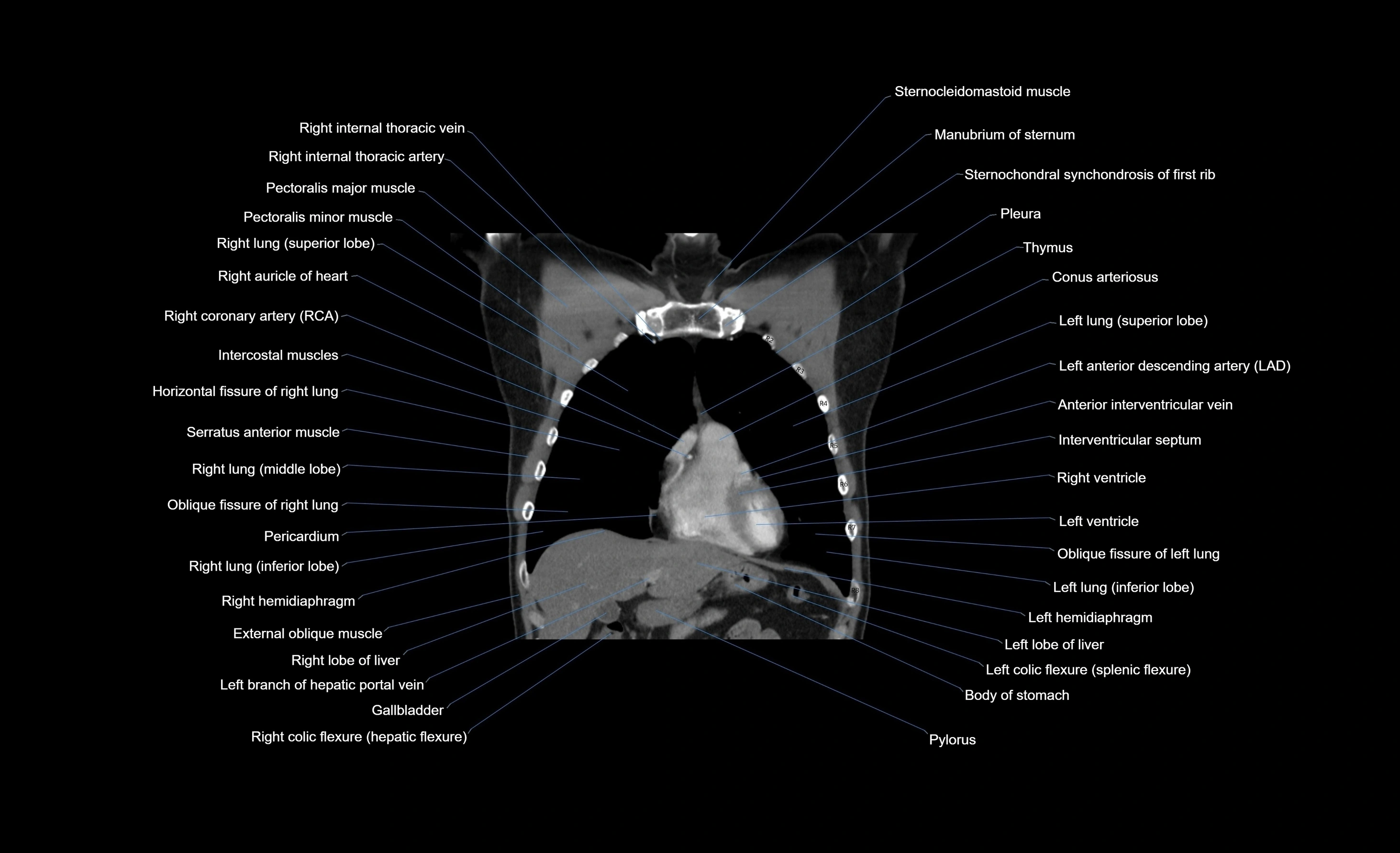
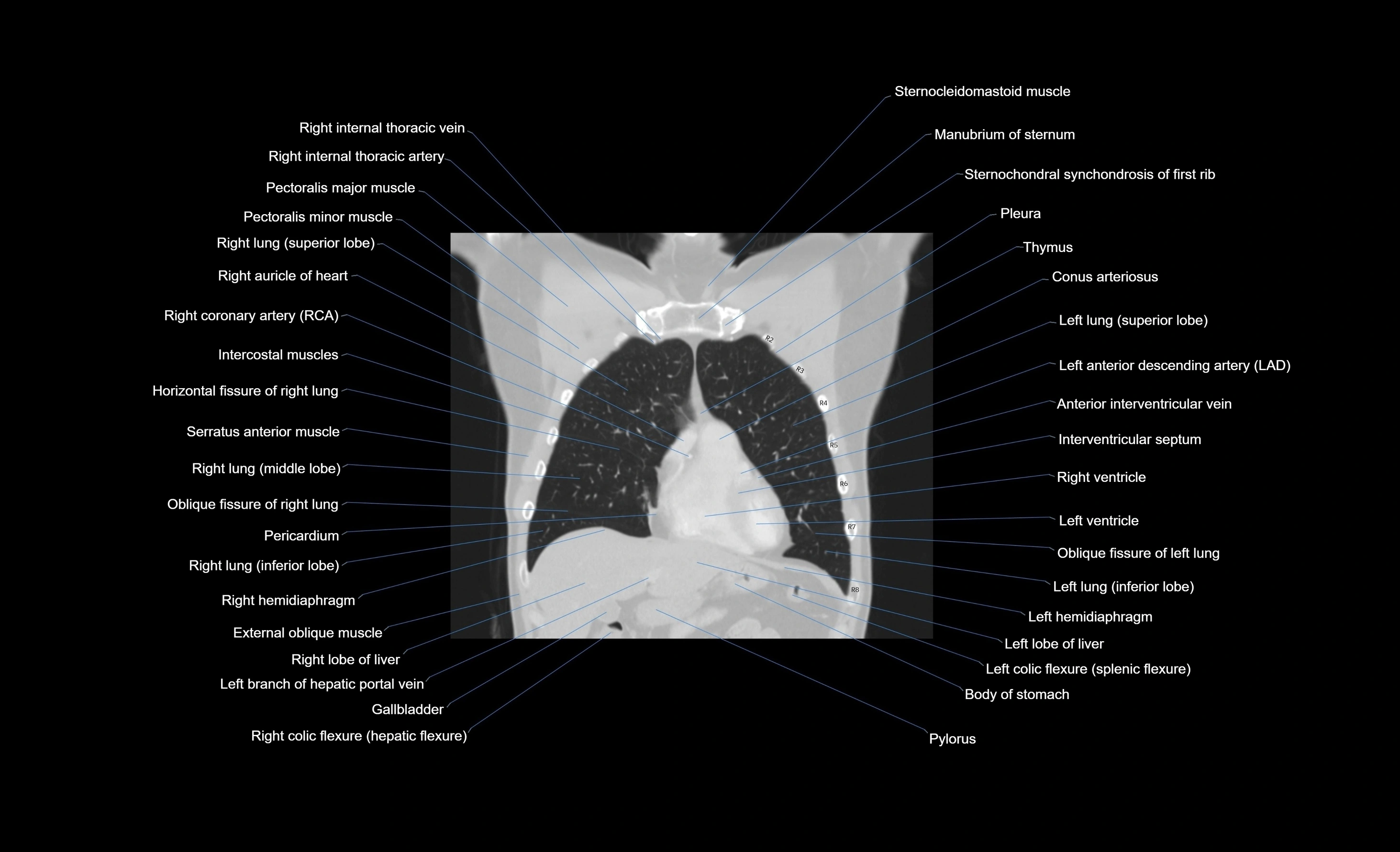
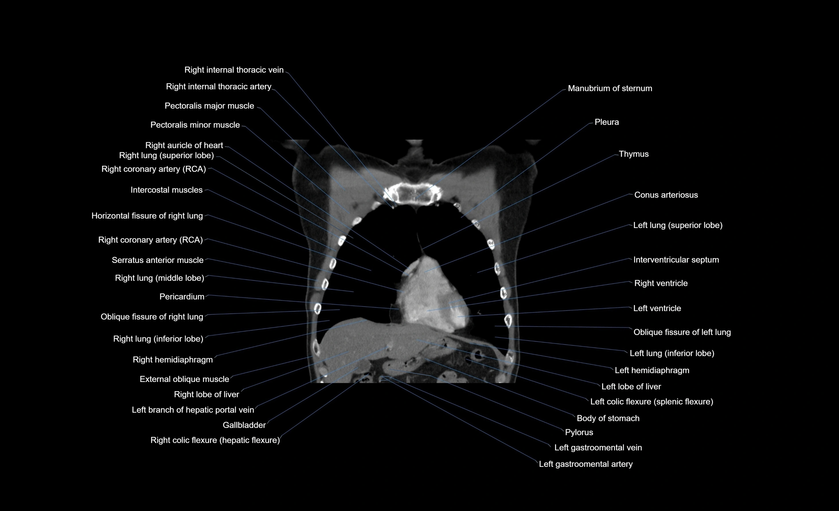
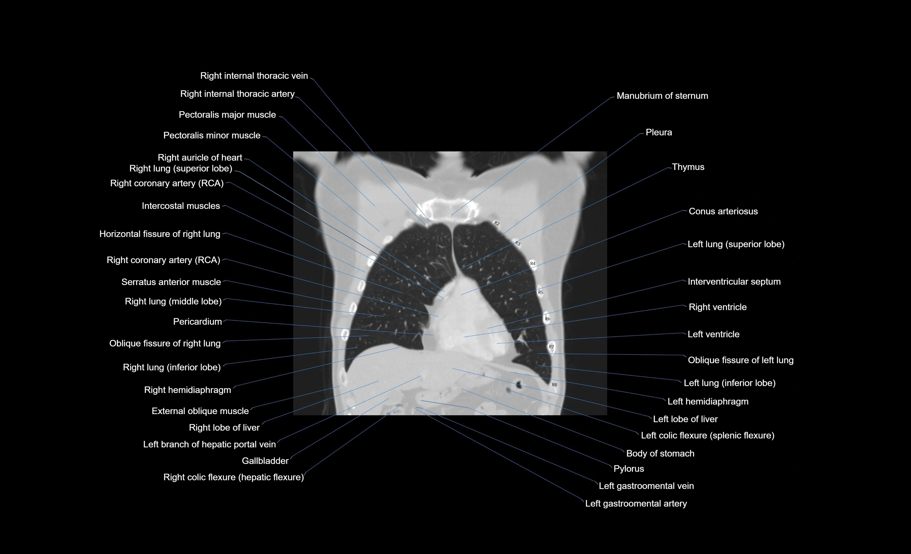
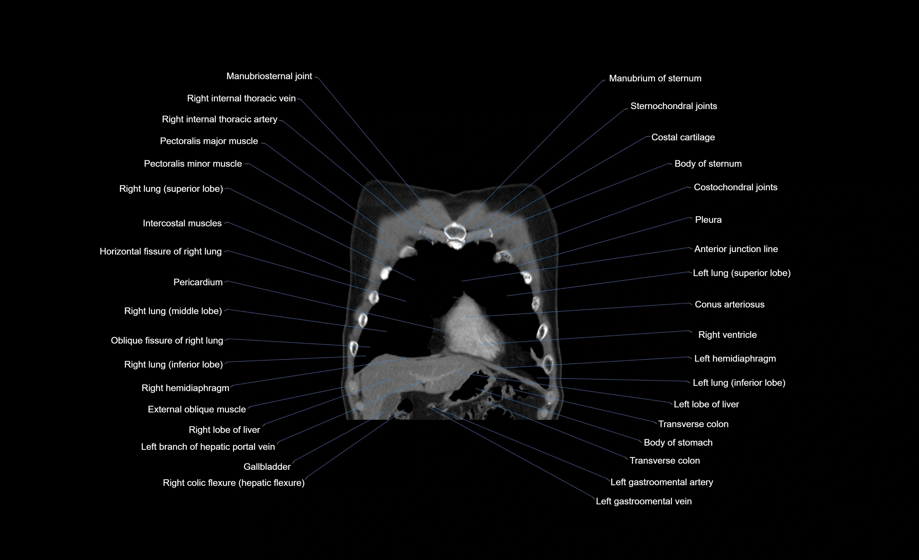
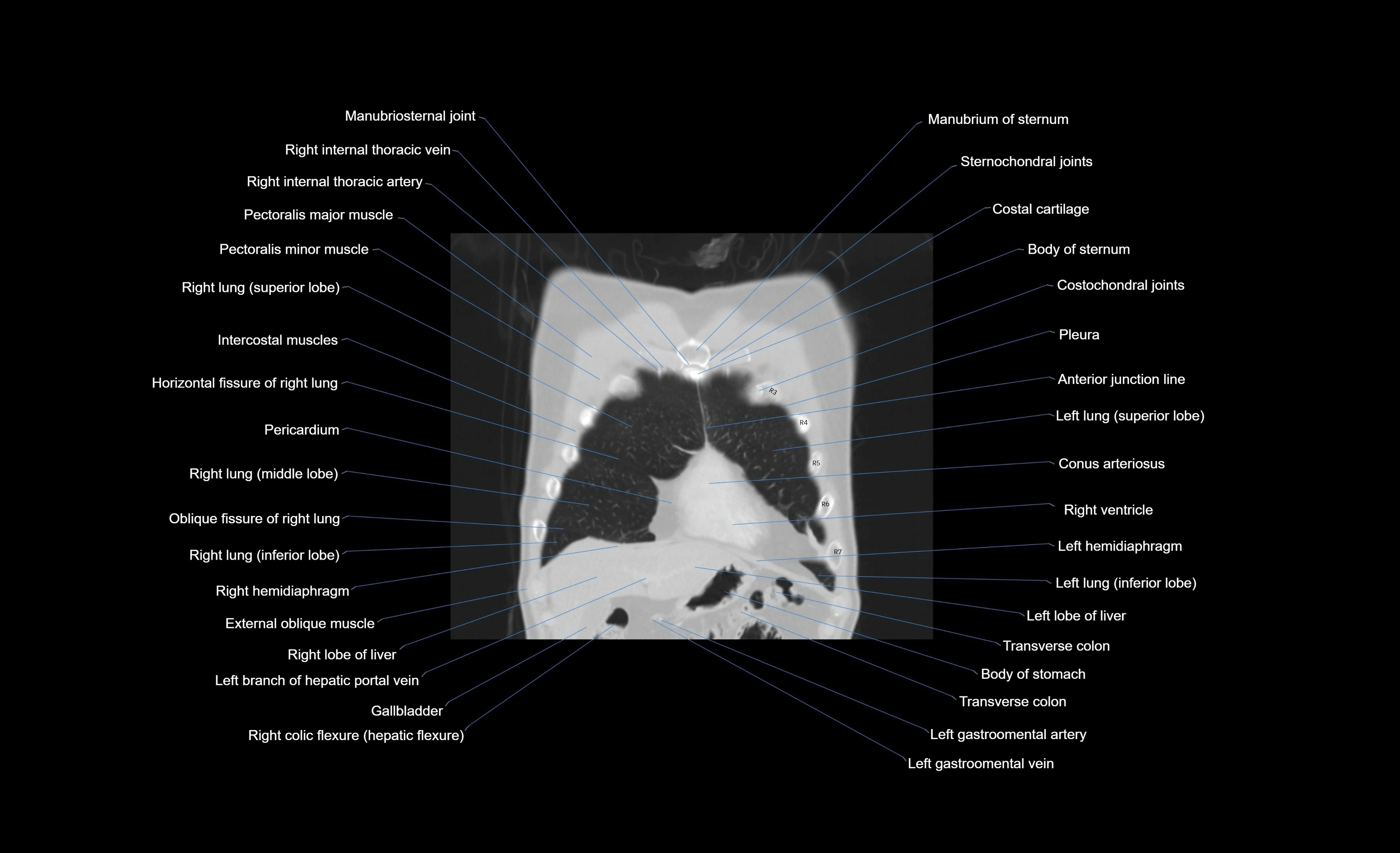
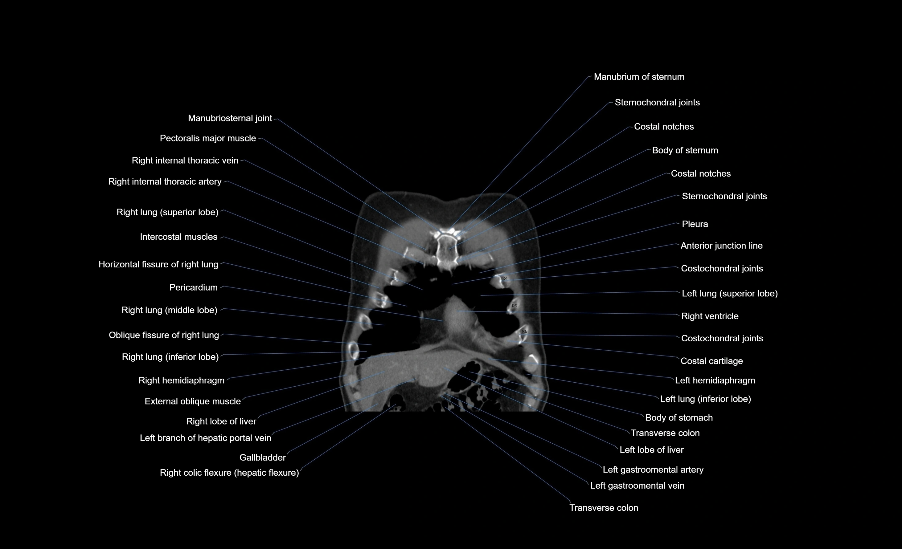
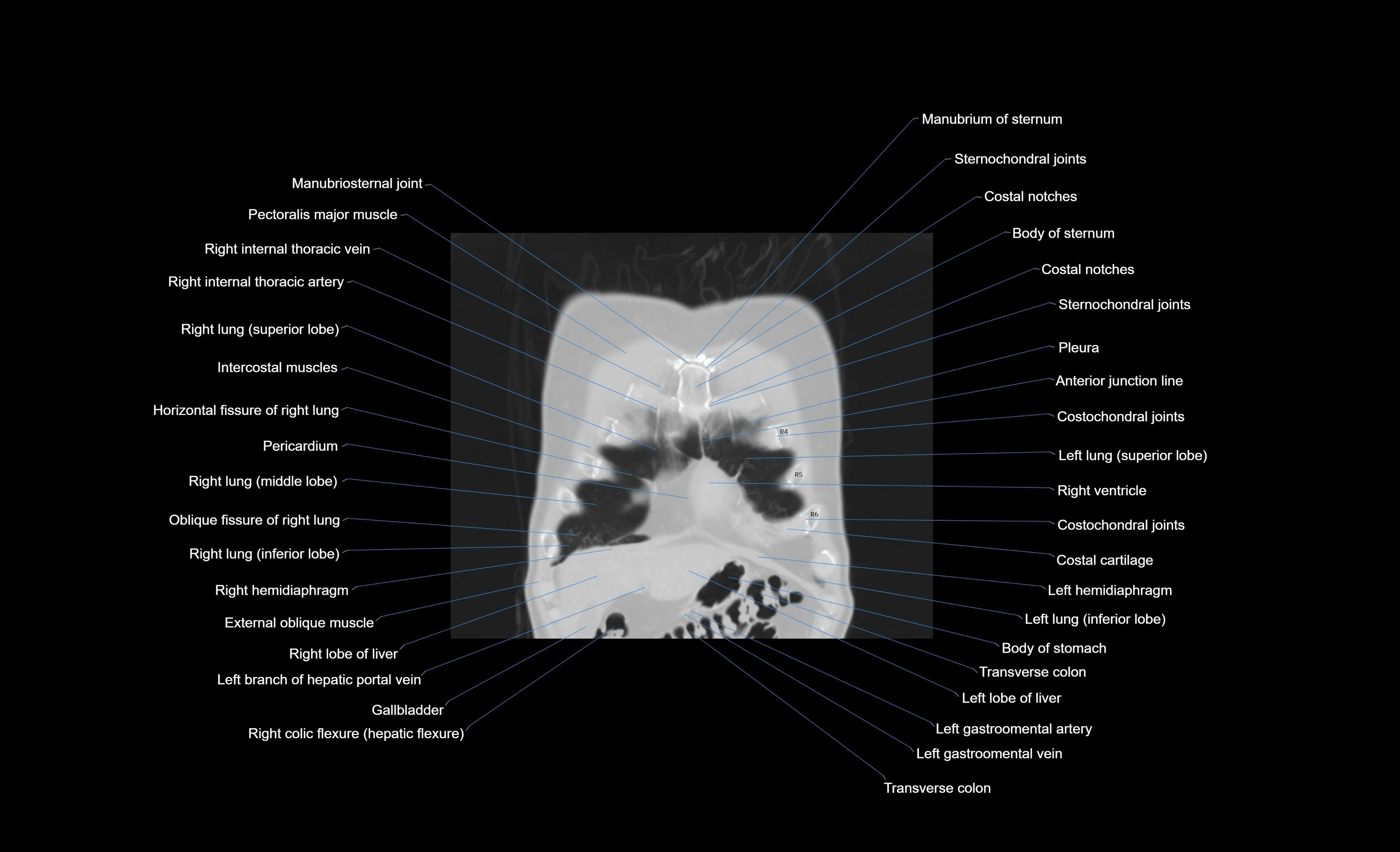
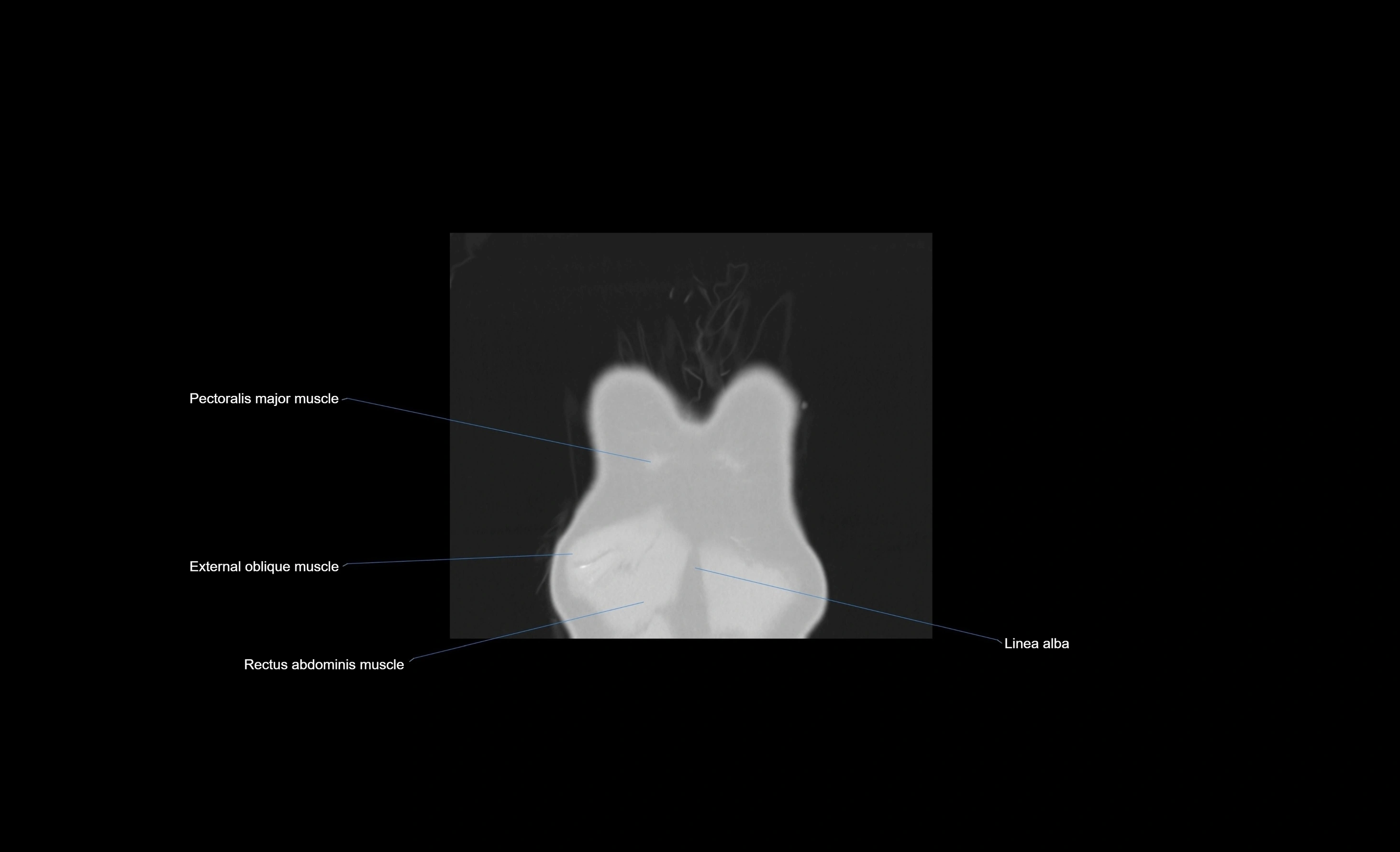
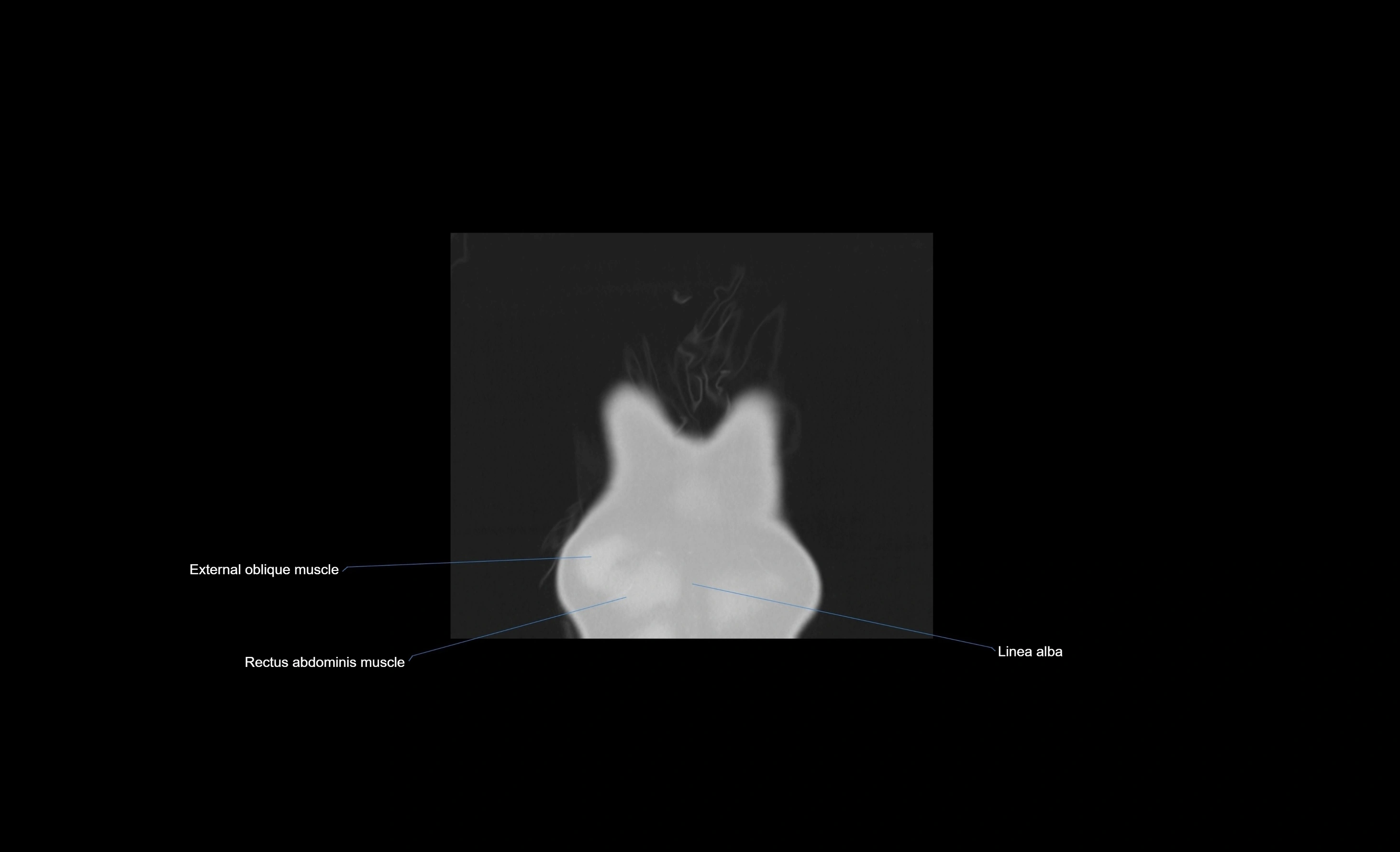
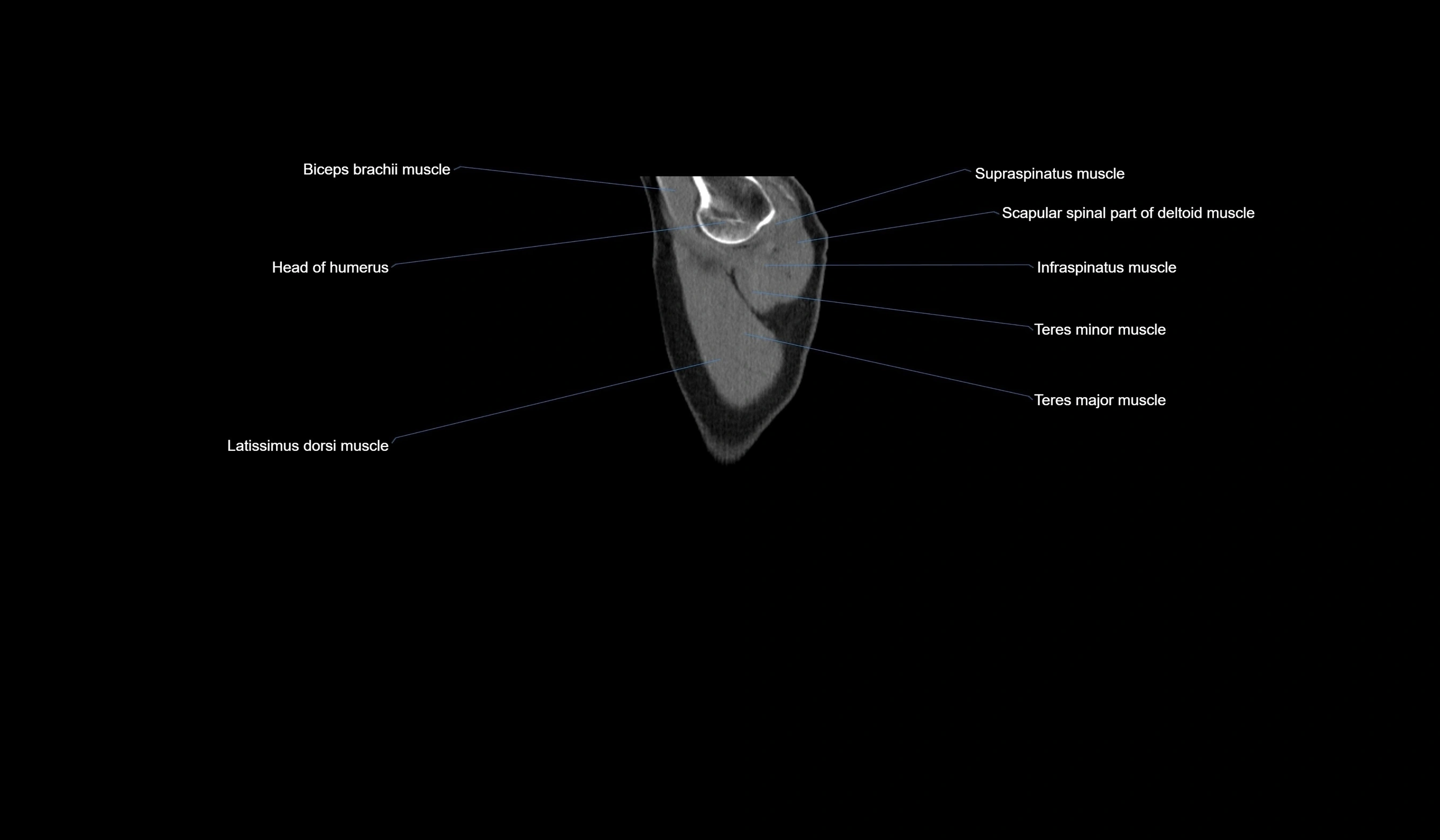
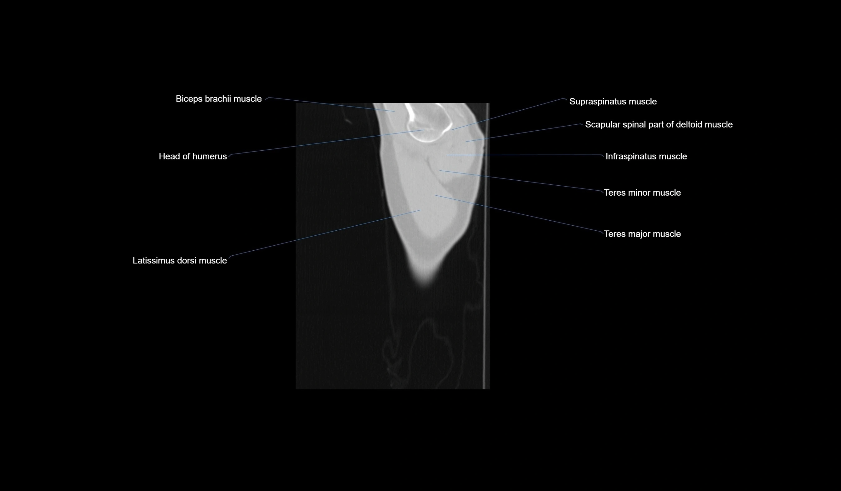
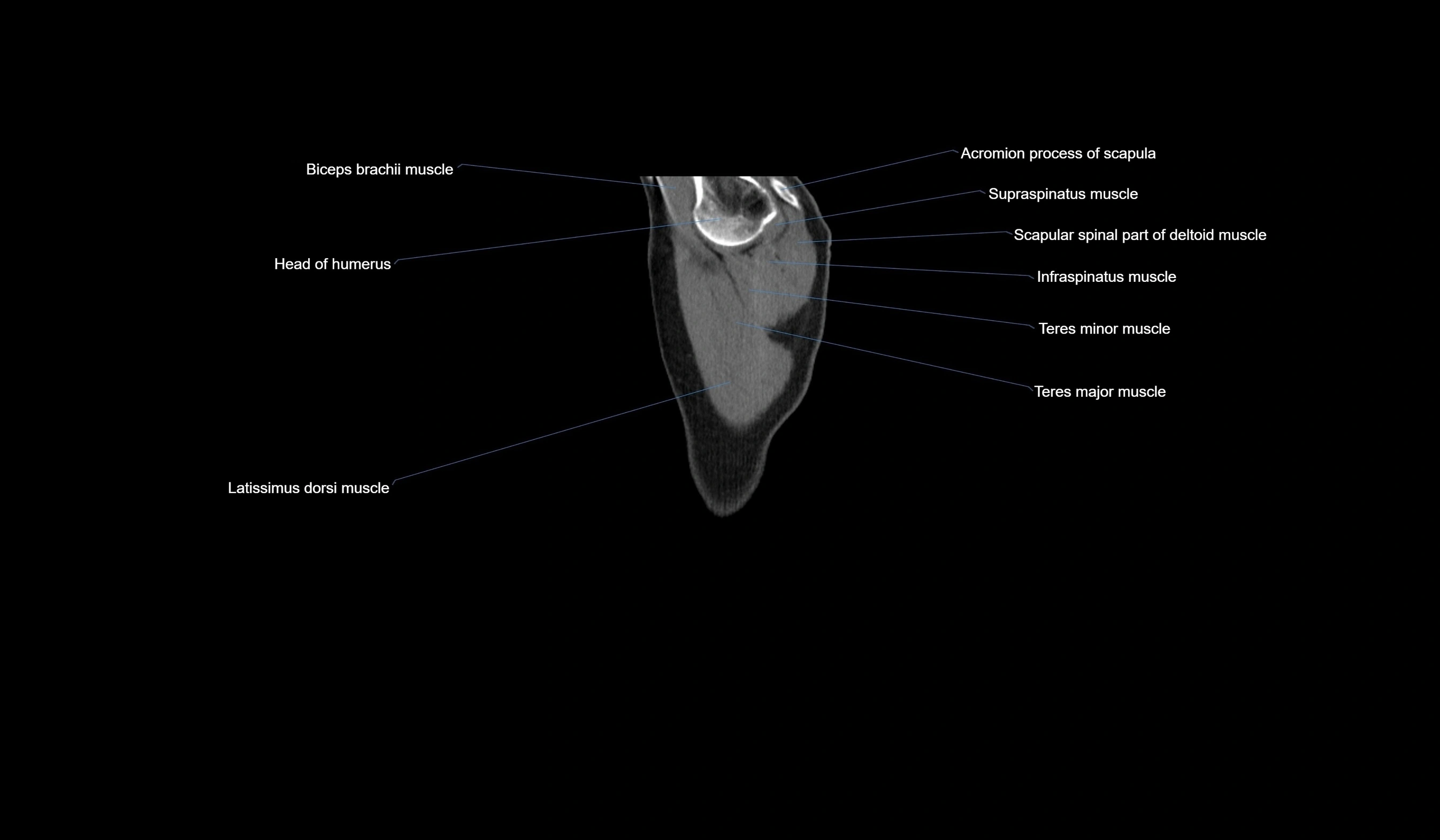
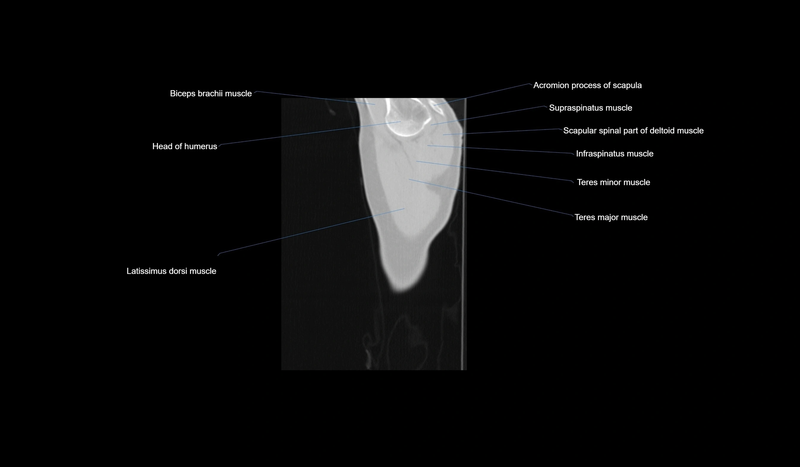
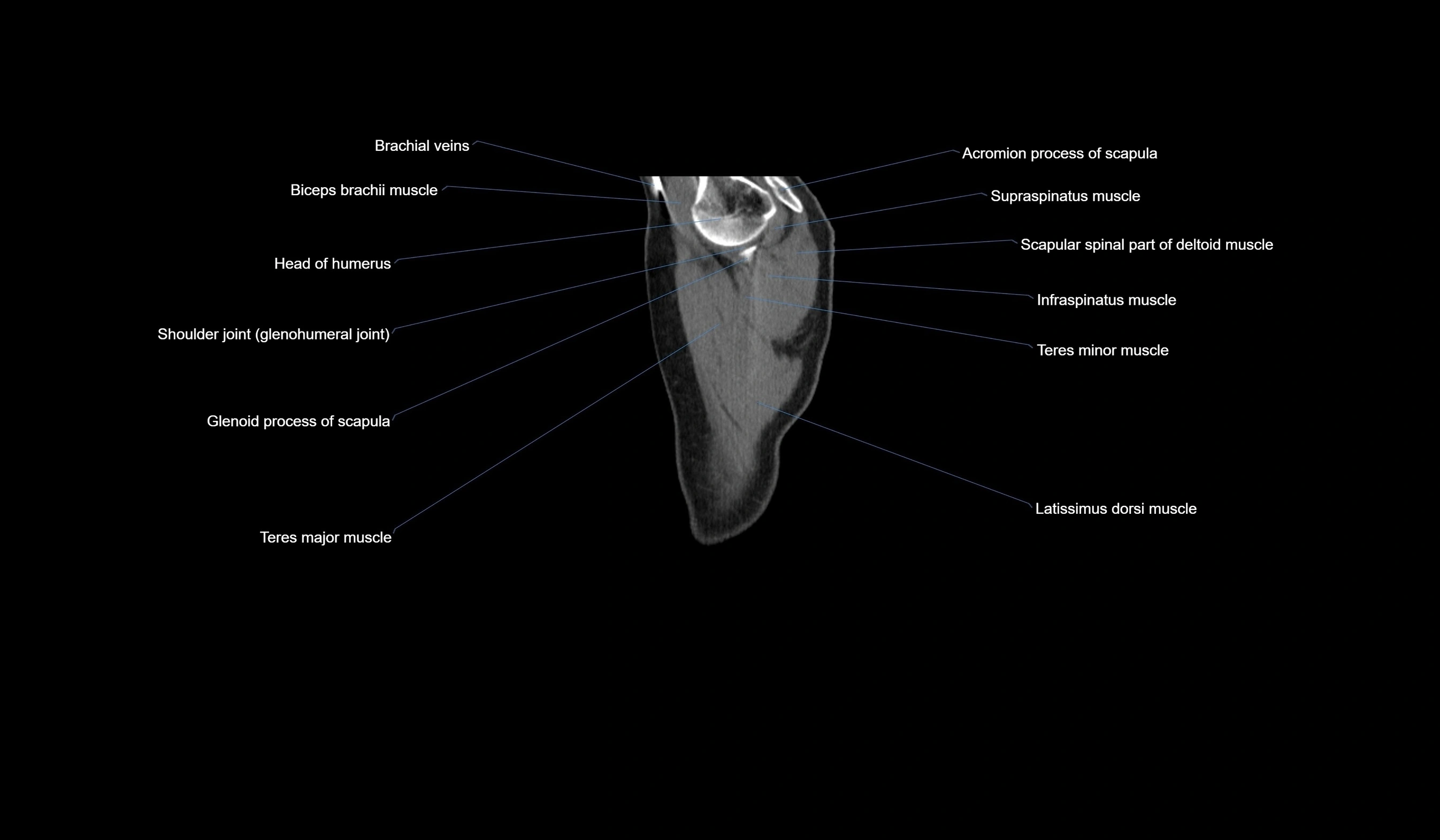
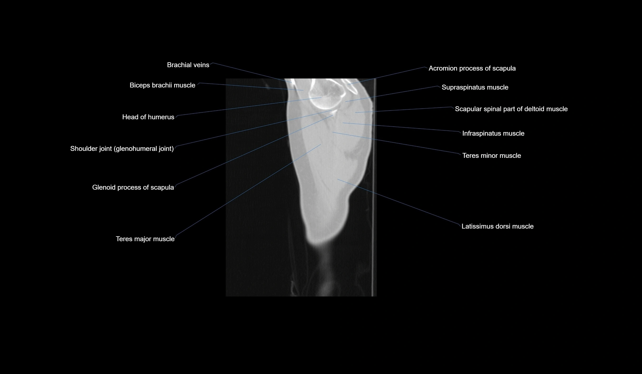
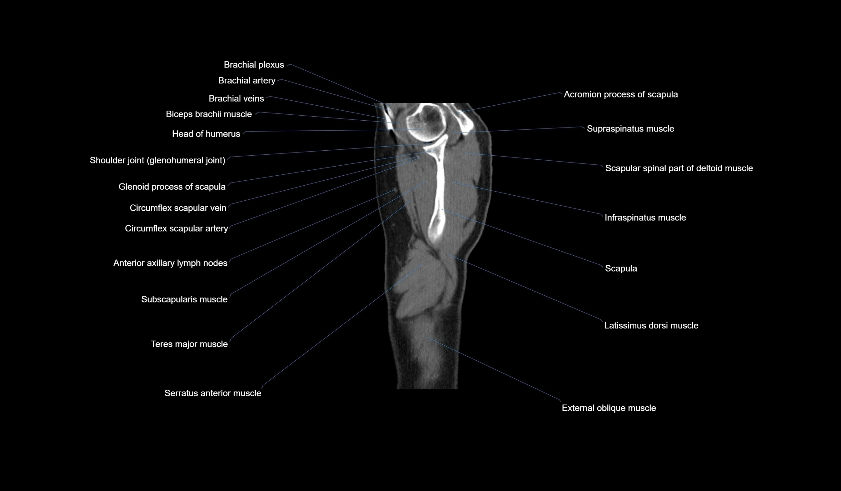
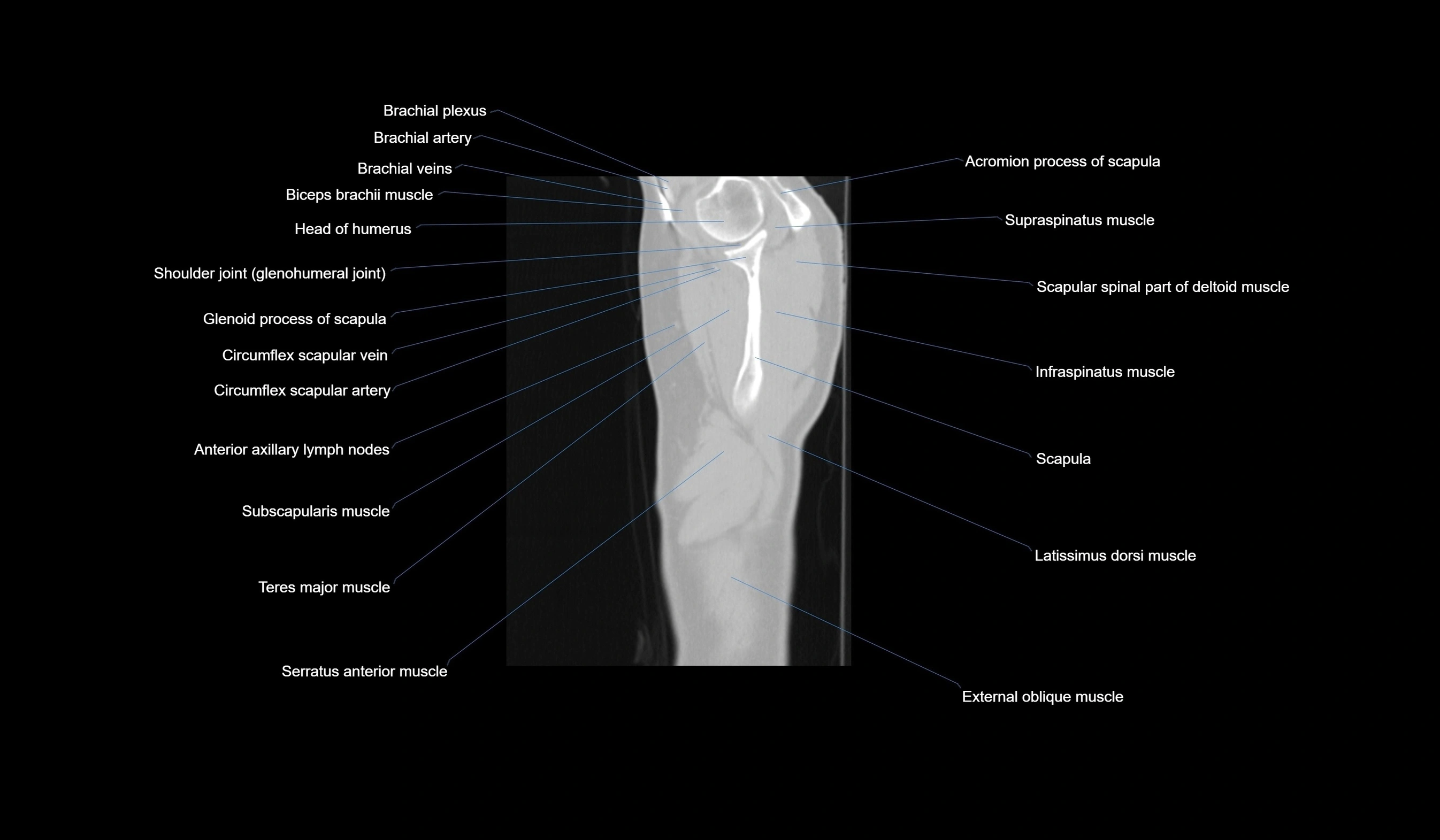
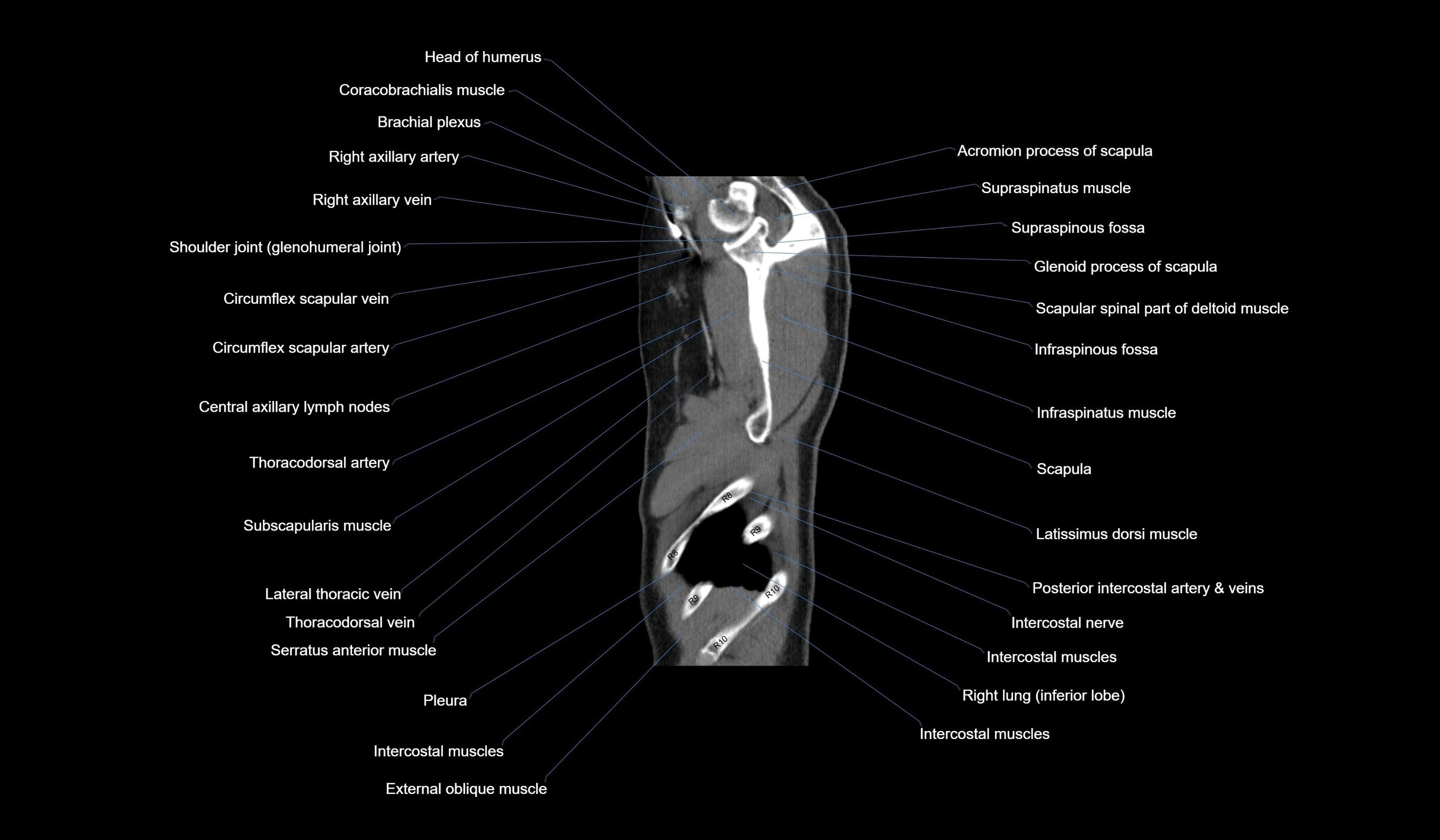
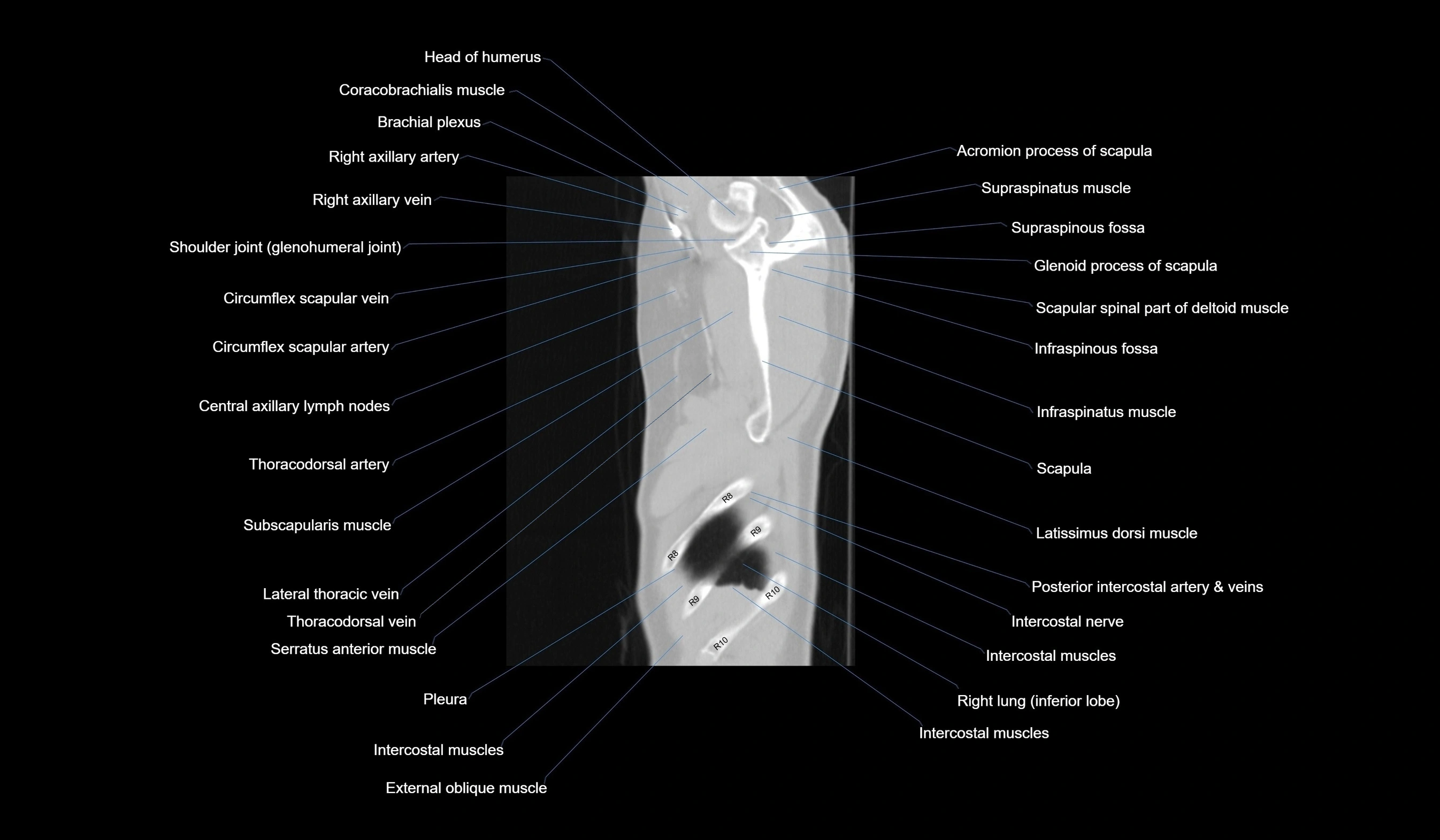
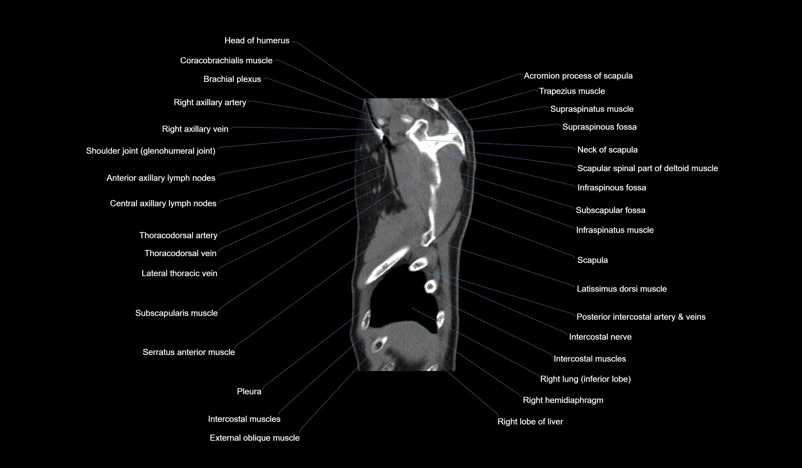
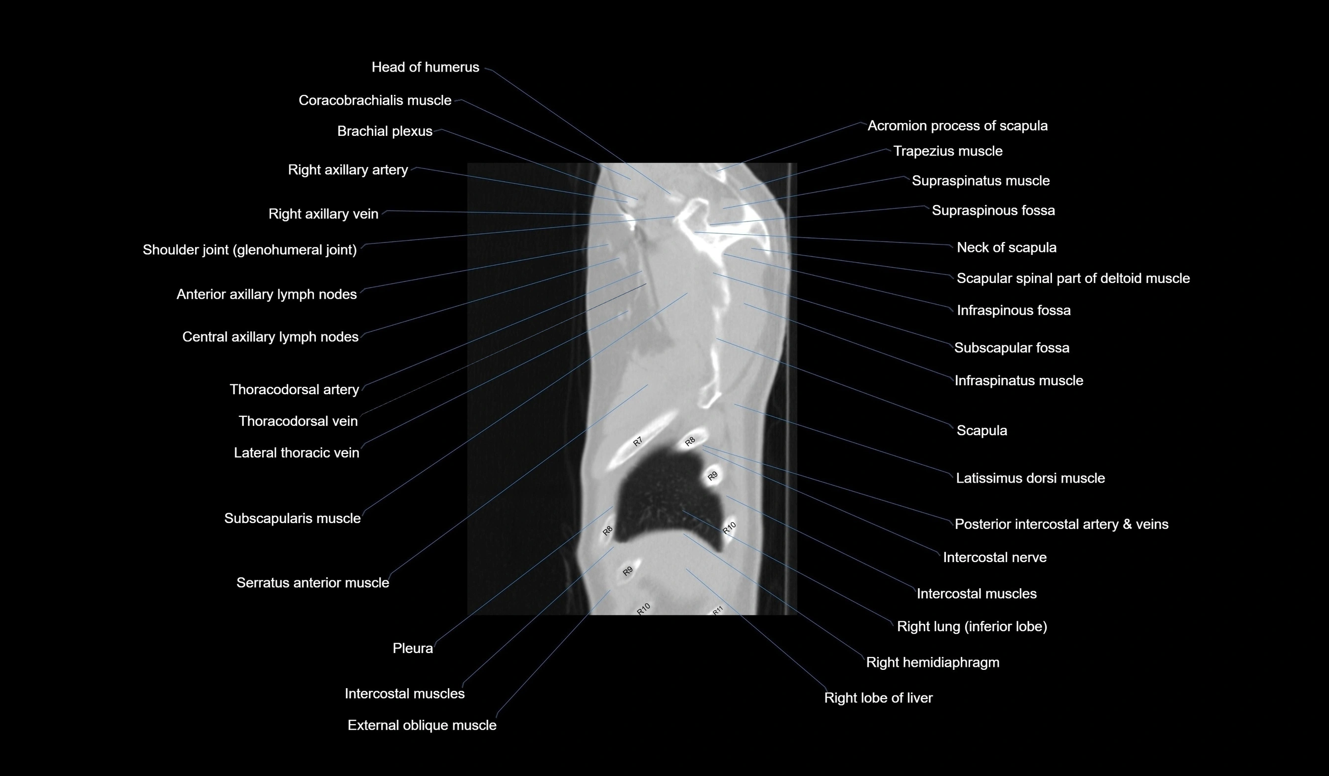
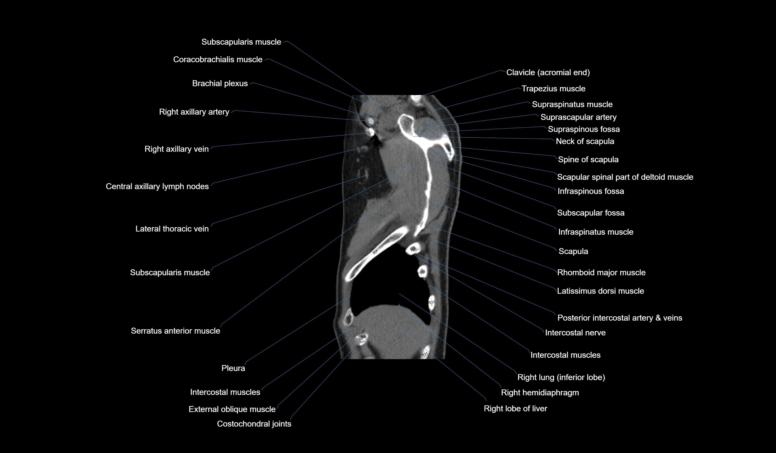
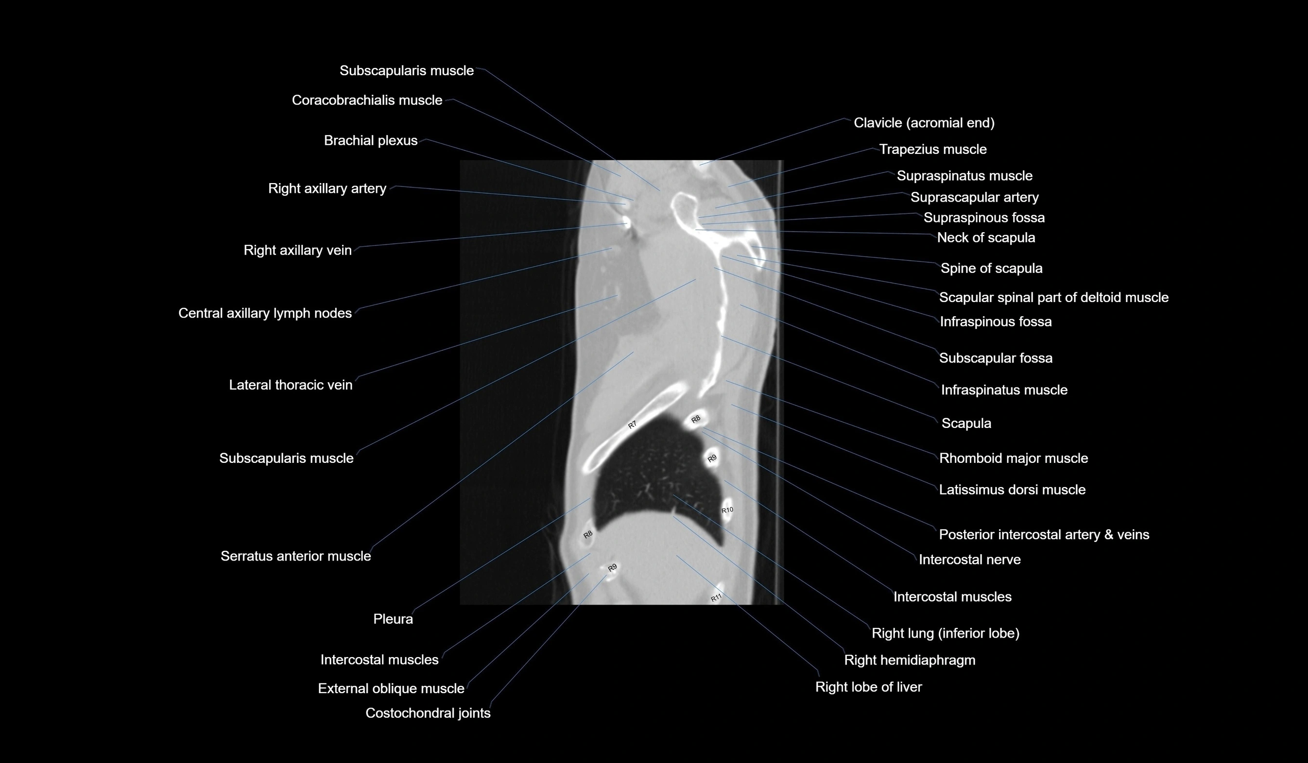
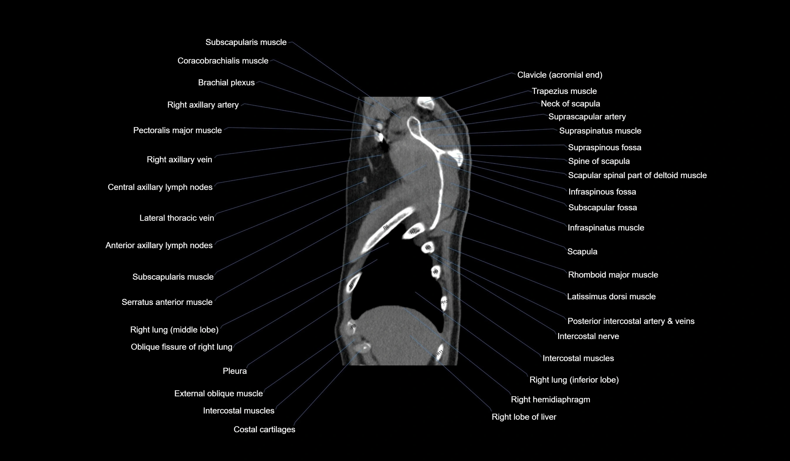
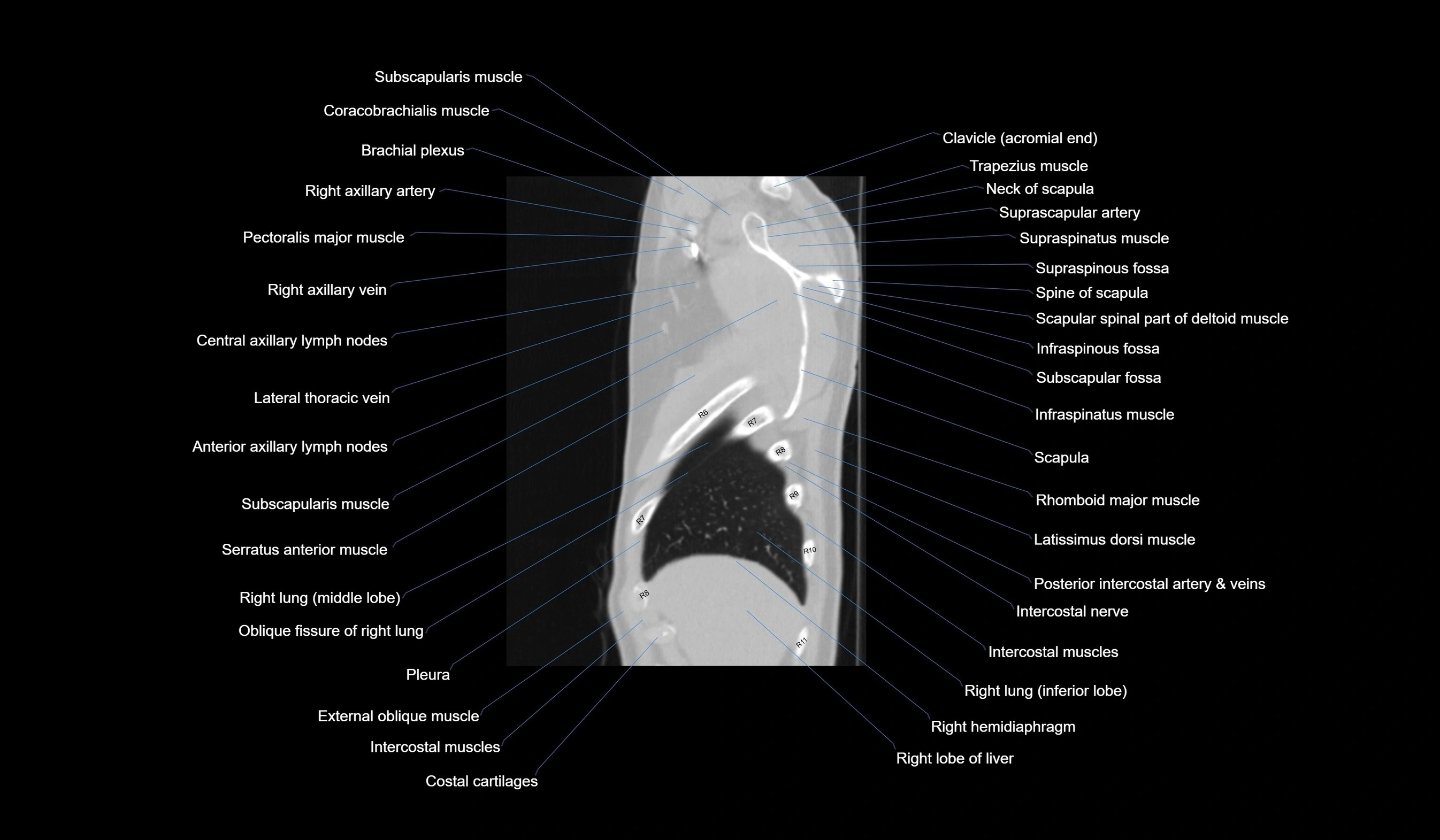
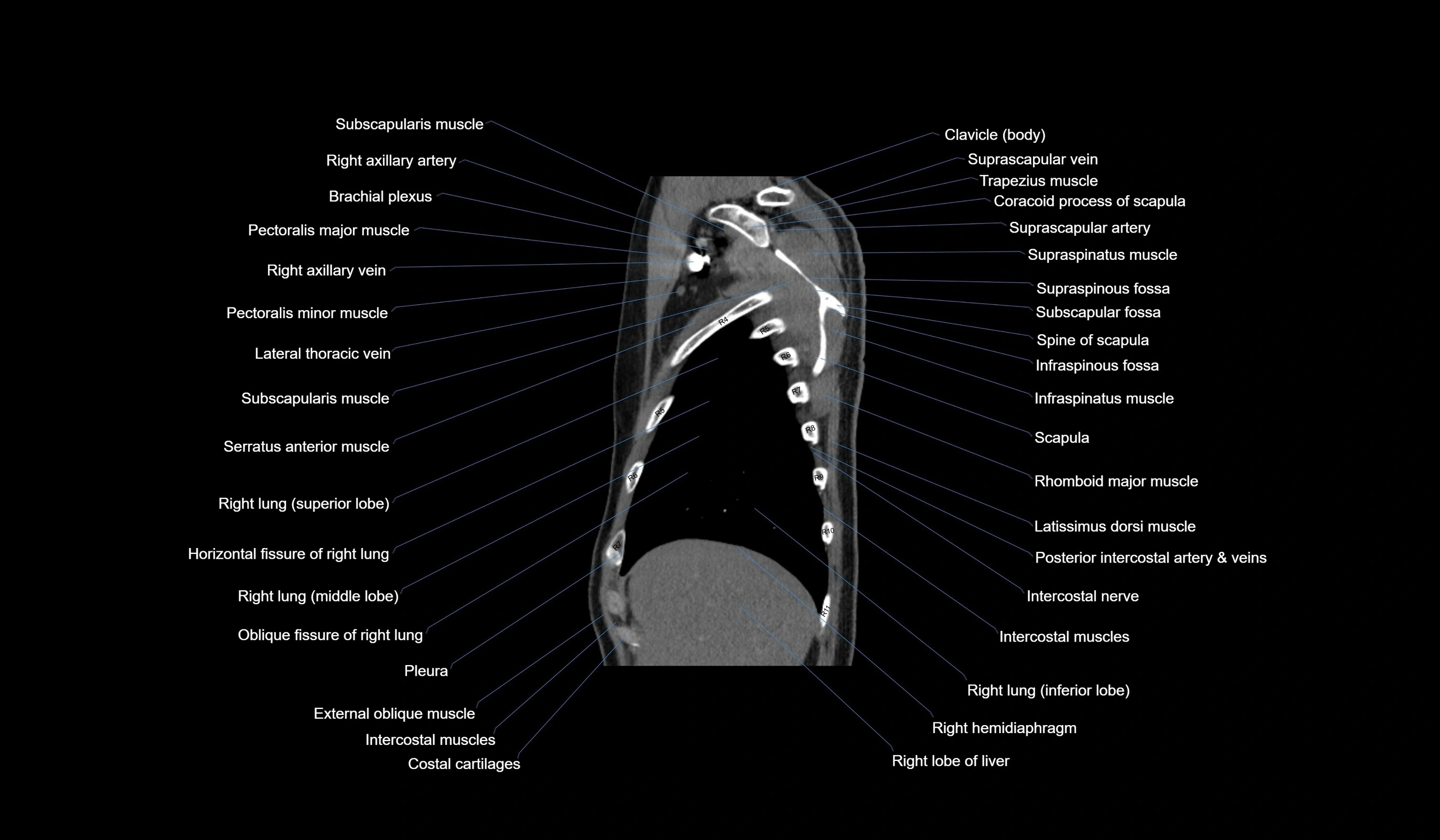
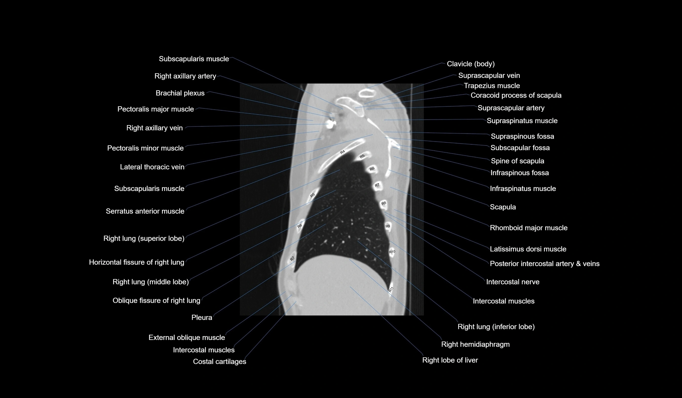
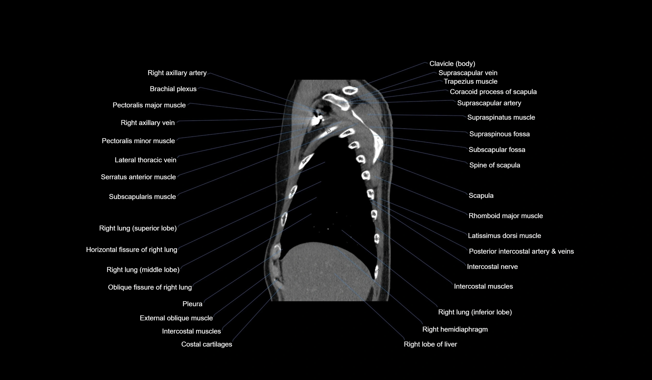
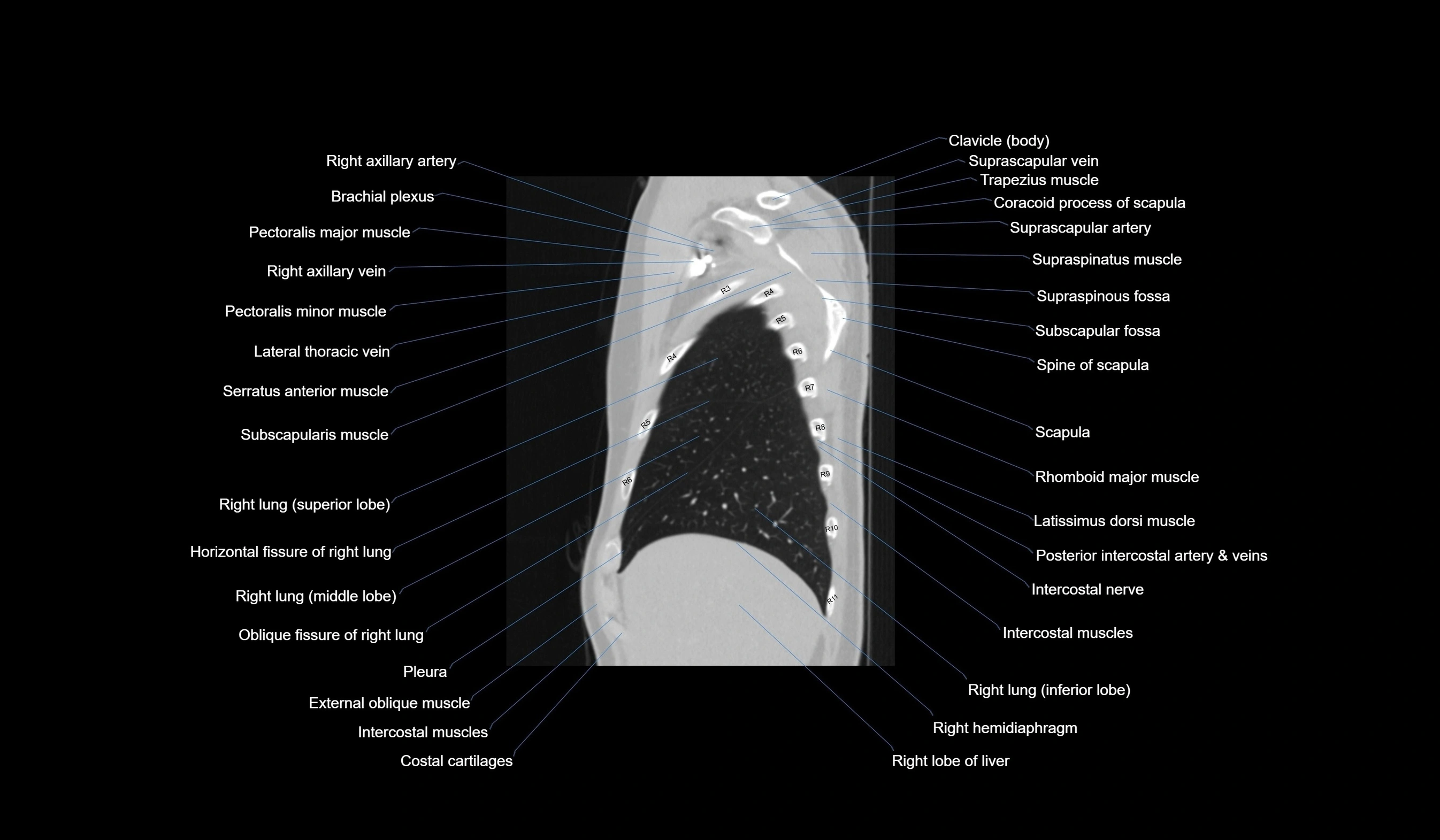
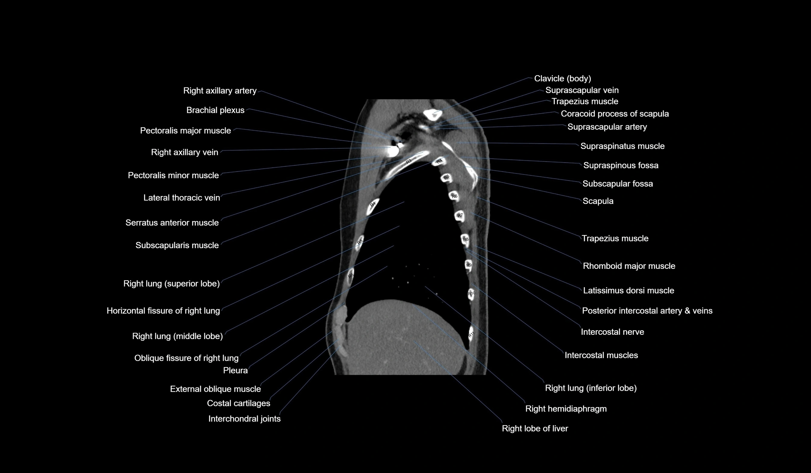
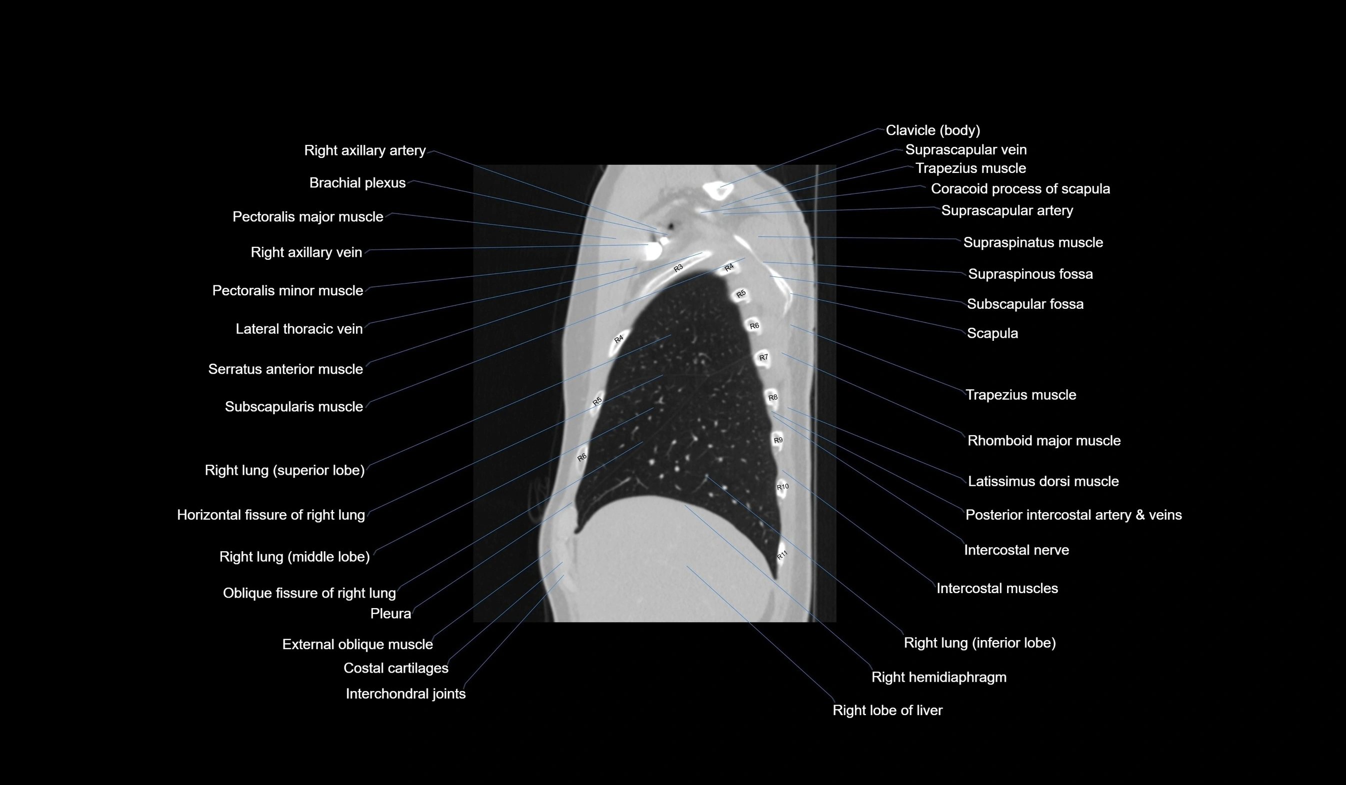
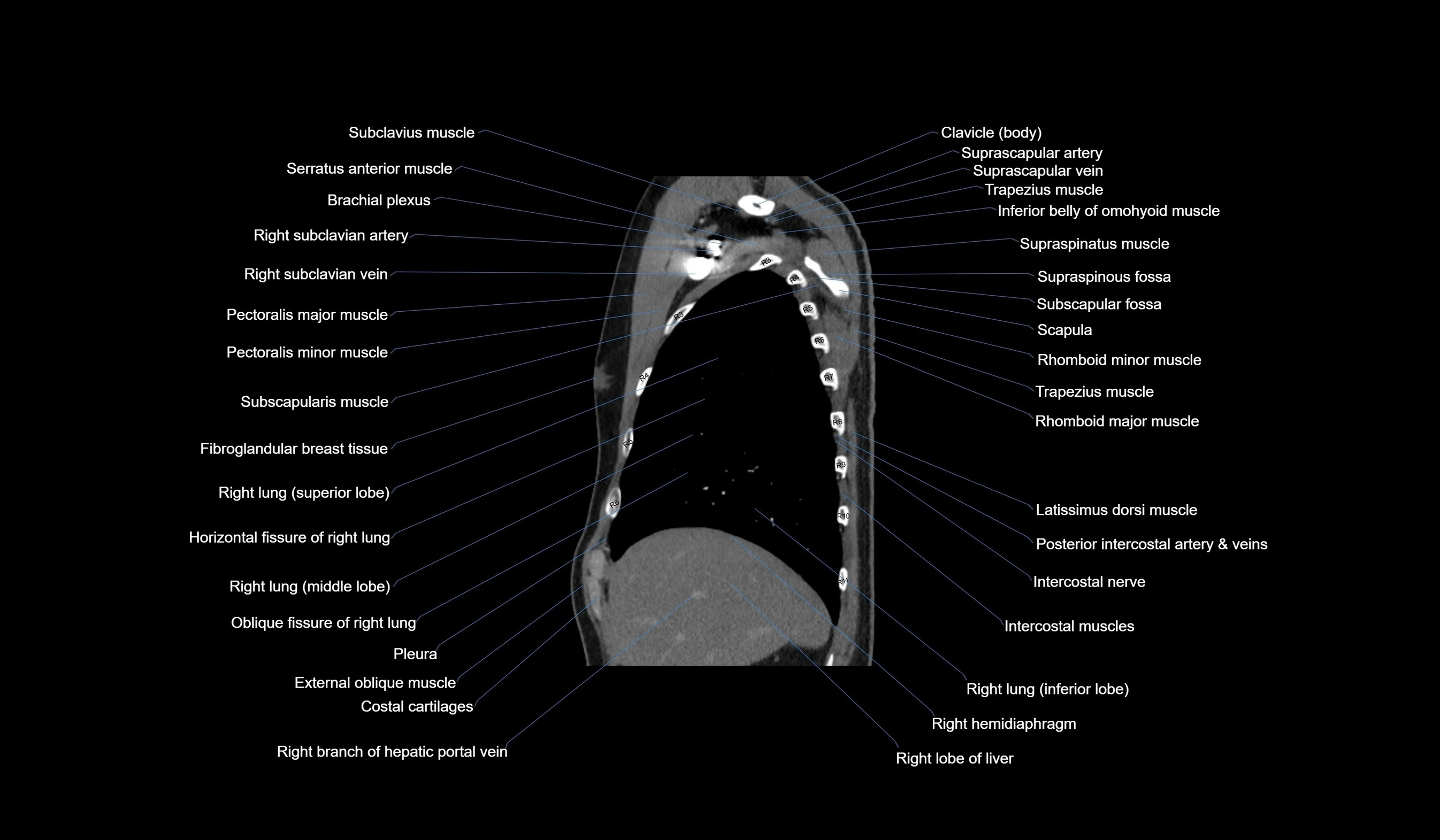
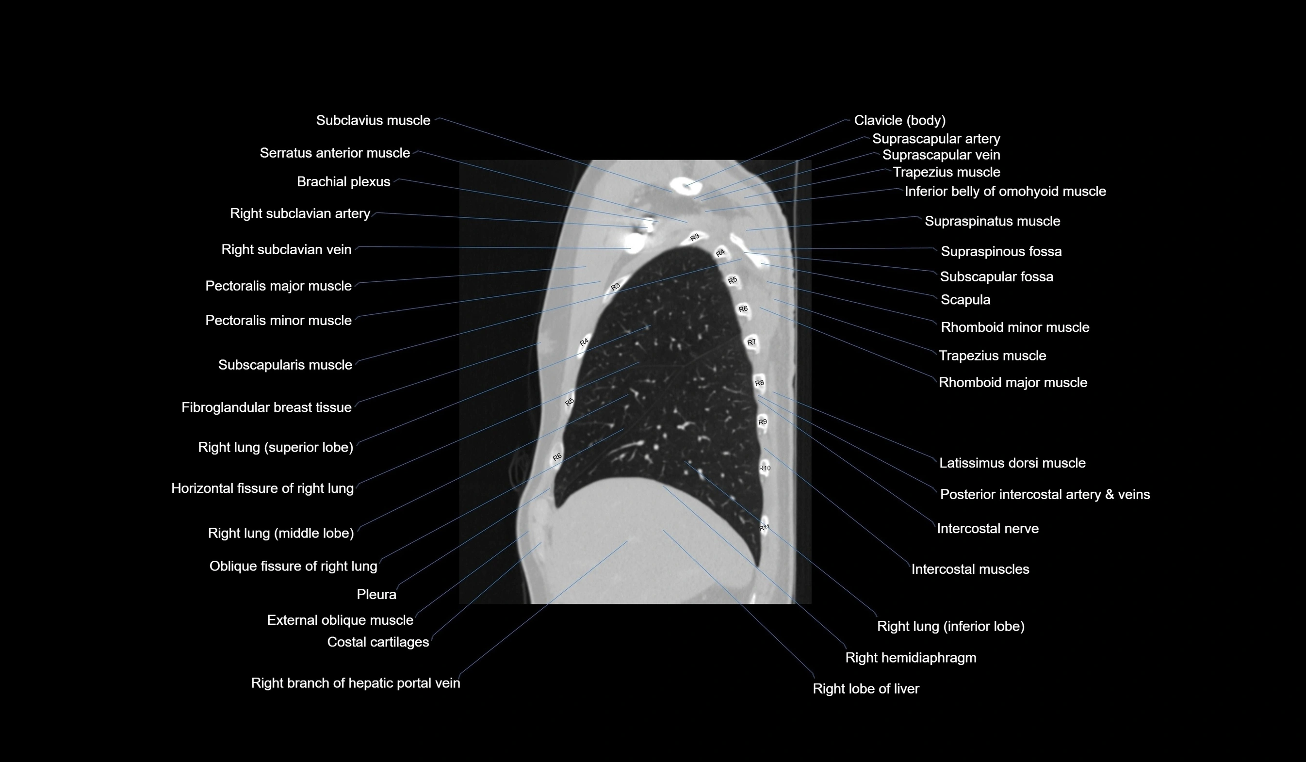
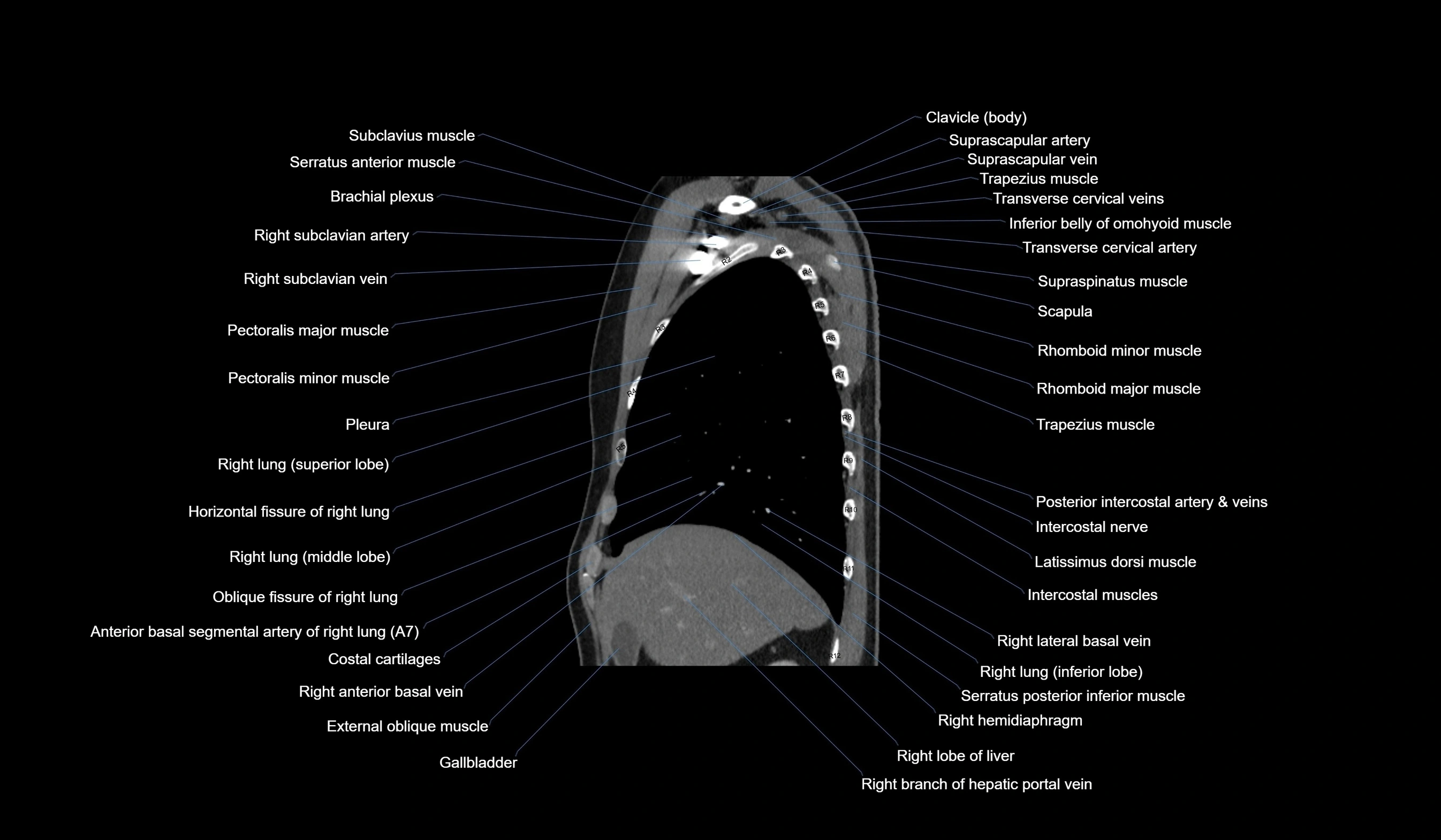
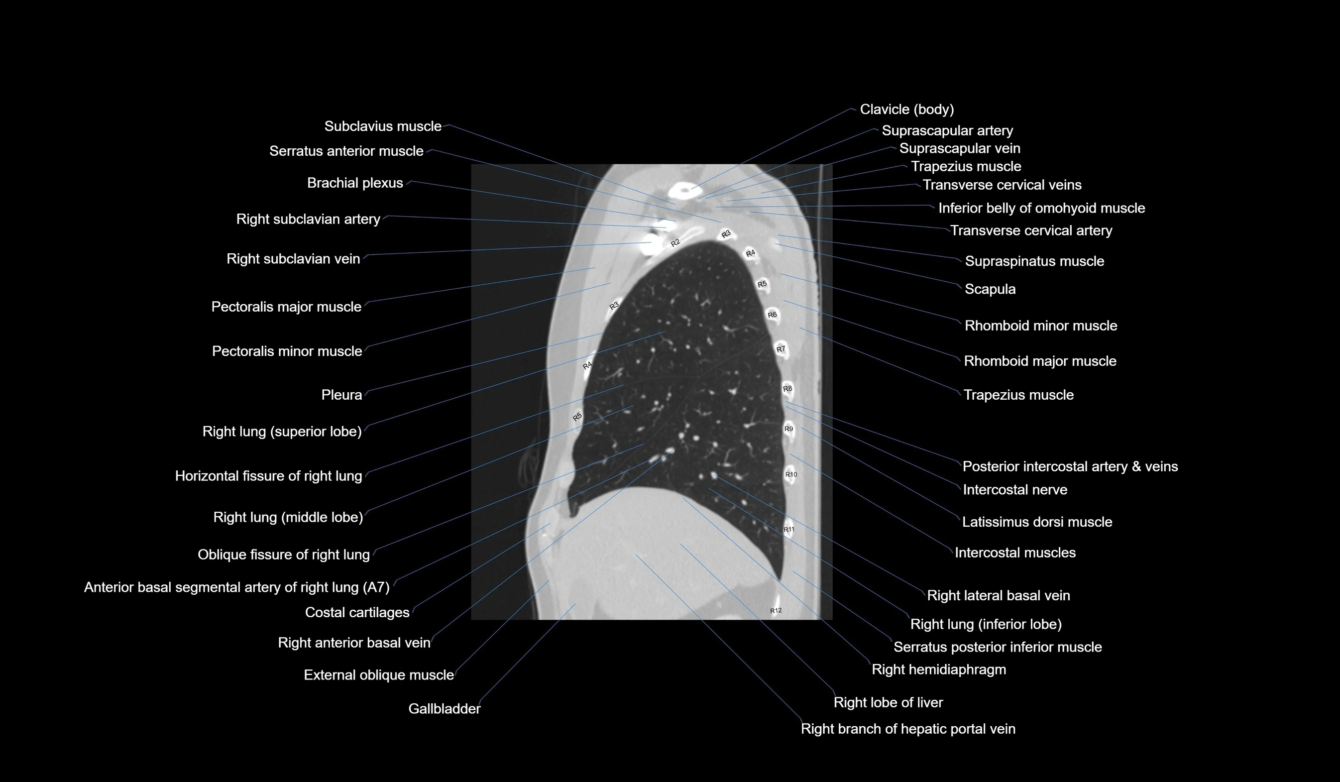
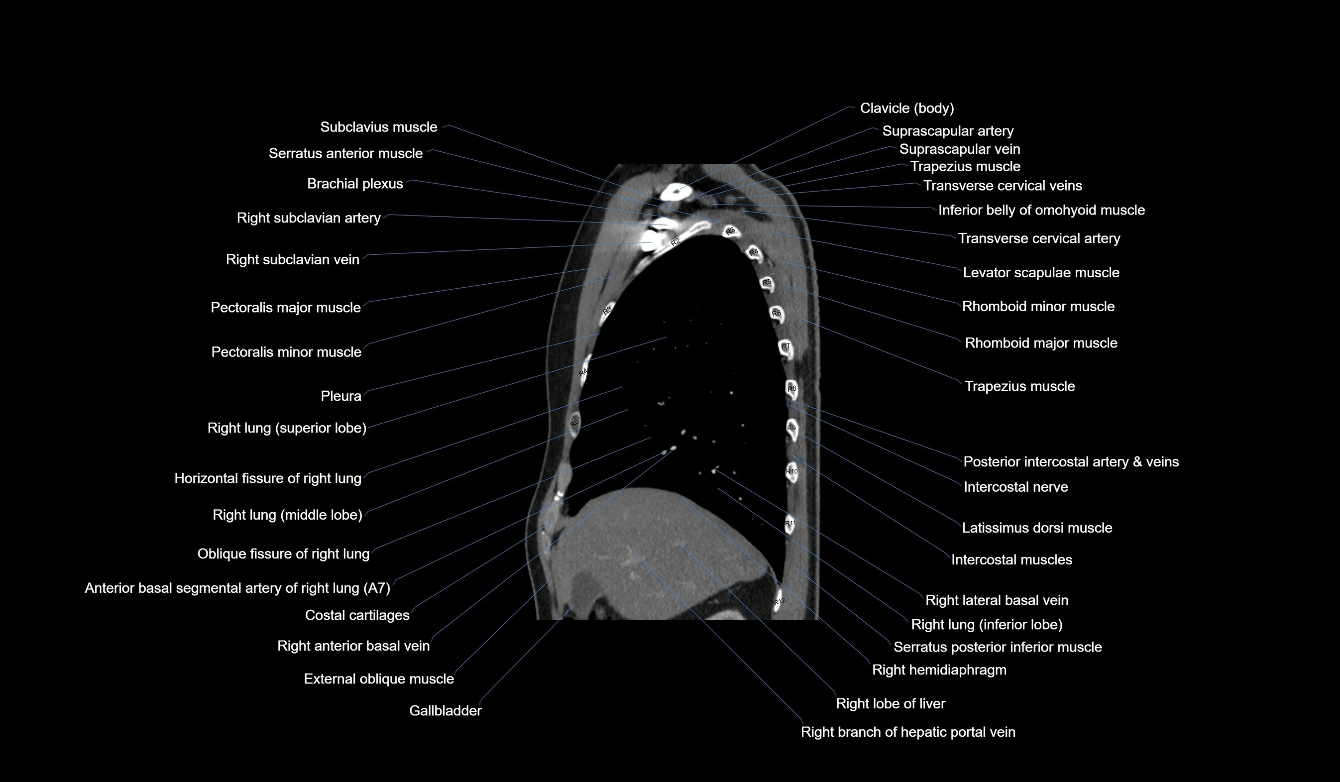
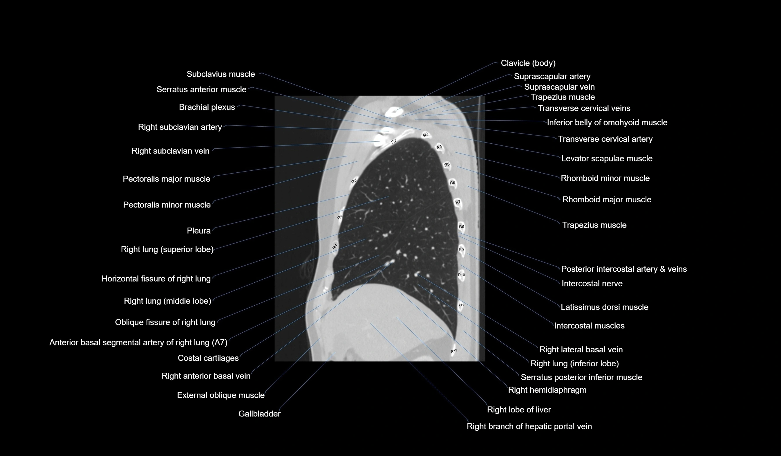
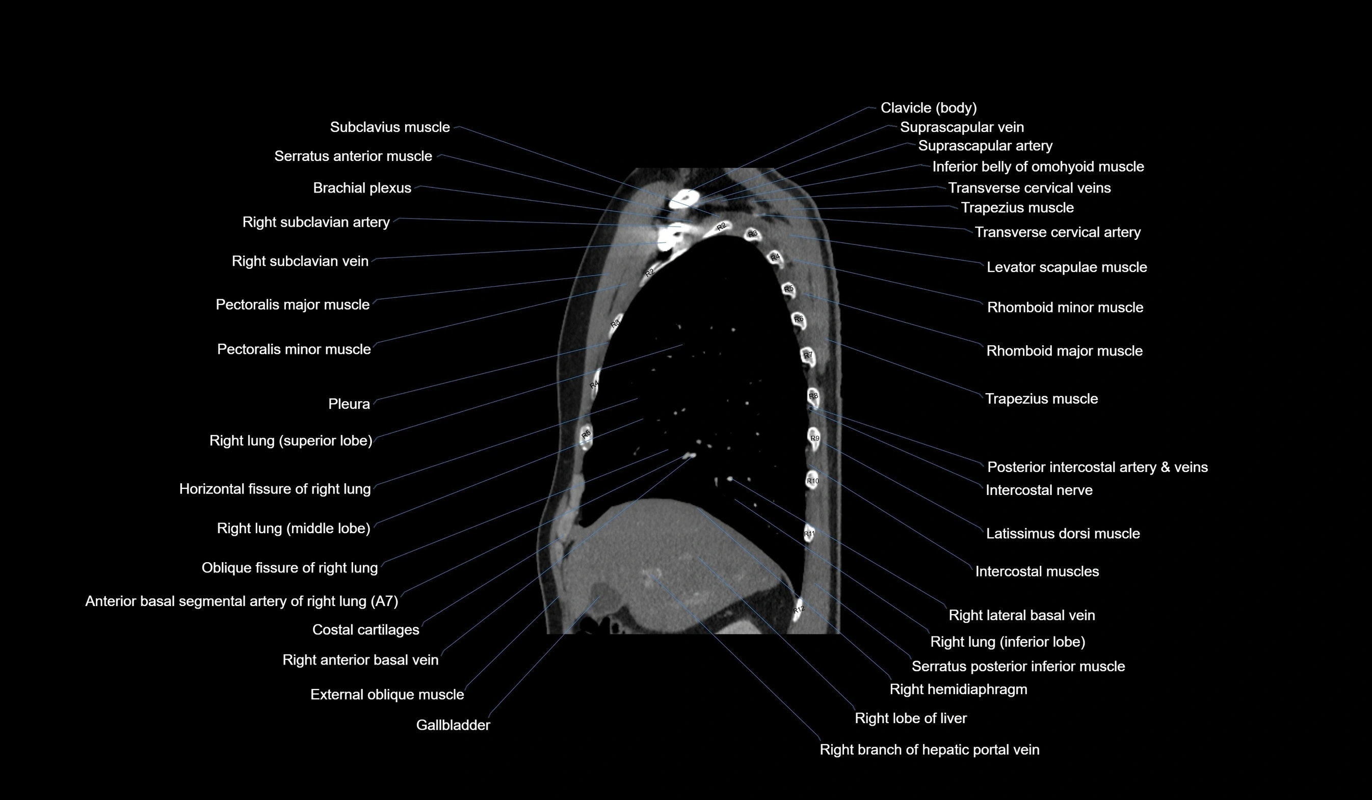
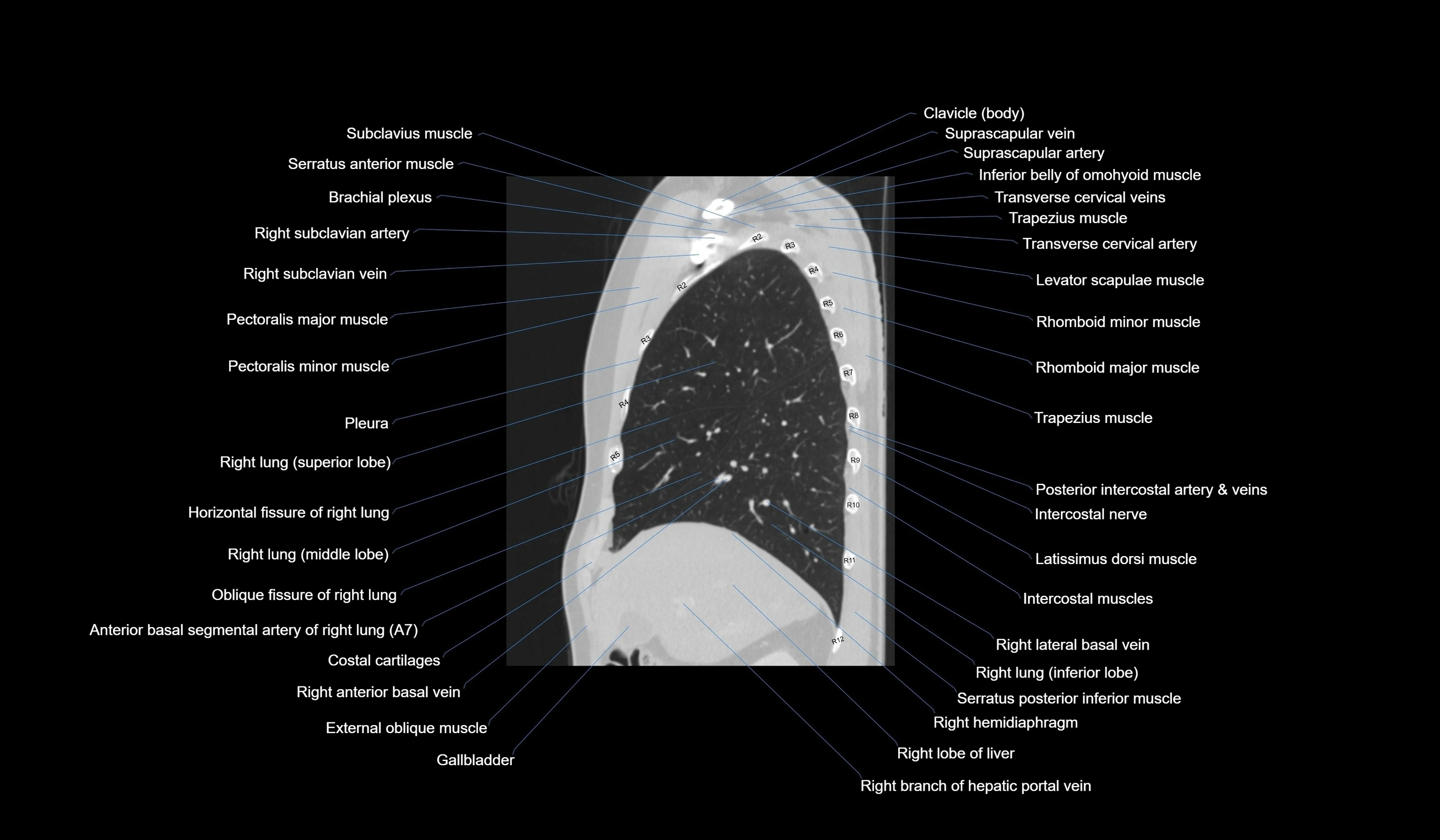
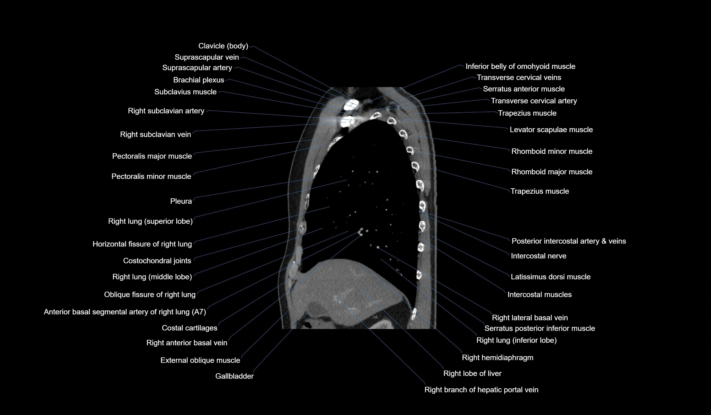
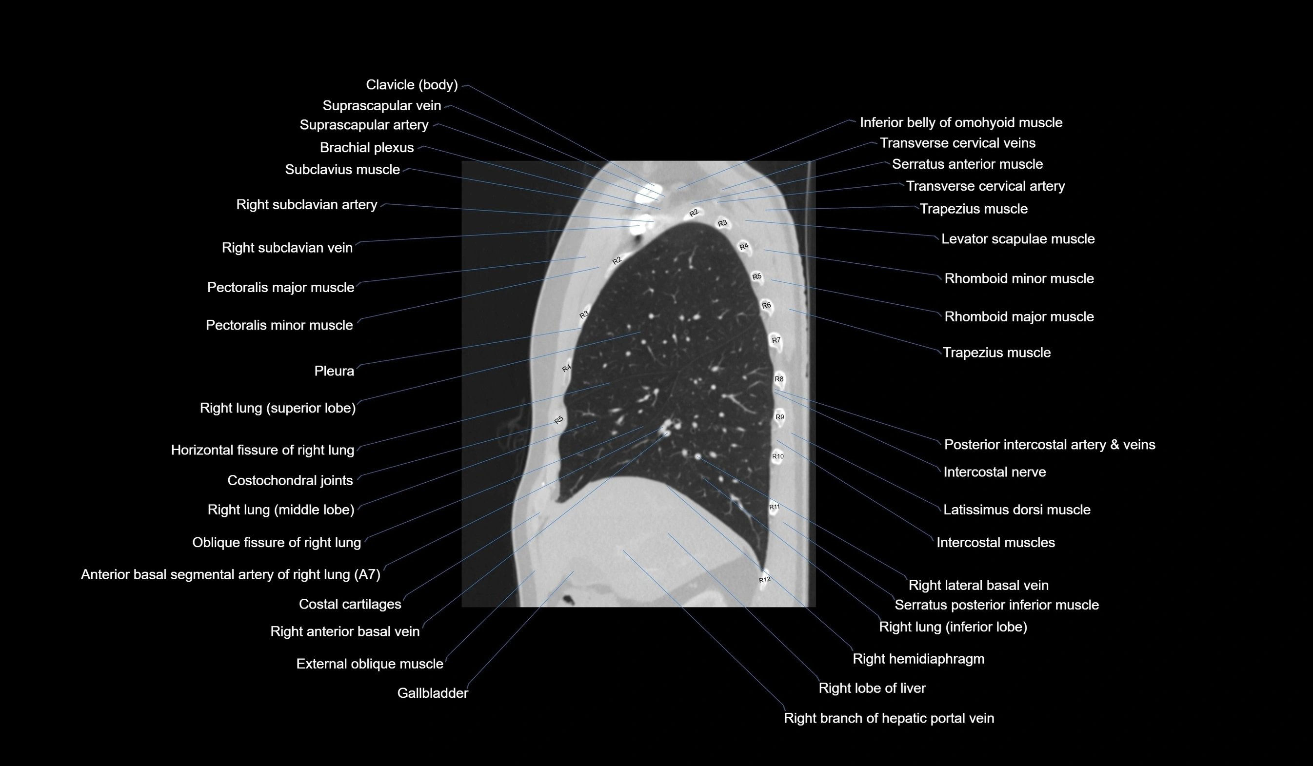
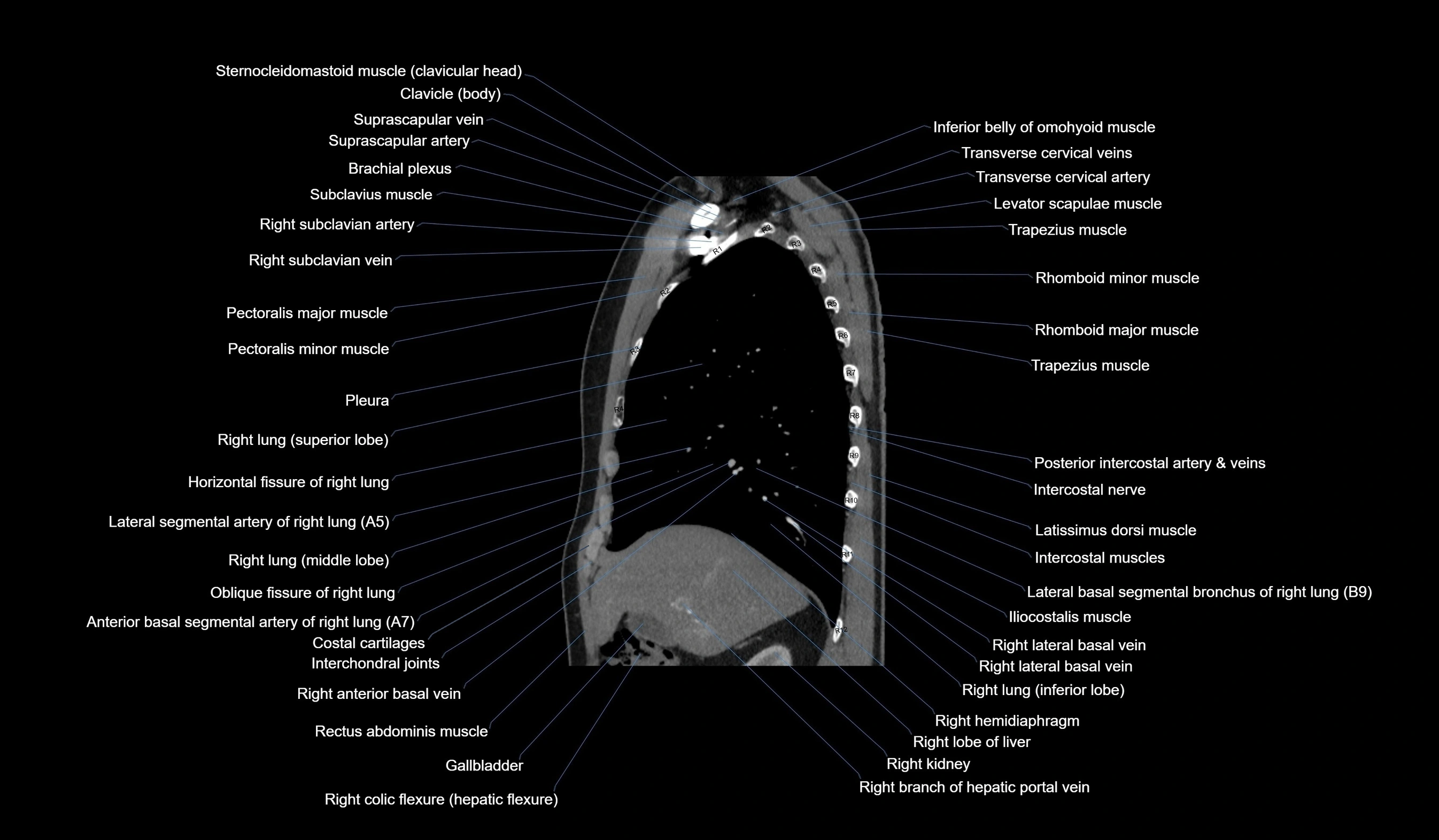
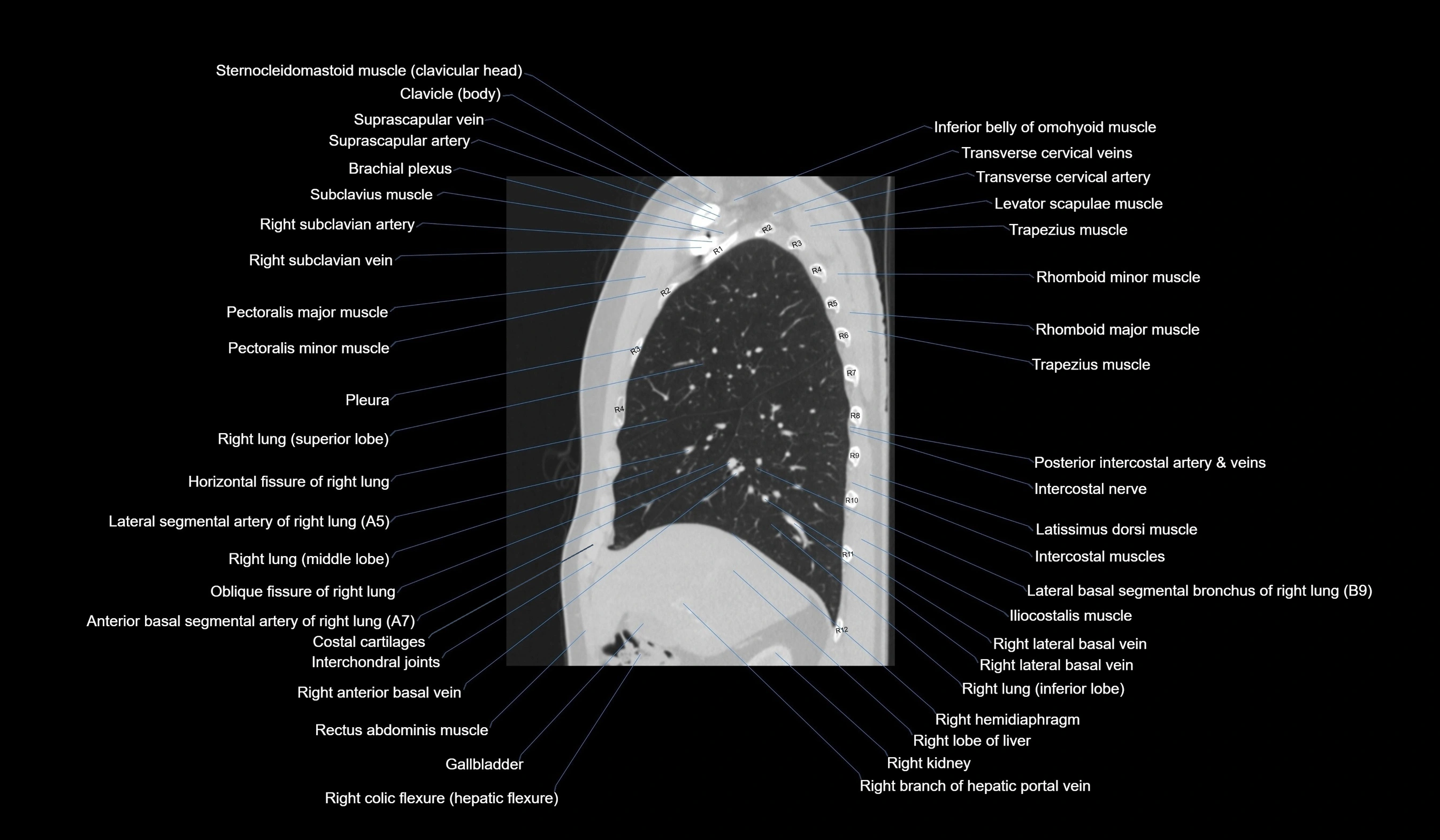
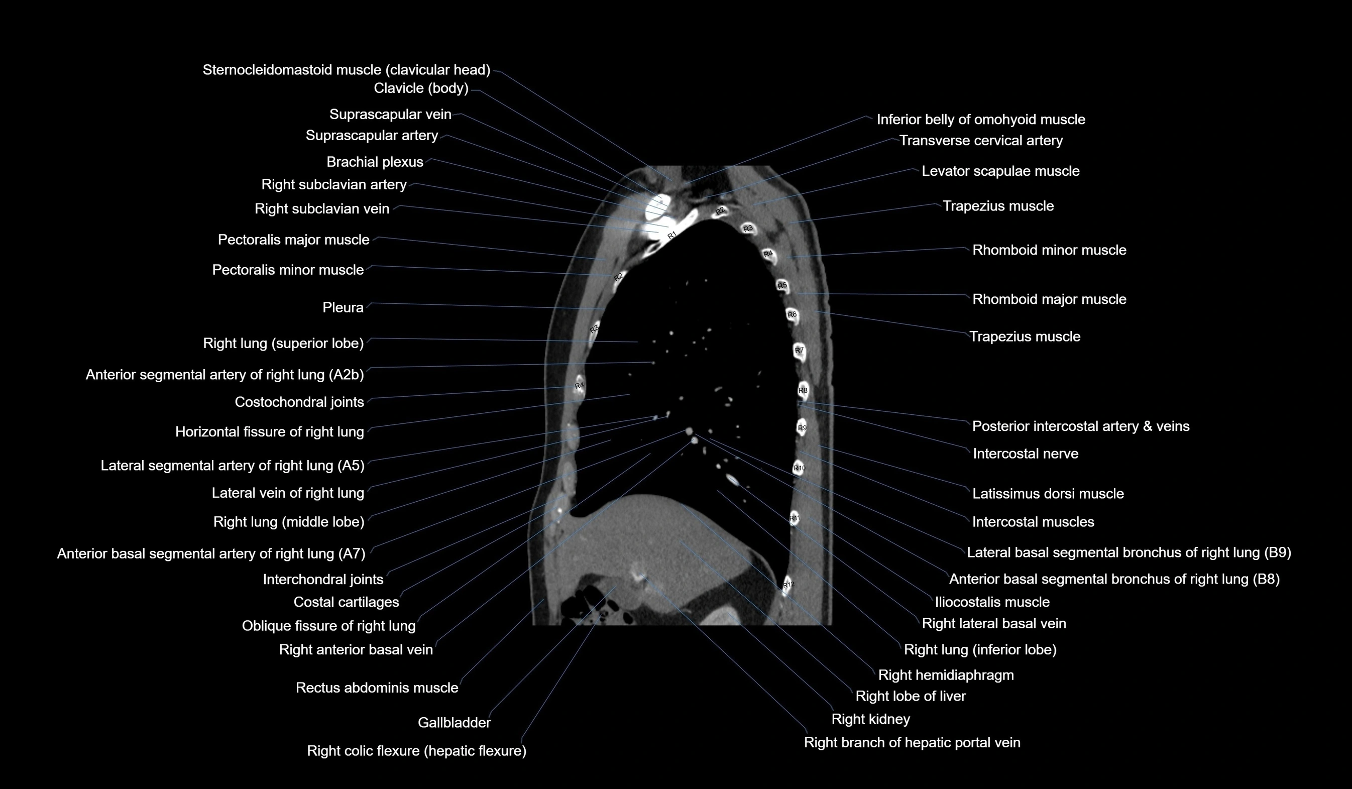
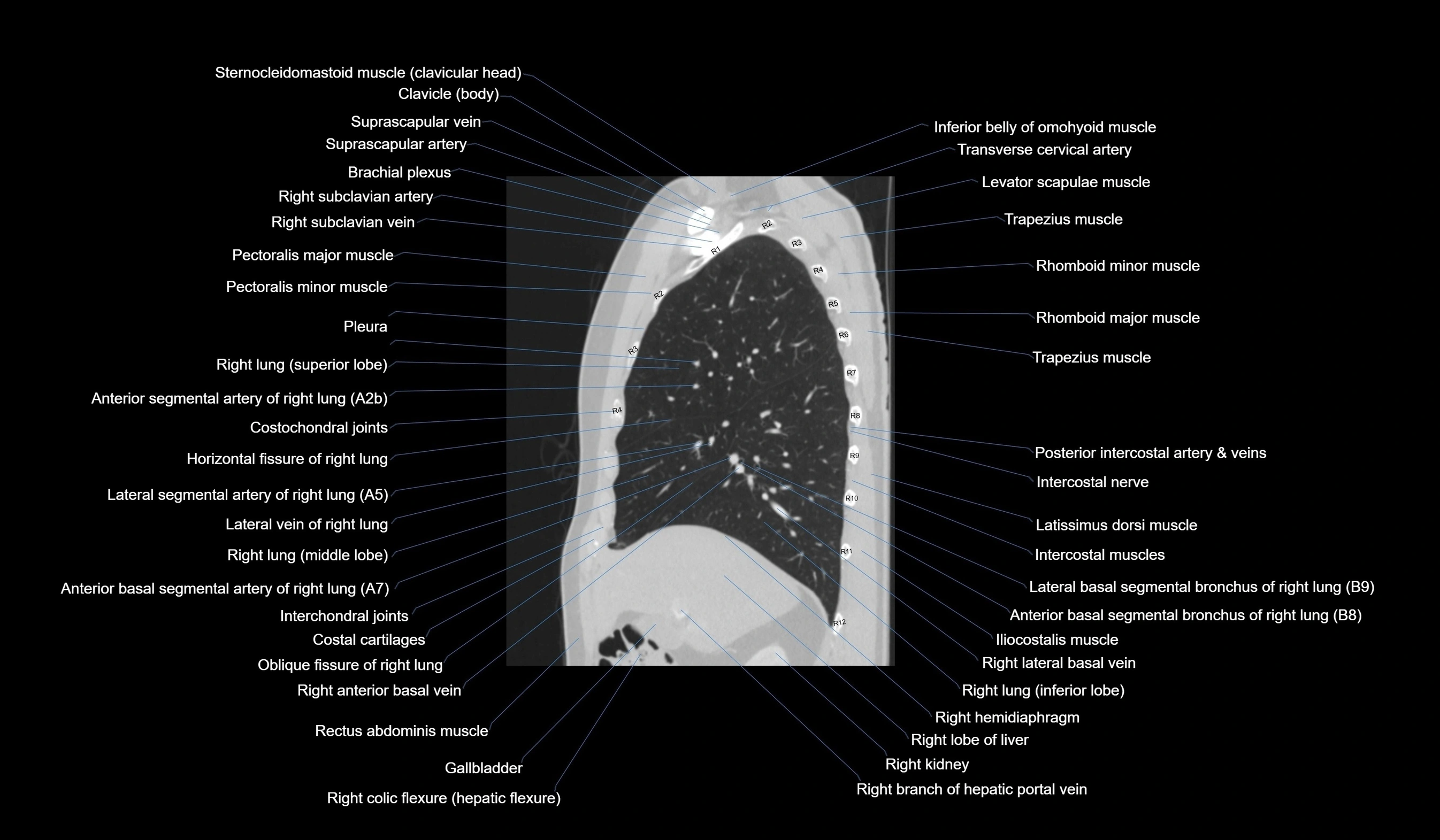
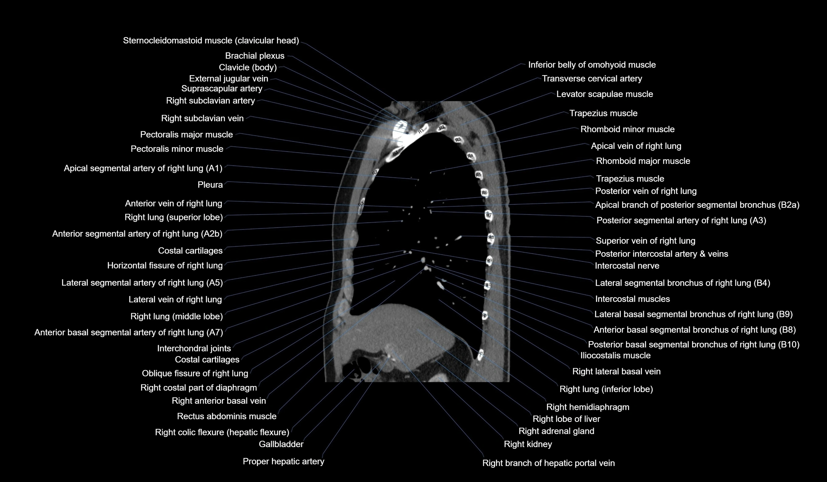
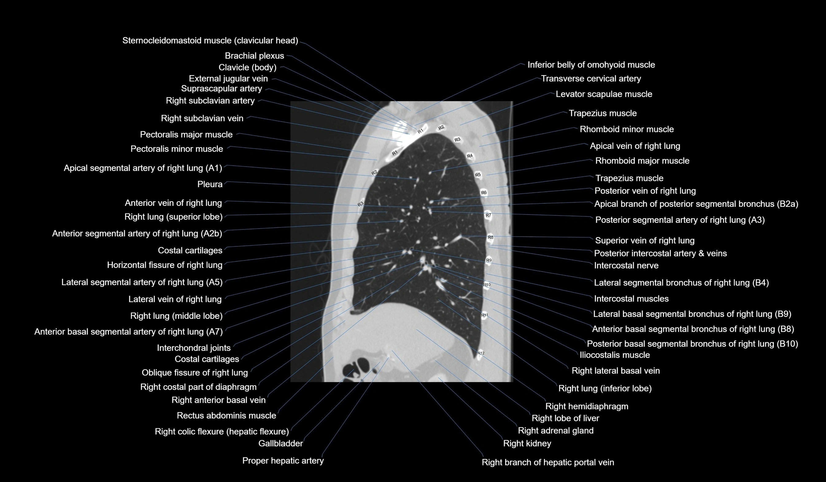
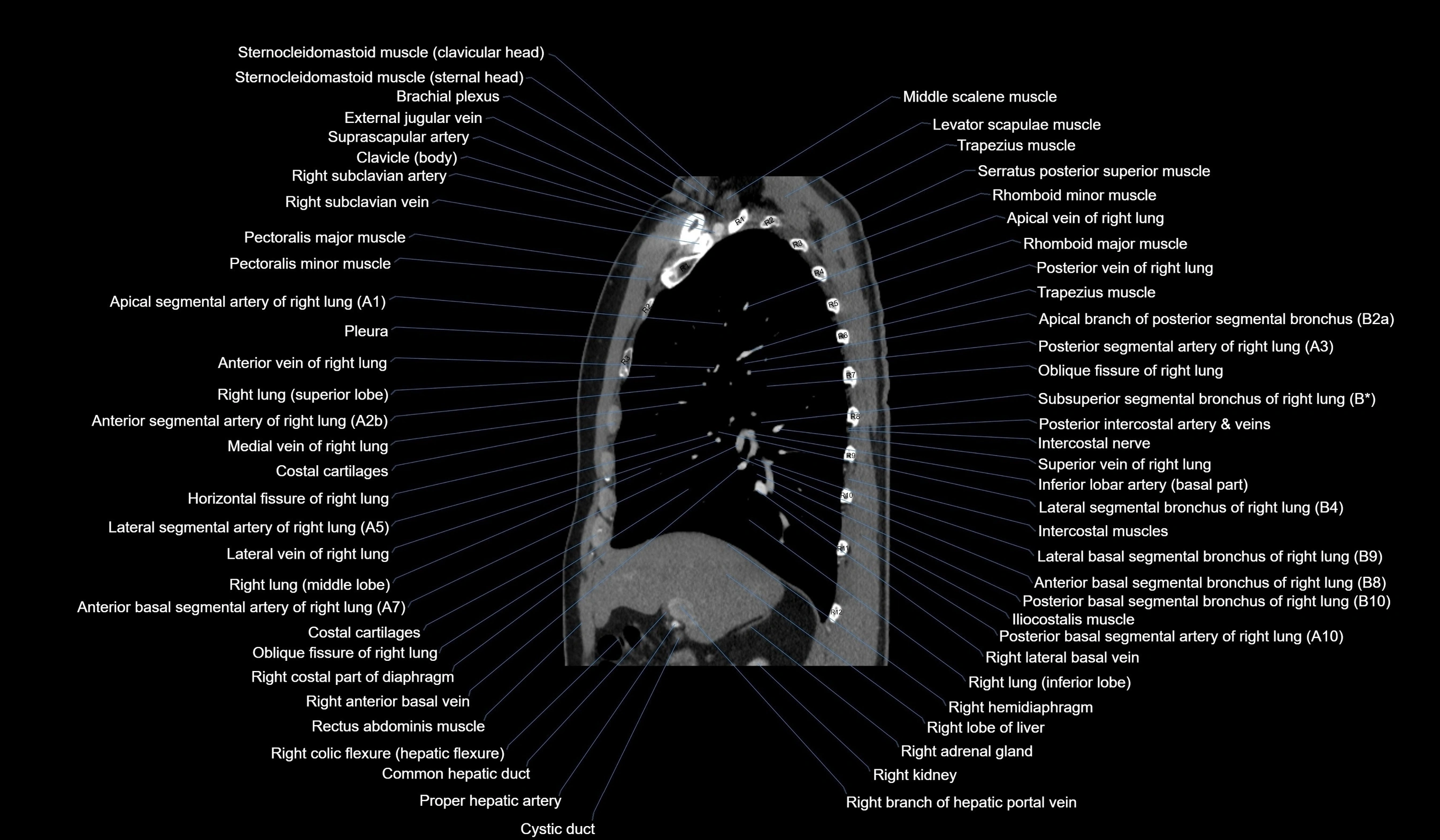
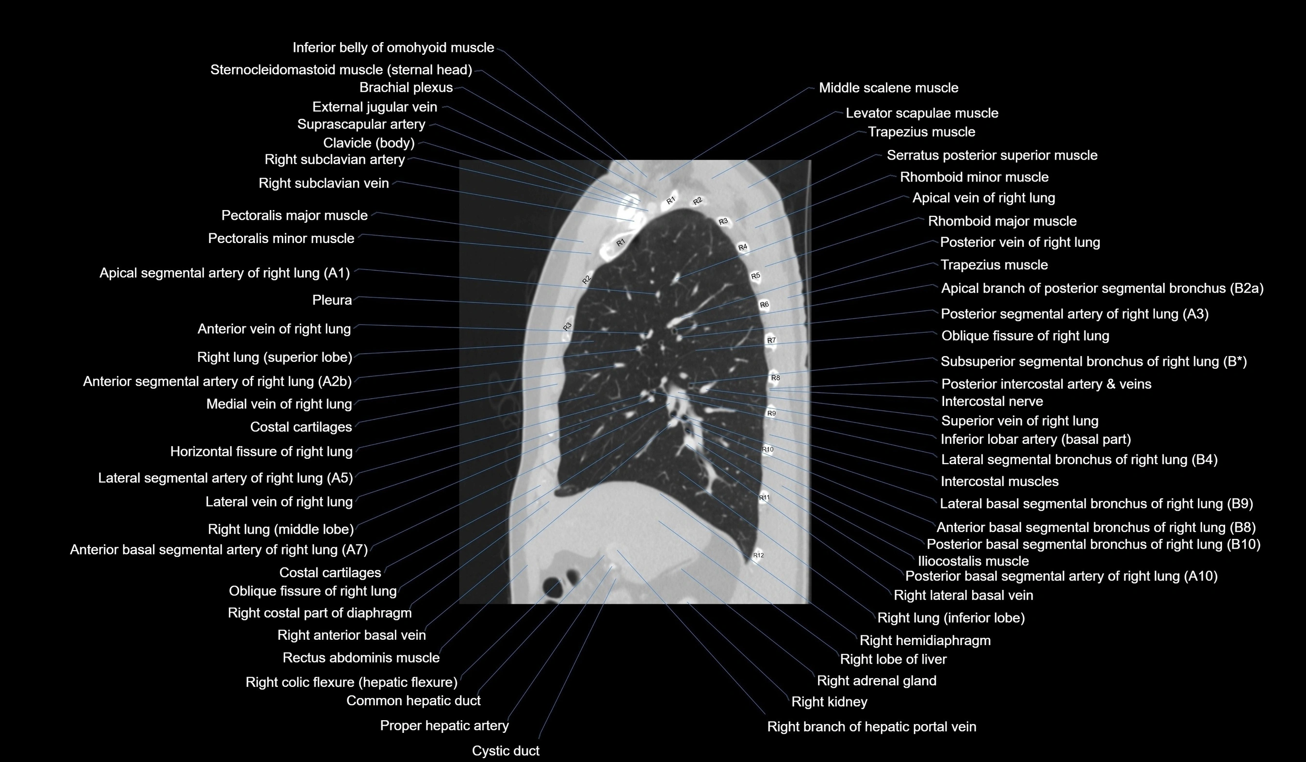
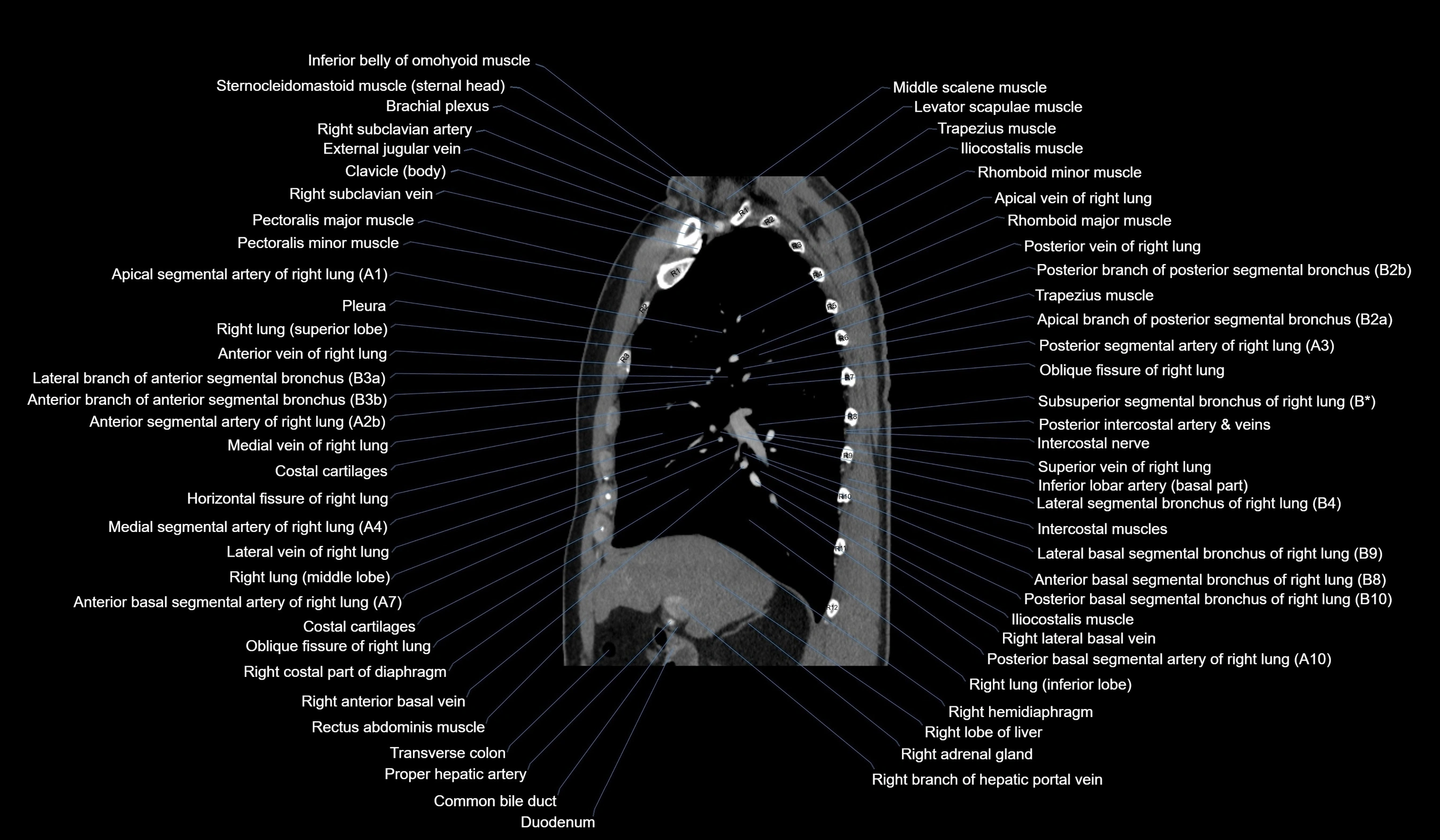
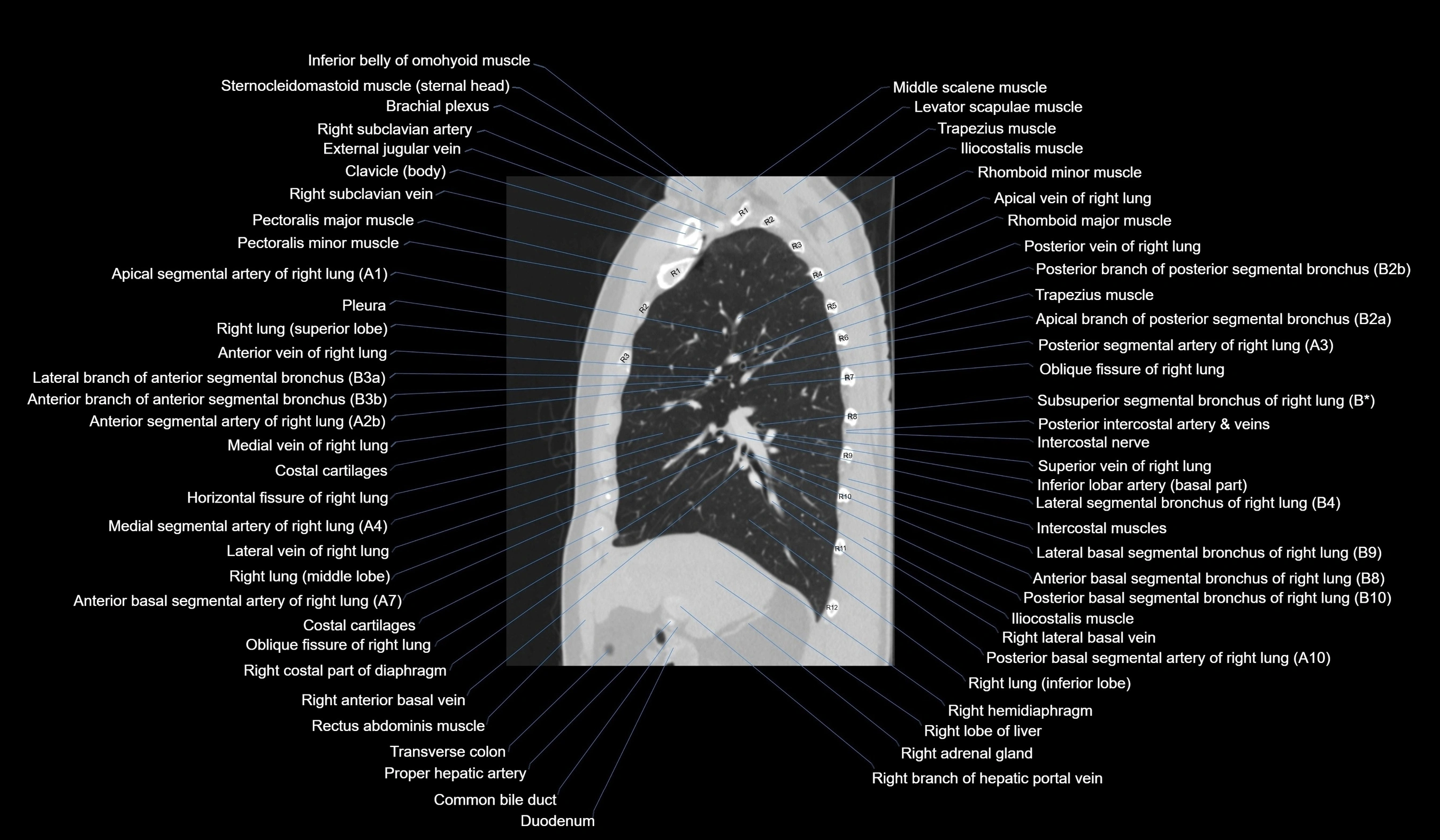
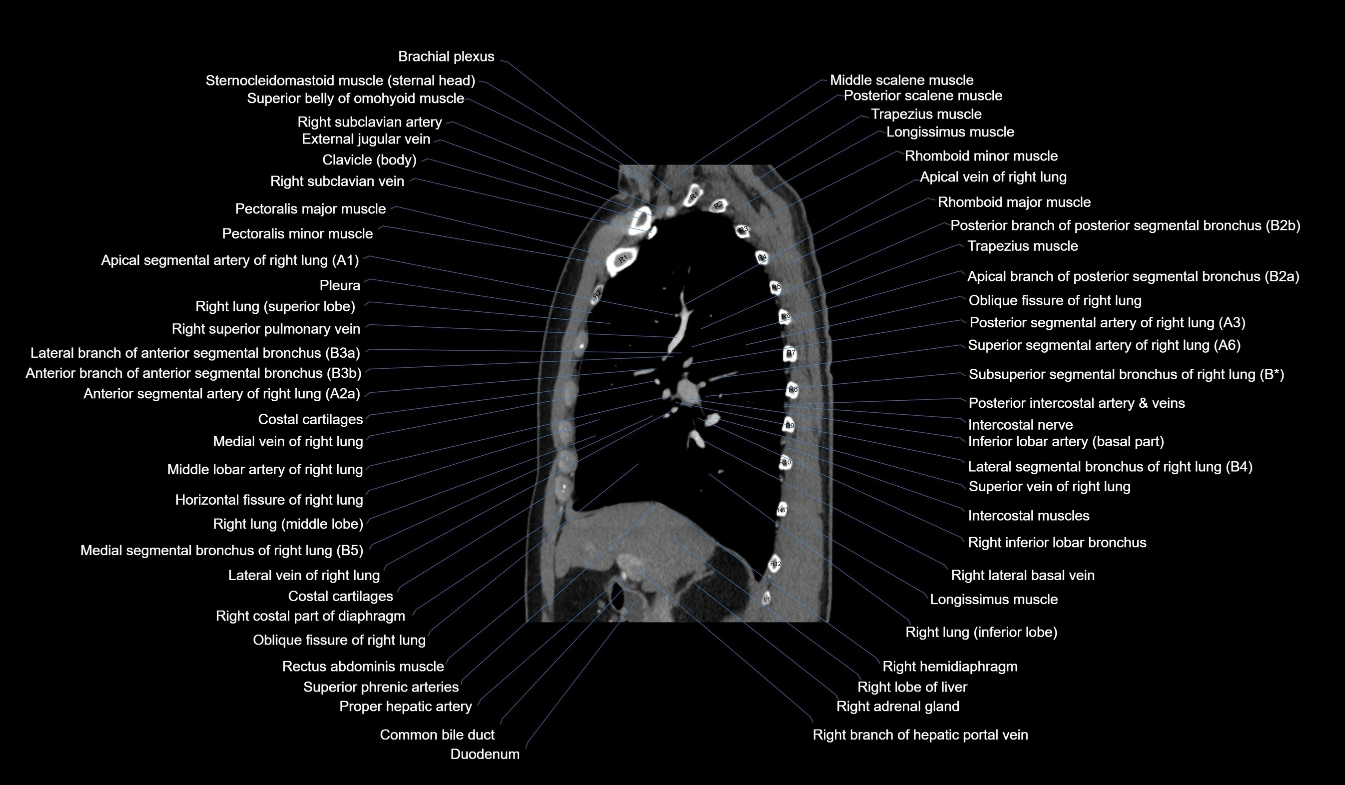
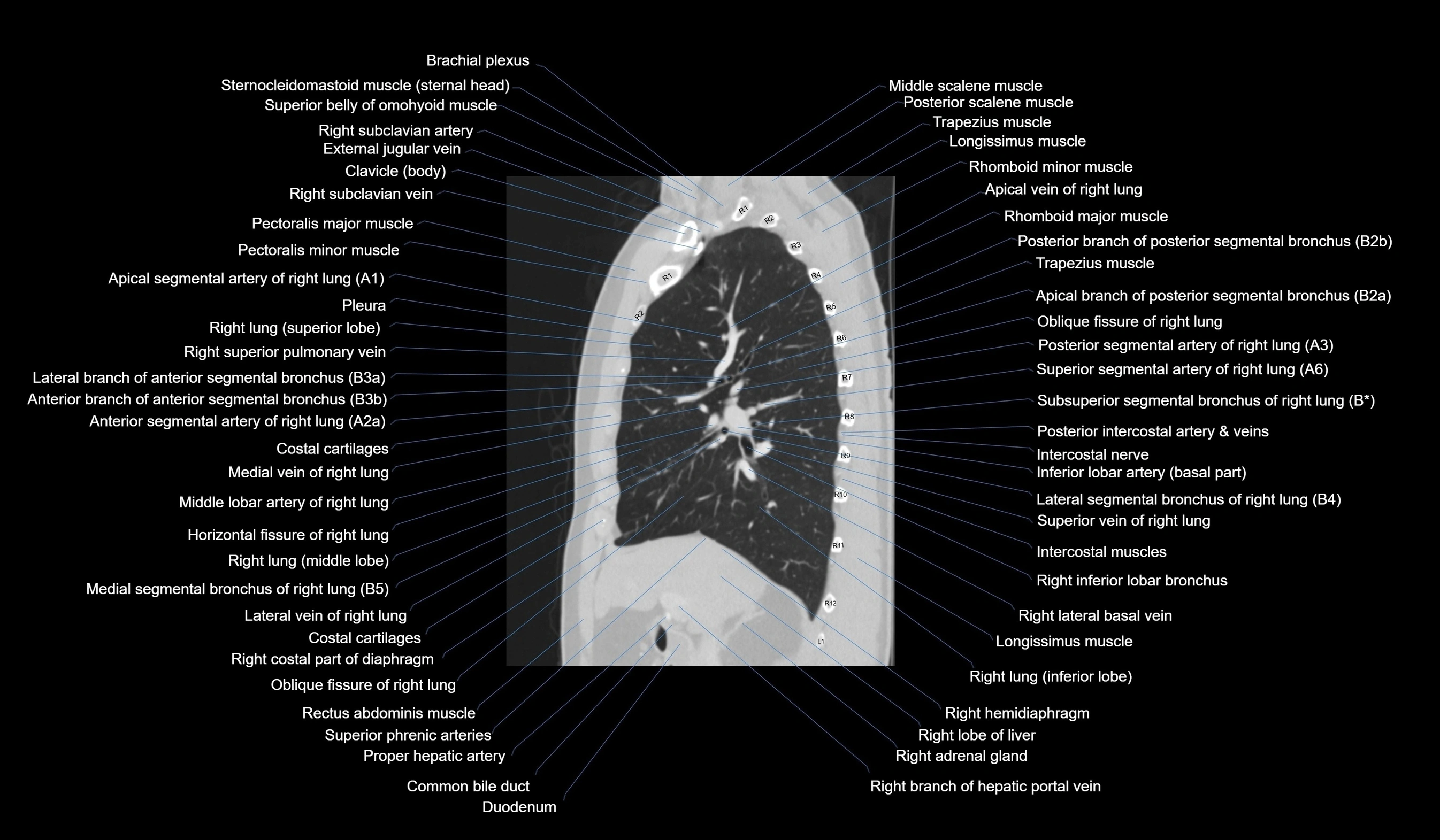
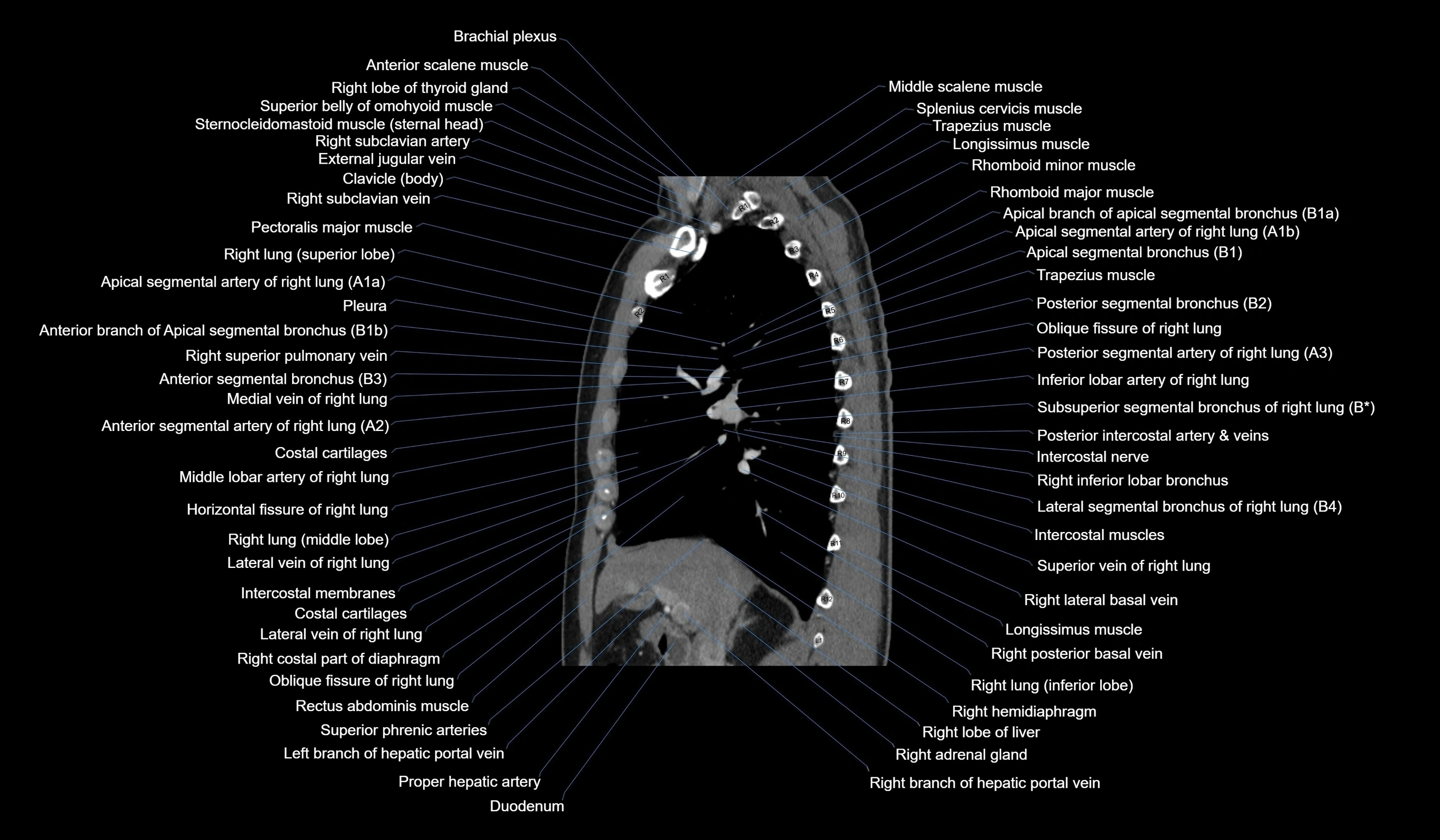
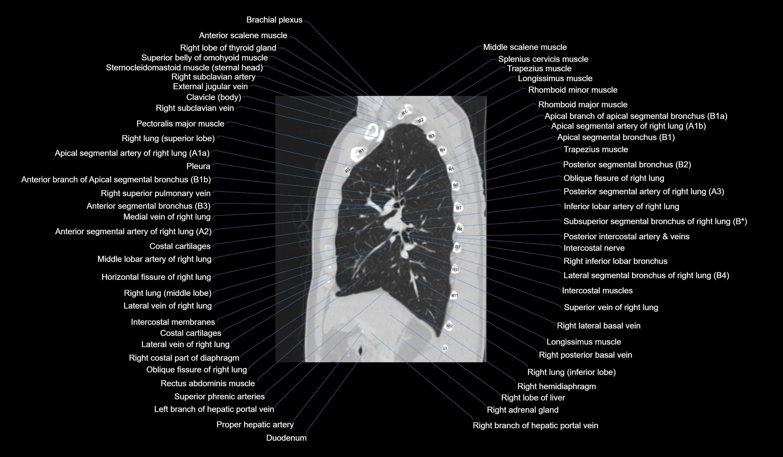
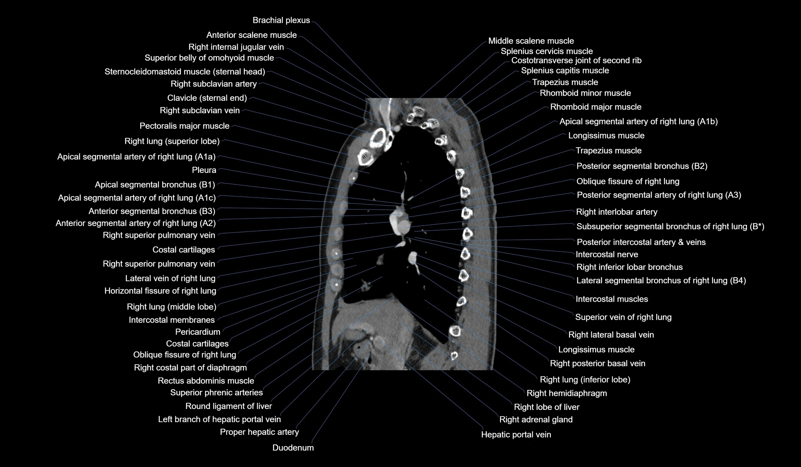
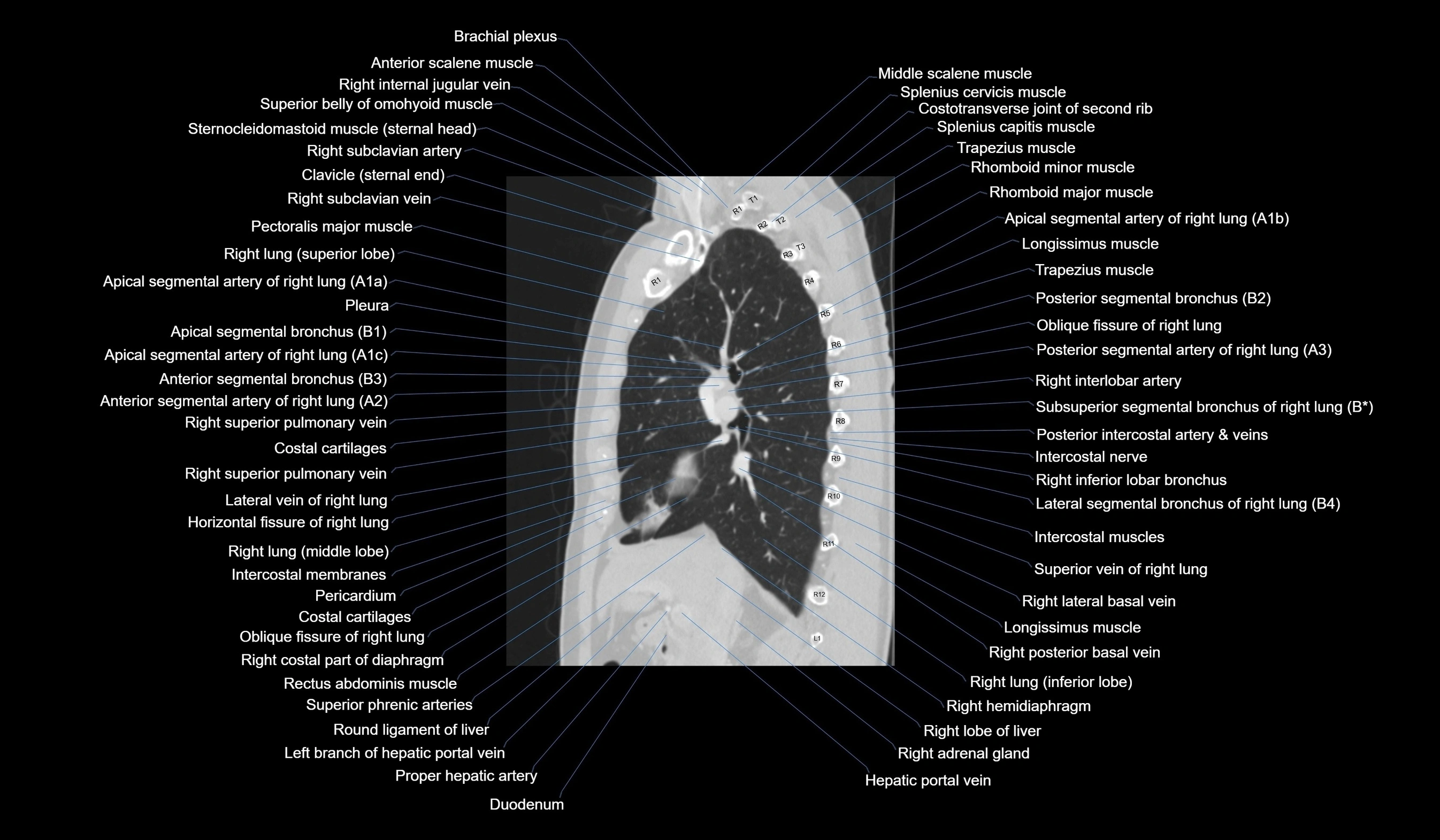
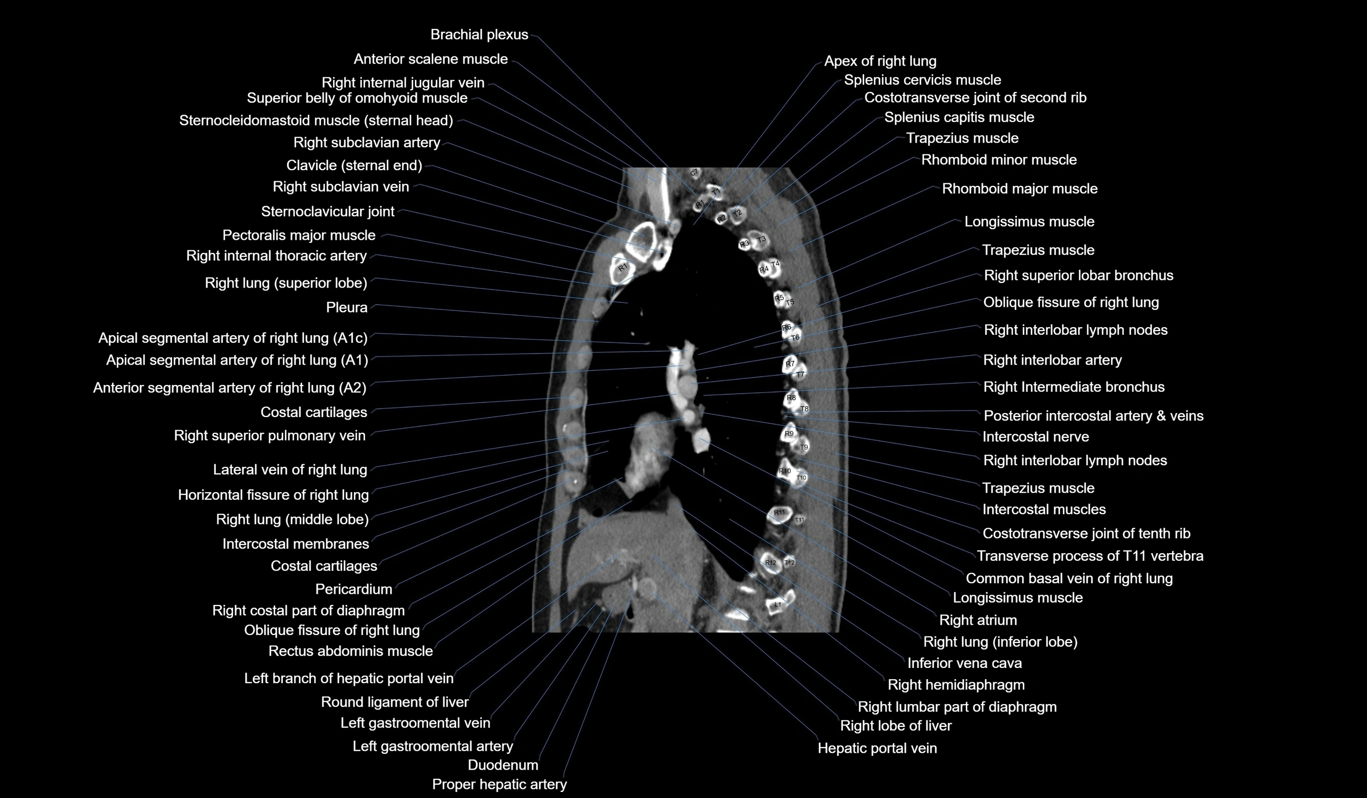
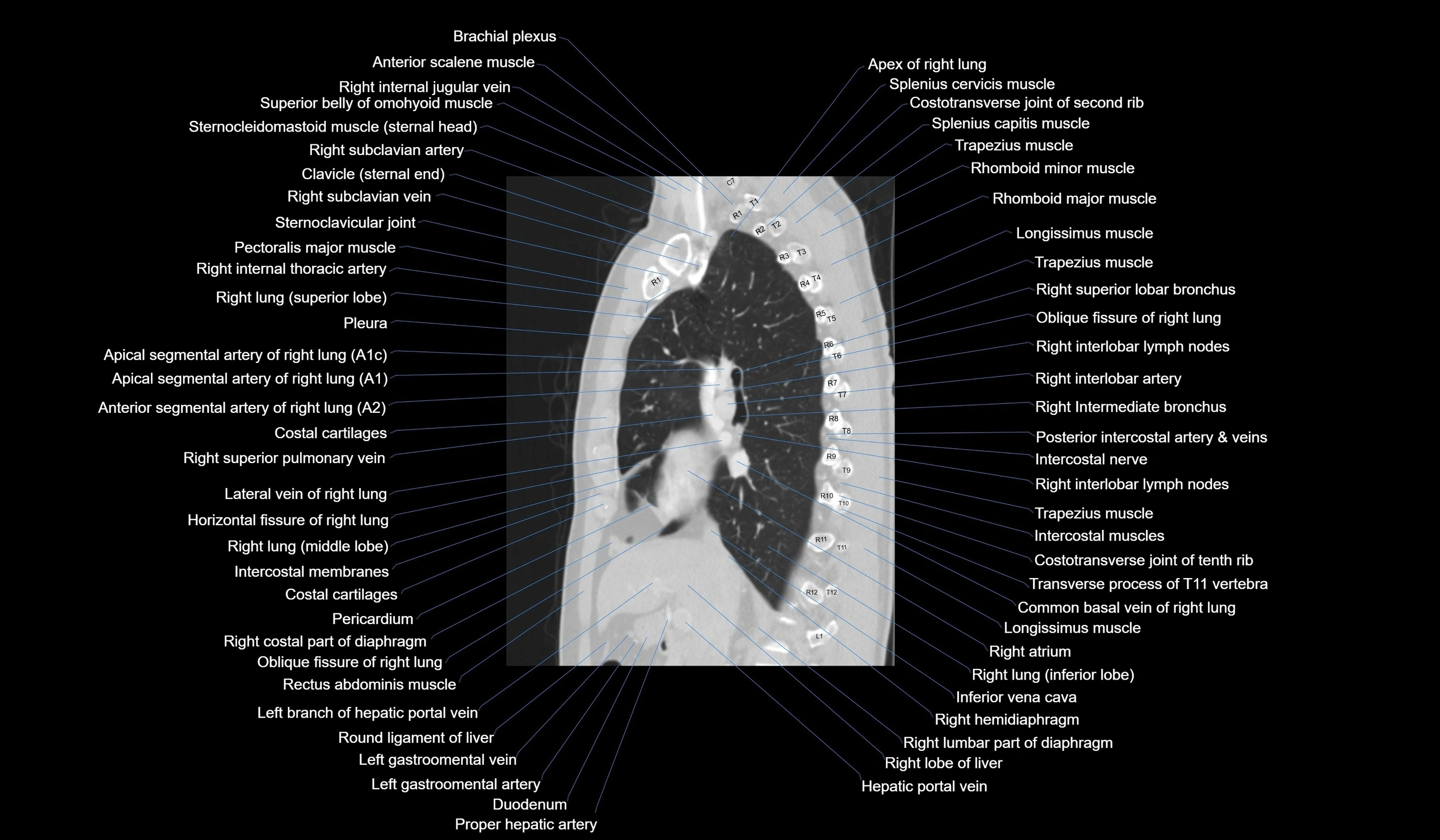
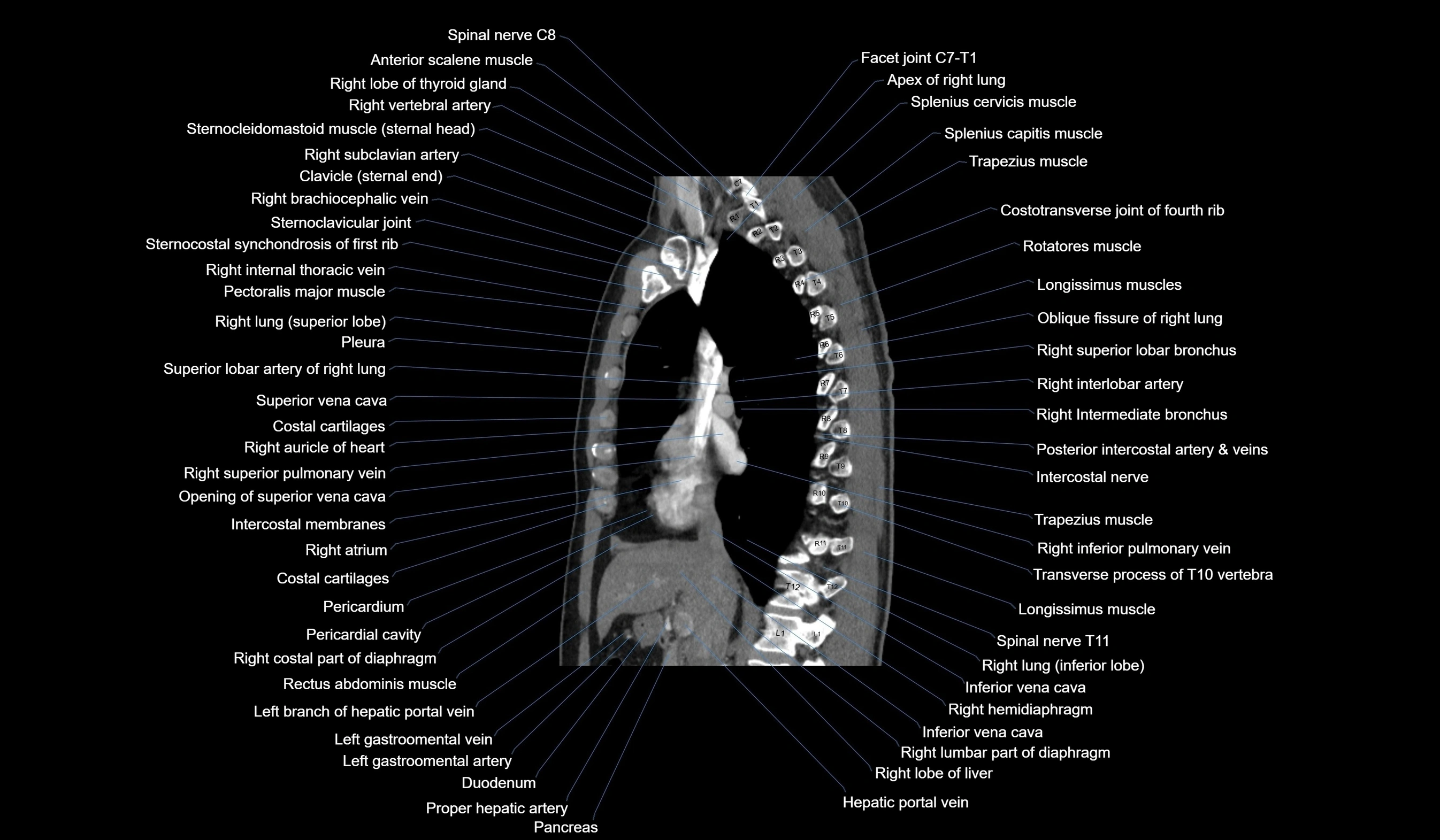
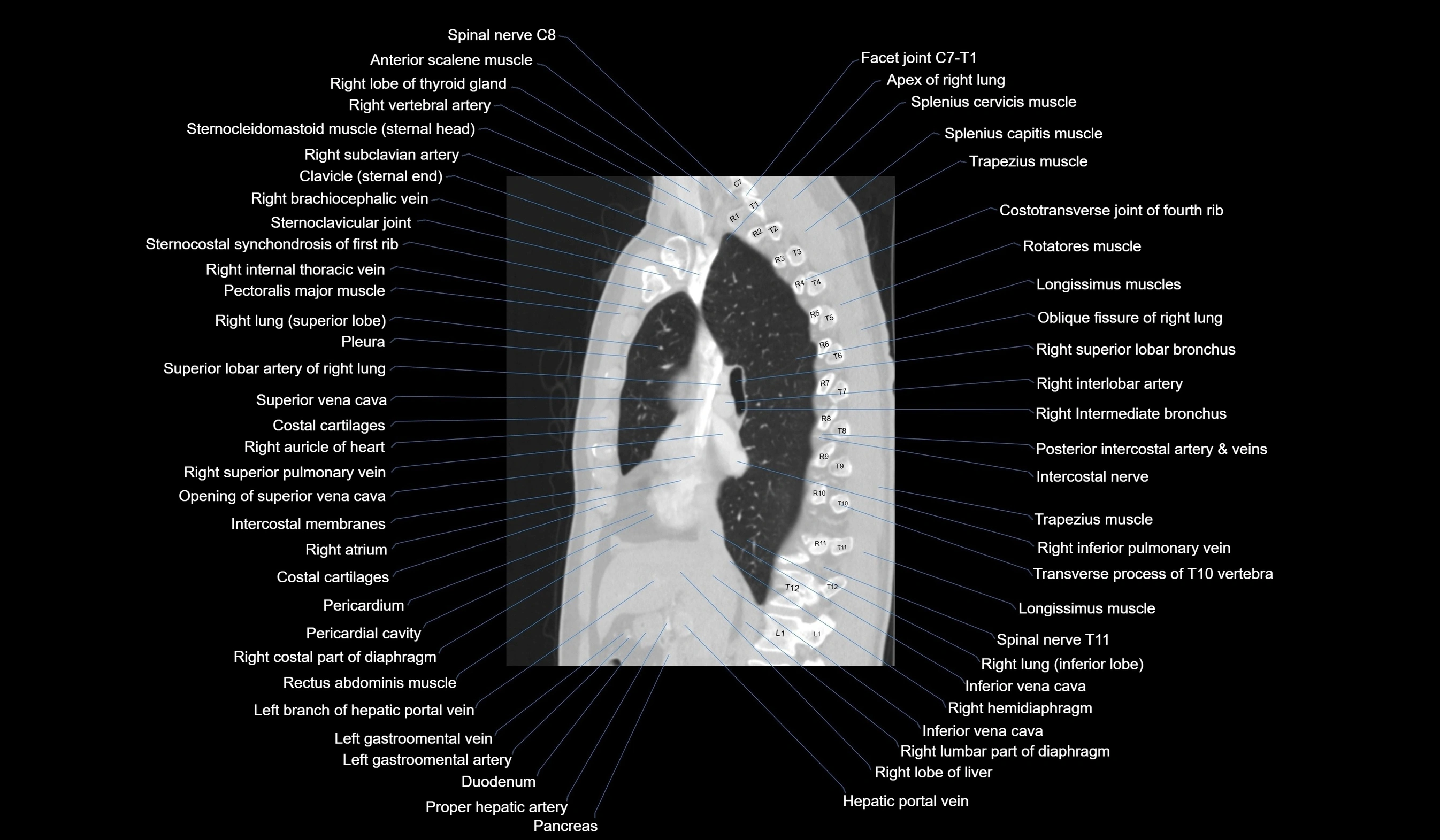
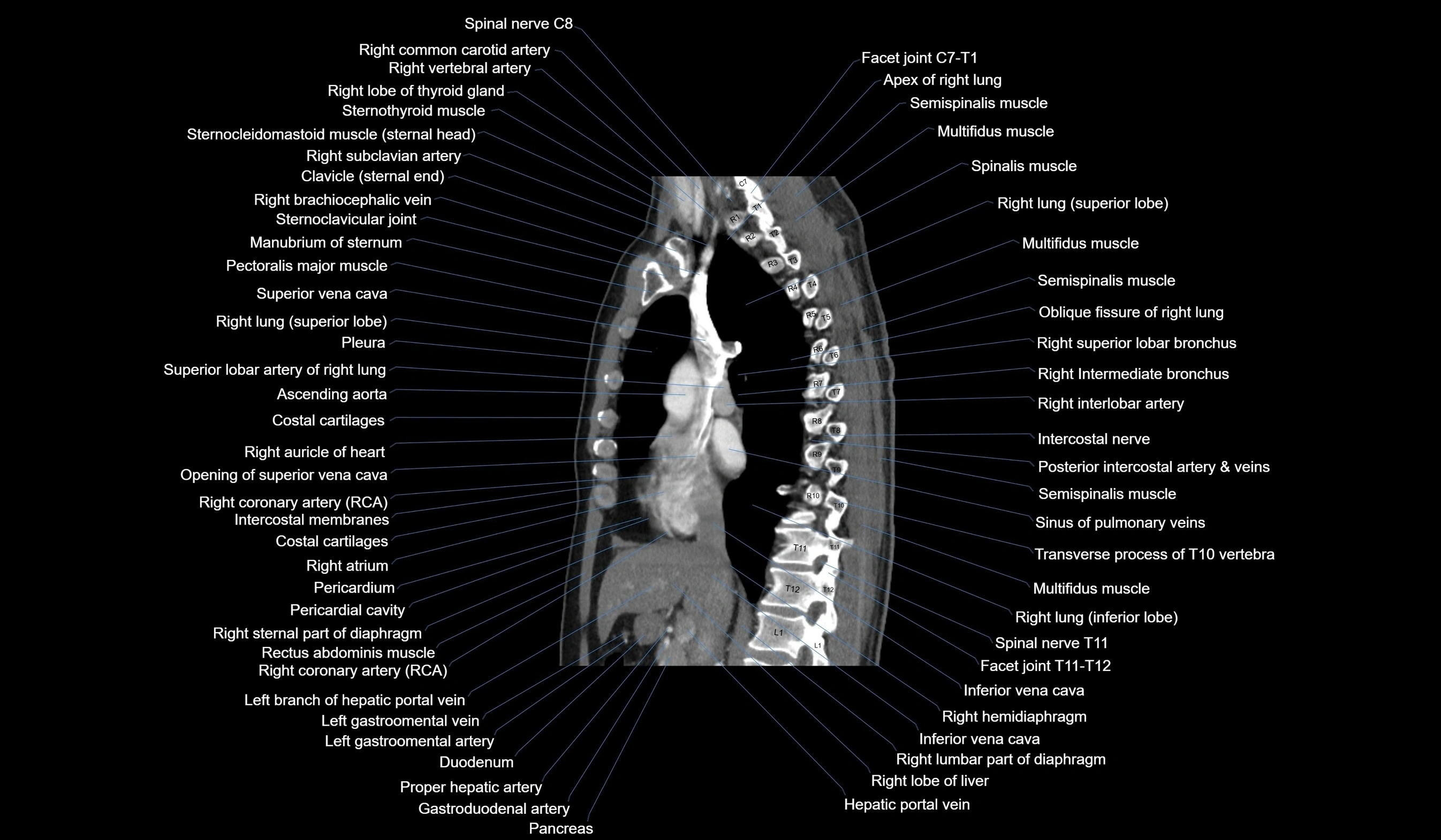
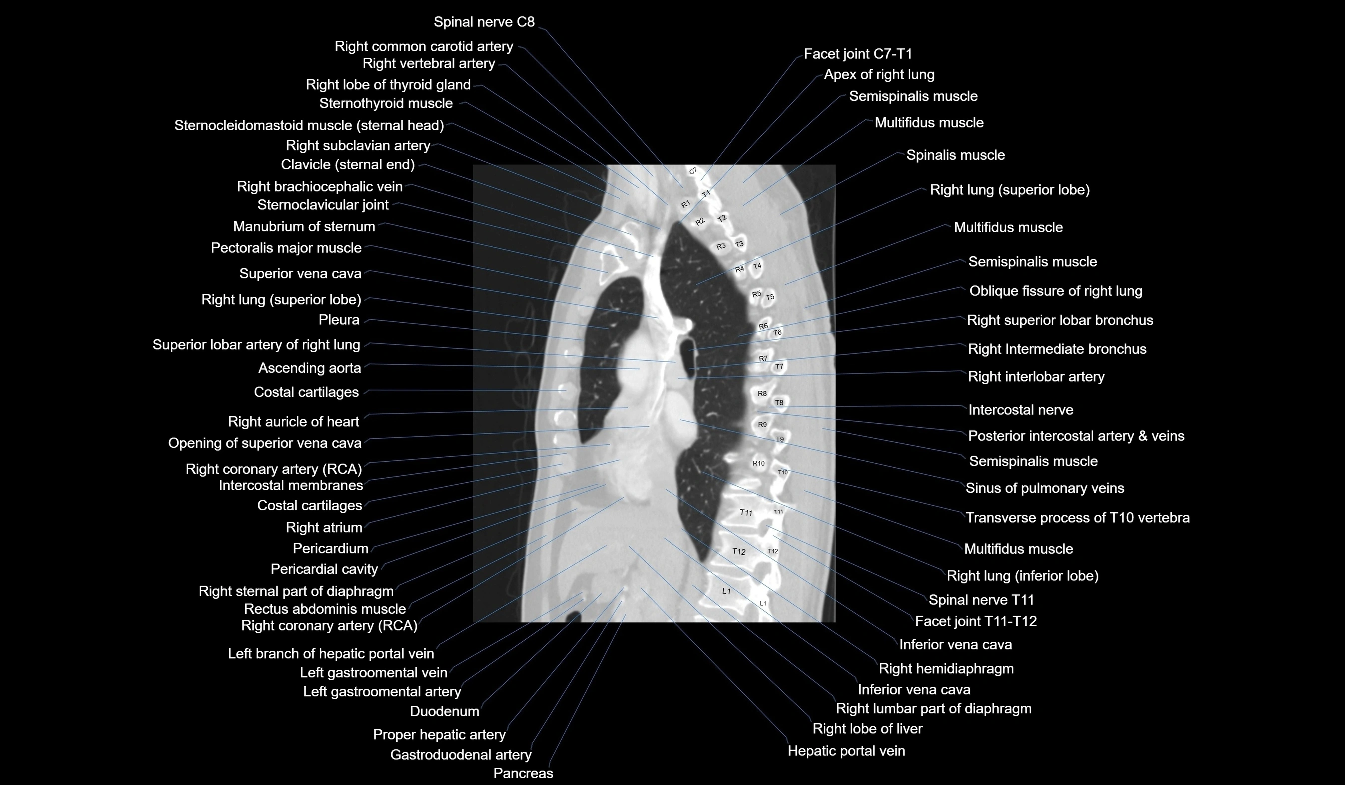
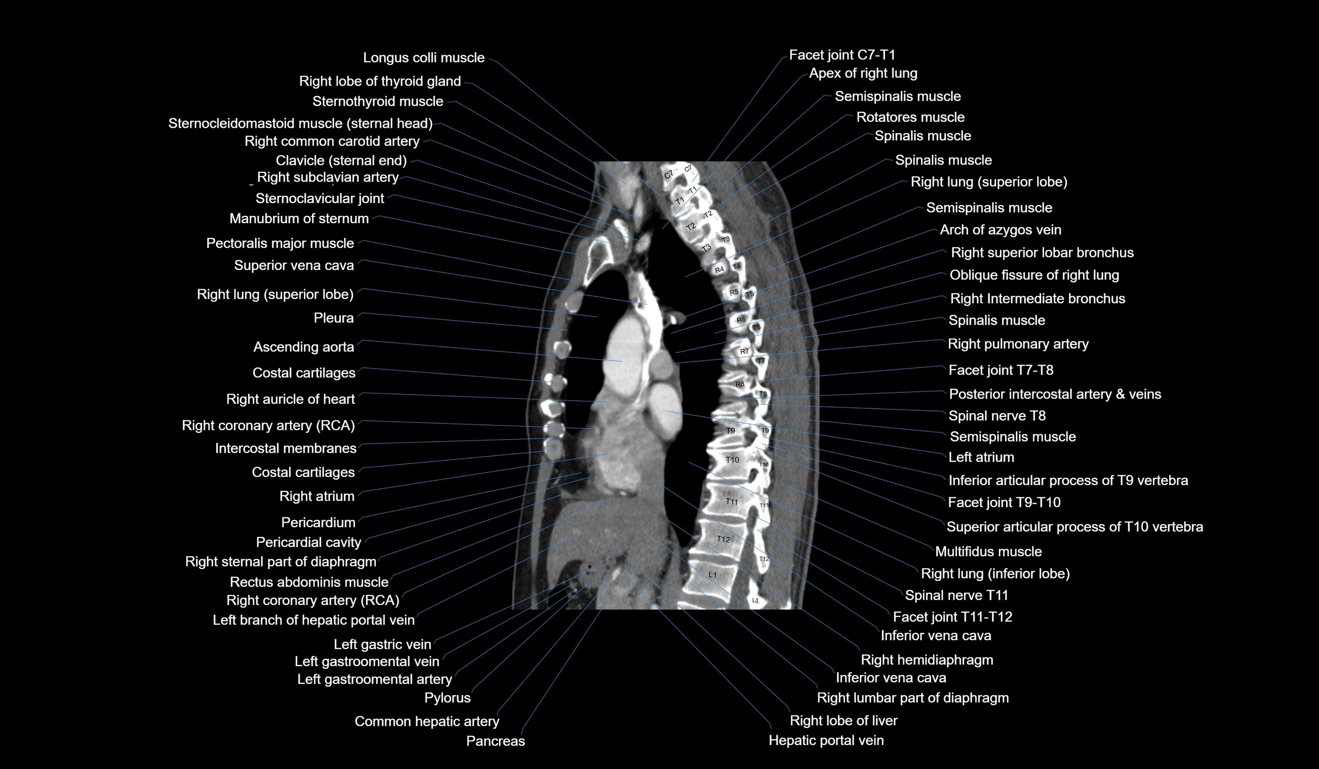
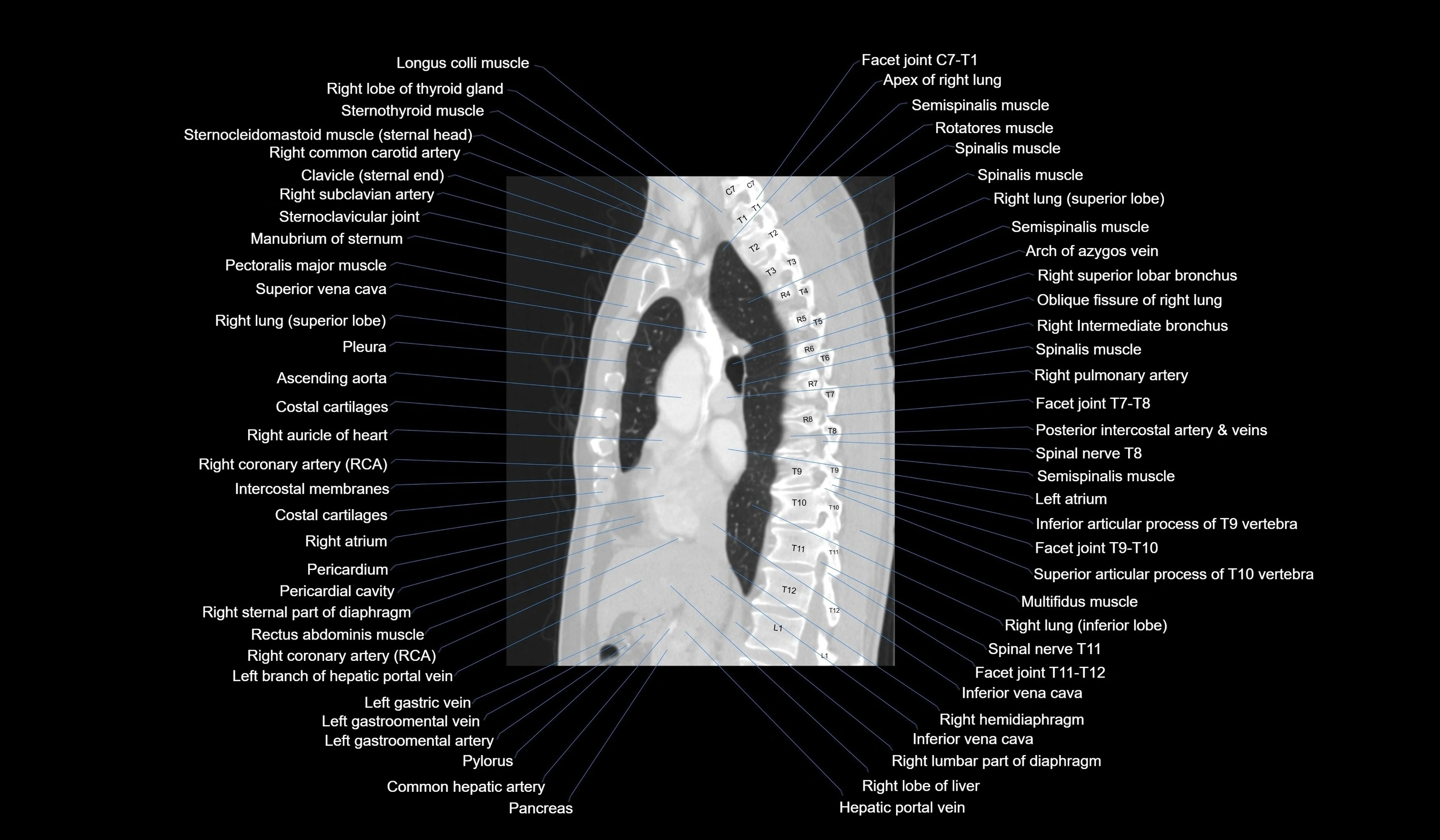
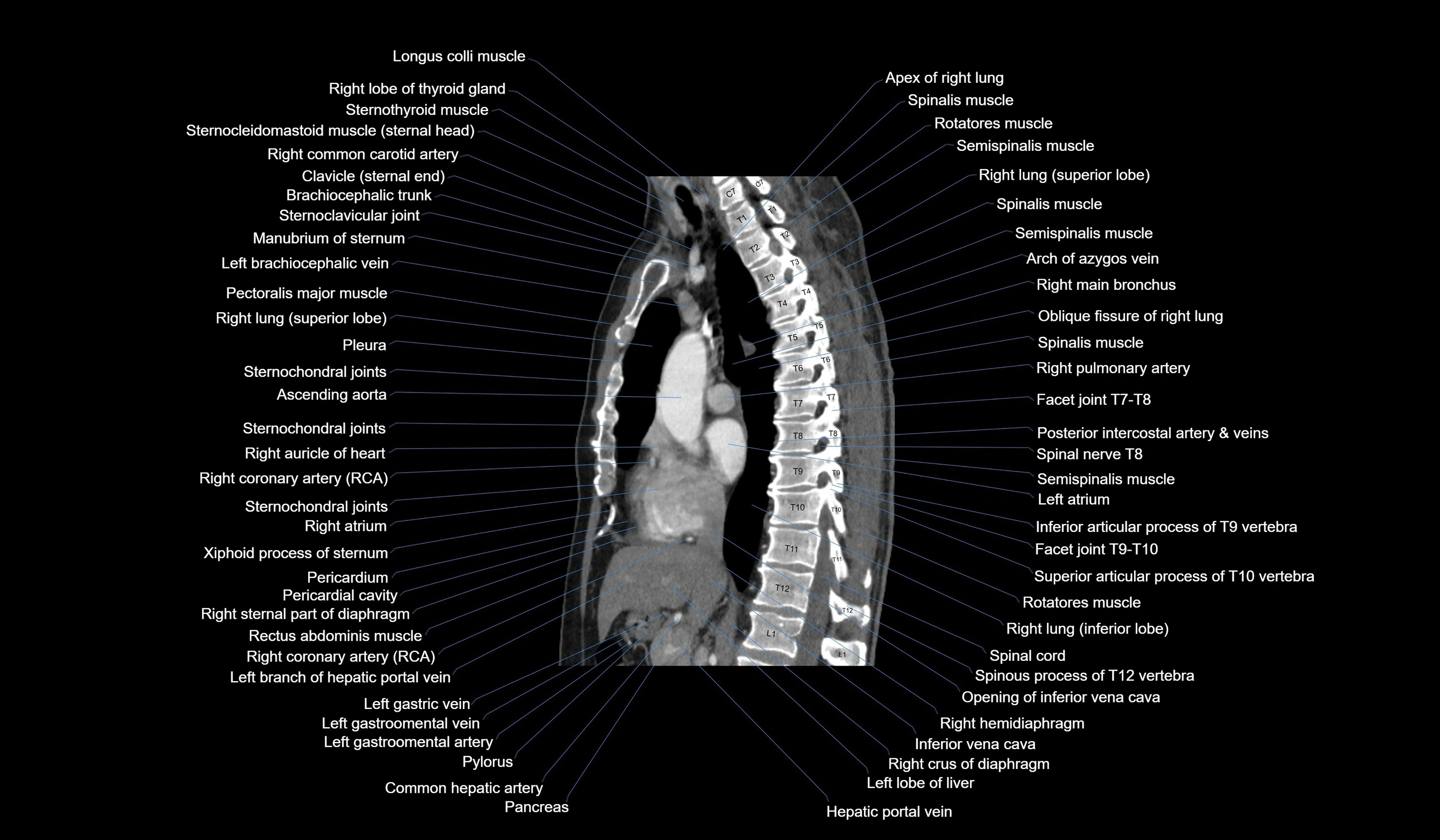
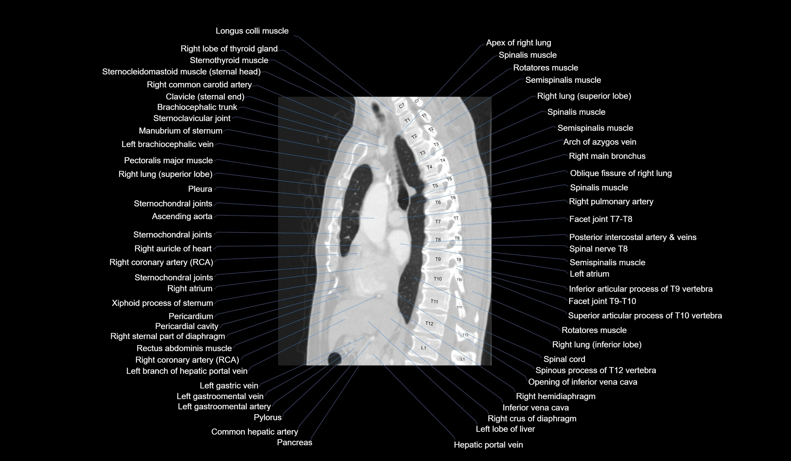
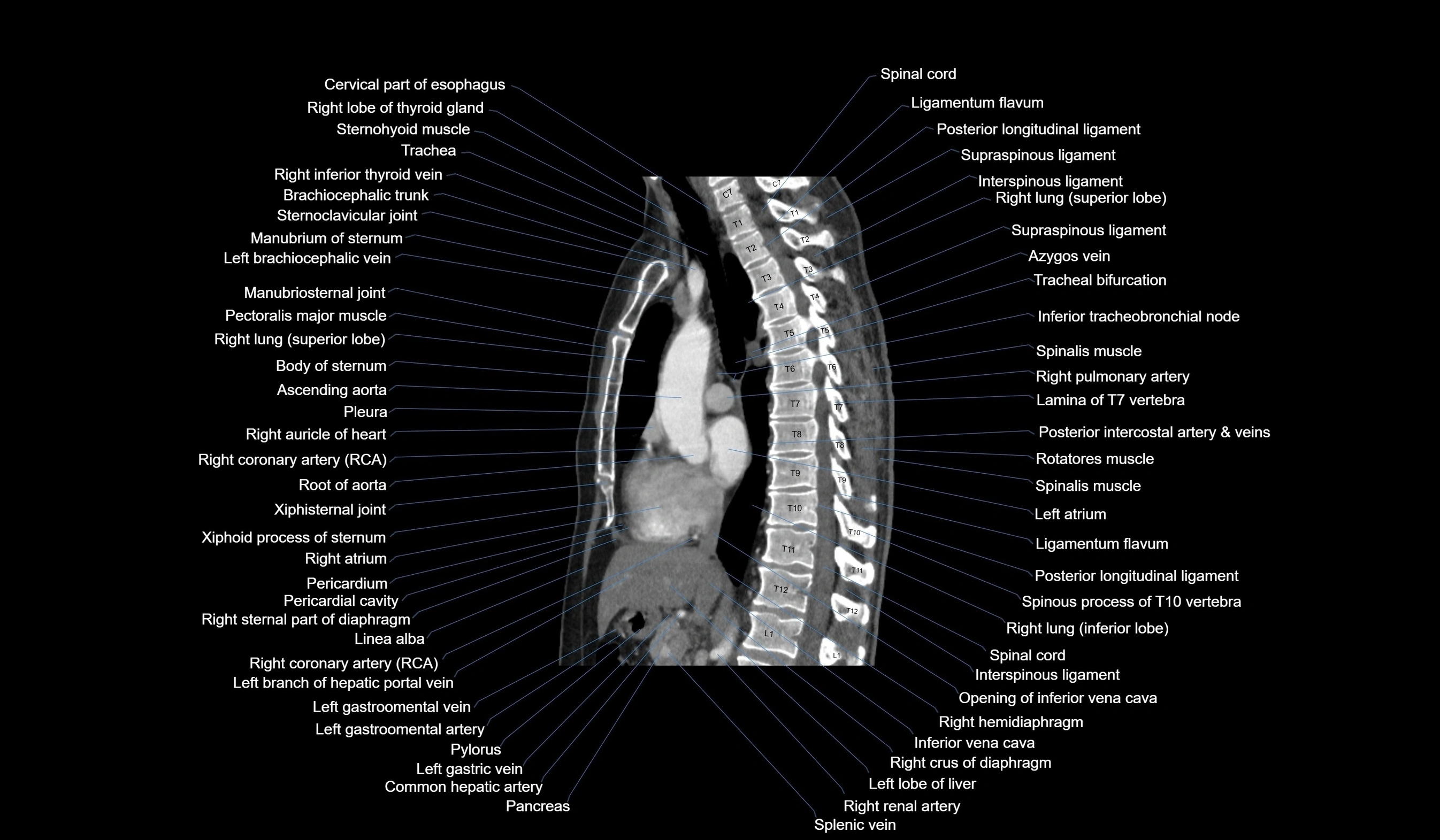
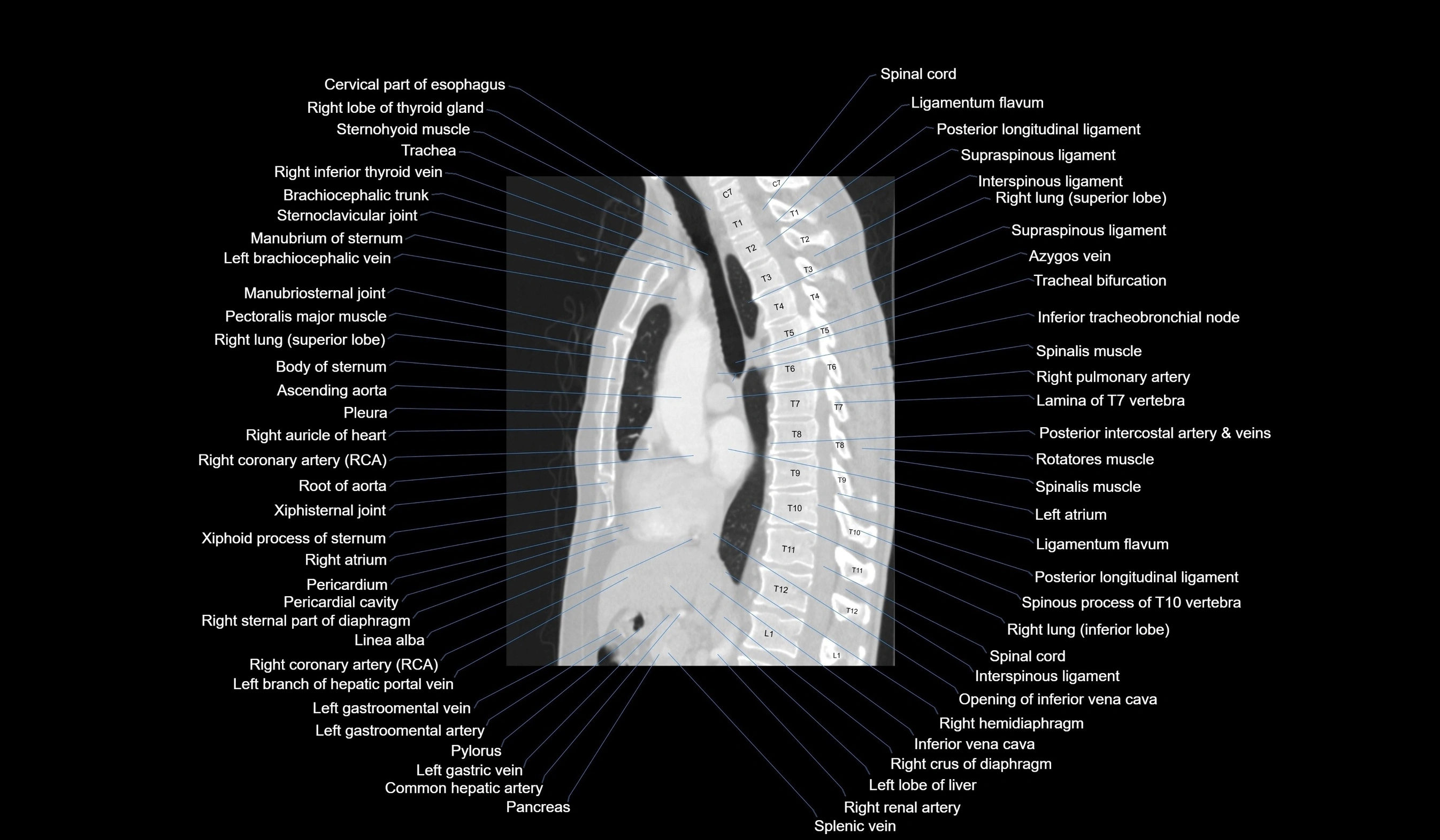
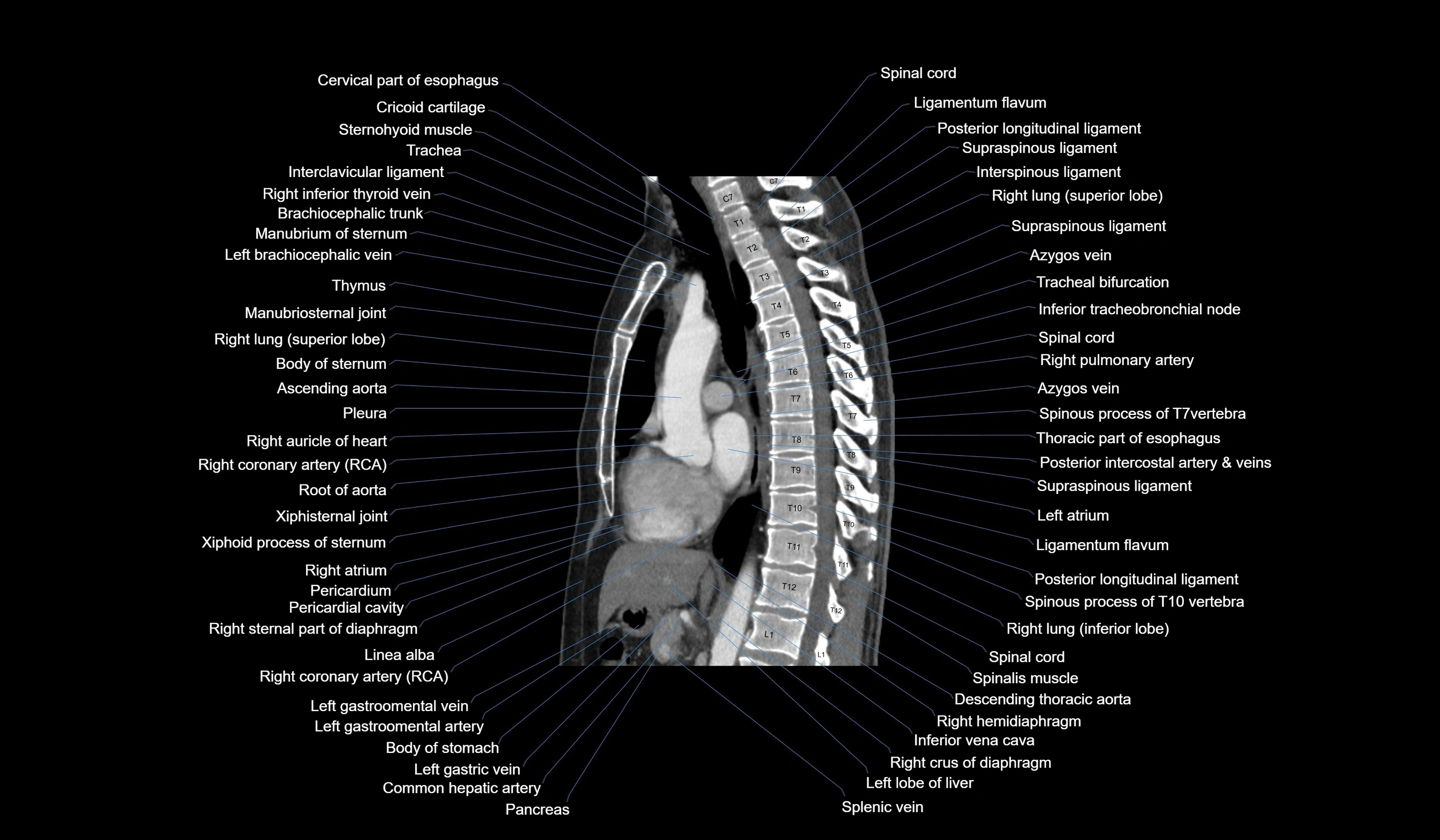
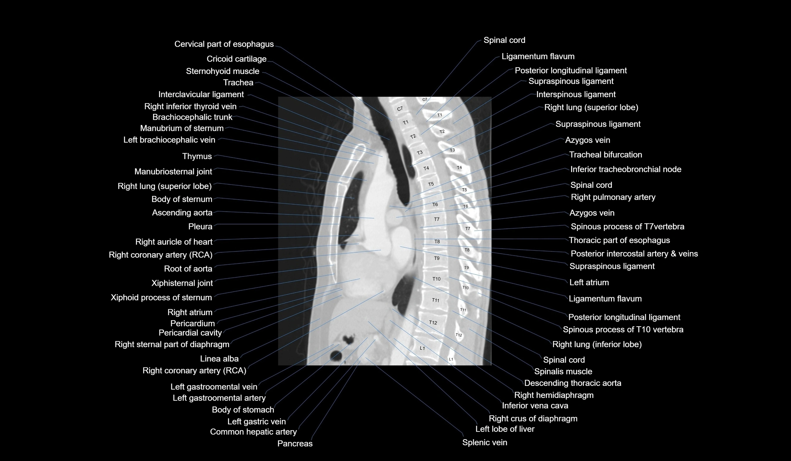
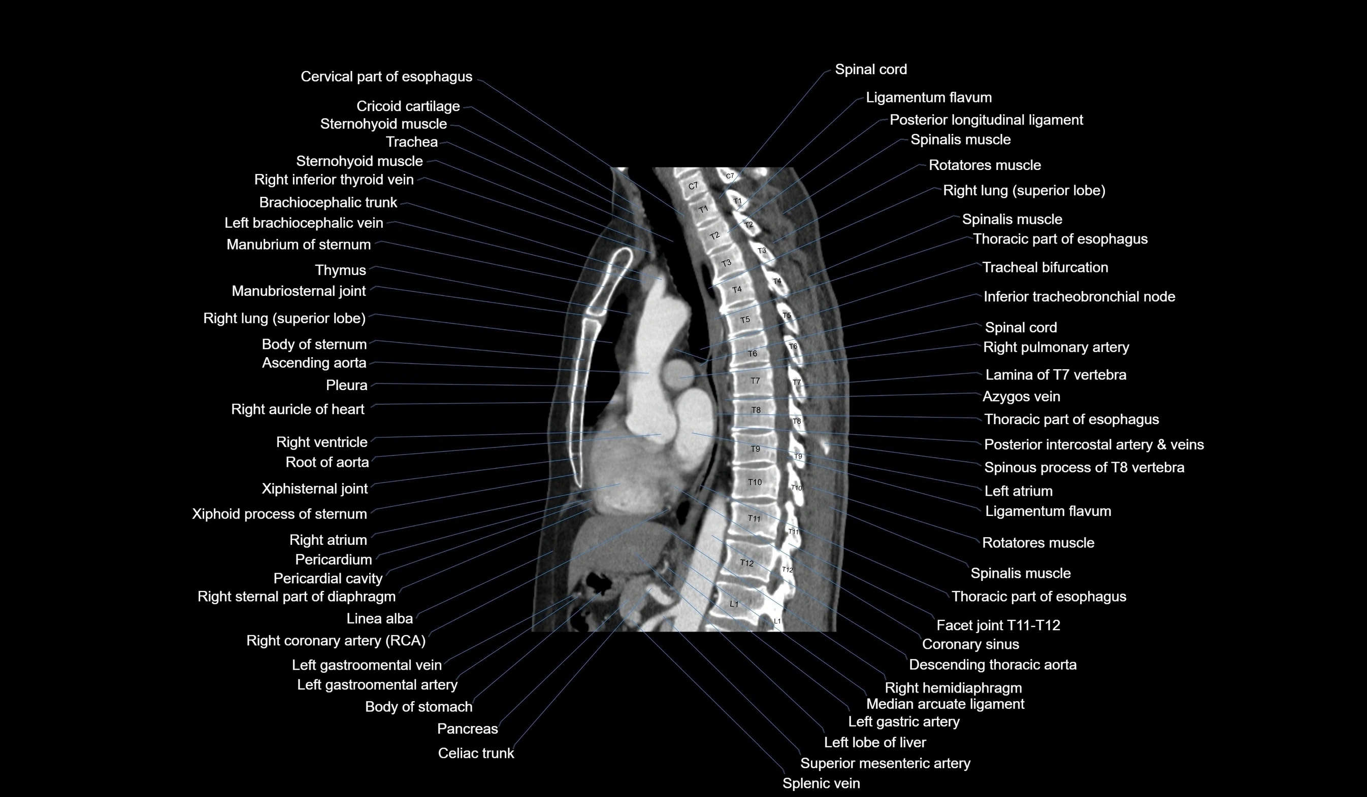
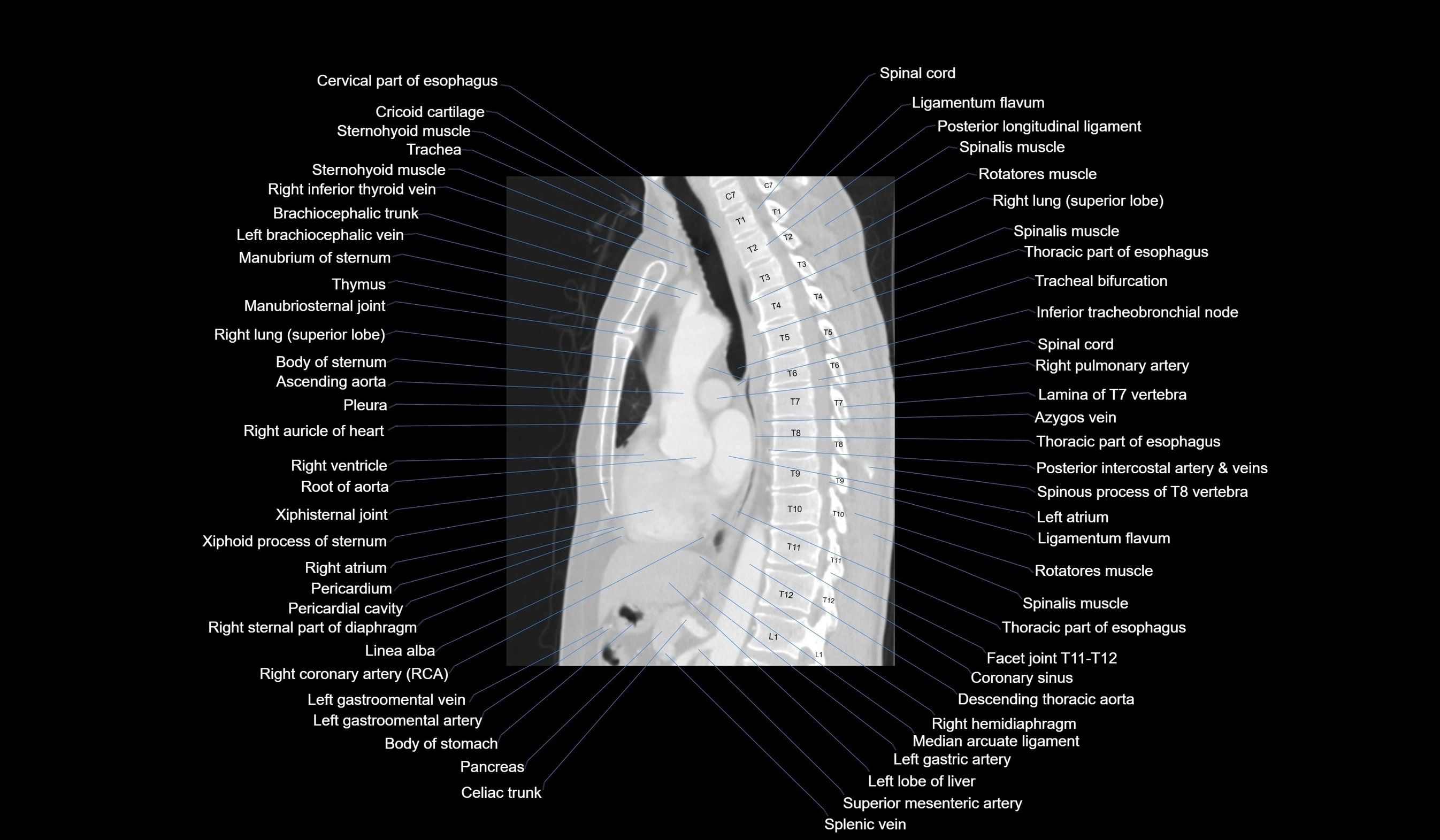
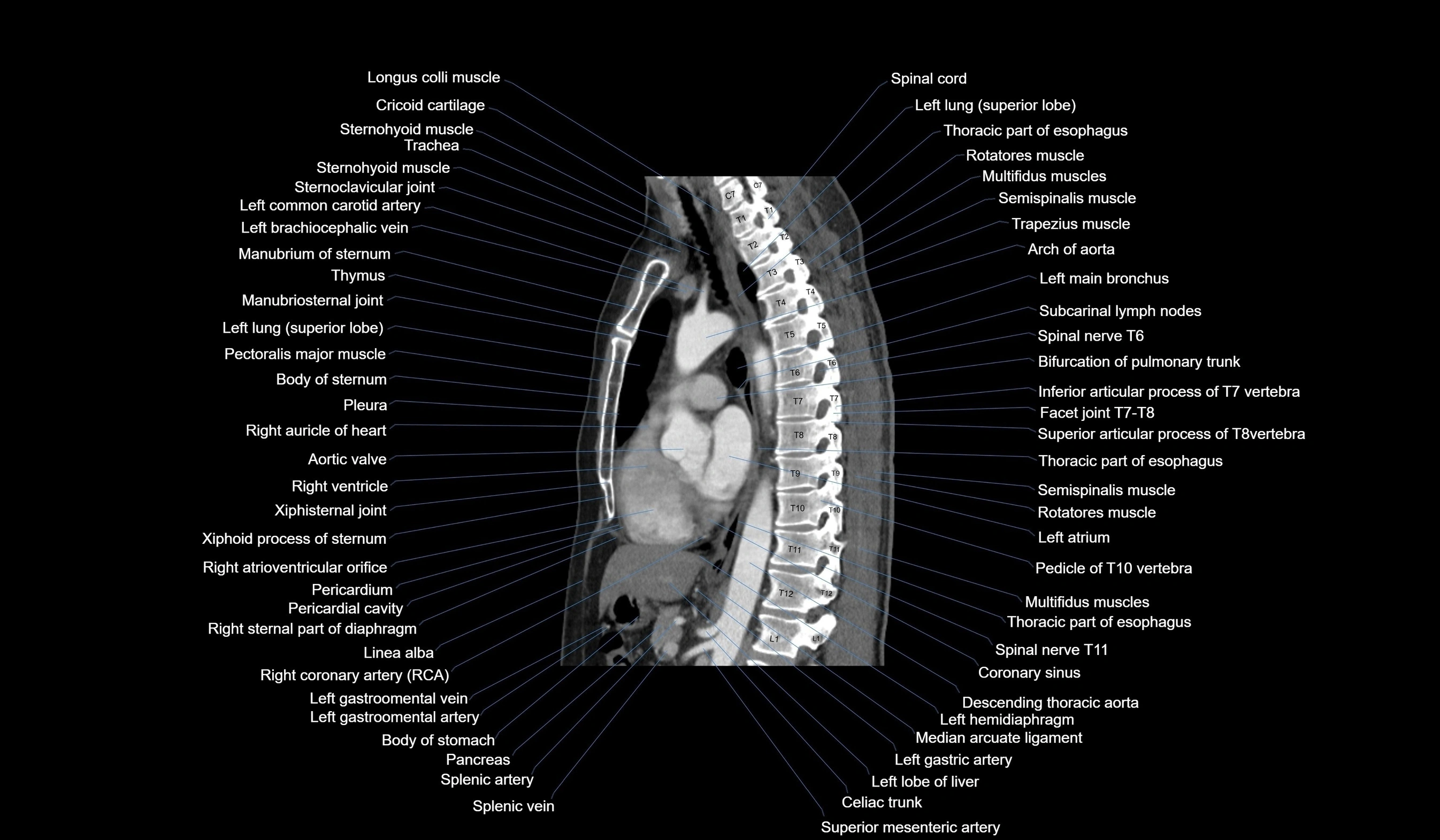
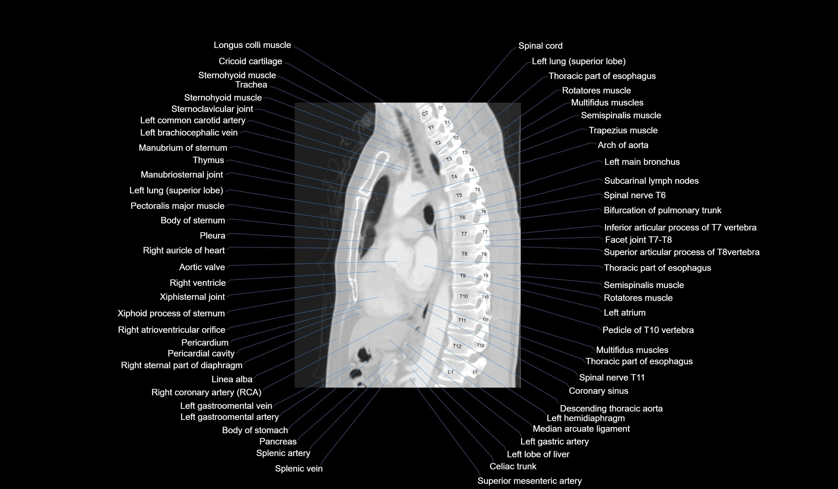
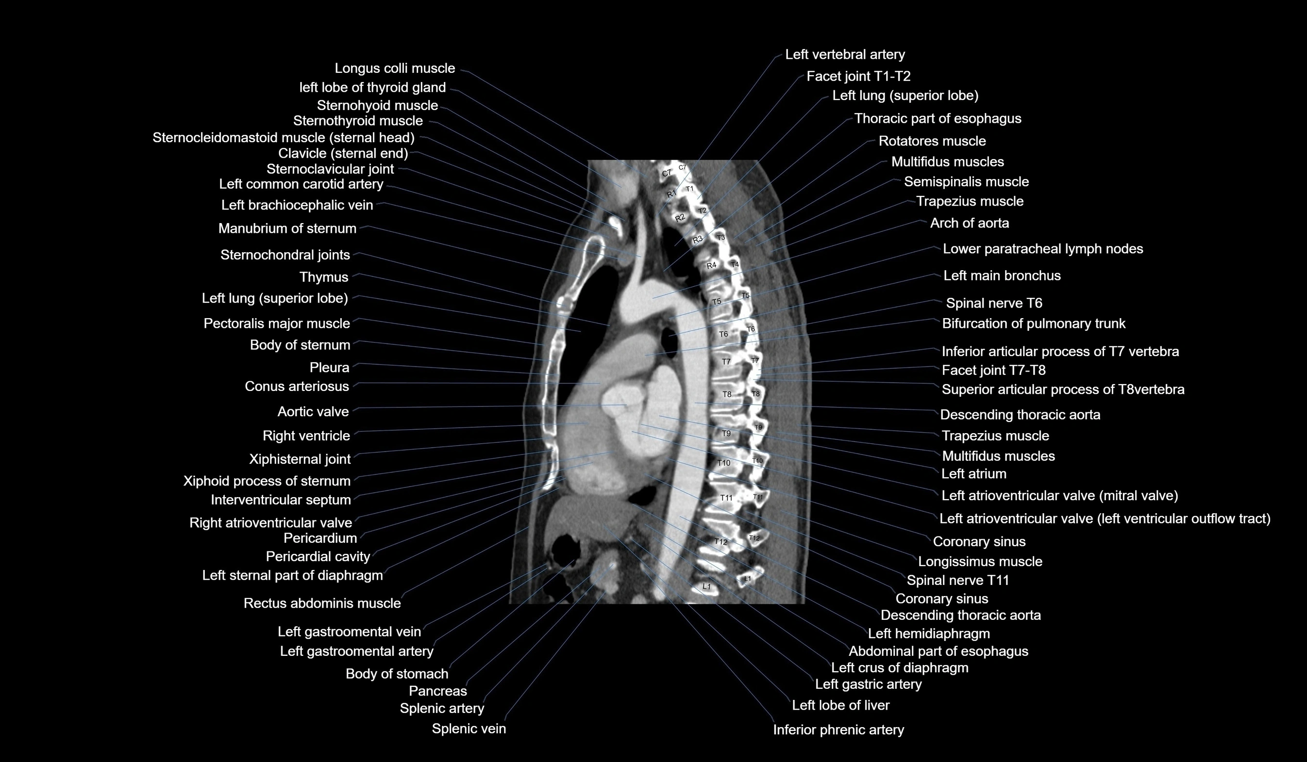
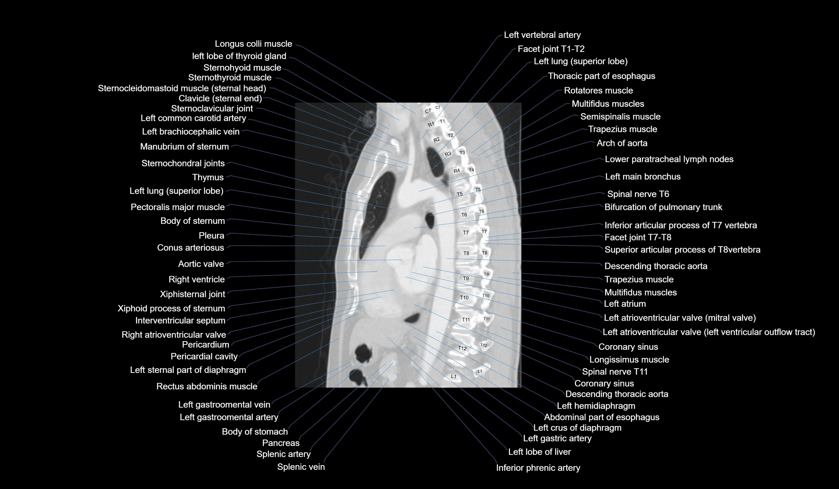
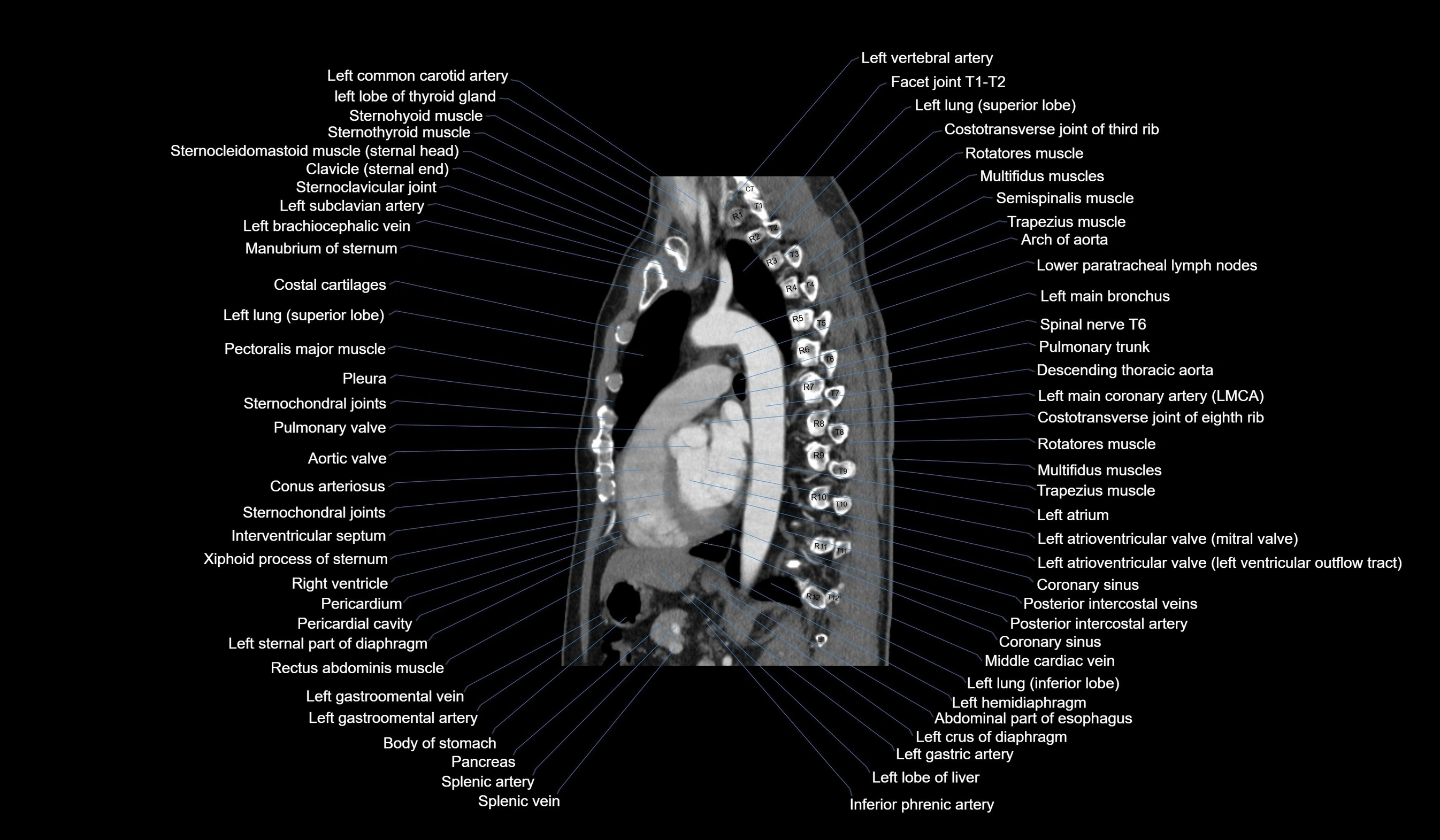
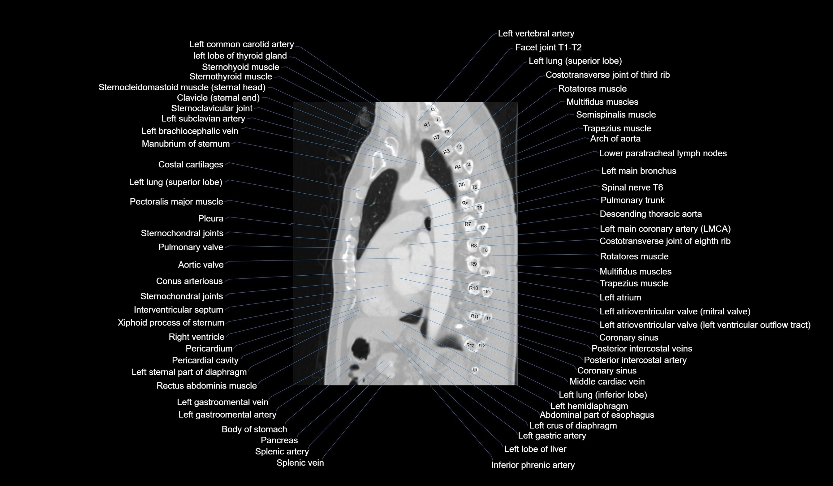
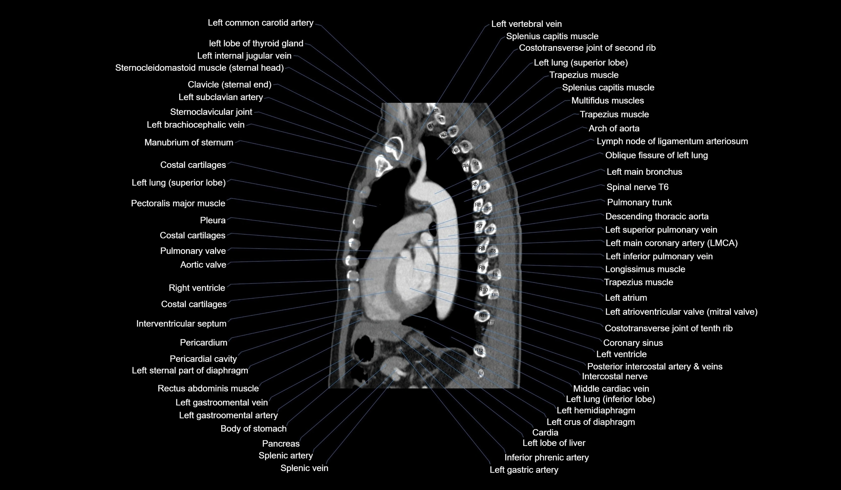
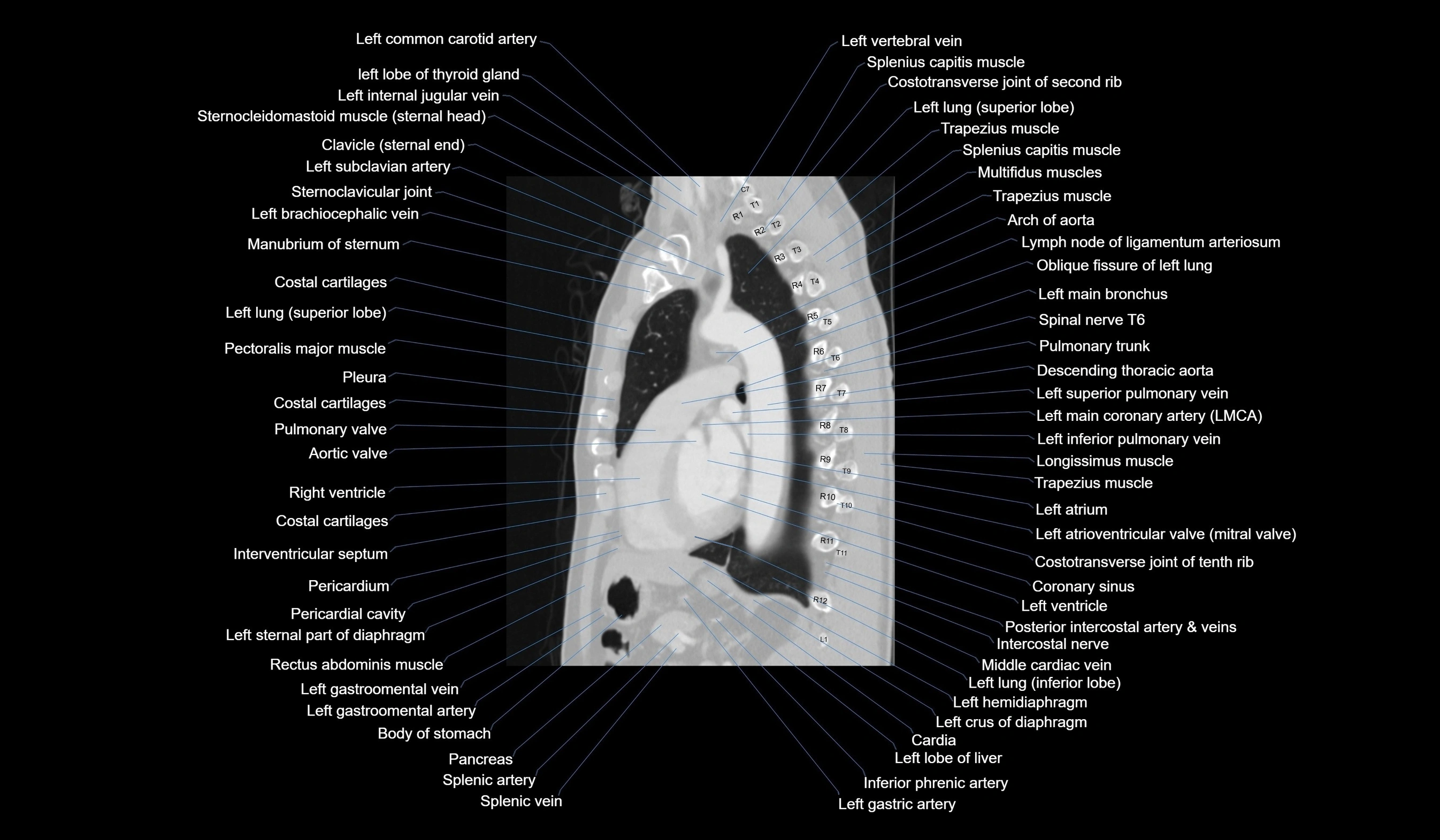
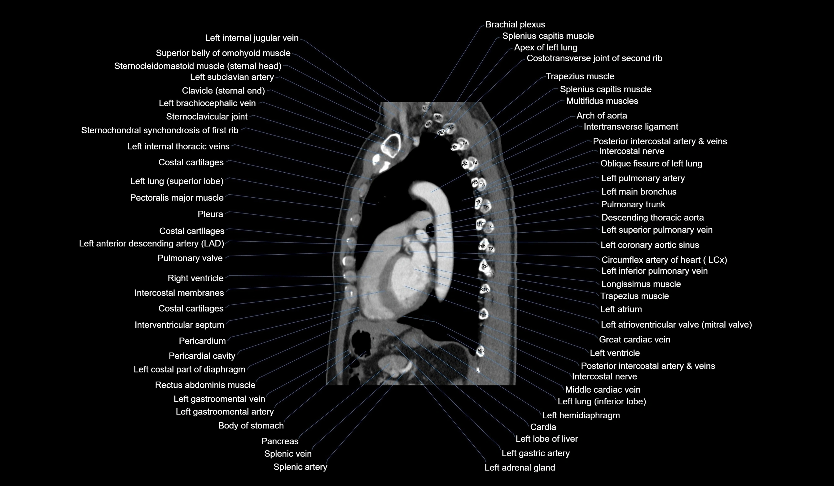
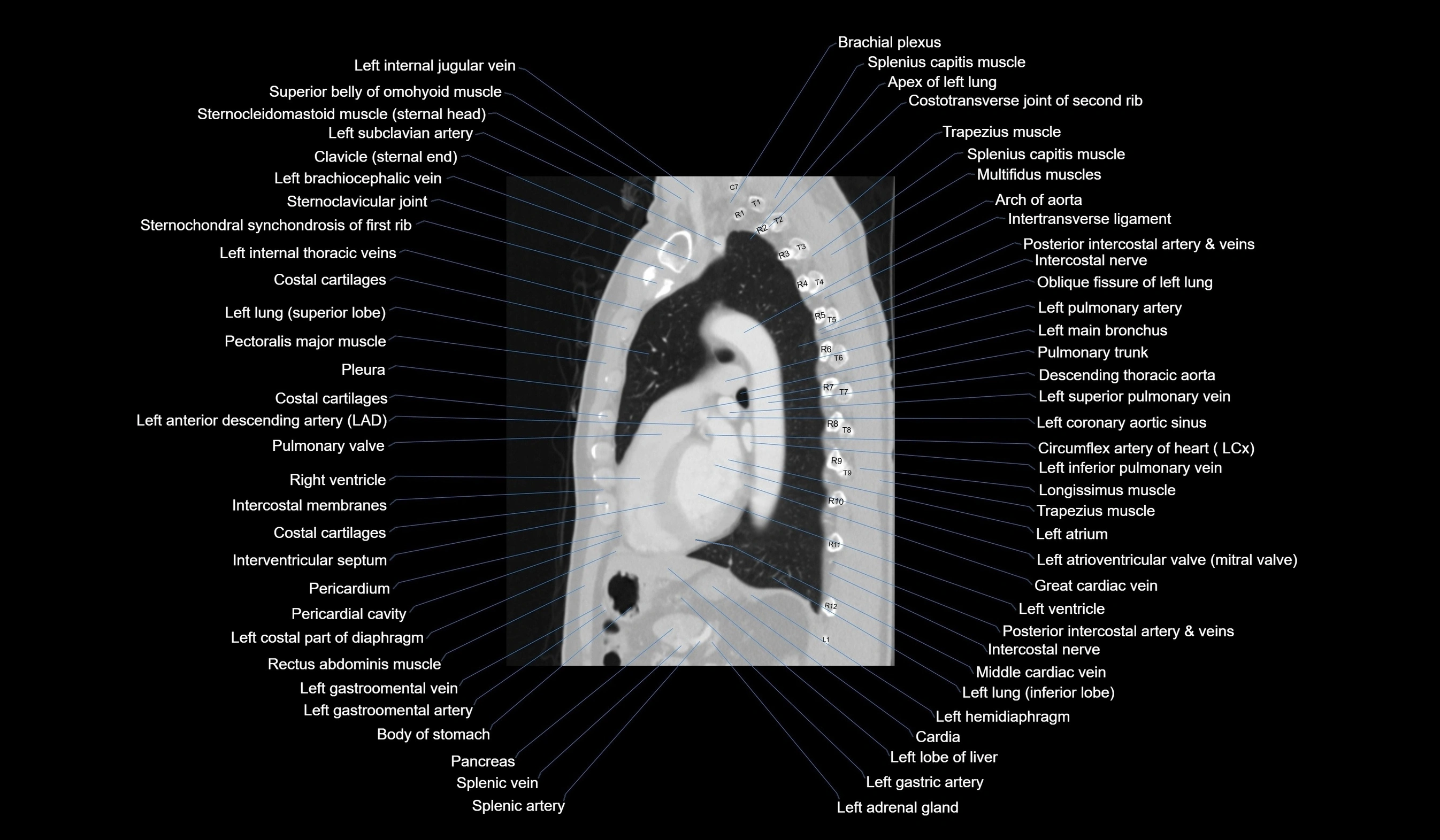
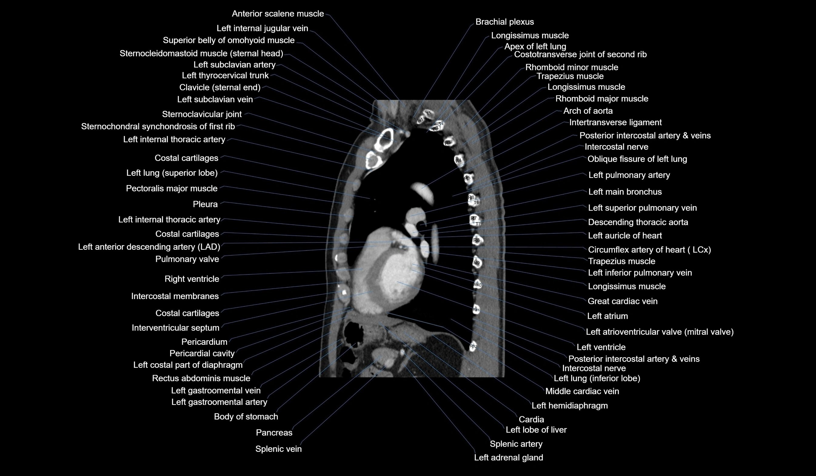
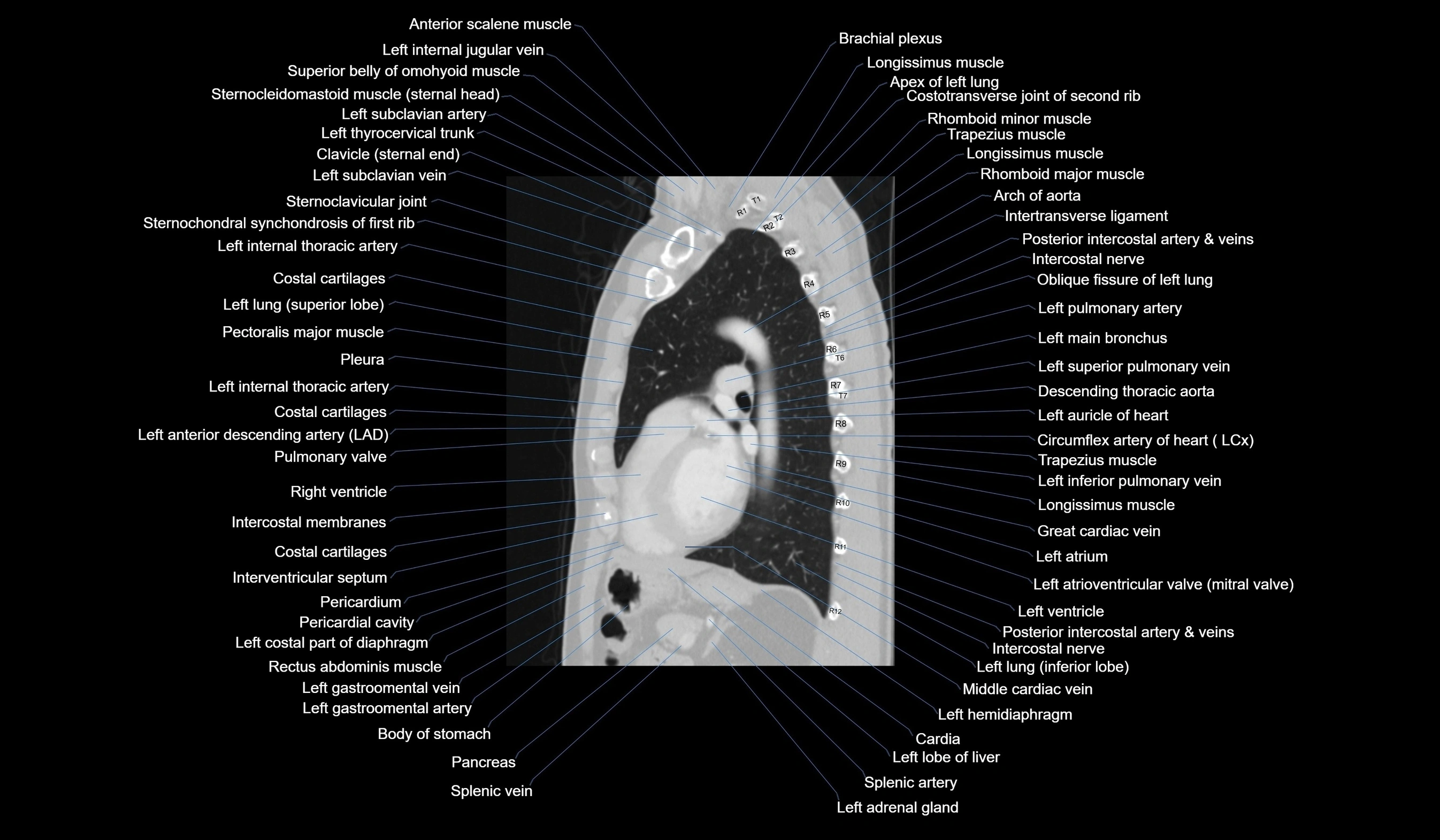
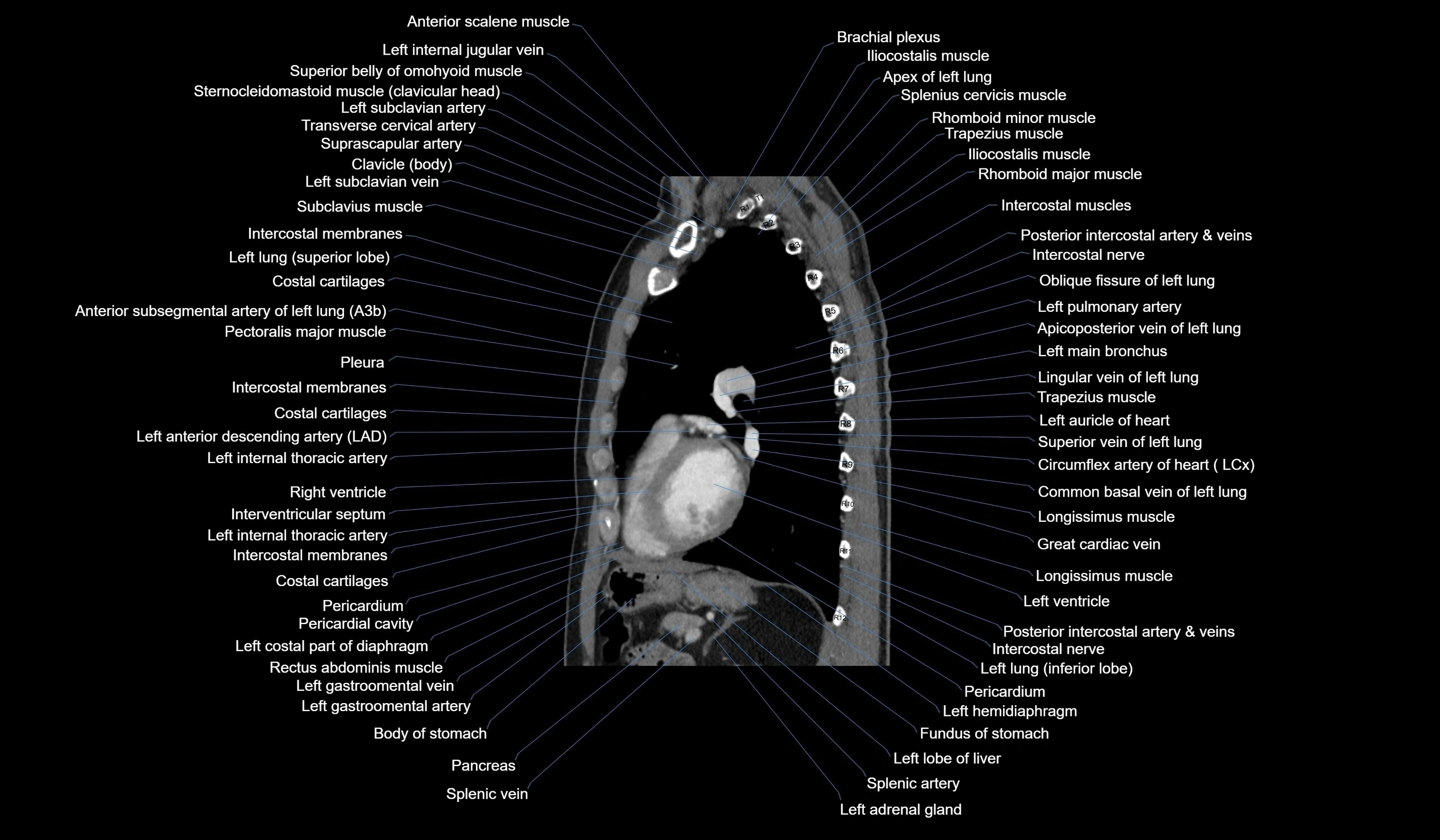
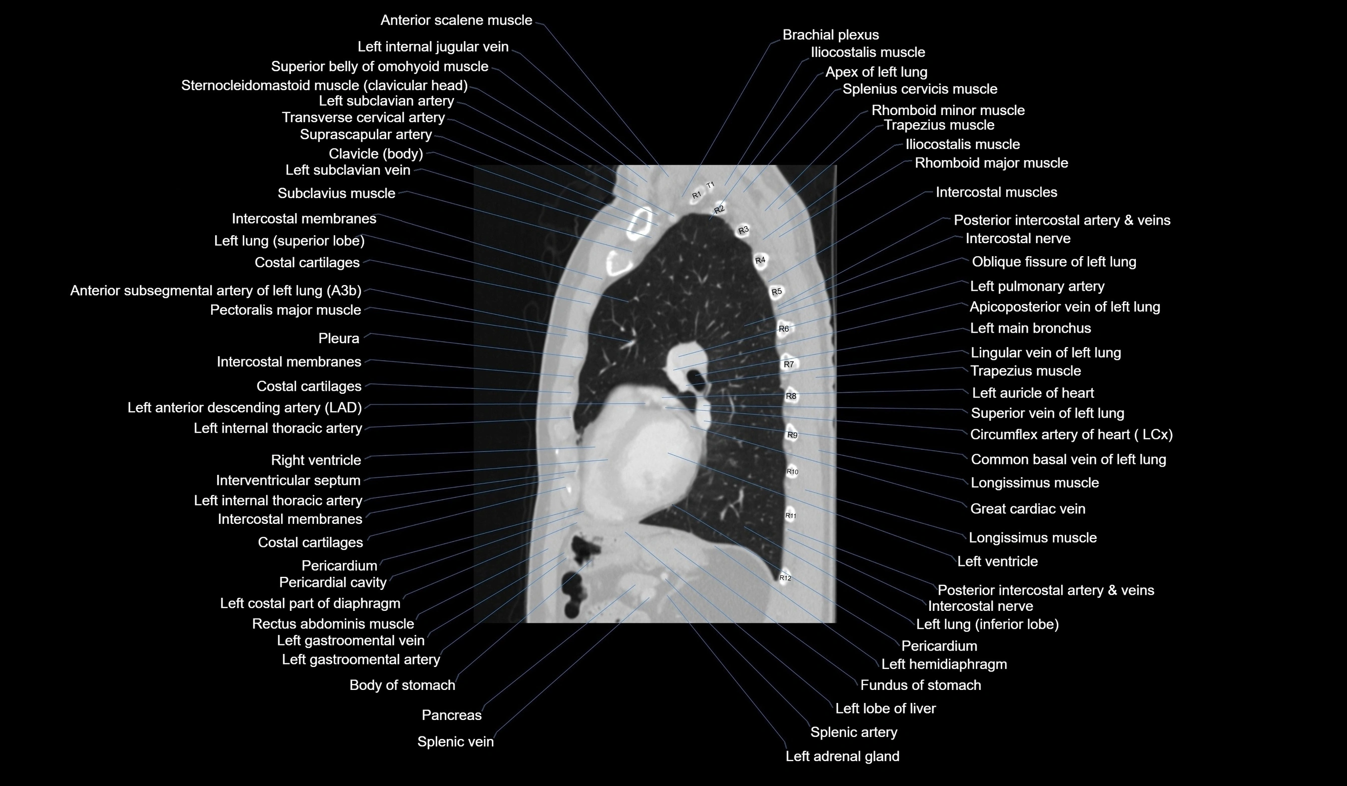
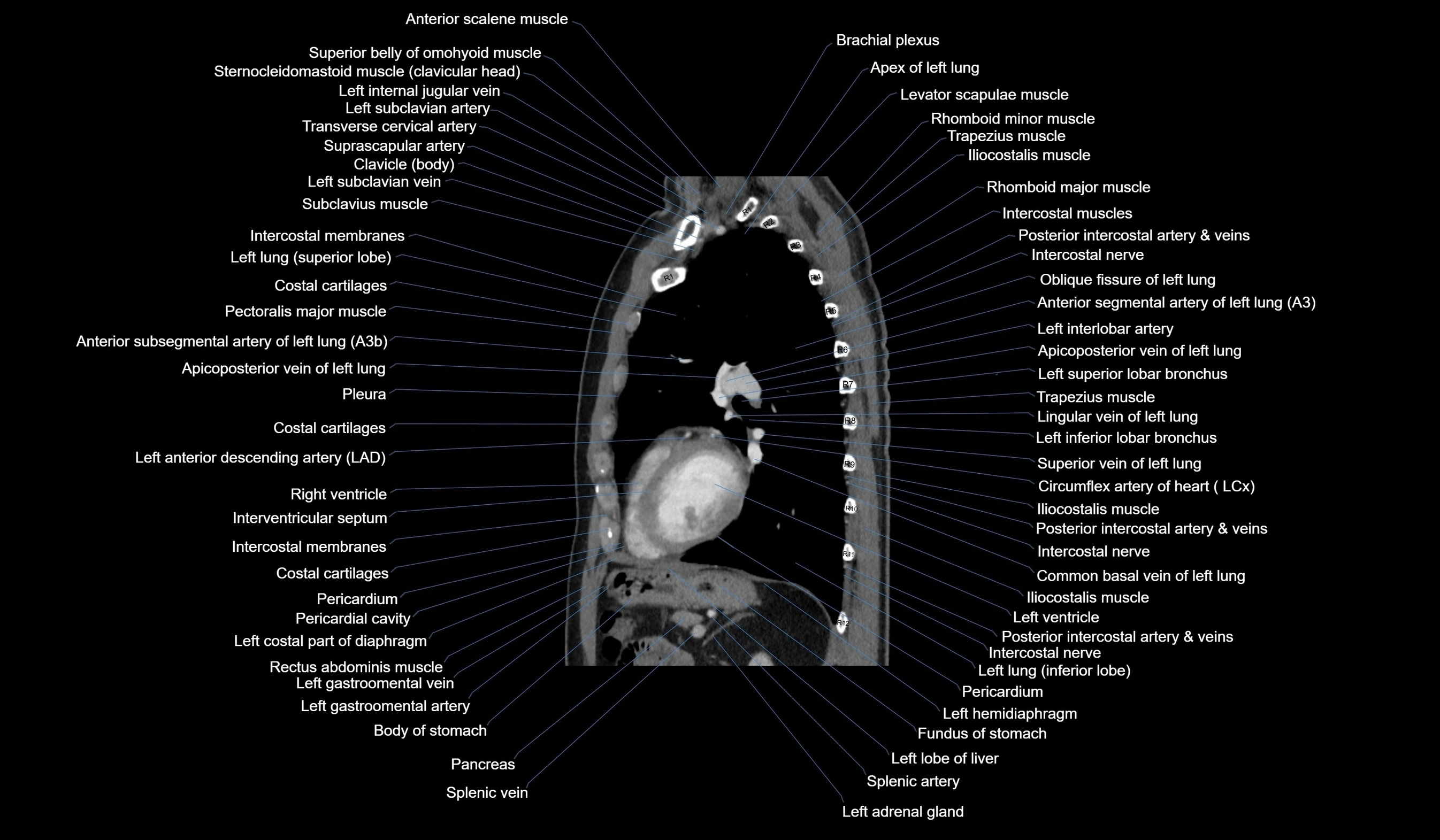
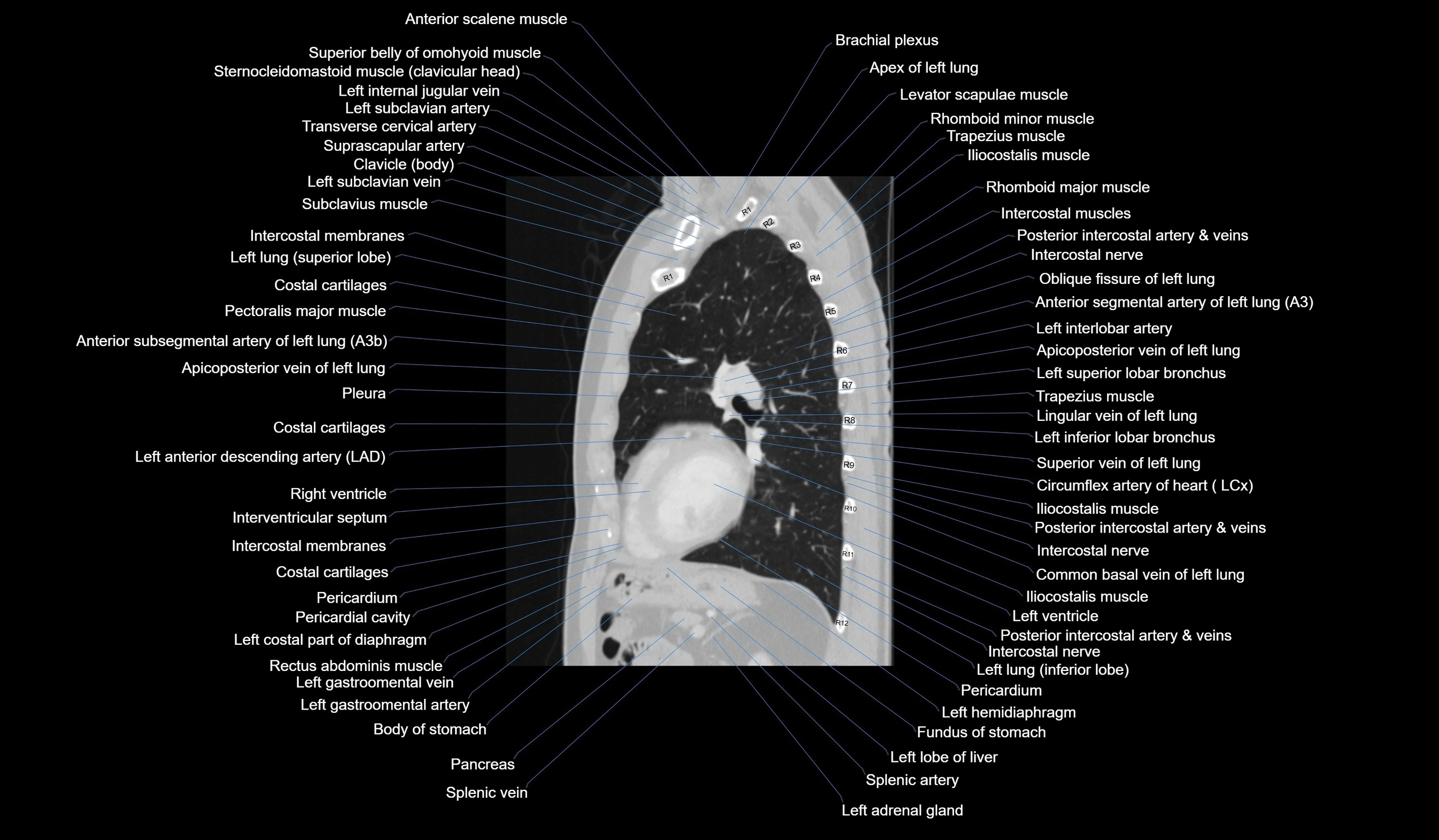
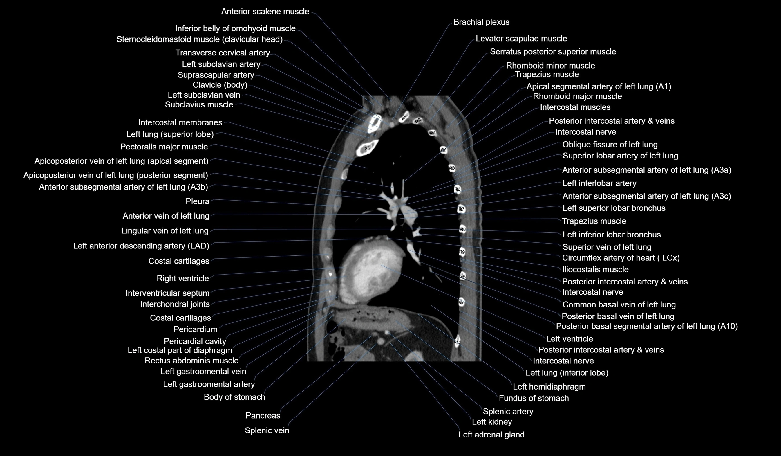
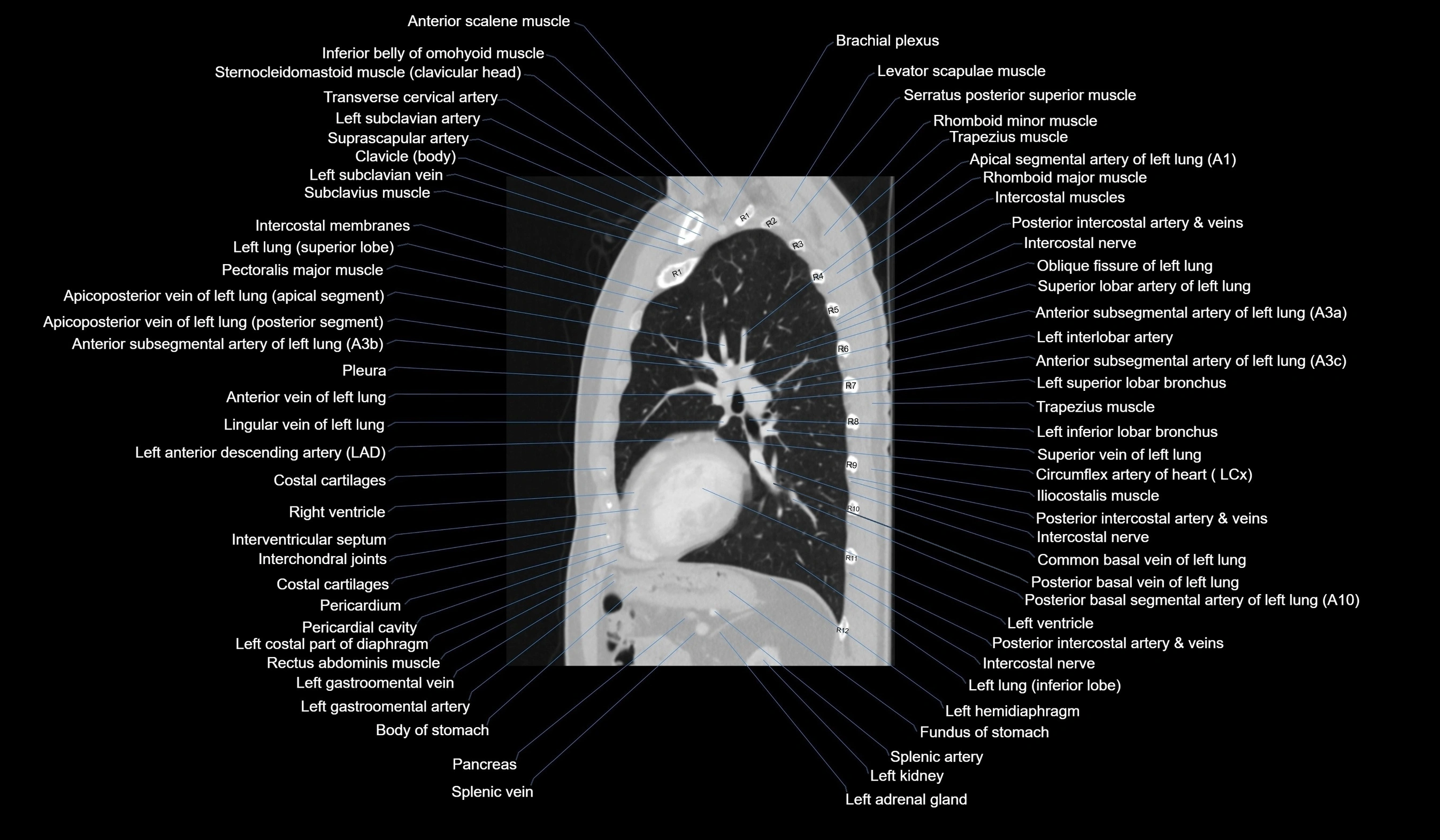
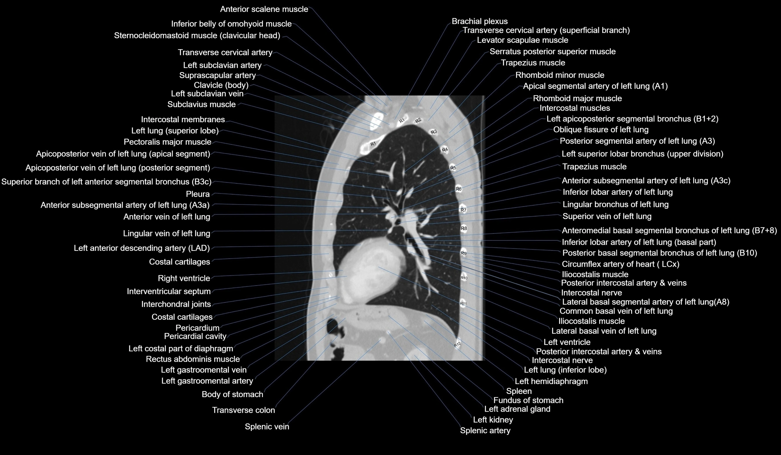
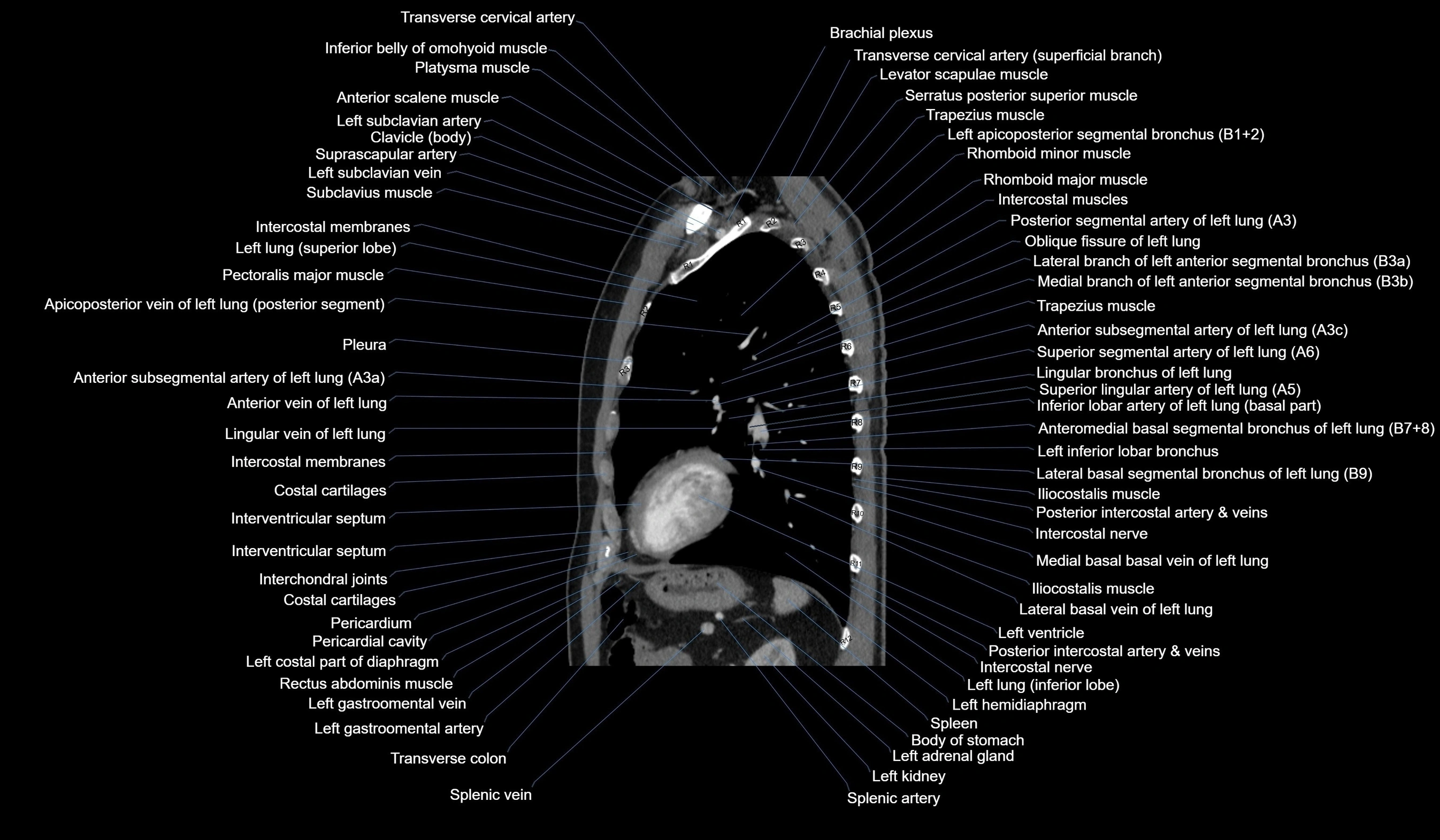
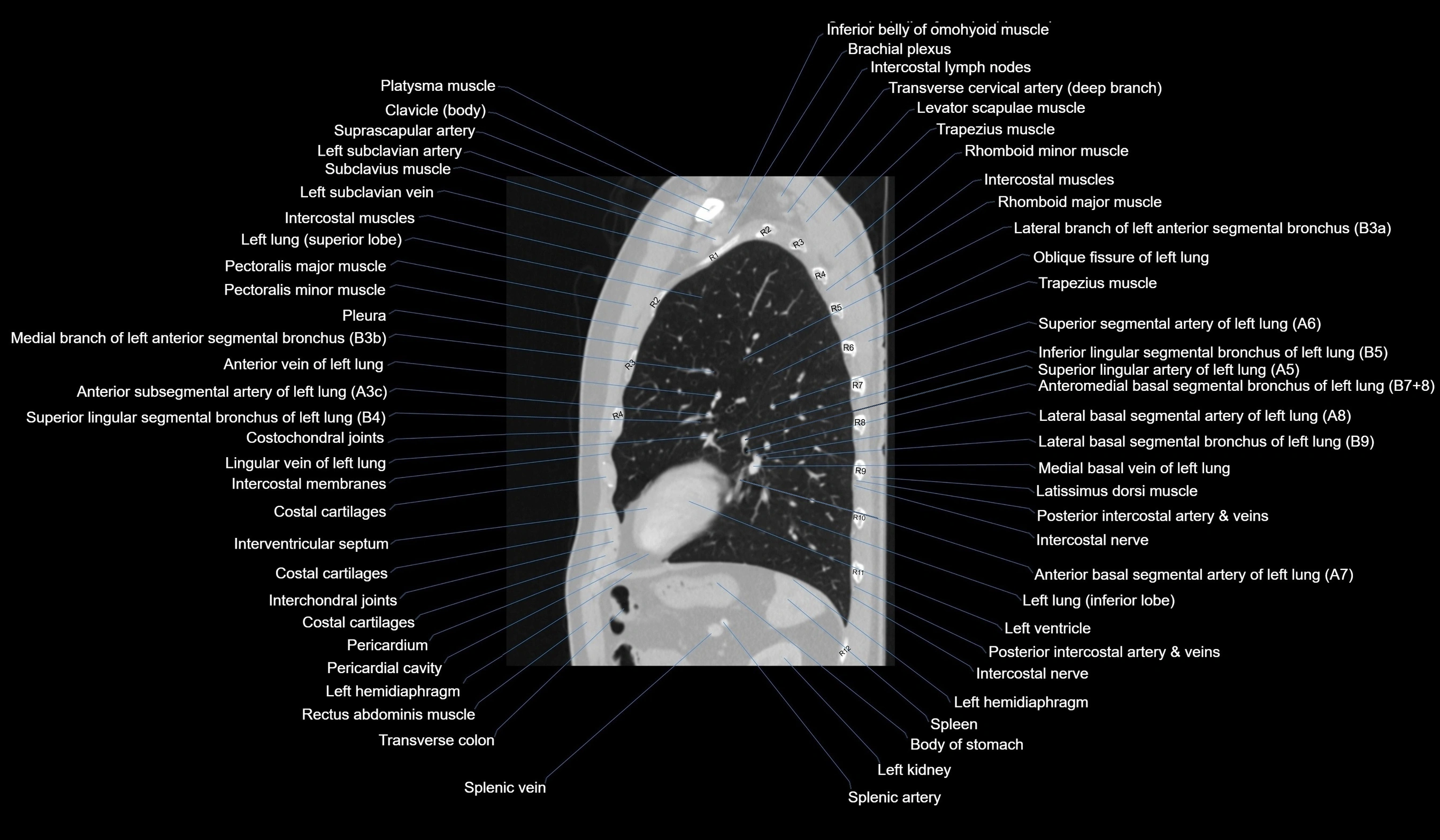
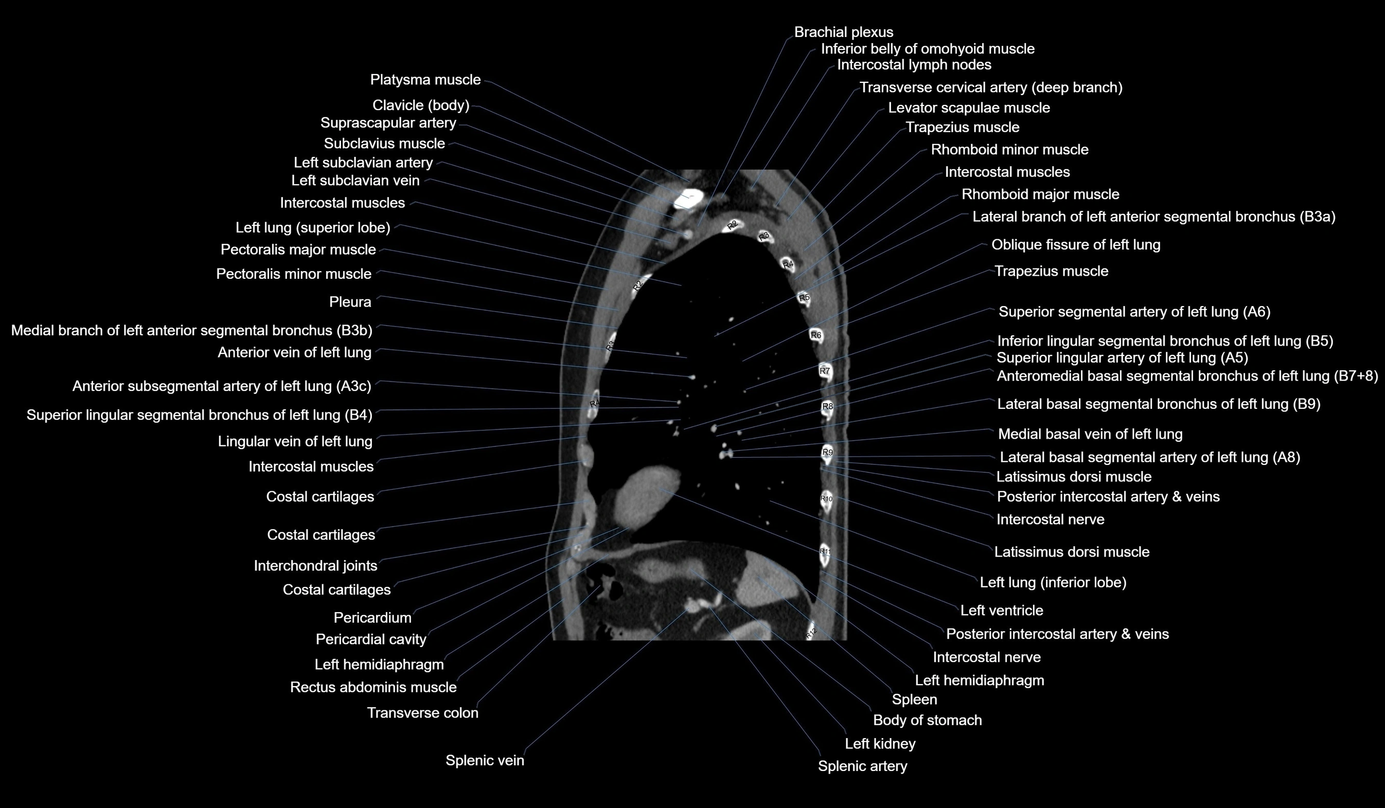
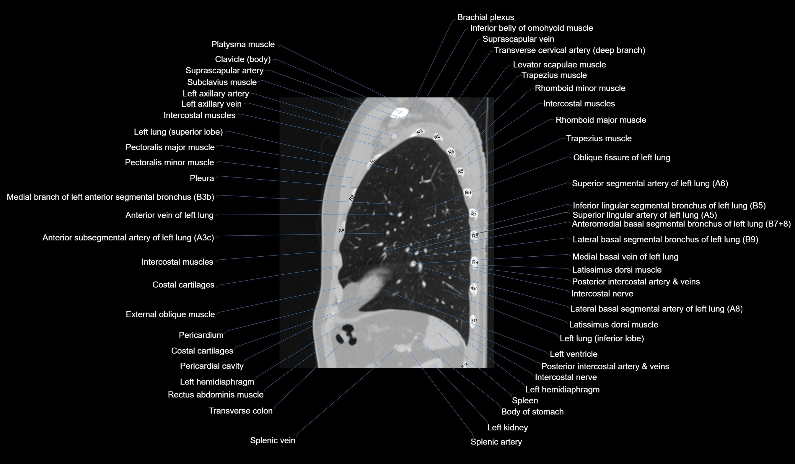
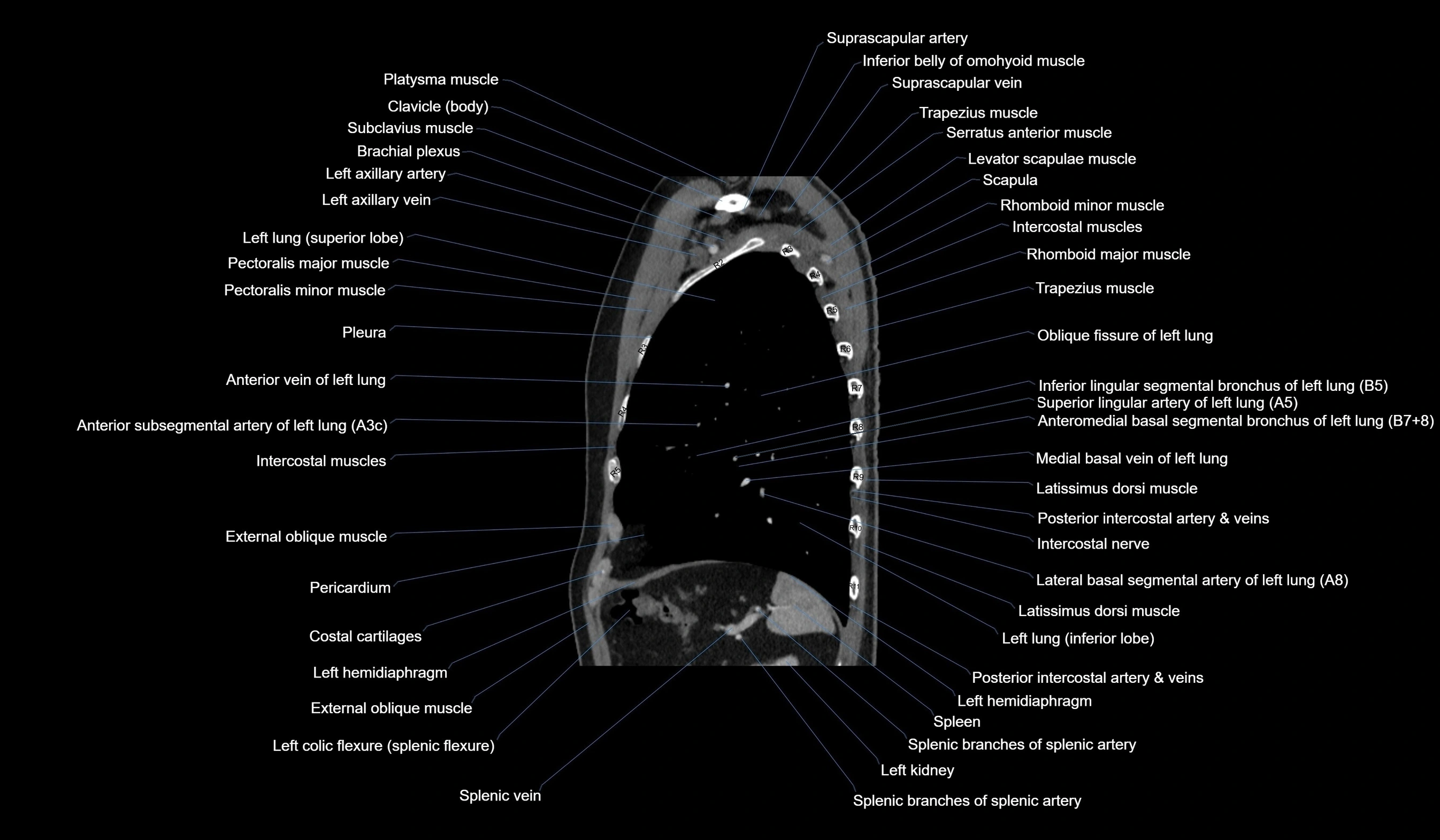
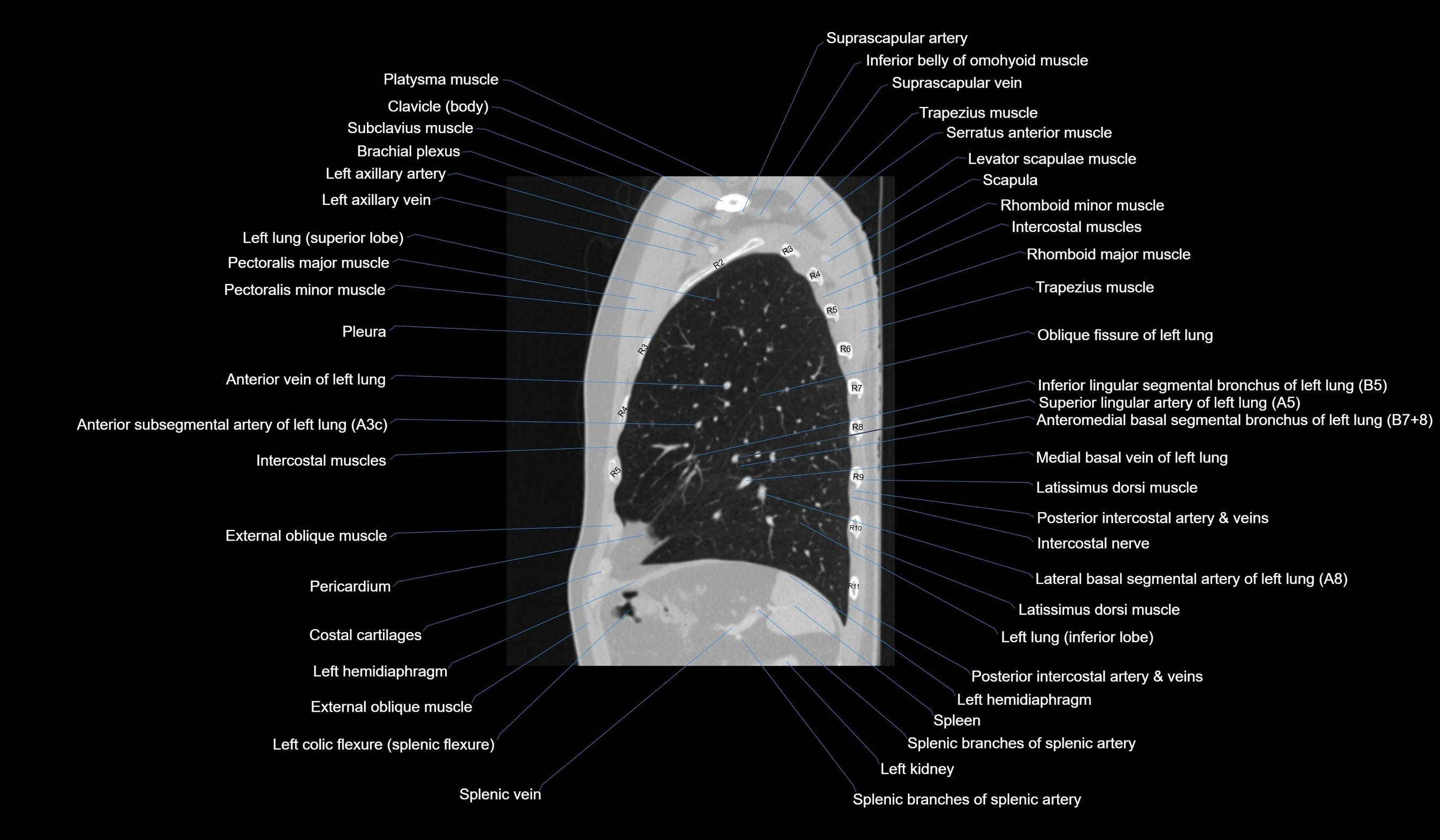
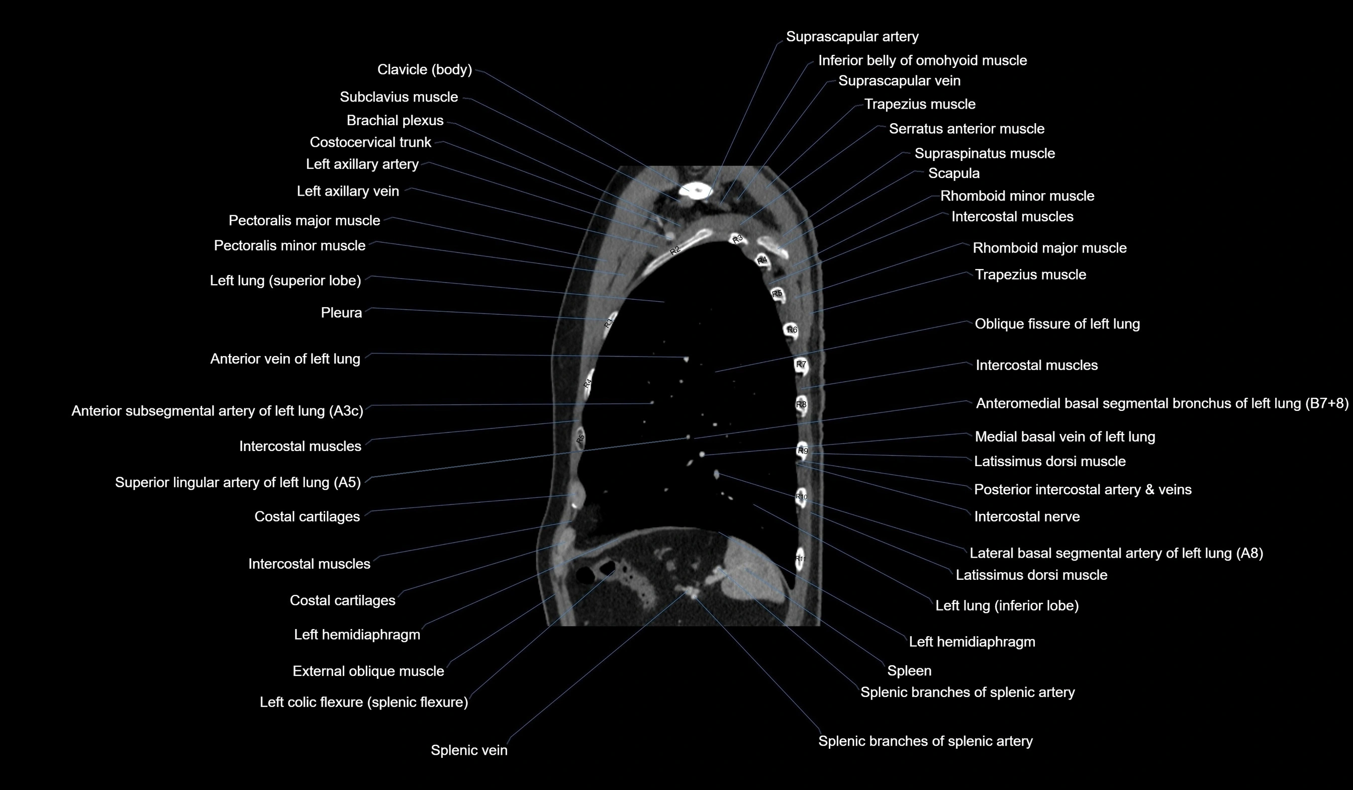
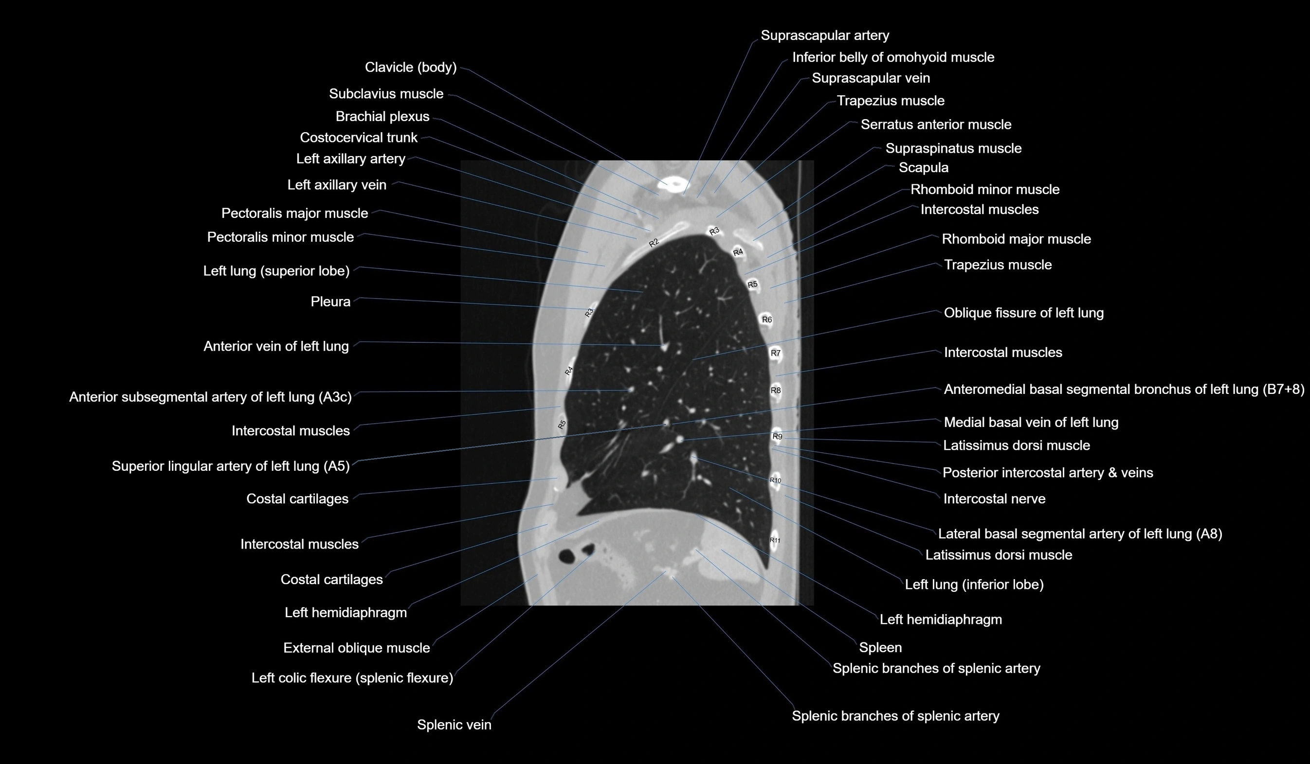
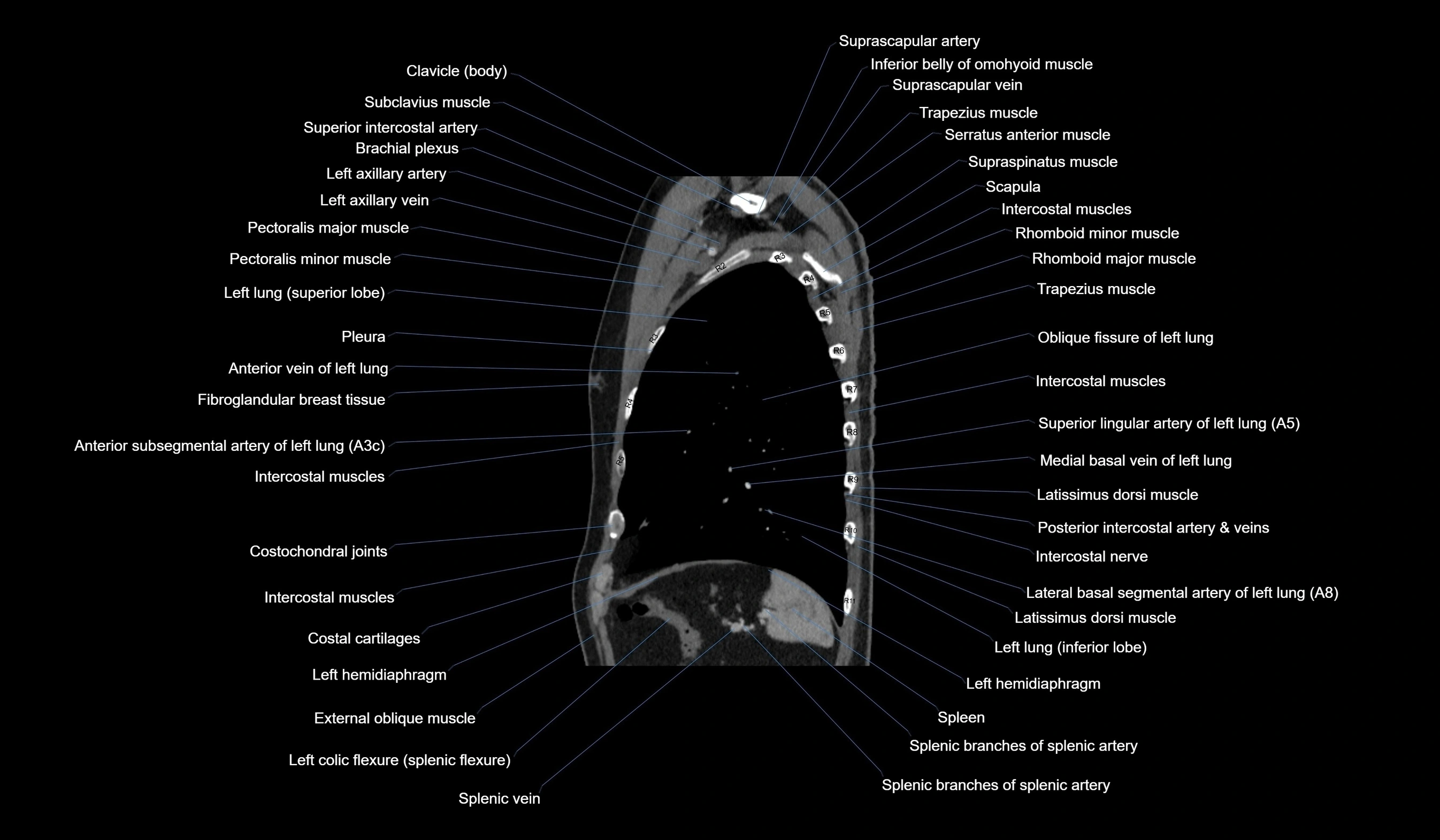
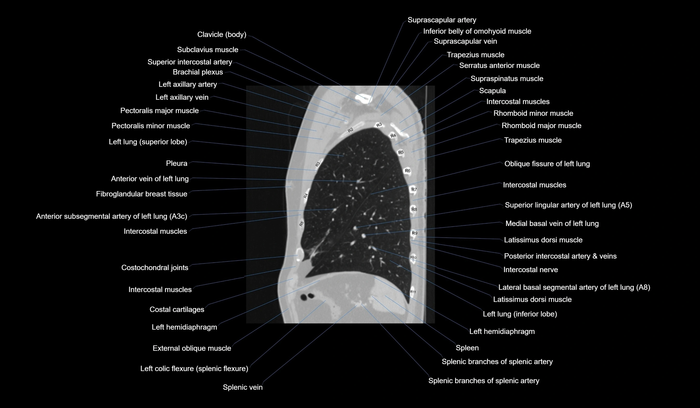
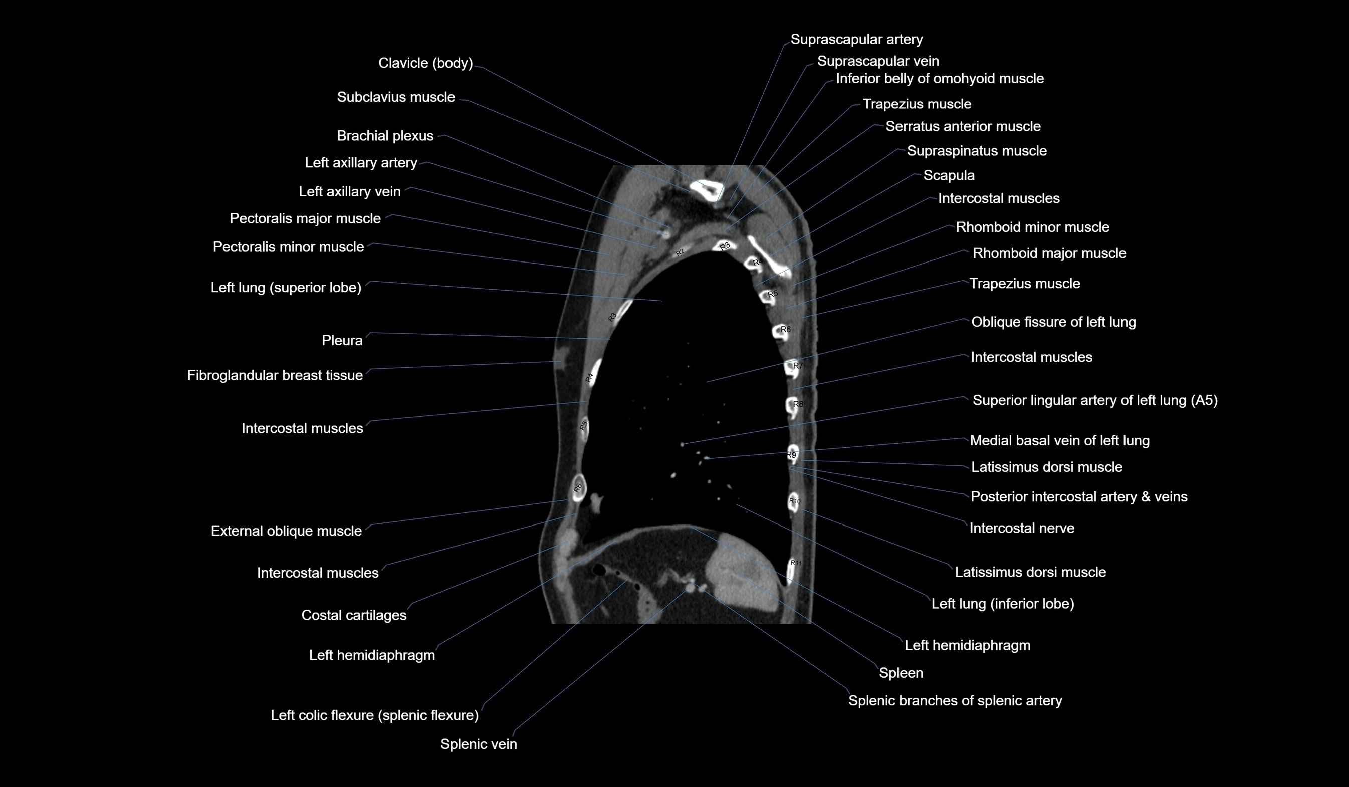
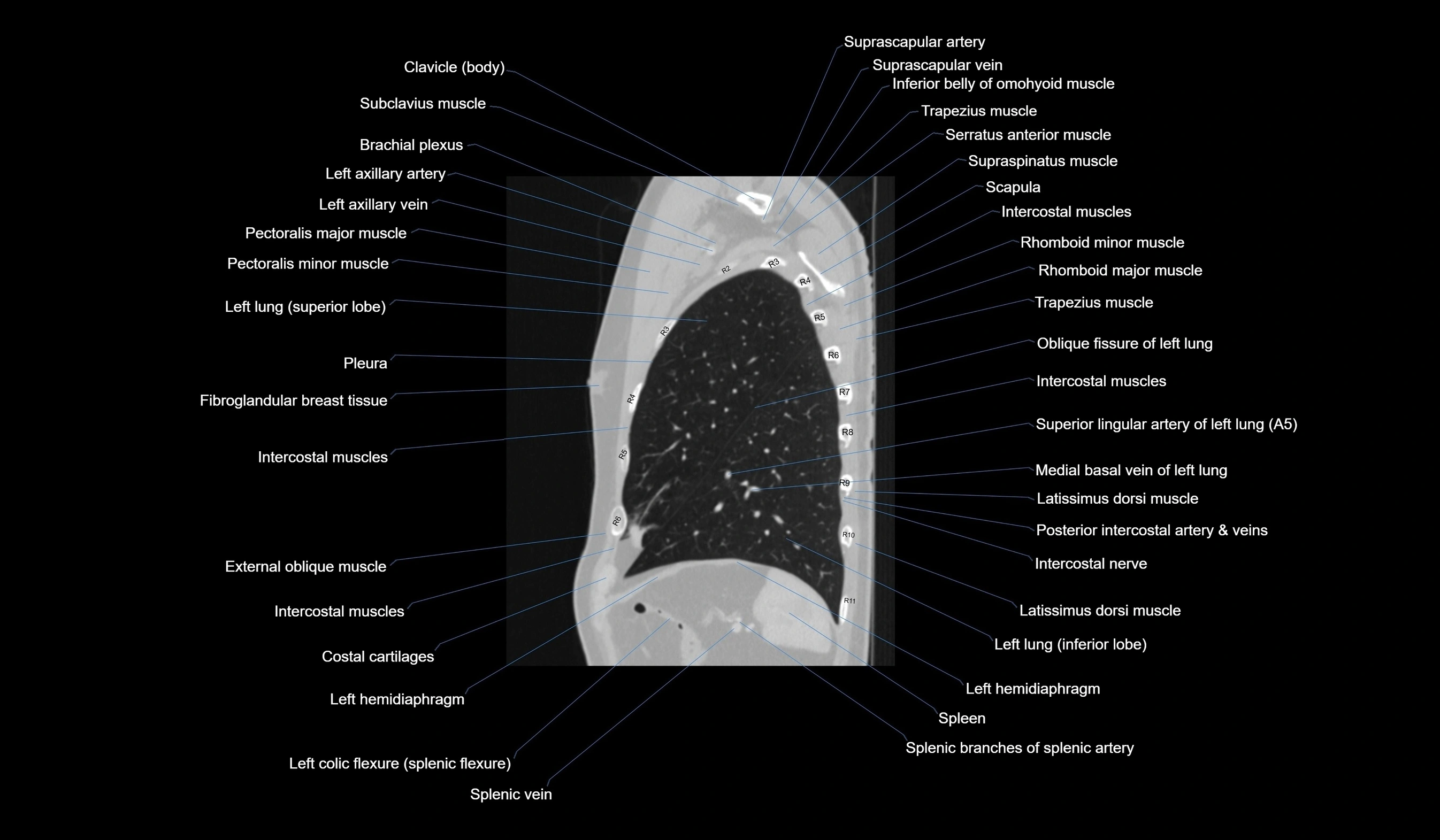
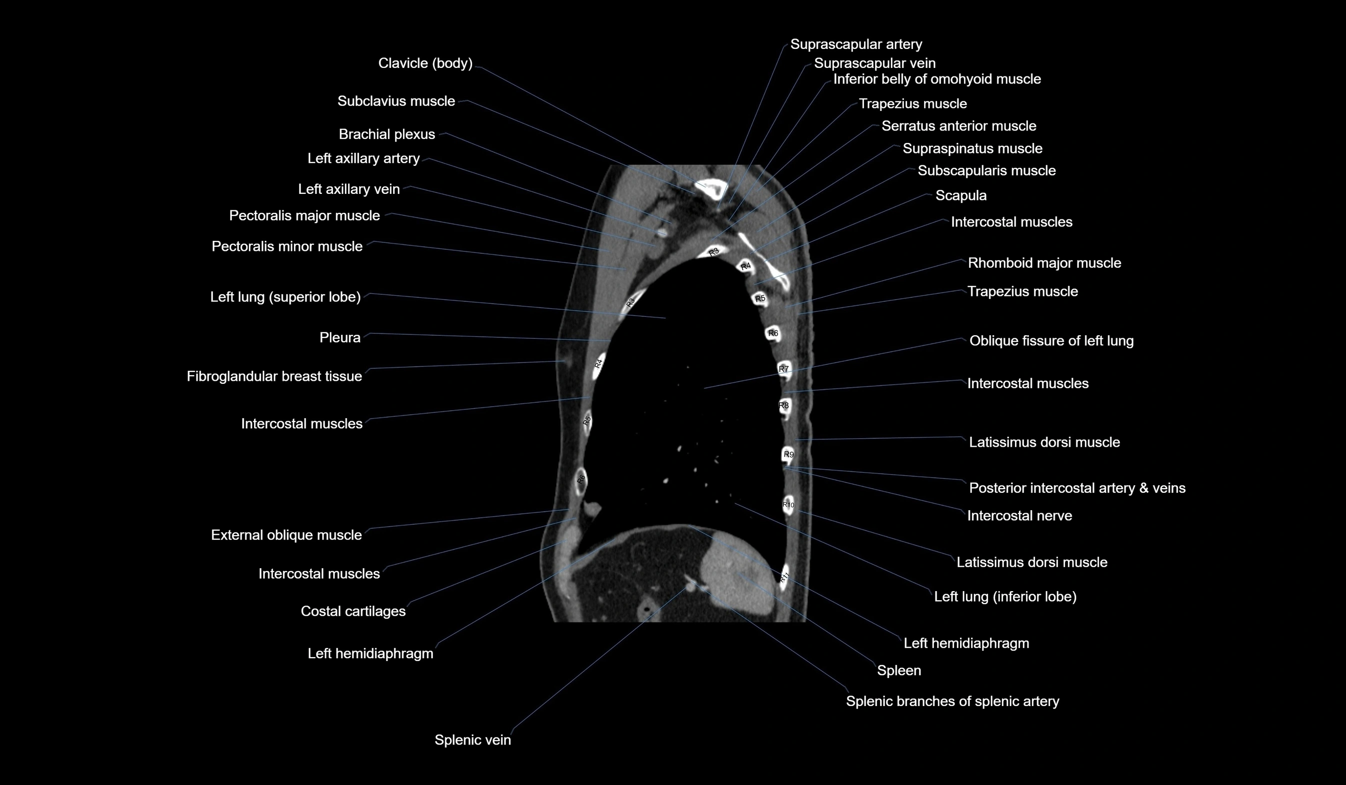
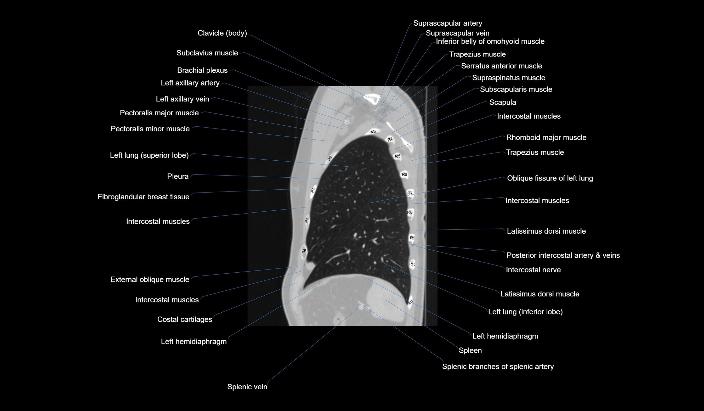
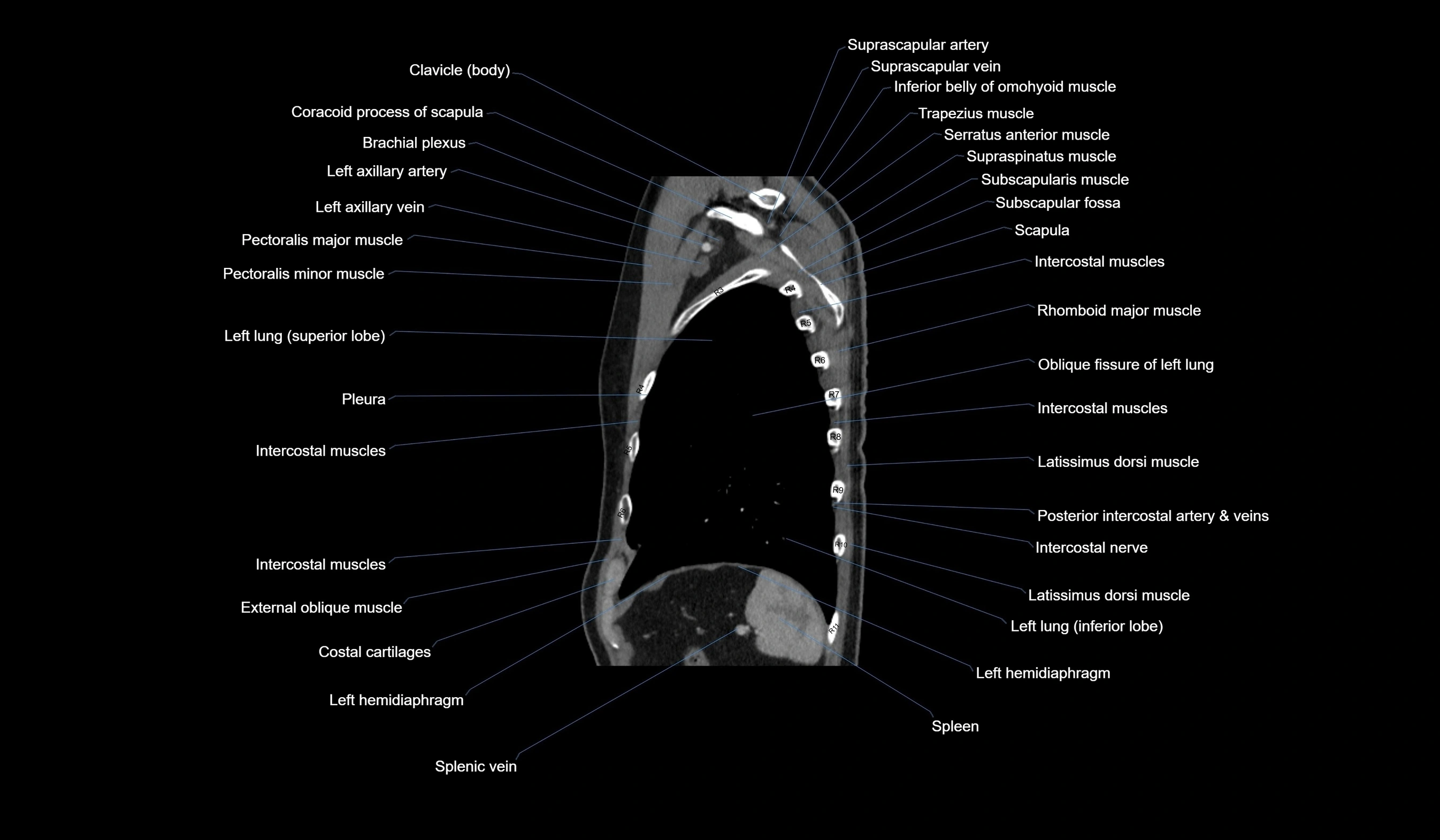
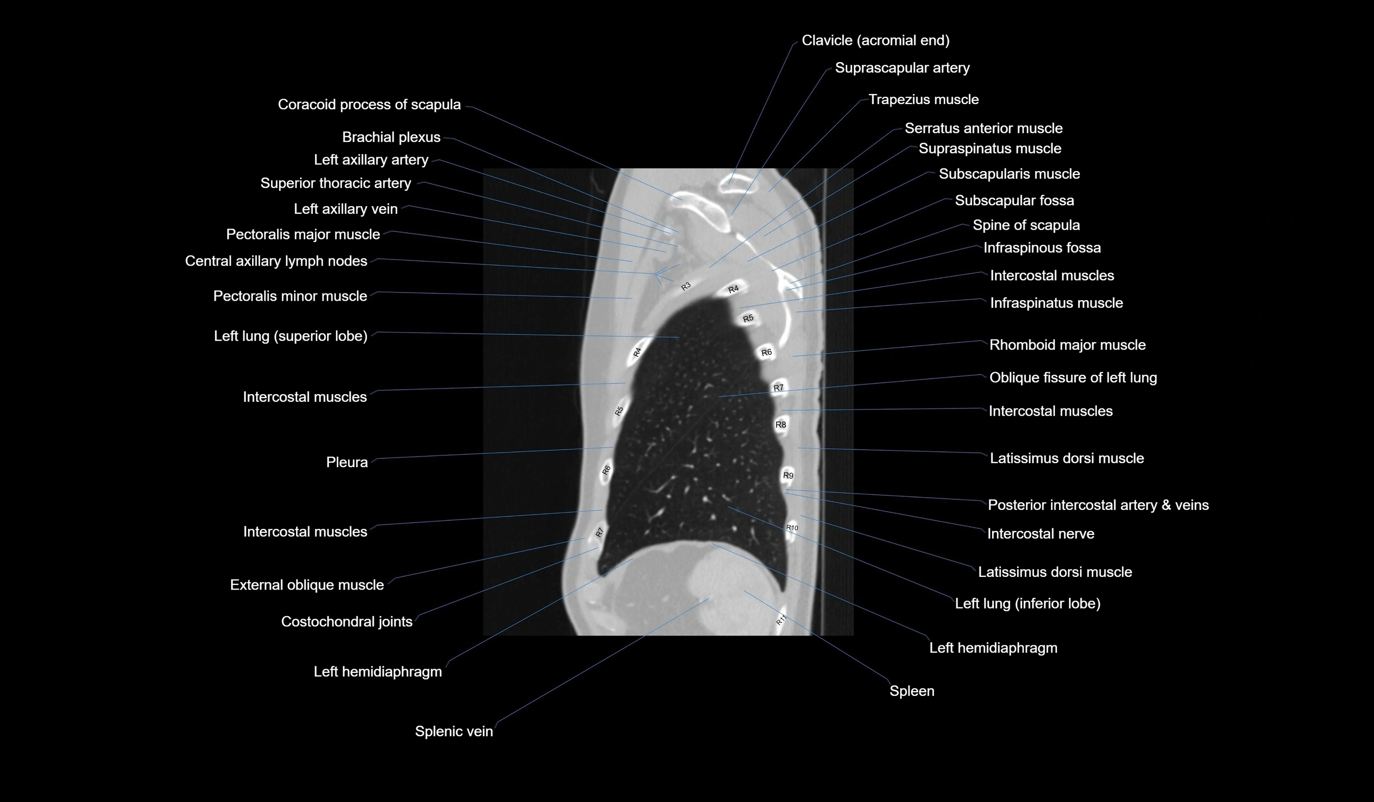
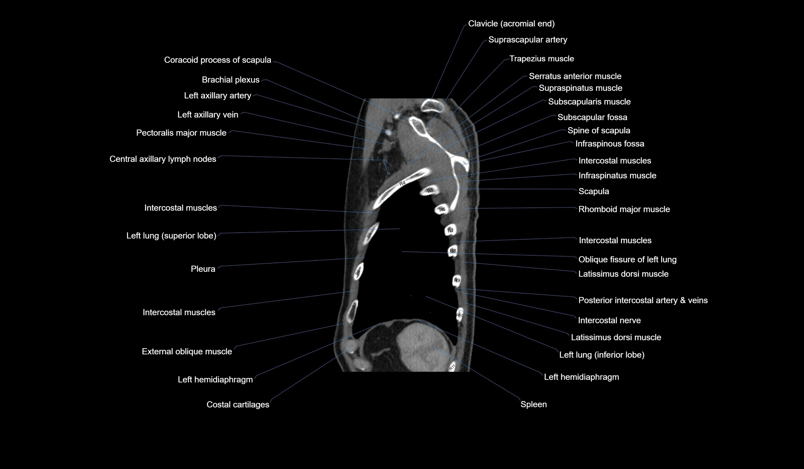
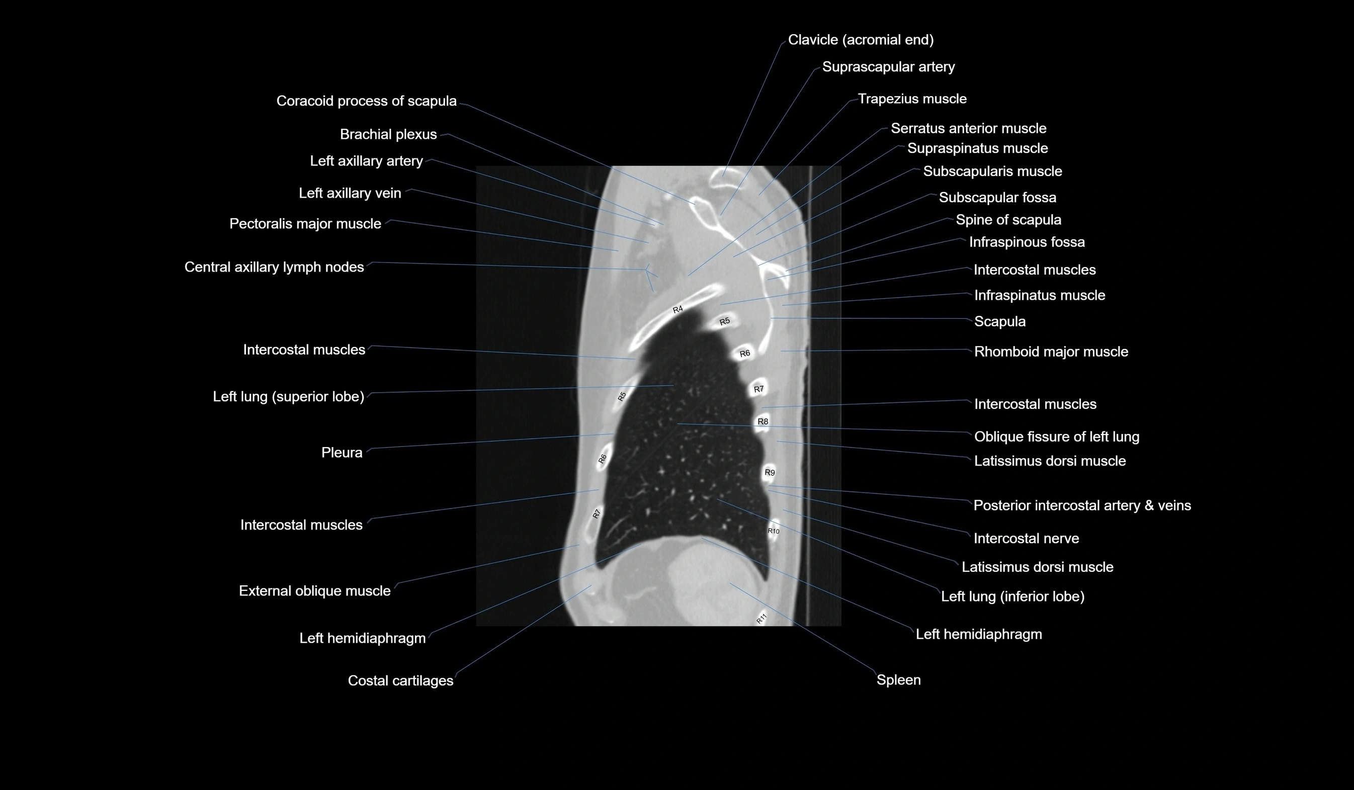
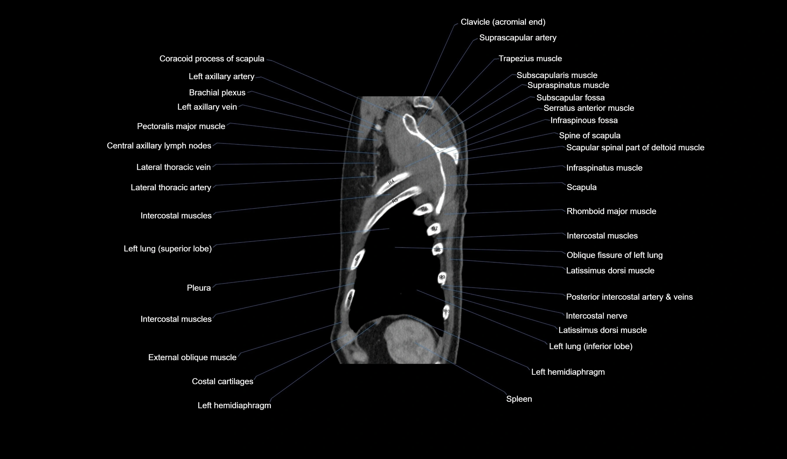
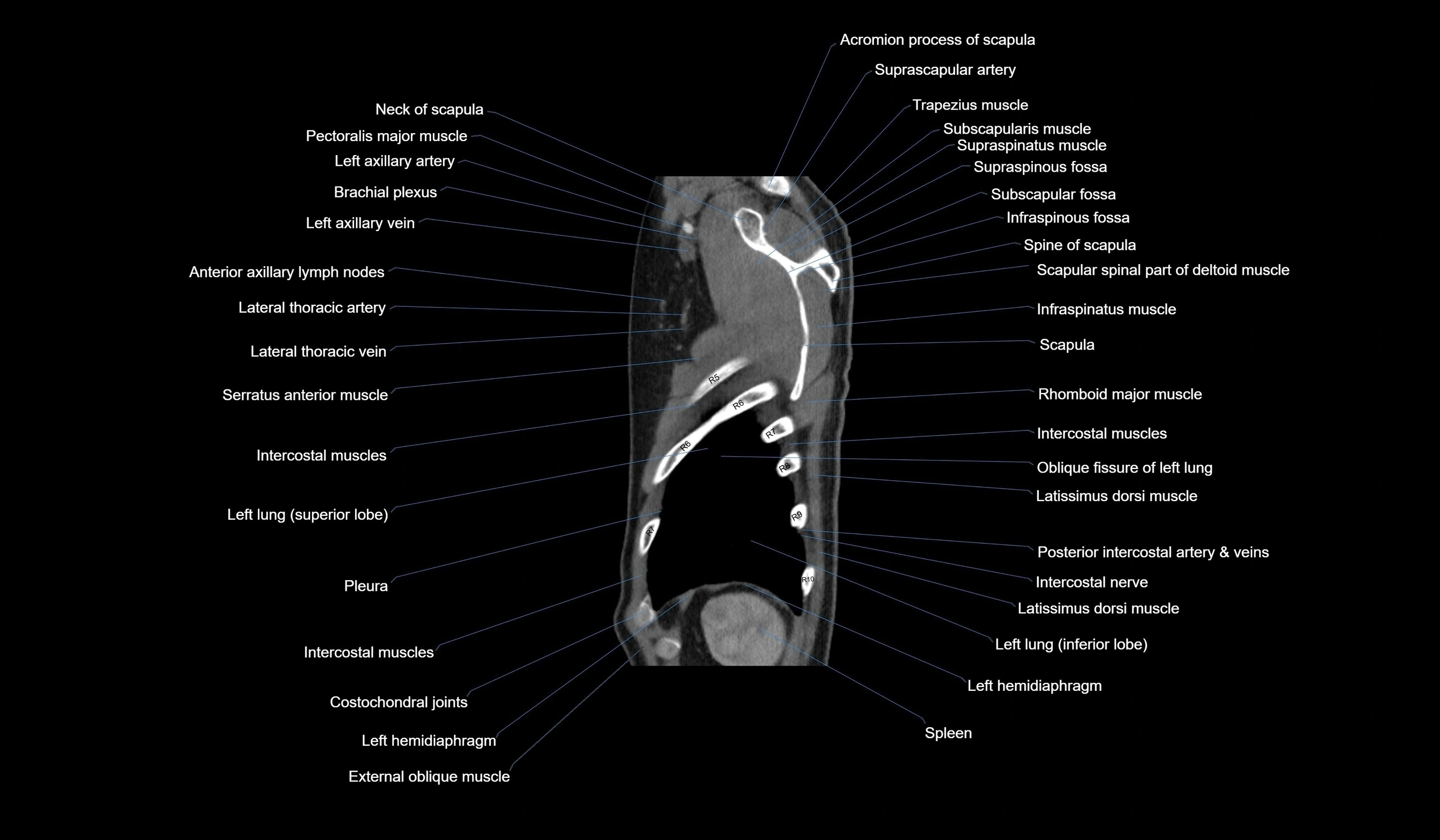
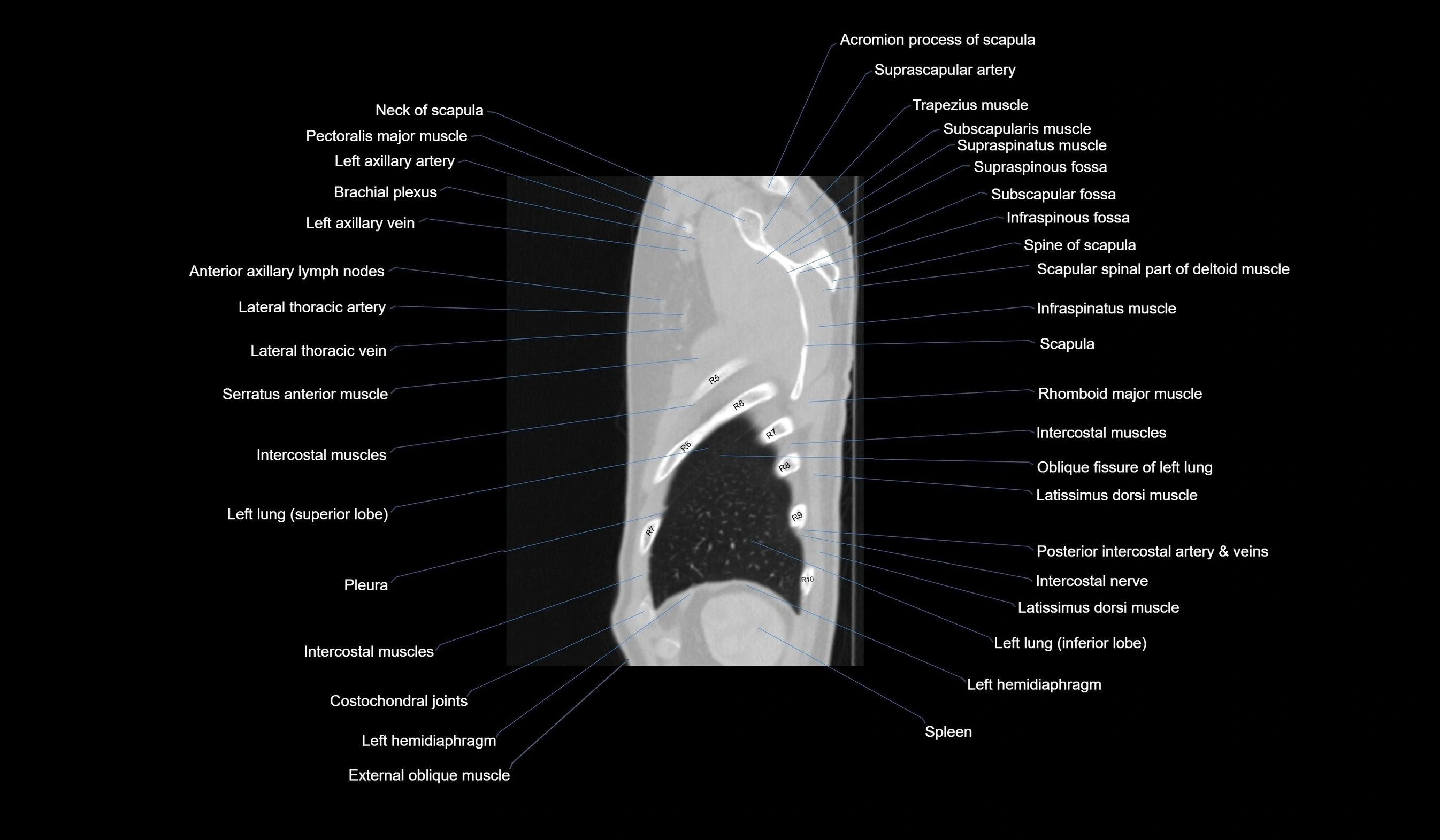
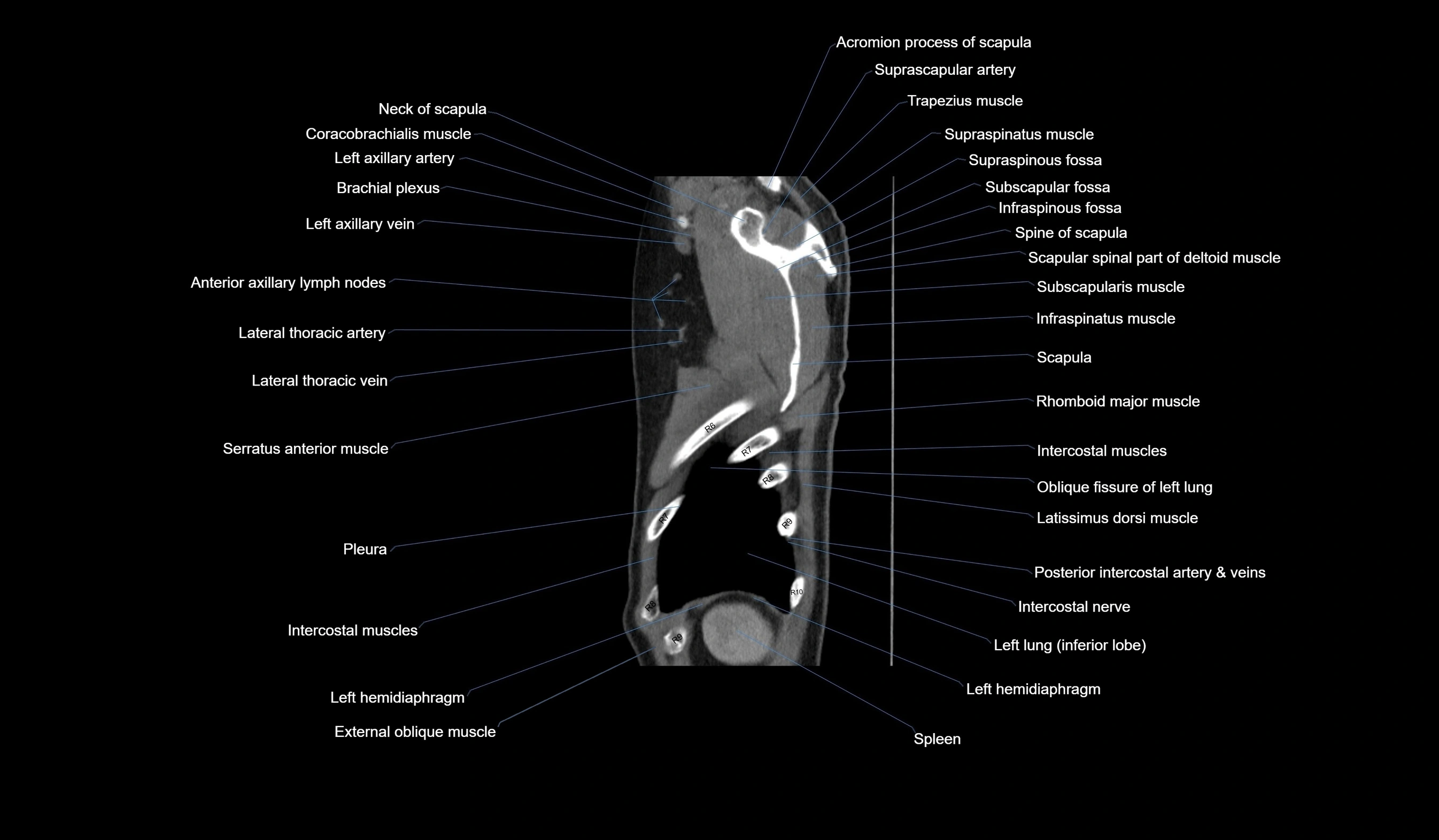
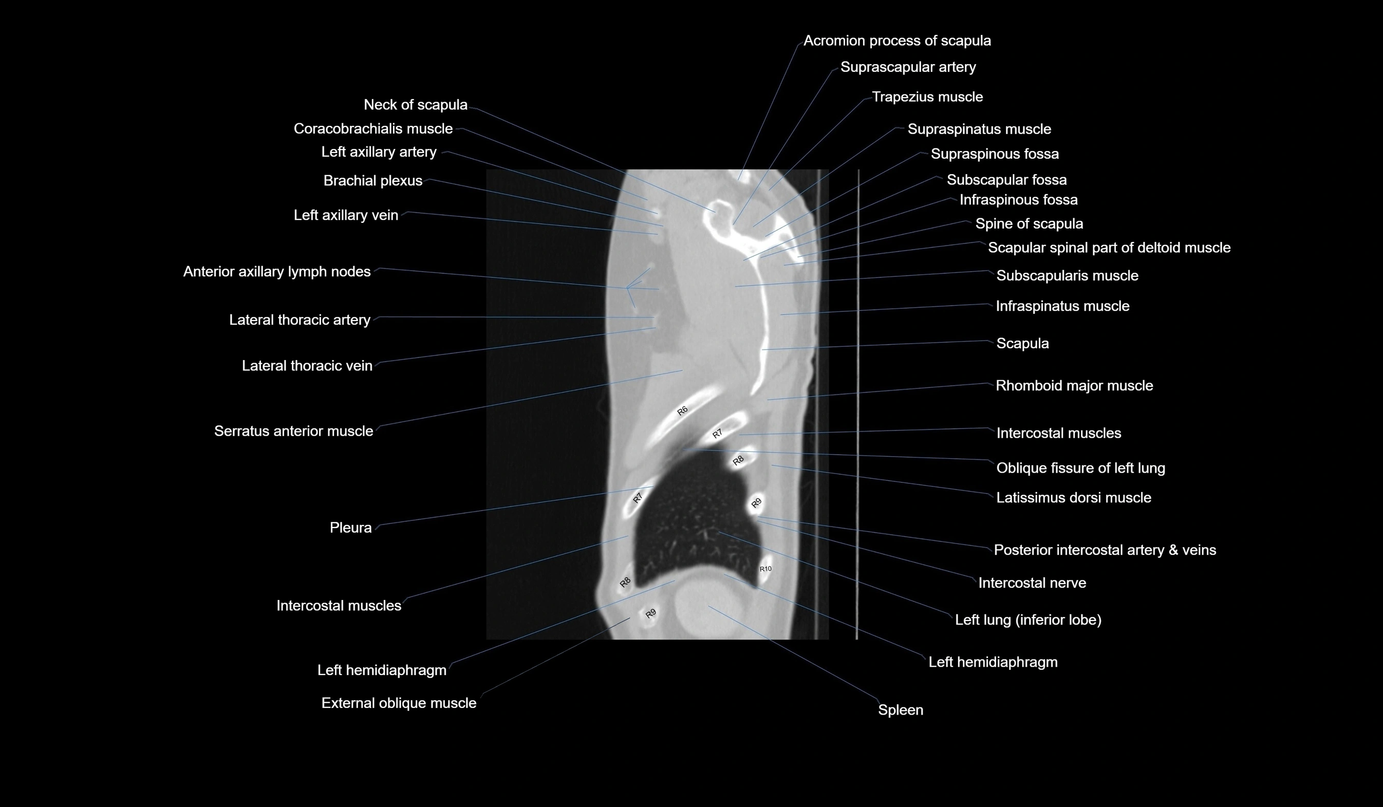
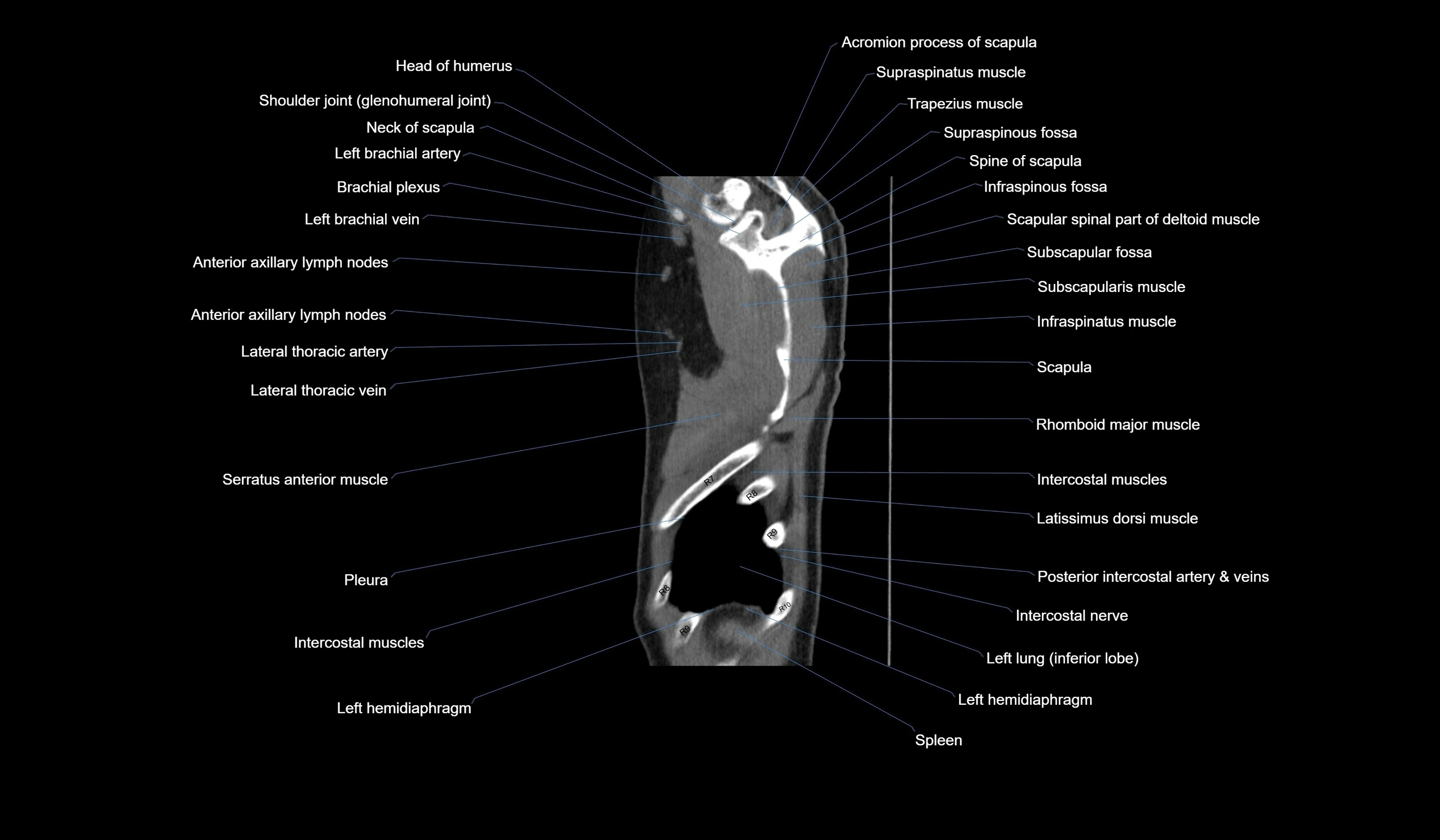
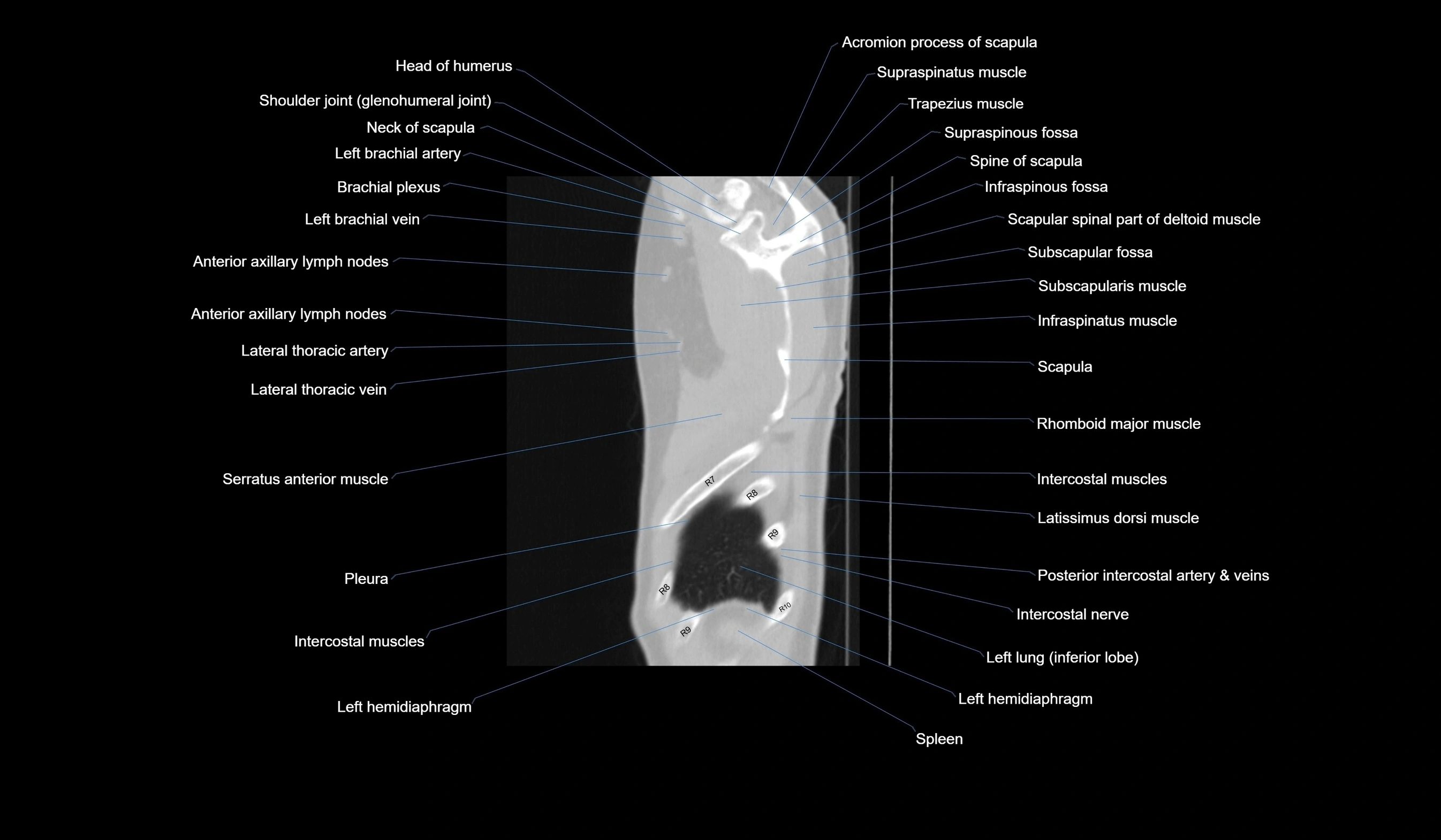
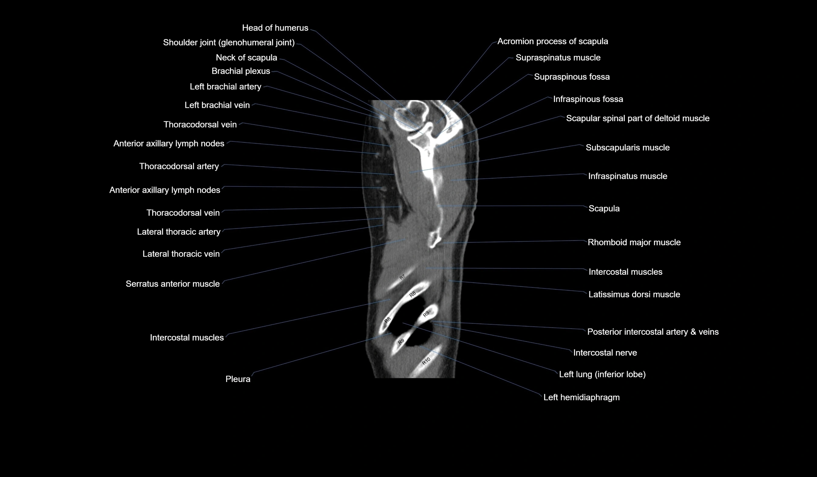
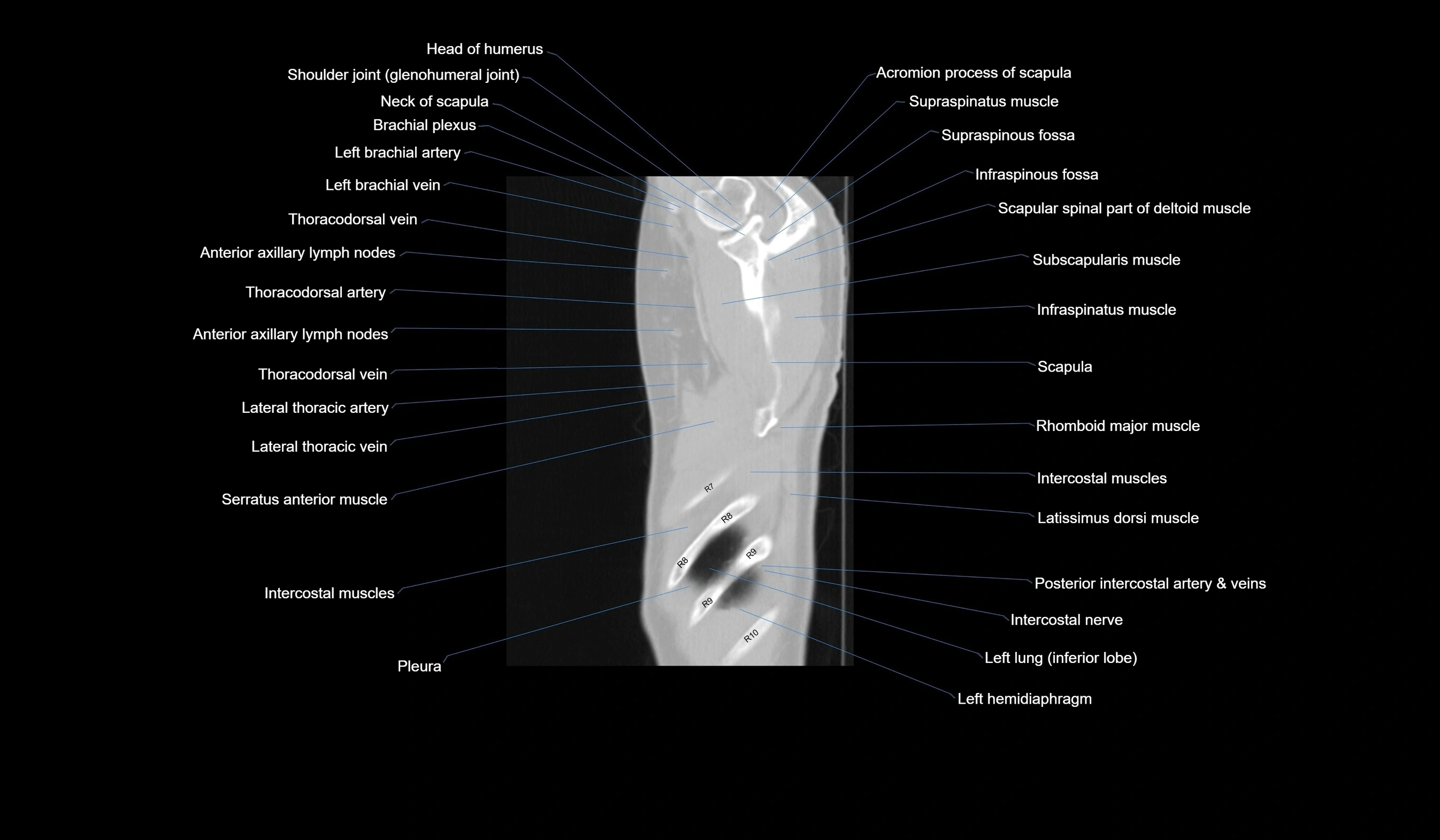
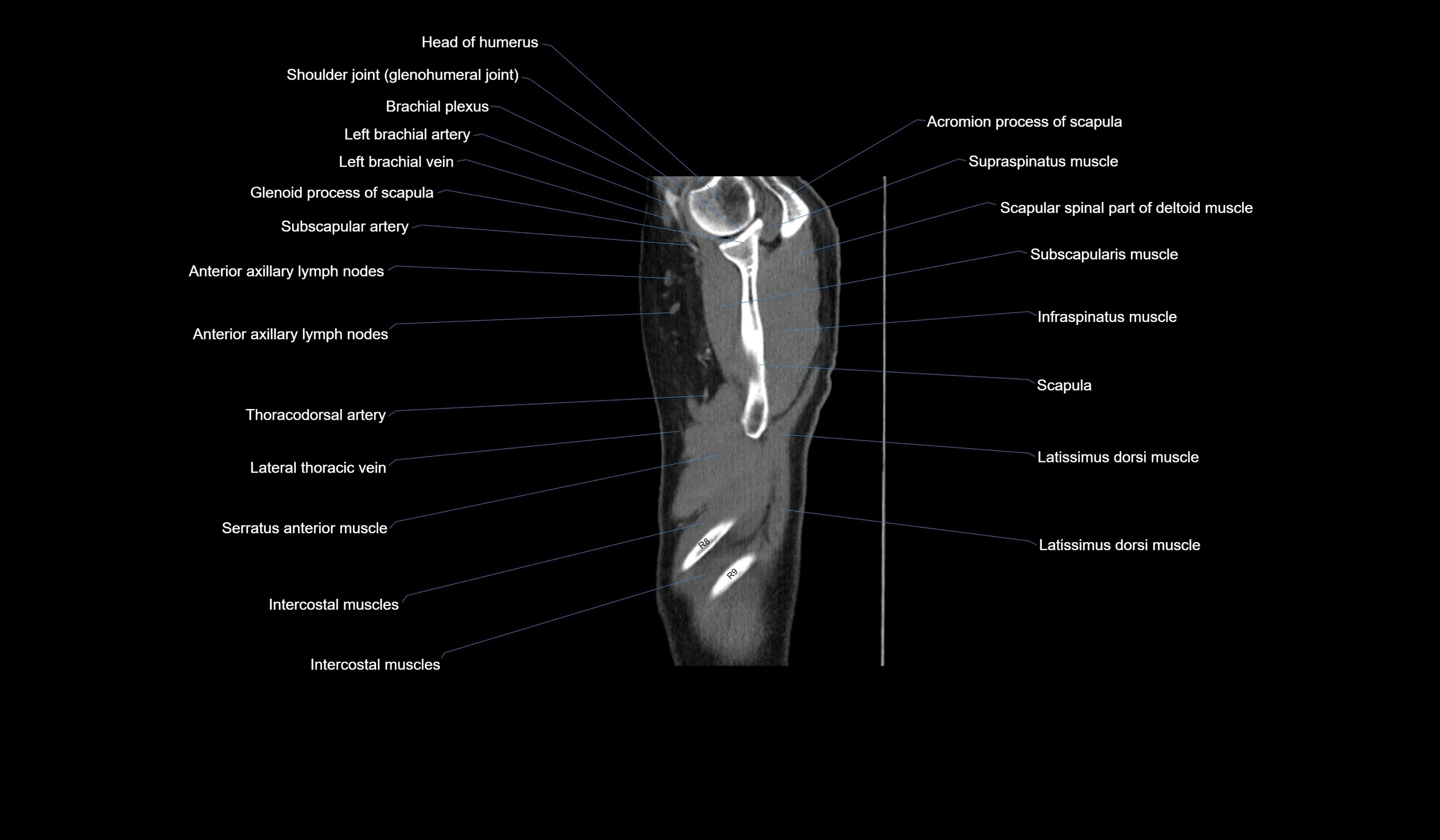
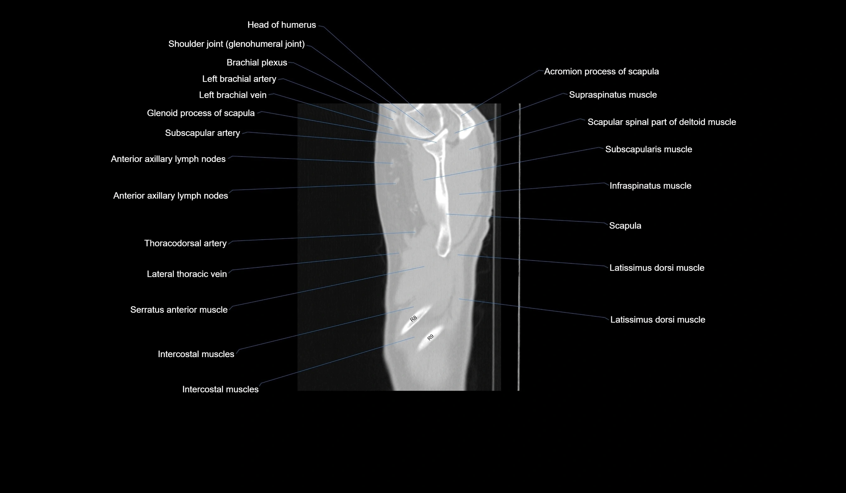
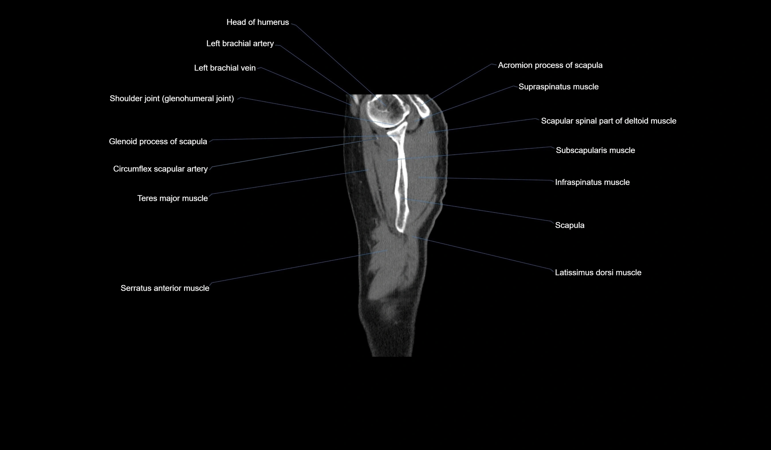
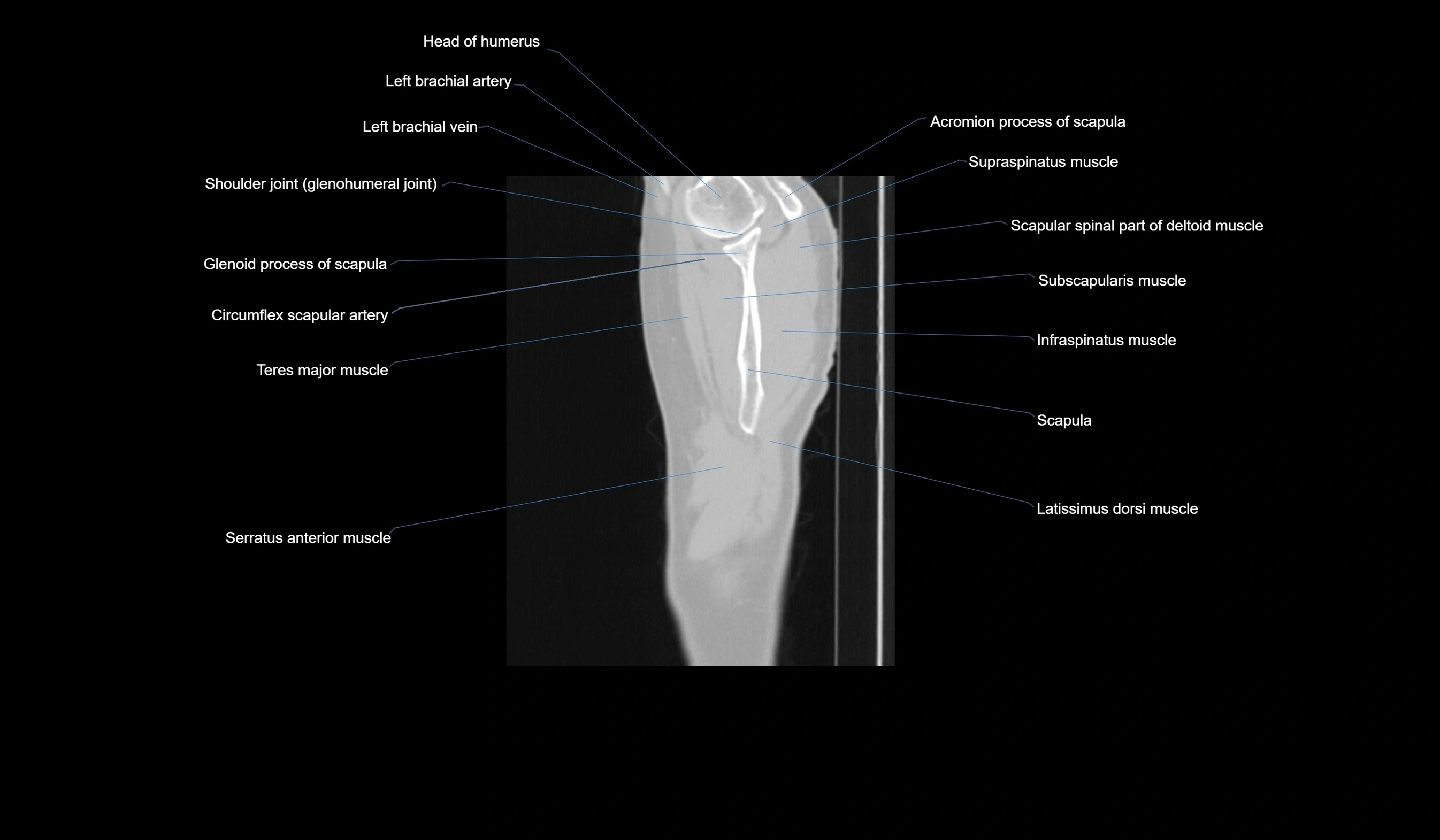
CT images

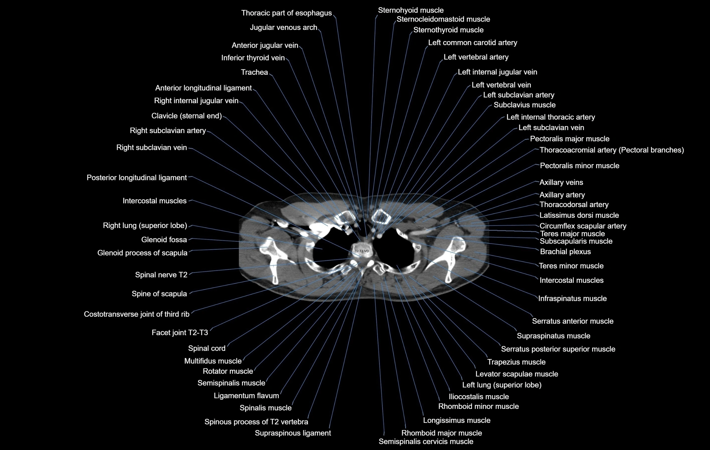
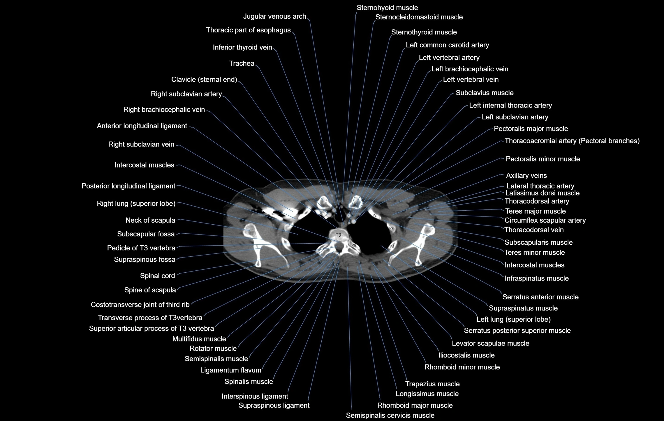
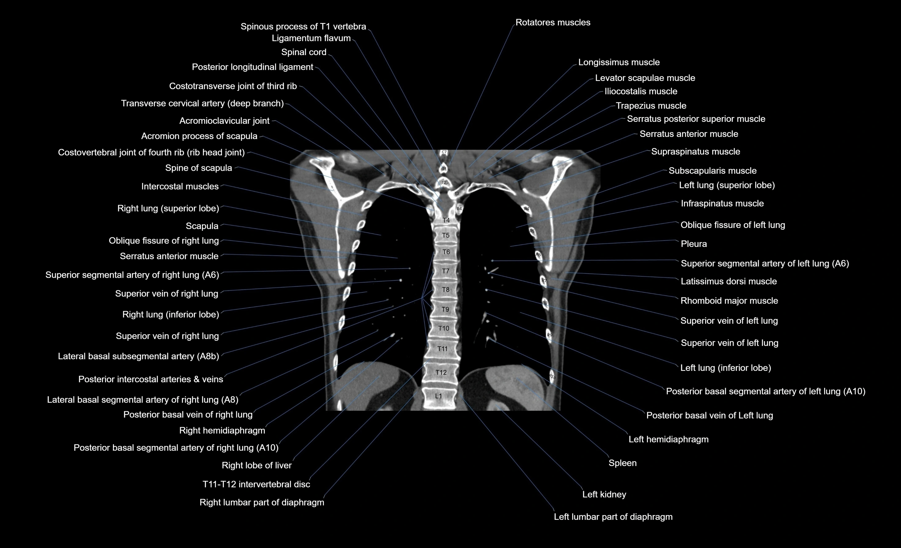
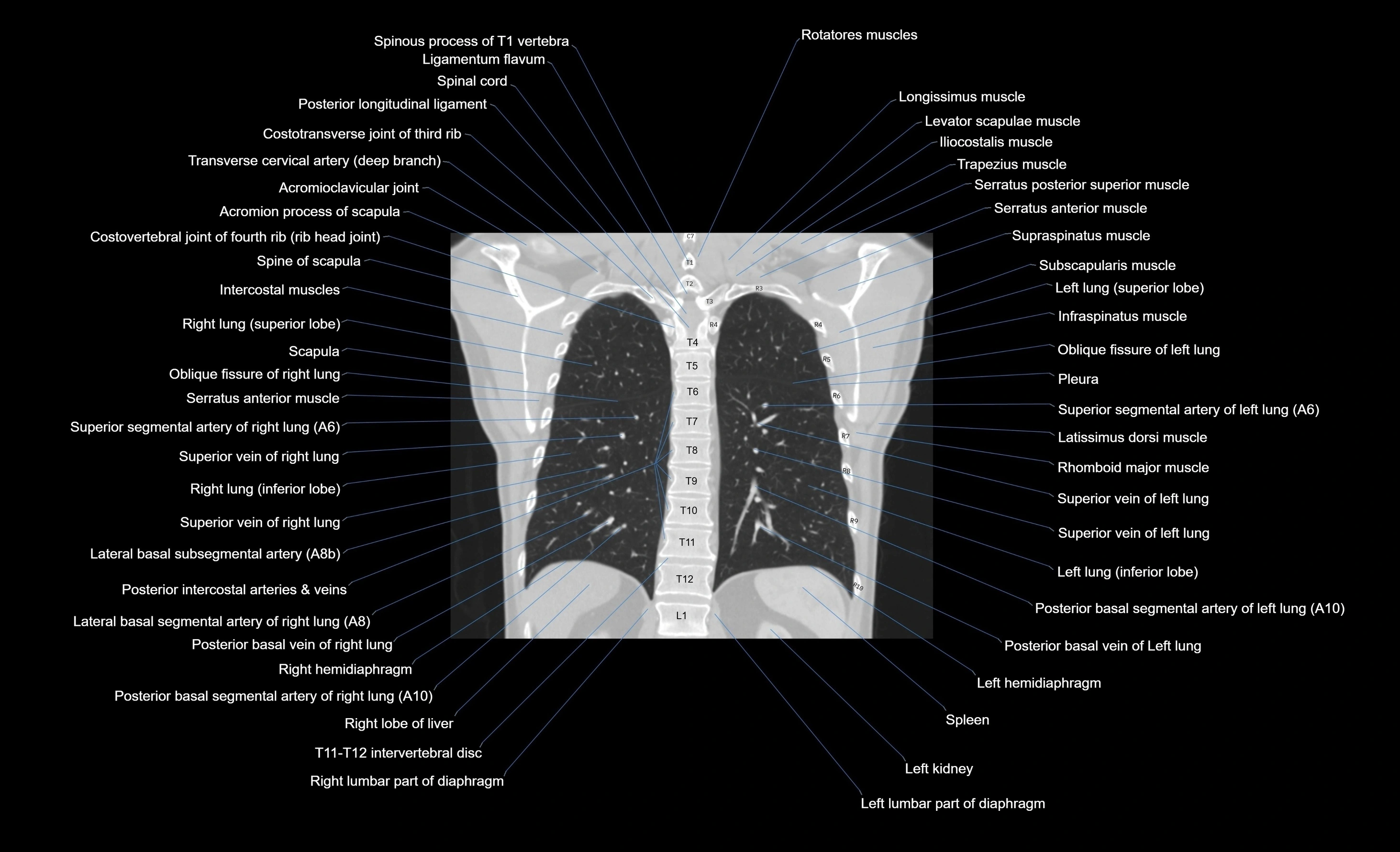
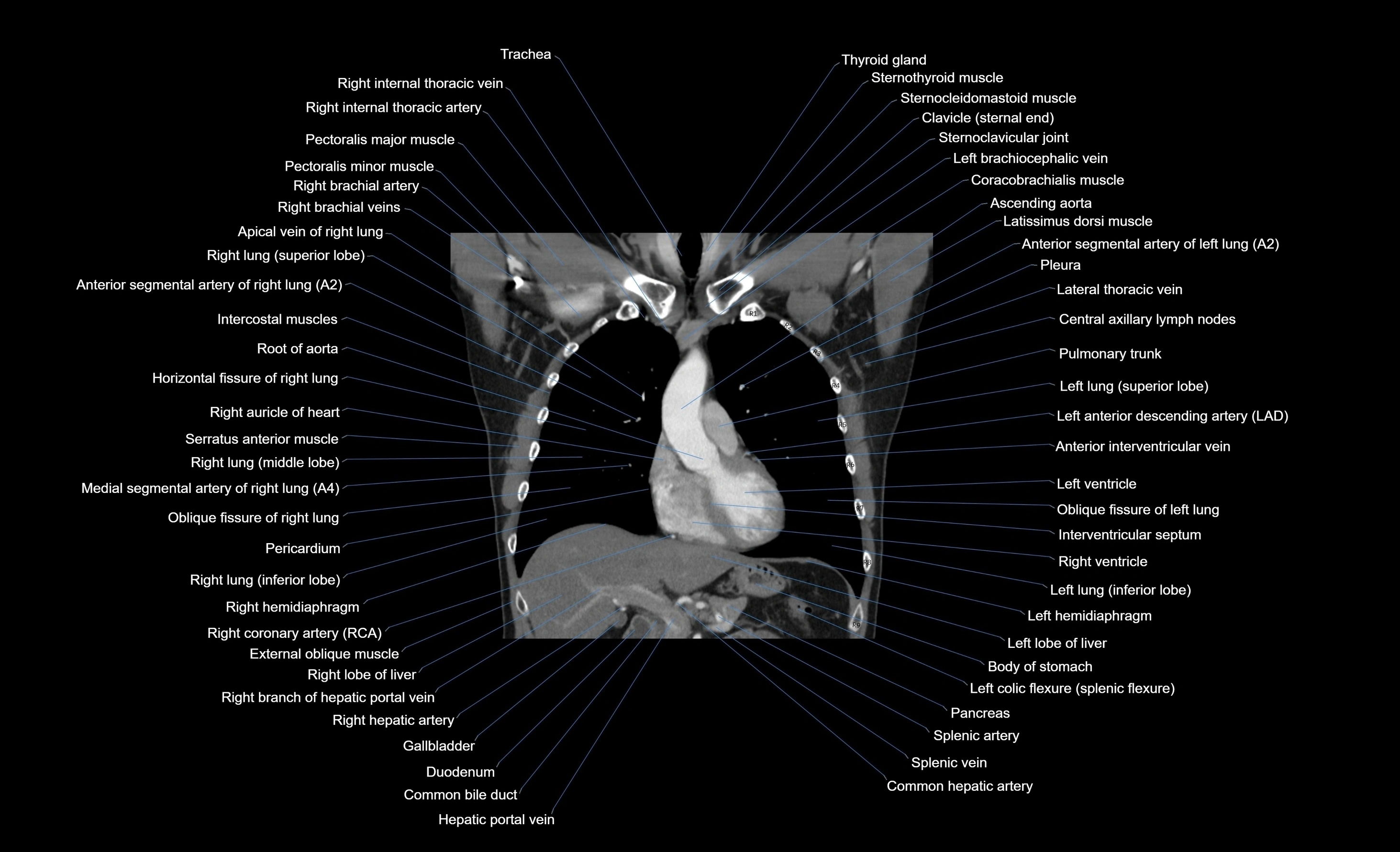
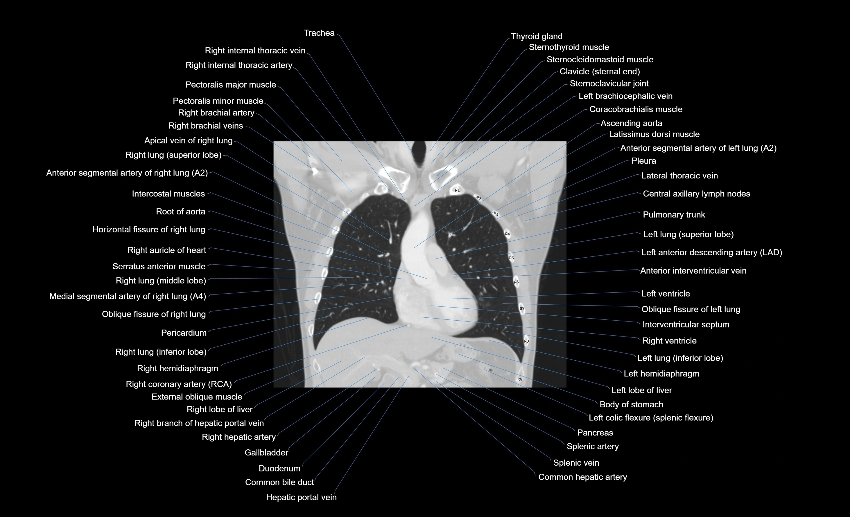
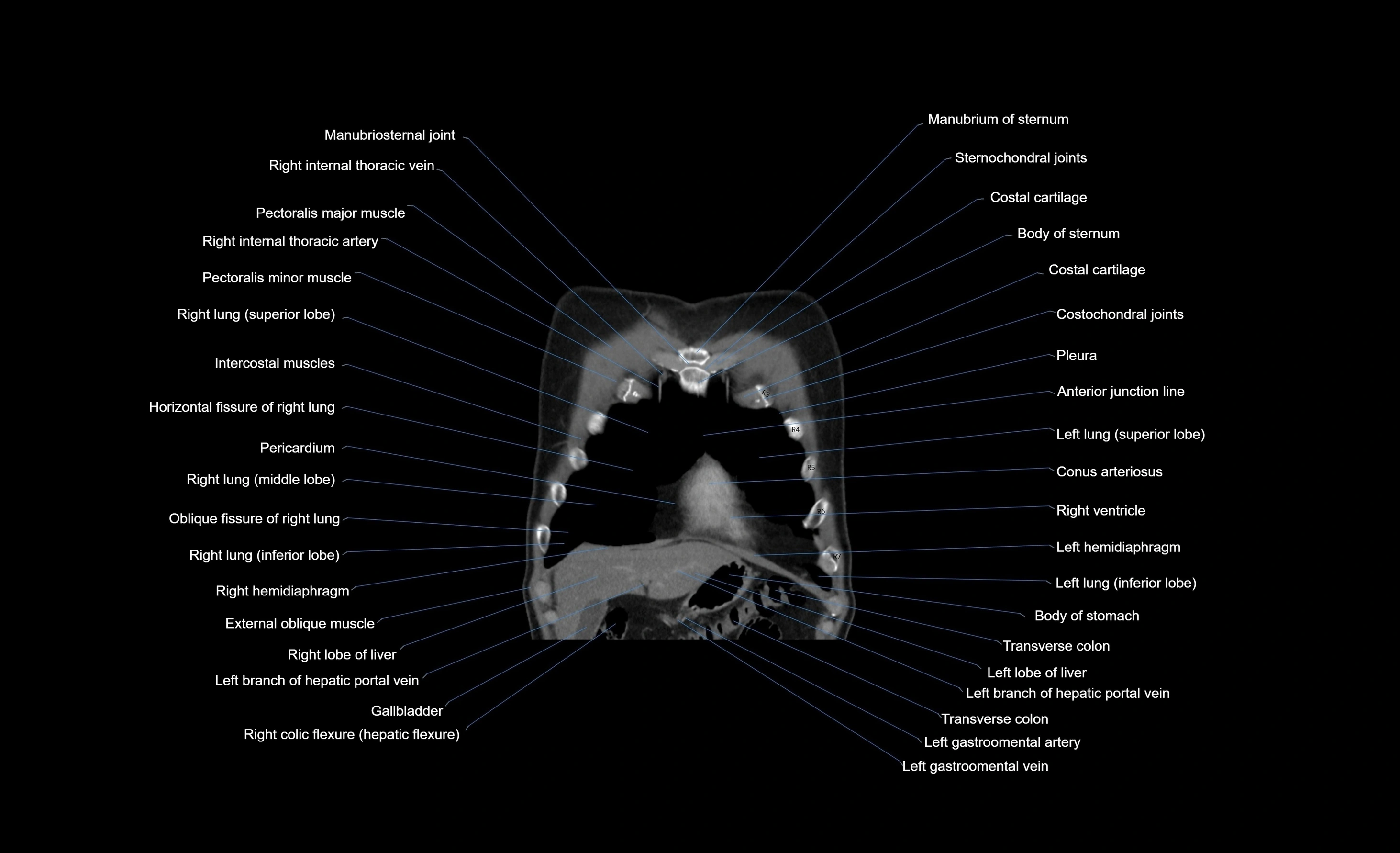
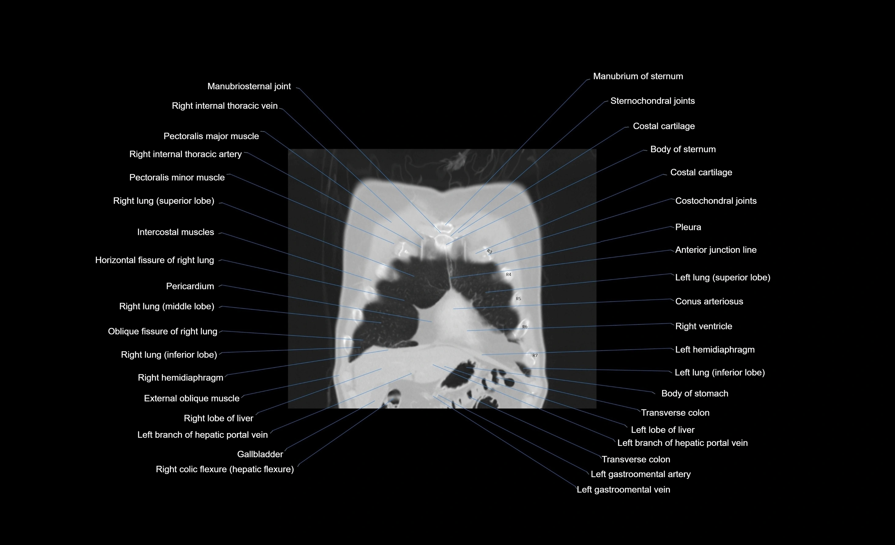
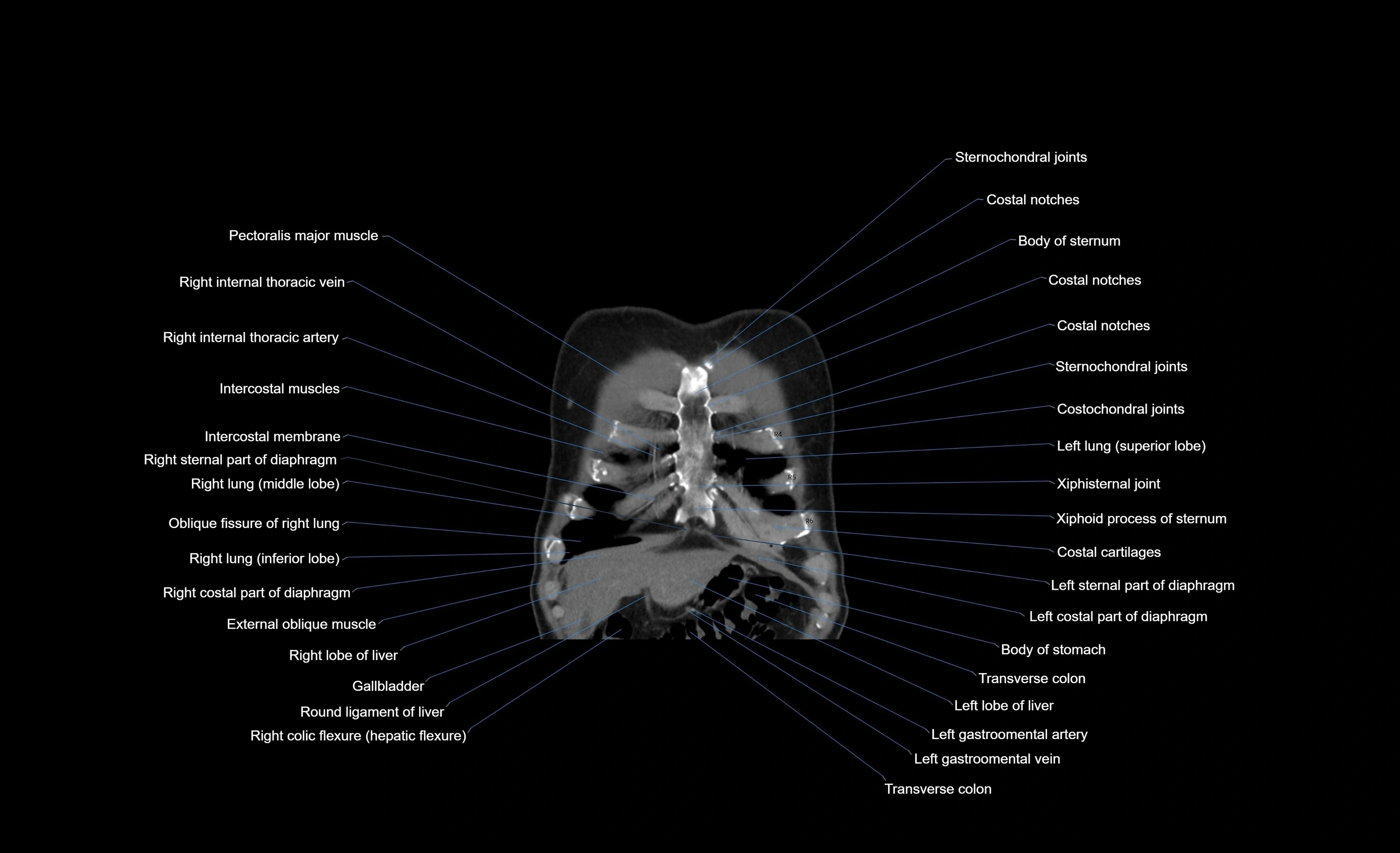
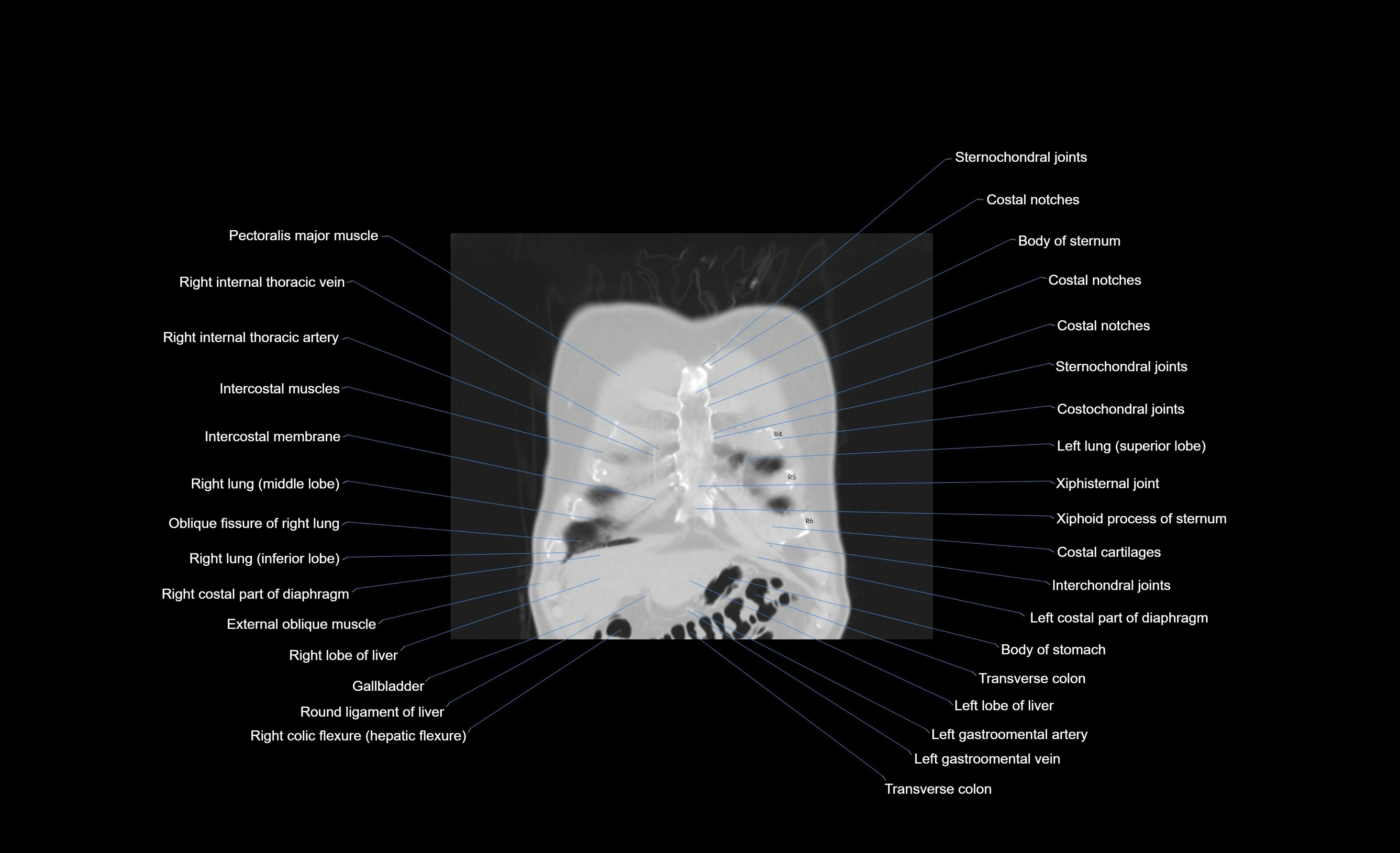
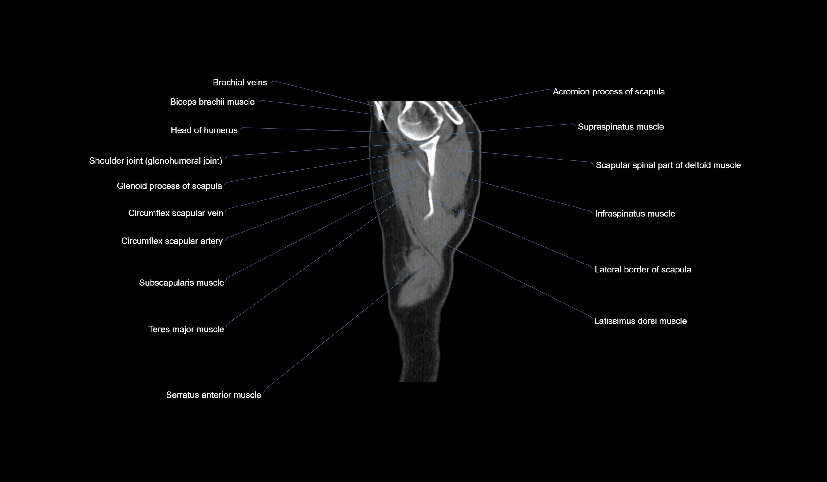
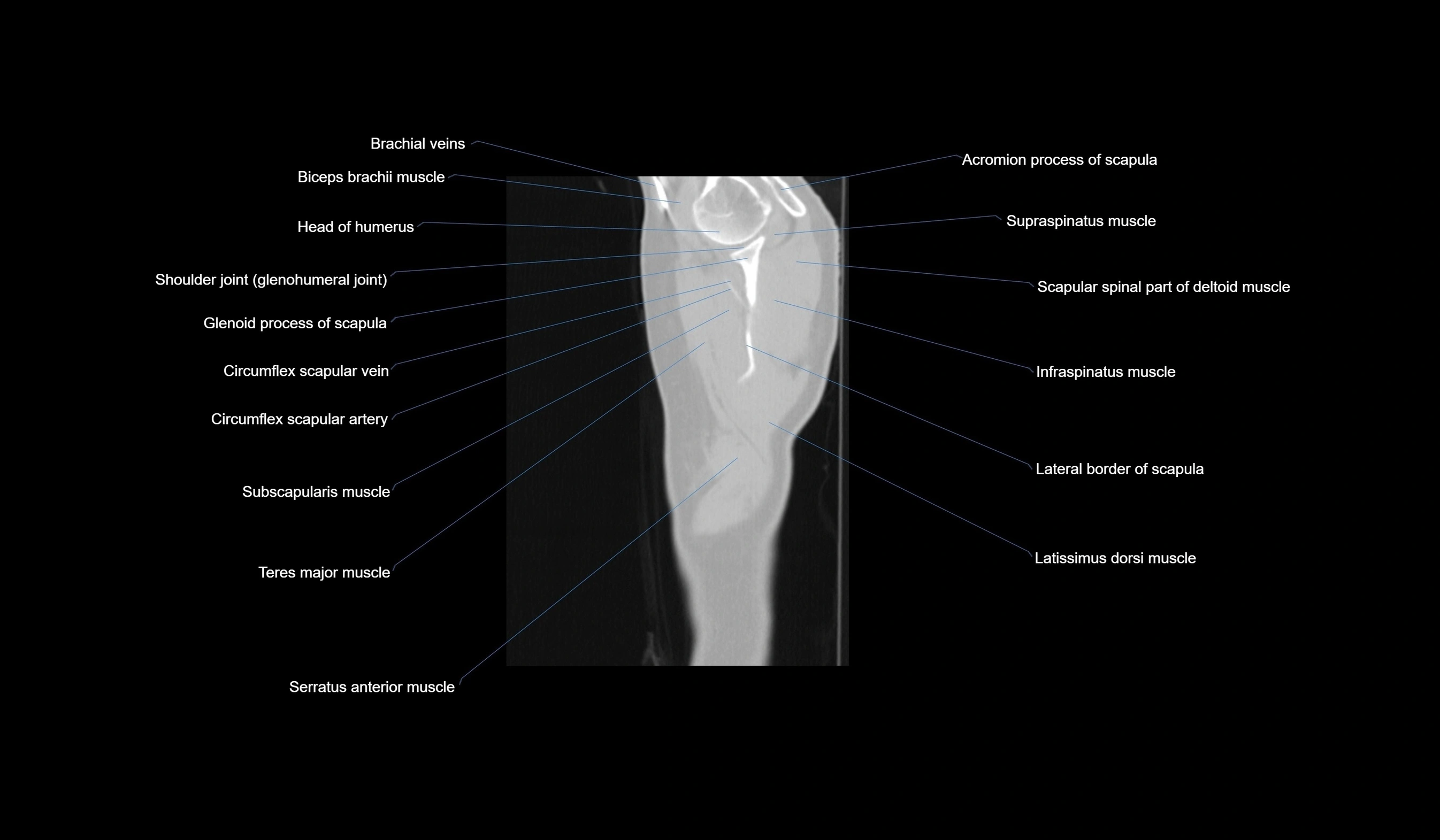
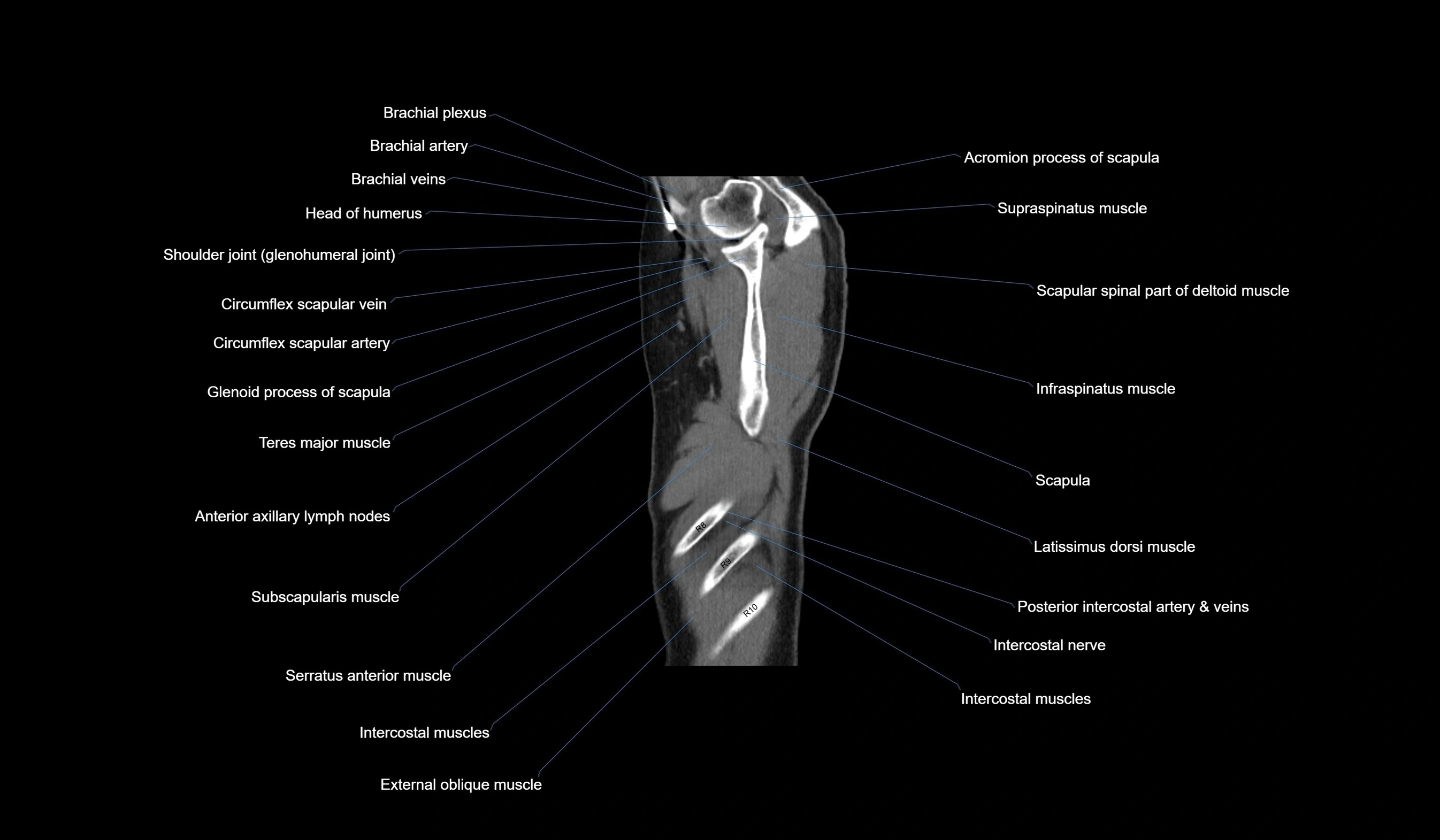
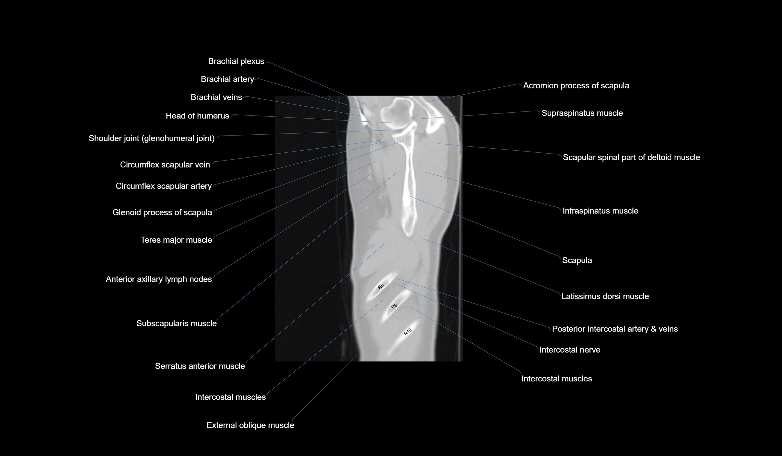
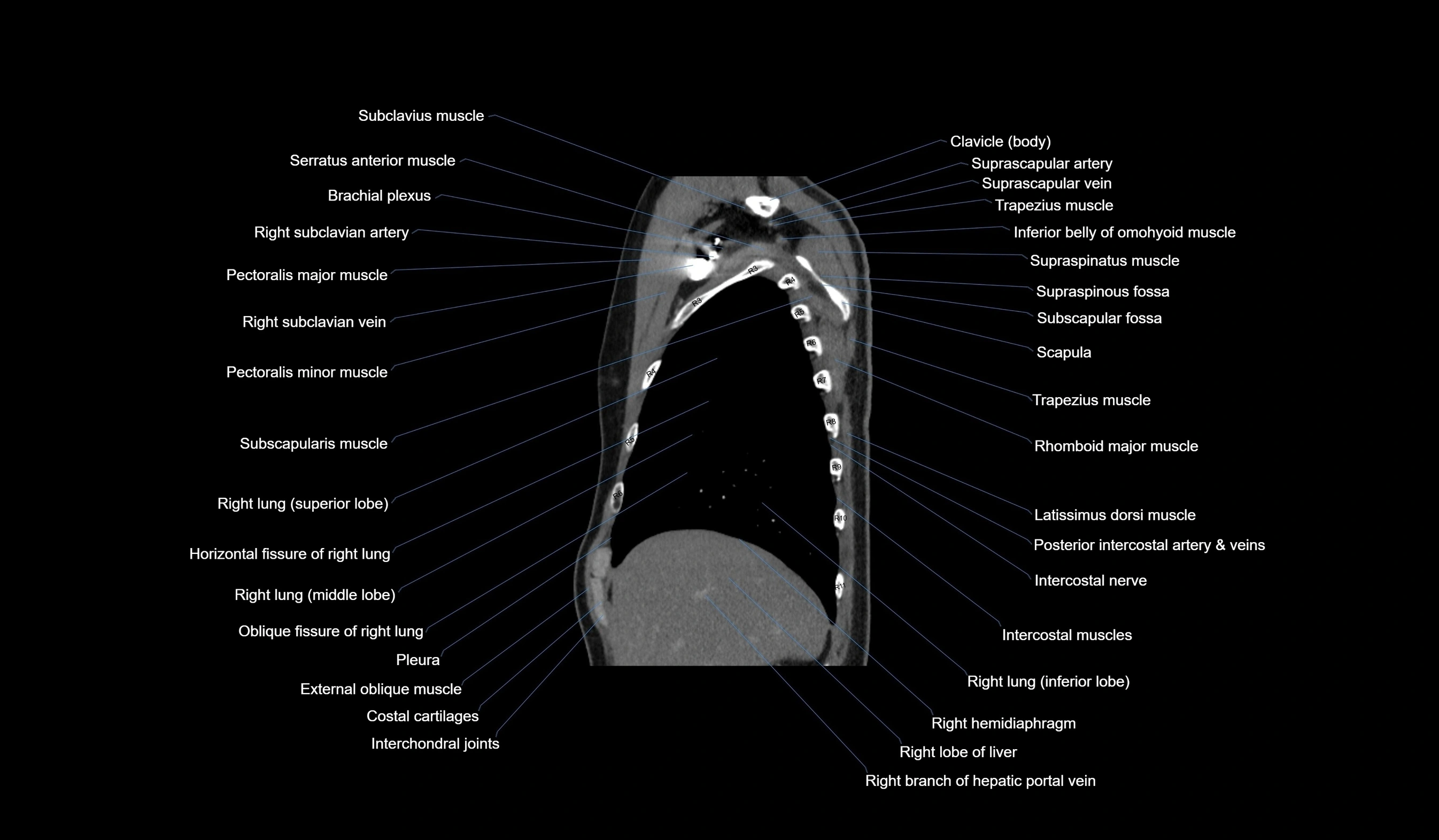
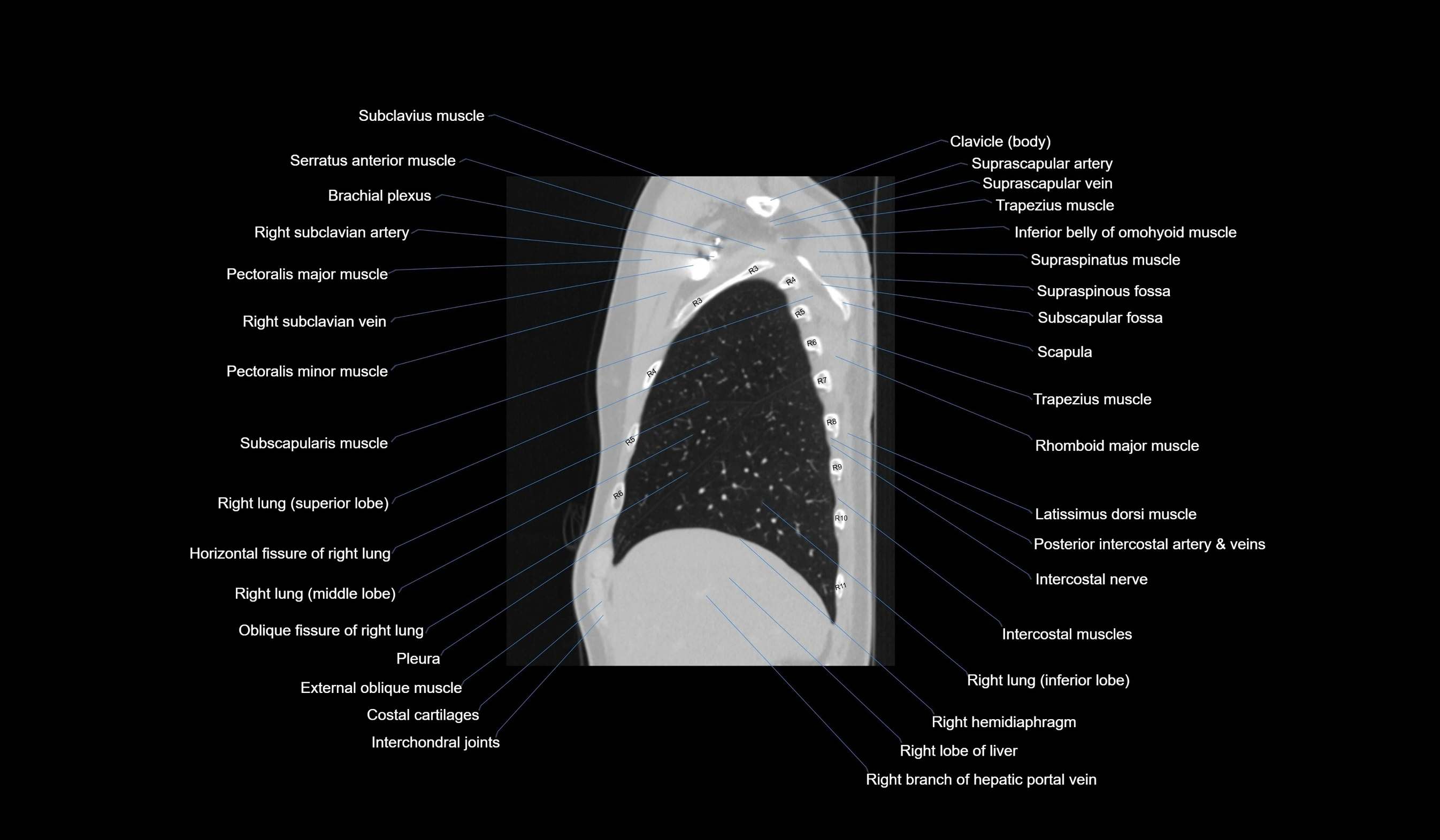
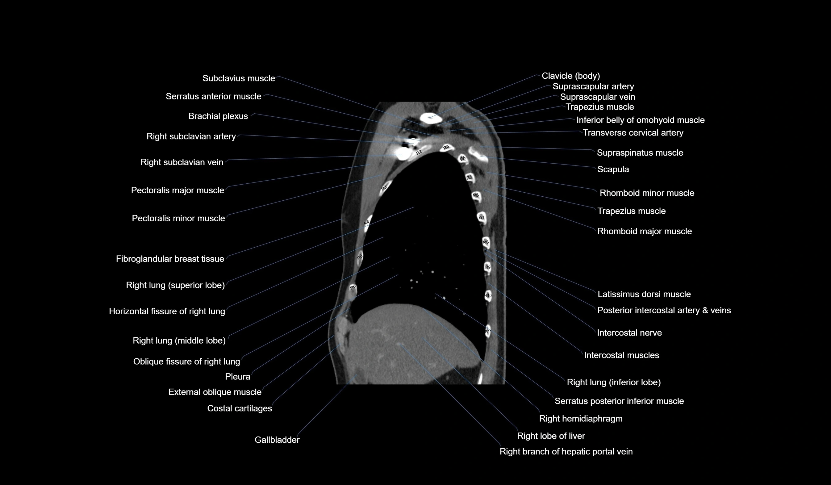
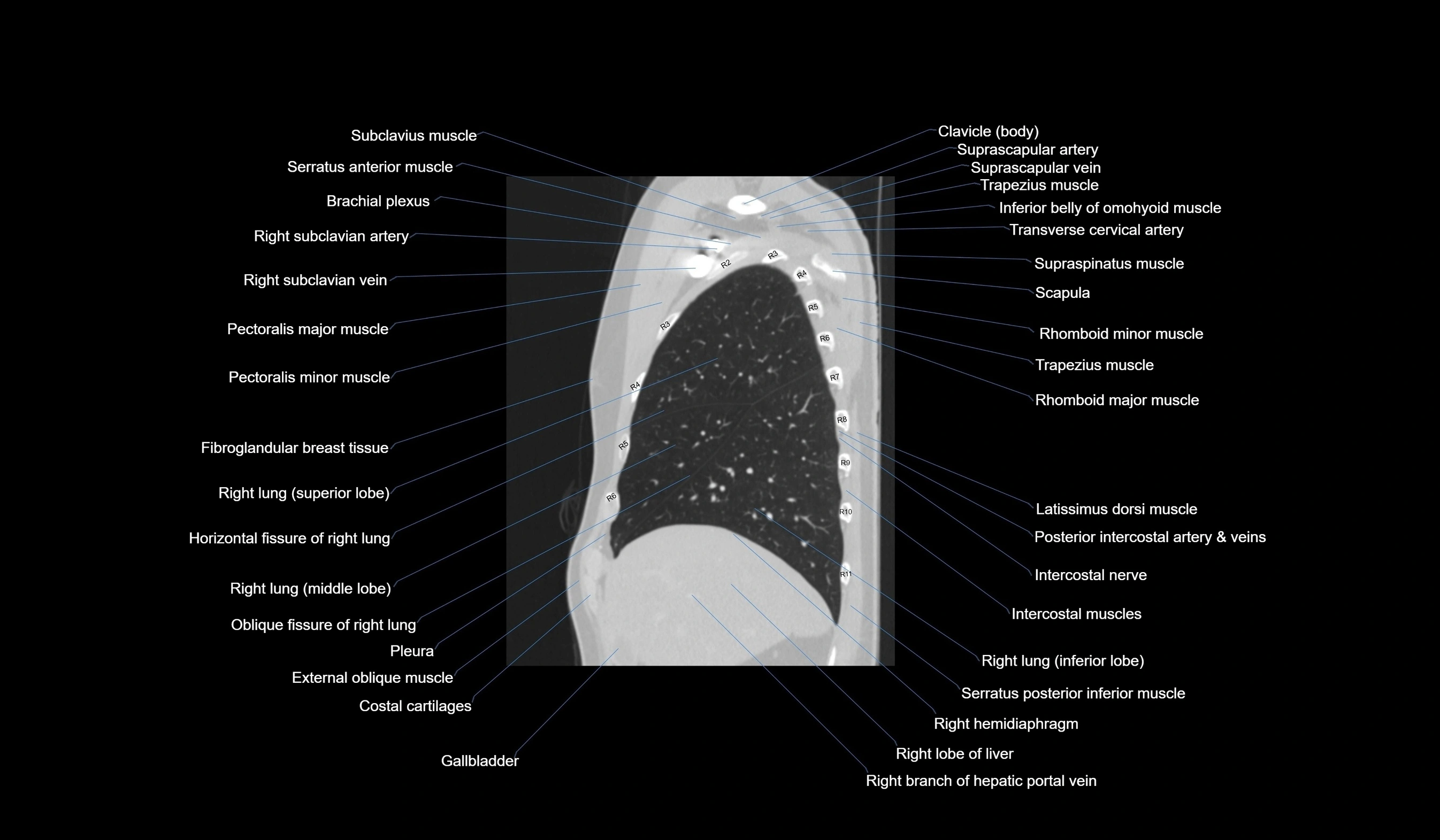
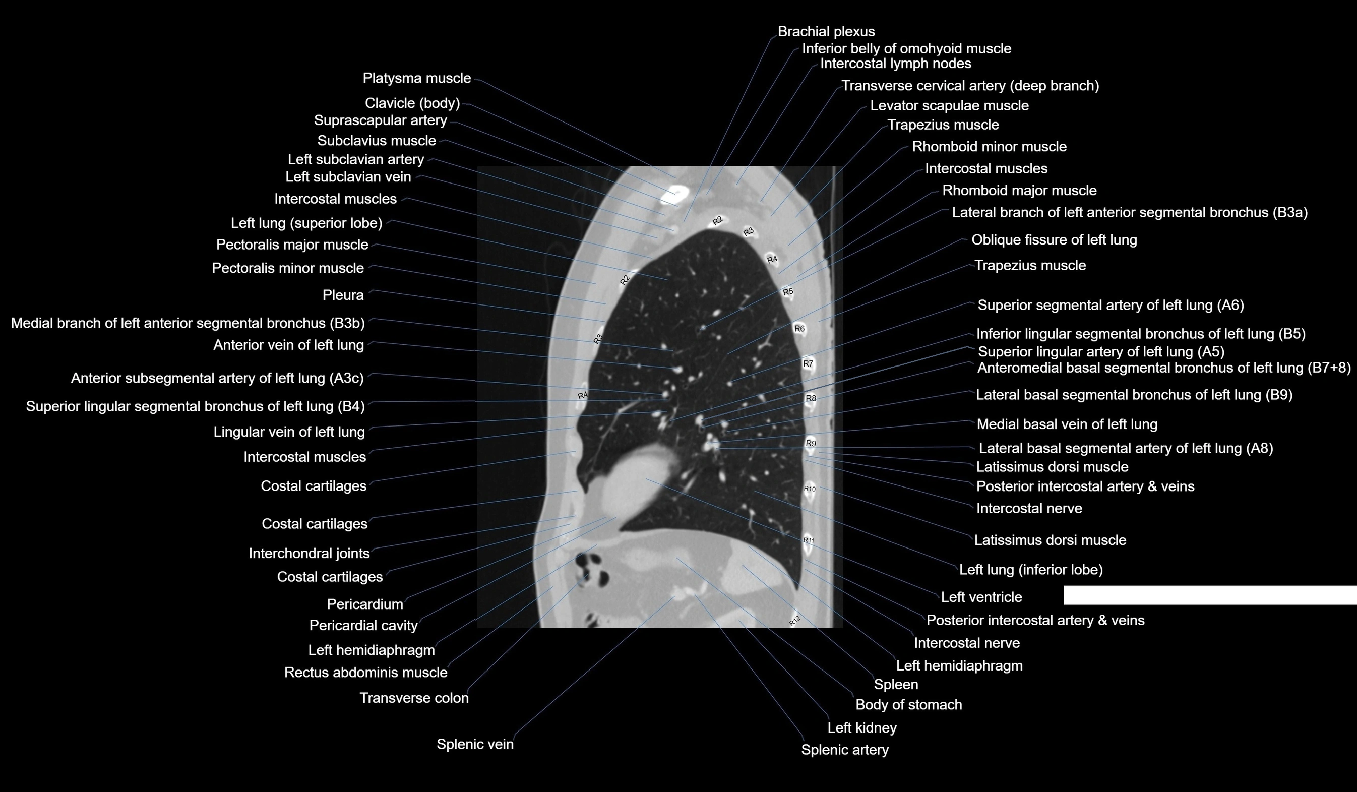
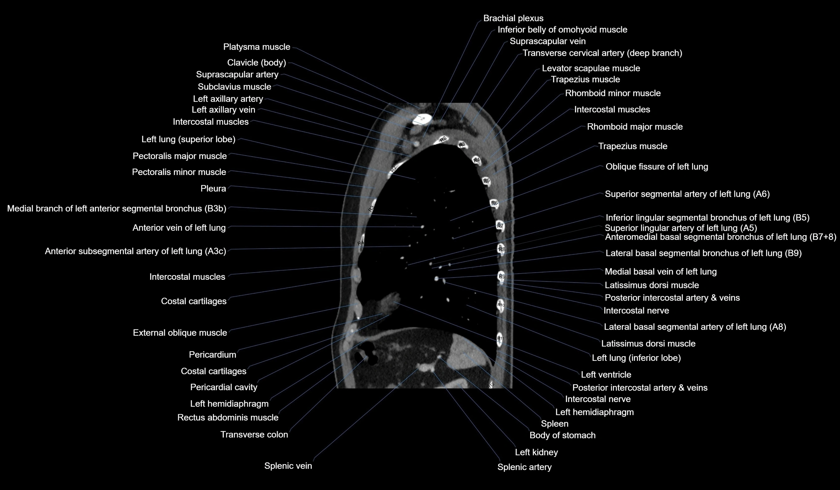
CT images
