


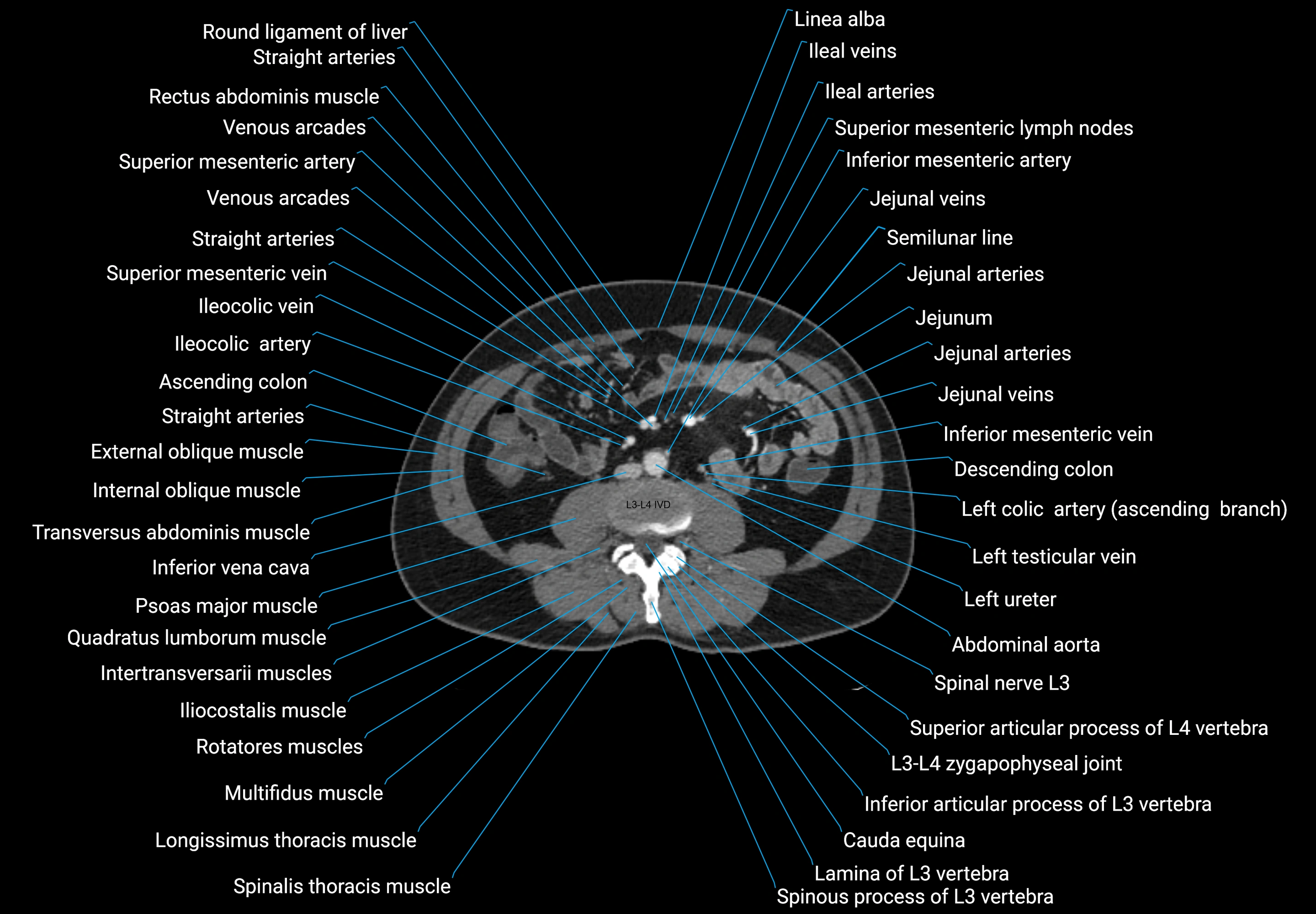
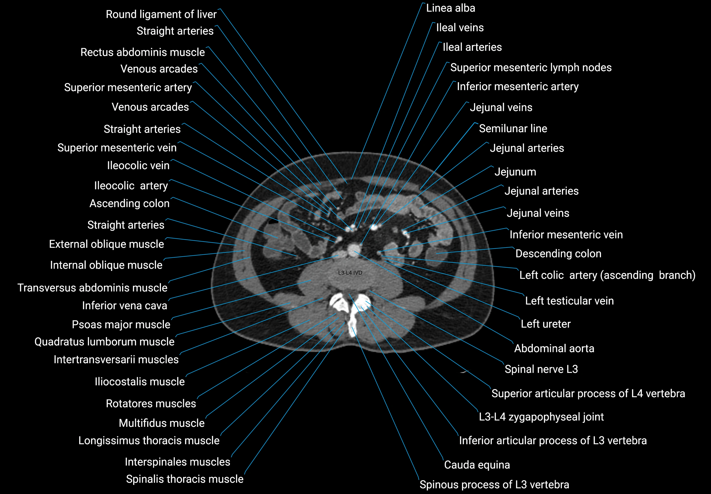
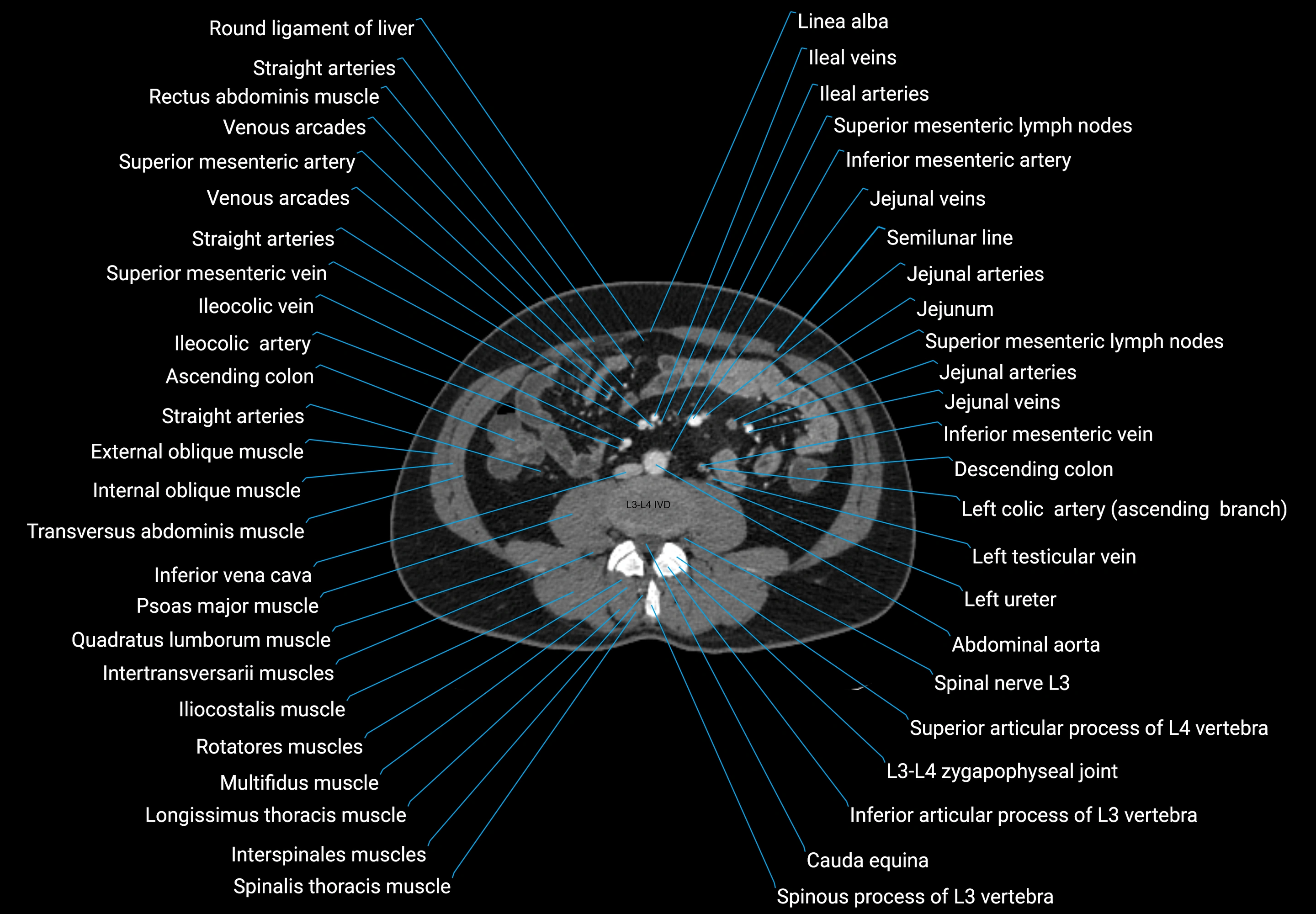
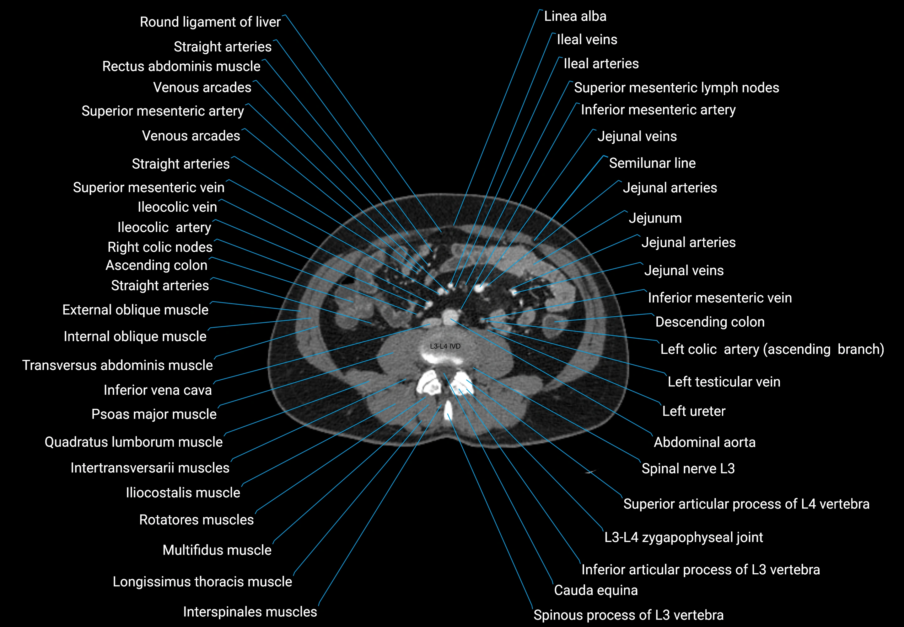


Topic
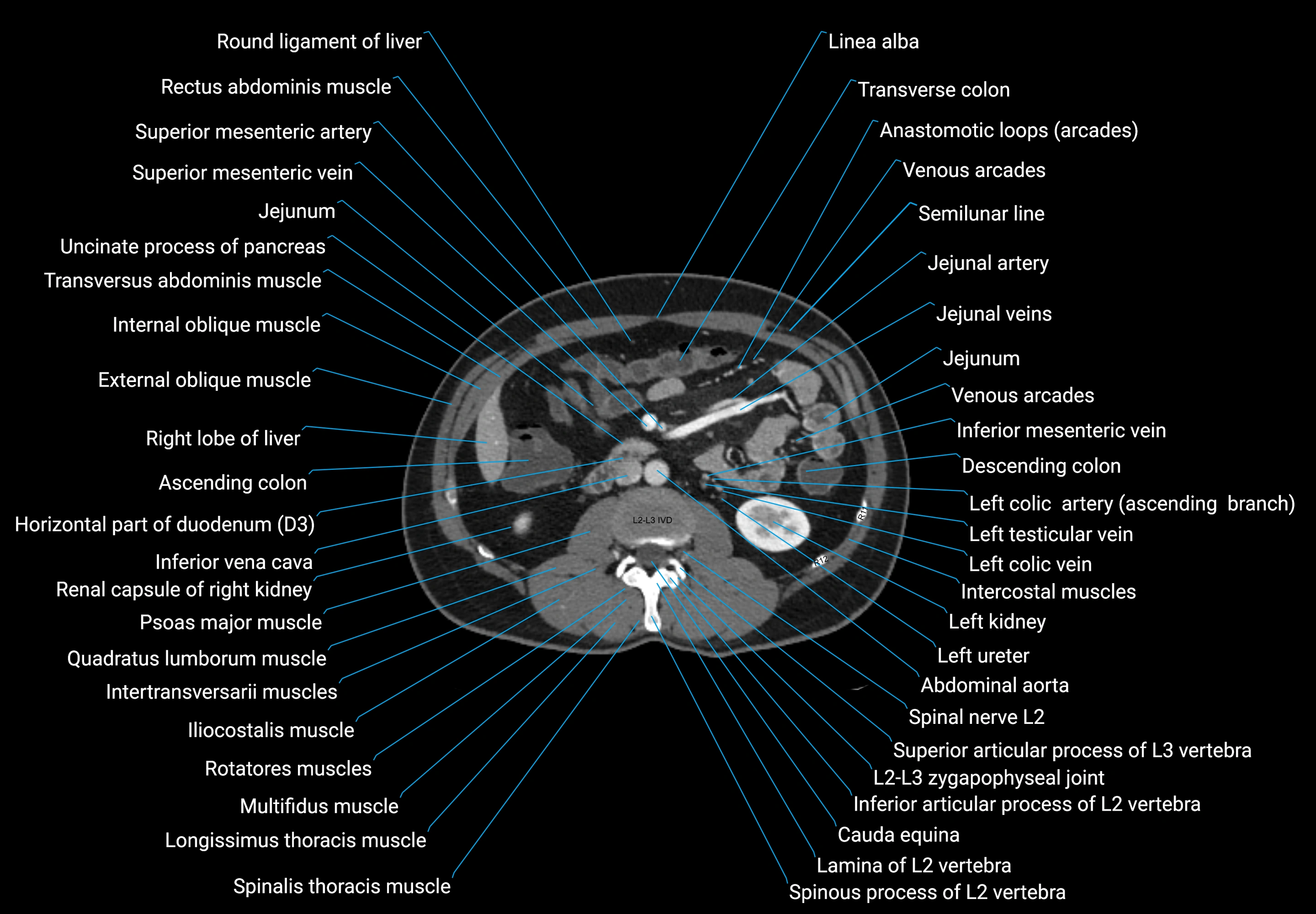
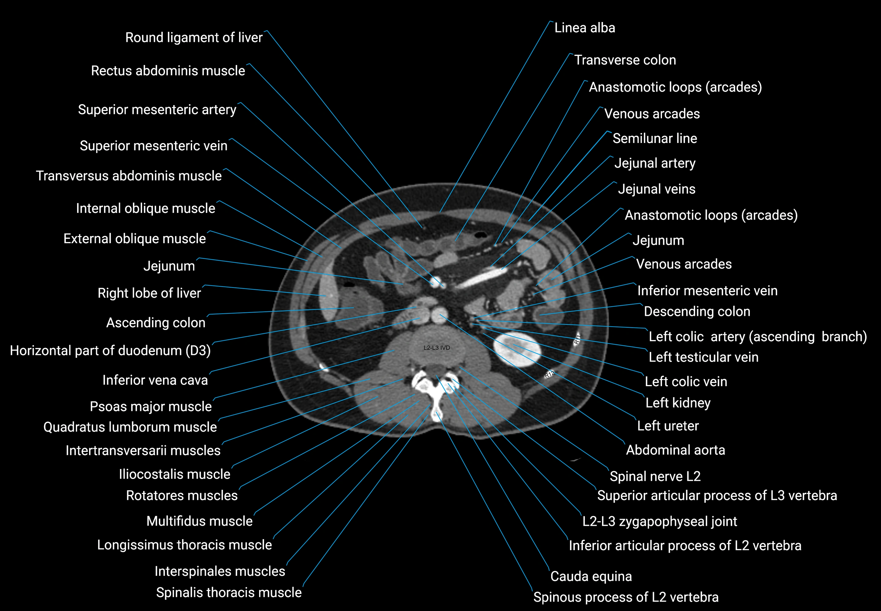
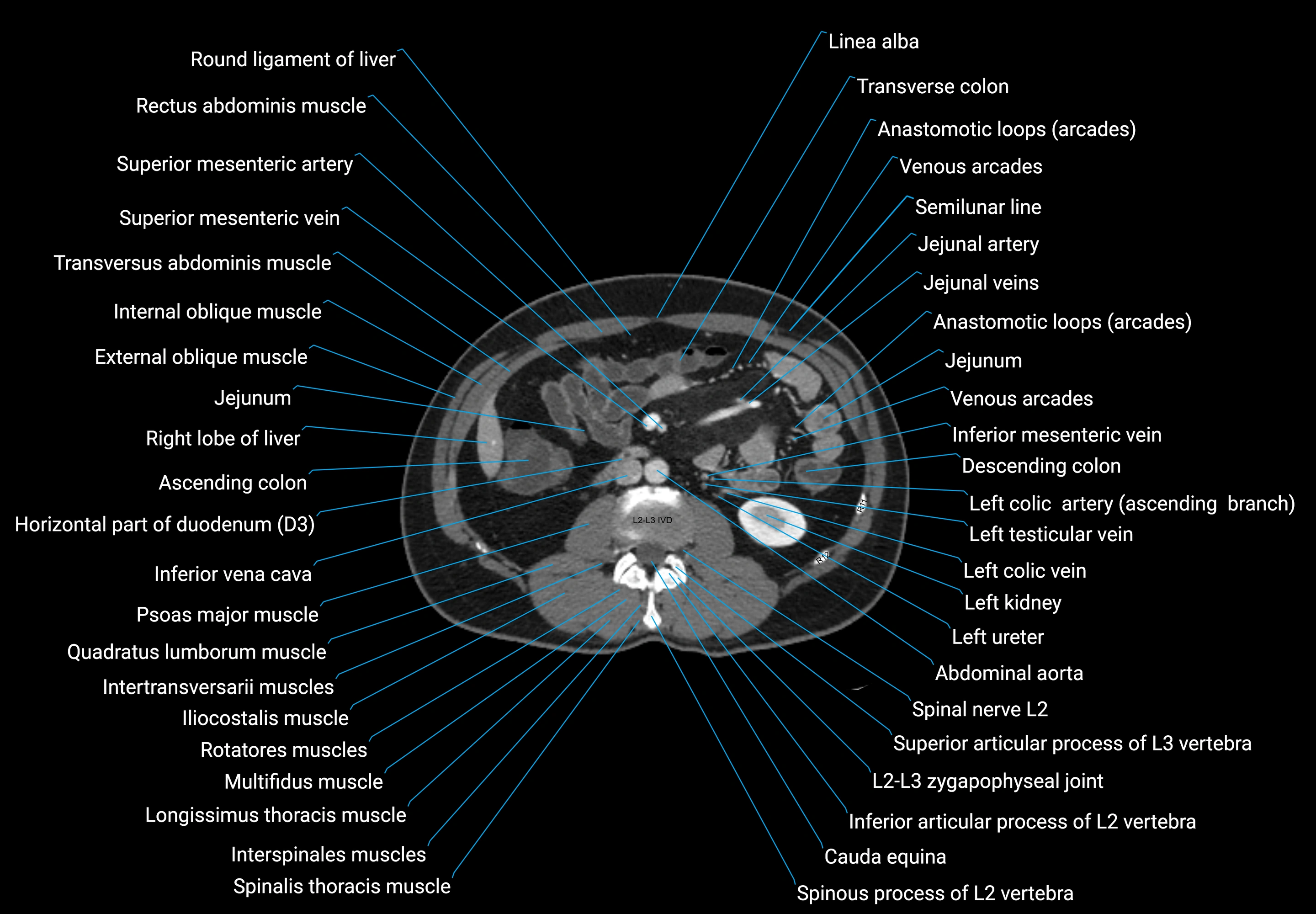
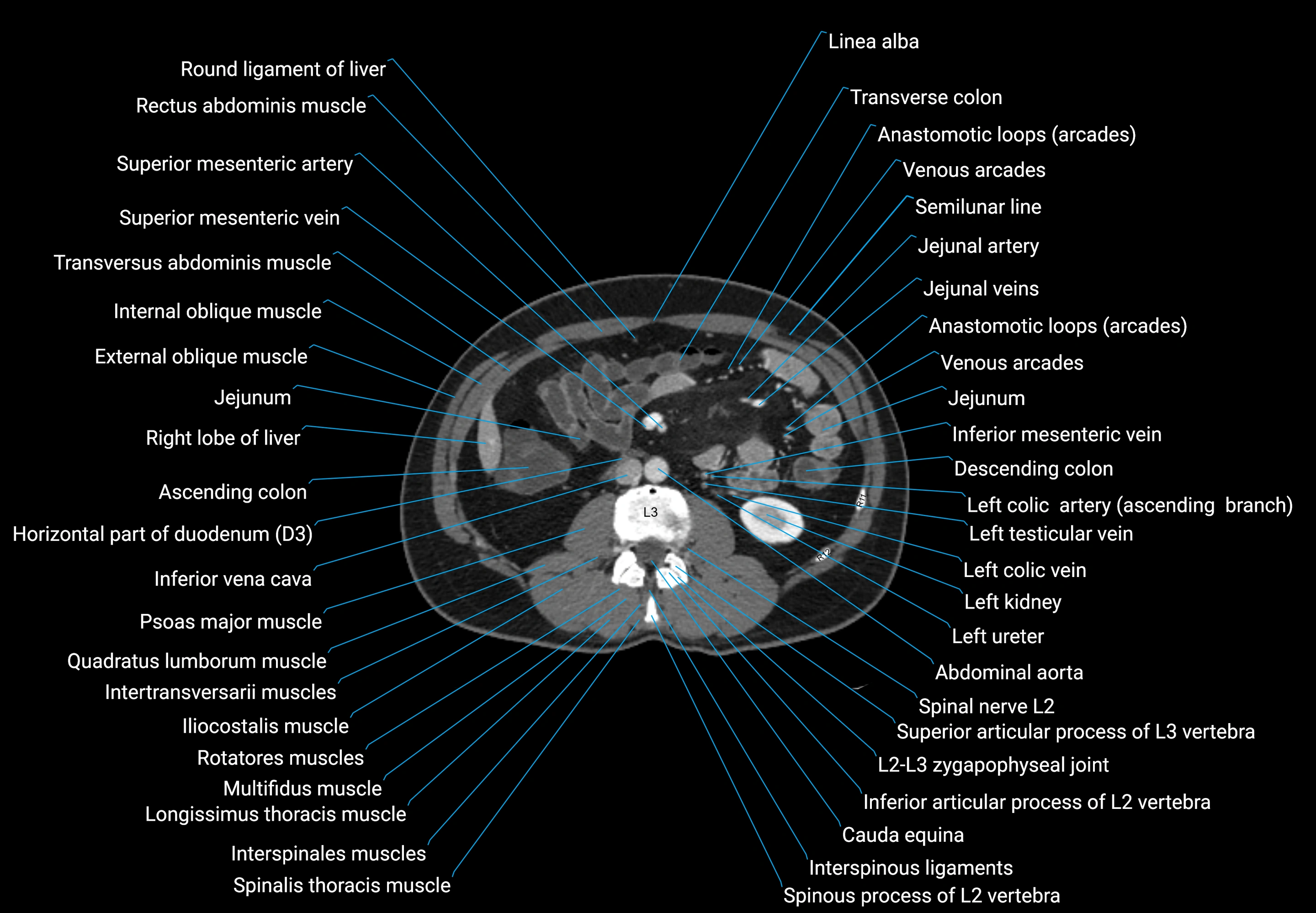
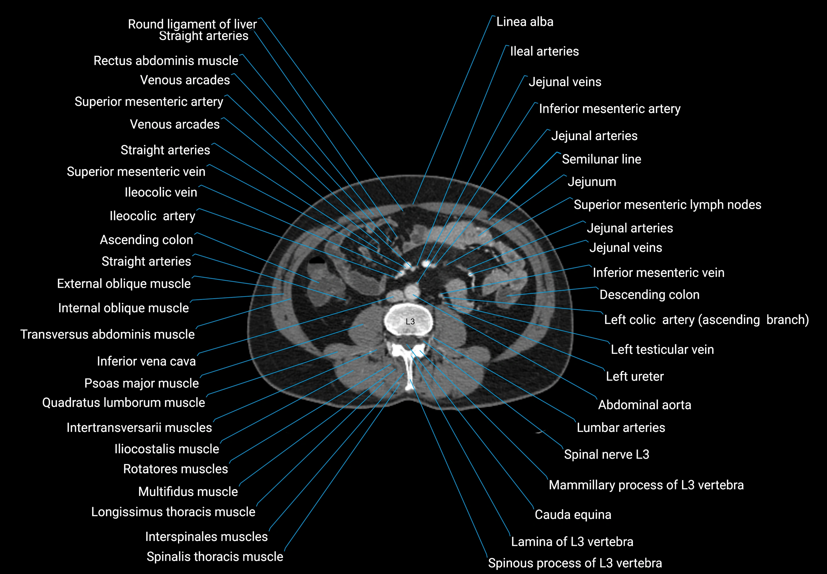
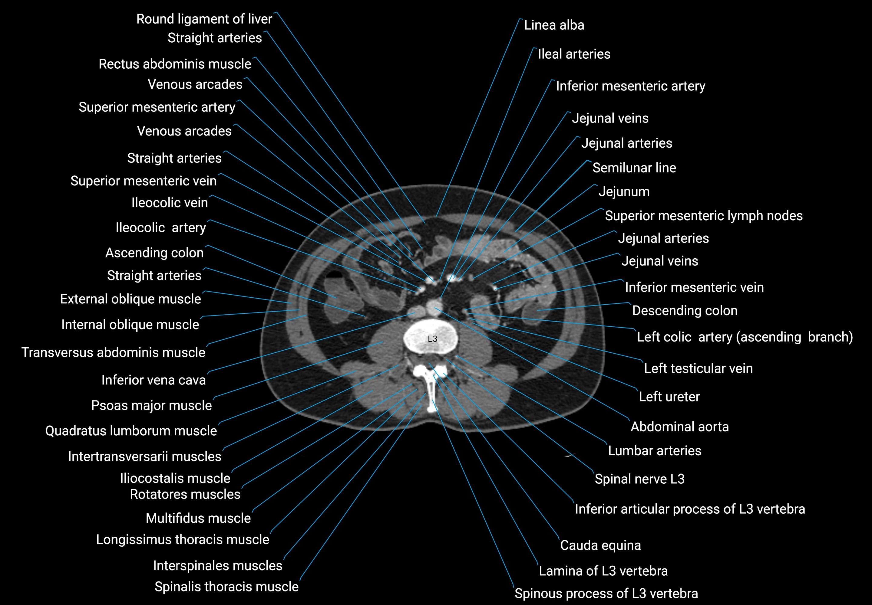
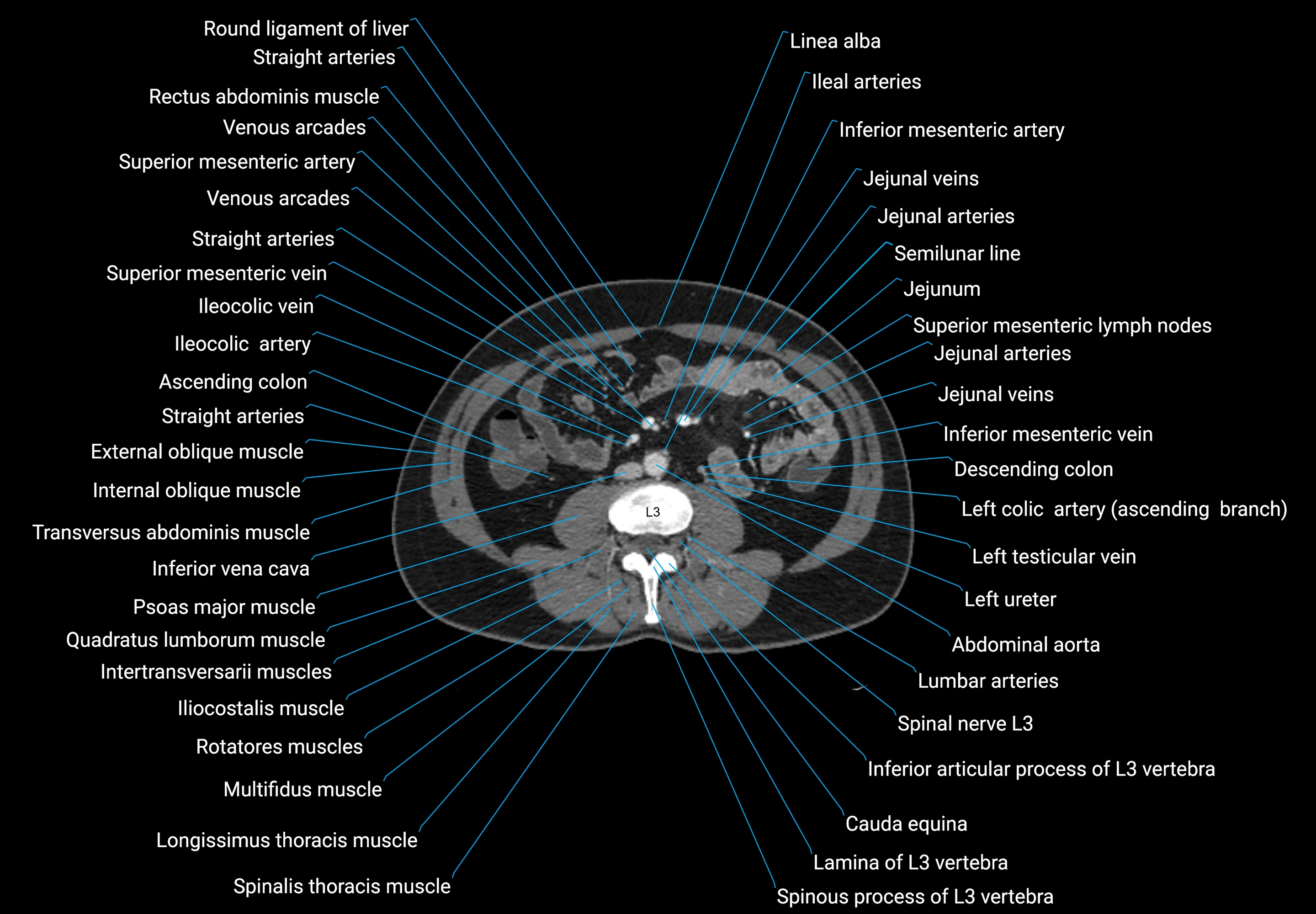
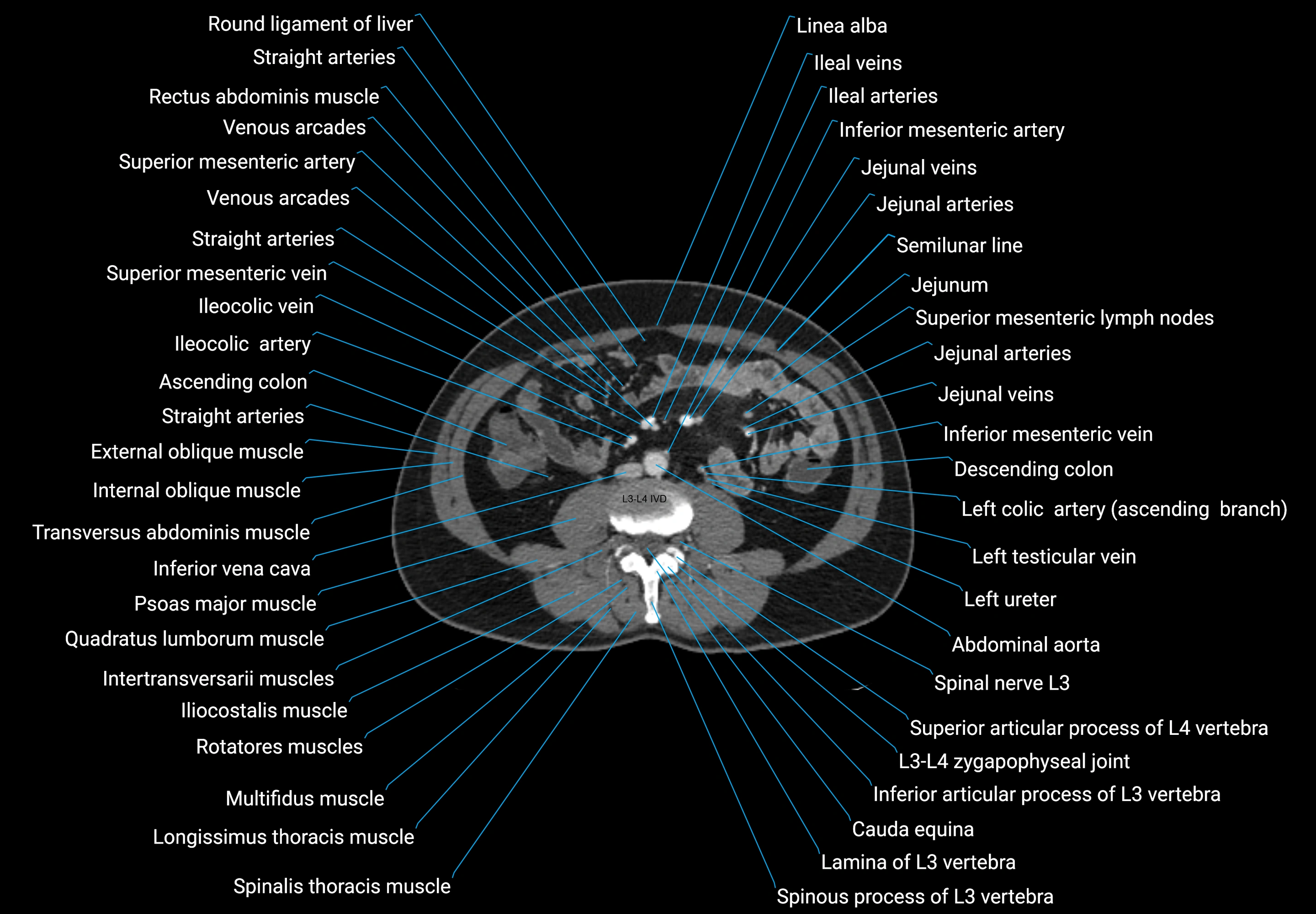
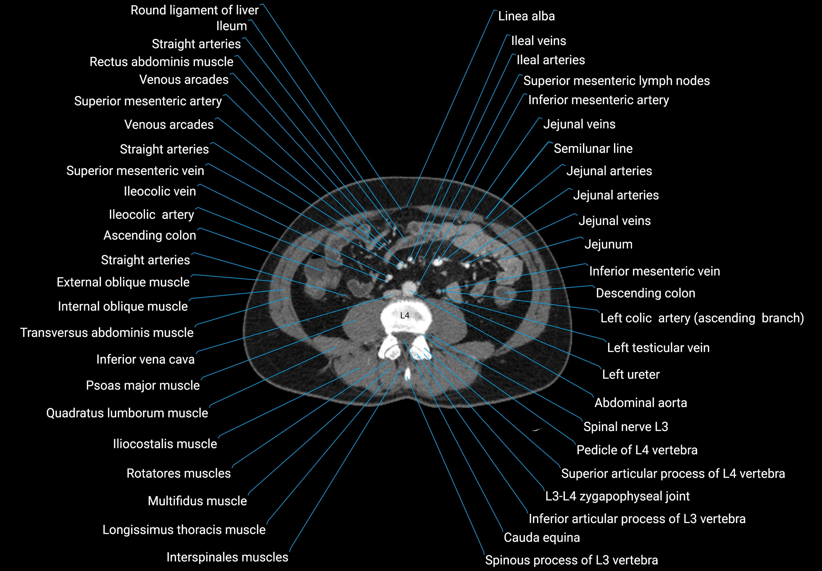
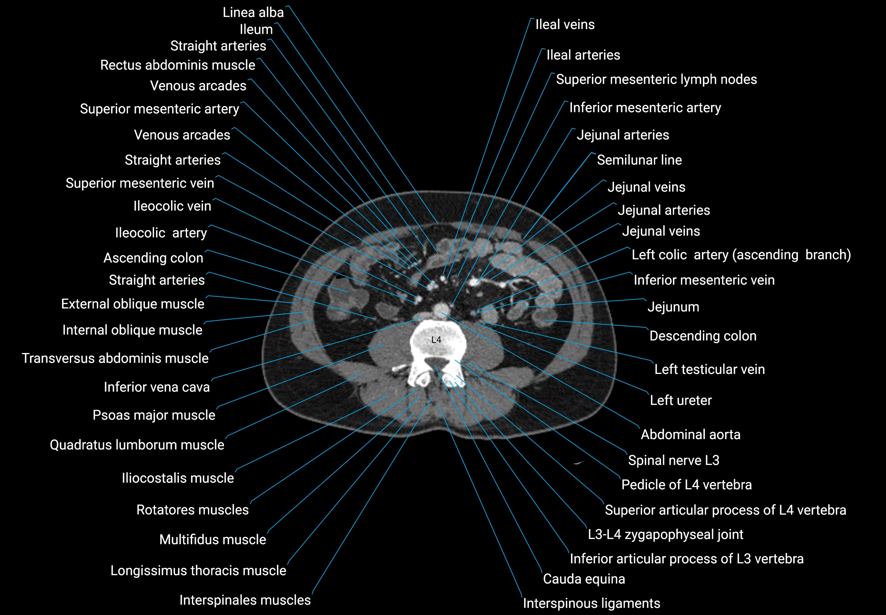
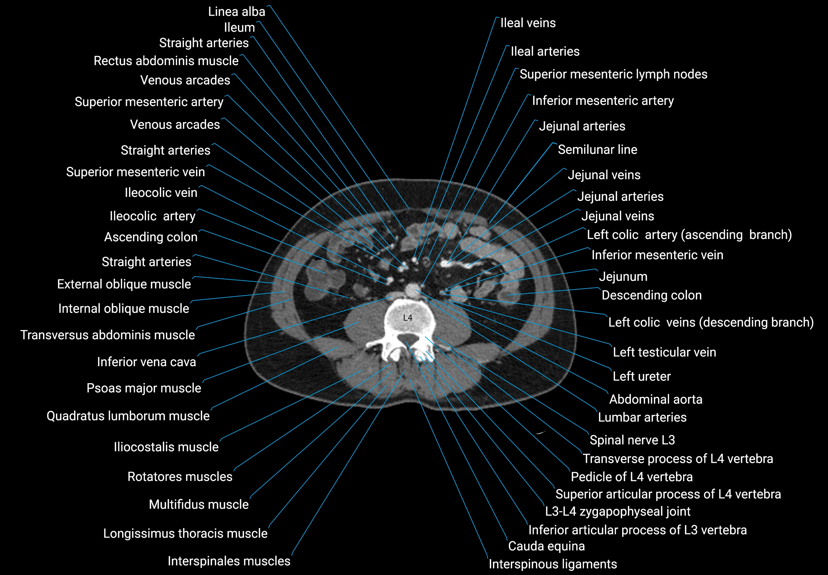
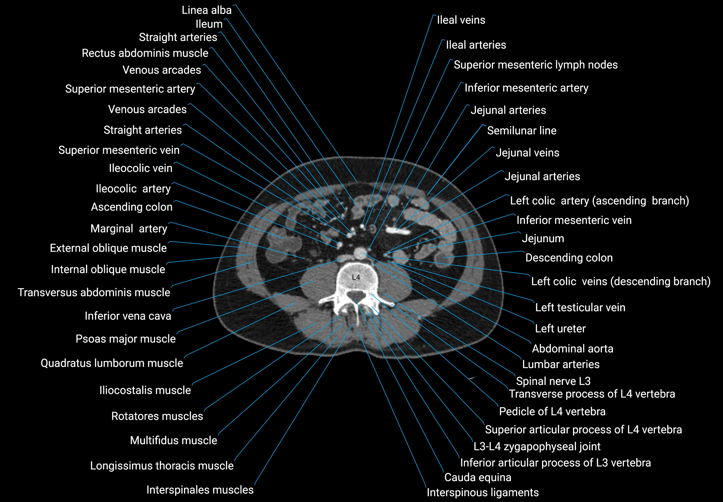
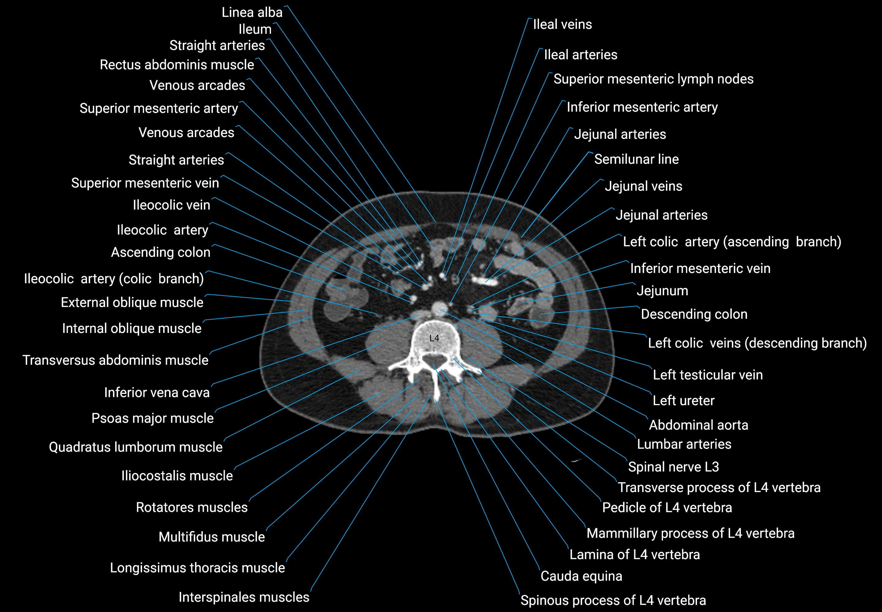
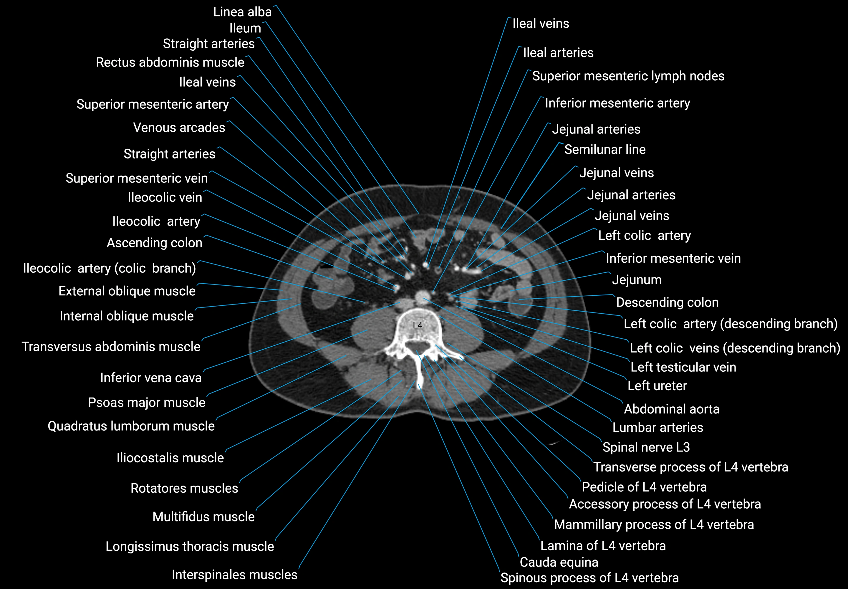
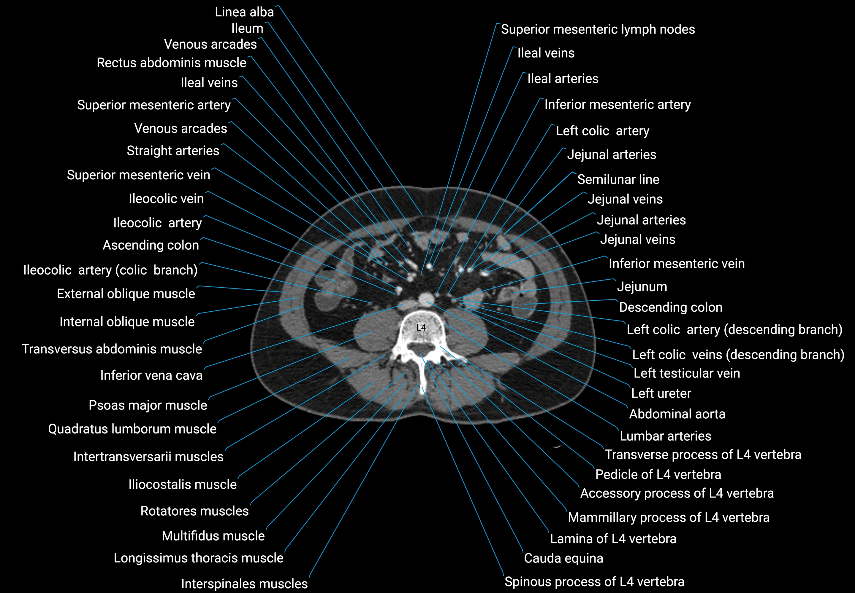
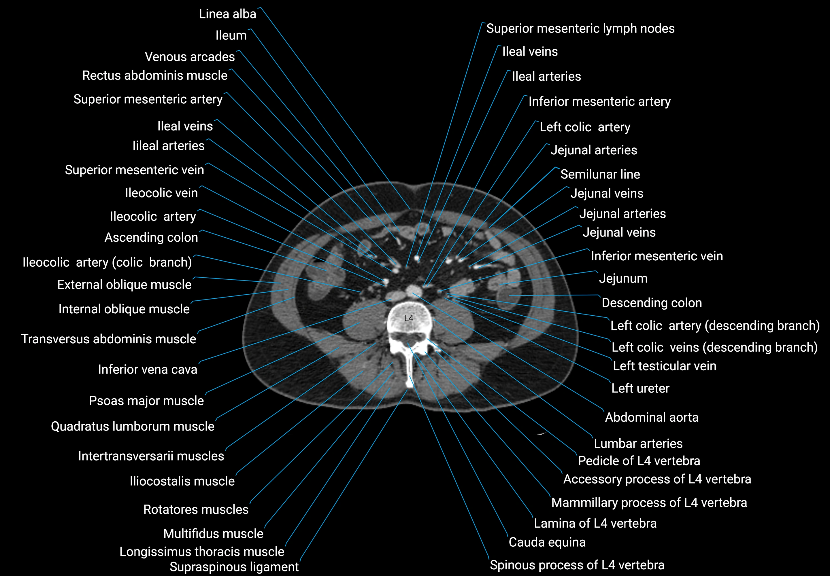
- Abdominal aorta
- Acetabular margin (Acetabular rim)
- Acetabular notch
- Acetabulum
- Adrenal glands
- Ala of ilium (wing of ilium)
- Annulus fibrosus of intervertebral disc
- Anterior lateral femoral cutaneous nerve
- Anterior right branch of portal vein
- Anterior rim of acetabulum
- Anterior sacroiliac ligament
- Aortic bifurcation
- Apex of the heart
- Ascending colon
- Azygos vein
- Basivertebral veins
- Body of ilium
- Body of rib
- Body of vertebra
- Cauda equina
- Cecum
- Celiac trunk
- Co (Coccyx)
- Common bile duct
- Common hepatic artery
- Common iliac lymph nodes
- Common iliac vein
- Conus medullaris
- Costotransverse joint
- Costotransverse ligament
- Descending colon
- Descending thoracic aorta
- Dorsal exiting nerve root
- Dorsal ramus of spinal nerve
- Dorsal root ganglion of spinal nerve
- Dorsal traversing nerve root
- Exiting nerve root of spinal nerve S1
- Exiting nerve root of spinal nerve S2
- Exiting nerve root of spinal nerve S3
- Exiting nerve root of spinal nerve S4
- Exiting nerve root of spinal nerve S5
- External oblique muscle
- Falciform ligament (liver)
- Femoral nerve
- Filum terminale internum
- Fissure for ligamentum teres
- Fissure for ligamentum venosum
- Fovea for ligament of head of femur
- Gallbladder
- Gastroduodenal artery
- Greater sciatic notch
- Head of femur
- Head of rib
- Hemiazygos vein
- Hepatic portal vein
- Hip joint
- Iliac bone
- Iliac fossa
- Iliocostalis lumborum muscle
- Iliohypogastric nerve
- Ilioinguinal nerve
- Iliolumbar ligament
- Iliopsoas muscle
- Inferior articular process of vertebra
- Inferior gluteal nerve
- Inferior mesenteric artery (IMA)
- Inferior mesenteric vein
- Inferior rectal nerve
- Inferior rim of acetabulum
- Intercostal muscles
- Intermediate sacral crest
- Internal iliac lymph nodes
- Internal iliac vein
- Internal oblique muscle
- Internal thoracic artery
- Interosseous sacroiliac ligament
- Interspinales lumborum muscle
- Interspinales muscles
- Intertransversarii muscle
- Intertransverse ligament
- Intertrochanteric crest
- Intervertebral disc space
- Intervertebral foramen
- Intra-articular ligament of head of rib
- Ischial spine
- Ischial tuberosity
- Jejunal arteries
- L (Lumbar spine)
- Lamina of vertebra
- Lateral aortic lymph nodes
- Lateral intertransversarii lumborum muscle
- Lateral part of sacrum
- Lateral sacral artery
- Lateral sacral crest
- Lateral sacral vein
- Latissimus dorsi muscle
- Left adrenal gland
- Left branch of portal vein
- Left colic artery
- Left gastric artery
- Left hepatic artery
- Left phrenic nerve
- Ligamentum teres (round ligament of the liver)
- Ligamentum venosum
- Liver
- Lumbar arteries
- Lumbar part of diaphragm
- Lumbar veins
- Lumbosacral joint
- Lumbosacral trunk
- Medial intertransversarii lumborum
- Median sacral crest
- Median sacral vein
- Mesorectal free fluid
- Neck of rib
- Obturator foramen
- Pancreas
- Pedicle of vertebra
- Penis
- Perineal nerves
- Phrenicomediastinal recess
- Phrenoesophageal ligament
- Piriformis muscle
- Posterior inferior iliac spine
- Posterior lateral femoral cutaneous nerve
- Posterior right branch of portal vein
- Posterior rim of acetabulum
- Psoas major muscle
- Quadratus lumborum muscle
- Ramus of ischium
- Rectum
- Renal artery
- Renal pelvis
- Renal vein
- Ribs
- Right branch of portal vein
- Right hepatic artery
- Rotatores lumborum muscles
- Rotatores muscle
- Rotatores thoracis muscles
- S (Sacral spine)
- Sacral cornu (sacral horn)
- Sacral hiatus
- Sacral plexus
- Sacral splanchnic nerves
- Sacrococcygeal joint
- Sciatic nerve
- Serratus posterior inferior muscle
- Short gastric veins
- Sigmoid colon
- Spinal dura mater
- Spinal epidural space
- Spinal nerve Co1
- Spinal nerve L1
- Spinal nerve L2
- Spinal nerve L3
- Spinal nerve L4
- Spinal nerve L5
- Spinal nerve S1
- Spinal nerve S2
- Spinal nerve S3
- Spinal nerve S4
- Spinal nerve S5
- Spinal nerves
- Spinalis thoracis muscle
- Spinous process of vertebra
- Splenic artery
- Sternal part of diaphragm
- Superior articular process of sacrum
- Superior articular process of vertebra
- Superior cluneal nerves
- Superior epigastric veins
- Superior gluteal nerve
- Superior pubic ramus
- Superior rim of acetabulum
- Supraspinous ligament
- T (Thoracic spine)
- Thoracic duct
- Transverse abdominal muscle
- Transverse colon
- Transverse ridges
- Trochanteric fossa
- Umbilical vein
- Urinary bladder
- Zygapophyseal joint
- left gastro-omental artery (left gastroepiploic artery)
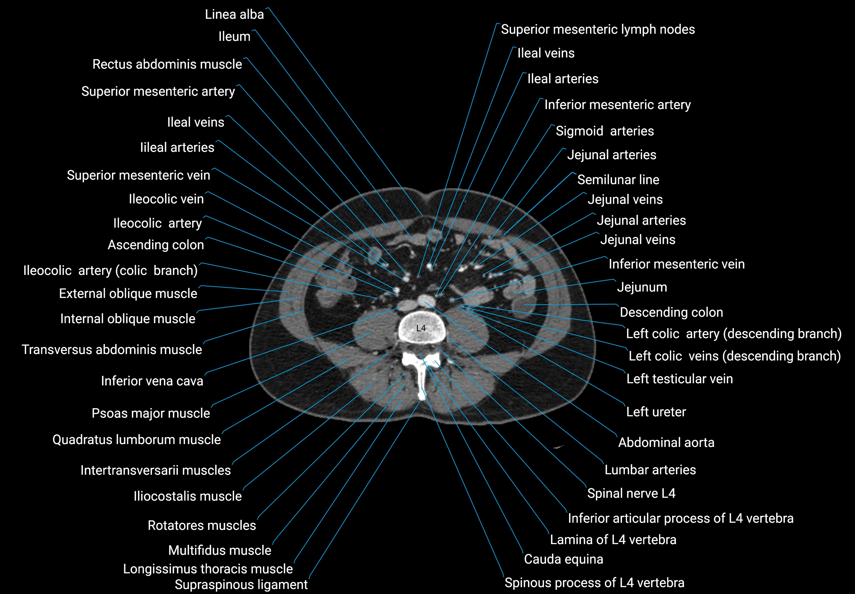
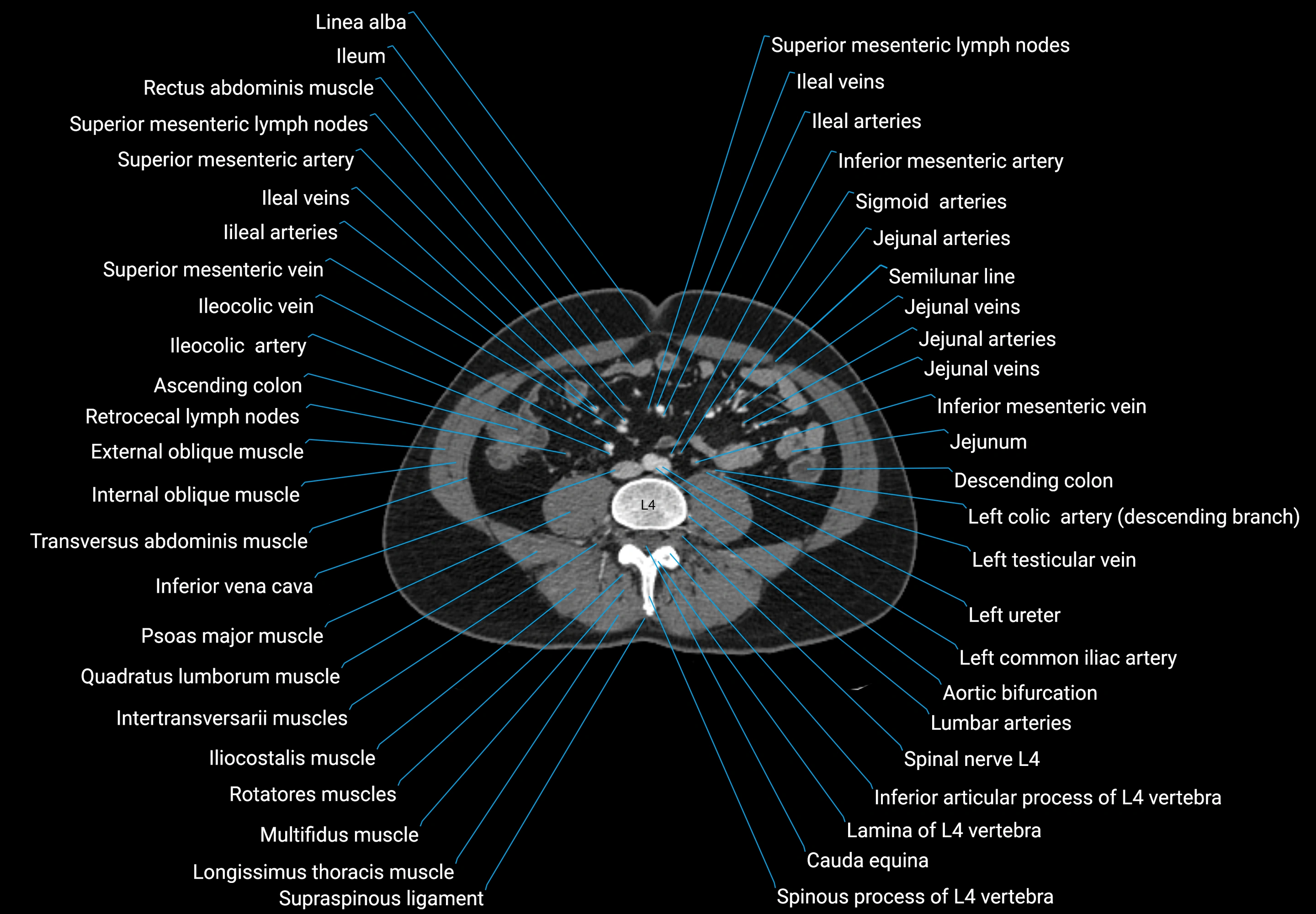
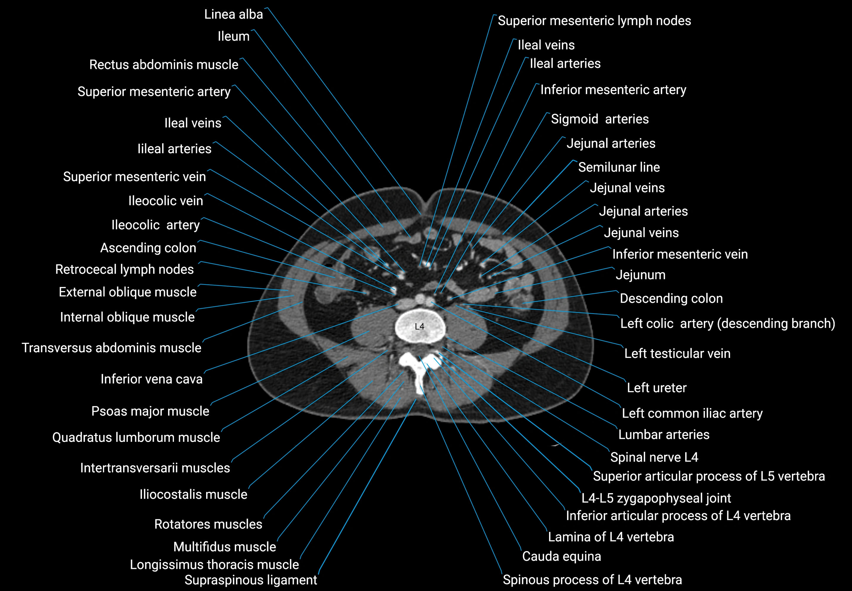
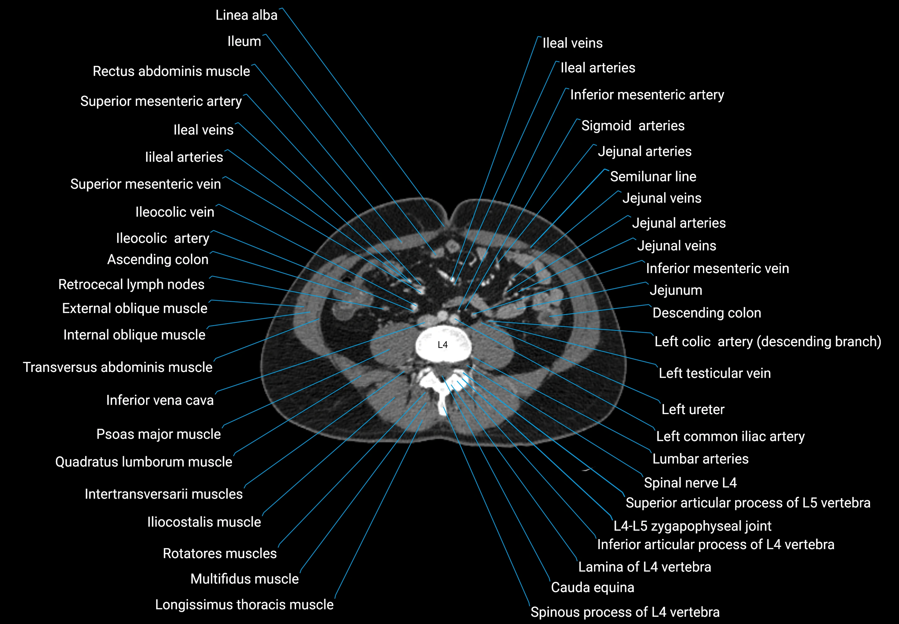
The abdominal aorta is the continuation of the thoracic aorta, beginning at the level of the aortic hiatus of the diaphragm (T12 vertebra) and terminating at the level of the L4 vertebra where it bifurcates into the right and left common iliac arteries. It lies slightly to the left of the midline and courses anterior to the vertebral bodies, surrounded by the retroperitoneal structures of the abdomen.
The abdominal aorta gives off numerous visceral and parietal branches, supplying the abdominal organs, pelvic structures, and lower limbs. It is the main conduit of oxygenated blood from the heart to the abdomen and lower body. The aorta is clinically significant as the common site of aneurysm, dissection, atherosclerosis, and traumatic injury.
Synonyms
-
Aorta abdominalis
-
Infradiaphragmatic aorta
-
Abdominal portion of aorta
Function
-
Conducts oxygenated blood from the thoracic aorta to abdominal, pelvic, and lower limb structures
-
Provides direct arterial supply to major abdominal organs (liver, spleen, kidneys, intestines)
-
Maintains systemic blood flow and hemodynamic regulation
-
Plays a central role in surgical and interventional procedures (aneurysm repair, stent grafts)
Branches
-
Unpaired visceral branches: celiac trunk, superior mesenteric artery (SMA), inferior mesenteric artery (IMA)
-
Paired visceral branches: middle suprarenal arteries, renal arteries, gonadal arteries (testicular or ovarian)
-
Parietal branches: inferior phrenic arteries, lumbar arteries, median sacral artery
-
Terminal branches: right and left common iliac arteries
MRI Appearance
T1-weighted images:
-
Flowing blood appears as a signal void (black lumen)
-
Vessel wall appears as a thin hypointense rim; retroperitoneal fat enhances contrast
T2-weighted images:
-
Lumen remains a signal void due to flow
-
Adjacent edema, hematoma, or aneurysm wall thrombus may appear hyperintense
STIR (Short Tau Inversion Recovery):
-
Fat suppression improves visualization of the aortic wall and periaortic tissues
-
Wall edema, inflammation, or periaortic hematoma appears hyperintense
-
Useful in vasculitis, dissection, or trauma
T1 Post-Contrast (Gadolinium-enhanced):
-
Aortic lumen enhances brightly and homogeneously
-
Clearly demonstrates aneurysm, stenosis, dissection, mural thrombus, or aortic wall enhancement in vasculitis
MRA (Magnetic Resonance Angiography):
-
Contrast-enhanced MRA provides high-resolution imaging of the aorta and its branches
-
Allows 3D reconstruction of visceral, parietal, and terminal branches
-
Excellent for evaluating aneurysm size, dissection flap, stenosis, or preoperative planning
-
Non-invasive alternative to conventional angiography
CT Appearance
Non-contrast CT:
-
Appears as a tubular soft tissue structure anterior to vertebral bodies
-
Calcified atherosclerotic plaques appear as hyperdense foci along the wall
-
Useful for screening abdominal aortic aneurysm (AAA) size and mural calcification
Contrast-enhanced CT (CTA):
-
Gold standard for abdominal aortic imaging
-
Provides excellent detail of lumen, wall, aneurysm, thrombus, and branch vessels
-
Multiplanar and 3D reconstructions help in aneurysm measurement, stent graft planning, and dissection evaluation
-
Detects acute rupture, traumatic injury, or occlusion with high sensitivity
MRI images

MRI images

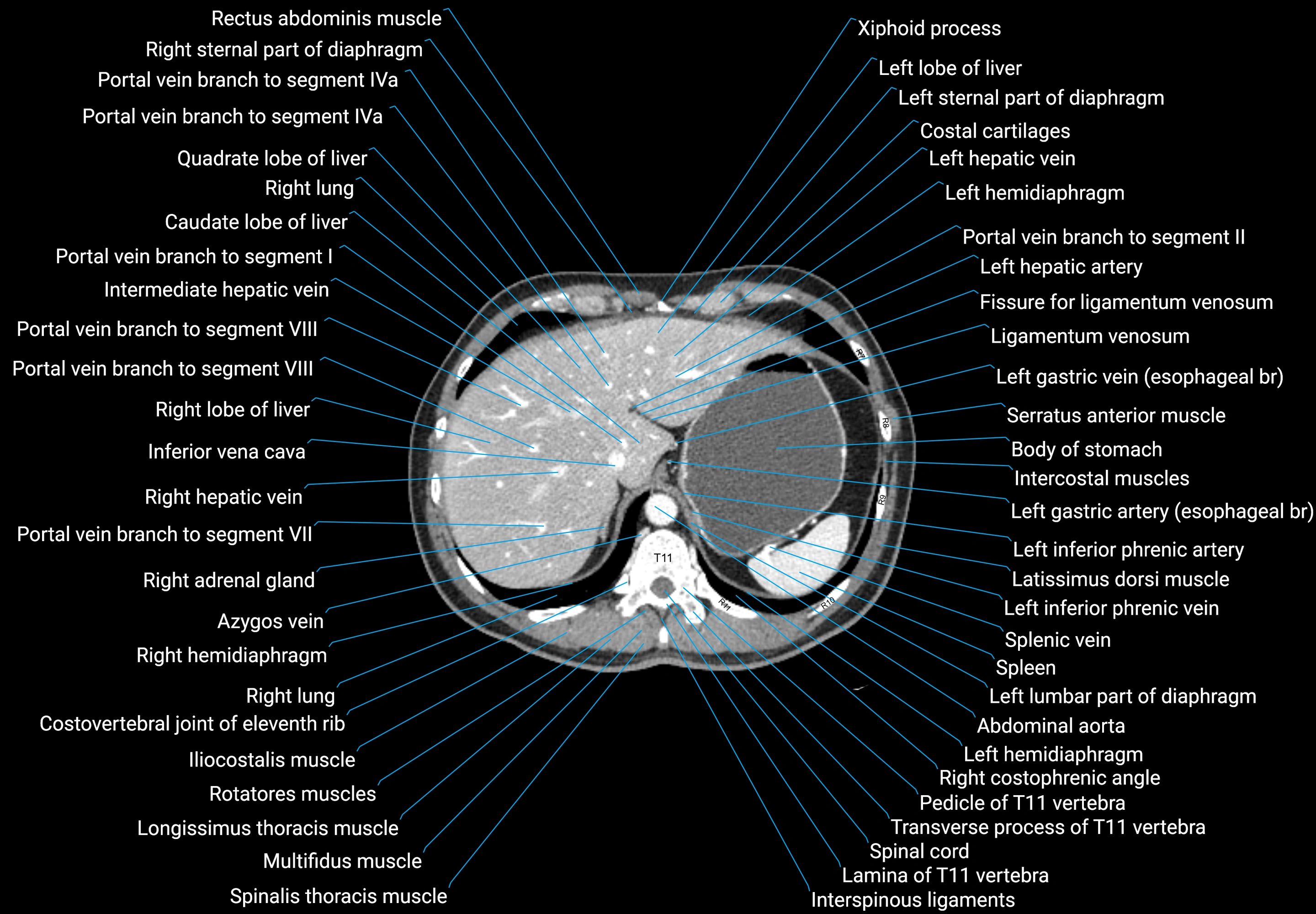
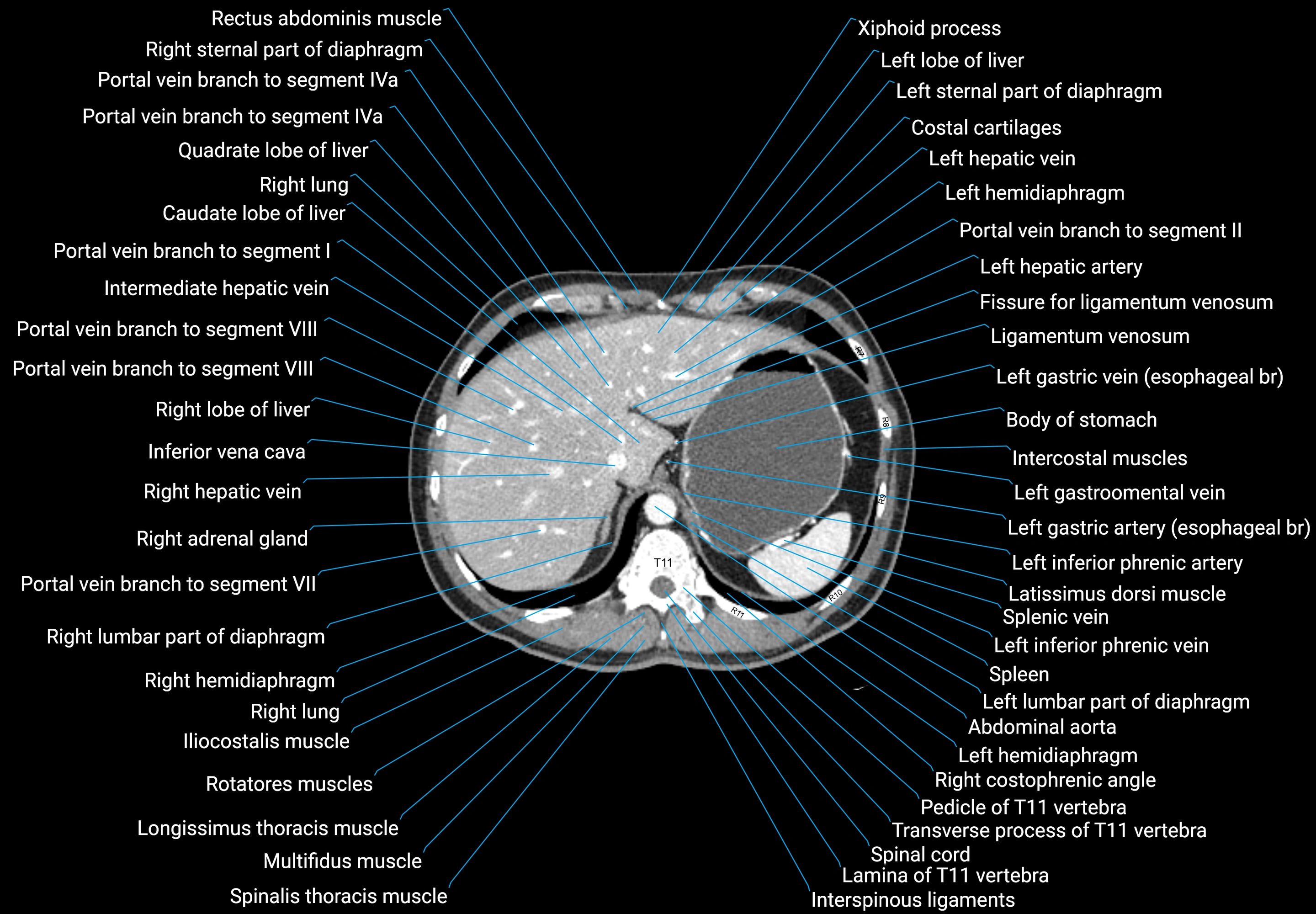
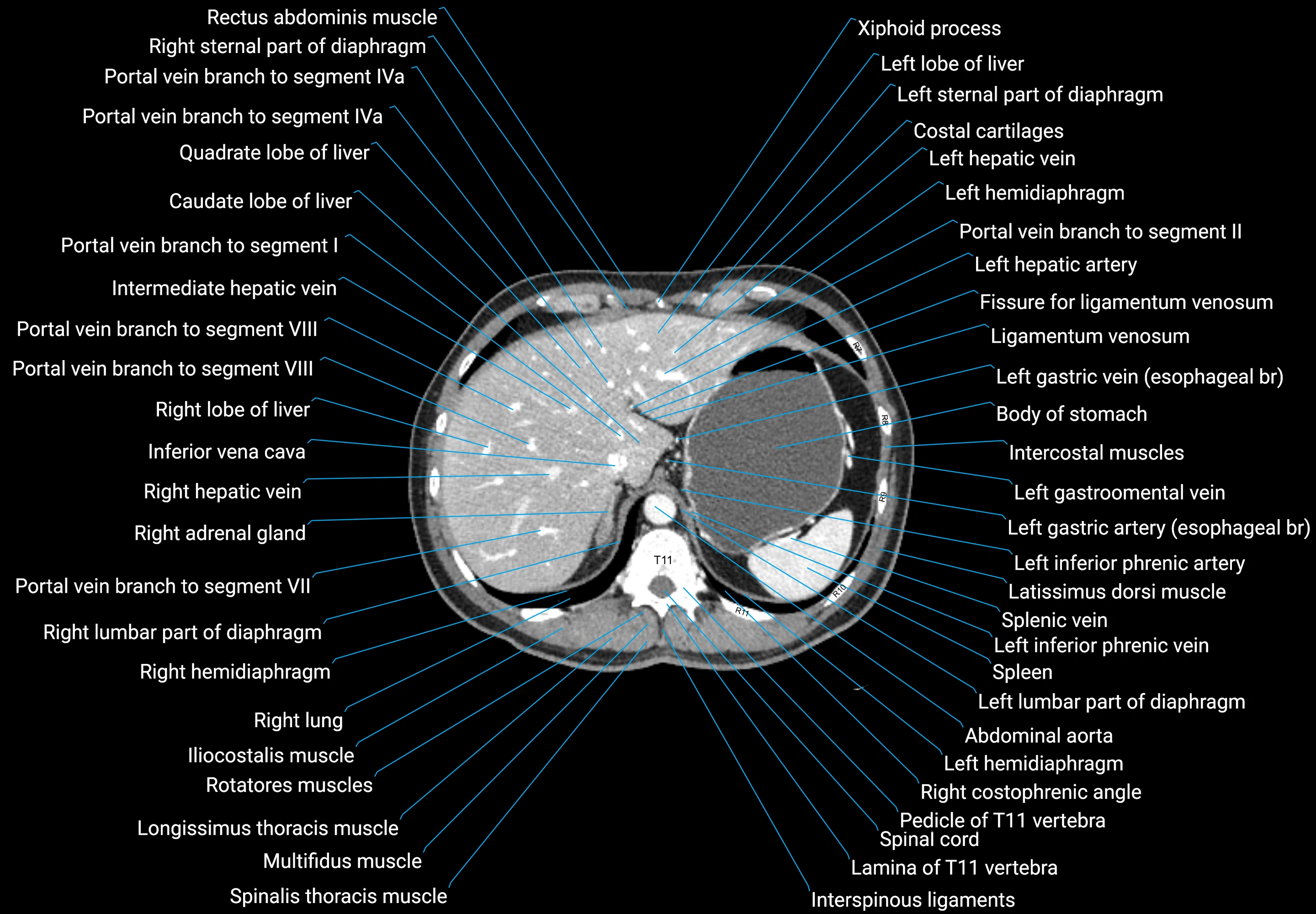
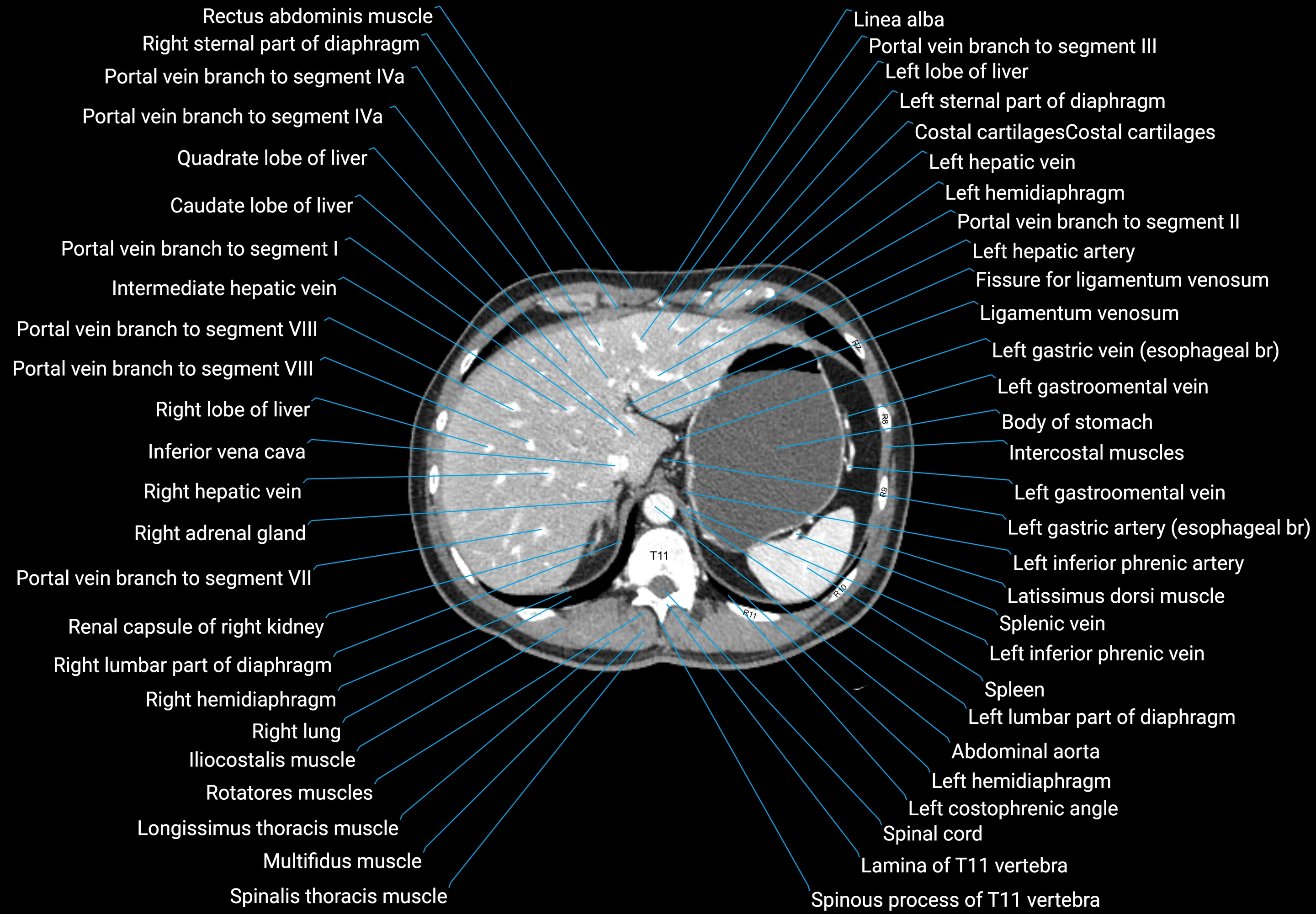
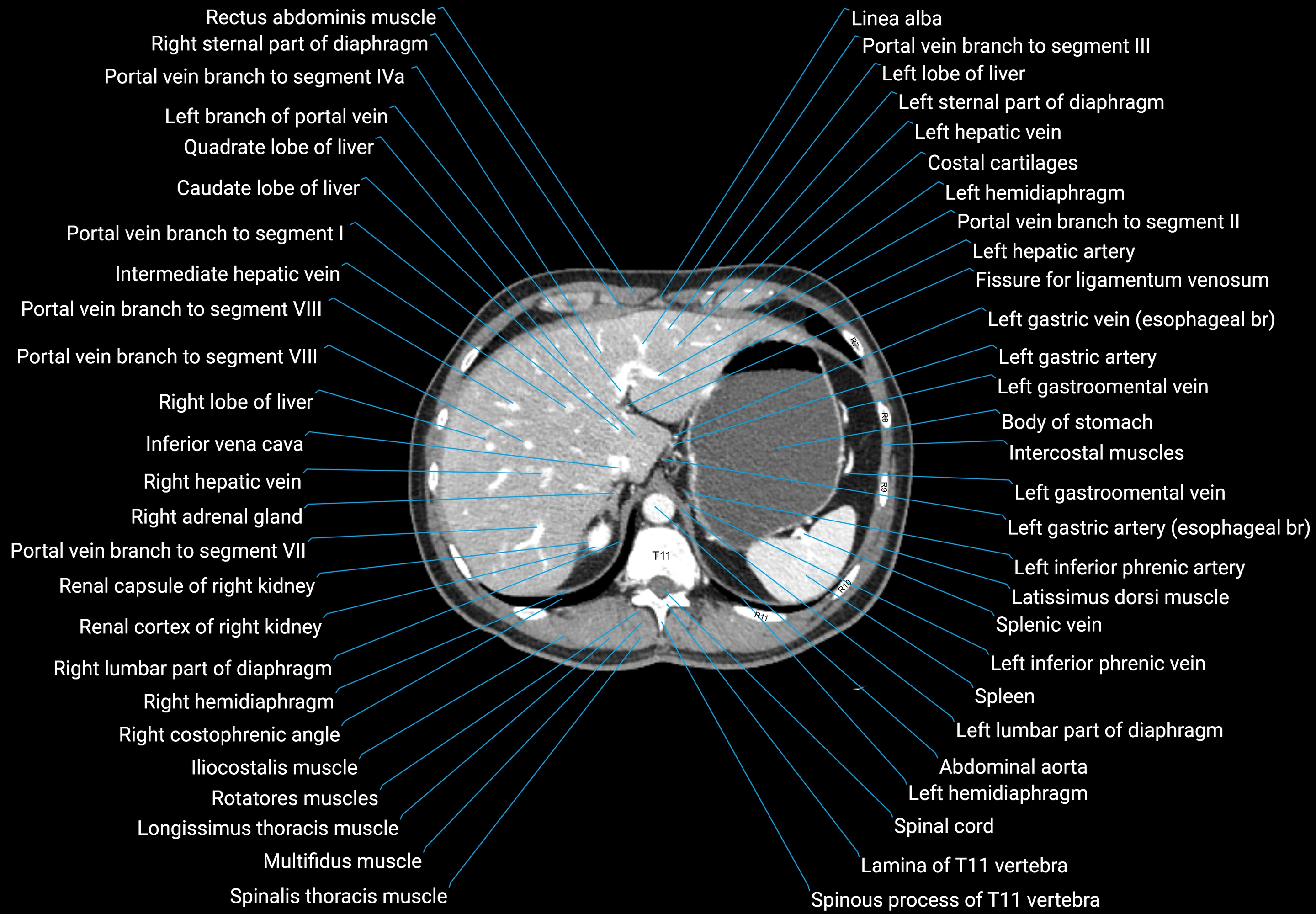
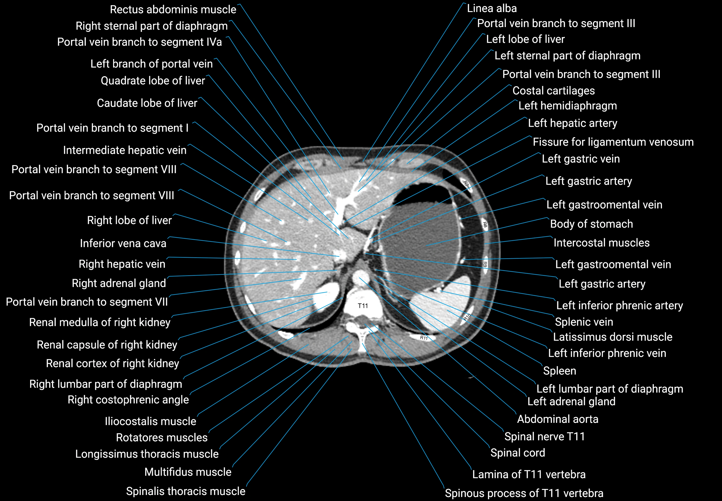
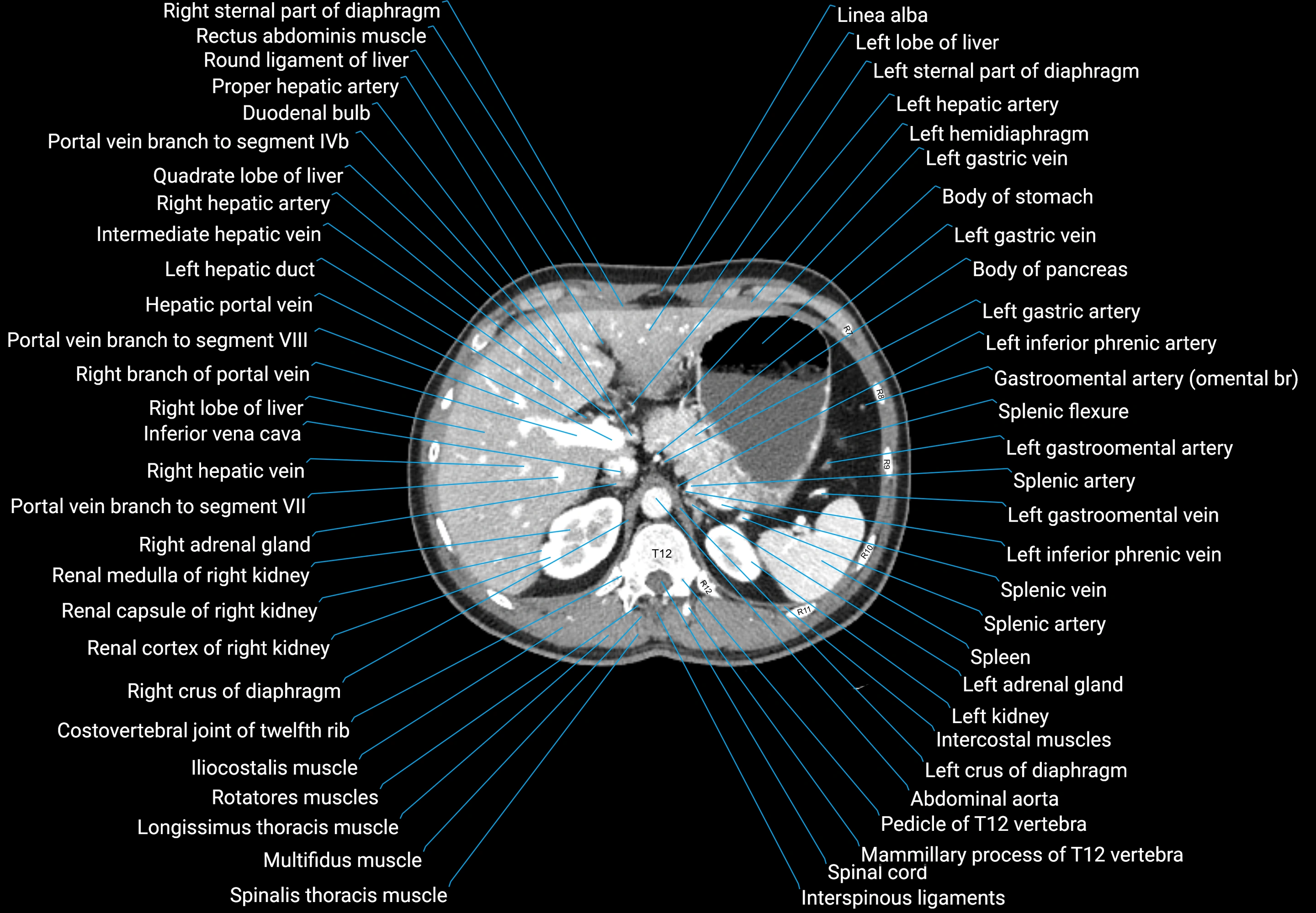
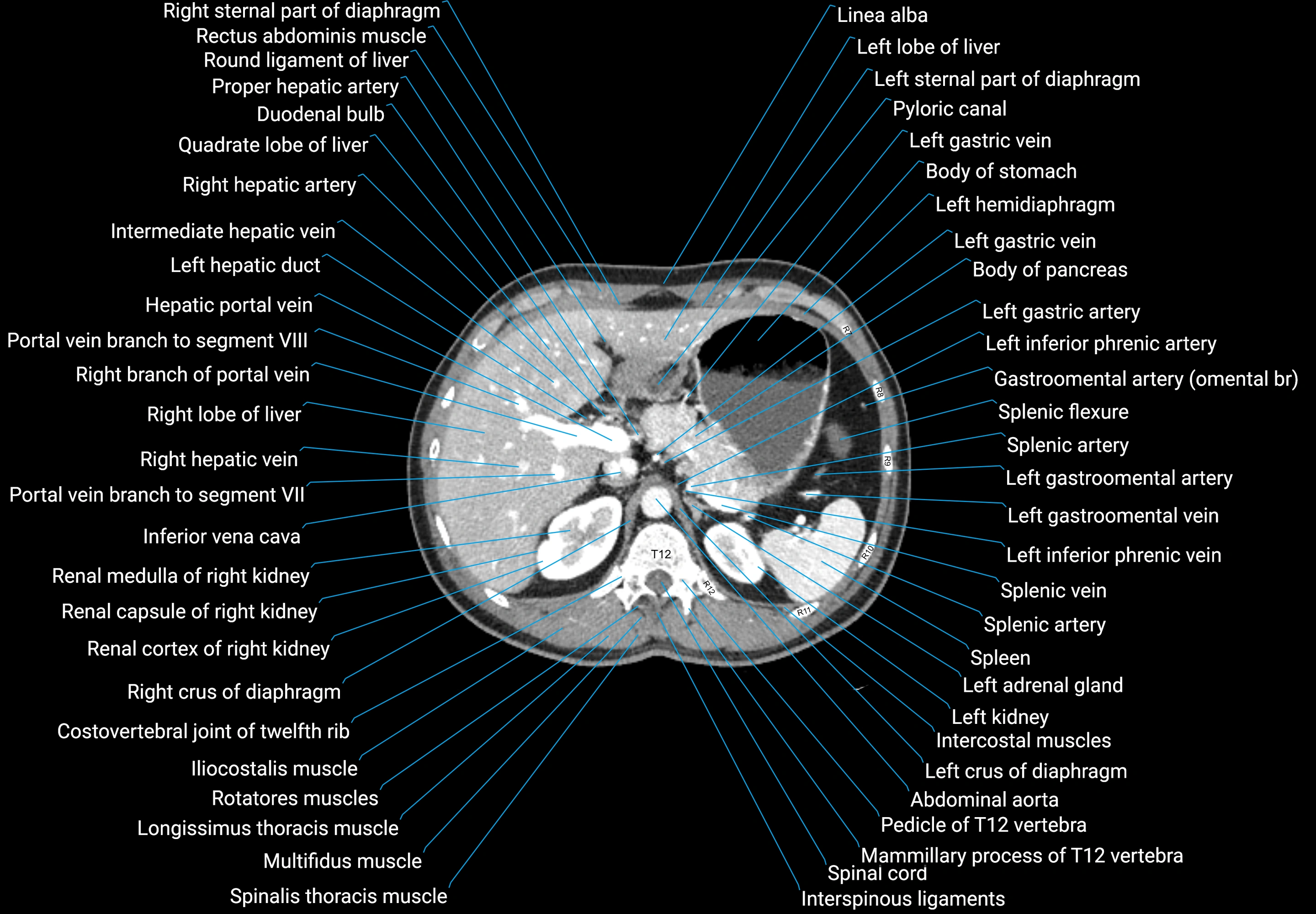
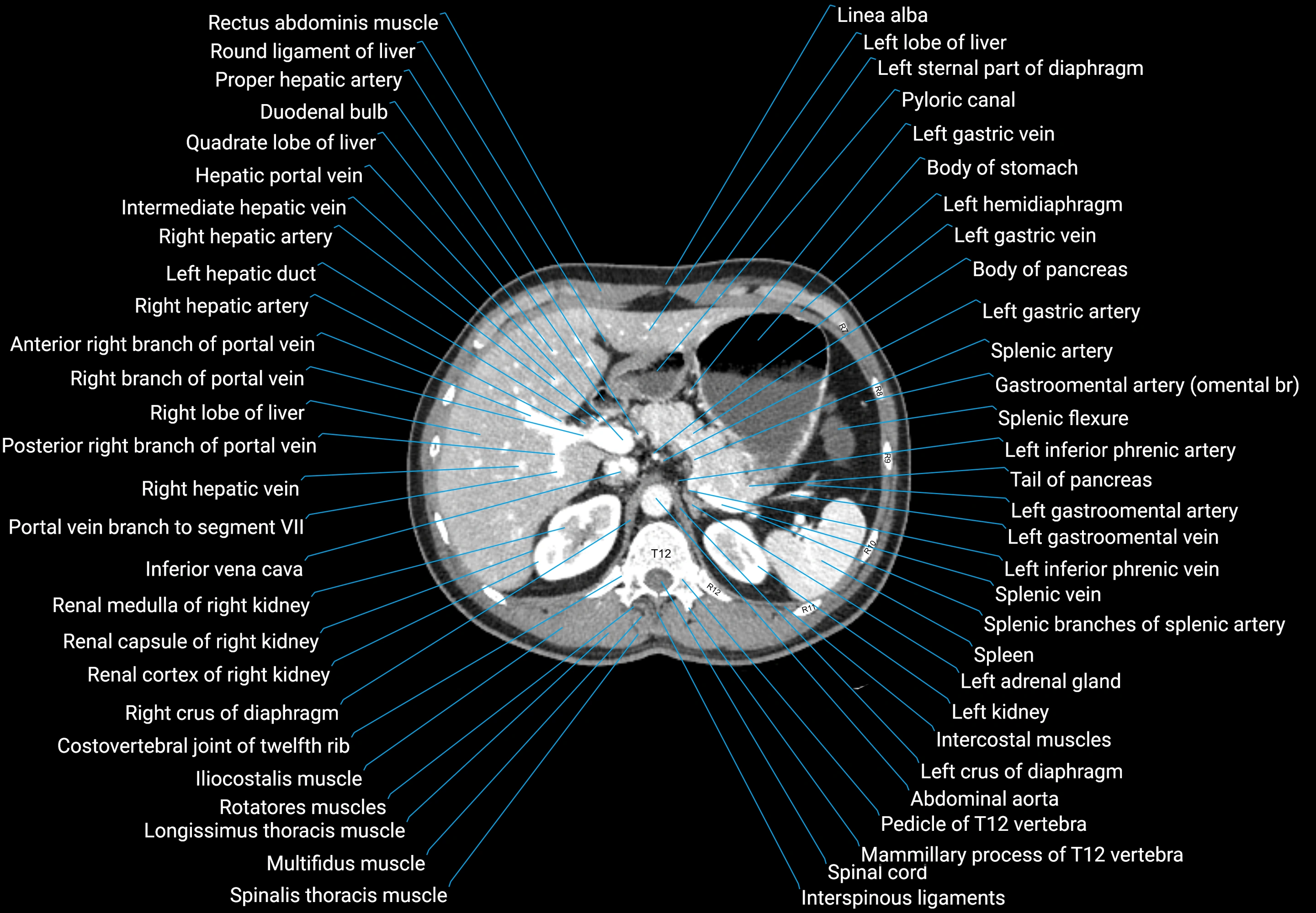
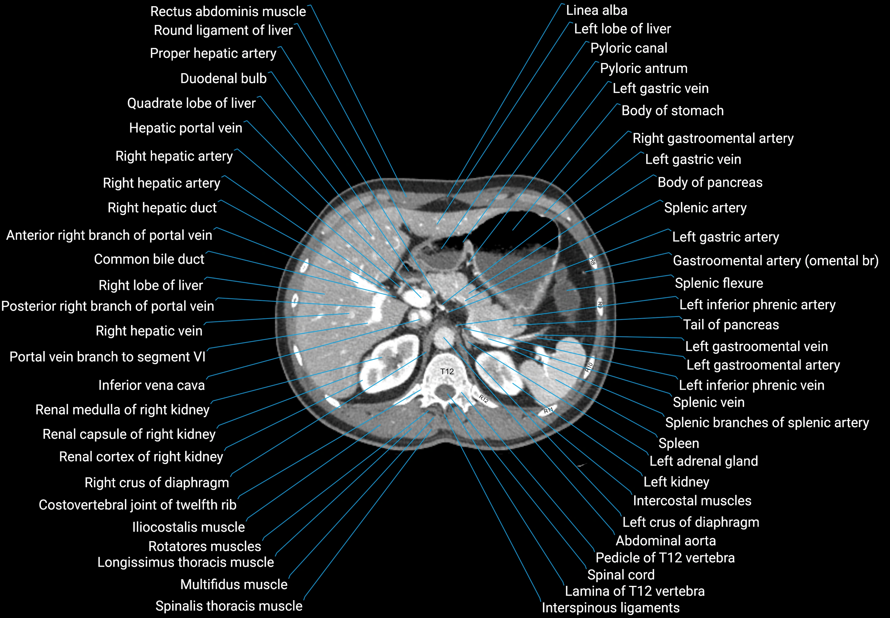
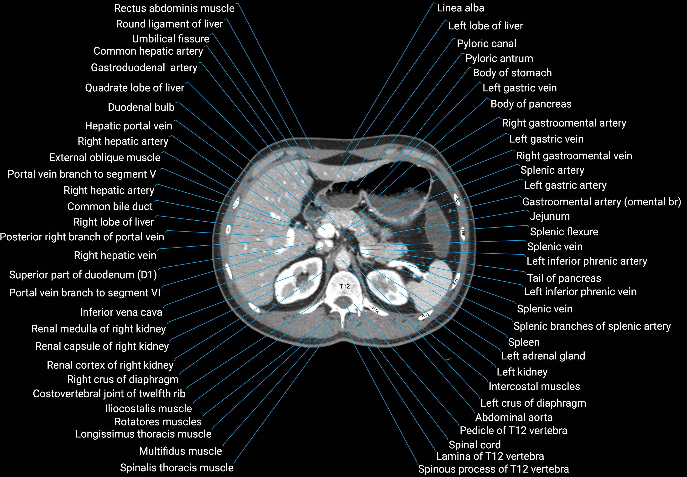
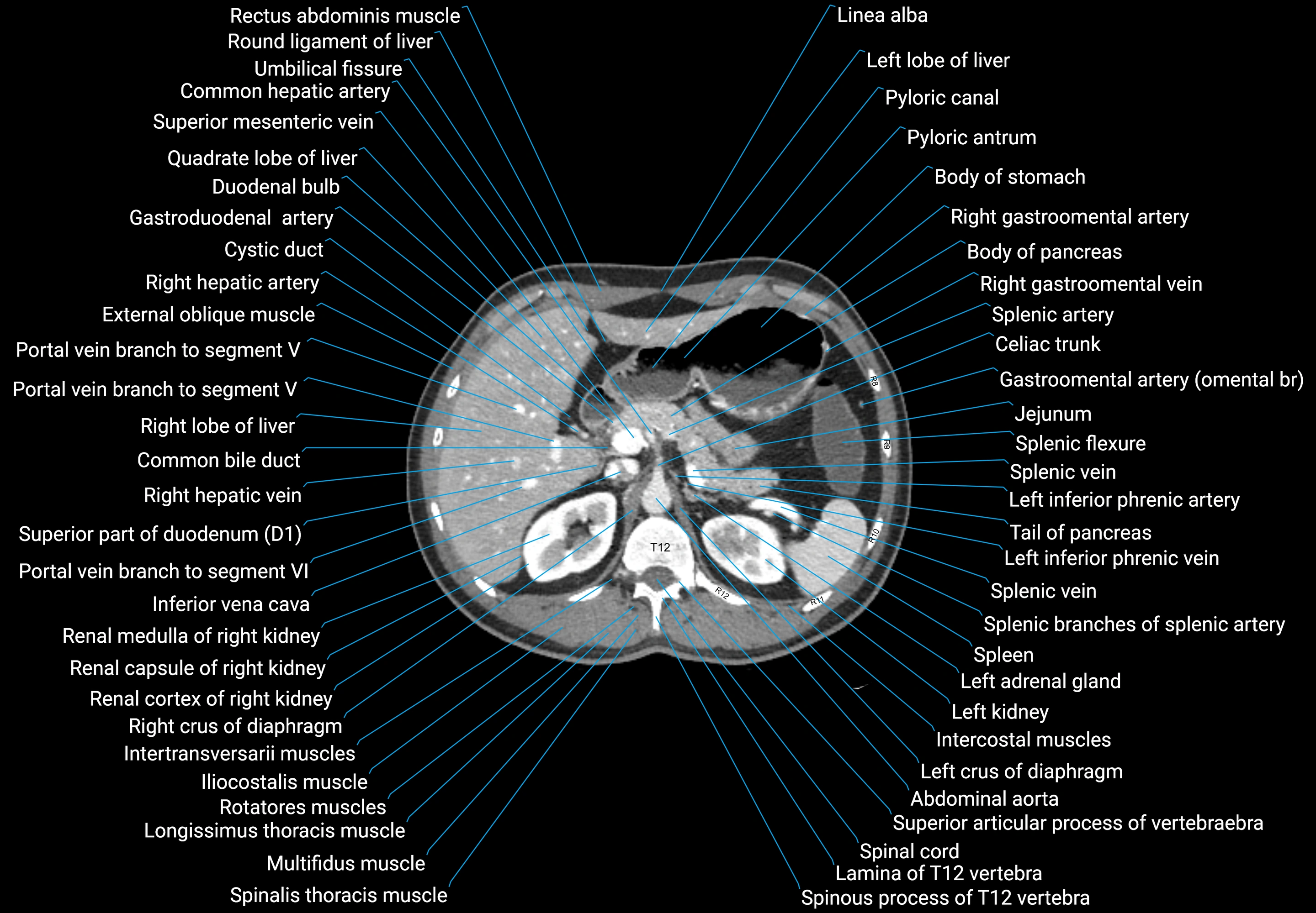
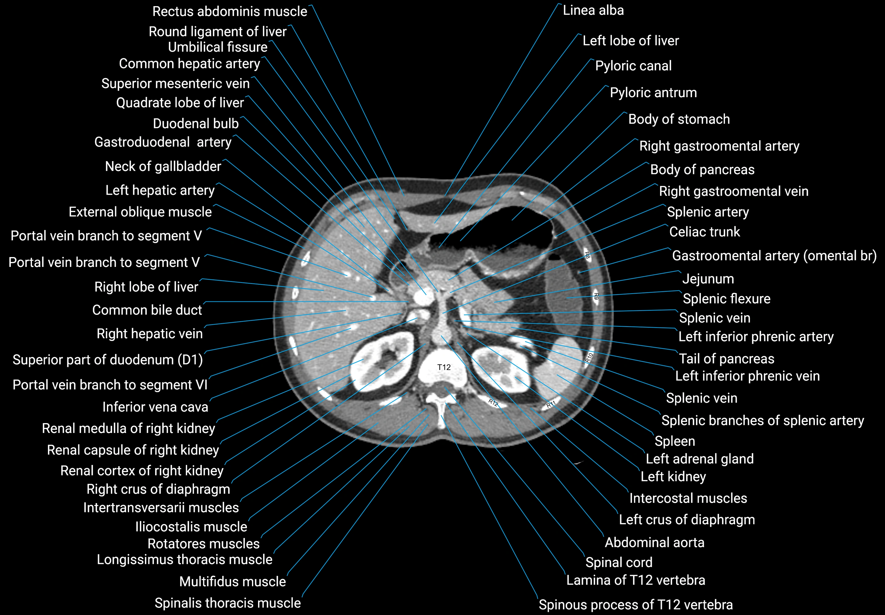
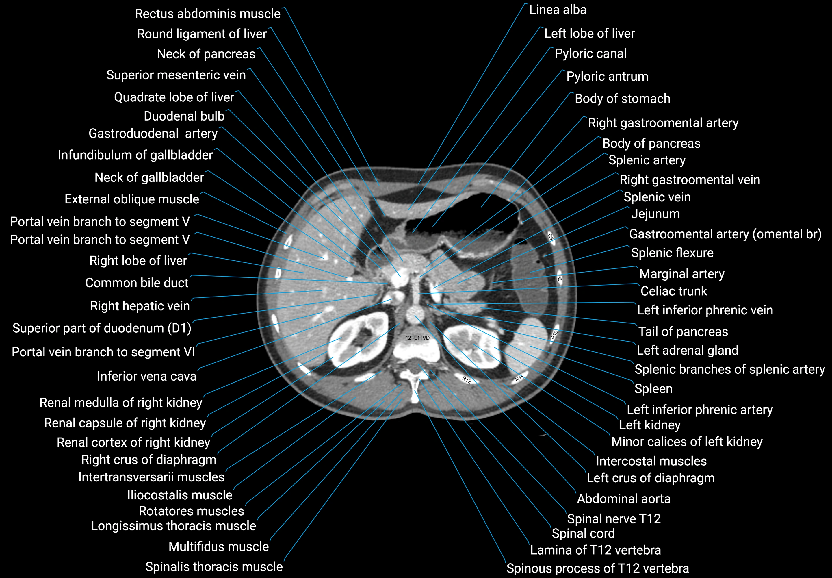
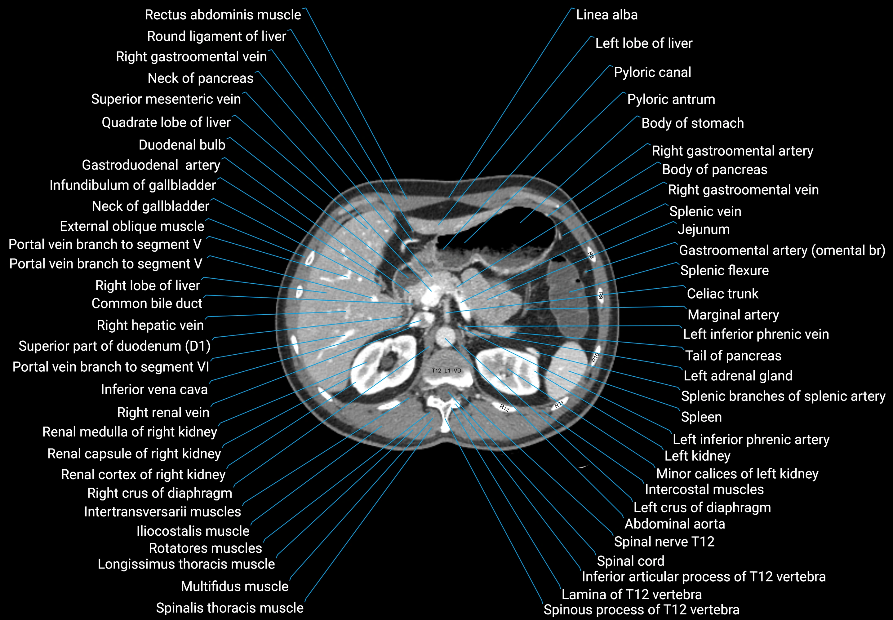
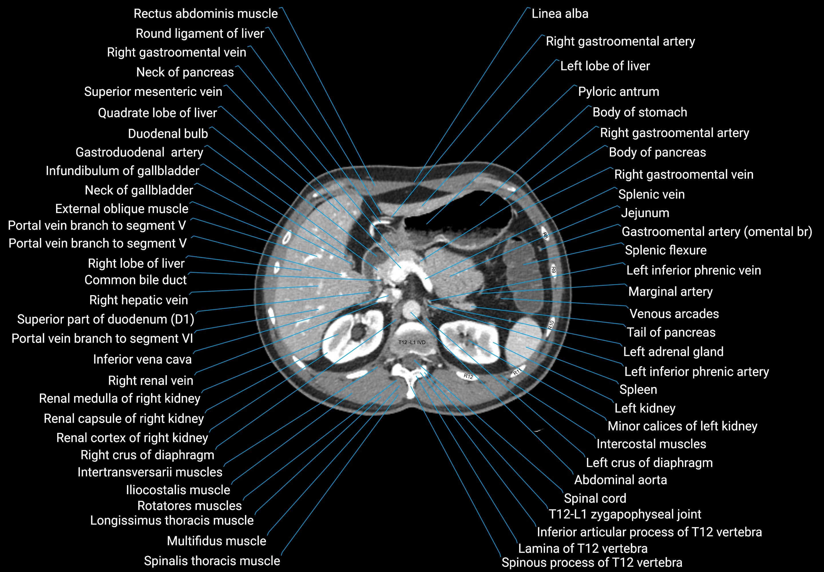
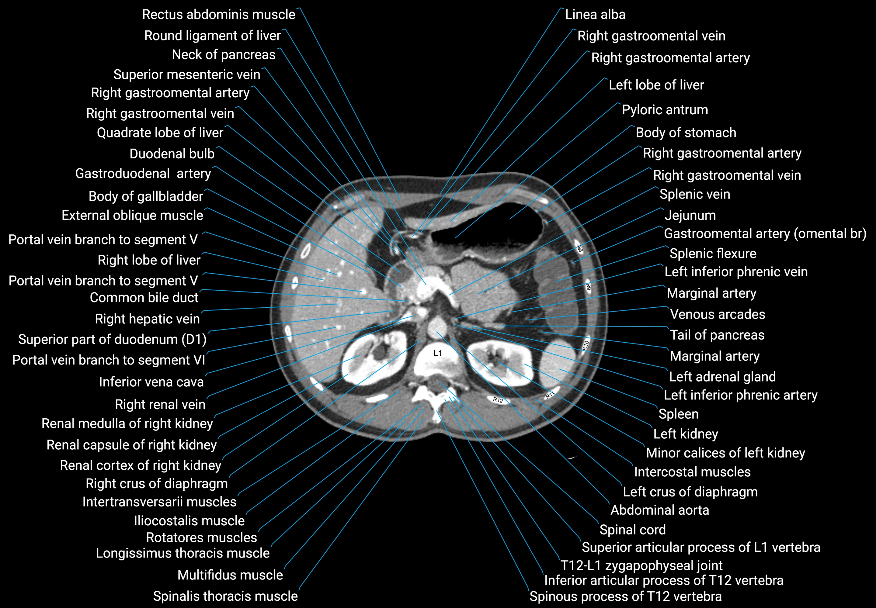
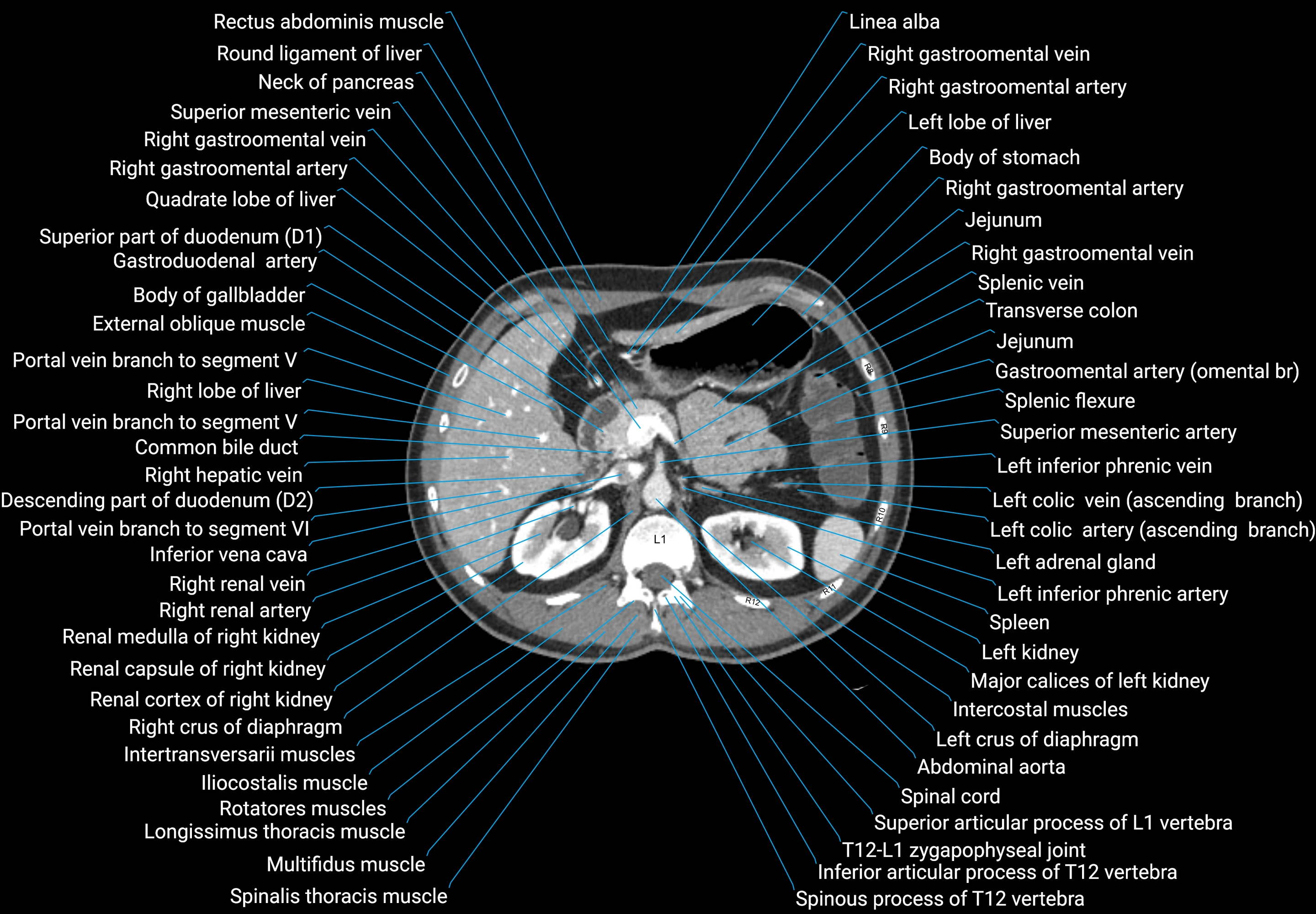
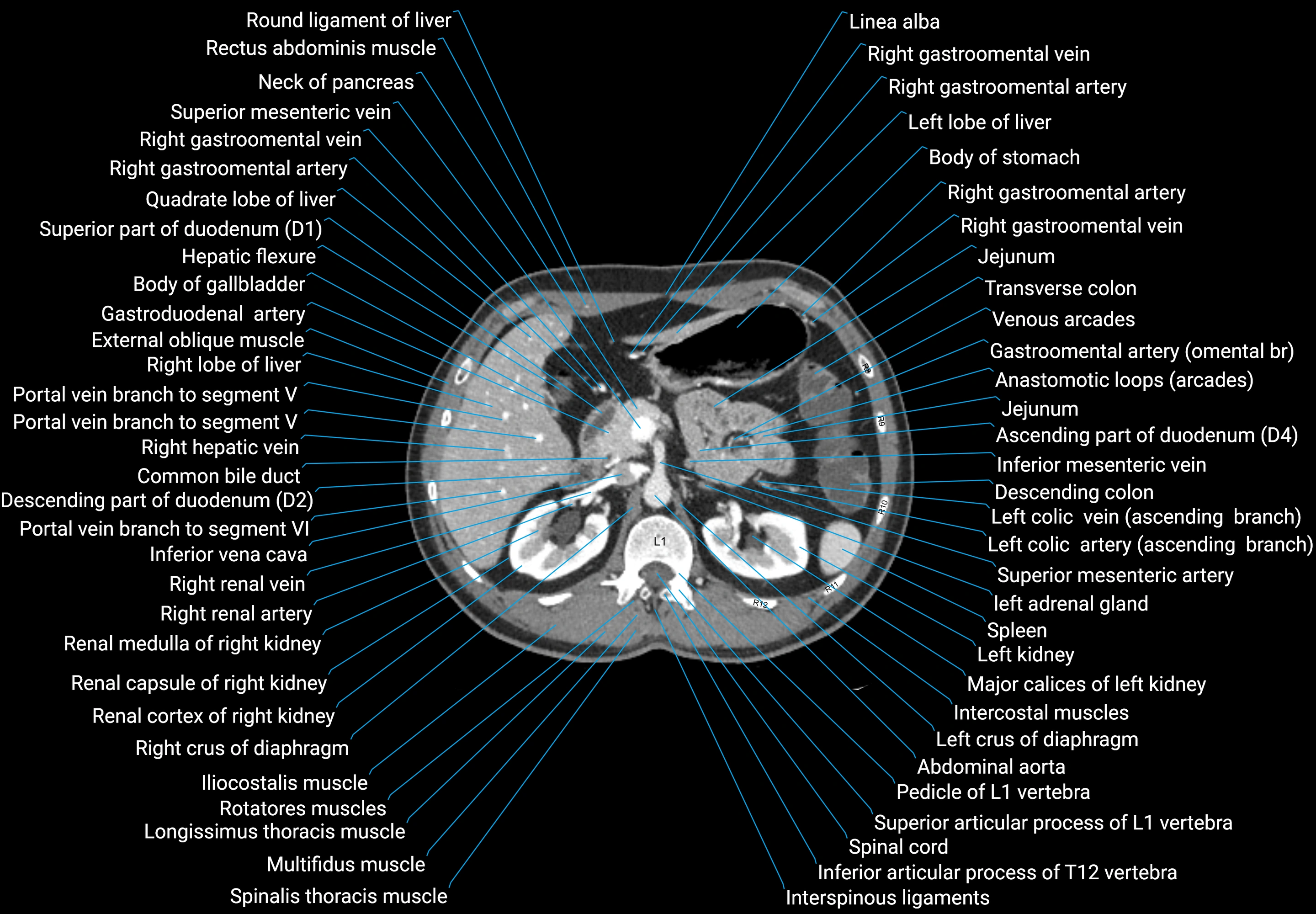
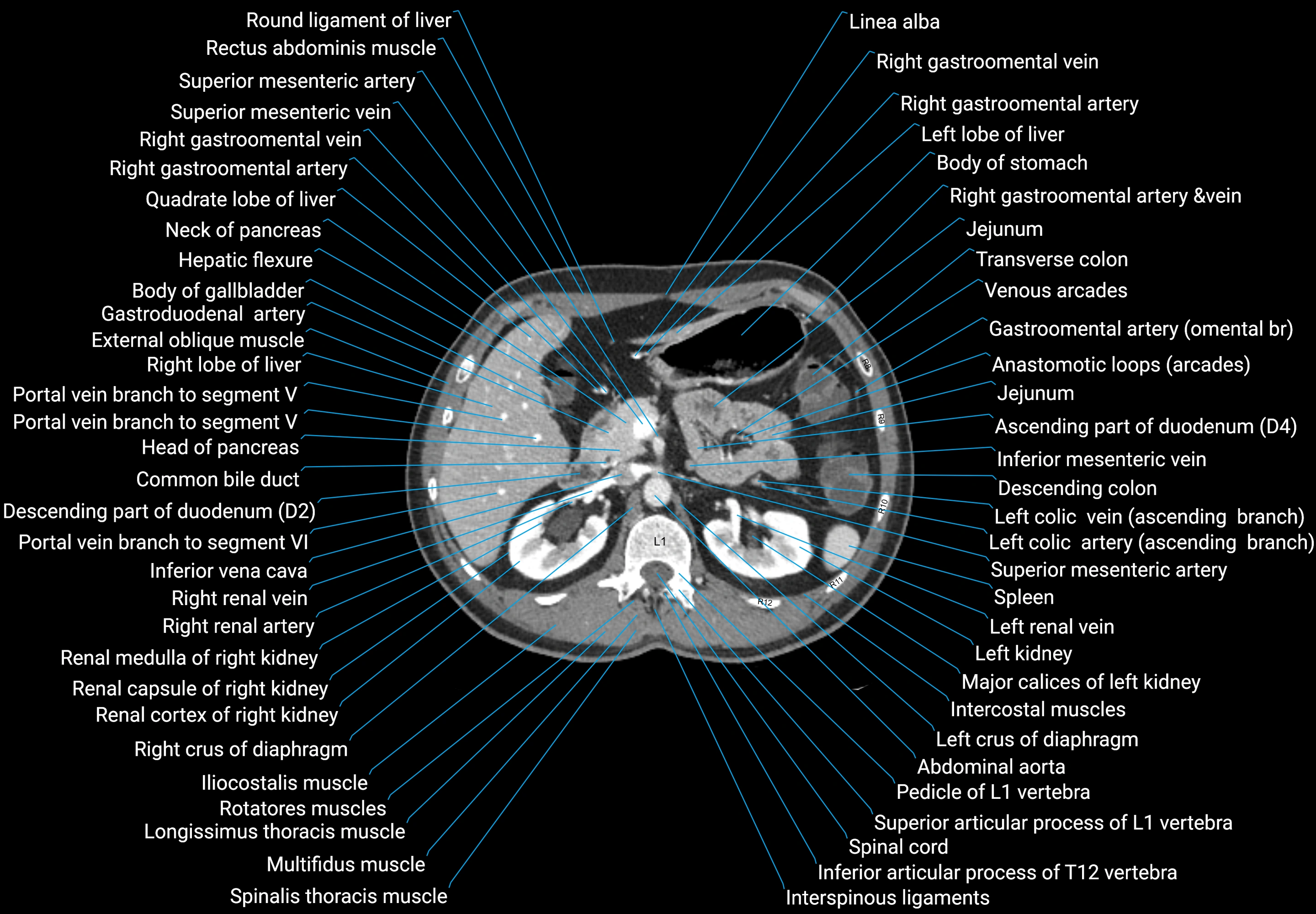
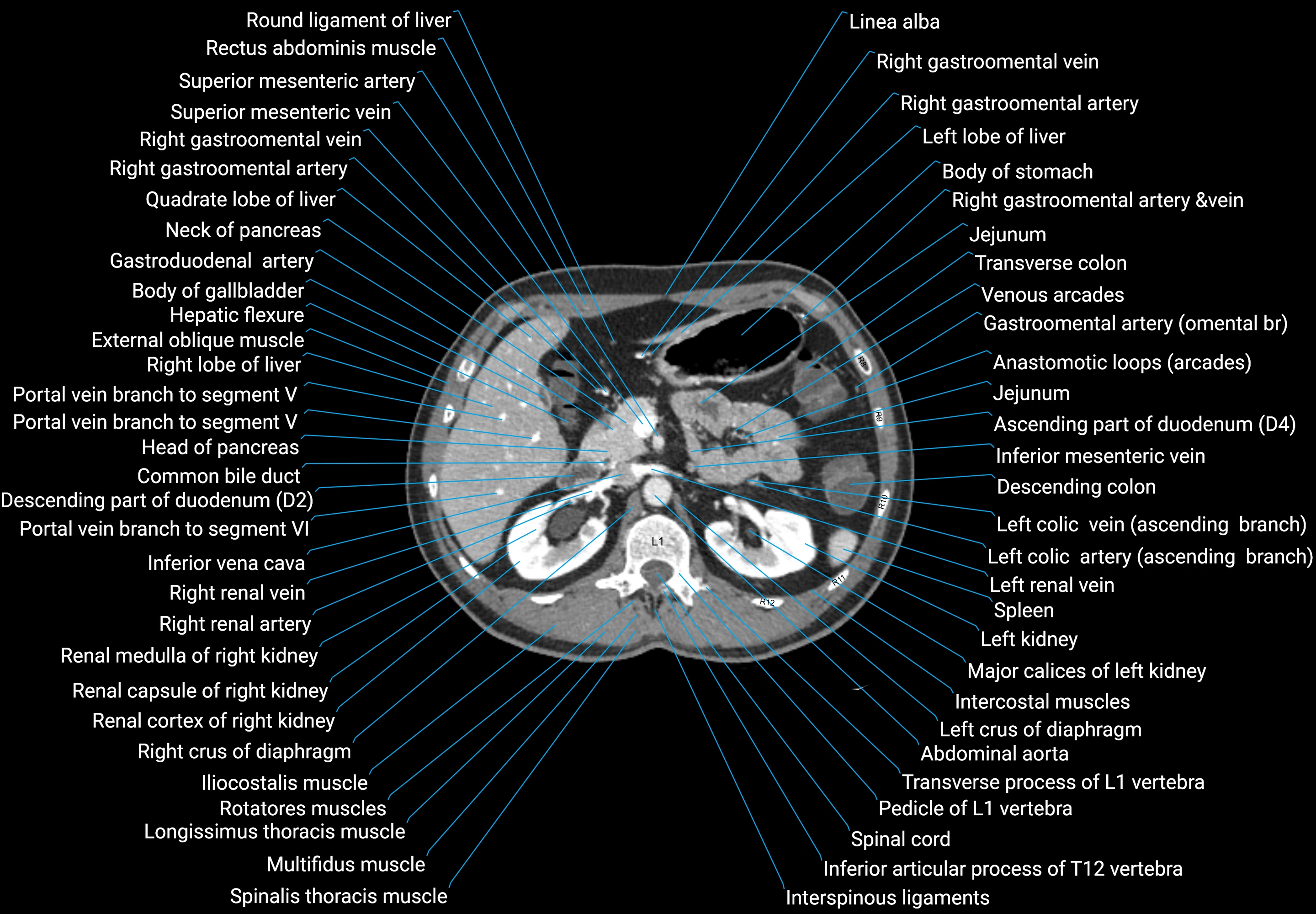
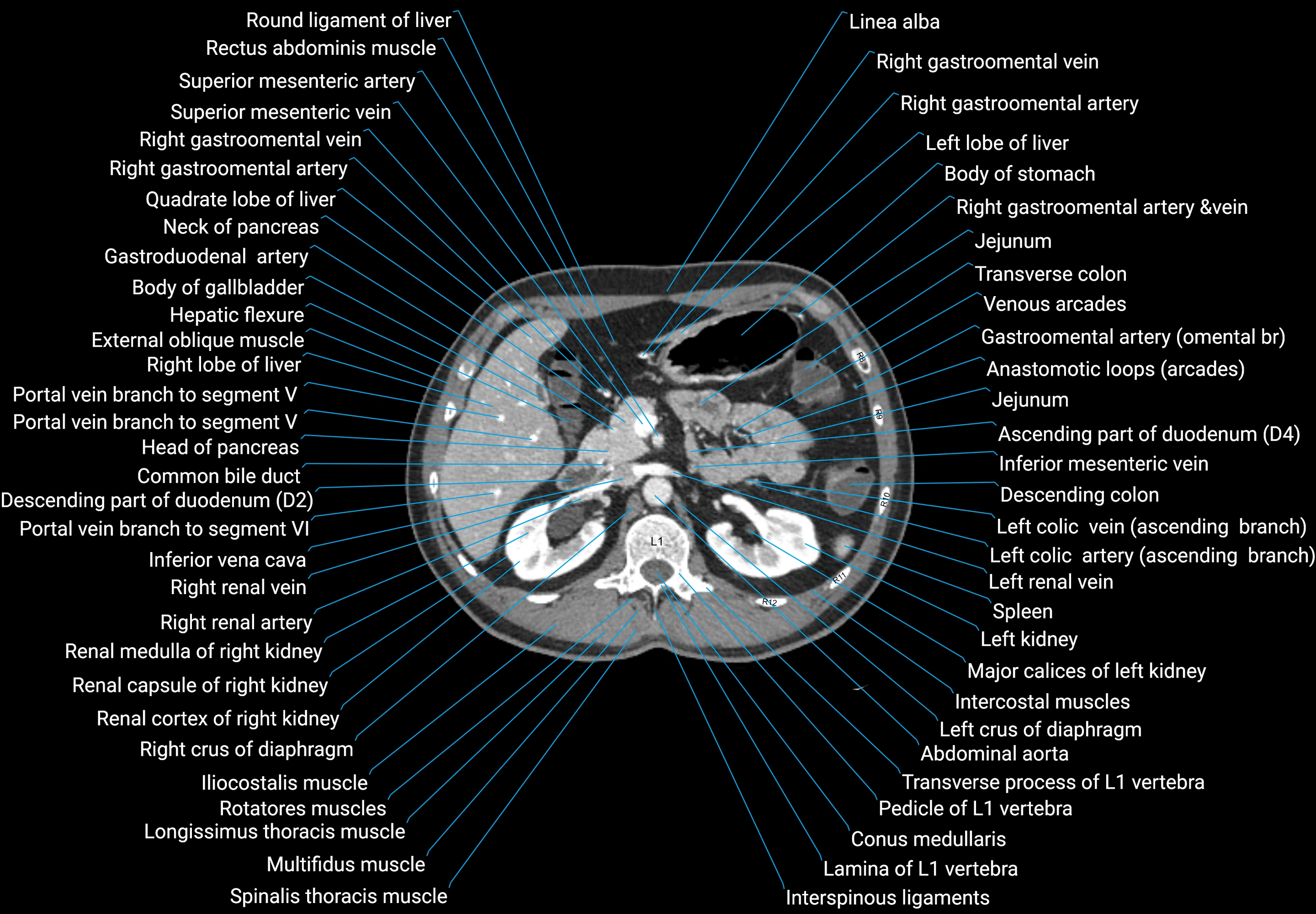
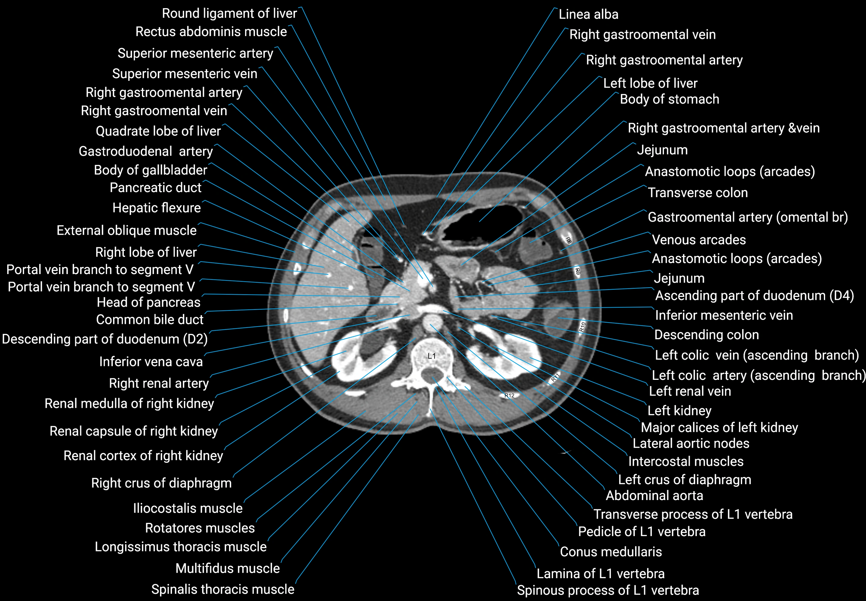
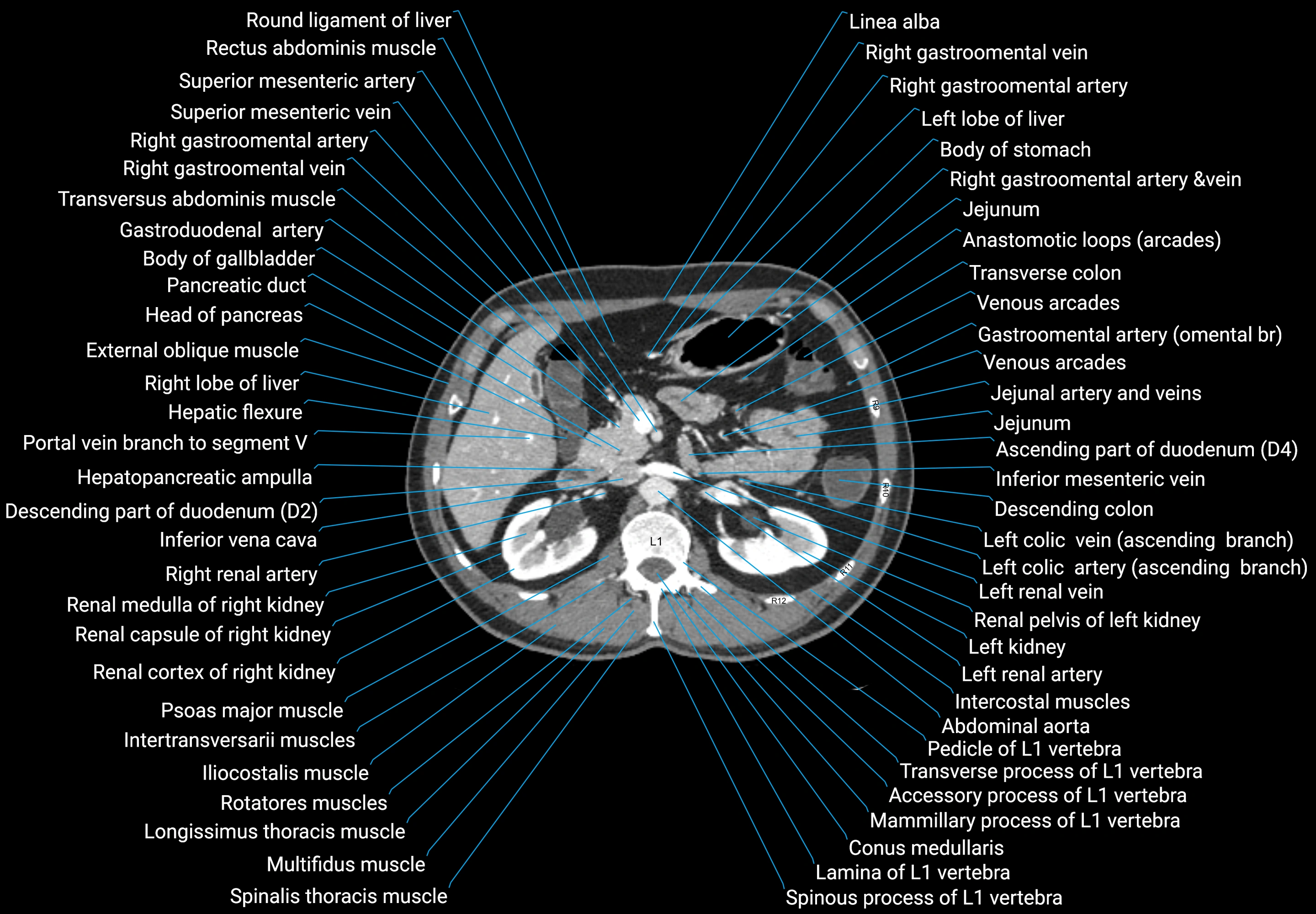
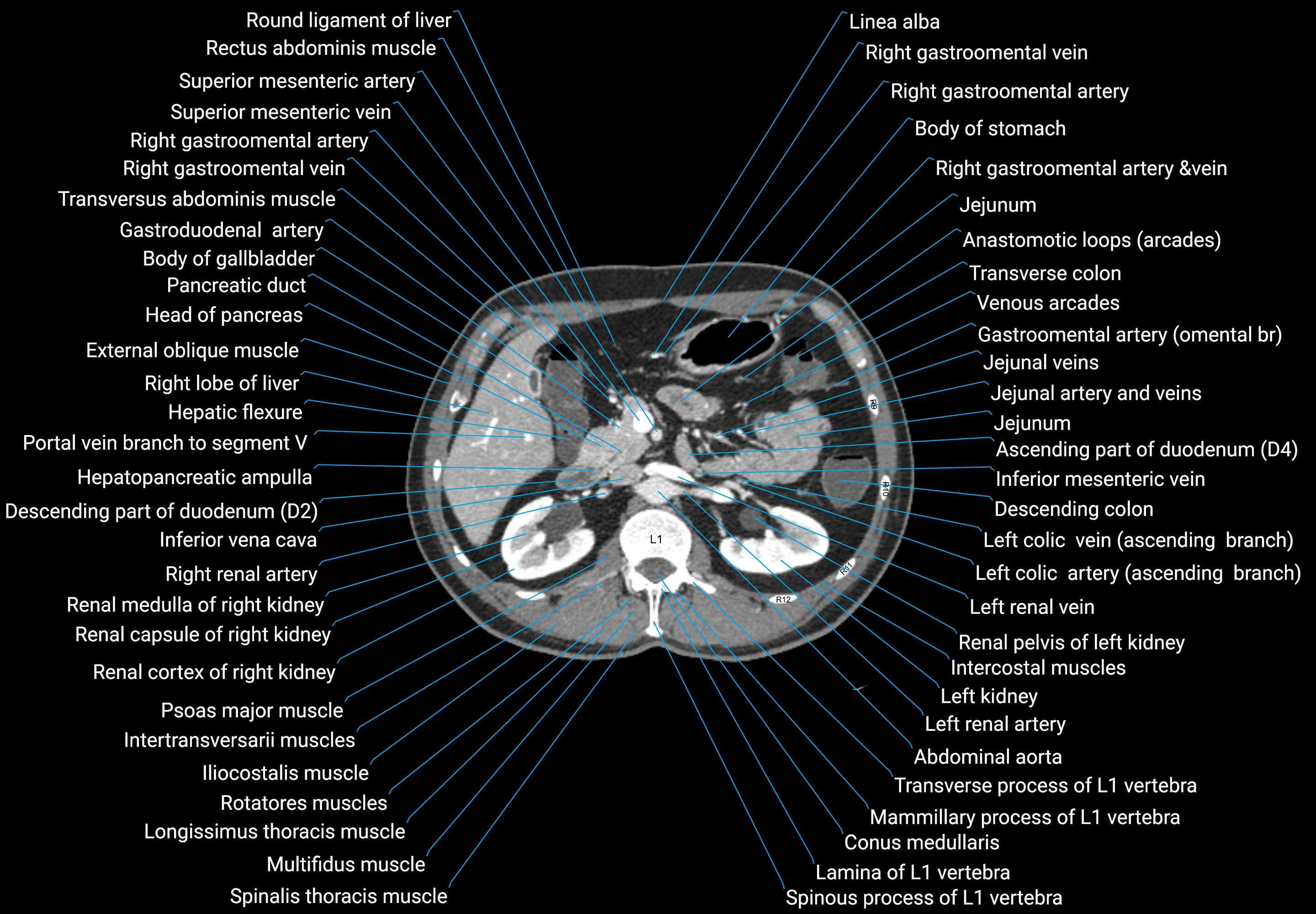
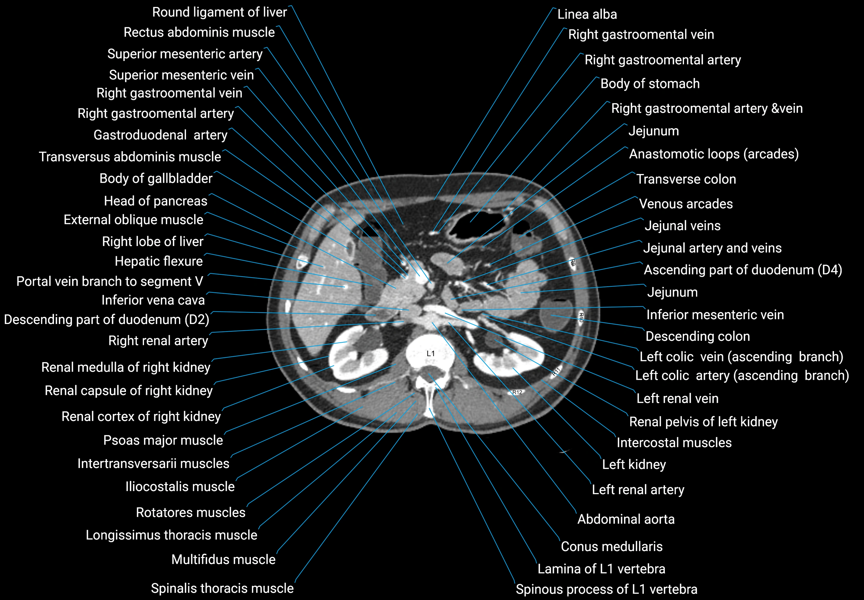
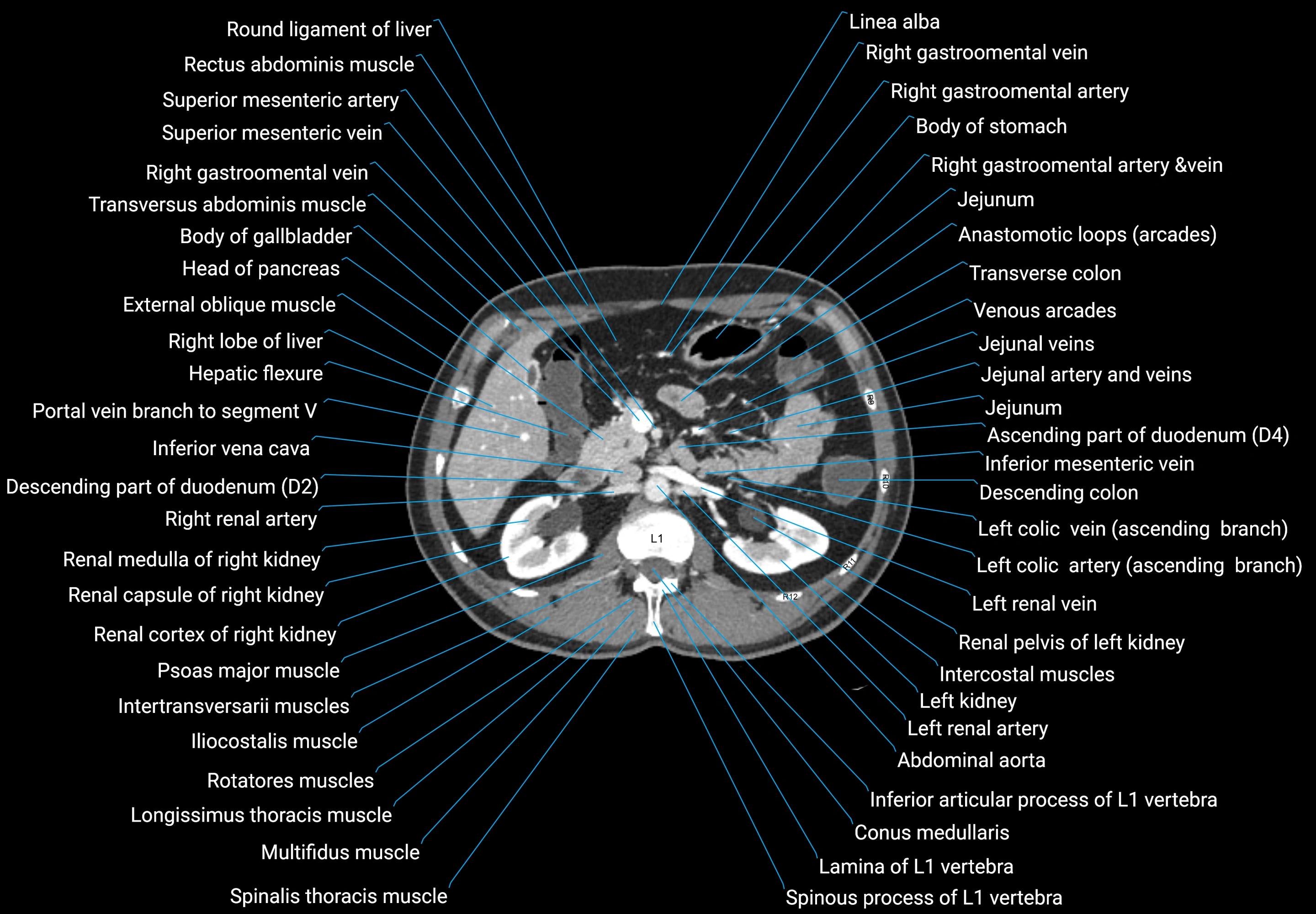
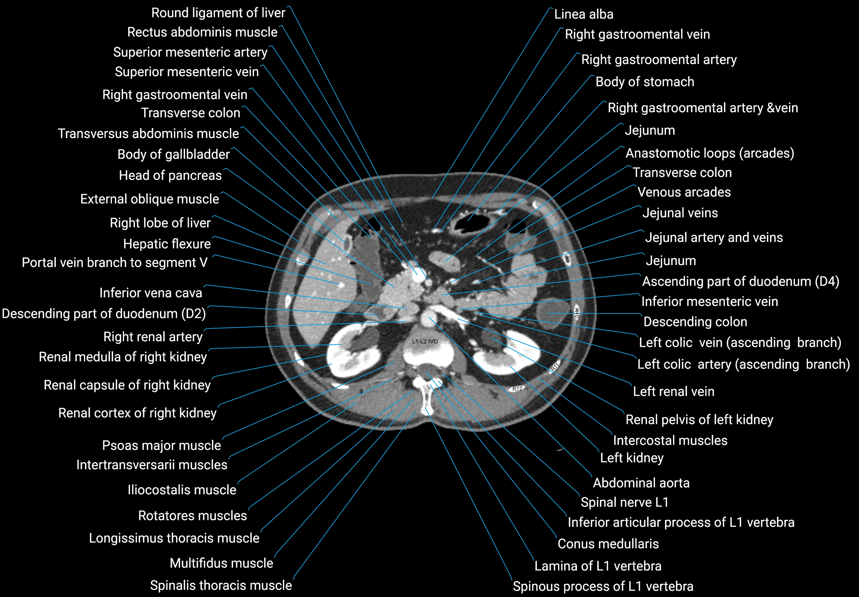
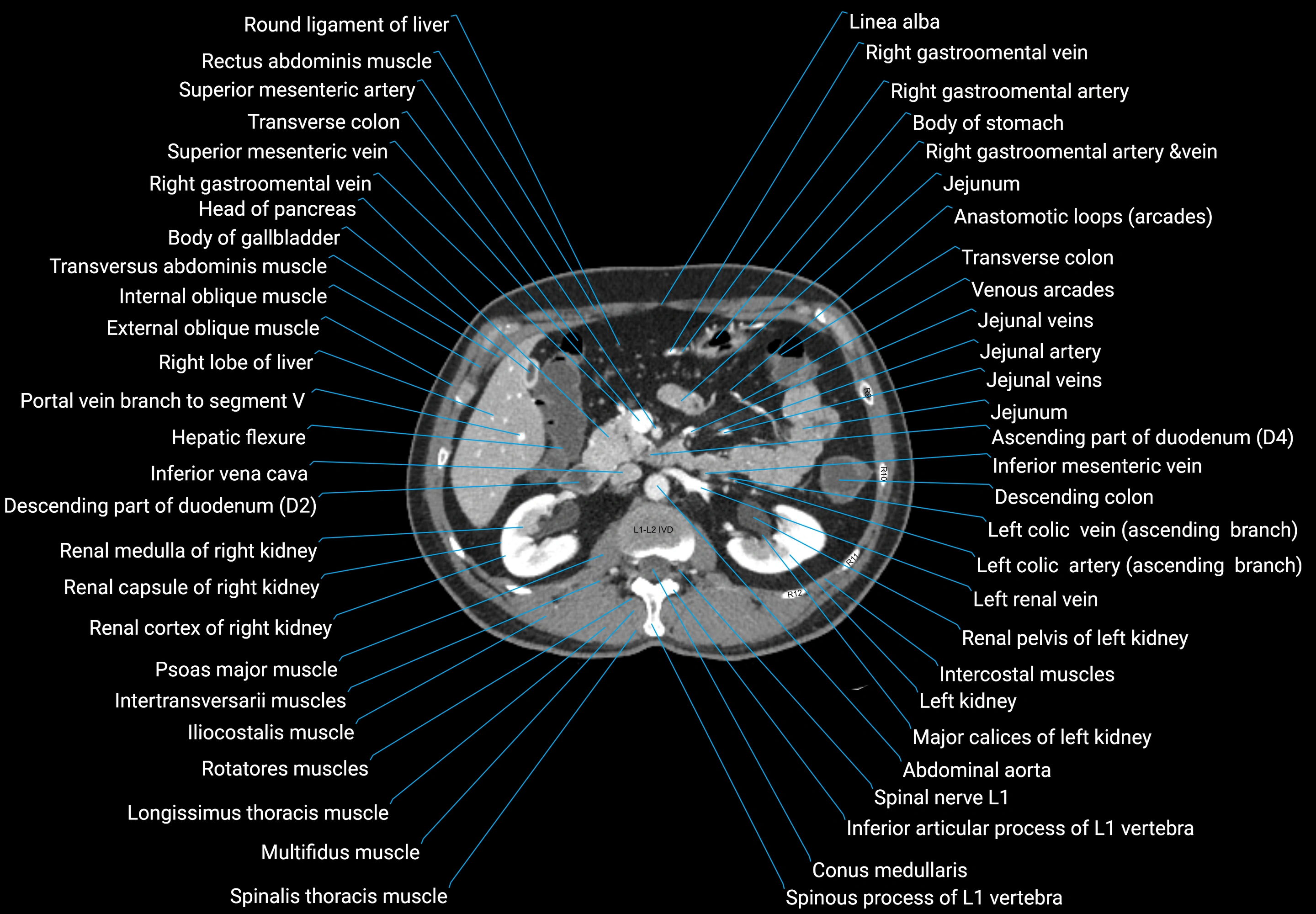
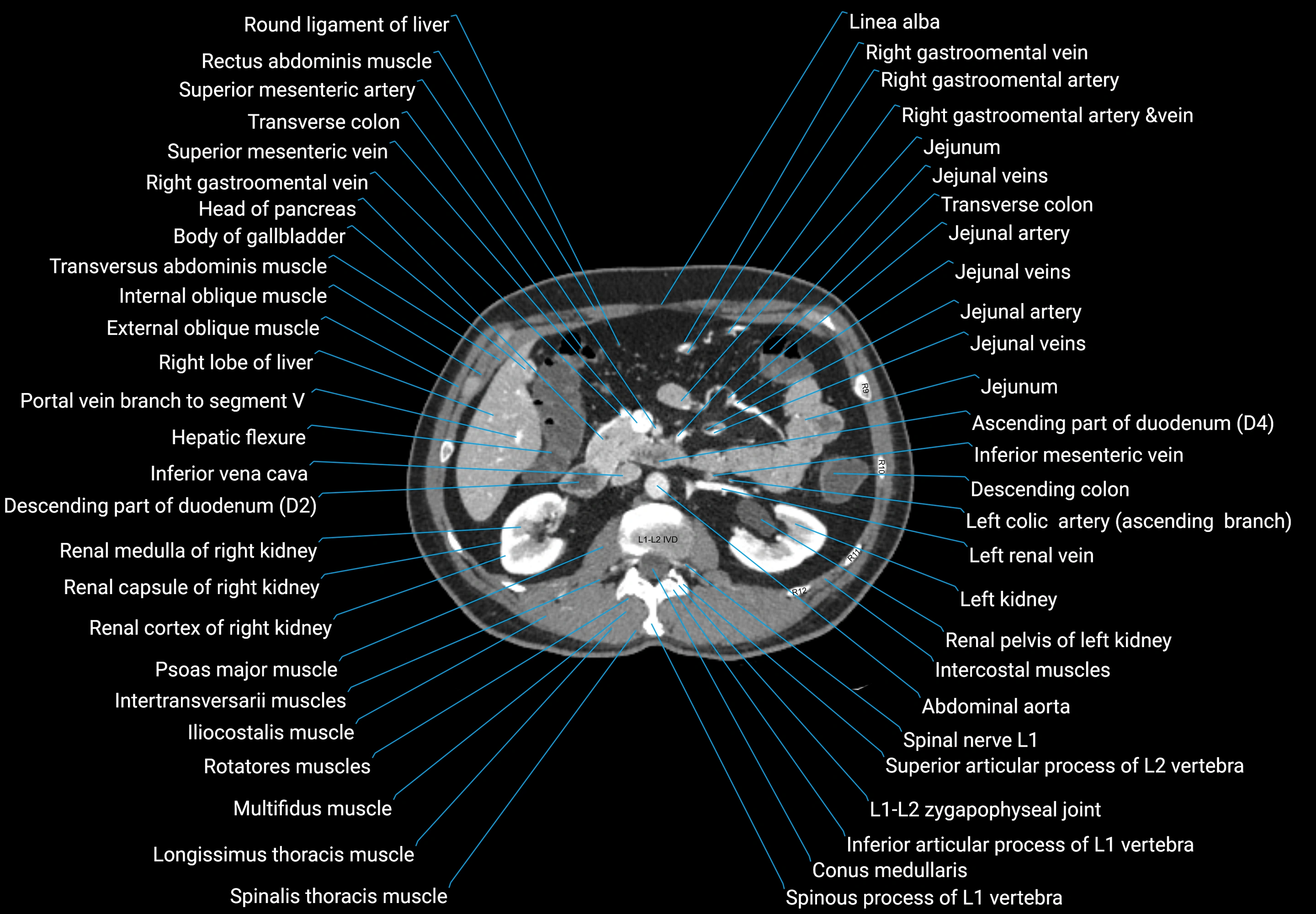
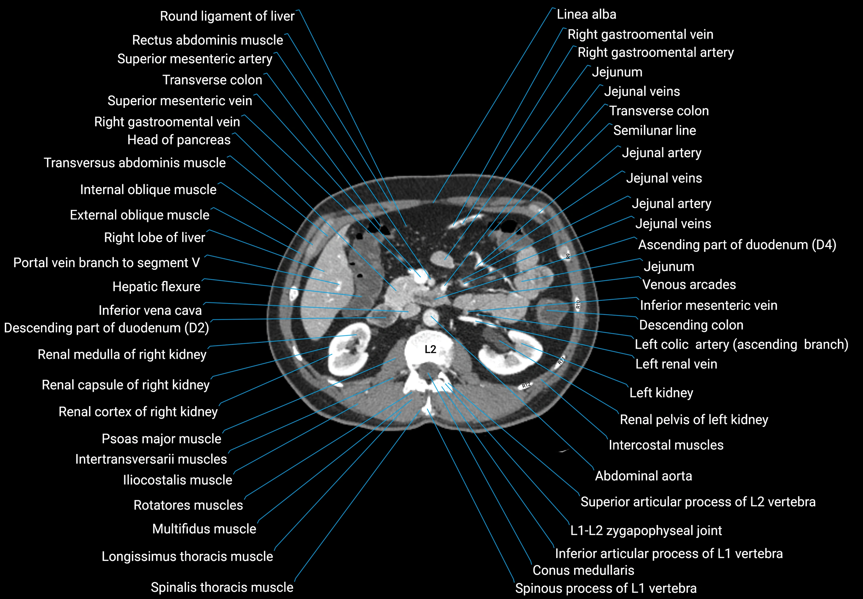
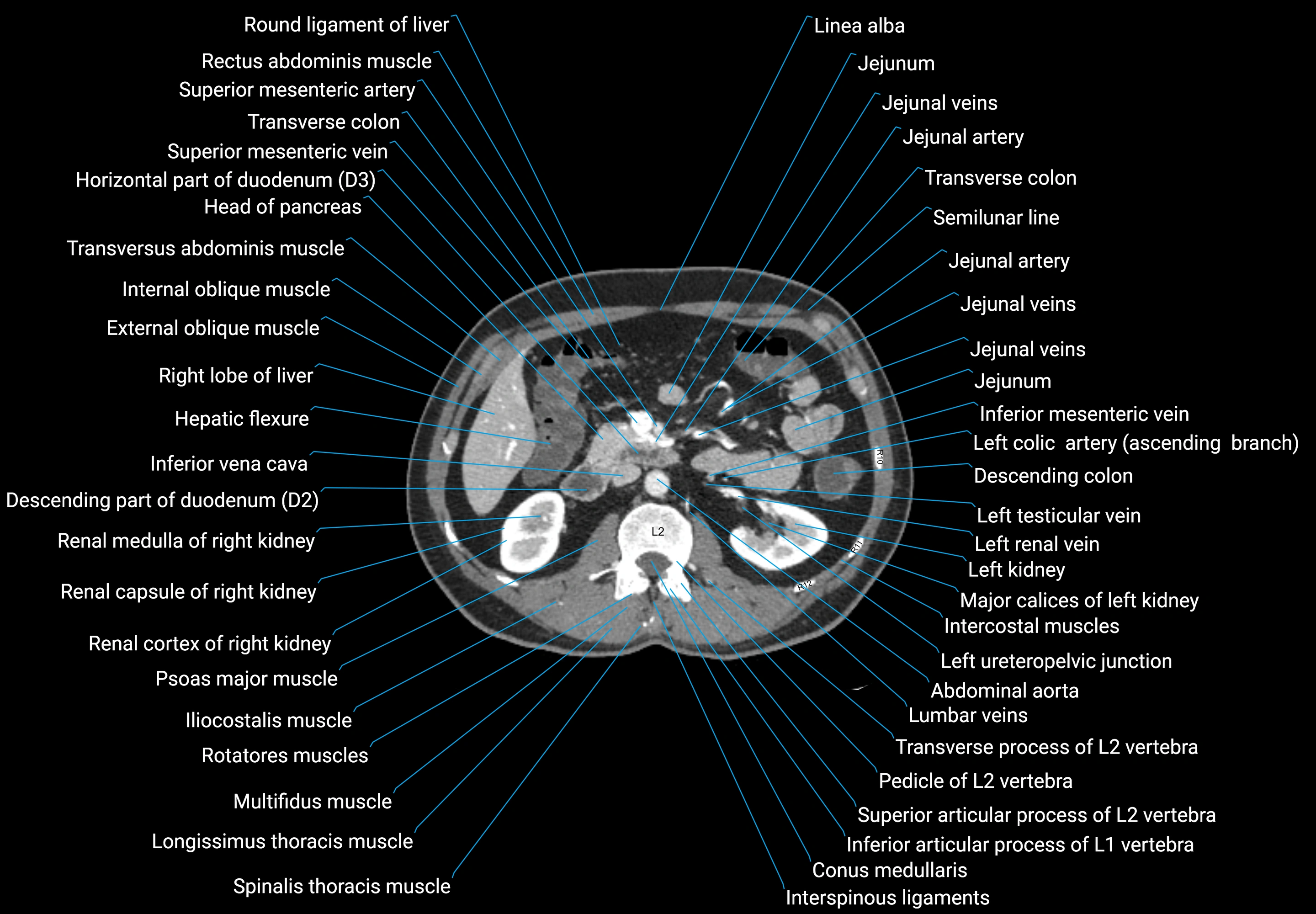
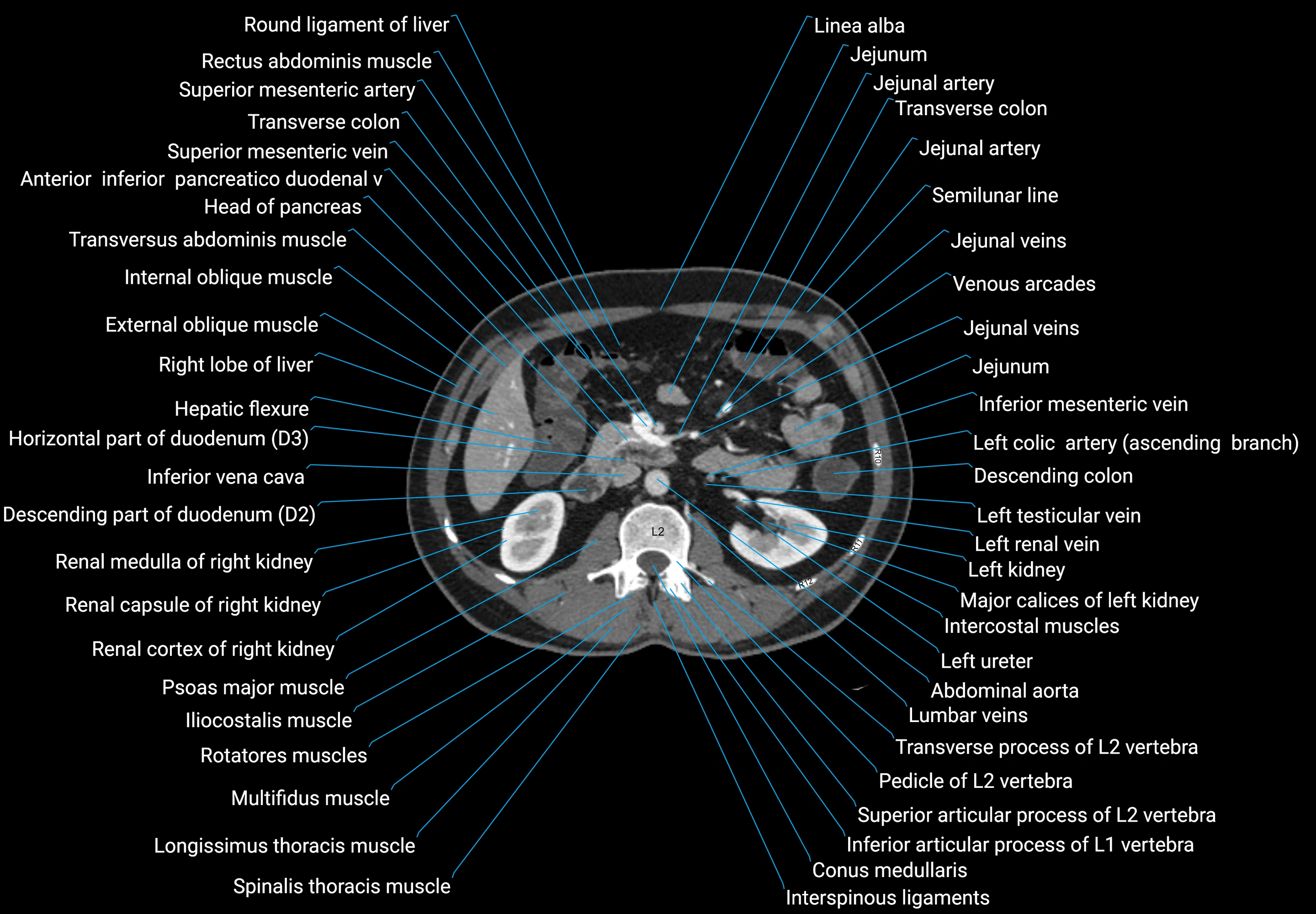
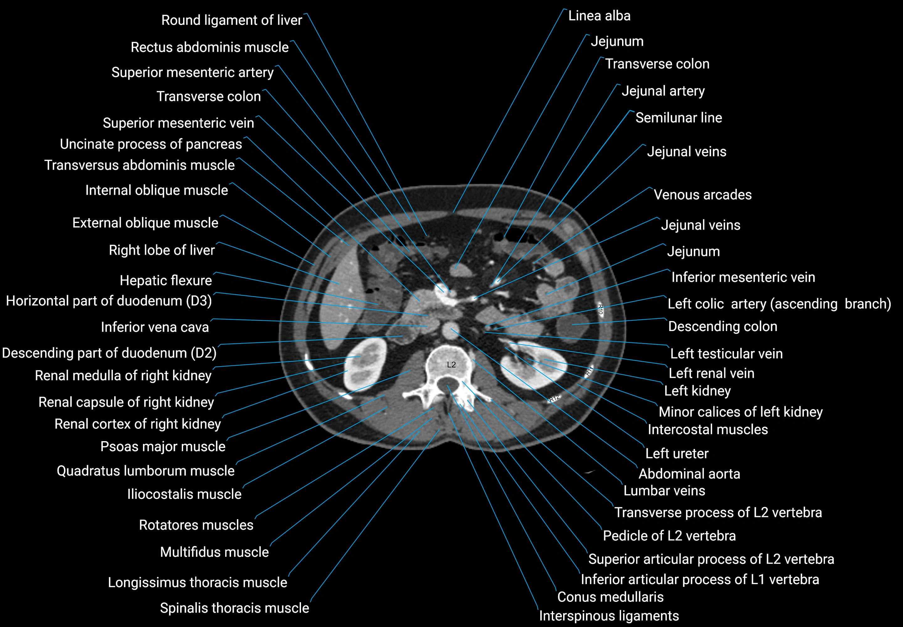
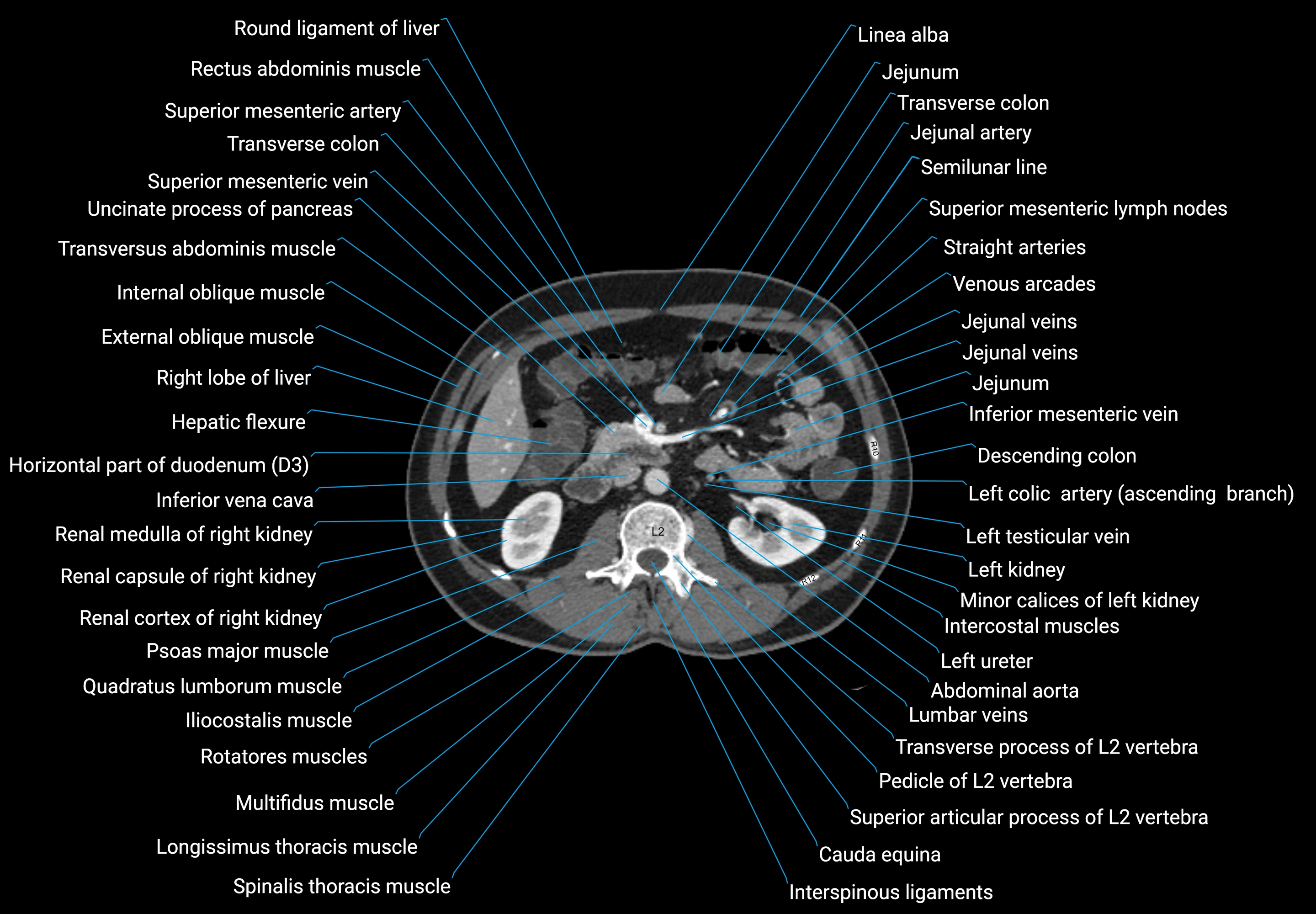
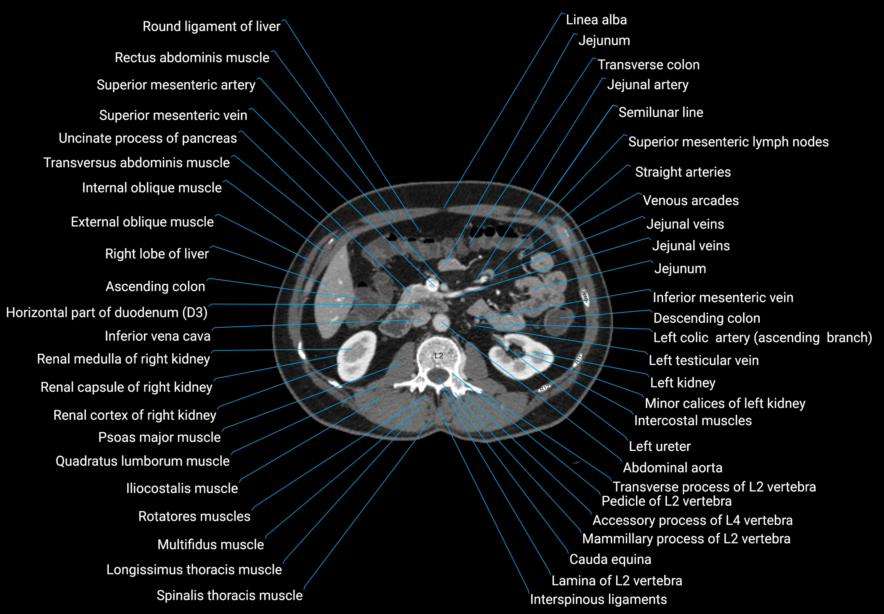
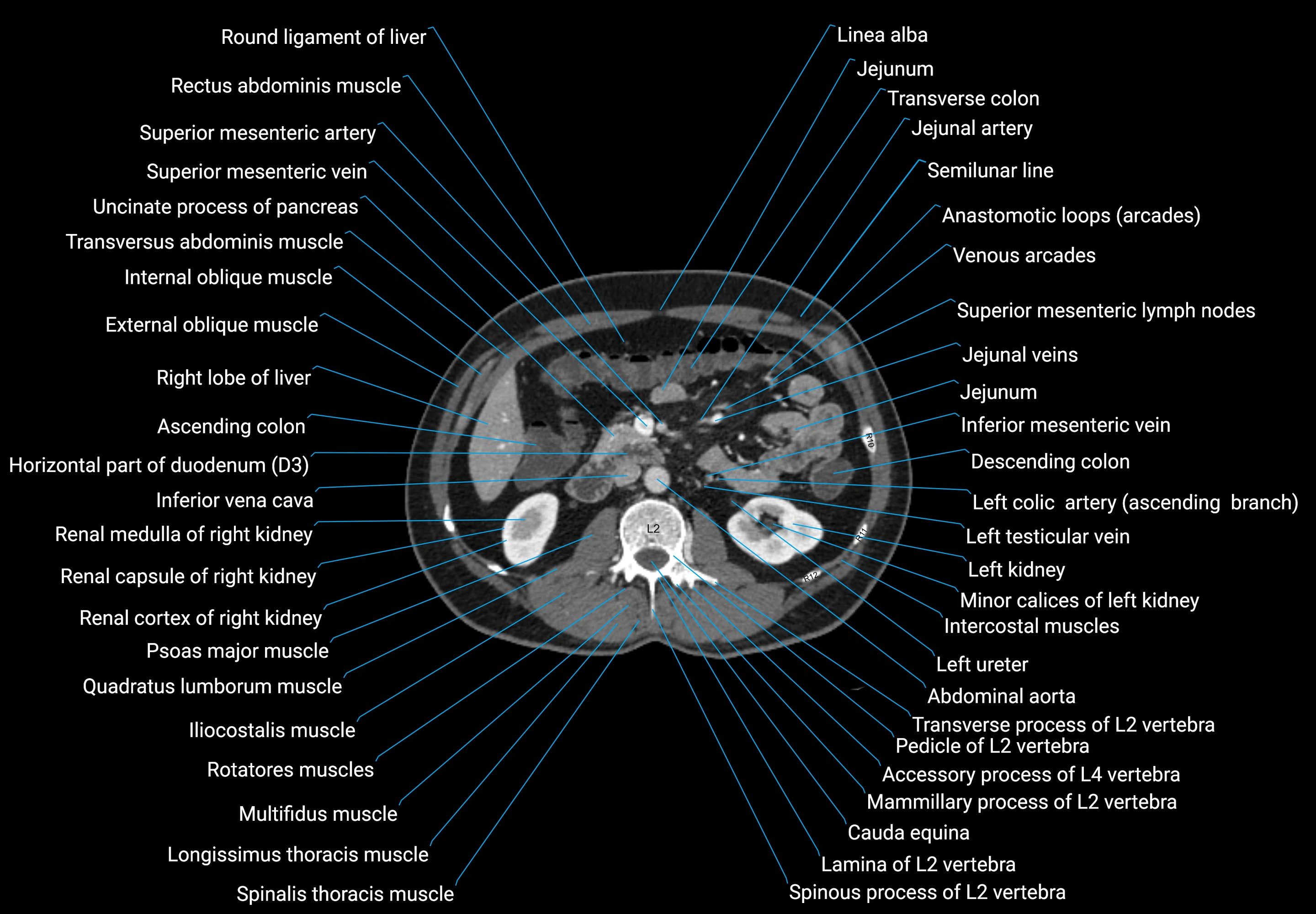
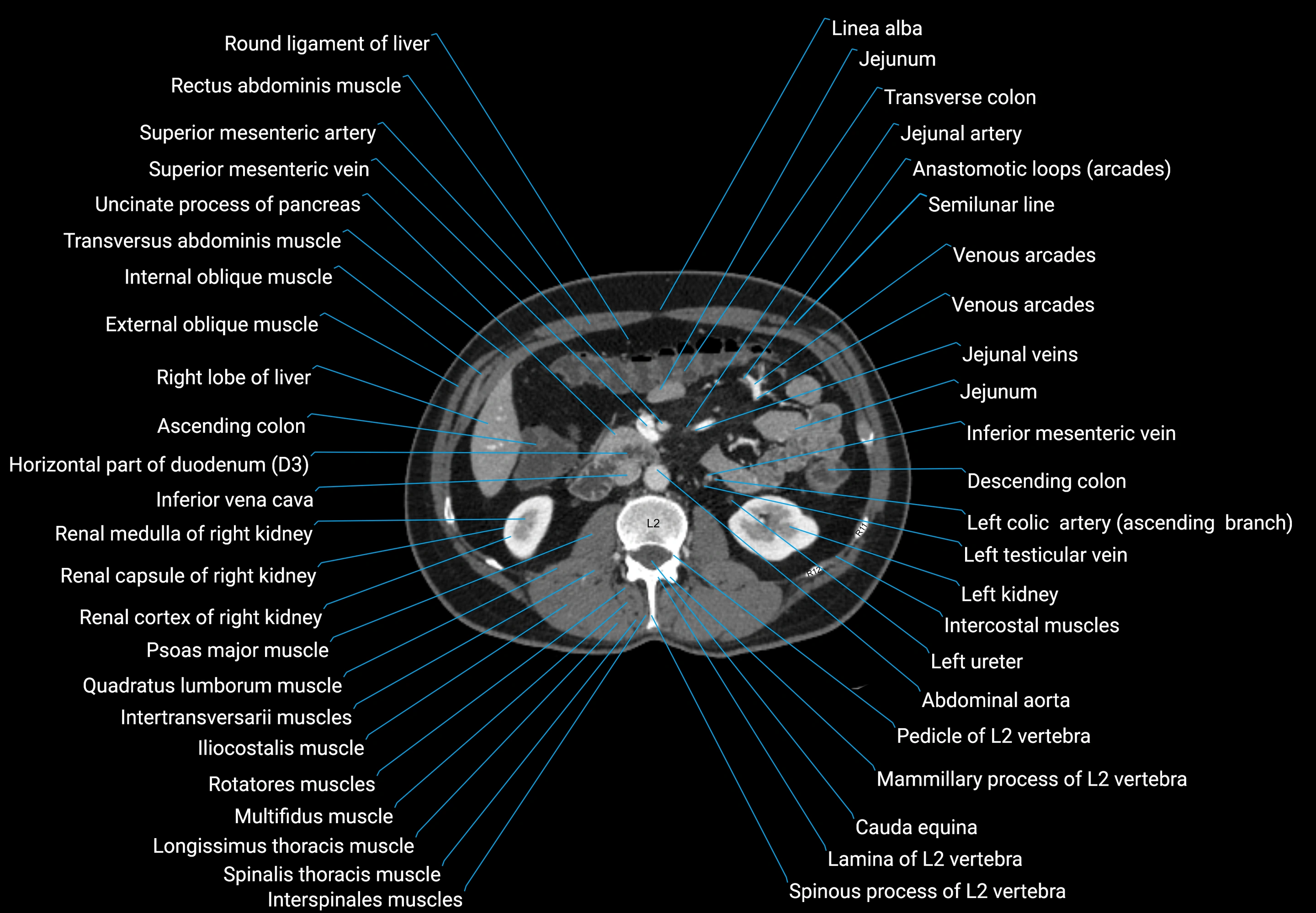
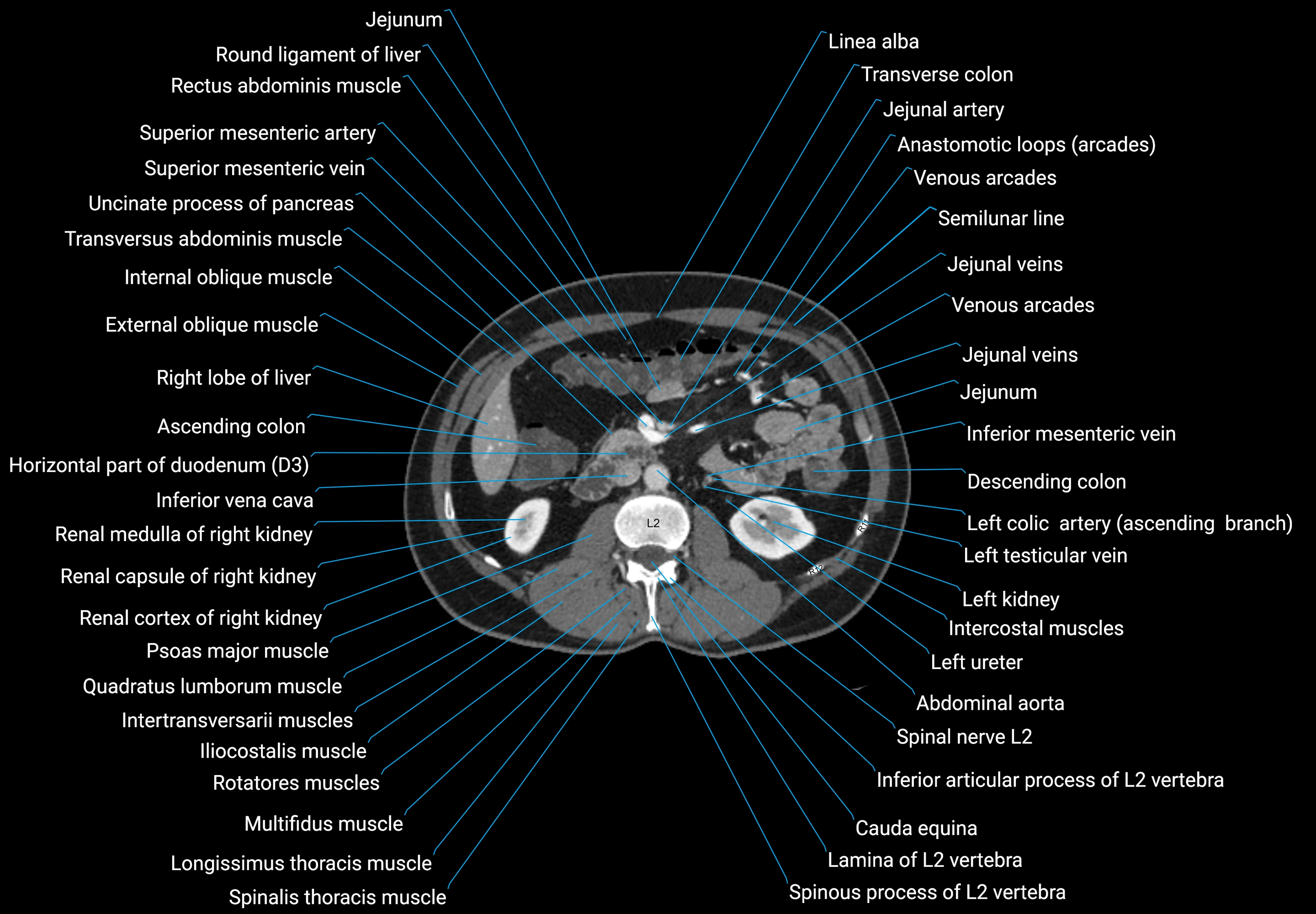
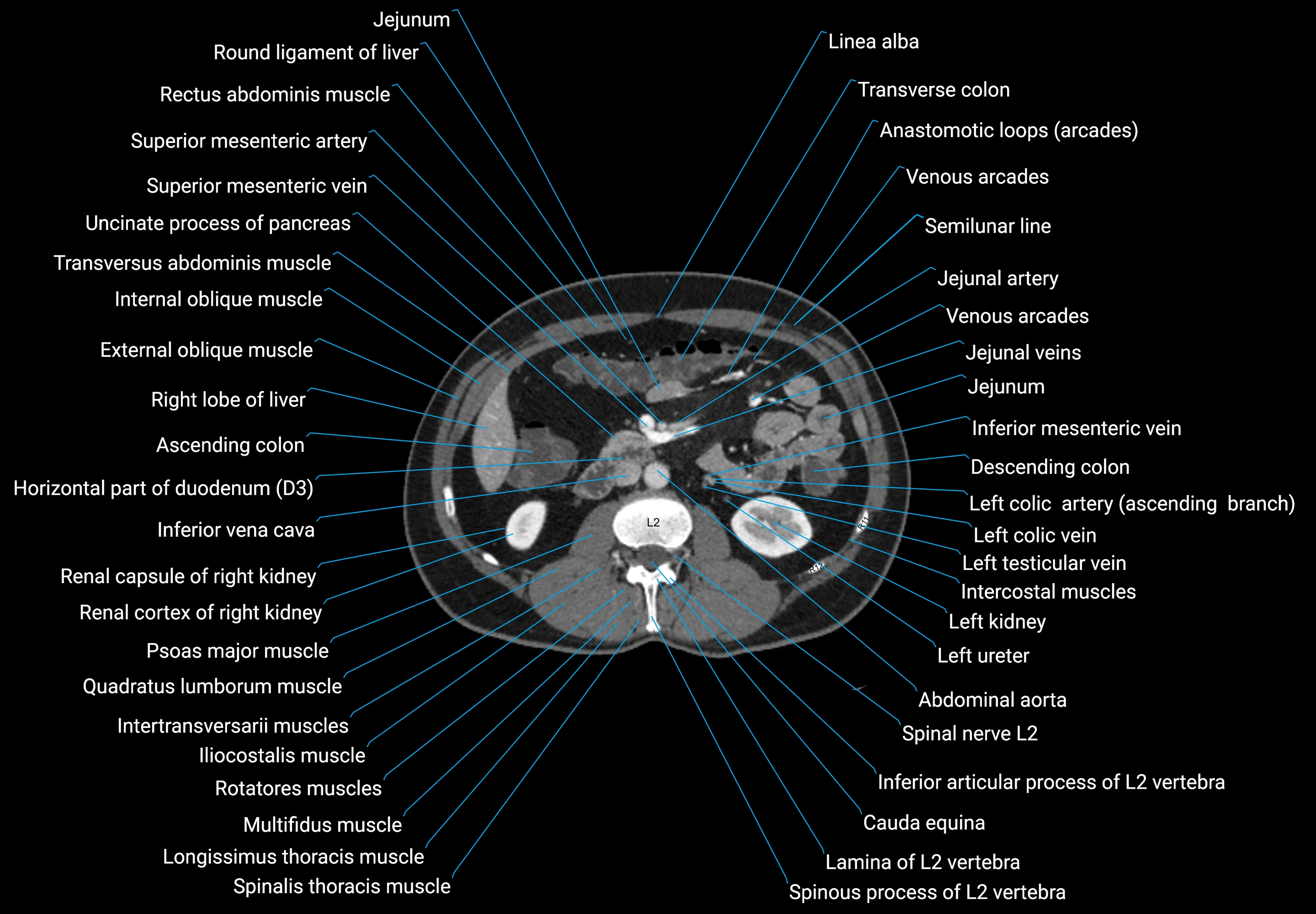
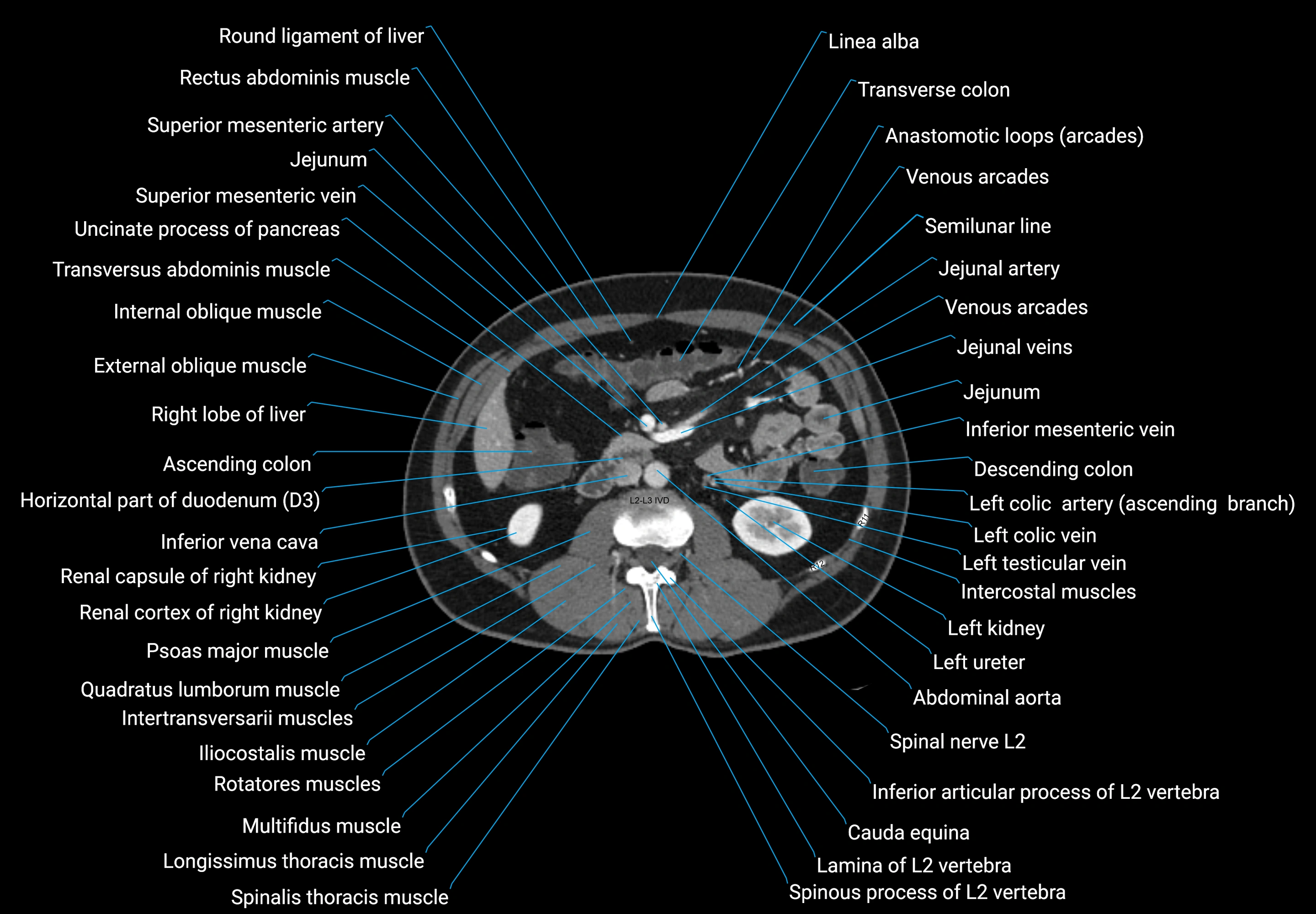
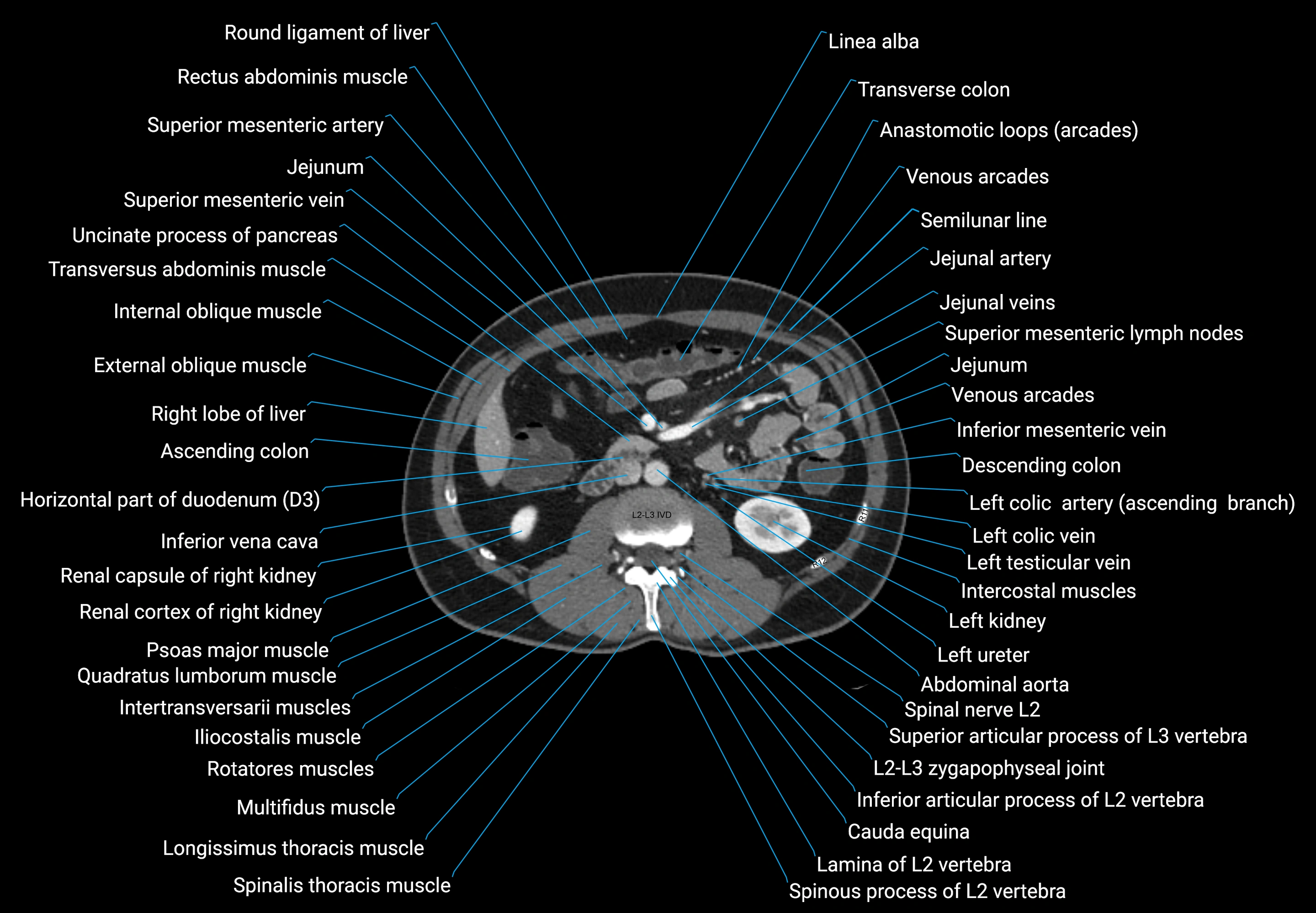
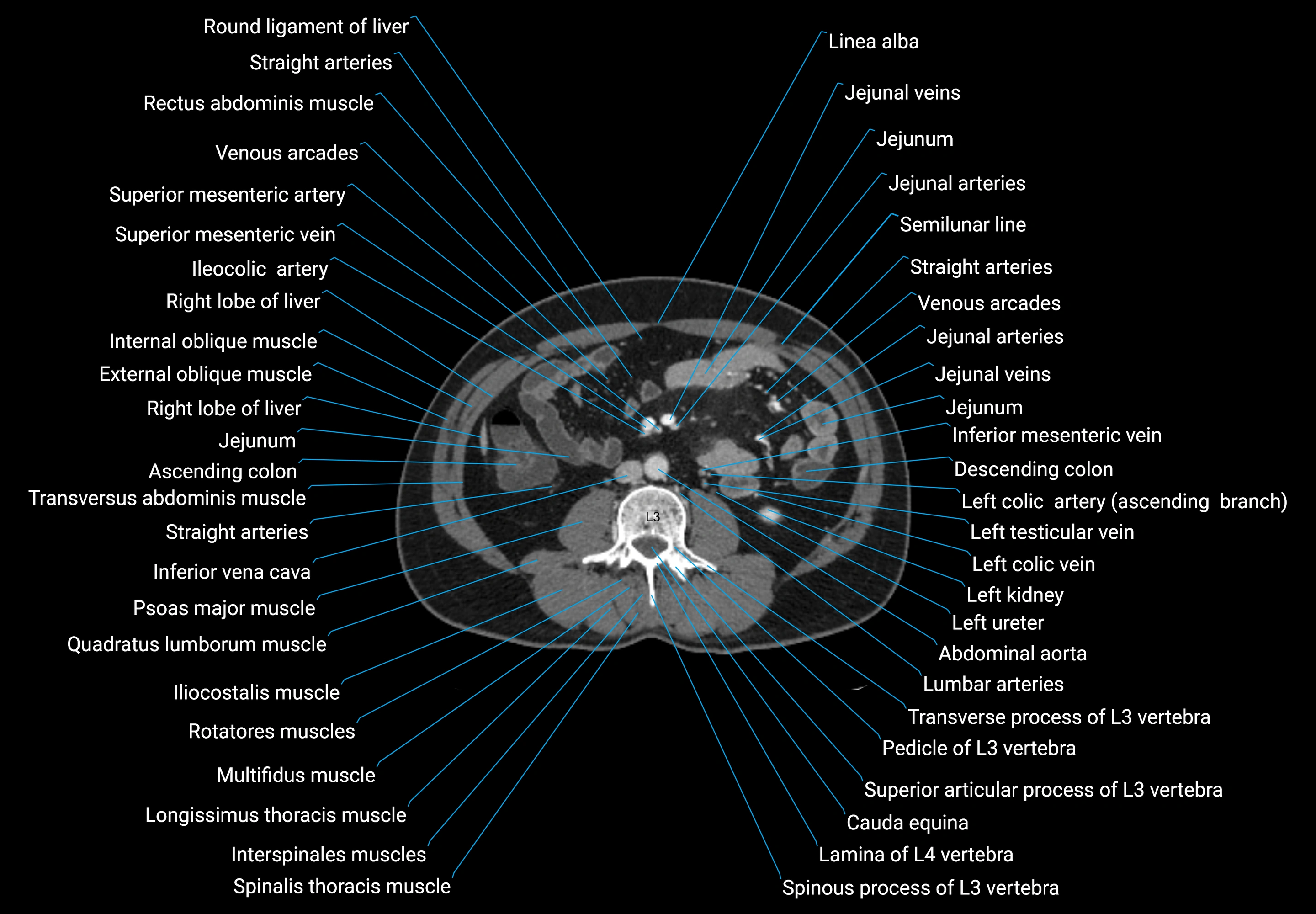
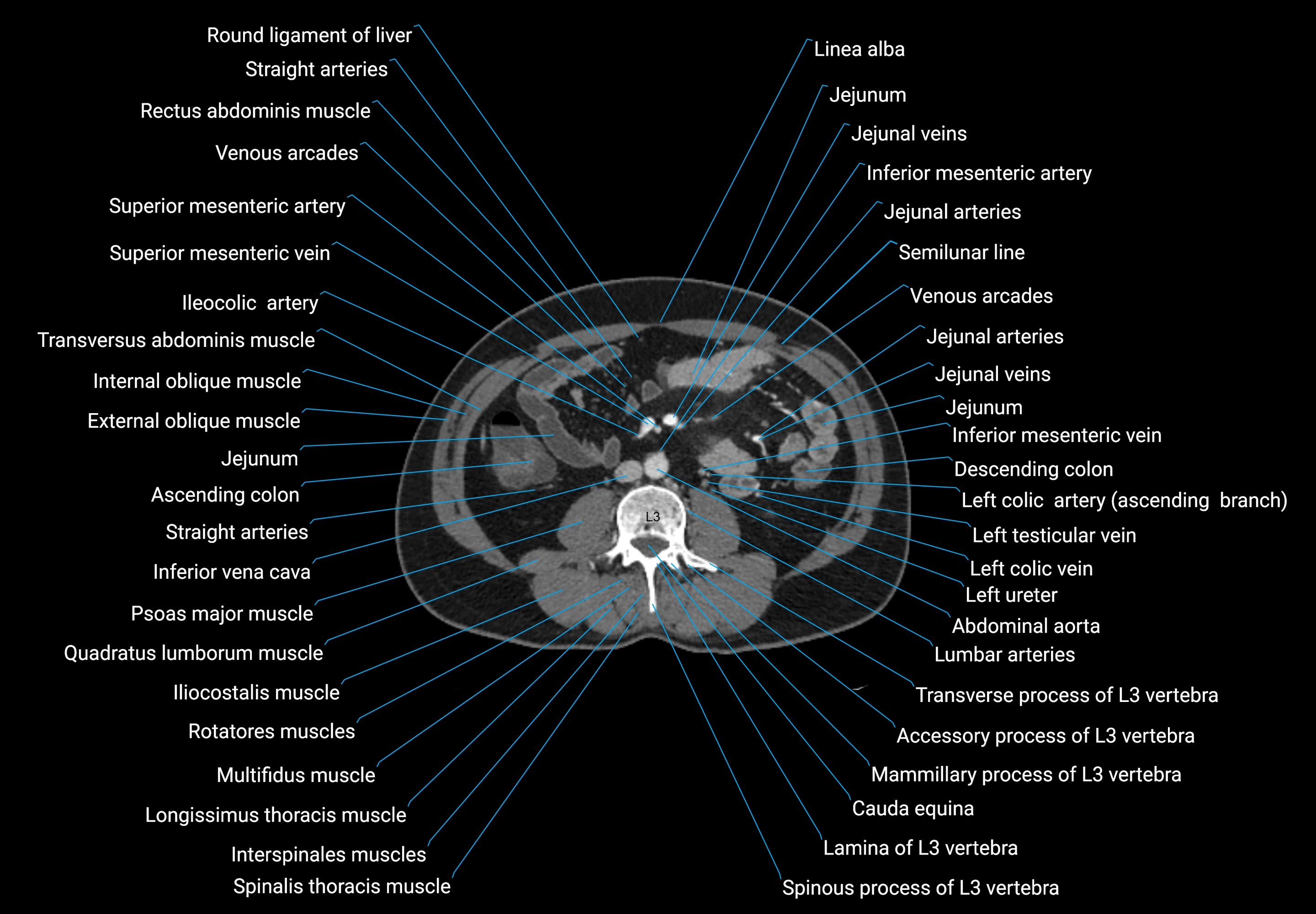
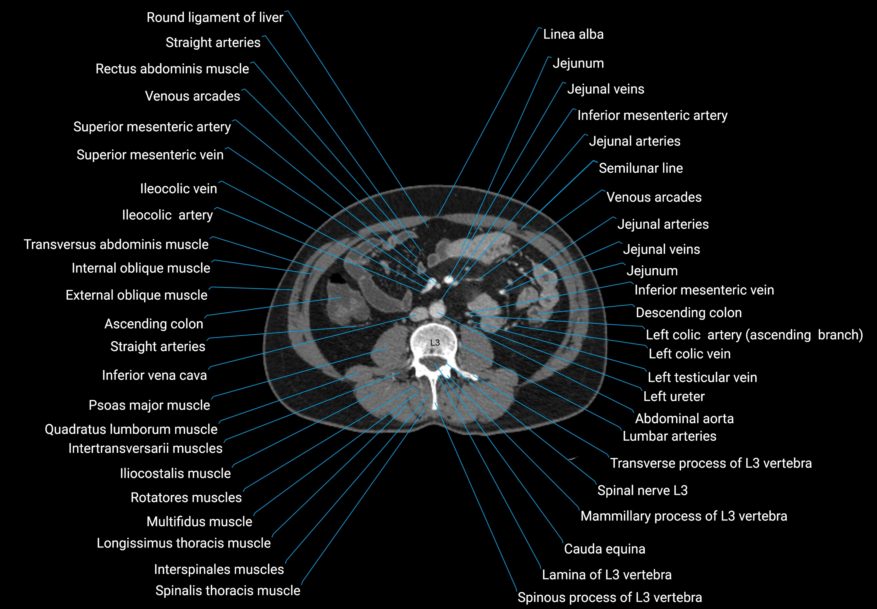
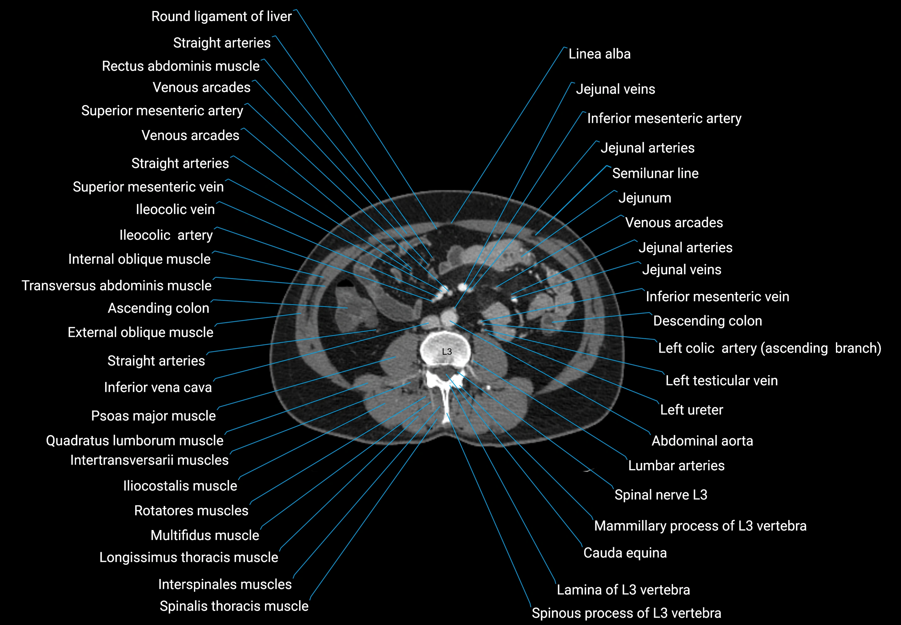
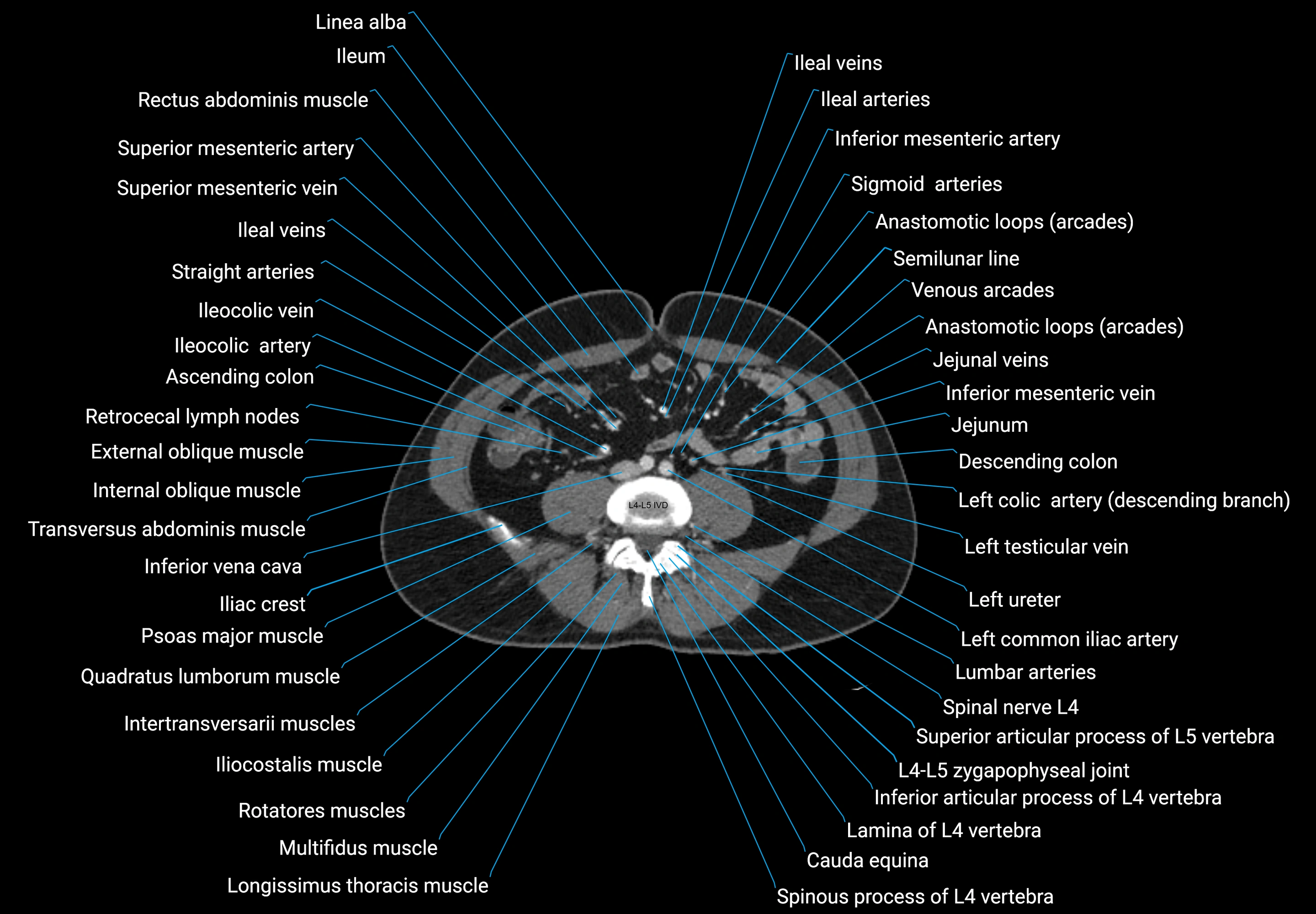
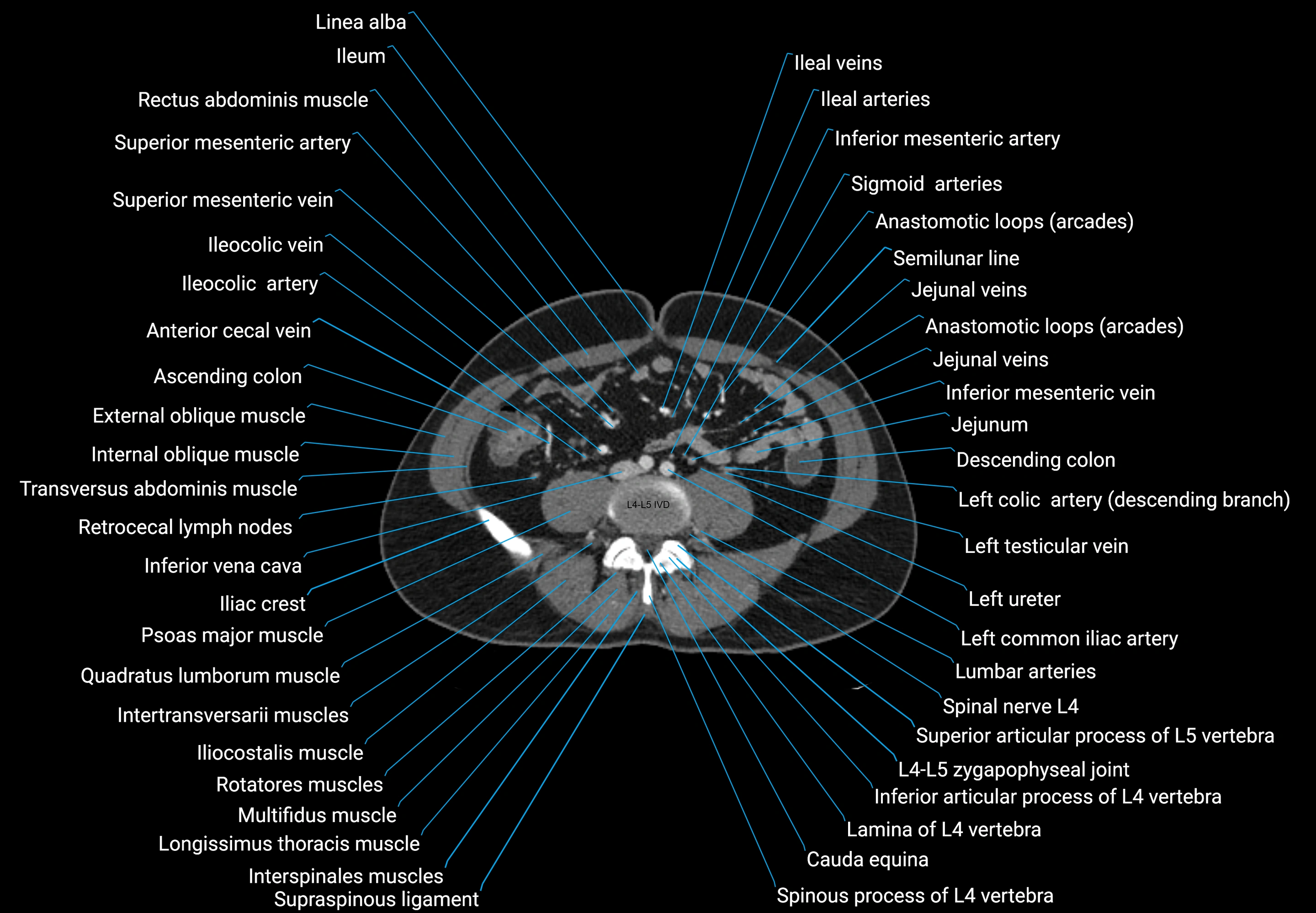
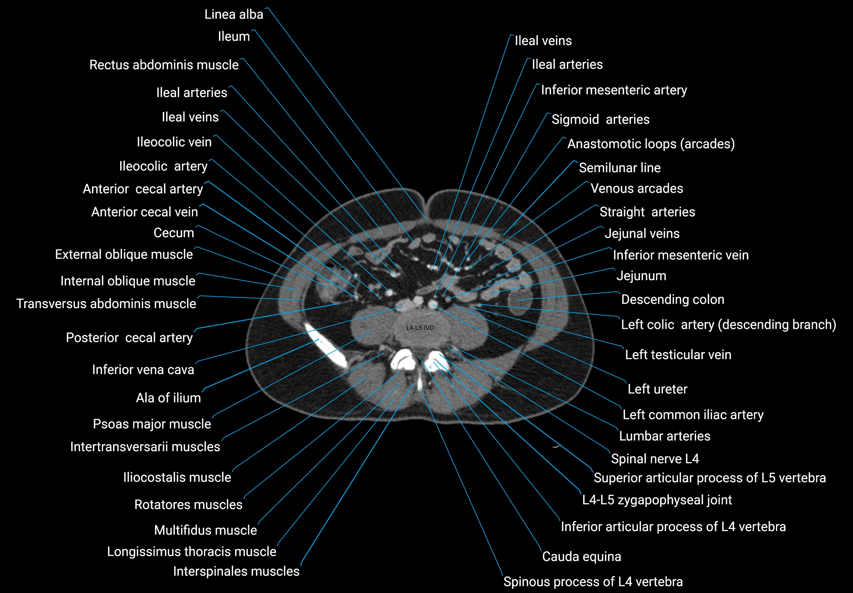
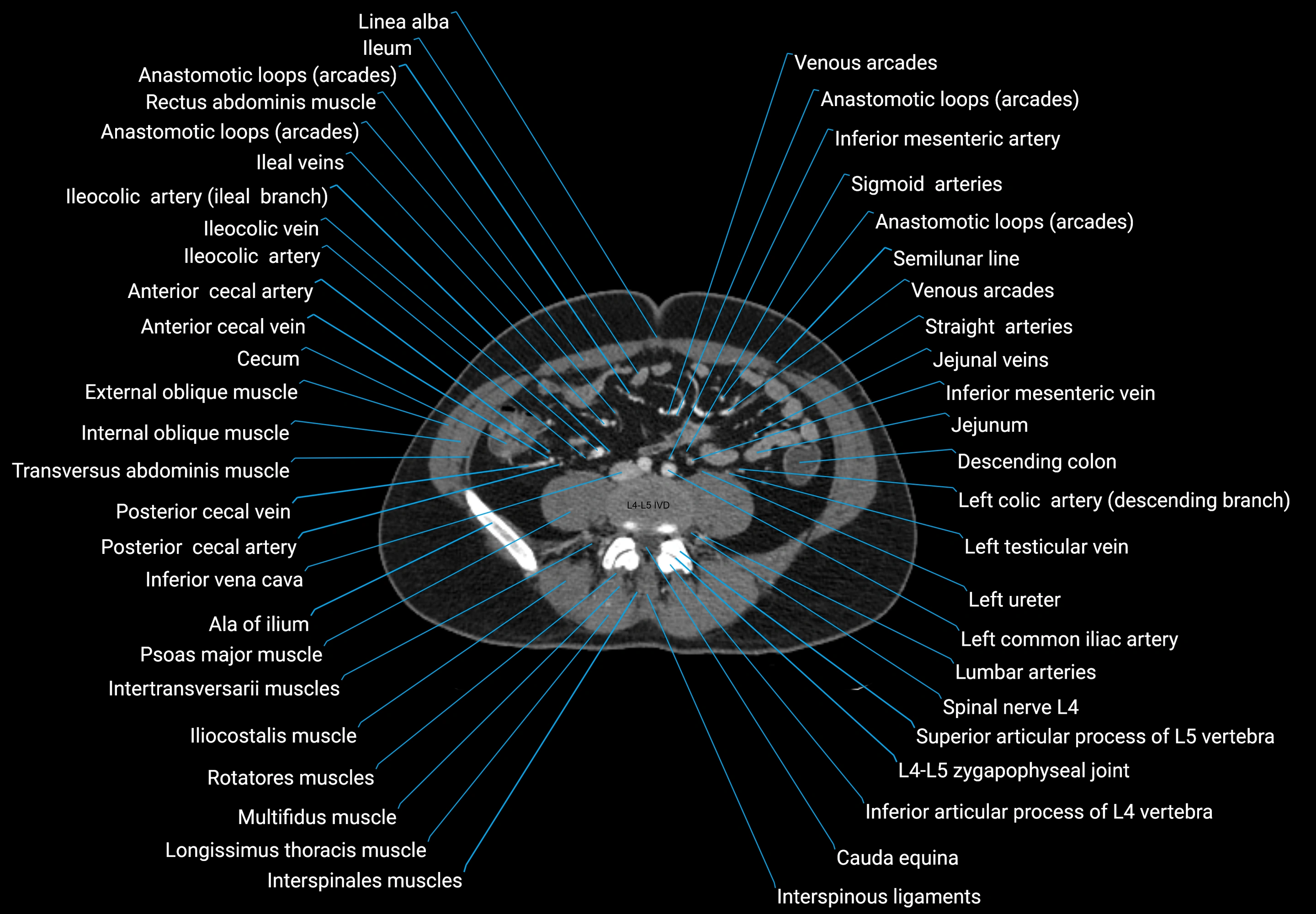
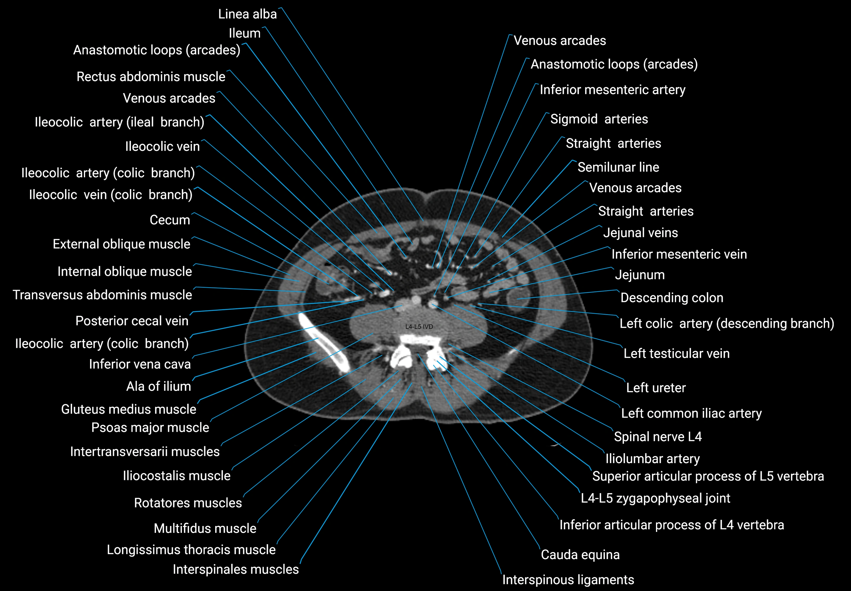
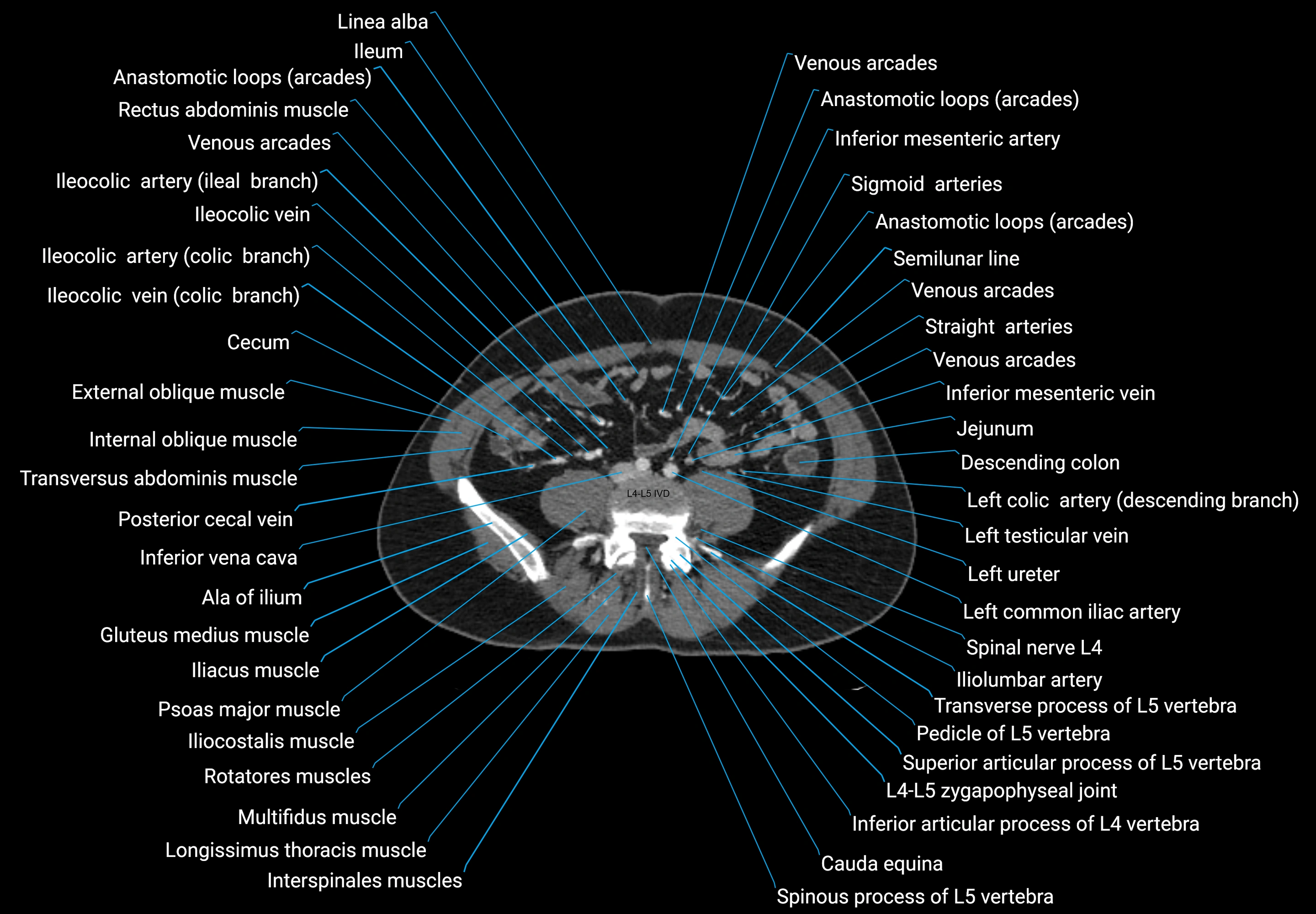
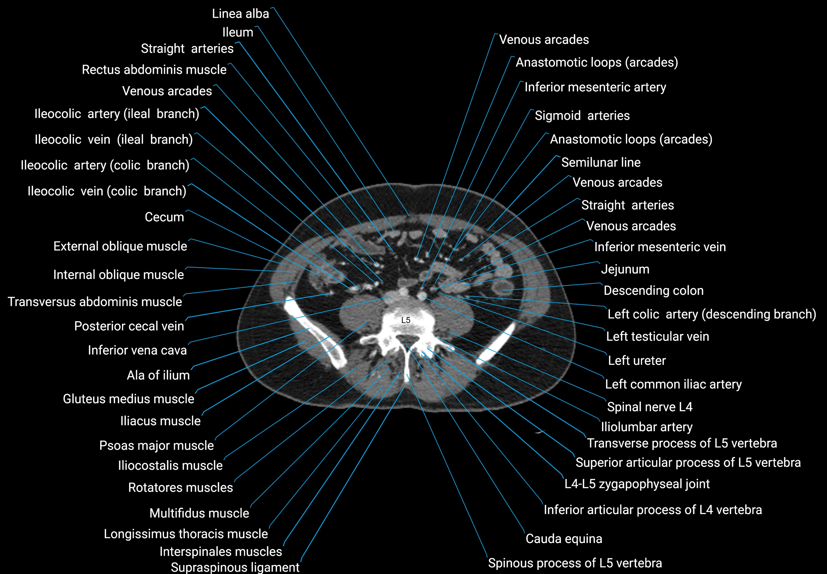
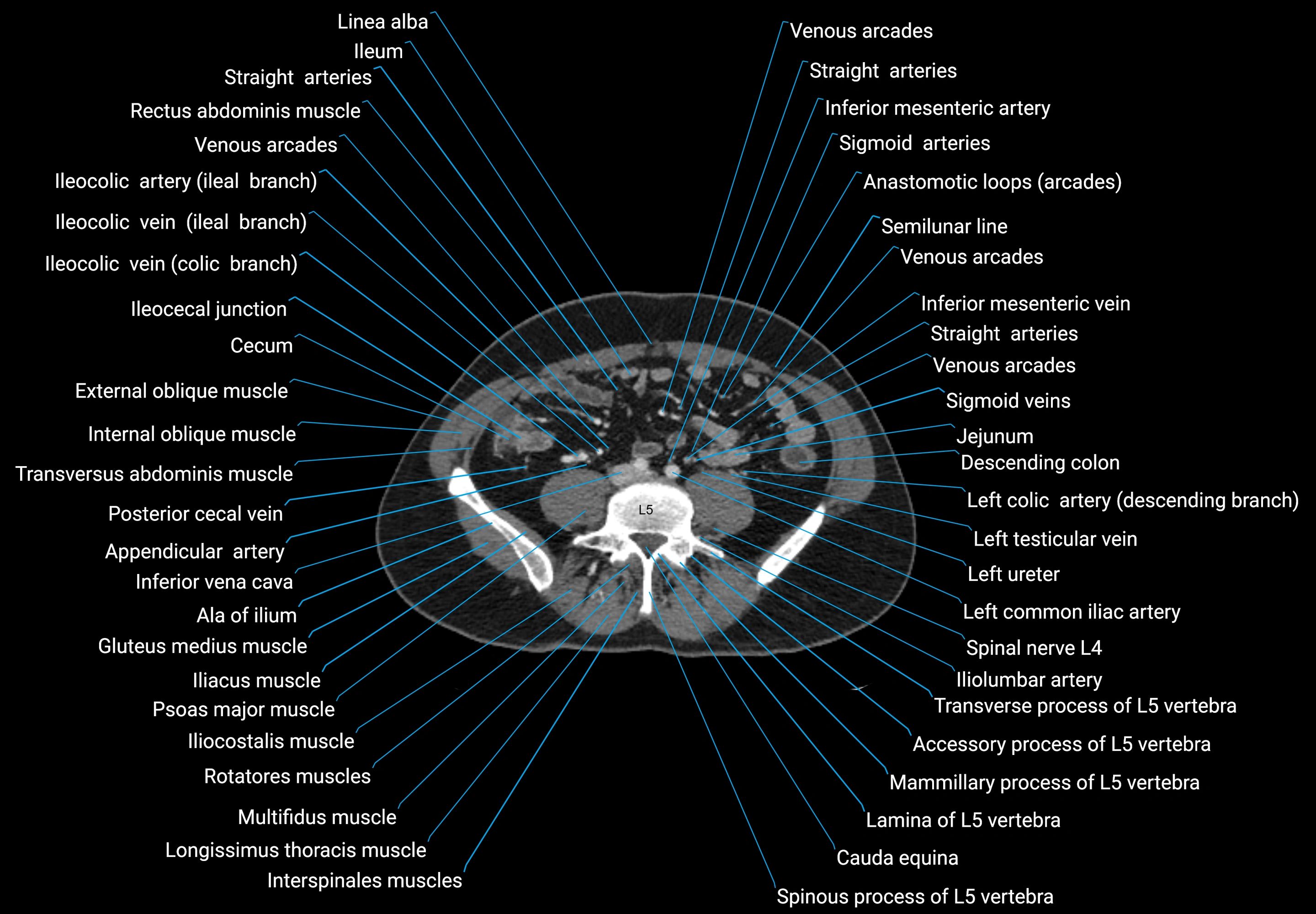
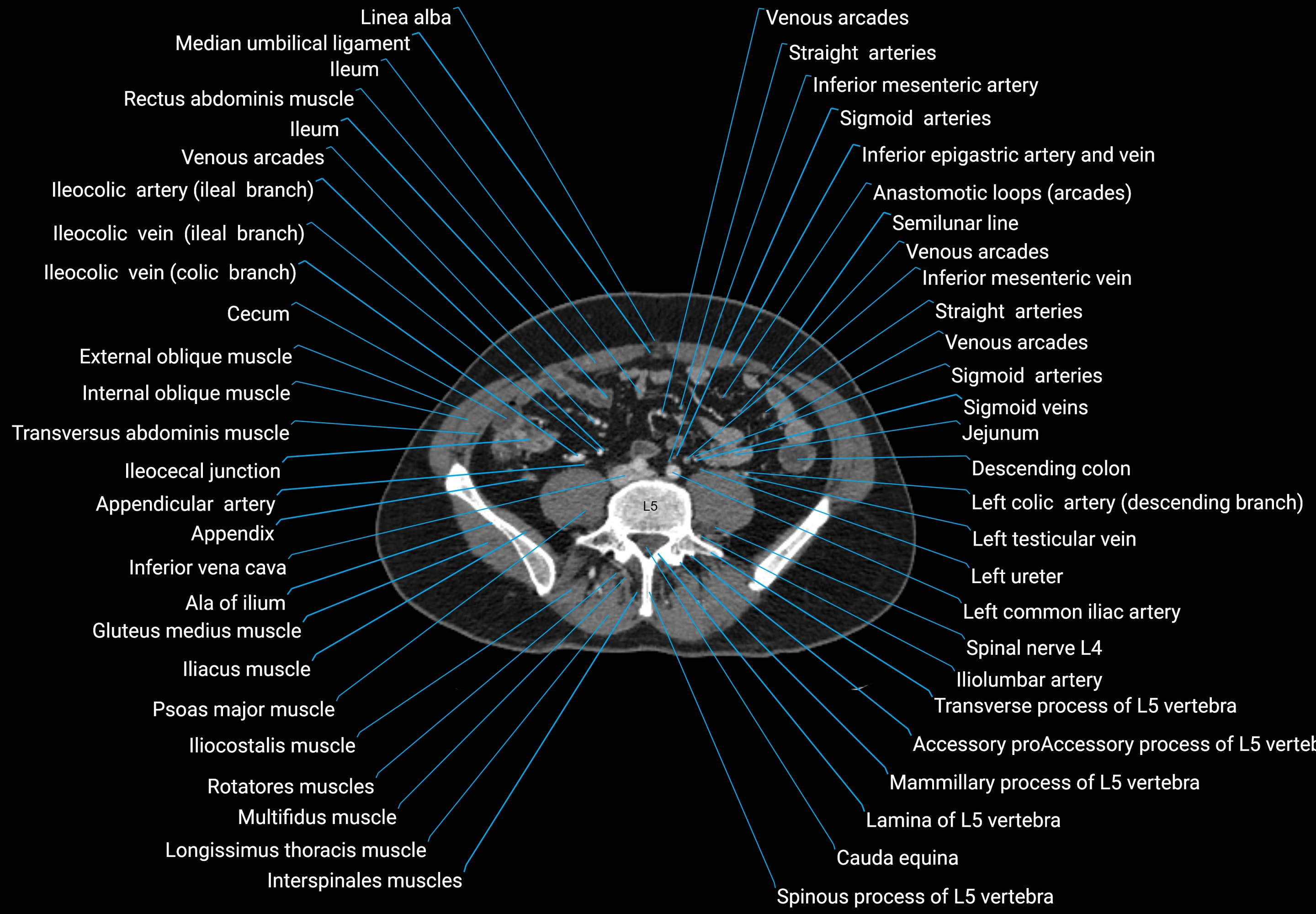
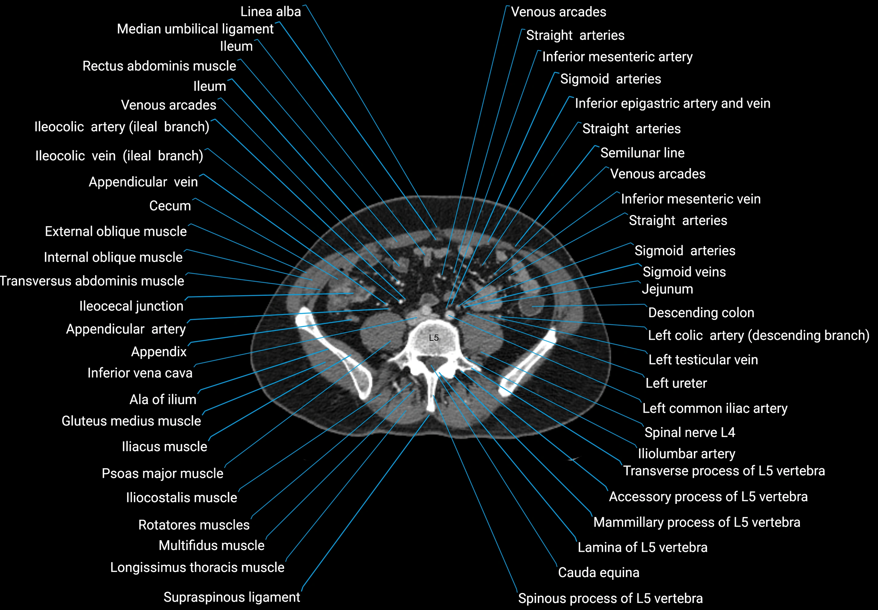
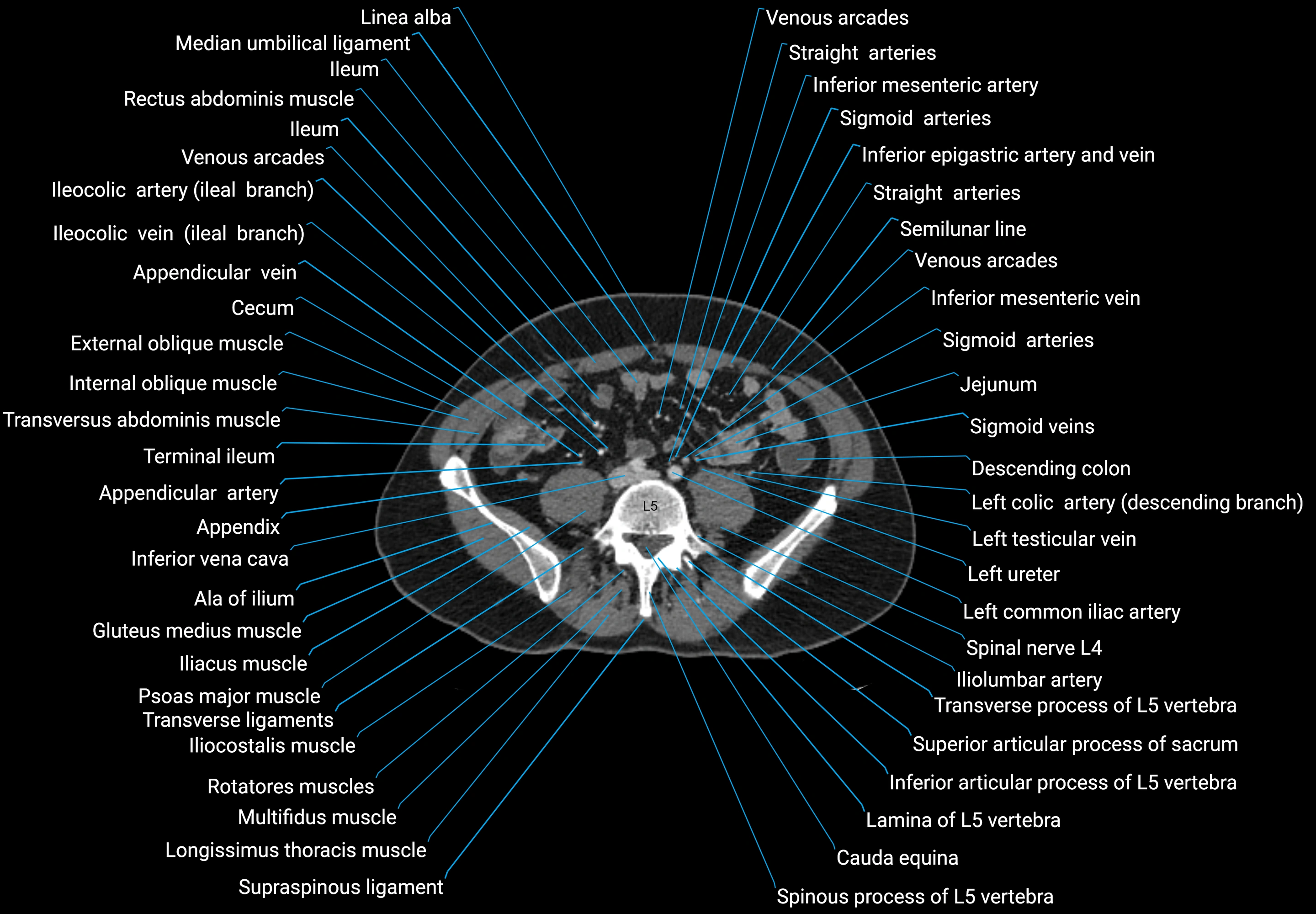
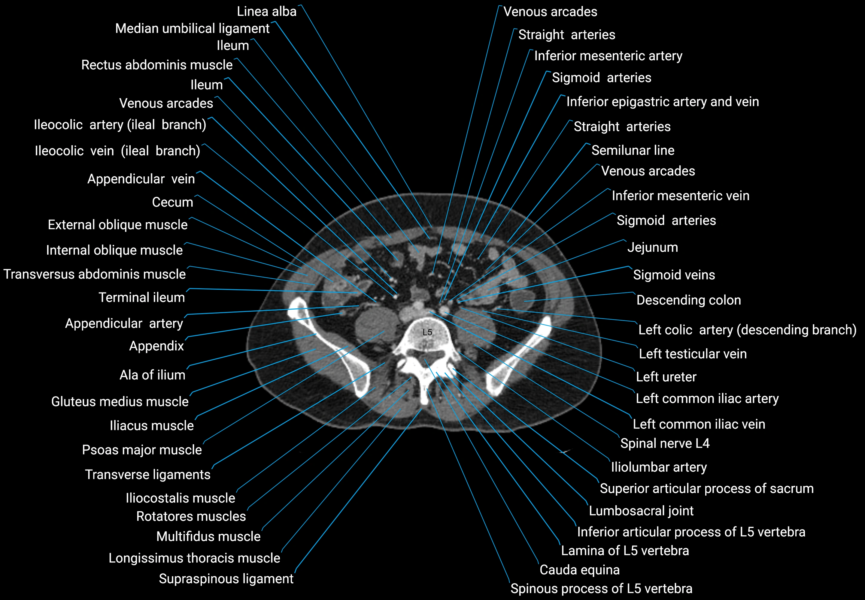
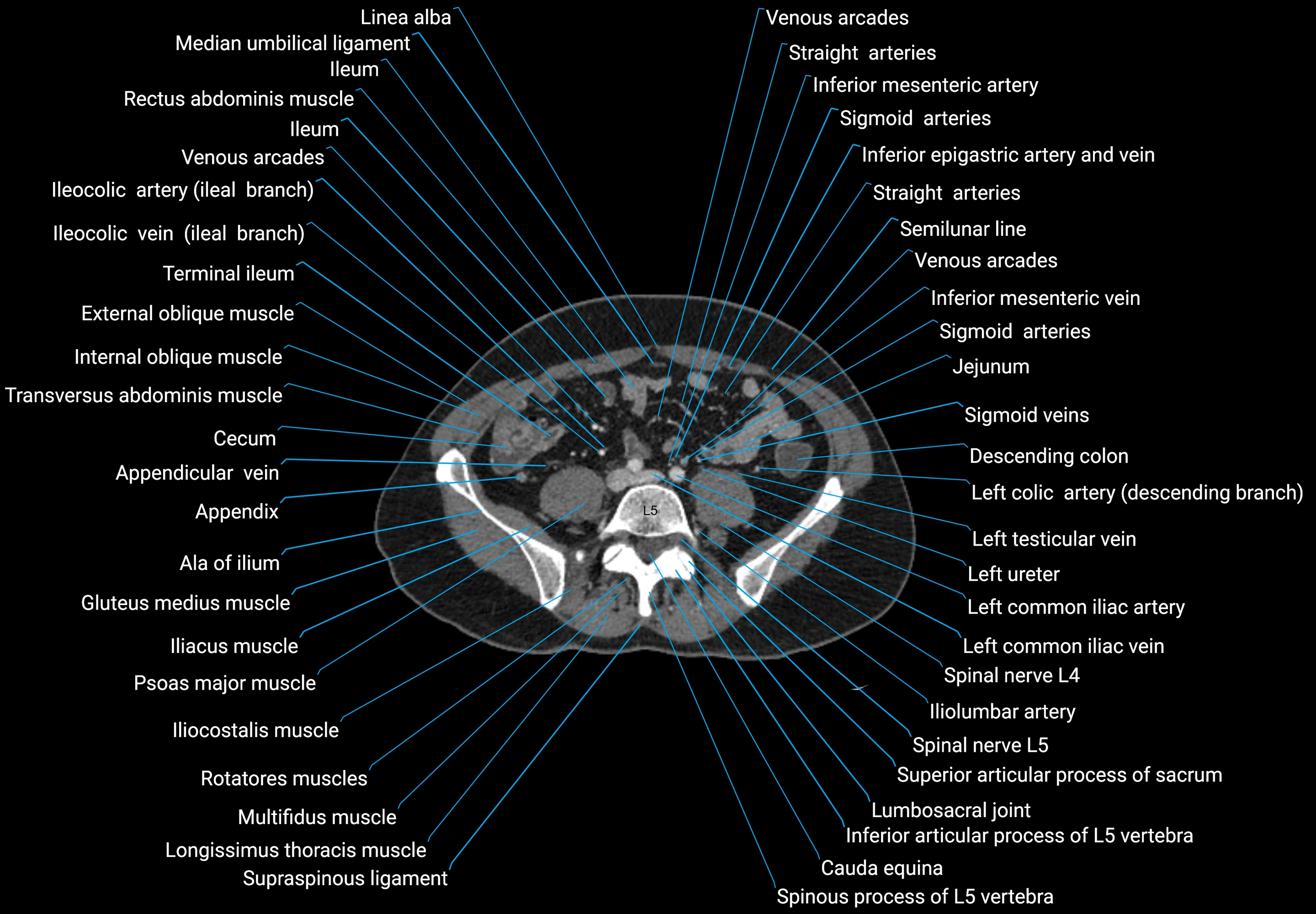
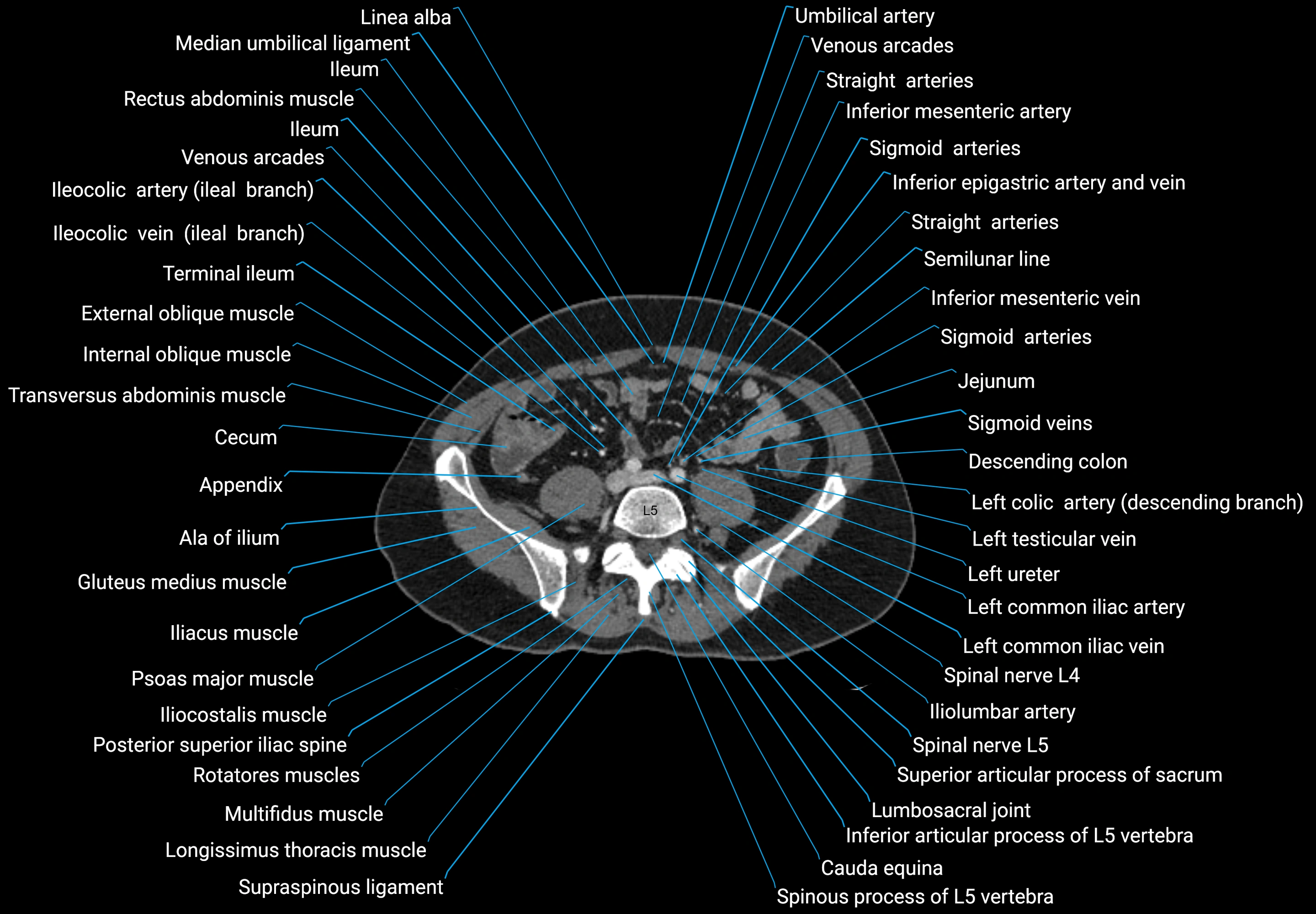
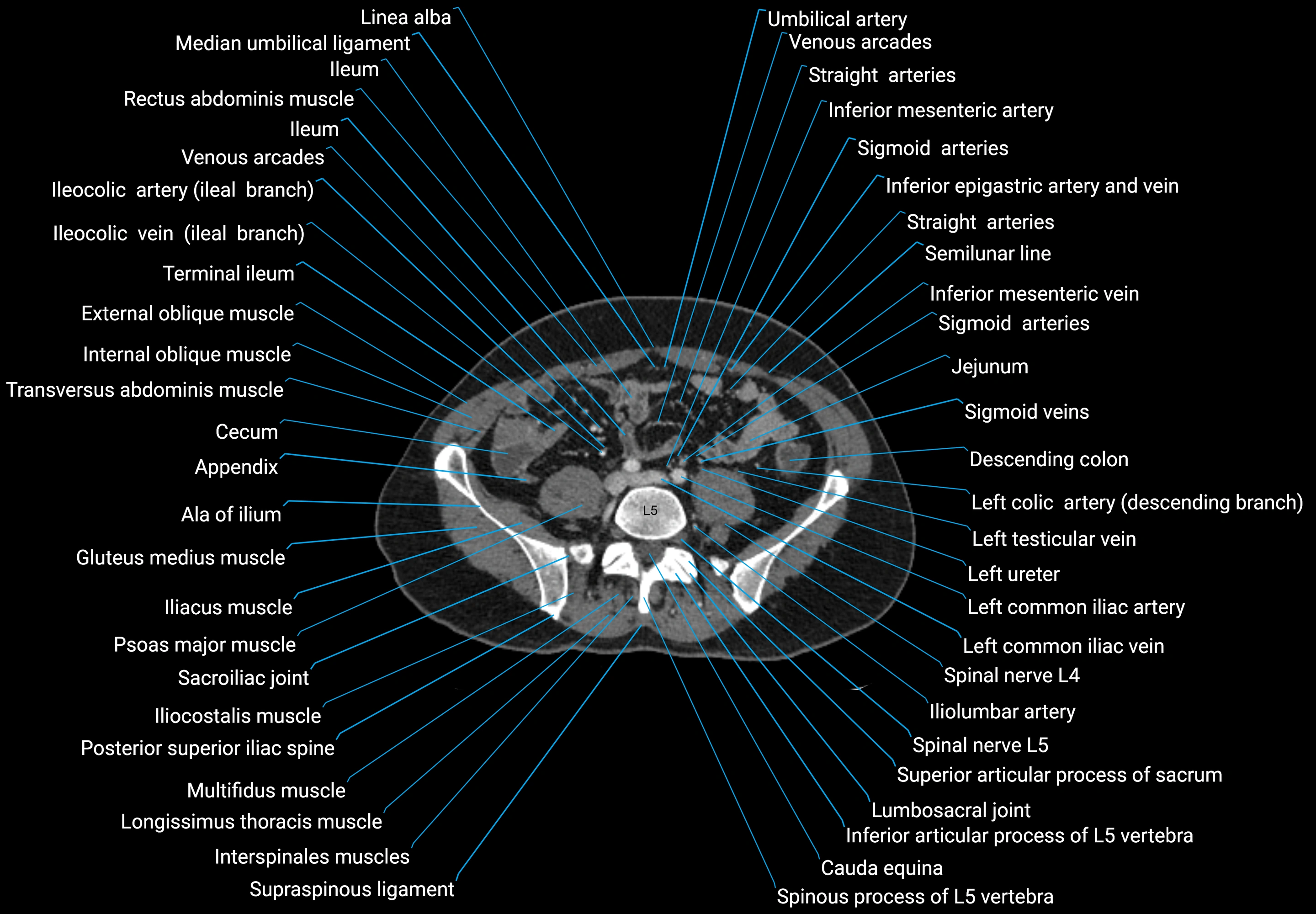
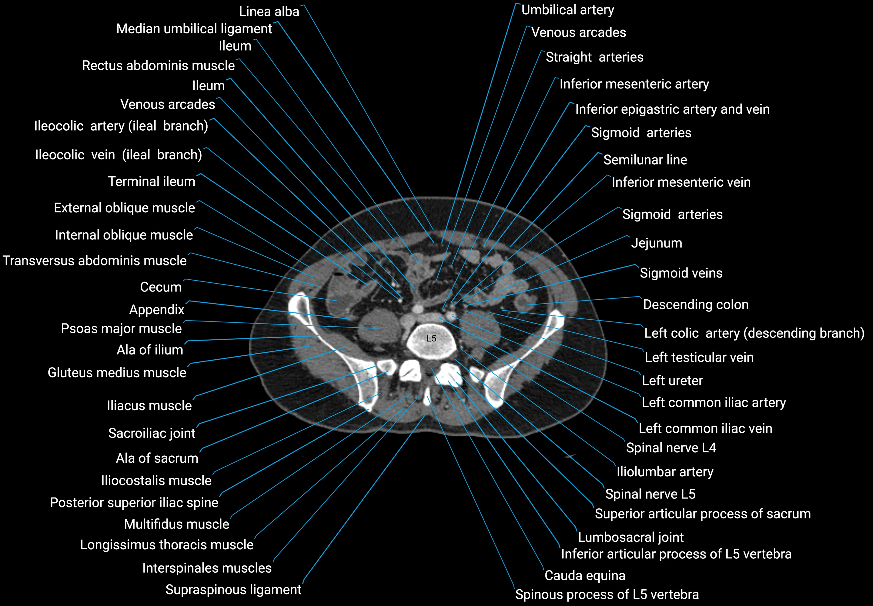
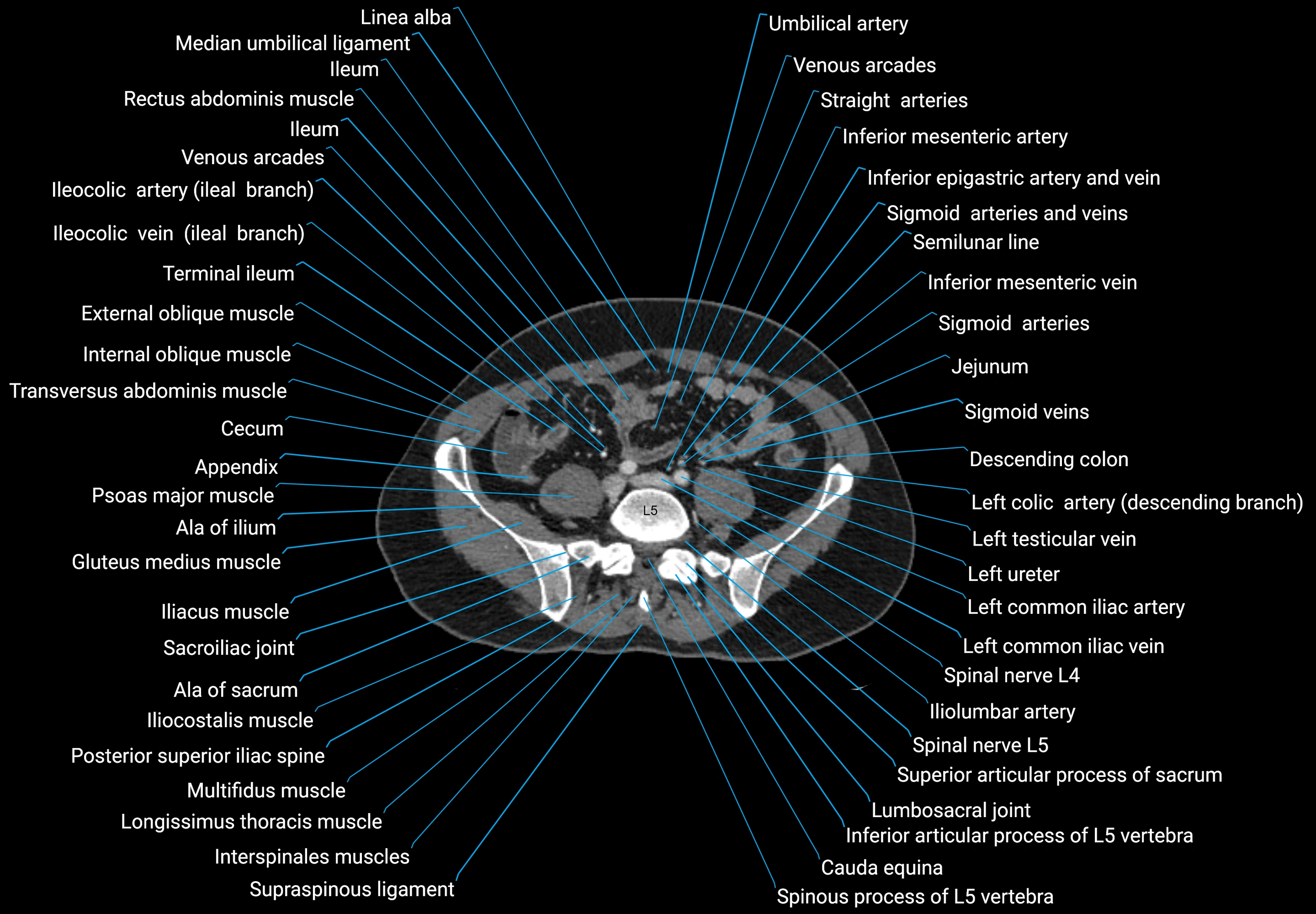
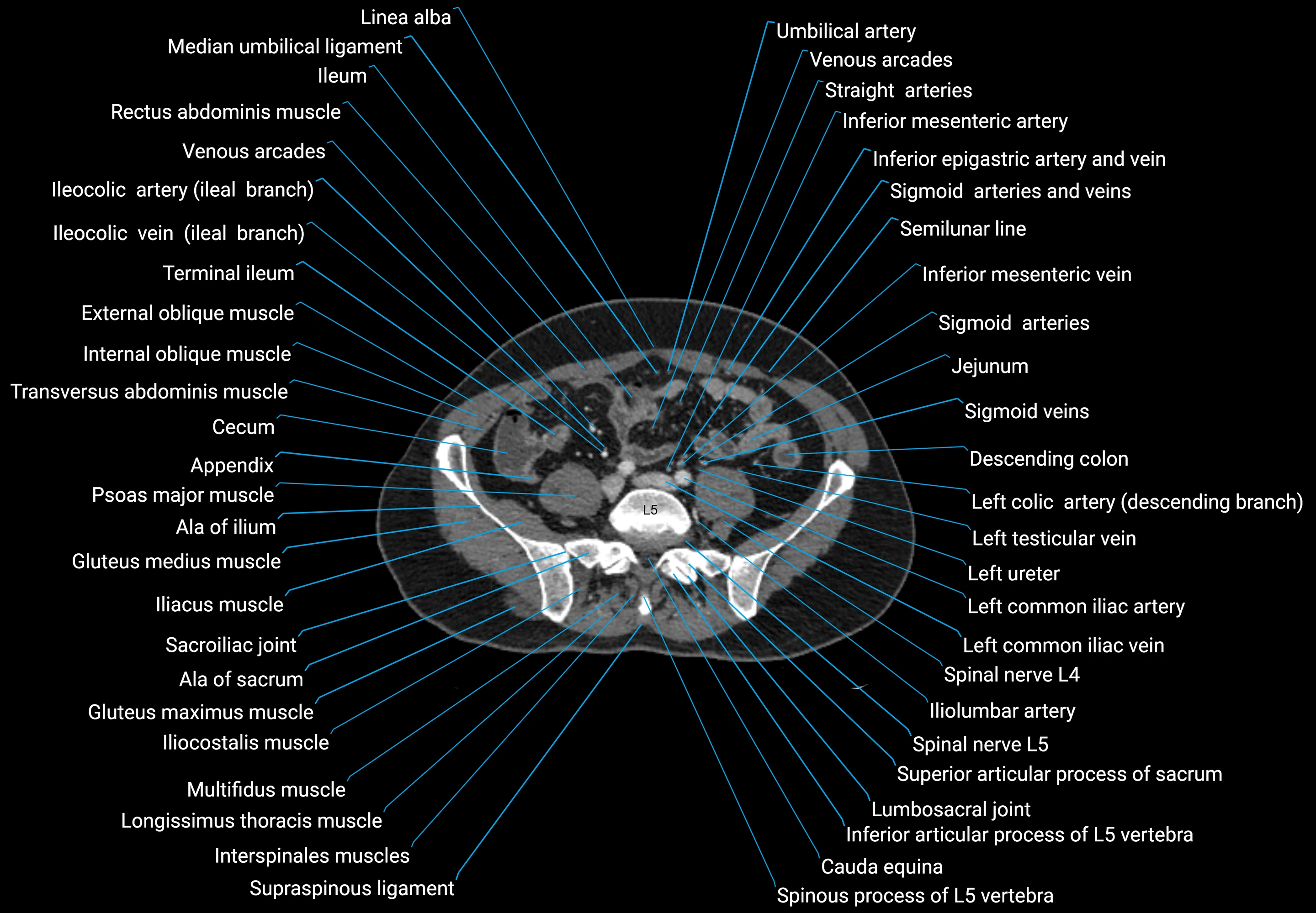
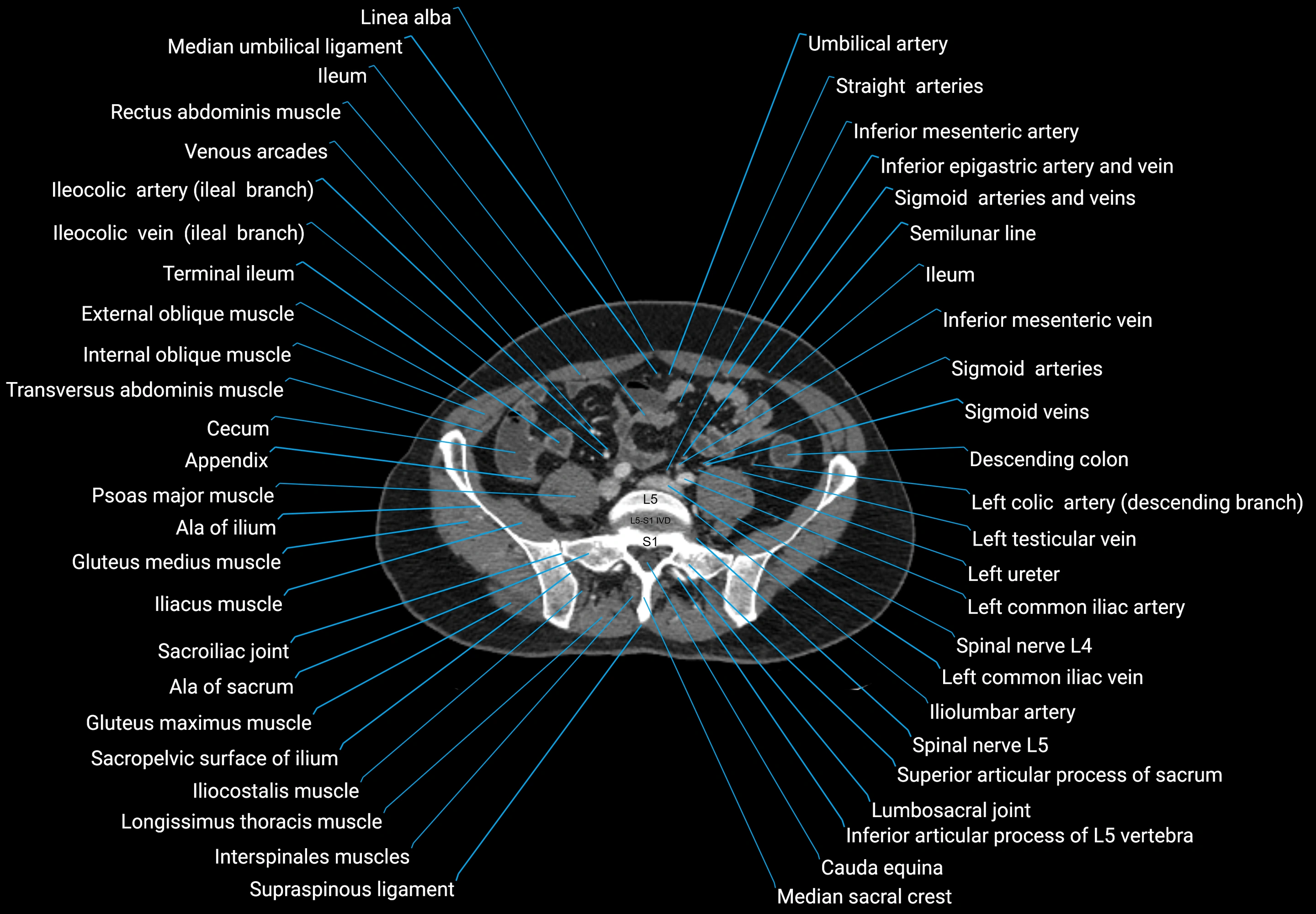
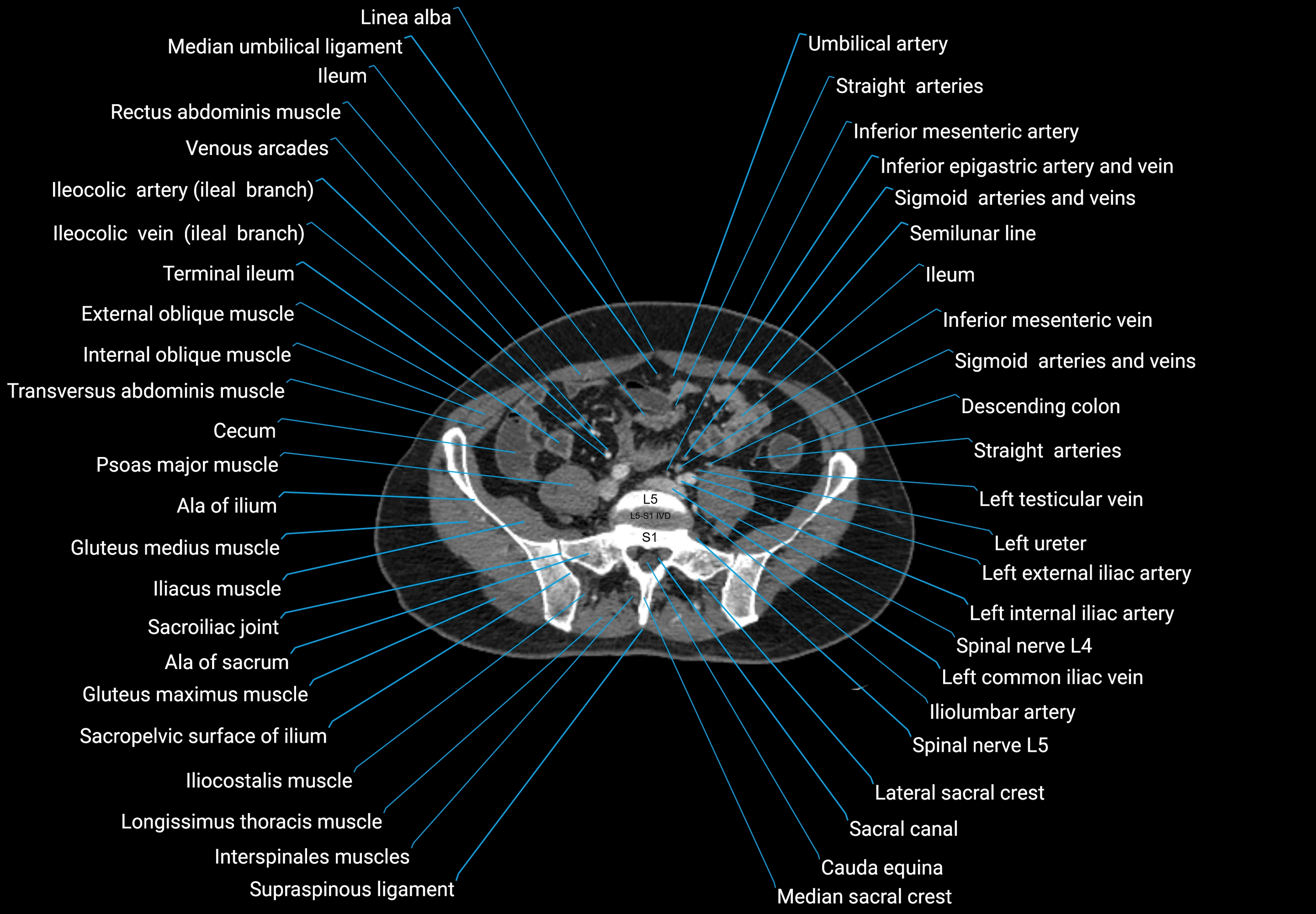
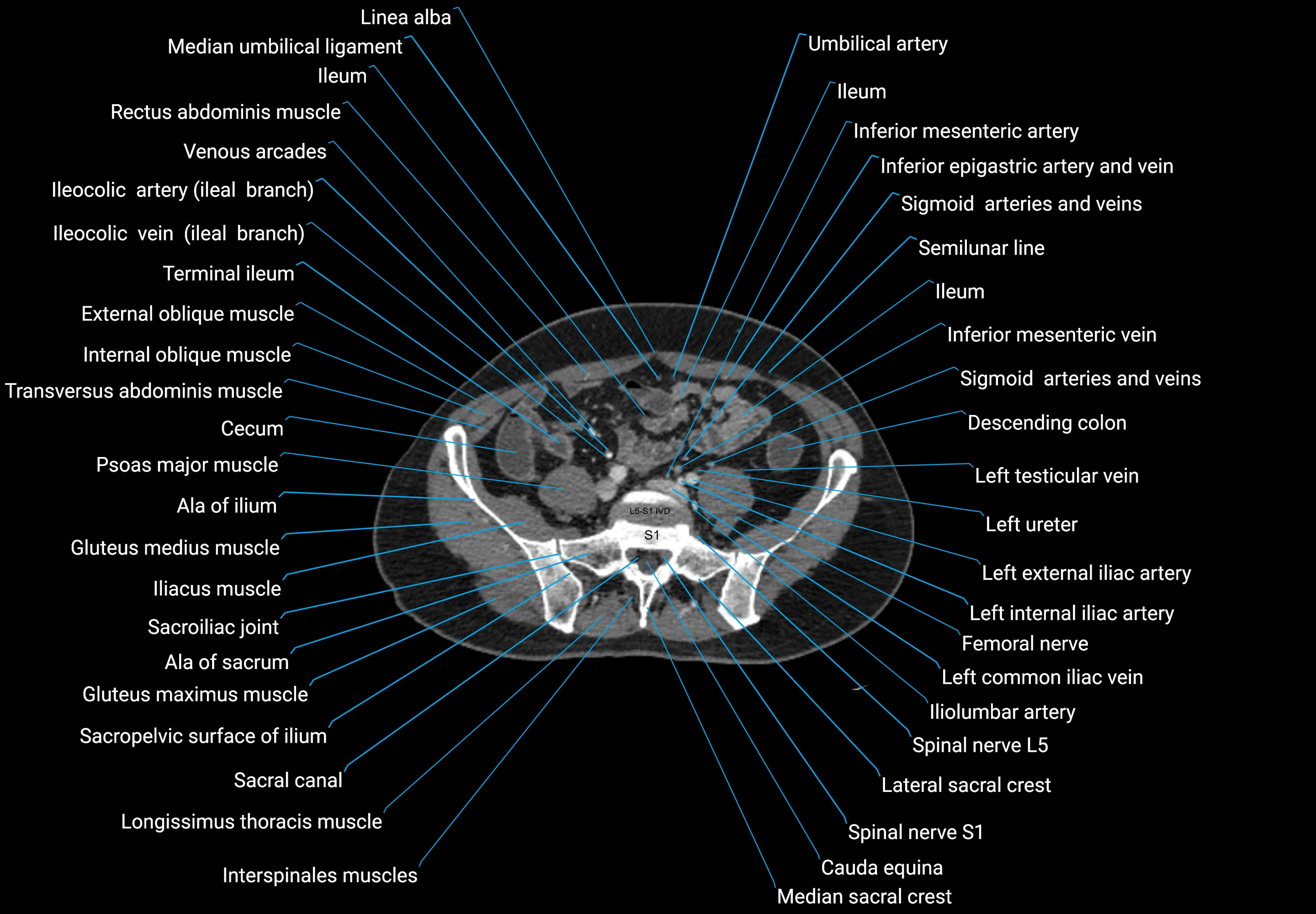
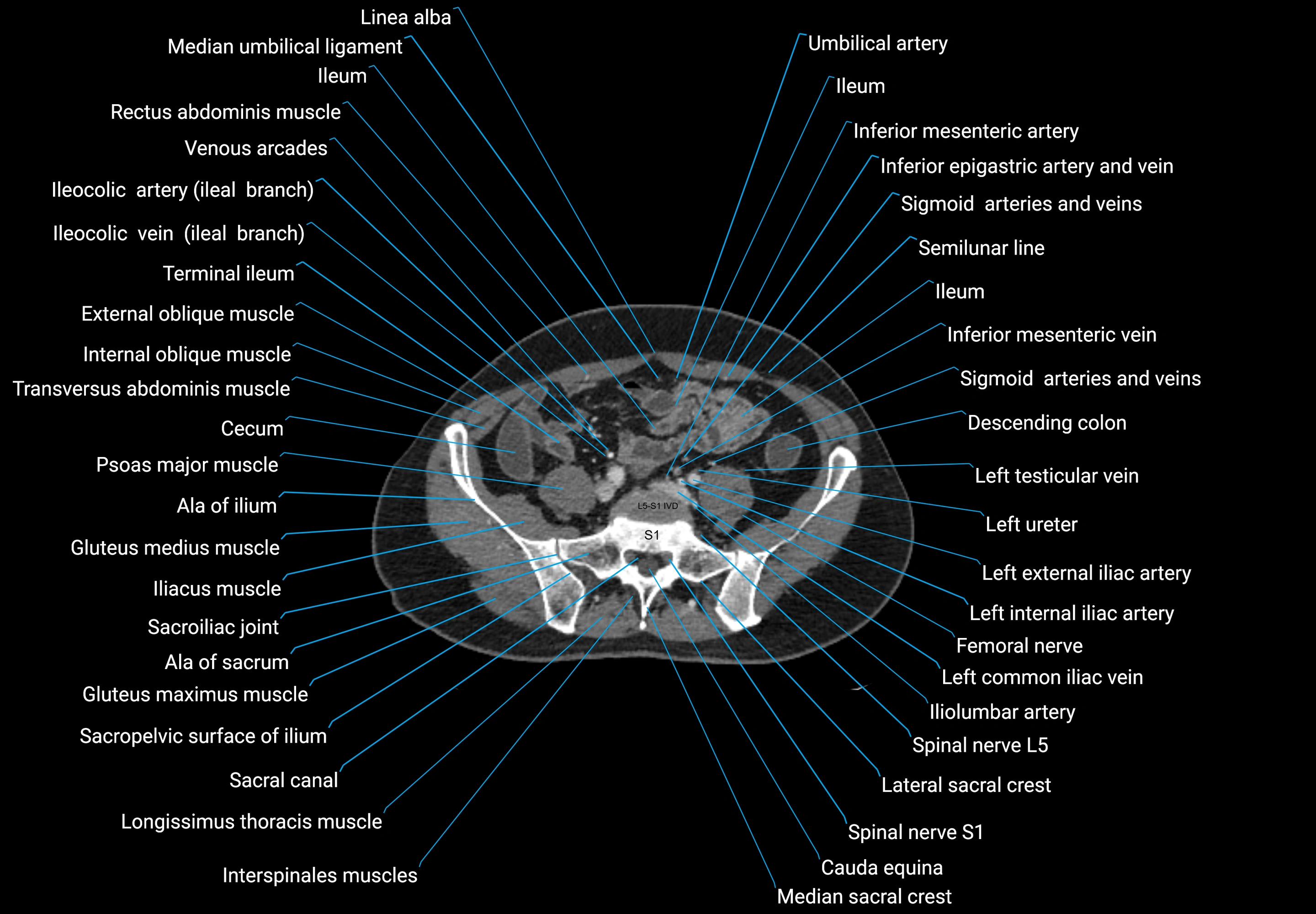
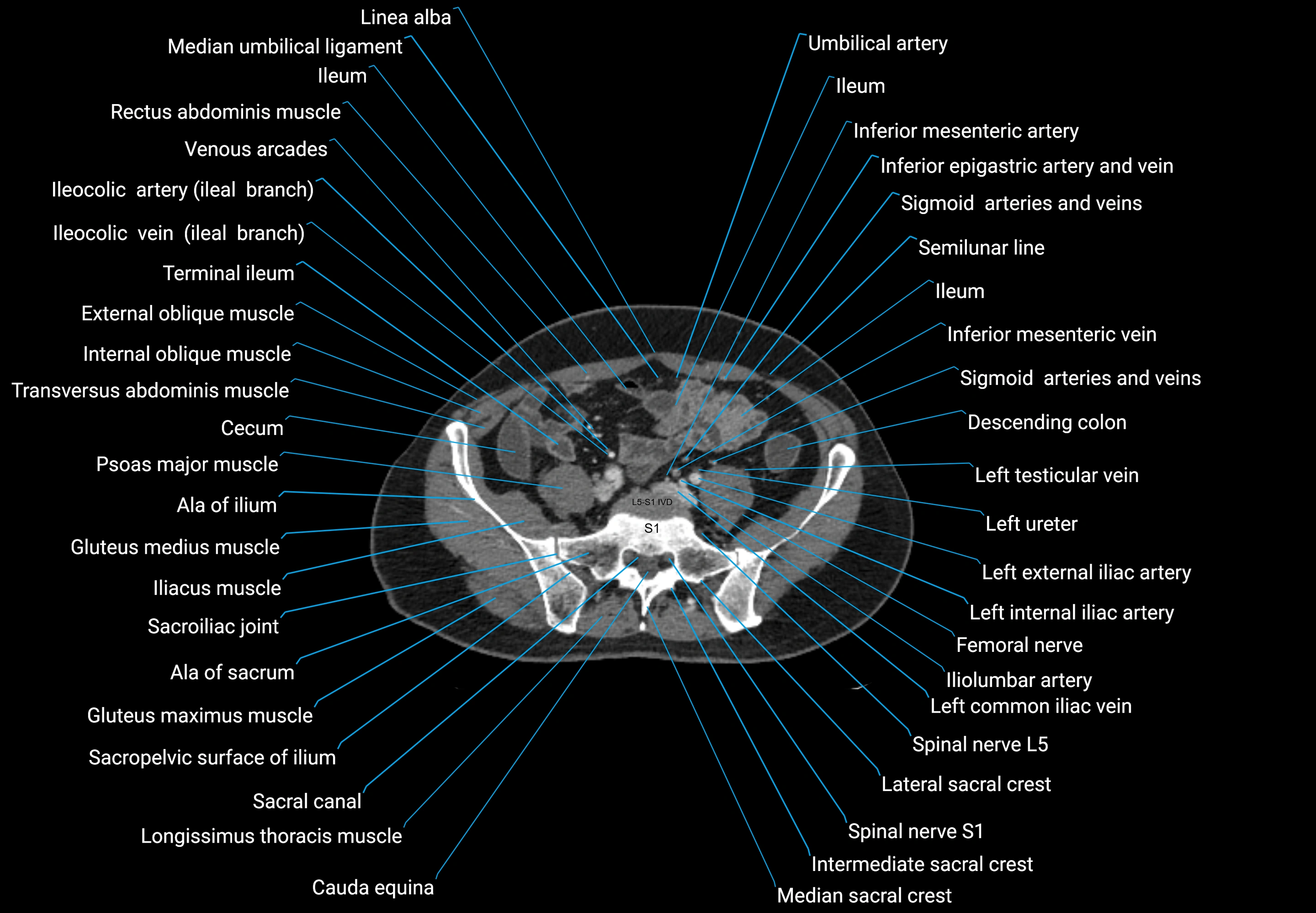
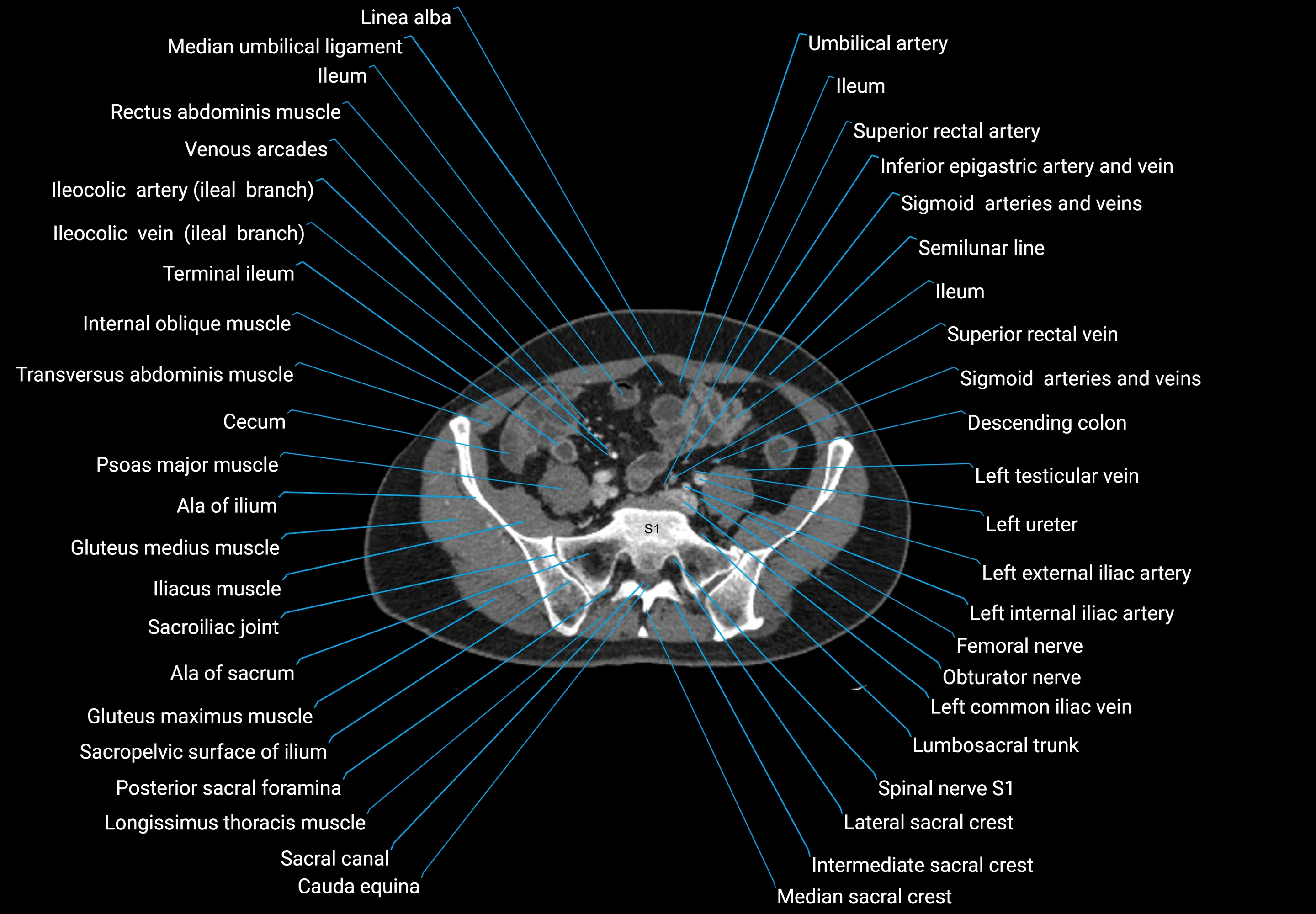
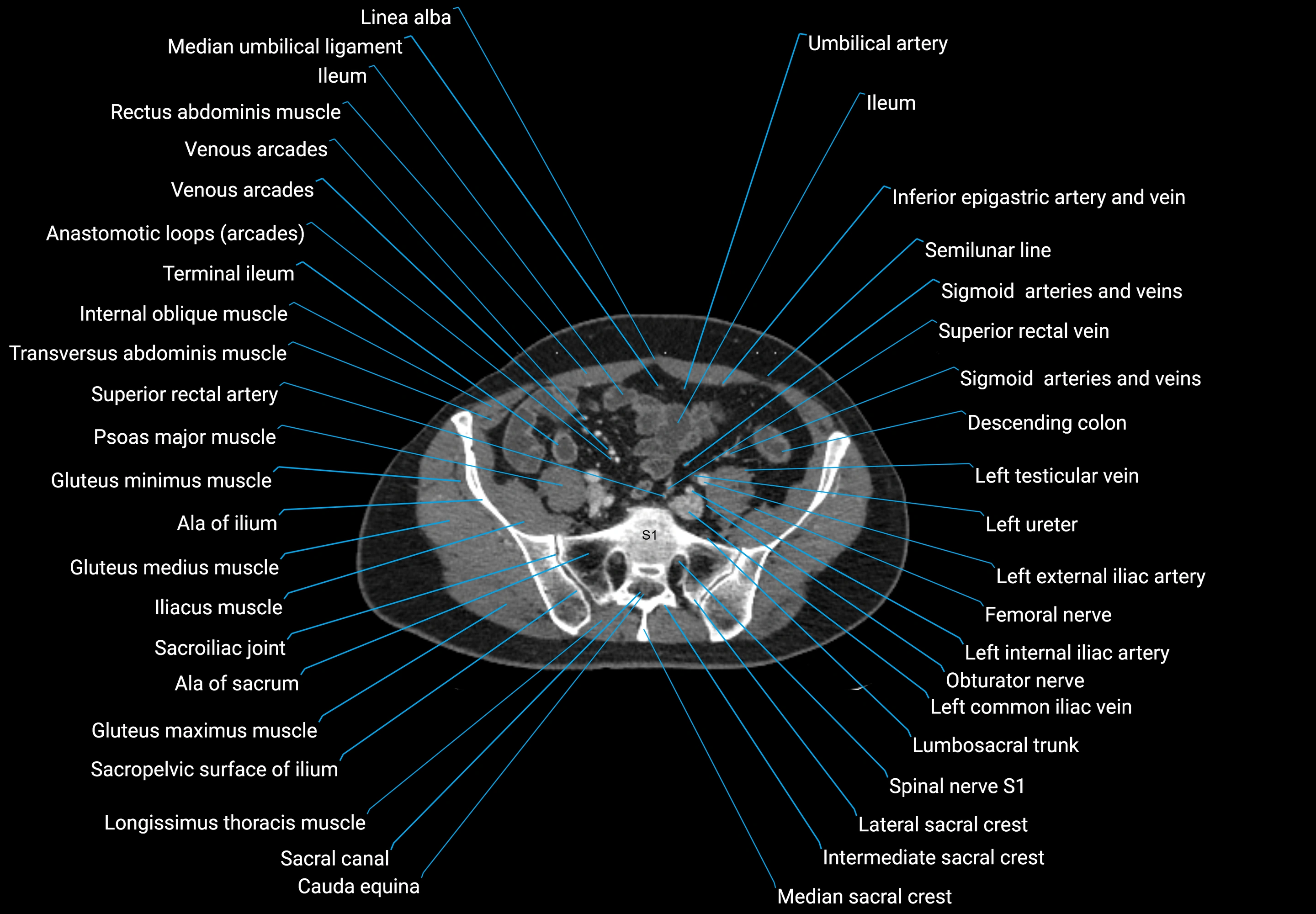
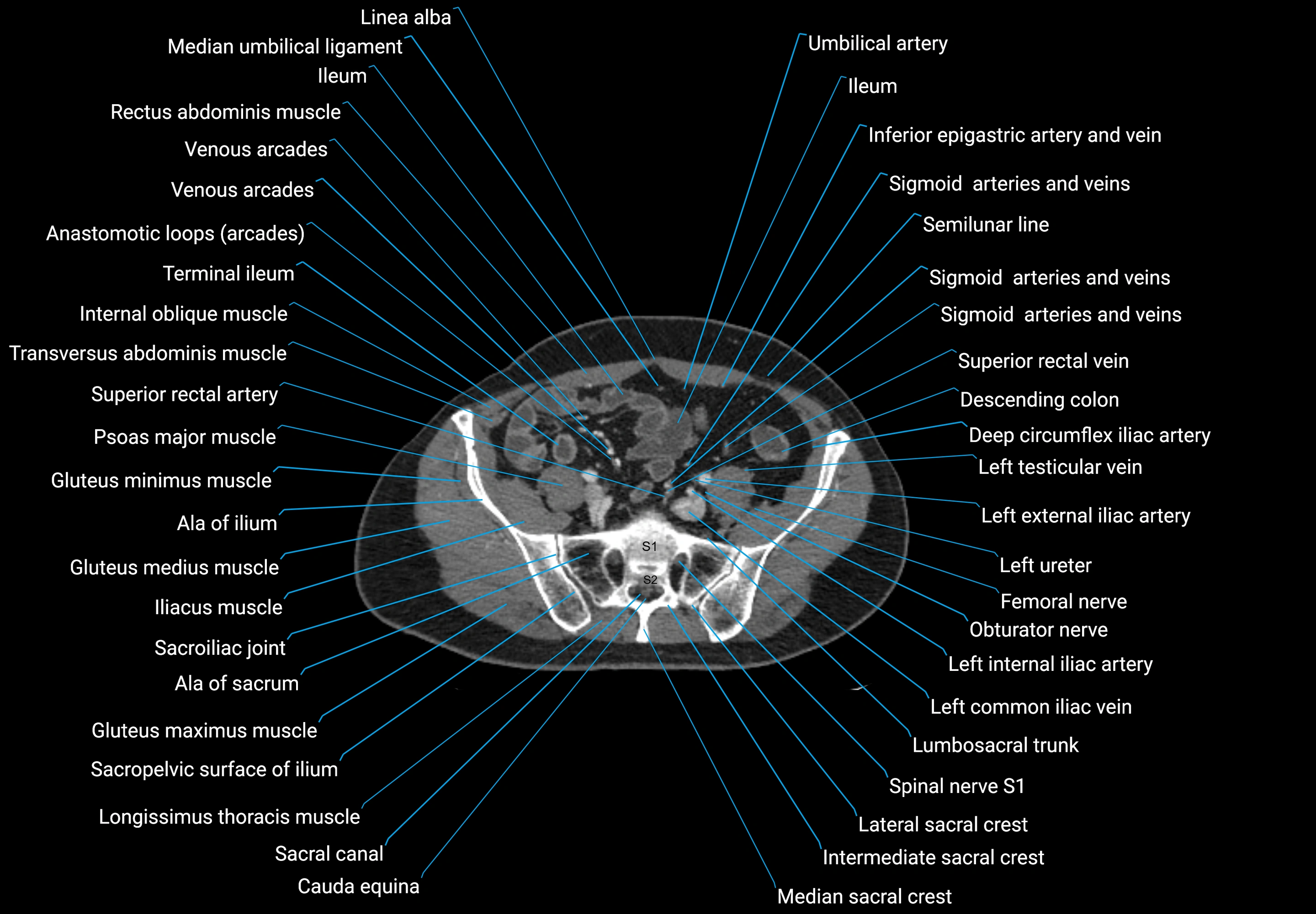
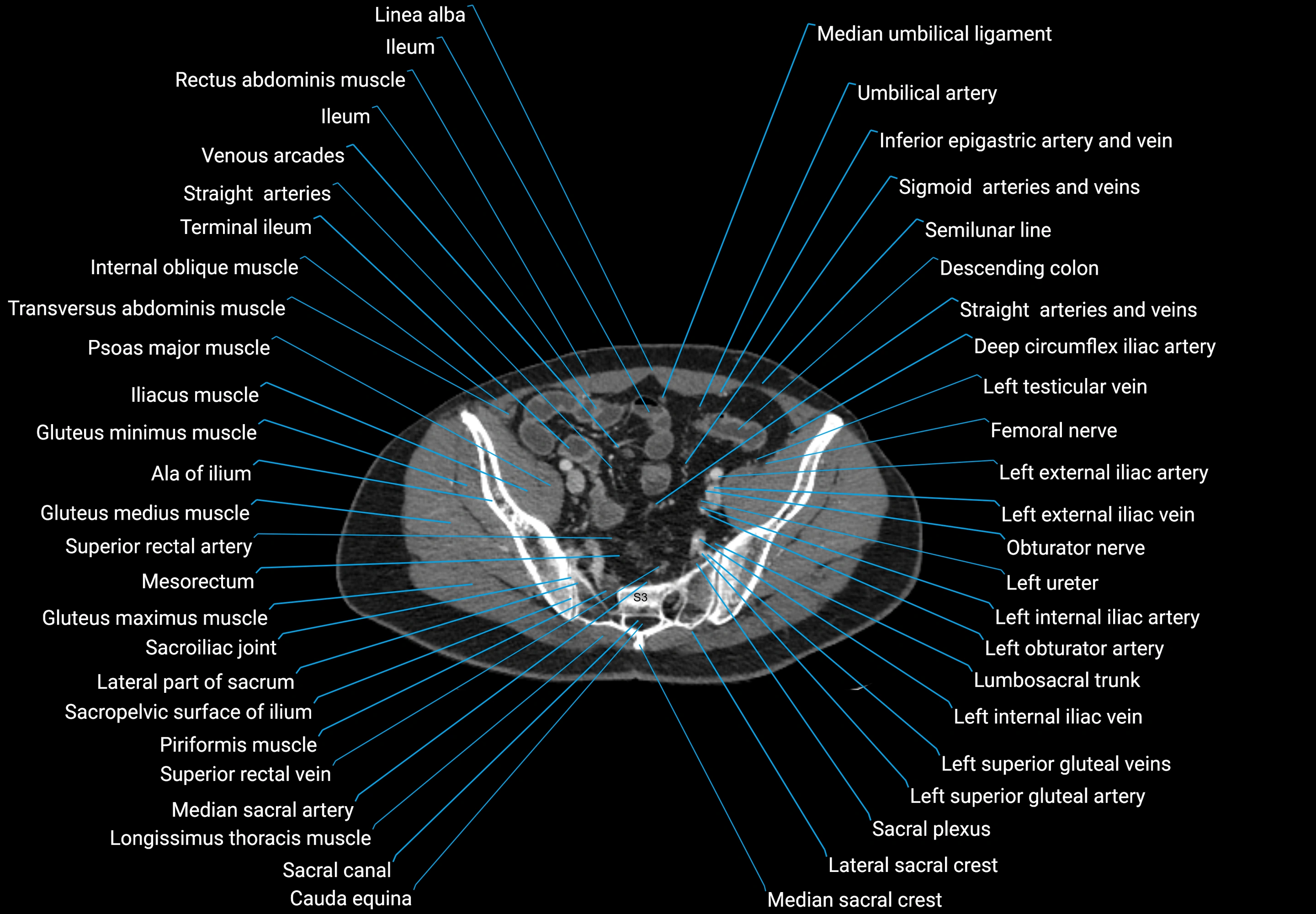
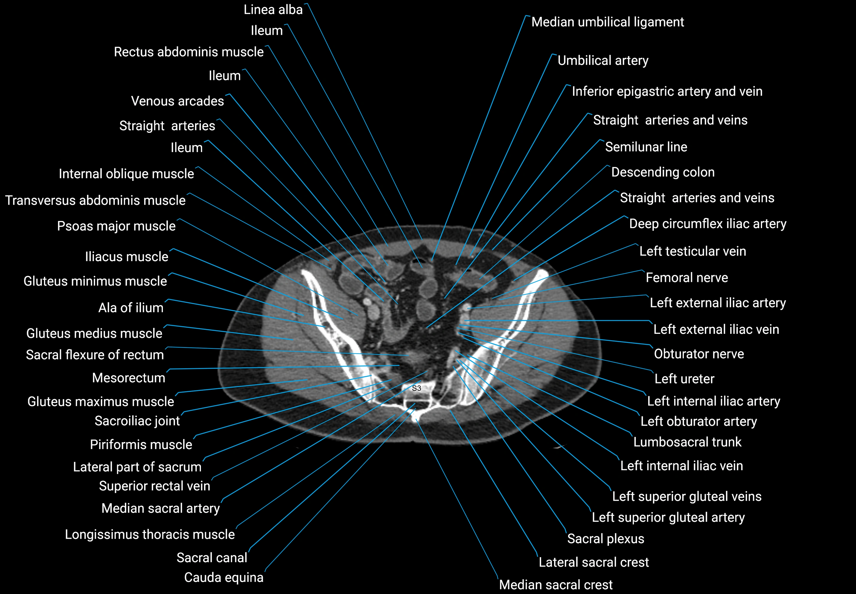
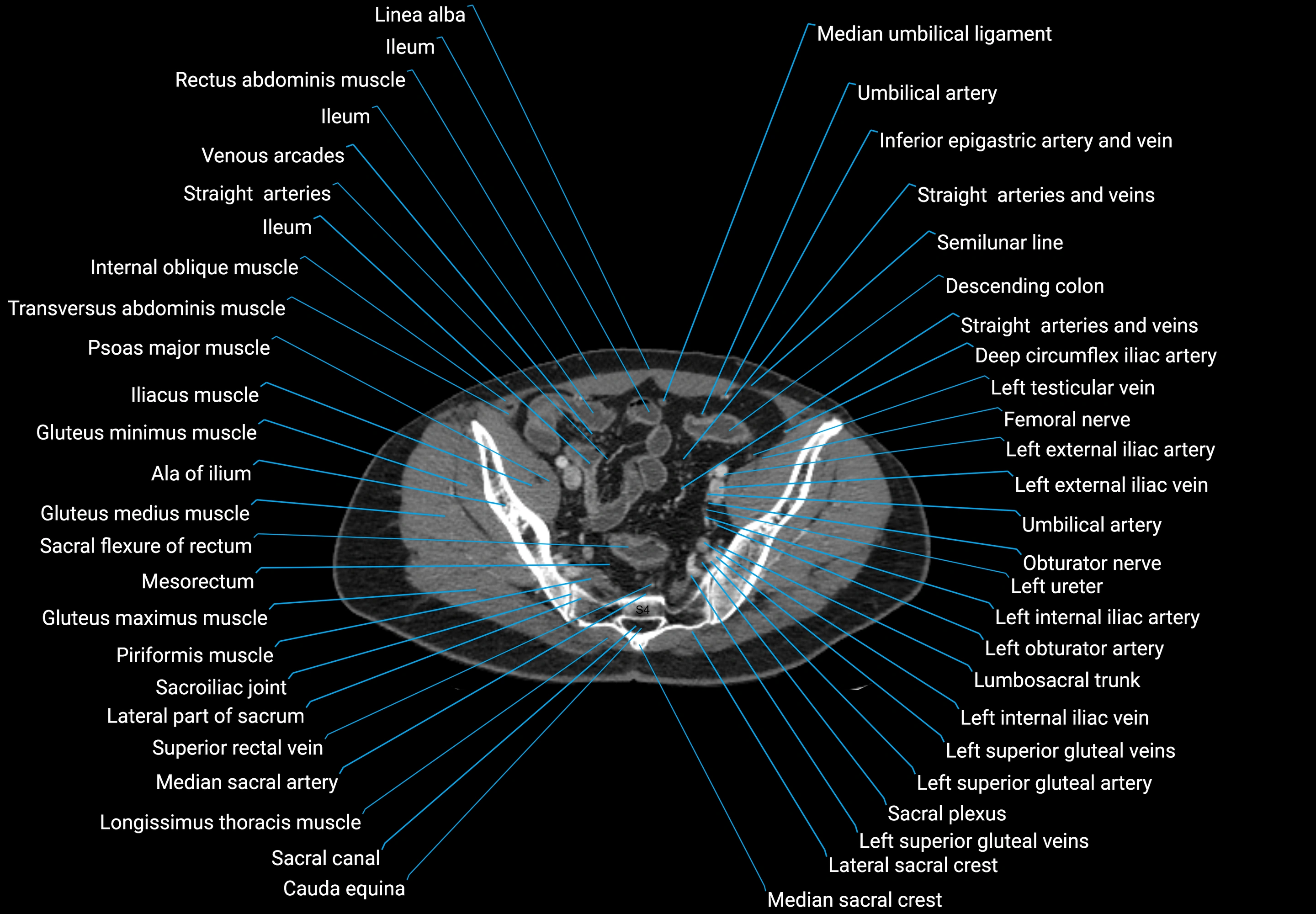
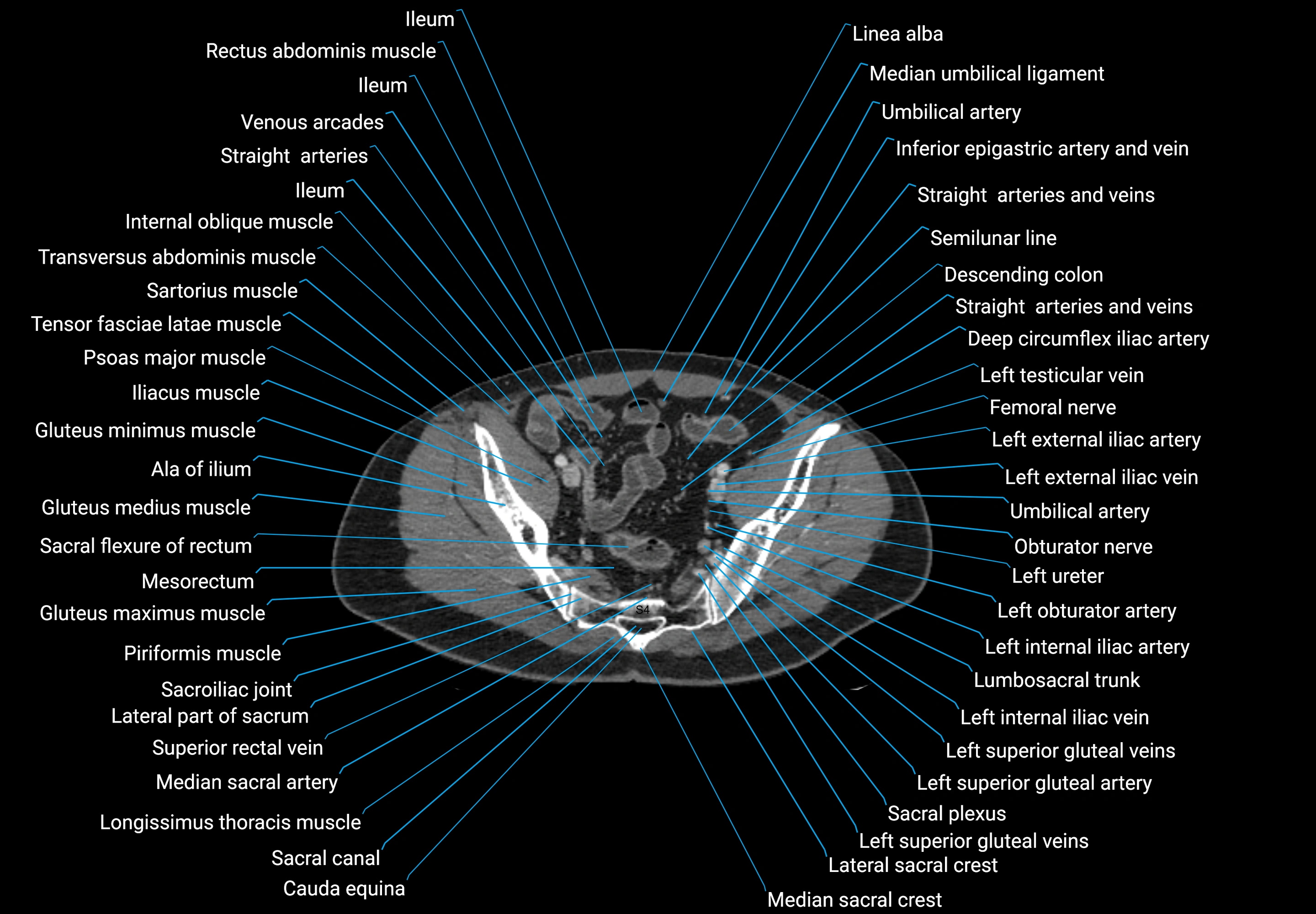
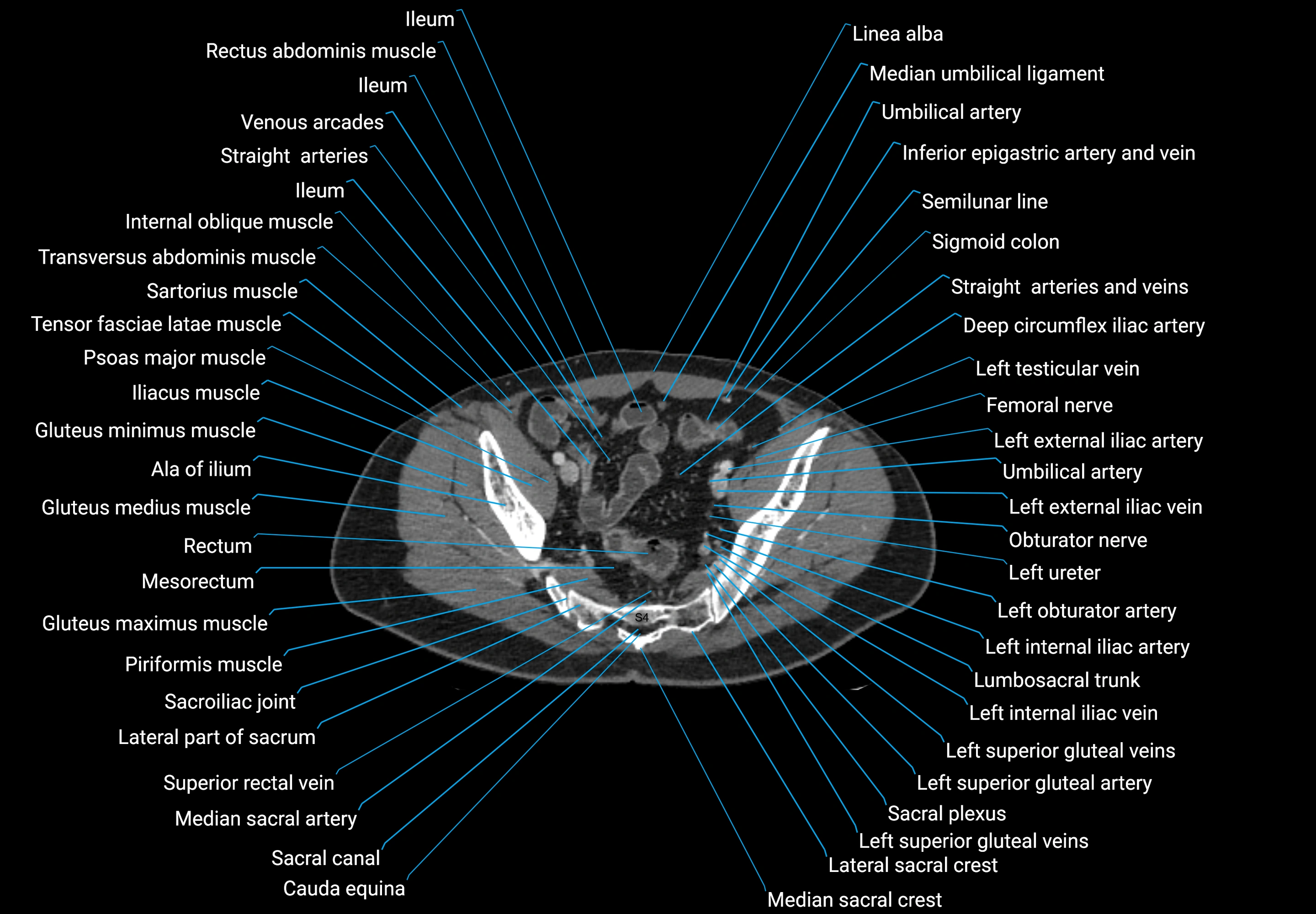
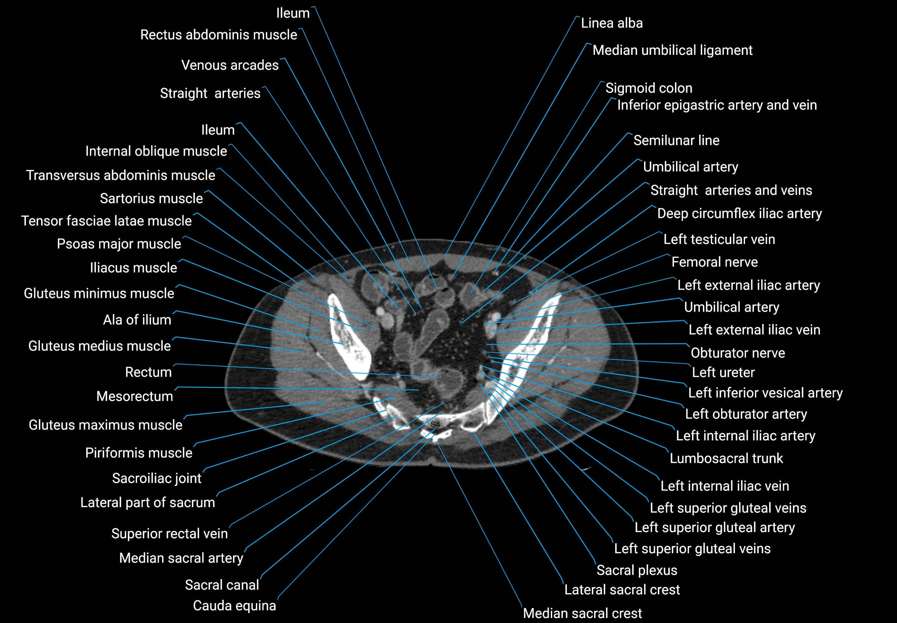
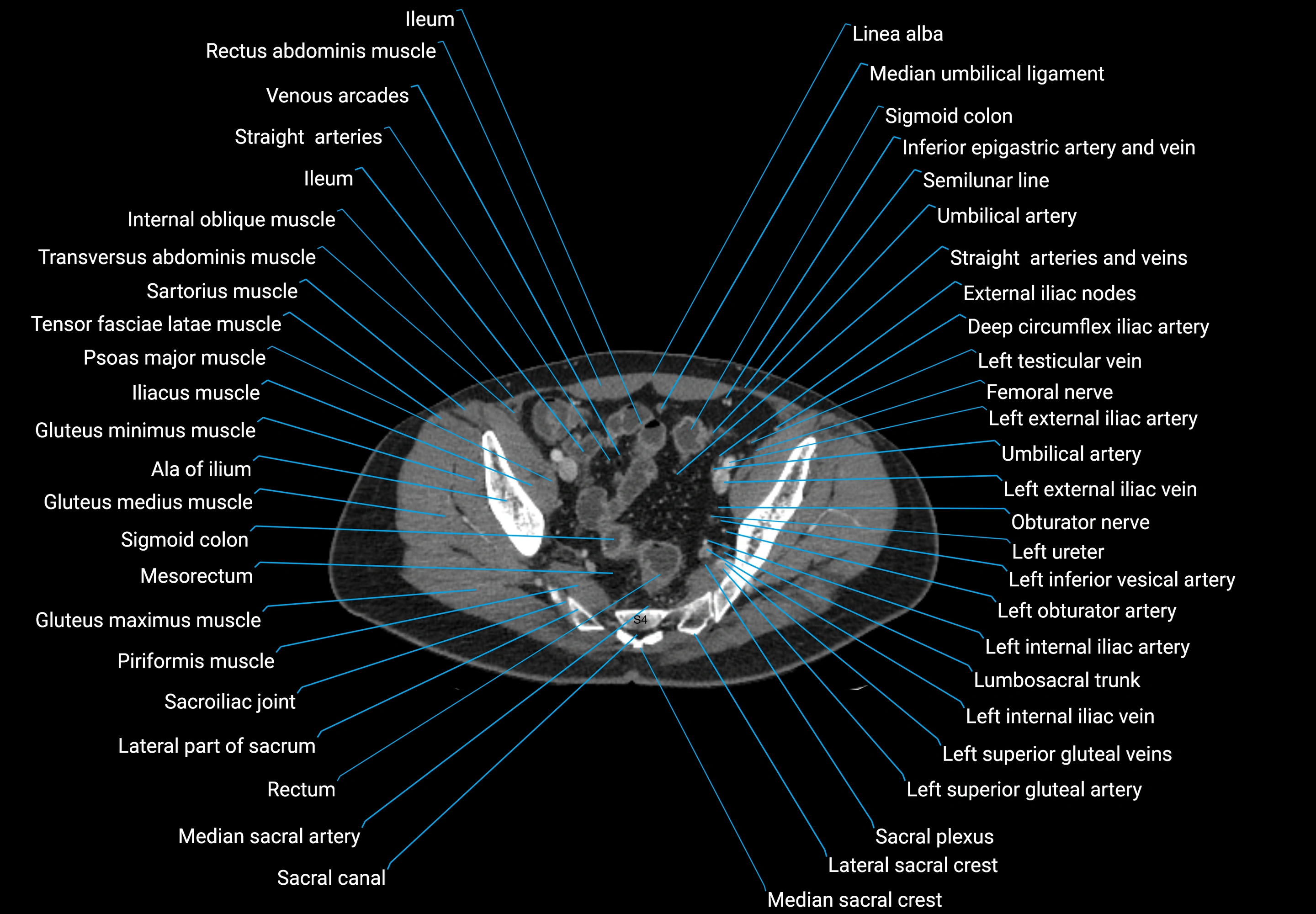
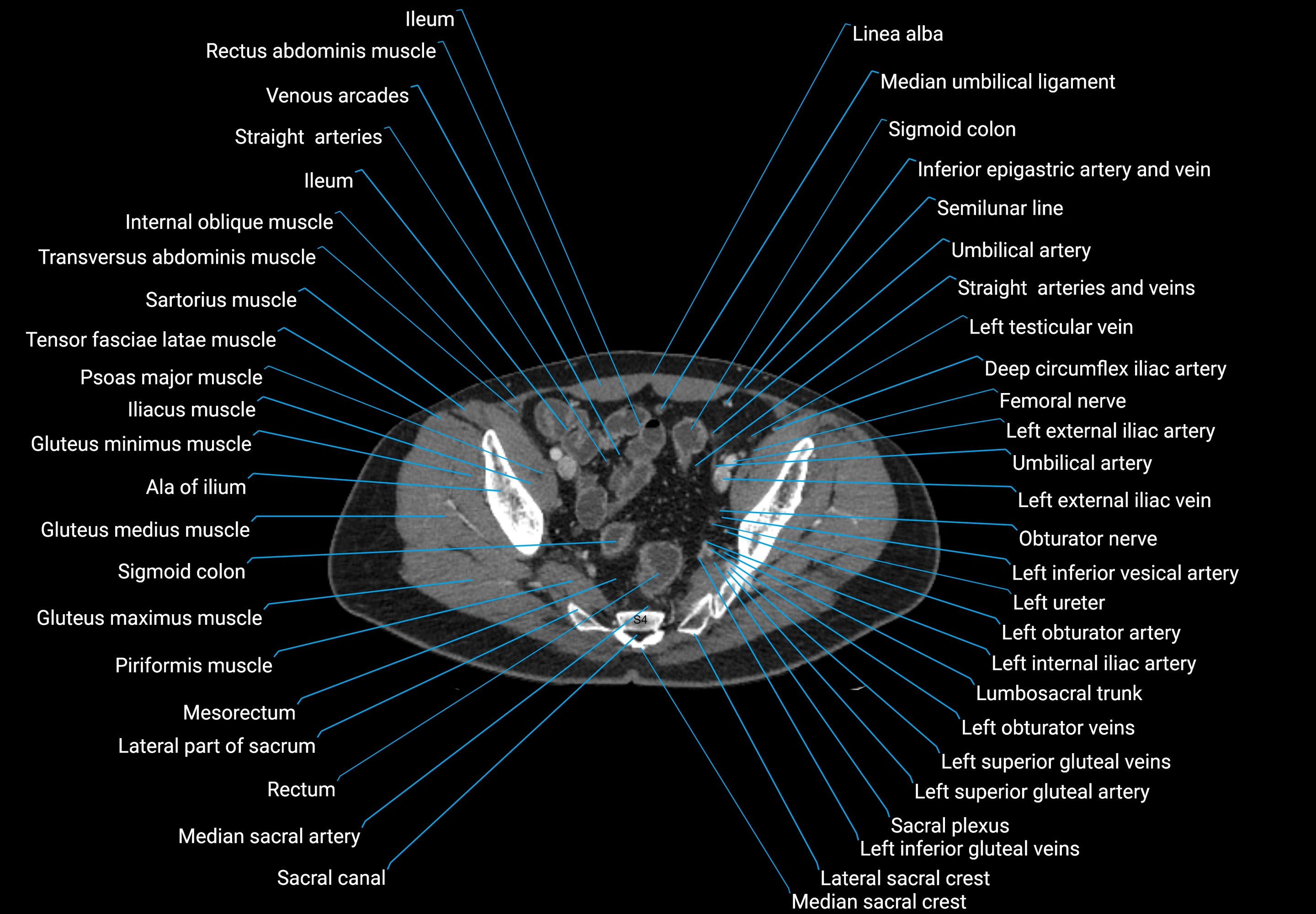
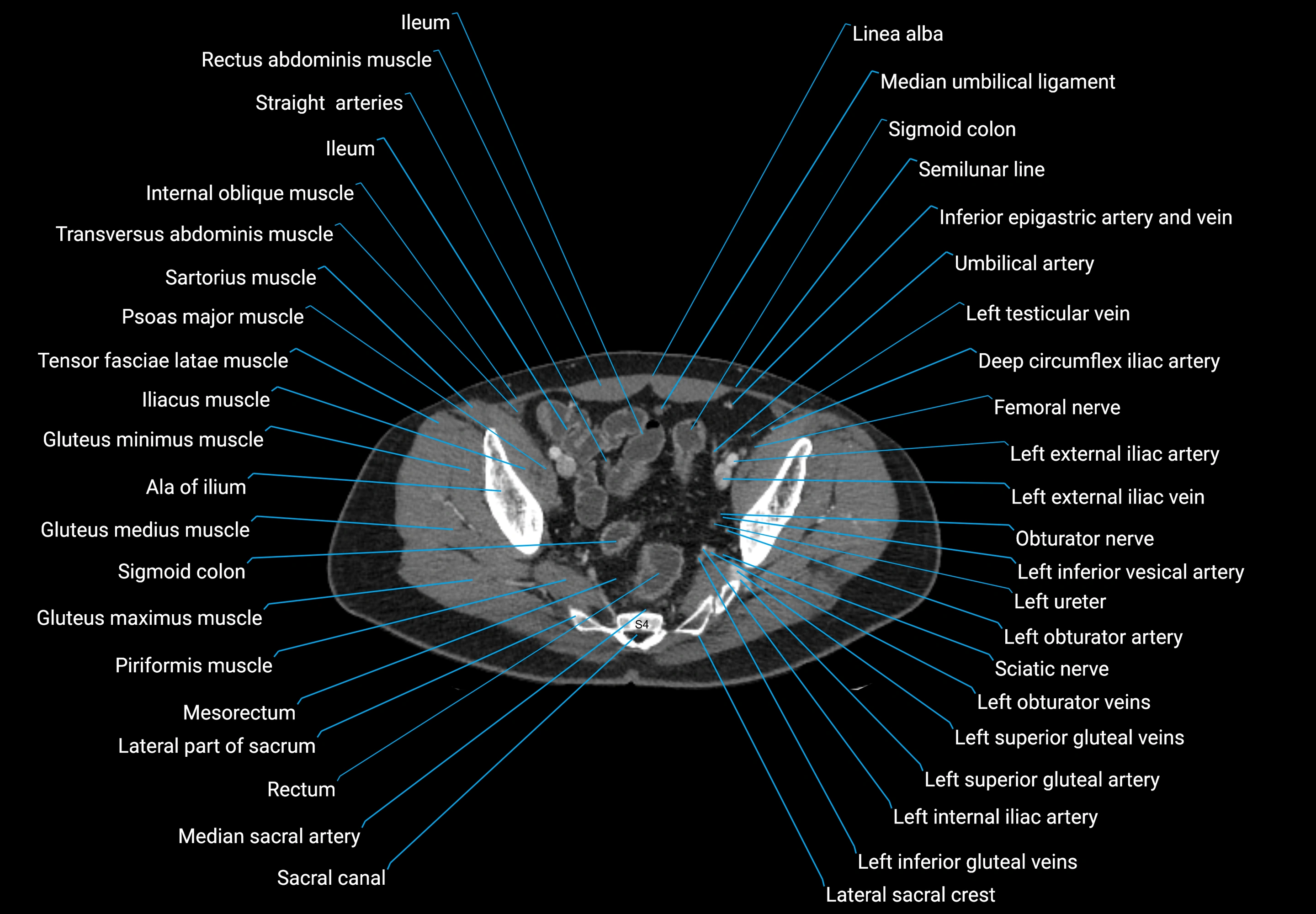
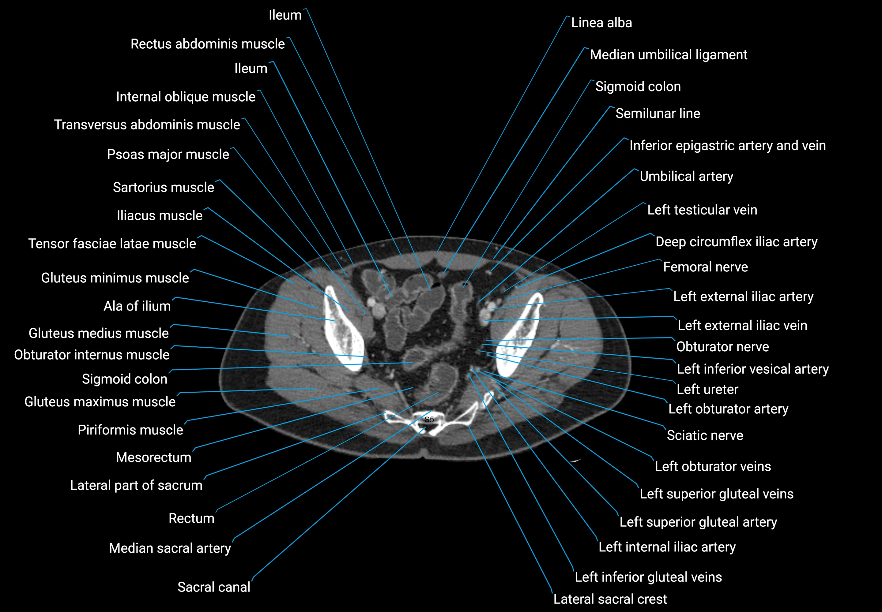
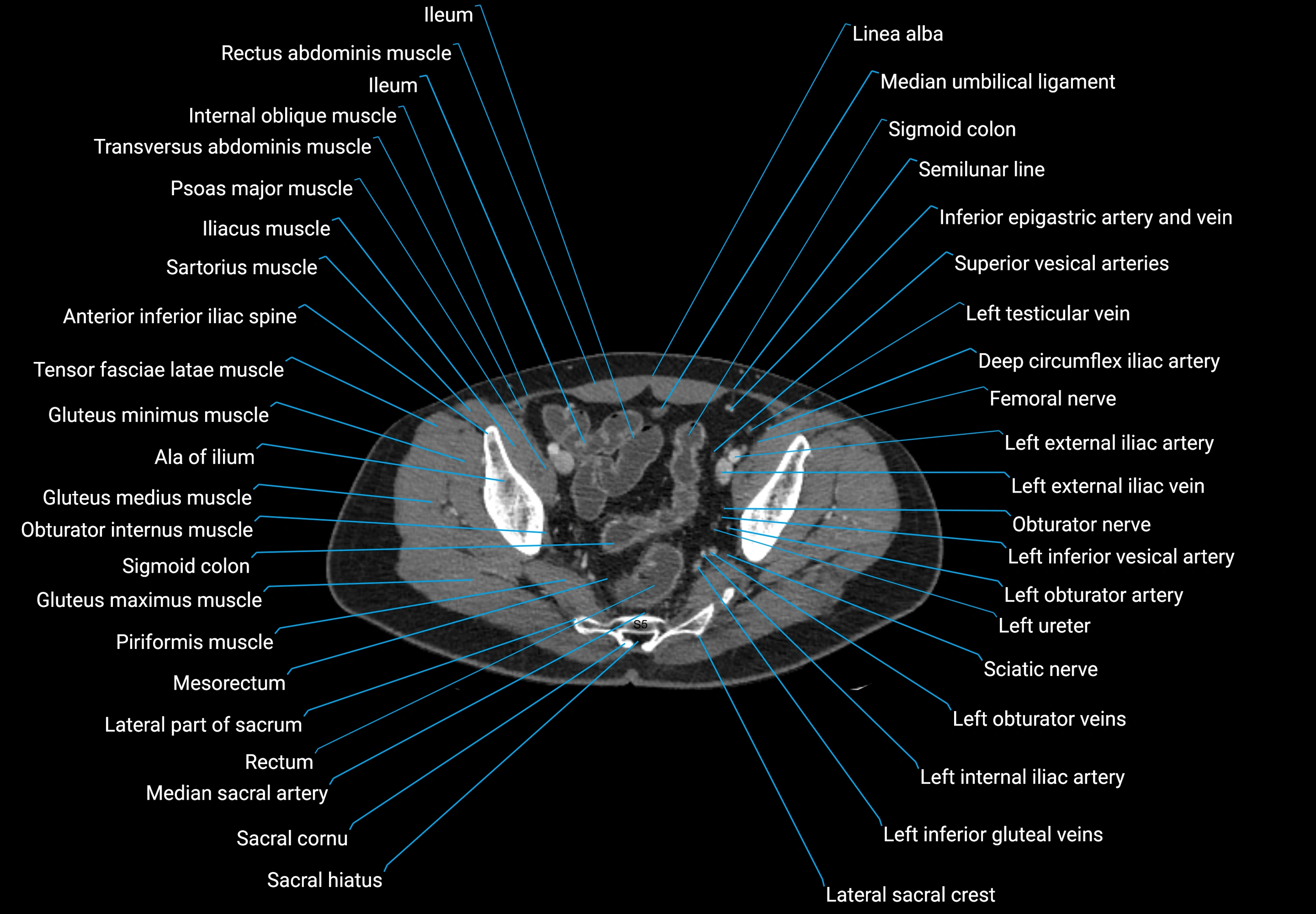
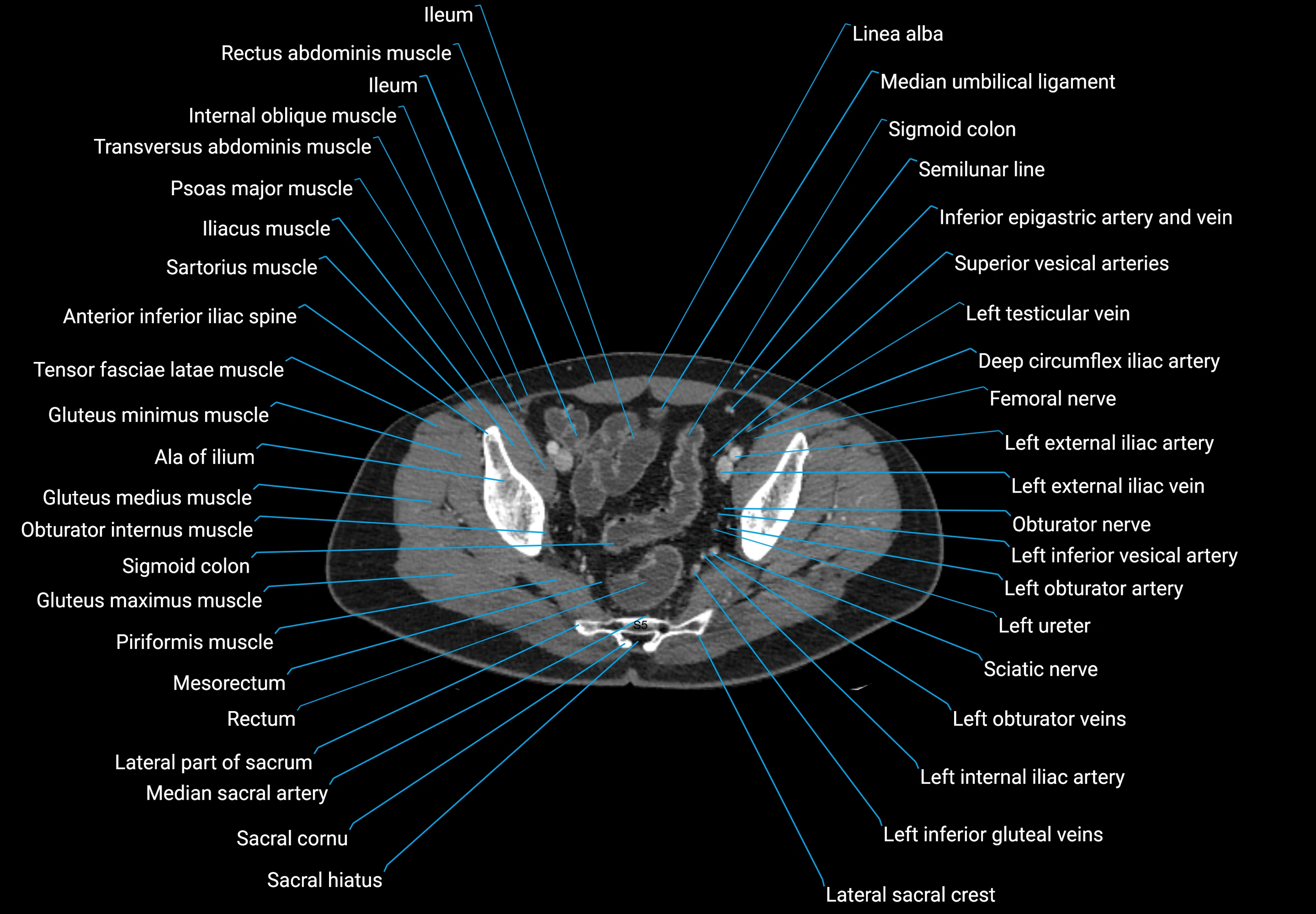
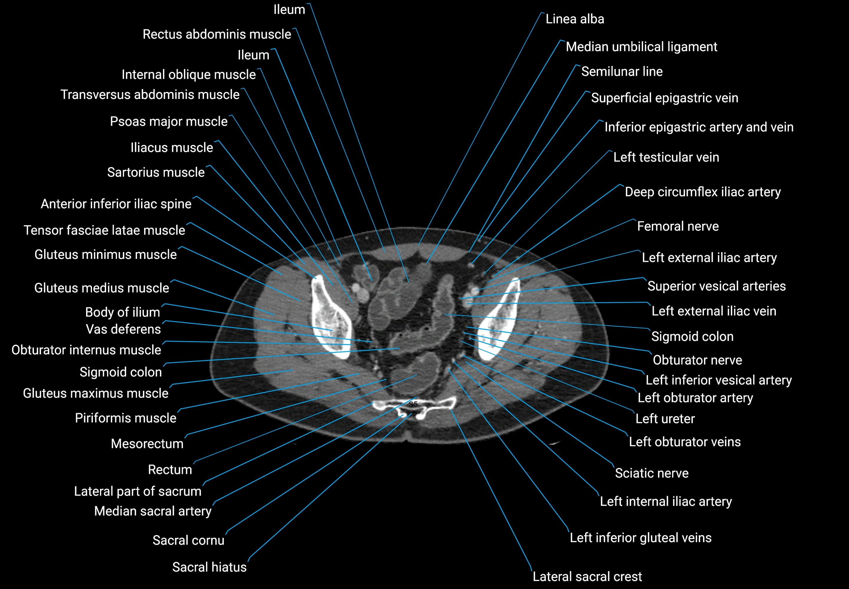
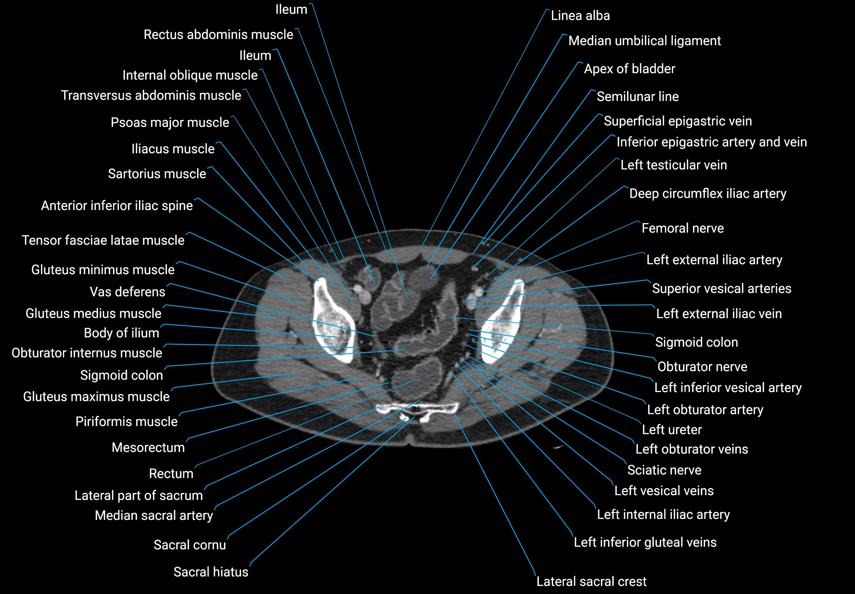
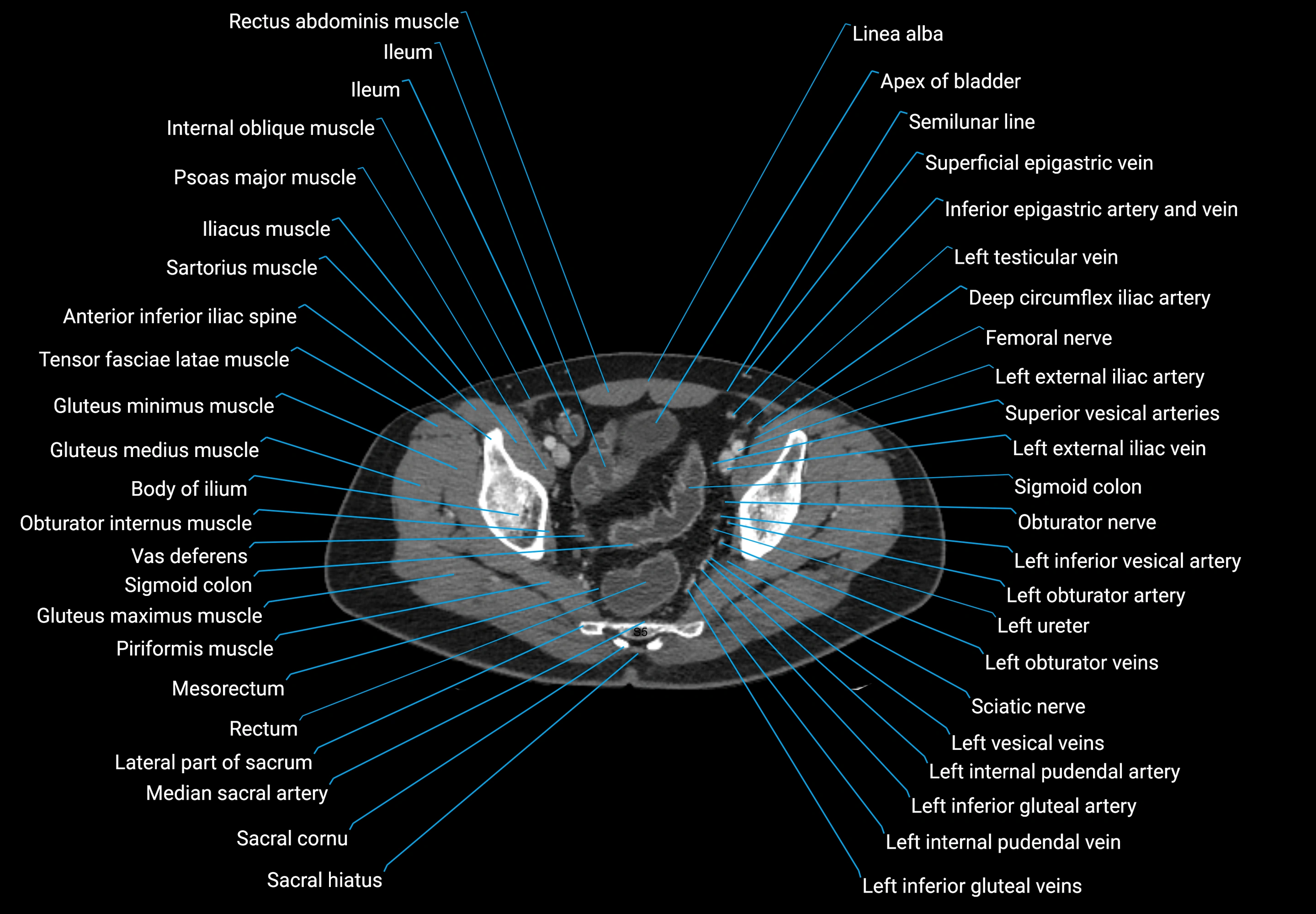
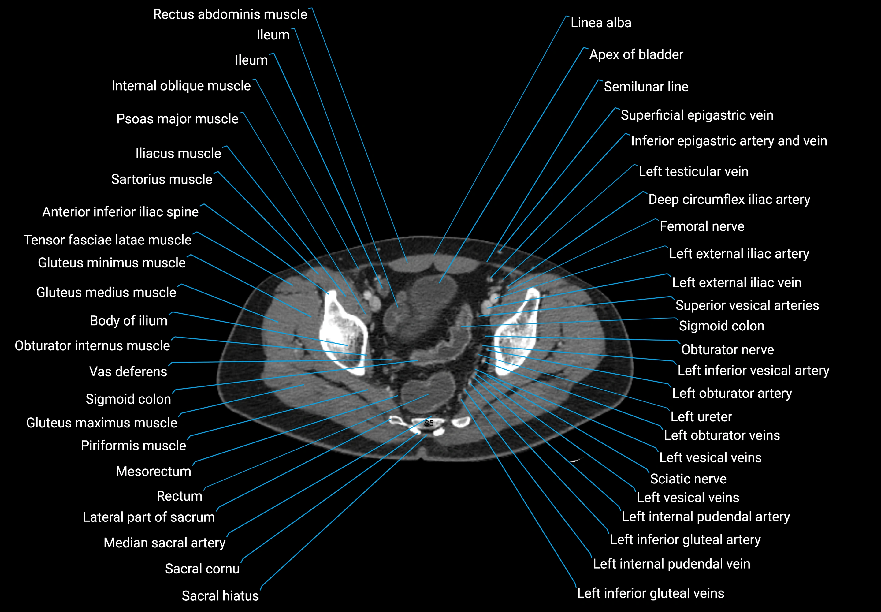
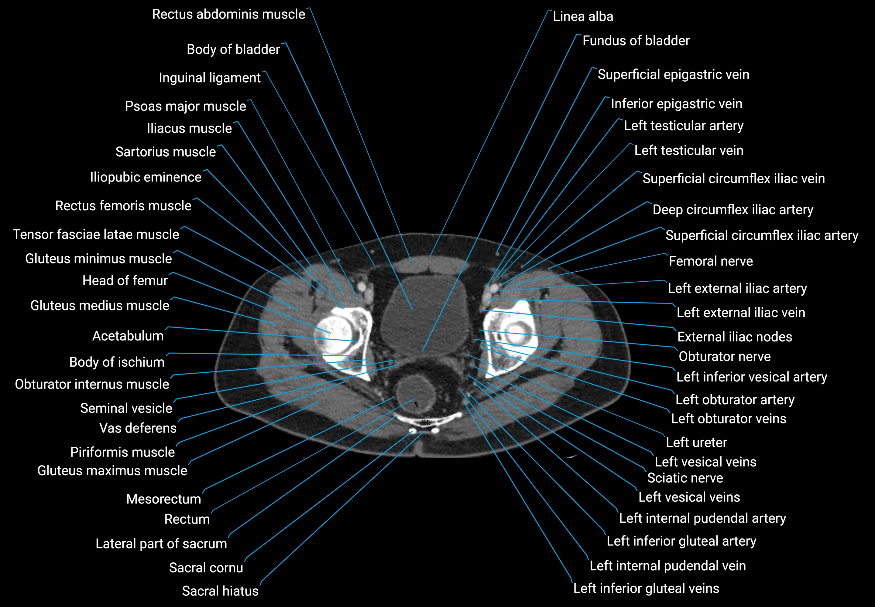
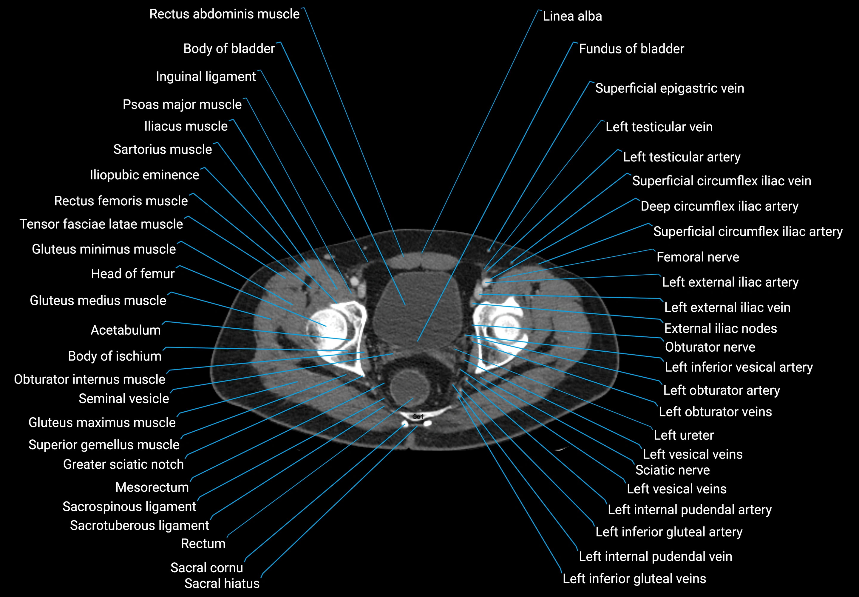
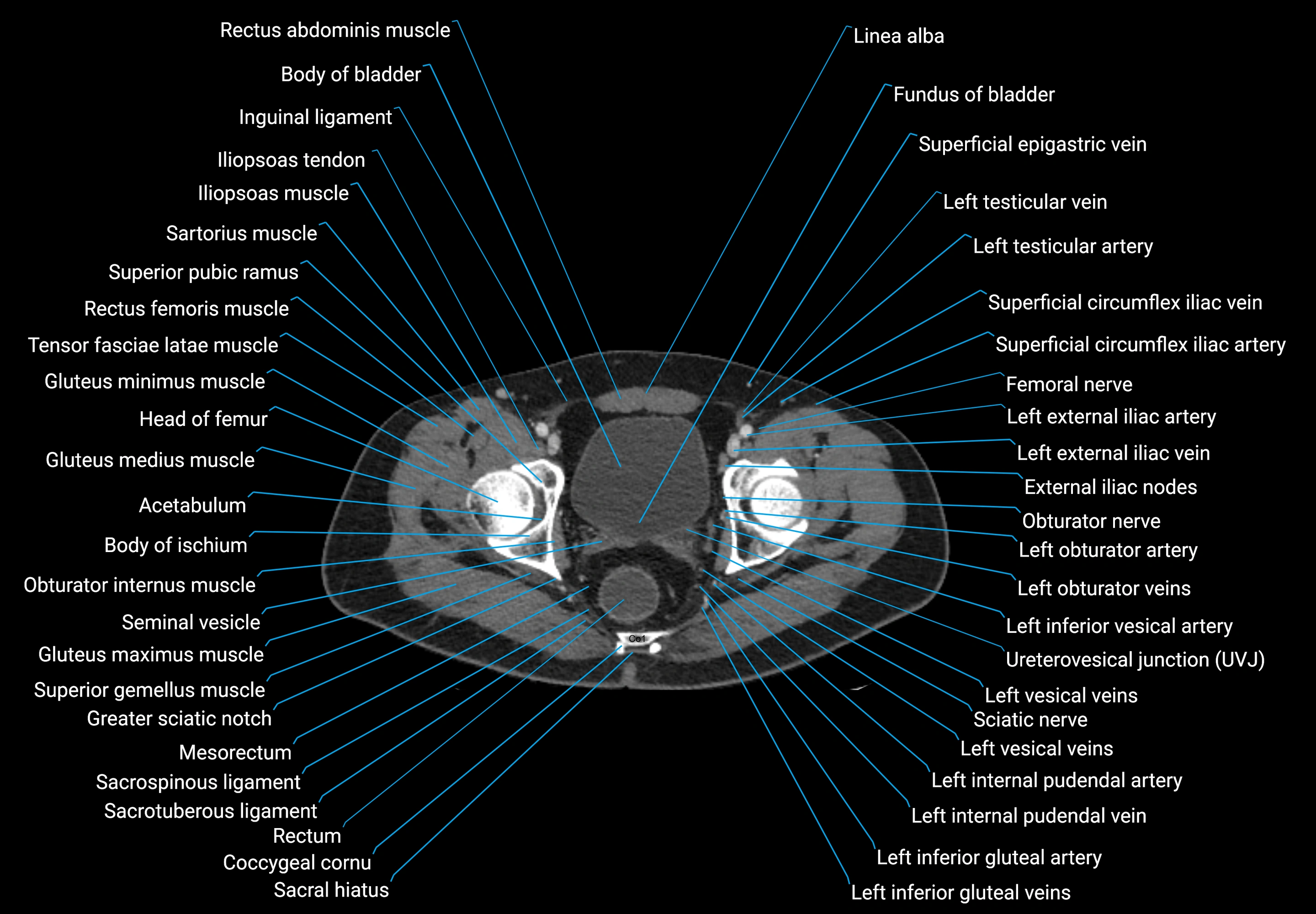
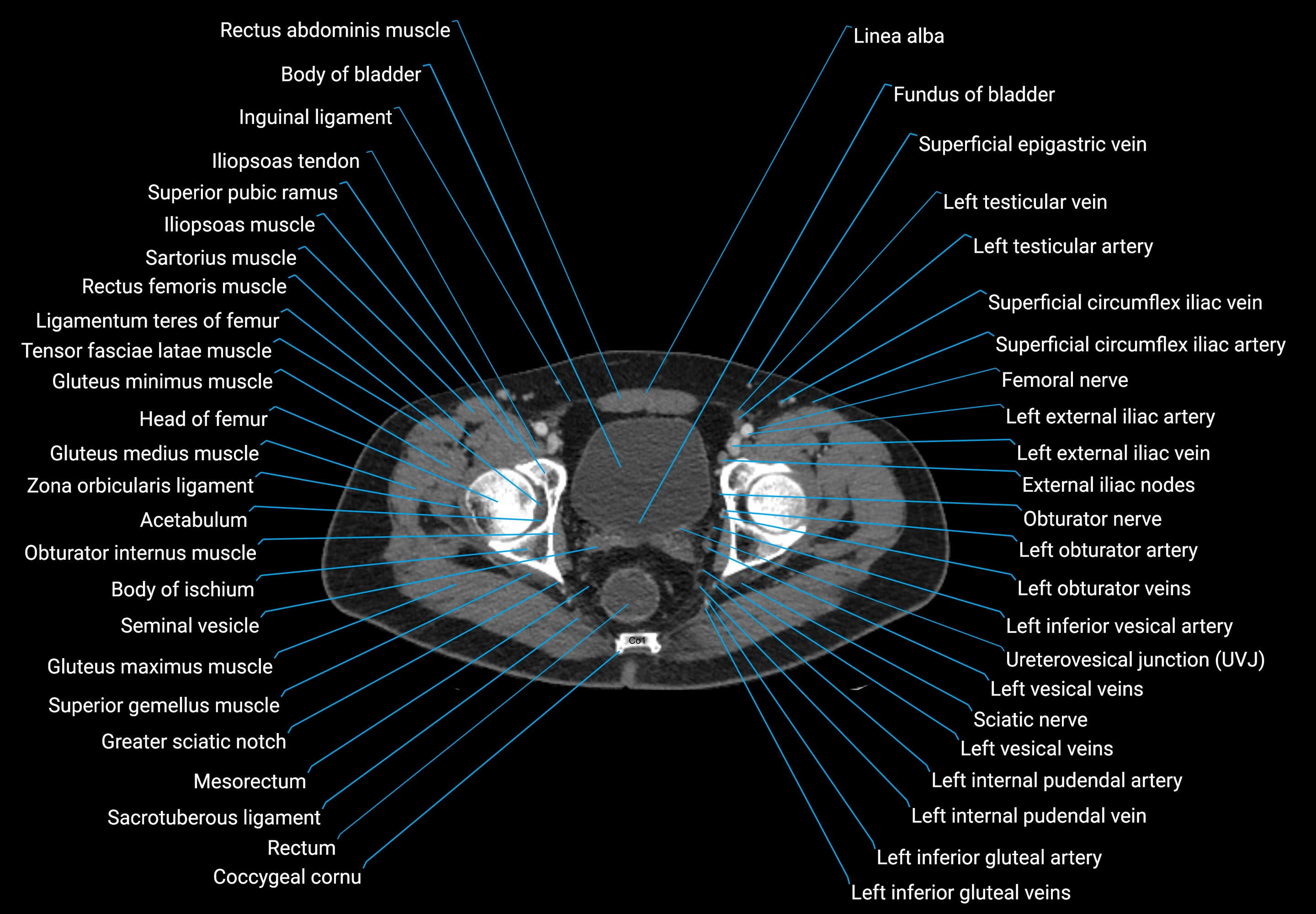
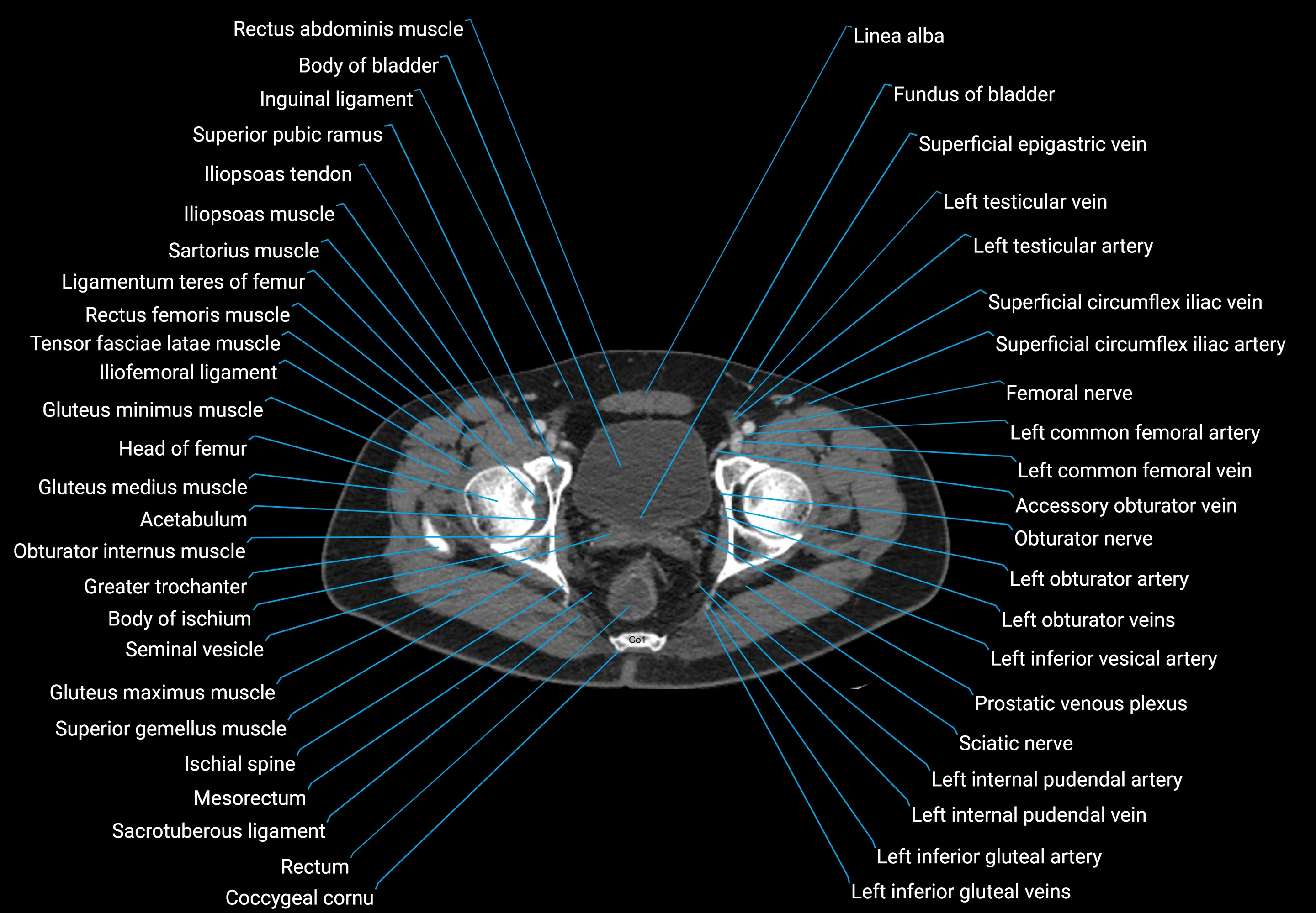
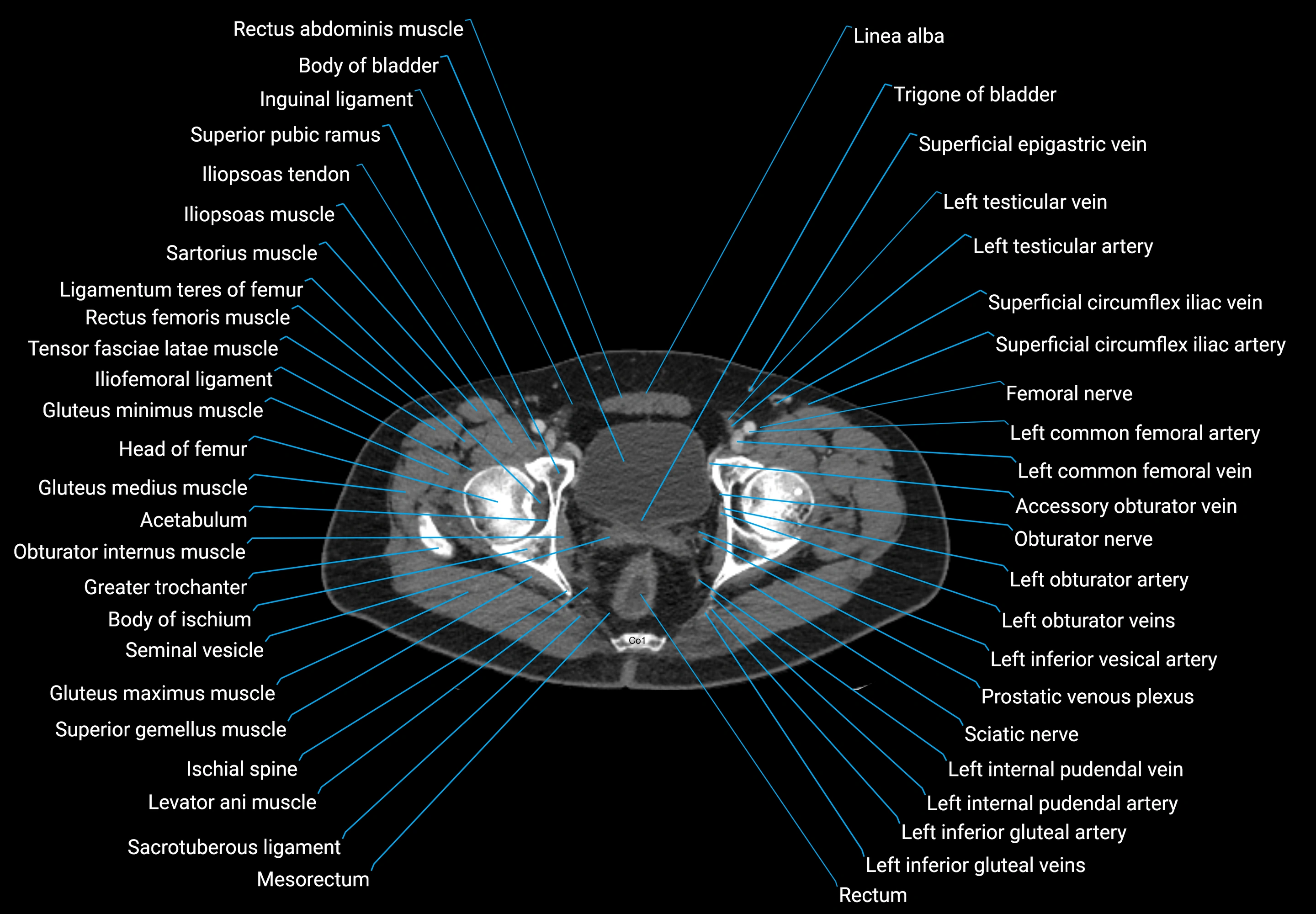
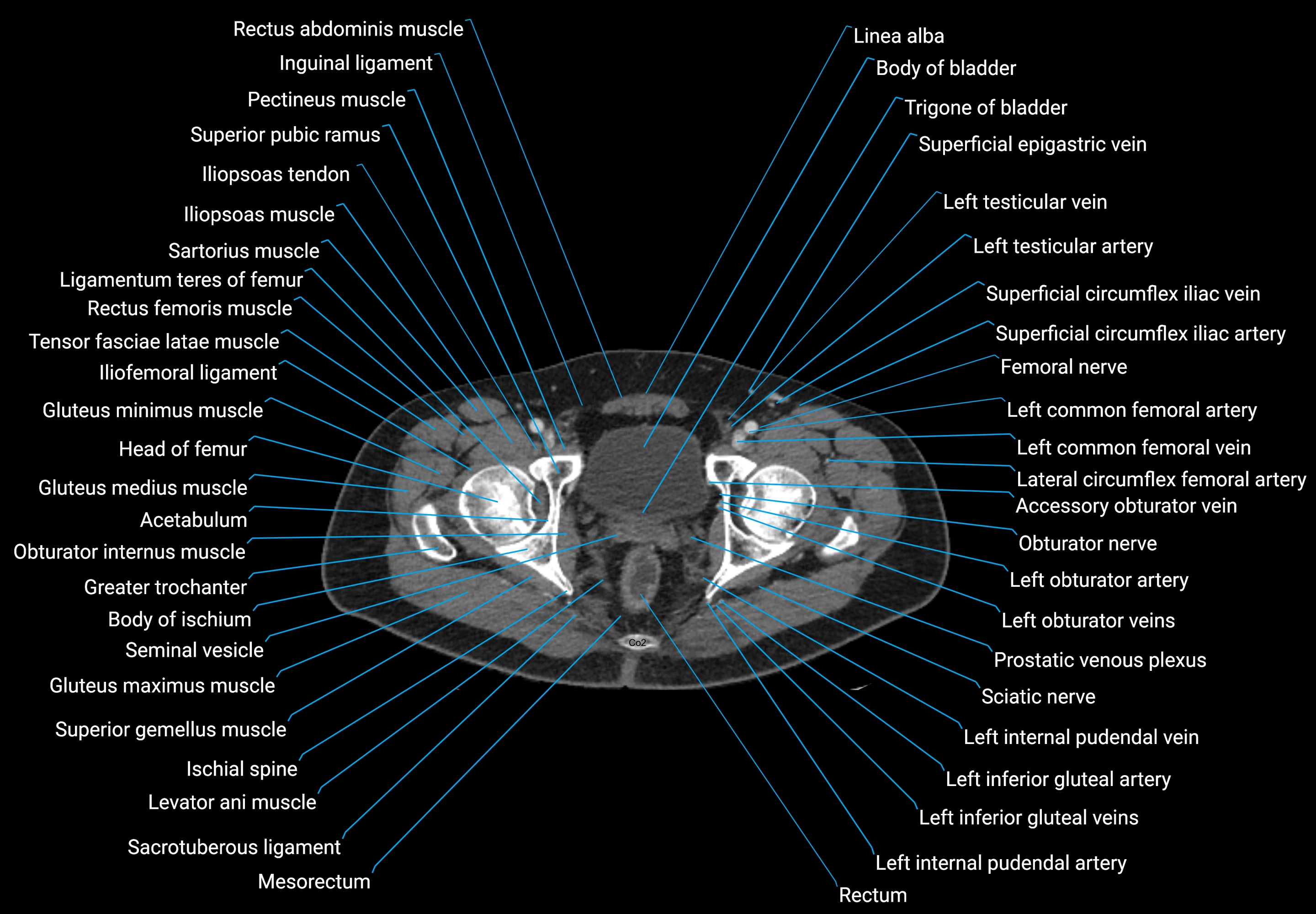
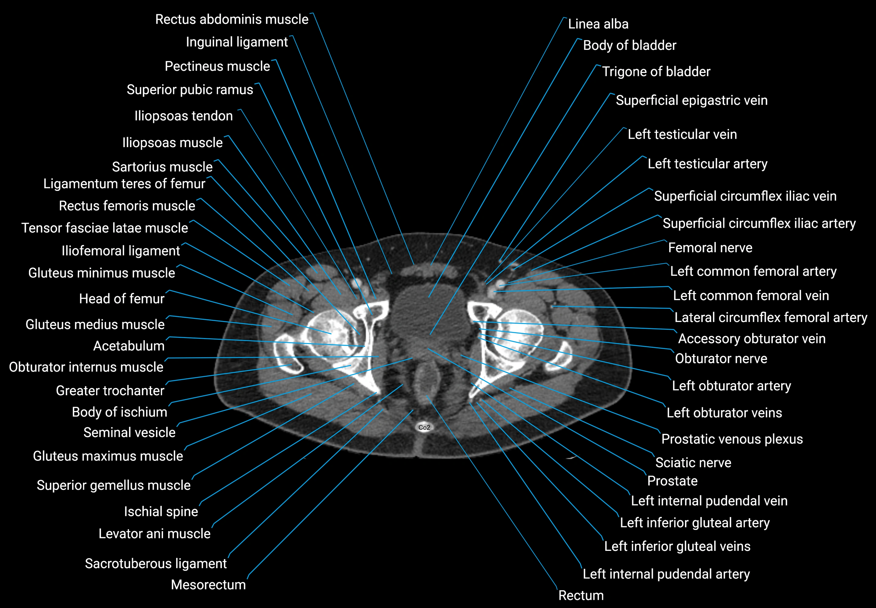
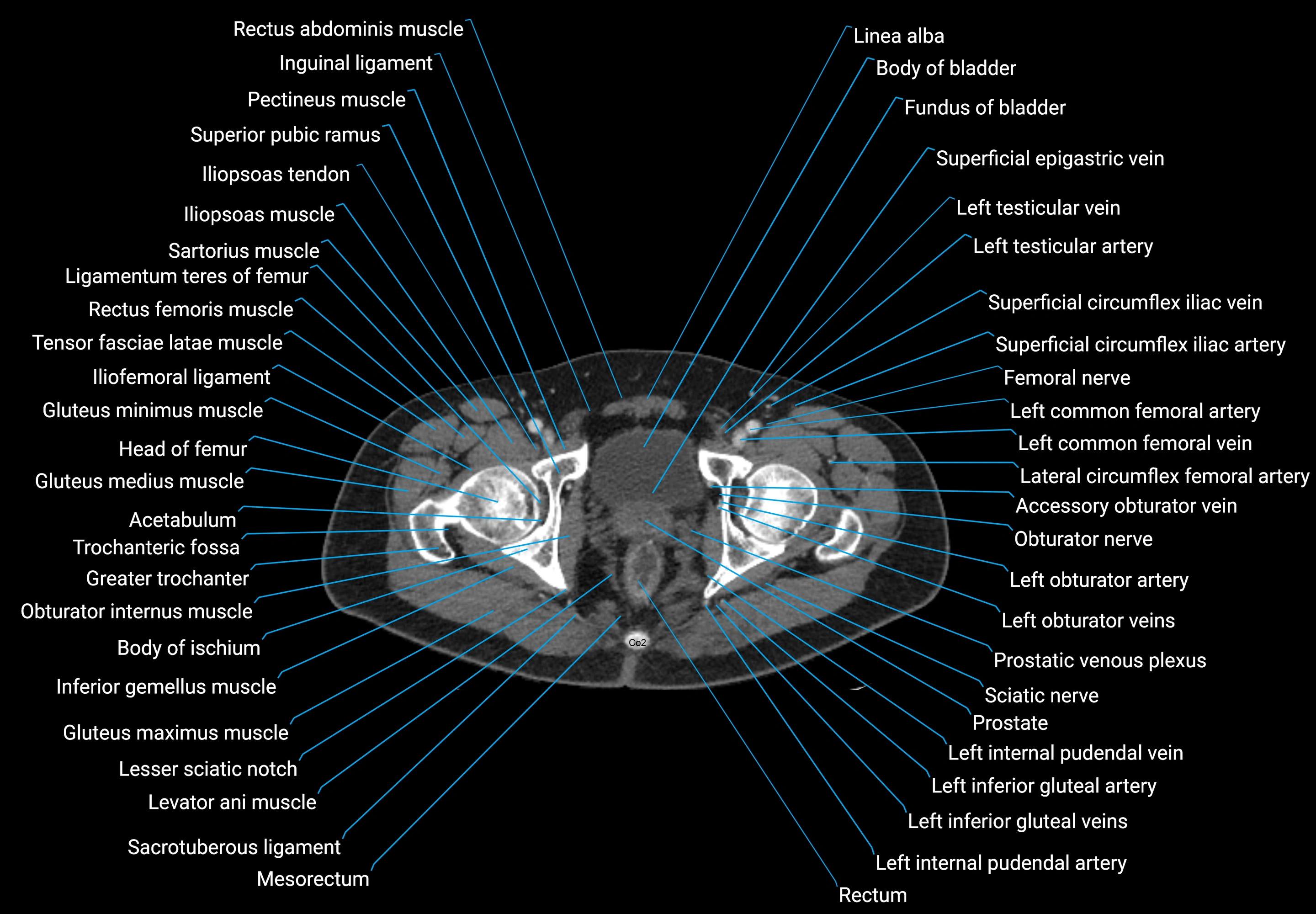
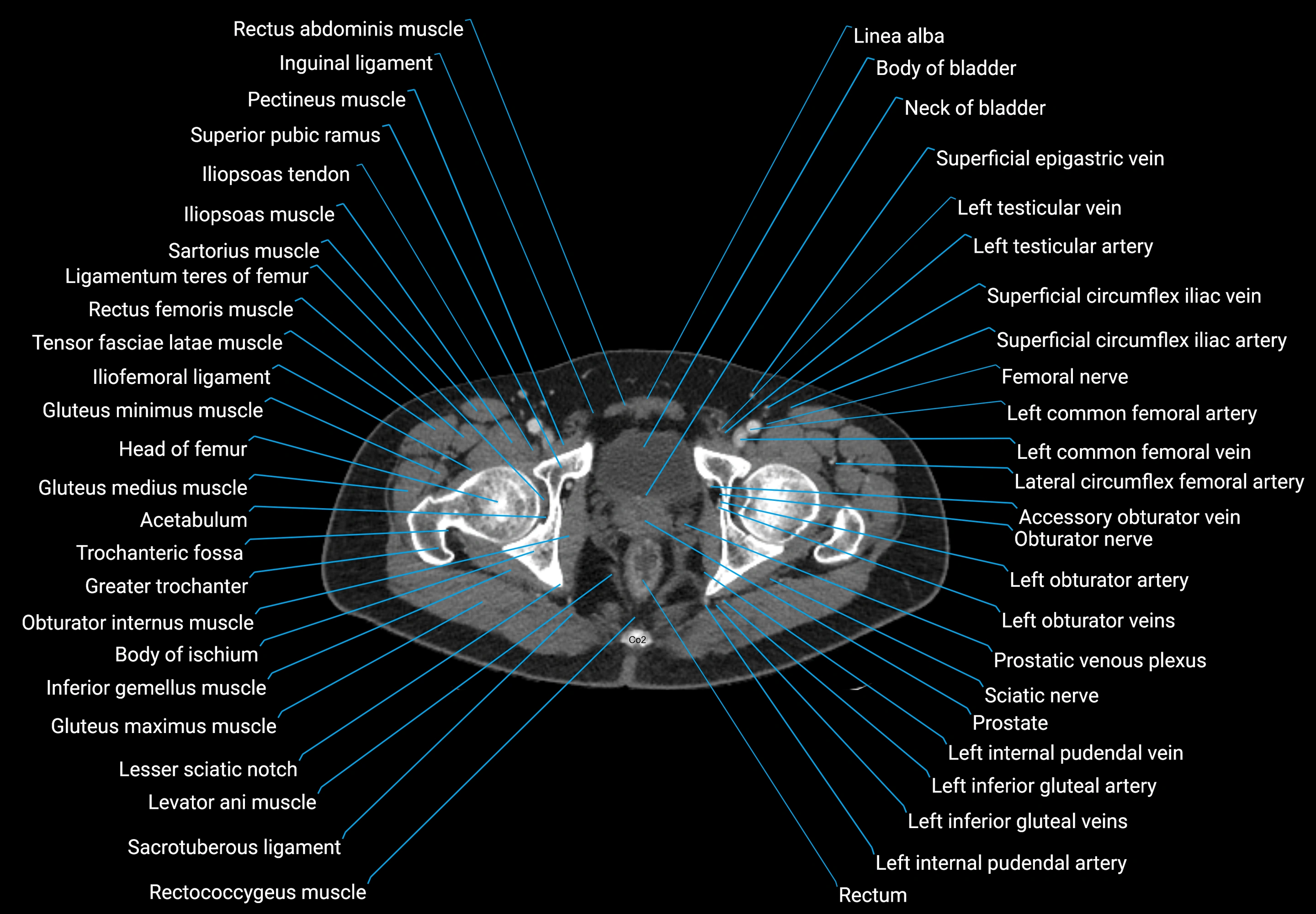
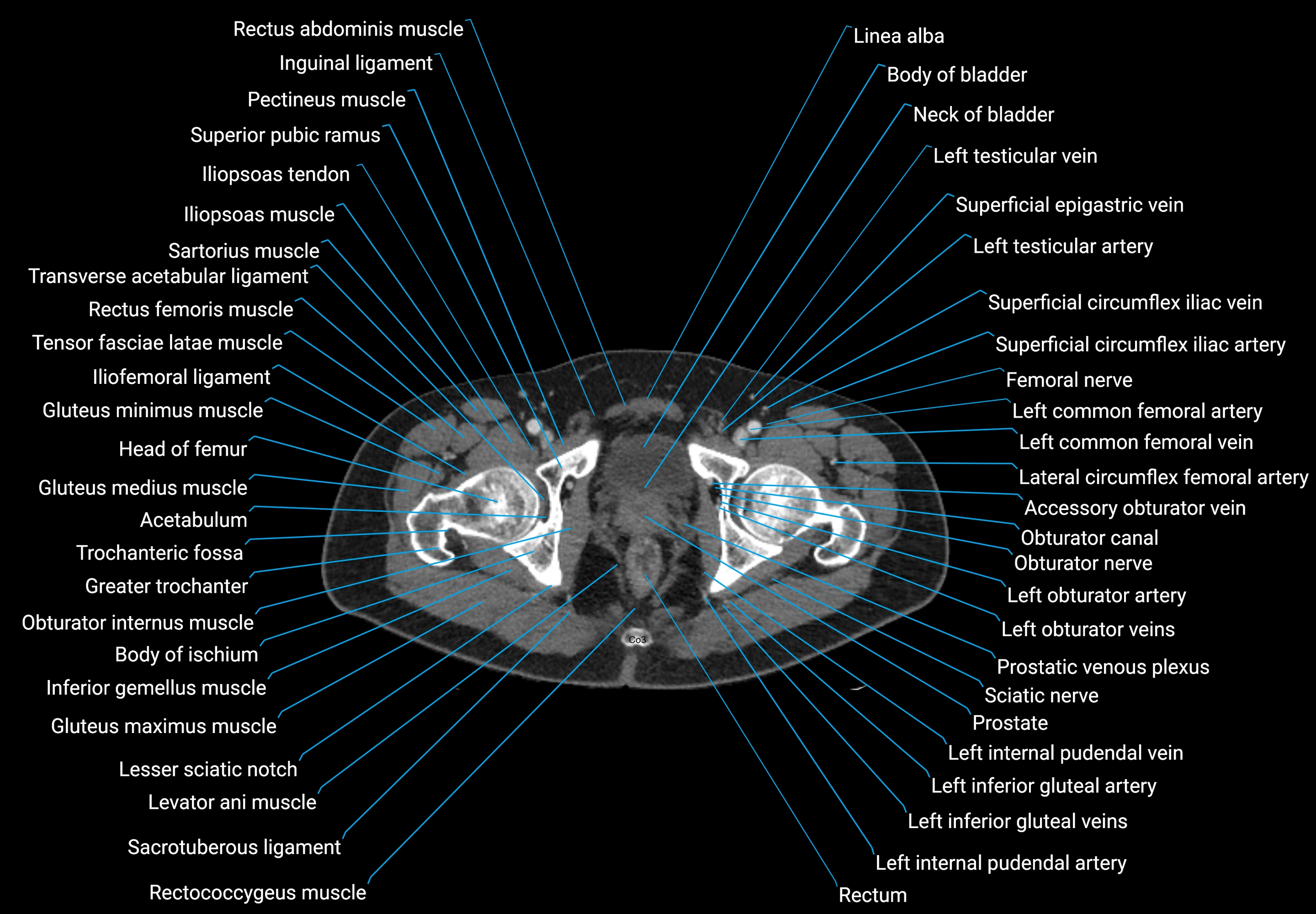
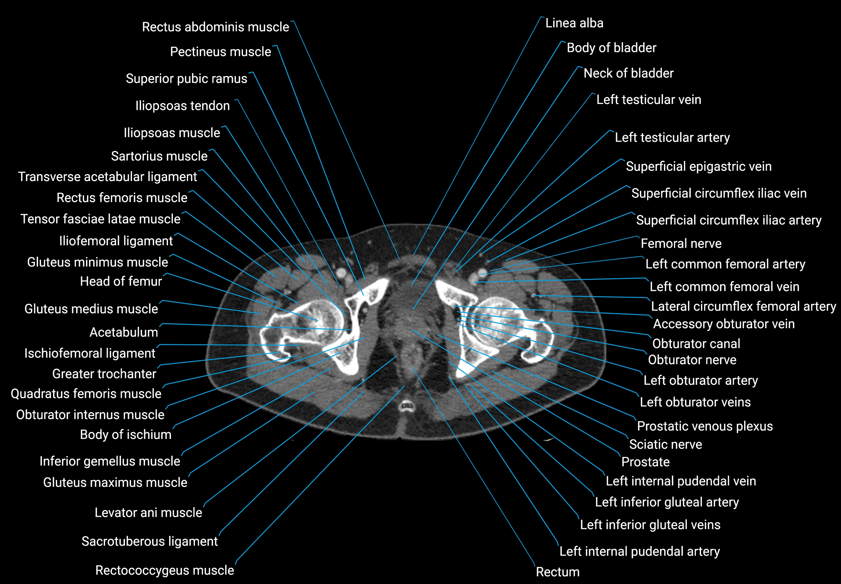
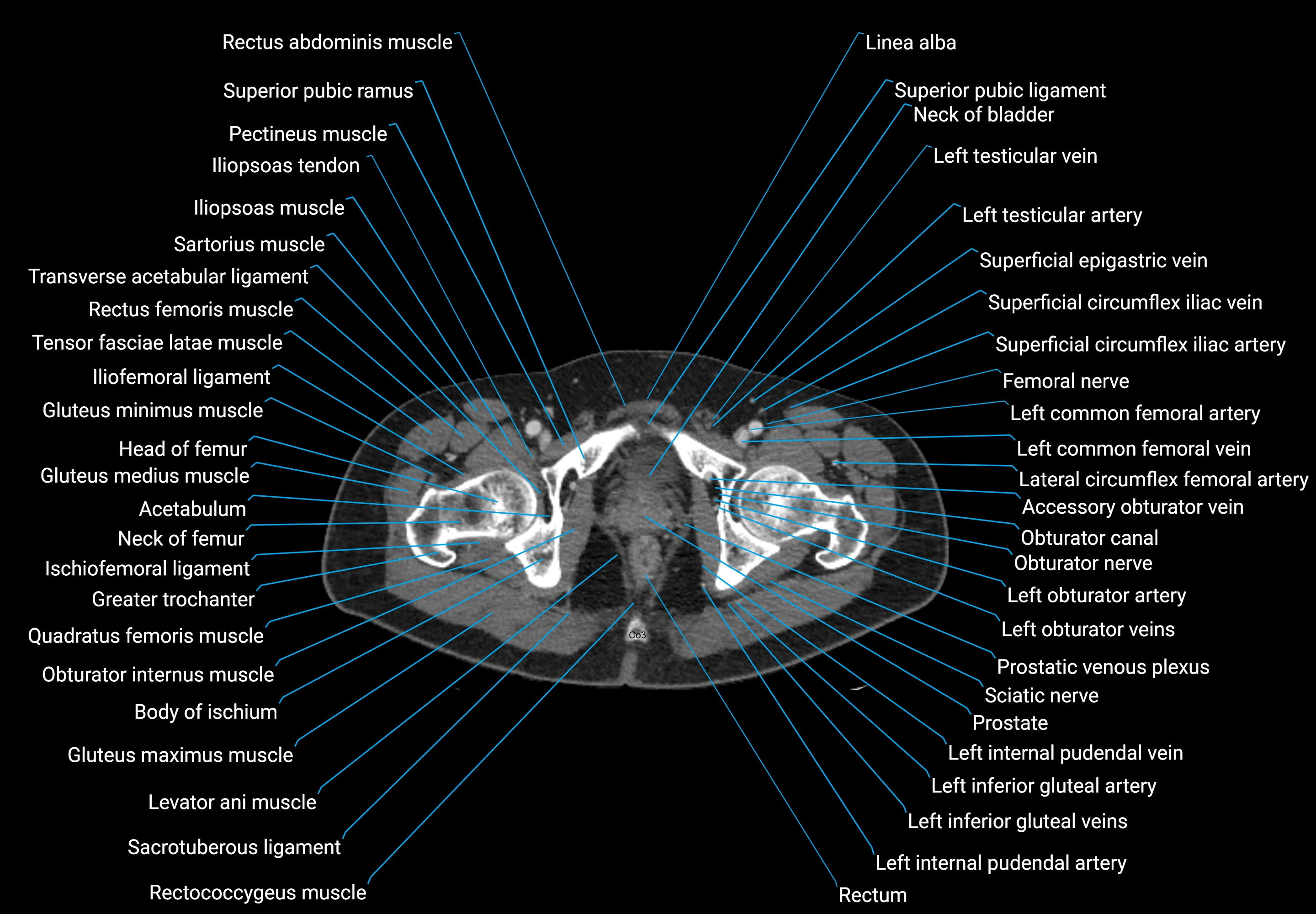
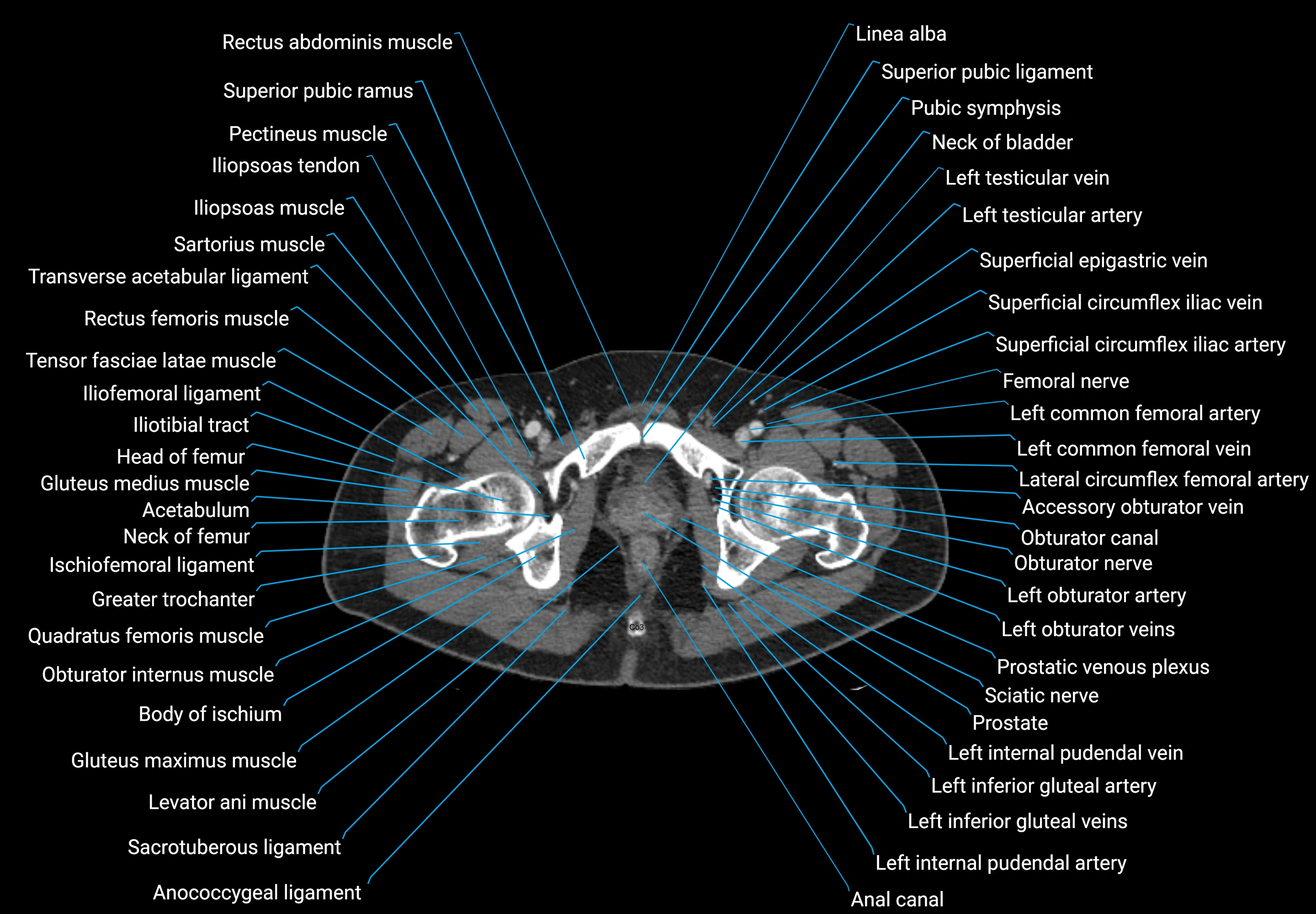
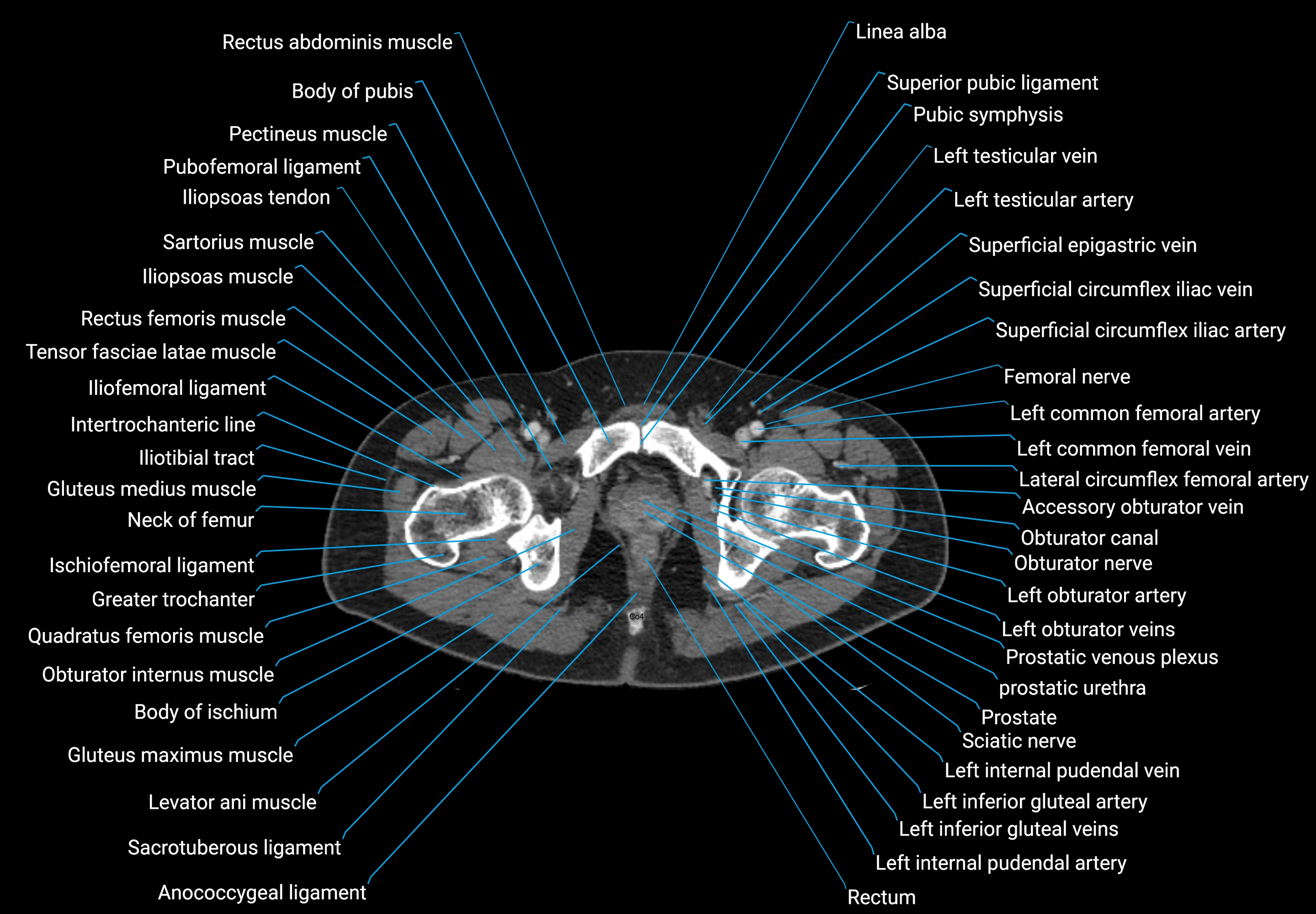
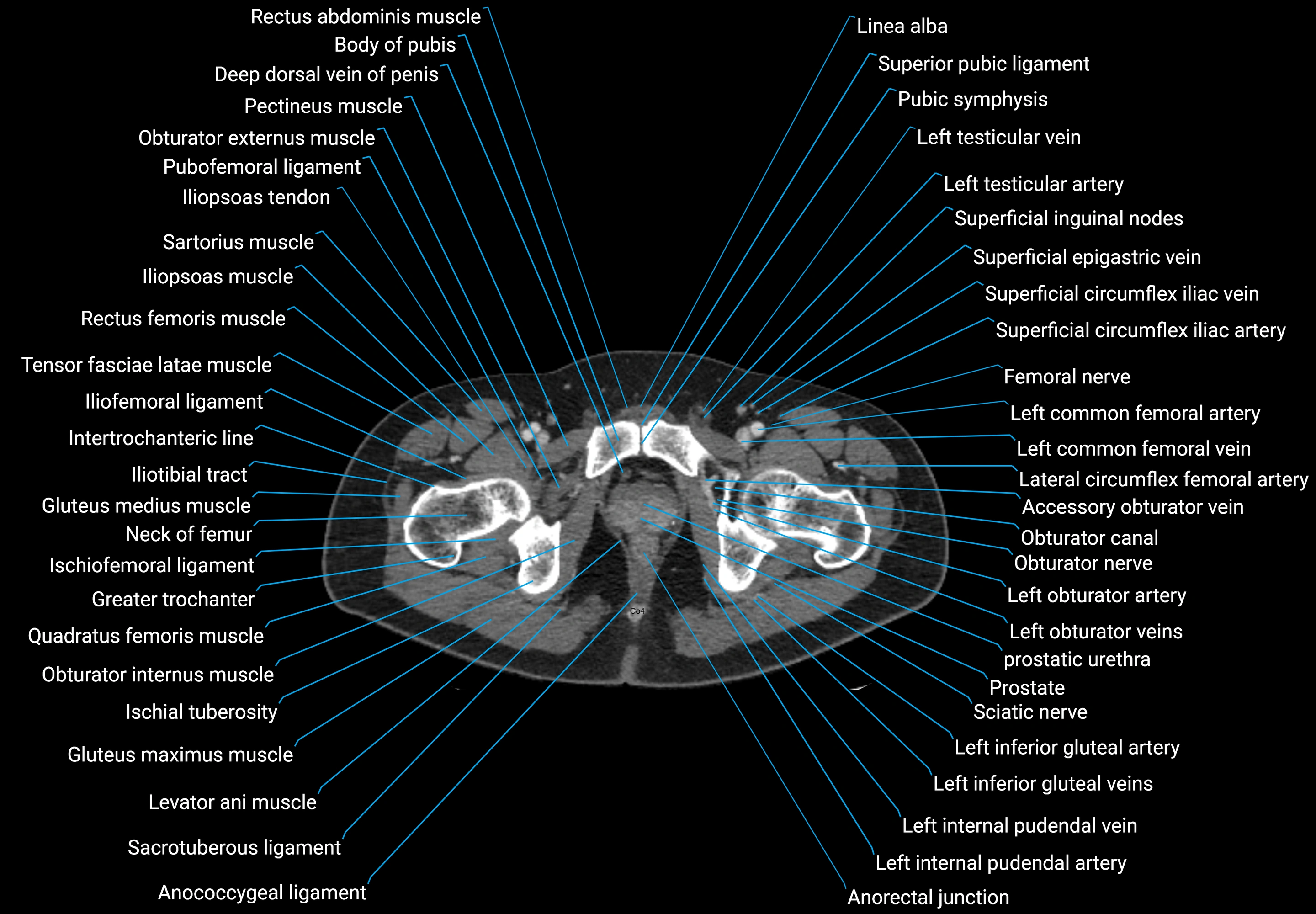
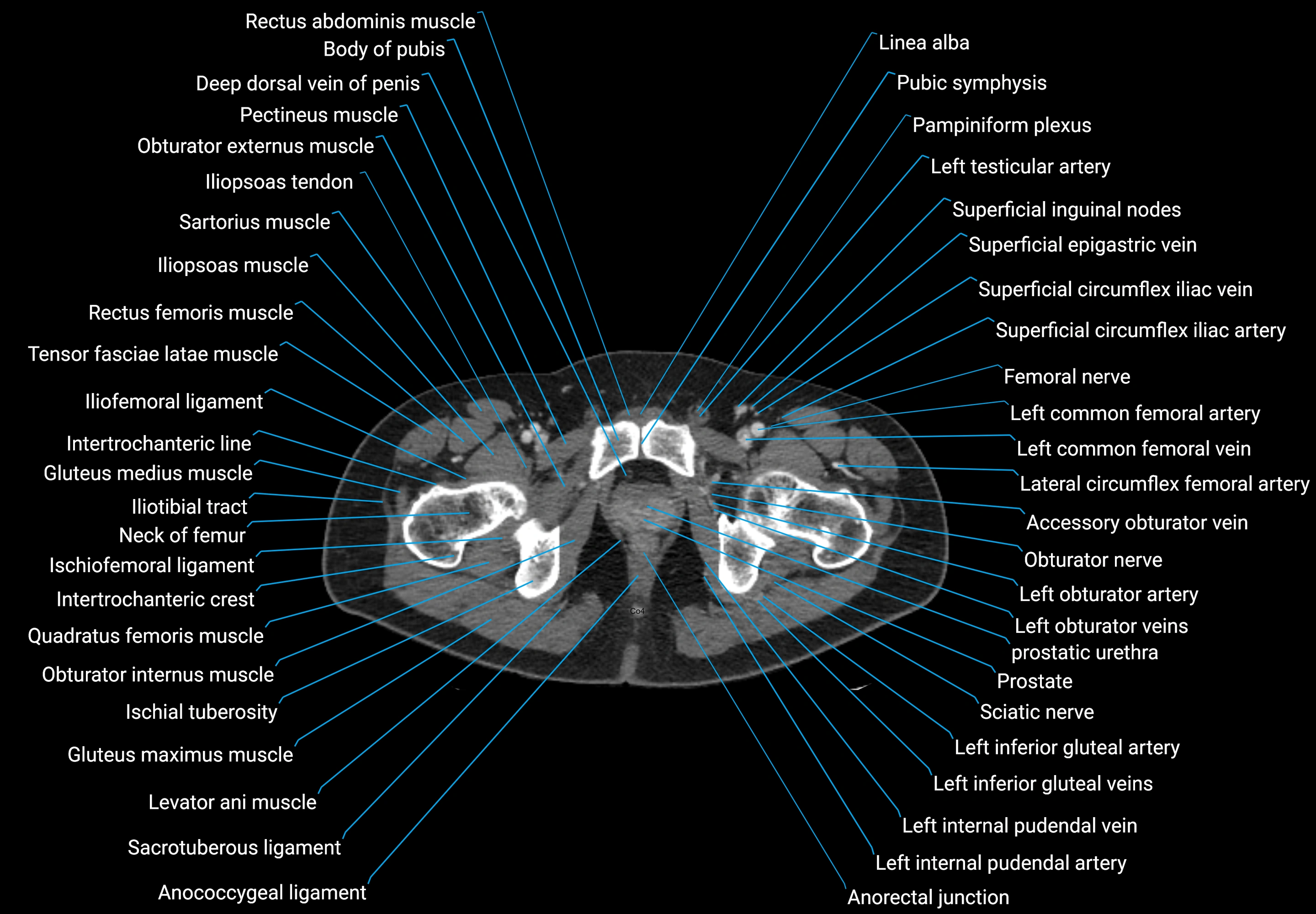
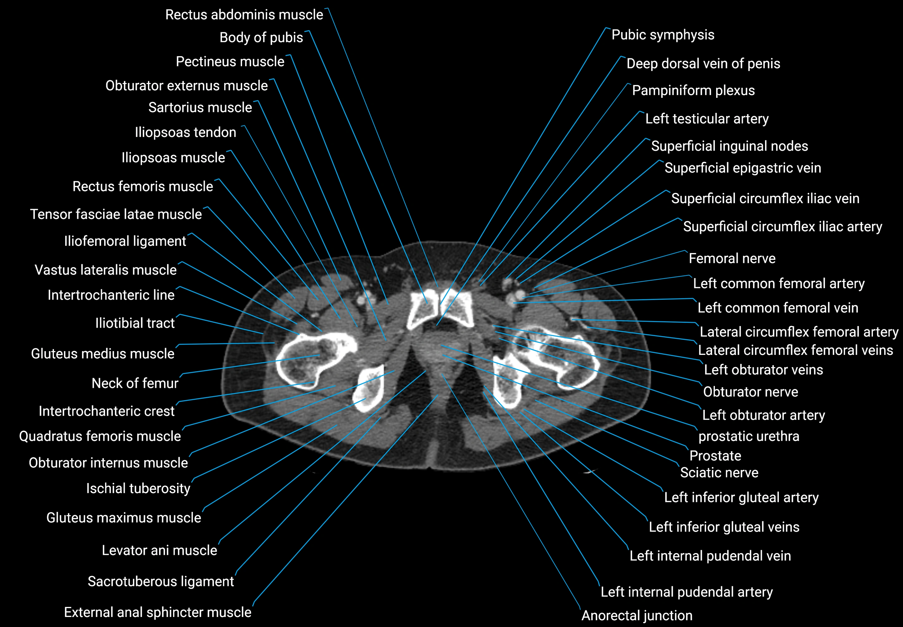
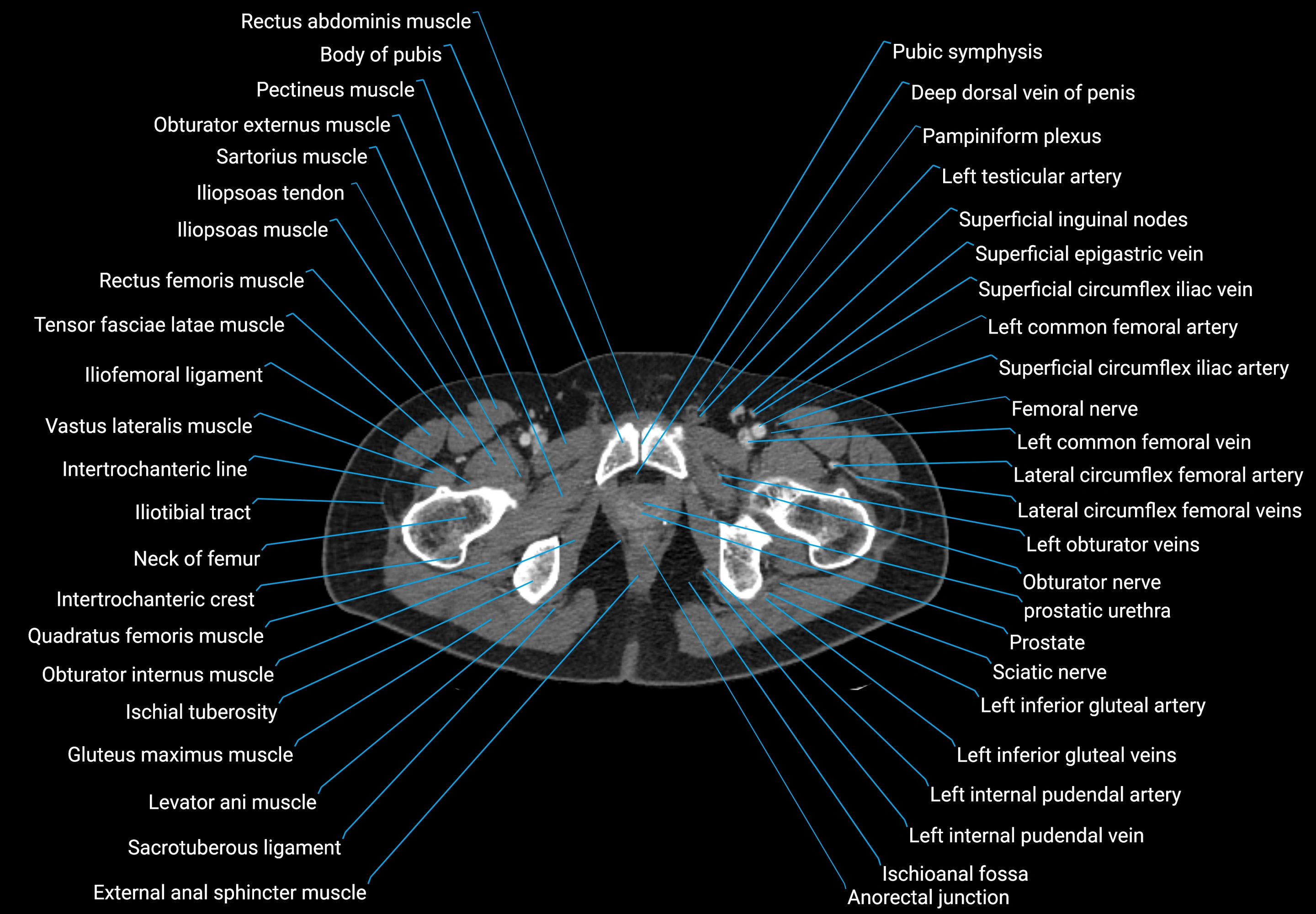
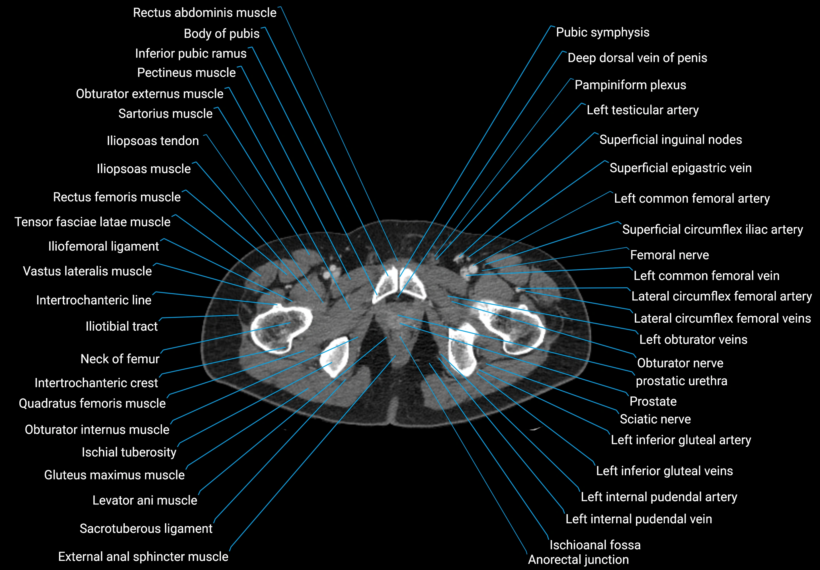
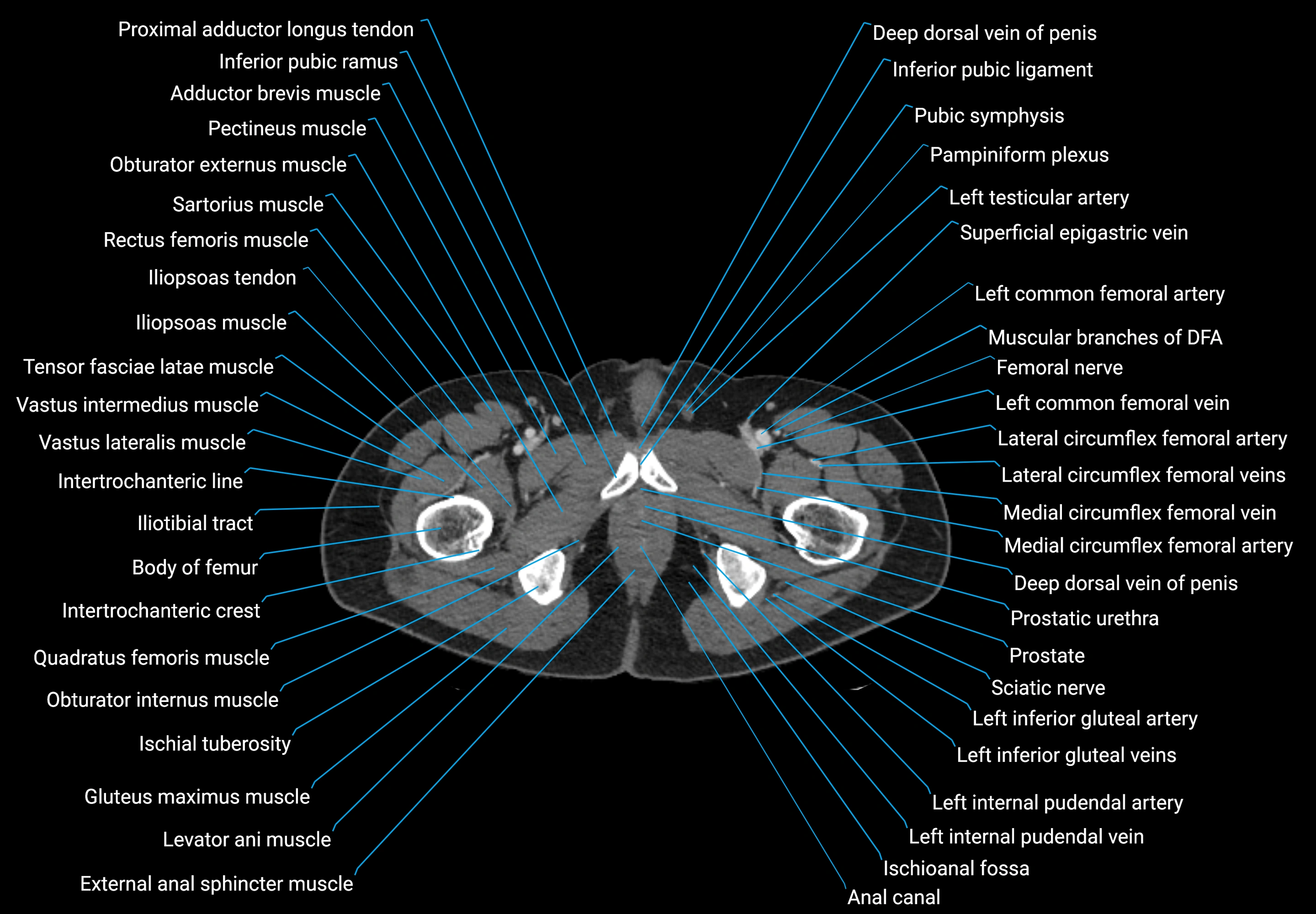
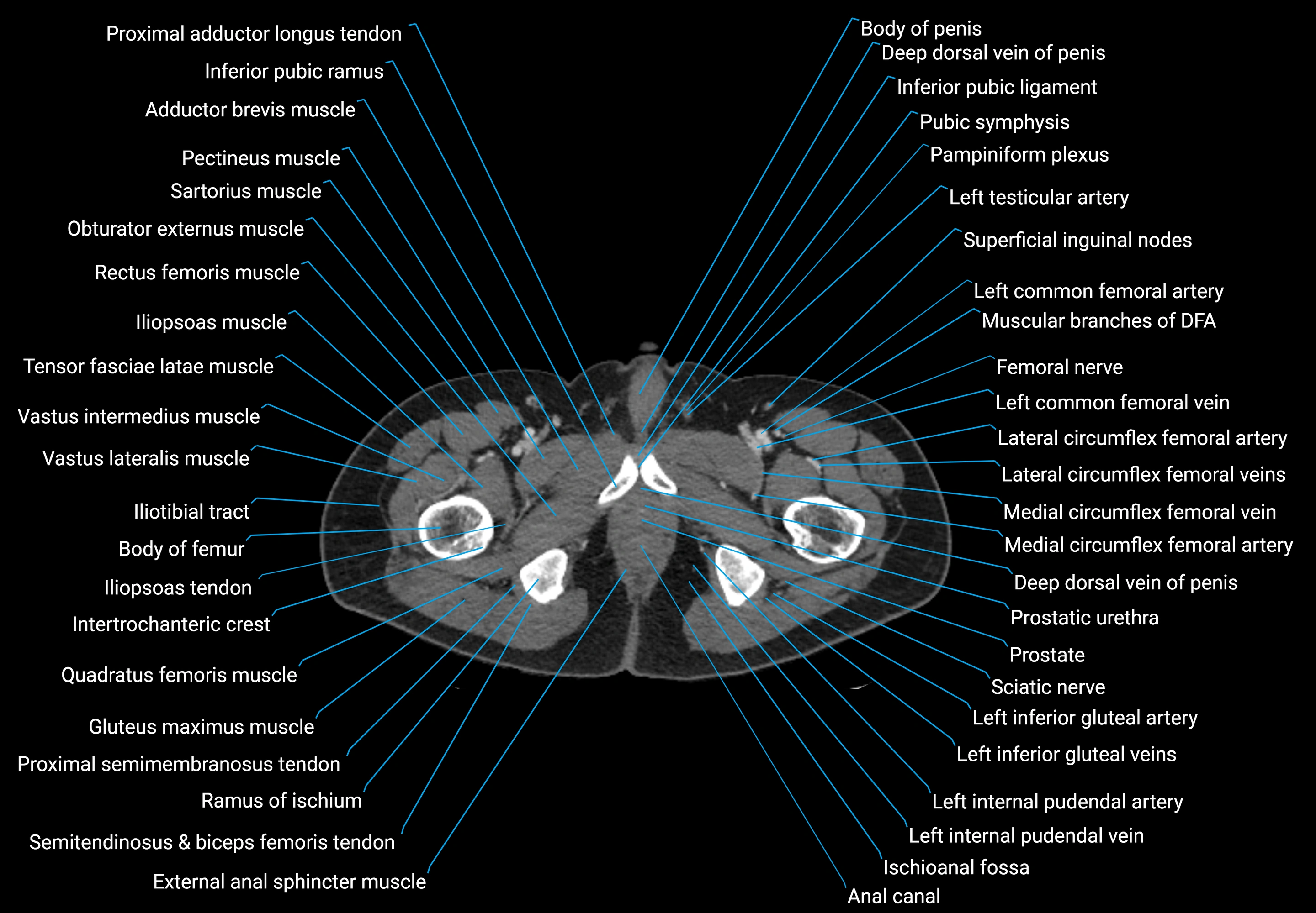
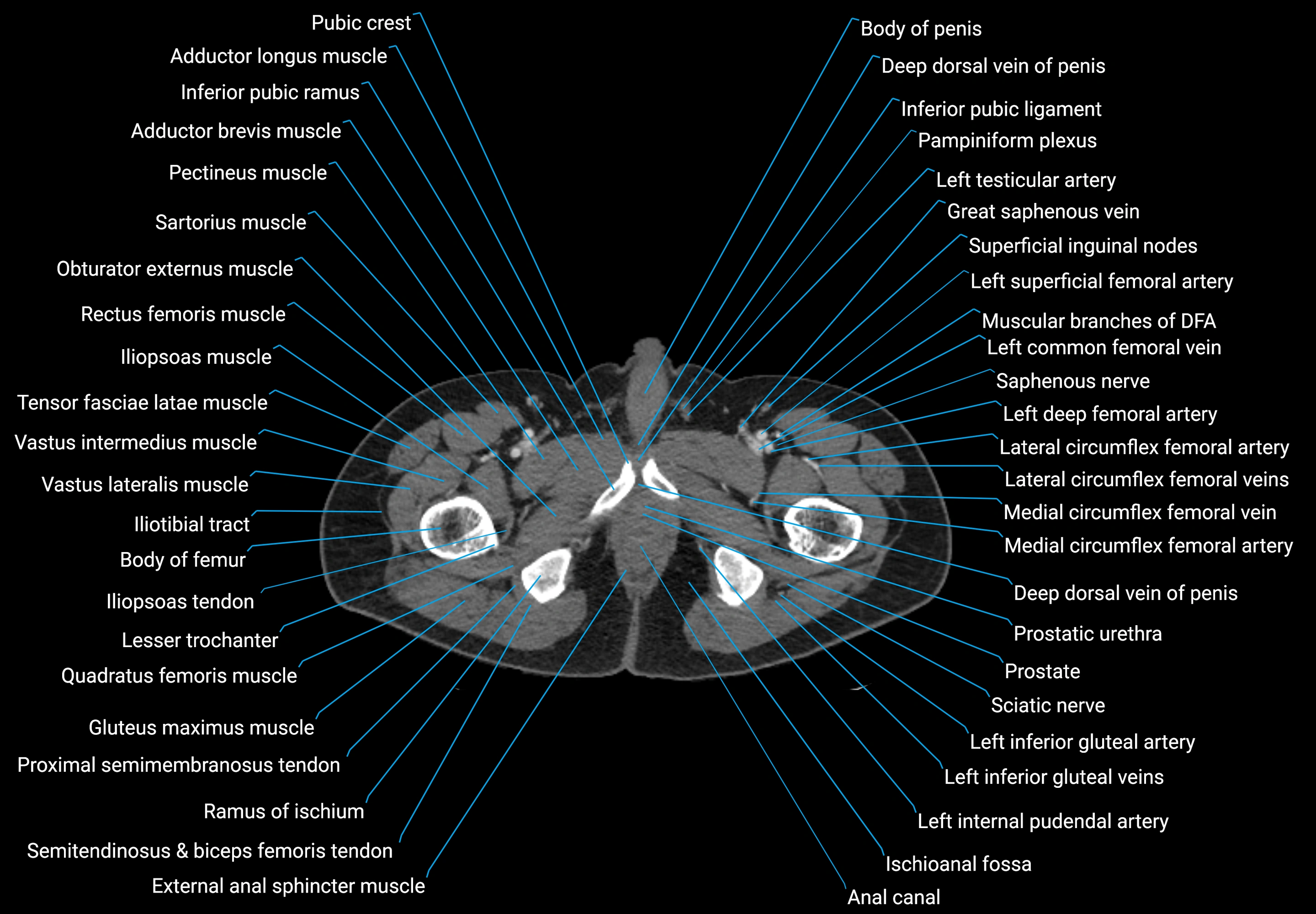
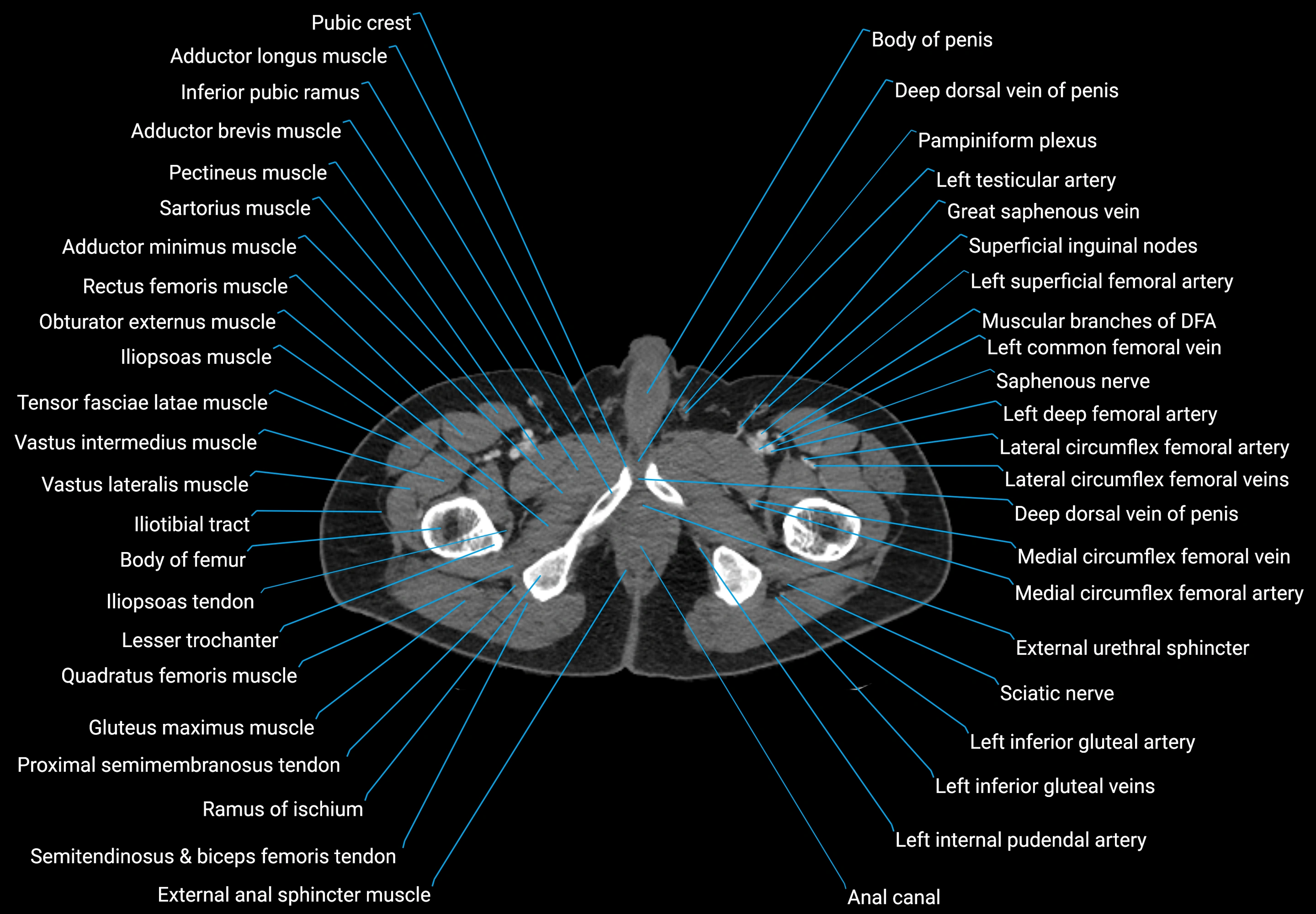
CT images

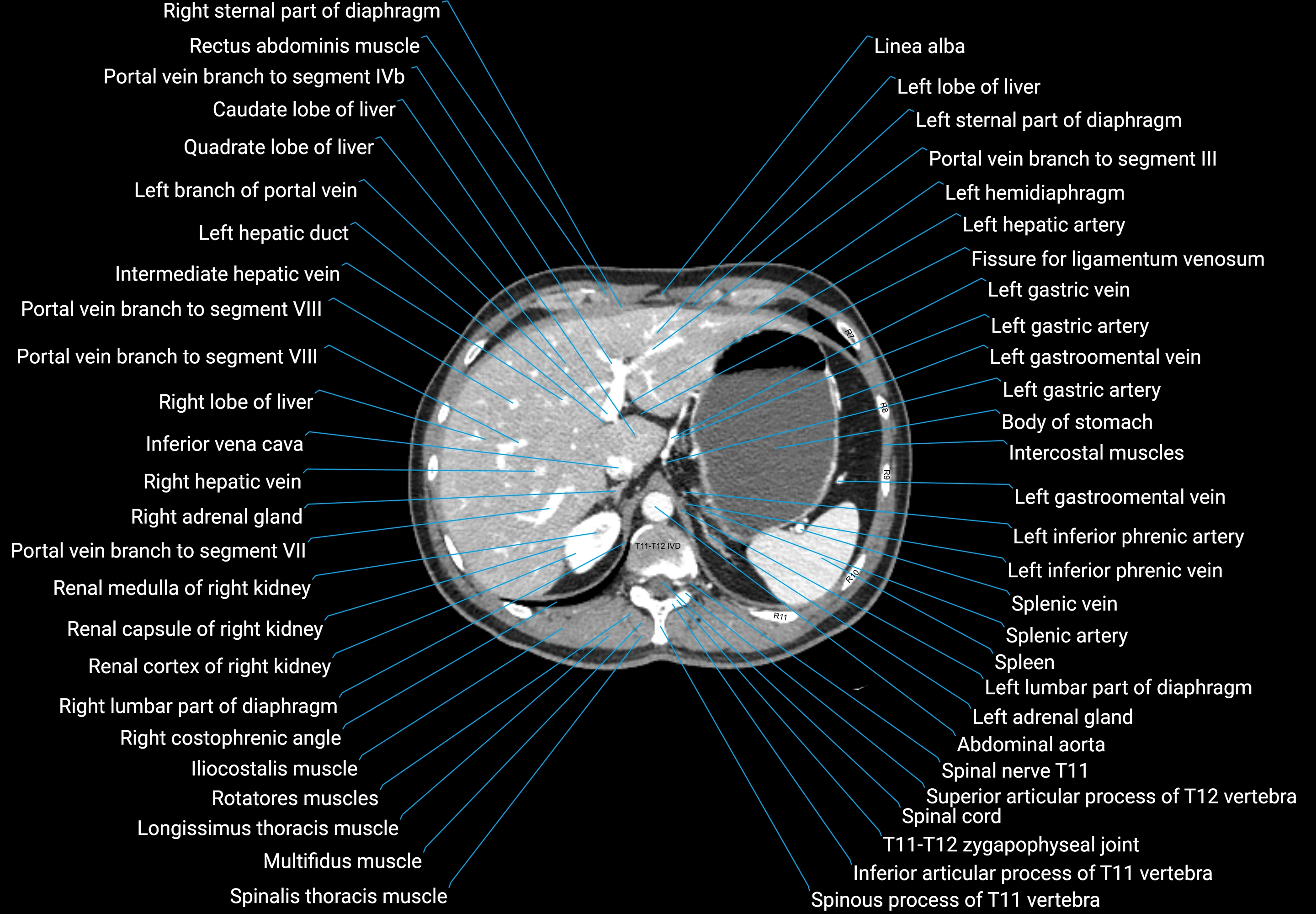
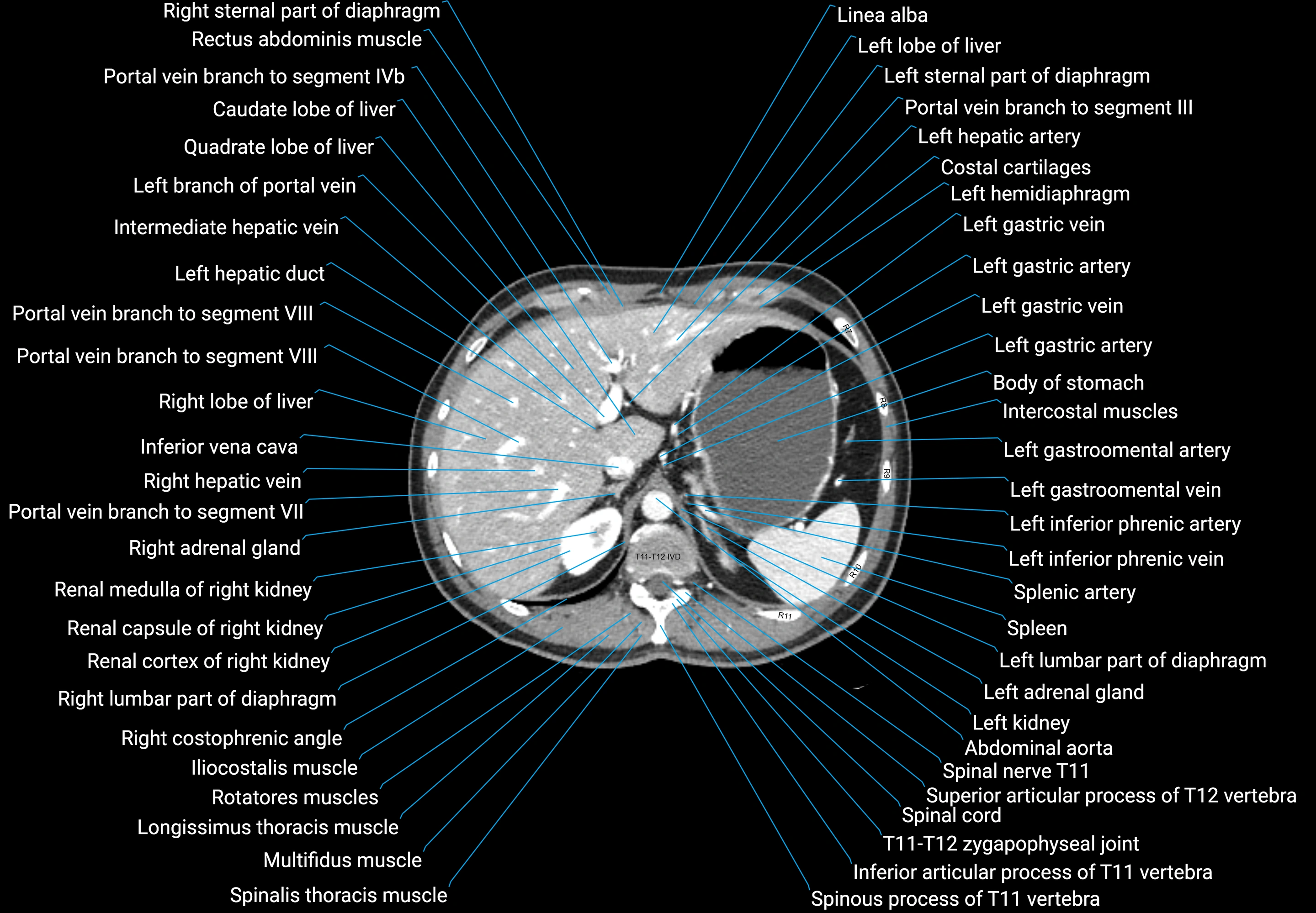
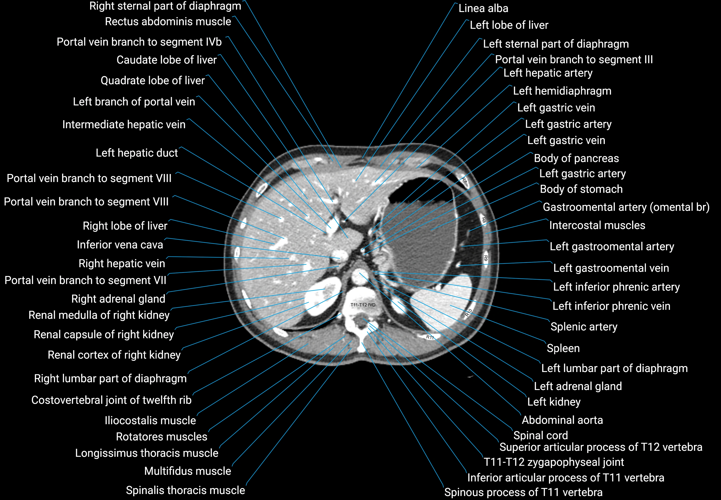
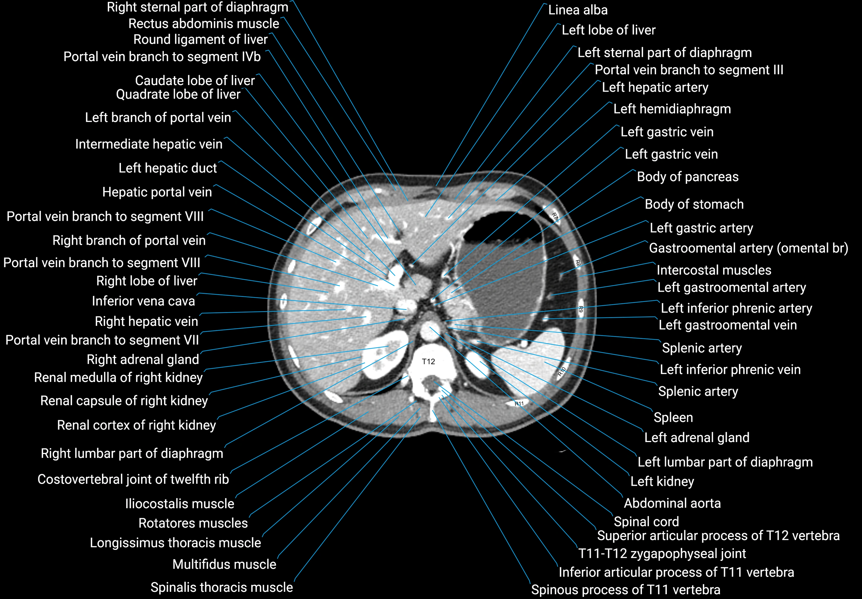
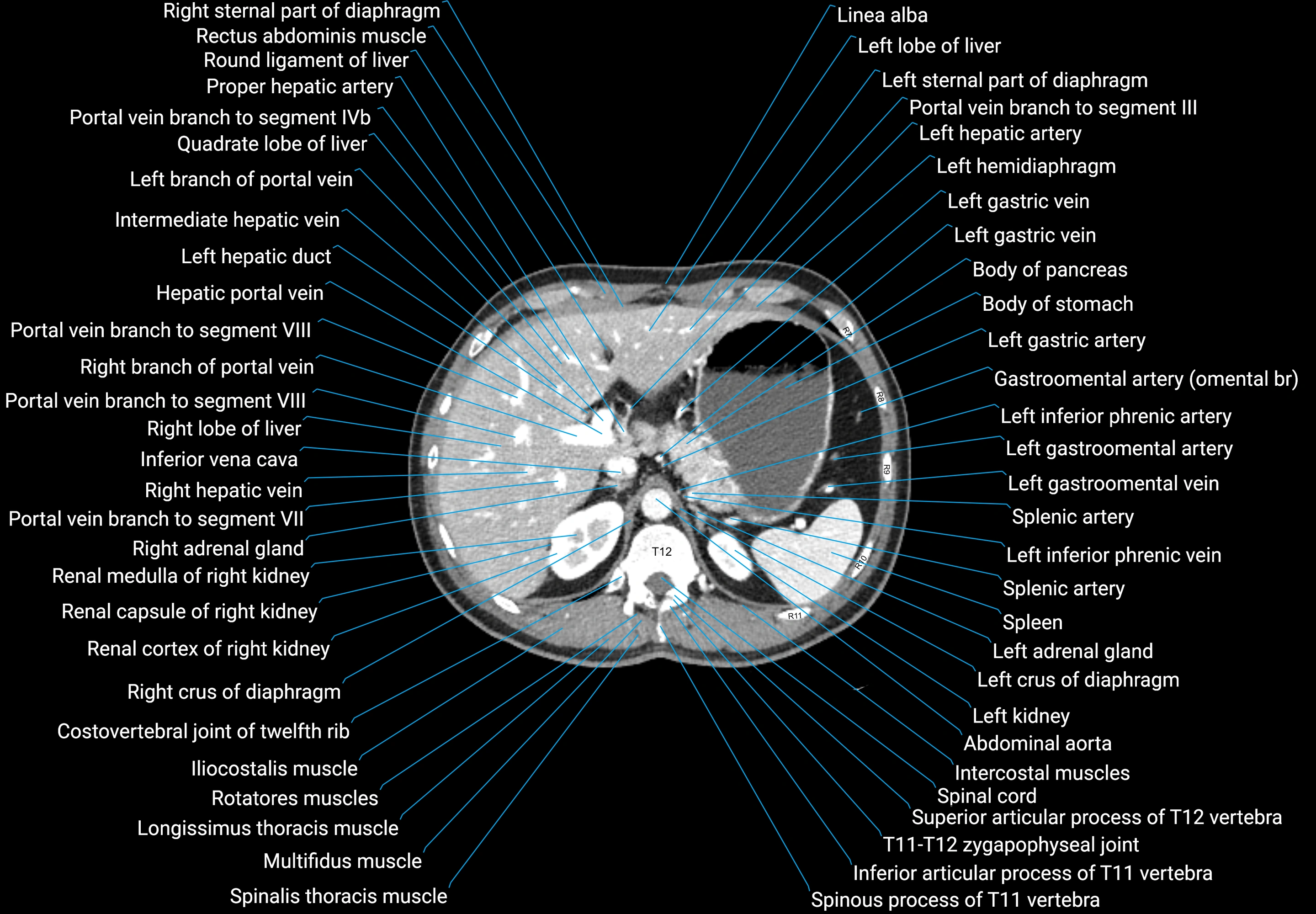
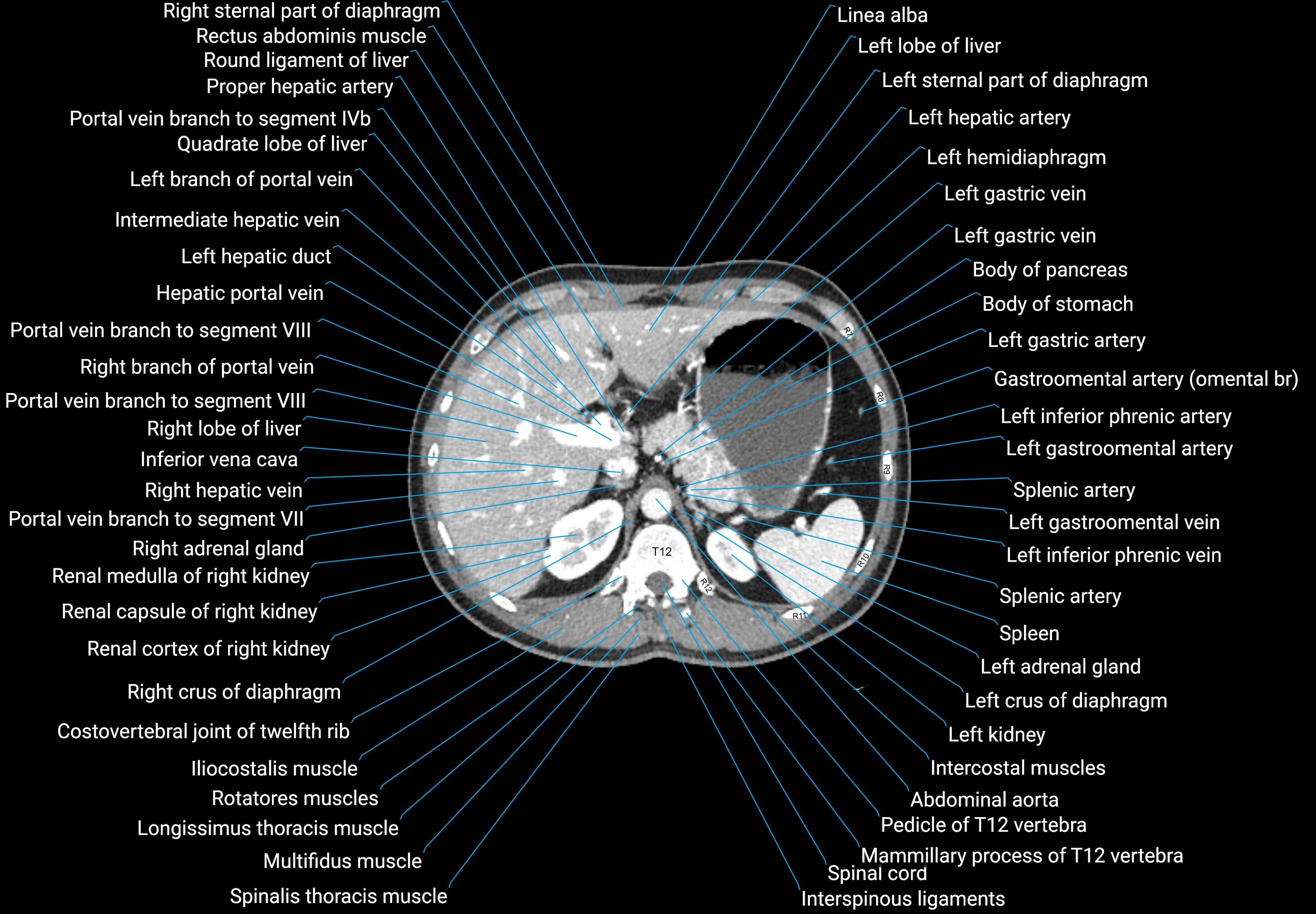
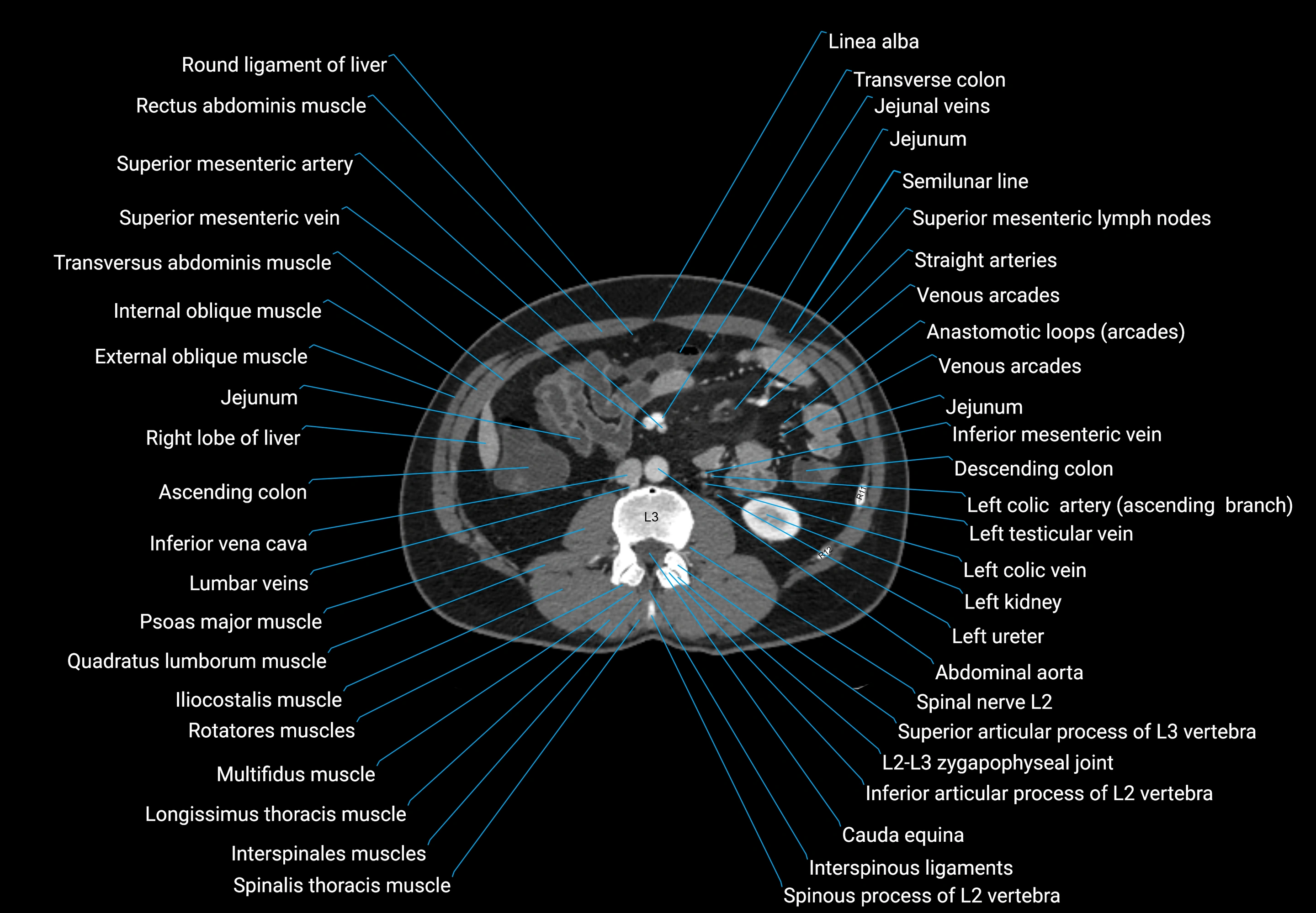
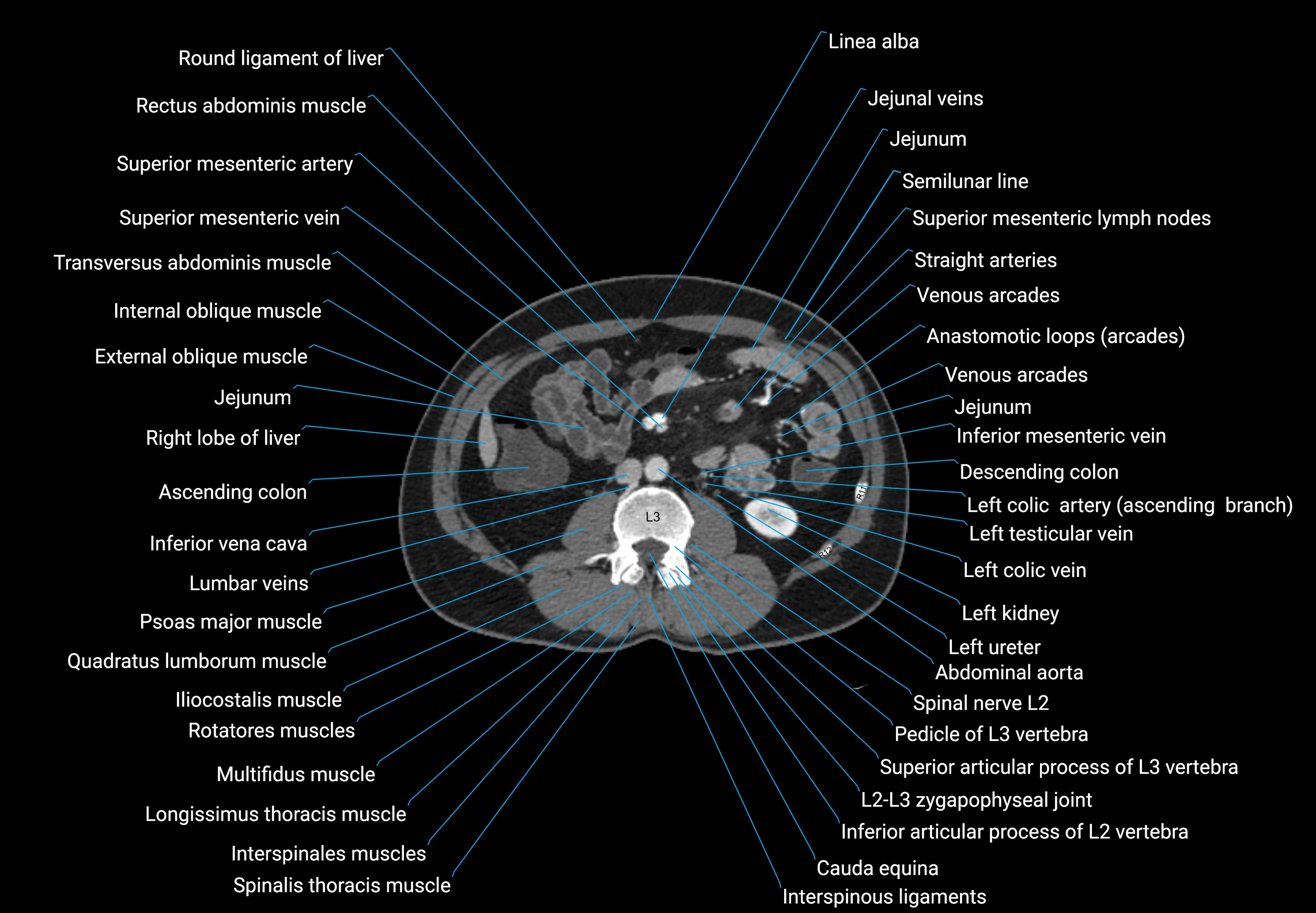
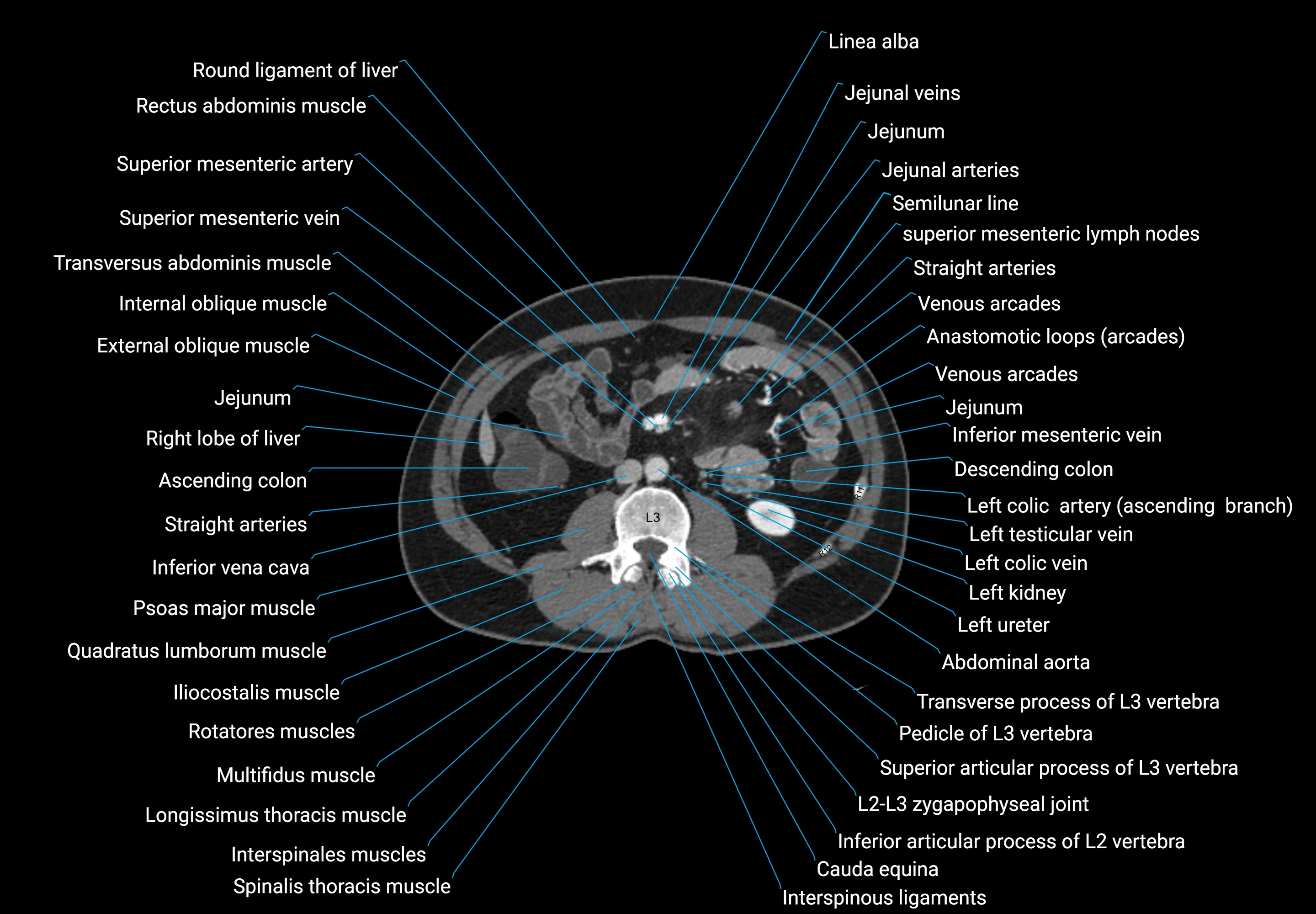
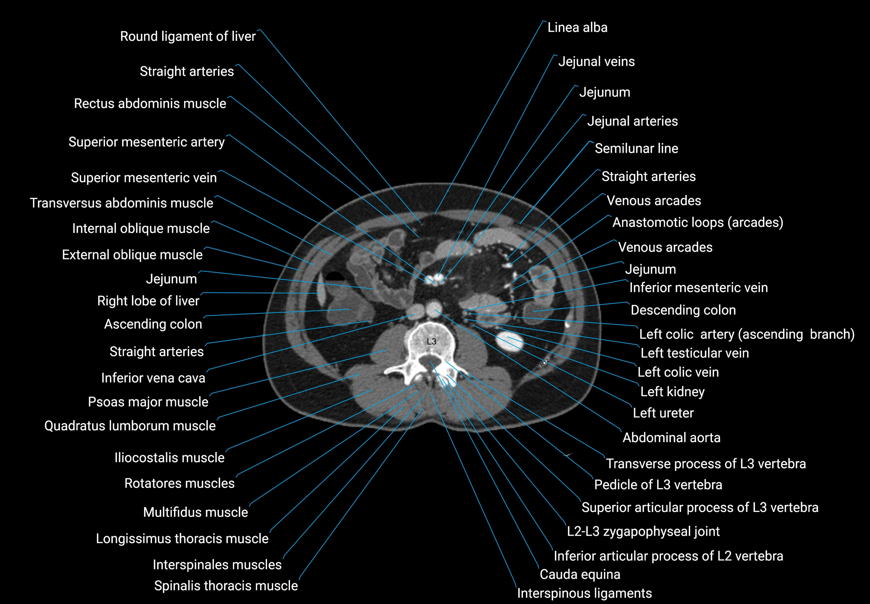
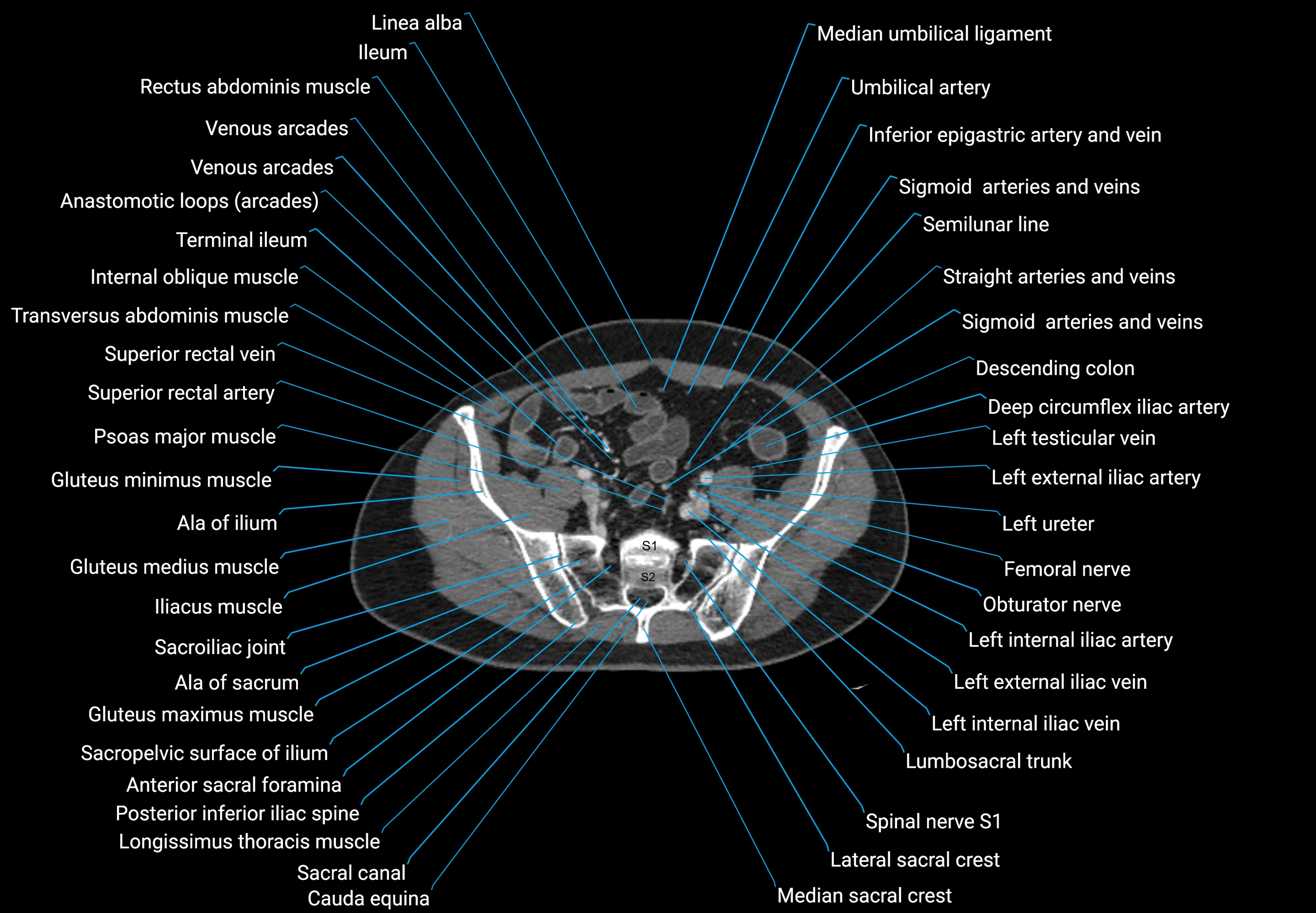
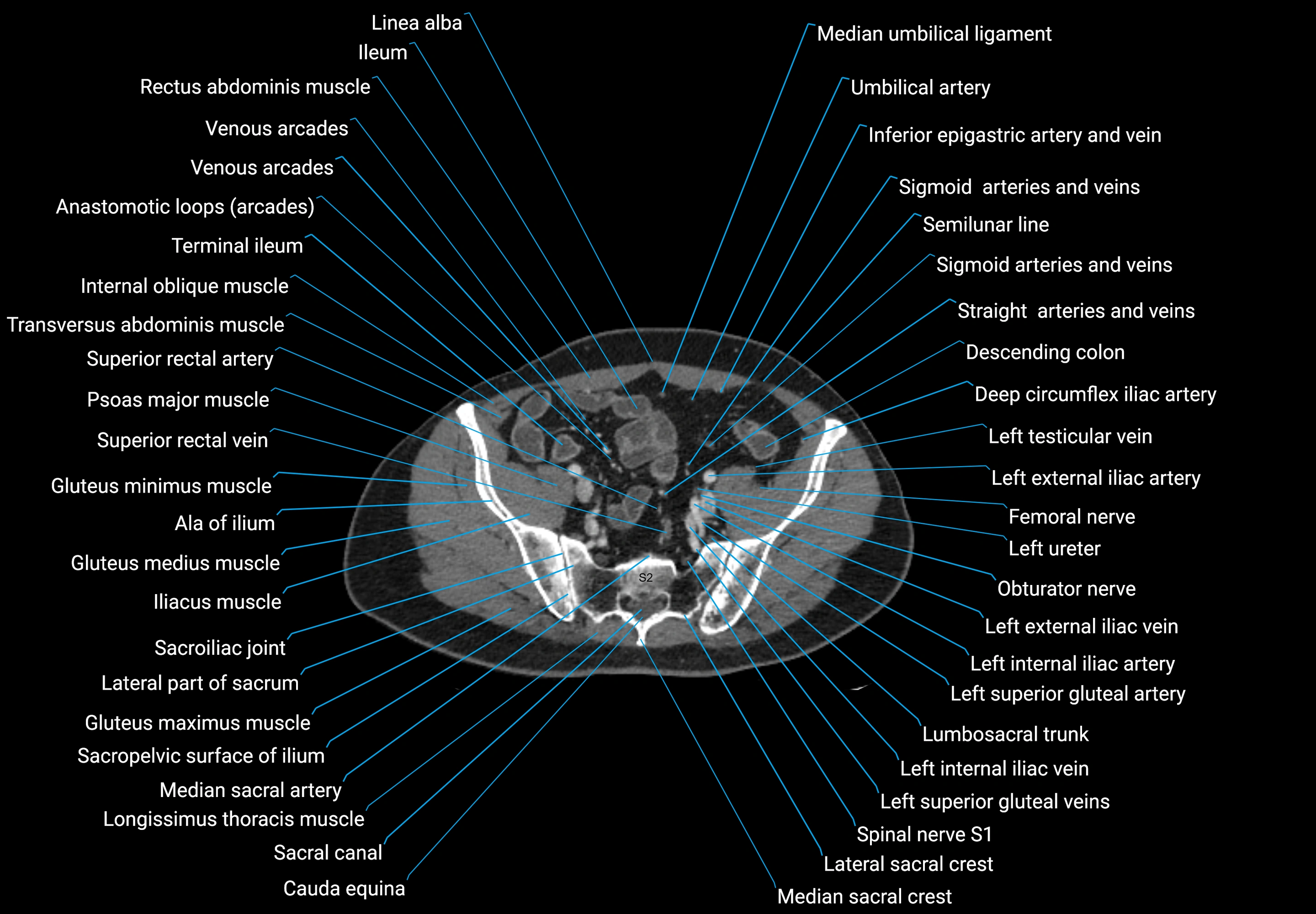
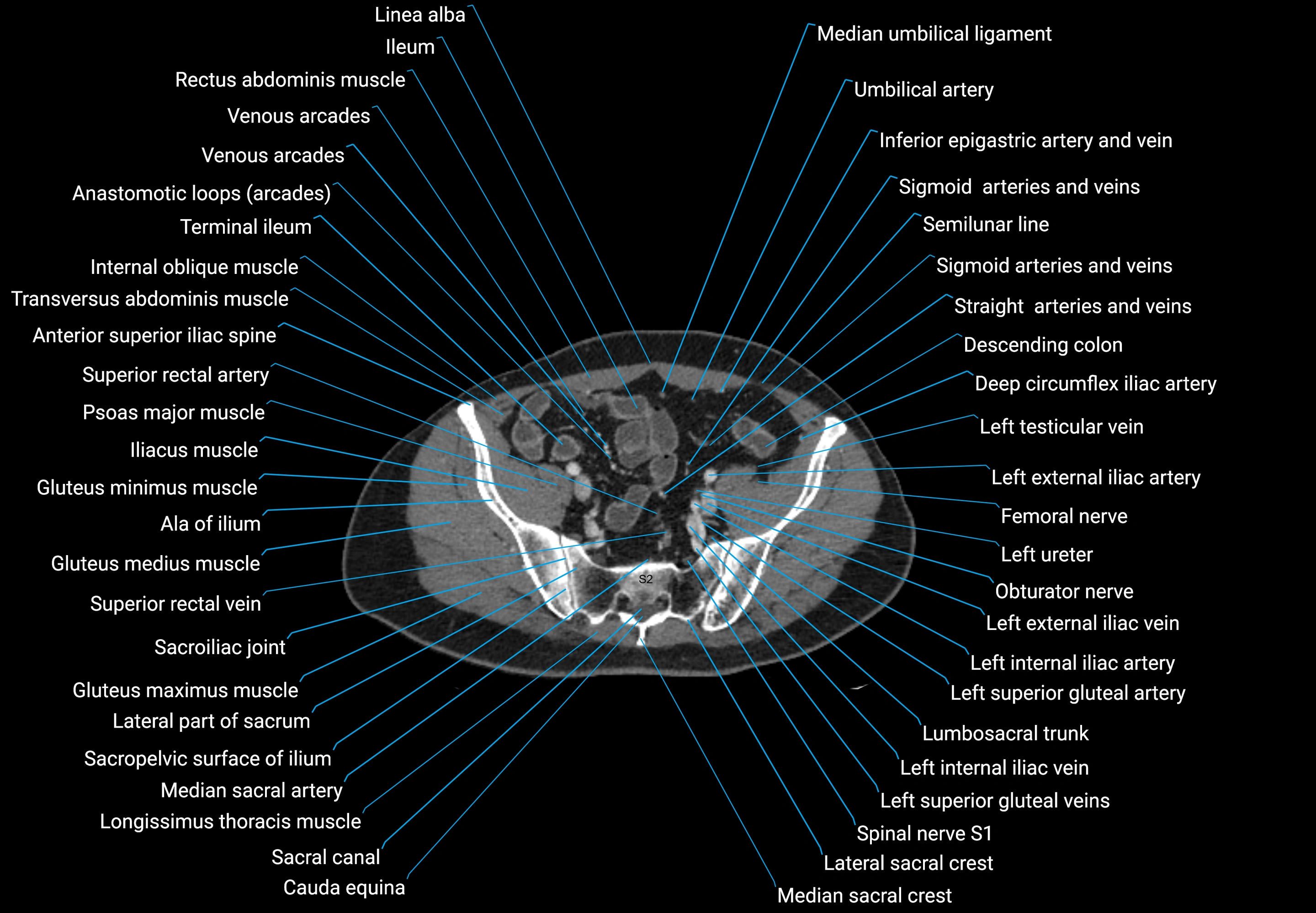
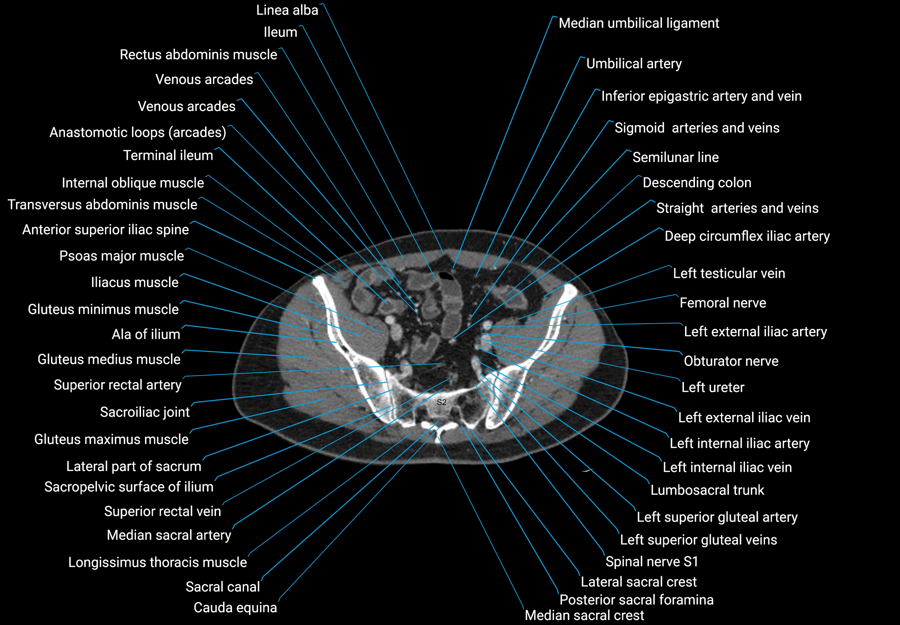
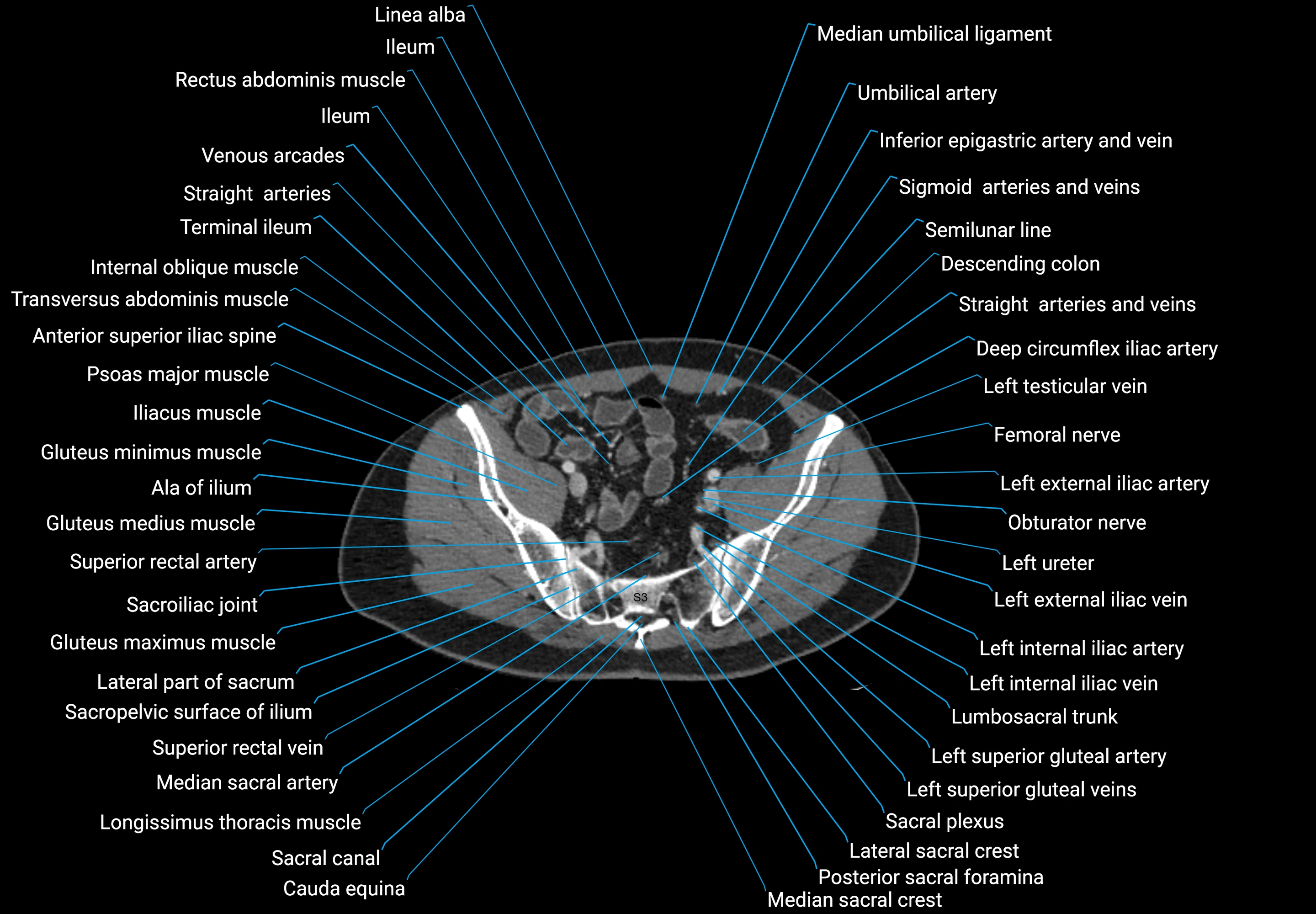
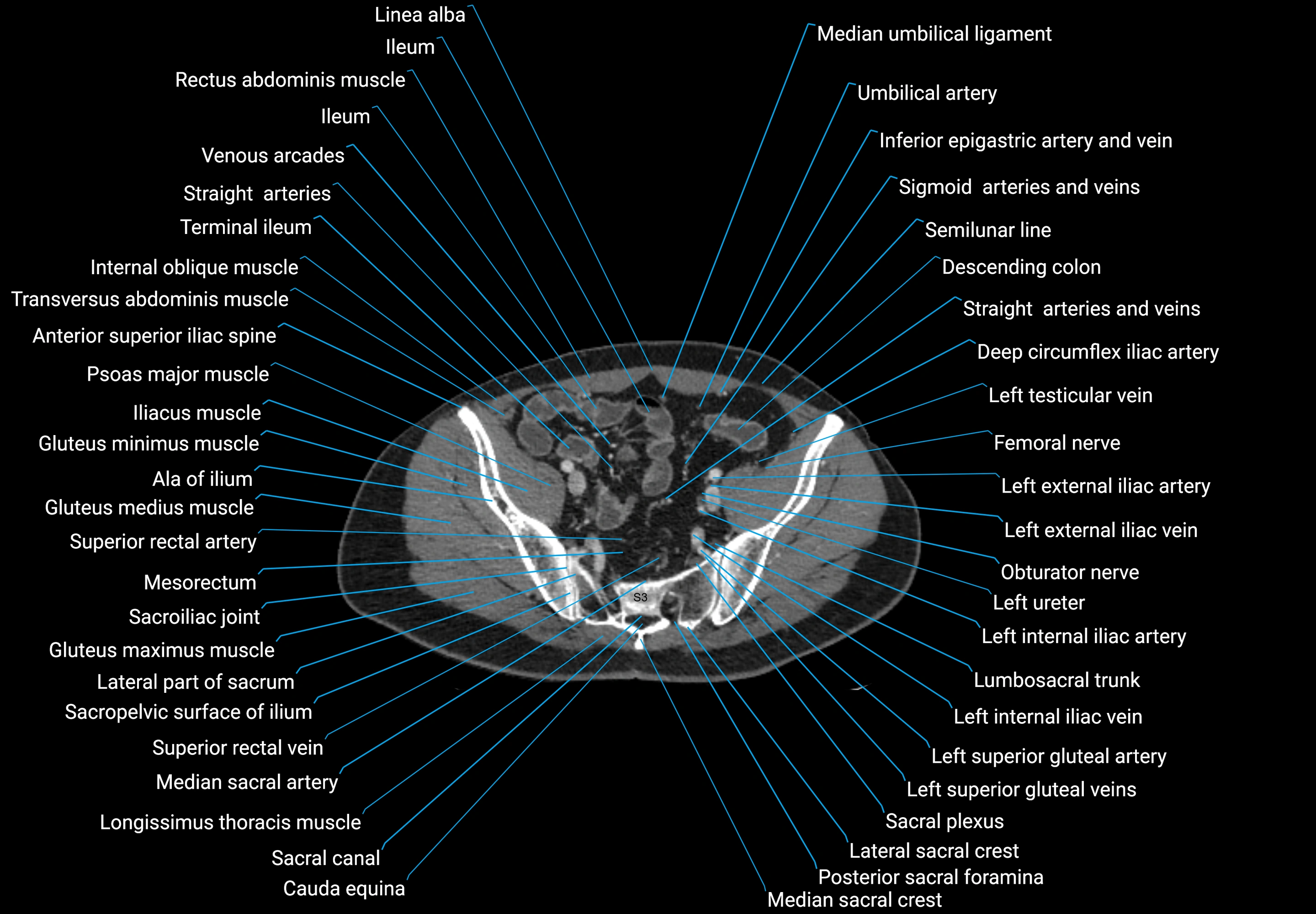
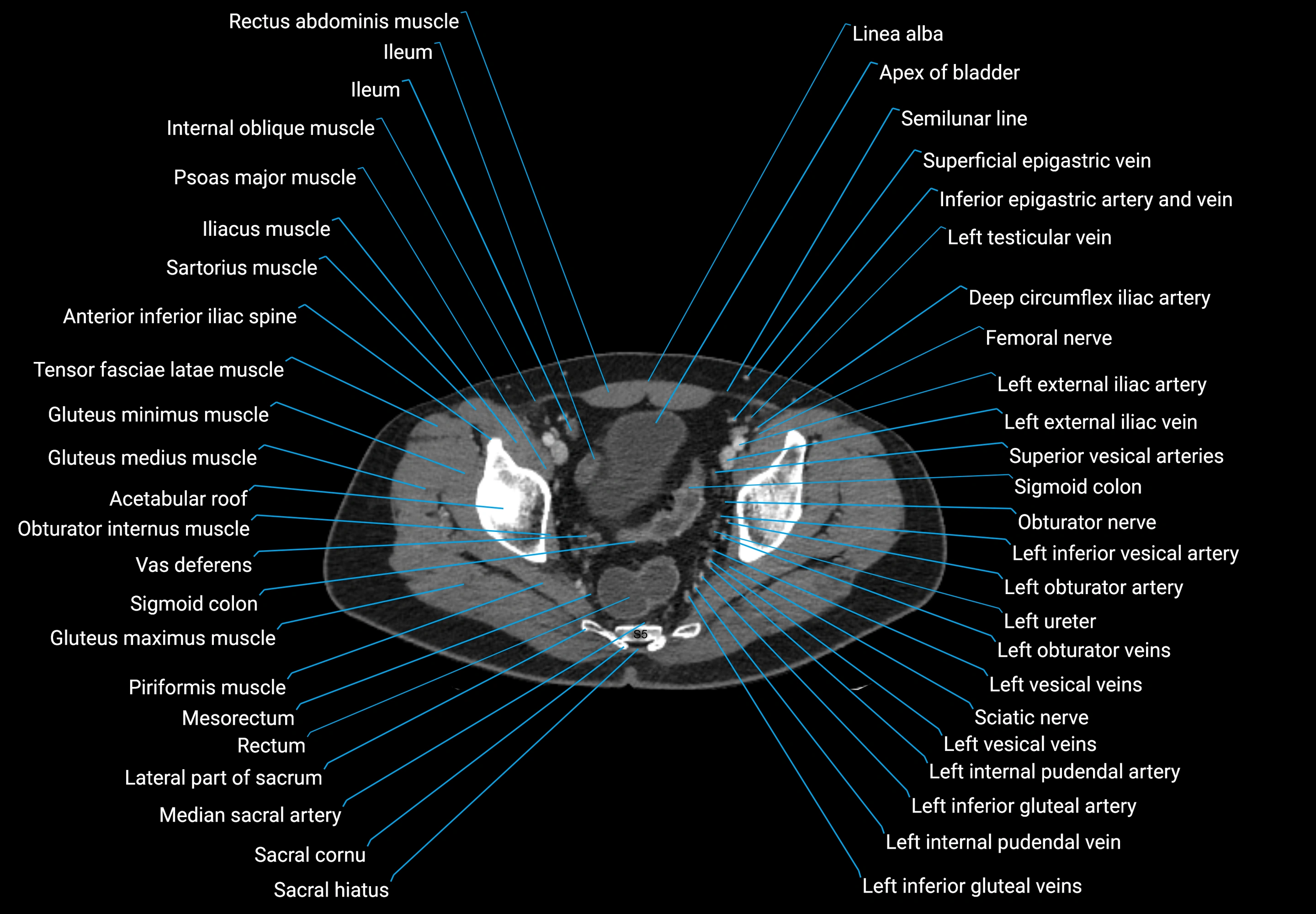
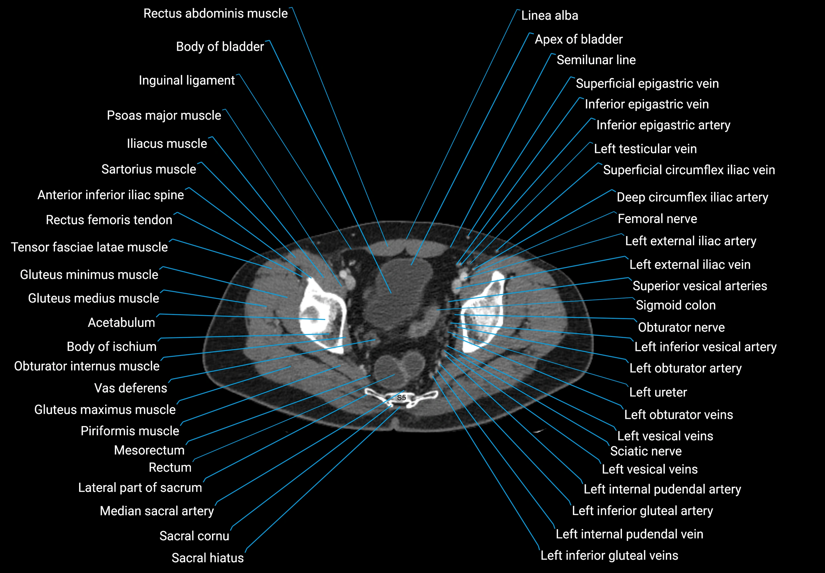
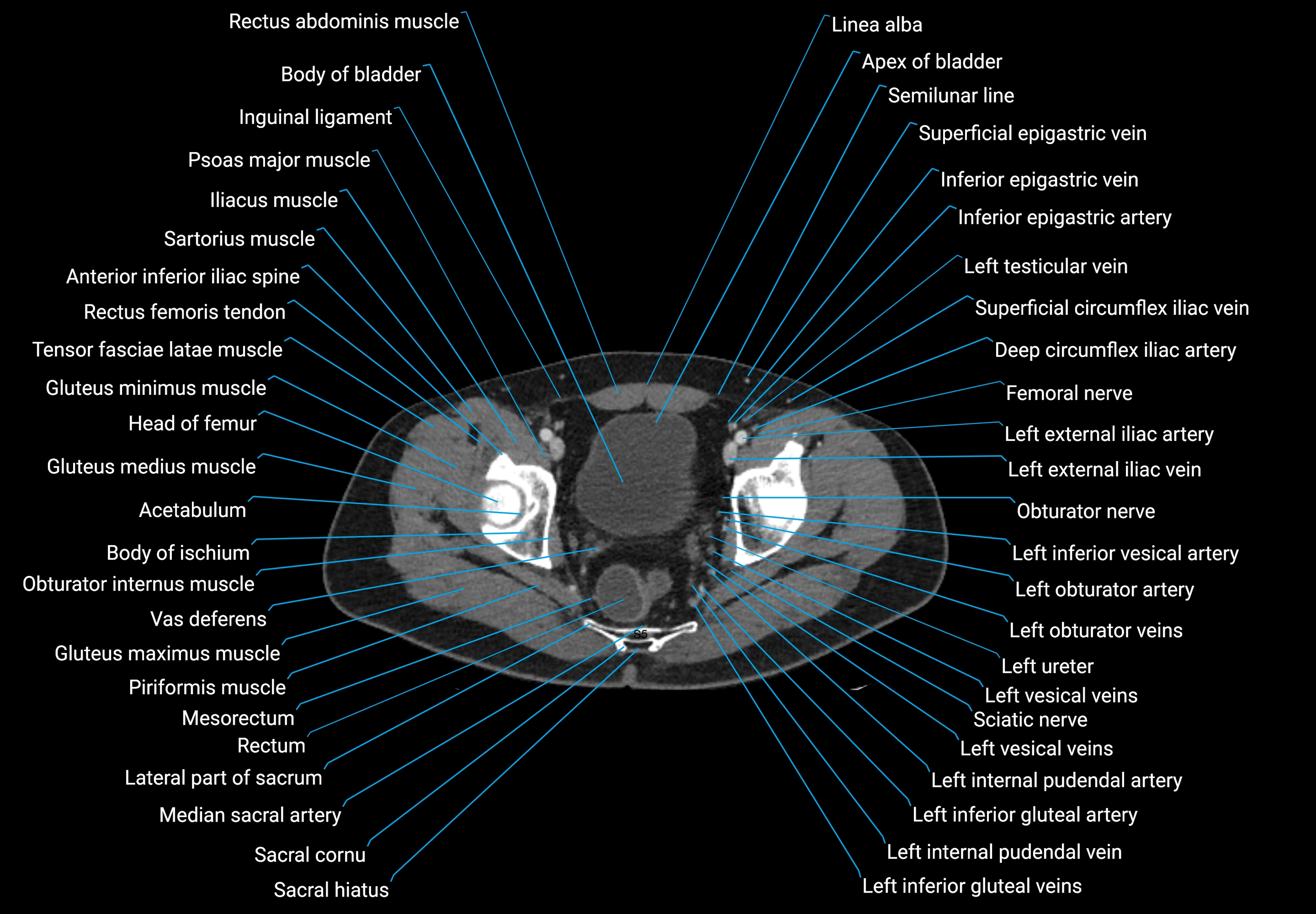
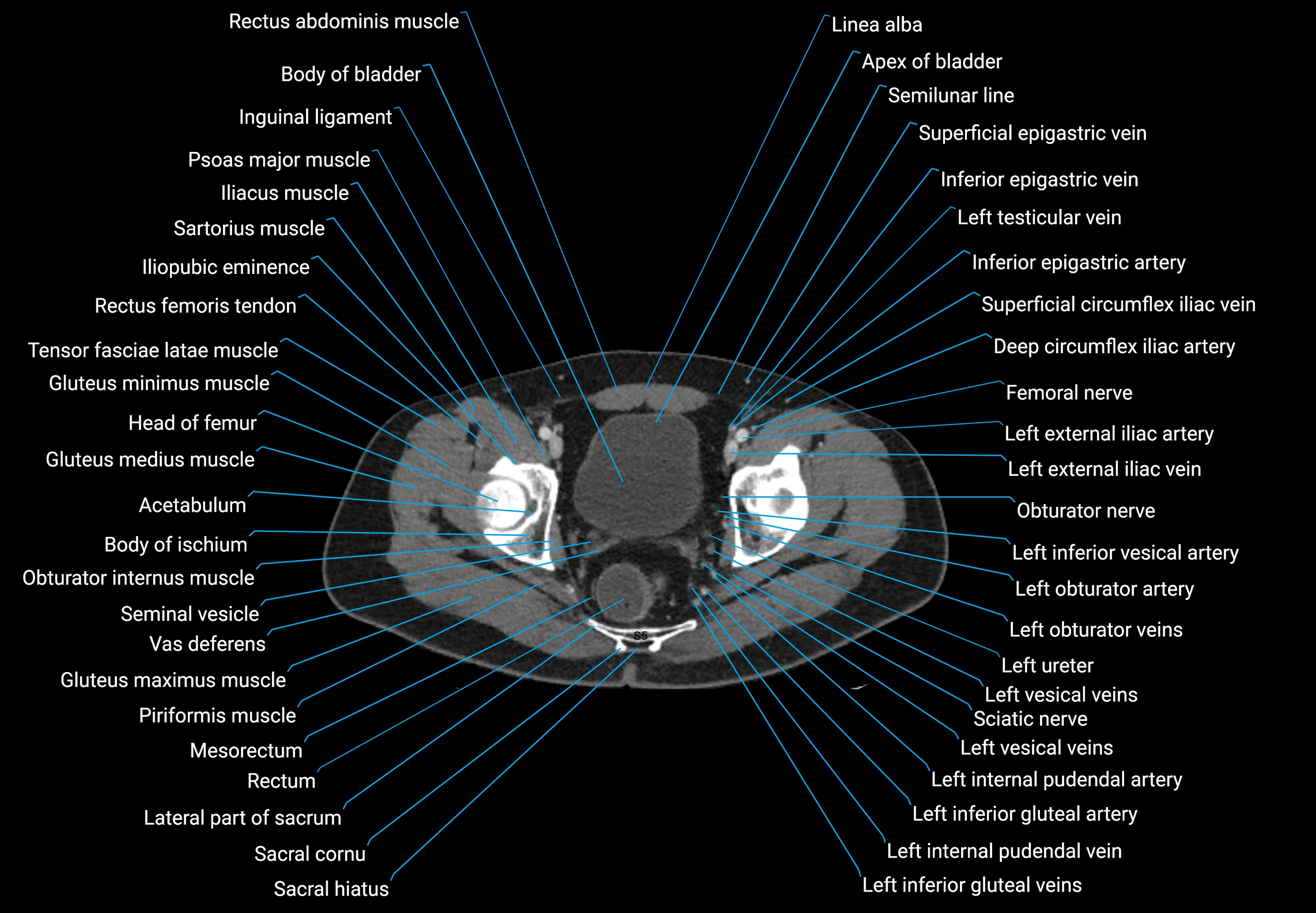
CT images
