


Topic
- Abductor digiti minimi muscle (hand)
- Abductor pollicis brevis muscle
- Abductor pollicis longus muscle
- Accessory cephalic vein
- Accessory median cubital vein
- Acromial end of clavicle
- Acromial part of deltoid muscle
- Acromioclavicular joint
- Acromioclavicular ligament
- Acromion process of scapula
- Adductor pollicis muscle
- Adductor pollicis muscle (Oblique head)
- Adductor pollicis muscle (Transverse head)
- Adipose tissue (Shoulder)
- Anatomical neck of humerus
- Anconeus muscle
- Annular ligament of radius
- Antebrachial fascia
- Anterior fat pad of elbow joint
- Anterior interosseous artery
- Anterior interosseous veins
- Anterior rim of glenoid
- Anterior ulnar recurrent artery
- Articular cartilage of glenoid fossa
- Articular facet of head of radius
- Articularis cubiti muscle
- Axillary lymph nodes
- Axillary nerve
- Base of metacarpal bone
- Basilic vein
- Biceps brachii muscle
- Biceps brachii tendon (distal)
- Biceps pulley
- Bicipital aponeurosis
- Bicipital groove
- Body of humerus
- Body of metacarpal bone
- Body of radius
- Body of scapula
- Body of ulna
- Brachial artery
- Brachial fascia
- Brachial plexus
- Brachialis muscle
- Brachioradialis muscle
- Capitate
- Capitulum of humerus
- Carpal articular surface
- Carpal bones
- Carpometacarpal joints
- Cephalic vein
- Cephalic vein of forearm
- Circumflex scapular artery
- Circumflex subscapular artery
- Clavicular part of deltoid muscle
- Common extensor tendon
- Common flexor tendon
- Conoid ligament
- Conoid tubercle
- Coracoacromial ligament
- Coracobrachialis muscle
- Coracoclavicular Ligaments
- Coracohumeral ligament
- Coracoid process of scapula
- Coronoid fossa
- Coronoid process of ulna
- Crest of greater tubercle
- Crest of lesser tubercle
- Cubital anastomosis
- Cubital tunnel
- Deep brachial artery
- Deltoid Tendon (Proximal)
- Deltoid muscle
- Deltoid tendon (Distal)
- Deltoid tuberosity
- Distal radioulnar joint
- Dorsal radial tubercle (Lister’s tubercle)
- Dorsal scapular artery
- Dorsal scapular nerve
- Elbow joint
- Extensor carpi radialis brevis muscle
- Extensor carpi radialis longus muscle
- Extensor carpi ulnaris muscle
- Extensor carpi ulnaris muscle (Humeral Head)
- Extensor digiti minimi muscle
- Extensor digitorum muscle
- Extensor indicis muscle
- Extensor pollicis brevis muscle
- Extensor pollicis longus muscle
- Fifth metacarpal bone (metacarpal V)
- First dorsal interosseous muscle of hand
- First lumbrical muscle of hand
- First metacarpal bone (metacarpal I)
- First plantar interosseous muscle of hand
- Flexor carpi radialis muscle
- Flexor carpi ulnaris (ulnar head)
- Flexor carpi ulnaris muscle
- Flexor carpi ulnaris muscle (humeral head)
- Flexor digiti minimi brevis muscle (hand)
- Flexor digitorum profundus muscle
- Flexor digitorum superficialis muscle
- Flexor digitorum superficialis muscle (humeroulnar head)
- Flexor digitorum superficialis muscle (radial head)
- Flexor pollicis brevis muscle
- Flexor pollicis brevis muscle (Deep head)
- Flexor pollicis brevis muscle (Superficial head)
- Flexor pollicis longus muscle
- Flexor pollicis longus tendon
- Fourth dorsal interosseous muscle of hand
- Fourth lumbrical muscle of hand
- Fourth metacarpal bone (metacarpal IV)
- Glenohumeral joint capsule
- Glenohumeral ligaments
- Glenoid fossa
- Glenoid labrum
- Glenoid process of scapula
- Greater tubercle of humerus
- Hamate
- Head of humerus
- Head of metacarpal bone
- Head of radius
- Head of ulna
- Hook of hamate bone
- Humeroradial joint
- Humeroulnar joint
- Humerus
- Inferior acromioclavicular ligament
- Inferior angle of scapula
- Inferior belly of omohyoid muscle
- Inferior glenohumeral ligament
- Inferior glenohumeral ligament anterior band
- Inferior glenohumeral ligament posterior band
- Inferior glenohumeral ligament, axillary pouch
- Inferior lateral cutaneous nerve of arm
- Inferior transverse scapular ligament
- Inferior ulnar collateral artery
- Infraglenoid tubercle
- Infraspinatus muscle
- Infraspinatus tendon
- Infraspinous fossa
- Joint capsule of elbow
- Lateral border of humerus
- Lateral border of scapula
- Lateral collateral ligament complex of elbow
- Lateral cutaneous nerve of forearm
- Lateral epicondyle of humerus
- Lateral head of triceps brachii muscle
- Lateral intermuscular septum
- Lateral supracondylar ridge
- Lateral ulnar collateral ligament
- Latissimus dorsi tendon
- Lesser tubercle of humerus
- Long head of biceps brachii muscle
- Long head of biceps tendon
- Long head of triceps brachii muscle
- Long thoracic nerve
- Lunate
- Medial border of humerus
- Medial border of scapula
- Medial collateral ligament complex of elbow (ulnar collateral ligament)
- Medial collateral ligament of elbow (anterior bundle)
- Medial collateral ligament of elbow (posterior bundle)
- Medial collateral ligament of elbow (transverse bundle)
- Medial cutaneous nerve of forearm
- Medial cutaneous nerve of forearm (anterior branch)
- Medial cutaneous nerve of forearm (posterior branch)
- Medial epicondyle of humerus
- Medial head of triceps brachii muscle
- Medial intermuscular septum
- Medial supracondylar ridge
- Median antebrachial vein
- Median cubital vein
- Median nerve
- Metacarpal bones
- Midcarpal joint
- Middle collateral artery
- Middle glenohumeral ligament
- Musculocutaneous nerve
- Neck of radius
- Neck of scapula
- Oblique cord (ligament of Weitbrecht)
- Olecranon
- Olecranon fossa
- Opponens digiti minimi muscle (hand)
- Opponens pollicis muscle
- Osborne’s ligament (cubital tunnel retinaculum)
- Palmaris brevis muscle
- Palmaris longus muscle
- Pectoralis major muscle
- Pectoralis minor muscle
- Pisiform
- Posterior circumflex humeral artery
- Posterior circumflex humeral vein
- Posterior cutaneous nerve of arm
- Posterior cutaneous nerve of forearm
- Posterior fat pad of elbow joint
- Posterior interosseous artery
- Posterior interosseous veins
- Posterior rim of glenoid
- Posterior ulnar recurrent artery
- Pronator quadratus muscle
- Pronator teres muscle
- Pronator teres muscle (humeral head)
- Pronator teres muscle (ulnar head)
- Proximal radioulnar joint
- Quadrate ligament
- Radial artery
- Radial collateral artery
- Radial collateral ligament
- Radial fossa
- Radial nerve
- Radial nerve (deep branch)
- Radial nerve (superficial branch)
- Radial notch of ulna
- Radial recurrent artery
- Radial styloid process
- Radial tuberosity
- Radial veins
- Radiocarpal joint (wrist joint)
- Radius
- Recurrent interosseous artery
- Scaphoid
- Scapula
- Scapular body
- Scapular spinal part of deltoid muscle
- Second dorsal interosseous of hand
- Second lumbrical muscle of hand
- Second metacarpal bone (metacarpal II)
- Second plantar interosseous muscle of hand
- Shaft (body) of clavicle
- Shaft of humerus
- Short head of biceps brachii muscle
- Short head of the biceps brachii tendon
- Shoulder joint (glenohumeral joint)
- Spine of scapula
- Spiral glenohumeral ligament
- Sternoclavicular joint
- Subacromial space
- Subclavius muscle
- Subscapular artery
- Subscapular fossa
- Subscapularis muscle
- Subscapularis tendon
- Superior acromioclavicular ligament
- Superior angle of scapula
- Superior border of scapula
- Superior glenohumeral ligament
- Superior transverse scapular ligament
- Superior ulnar collateral artery
- Supinator muscle
- Supraclavicular fossa
- Supraglenoid tubercle
- Supraspinatus muscle
- Supraspinatus tendon
- Supratrochlear lymph nodes
- Surgical neck of humerus
- Teres major muscle
- Teres major tendon (Distal)
- Teres minor muscle
- Teres minor tendon (Distal)
- Third dorsal interosseous muscle of hand
- Third lumbrical muscle of hand
- Third metacarpal bone (metacarpal III)
- Third metacarpal styloid process
- Third plantar interosseous muscle of hand
- Thoracodorsal artery
- Trapezium
- Trapezoid
- Trapezoid Line
- Trapezoid ligament
- Triceps brachii muscle
- Triceps brachii tendon
- Triquetrum
- Trochlea of humerus
- Trochlear notch of ulna
- Tubercle of the scaphoid bone
- Tubercle of trapezium bone
- Tuberosity of ulna
- Ulna
- Ulnar artery
- Ulnar nerve
- Ulnar styloid process
- Ulnar veins
- supraspinous fossa of scapula
The abductor digiti minimi (ADM) is a superficial muscle located along the ulnar border of the palm in the hypothenar eminence of the hand. It is the most medial of the hypothenar muscles and lies superficial to the flexor digiti minimi brevis and opponens digiti minimi. The ADM plays a key role in abducting the little finger (fifth digit) away from the ring finger and contributes to grip and hand stability. It also assists in flexion of the metacarpophalangeal joint and extension of the interphalangeal joints through its connection with the extensor expansion.
Synonyms
-
Abductor of the little finger
-
Abductor minimi digiti
-
Abductor digiti quinti
Origin, Course, and Insertion
Origin: From the pisiform bone, the pisohamate ligament, and the tendon of the flexor carpi ulnaris.
Course: Muscle fibers run distally along the ulnar side of the hand, forming a flat tendon near the base of the little finger.
Insertion: Ulnar side of the base of the proximal phalanx of the fifth digit and the extensor expansion of the same finger.
Tendon Attachments
-
The tendon passes along the medial side of the fifth metacarpal and inserts into both the base of the proximal phalanx and dorsal digital expansion.
-
It may send a slip to the extensor digiti minimi tendon, facilitating combined extension and abduction.
Relations
Superficial: Palmar fascia and skin of the hypothenar eminence
Deep: Flexor digiti minimi brevis and opponens digiti minimi
Medially: Ulnar border of the hand
Laterally: Flexor tendons of the little finger
Proximally: Pisiform and ulnar artery and nerve branches
Distally: Proximal phalanx and extensor expansion of the little finger
Nerve Supply
Deep branch of the ulnar nerve (C8, T1)
Arterial Supply
Ulnar artery, via its deep palmar branch and ulnar digital artery to the little finger
Function
-
Abduction: Moves the little finger away from the ring finger at the metacarpophalangeal joint.
-
Flexion: Assists in flexing the proximal phalanx of the fifth digit.
-
Extension: Through the extensor expansion, aids in extending the distal phalanges.
-
Grip assistance: Contributes to hypothenar support and cupping of the hand during grasp.
-
Stabilization: Maintains the ulnar border of the palm during object manipulation.
Clinical Significance
-
Ulnar nerve injury: Paralysis or weakness of ADM leads to loss of little finger abduction and atrophy of the hypothenar eminence.
-
Muscle hypertrophy or fibrotic bands: May compress the ulnar nerve in Guyon’s canal.
-
Tendon tears or strain: Rare but can occur in repetitive hand use or trauma.
-
Surgical relevance: Important landmark in ulnar nerve decompression and hypothenar flap surgeries.
-
Imaging importance: MRI evaluates ADM for denervation changes, trauma, and space-occupying lesions in the hypothenar region.
MRI Appearance
T1-weighted images:
-
Normal muscle: intermediate signal intensity with distinct fascicular pattern.
-
Tendon: low signal (dark) extending to proximal phalanx of the little finger.
-
Fatty tissue of hypothenar region: bright, providing contrast with muscle.
-
Chronic denervation: increased intramuscular fat causing hyperintense signal.
T2-weighted images:
-
Normal muscle: intermediate-to-dark signal, slightly darker than on T1.
-
Tendon: uniformly dark.
-
Pathology: edema or inflammation produces bright hyperintense signal in acute injury or myositis.
STIR:
-
Normal muscle: intermediate-to-dark signal.
-
Pathology: bright hyperintense signal indicating edema, strain, or nerve-related denervation changes.
Proton Density Fat-Saturated (PD FS):
-
Normal ADM: intermediate-to-dark signal with smooth margins.
-
Muscle strain or tendinitis: bright focal hyperintensity within or around tendon.
-
Excellent for identifying subtle peritendinous edema or fascial inflammation.
T1 Fat-Sat Post-Contrast:
-
Normal muscle: mild homogeneous enhancement.
-
Active inflammation or tear: focal enhancement at musculotendinous junction.
-
Denervation or chronic fibrosis: little to no enhancement with volume loss and fatty infiltration.
CT Appearance
Non-Contrast CT:
-
Muscle: soft-tissue density, well defined in the hypothenar region.
-
Tendon: linear low-density structure extending to the base of the little finger.
-
Calcification or chronic scarring may appear as localized high-density foci.
-
Useful for assessing bony attachment sites at the pisiform and proximal phalanx.
Post-Contrast CT (standard):
-
Normal muscle: homogeneous mild enhancement.
-
Inflamed or injured muscle: increased enhancement and surrounding soft-tissue edema.
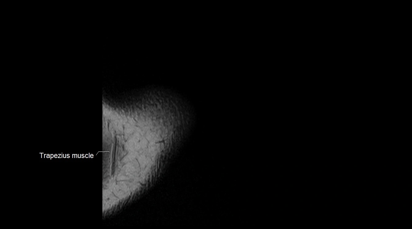
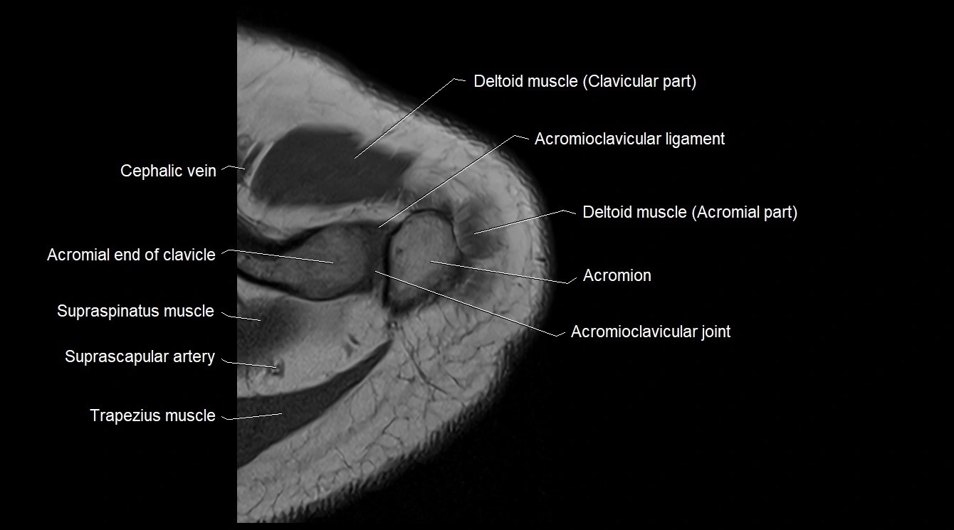
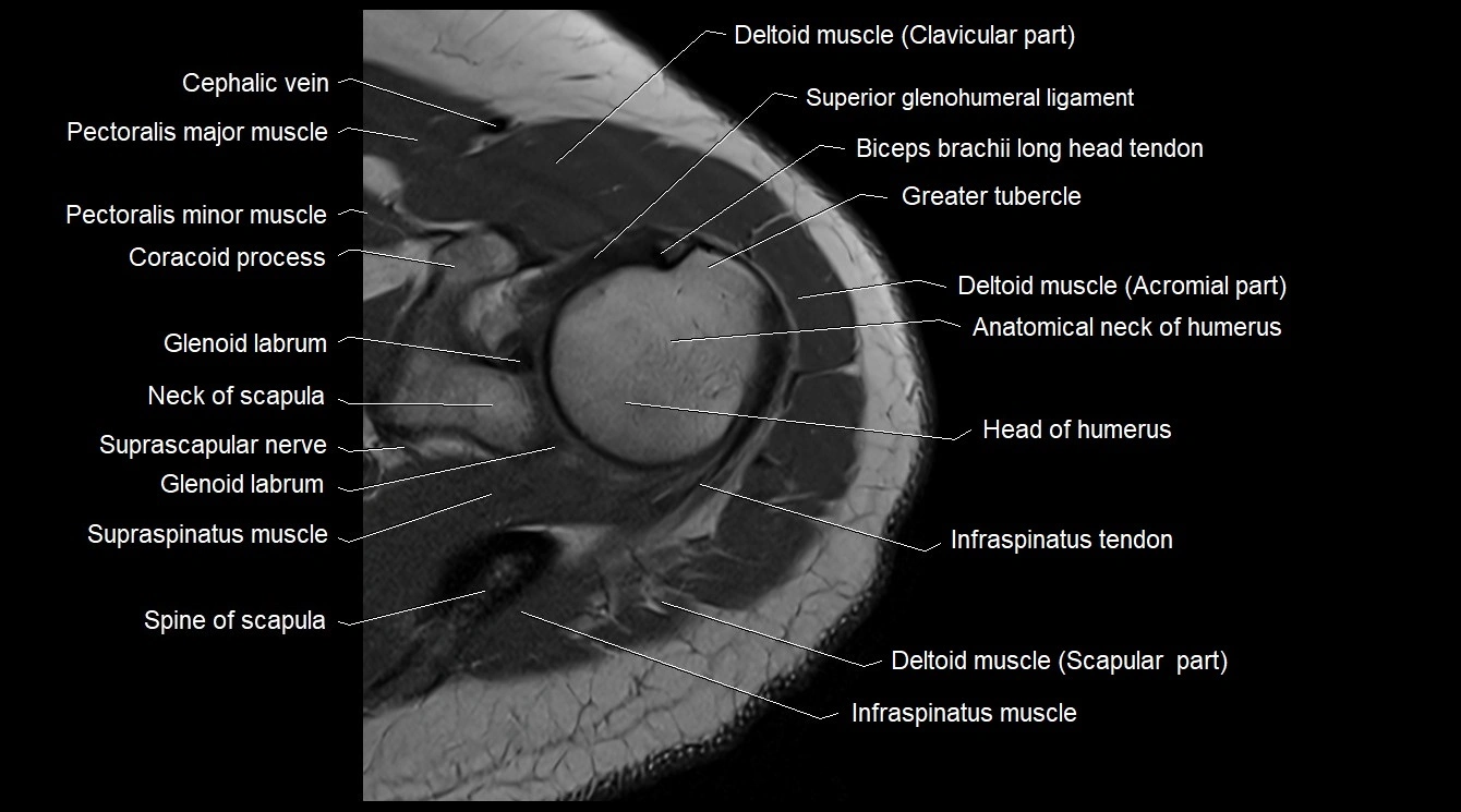
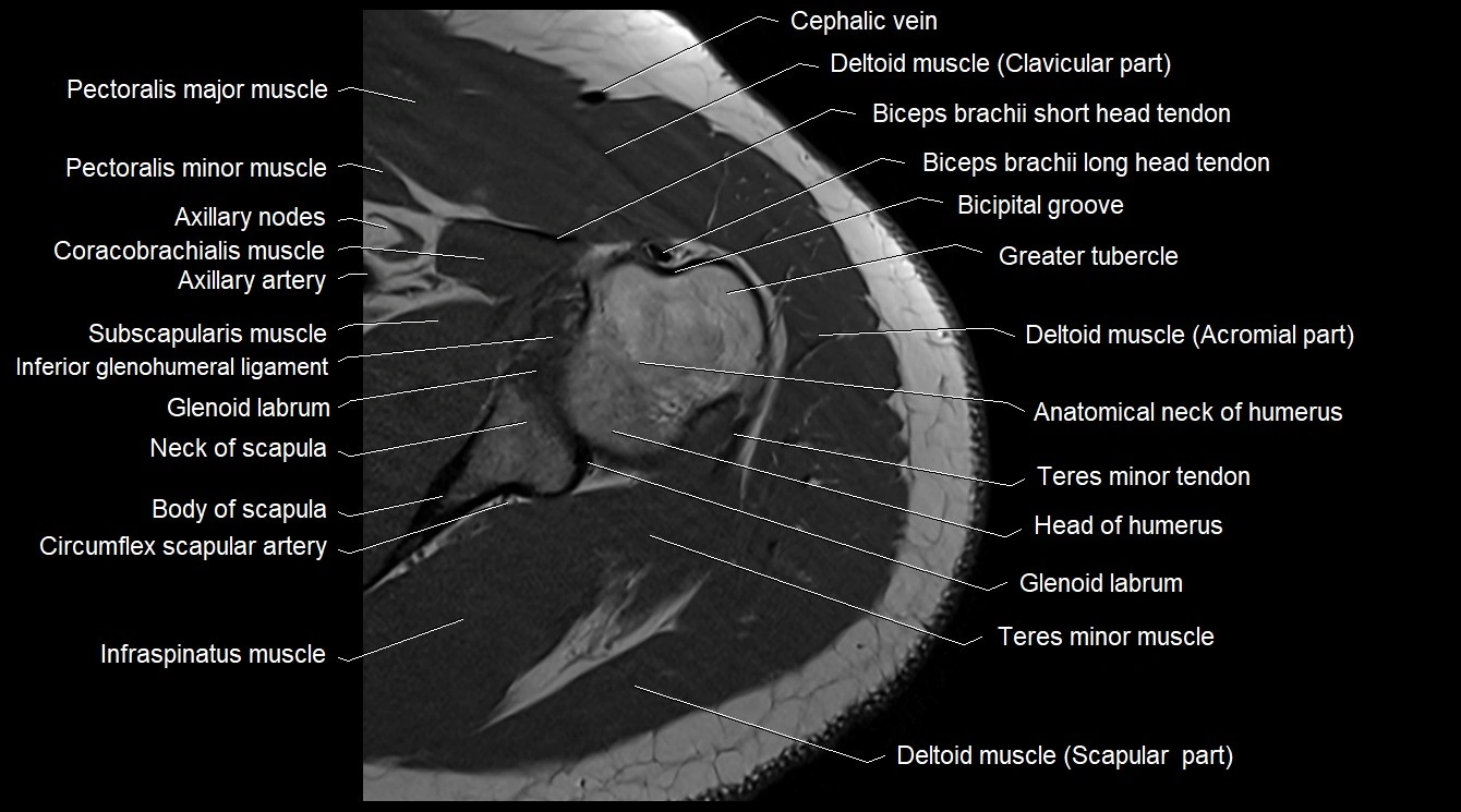
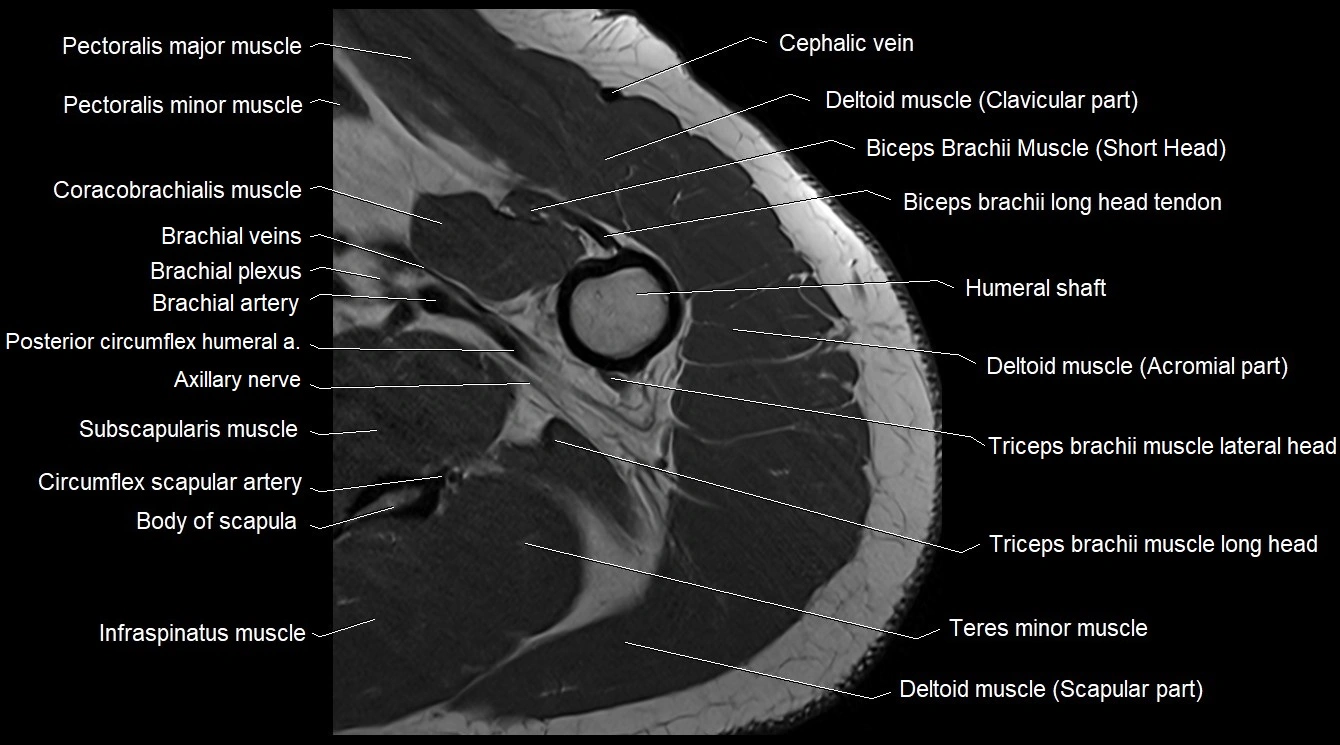
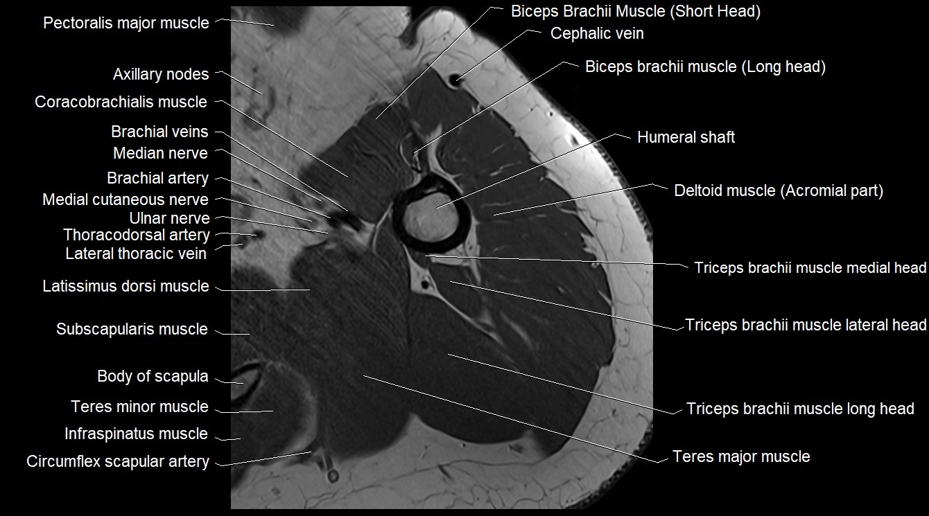
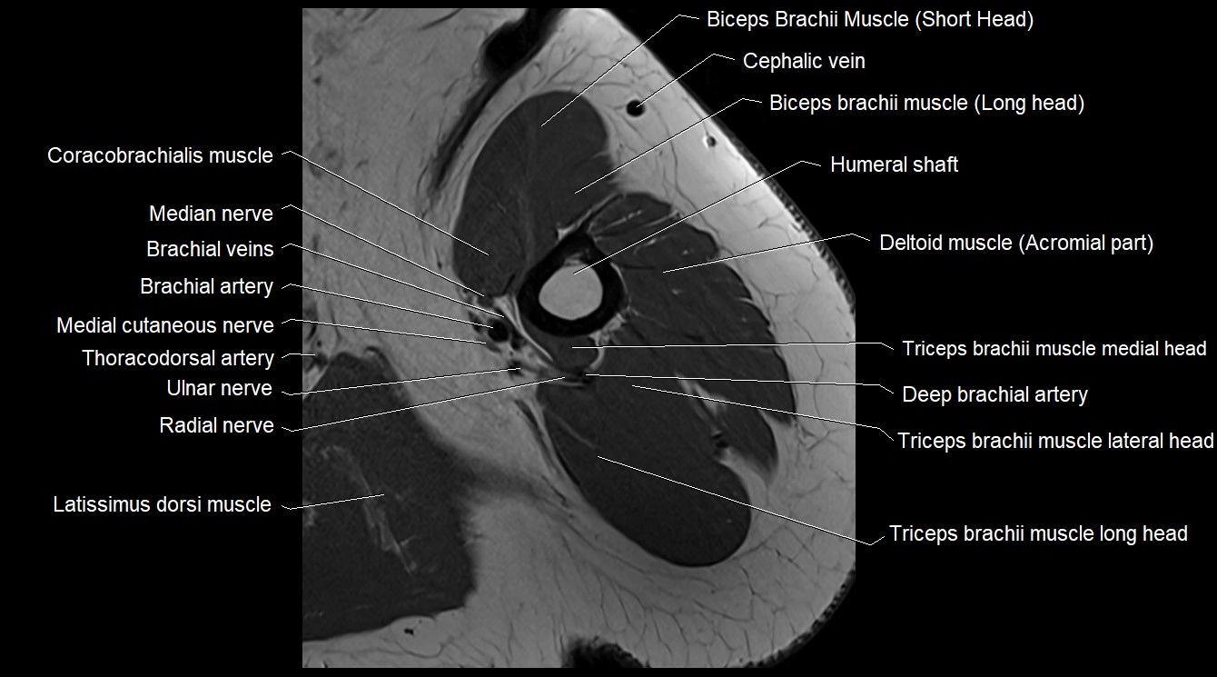
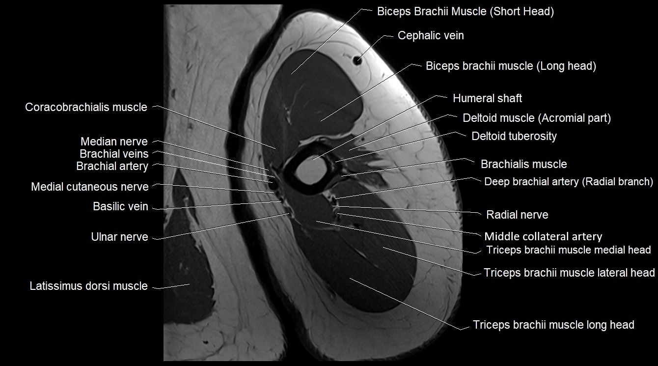
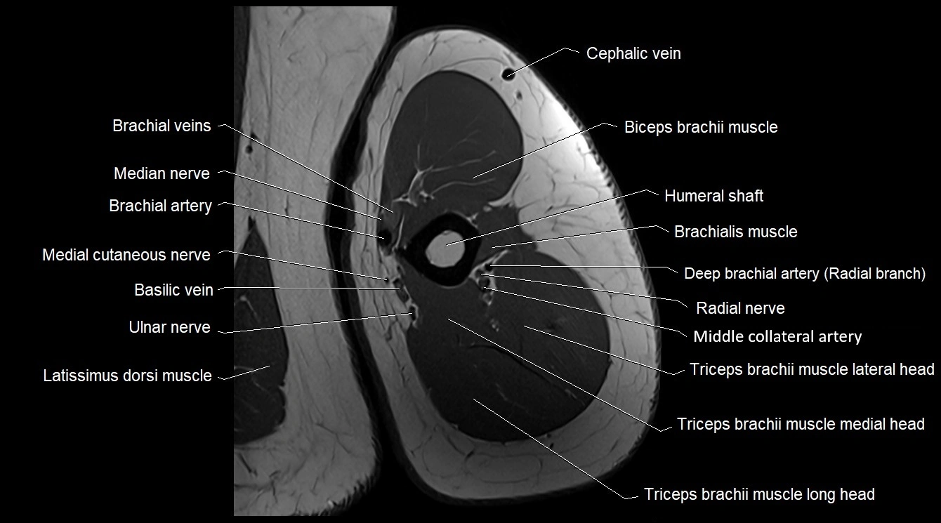
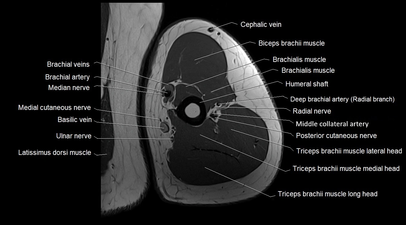
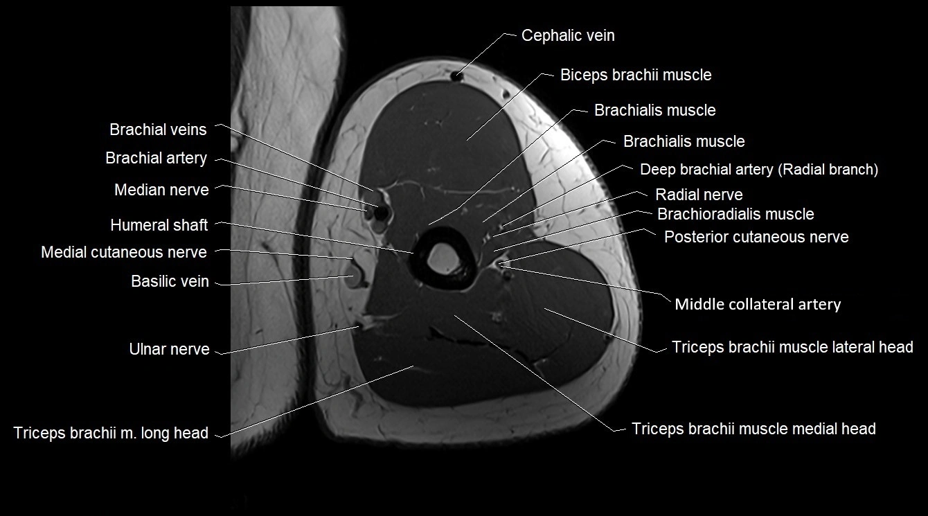
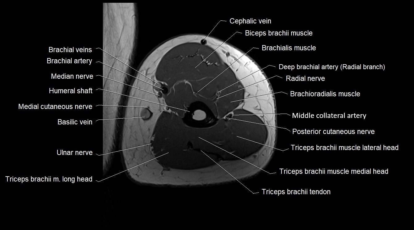
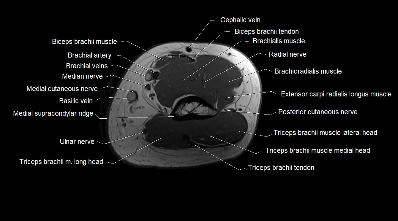
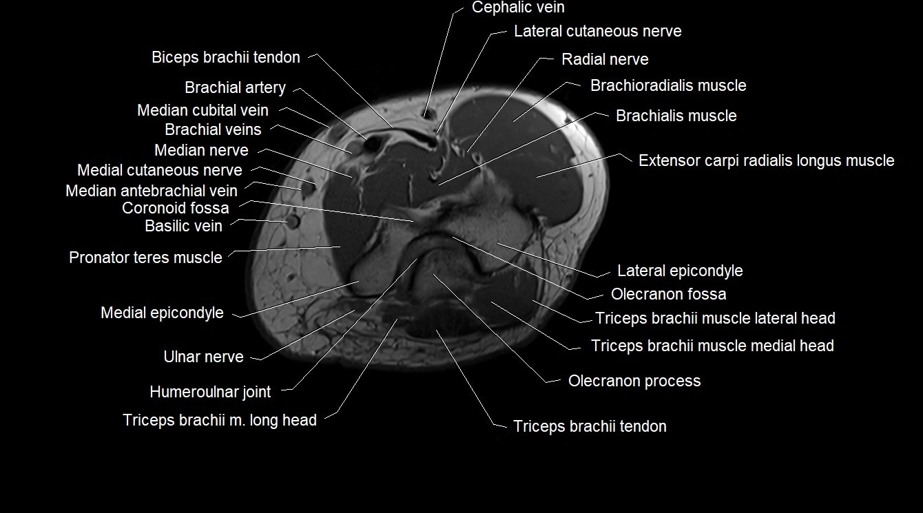
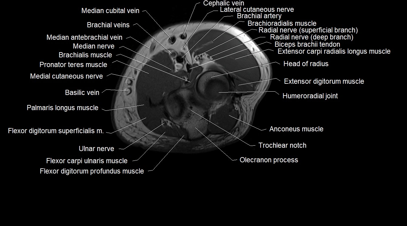
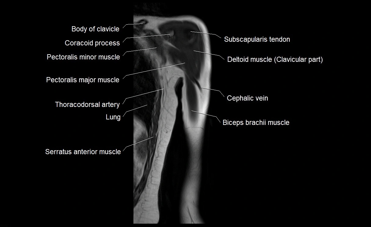
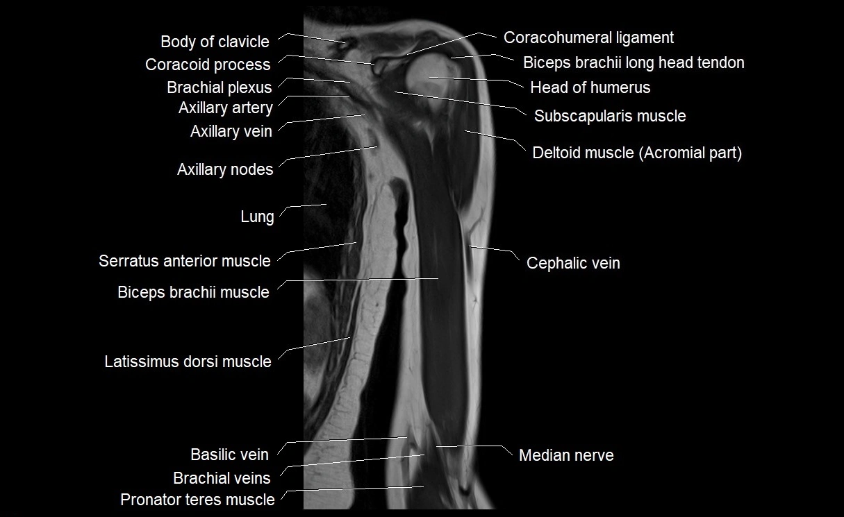
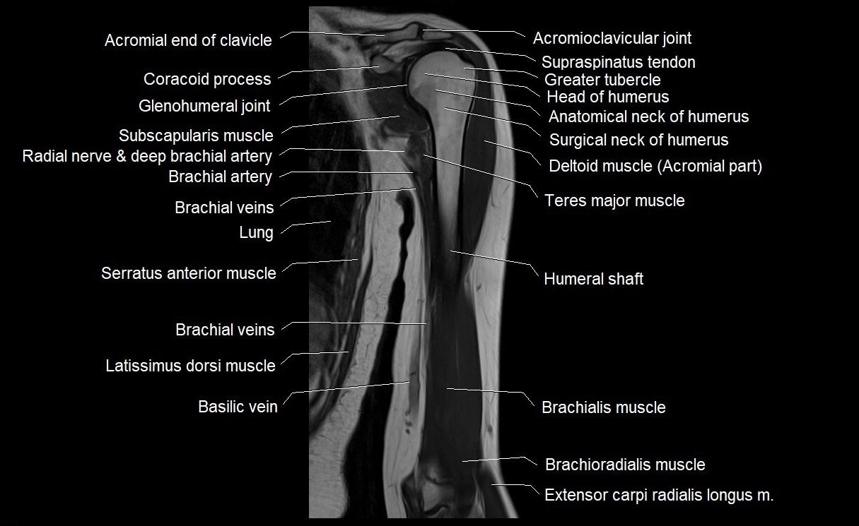
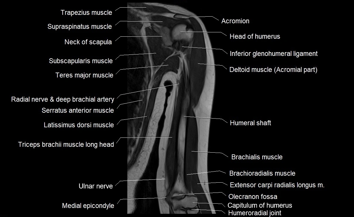
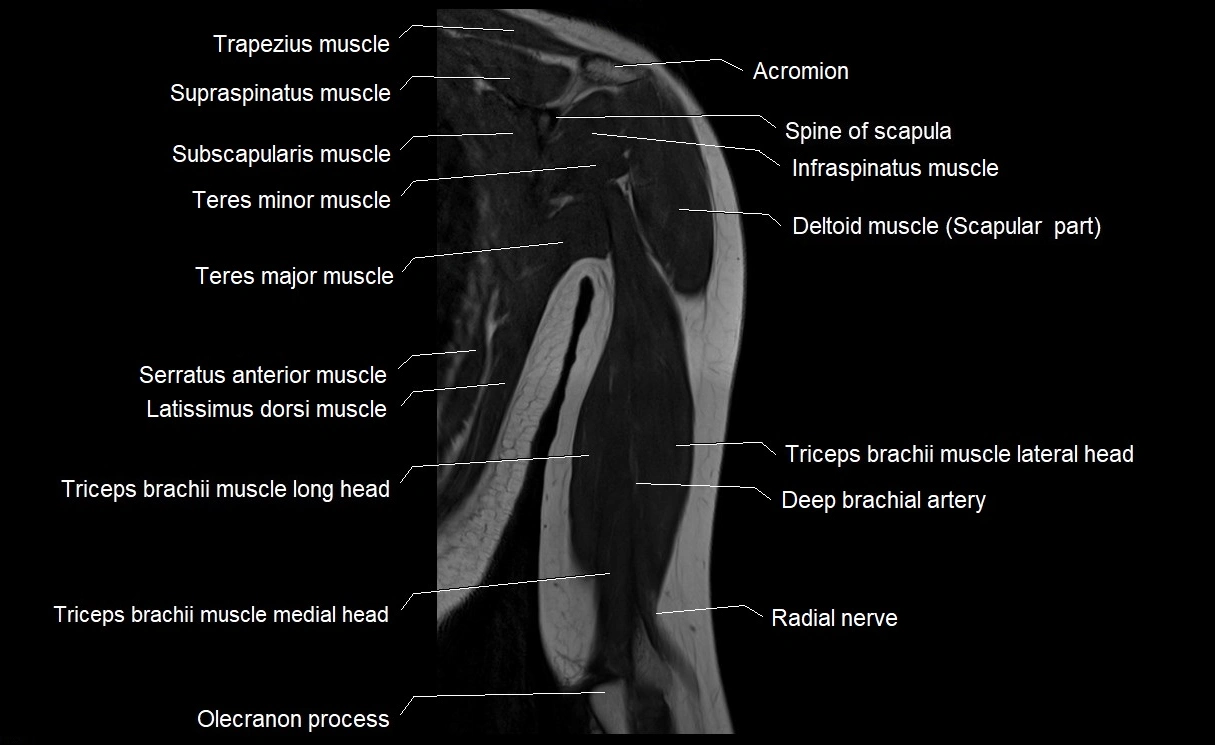
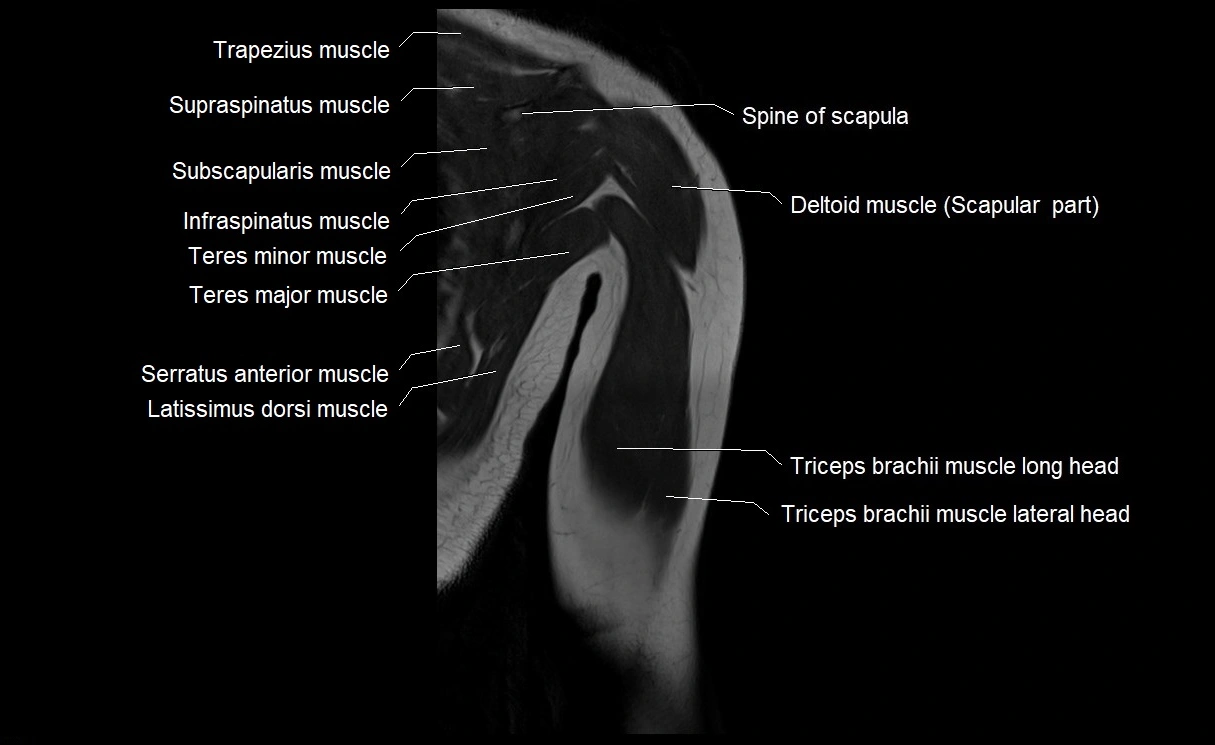
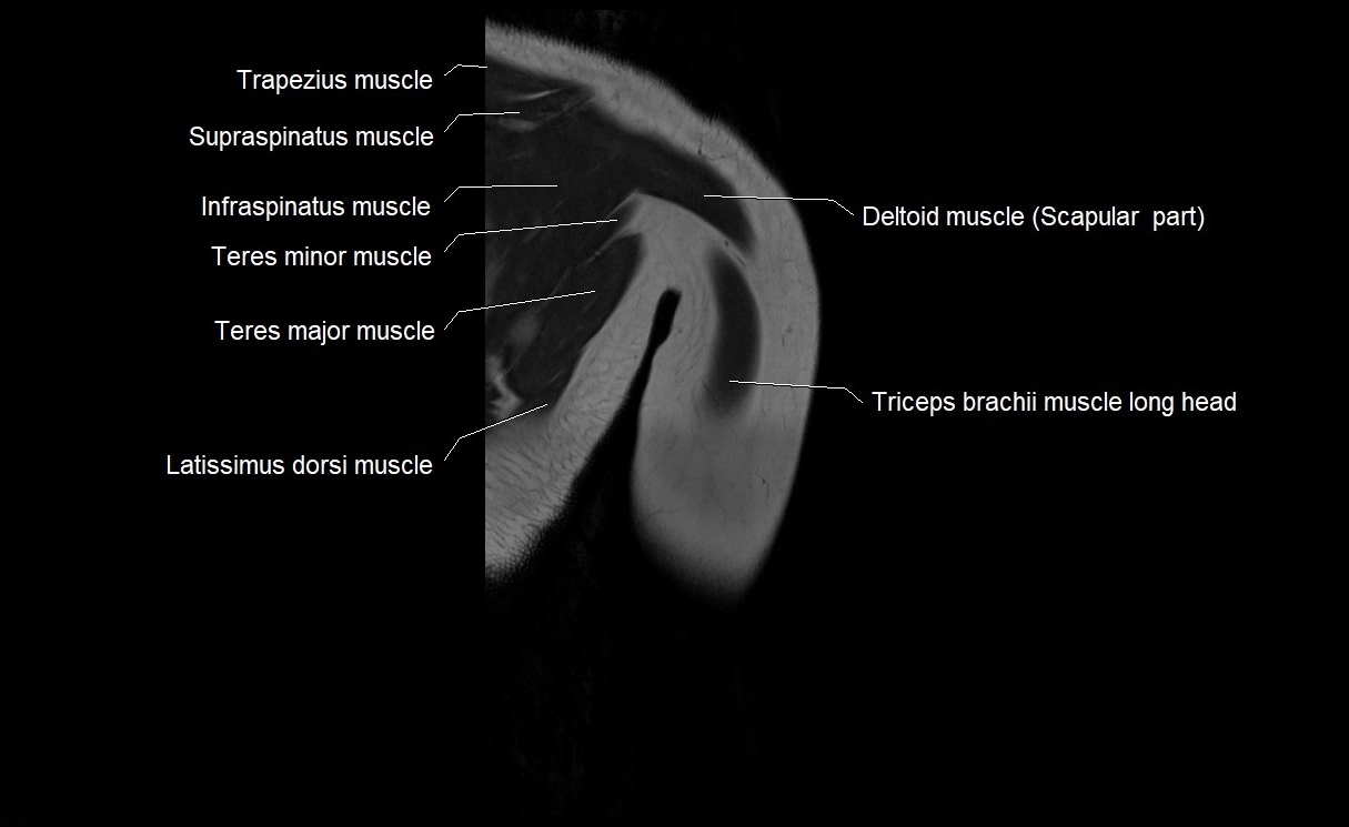
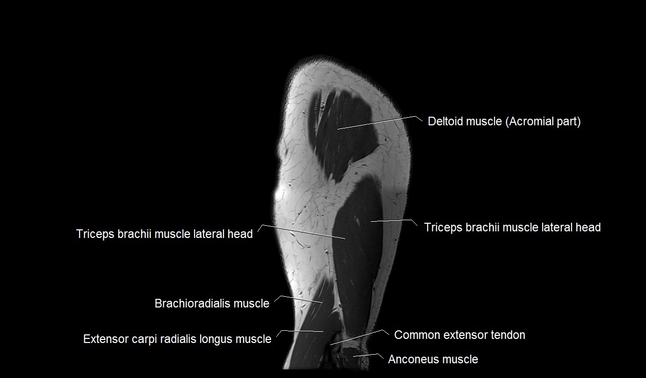
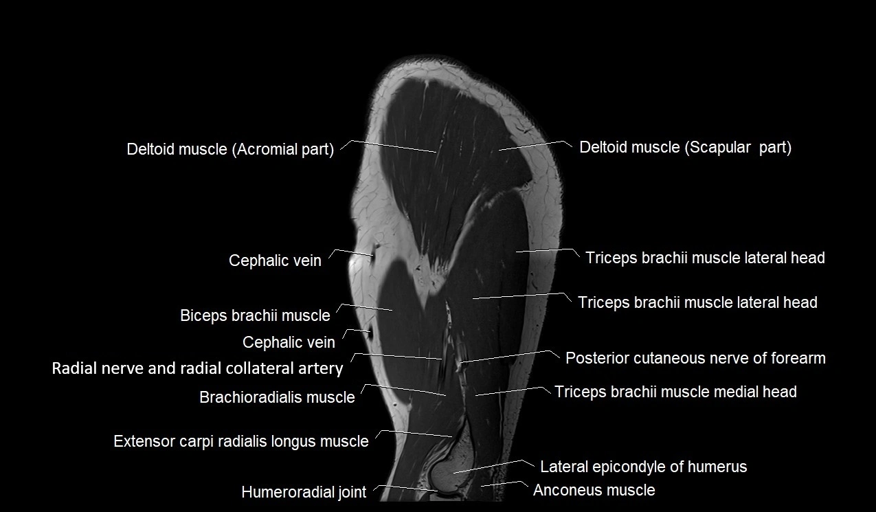
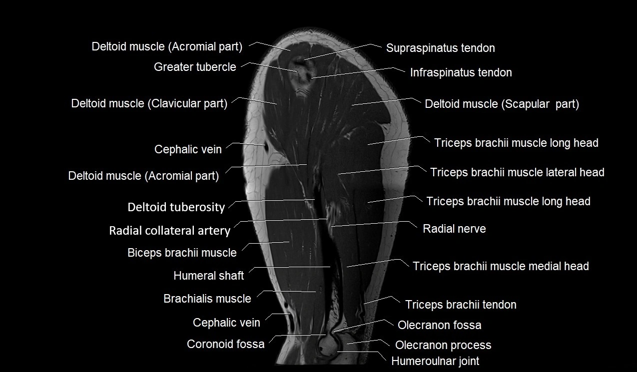
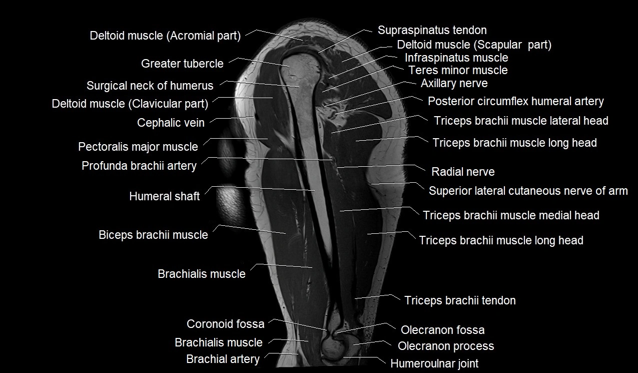
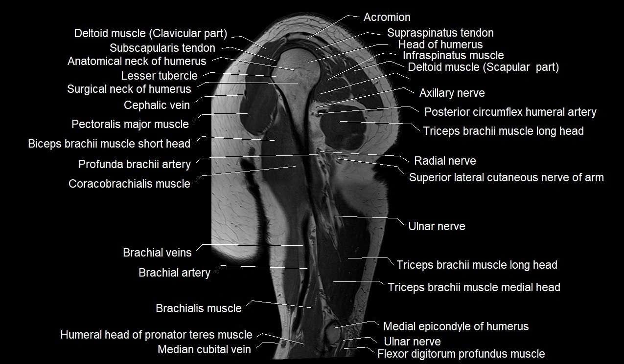
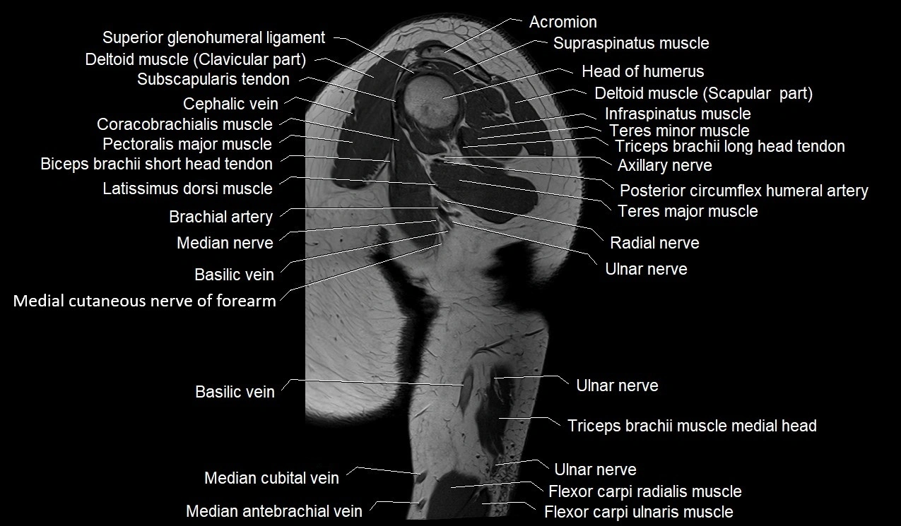
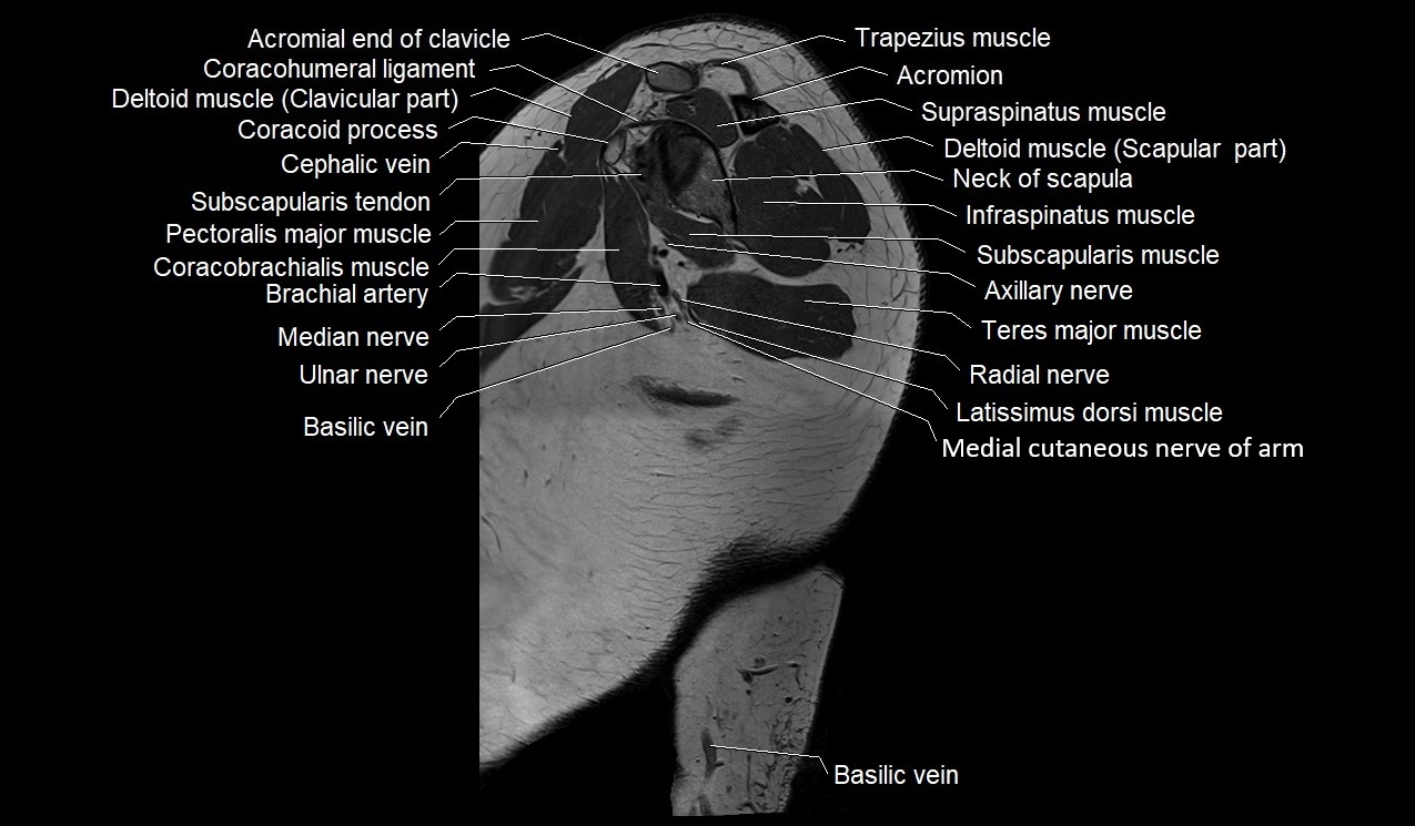
MRI image

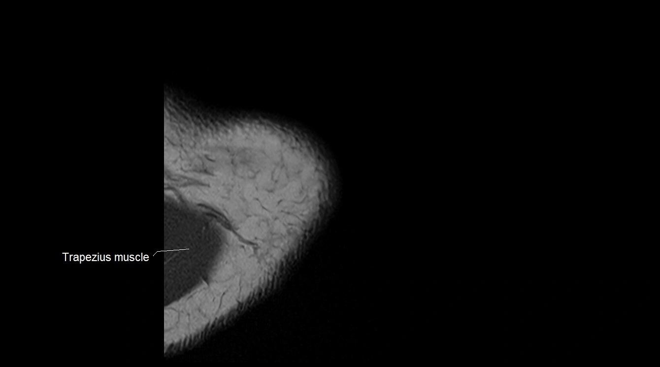
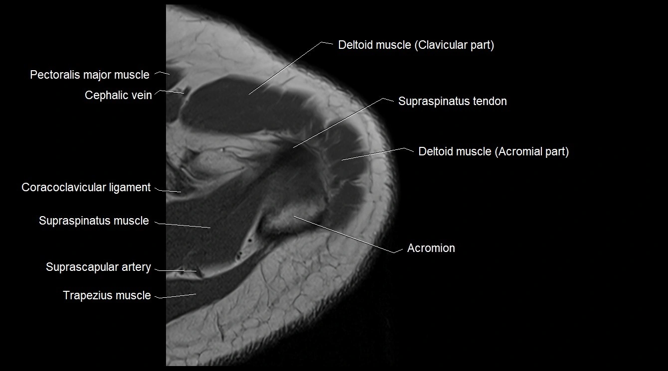
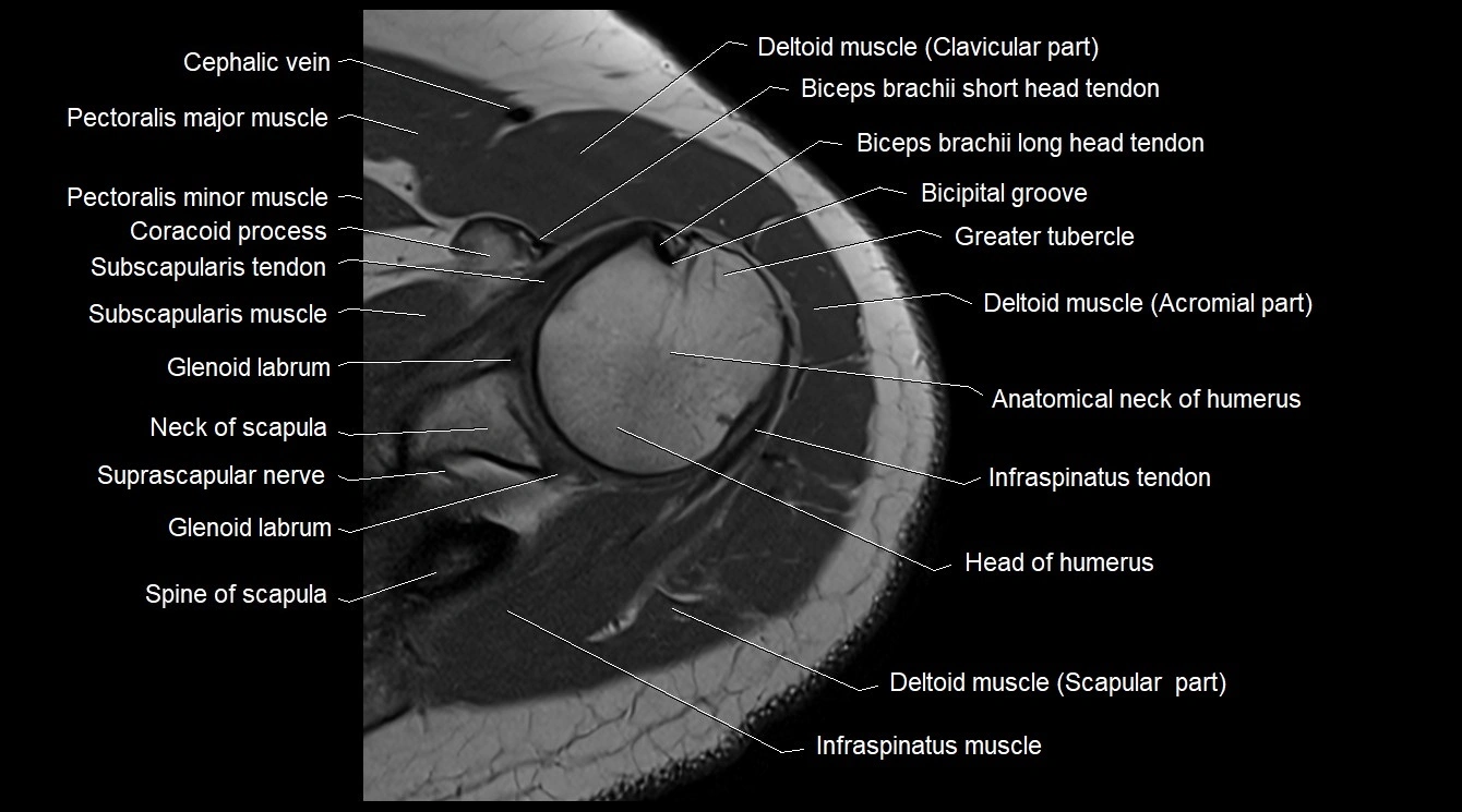
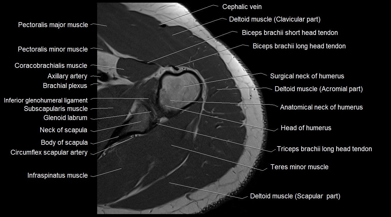
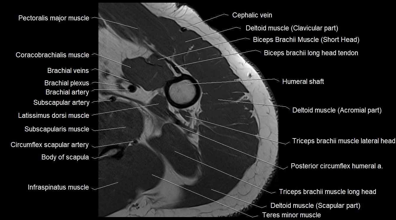
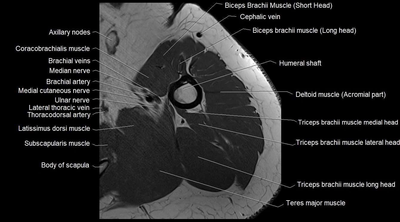
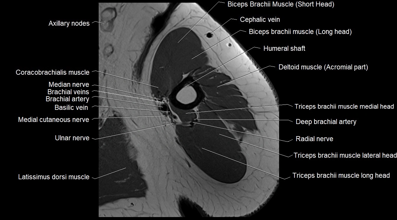
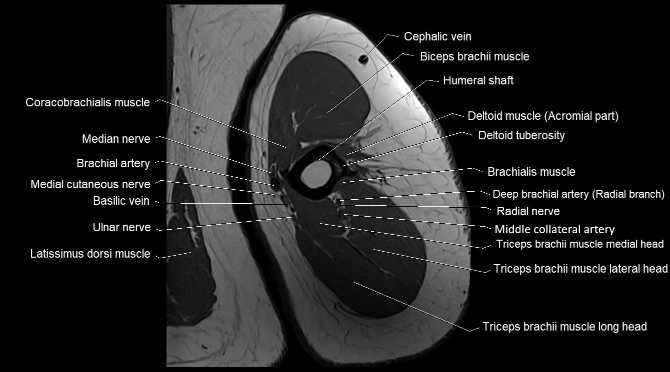
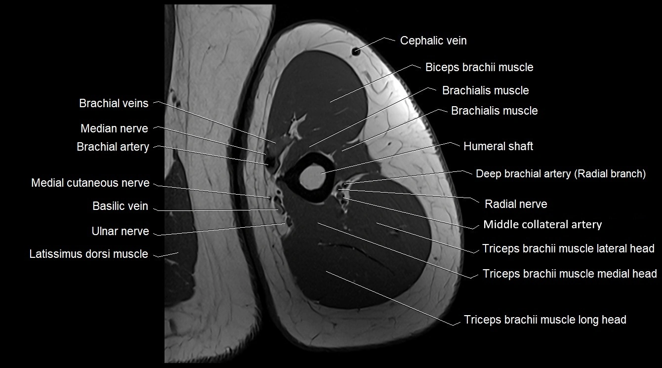
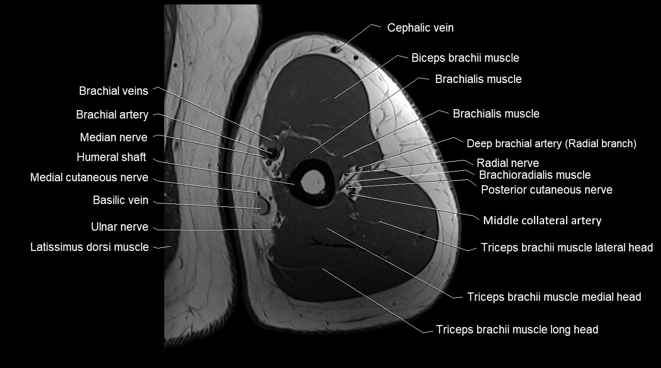
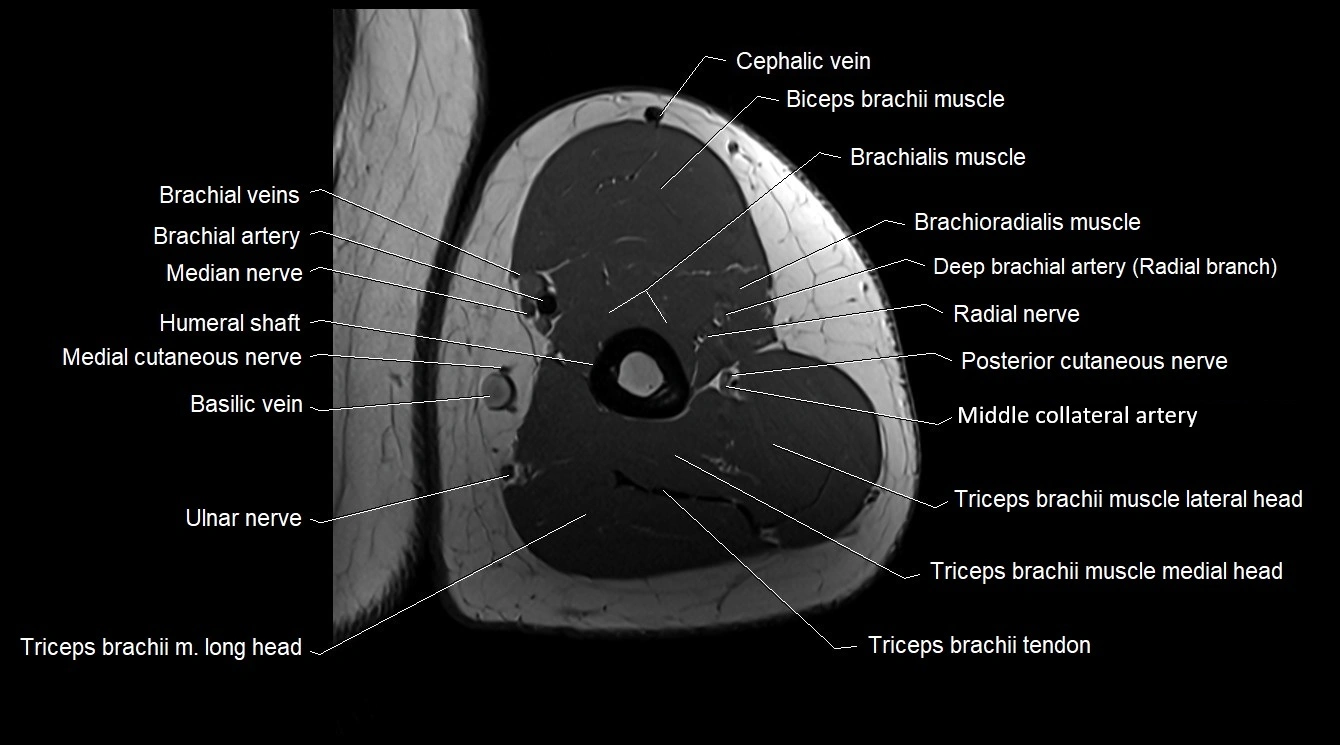
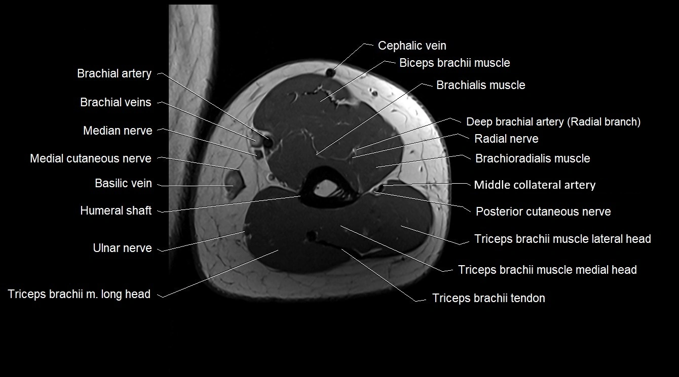
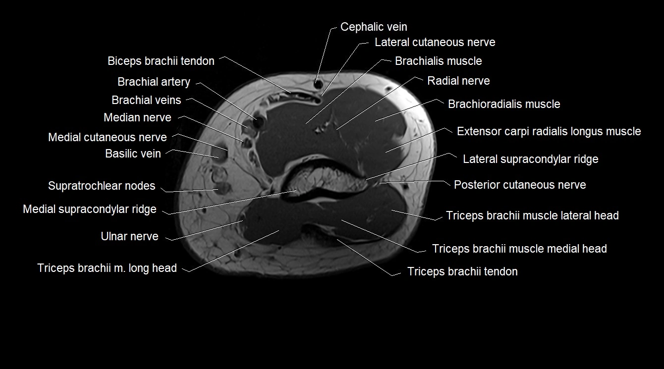
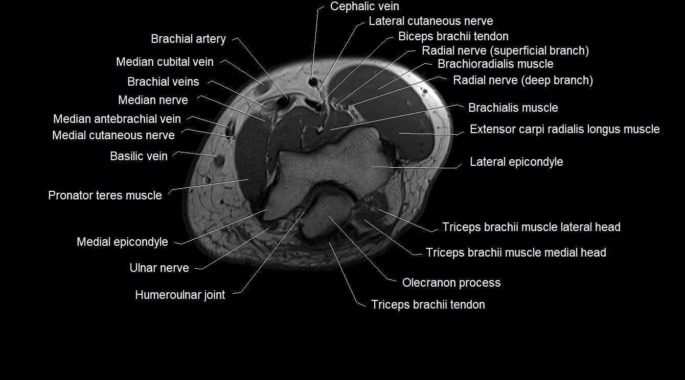
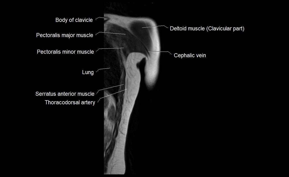
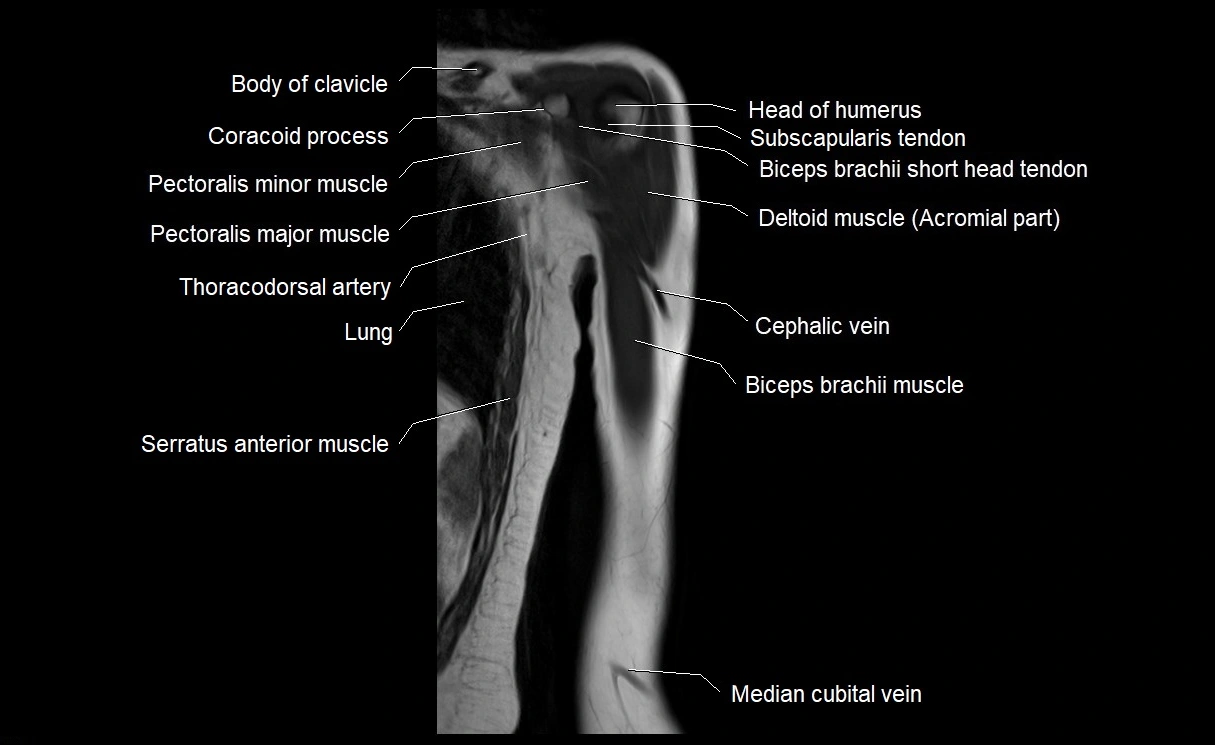
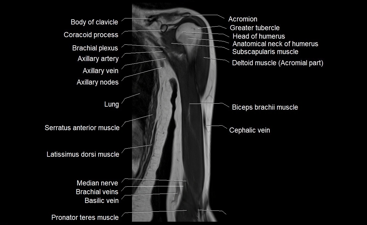
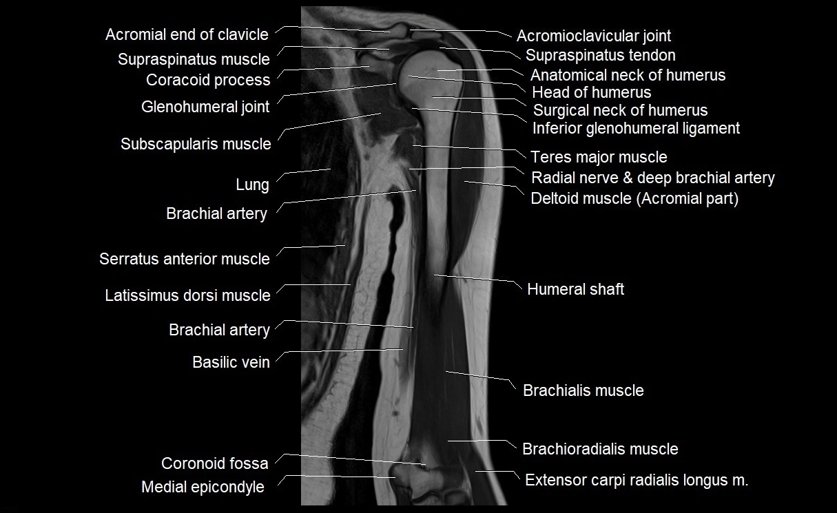
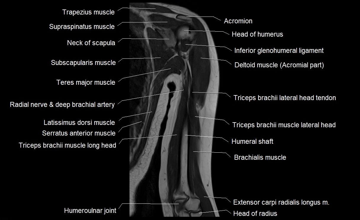
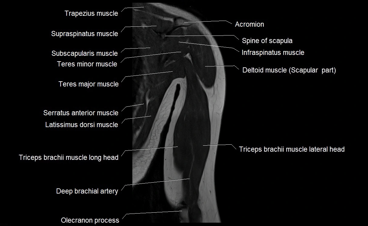
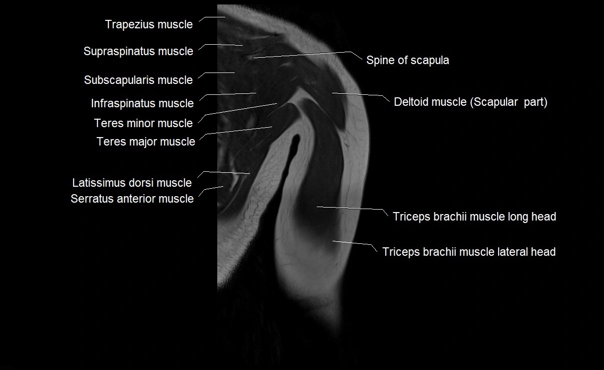
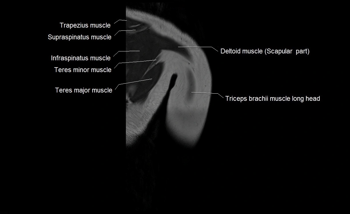
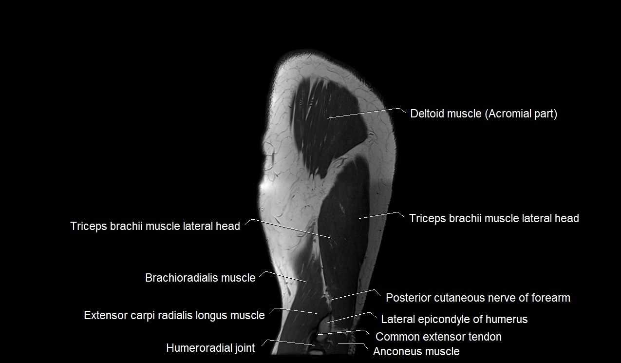
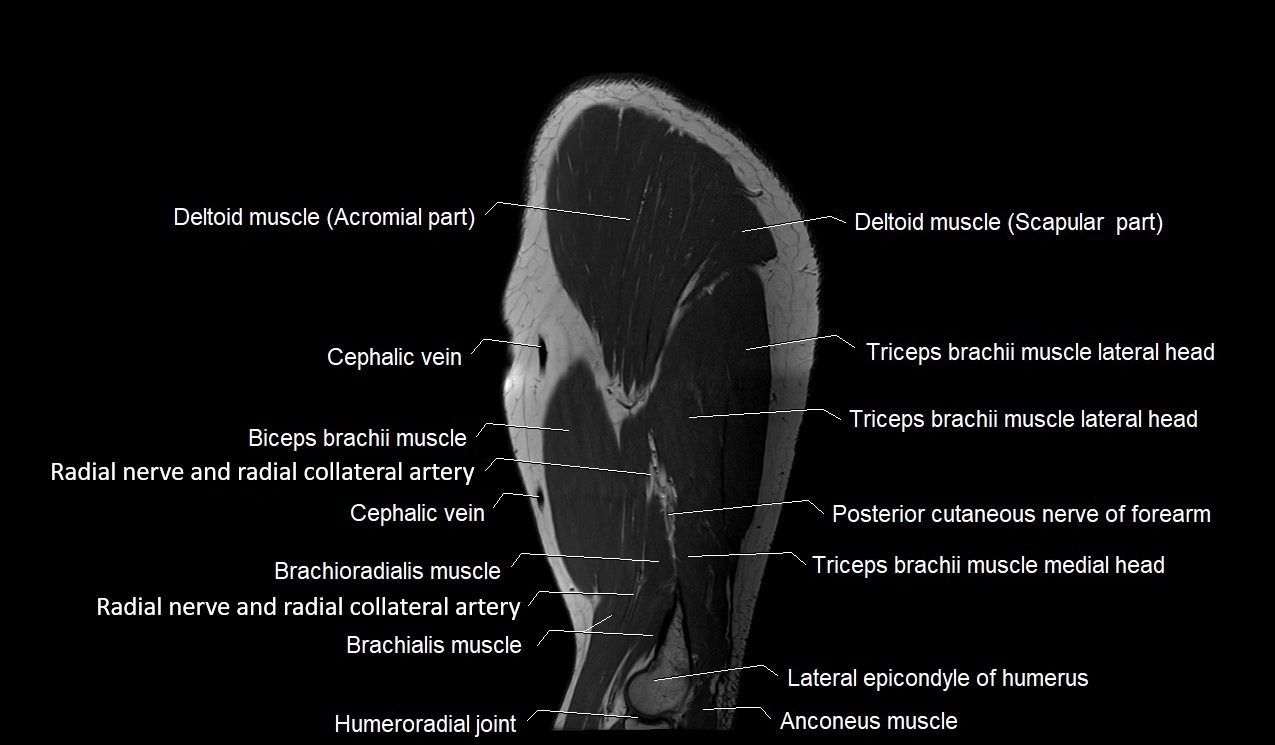
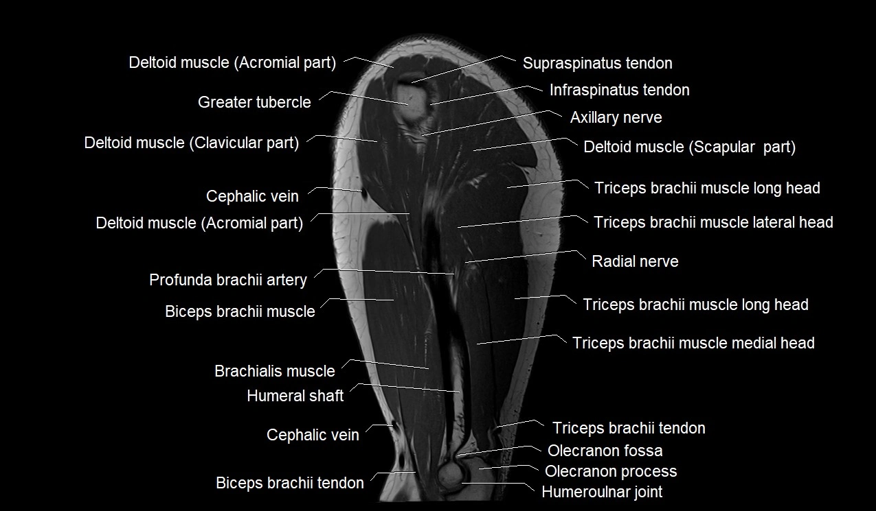
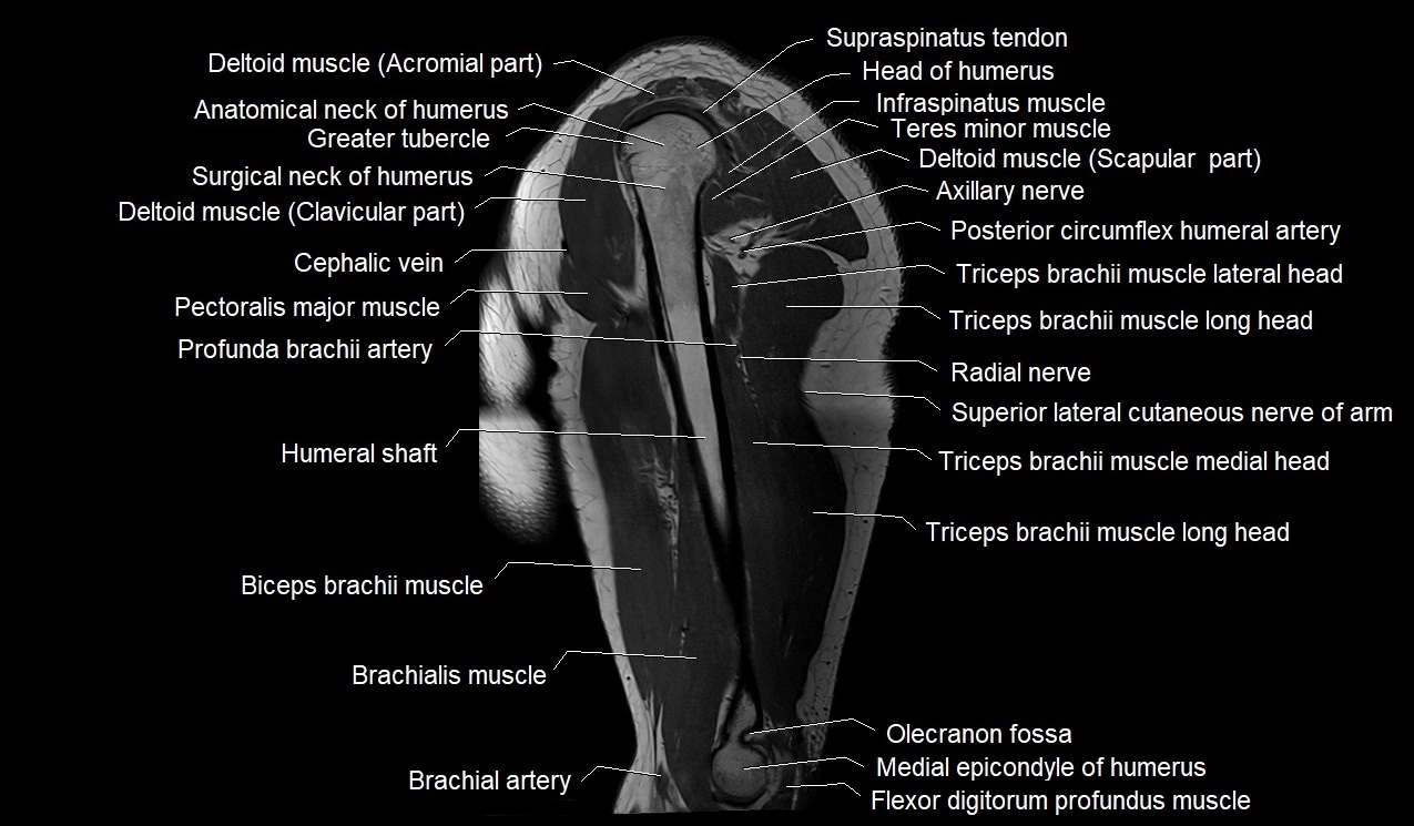
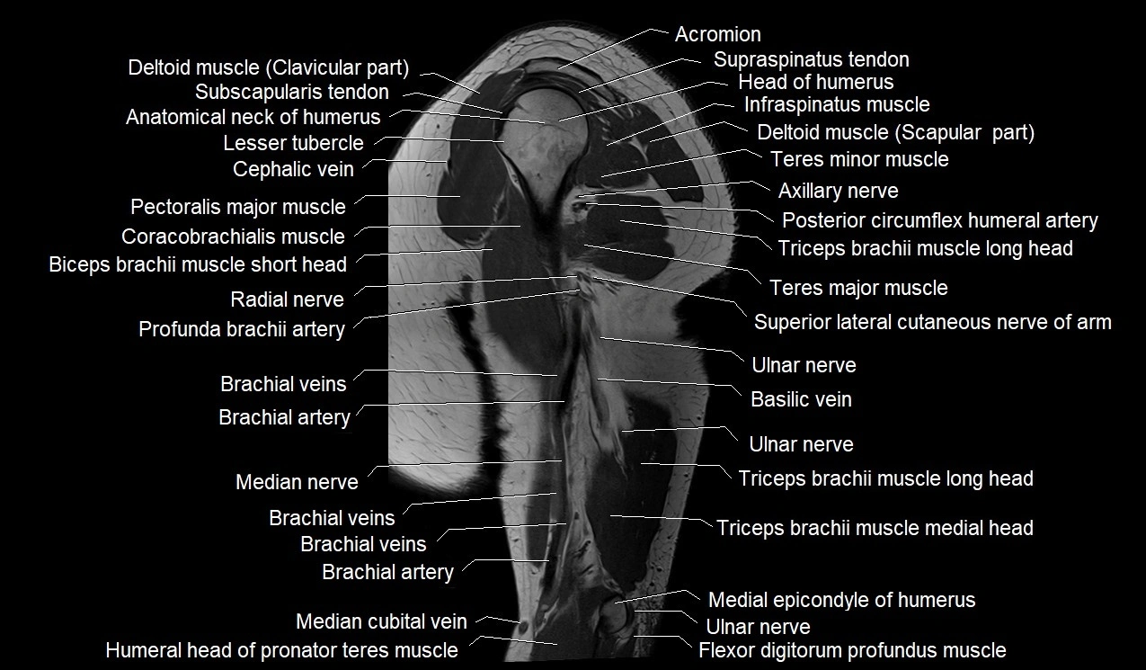
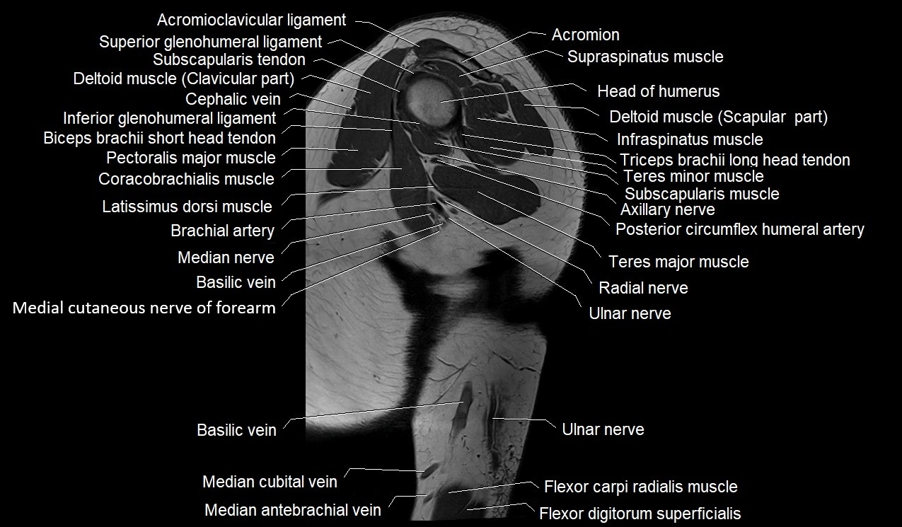
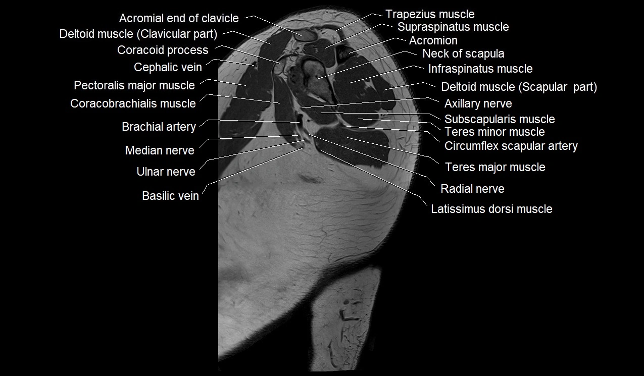
MRI image

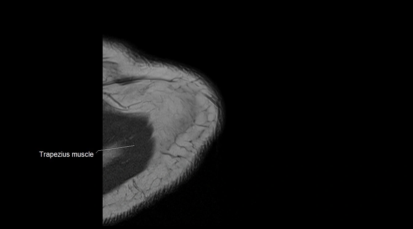
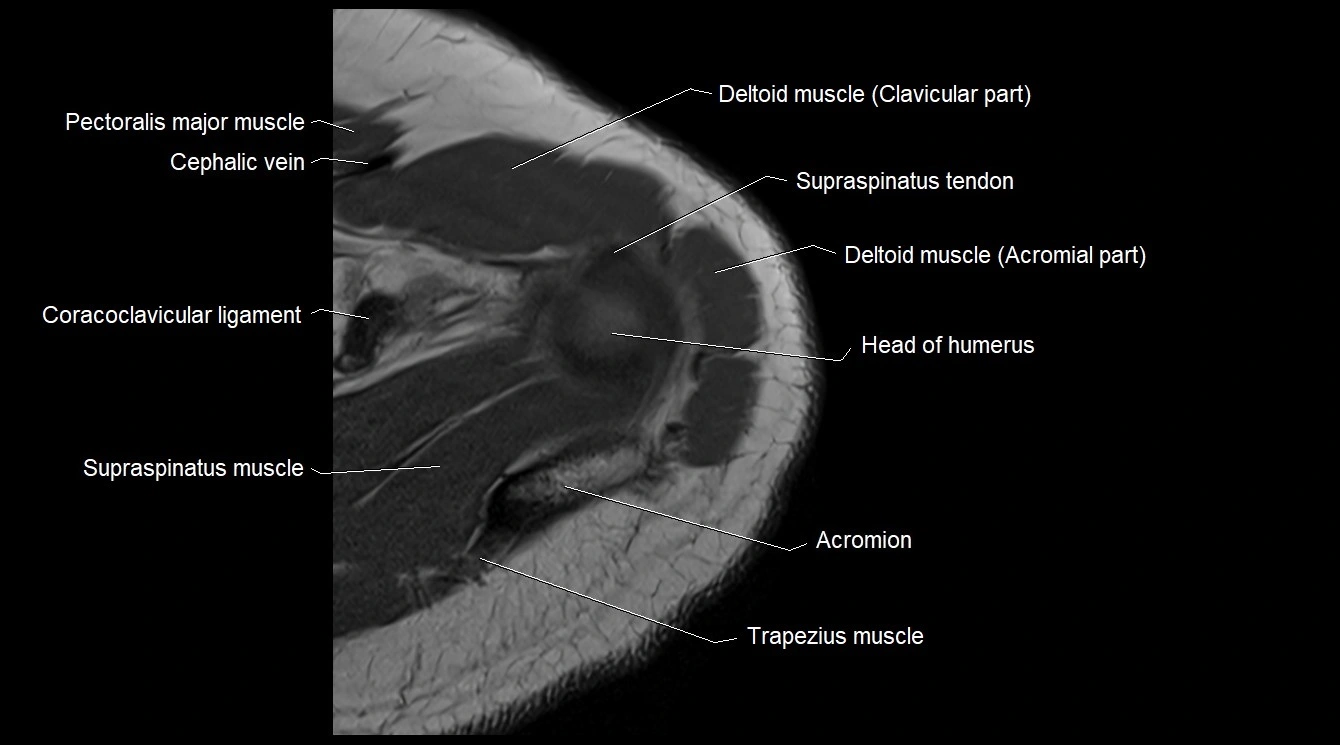
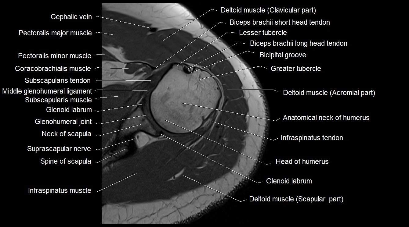
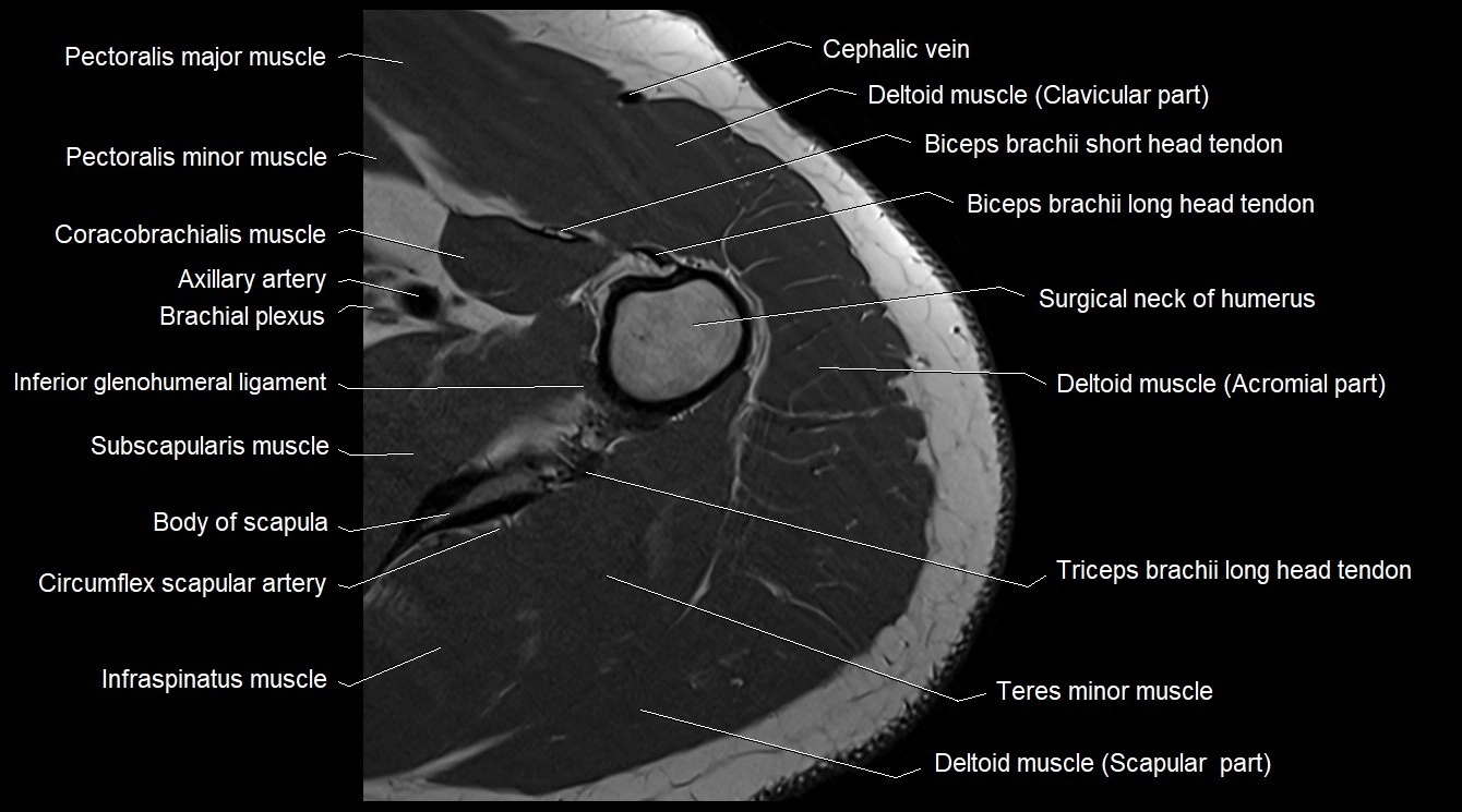
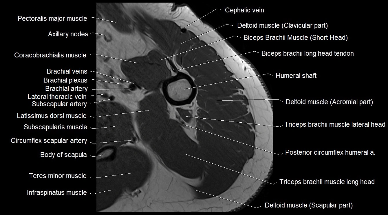
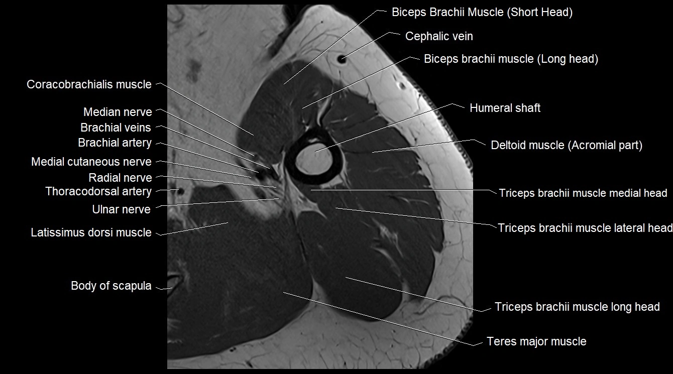
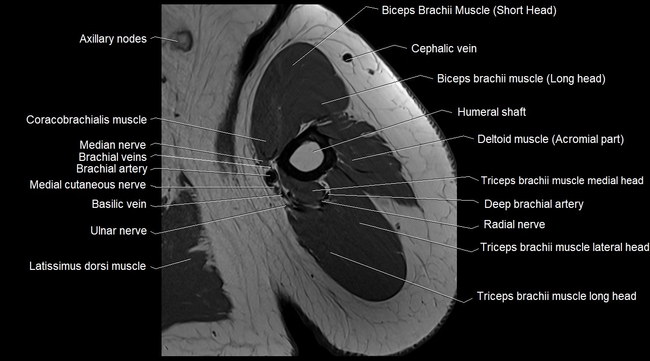
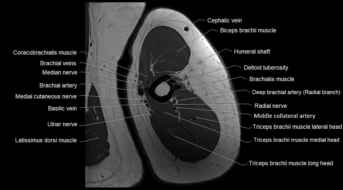
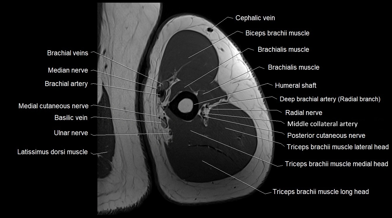
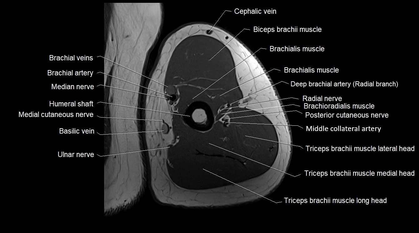
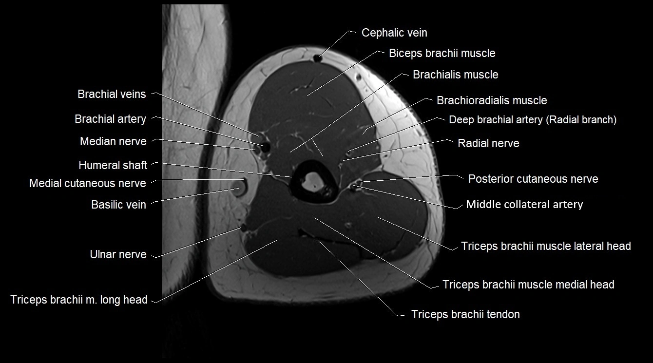
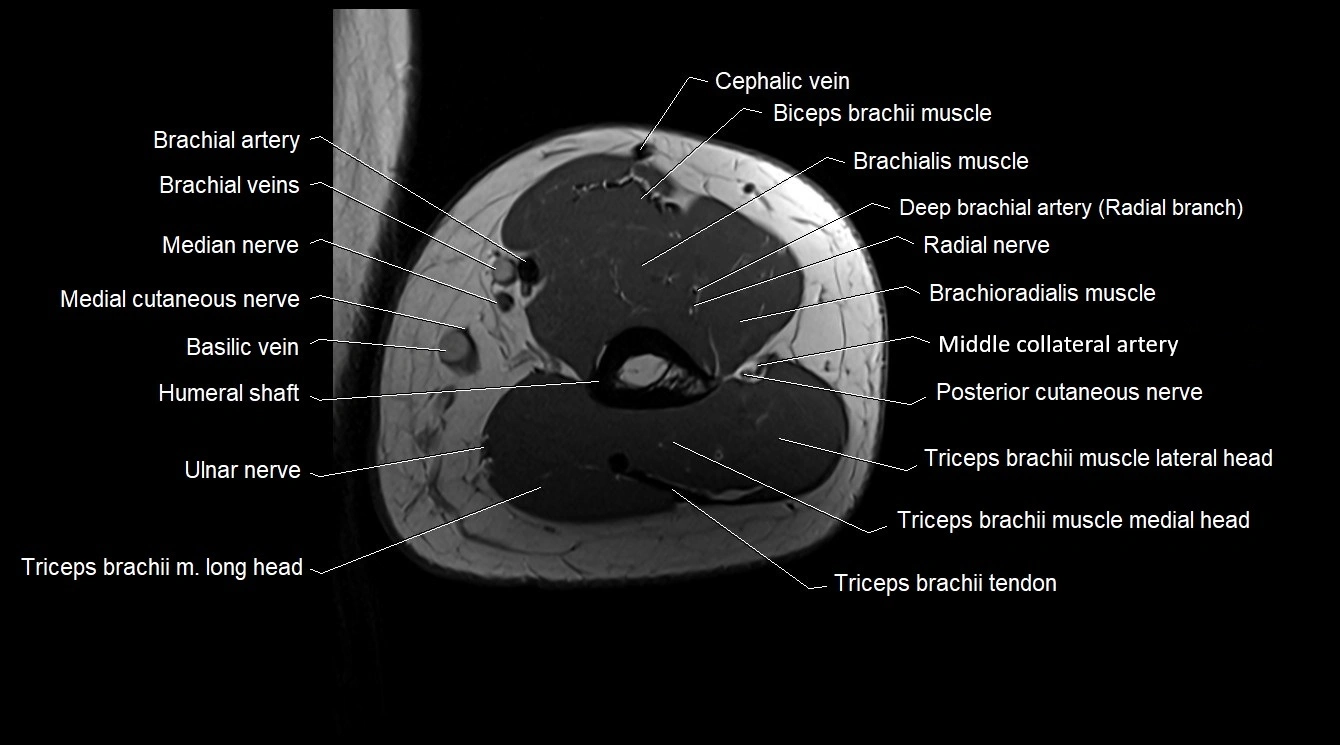
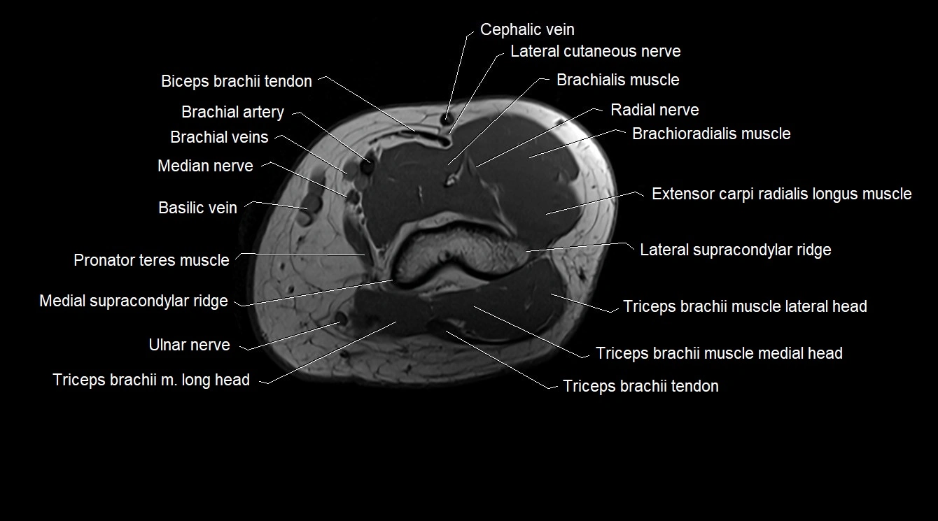
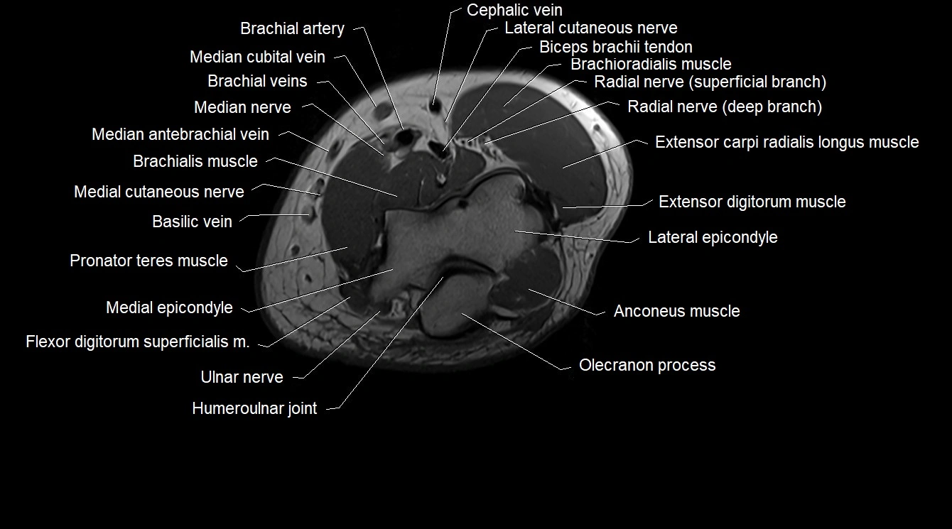
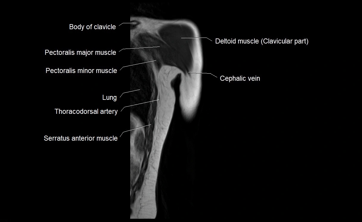
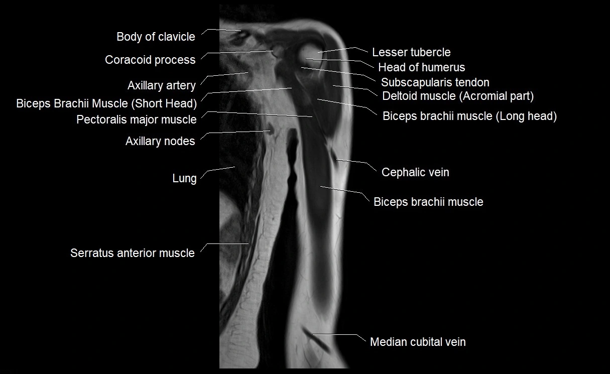
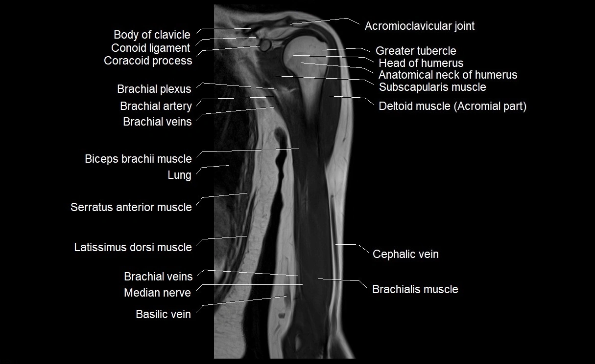
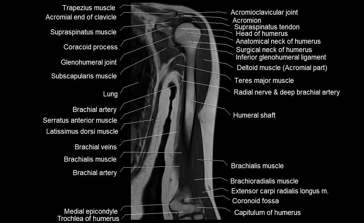
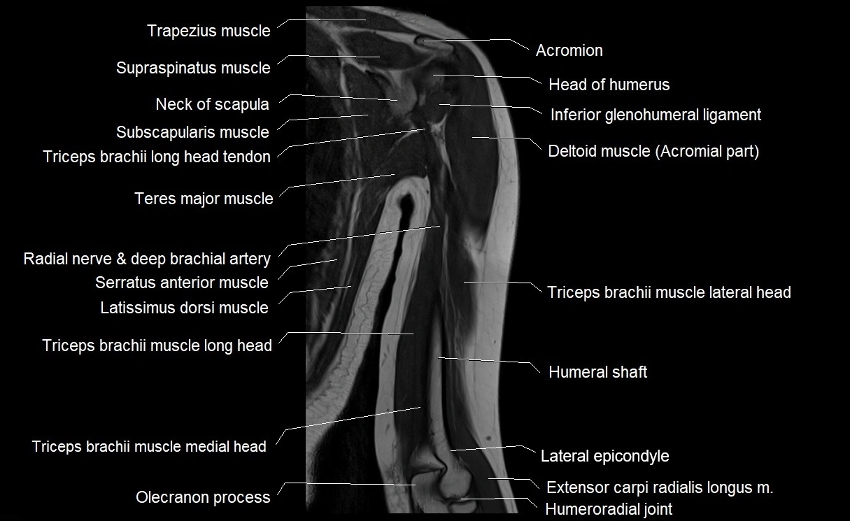
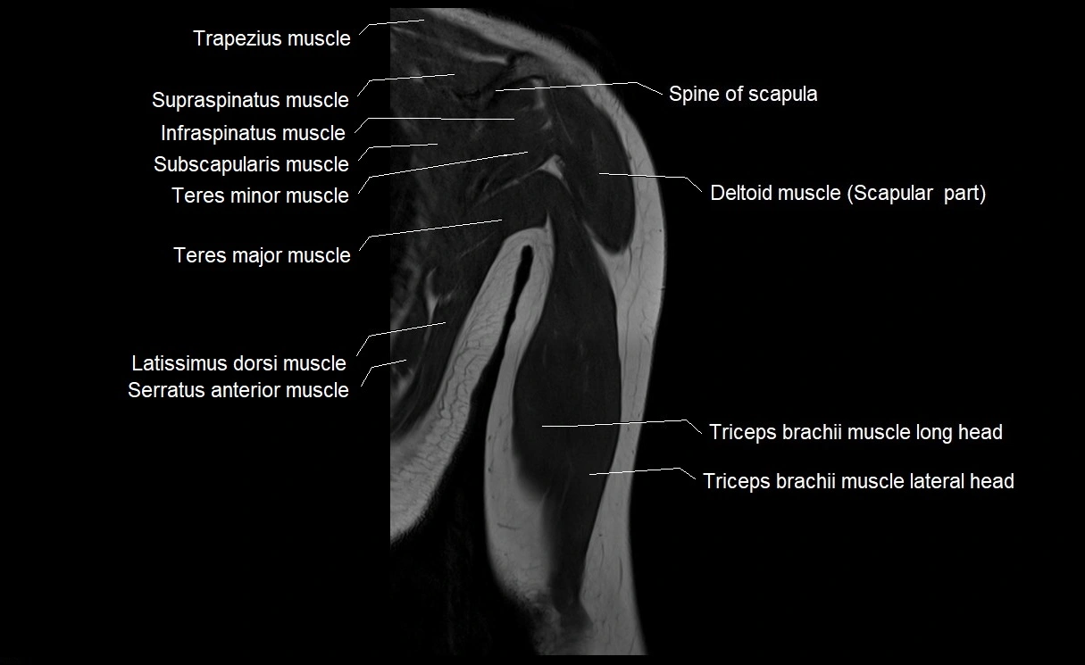
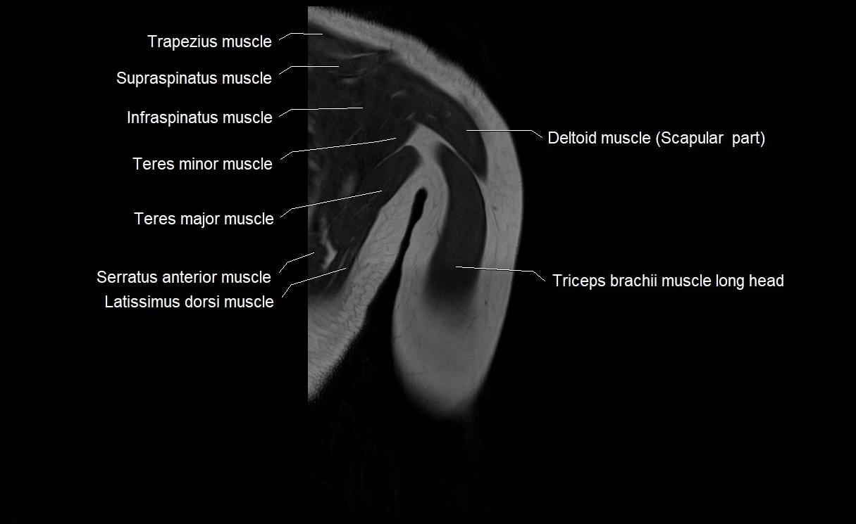
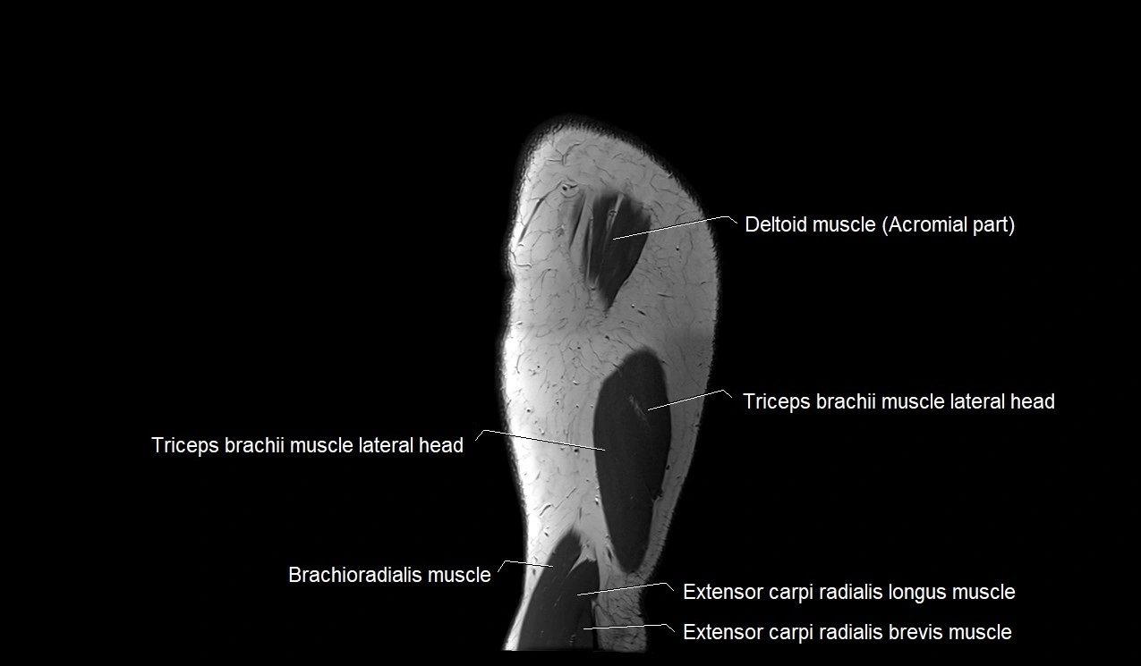
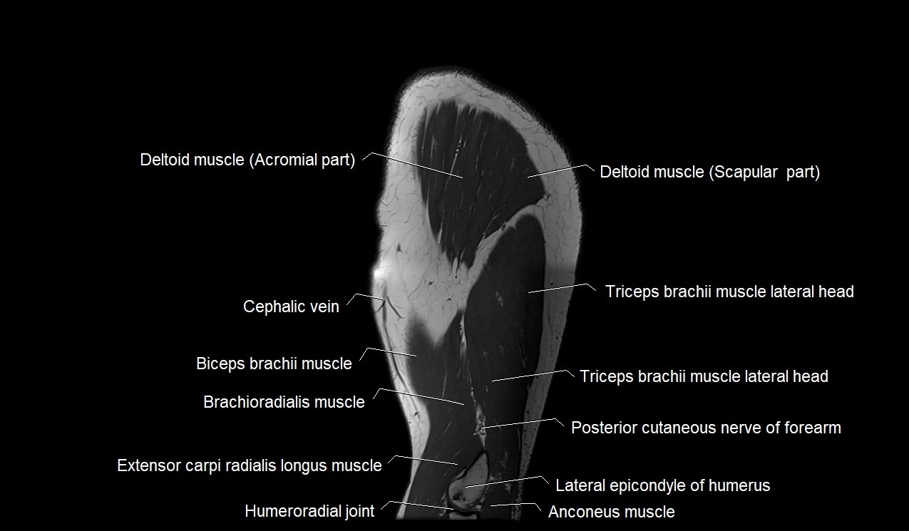
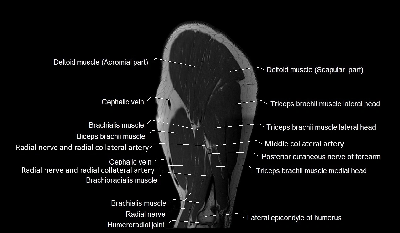
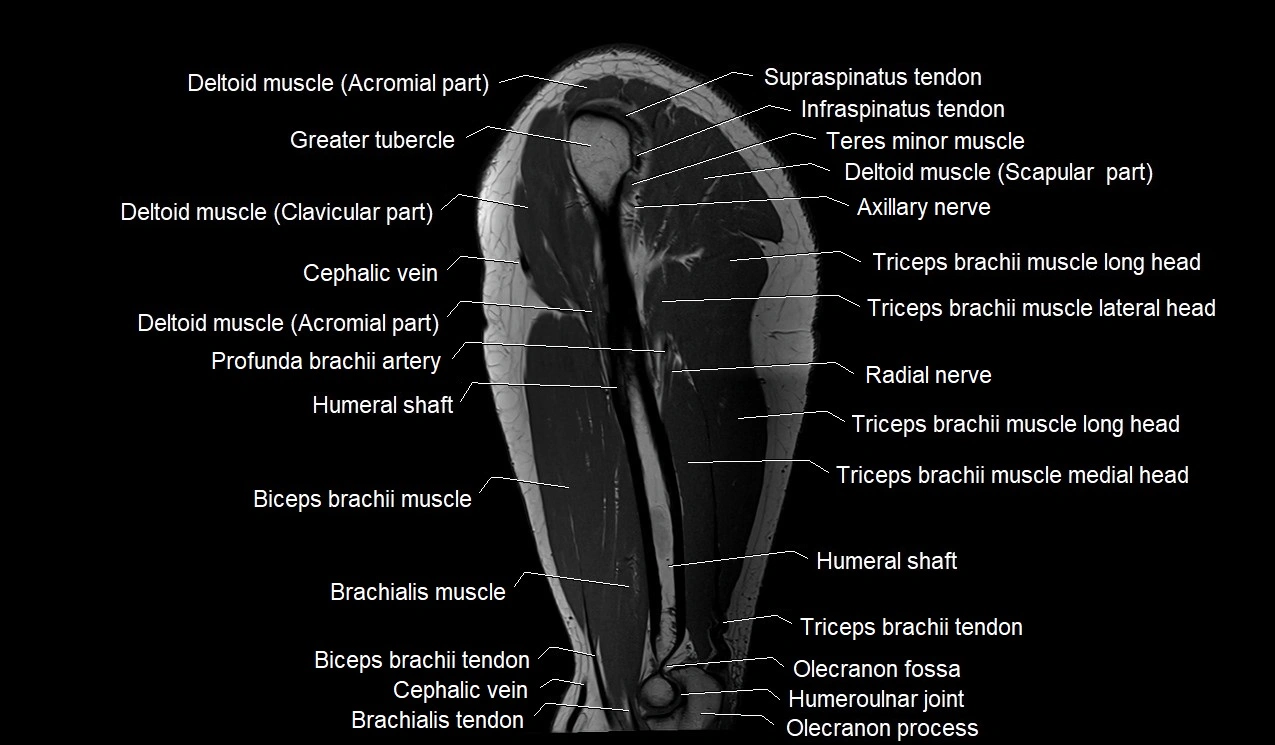
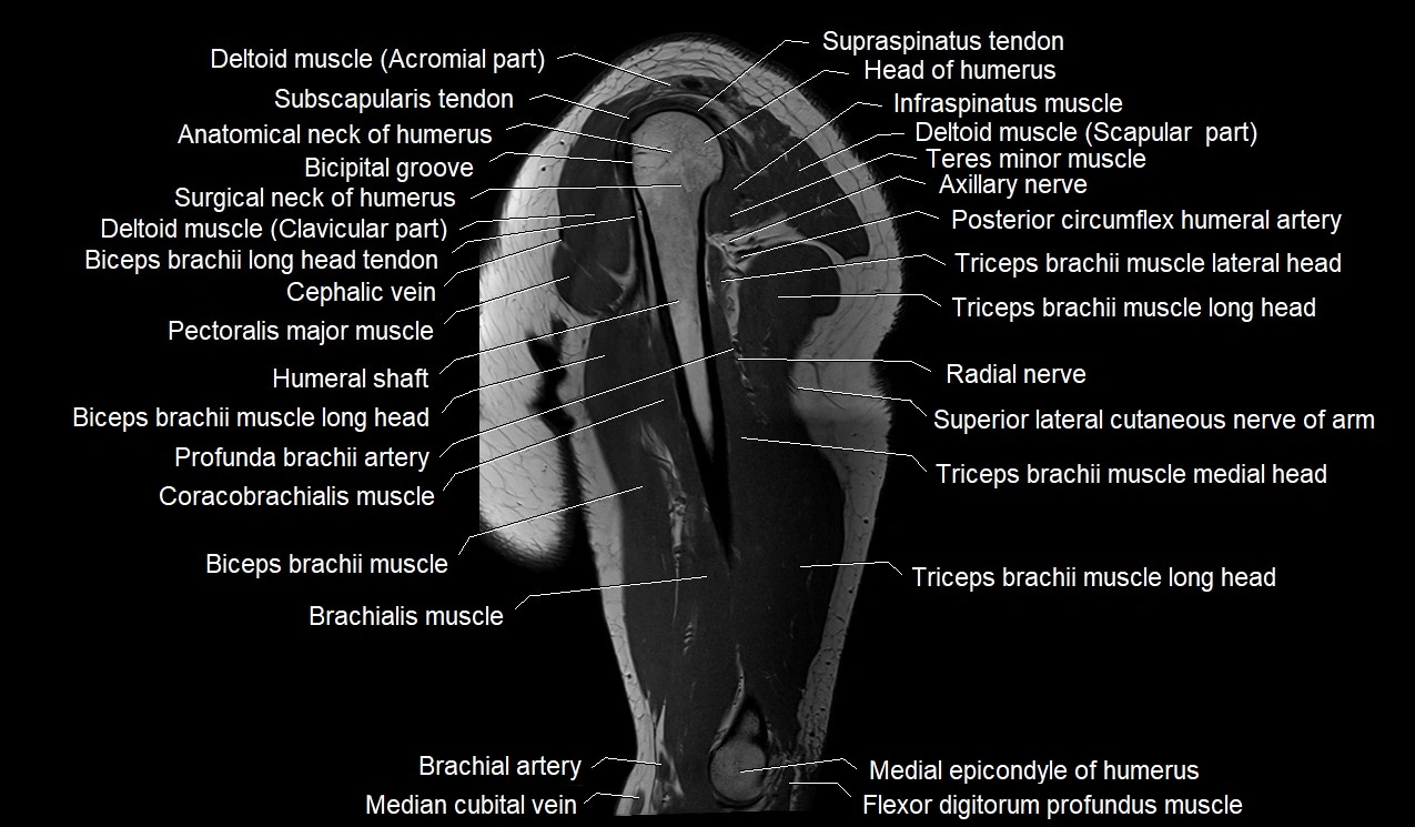
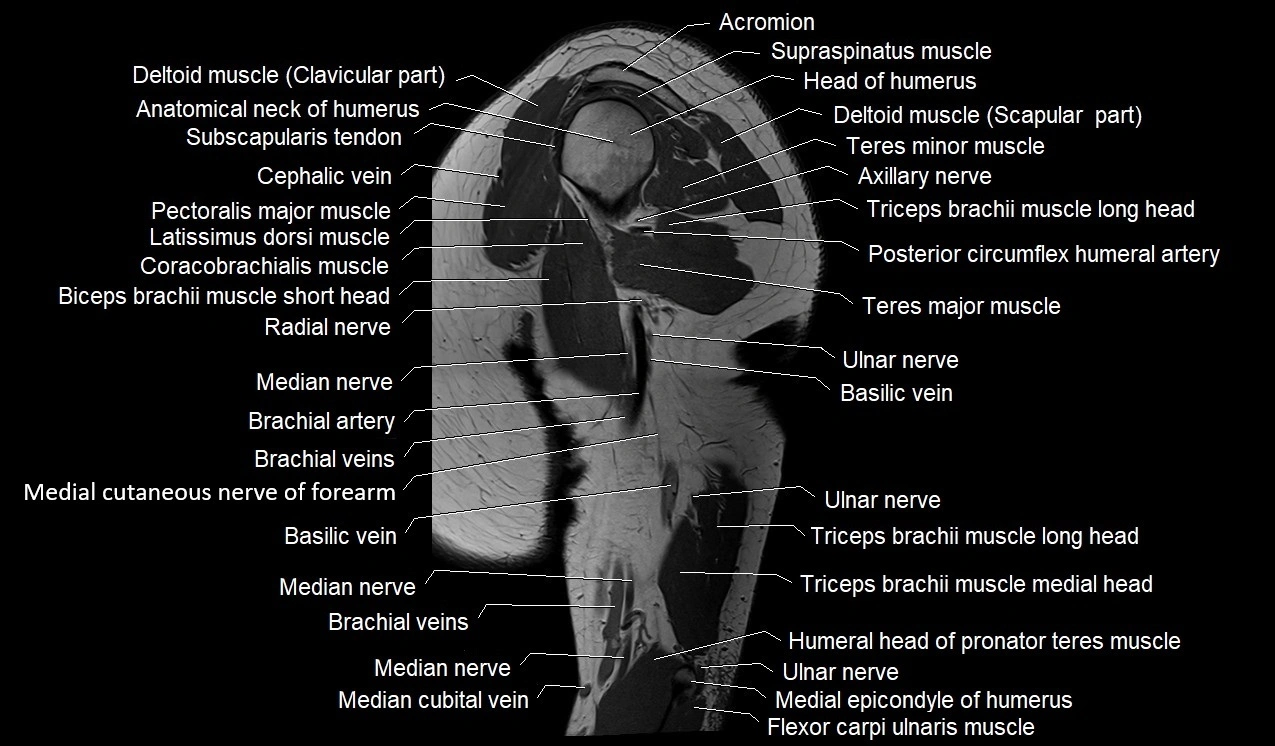
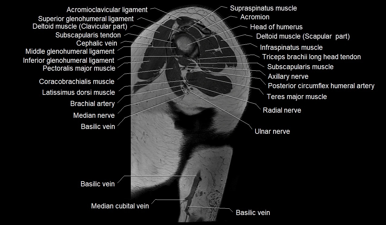
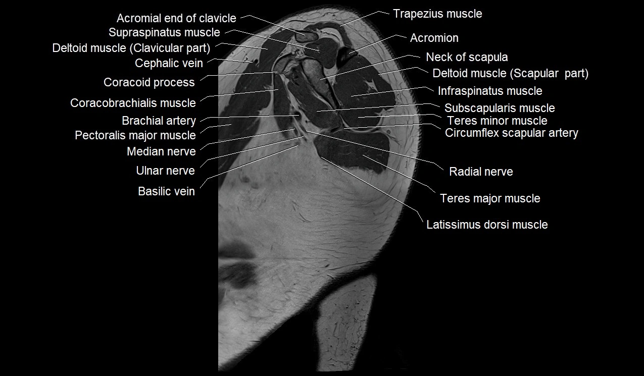
MRI image

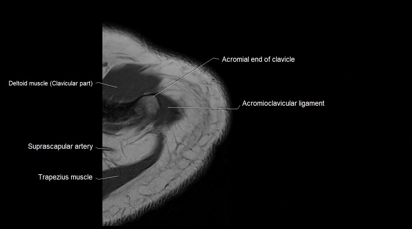
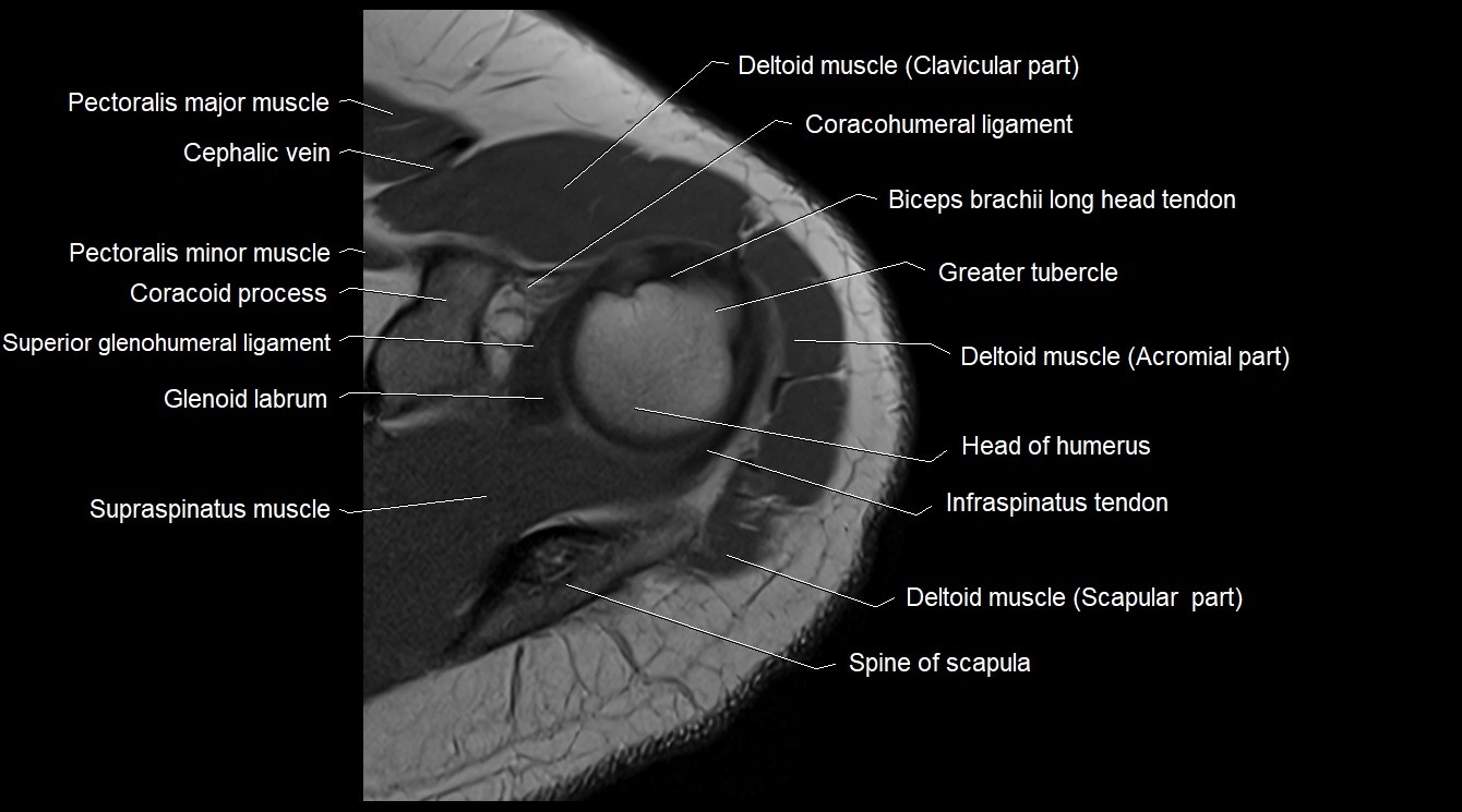
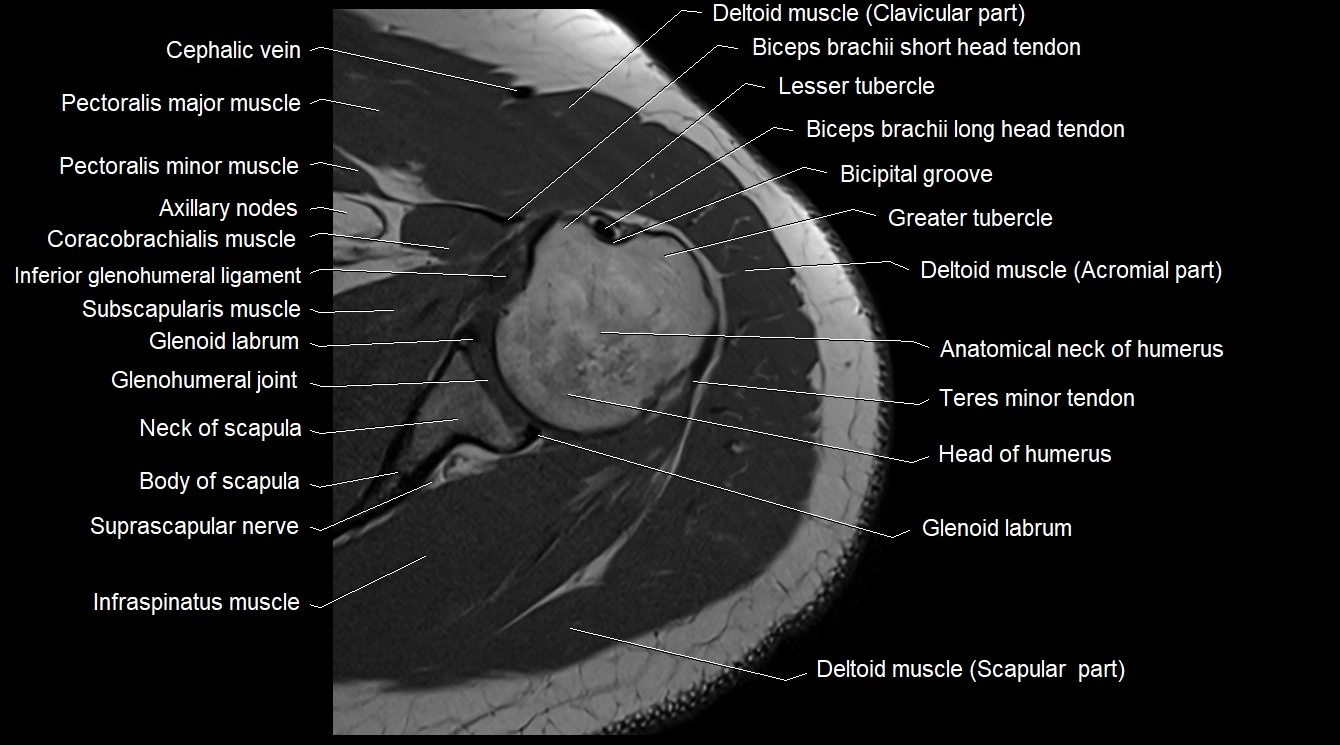
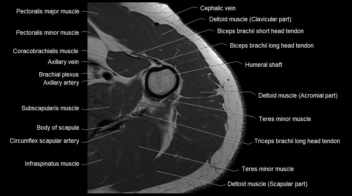
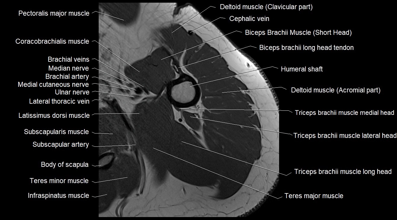
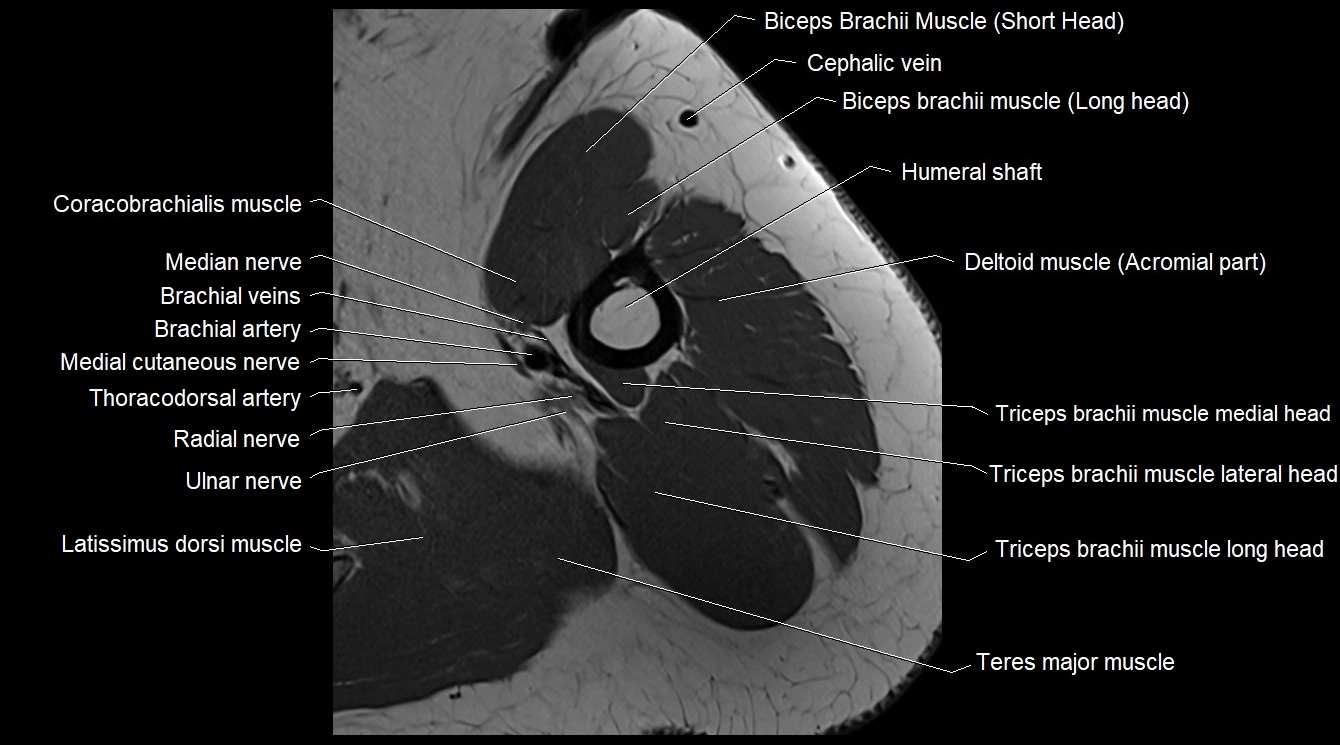
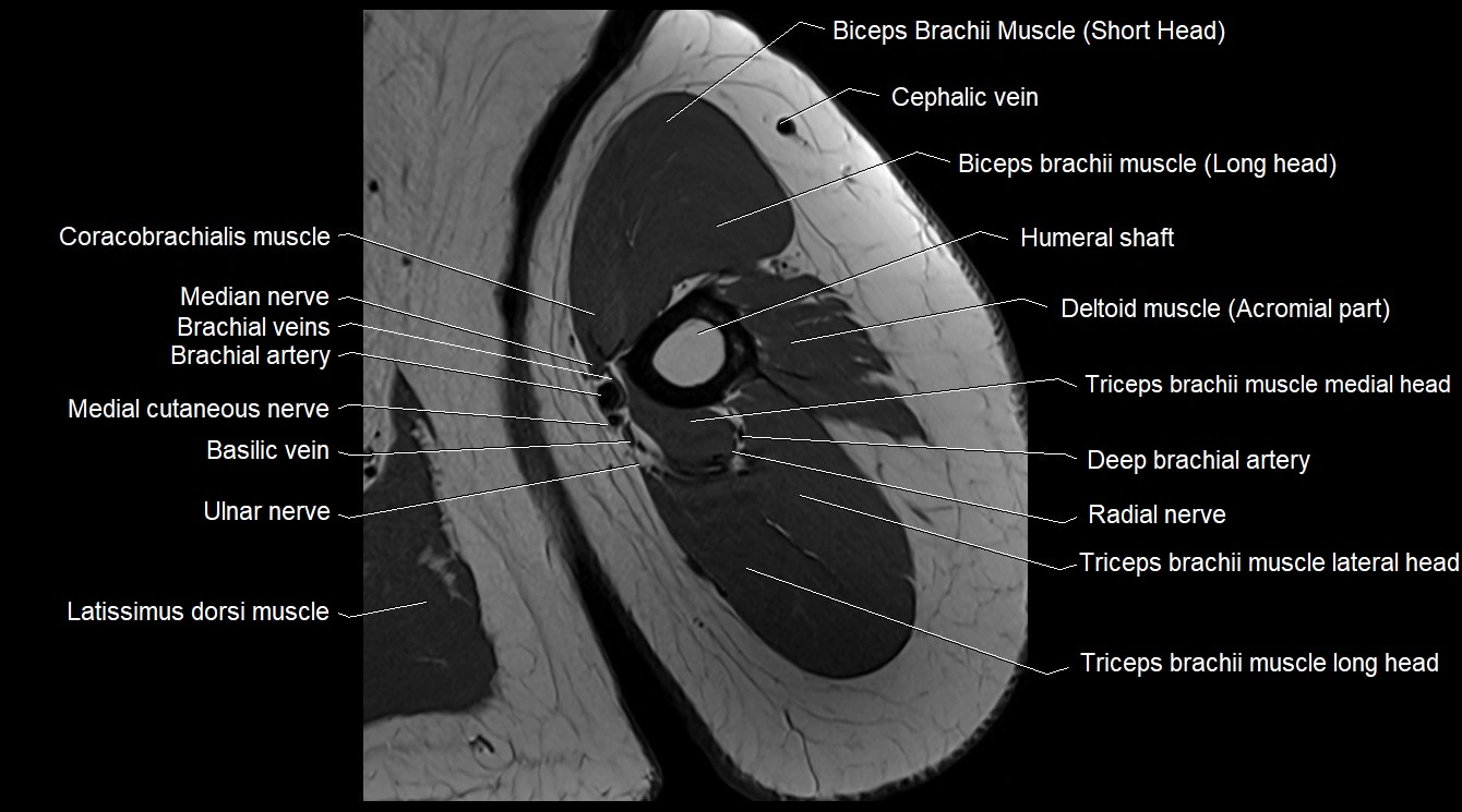
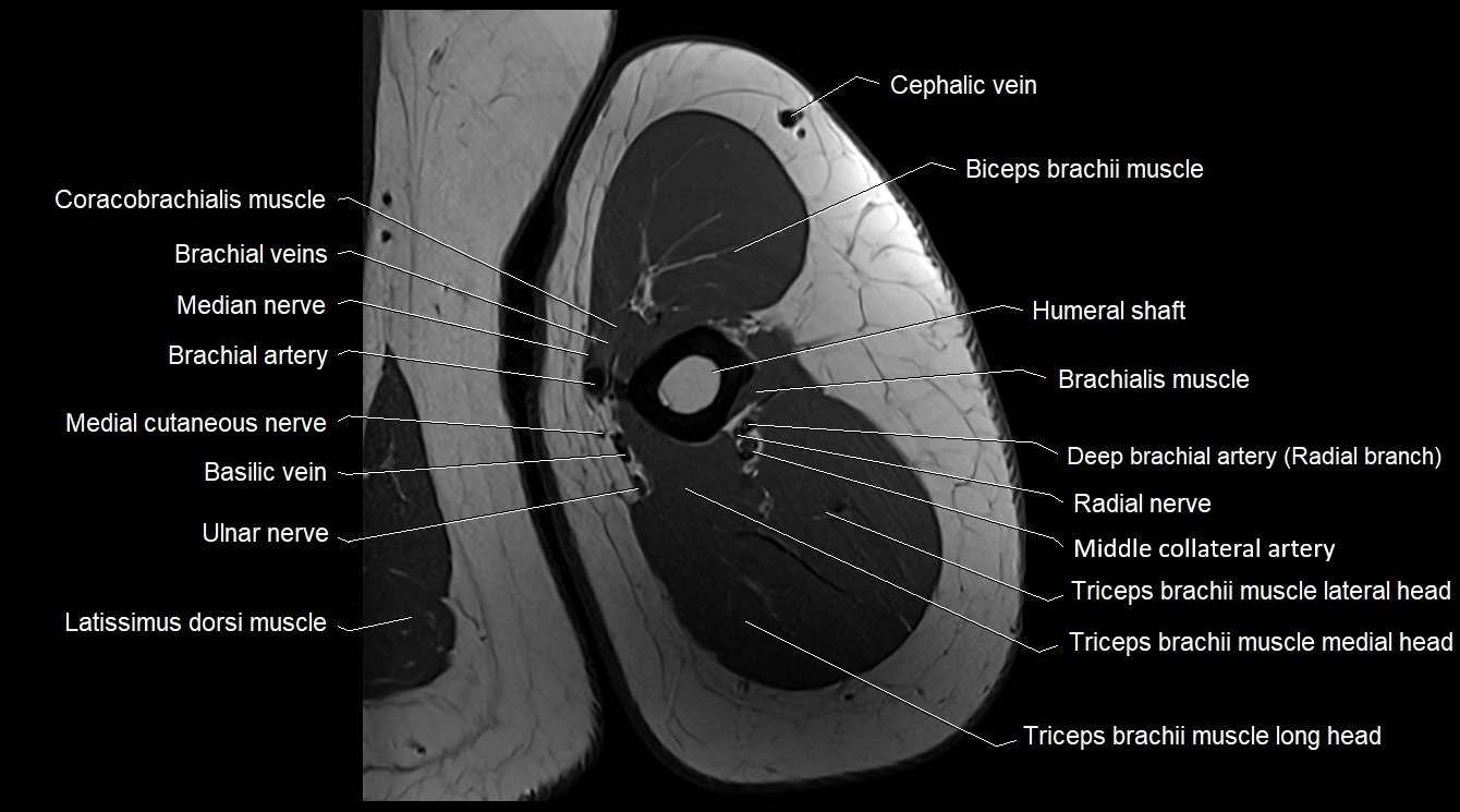
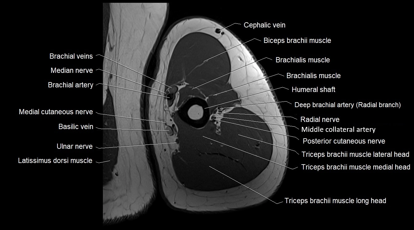
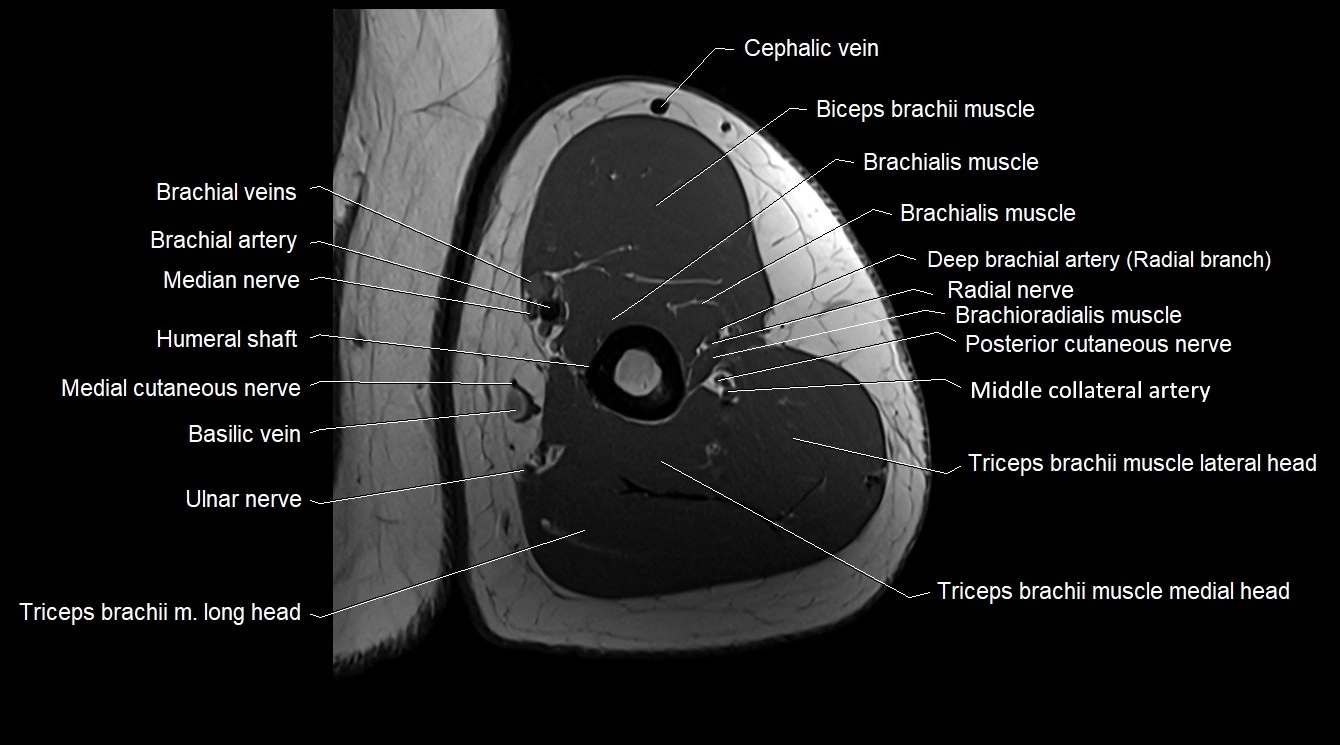
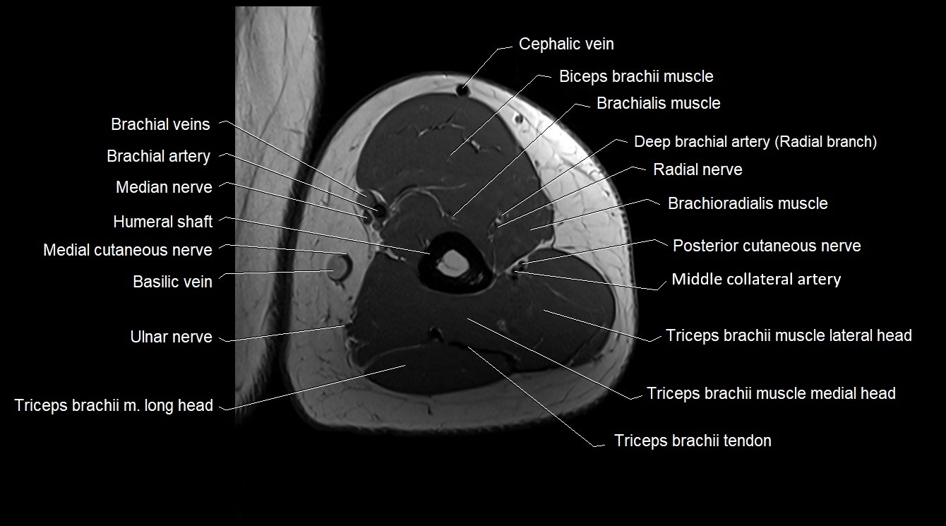
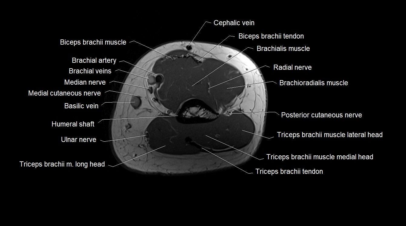
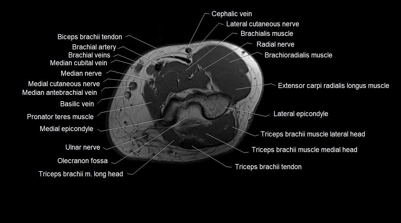
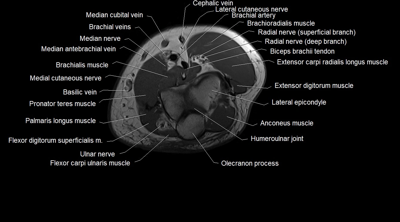
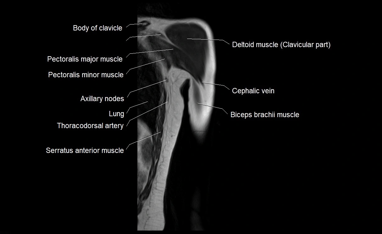
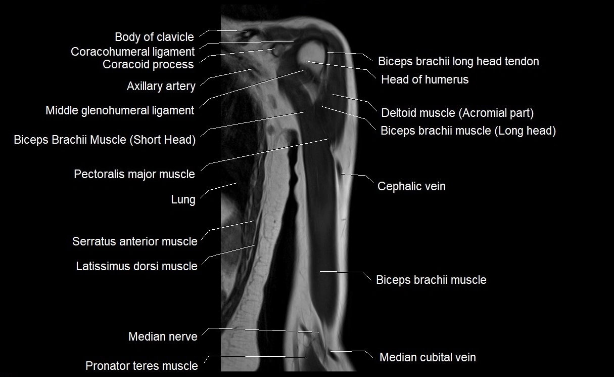
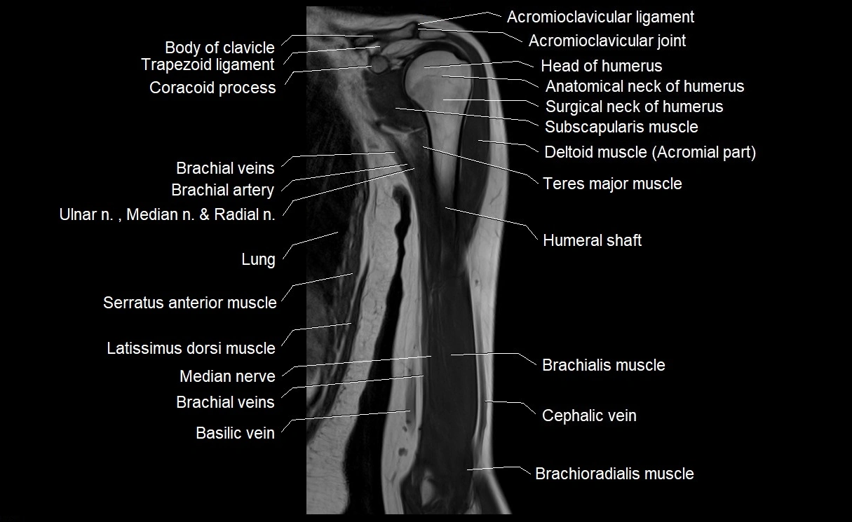
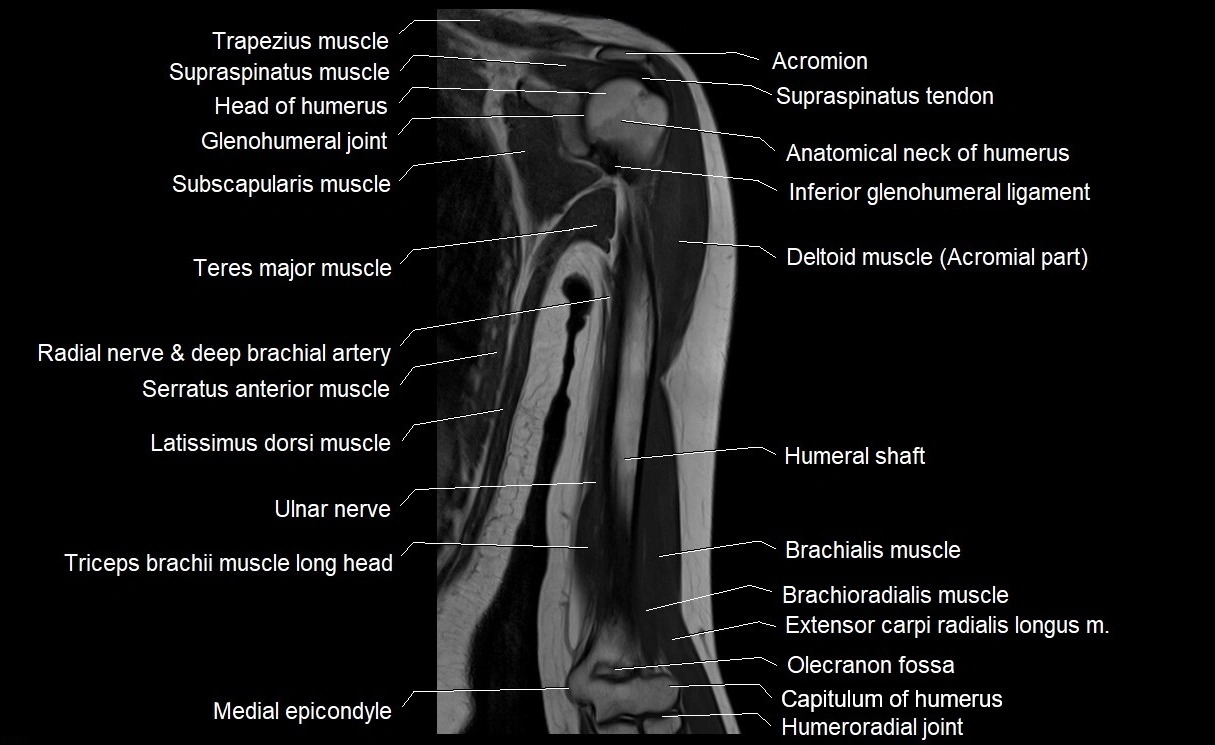
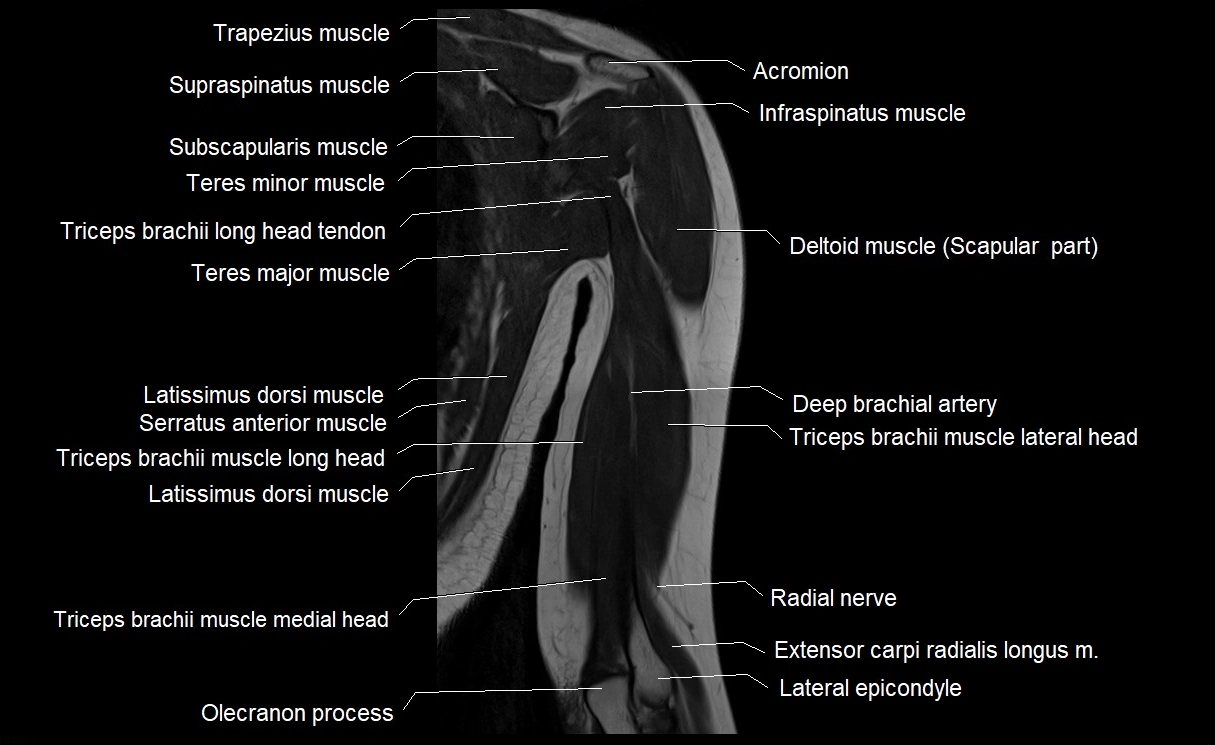
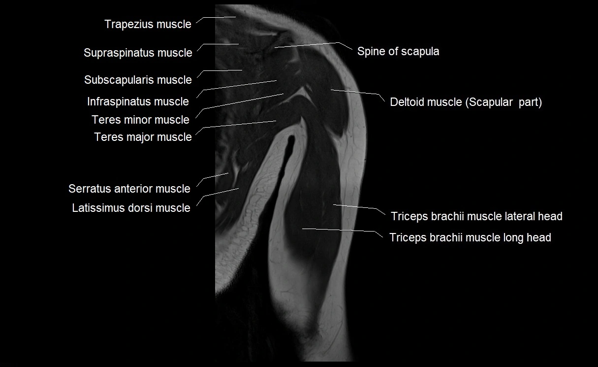
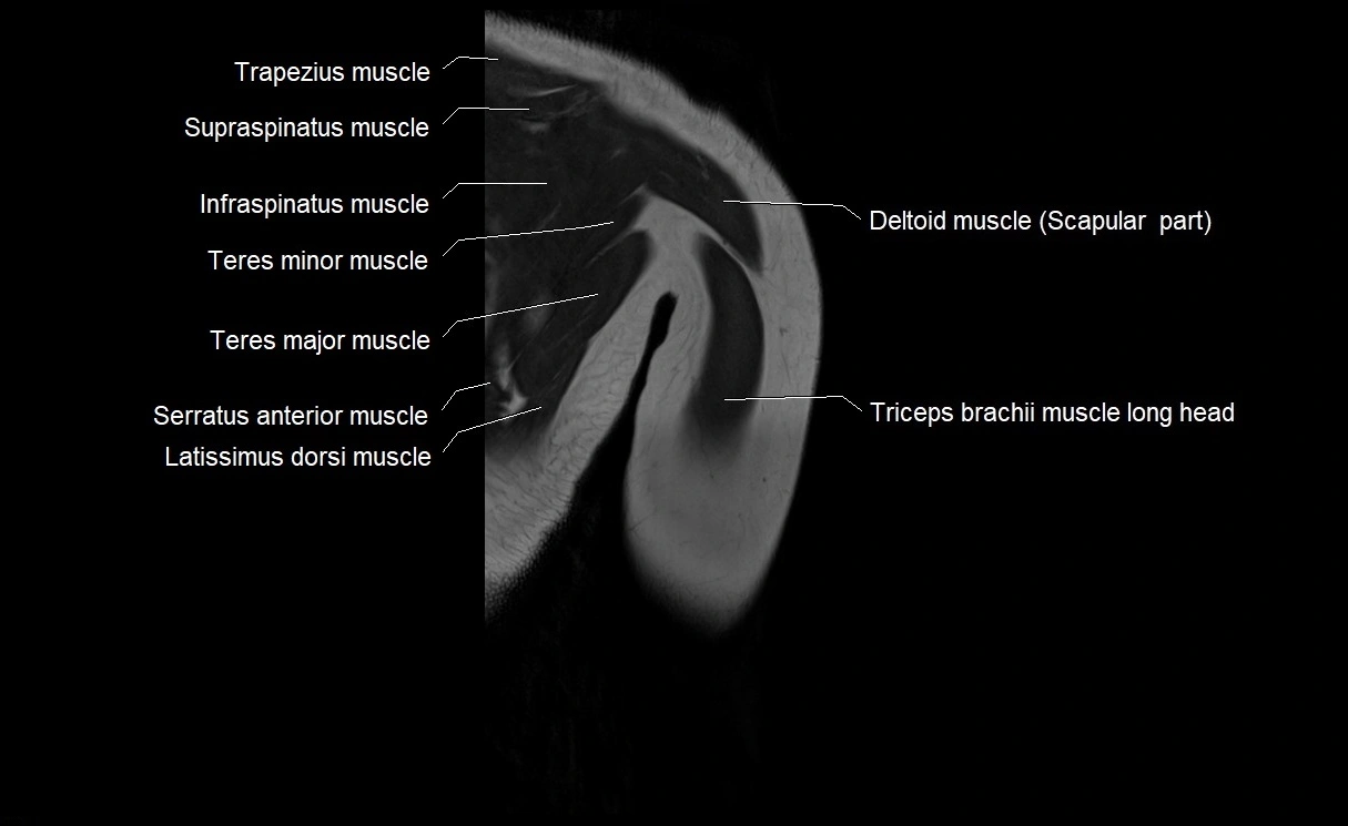
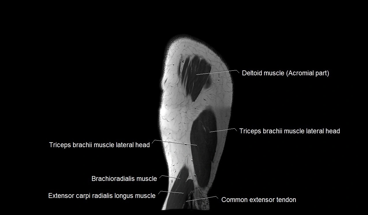
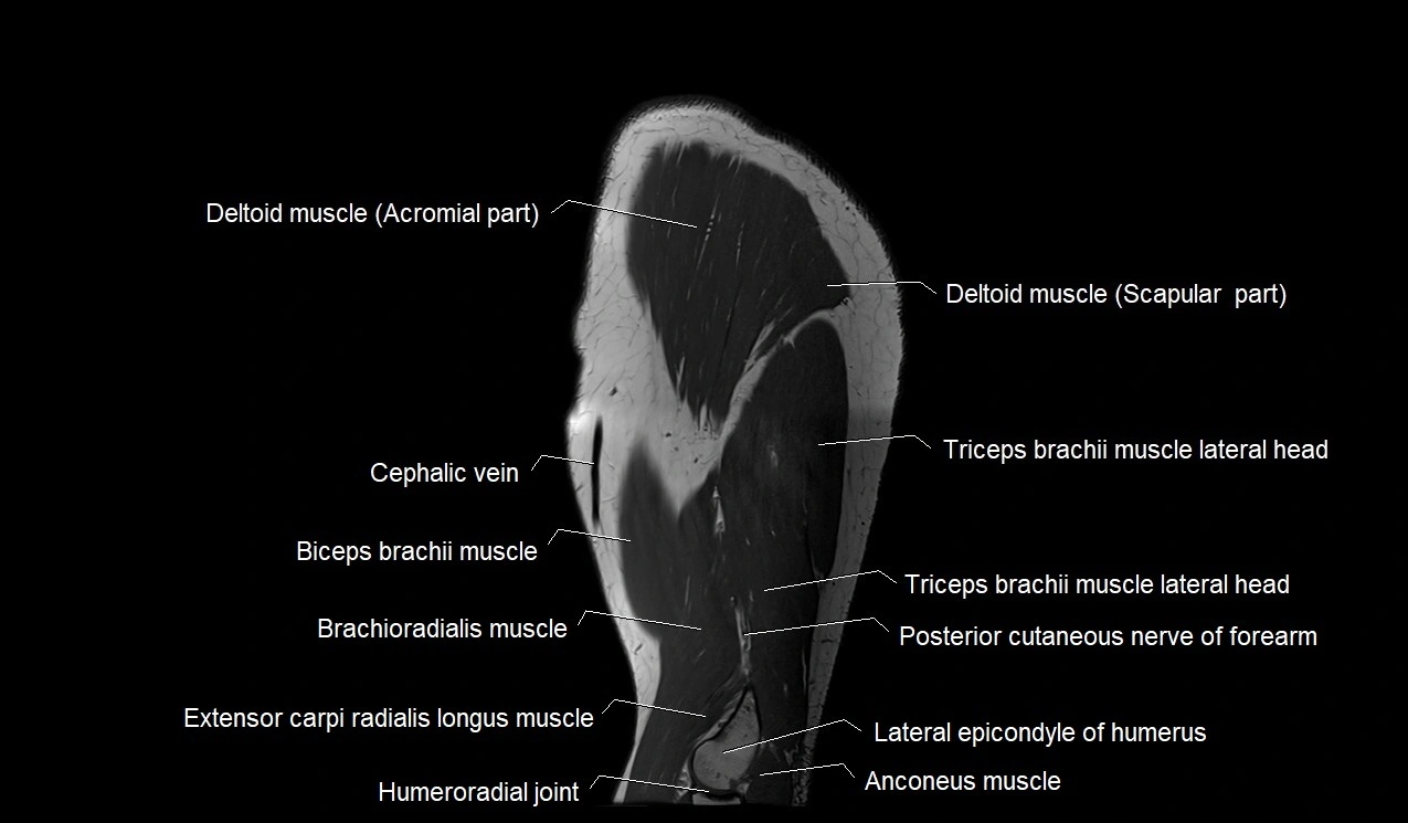
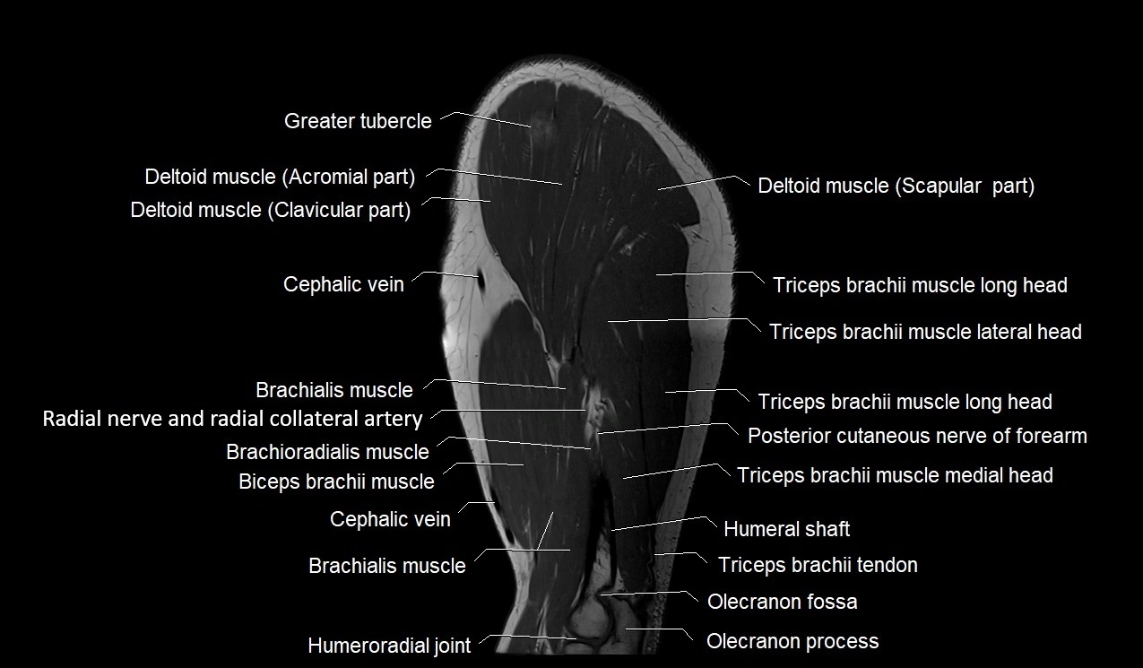
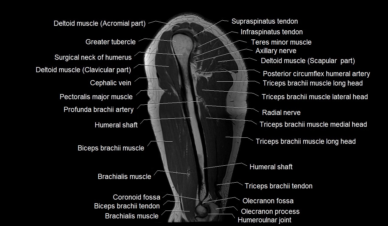
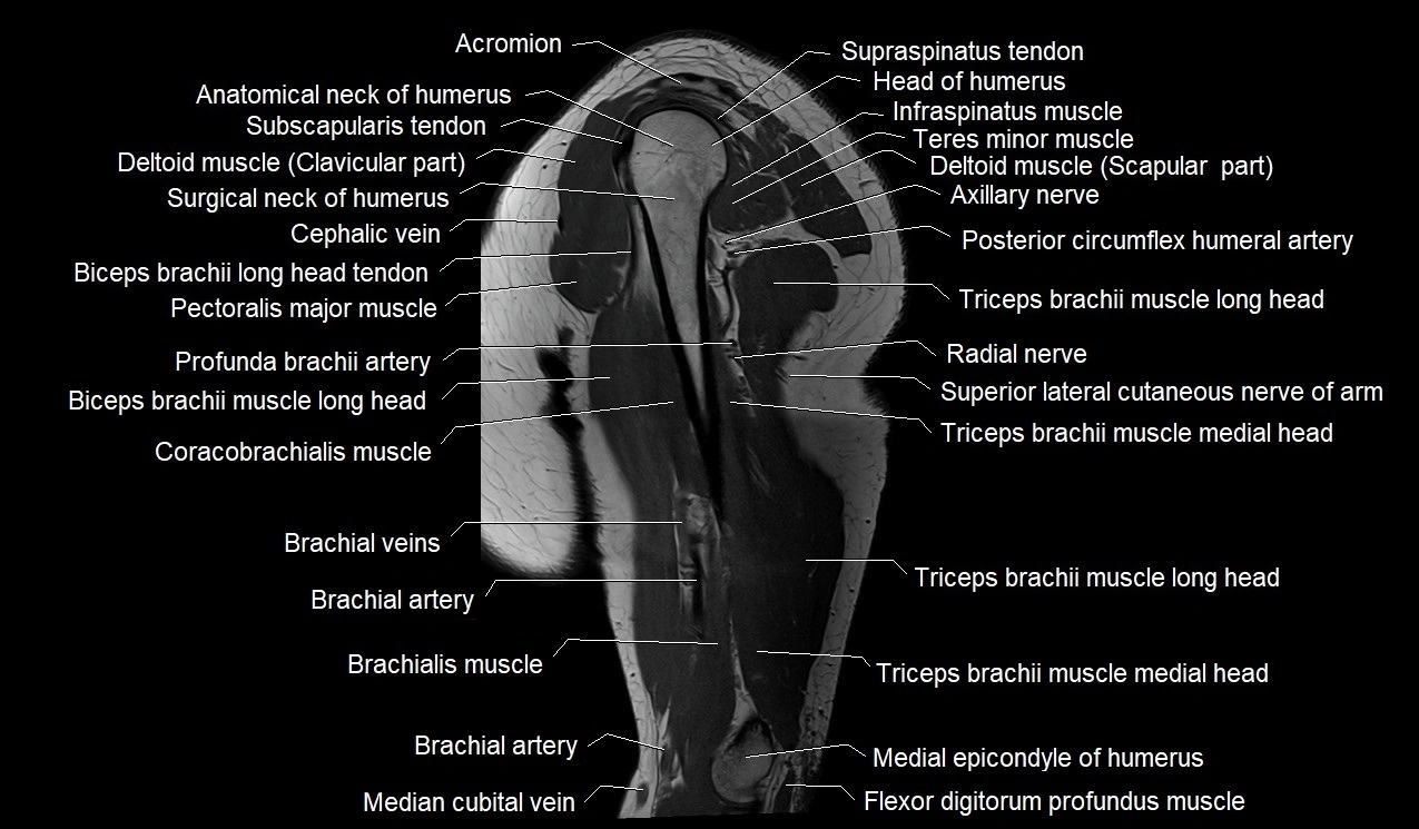
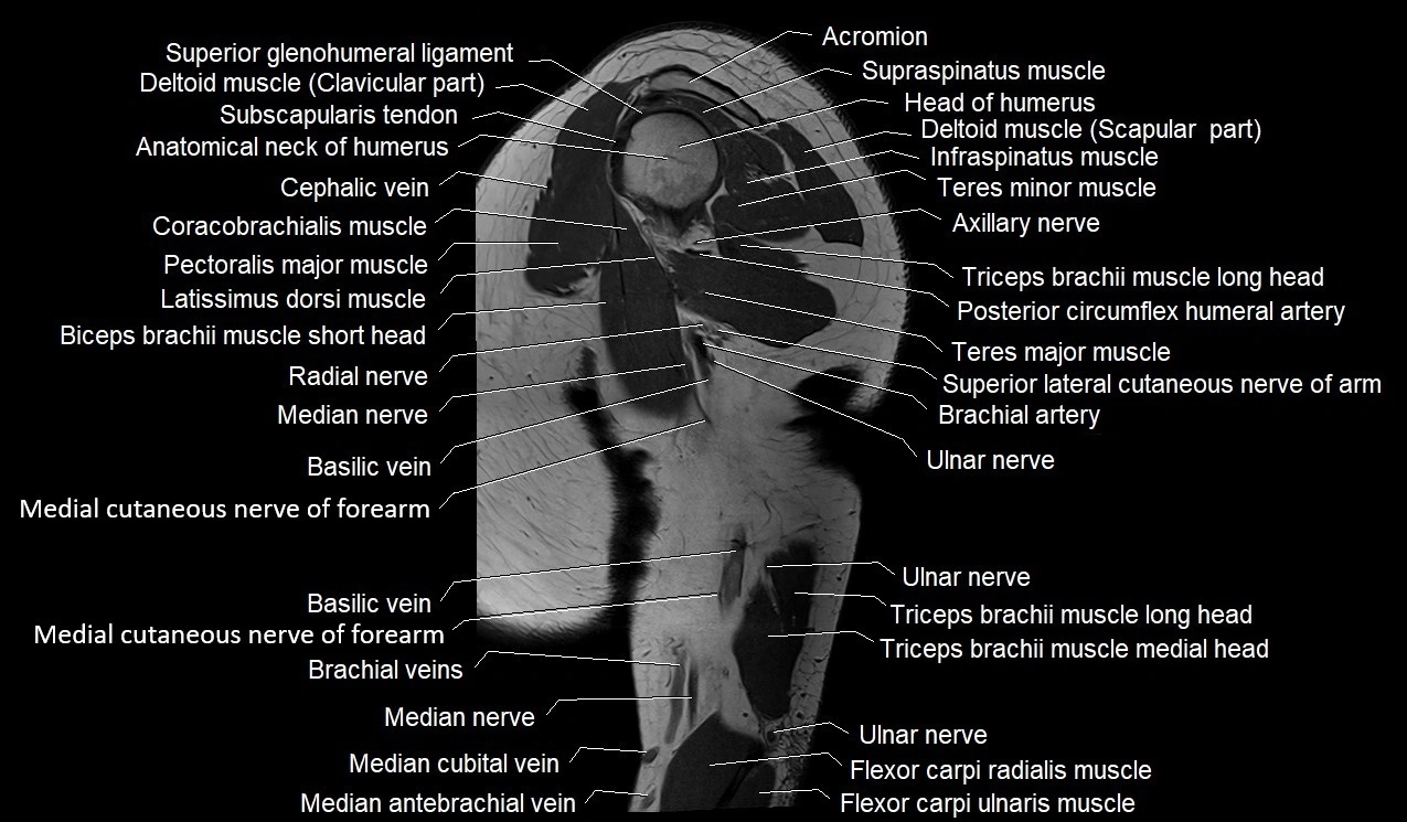
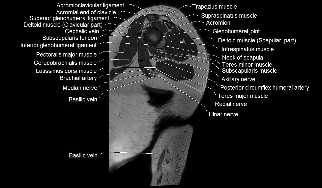
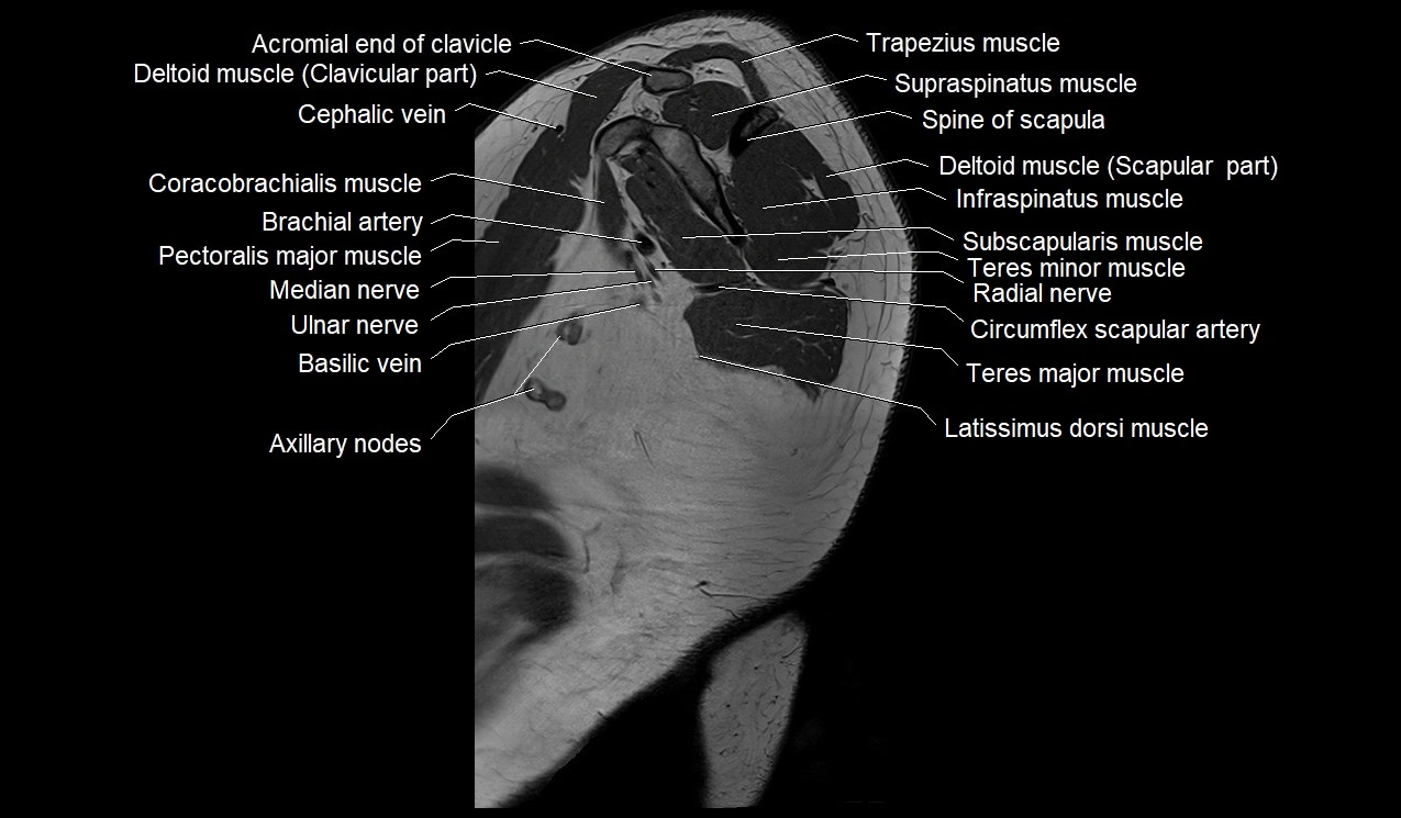
CT image


