


Topic
The abdominal aorta is the continuation of the thoracic aorta, beginning at the level of the aortic hiatus of the diaphragm (T12 vertebra) and terminating at the level of the L4 vertebra where it bifurcates into the right and left common iliac arteries. It lies slightly to the left of the midline and courses anterior to the vertebral bodies, surrounded by the retroperitoneal structures of the abdomen.
The abdominal aorta gives off numerous visceral and parietal branches, supplying the abdominal organs, pelvic structures, and lower limbs. It is the main conduit of oxygenated blood from the heart to the abdomen and lower body. The aorta is clinically significant as the common site of aneurysm, dissection, atherosclerosis, and traumatic injury.
Synonyms
-
Aorta abdominalis
-
Infradiaphragmatic aorta
-
Abdominal portion of aorta
Function
-
Conducts oxygenated blood from the thoracic aorta to abdominal, pelvic, and lower limb structures
-
Provides direct arterial supply to major abdominal organs (liver, spleen, kidneys, intestines)
-
Maintains systemic blood flow and hemodynamic regulation
-
Plays a central role in surgical and interventional procedures (aneurysm repair, stent grafts)
Branches
-
Unpaired visceral branches: celiac trunk, superior mesenteric artery (SMA), inferior mesenteric artery (IMA)
-
Paired visceral branches: middle suprarenal arteries, renal arteries, gonadal arteries (testicular or ovarian)
-
Parietal branches: inferior phrenic arteries, lumbar arteries, median sacral artery
-
Terminal branches: right and left common iliac arteries
MRI Appearance
T1-weighted images:
-
Flowing blood appears as a signal void (black lumen)
-
Vessel wall appears as a thin hypointense rim; retroperitoneal fat enhances contrast
T2-weighted images:
-
Lumen remains a signal void due to flow
-
Adjacent edema, hematoma, or aneurysm wall thrombus may appear hyperintense
STIR (Short Tau Inversion Recovery):
-
Fat suppression improves visualization of the aortic wall and periaortic tissues
-
Wall edema, inflammation, or periaortic hematoma appears hyperintense
-
Useful in vasculitis, dissection, or trauma
T1 Post-Contrast (Gadolinium-enhanced):
-
Aortic lumen enhances brightly and homogeneously
-
Clearly demonstrates aneurysm, stenosis, dissection, mural thrombus, or aortic wall enhancement in vasculitis
MRA (Magnetic Resonance Angiography):
-
Contrast-enhanced MRA provides high-resolution imaging of the aorta and its branches
-
Allows 3D reconstruction of visceral, parietal, and terminal branches
-
Excellent for evaluating aneurysm size, dissection flap, stenosis, or preoperative planning
-
Non-invasive alternative to conventional angiography
CT Appearance
Non-contrast CT:
-
Appears as a tubular soft tissue structure anterior to vertebral bodies
-
Calcified atherosclerotic plaques appear as hyperdense foci along the wall
-
Useful for screening abdominal aortic aneurysm (AAA) size and mural calcification
Contrast-enhanced CT (CTA):
-
Gold standard for abdominal aortic imaging
-
Provides excellent detail of lumen, wall, aneurysm, thrombus, and branch vessels
-
Multiplanar and 3D reconstructions help in aneurysm measurement, stent graft planning, and dissection evaluation
-
Detects acute rupture, traumatic injury, or occlusion with high sensitivity
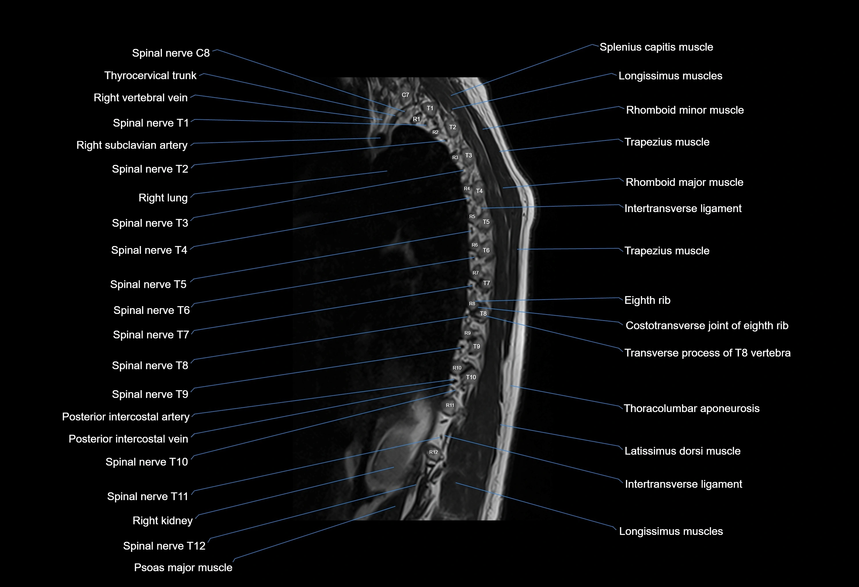
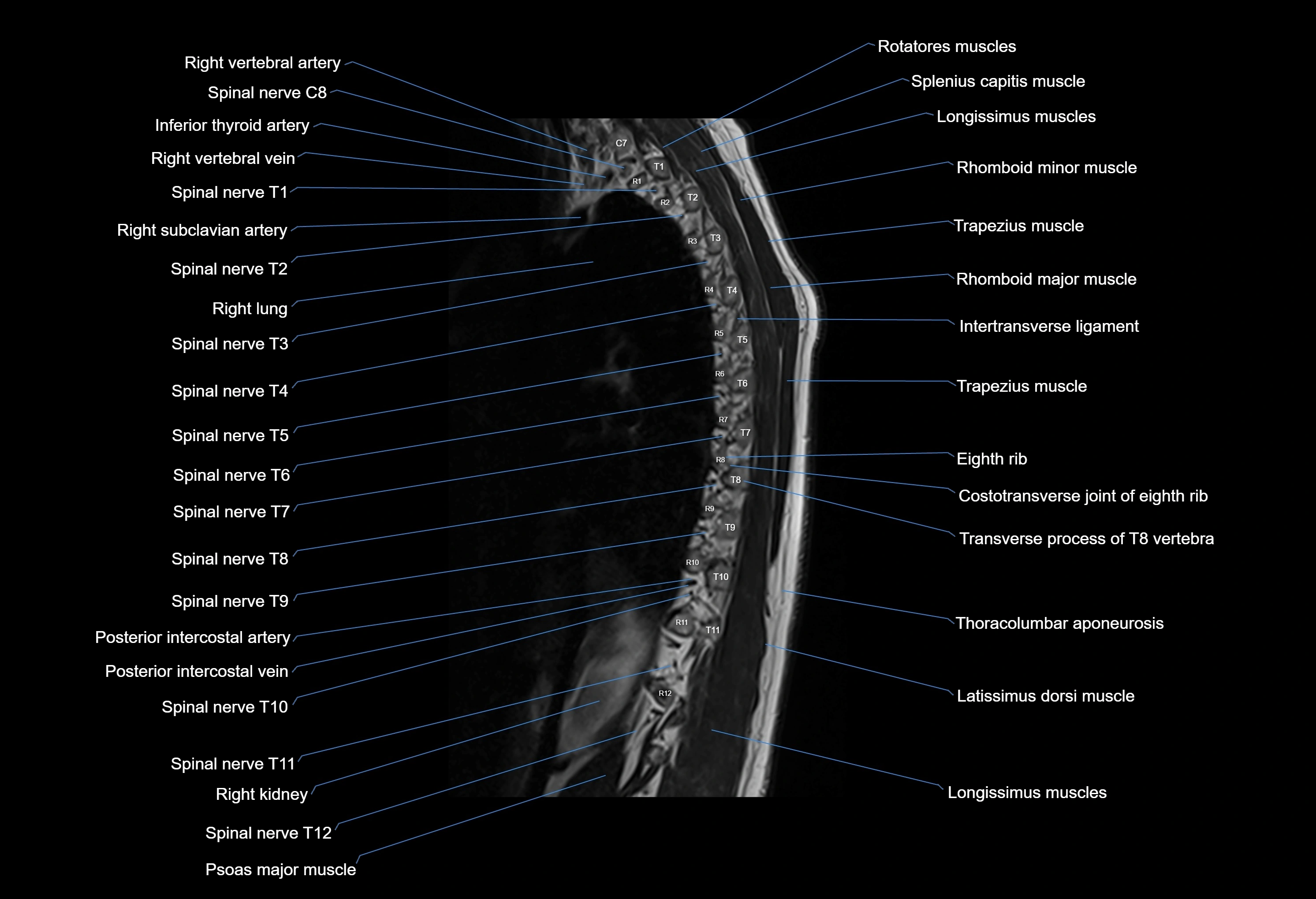
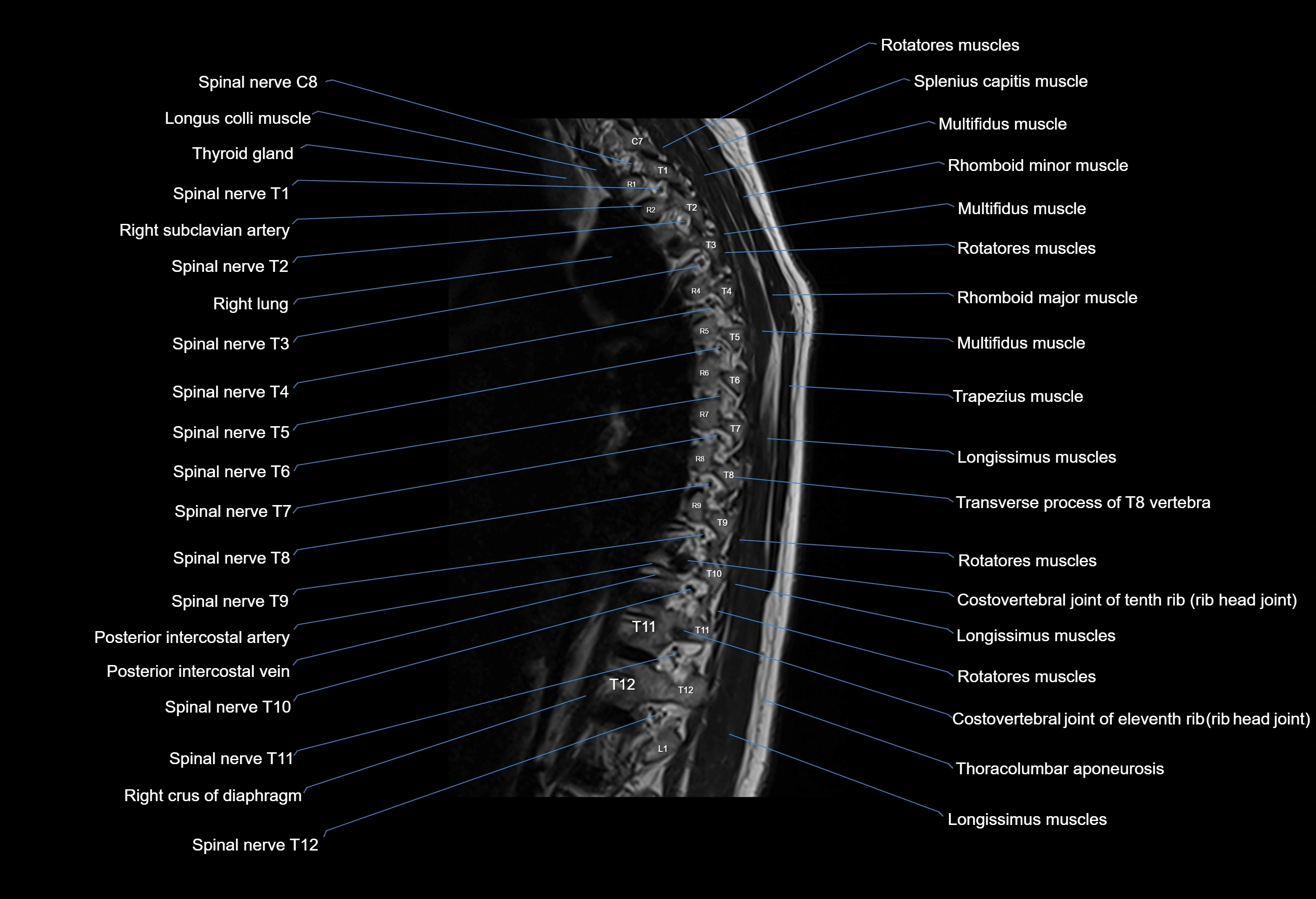
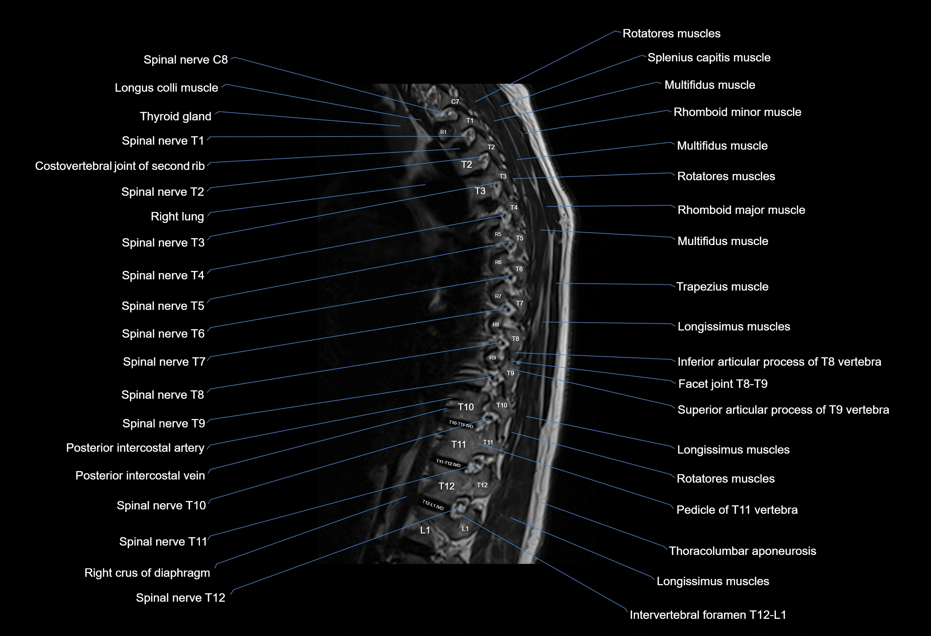
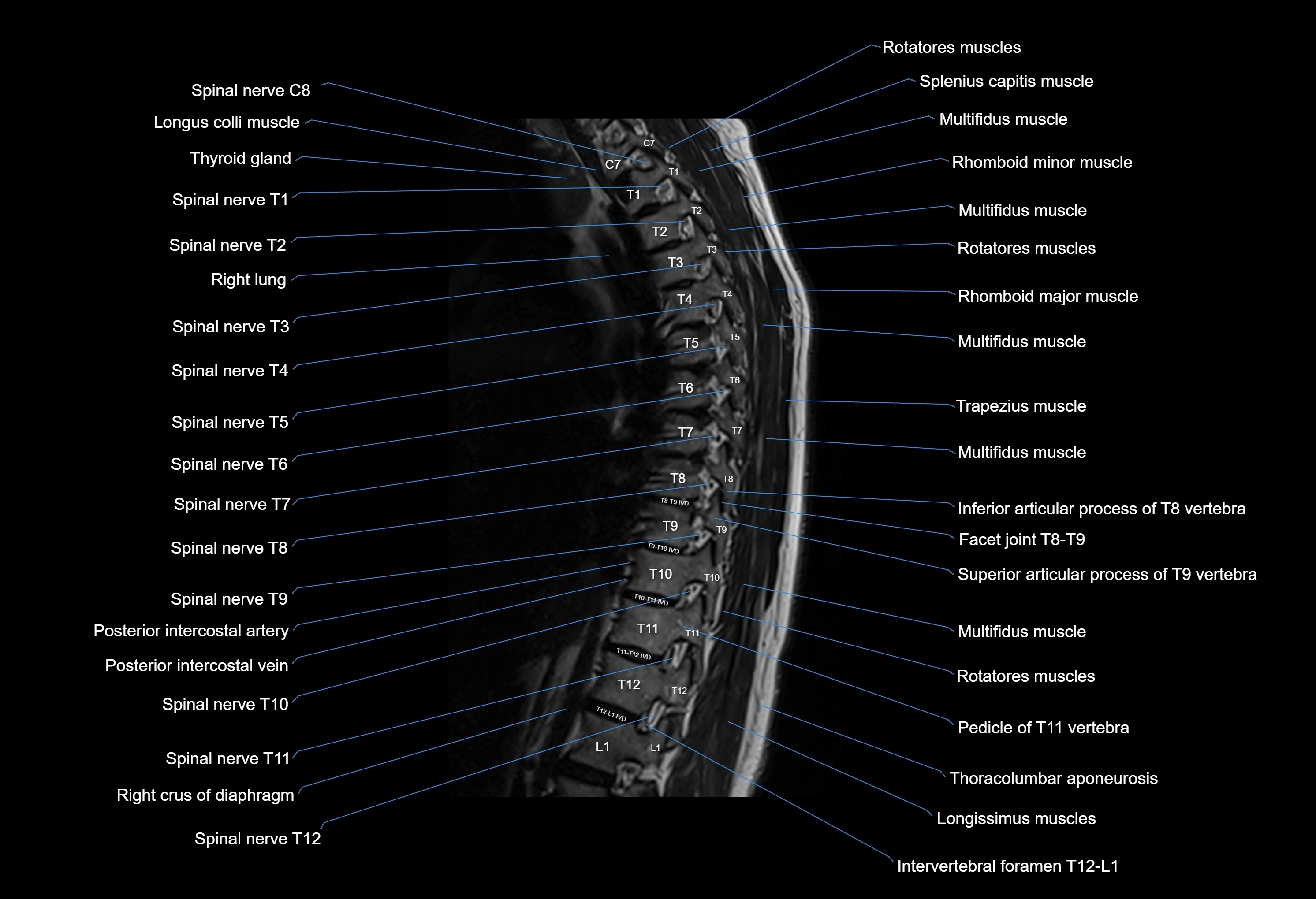
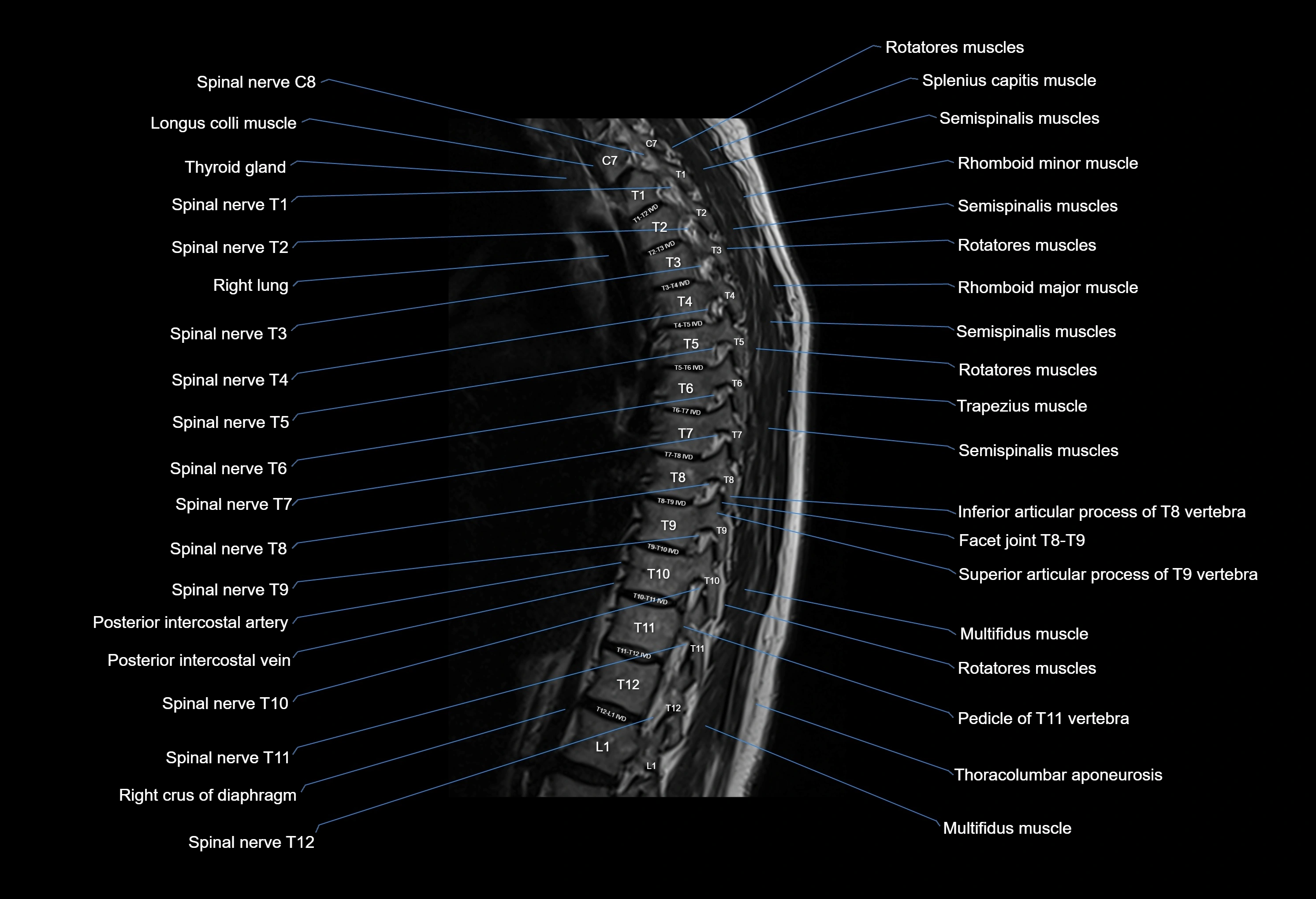
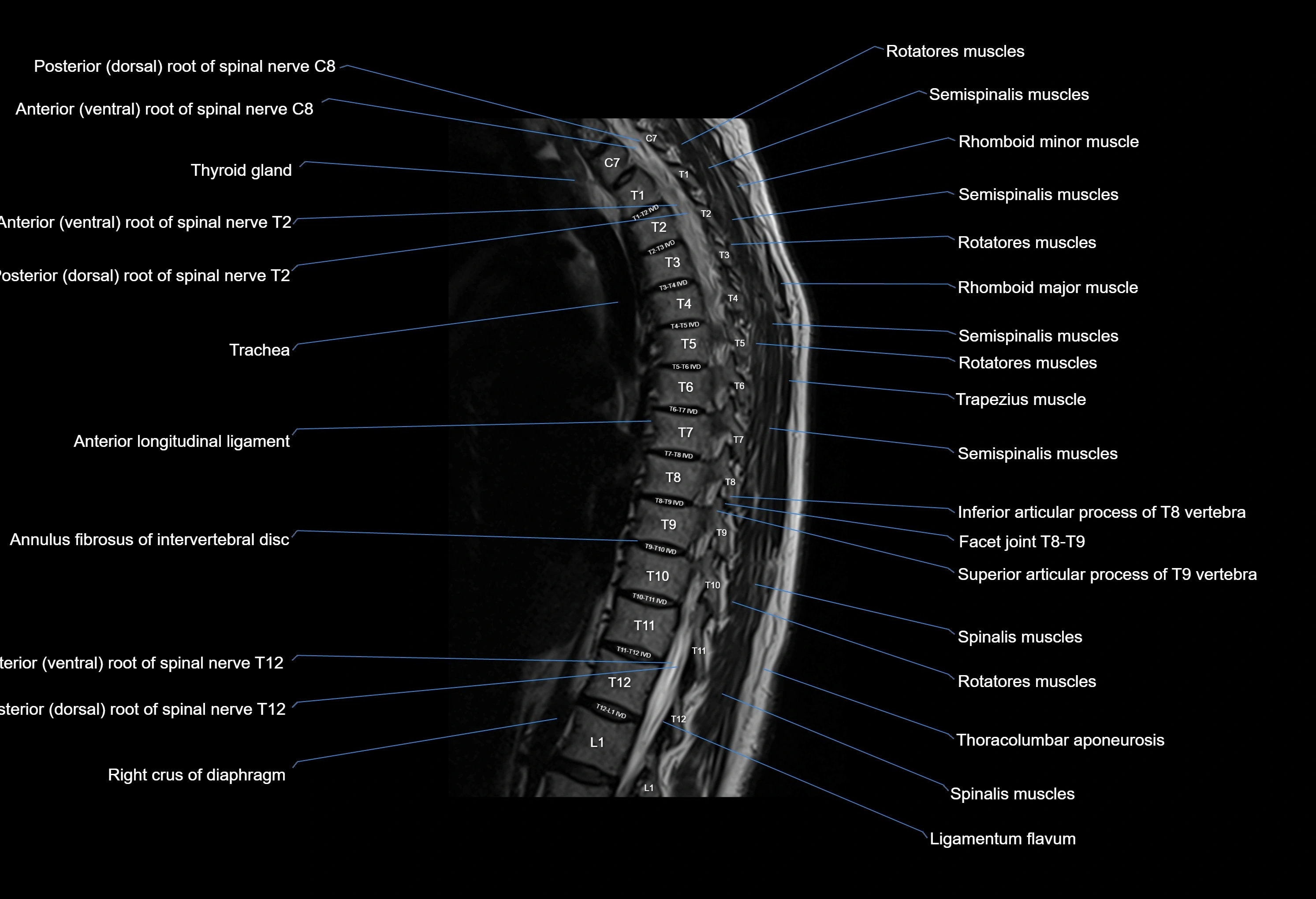
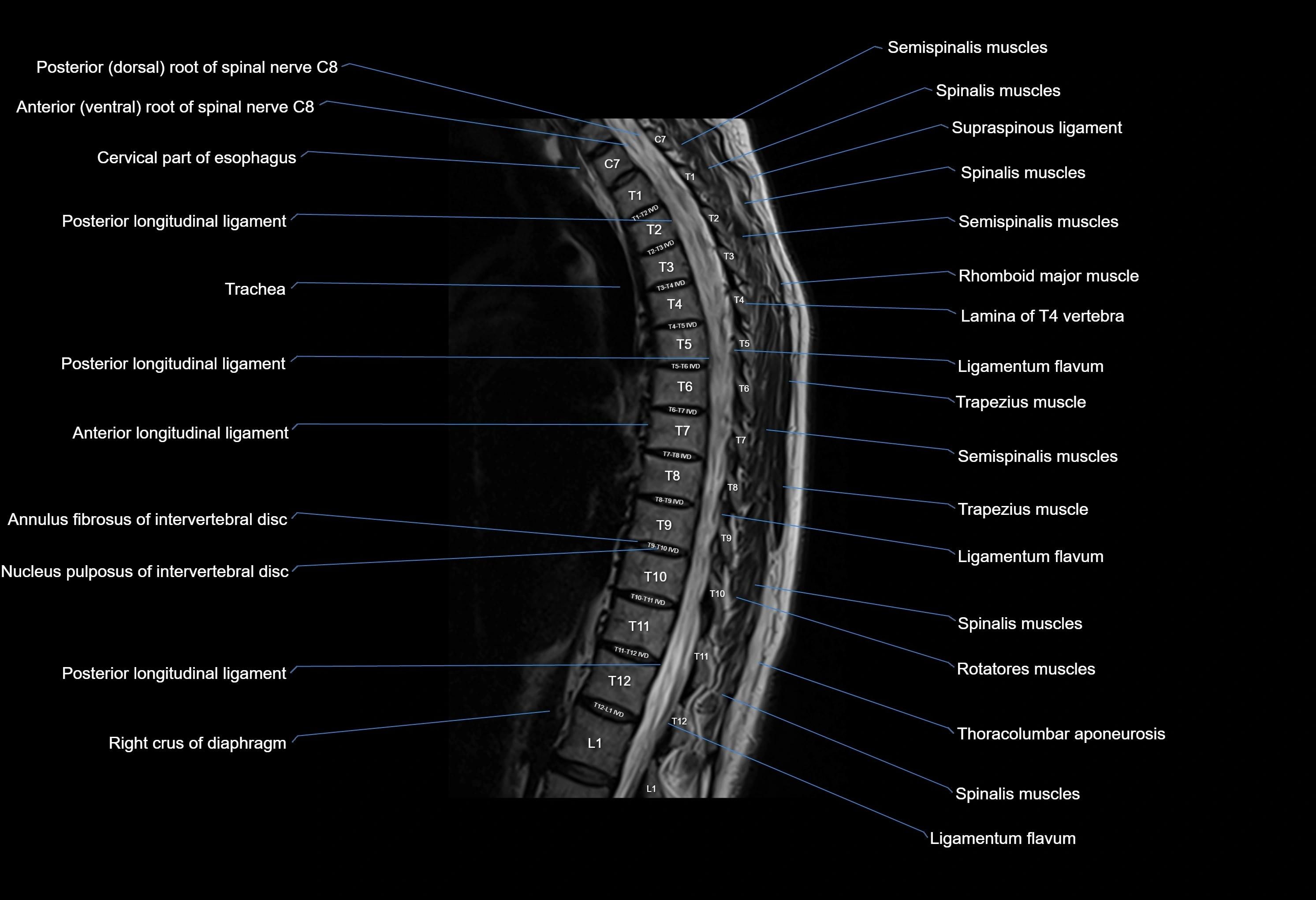
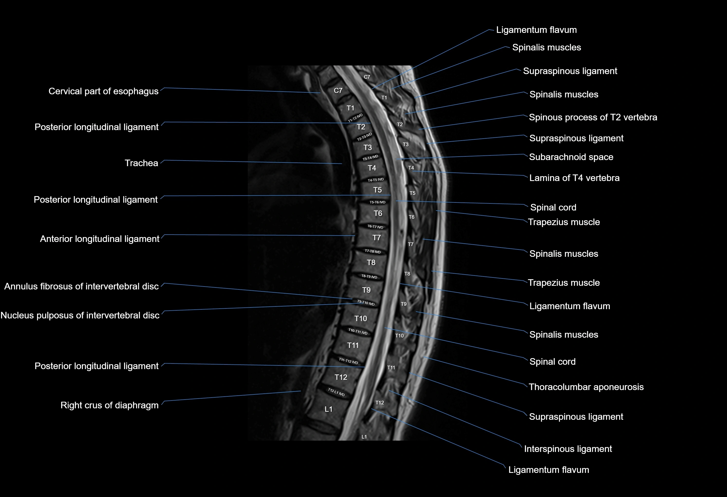
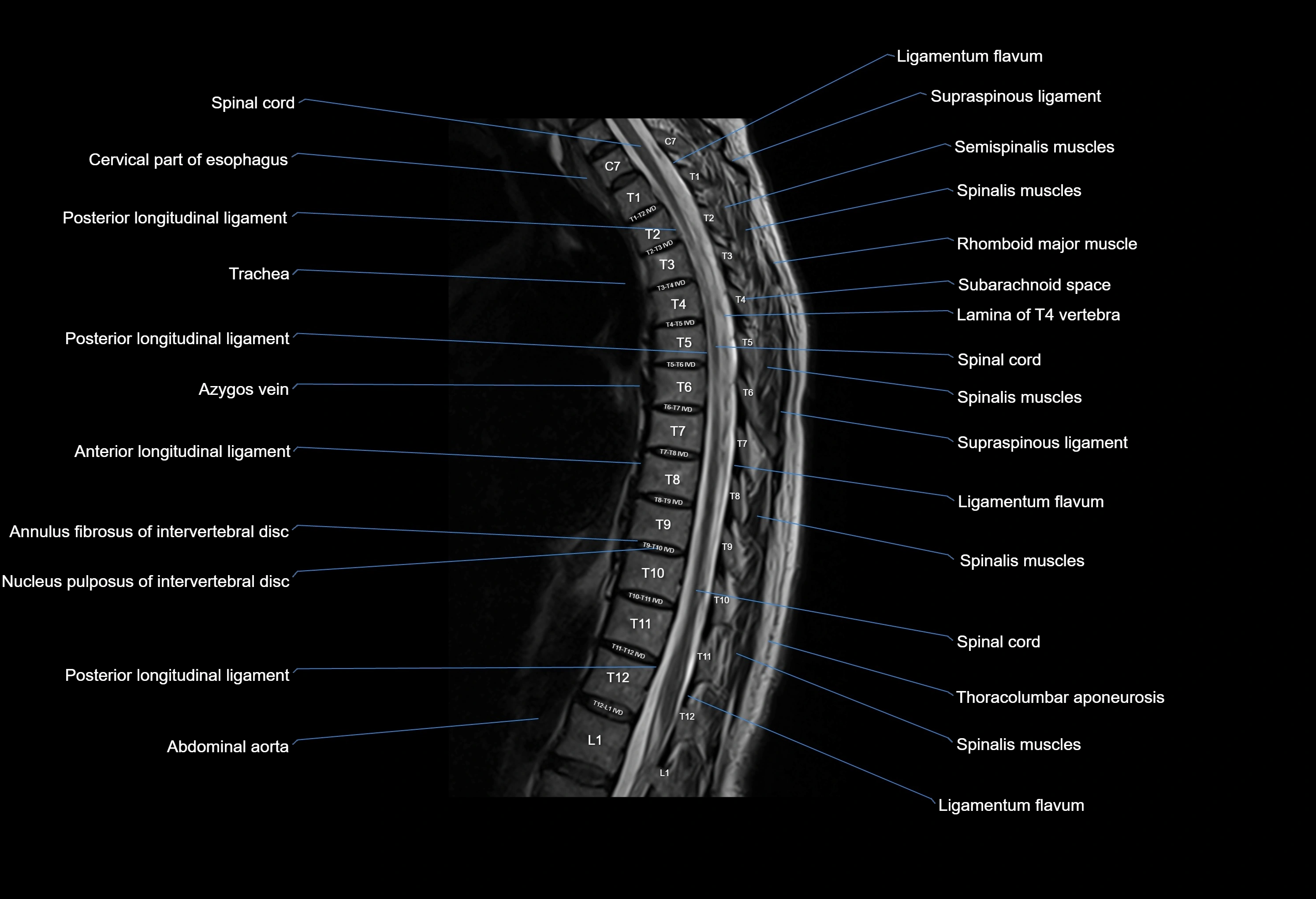
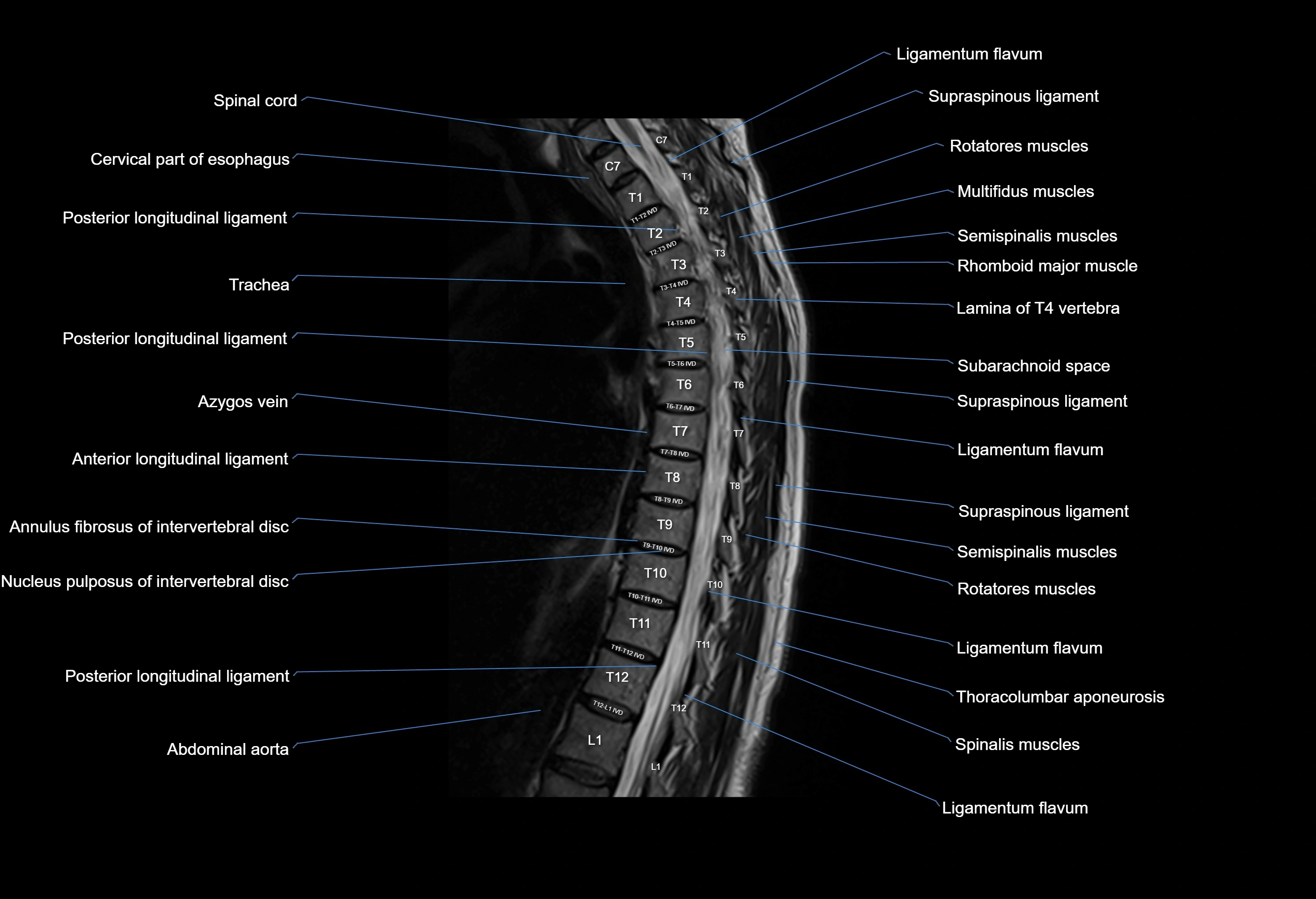
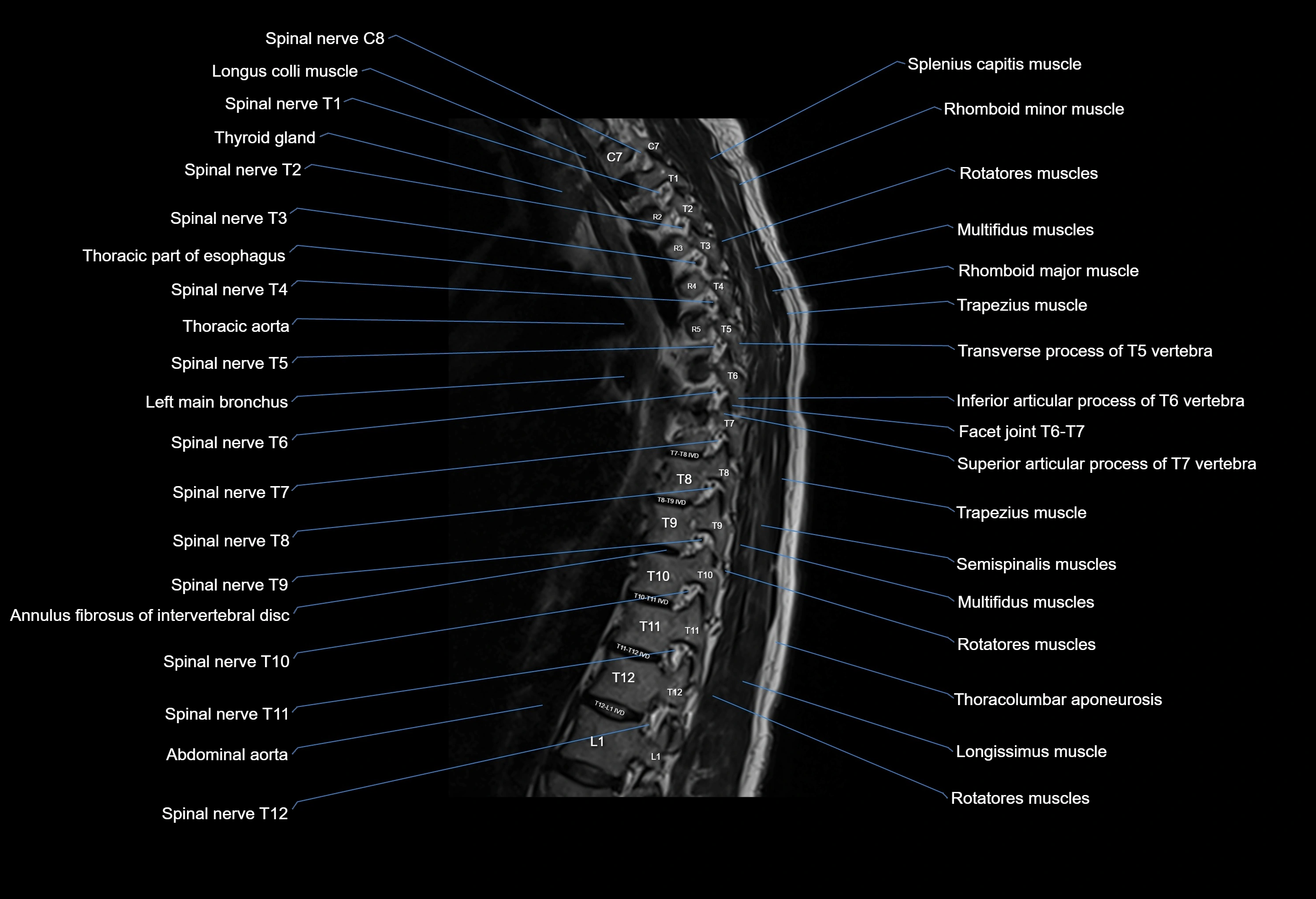
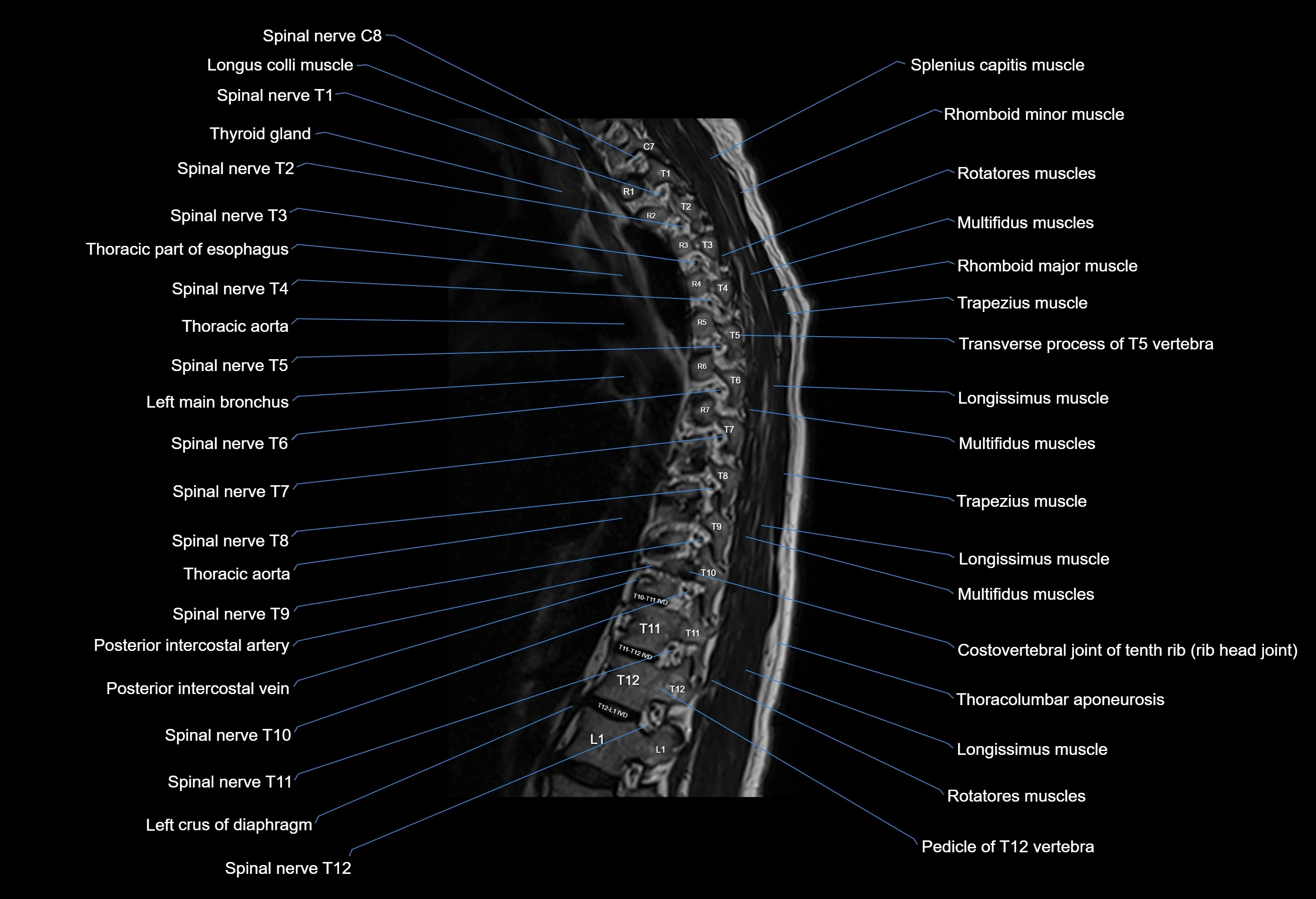
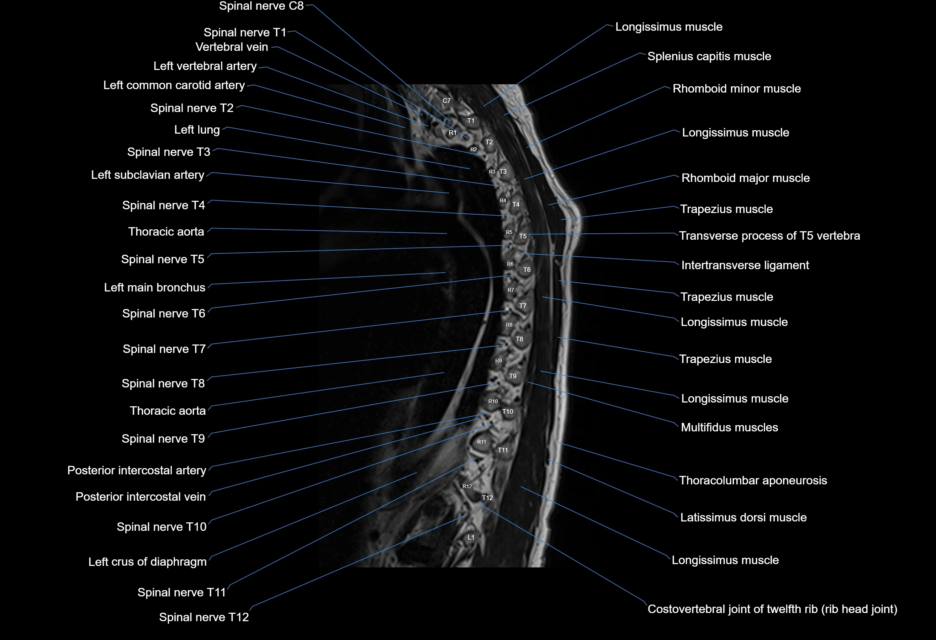
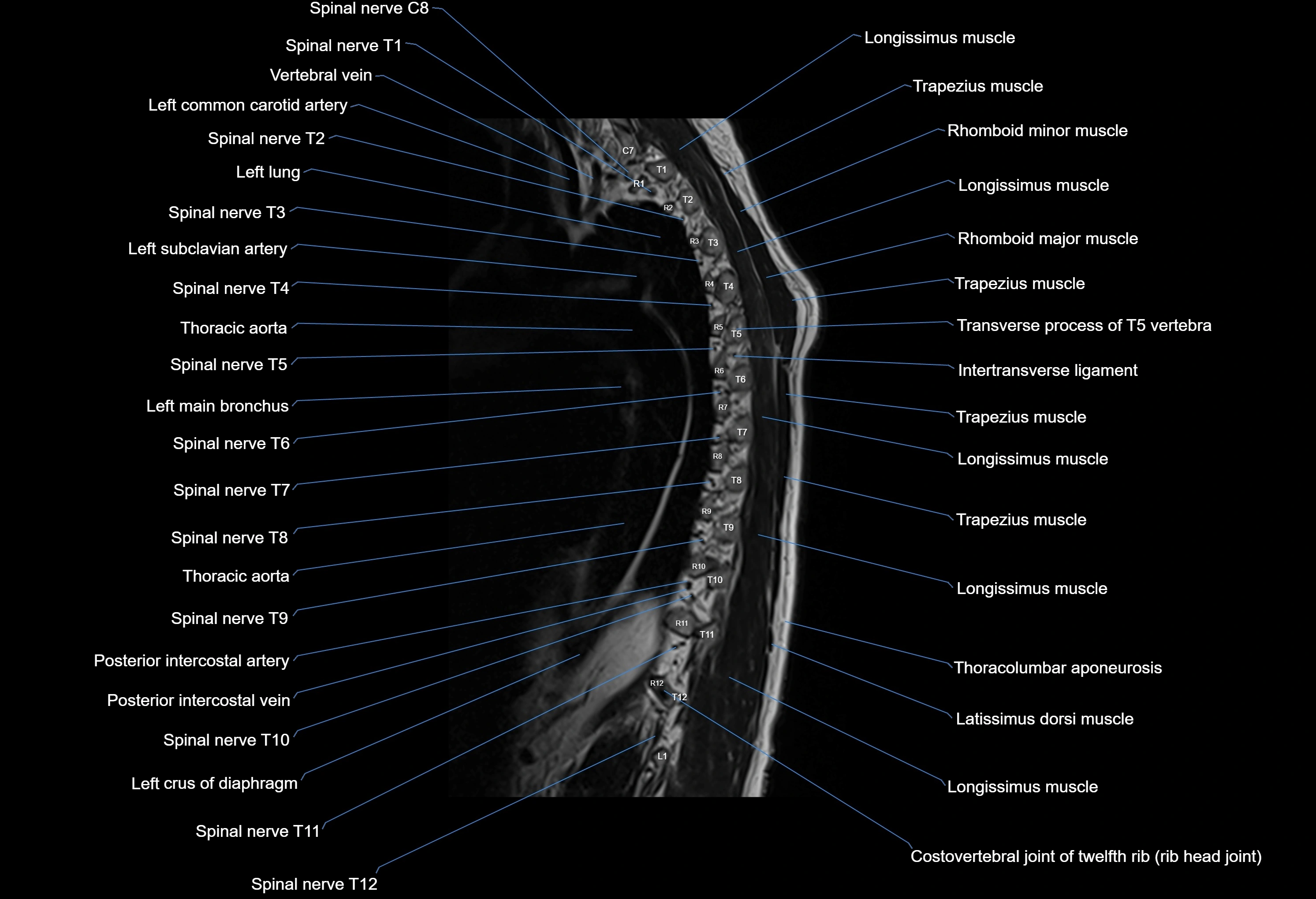
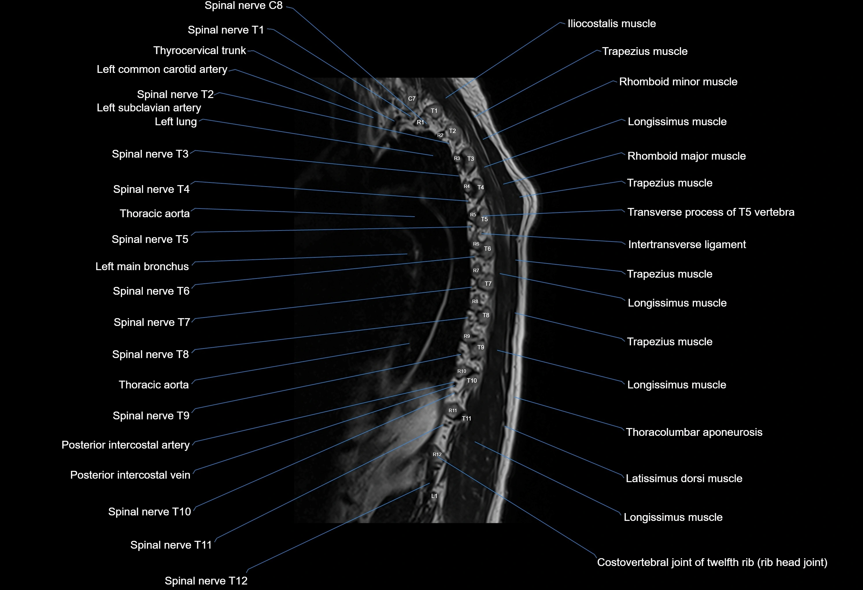
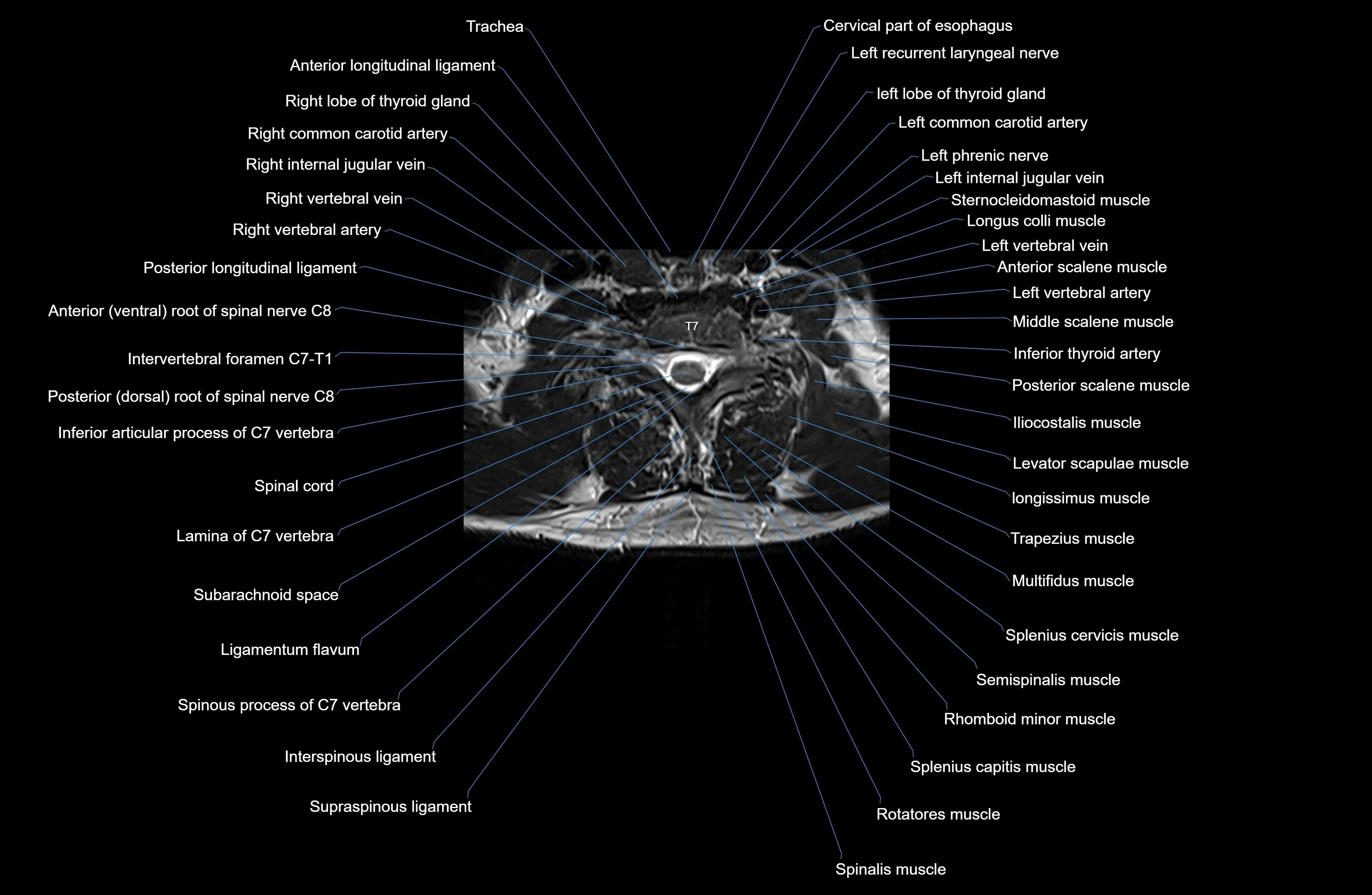
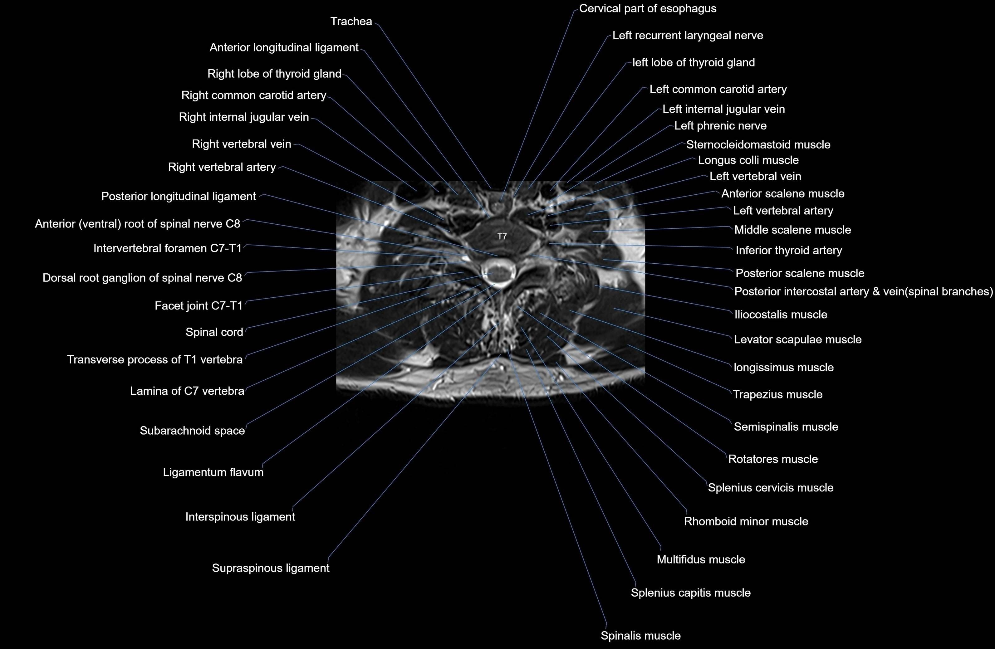
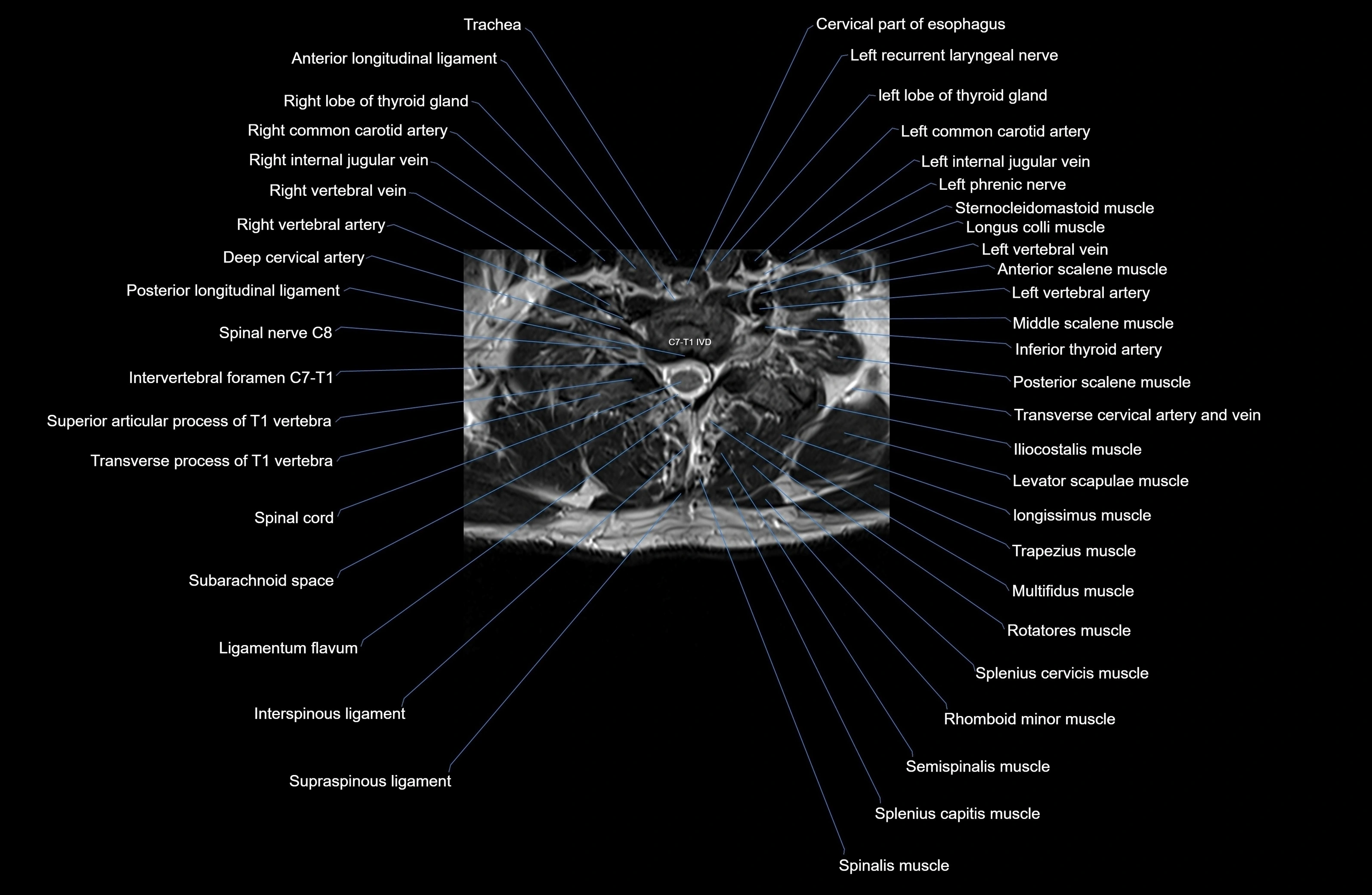
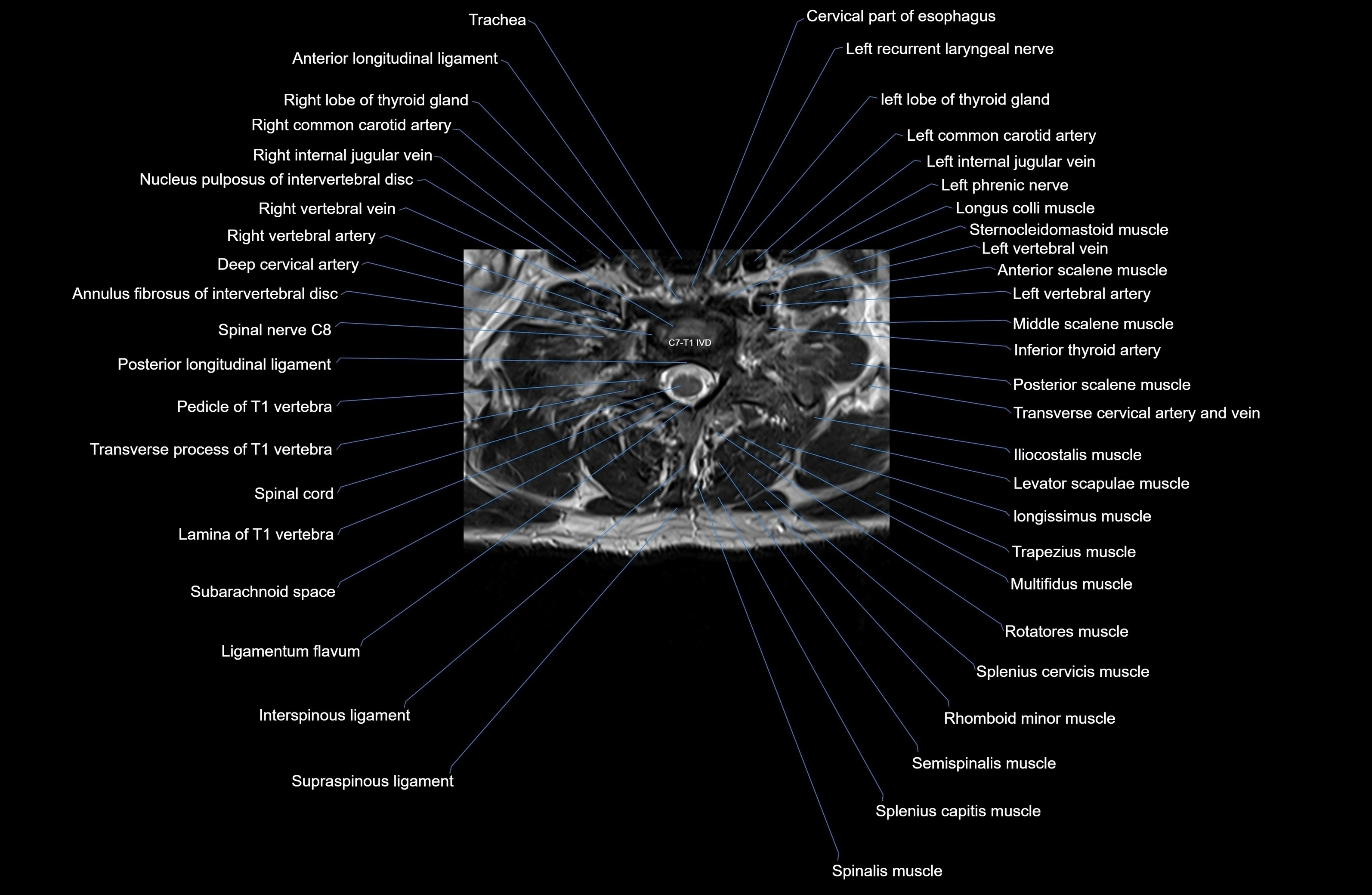
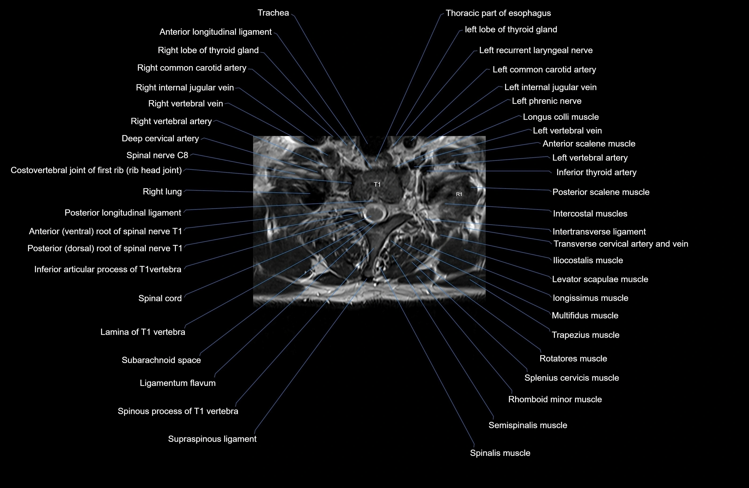
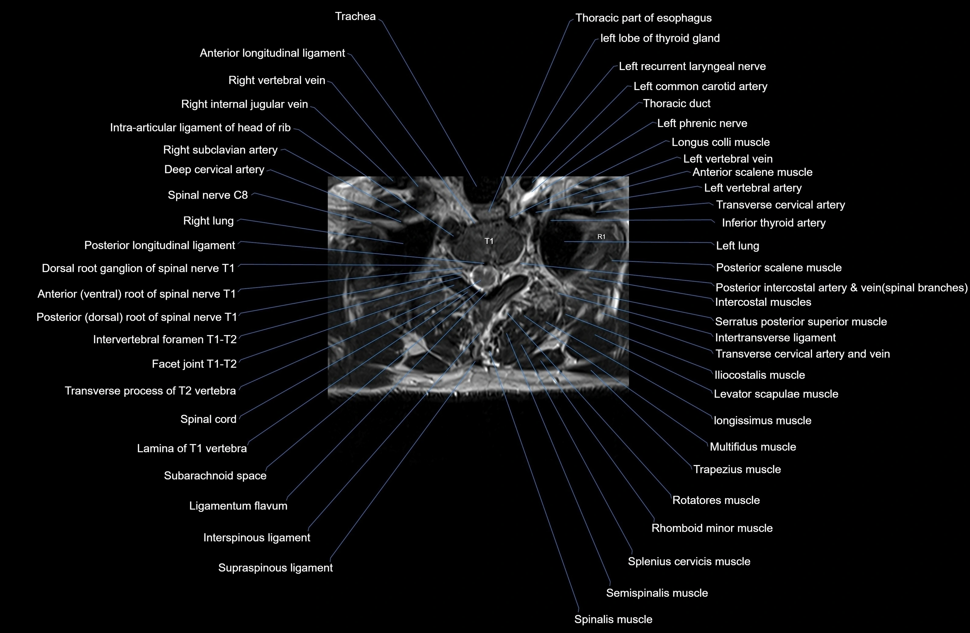
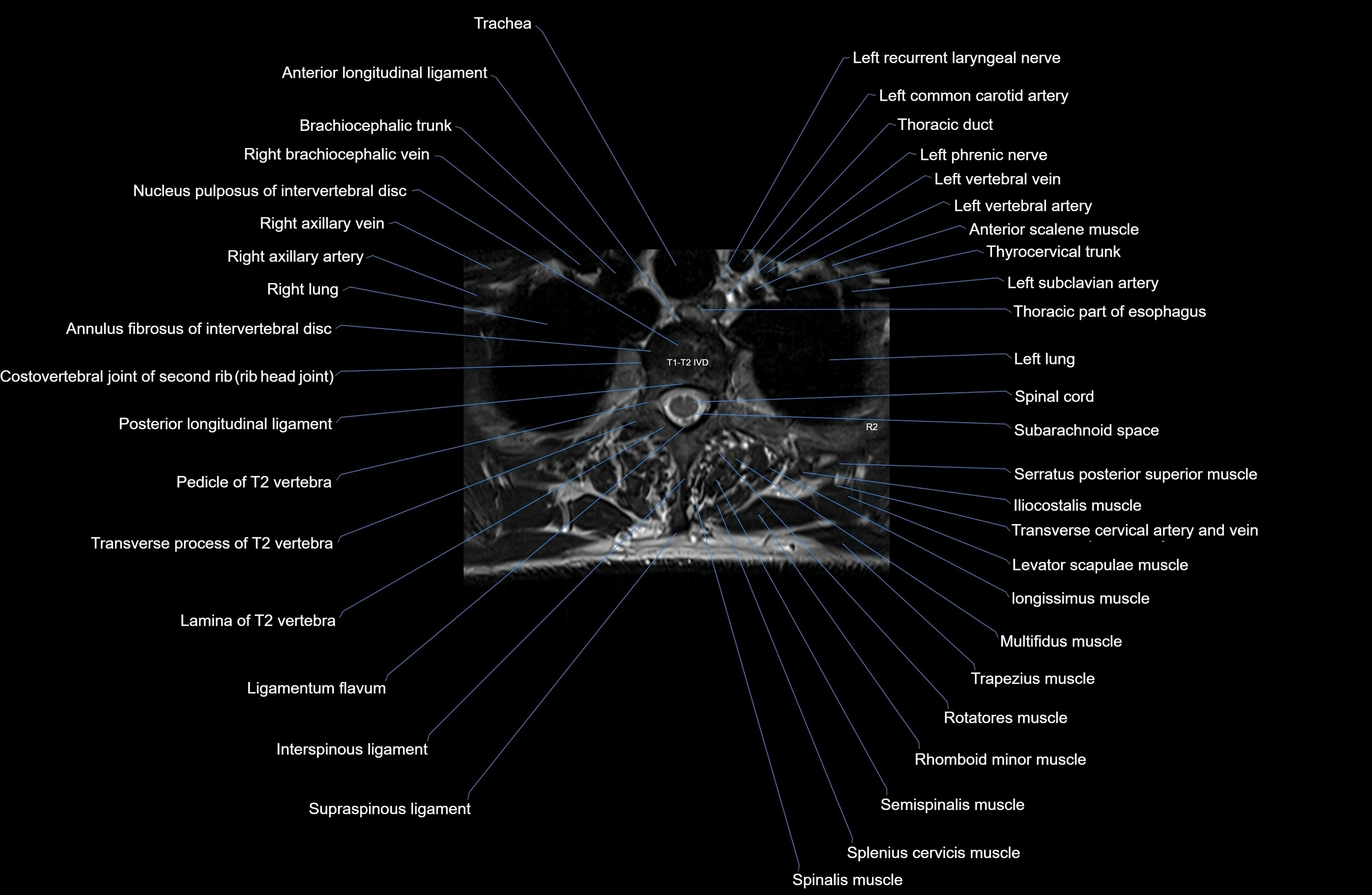
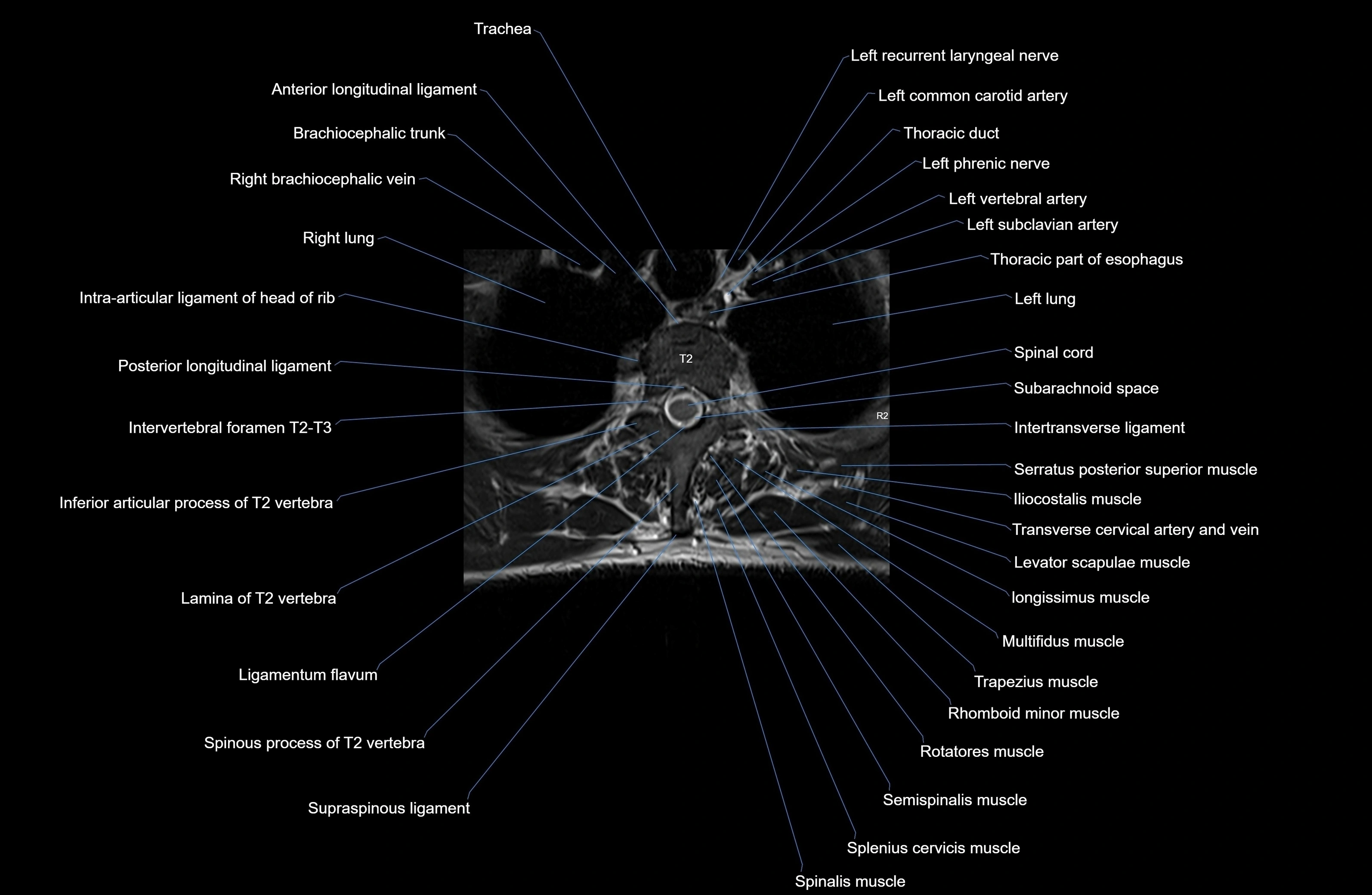
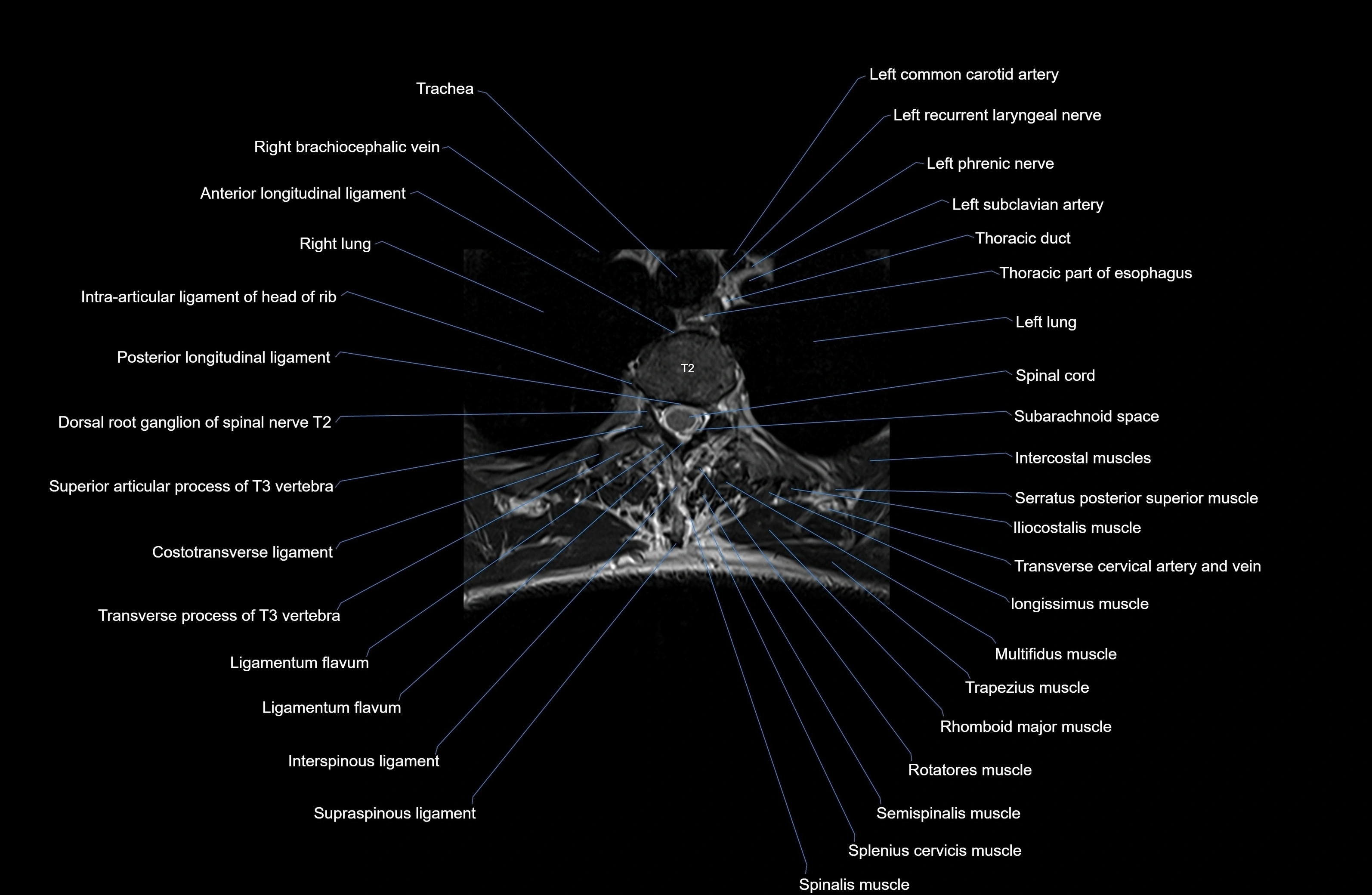
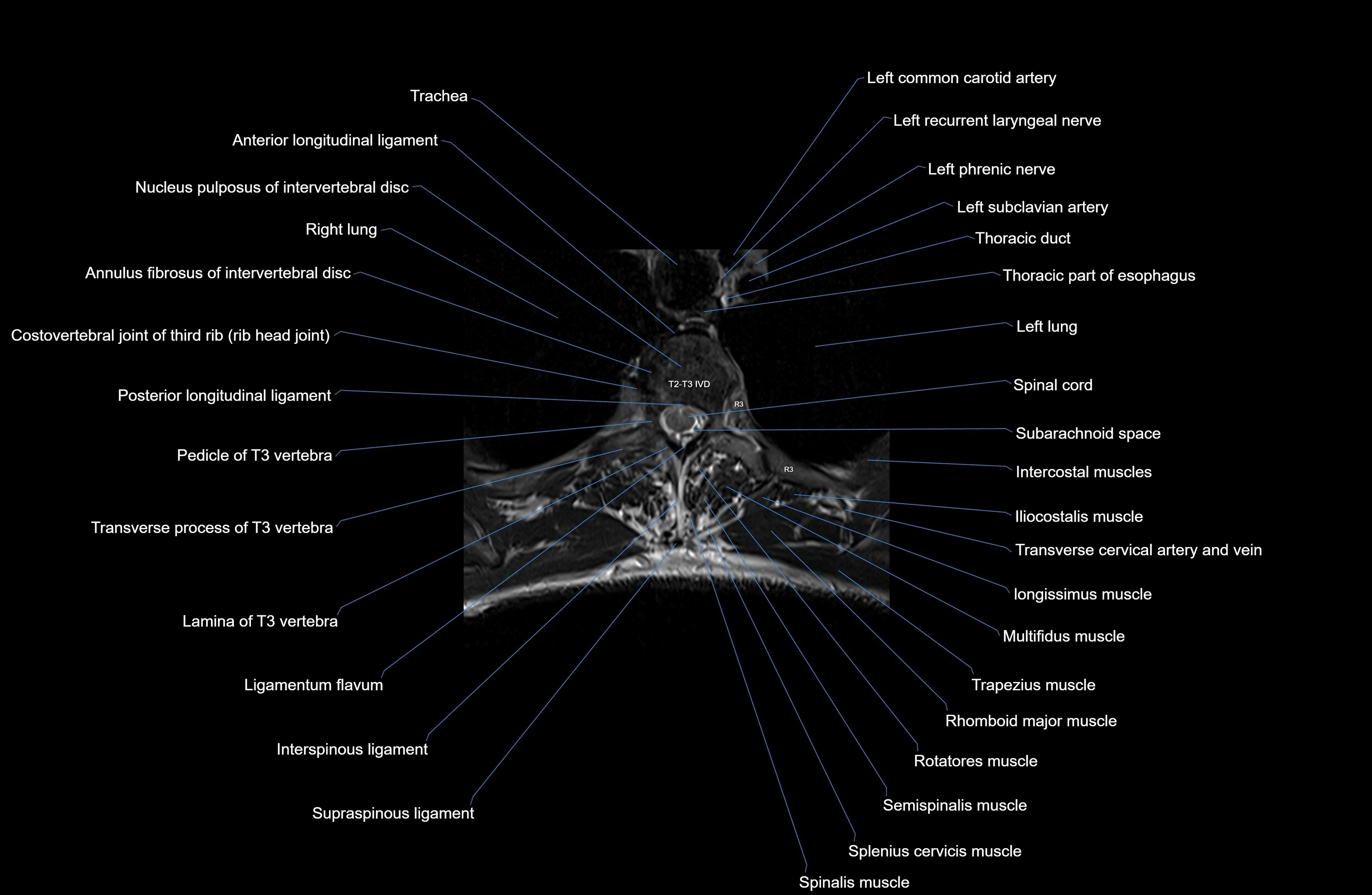
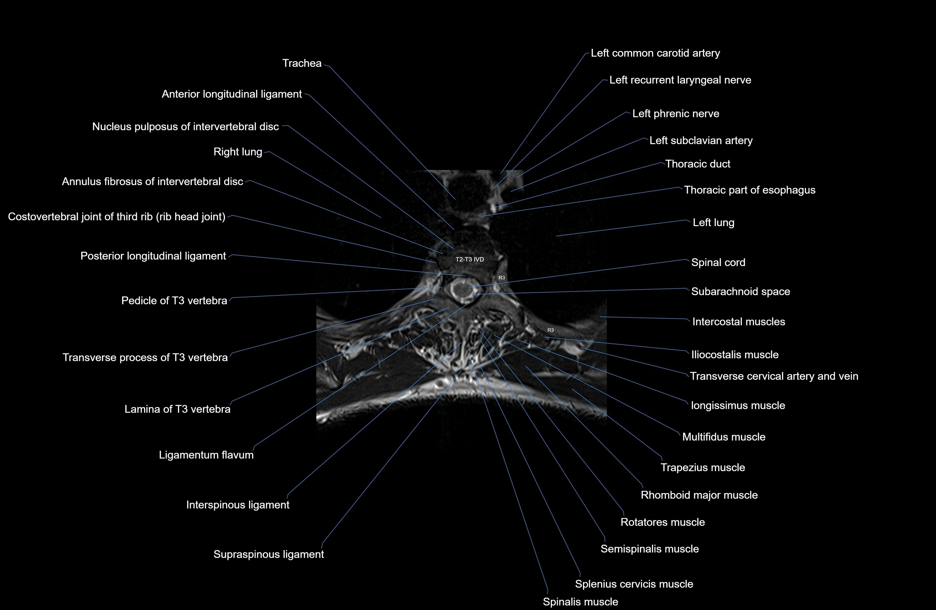
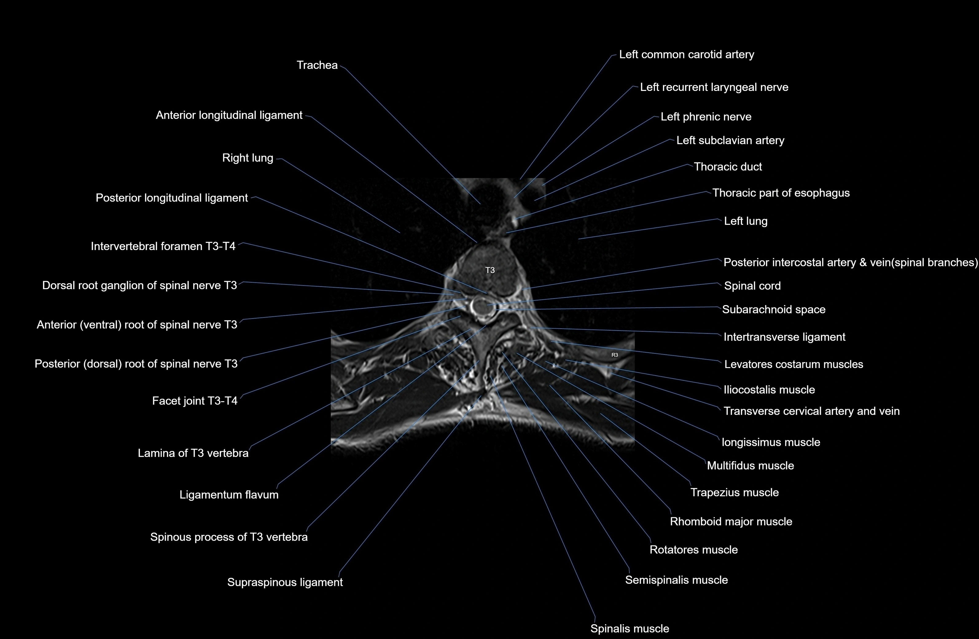
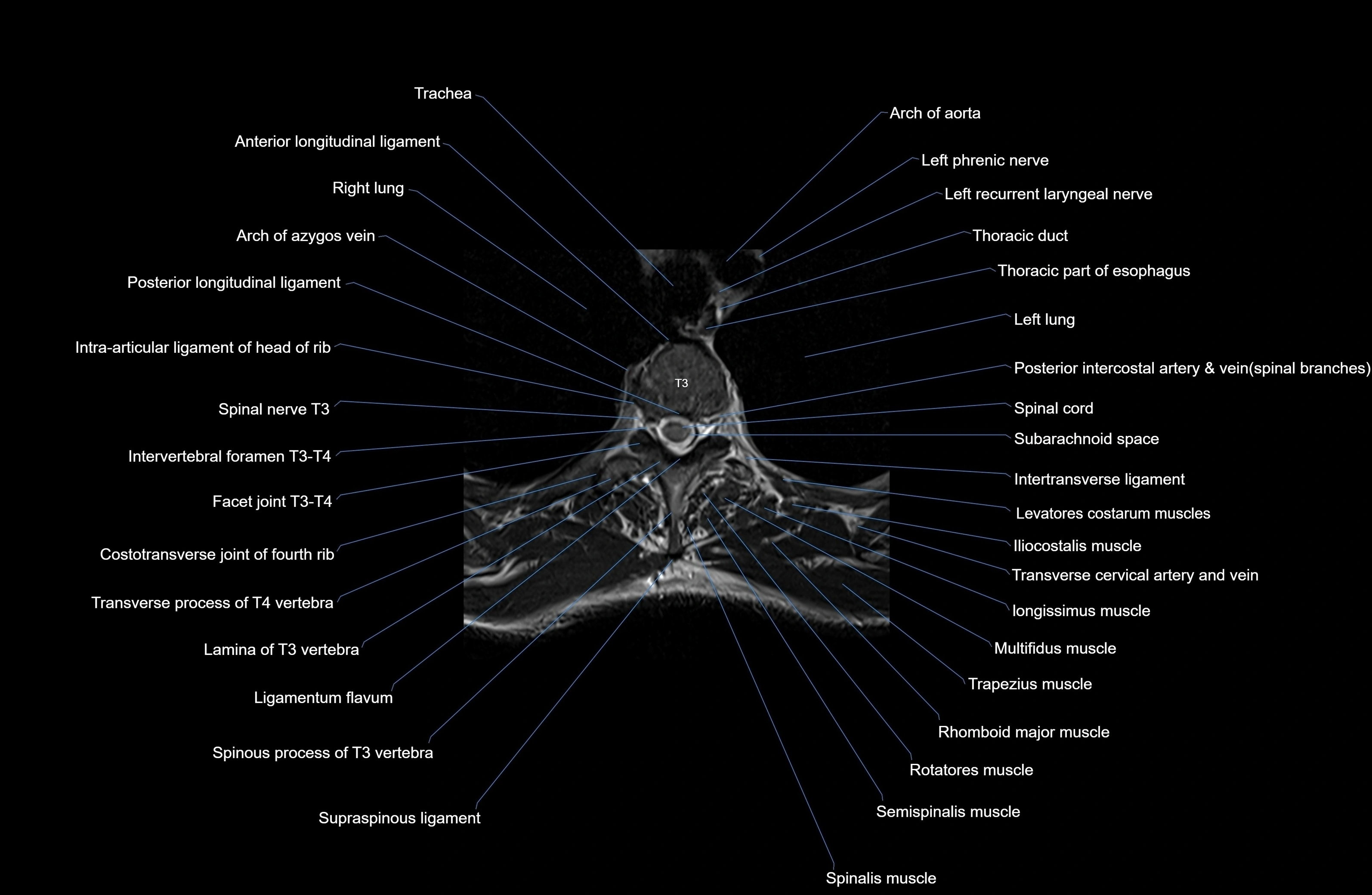
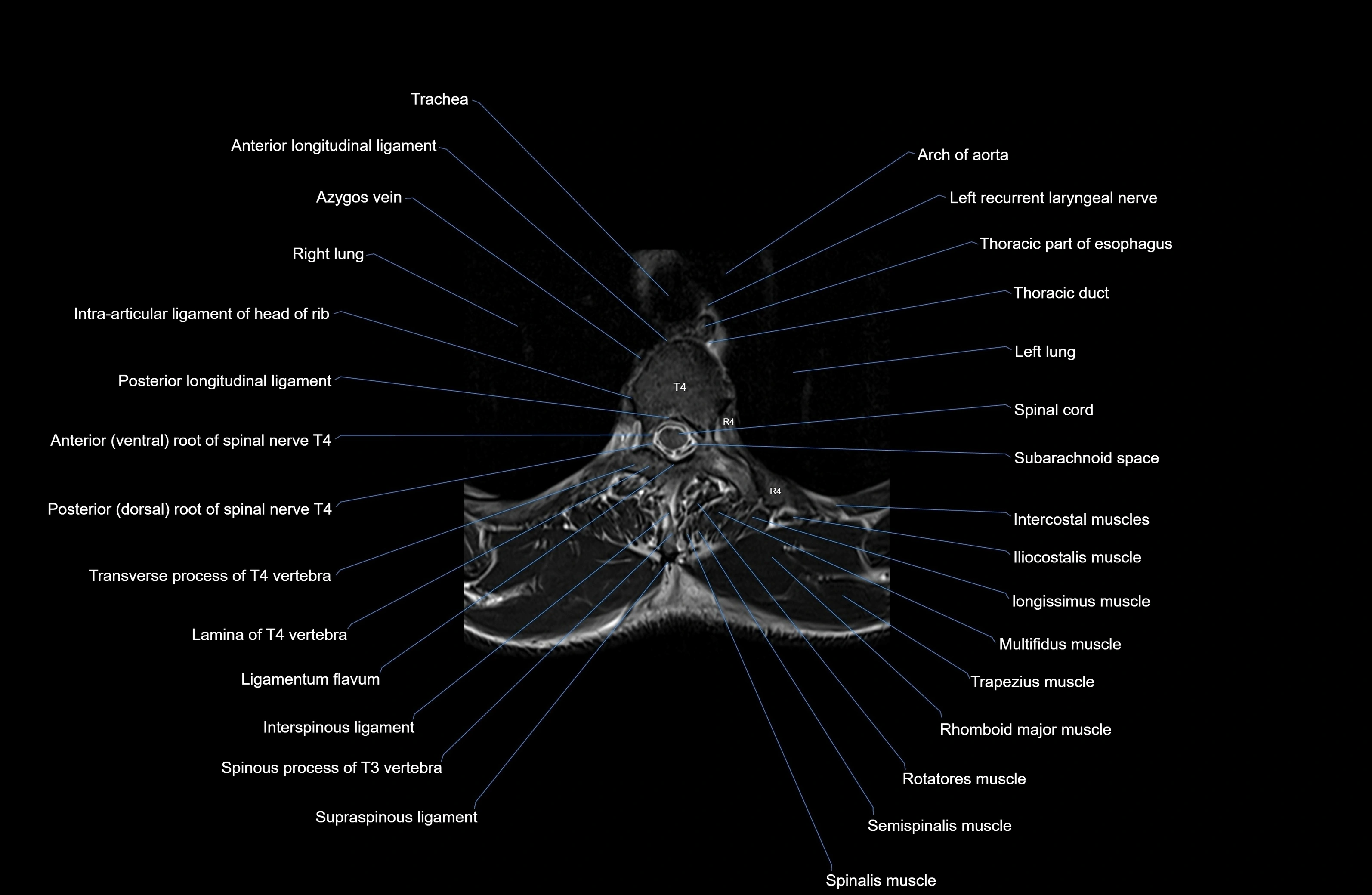
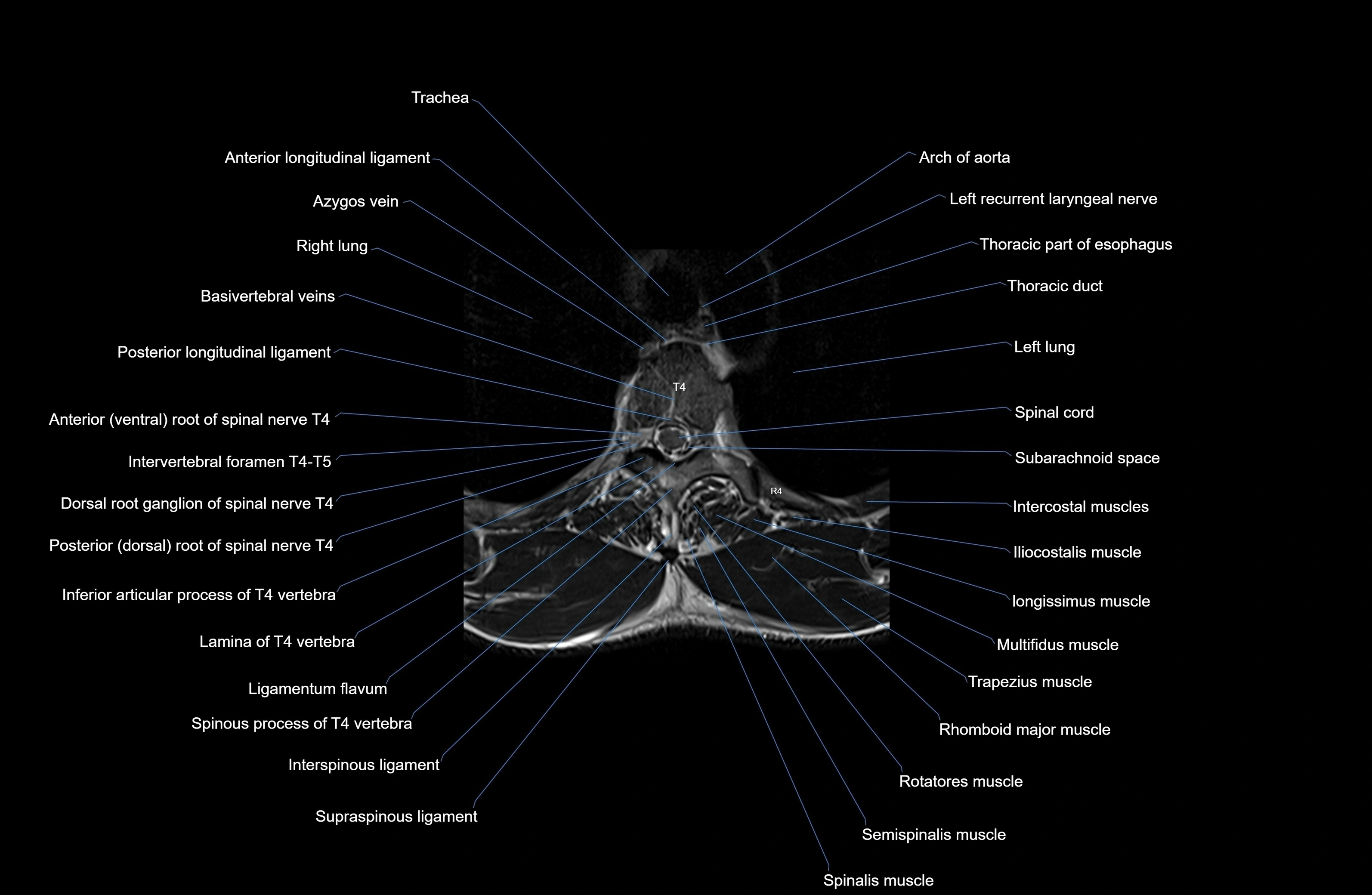
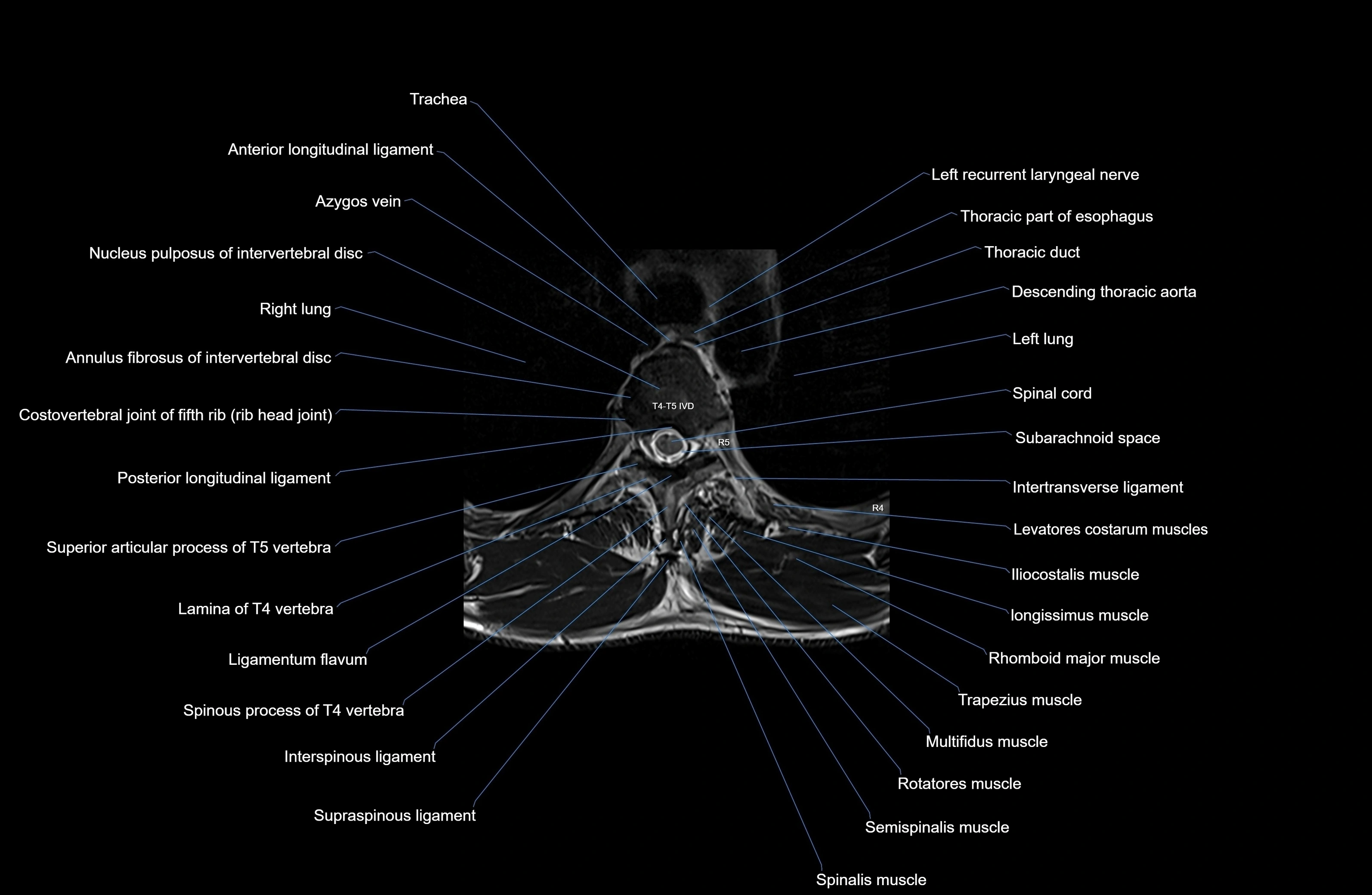
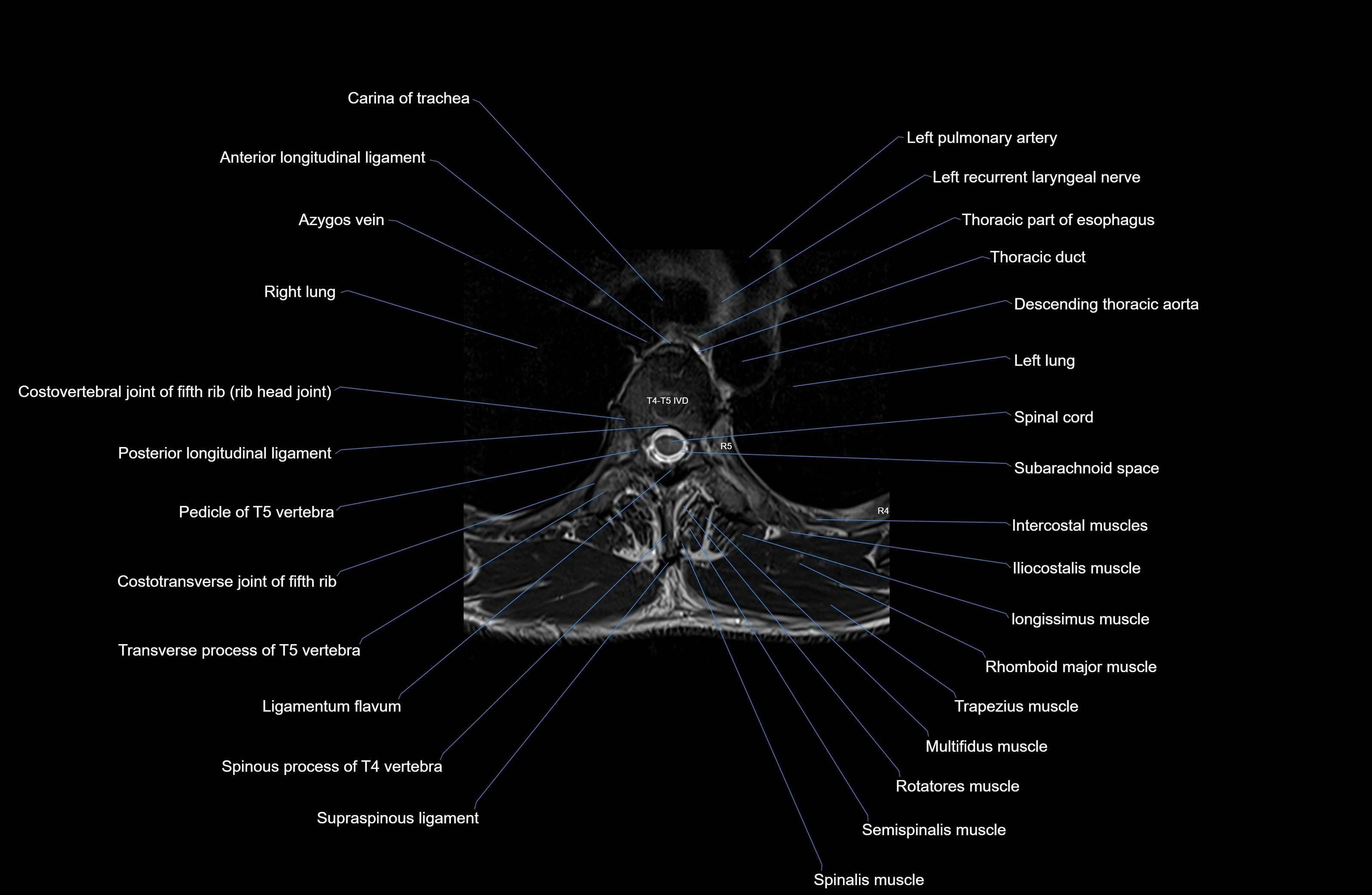
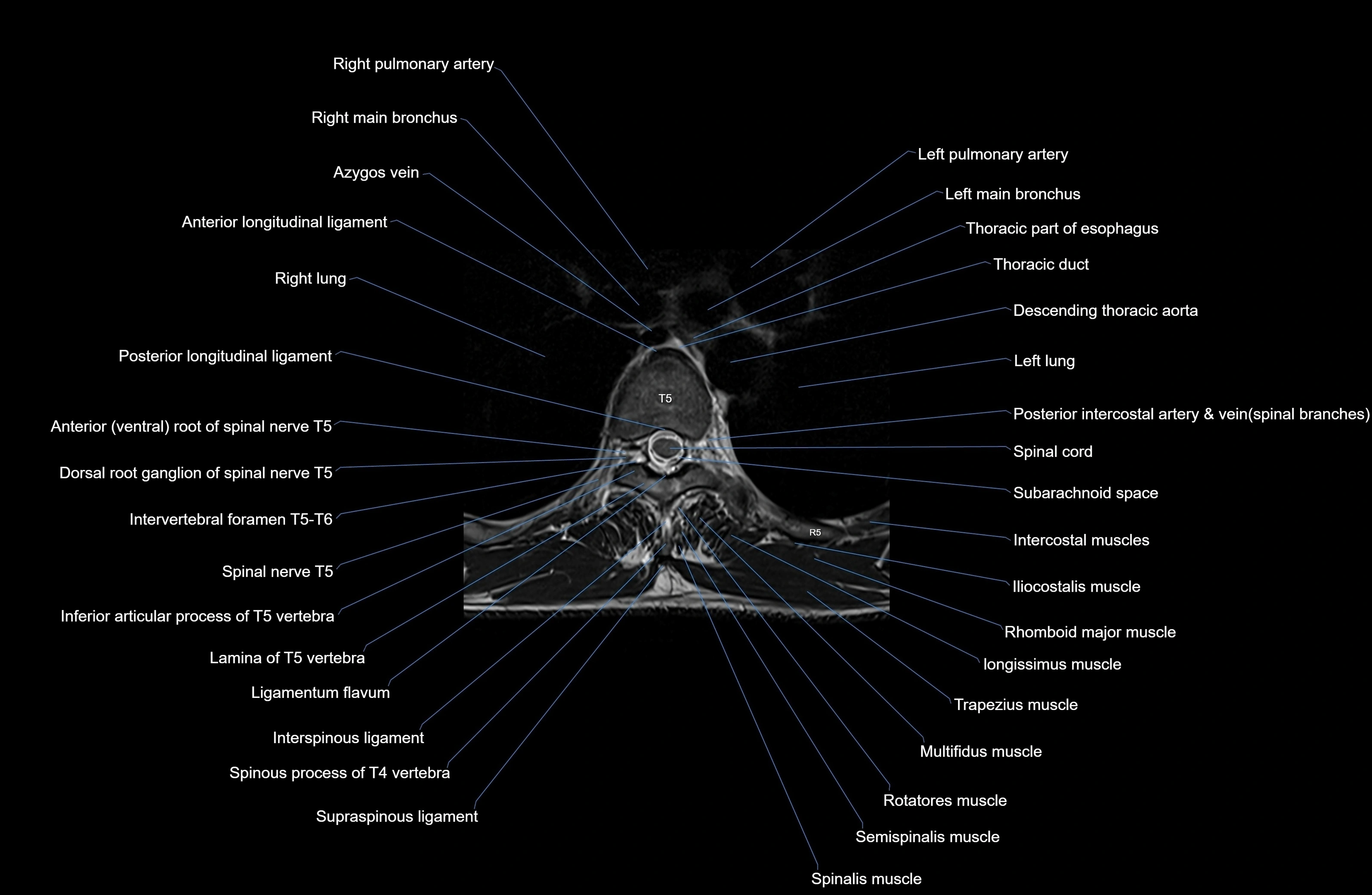
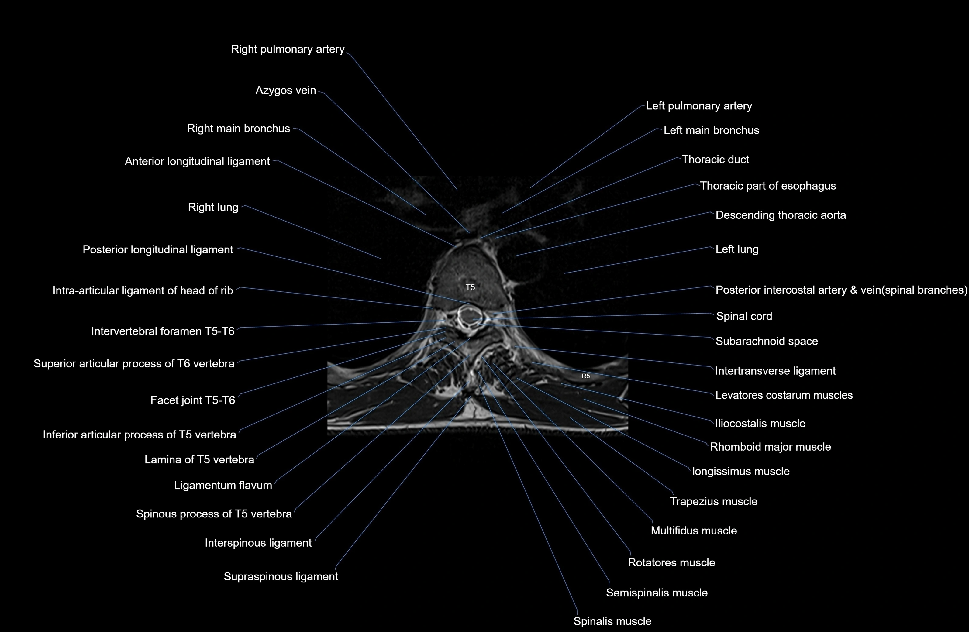
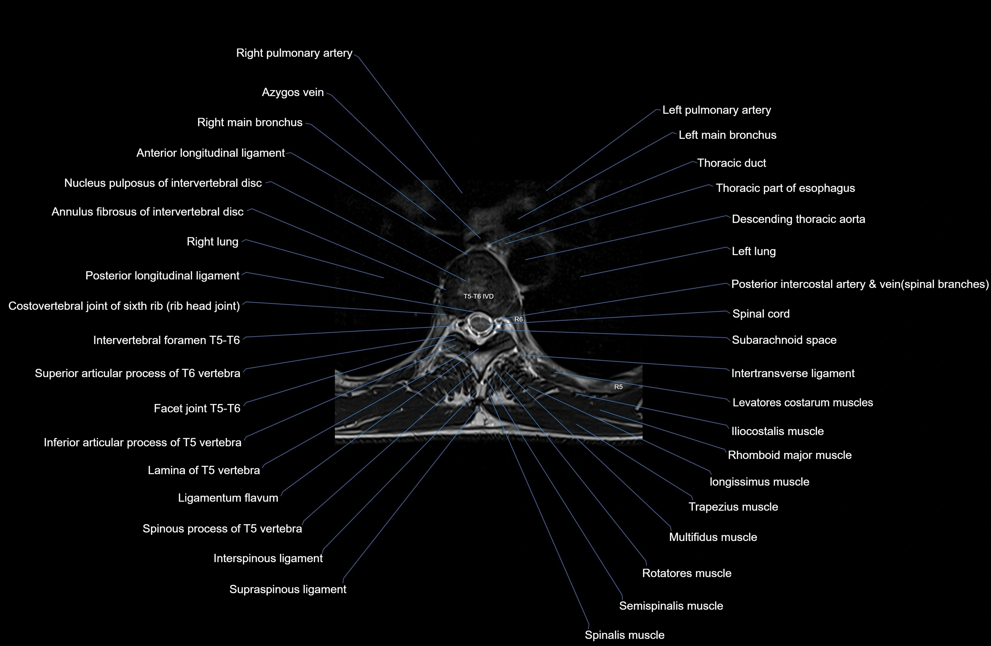
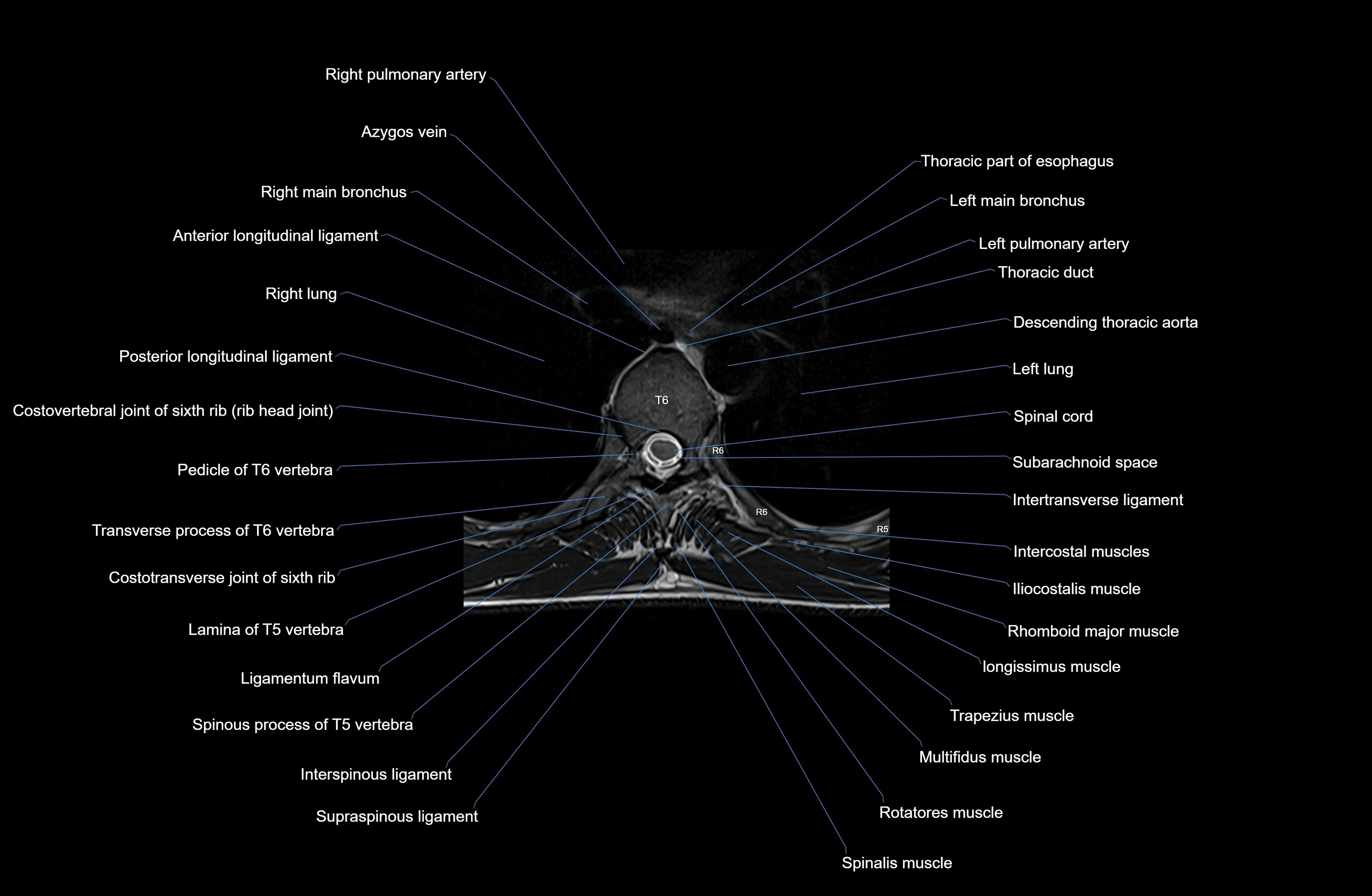
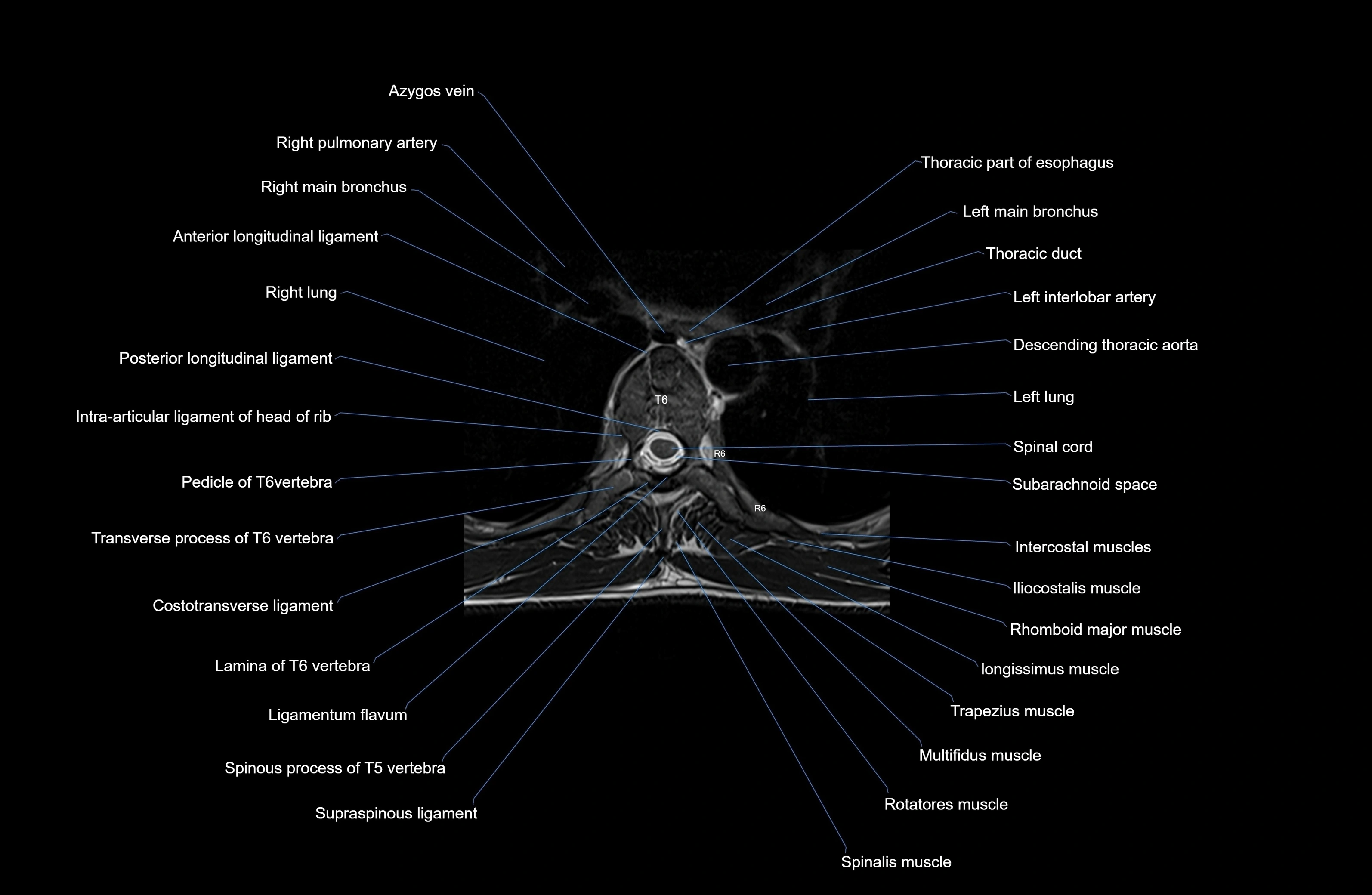
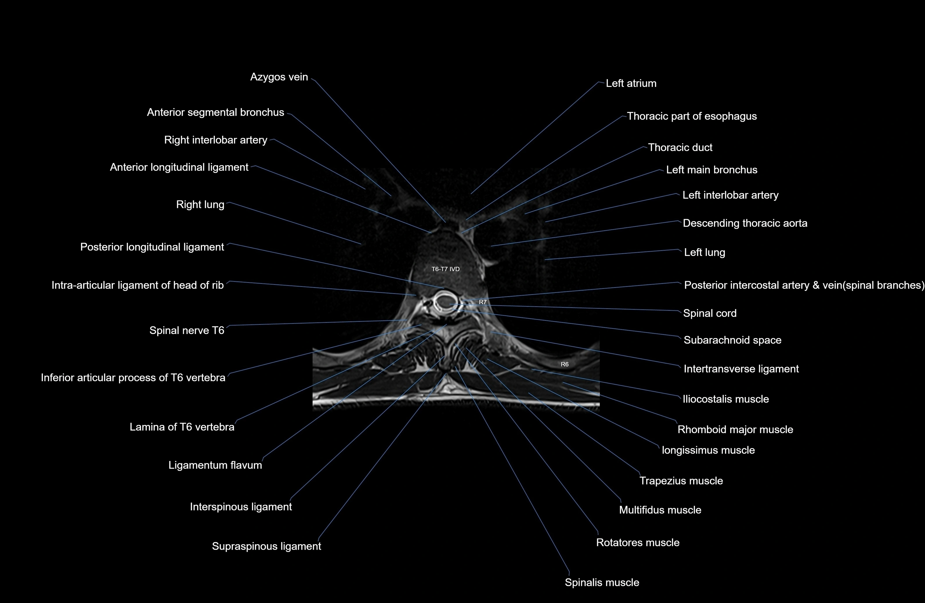
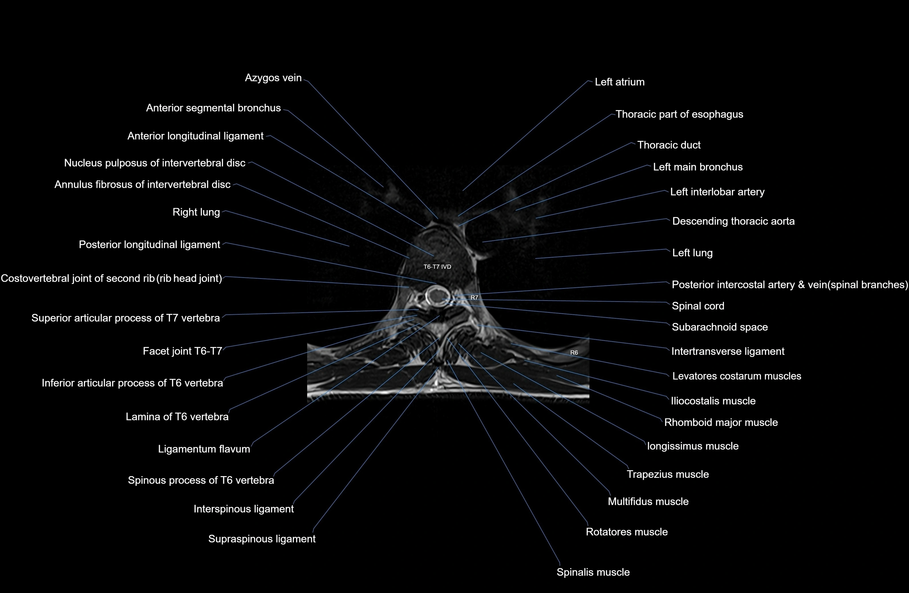
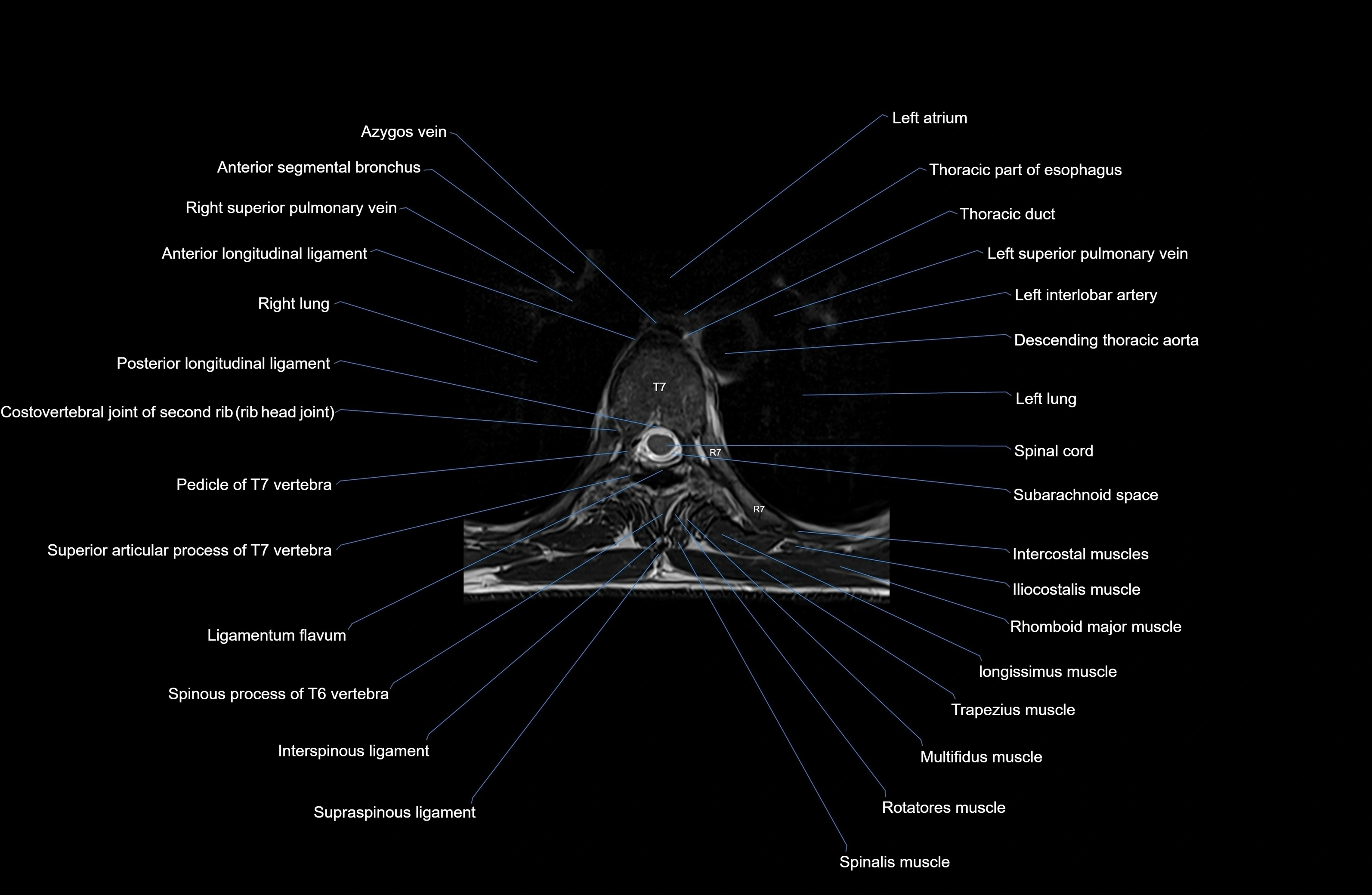
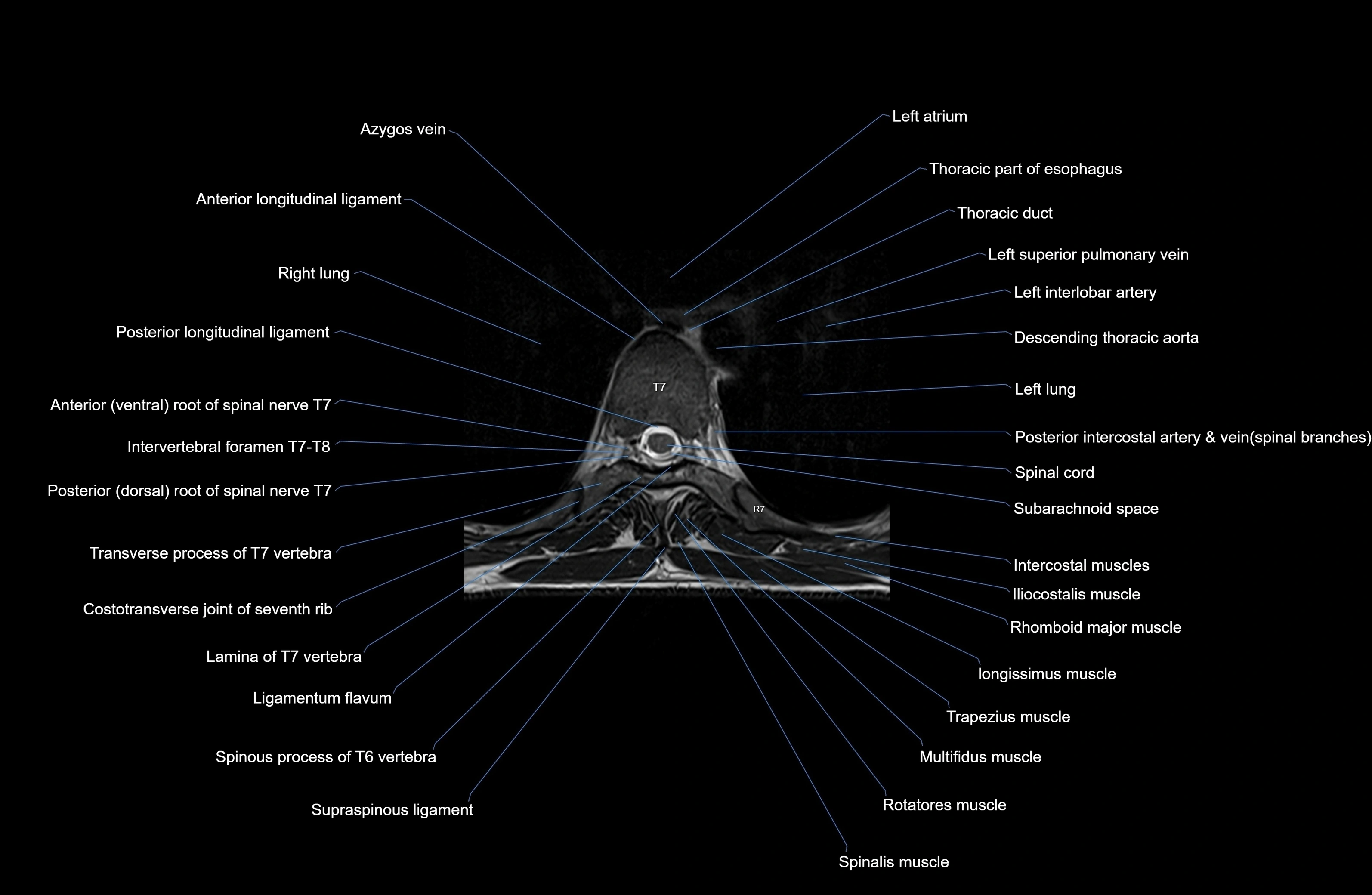
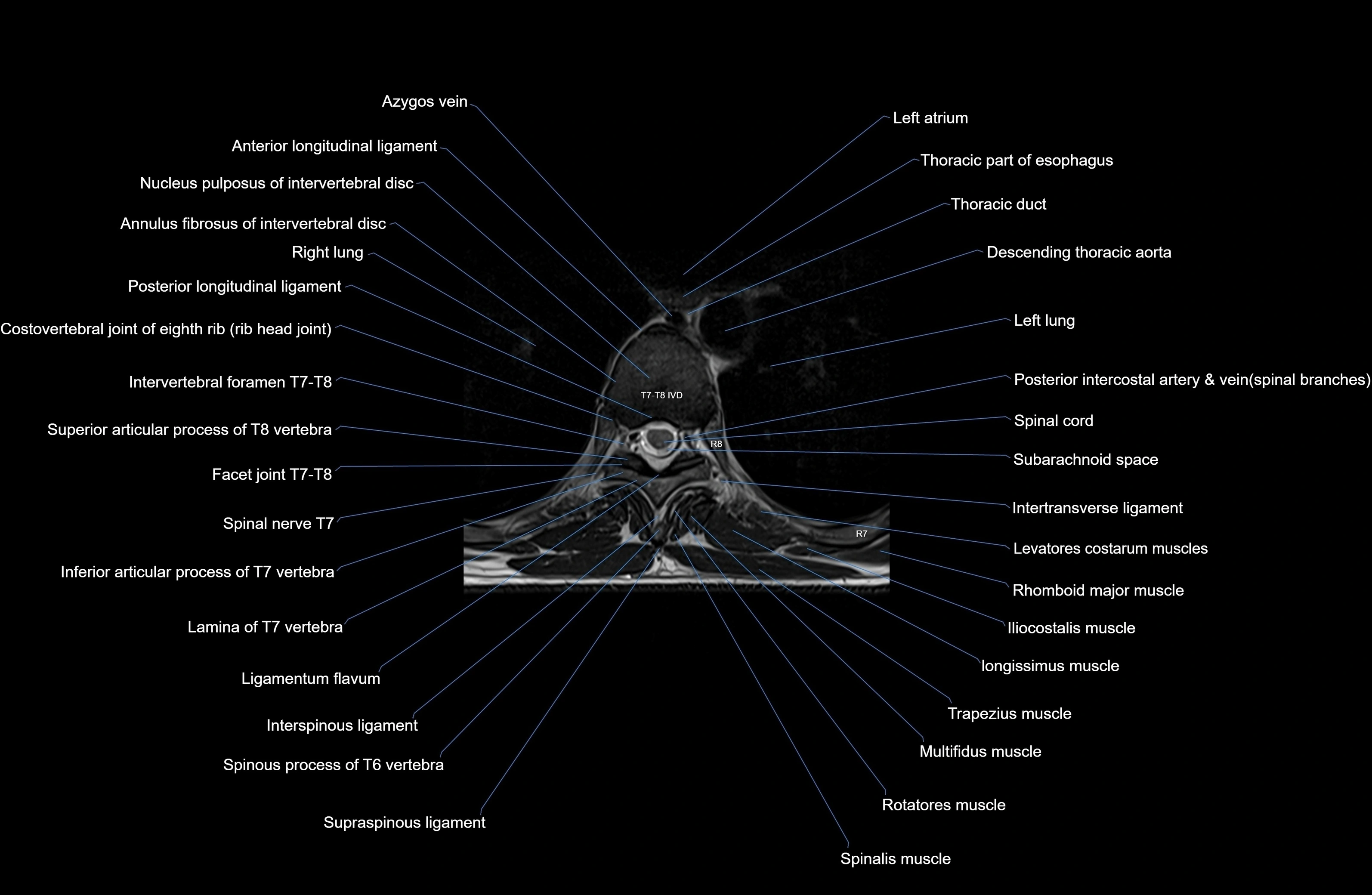
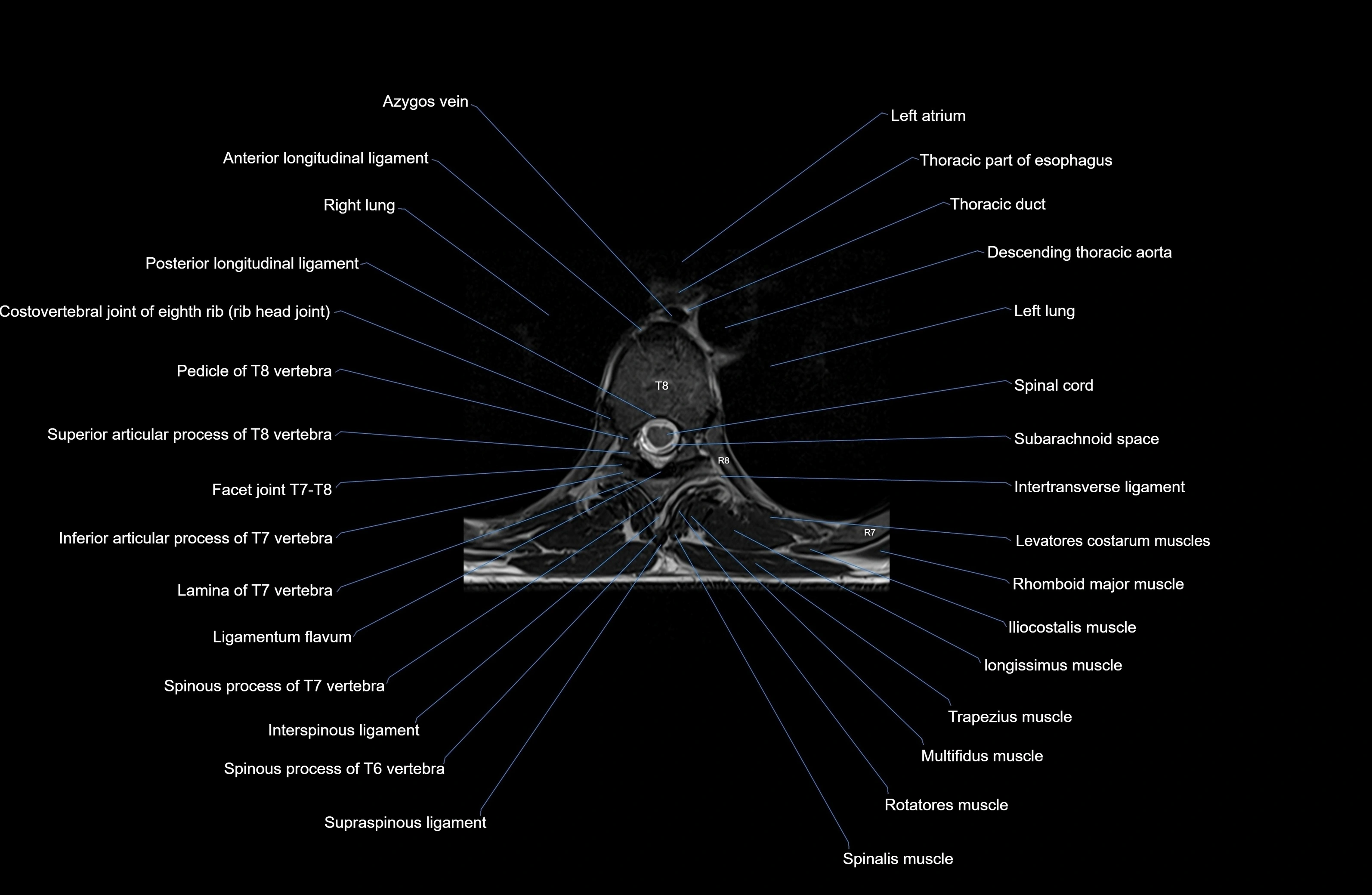
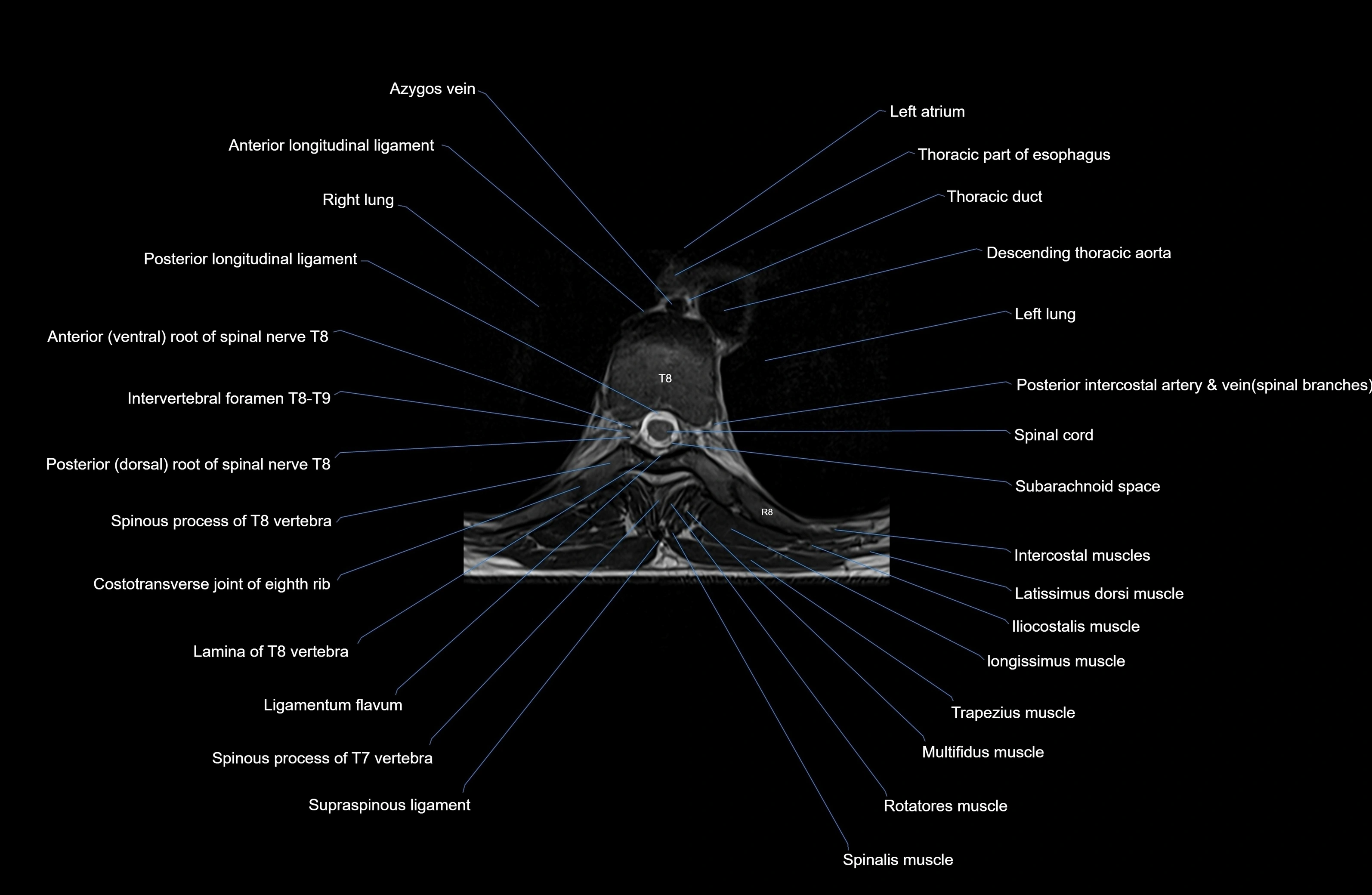
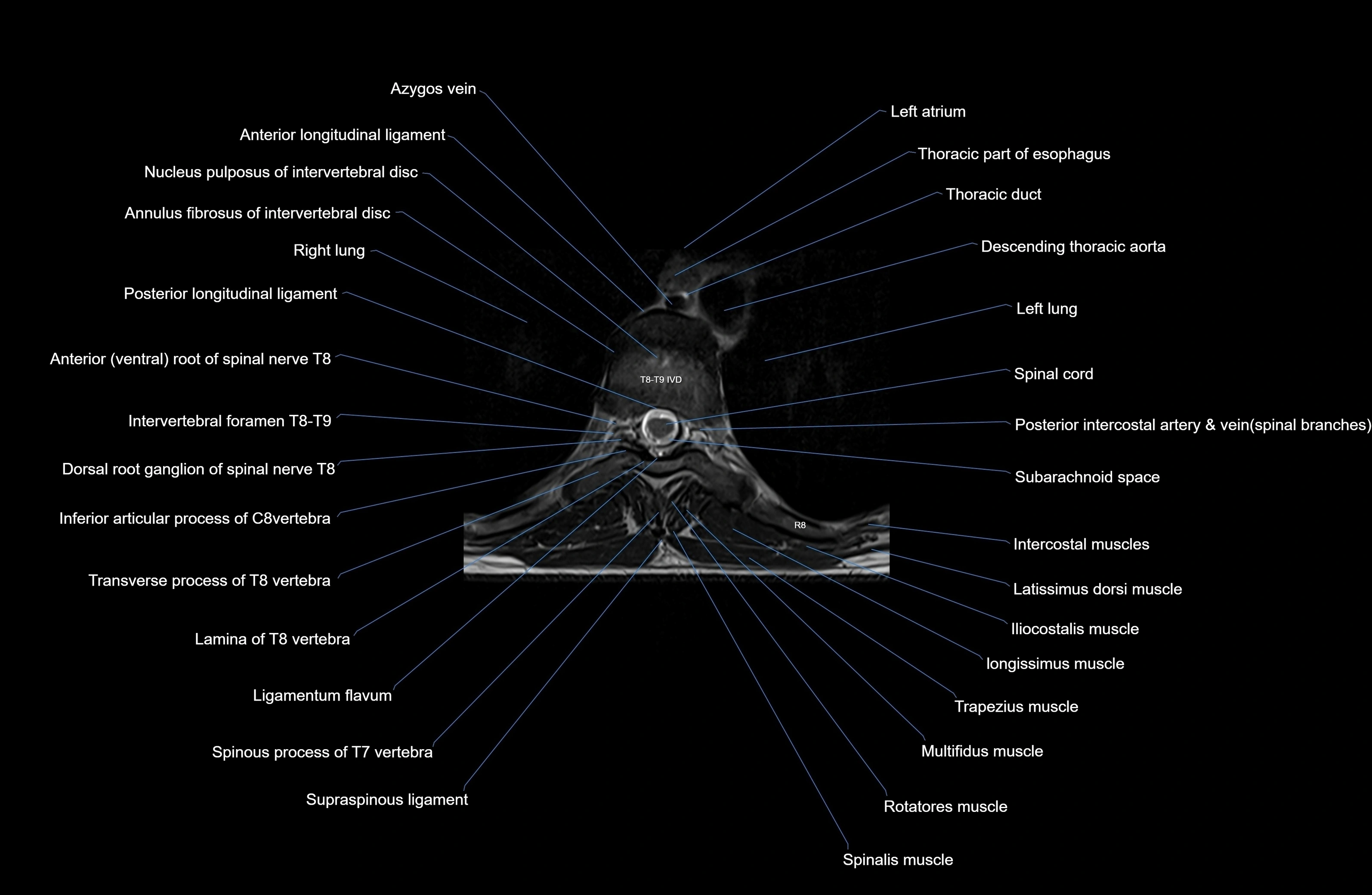
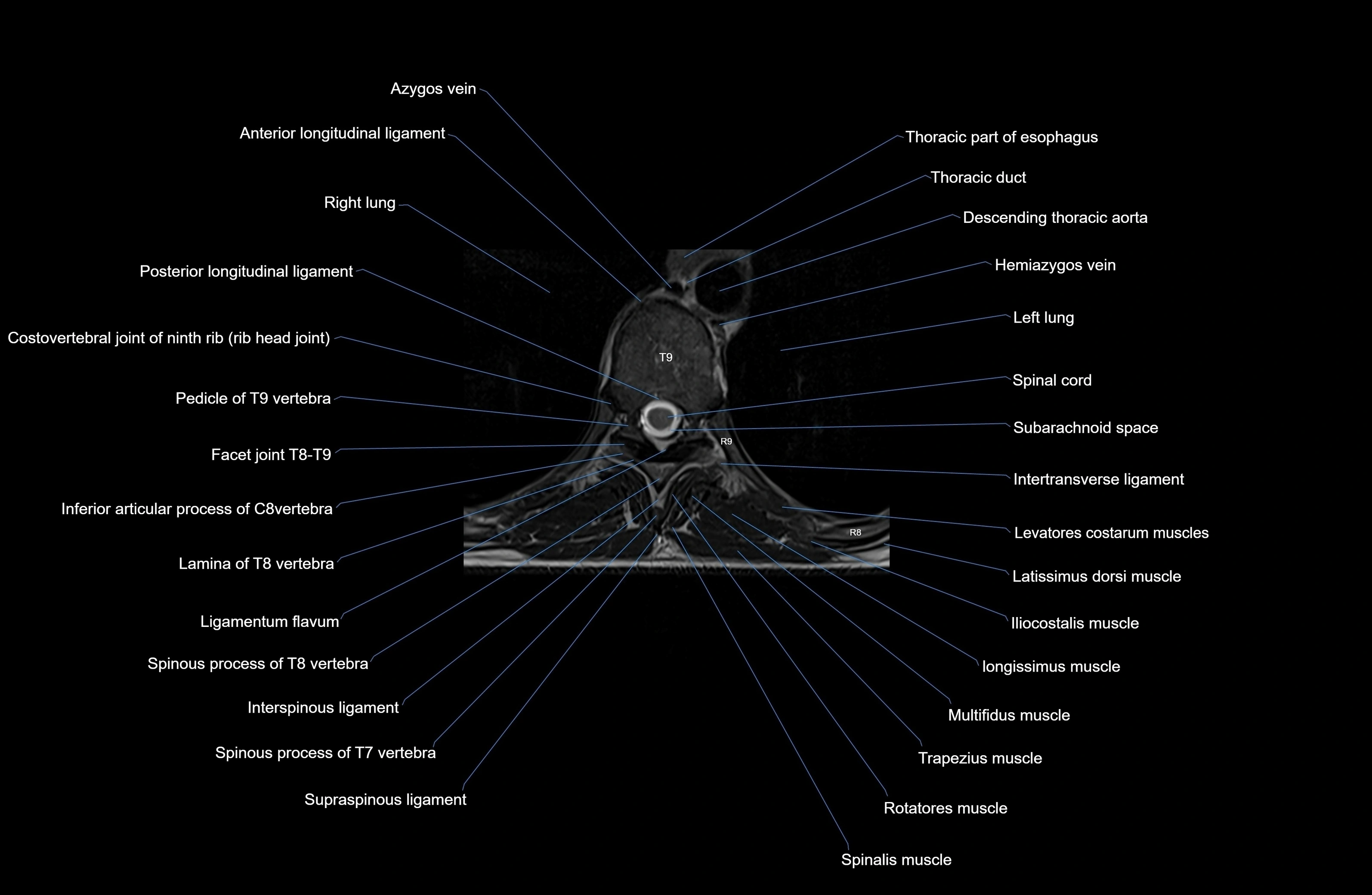
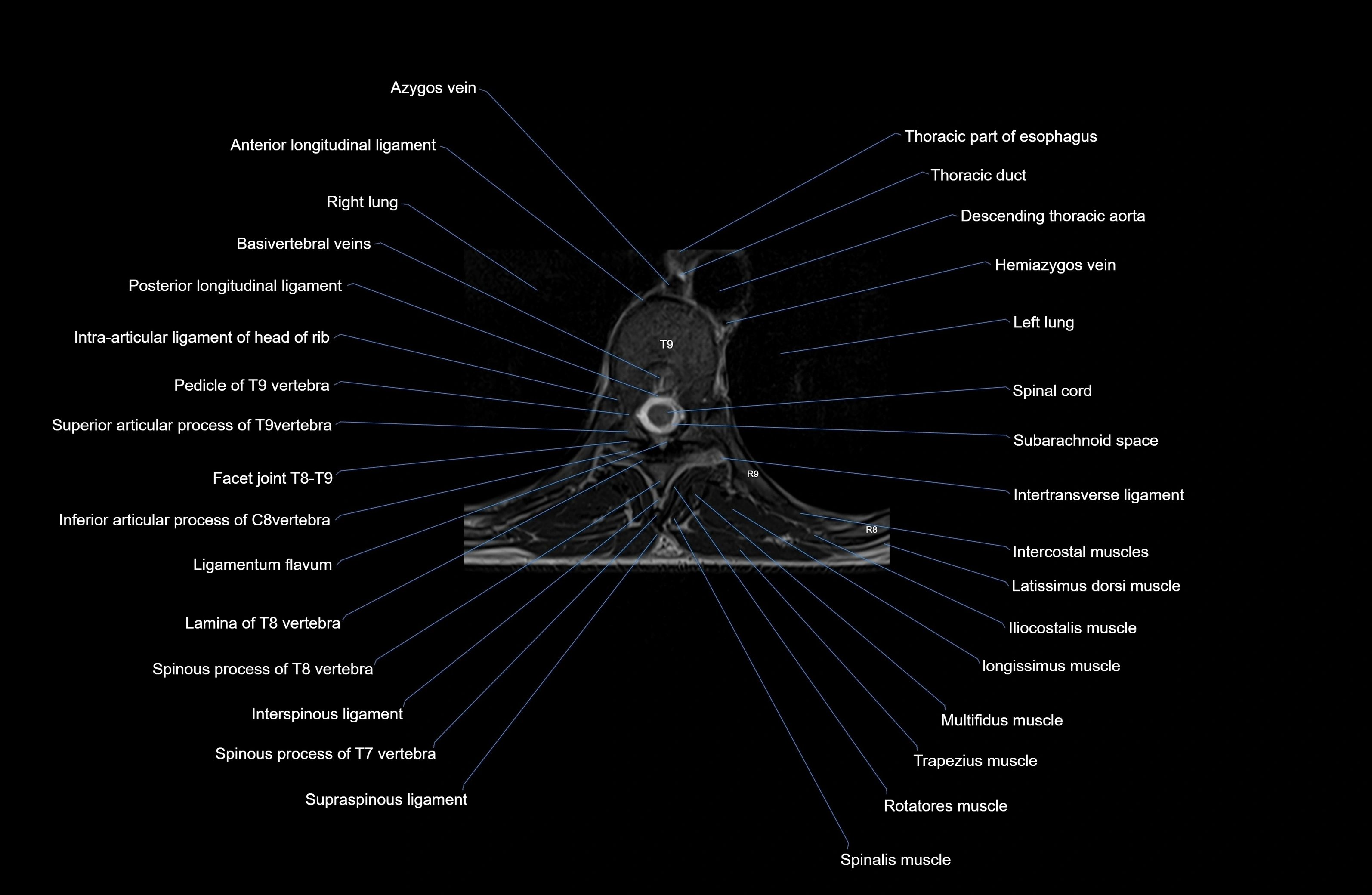
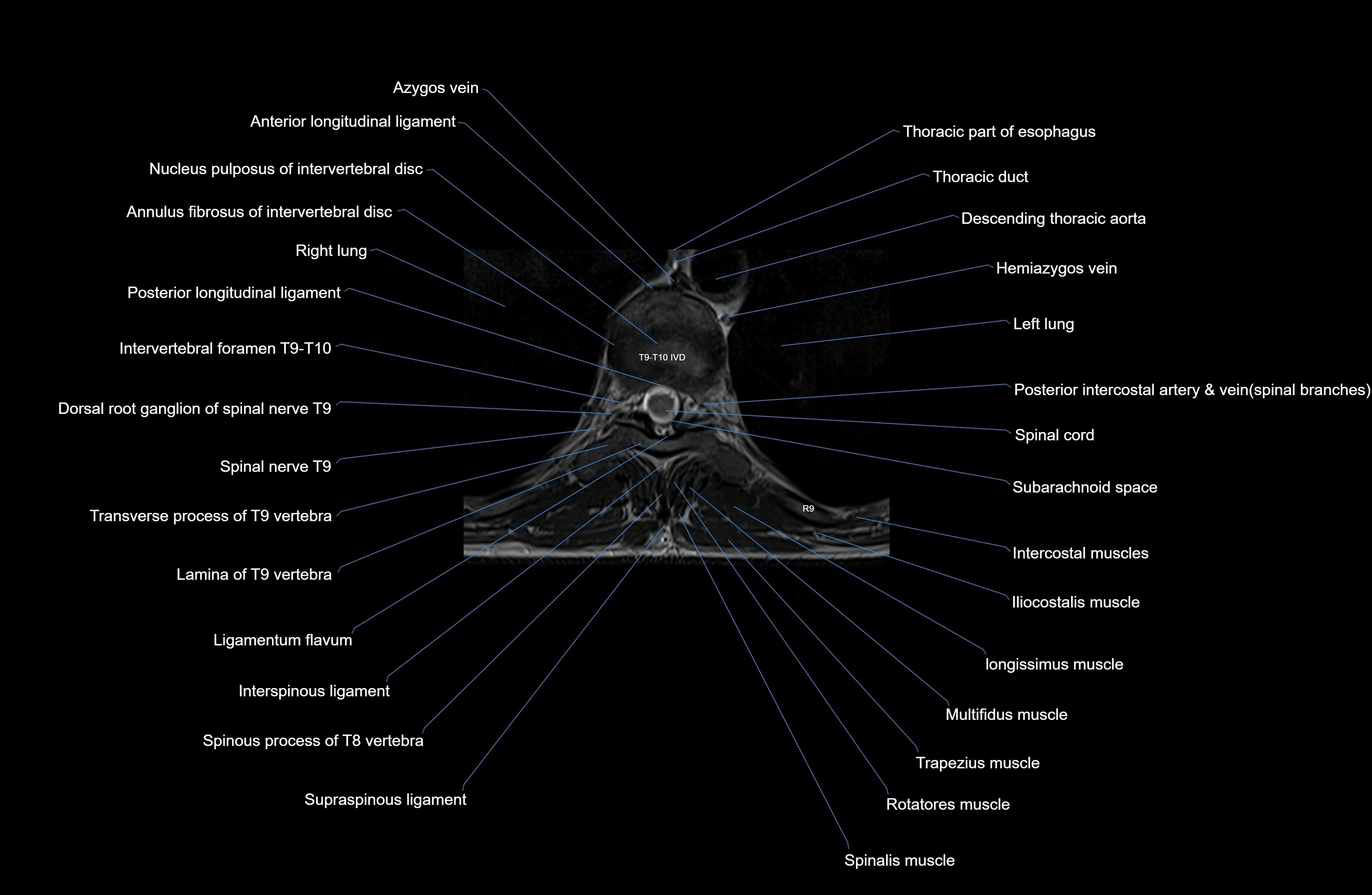
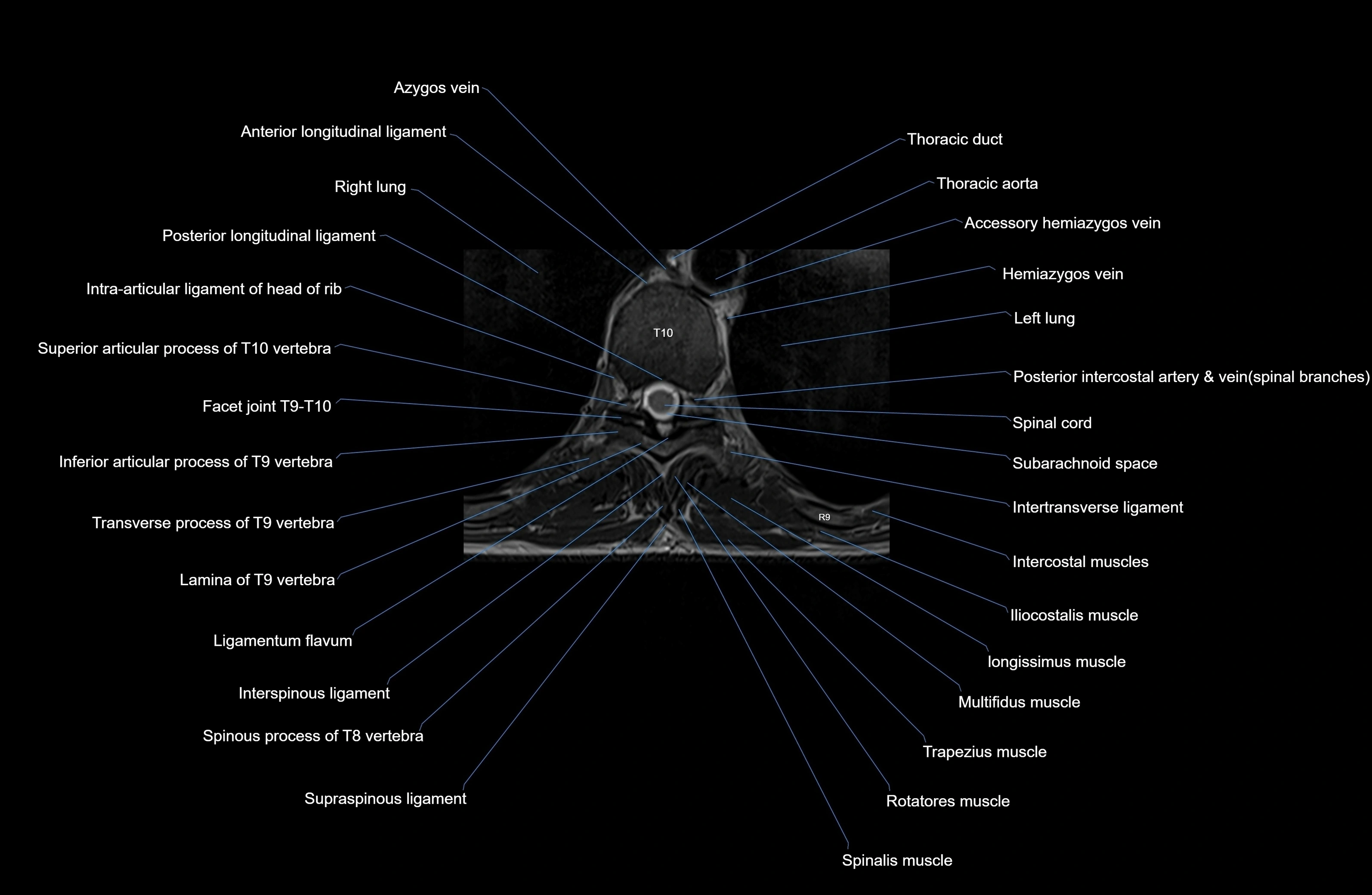
MRI images

MRI images

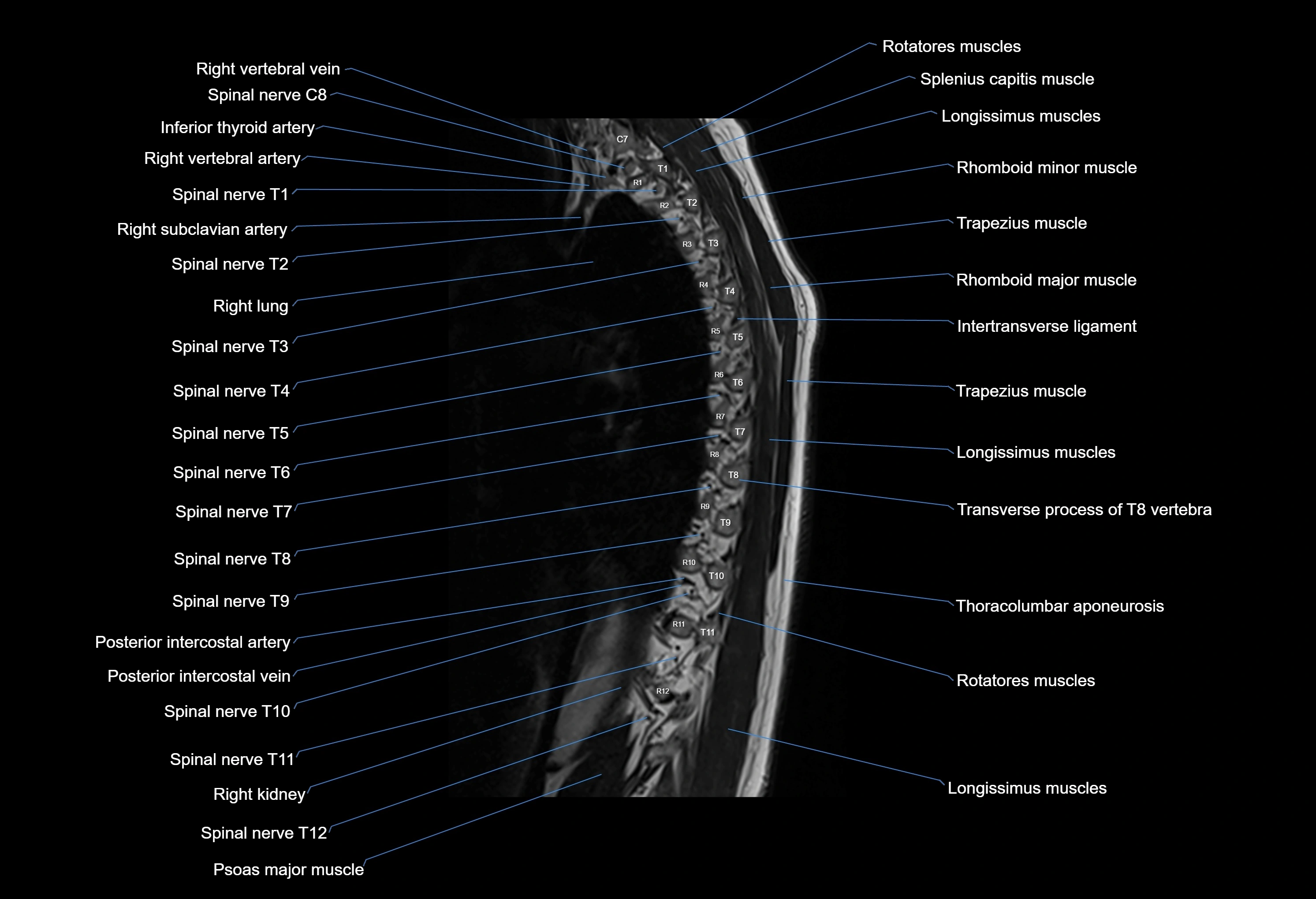
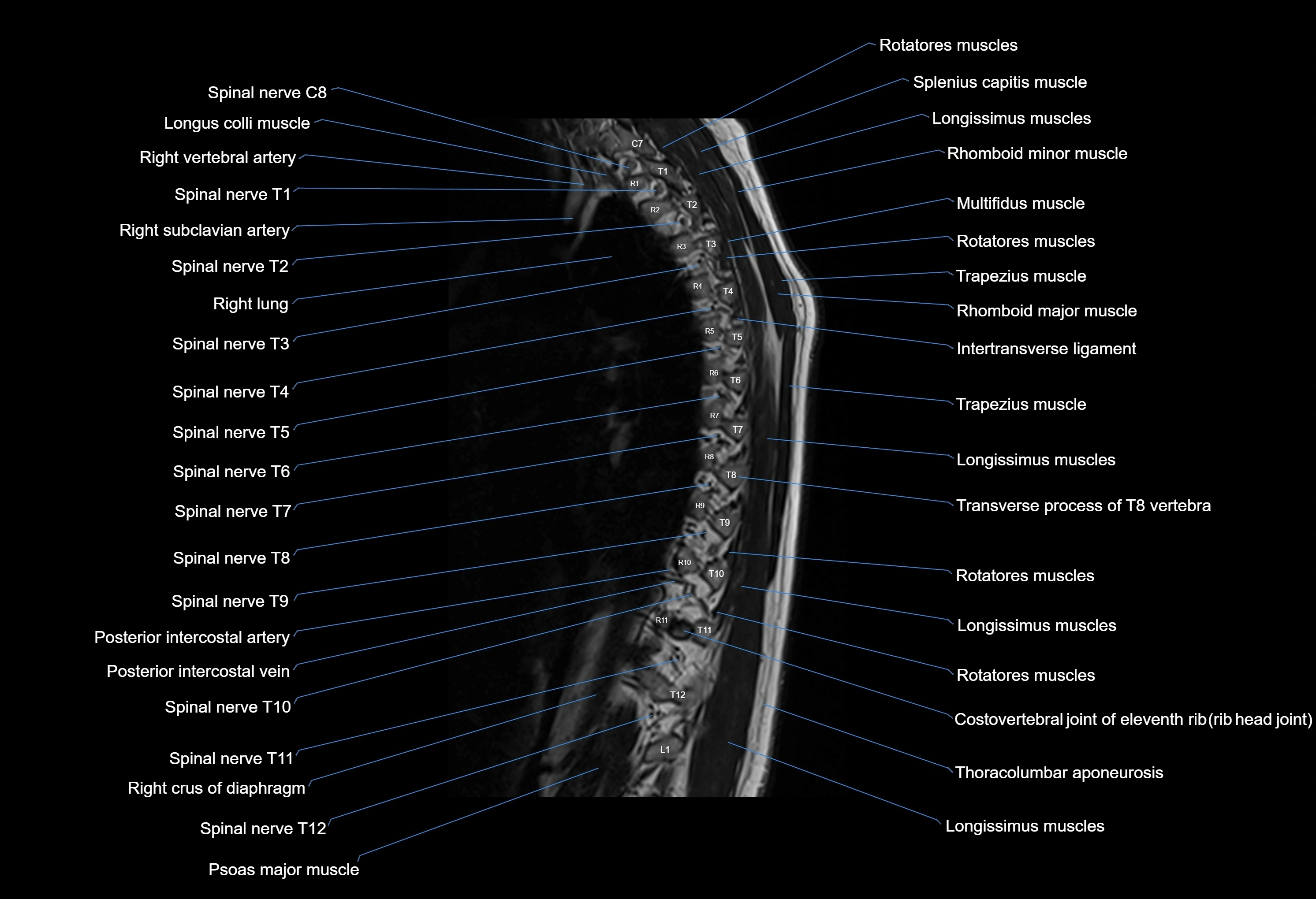
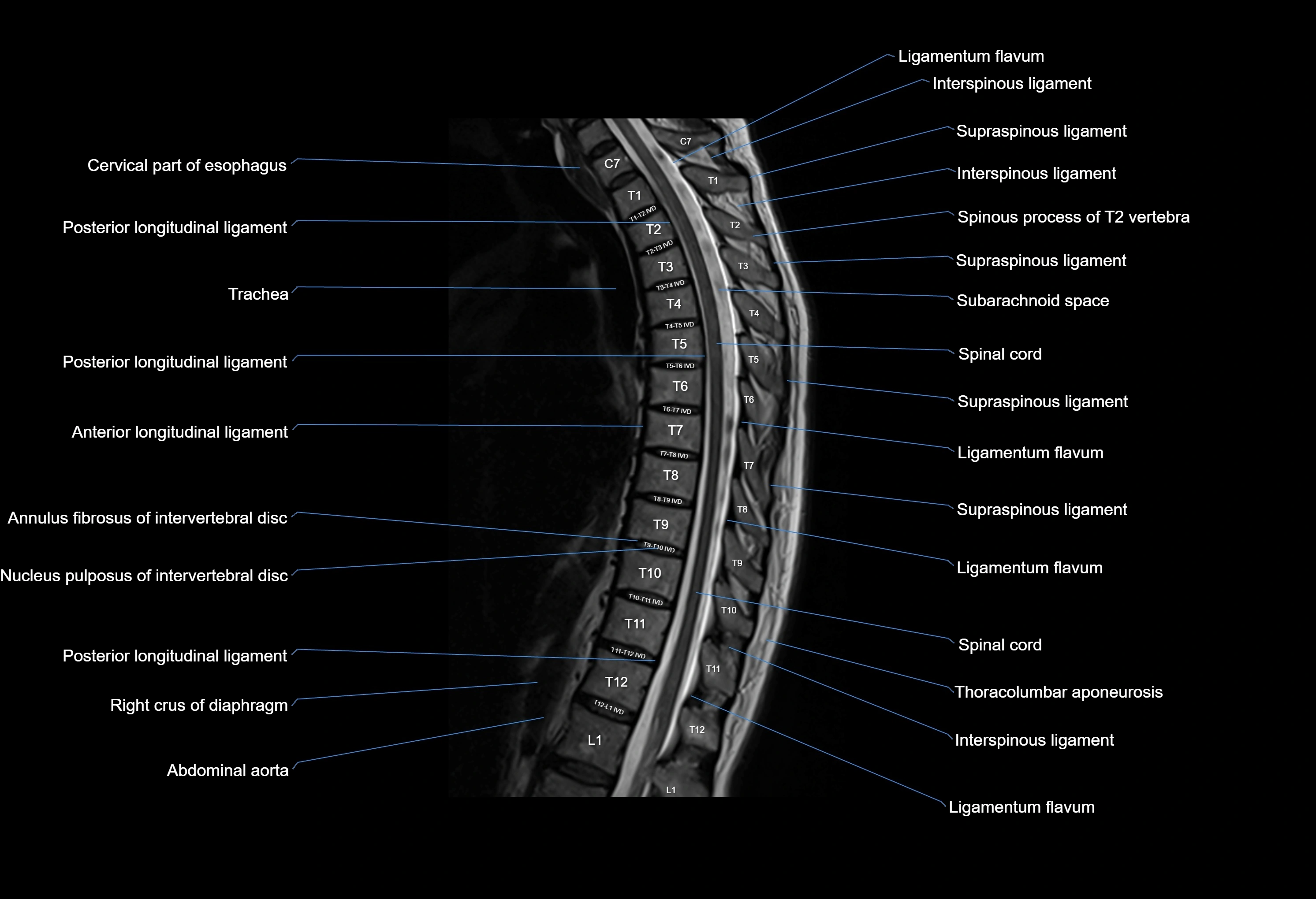
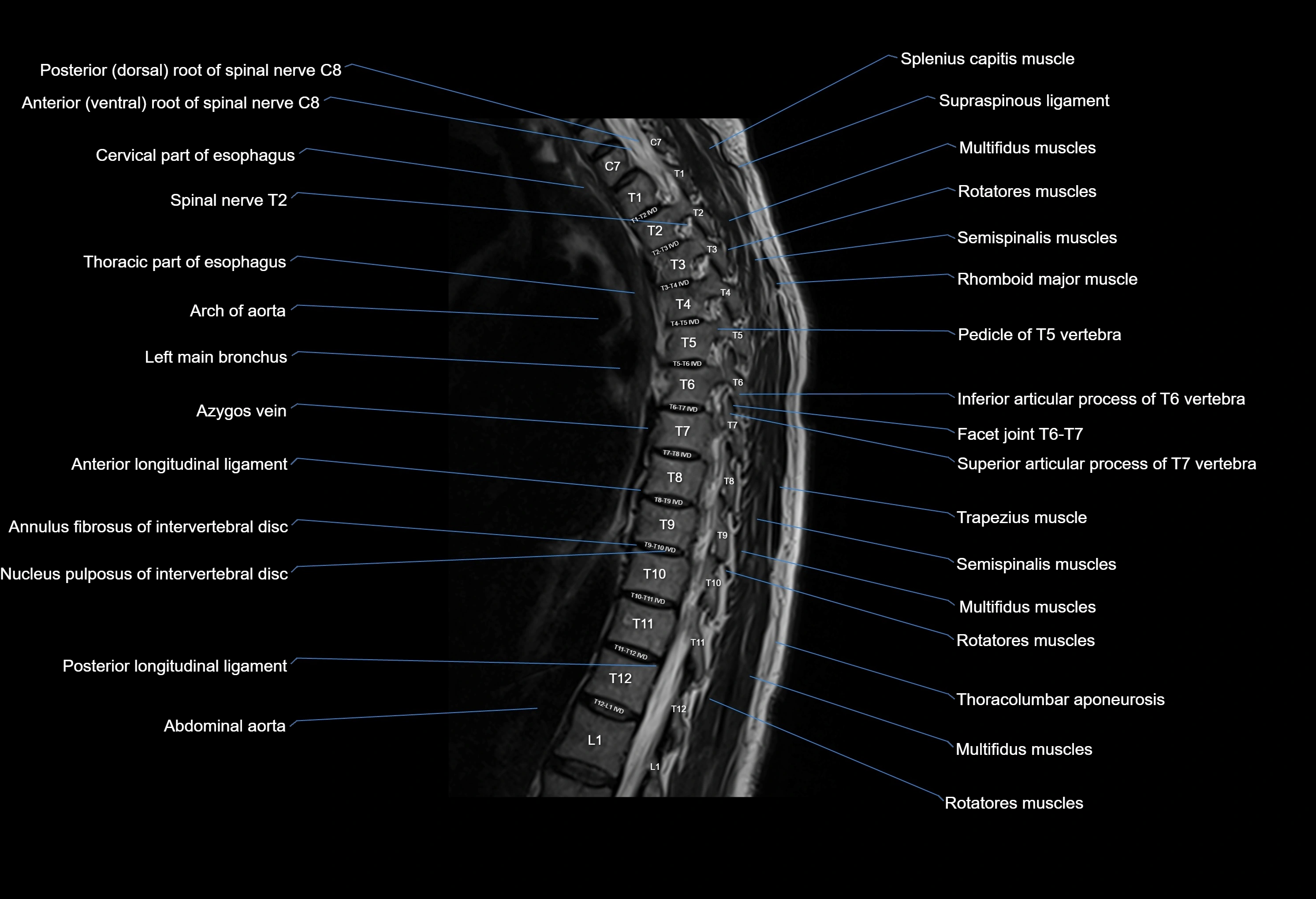
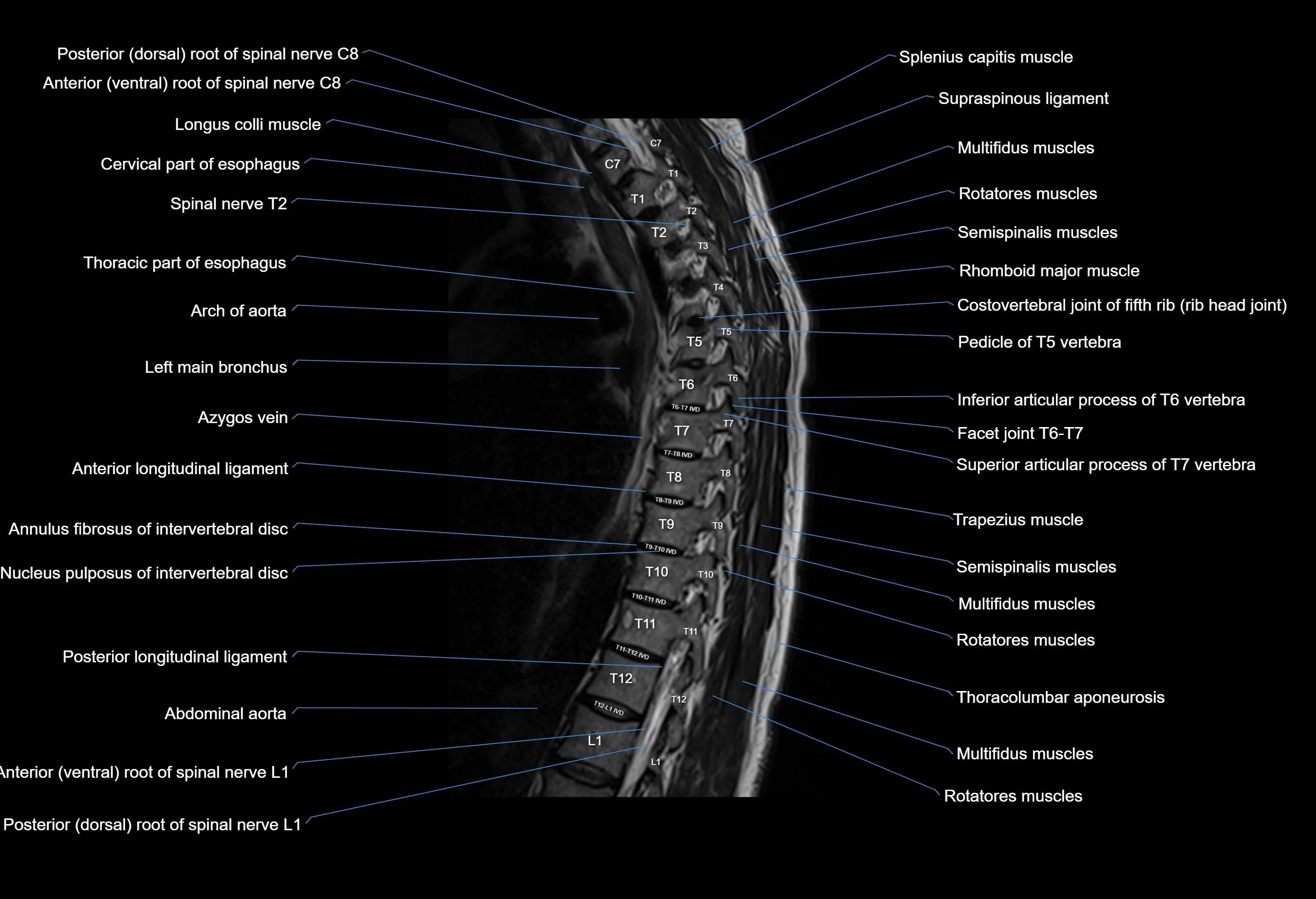
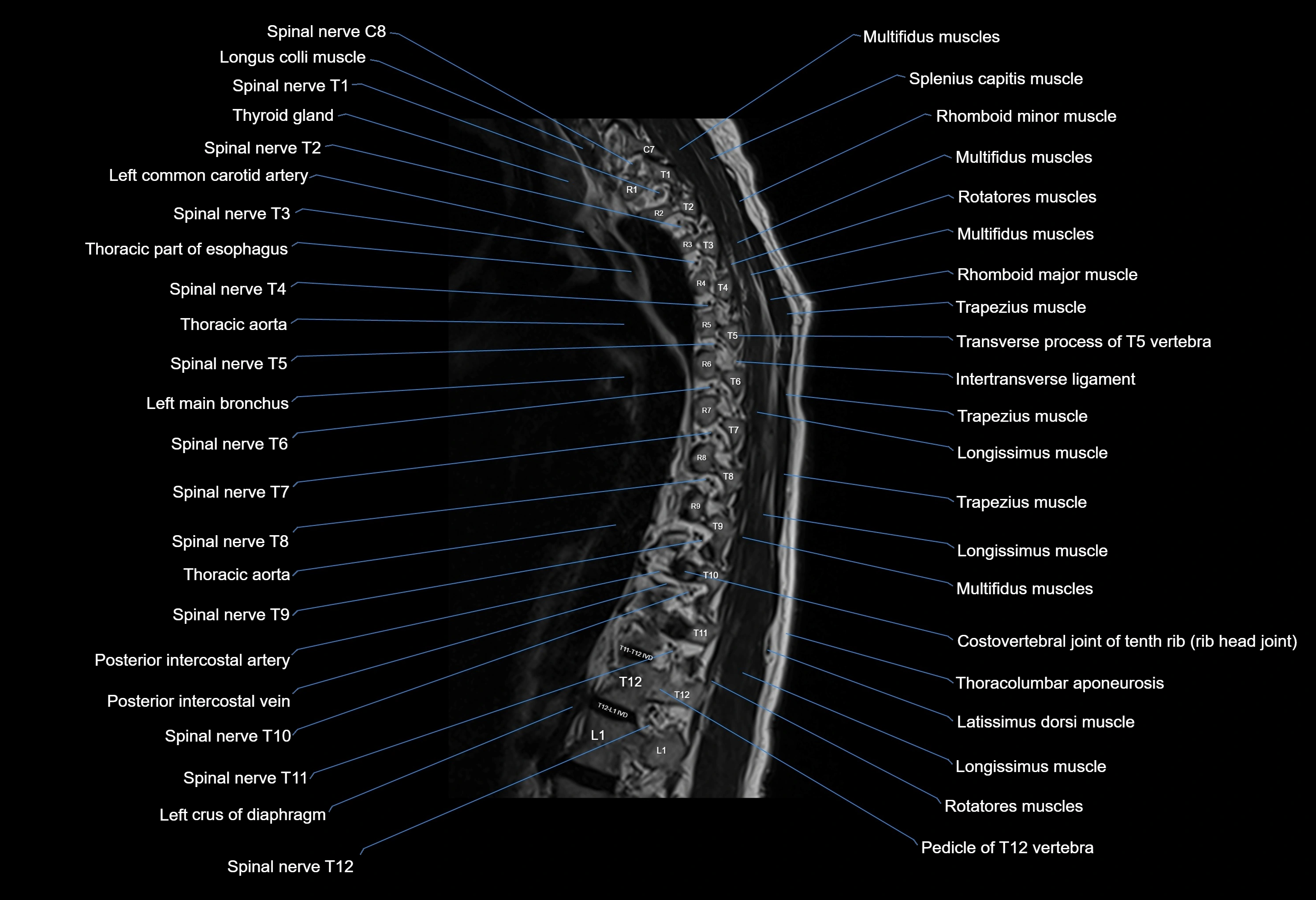
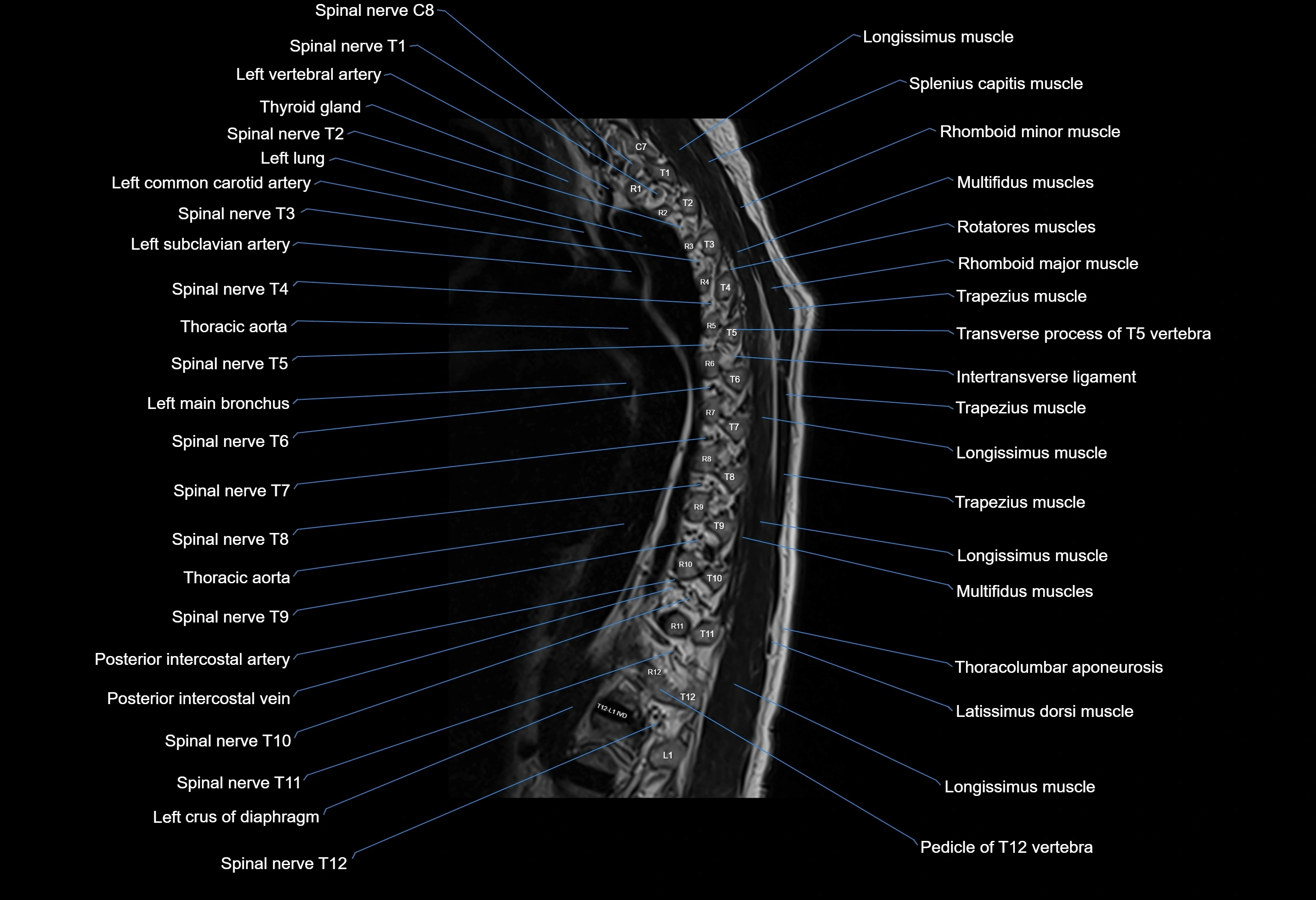
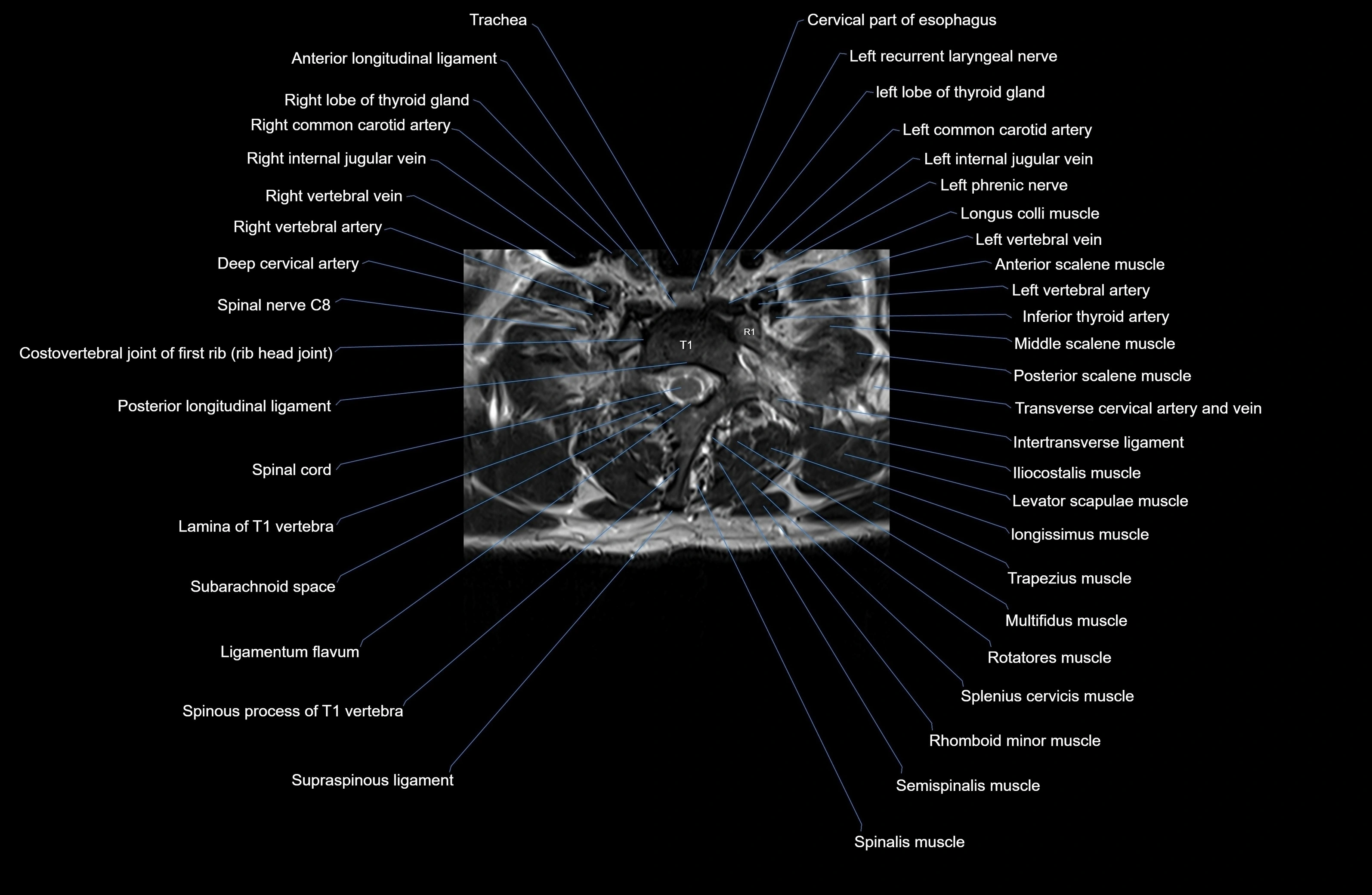
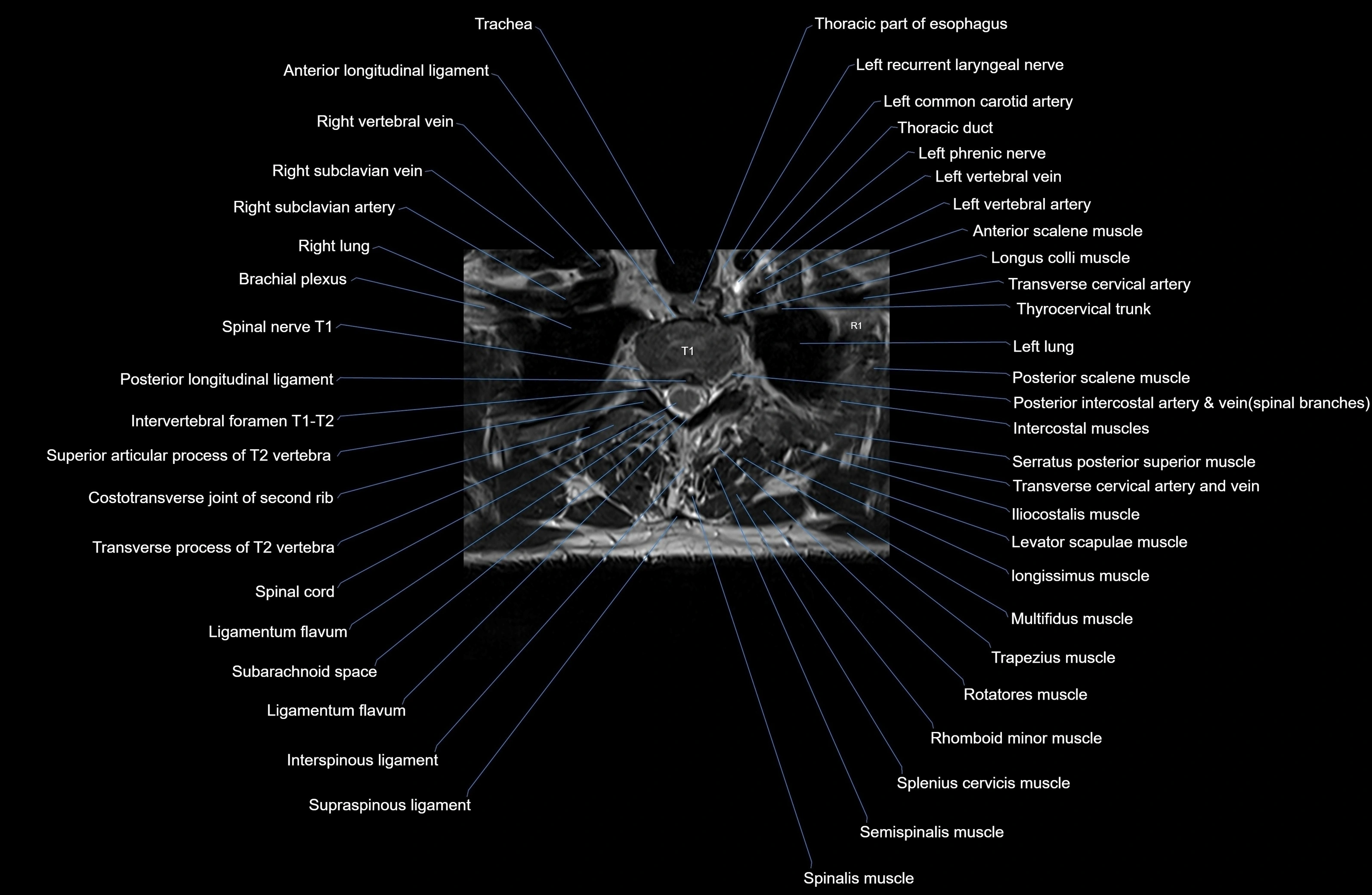
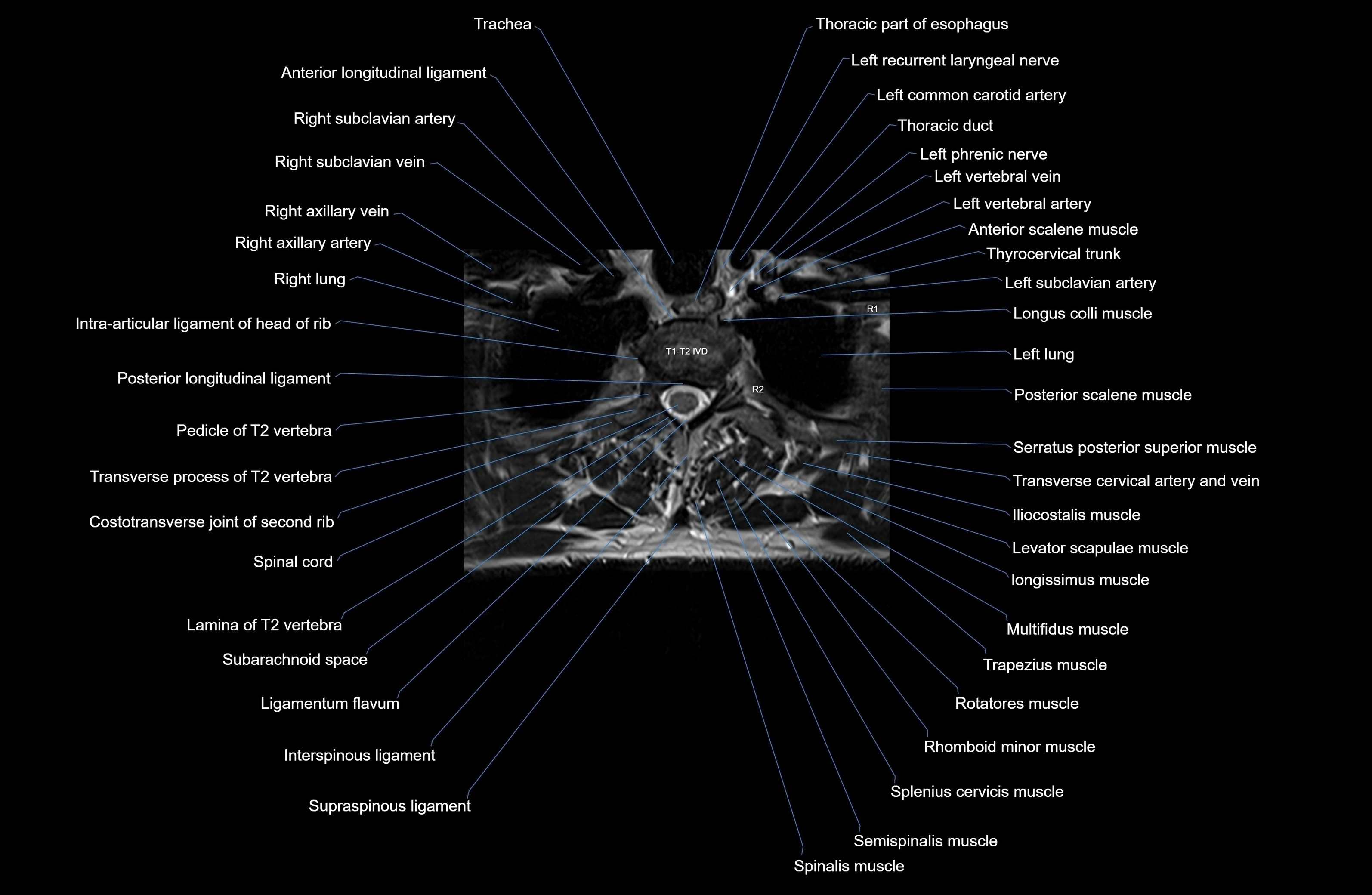
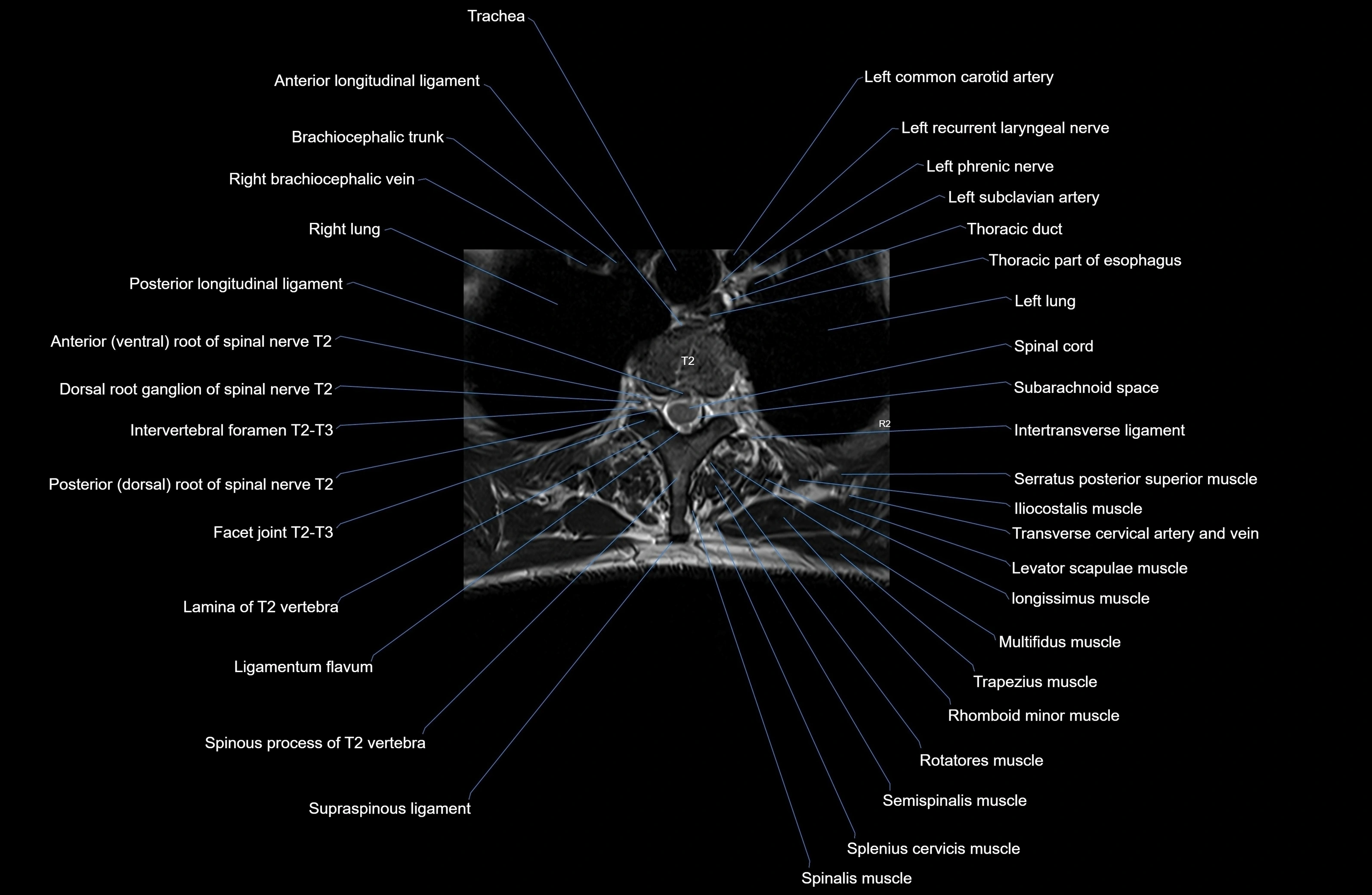
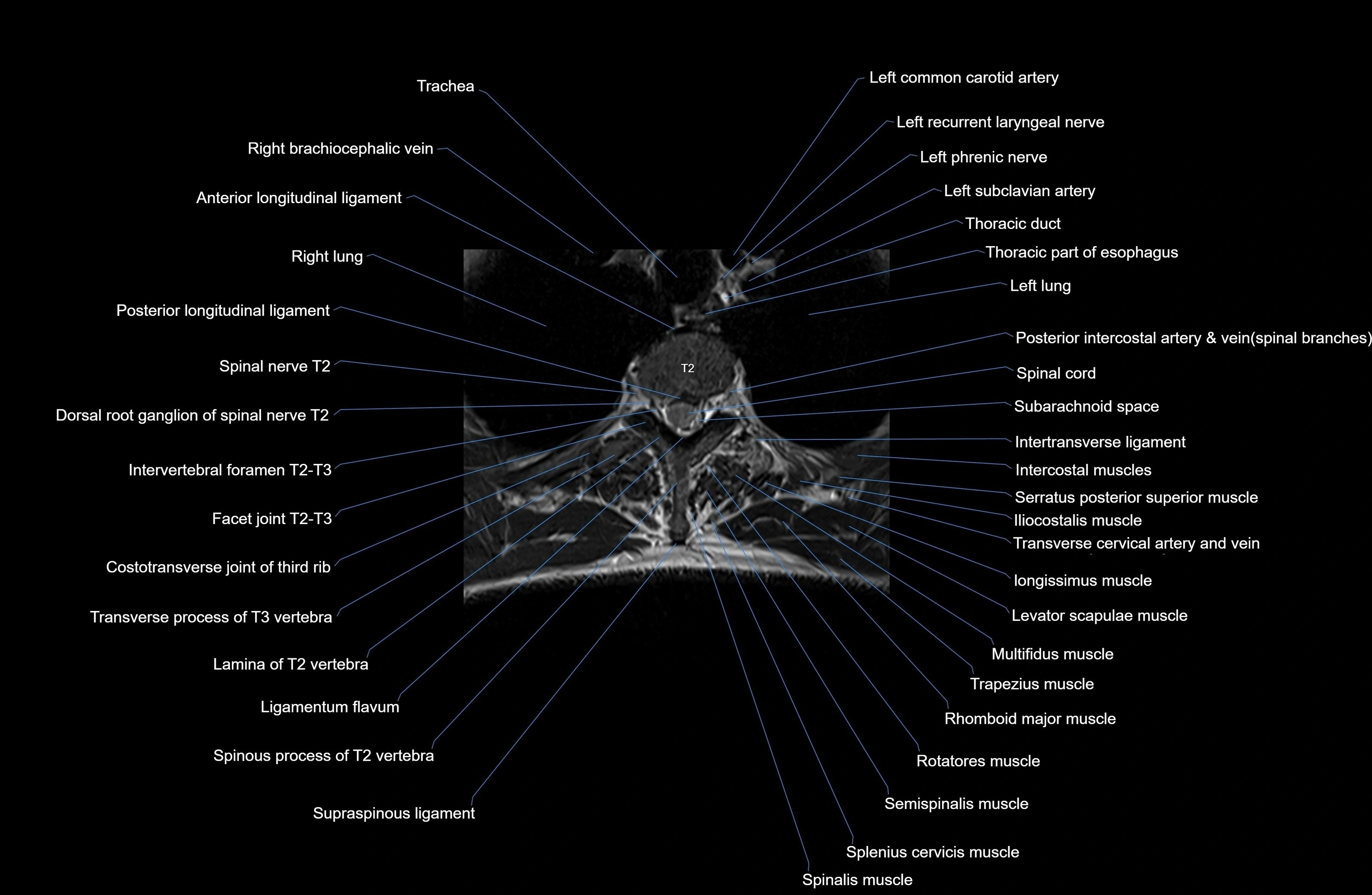
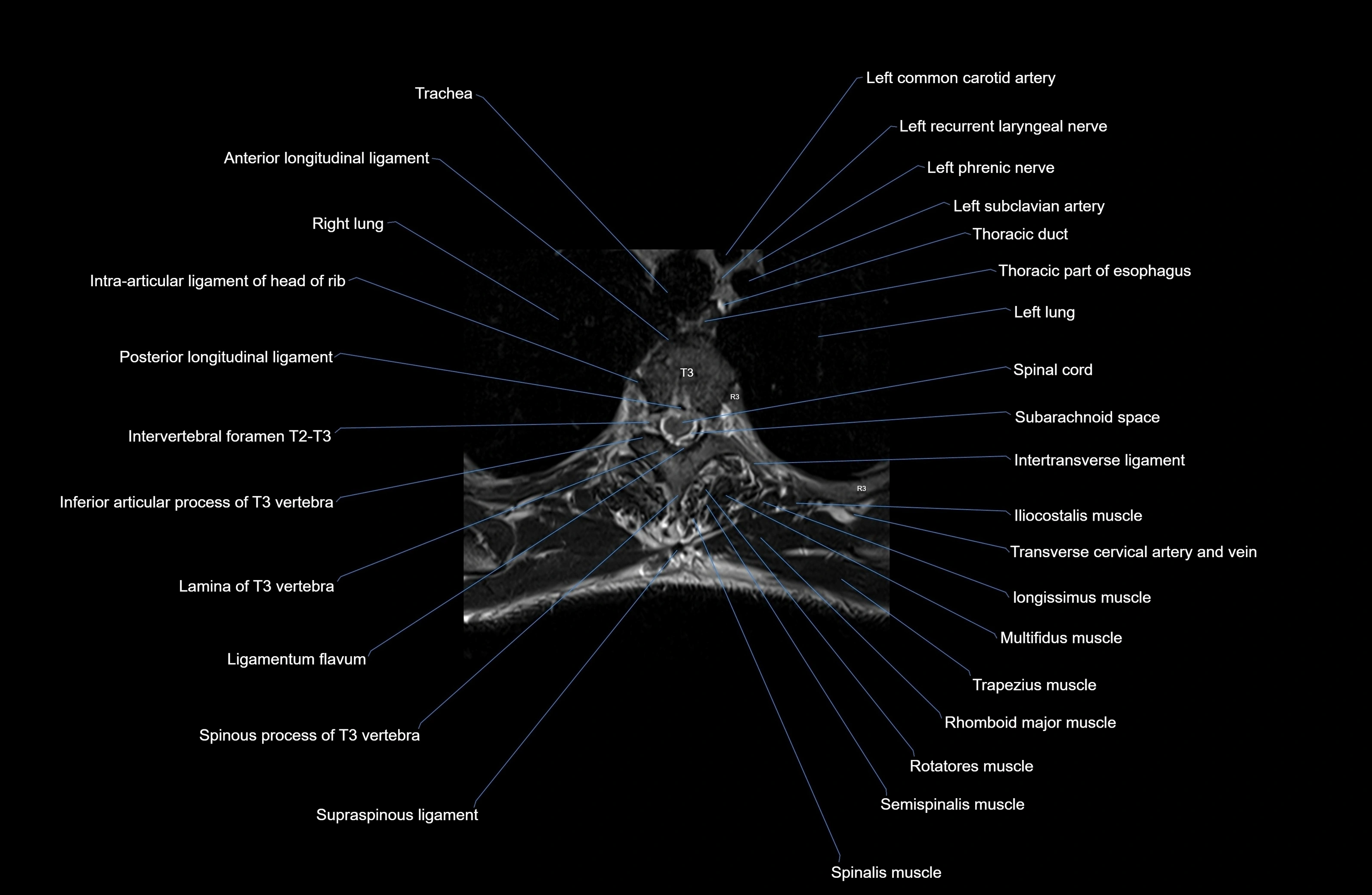
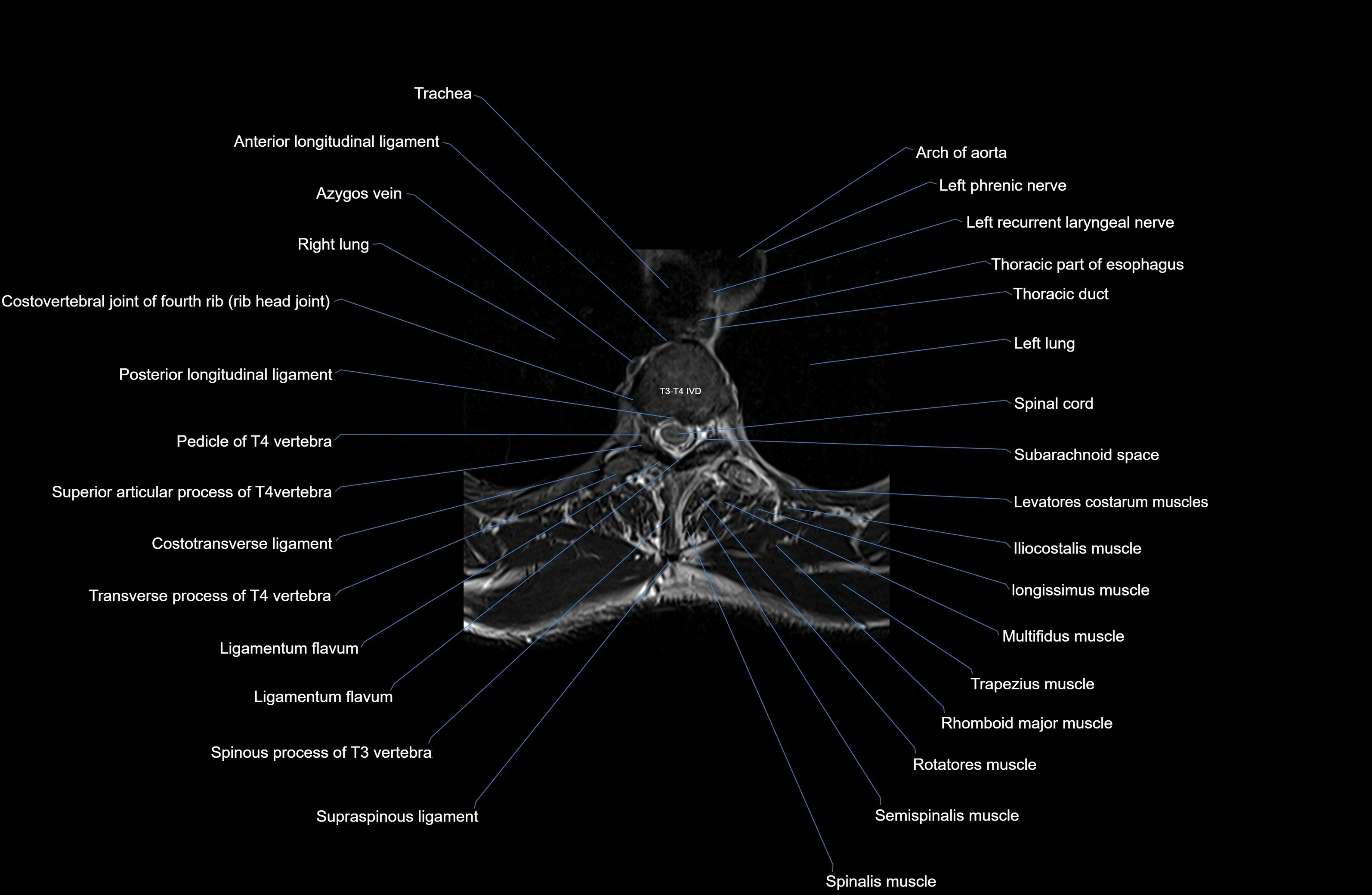
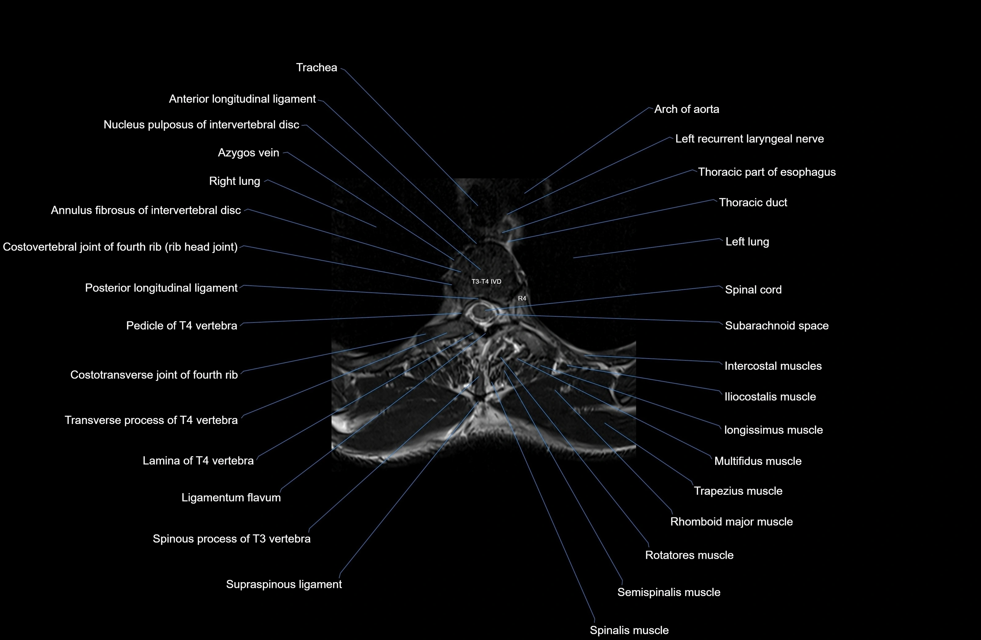
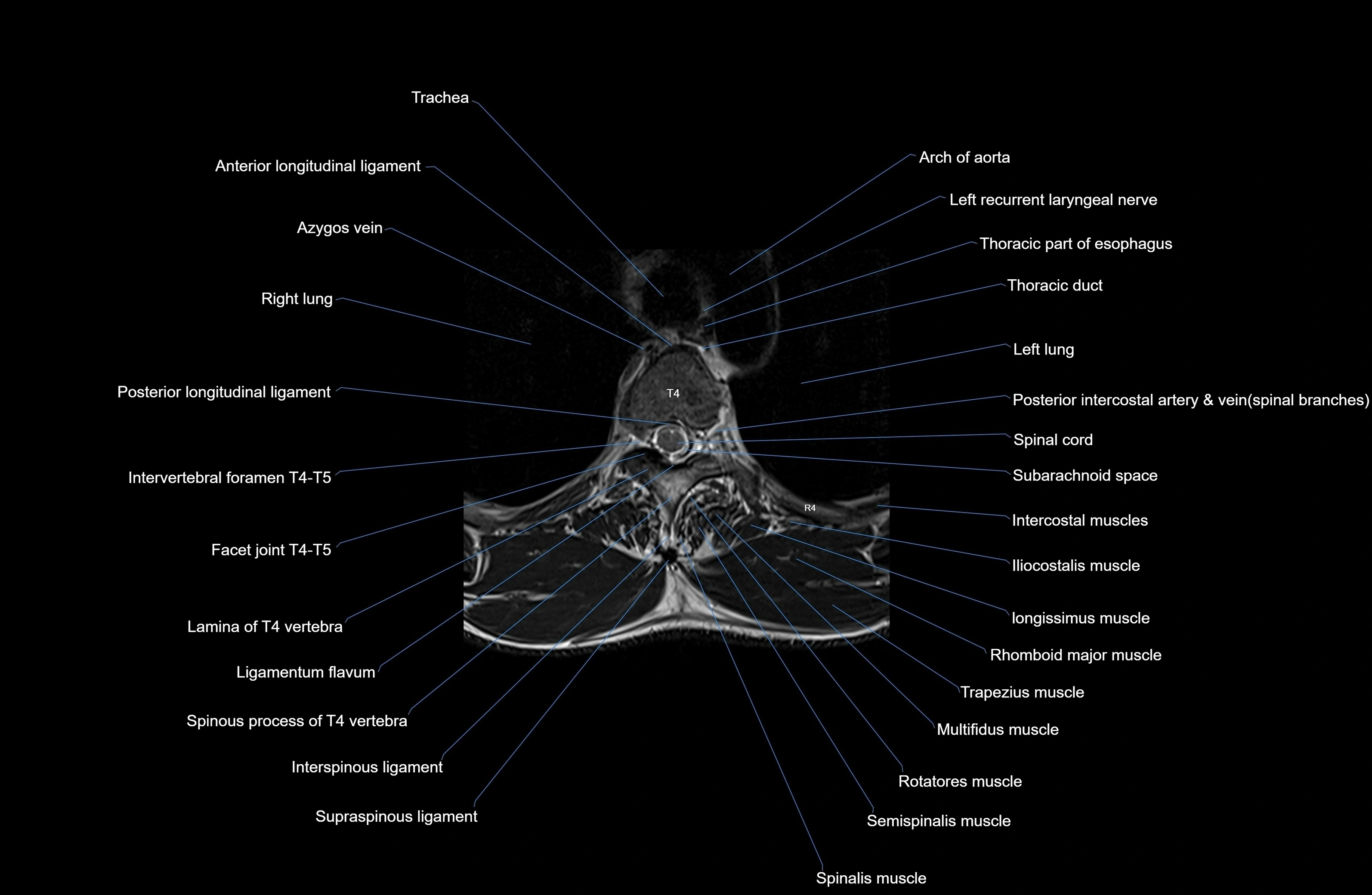
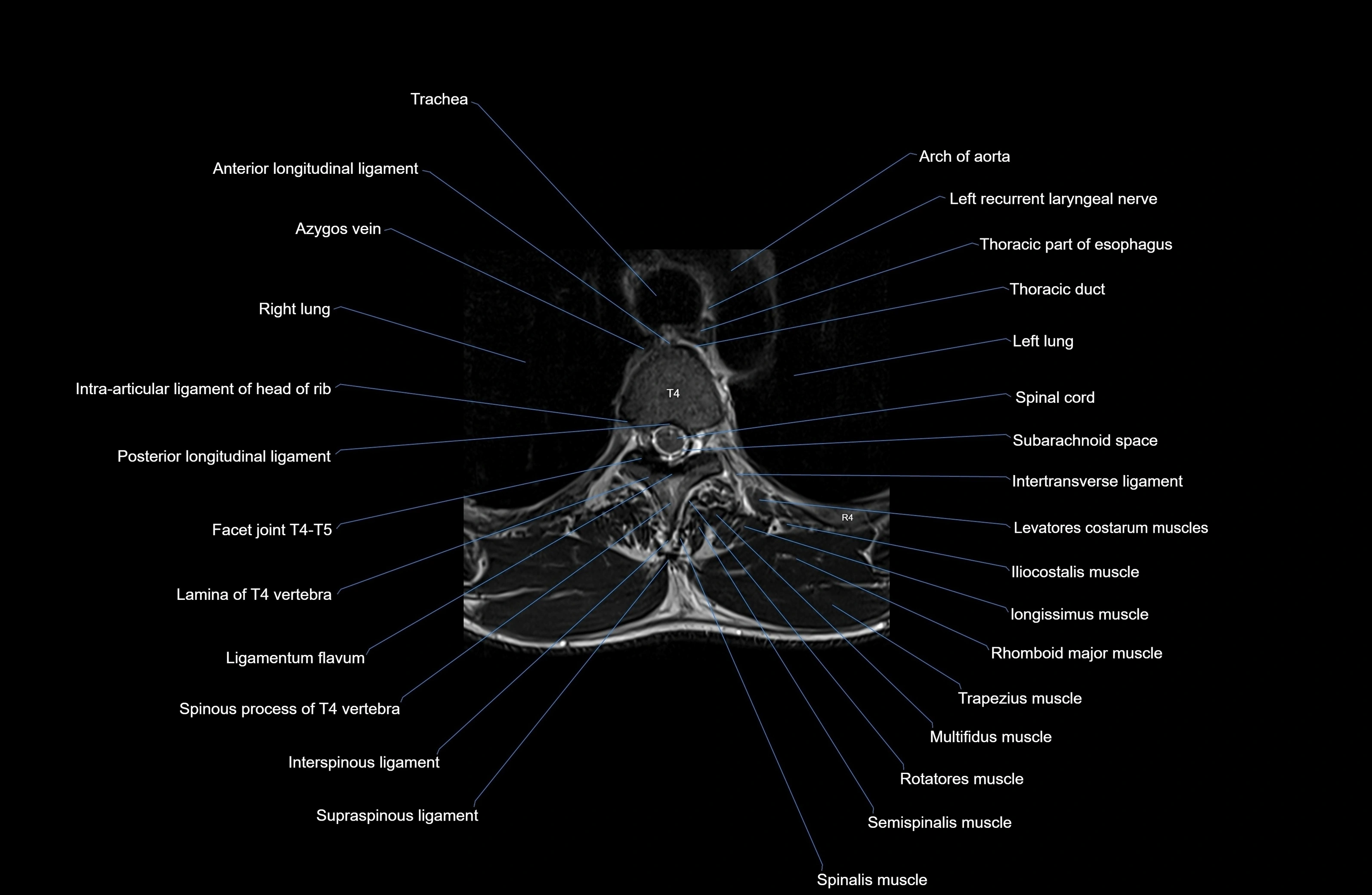
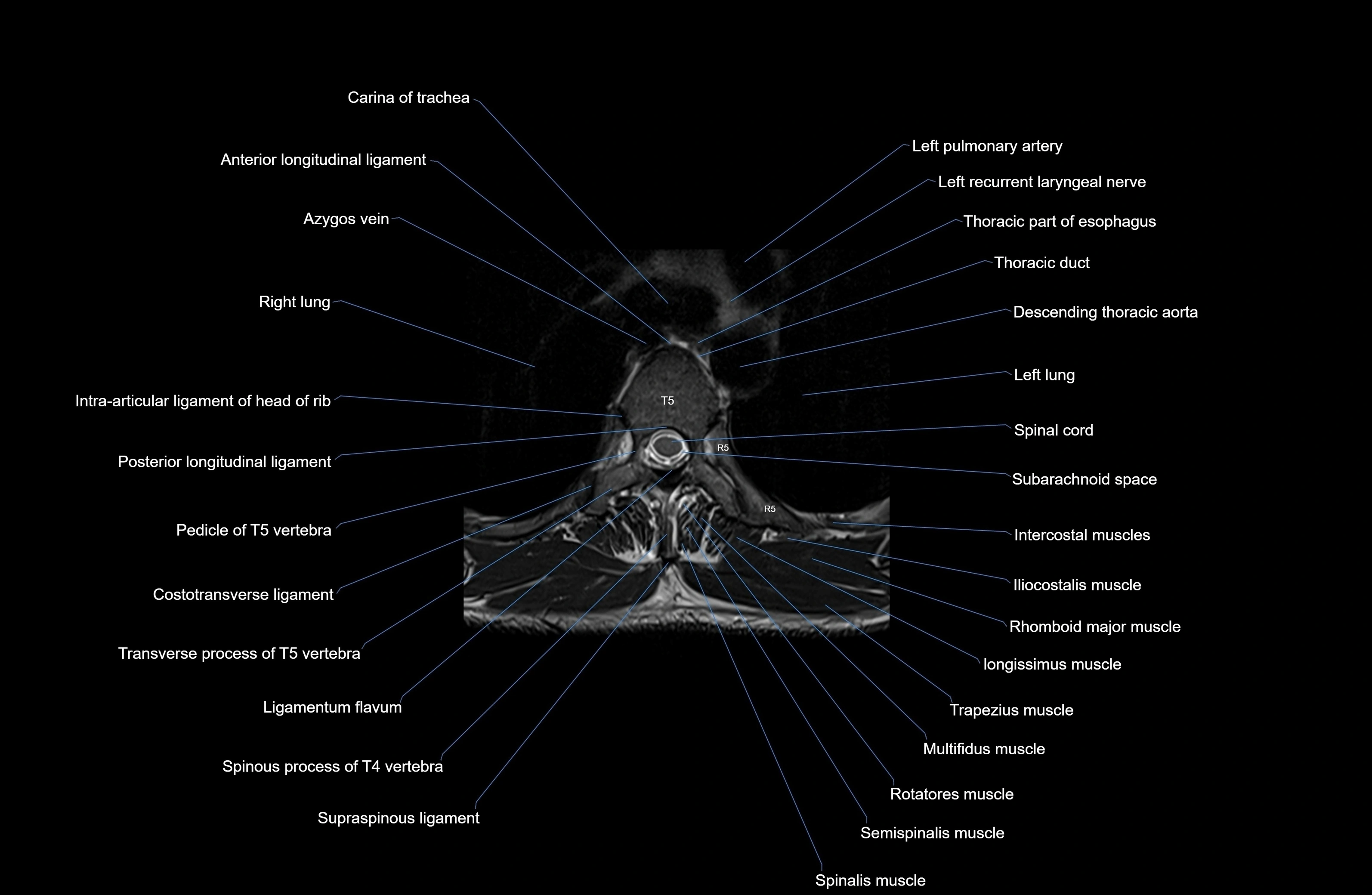
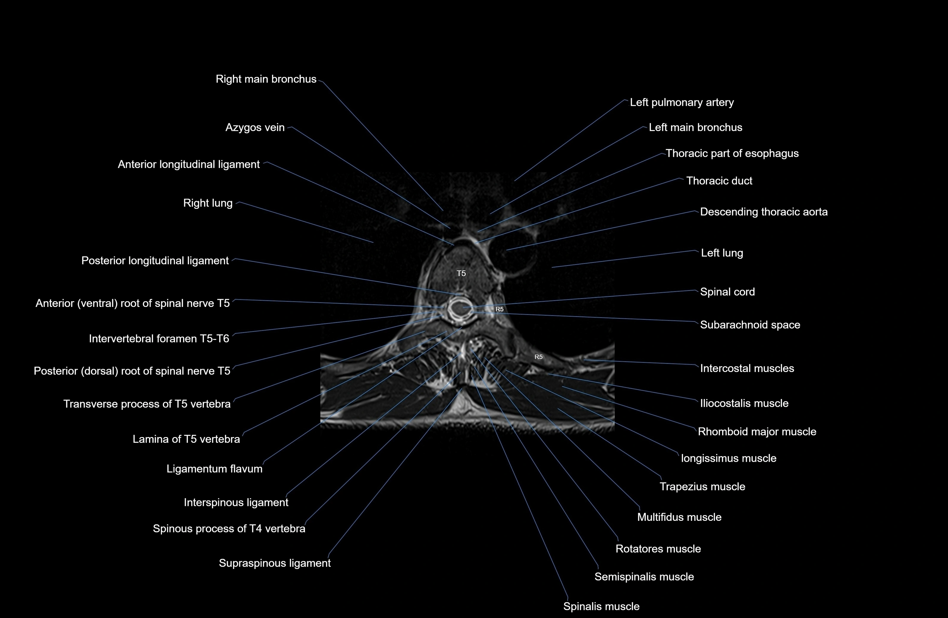
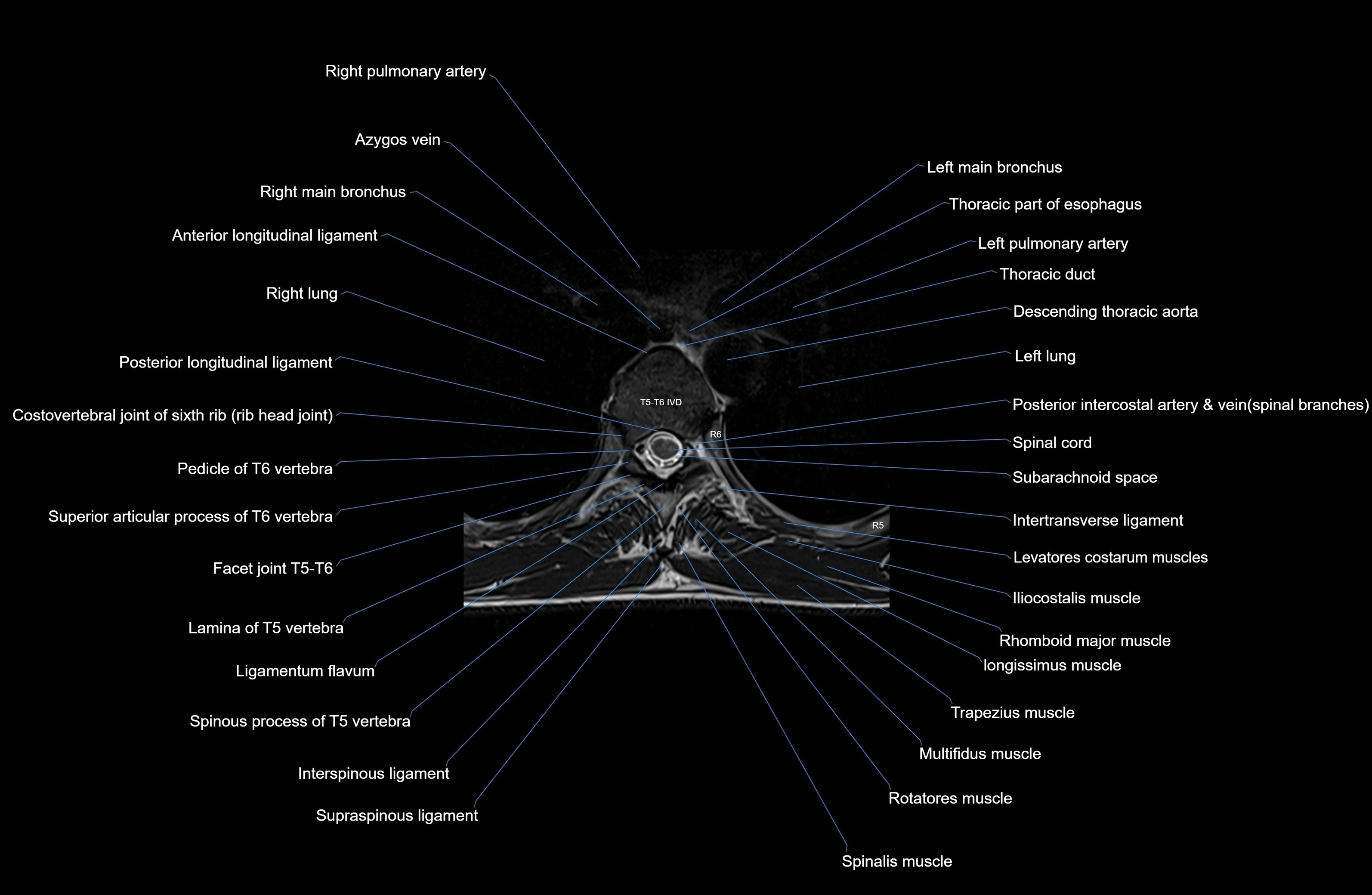
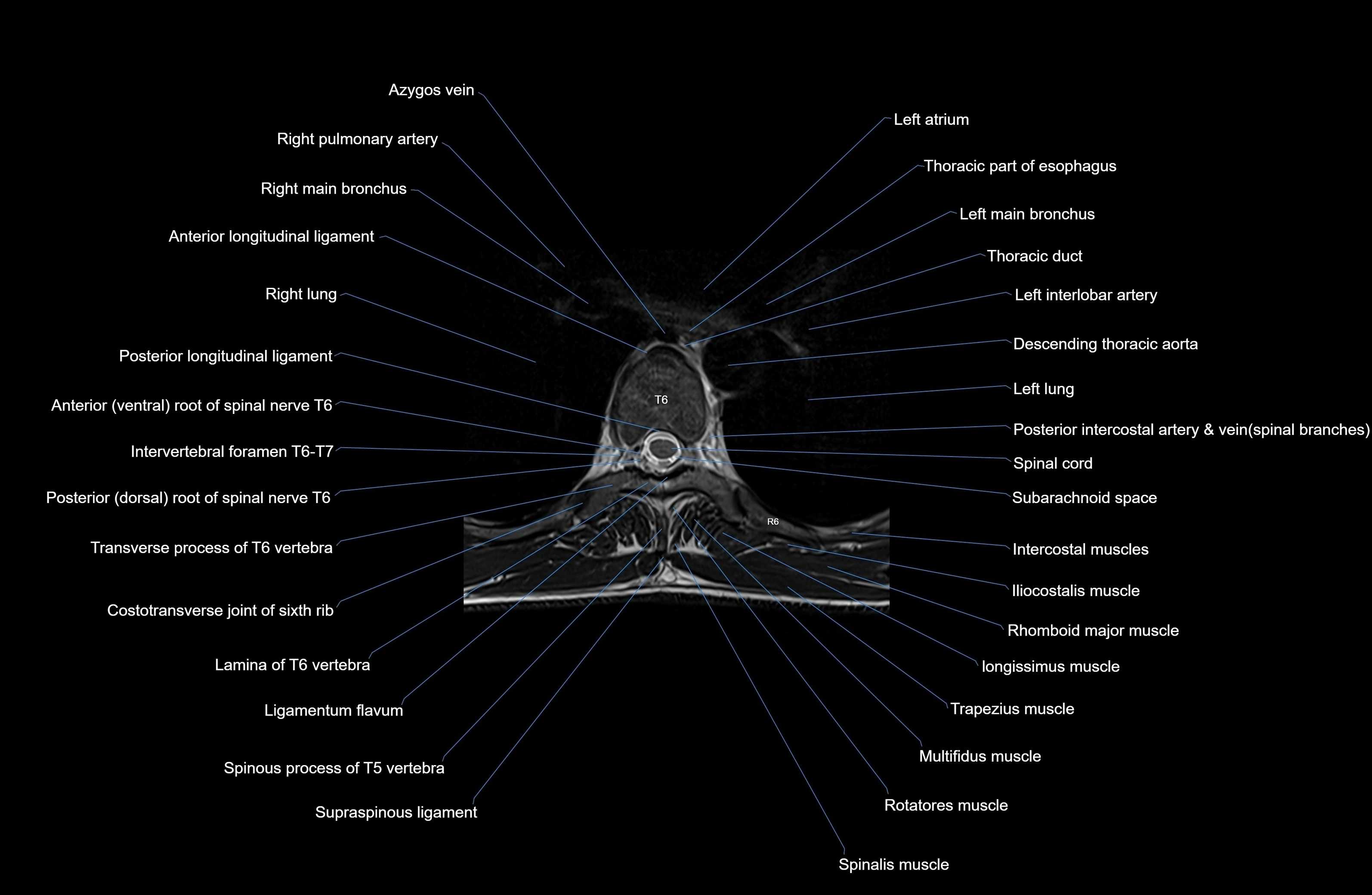
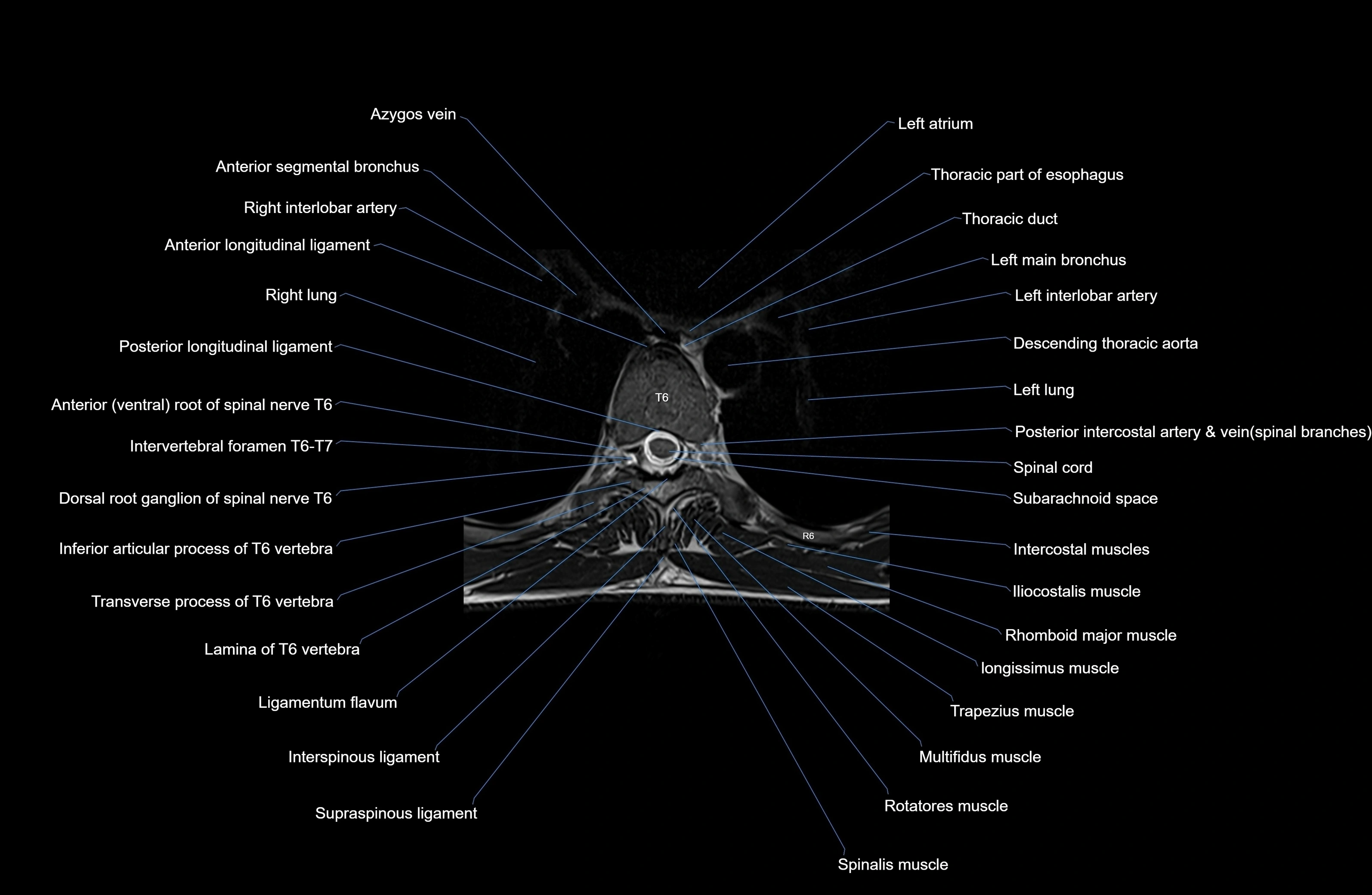
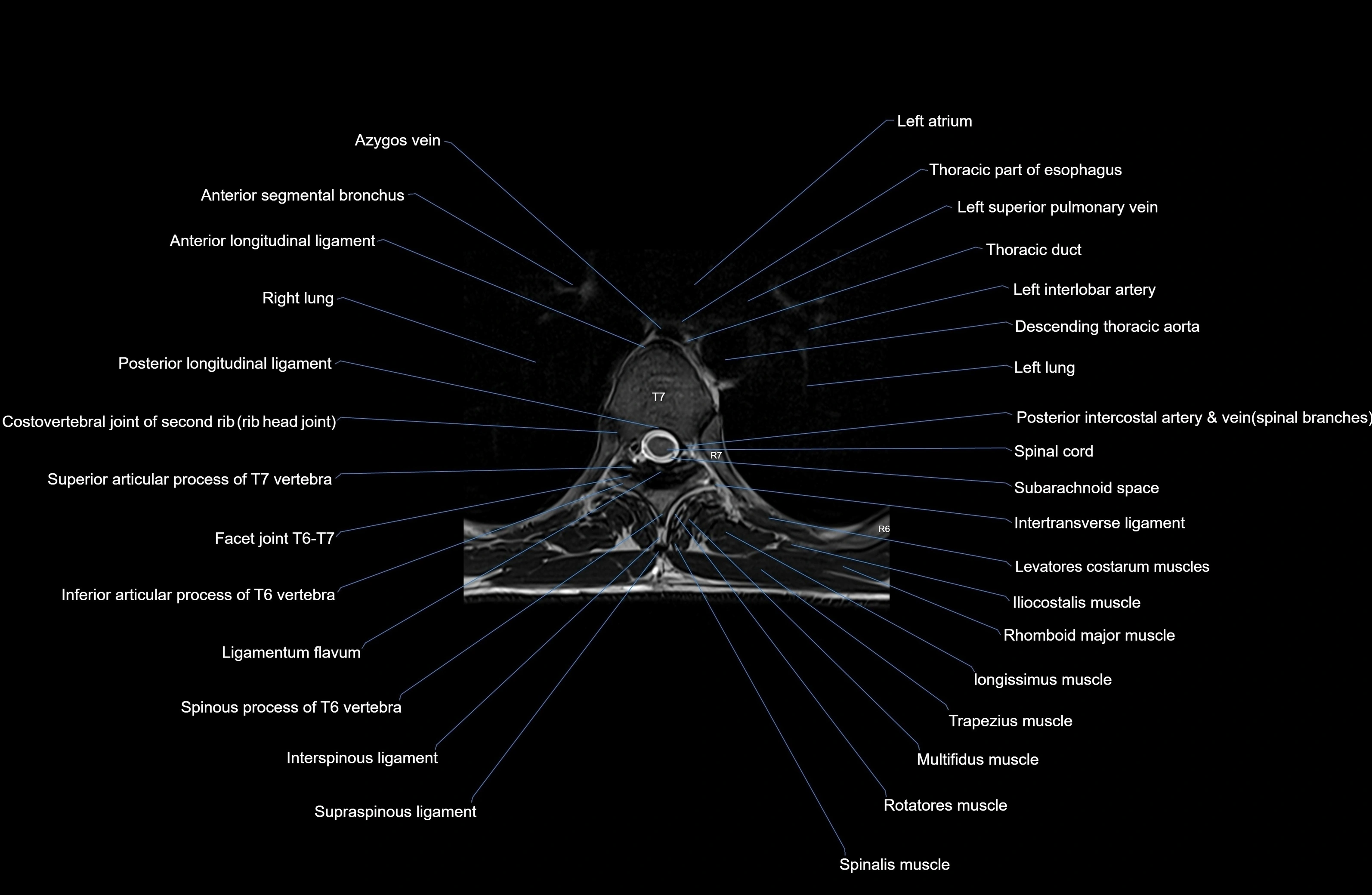
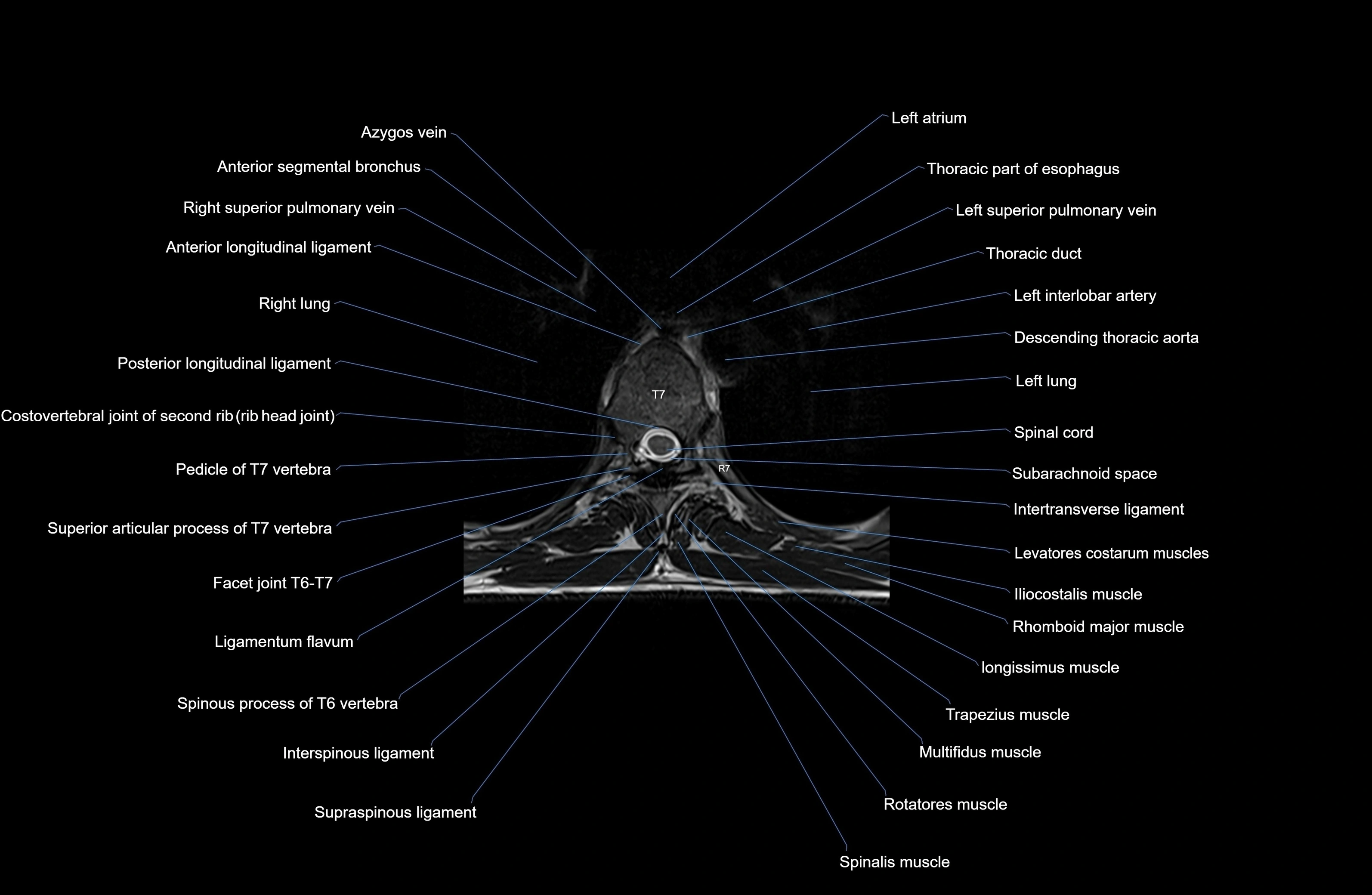
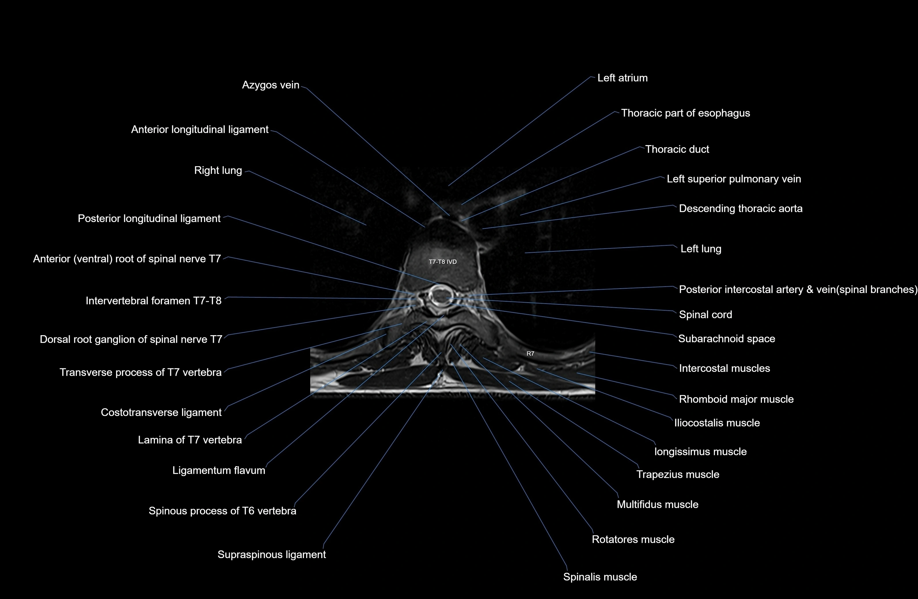
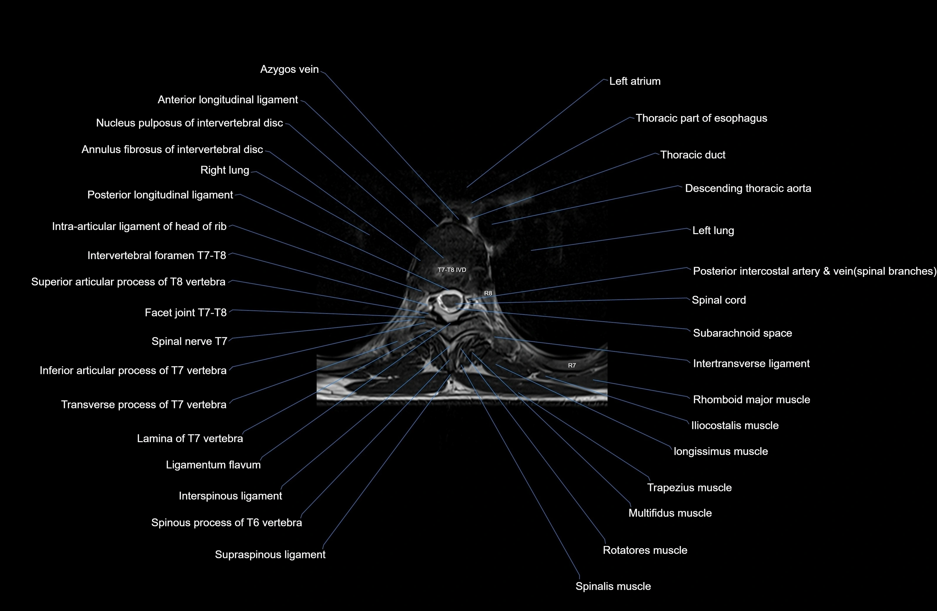
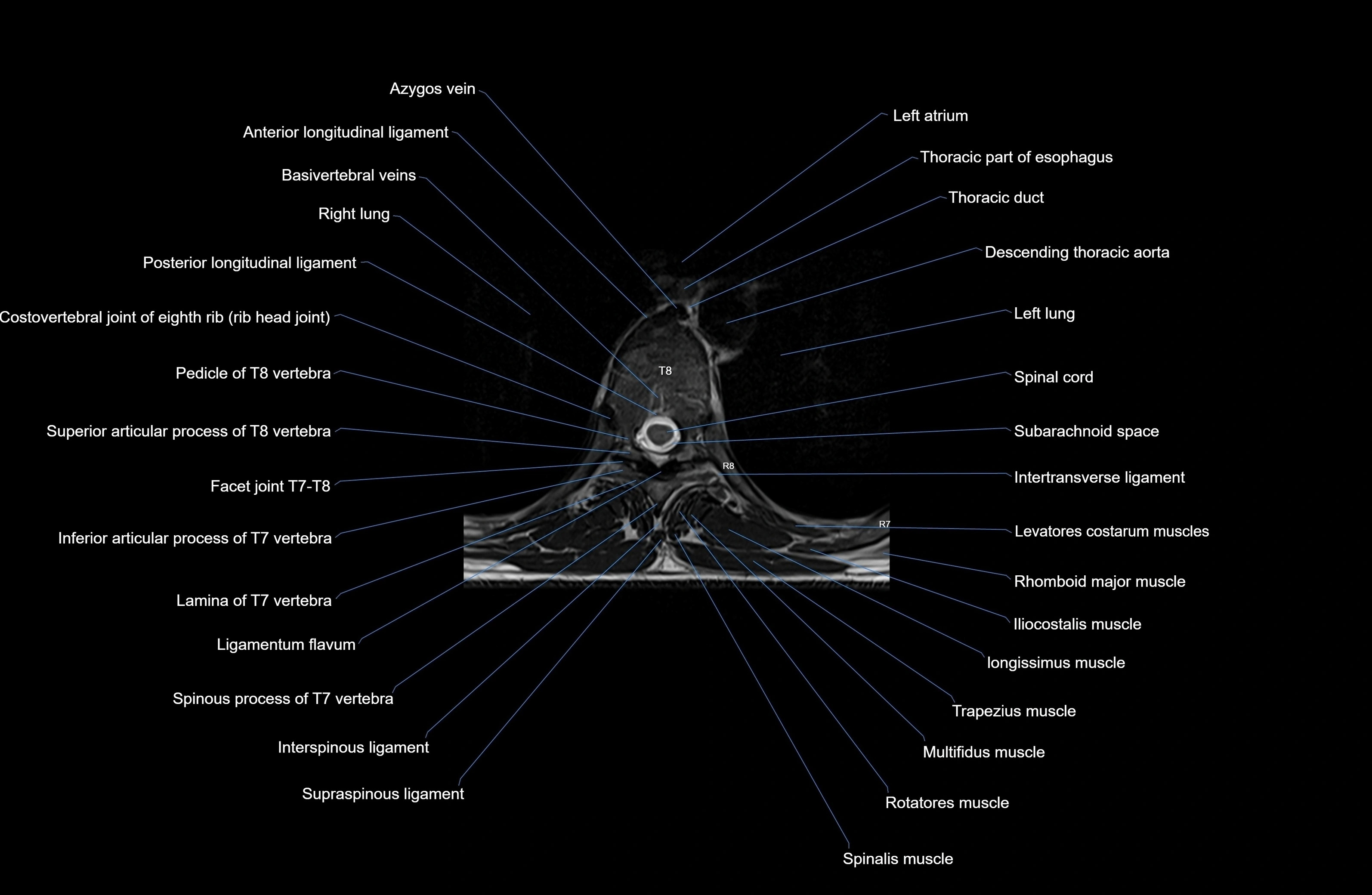
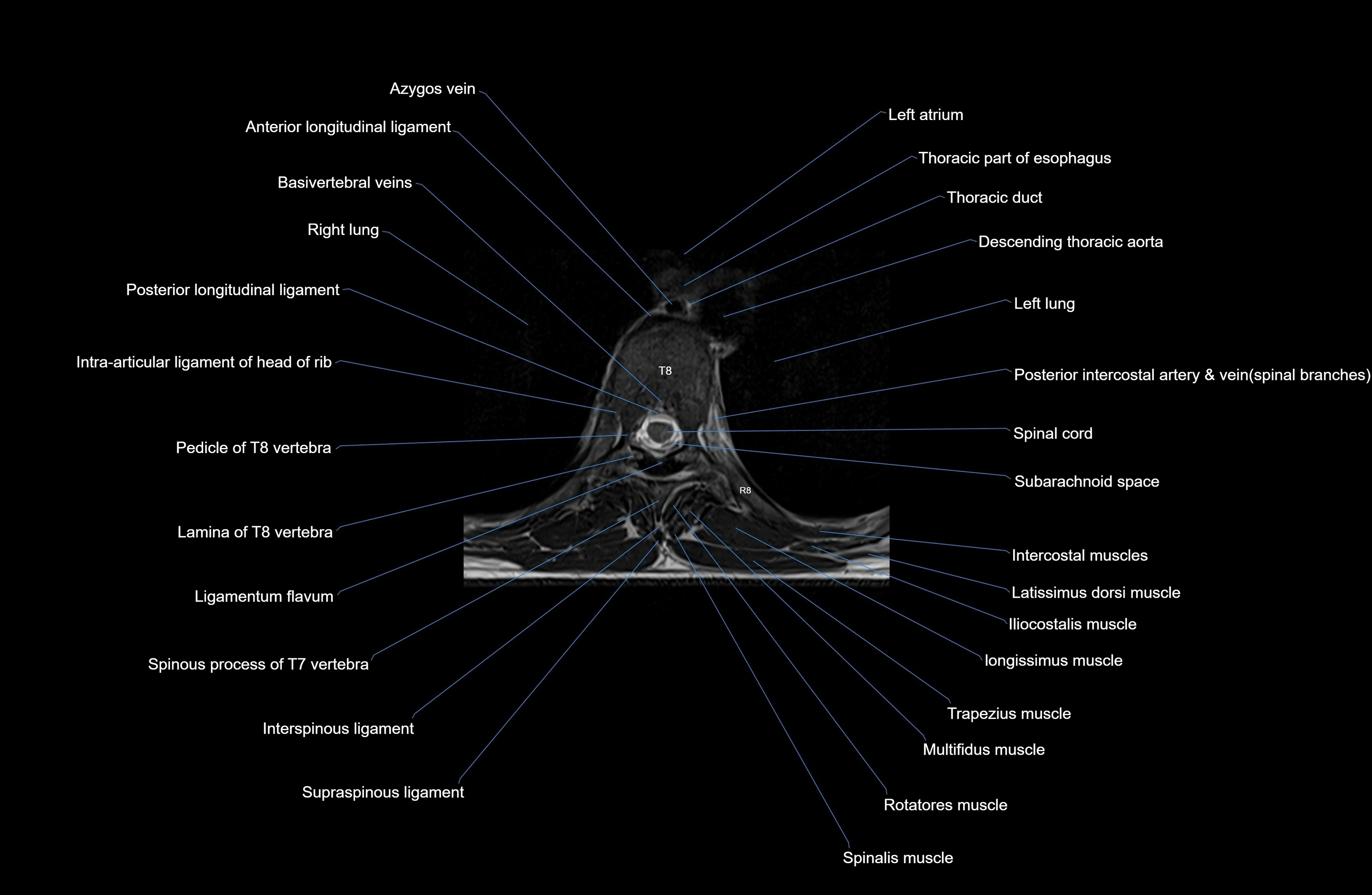
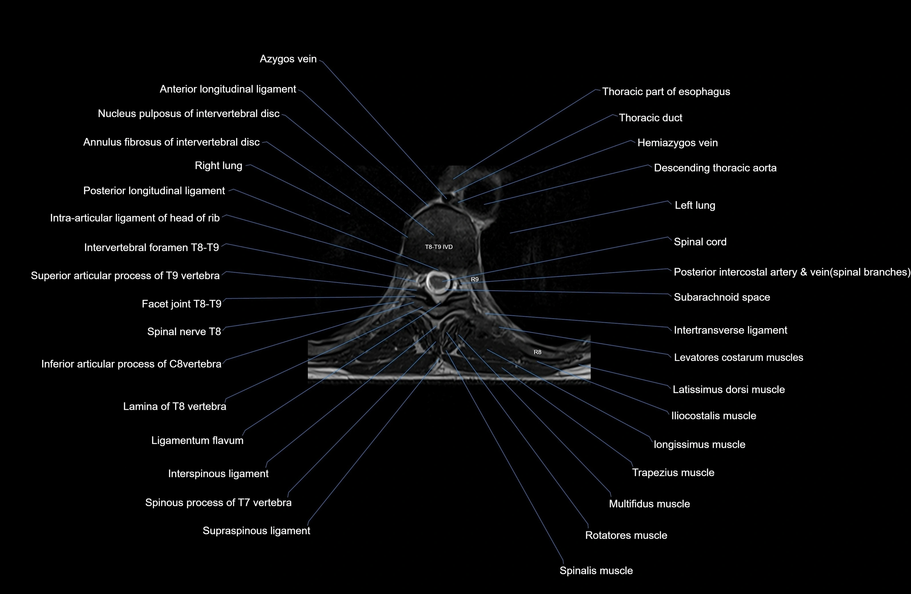
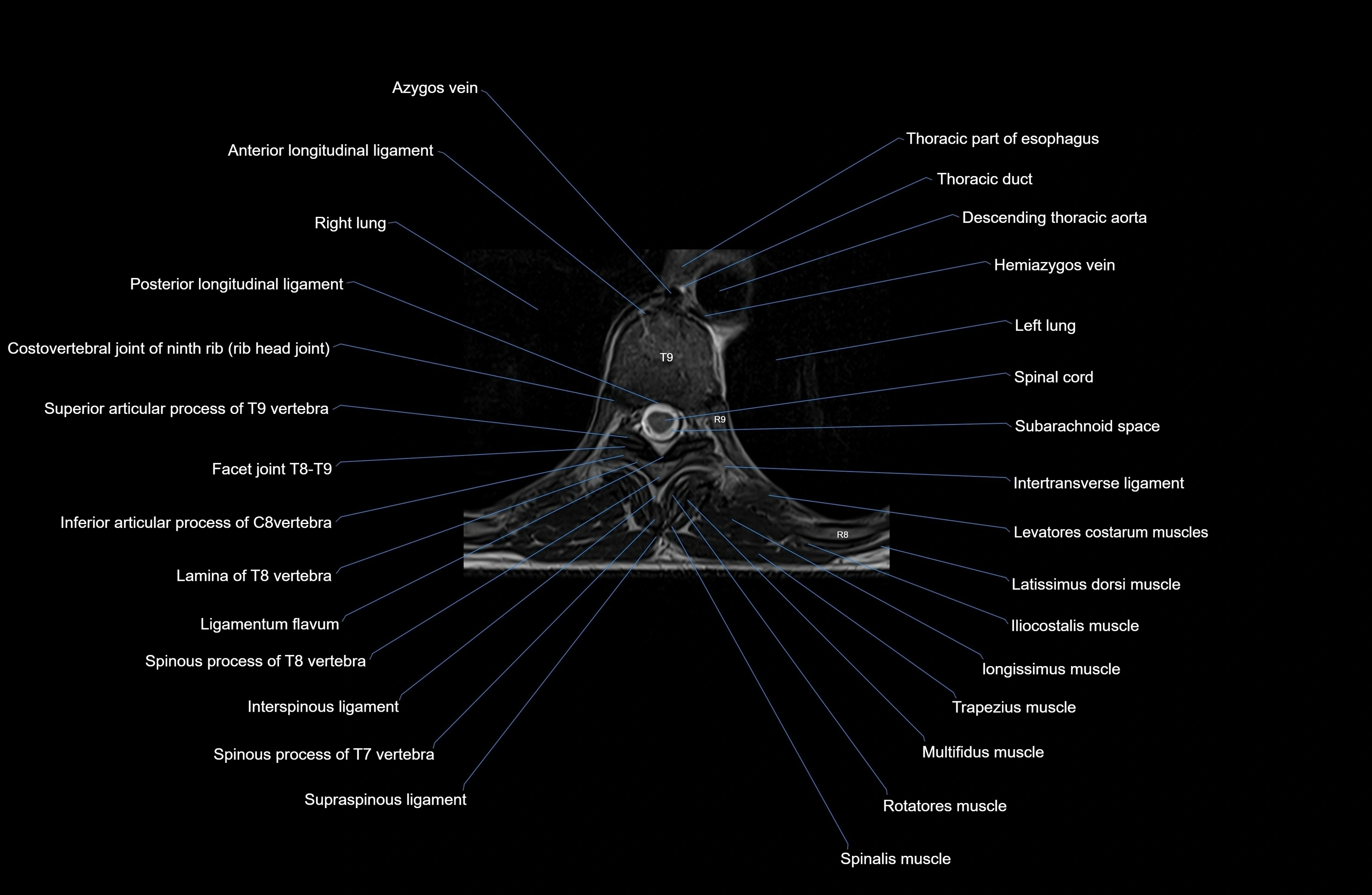
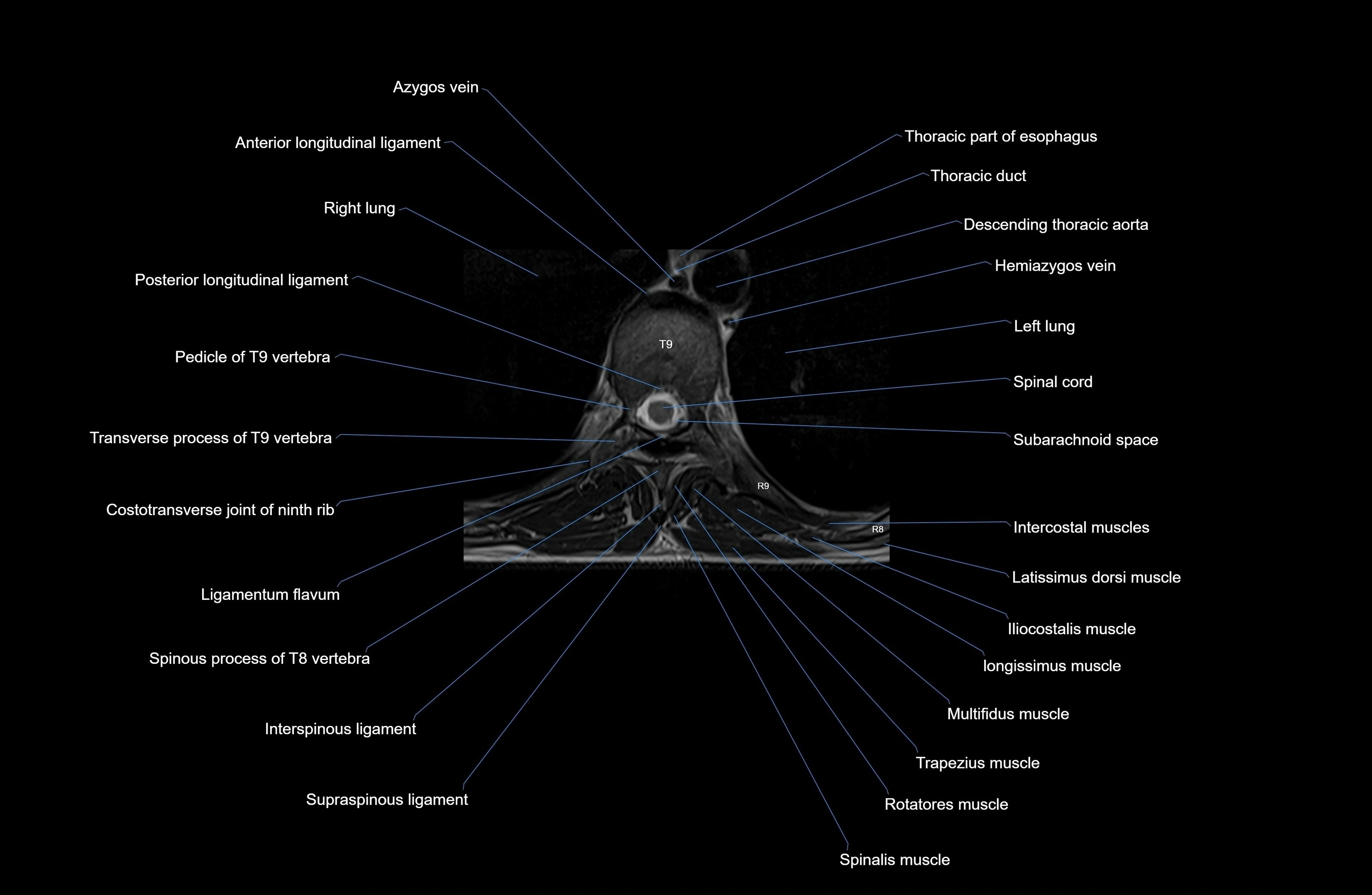
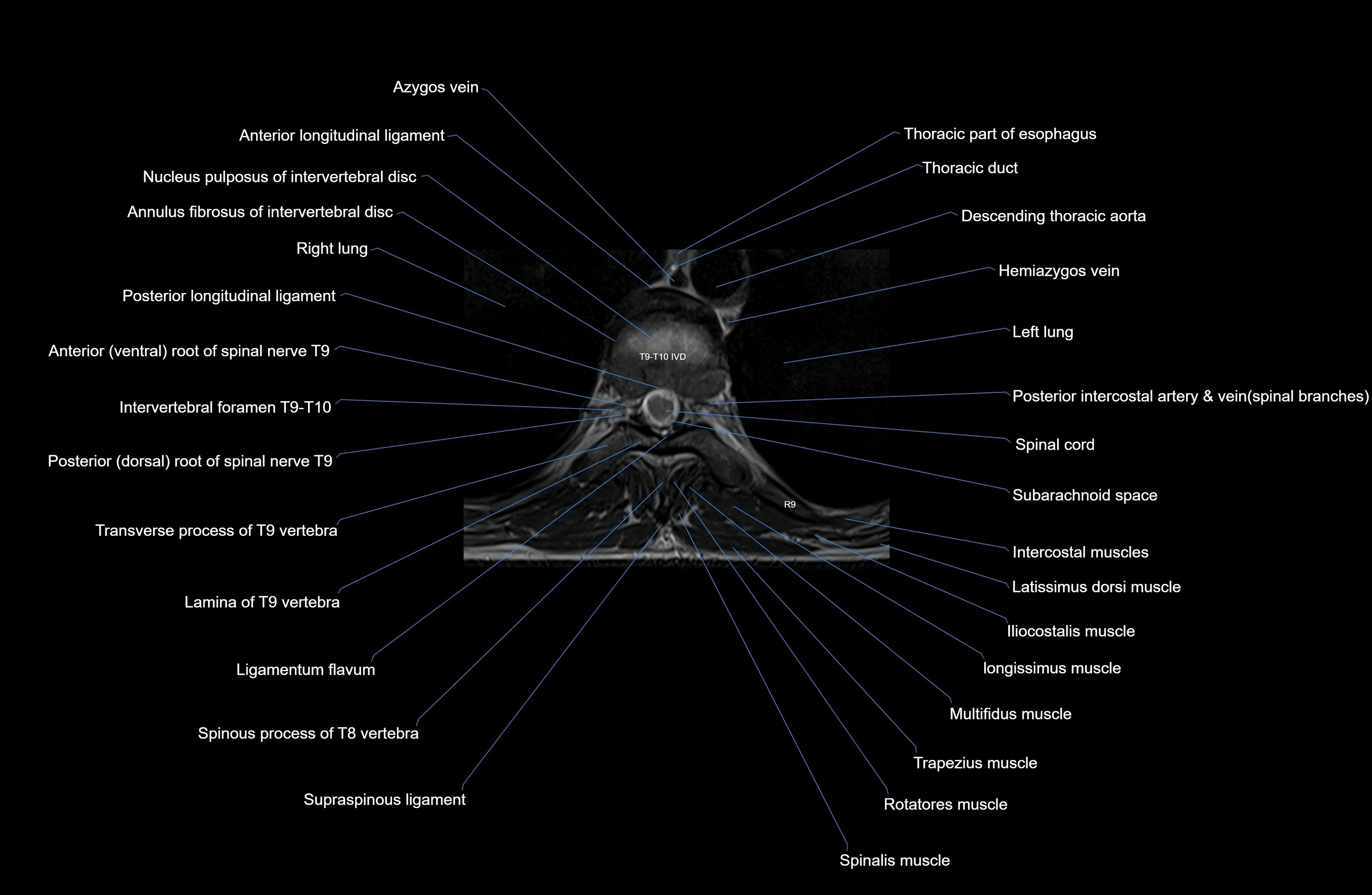
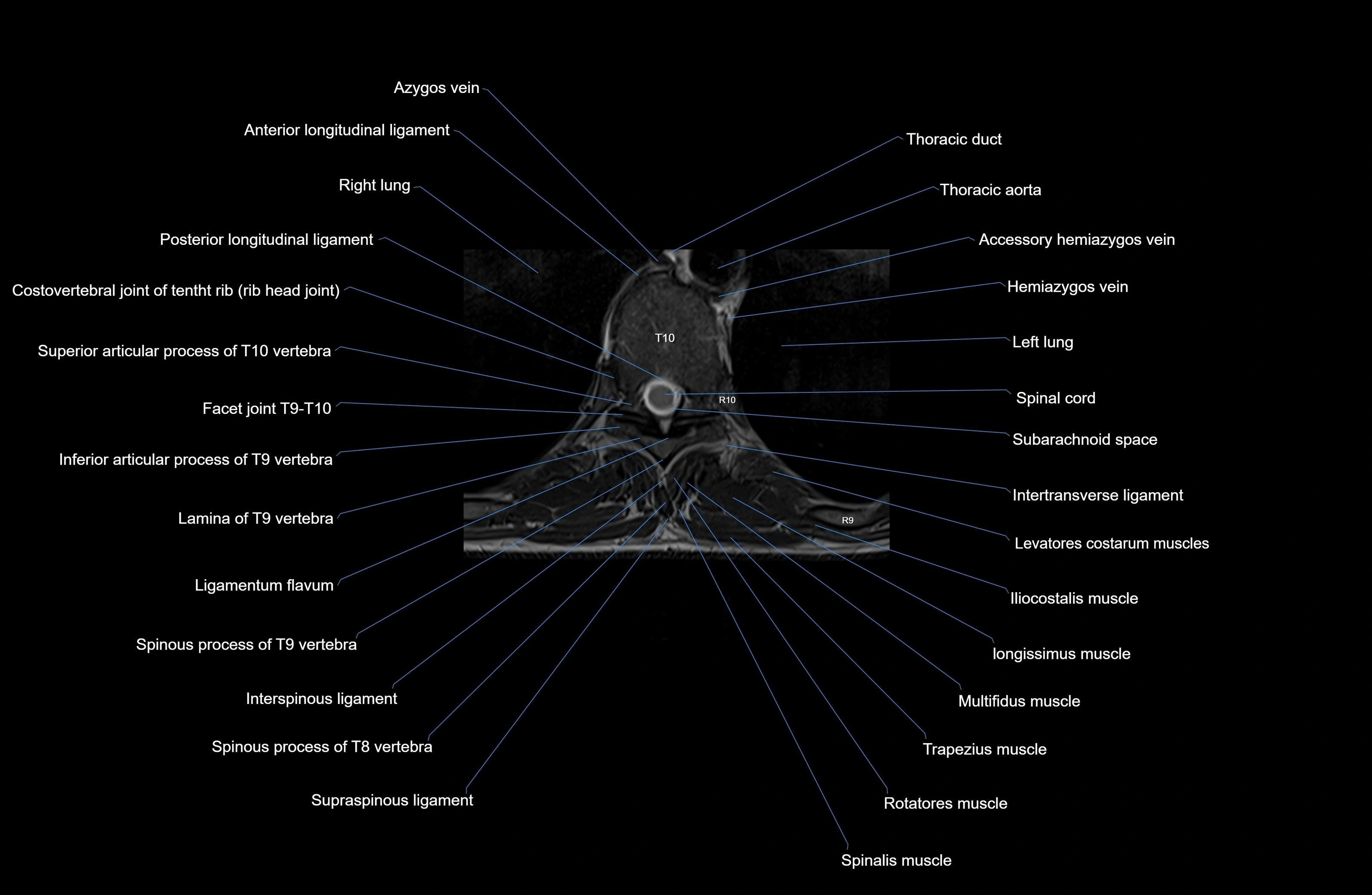
CT images

CT images


