


Topic
The ala of the ilium (iliac wing) is the large, expanded superior part of the ilium, forming the broad, fan-shaped lateral surface of the hip bone. It extends from the iliac crest superiorly to the arcuate line inferiorly and contributes significantly to the bony pelvis. Its external (gluteal) surface, internal (iliac fossa) surface, and iliac crest provide major muscular and ligamentous attachments, making it essential for pelvic stability and lower limb movement.
The external surface (gluteal surface) bears three rough curved lines (posterior, anterior, and inferior gluteal lines), which mark the origins of the gluteus maximus, medius, and minimus muscles. The internal surface (iliac fossa) provides attachment to the iliacus muscle and forms part of the lateral wall of the pelvic cavity. Posteriorly, the auricular surface articulates with the sacrum at the sacroiliac joint. The iliac crest forms the superior border, serving as a palpable surface landmark in clinical practice.
The ala of the ilium plays a central role in transmitting weight from the axial skeleton to the lower limbs and provides broad surfaces for muscle attachment, aiding in locomotion, posture, and abdominal wall support.
Synonyms
-
Iliac wing
-
Wing of ilium
-
Ala ossis ilii
Function
-
Provides attachment for major abdominal, pelvic, and gluteal muscles
-
Forms the lateral wall of the pelvis and contributes to the iliac fossa
-
Articulates with the sacrum at the sacroiliac joint for weight transmission
-
Serves as a palpable landmark in clinical examination and surgical approaches
Muscle Attachments
-
External surface:
-
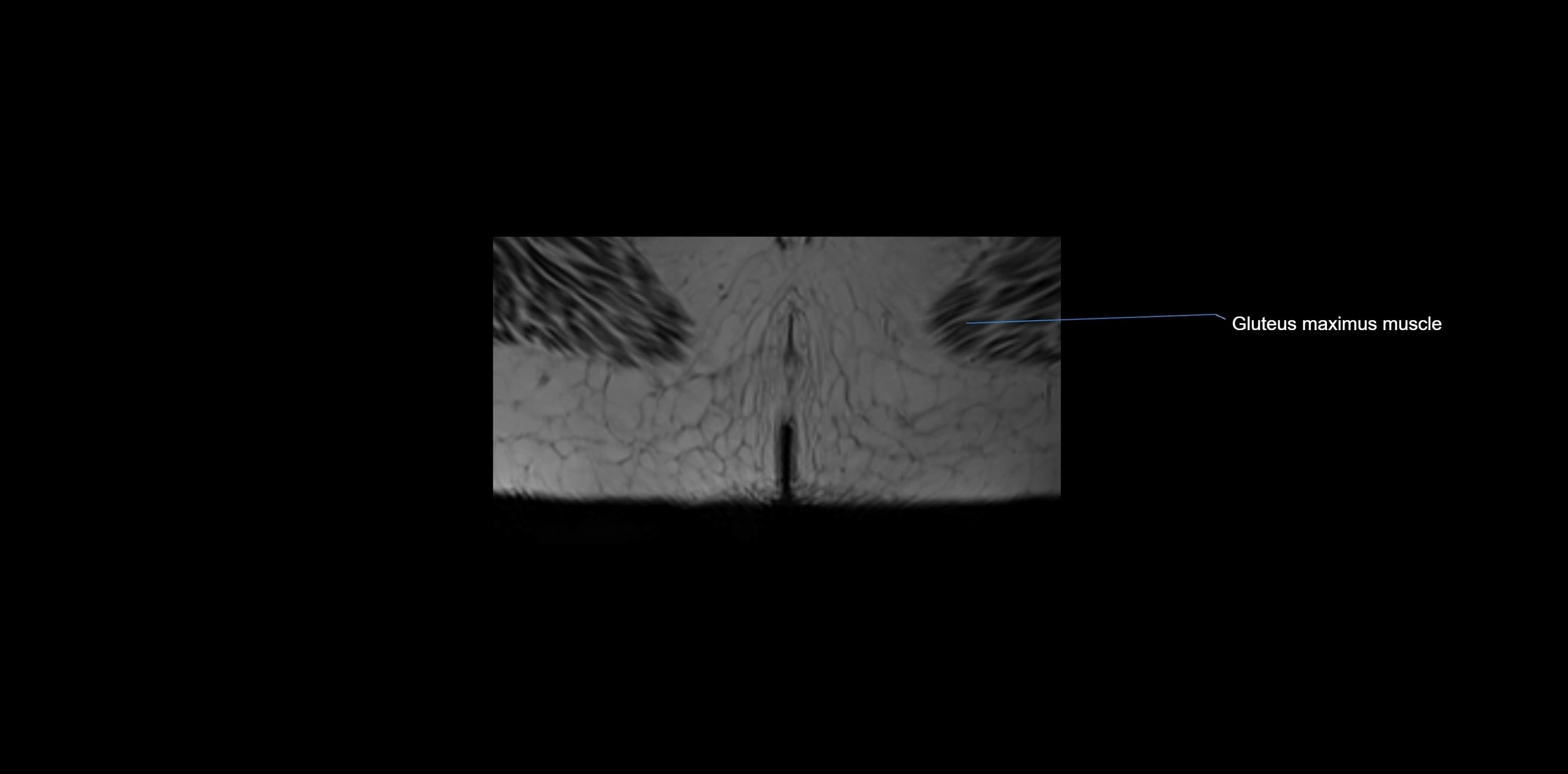
Gluteus maximus (posterior to posterior gluteal line)
-
Gluteus medius (between posterior and anterior gluteal lines)
-
Gluteus minimus (between anterior and inferior gluteal lines)
-
-
Internal surface:
-
Iliacus (in iliac fossa)
-
Origin for part of psoas major
-
-
Crest attachments:
-
Tensor fasciae latae, abdominal wall muscles (external oblique, internal oblique, transversus abdominis), latissimus dorsi (via thoracolumbar fascia), quadratus lumborum
-
Nerve Supply
-
Indirect, via muscles attached:
-
Gluteal nerves (superior and inferior) for gluteal muscles
-
Femoral nerve for iliacus
-
Thoracoabdominal nerves, iliohypogastric, ilioinguinal nerves for abdominal wall attachments
-
Arterial Supply
-
Iliolumbar artery (branch of internal iliac artery)
-
Superior gluteal artery (external surface attachments)
-
Deep circumflex iliac artery (iliac crest region)
-
Small contributions from lumbar arteries
Venous Drainage
-
Follows arterial supply:
-
Iliolumbar vein → internal iliac vein
-
Superior gluteal vein → internal iliac vein
-
Deep circumflex iliac vein → external iliac vein
-
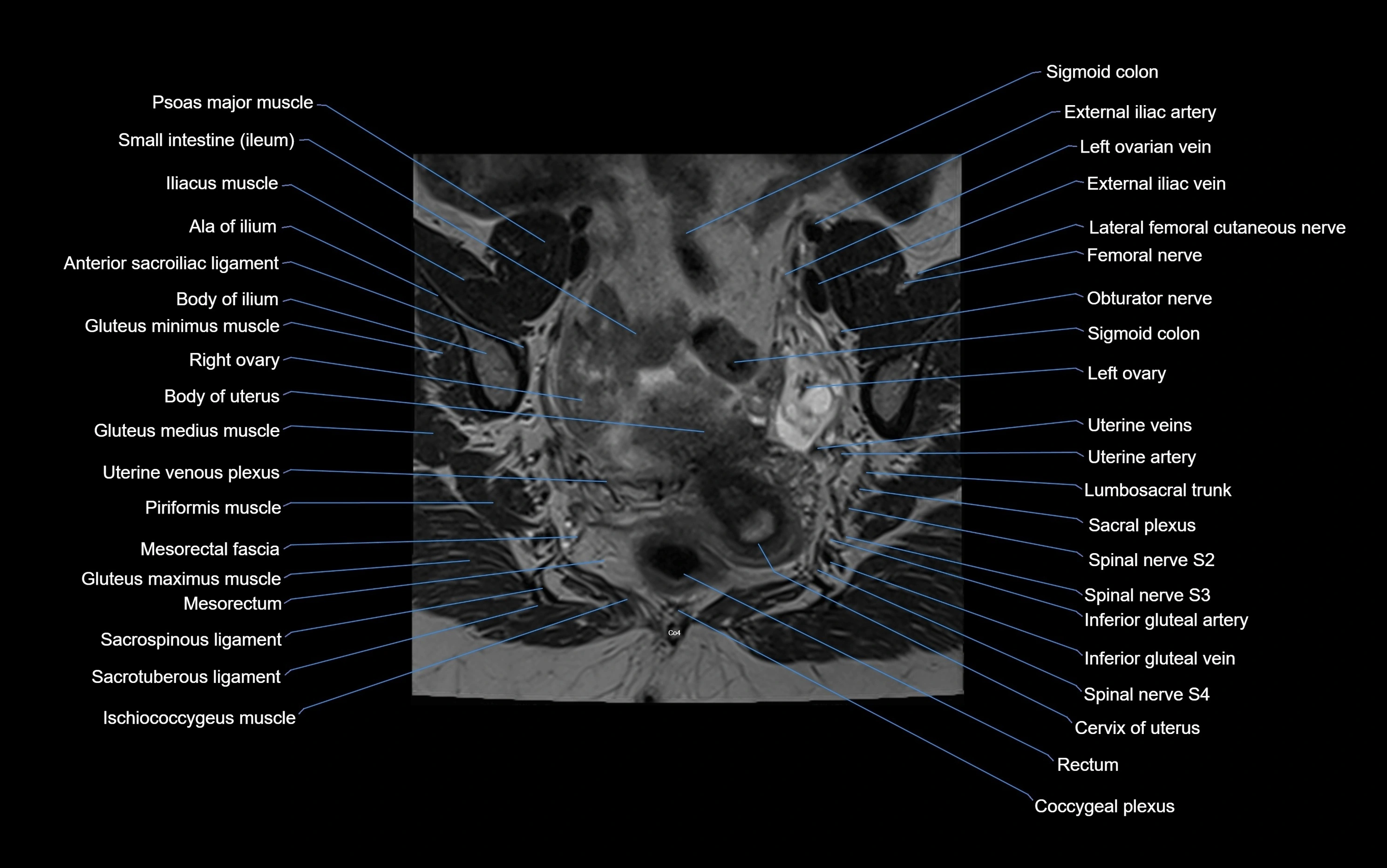
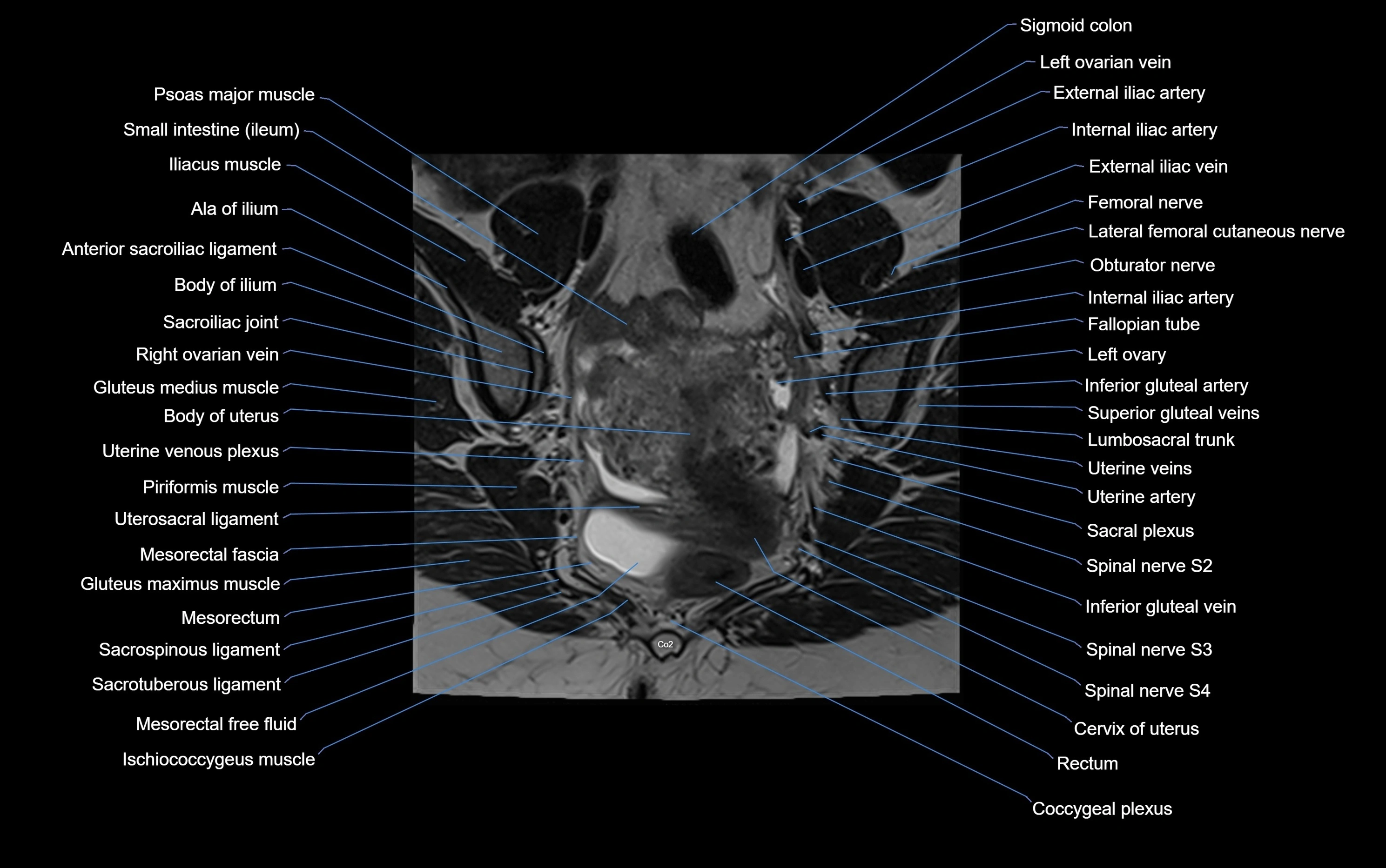
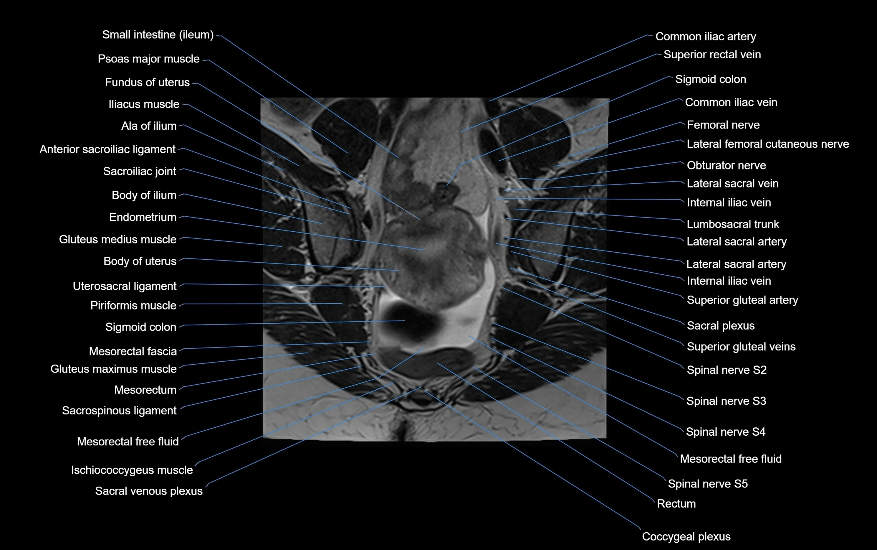
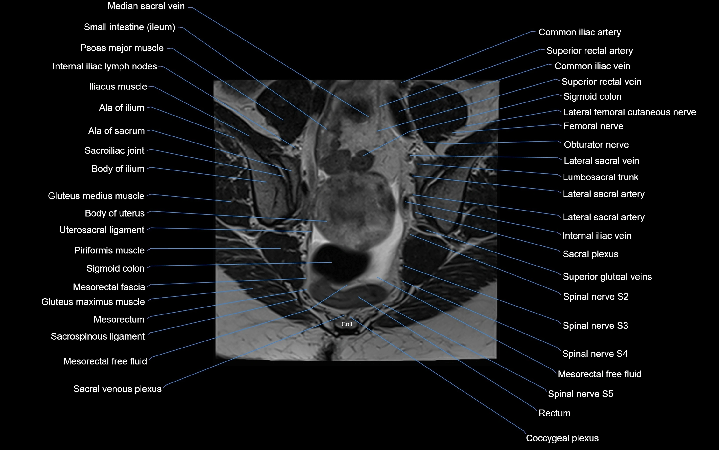
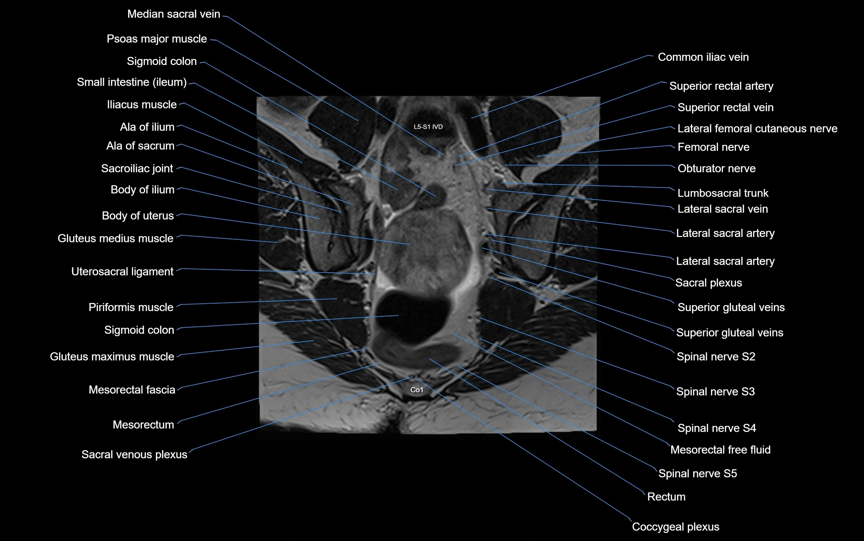
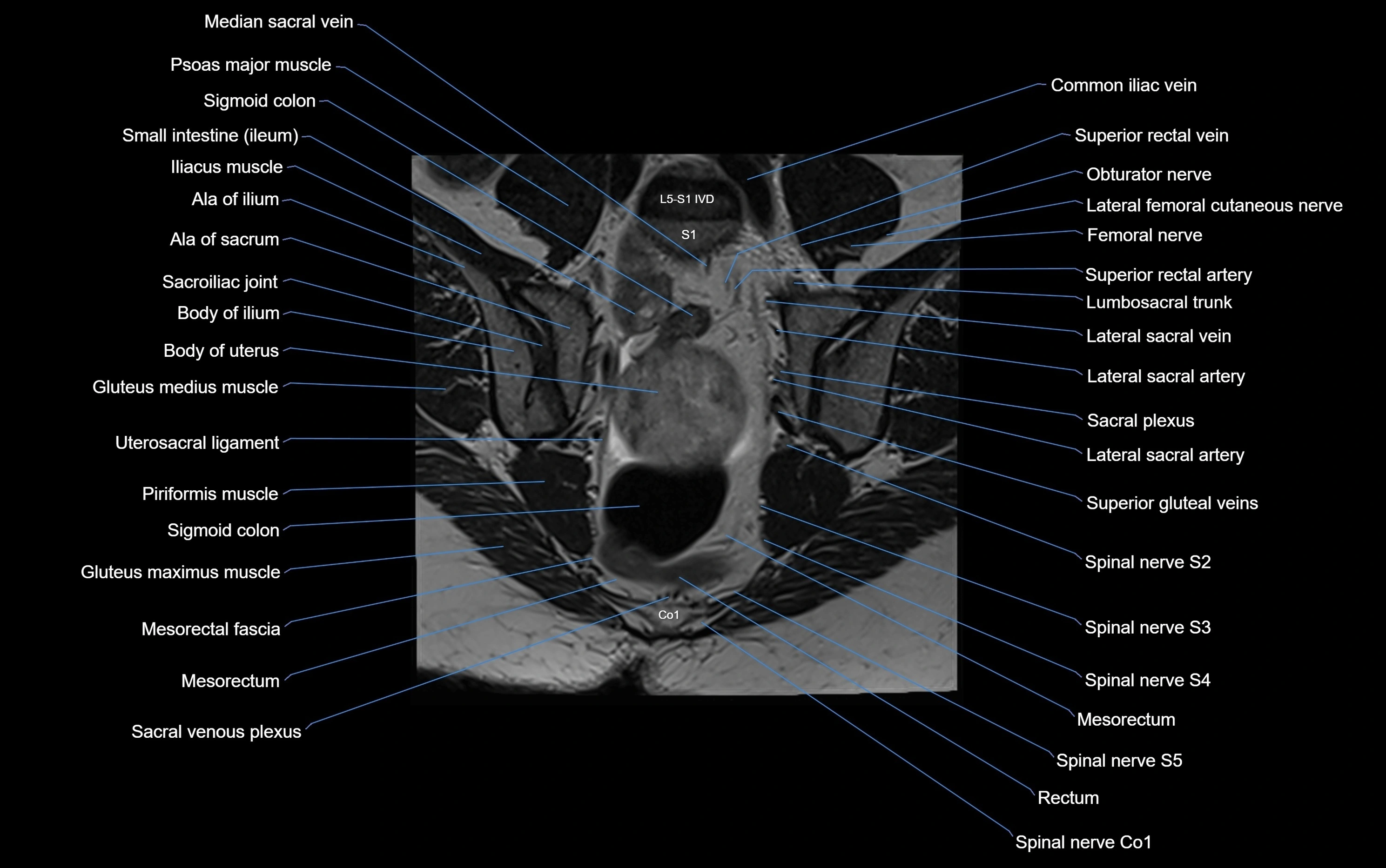
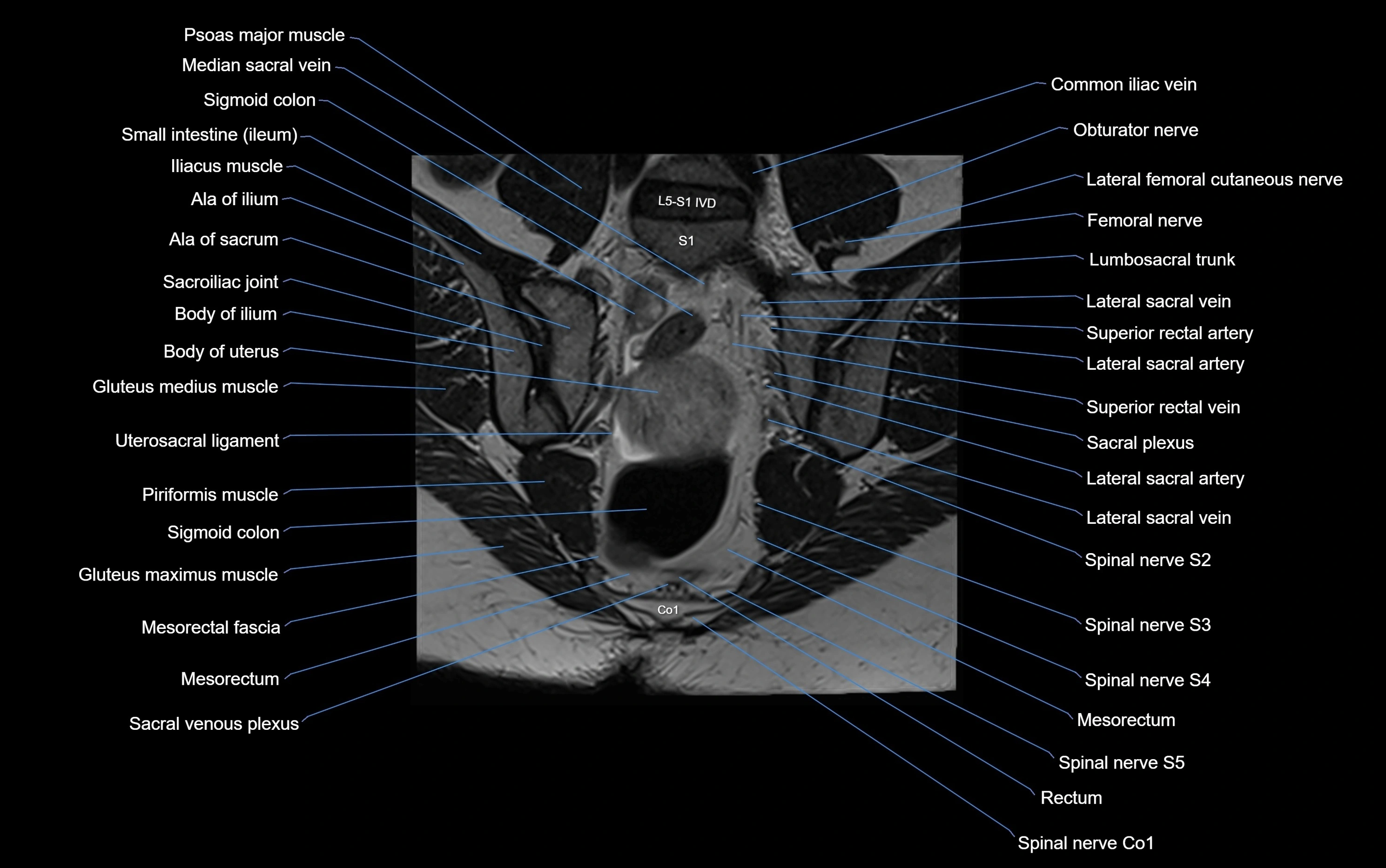
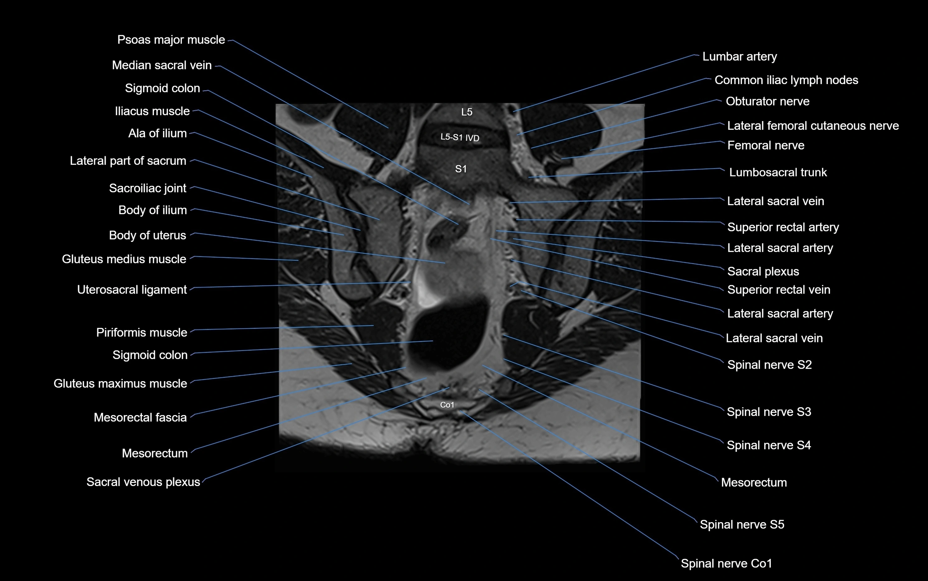
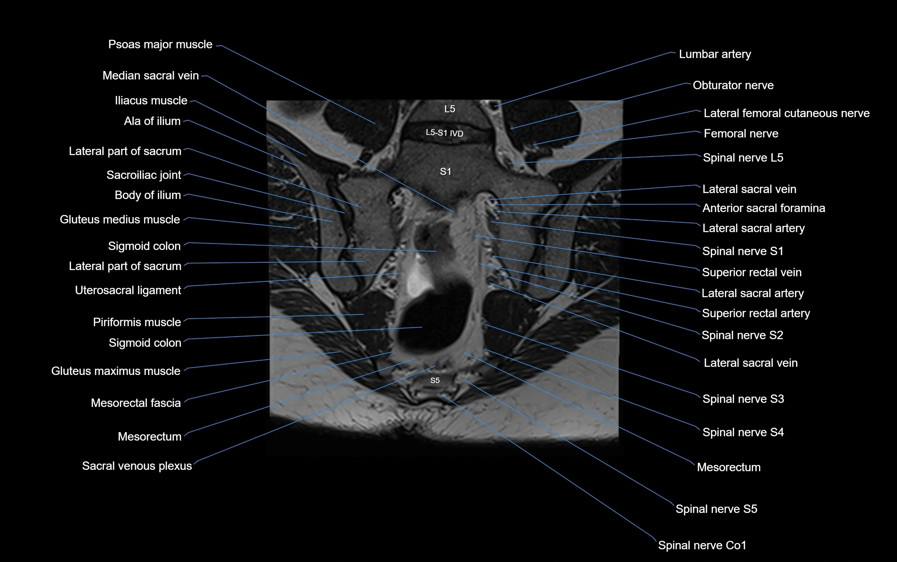
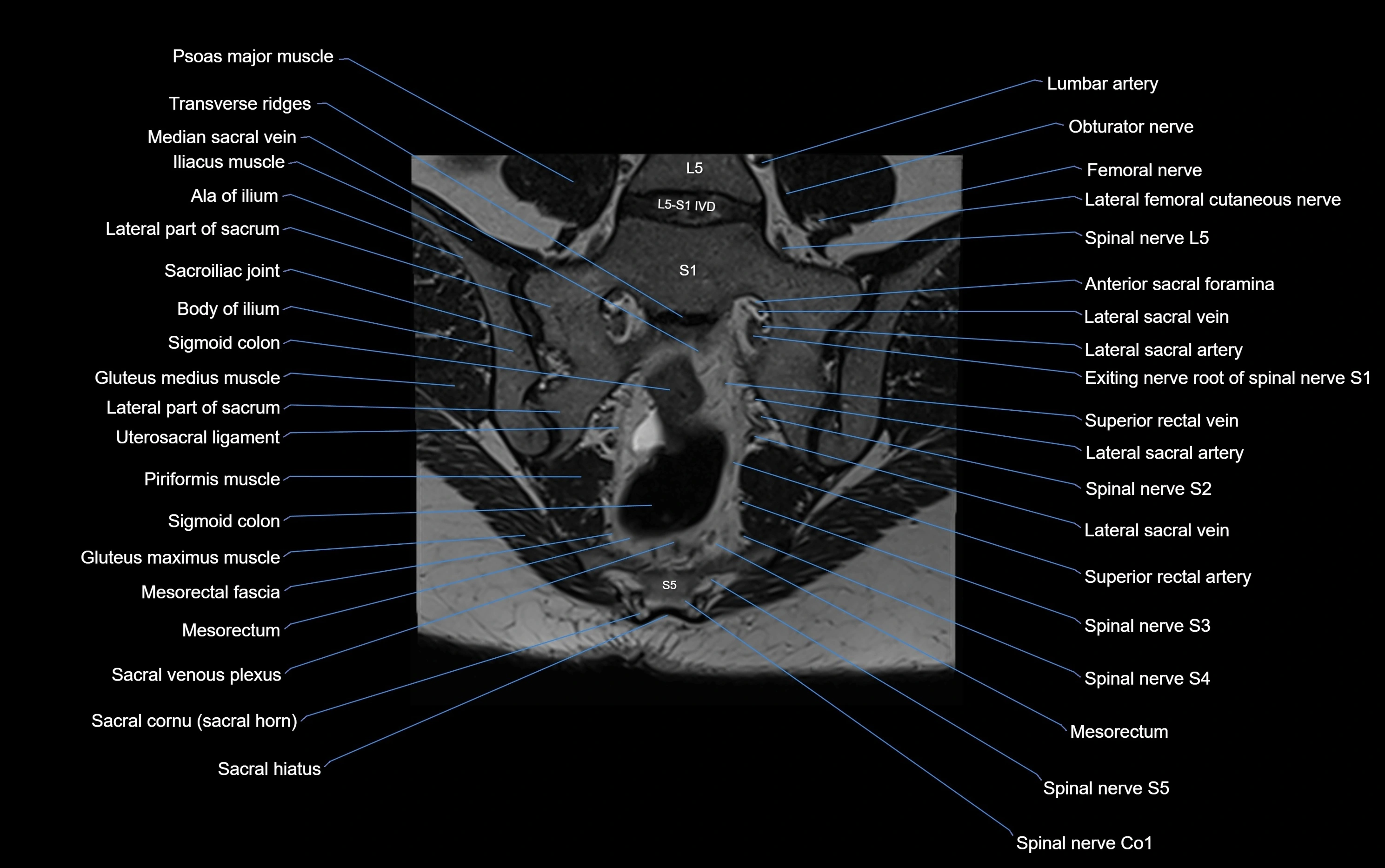
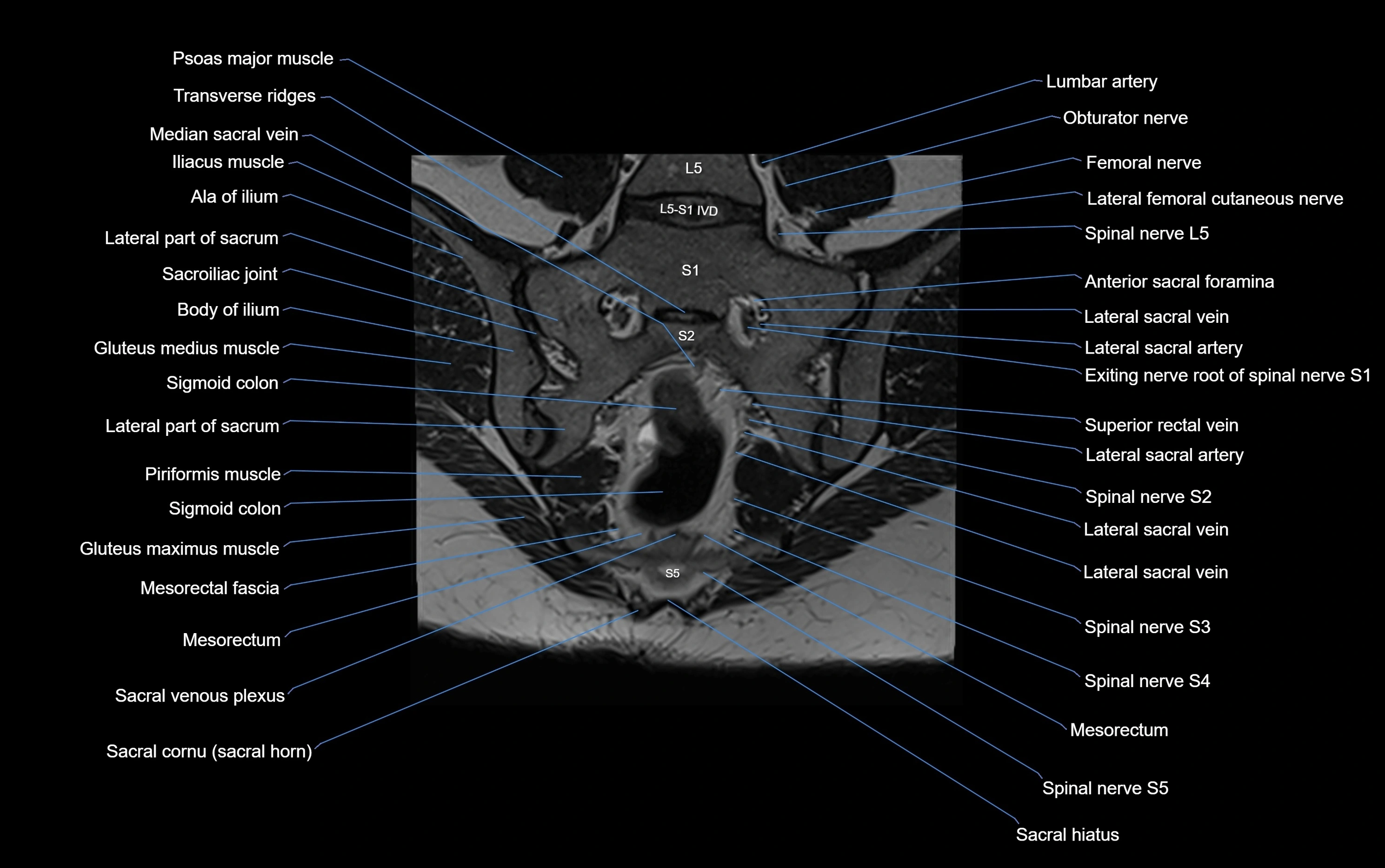
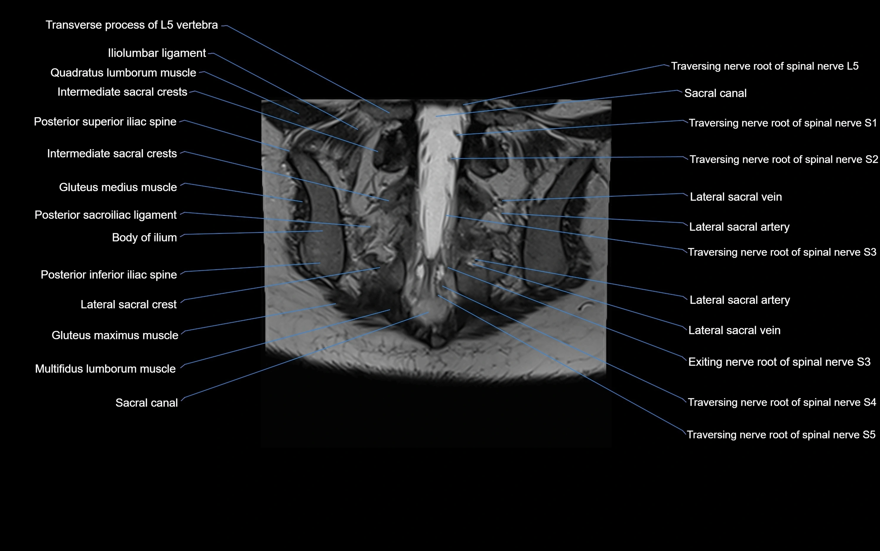
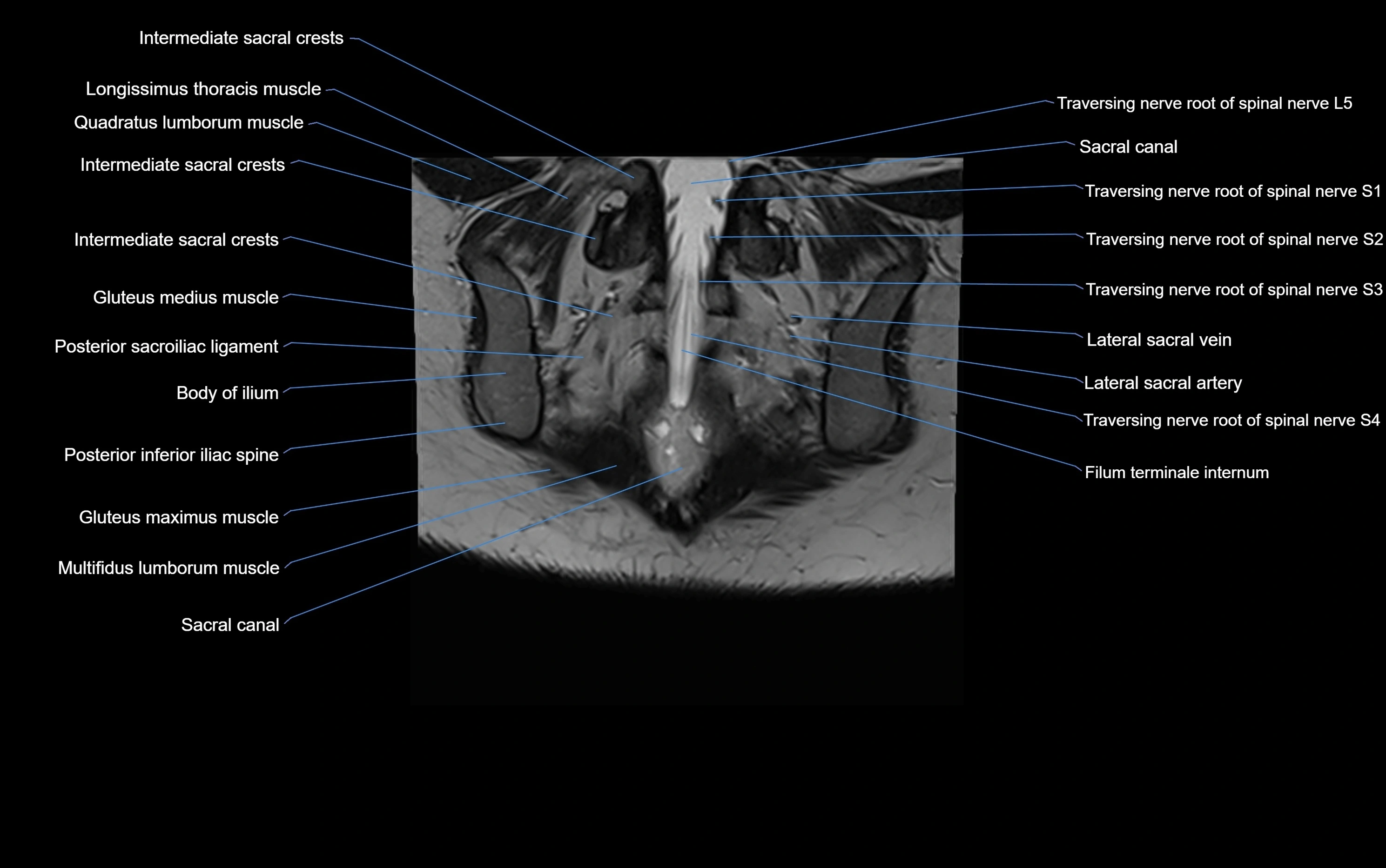
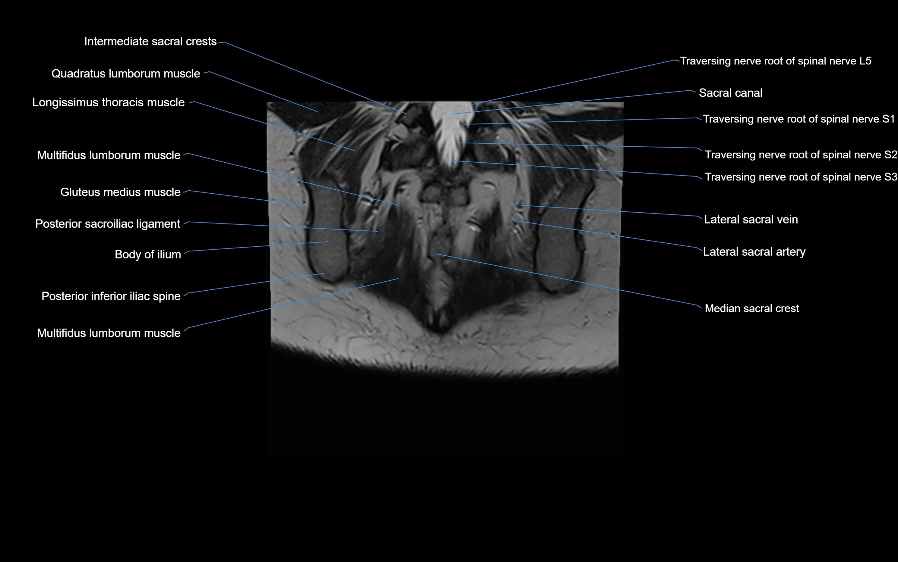
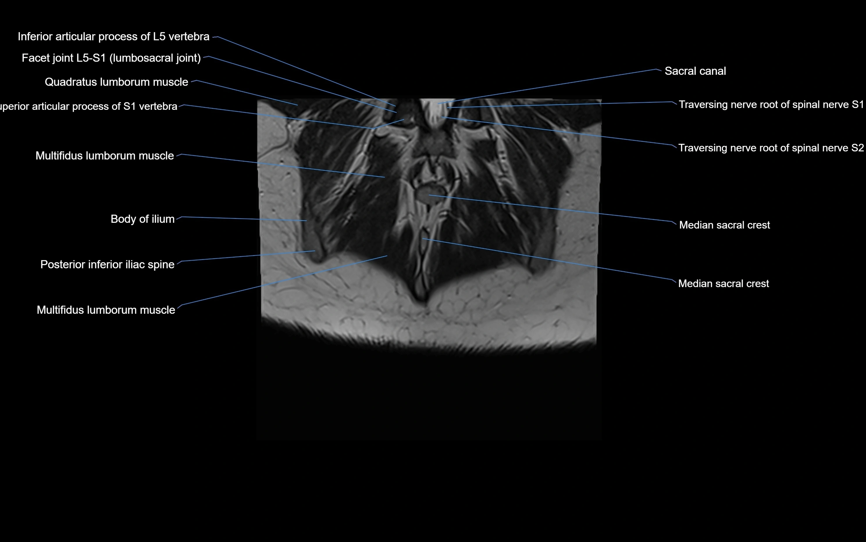
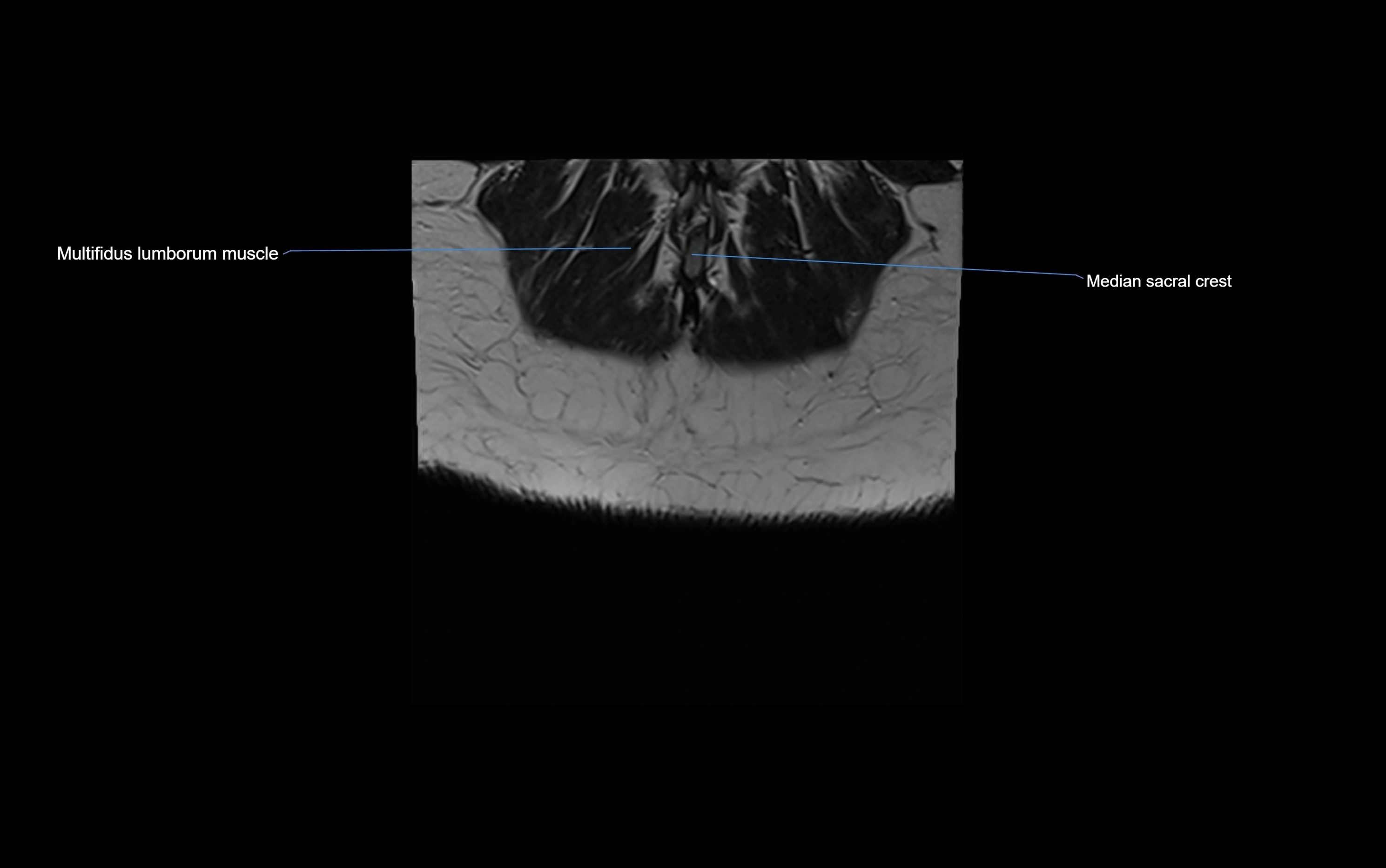
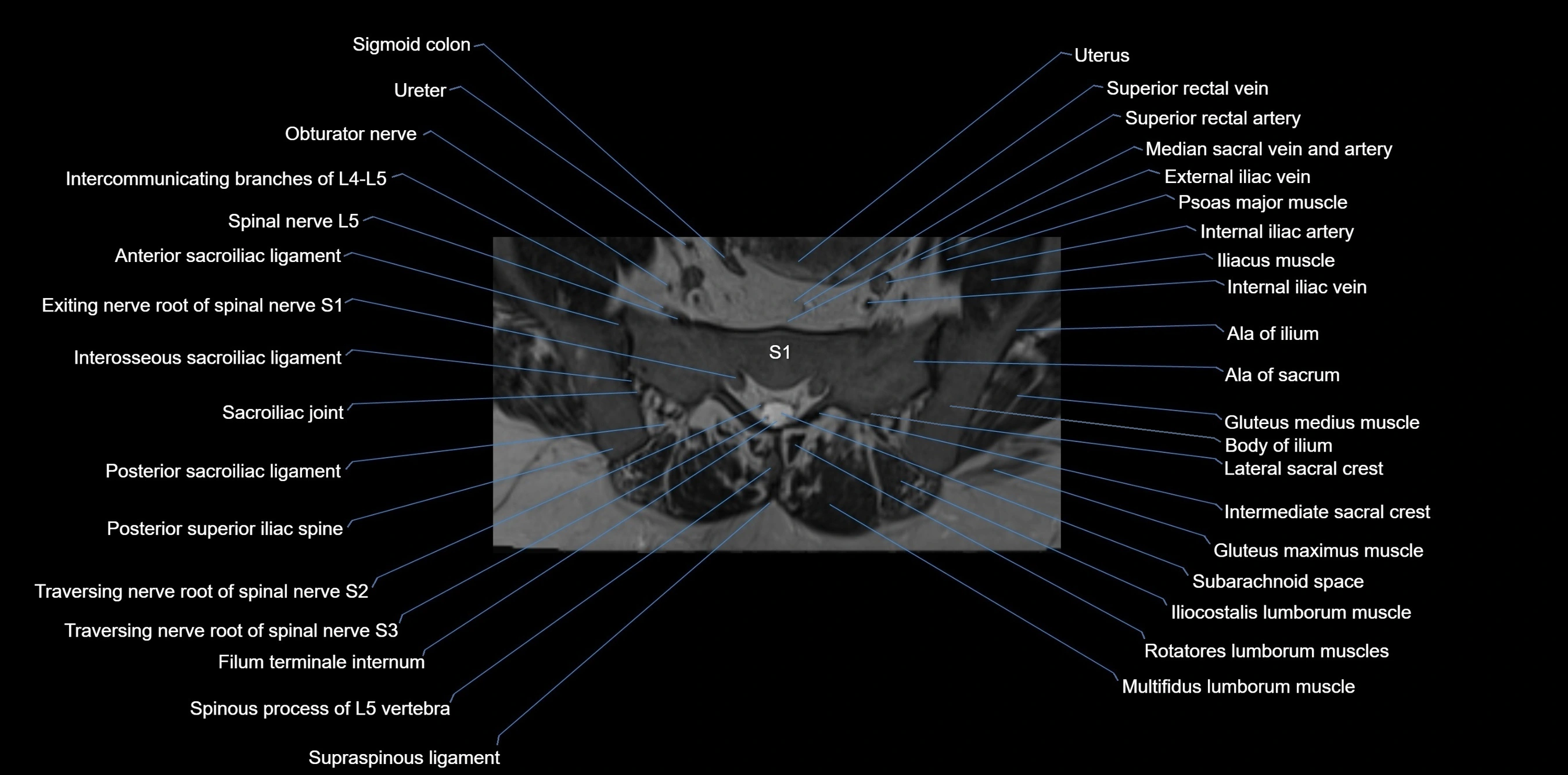
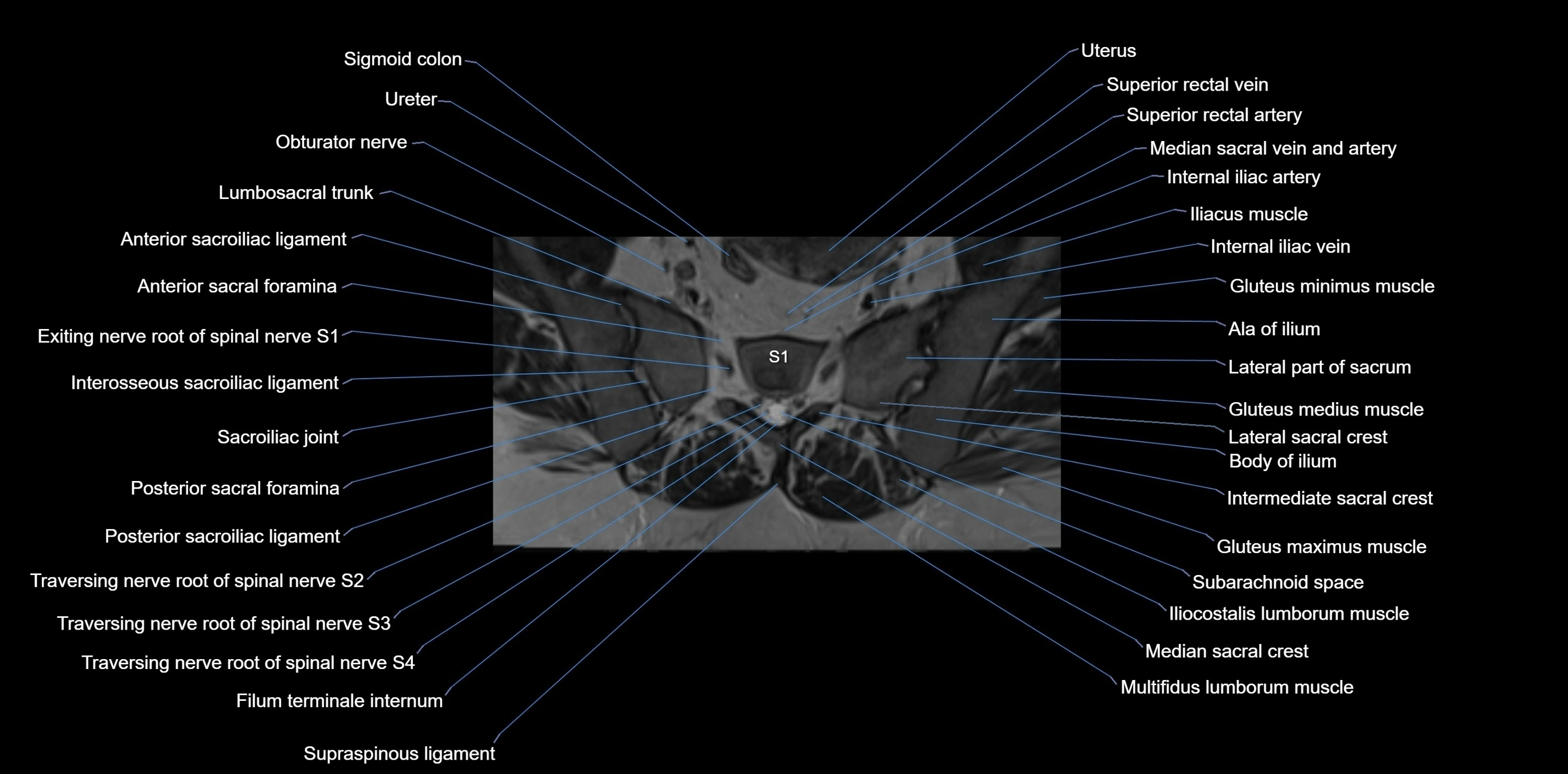
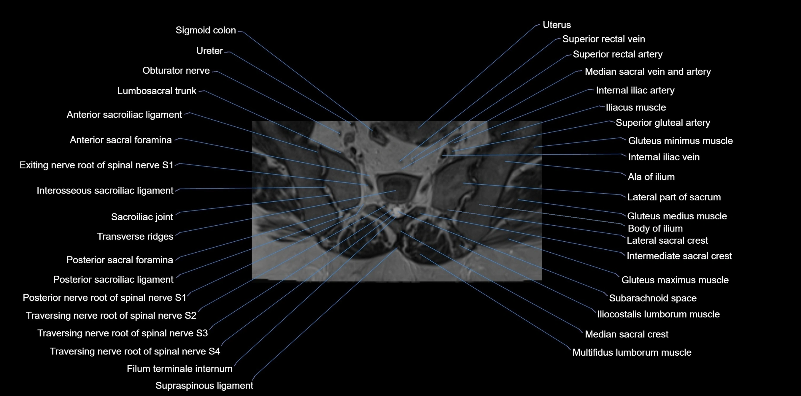
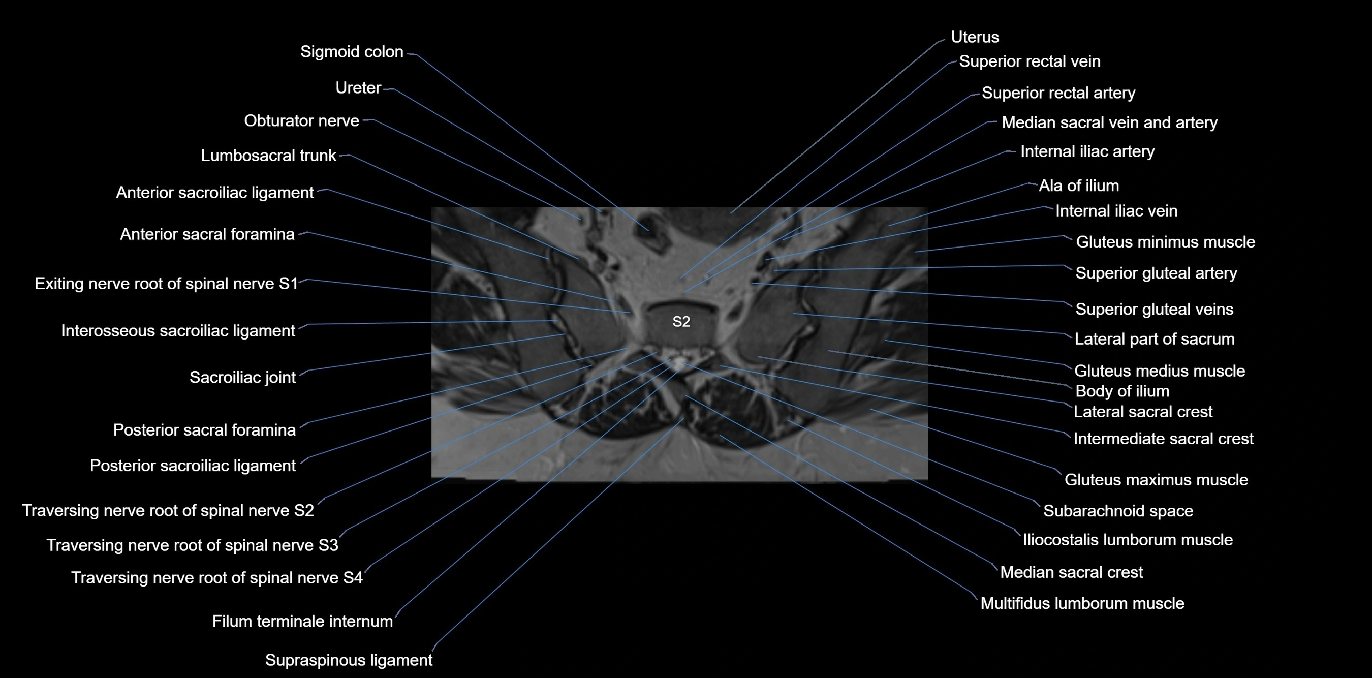
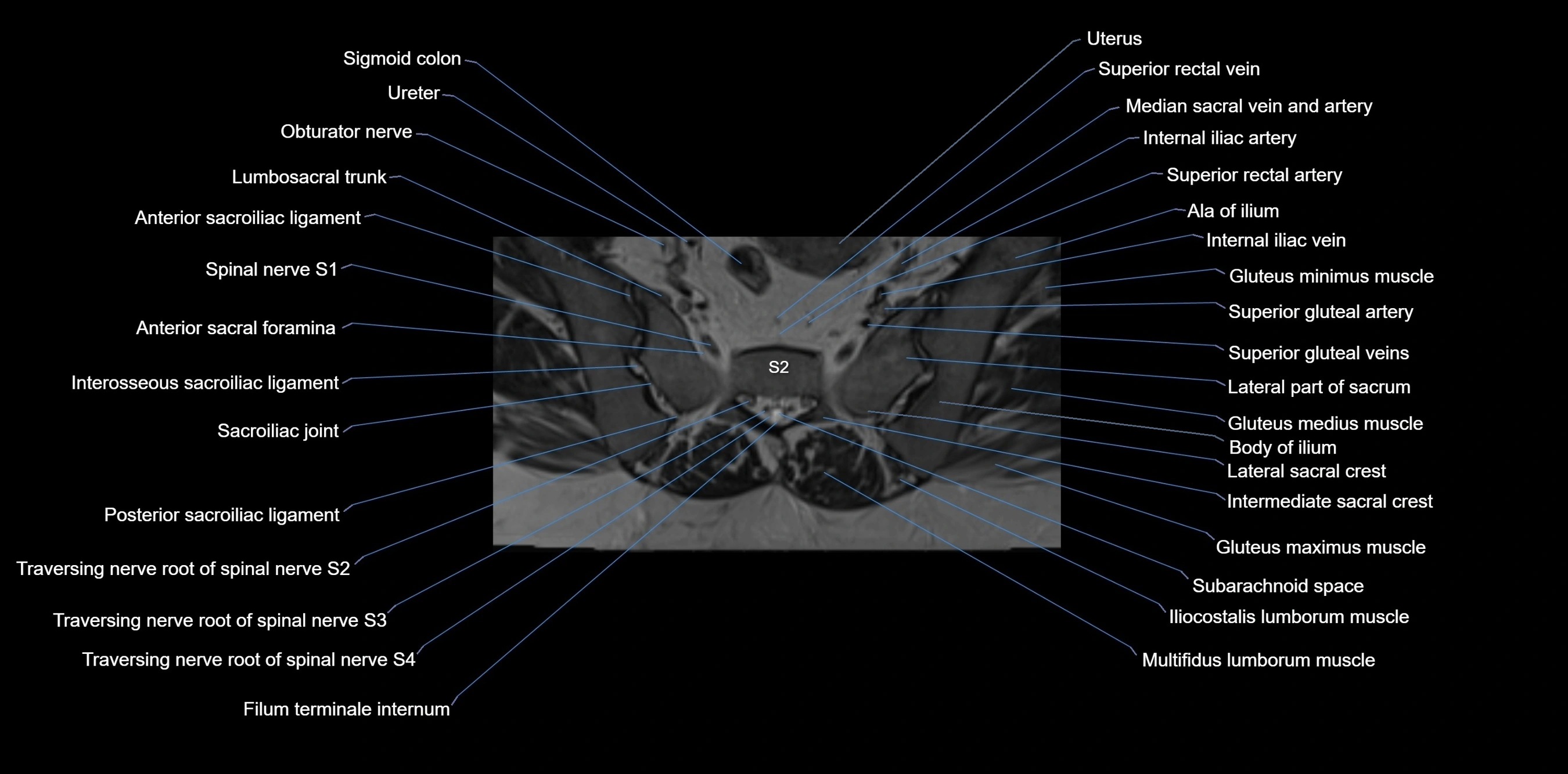
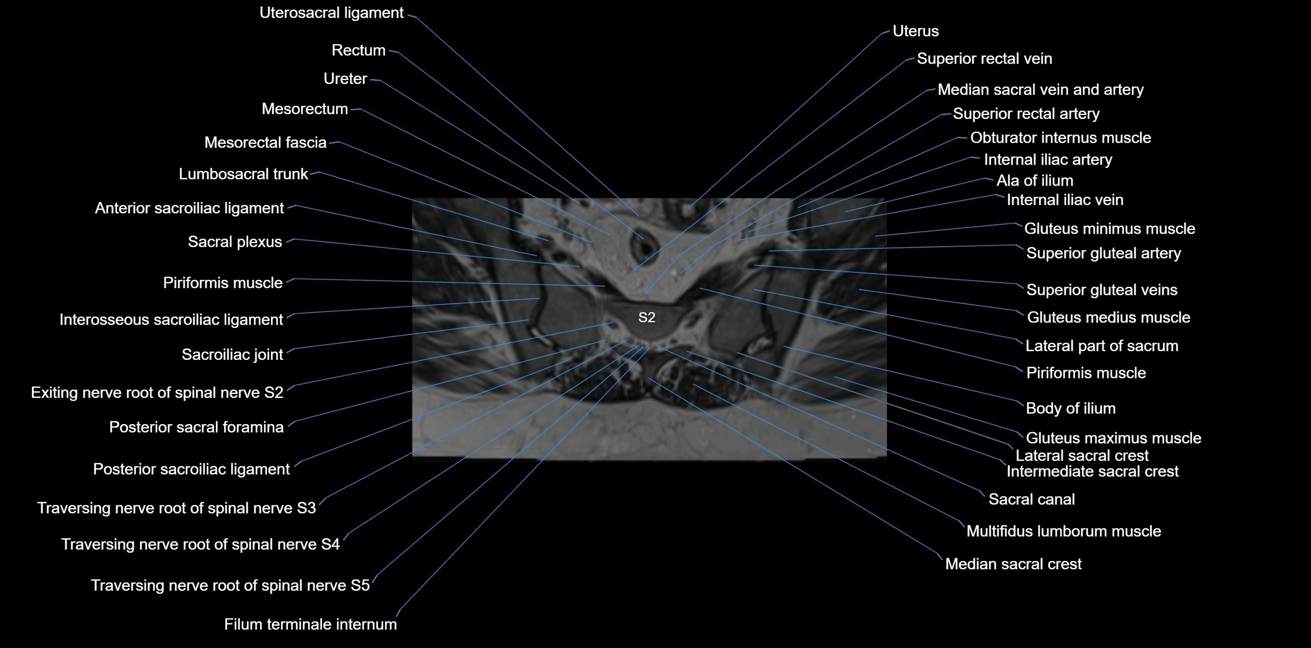
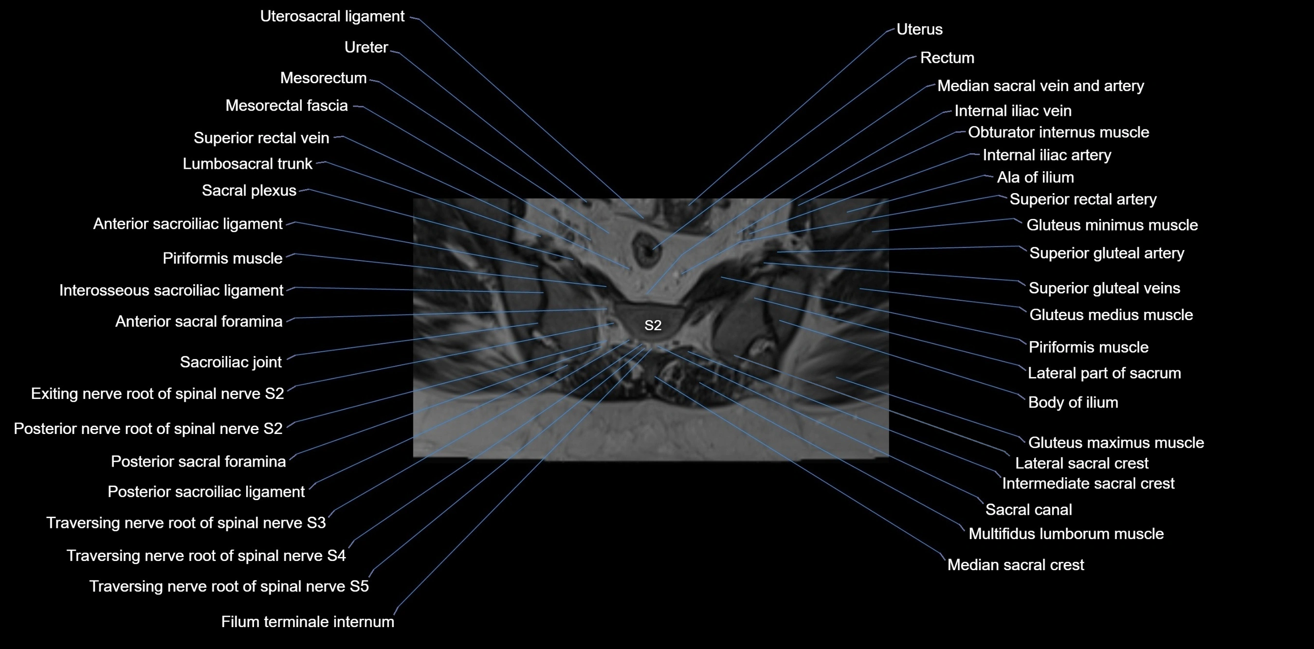
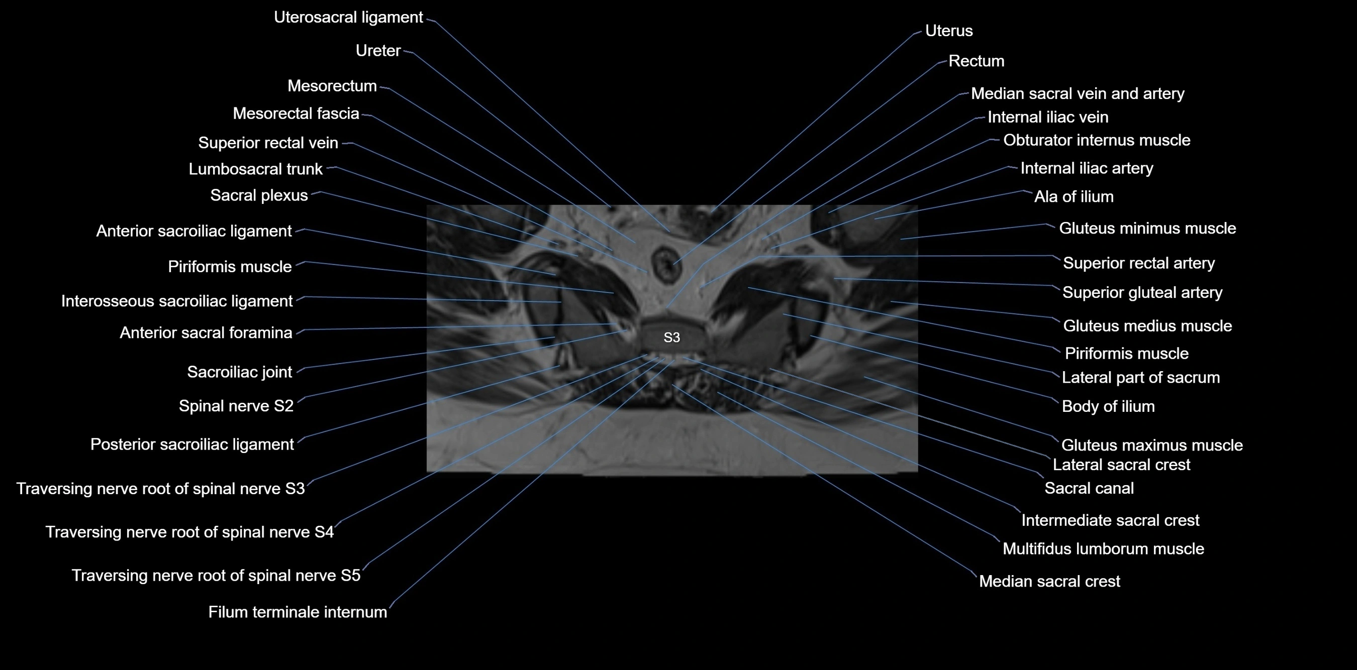
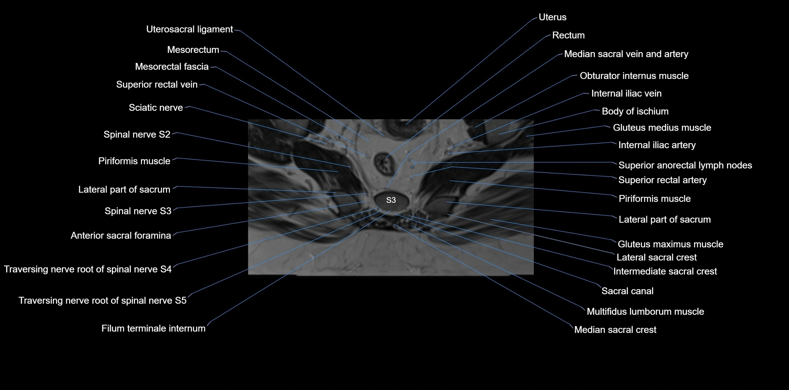
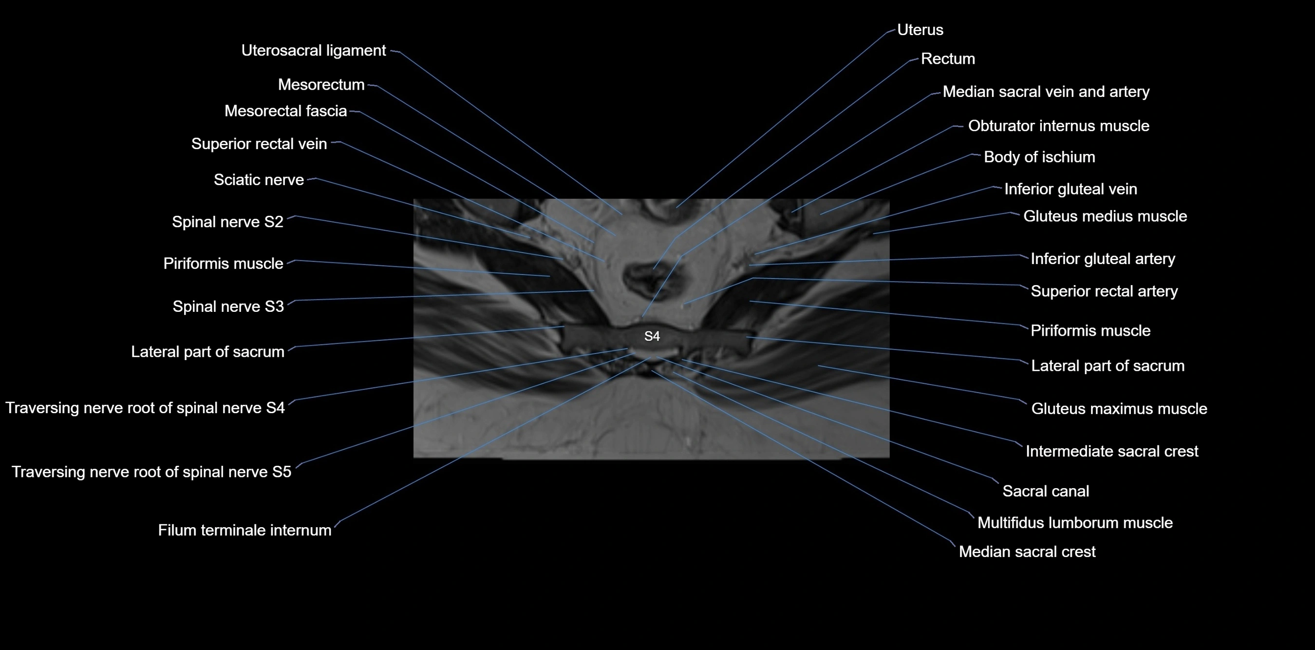
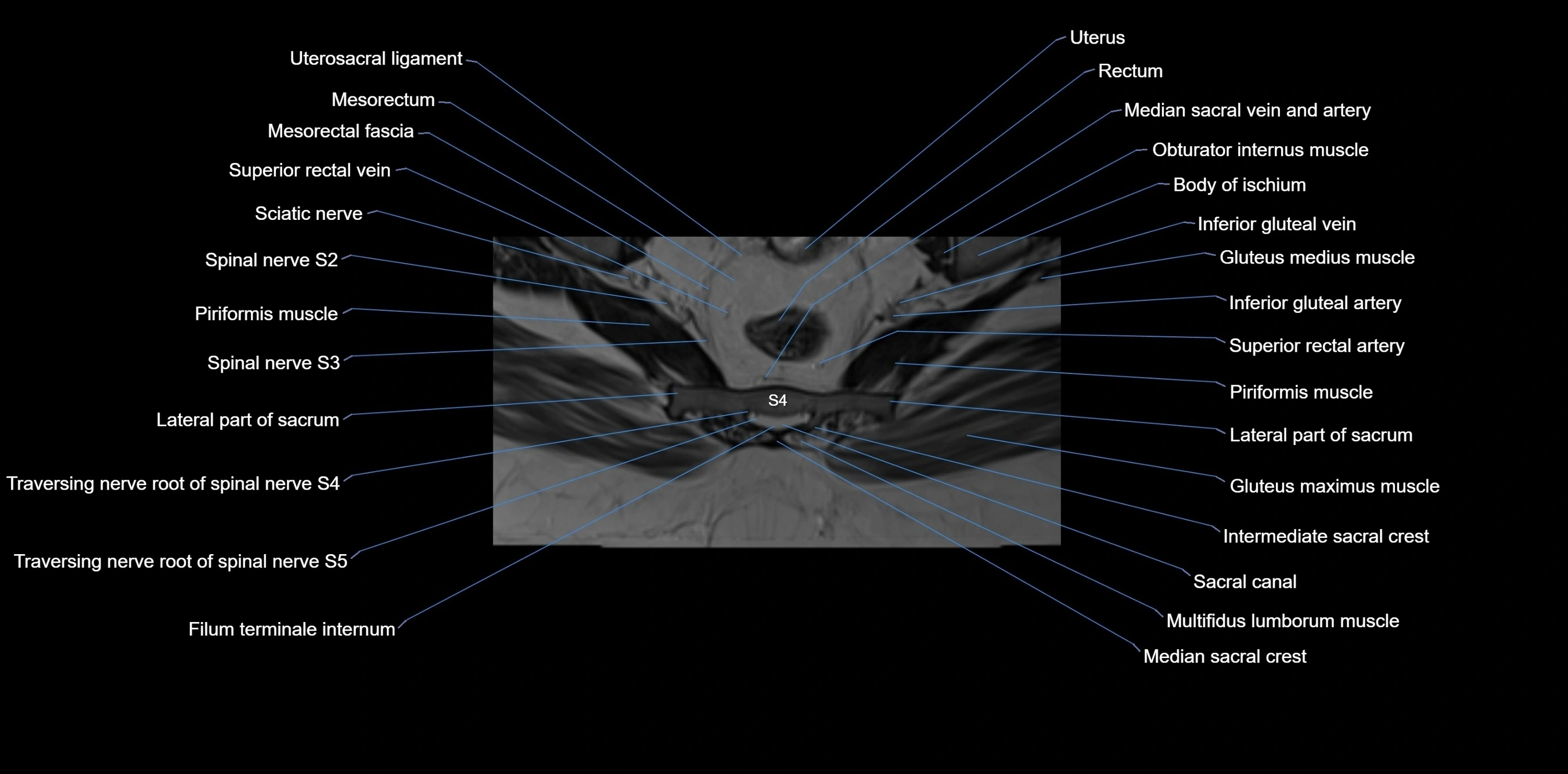
MRI Appearance
T1-weighted images:
-
Cortical bone appears very low signal (dark); marrow shows intermediate signal
-
Iliac fossa fat is bright against low-signal cortex
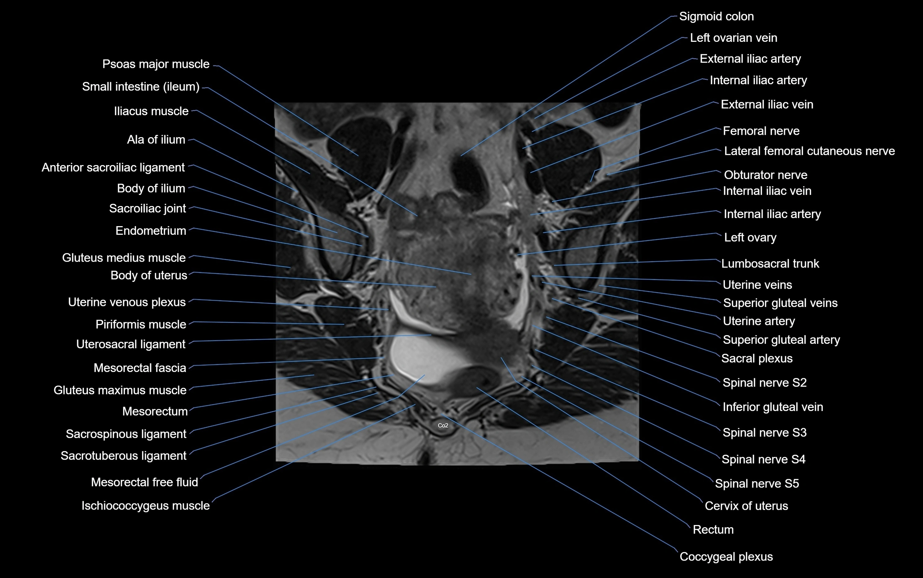
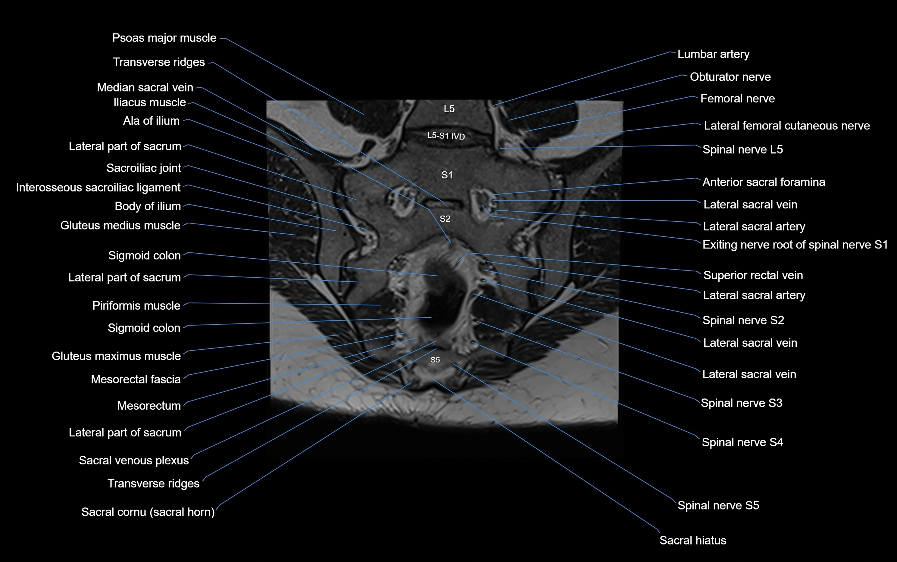
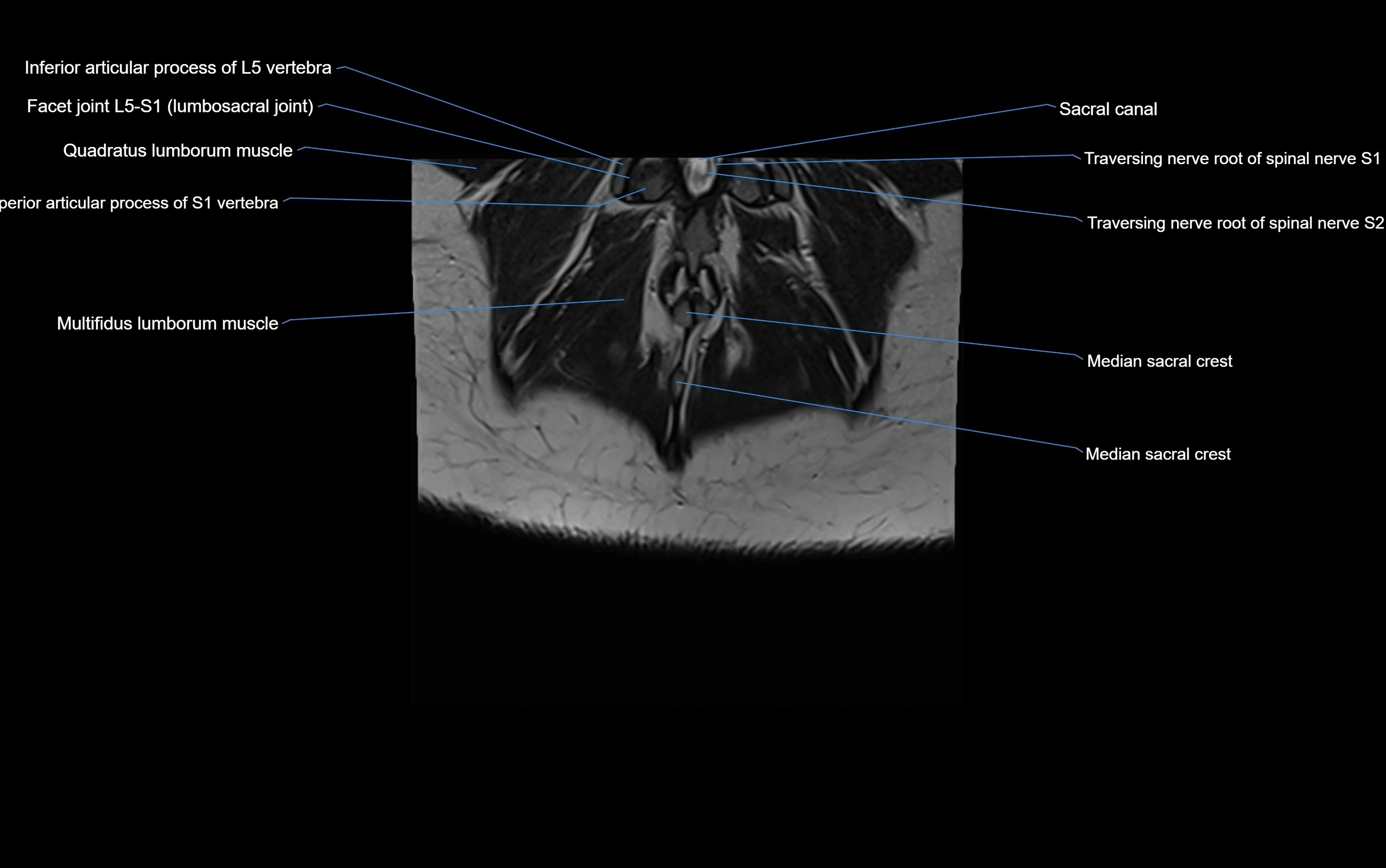
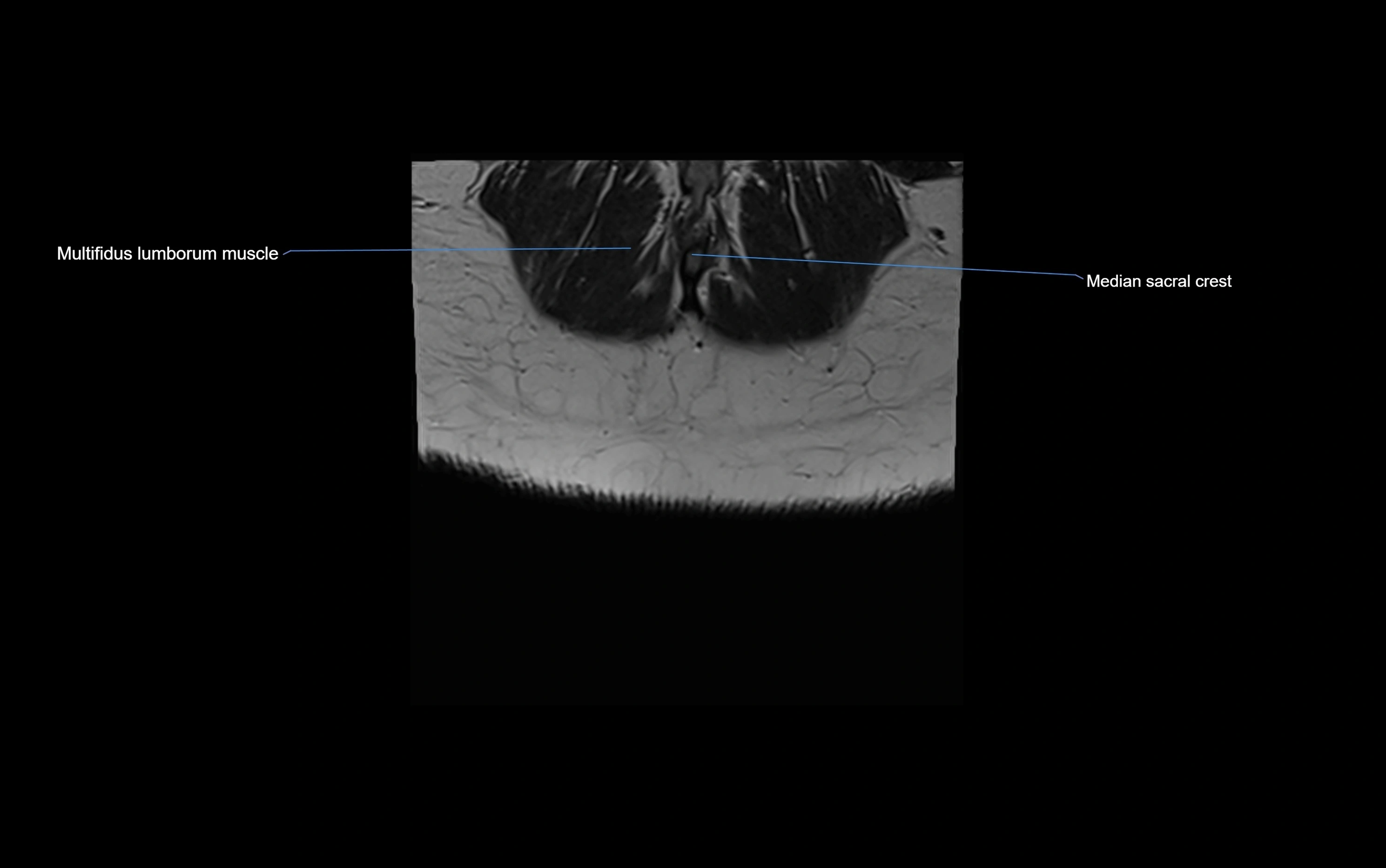
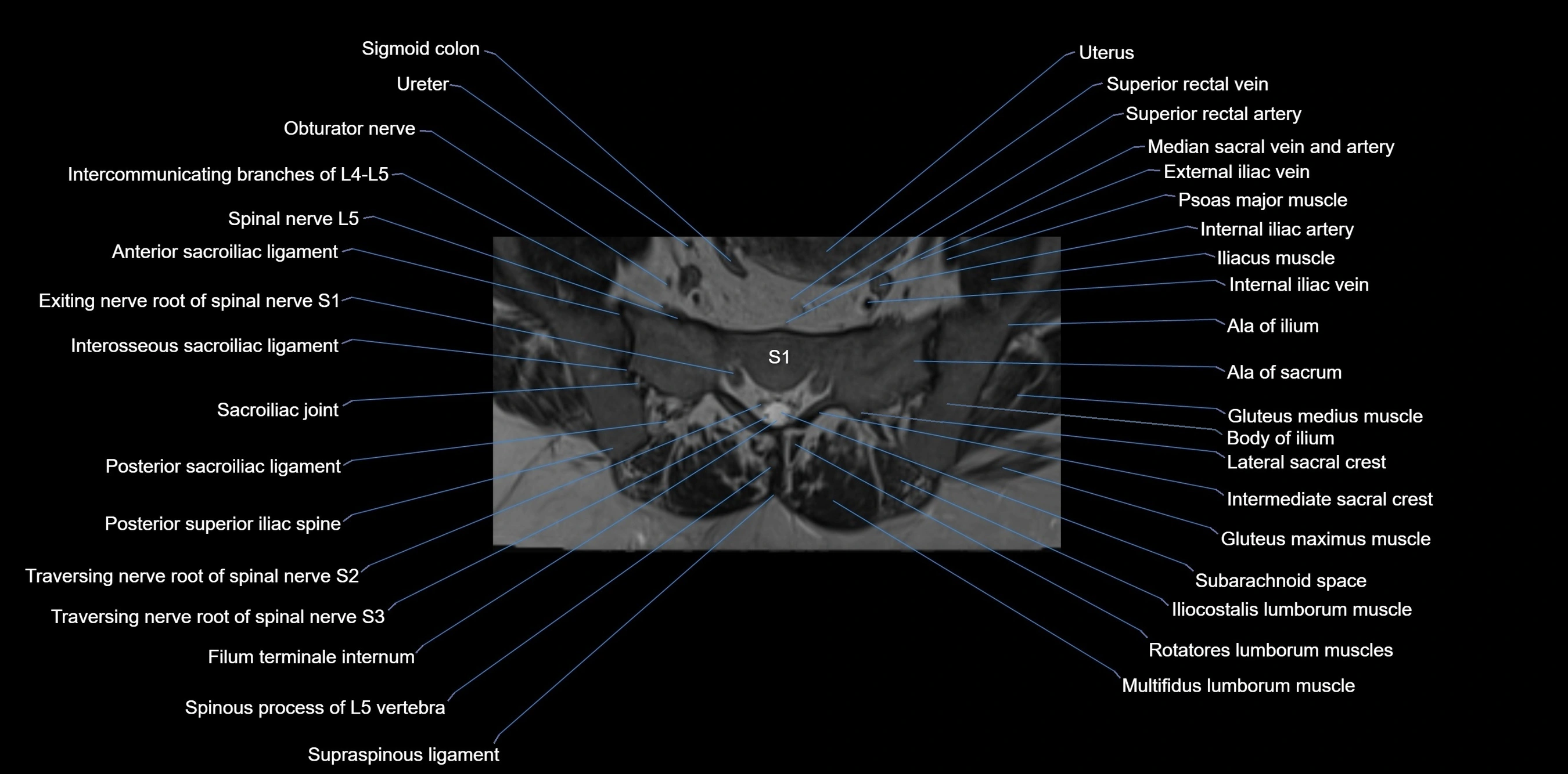
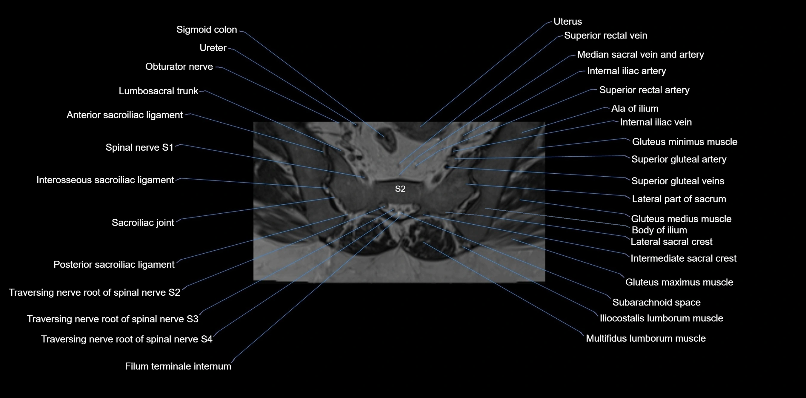
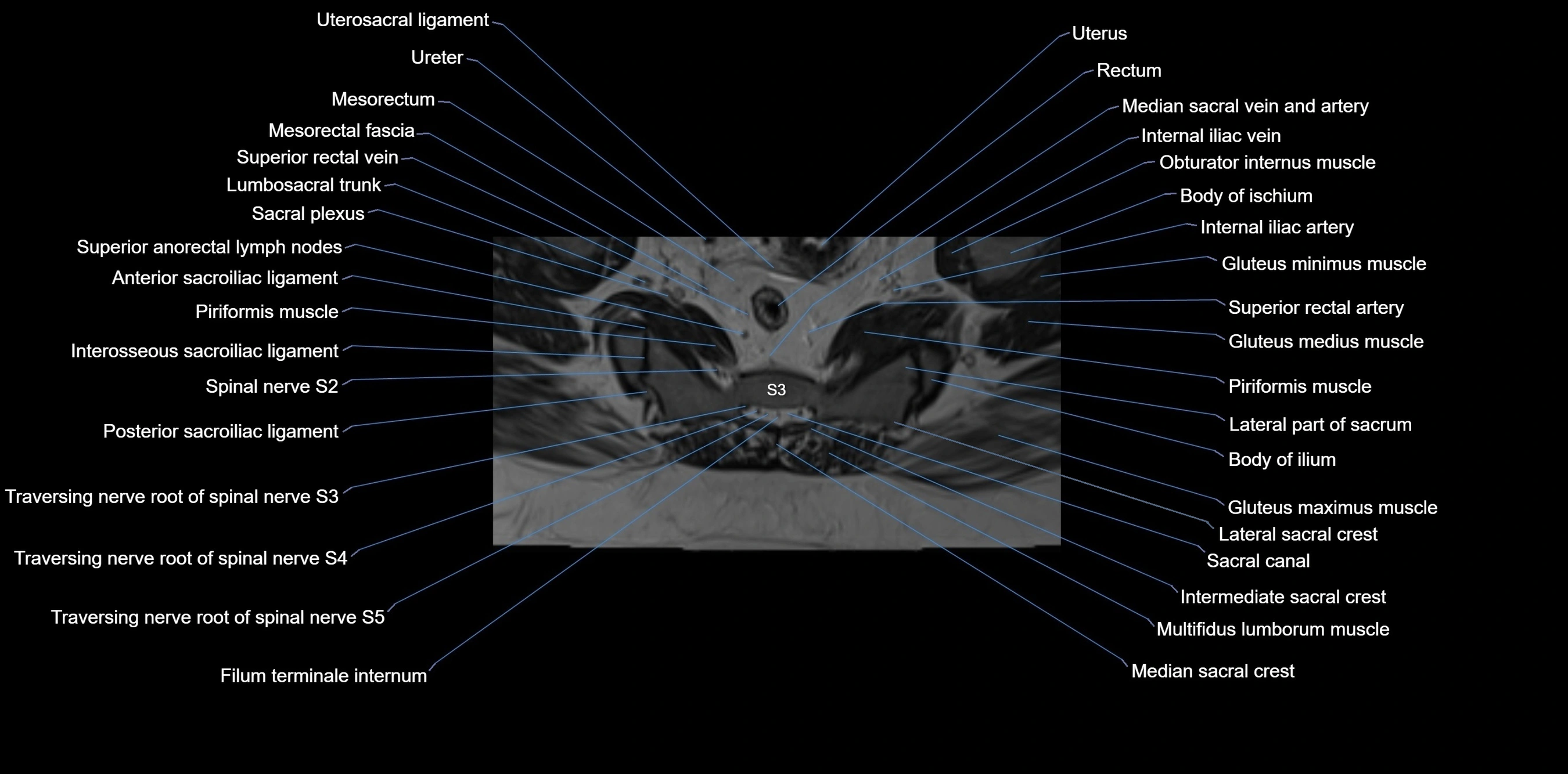
T2-weighted images:
-
Cortical bone remains dark
-
Marrow signal varies depending on fat content; edema or tumor shows hyperintensity
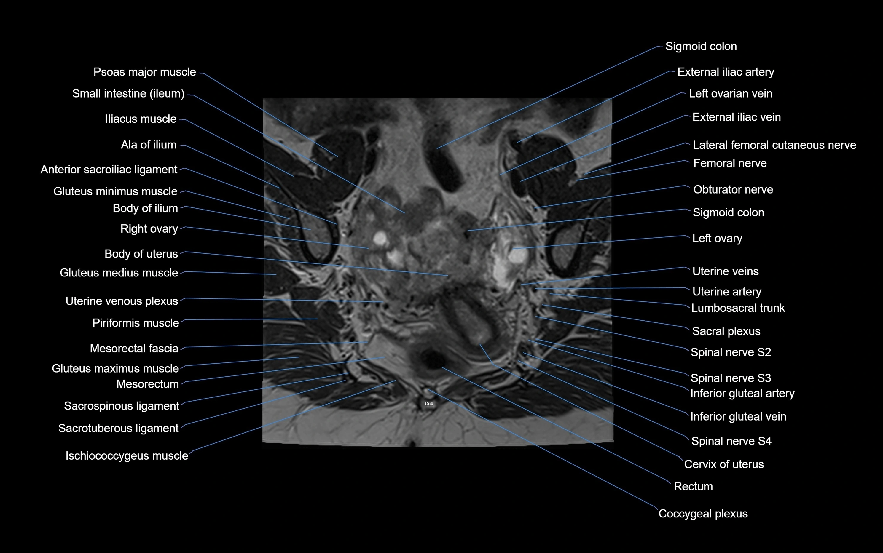
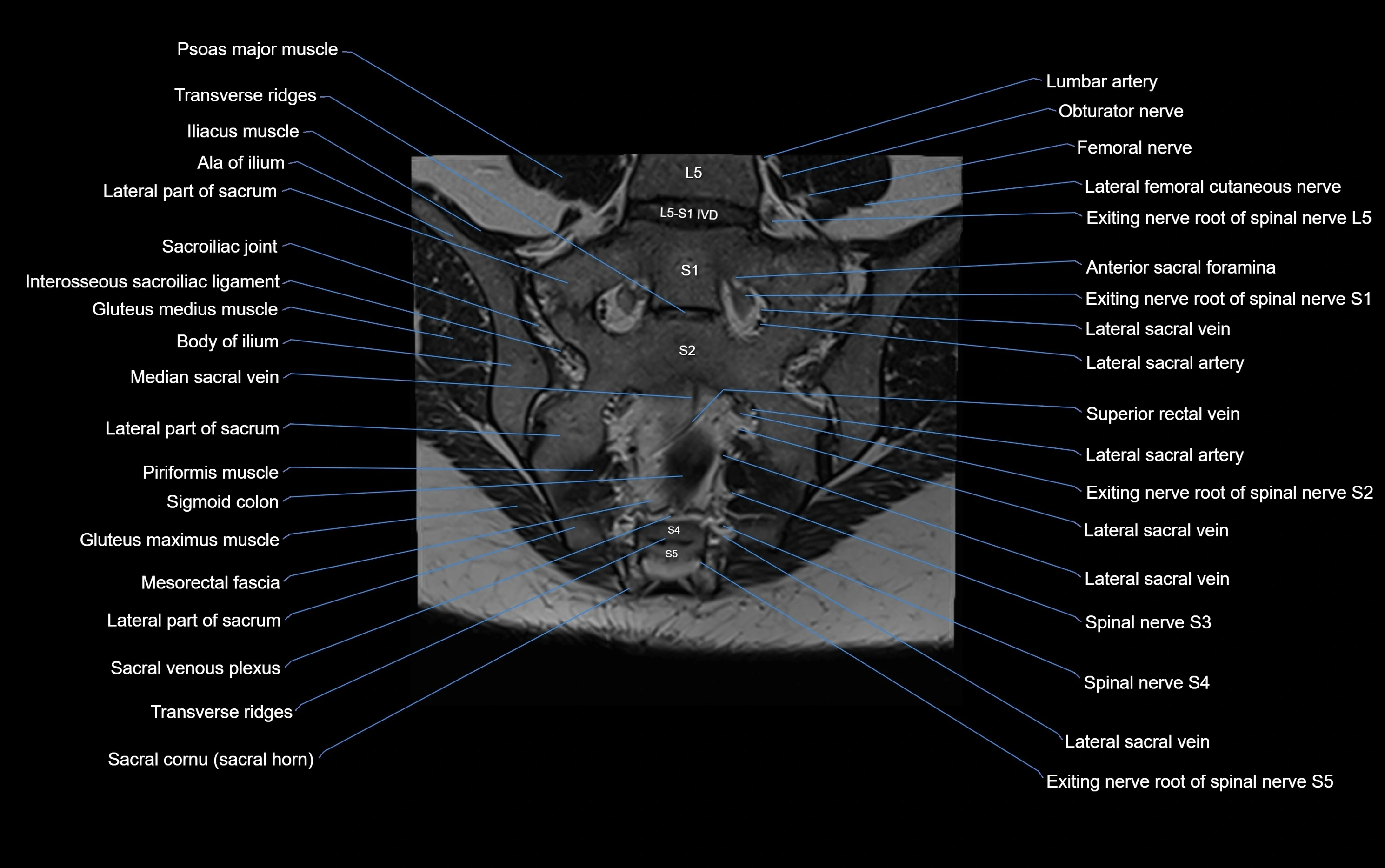
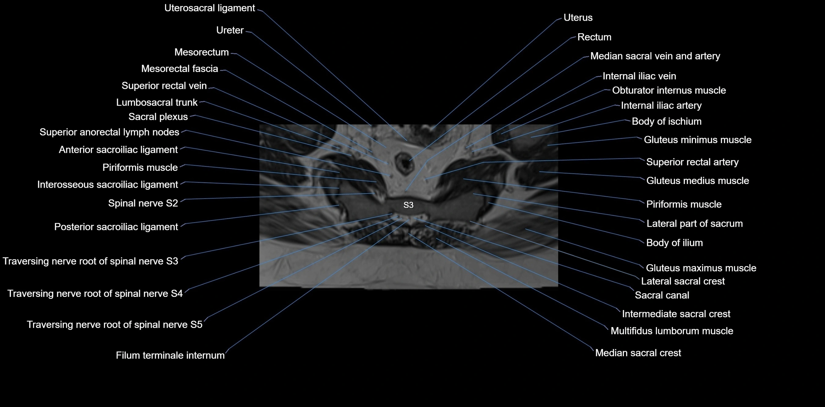
STIR:
-
Suppresses fat, making bone marrow edema, fractures, or infiltrative lesions appear bright
-
Excellent for trauma, sacroiliitis, and metastatic evaluation
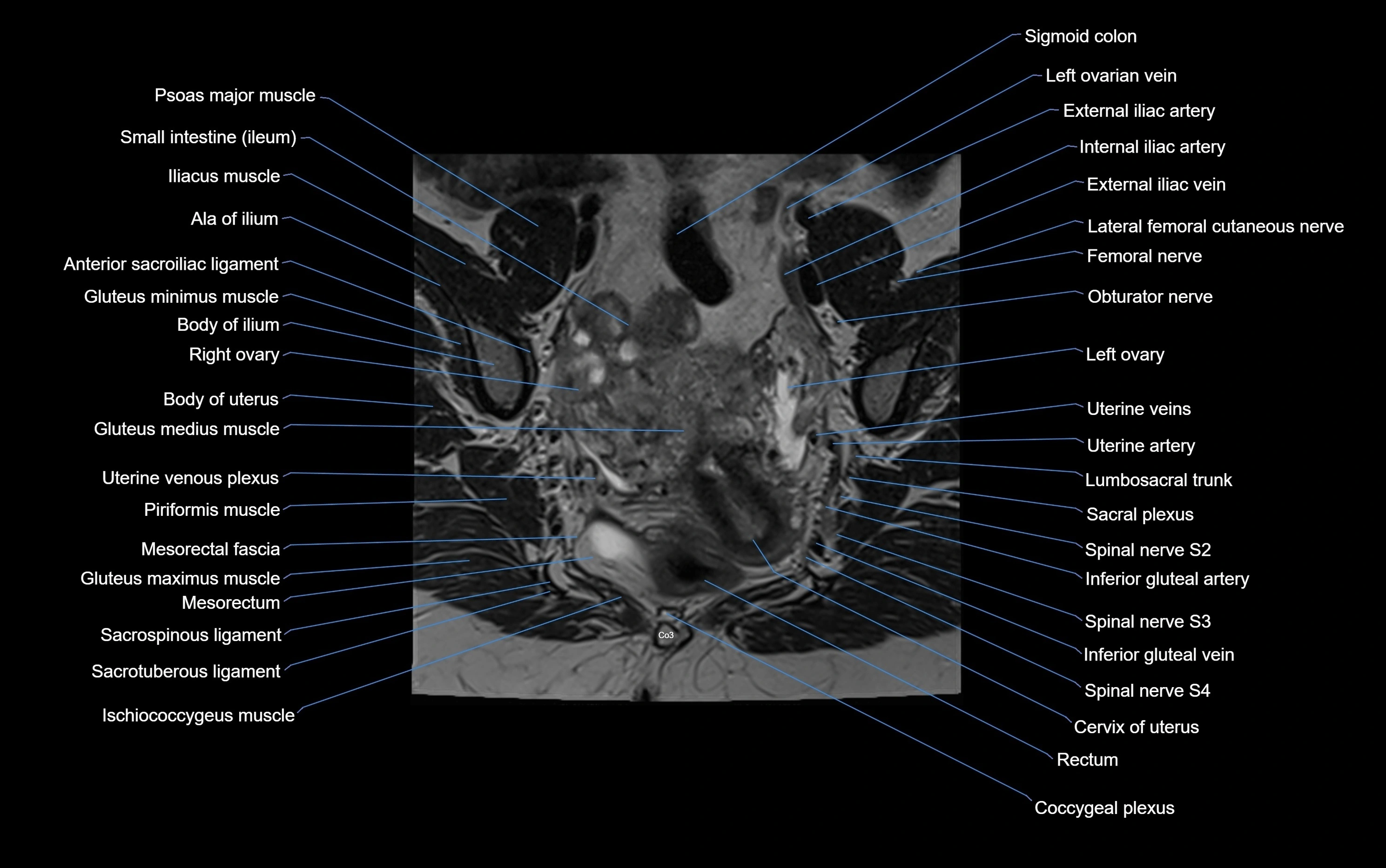
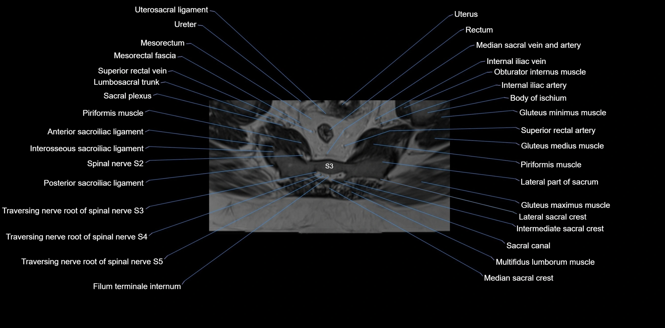
T1 Fat-Saturated (Pre-contrast):
-
Marrow: intermediate signal, fat suppressed
-
Useful for detecting subtle marrow abnormalities adjacent to iliac cortex
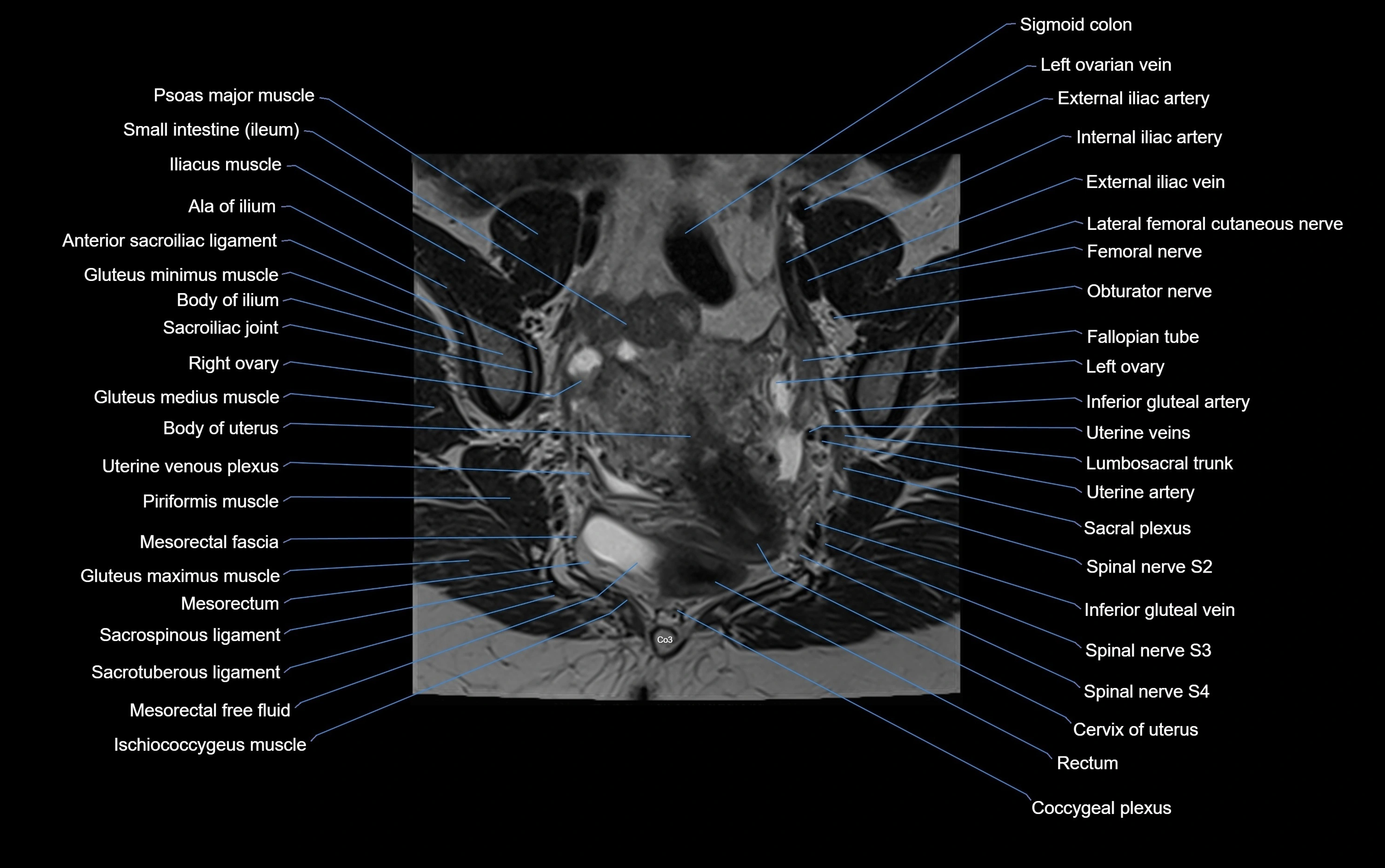
T1 Fat-Saturated Post-Contrast (Gadolinium):
-
Enhances vascularized structures, marrow pathology, tumors, and inflammatory changes
-
Highlights soft tissue or bone invasion in pelvic neoplasms
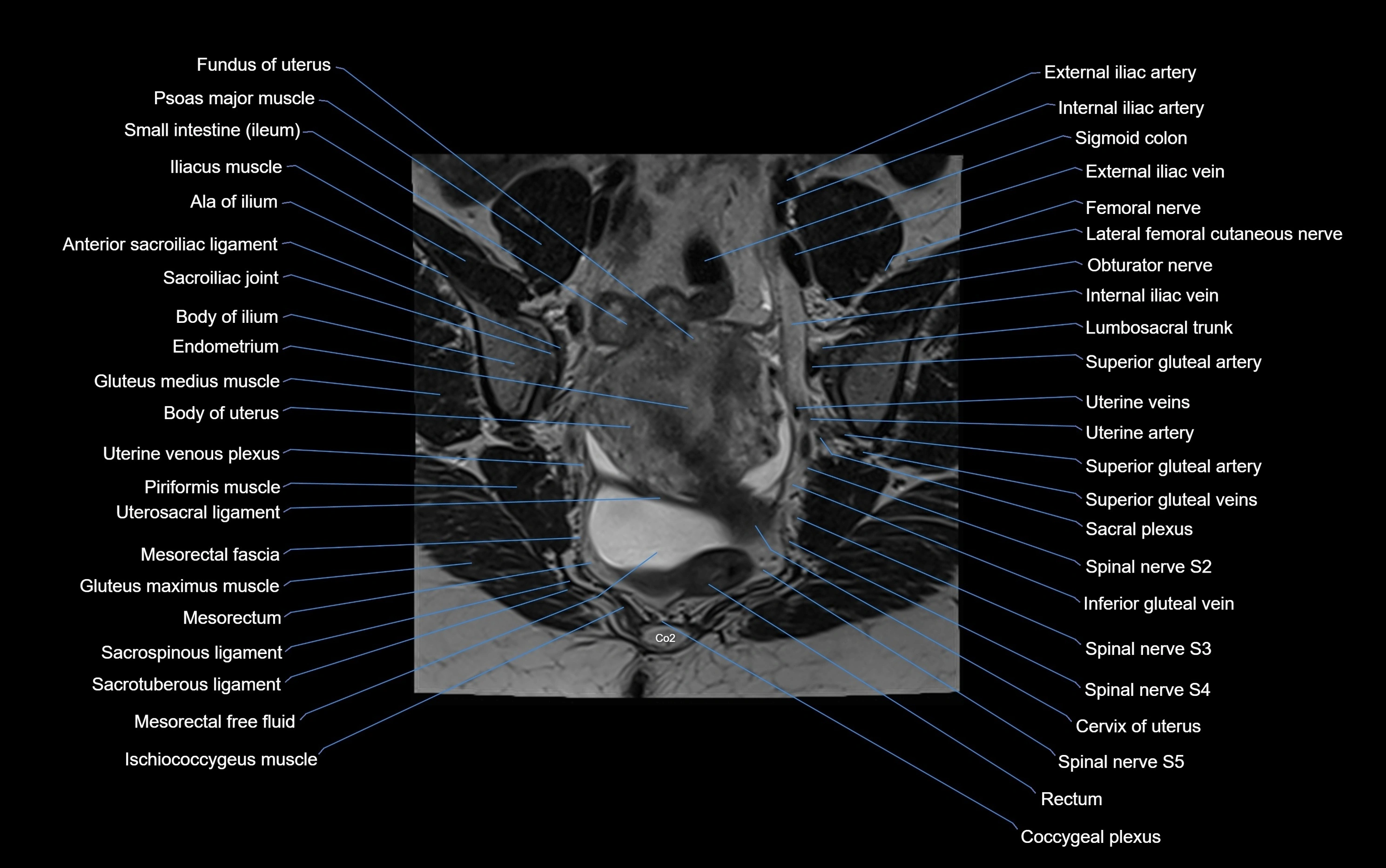
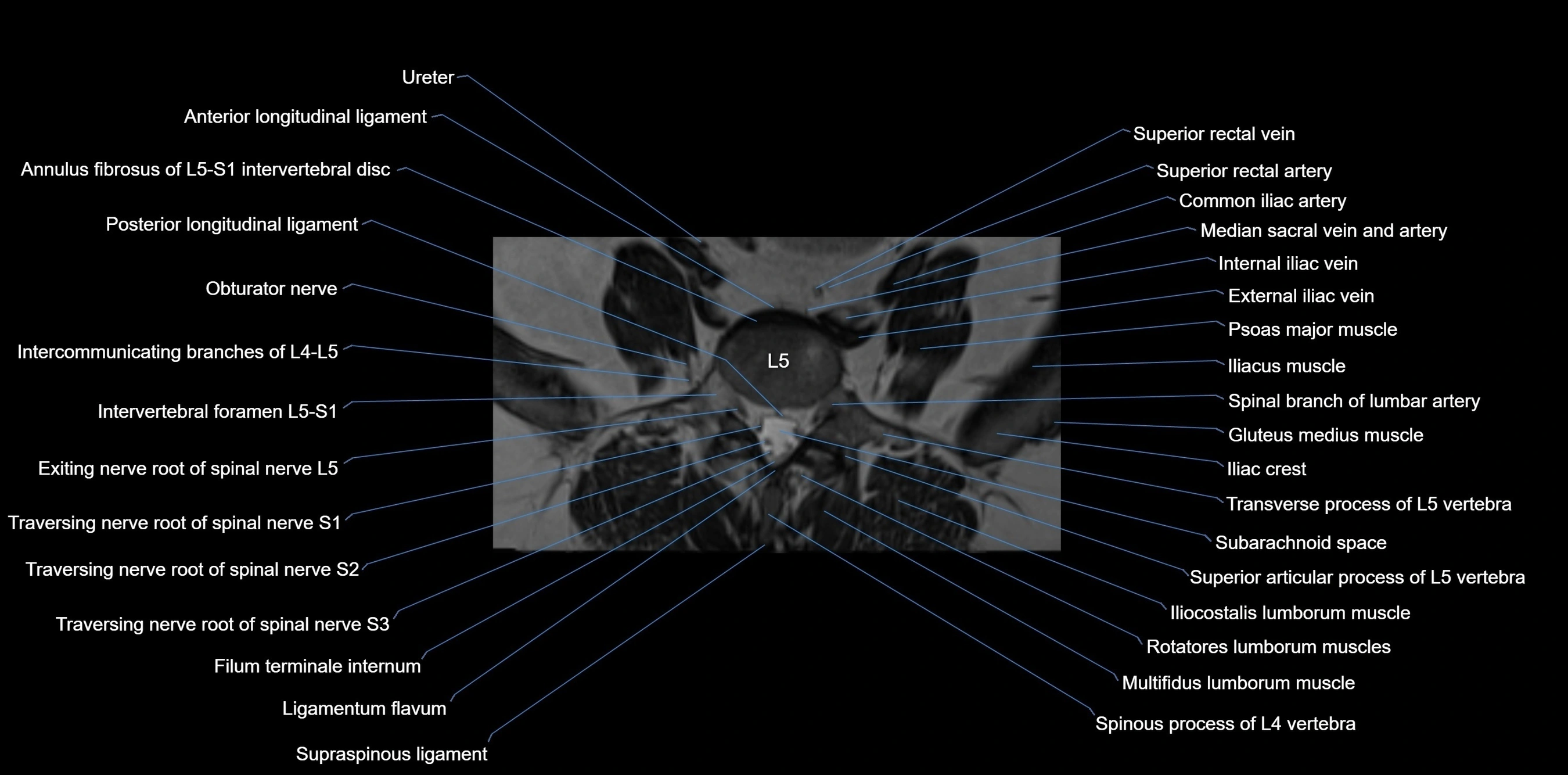
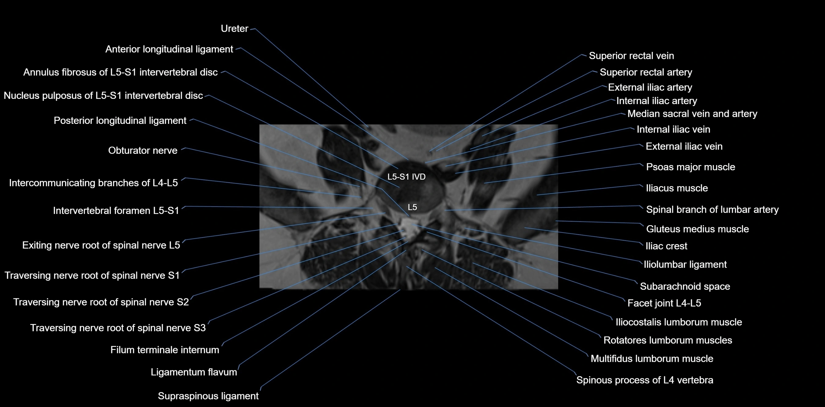
MRI Non-Contrast 3D Imaging:
-
Provides 3D morphology of iliac wing, crest, and articulations
-
Used in preoperative planning for pelvic surgery and trauma reconstruction
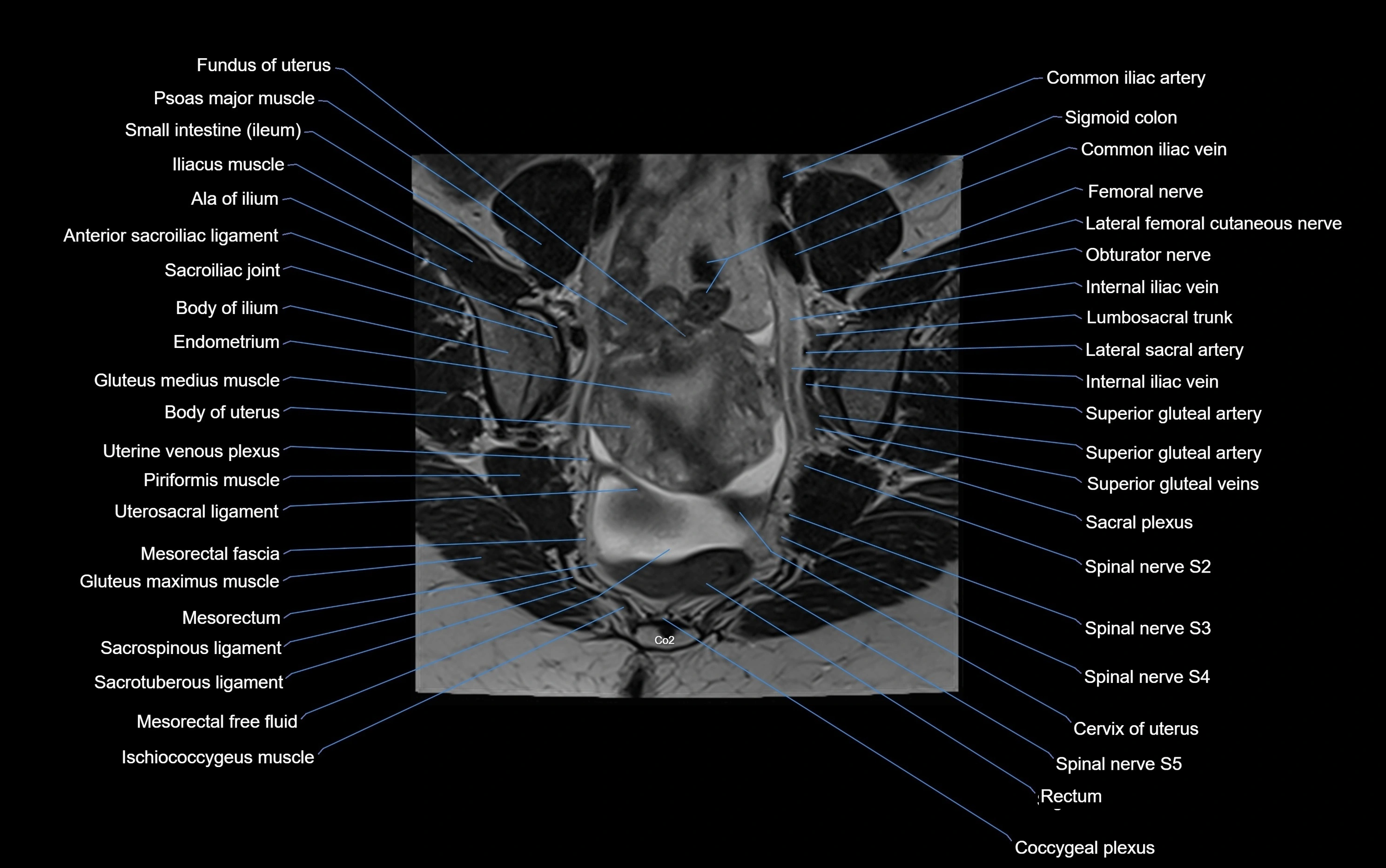
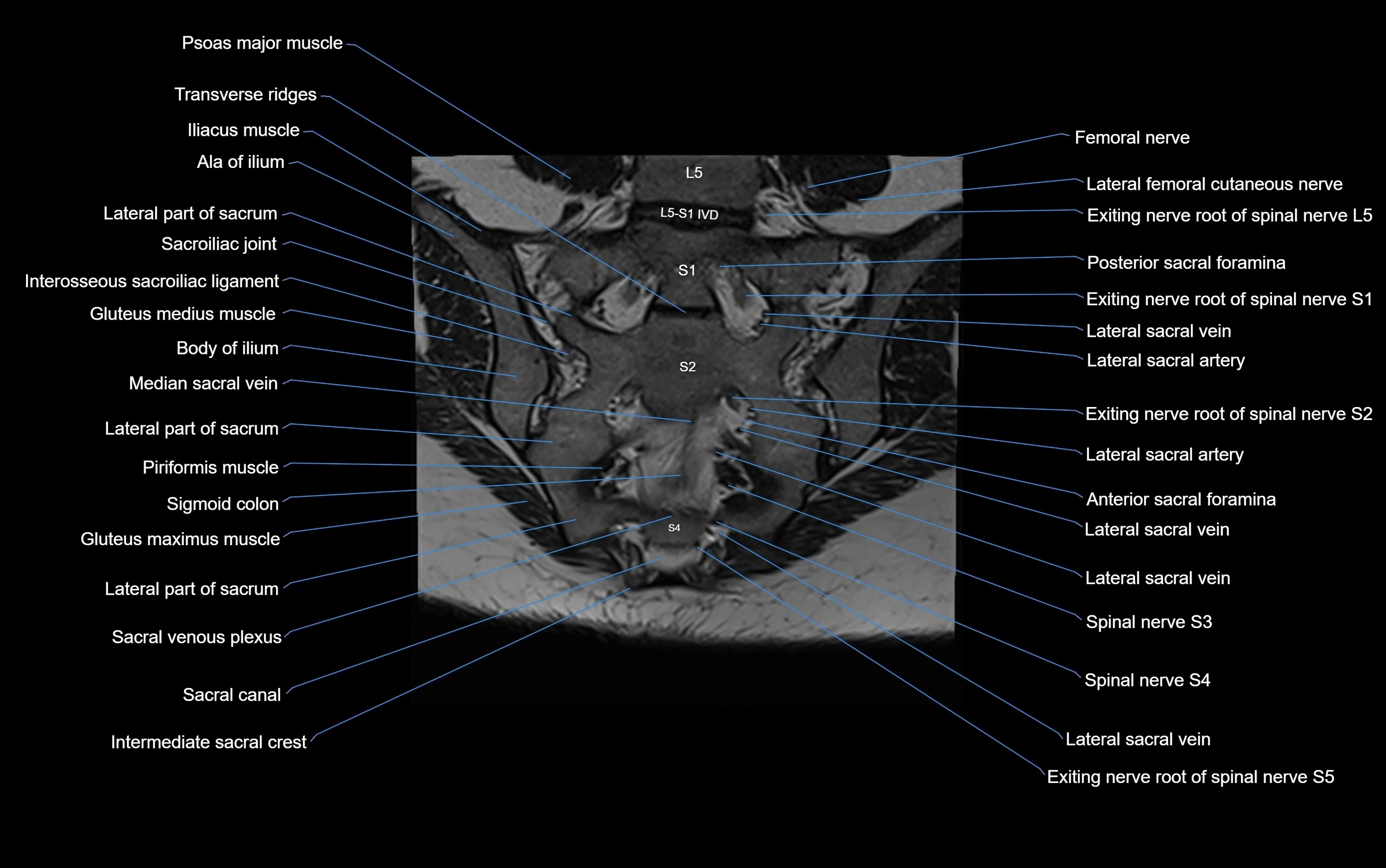
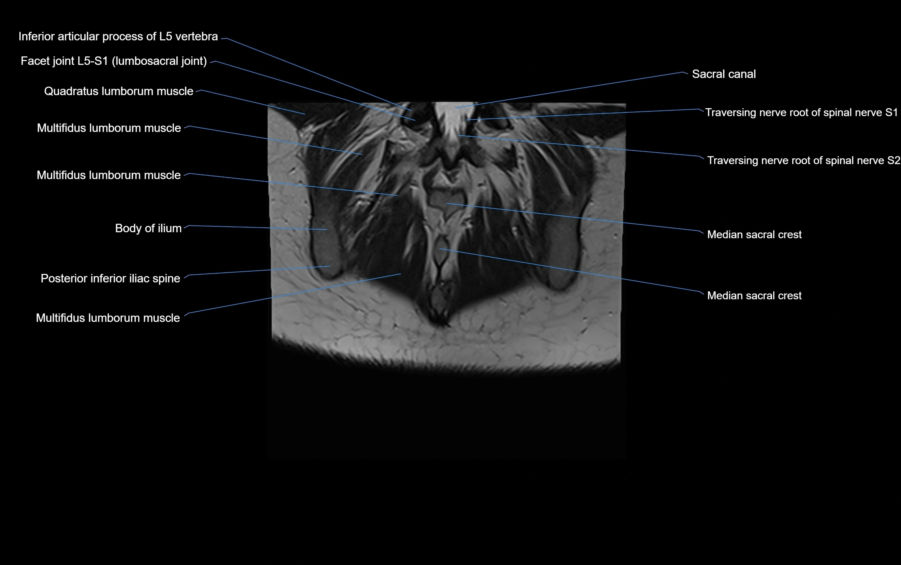
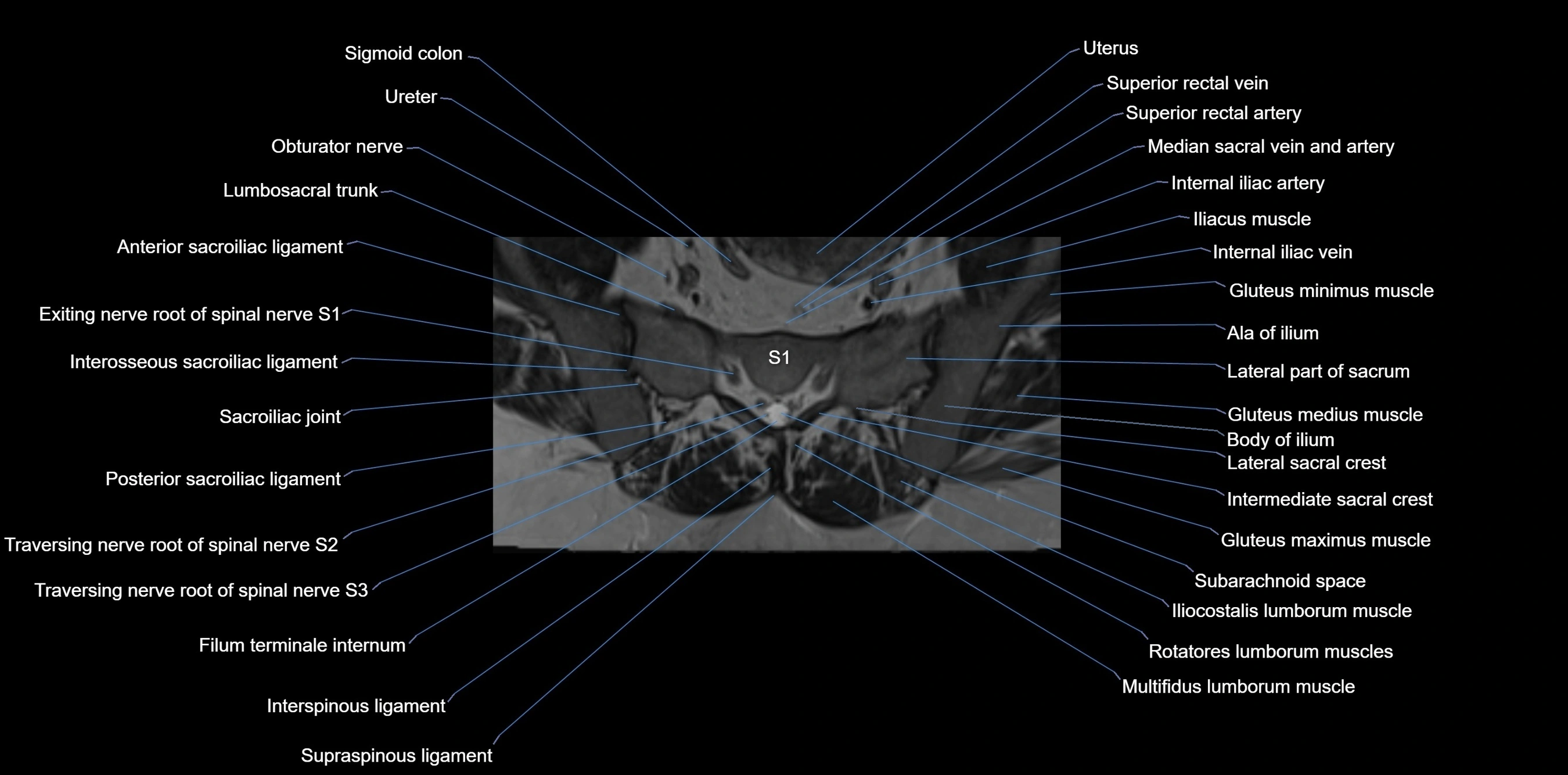
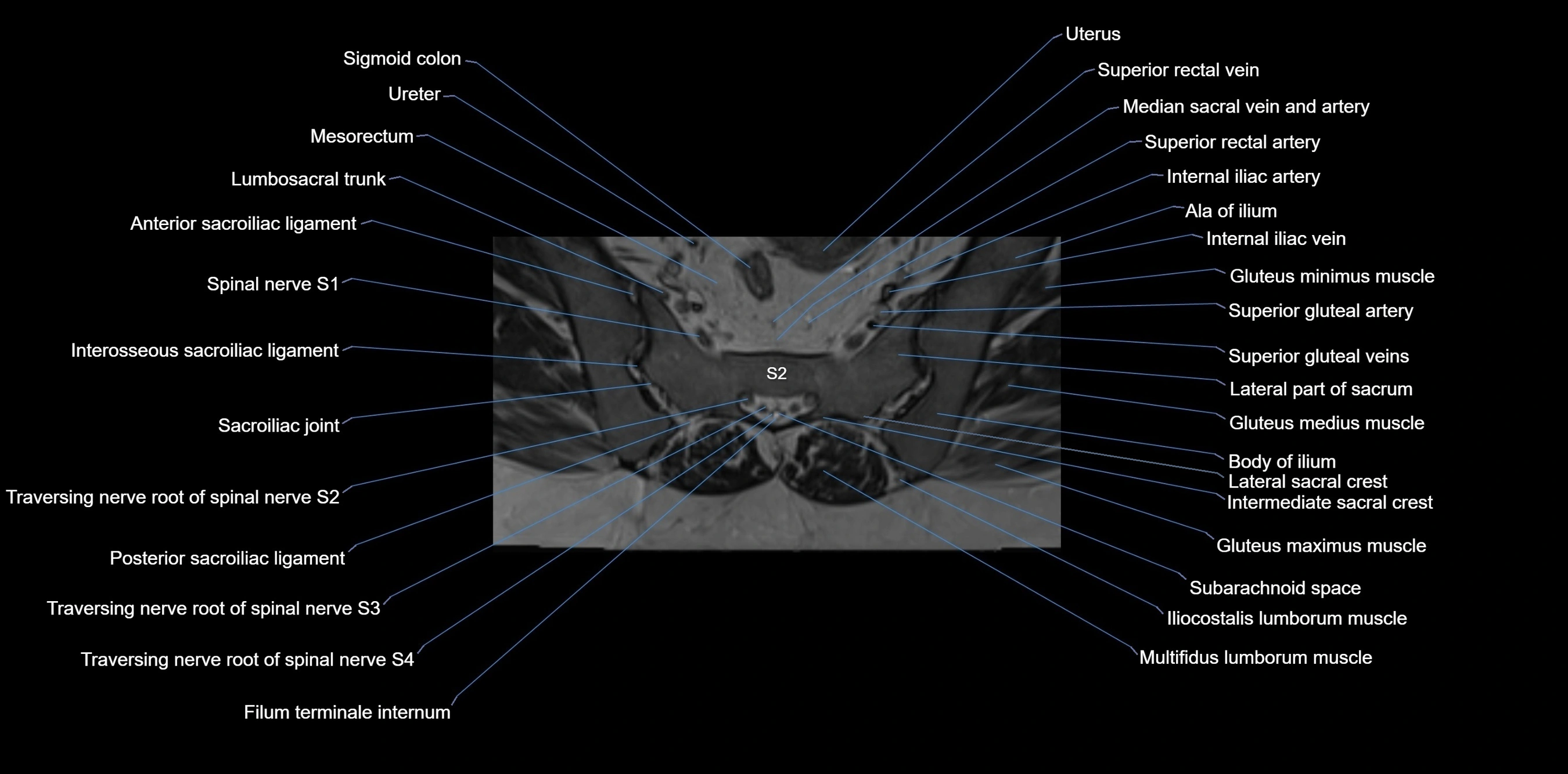
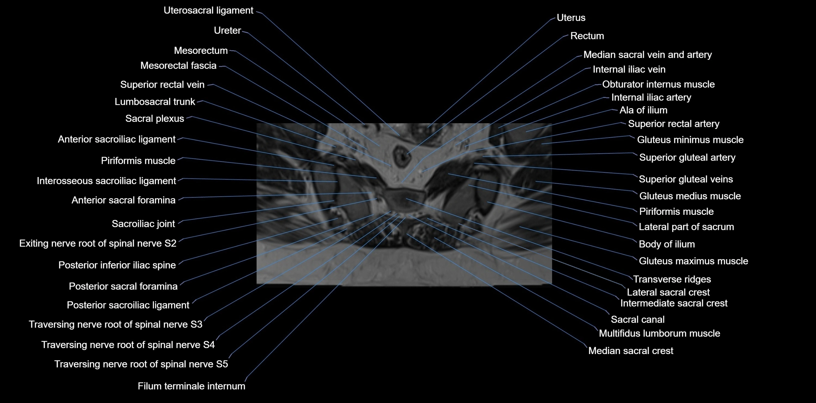
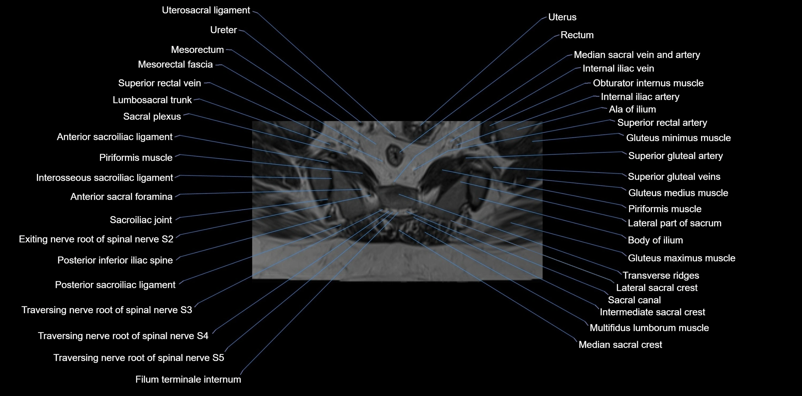
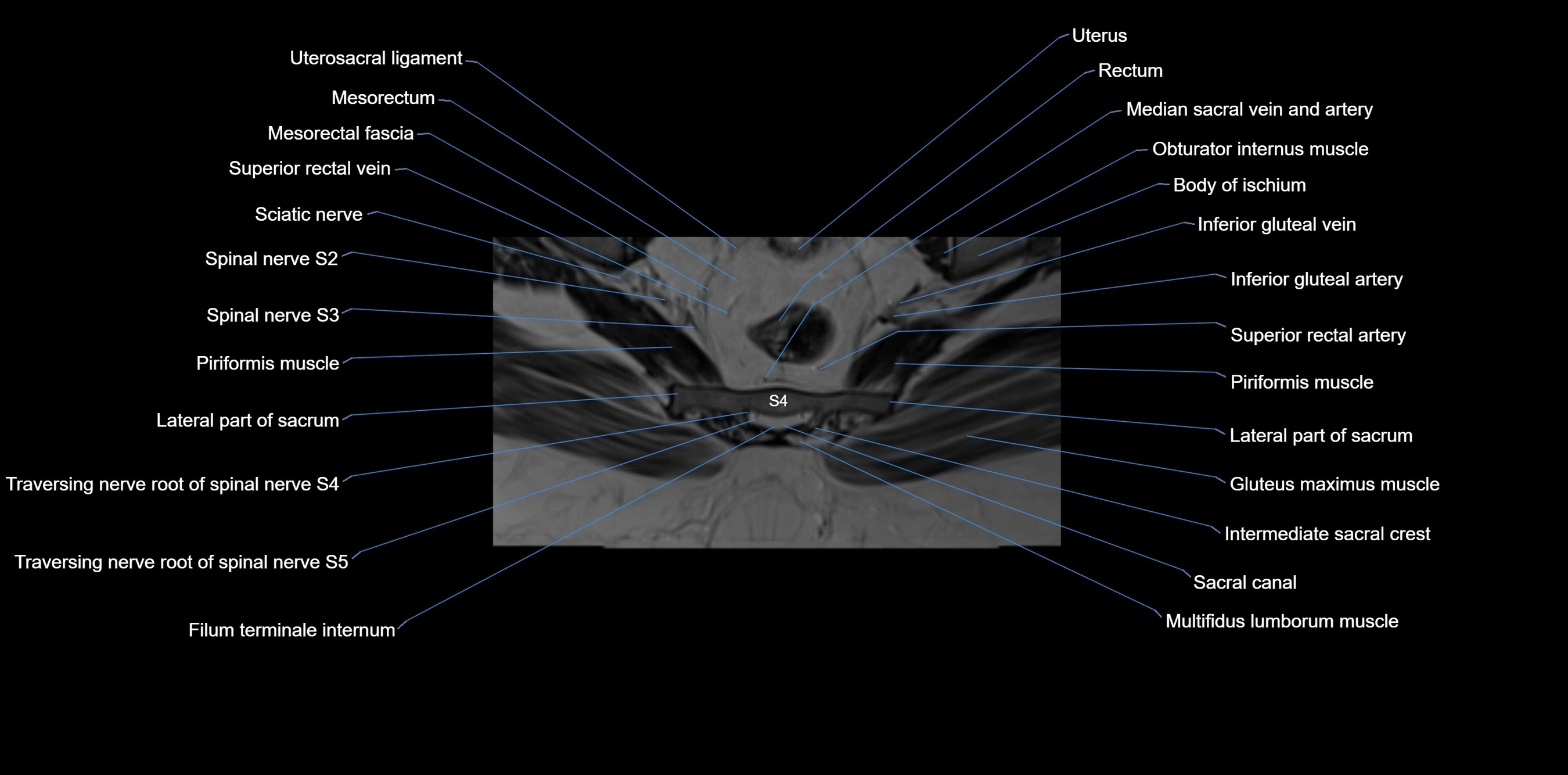
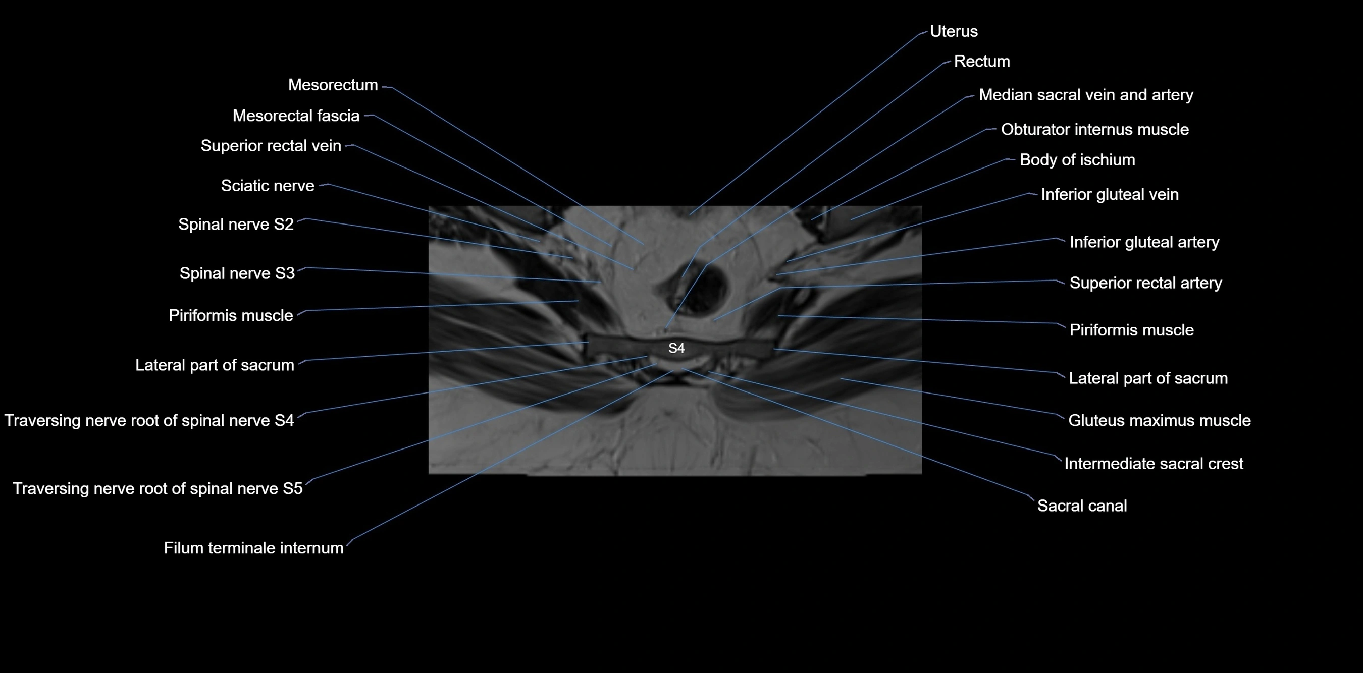
CT Appearance
CT Pre-Contrast:
-
Excellent depiction of cortical bone thickness, fractures, and trabecular pattern
-
Useful in trauma and degenerative joint assessment
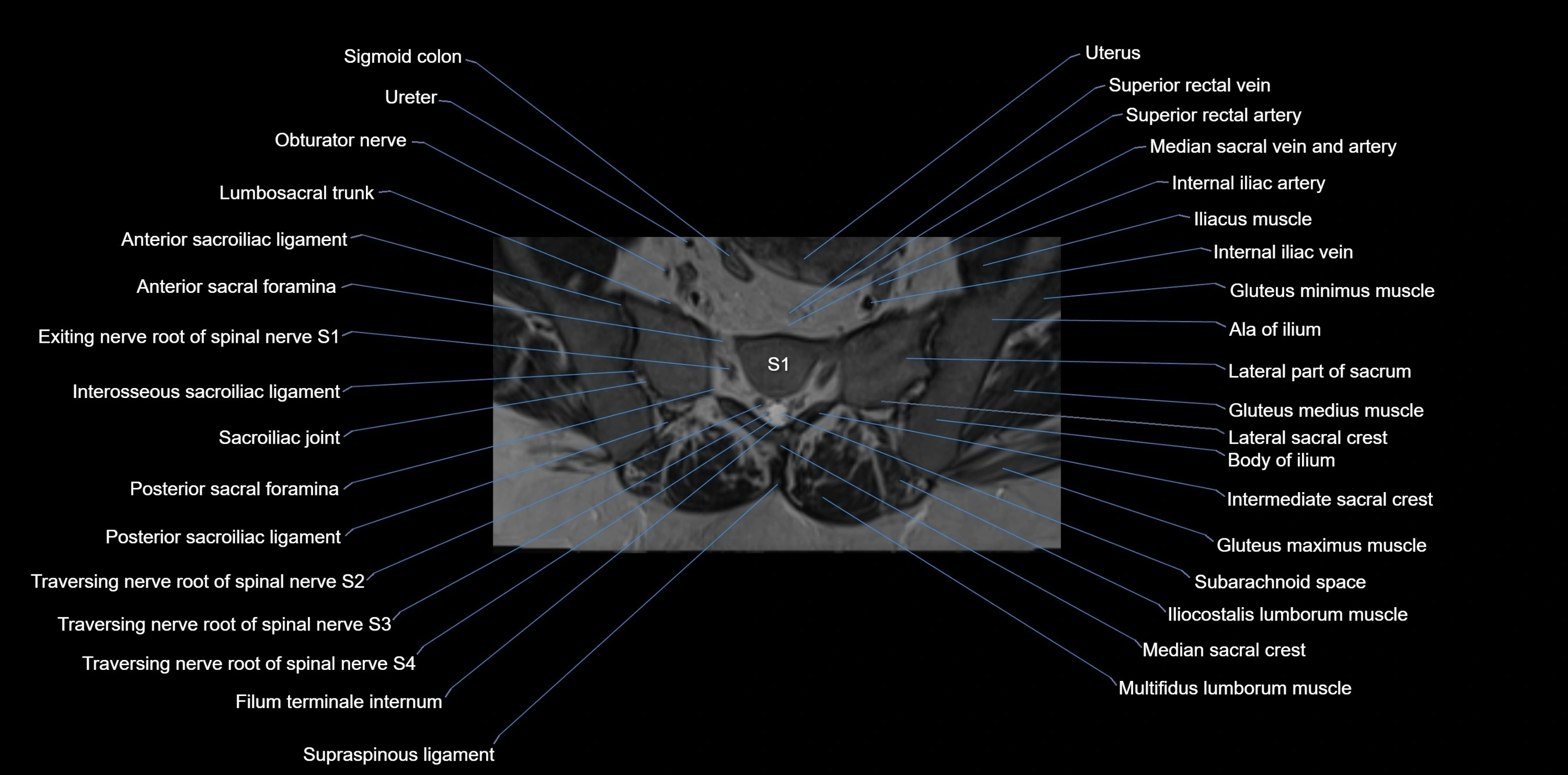
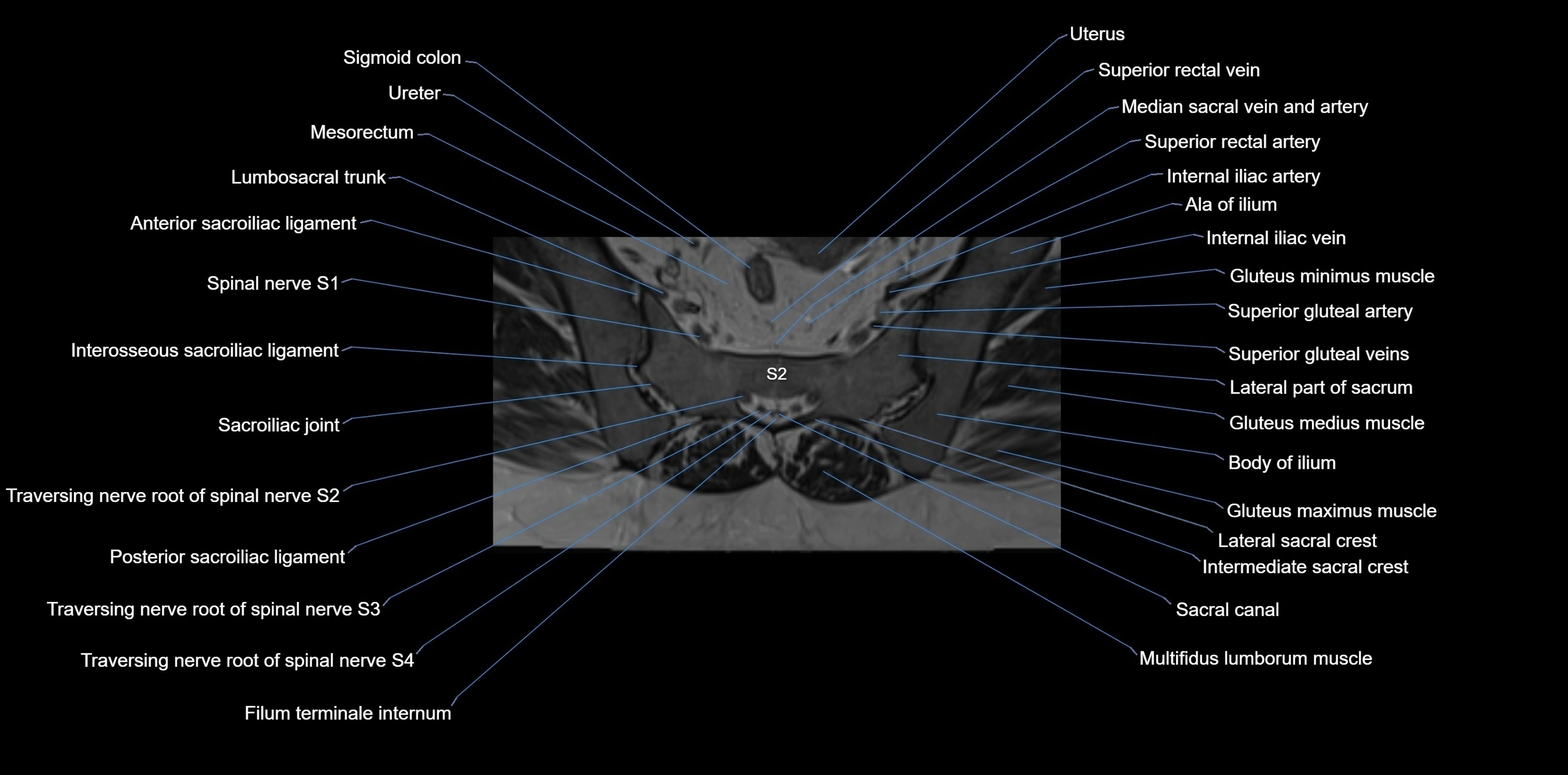
CT Post-Contrast:
-
Highlights adjacent soft tissue structures, tumors, and inflammatory processes
-
Defines vascular relationships in surgical planning
-
3D reconstructions show iliac morphology, fracture lines, and joint surfaces in detail
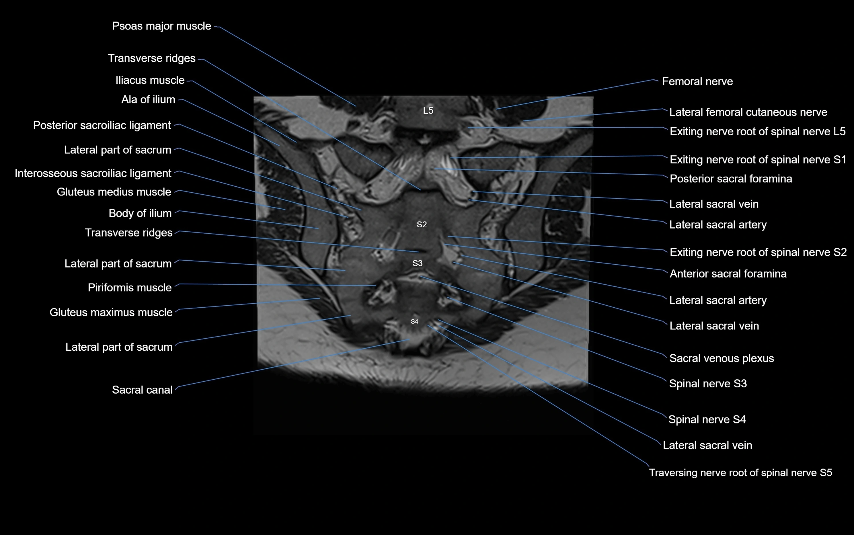
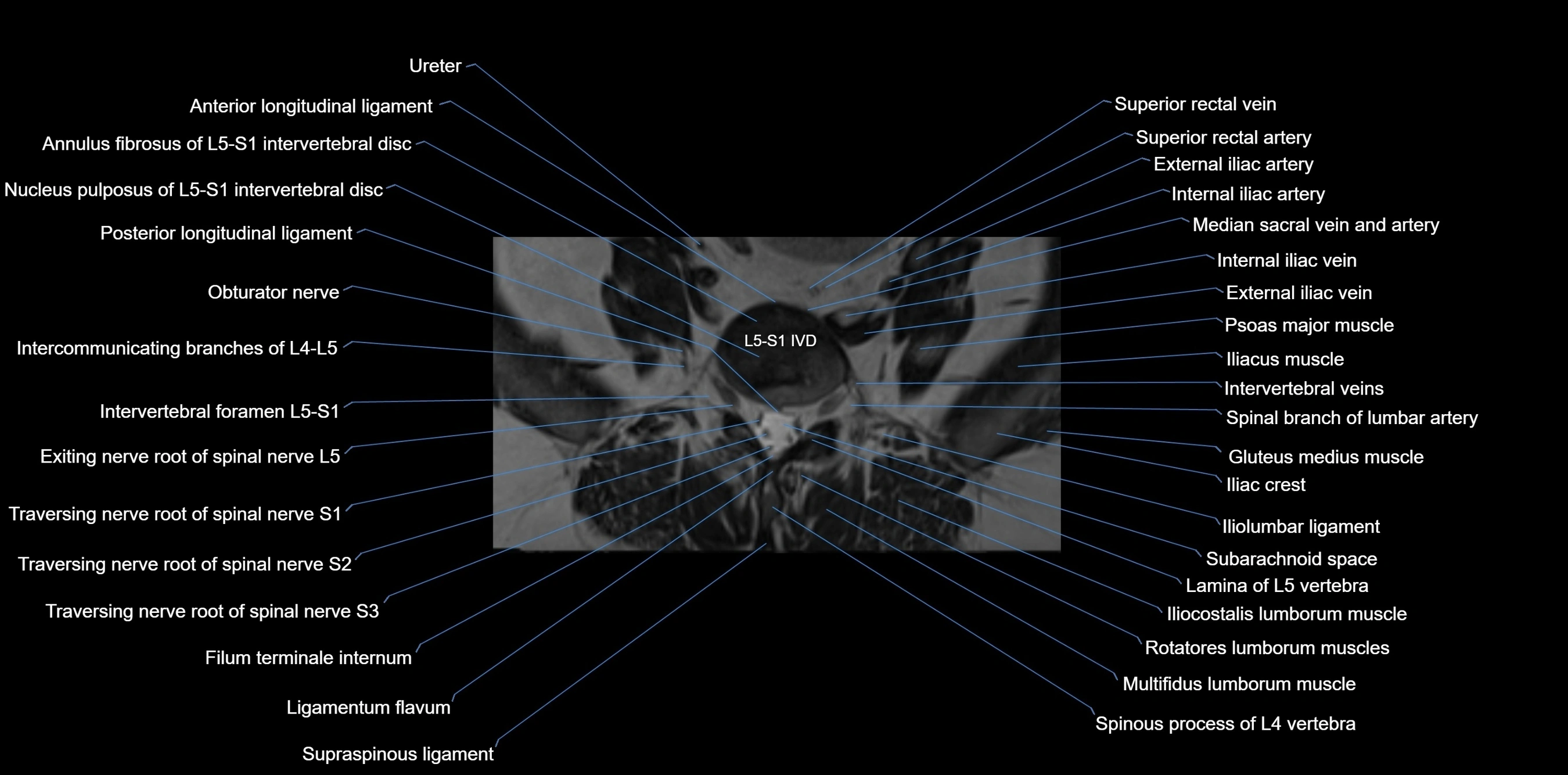
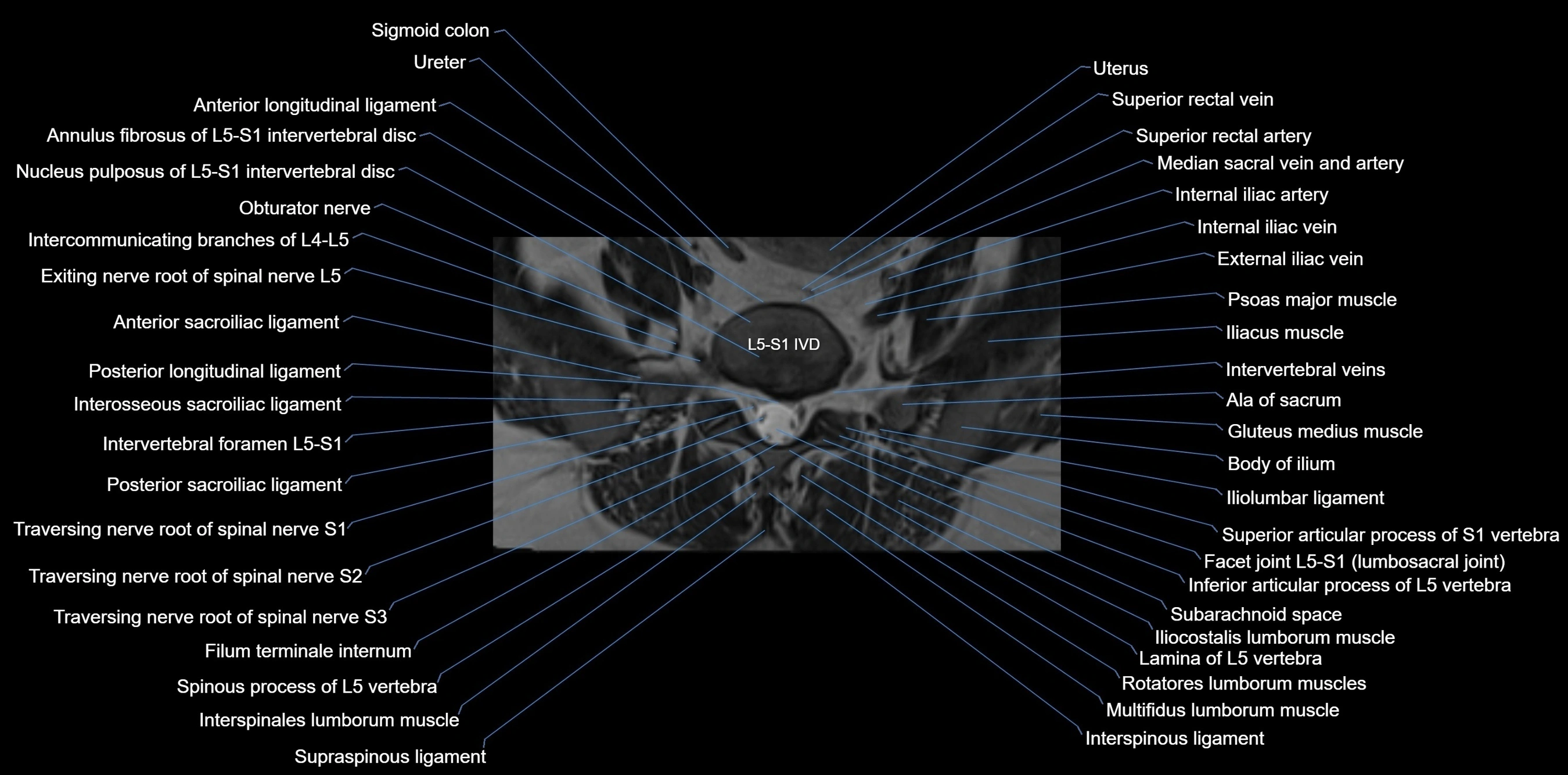
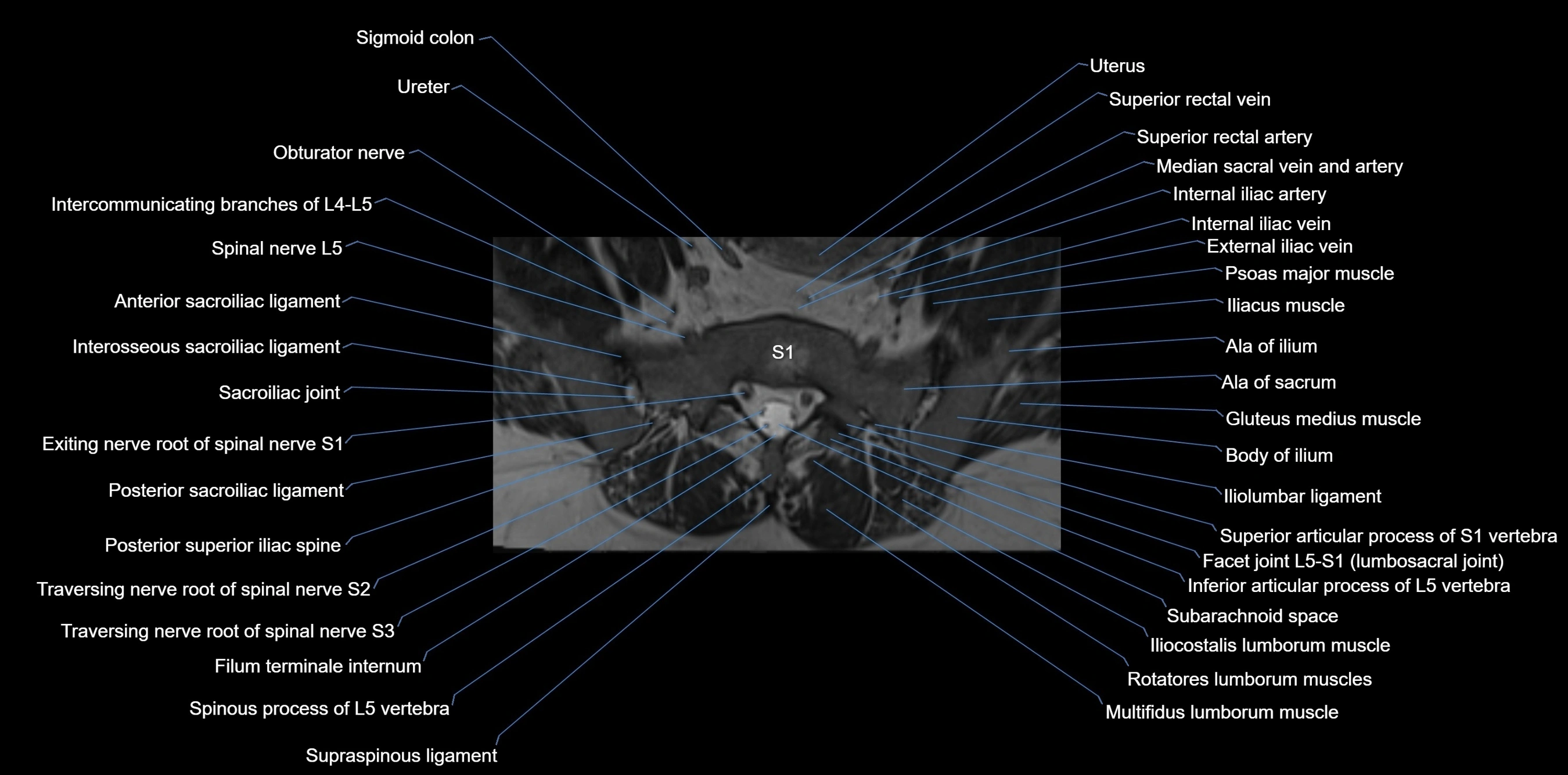
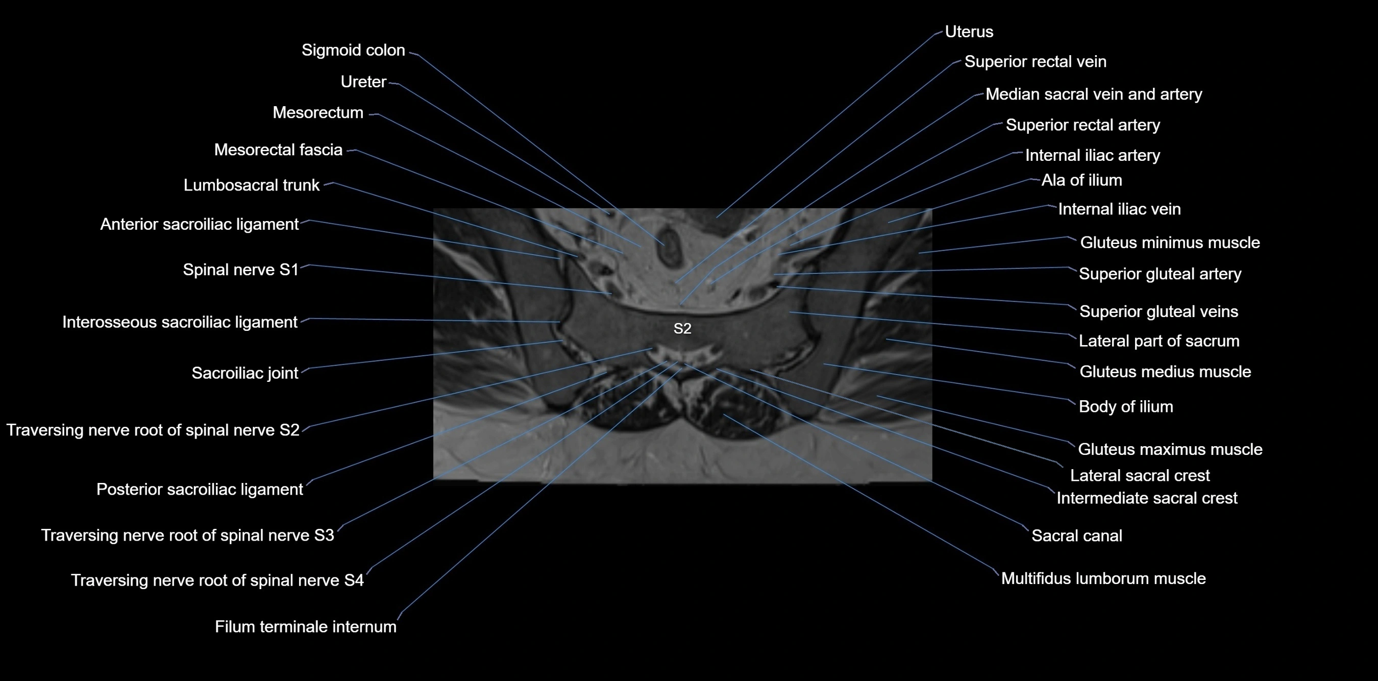
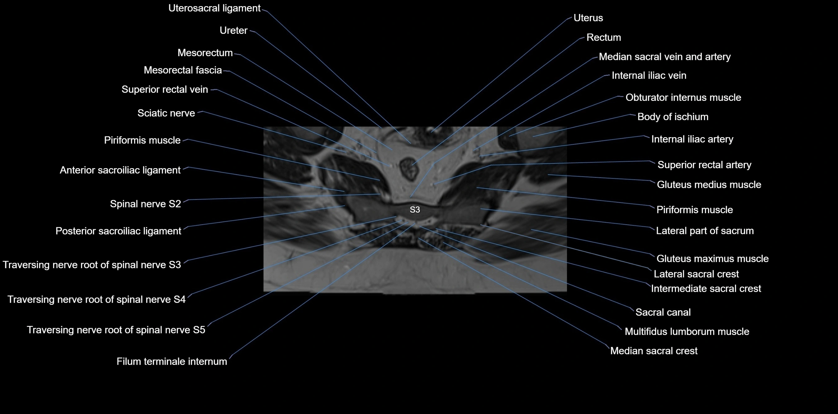
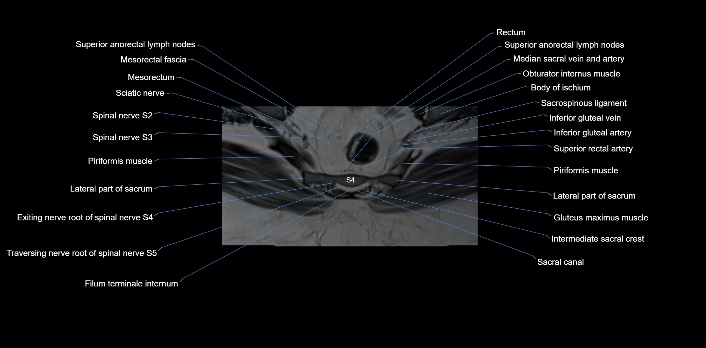
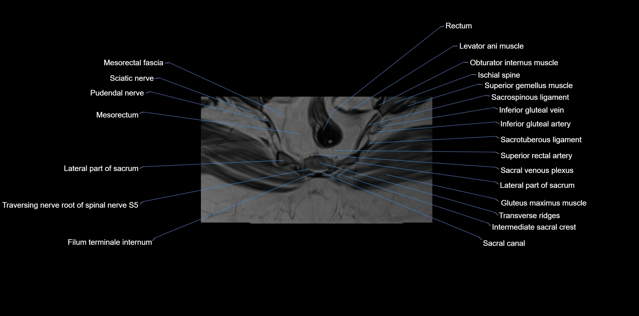
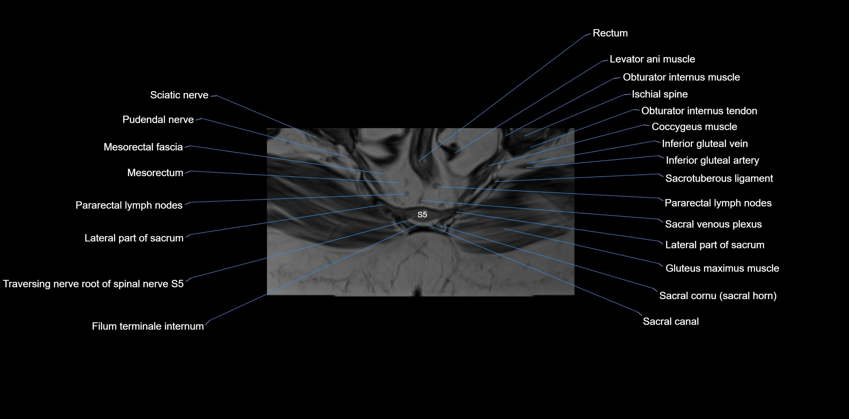
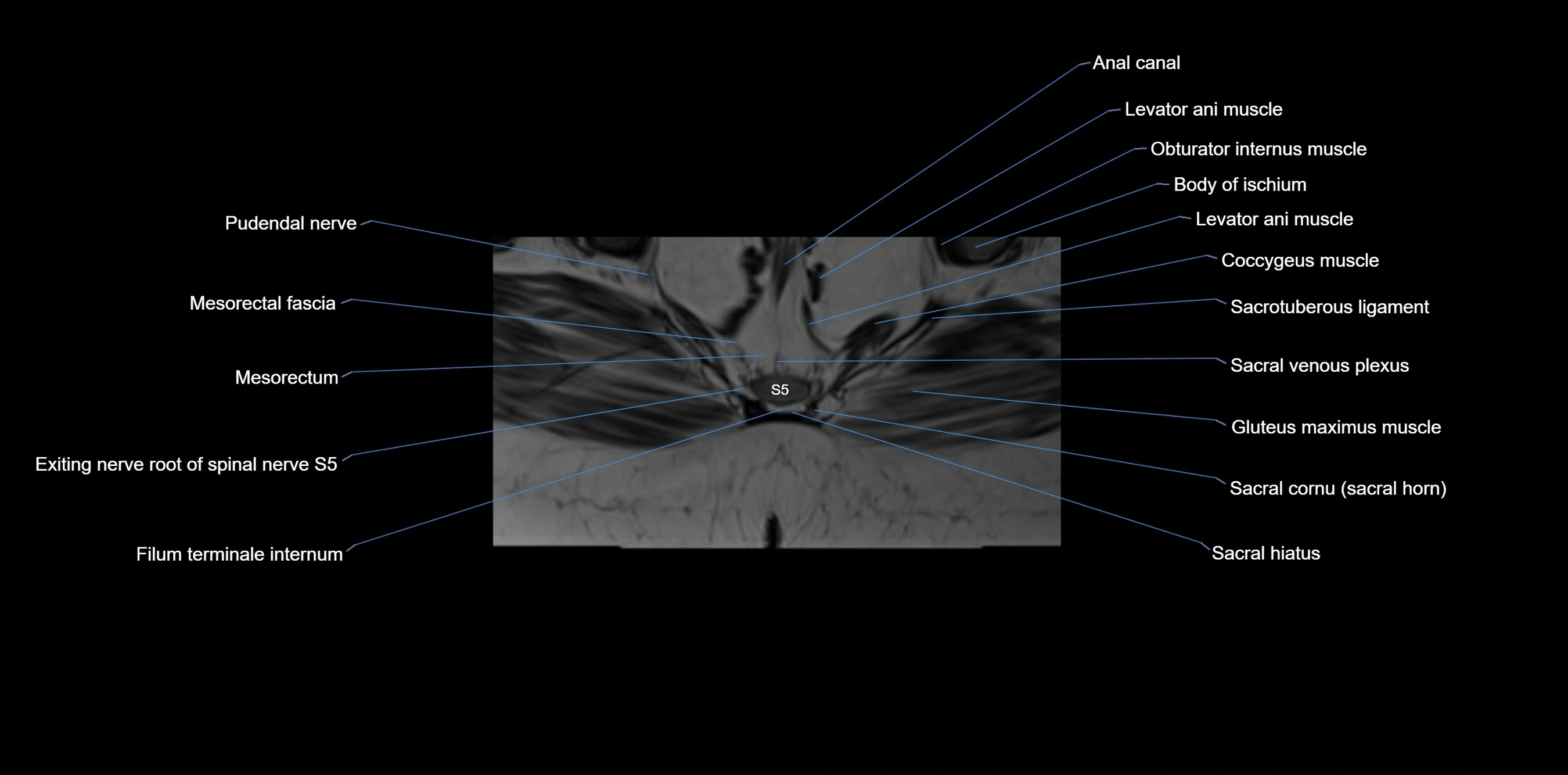
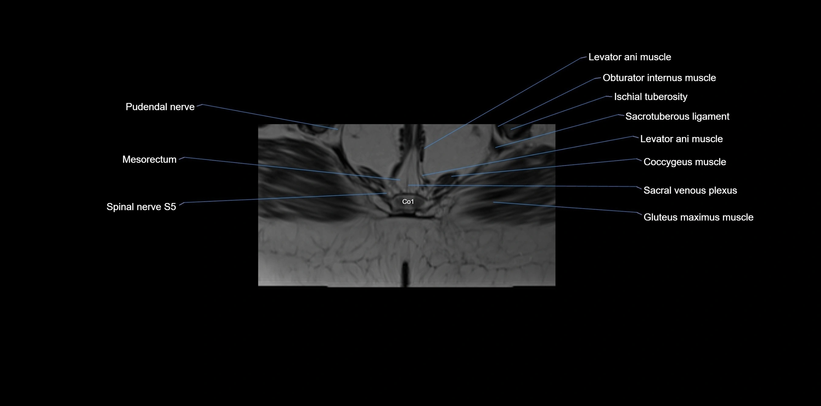
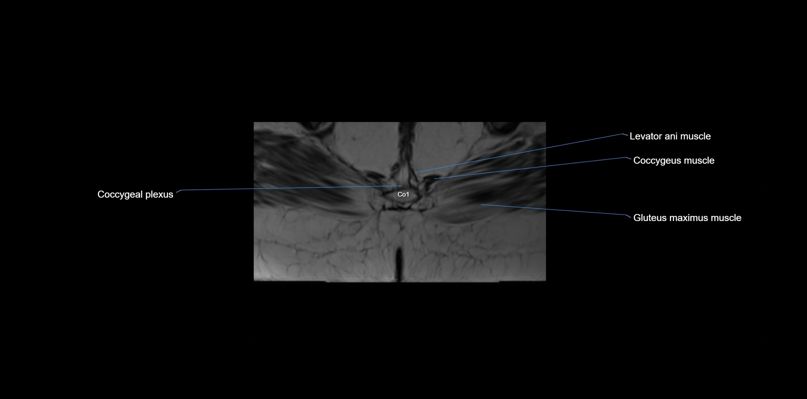
MRI image

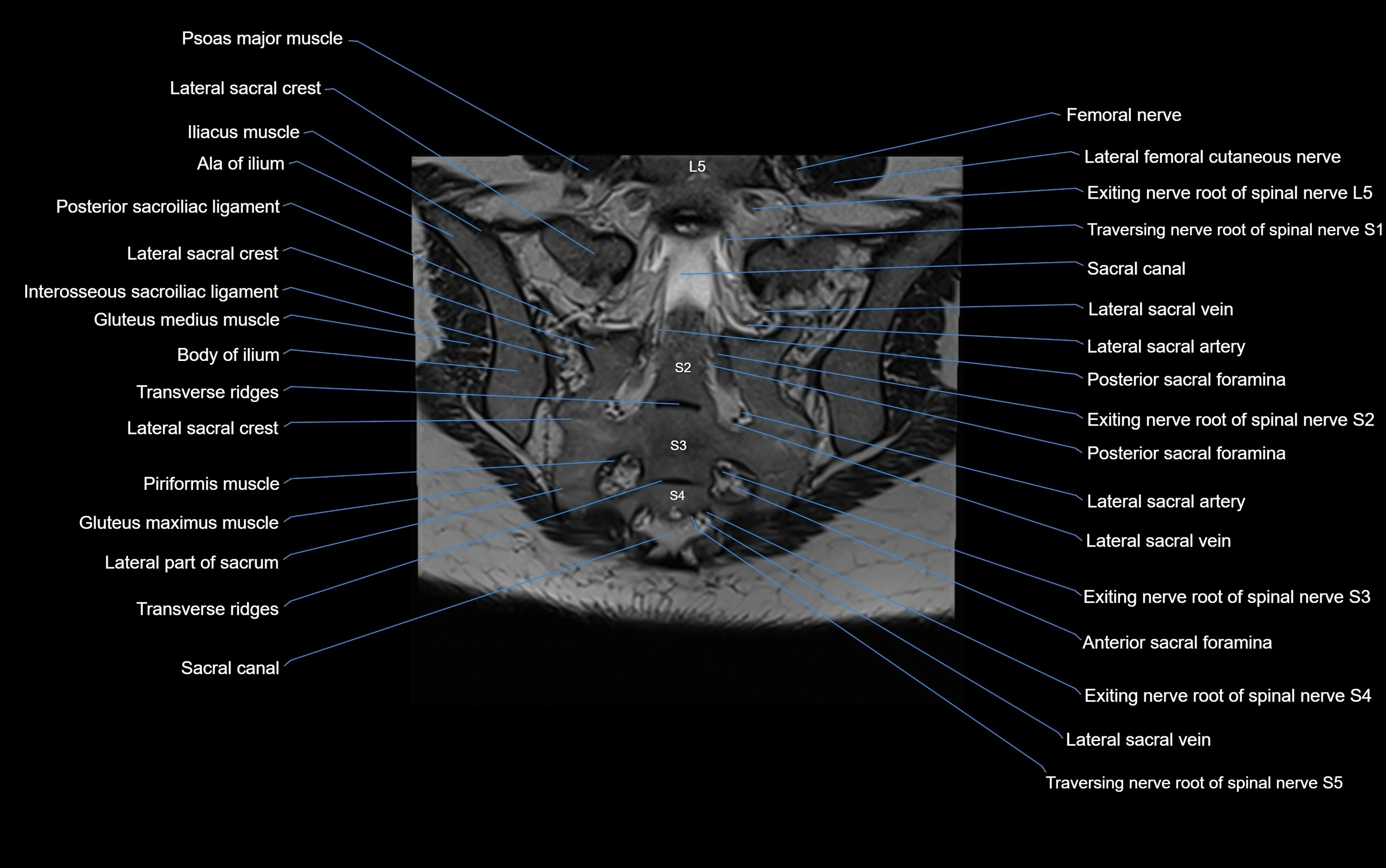
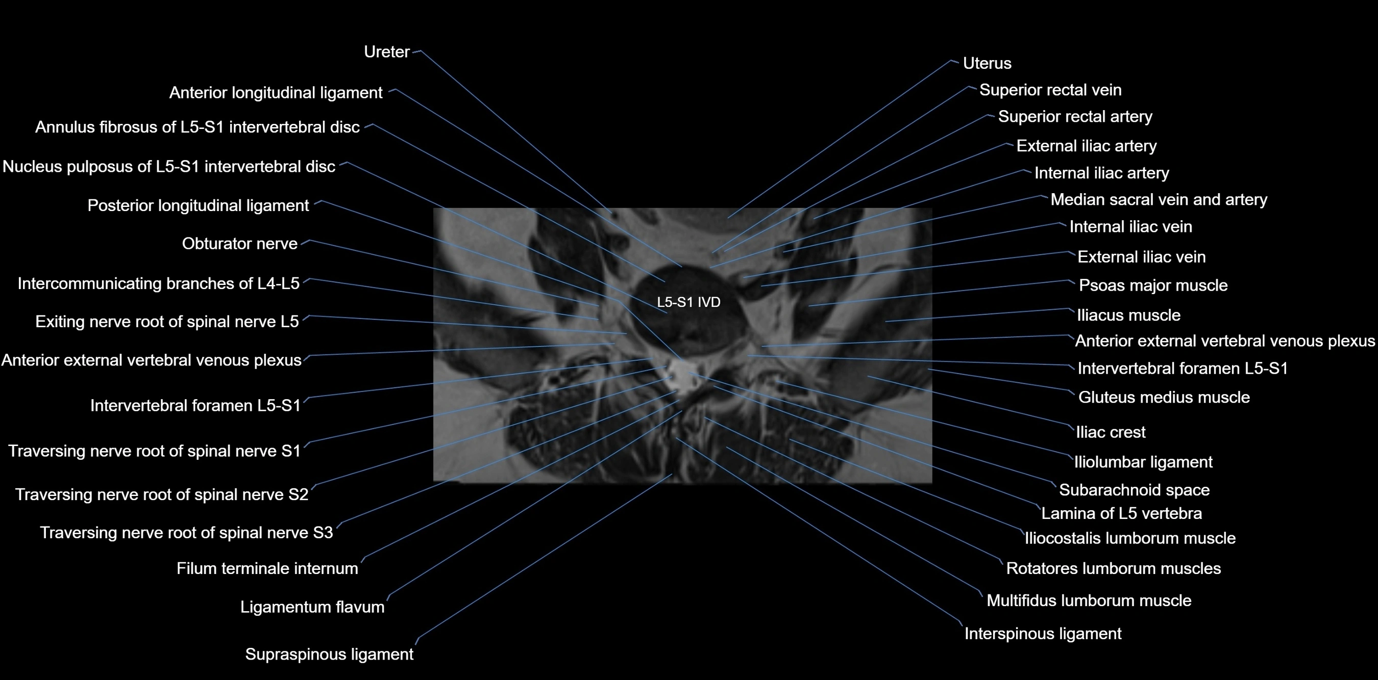
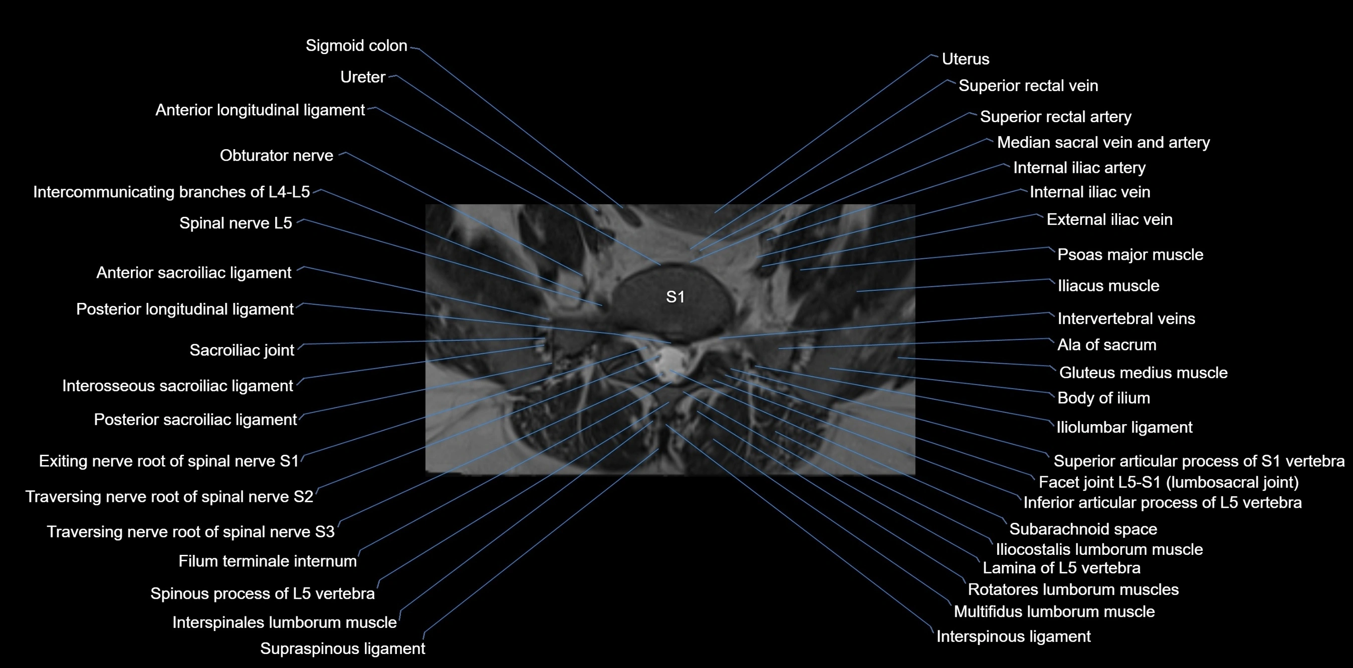
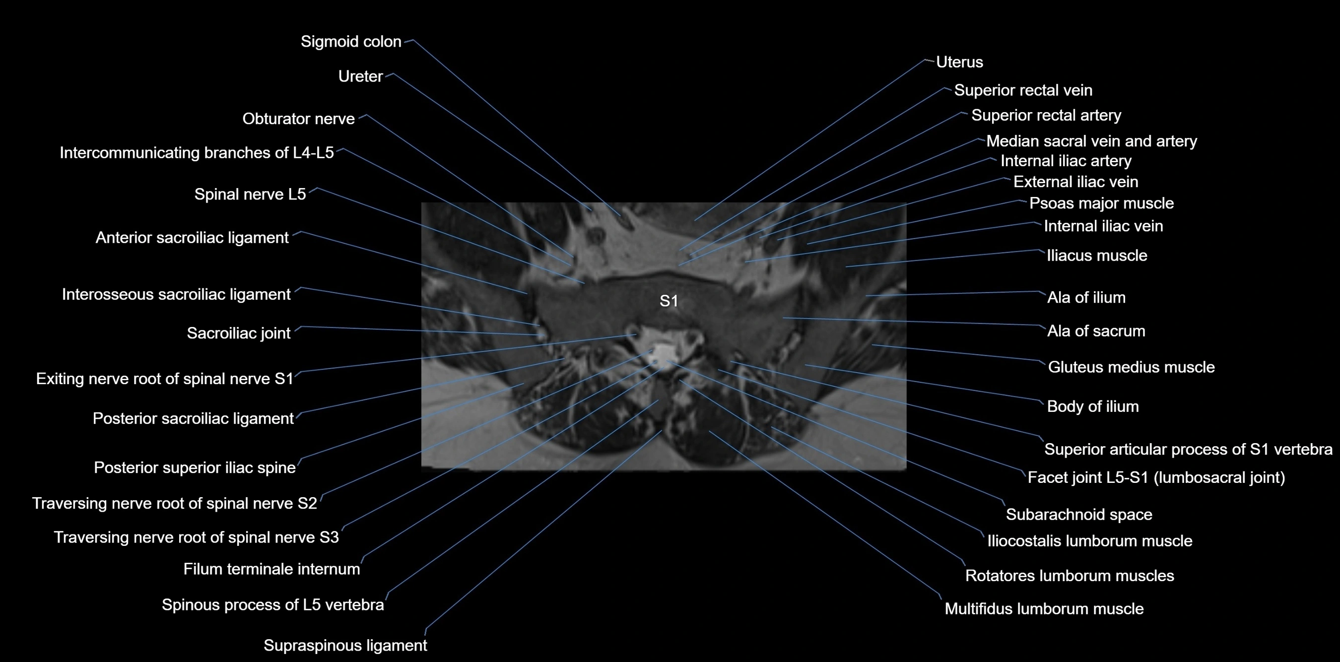
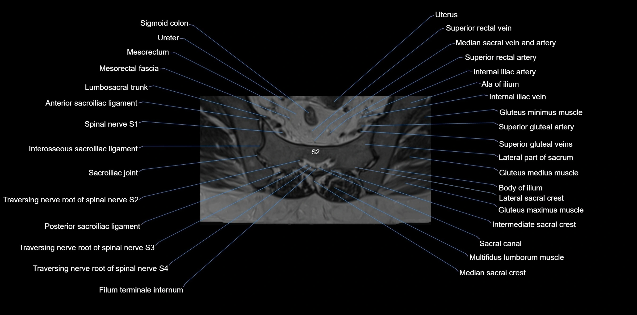
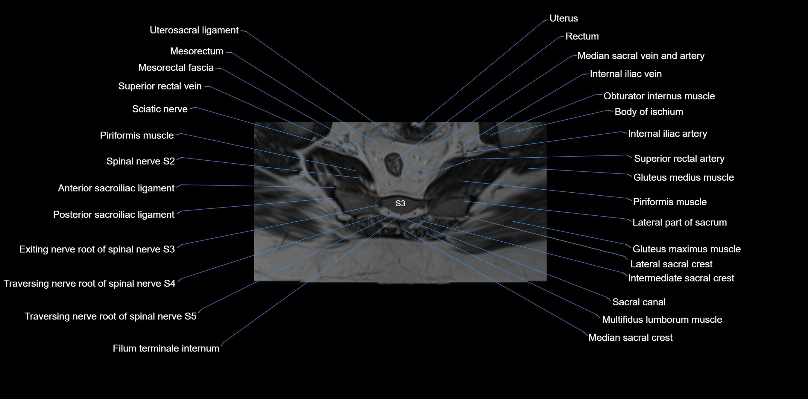
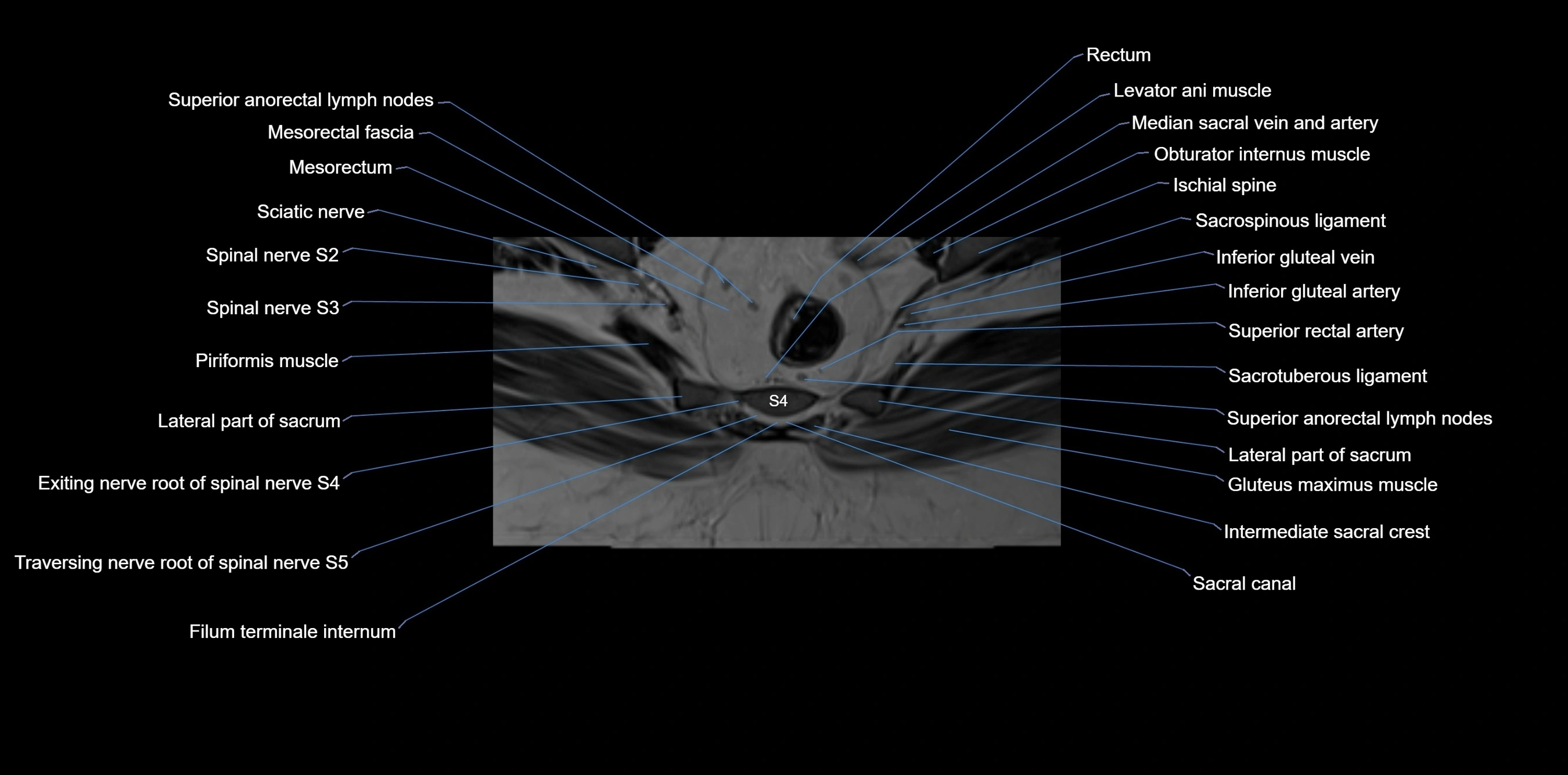
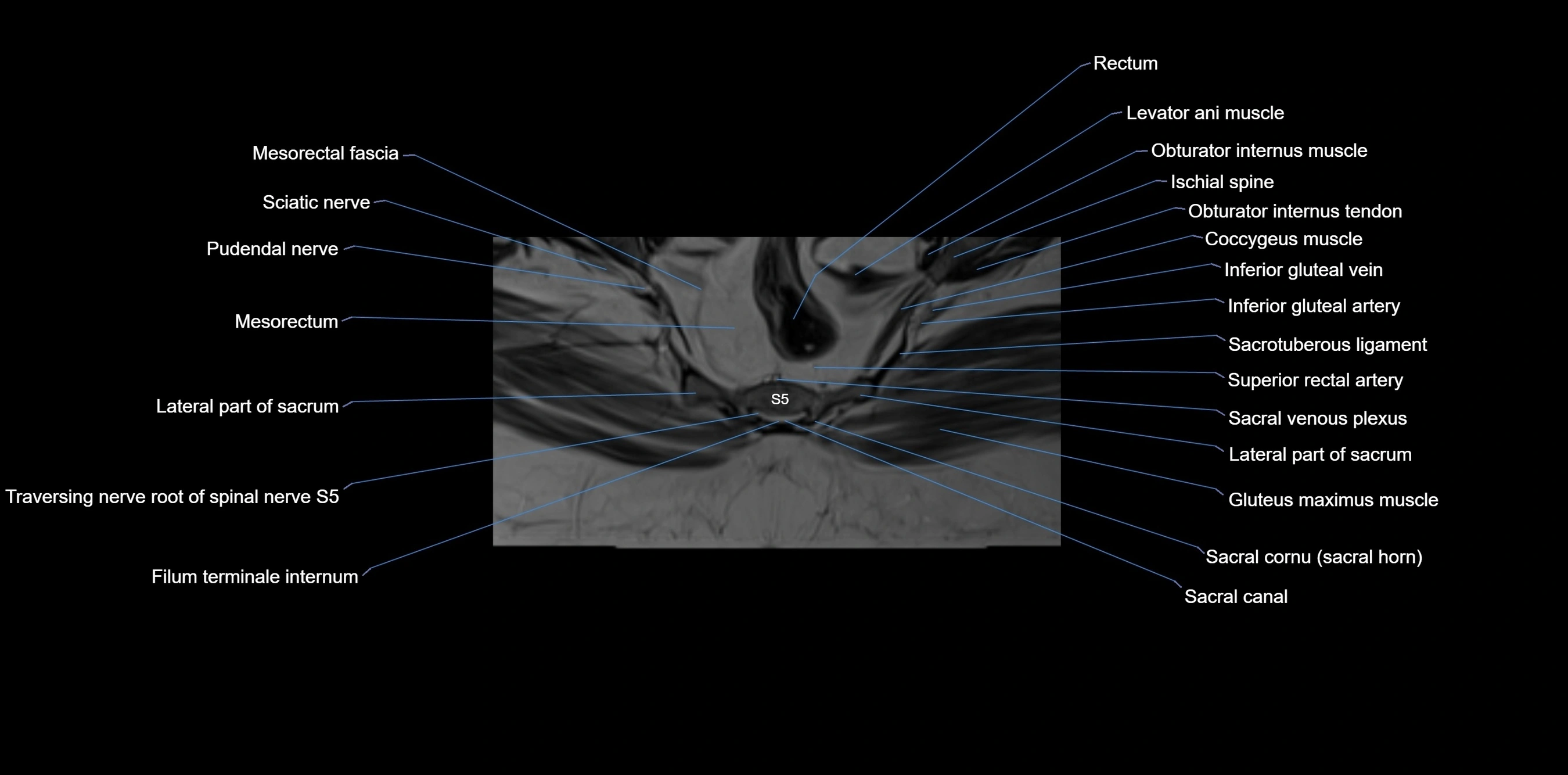
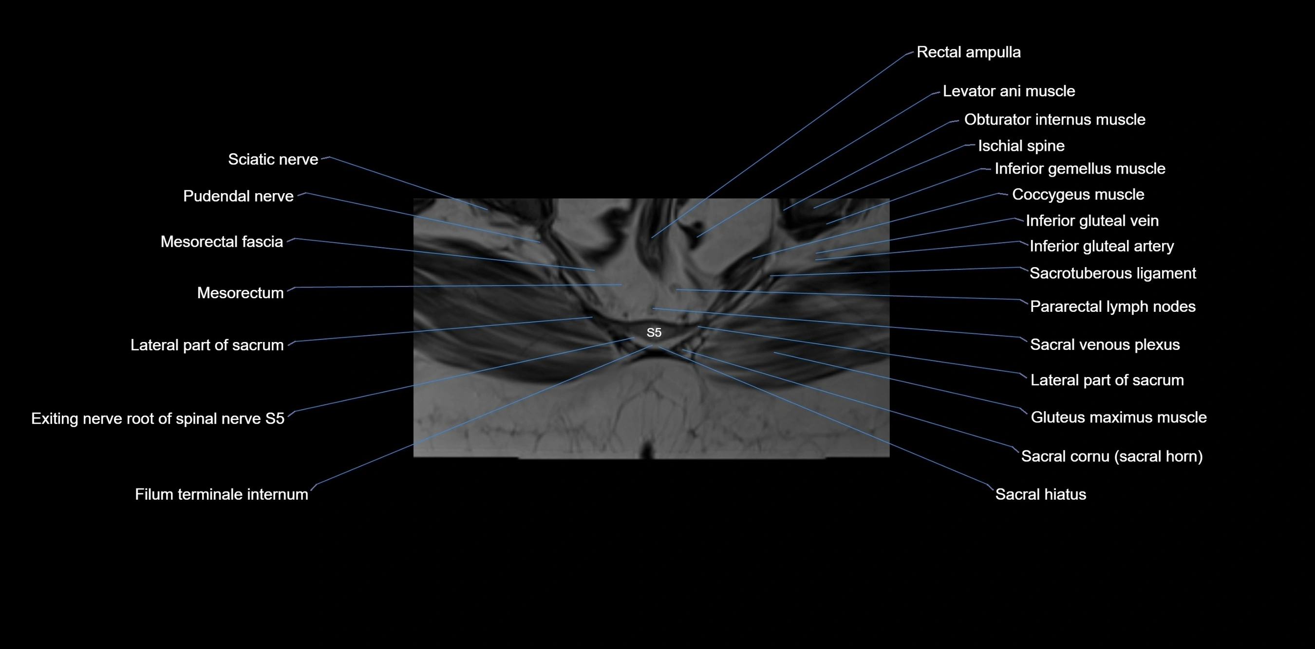
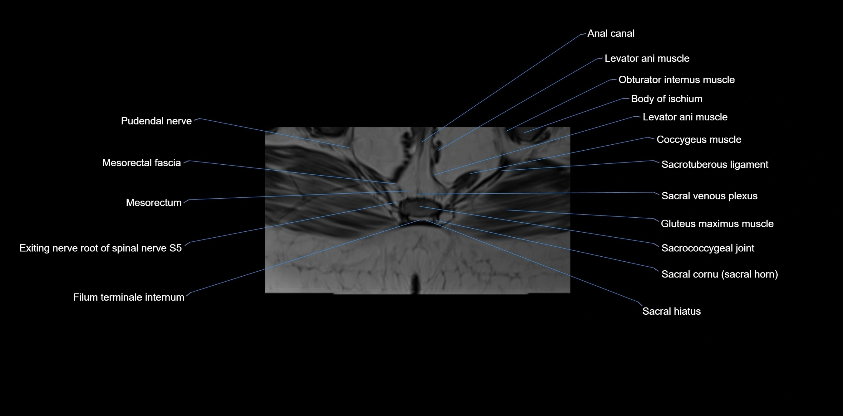
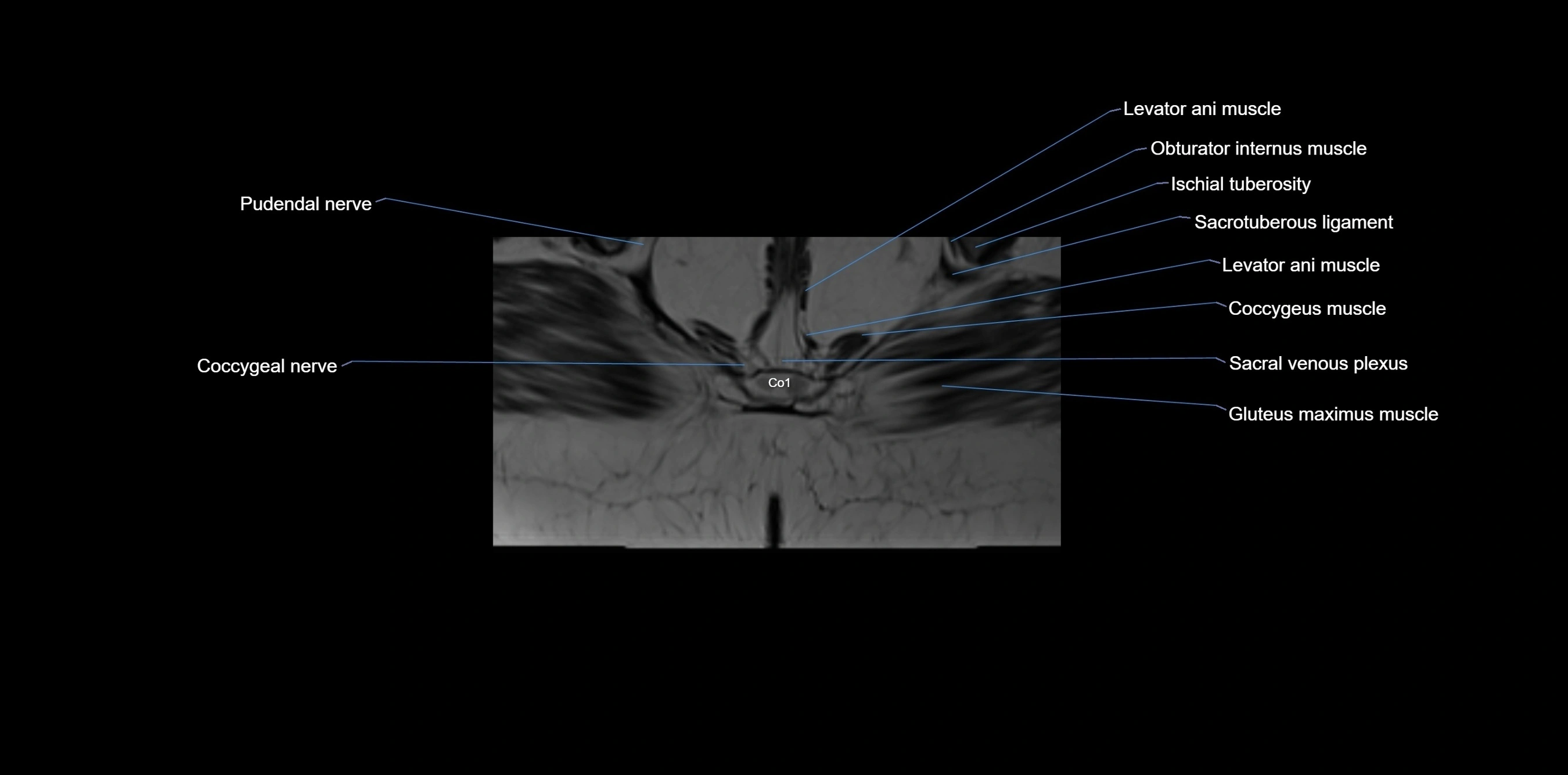
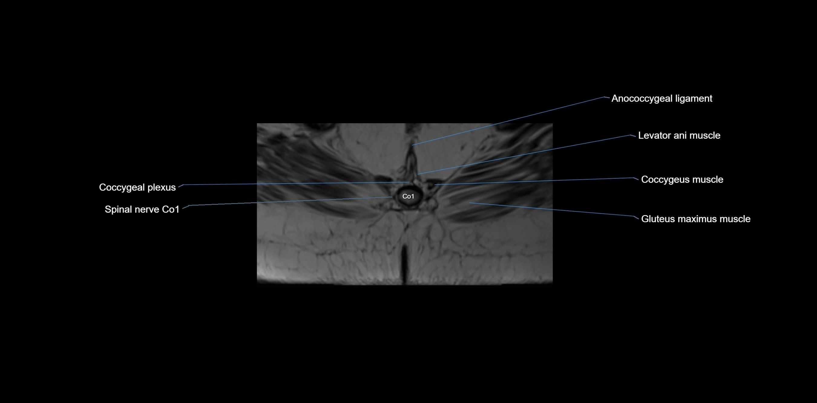
MRI image

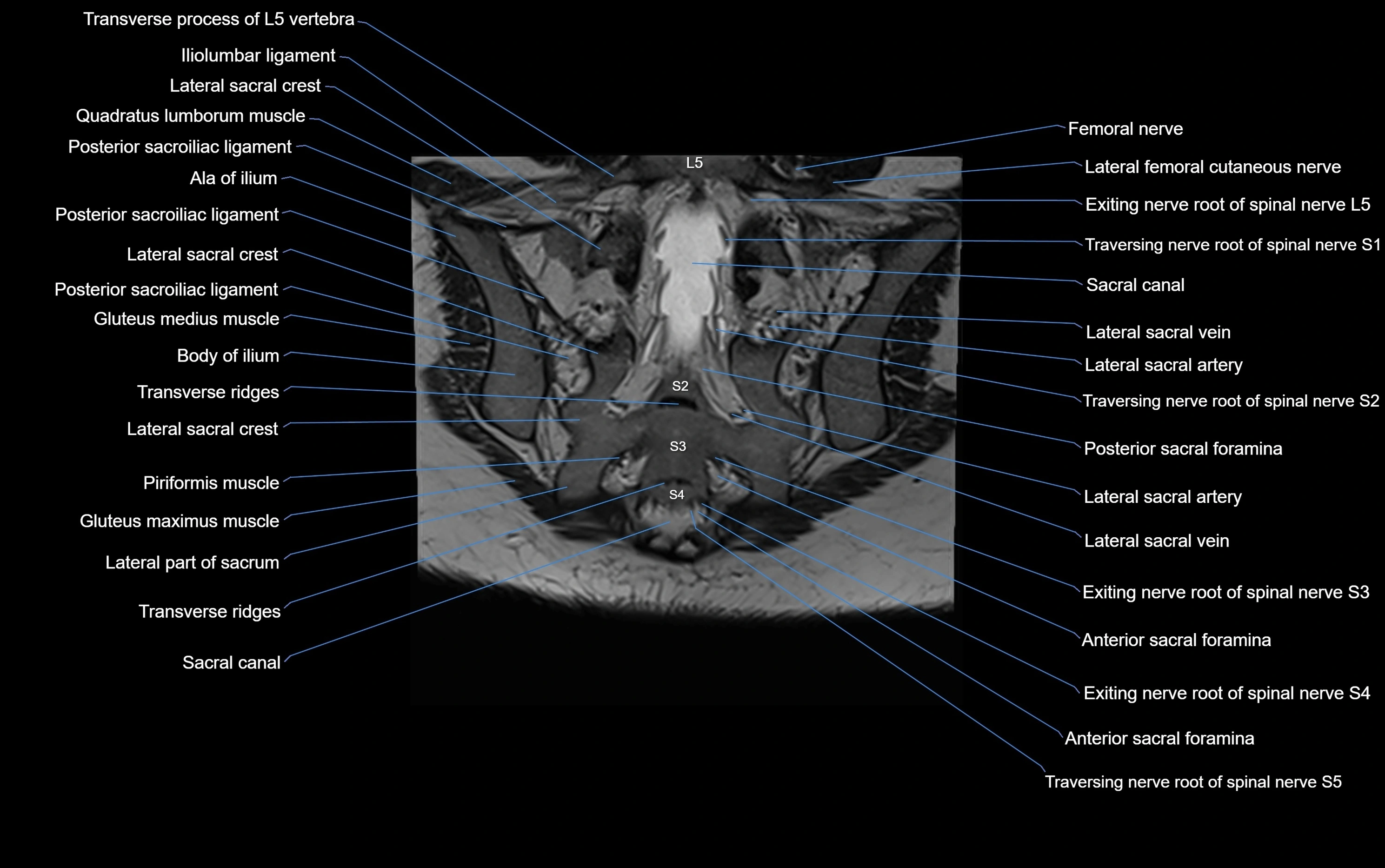
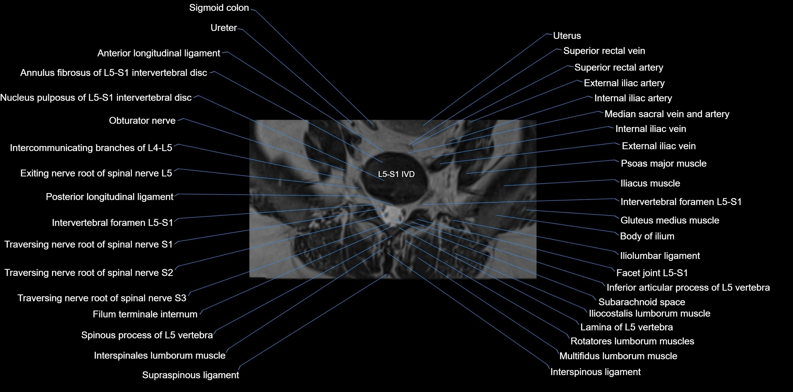
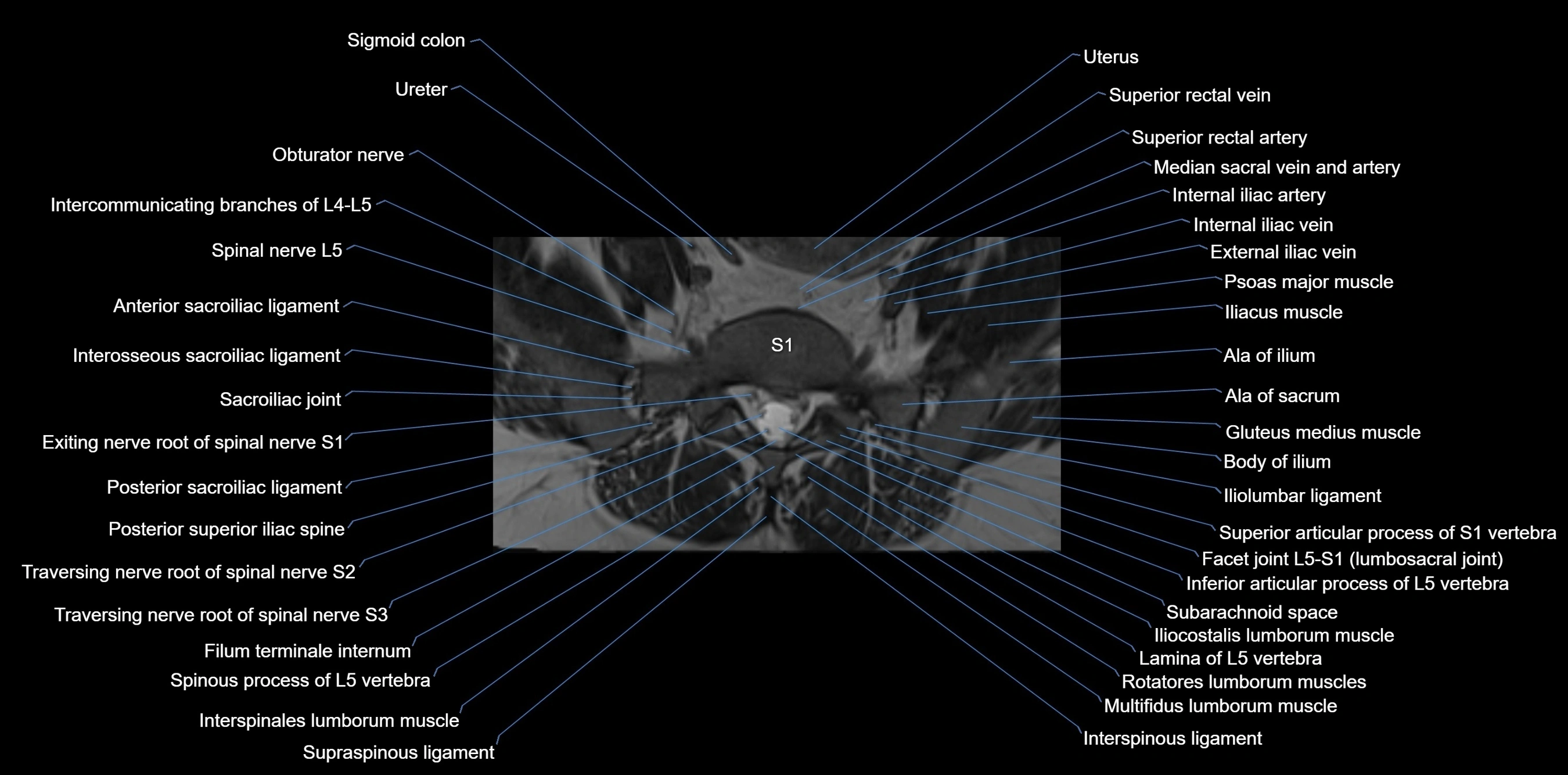
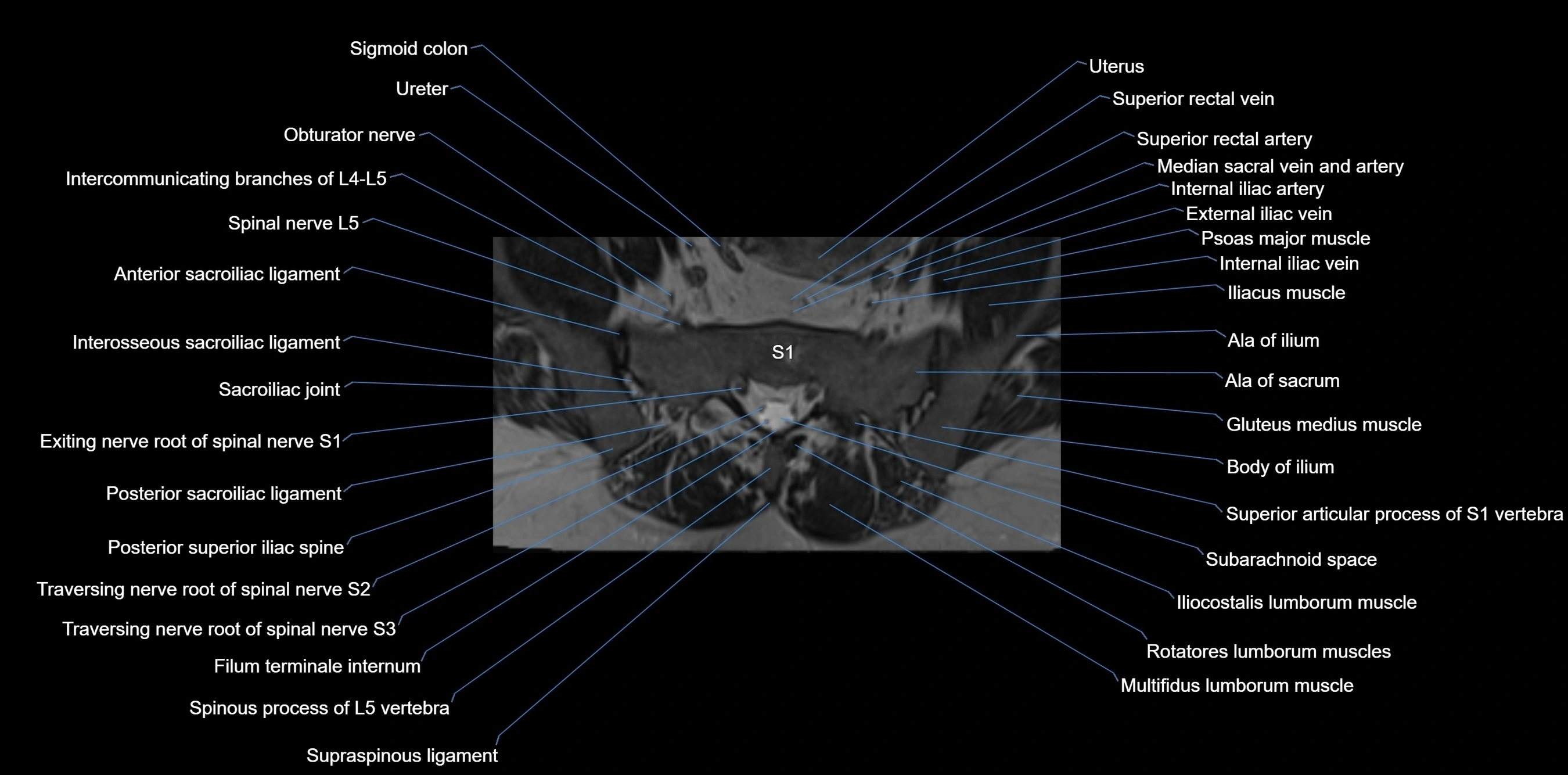
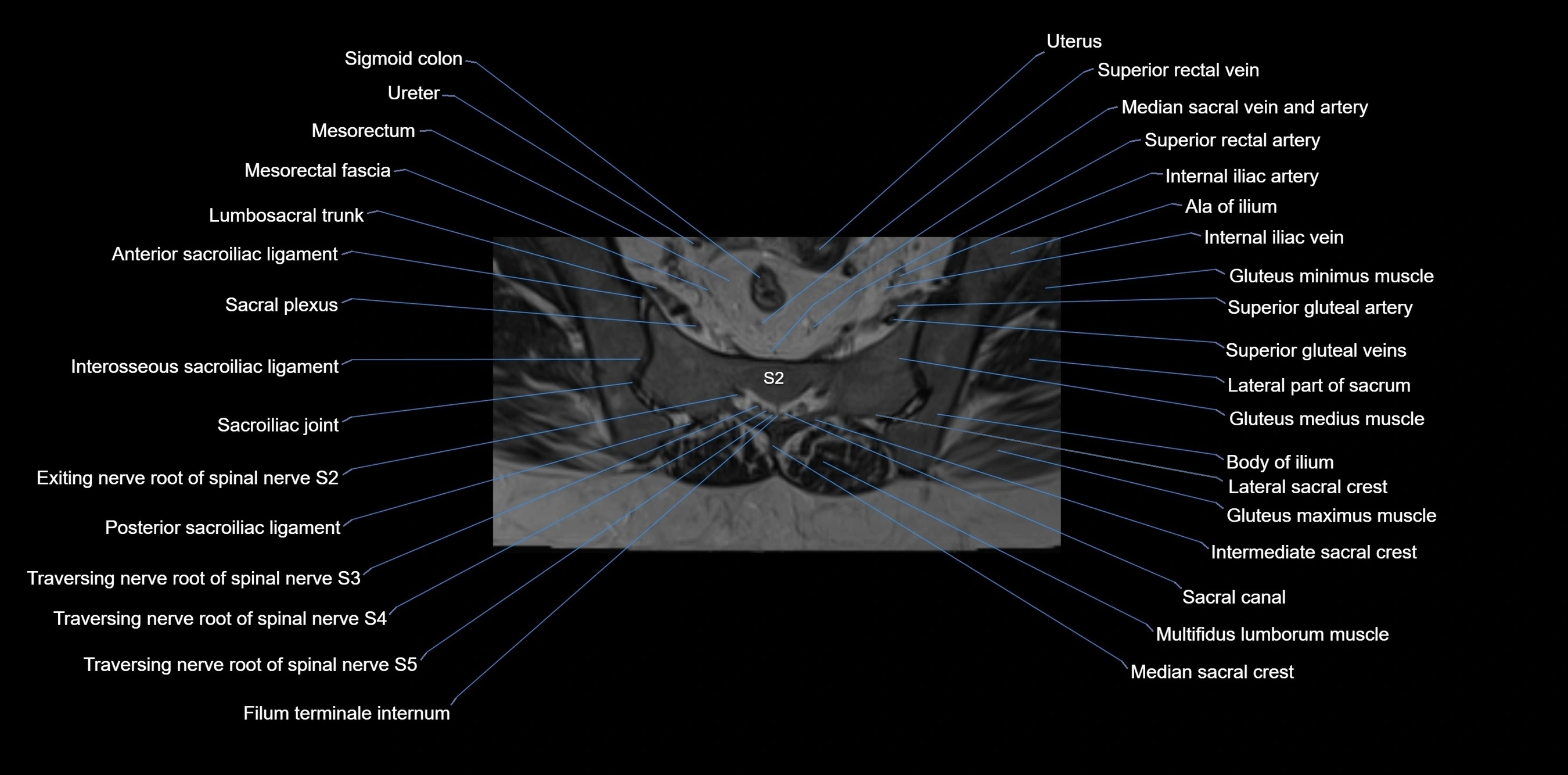
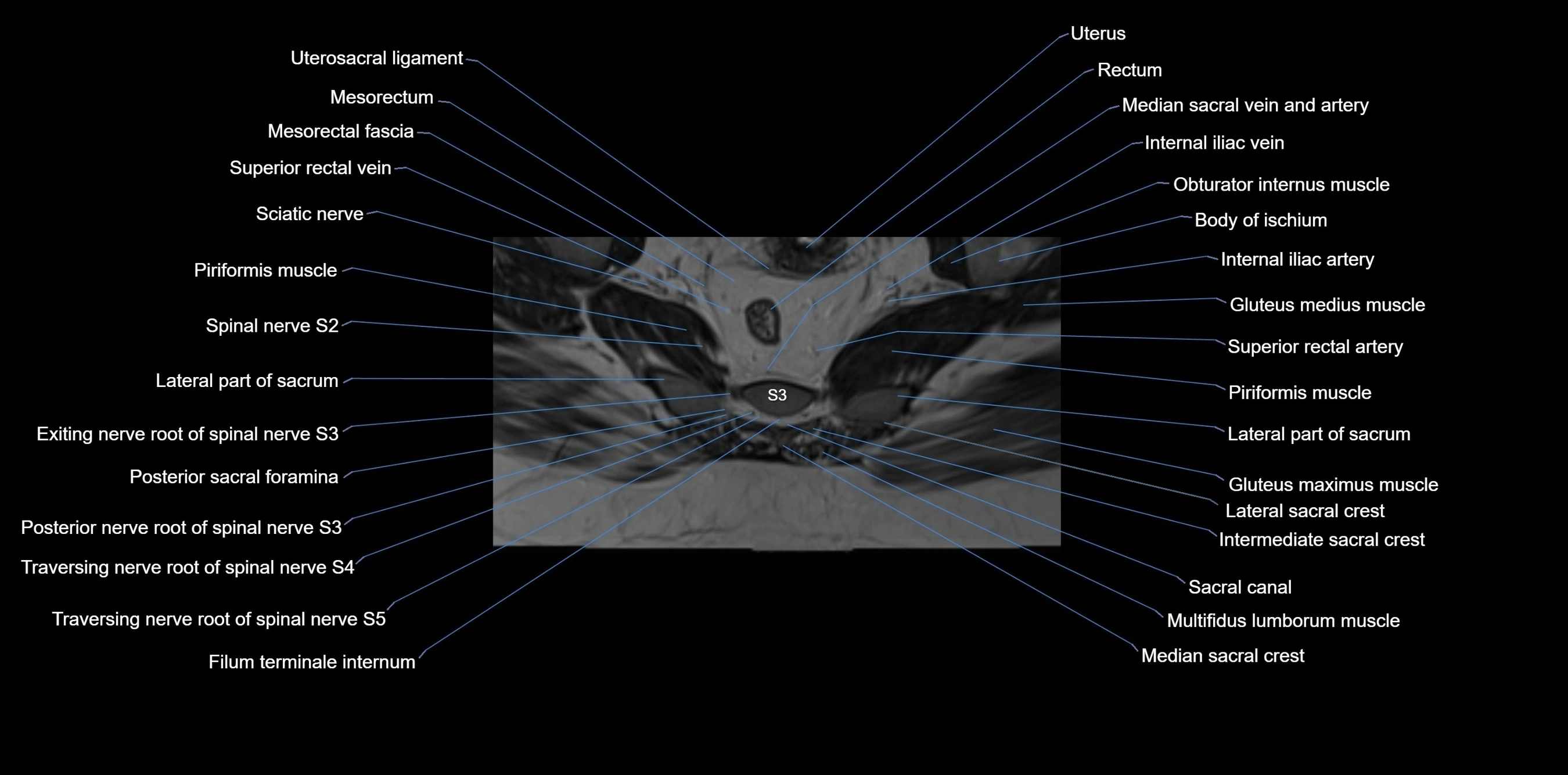
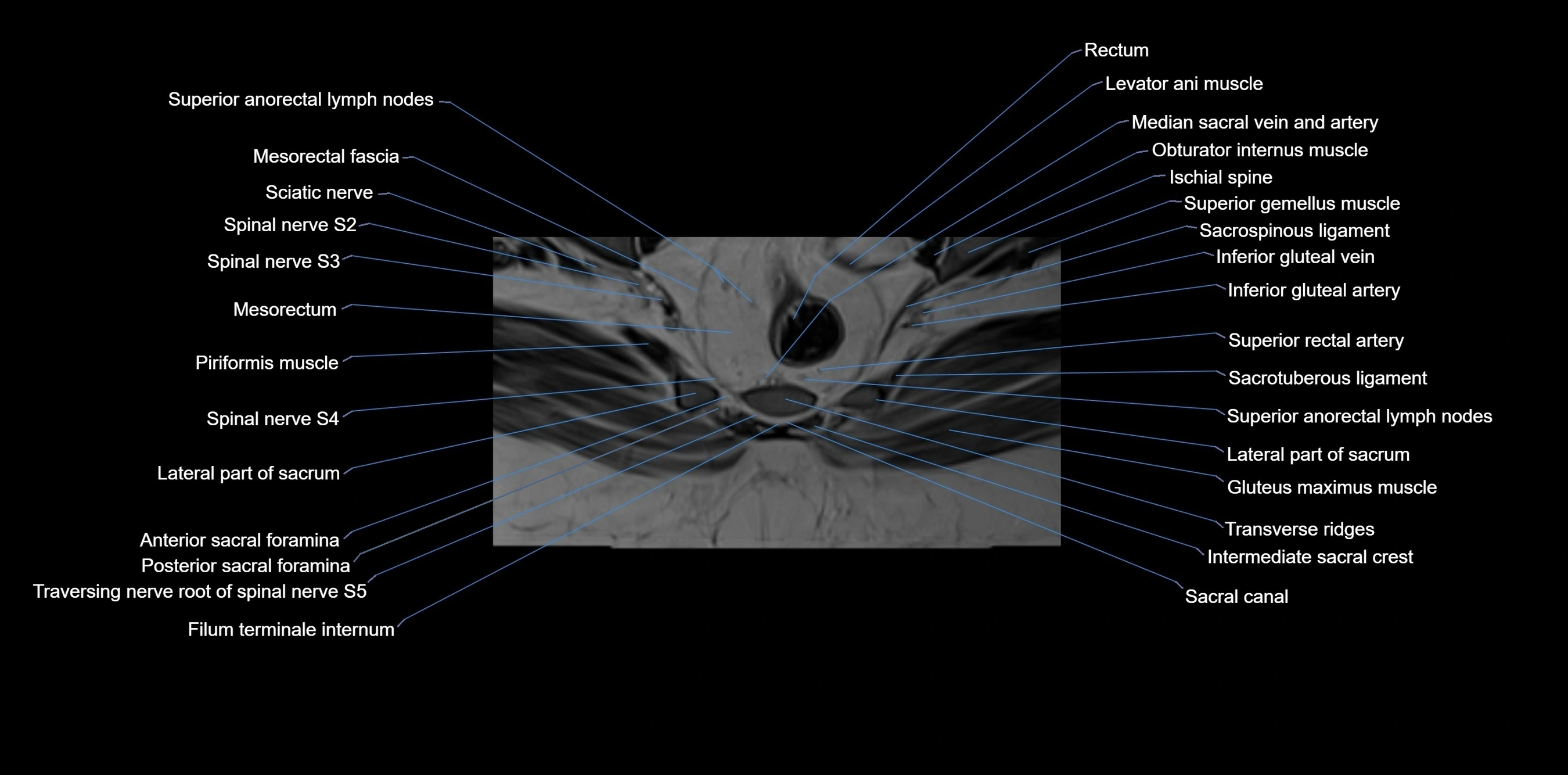
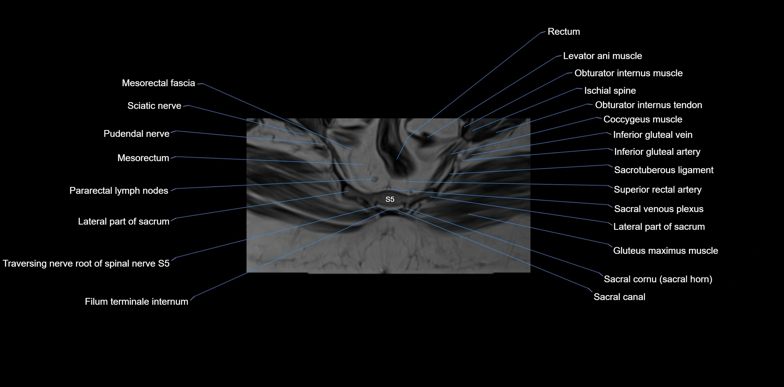
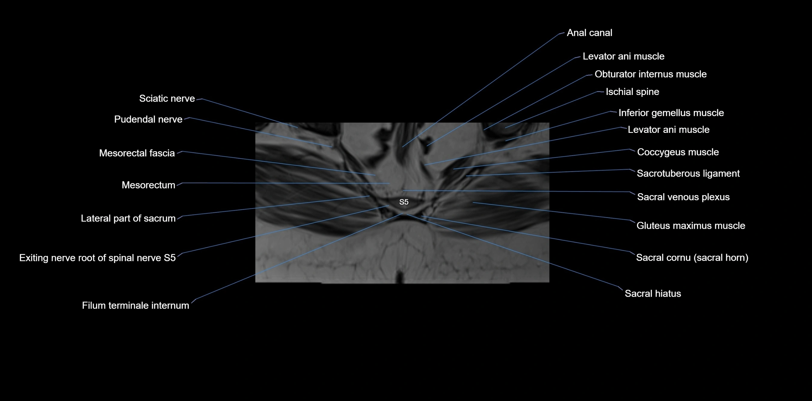
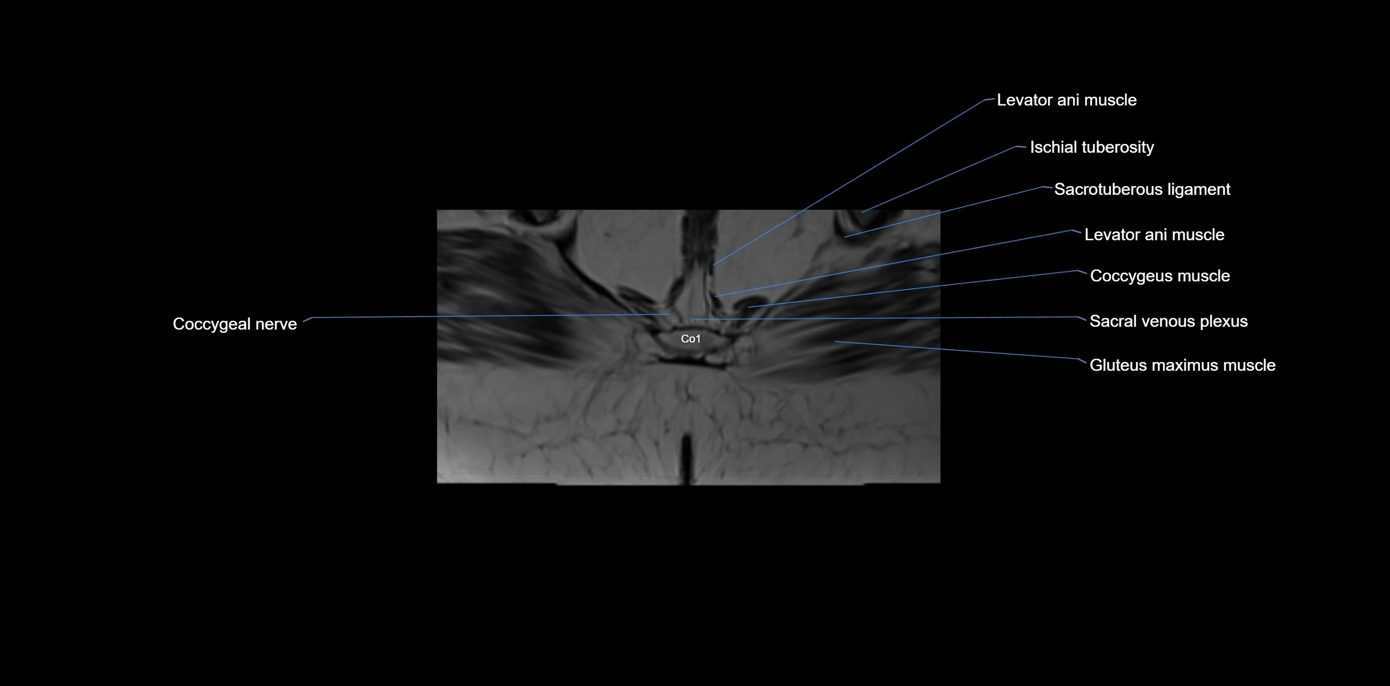
CT image

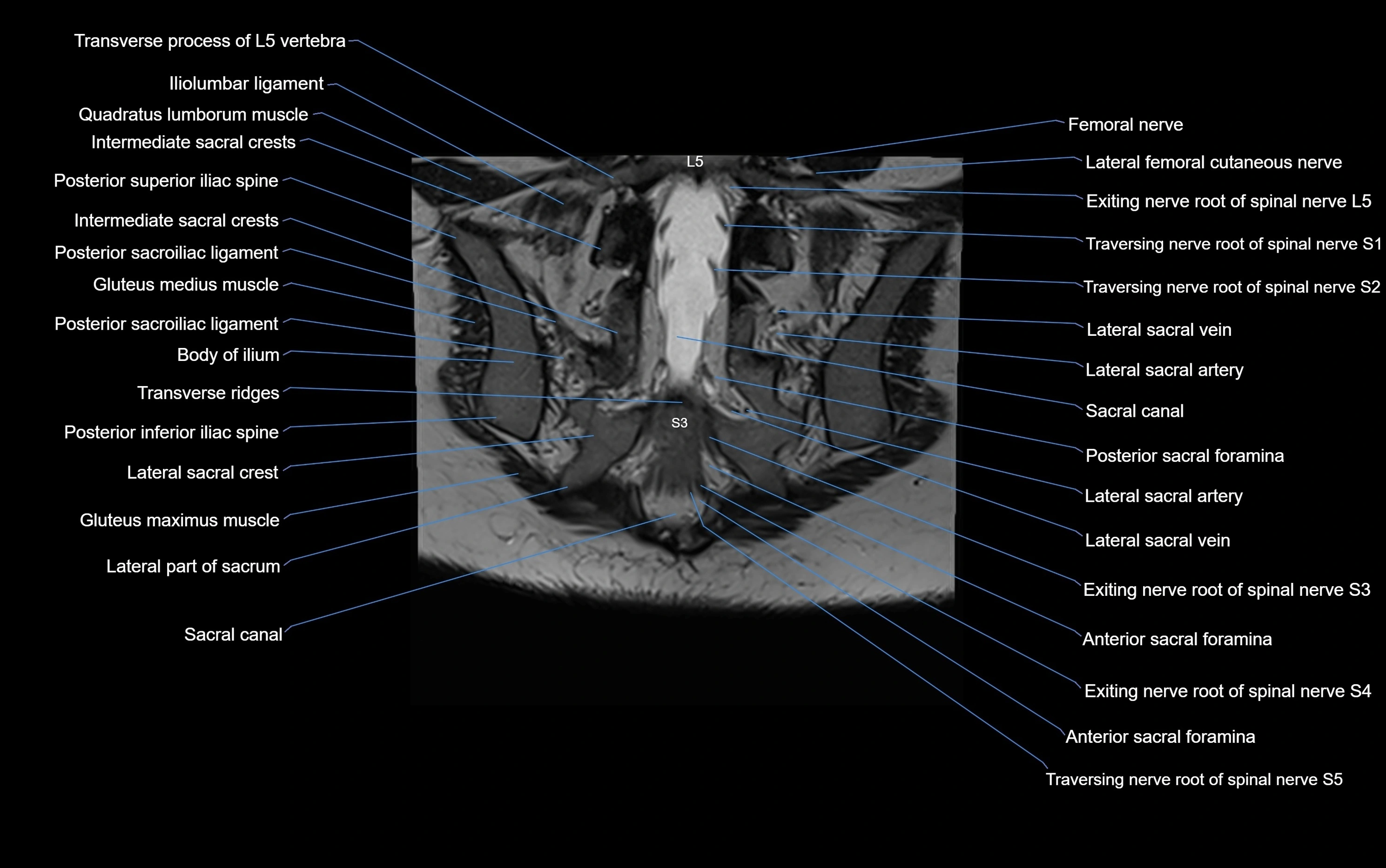
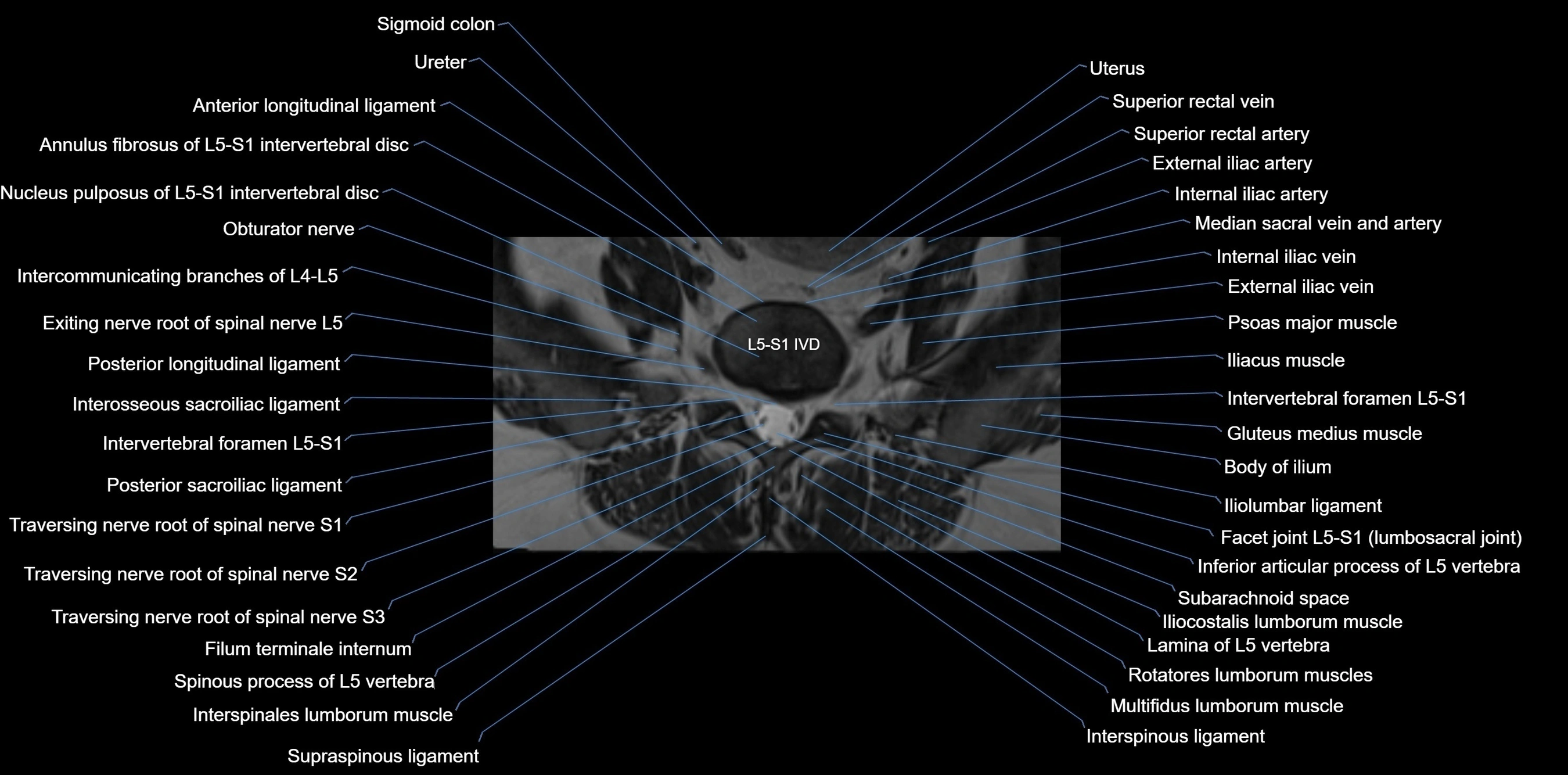
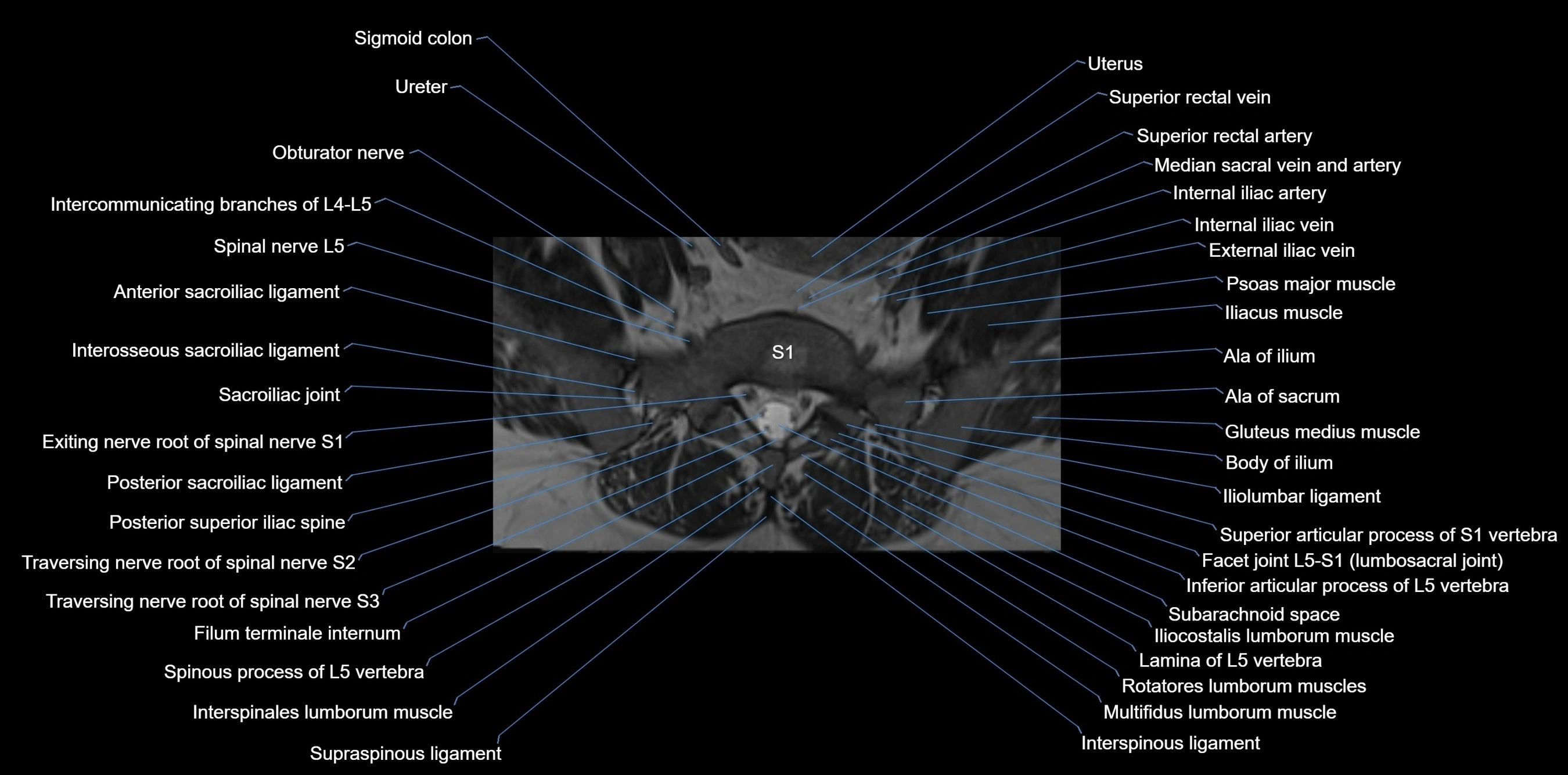
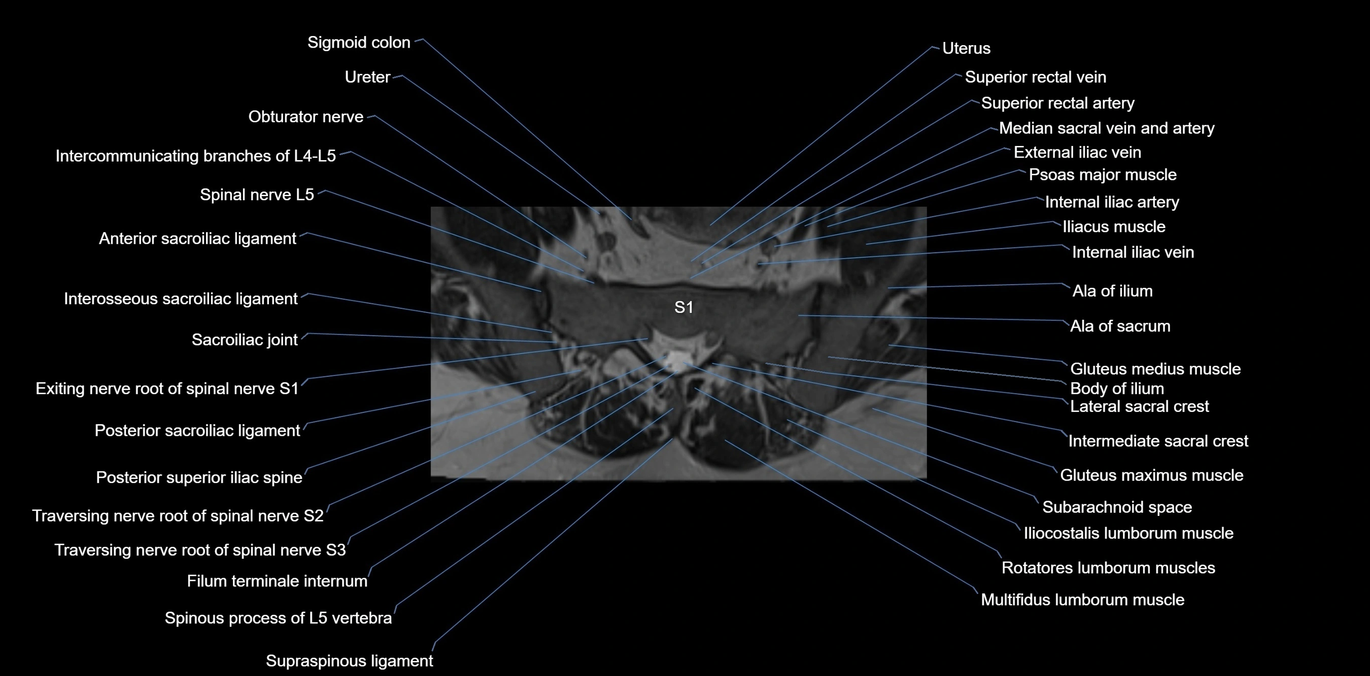
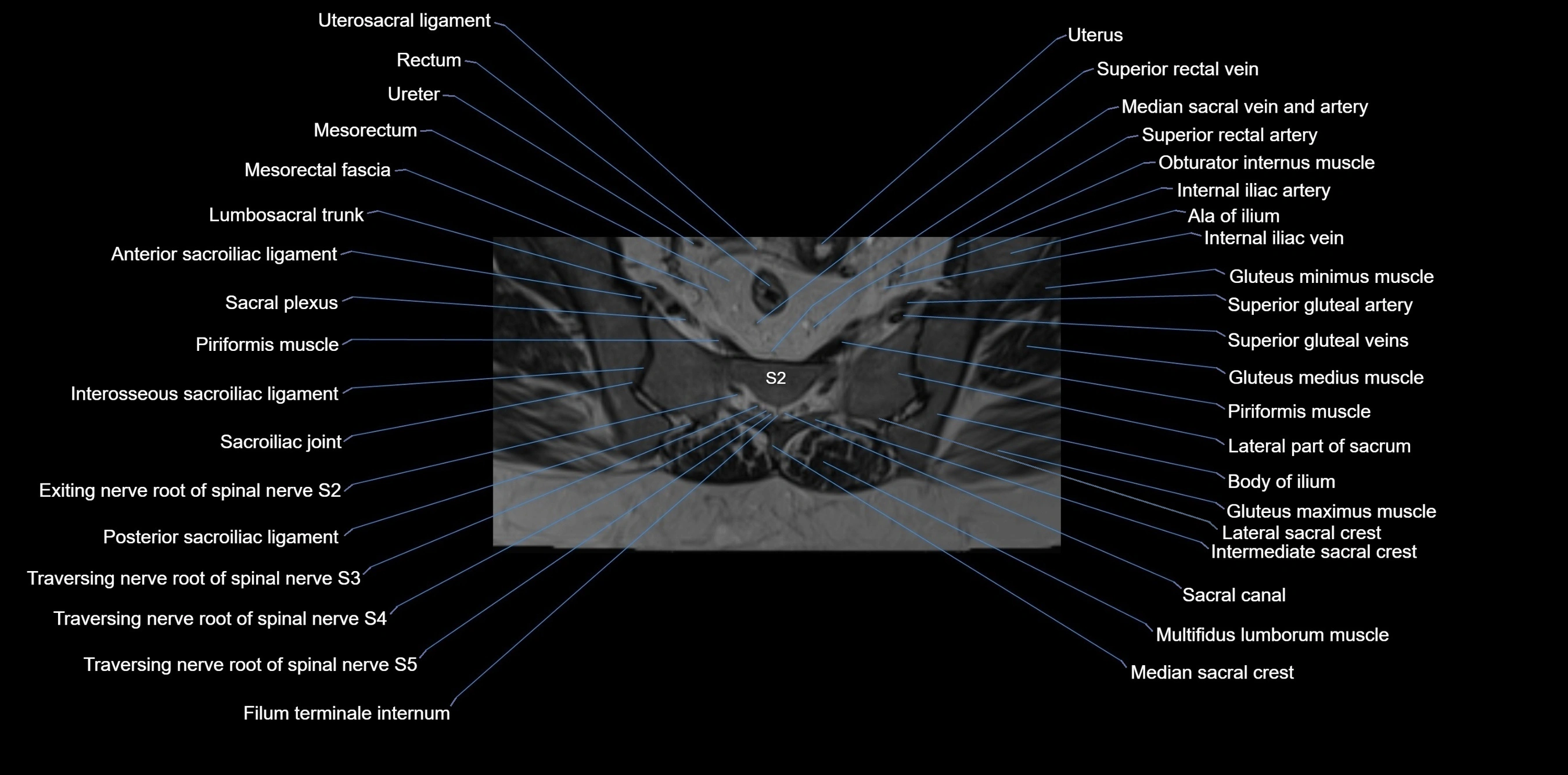
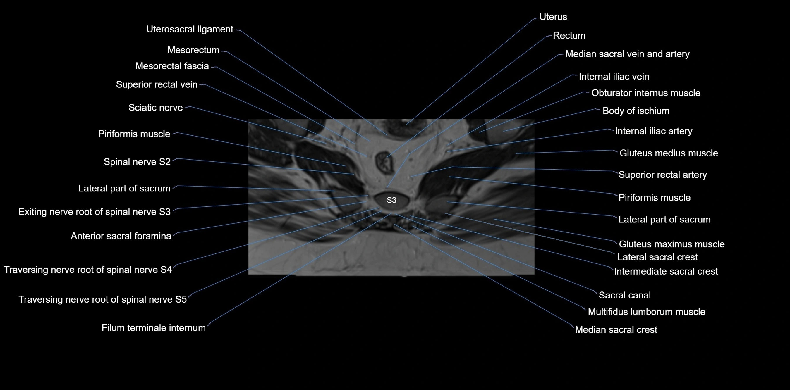
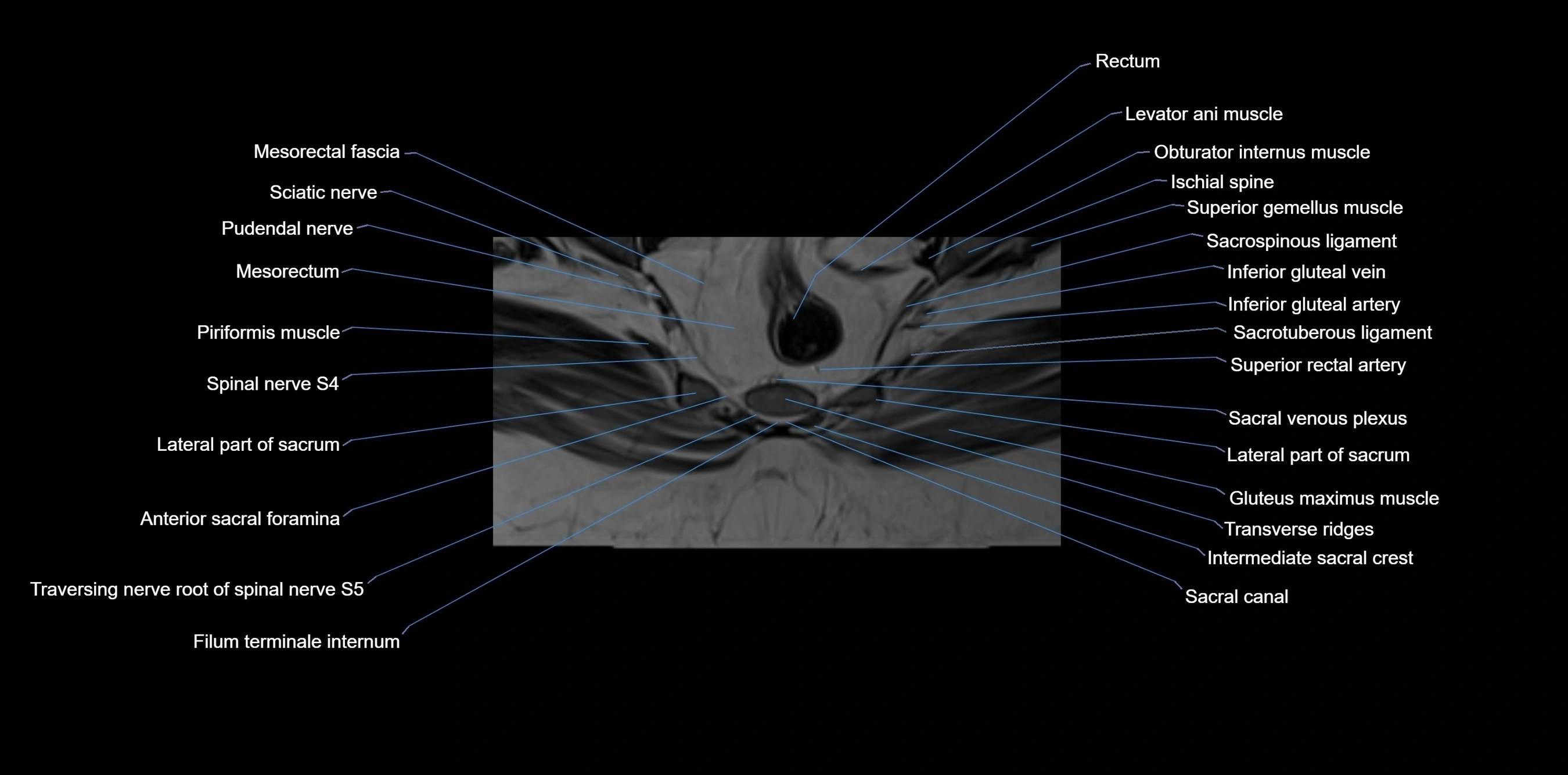
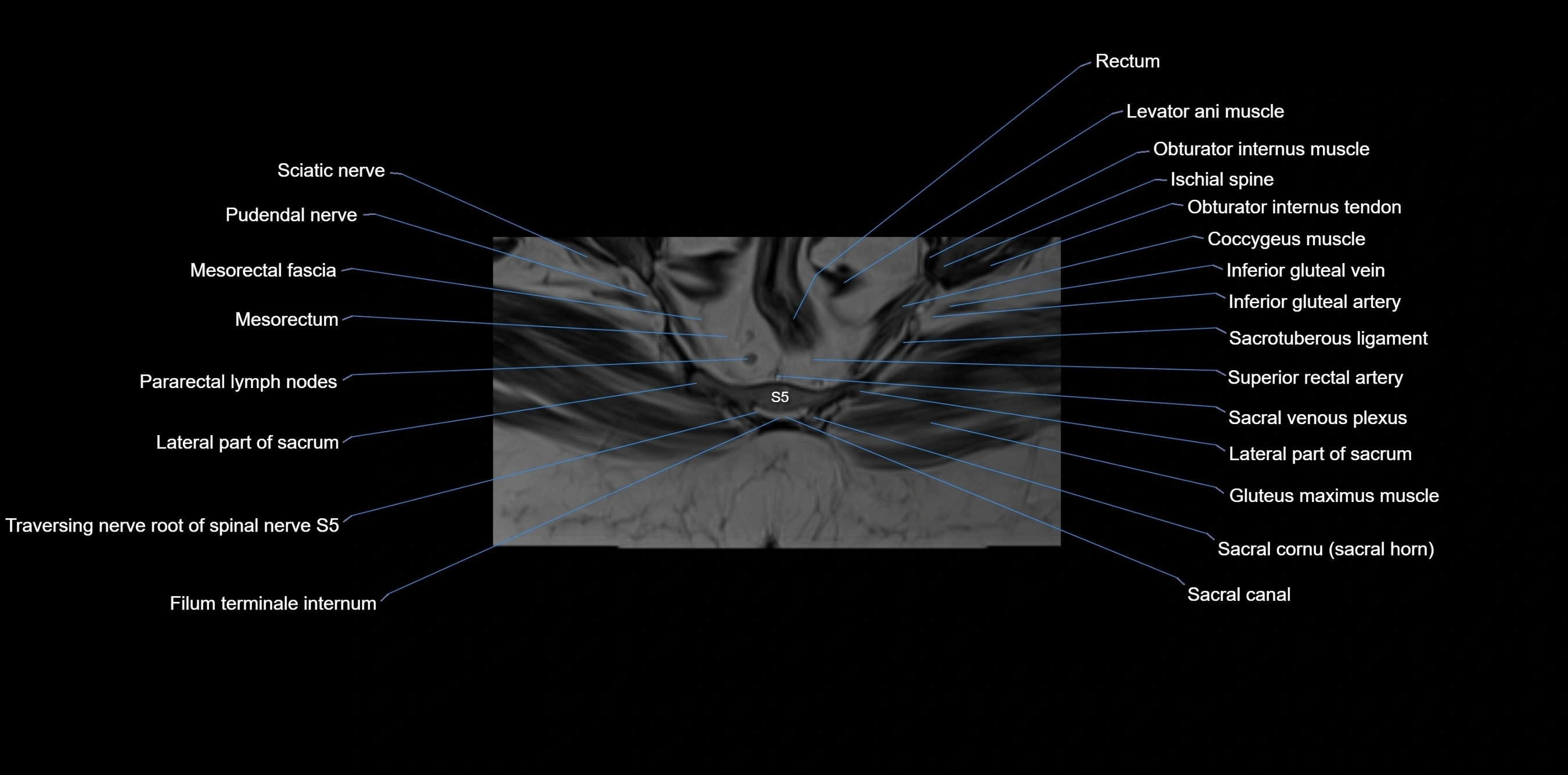
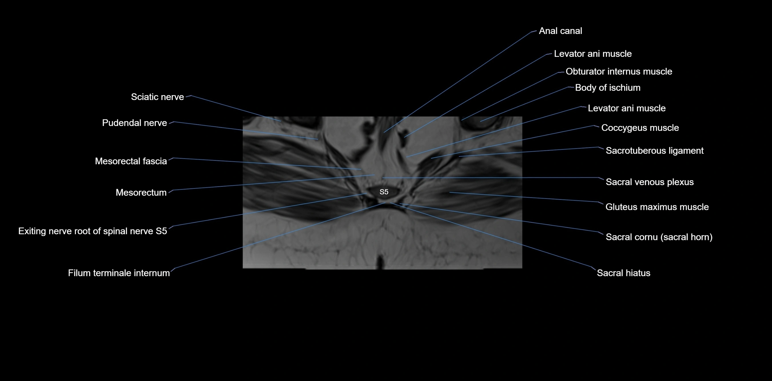
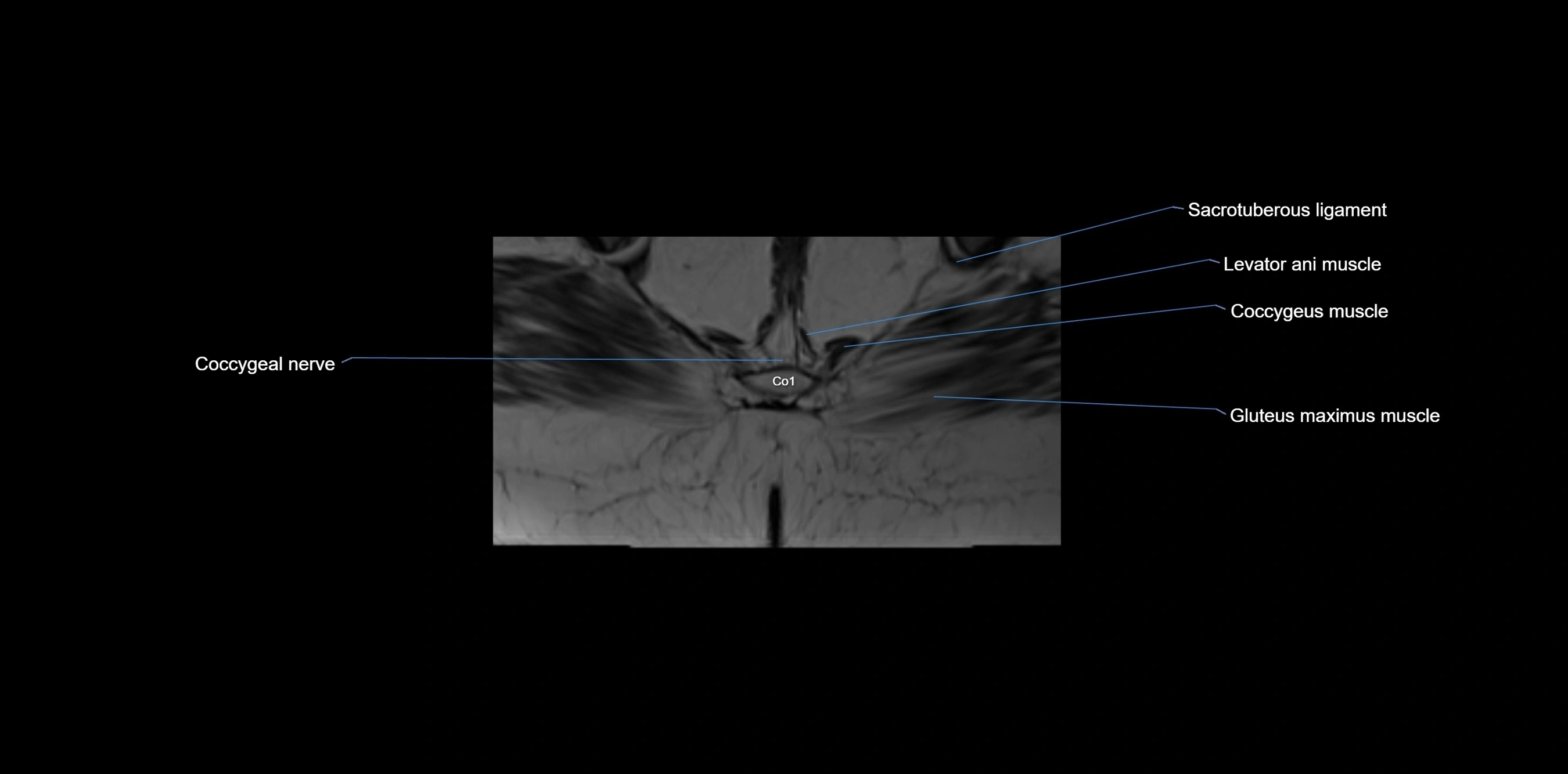
CT VRT image


