


Topic
- Accessory obturator vein
- Accessory saphenous vein
- Adductor brevis muscle
- Adductor longus muscle
- Adductor magnus muscle
- Adductor minimus muscle
- Ampulla of vas deferens
- Anal canal
- Anococcygeal body (anococcygeal ligament)
- Anterior Fibromuscular Stroma of prostate
- Apex of urinary bladder
- Areolar tissue of penis
- Body of epididymis
- Body of urinary bladder
- Buck's fascia (Deep fascia of penis)
- Bulb of Penis
- Bulbospongiosus muscle (Male)
- Bulbourethral gland (Cowper’s glands)
- Central zone of prostate
- Coccygeus muscle
- Corona of glans penis
- Coronal sulcus
- Corpus cavernosum
- Corpus spongiosum
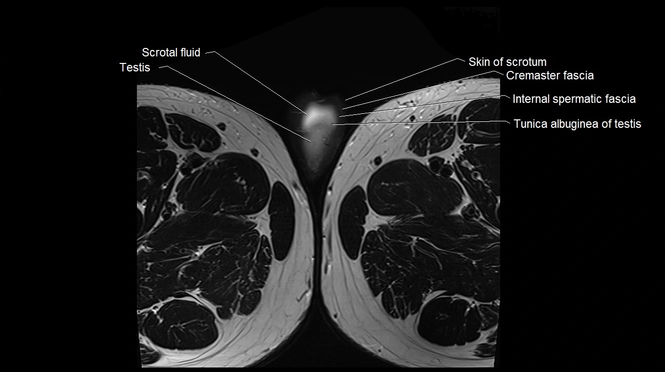
- Cremaster fascia
- Cremaster muscle
- Crus of penis
- Dartos fascia
- Deep circumflex iliac artery
- Deep dorsal vein of penis
- Deep dorsal vein of the penis
- Deep femoral artery (profunda femoris)
- Deep transverse perineal muscle
- Ejaculatory duct
- Epididymis
- External anal sphincter
- External iliac artery
- External iliac lymph nodes
- External iliac vein
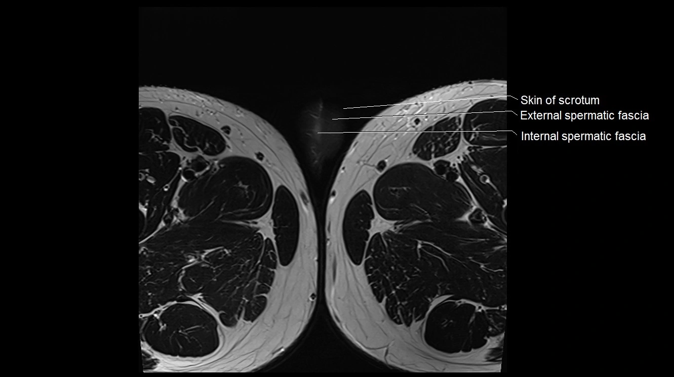
- External spermatic fascia
- External urethral orifice
- External urethral sphincter (male)
- Fascia of pelvic diaphragm
- Female urethra
- Femoral artery
- Femoral vein
- Foreskin
- Fundus of urinary bladder
- Genitofemoral nerve
- Glans penis
- Gluteal lymph nodes
- Gracilis muscle
- Head of epididymis
- Iliococcygeus muscle
- Iliofemoral ligament
- Iliopsoas tendon
- Iliotibial tract
- Inferior gemellus muscle
- Inferior gluteal artery
- Inferior gluteal vein
- Inferior pubic ligament
- Inguinal ligament
- Inguinal lymph nodes
- Intermediate lacunar external iliac lymph nodes
- Internal anal sphincter
- Internal iliac artery
- Internal pudendal artery
- Internal pudendal vein
- Internal spermatic fascia
- Internal urethral orifice
- Internal urethral sphincter (male)
- Ischioanal fossa
- Ischiocavernosus muscle (Male)
- Ischiococcygeus muscle
- Lateral circumflex femoral artery
- Lateral circumflex femoral veins
- Lateral fornix of cervix
- Levator ani muscle
- Lobule of testis
- Meatus of the urethra
- Medial circumflex femoral artery
- Medial circumflex femoral vein
- Medial cluneal nerves
- Median umbilical ligament
- Mediastinum testis
- Membranous urethra
- Mesorectal fascia
- Mesorectum
- Neck of urinary bladder
- Obturator artery
- Obturator externus muscle
- Obturator externus tendon
- Obturator internus muscle
- Obturator internus tendon
- Obturator lymph nodes
- Obturator vein
- Pampiniform plexus
- Parietal tunica vaginalis
- Pectineus muscle
- Penile urethra
- Penis
- Peripheral zone of prostate
- Posterior femoral cutaneous nerve
- Presacral fascia
- Prostatic urethra
- Pubic symphysis
- Pubic tubercle
- Puboanalis muscle
- Pubococcygeus muscle
- Puboprostatic ligament
- Puboprostaticus muscle
- Puborectalis muscle
- Pudendal artery
- Pudendal vein
- Pyramidal muscle (pyramidalis muscle)
- Rectal proper fascia (Fascia propria of the rectum)
- Rectococcygeal muscle
- Rectoprostatic fascia (Denonvilliers' fascia)
- Rectosacral fascia (Waldeyer's fascia)
- Rectovesical pouch
- Rectum
- Rectus femoris muscle
- Retropubic space
- Sacral lymph nodes
- Sacrospinous ligament
- Sacrotuberous ligament
- Sartorius muscle
- Scrotal fluid
- Seminal vesicle
- Septum of scrotum
- Septum of testis
- Septum of the penis (Penile septum)
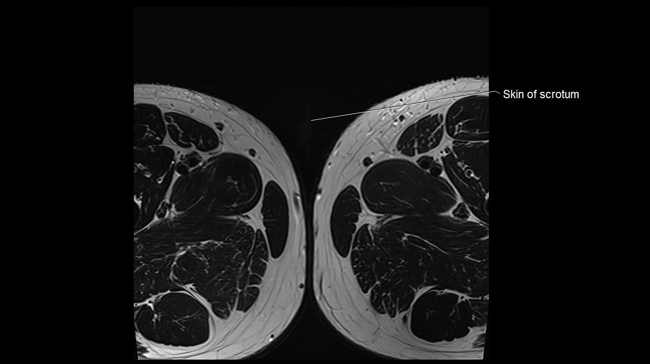
- Skin of scrotum
- Spermatic cord
- Spermatic cord nerves
- Subcutaneous tissue (scrotum)
- Superficial circumflex iliac artery
- Superficial circumflex iliac vein
- Superficial dorsal vein of penis
- Superficial femoral artery
- Superficial transverse perineal muscle
- Superior gemellus muscle
- Superior gluteal artery
- Superior pubic ligament
- Superior rectal vein
- Tail of epididymis
- Tensor fasciae latae muscle
- Testicular artery
- Testis
- Transitional zone of prostate
- Transverse perineal muscle
- Traversing nerve root of spinal nerve
- Trigone of urinary bladder
- Tunica albuginea (penis)
- Tunica albuginea of testis
- Umbilical artery
- Union of common iliac veins
- Ureteric Orifice
- Urinary bladder
- Vas deferens
- Vastus lateralis muscle
- Vastus medialis muscle
- Vesical veins
- Vesical venous plexus
- Visceral tunica vaginalis
- common iliac artery
- great saphenous vein
The accessory obturator vein (AOV) is an anatomical variant that accompanies the accessory obturator artery when present (10–30% of individuals). It usually arises from the external iliac vein or inferior epigastric vein, coursing along the superior pubic ramus toward the obturator canal, where it anastomoses with the obturator vein (draining into the internal iliac vein).
This vein may form part of the corona mortis (“crown of death”), an anastomotic venous (and sometimes arterial) connection between the external and internal iliac systems. This is clinically important in pelvic trauma, hernia repairs, and pelvic surgery, as injury can result in severe, sometimes fatal hemorrhage.
Synonyms
-
Aberrant obturator vein
-
Variant obturator vein
-
Venous component of corona mortis
Function
-
Provides collateral venous drainage between external and internal iliac venous systems
-
Drains blood from the obturator region, pubic bone, and adjacent pelvic structures
-
Acts as a potential pathway for collateral circulation in pelvic venous obstruction
-
Clinically relevant during hernia repair, orthopedic, gynecologic, and urologic surgery
MRI Appearance
T1-weighted images:
-
Vein appears as a linear hypointense structure along the superior pubic ramus
-
Surrounded by intermediate-to-high signal fat planes
T2-weighted images:
-
Normal vein lumen appears as a signal void due to fast blood flow
-
Thrombosis or slow flow may show hyperintense intraluminal signal
STIR:
-
Fat suppression enhances vein visibility against pelvic fat
-
Shows venous wall inflammation or perivascular edema as hyperintense regions
T1 Fat-Suppressed Post-Gadolinium:
-
Enhances brightly, outlining its course and anastomoses with obturator vein
-
Useful for detecting venous malformations, thrombosis, or extrinsic compression
MRV (Magnetic Resonance Venography):
-
Demonstrates the origin, course, and venous anastomoses of the AOV
-
Highlights corona mortis when present
-
Useful in preoperative venous mapping, trauma evaluation, and pelvic varicosity assessment
CT Appearance
Non-contrast CT:
-
Vein may be faintly visible as a tubular structure along superior pubic ramus
-
Often difficult to differentiate from adjacent fat and small vessels without contrast
CT Post-Contrast:
-
Enhances as a small but distinct vessel coursing toward the obturator canal
-
May reveal engorgement, compression, or collateral channels in pathology
CTV (CT Venography):
-
Clearly delineates the AOV and its connection to the obturator vein
-
Shows its role in corona mortis formation
-
Multiplanar and 3D reconstructions map venous variations and aid in surgical and trauma planning
-
Detects thrombosis, varicosities, or abnormal venous drainage pathways
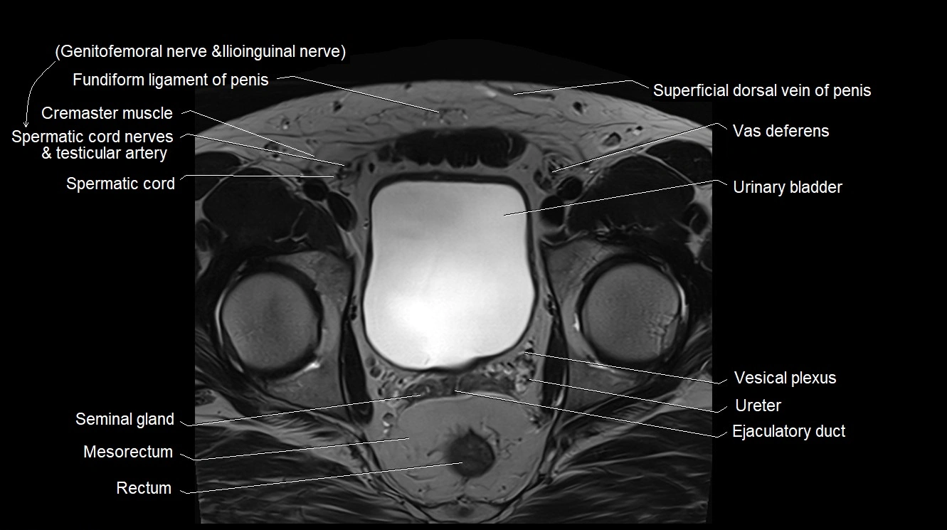
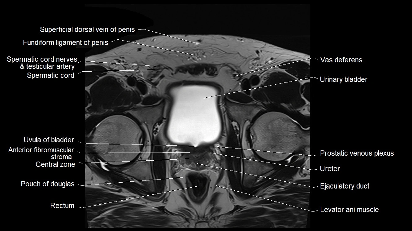
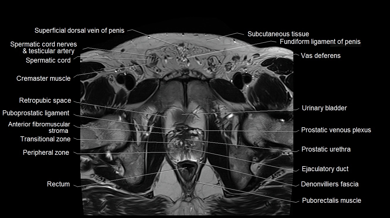
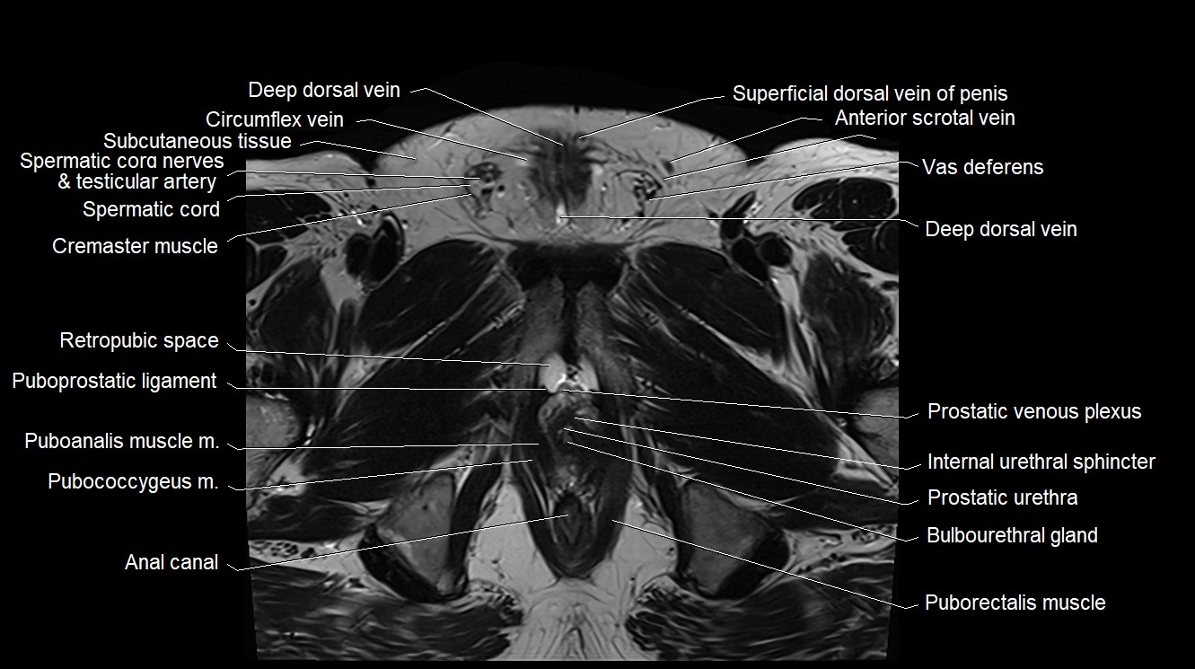
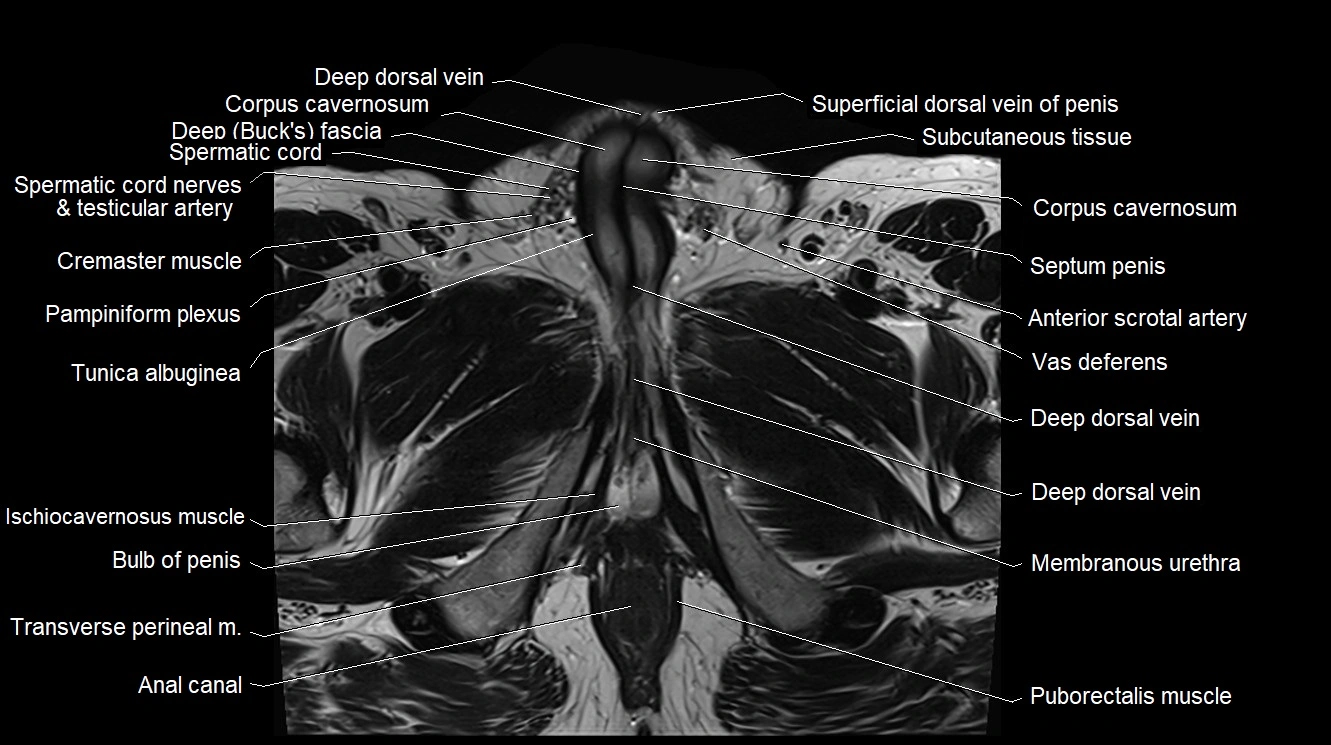
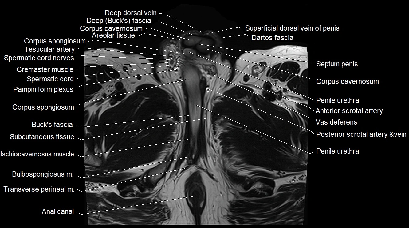
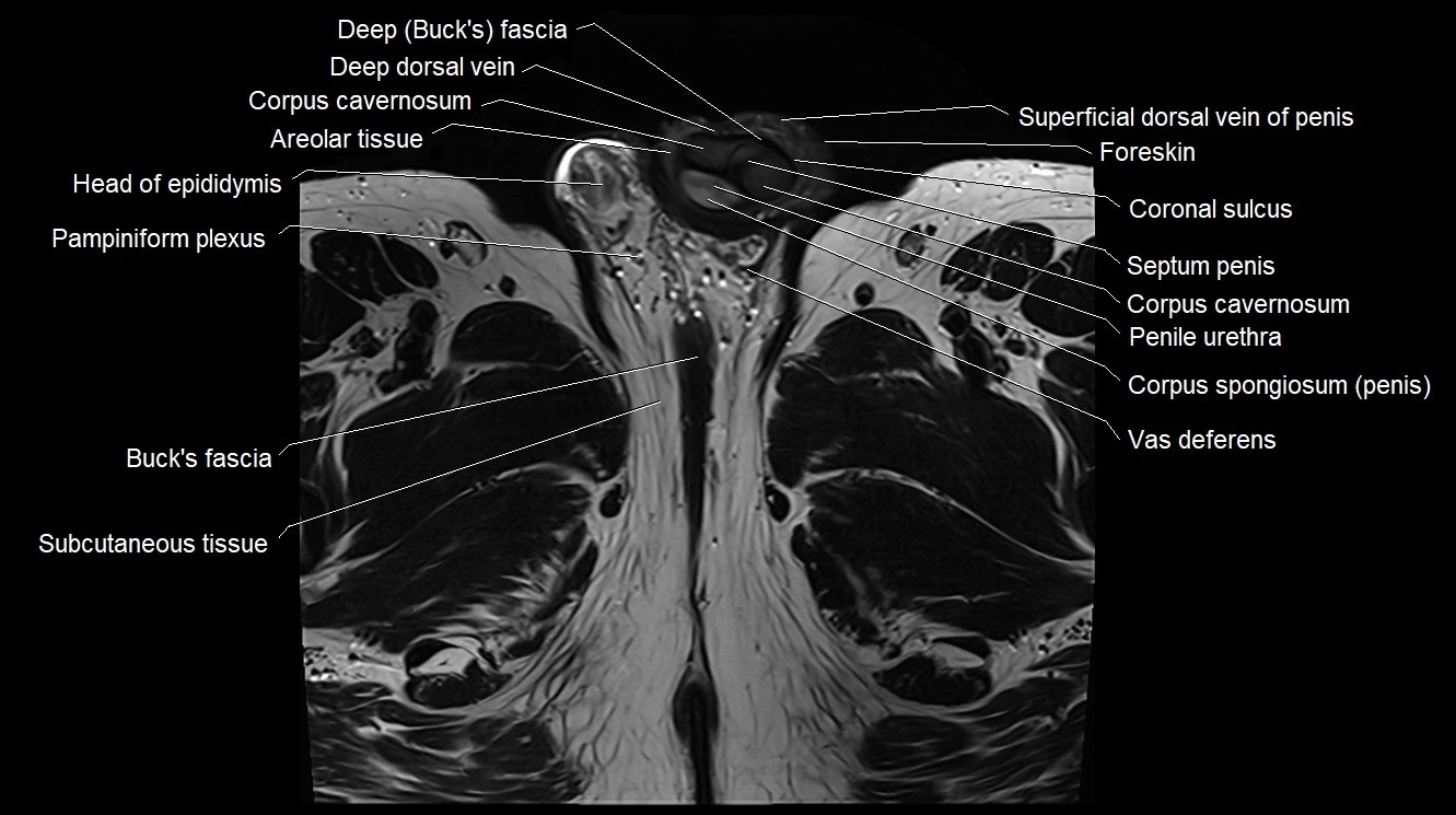
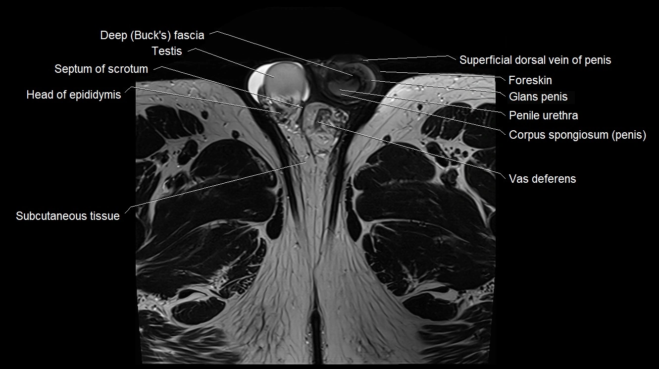
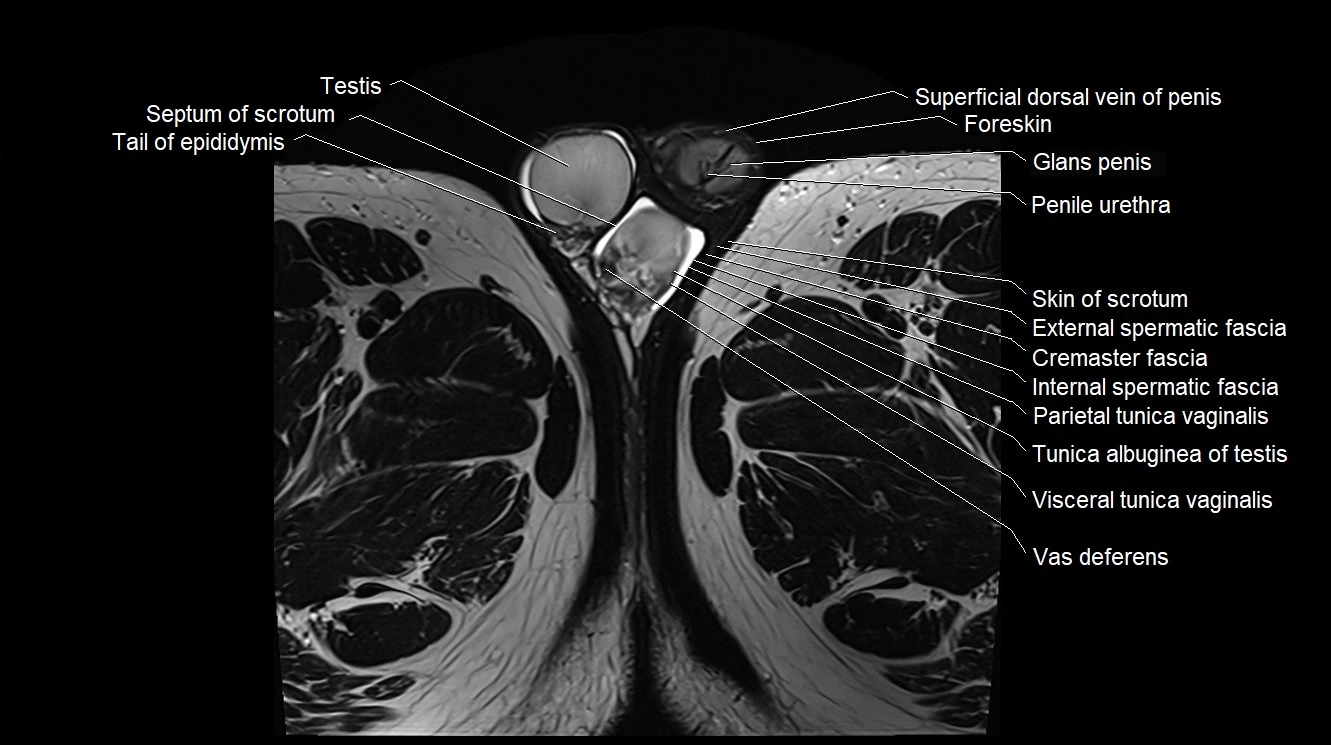
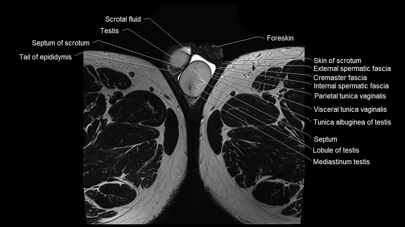
MRI image

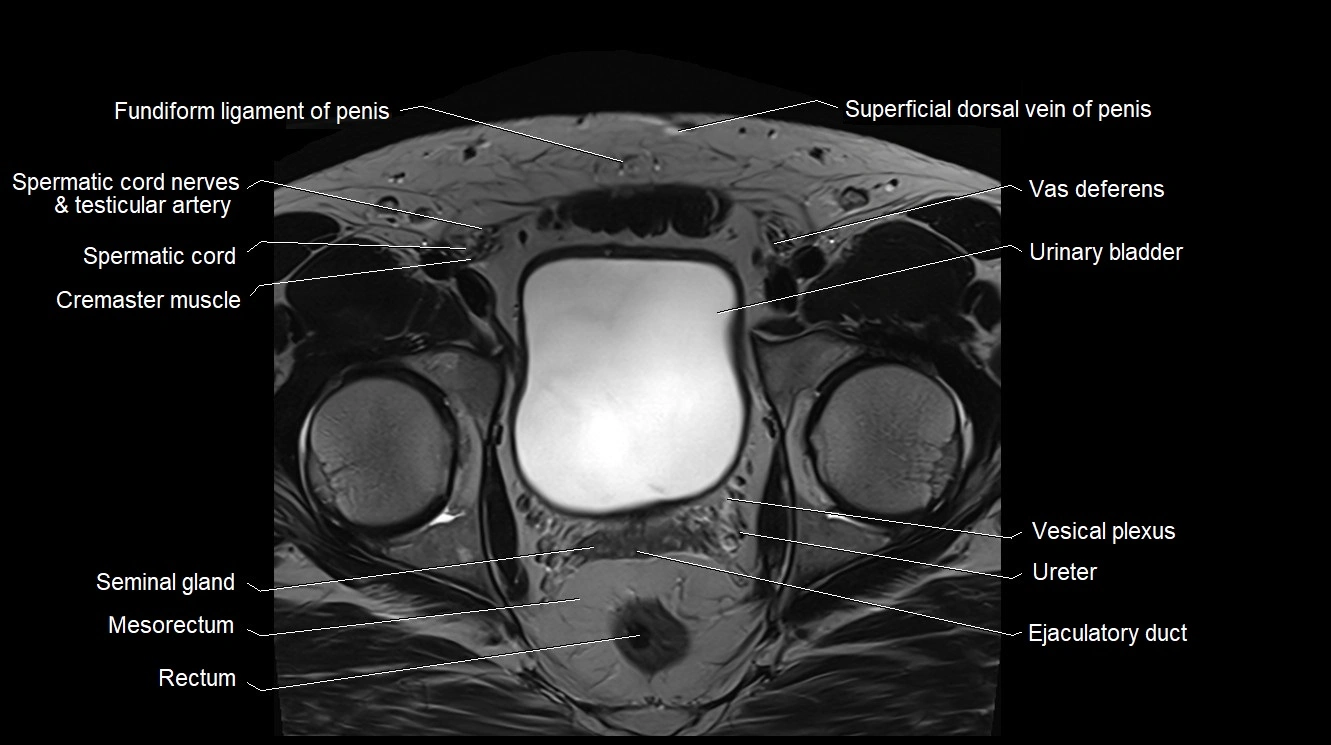
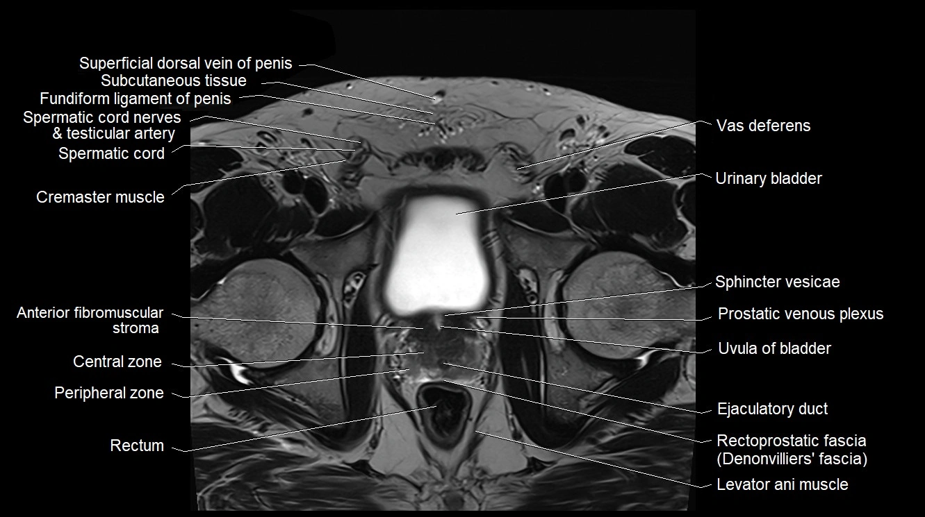
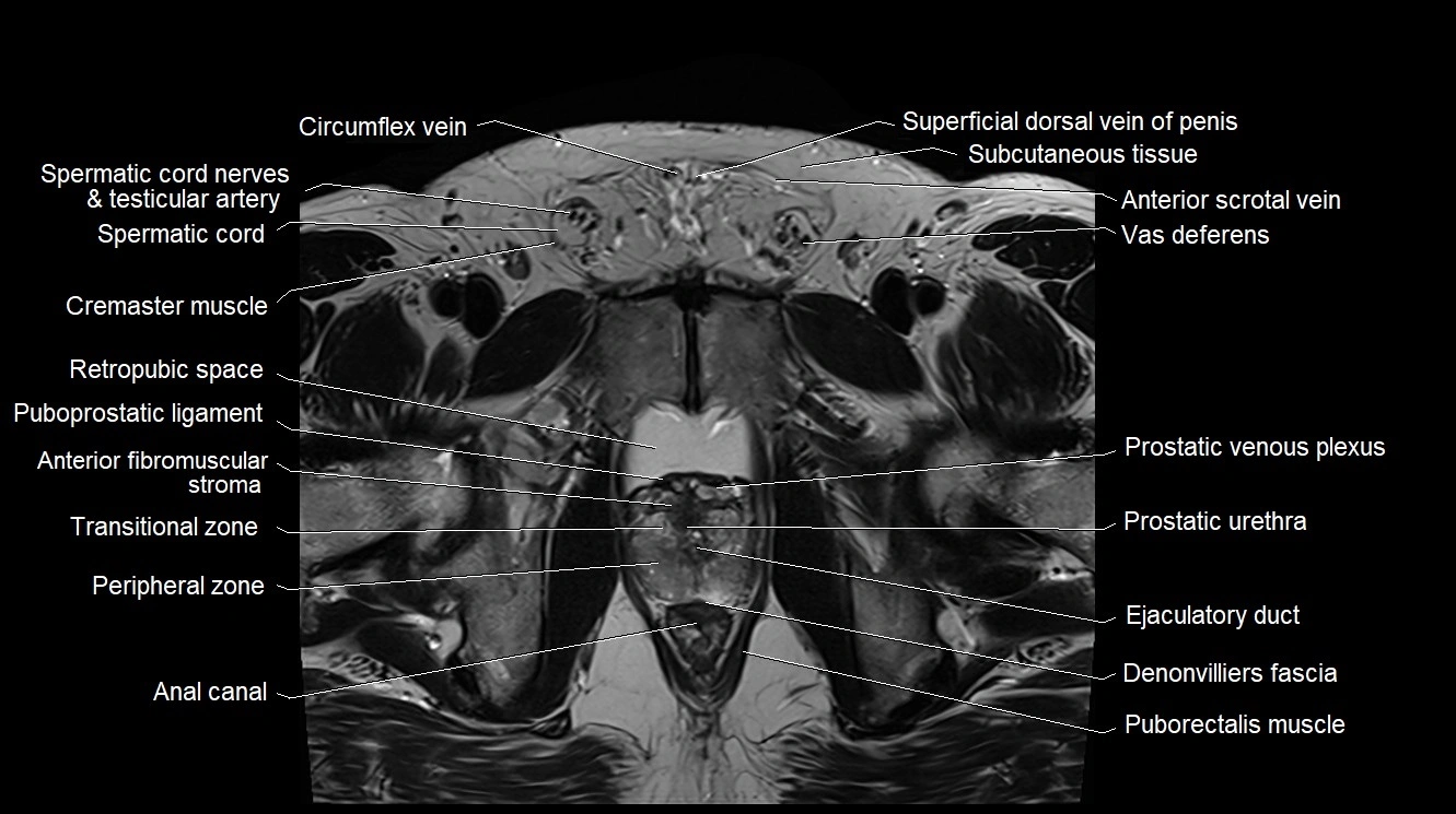
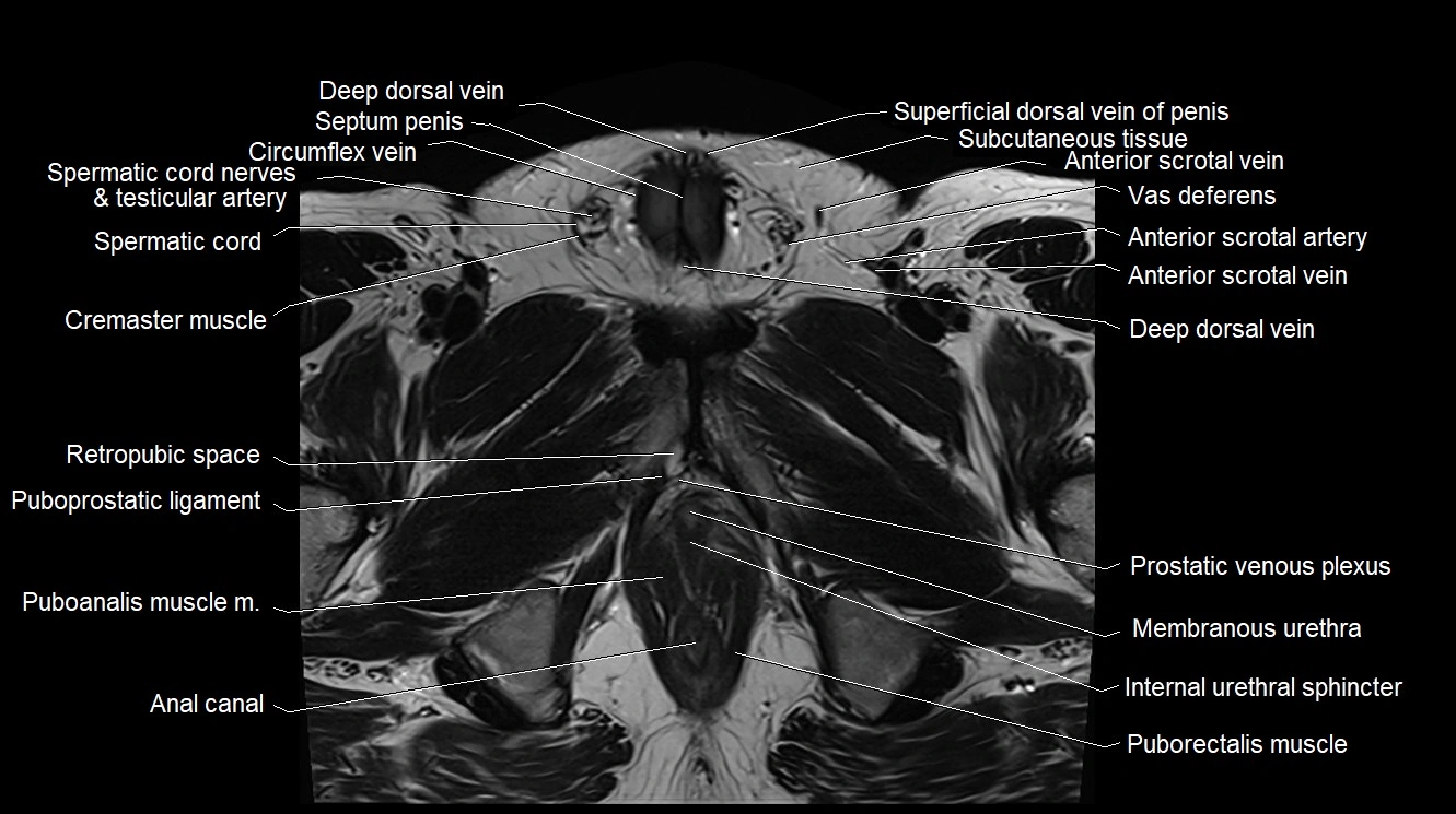
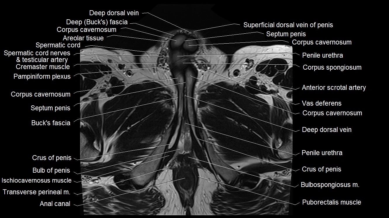
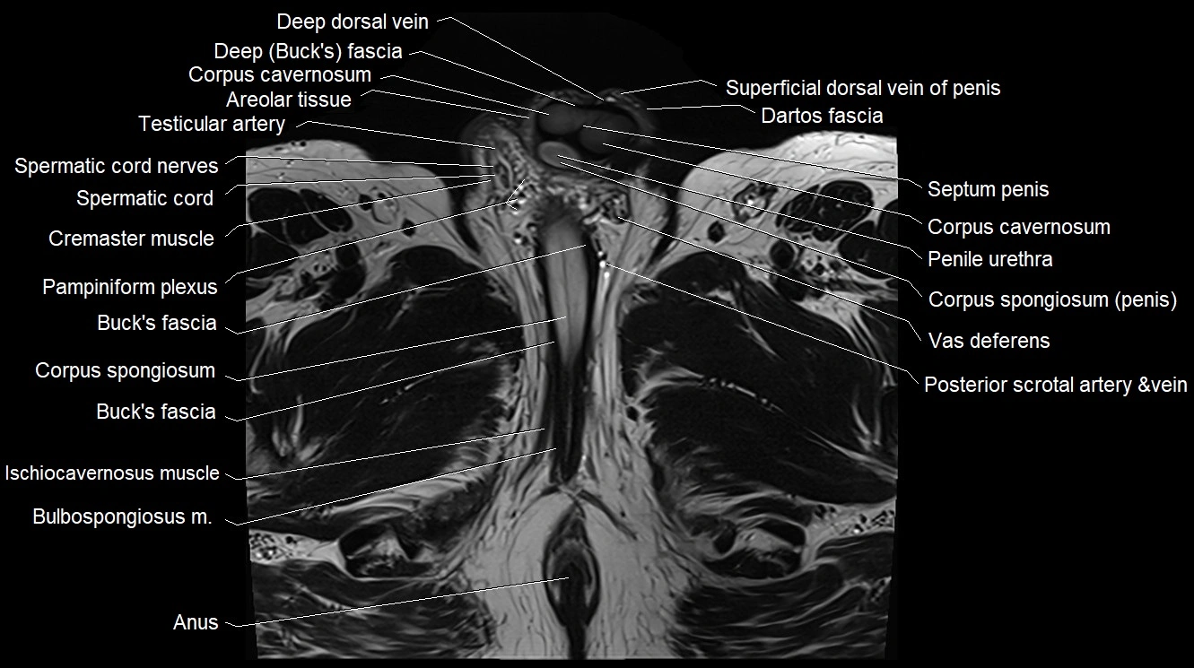
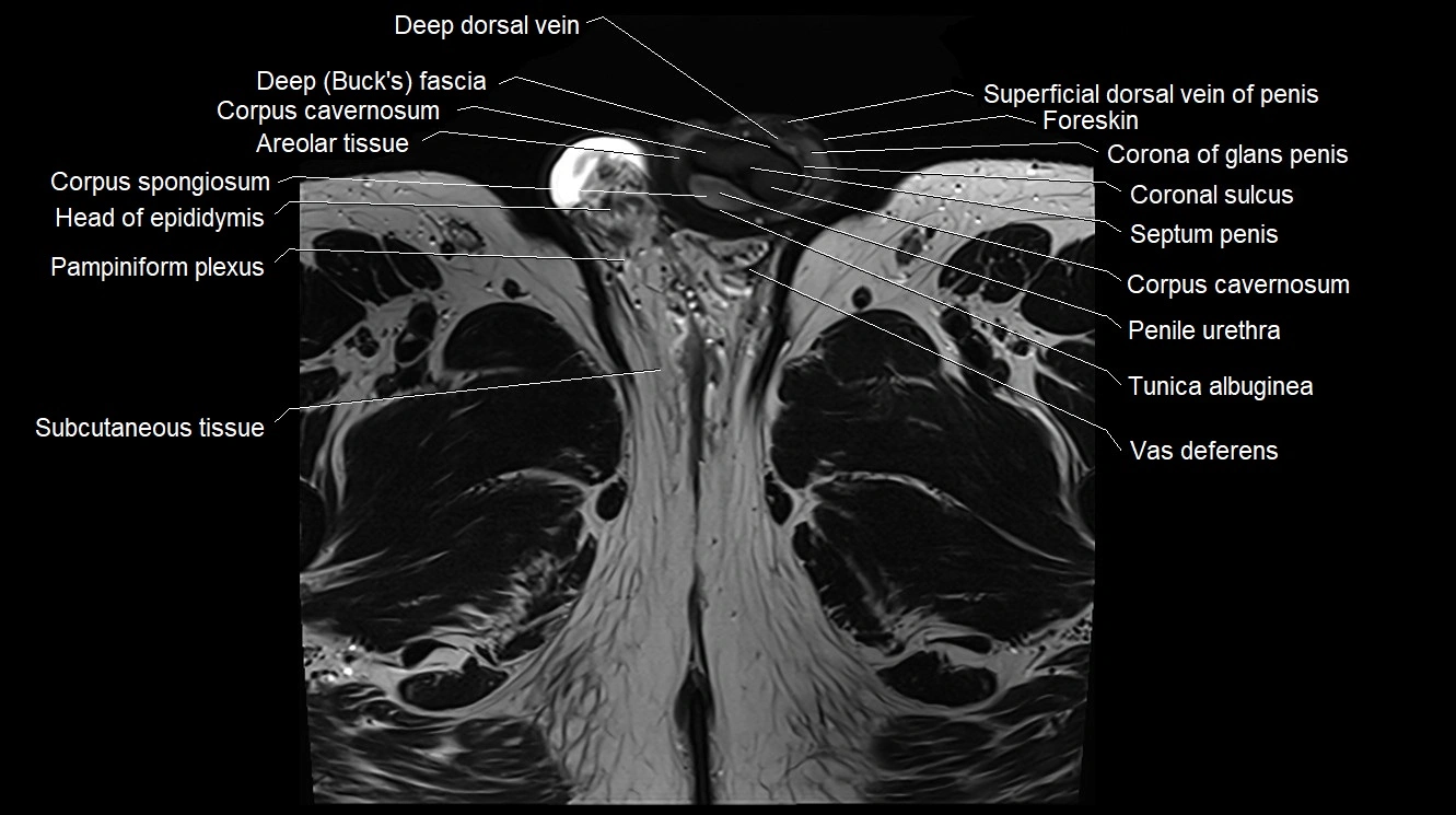
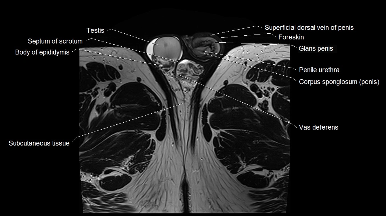
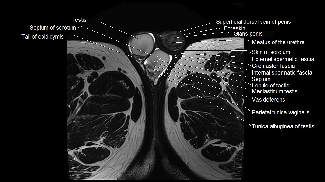
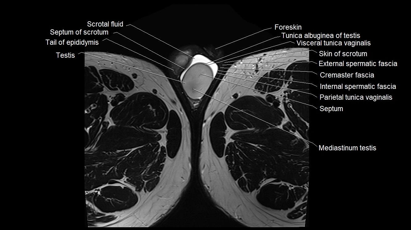
MRI image

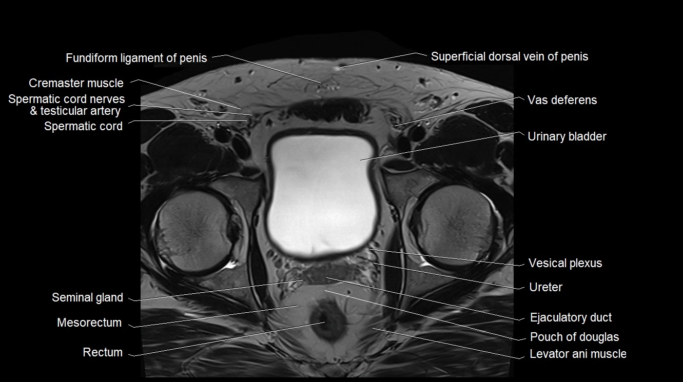
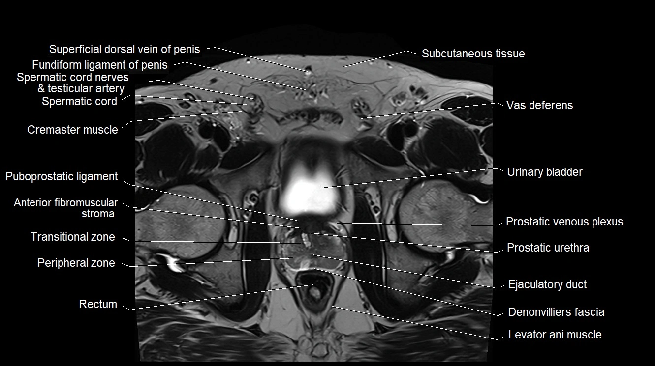
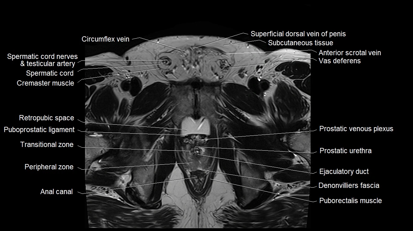
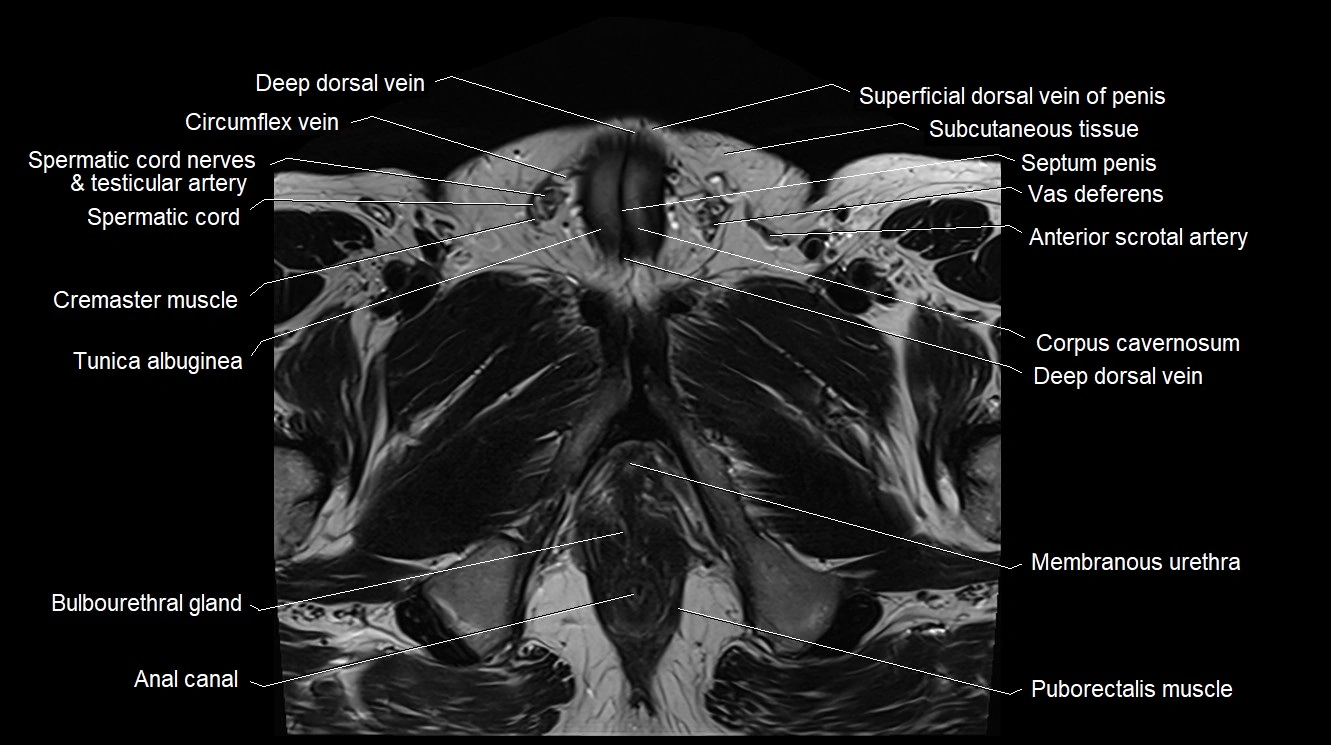
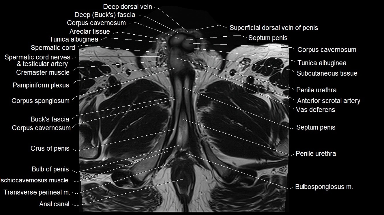
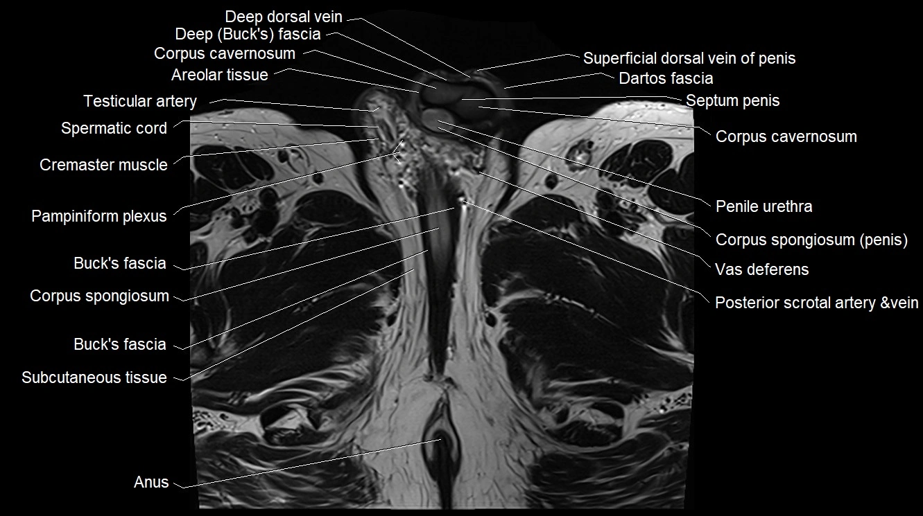
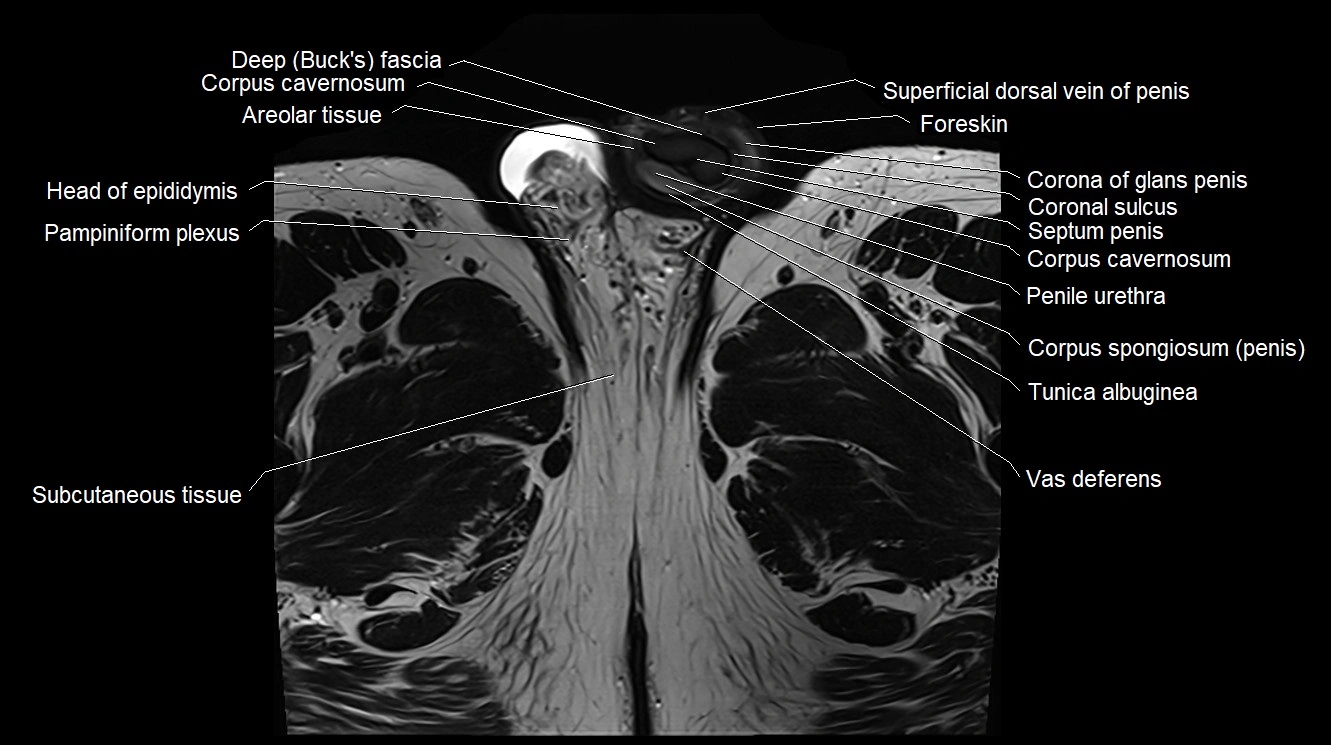
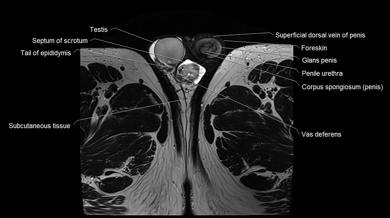
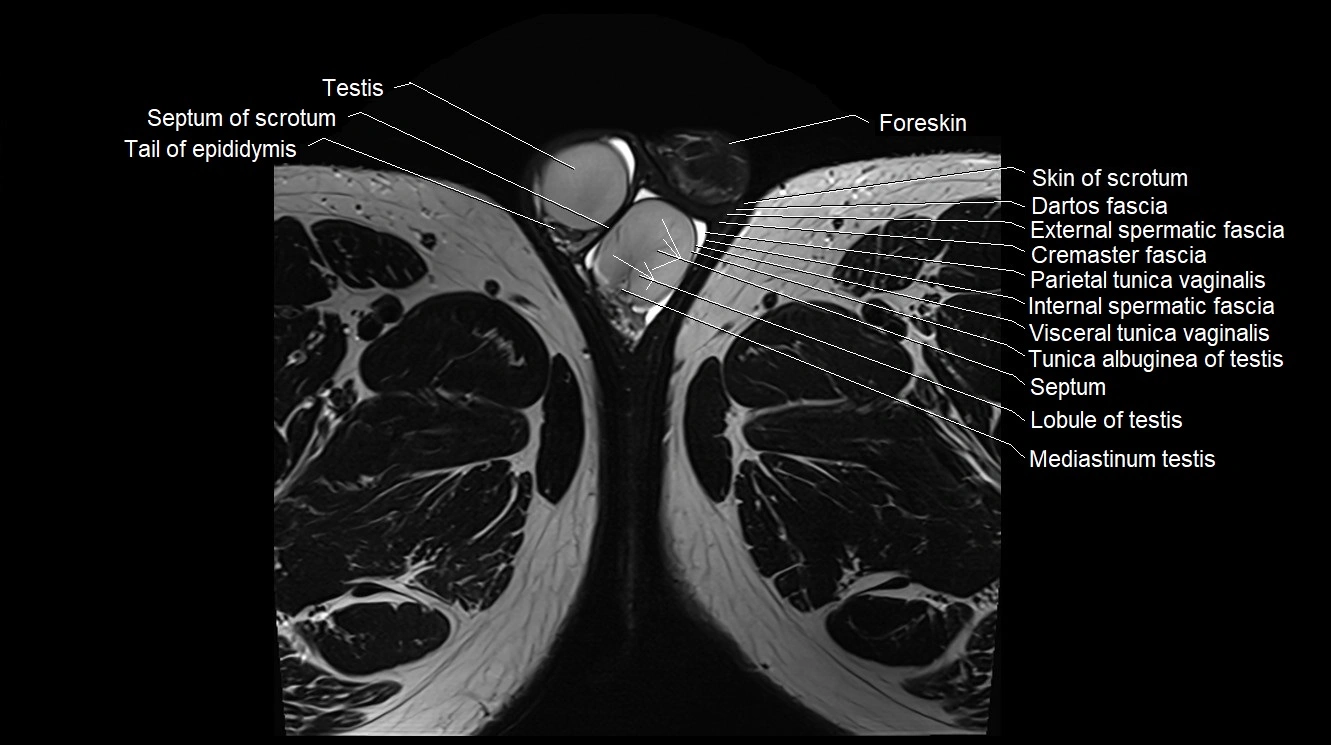
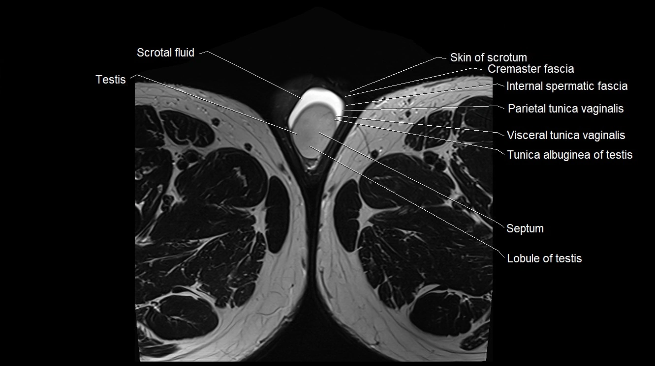
MRI image

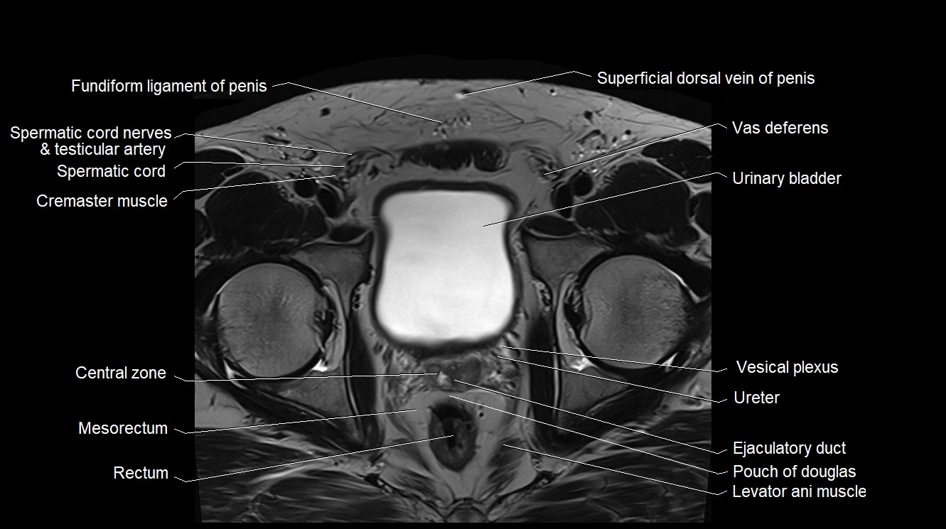
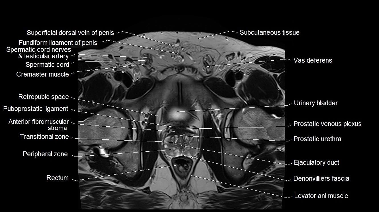
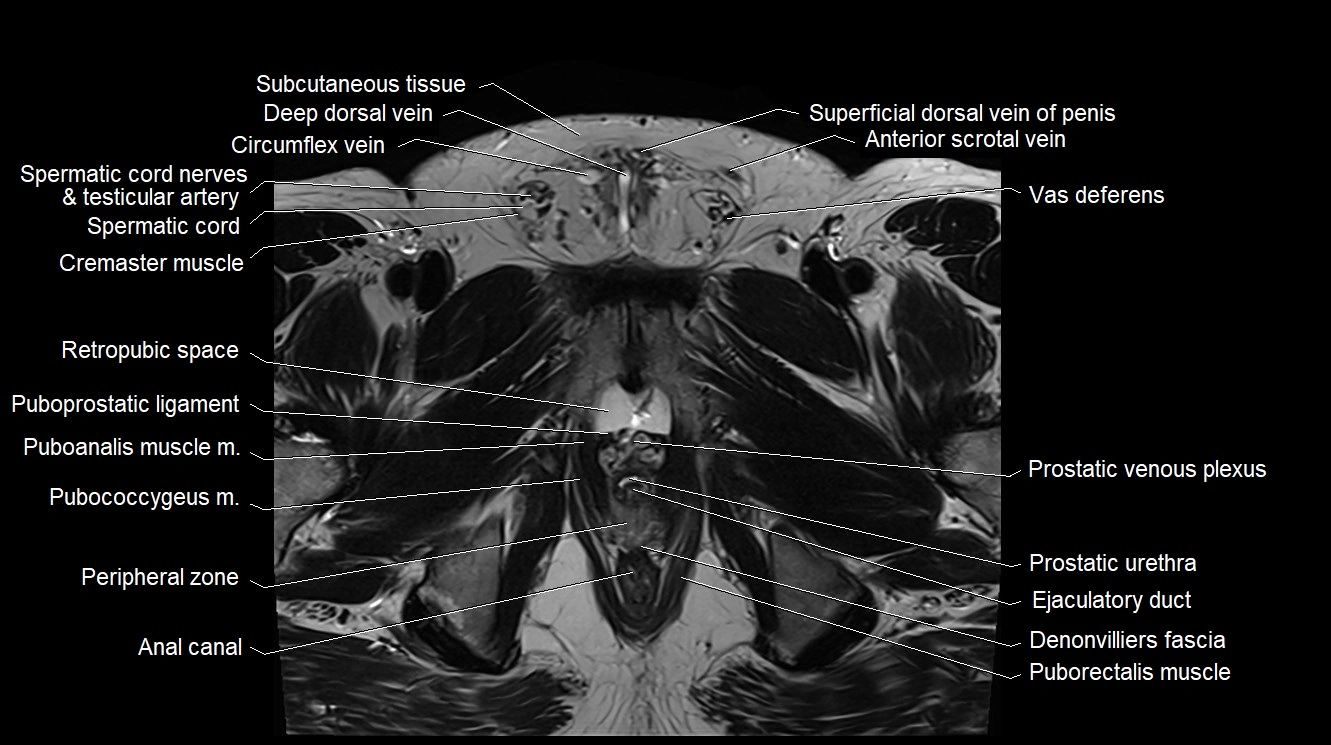
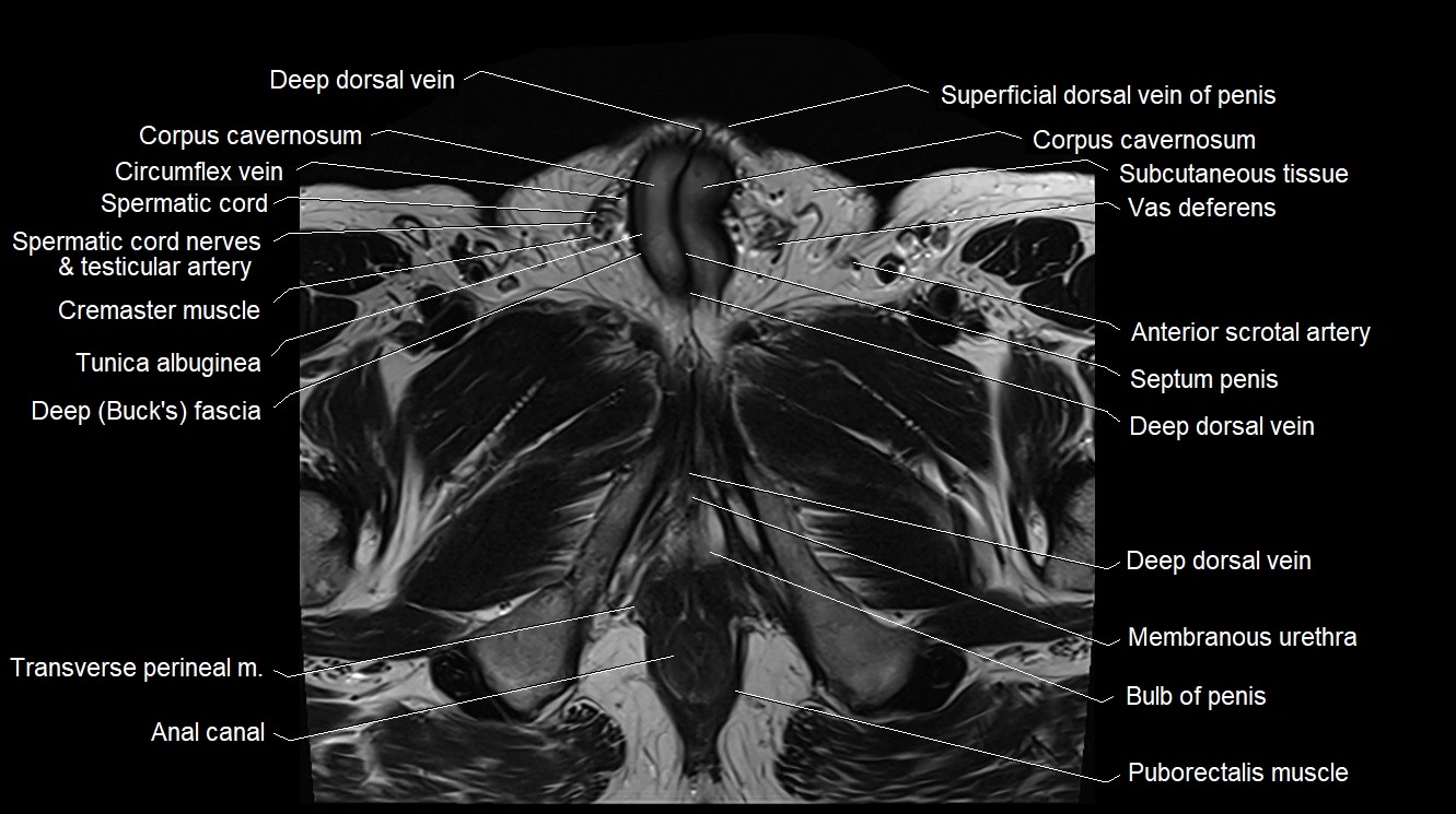
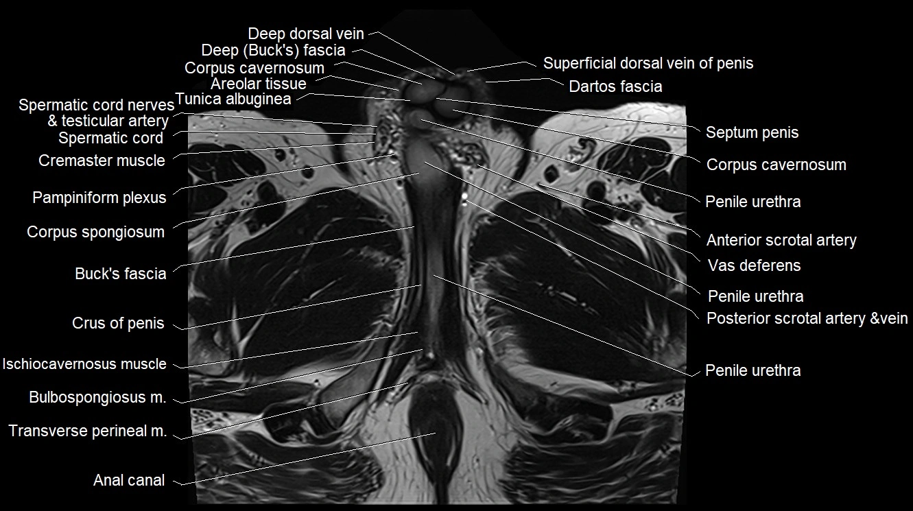
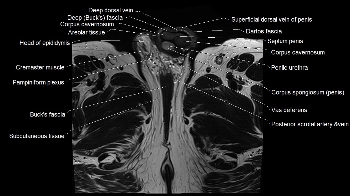
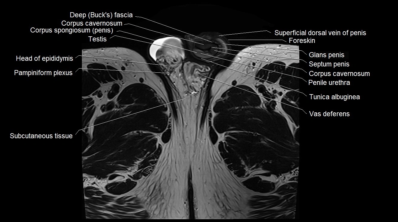
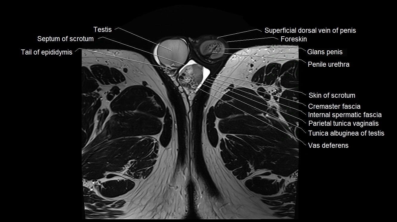
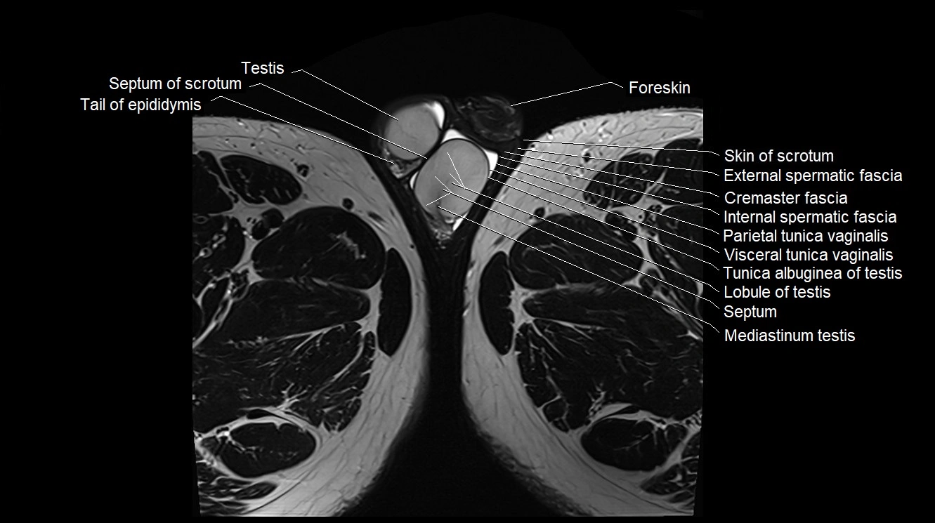
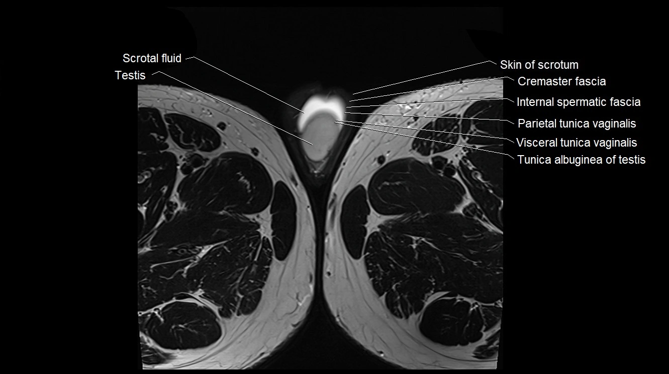
MRI image


