


Topic
- Abducens nerve (Cranial nerve VI)
- Abducens nerve (orbital part )
- Accessory Nerve (Cranial nerve XI)
- Alveolar arch of maxilla
- Anterior belly of digastric muscle
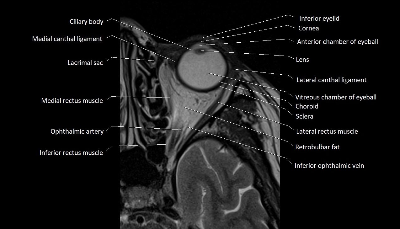
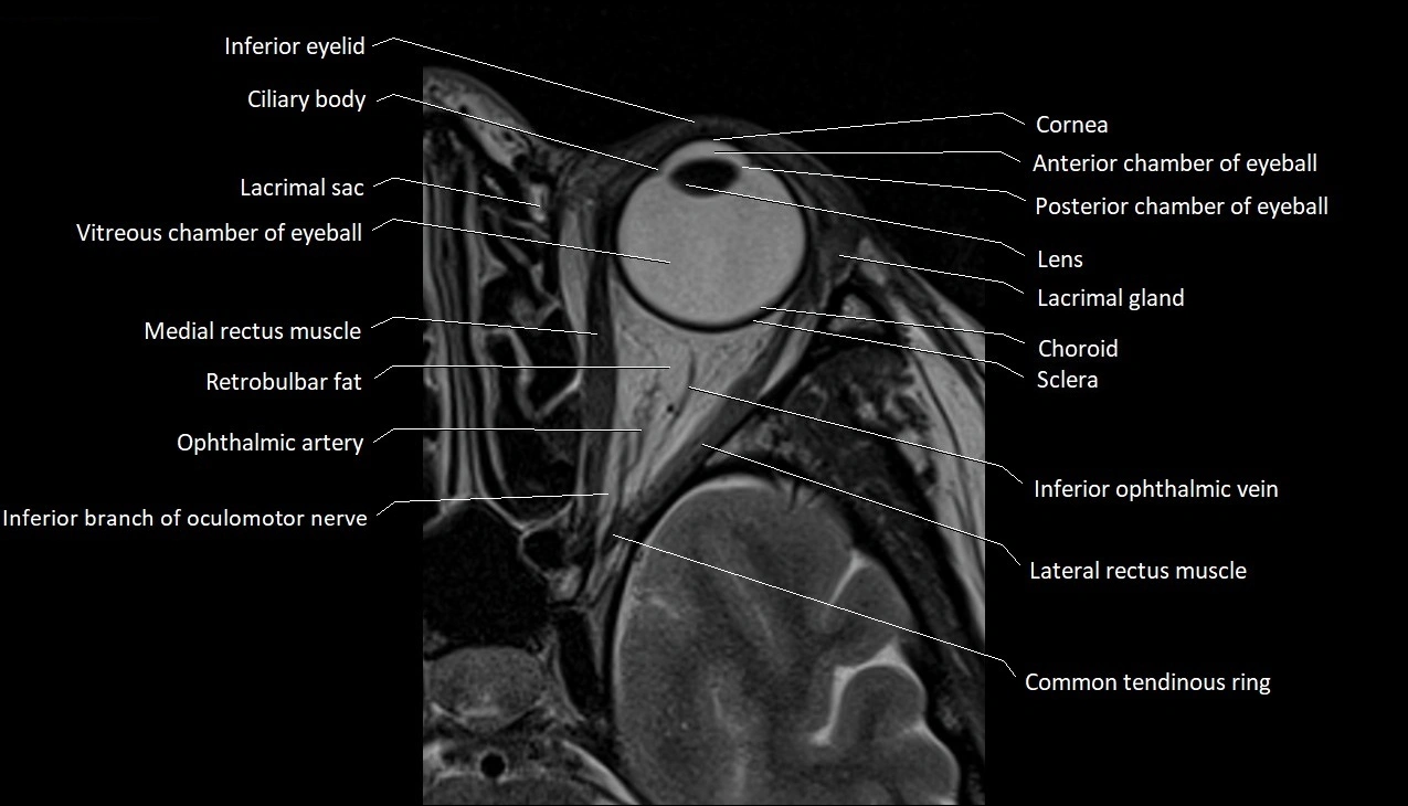
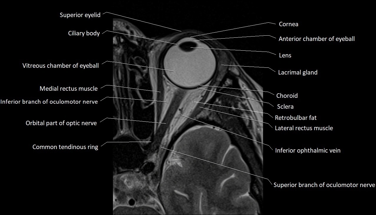
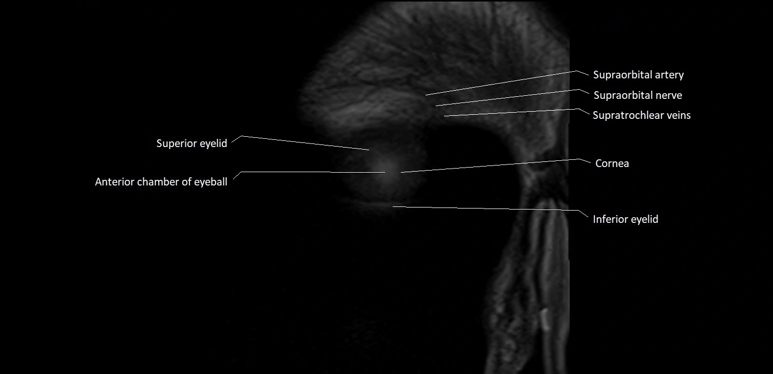
- Anterior chamber of eyeball
- Anterior cochlear nucleus
- Anterior ethmoidal air cells
- Apex of nose
- Cartilaginous part of nasal septum
- Choroid
- Cochlea
- Cochlear nerve (Cranial nerve VIII)
- Common tendinous ring (Annulus of zinn)
- Cornea
- Eyeball
- Facial Nerve (Cranial nerve VII)
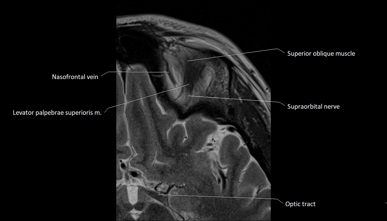
- Frontal nerve
- Glossopharyngeal nerve (Cranial nerve IX)
- Hypoglossal Nerve (Cranial nerve XII)
- Inferior branch vestibular nerve
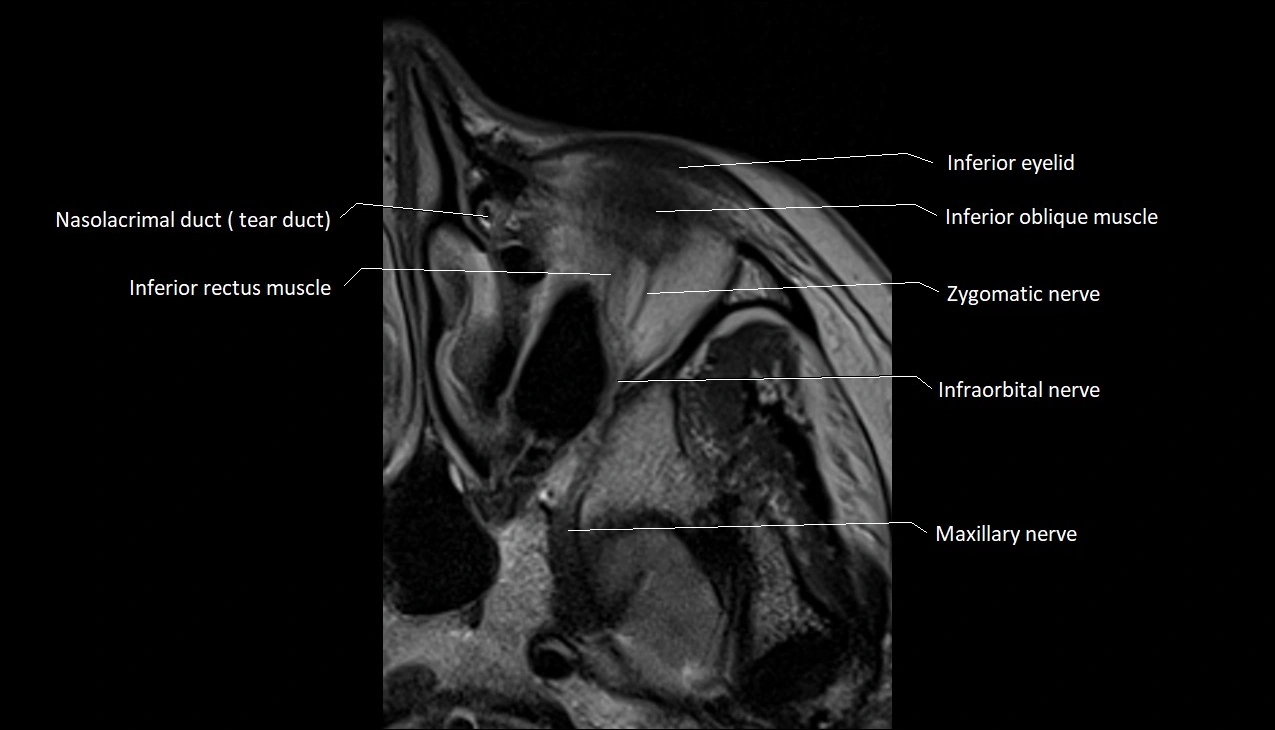
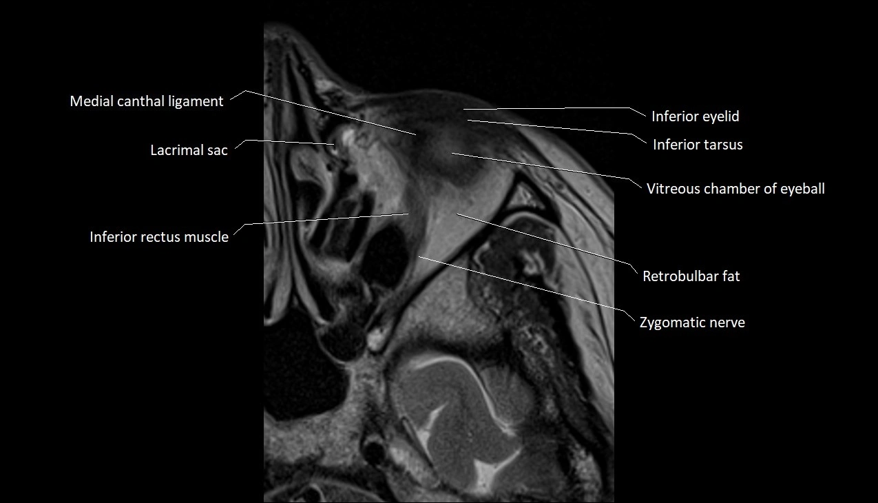
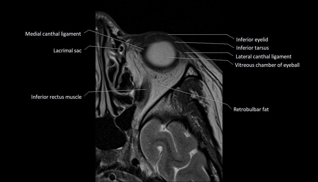
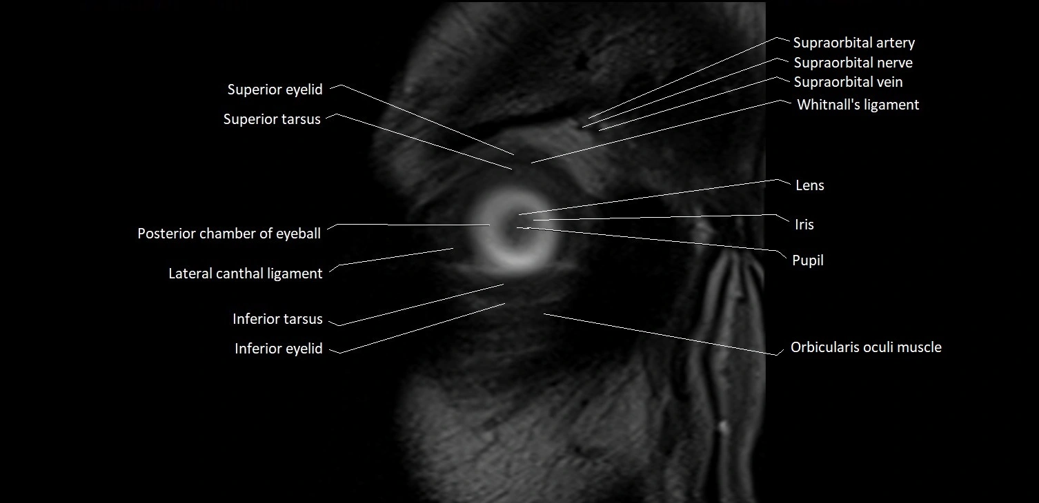
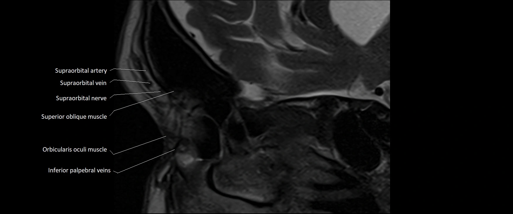
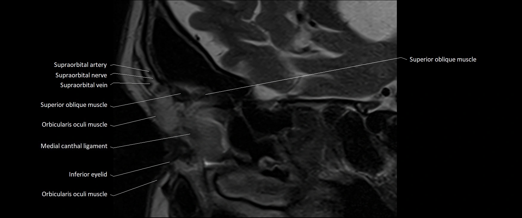
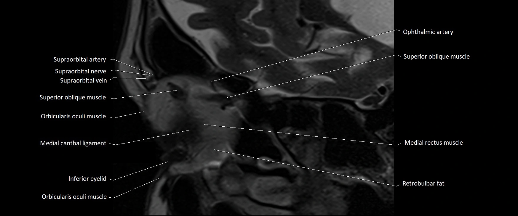
- Inferior eyelid
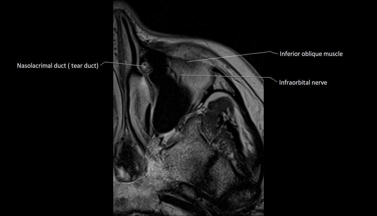
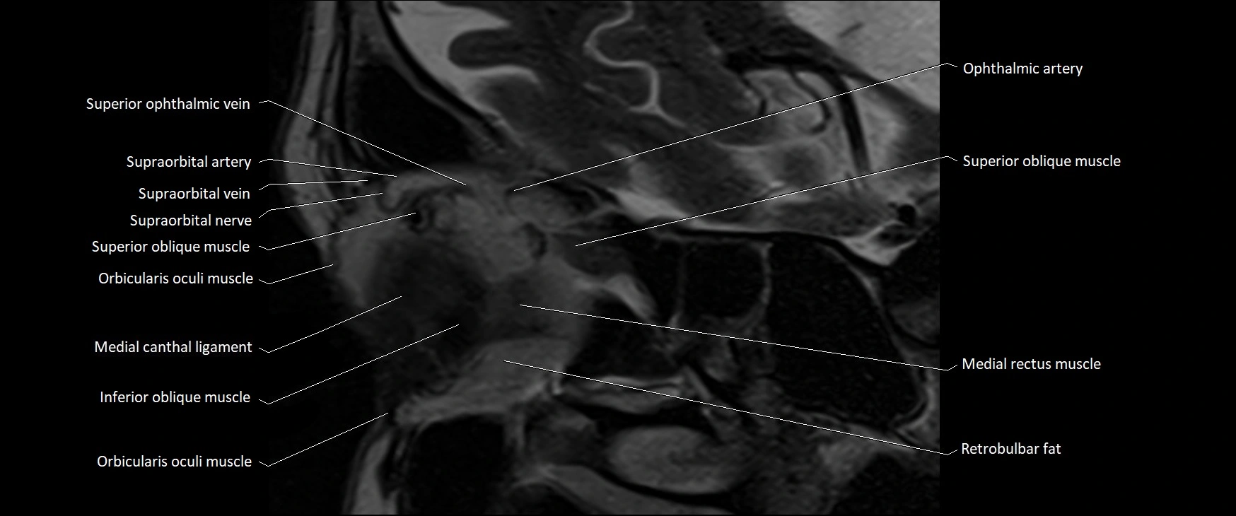
- Inferior oblique muscle
- Inferior ophthalmic vein
- Inferior rectus muscle
- Inferior salivatory nucleus
- Inferior tarsus
- Inferior vestibular nucleus
- Infraorbital margin
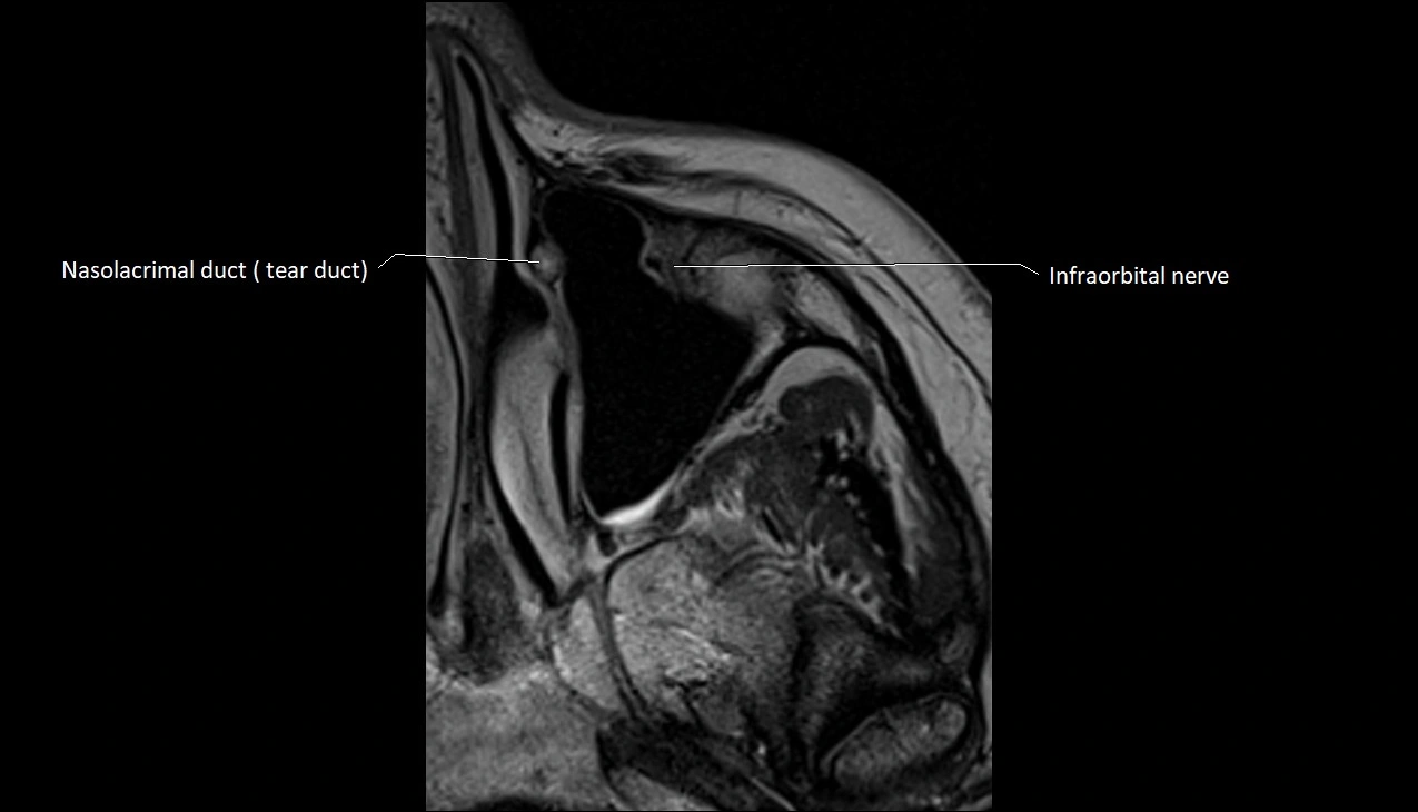
- Infraorbital nerve
- Internal occipital crest
- Intracanalicular part of optic nerve
- Intracranial part of optic nerve
- Iris
- Jugular foramen
- Lacrimal artery
- Lacrimal bone
- Lacrimal canaliculi
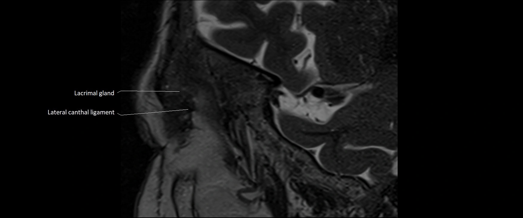
- Lacrimal gland
- Lacrimal nerve
- Lacrimal nucleus
- Lacrimal vein
- Lateral canthal ligament
- Lateral nasal cartilage
- Lateral rectus muscle
- Lateral vestibular nucleus
- Lateral wall of the orbit
- Levator palpebrae superioris muscle
- Lockwood’s ligament
- Major alar cartilage
- Maxillary nerve
- Medial canthal ligament
- Medial rectus muscle
- Medial vestibular nucleus
- Medial wall of orbit
- Mesencephalic nucleus of trigeminal nerve
- Middle nasal concha
- Minor alar cartilage
- Motor nucleus of facial nerve
- Motor nucleus of trigeminal nerve
- Nasal spine of frontal bone
- Nasofrontal vein
- Nasolacrimal duct (Tear duct)
- Nucleus of abducens nerve
- Nucleus of hypoglossal nerve
- Nucleus of oculomotor nerve
- Nucleus of solitary tract
- Nucleus of trochlear nerve
- Oculomotor Nerve (Cranial Nerve III)
- Oculomotor nerve (Superior branch)
- Oculomotor nerve (inferior branch)
- Olfactory Nerve (Cranial Nerve I)
- Olfactory bulb
- Olfactory tract
- Opening of nasolacrimal duct
- Optic Nerve (Cranial Nerve II)
- Optic canal
- Optic chiasm
- Optic disc
- Optic nerve sheath
- Orbicularis oculi muscle
- Orbicularis oculi muscle (Orbital part)
- Orbicularis oculi muscle (Preseptal part)
- Orbicularis oris muscle
- Orbital cavity
- Orbital part of optic nerve
- Orbital part of orbicularis oculi muscle
- Orbital surface of maxilla
- Oropharynx
- Palatine glands
- Palatine process of maxilla
- Palatoglossus muscle
- Palatopharyngeus muscle
- Posterior belly of digastric muscle
- Posterior chamber of eyeball
- Posterior cochlear nucleus
- Posterior cricoarytenoid muscle
- Preseptal part of orbicularis oculi muscle
- Principal sensory nucleus of the trigeminal nerve
- Pupil
- Rectus capitis anterior muscle
- Rectus capitis lateralis muscle
- Rectus capitis posterior major muscle
- Rectus capitis posterior minor muscle
- Retina
- Retrobulbar fat
- Risorius muscle
- Rotatores cervicis muscle
- Sclera
- Sella turcica
- Semicircular Canals
- Semispinalis capitis muscle
- Semispinalis cervicis muscle
- Septum of sphenoid sinus
- Serratus anterior muscle
- Spinal nucleus of trigeminal nerve
- Spinalis cervicis muscle
- Splenius capitis muscle
- Splenius cervicis muscle
- Sternocleidomastoid muscle
- Sternothyroid muscle
- Styloglossus muscle
- Stylohyoid muscle
- Stylopharyngeus muscle
- Subarachnoid space of optic nerve
- Subclavius muscle
- Superciliary arch
- Superior branch of vestibular nerve
- Superior cornu of thyroid cartilage
- Superior eyelid
- Superior oblique muscle
- Superior ophthalmic vein
- Superior rectus muscle
- Superior salivatory nucleus
- Superior tarsus
- Superior vestibular nucleus
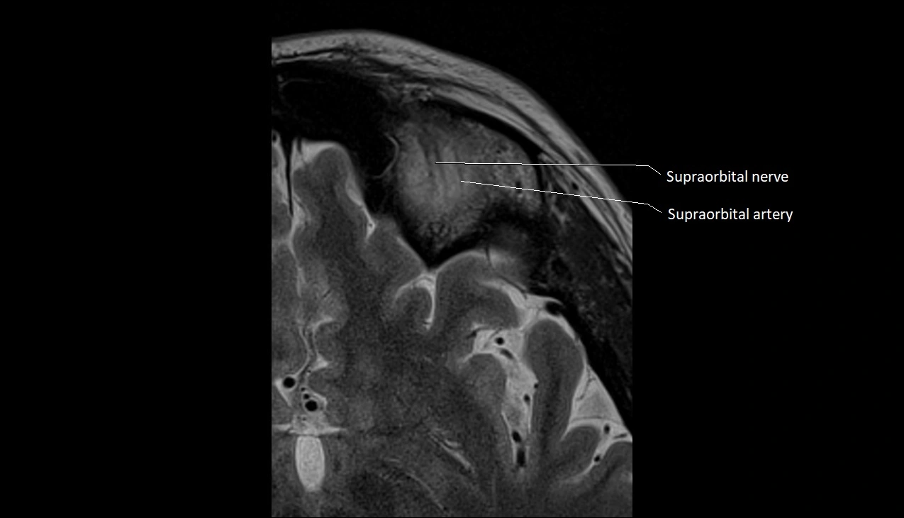
- Supraorbital artery
- Supraorbital margin
- Supraorbital nerve
- Supraorbital vein
- Supratrochlear artery
- Supratrochlear nerve
- Supratrochlear vein
- Trapezius muscle
- Trigeminal ganglion
- Trigeminal nerve (Cranial nerve V)
- Trochlear nerve (Cranial nerve IV)
- Vagus nerve (Cranial nerve X)
- Vertebral artery
- Vestibular ganglion
- Vestibule
- Vestibulocochlear nerve (Cranial nerve VIII)
- Vitreous chamber of eyeball
- Whitnall's ligament
- Zygomatic nerve
- lens of the eye
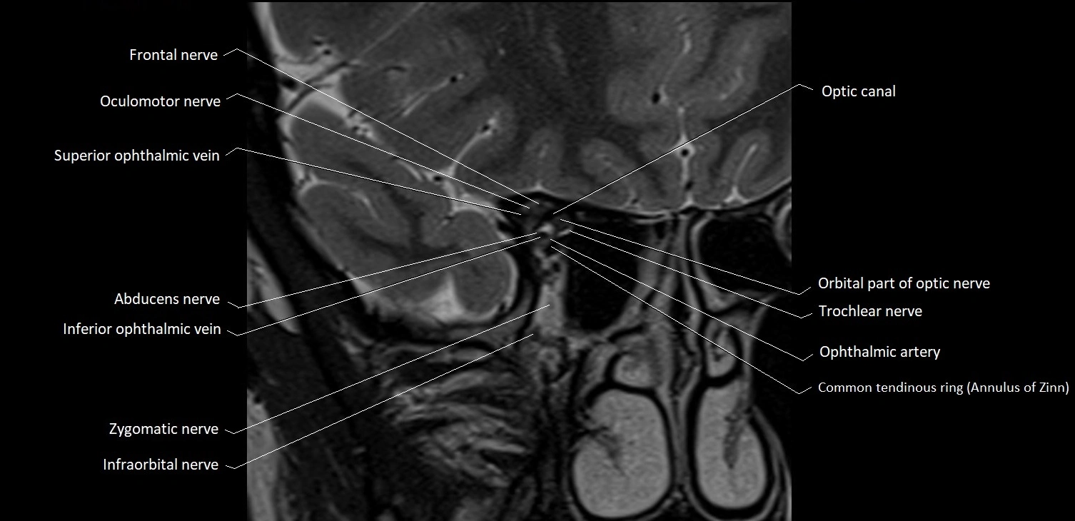
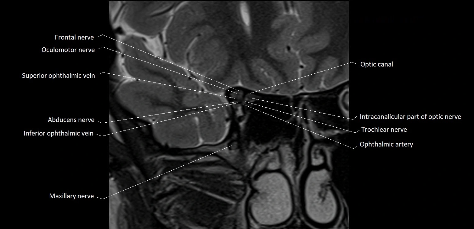
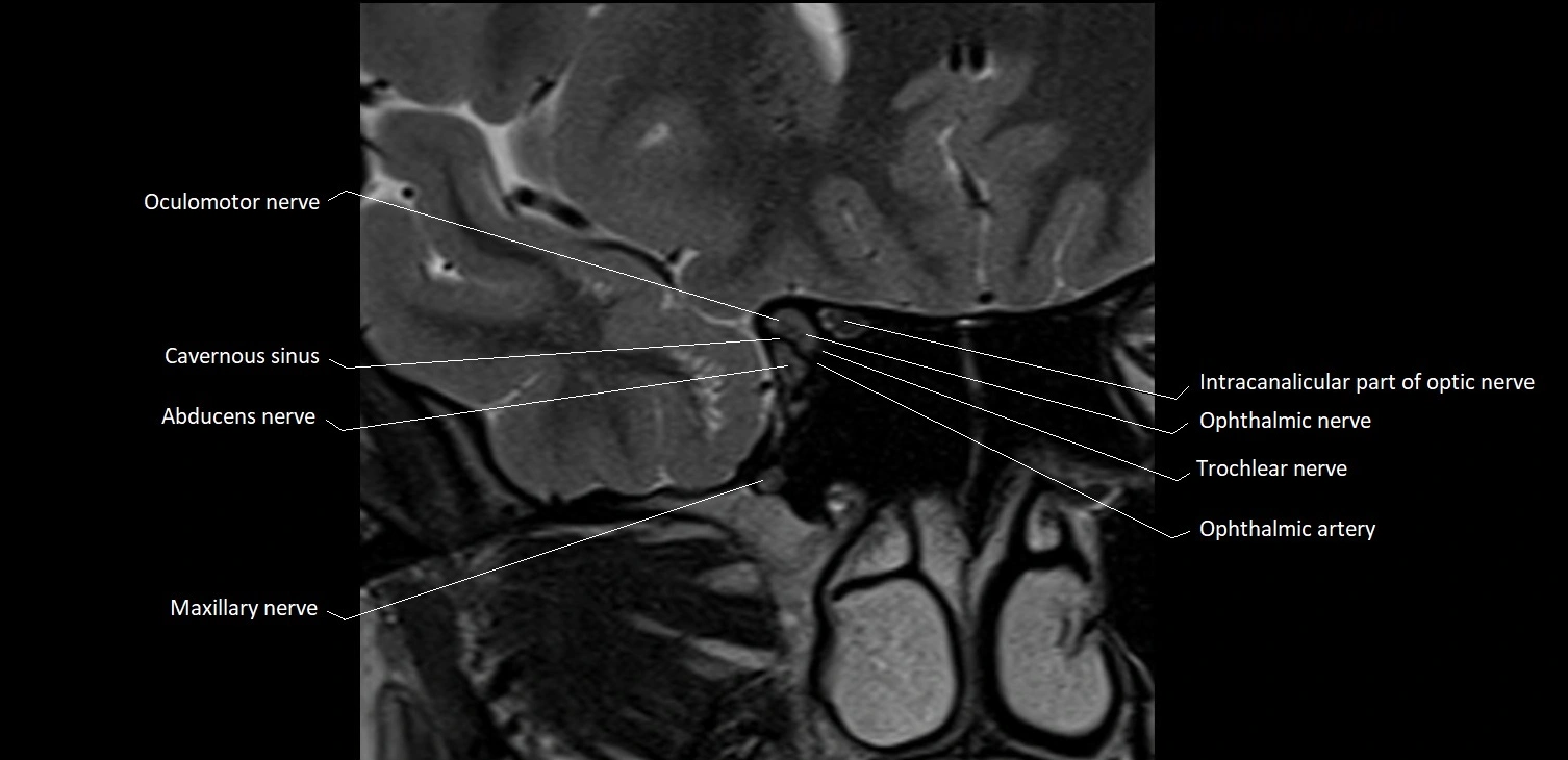
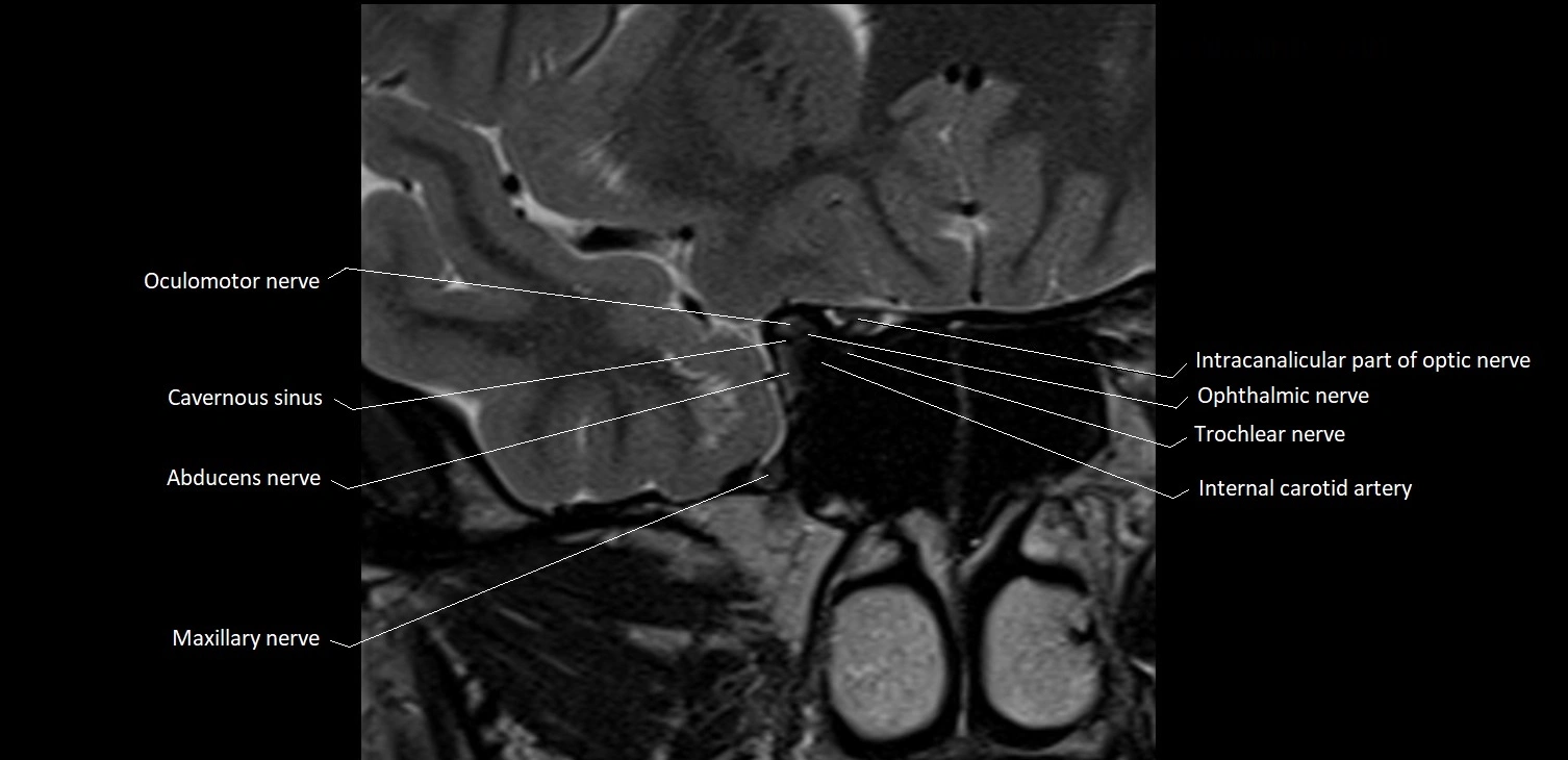
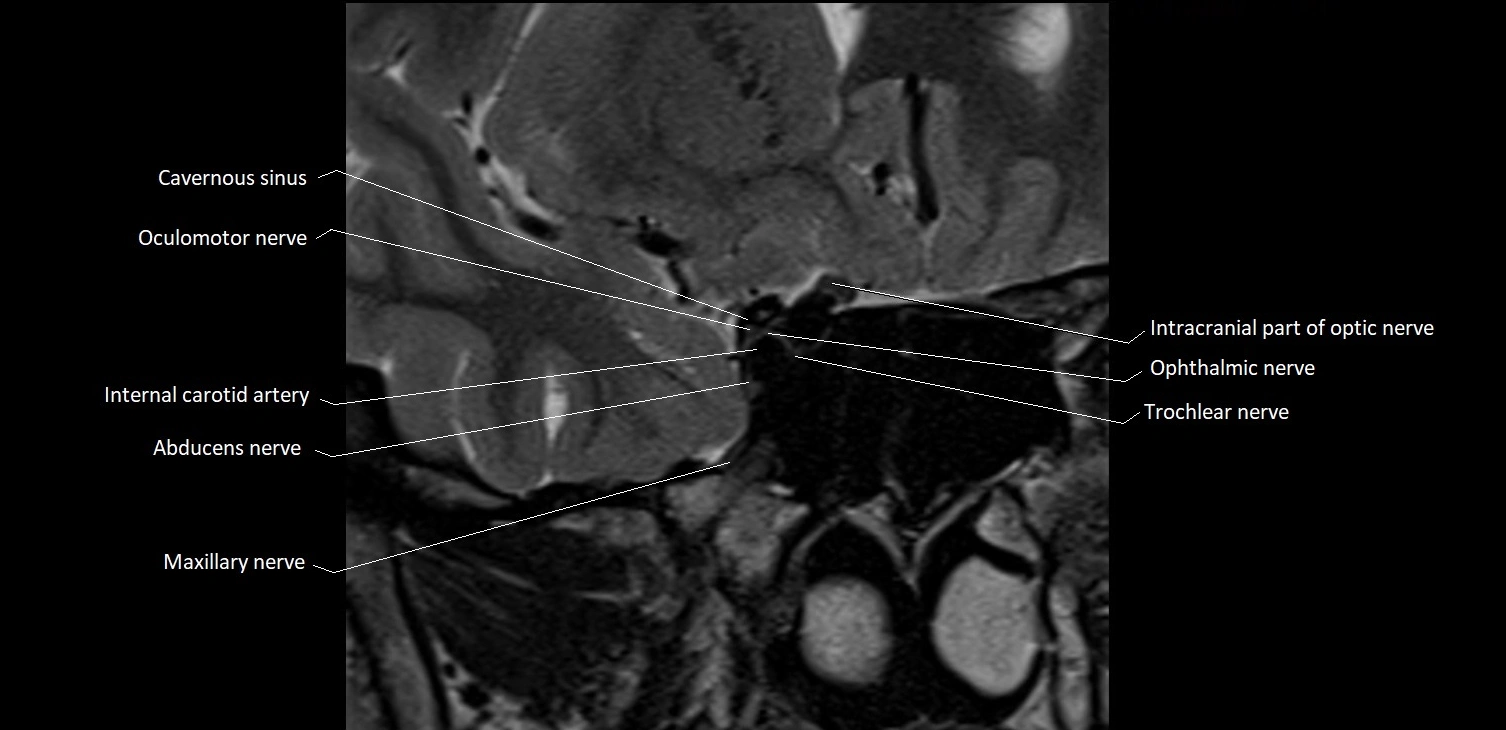
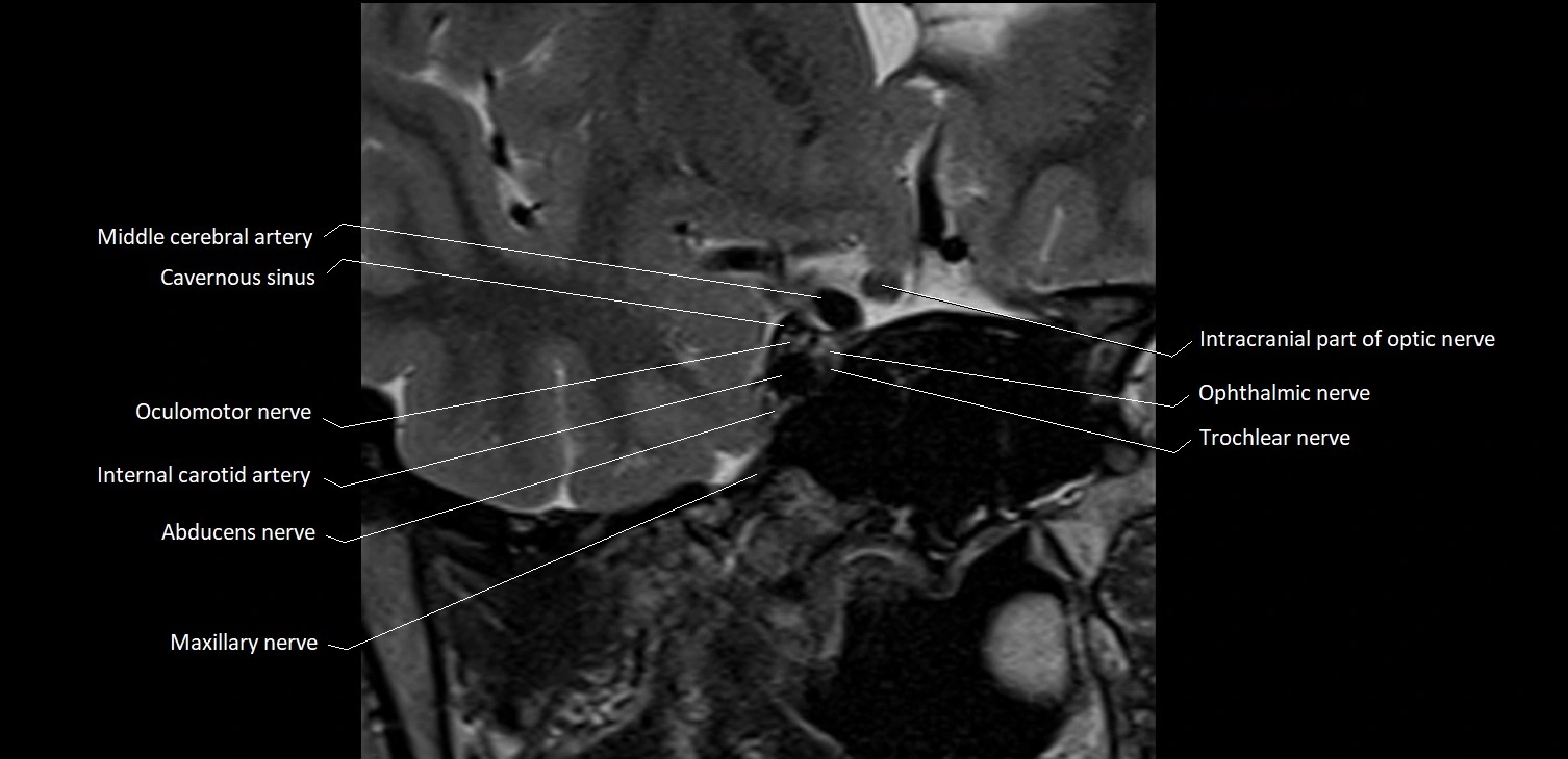
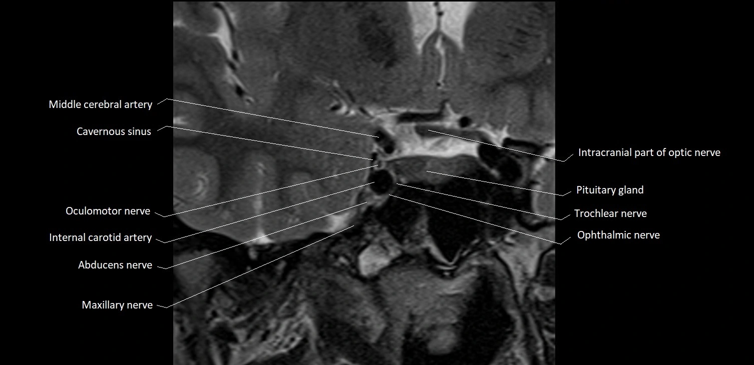
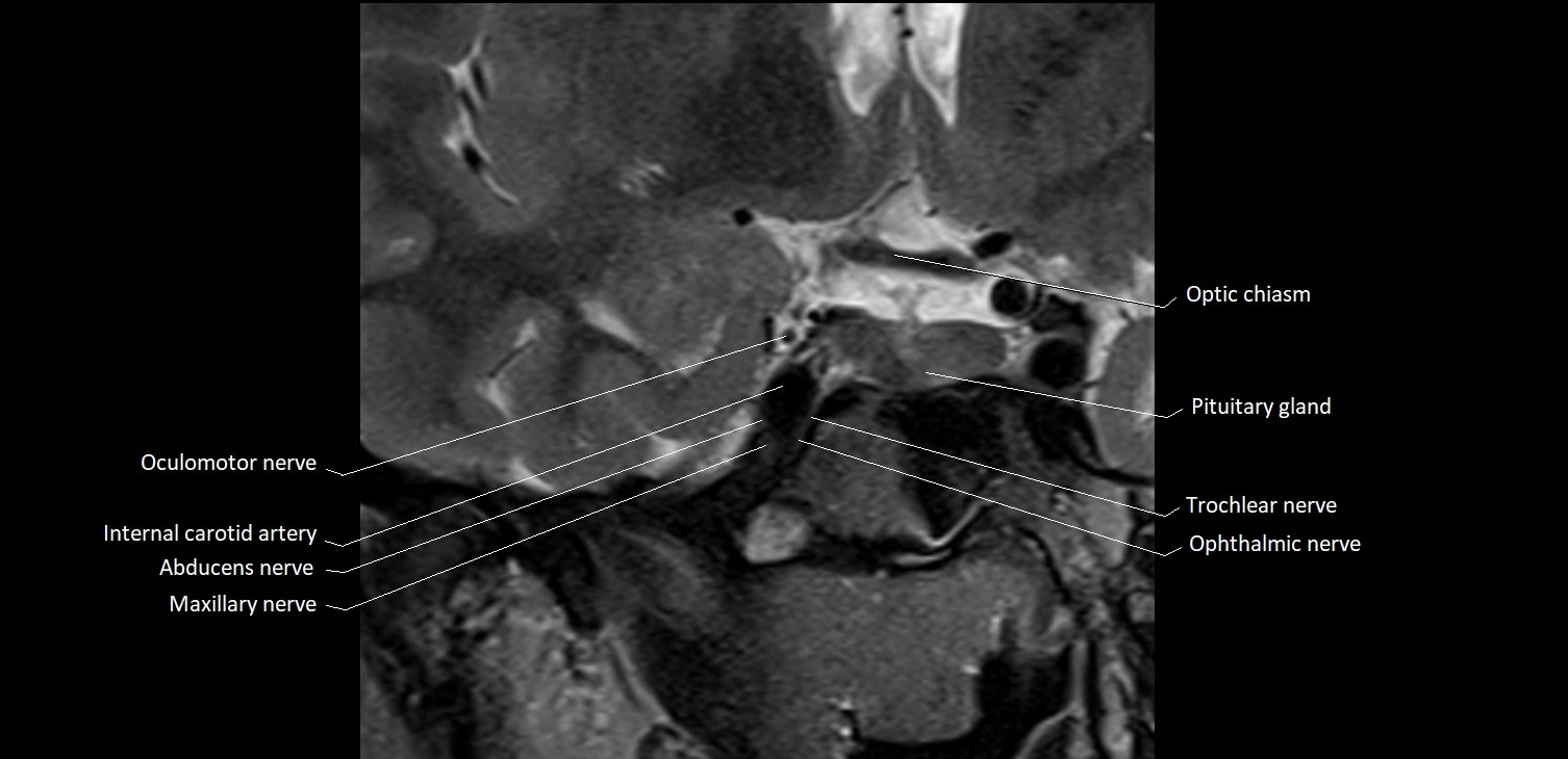
The Abducens nerve (Cranial nerve VI) is a purely motor cranial nerve responsible for innervating the lateral rectus muscle of the eye, which is crucial for lateral movement (abduction) of the eyeball. It arises from the abducens nucleus in the dorsal pons, emerges at the pontomedullary junction, and travels a long intracranial course before entering the orbit via the superior orbital fissure. Because of its long path and proximity to the clivus, it is particularly susceptible to injury from increased intracranial pressure or trauma.
Synonyms
-
Sixth cranial nerve
-
CN VI
-
N. abducens (Latin)
-
Nervus abducens
Function
-
Innervates the lateral rectus muscle of the eye
-
Responsible for abduction of the eyeball (moving the eye outward, away from the midline)
-
Is a purely motor nerve (no sensory or autonomic fibers)
-
Lesion results in inability to abduct the affected eye, leading to horizontal diplopia (double vision)
MRI Appearance
-
The abducens nerve is a small, thin, linear structure
-
Best visualized on high-resolution T2-weighted 3D MRI sequences (e.g., FIESTA or CISS)
-
Seen as a hypointense (dark) line running from the brainstem at the pontomedullary junction, traversing the prepontine cistern, and entering Dorello’s canal under the petrosphenoidal ligament, then into the cavernous sinus, and finally the orbit
-
May be challenging to visualize in standard MRI due to its small size
-
Pathology may be inferred by absence, displacement, or enhancement of the nerve
CT Appearance
-
The nerve itself is not directly visualized on conventional CT due to its small size and soft tissue density
-
Indirect signs: assessment of the bony course, such as the Dorello’s canal, superior orbital fissure, or adjacent pathologies (fractures, masses, or inflammation) that could impinge the nerve
-
CT is mainly used to exclude structural lesions or fractures that might affect the course of CN VI
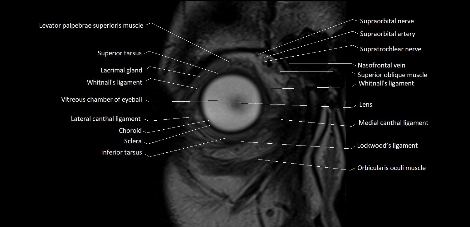
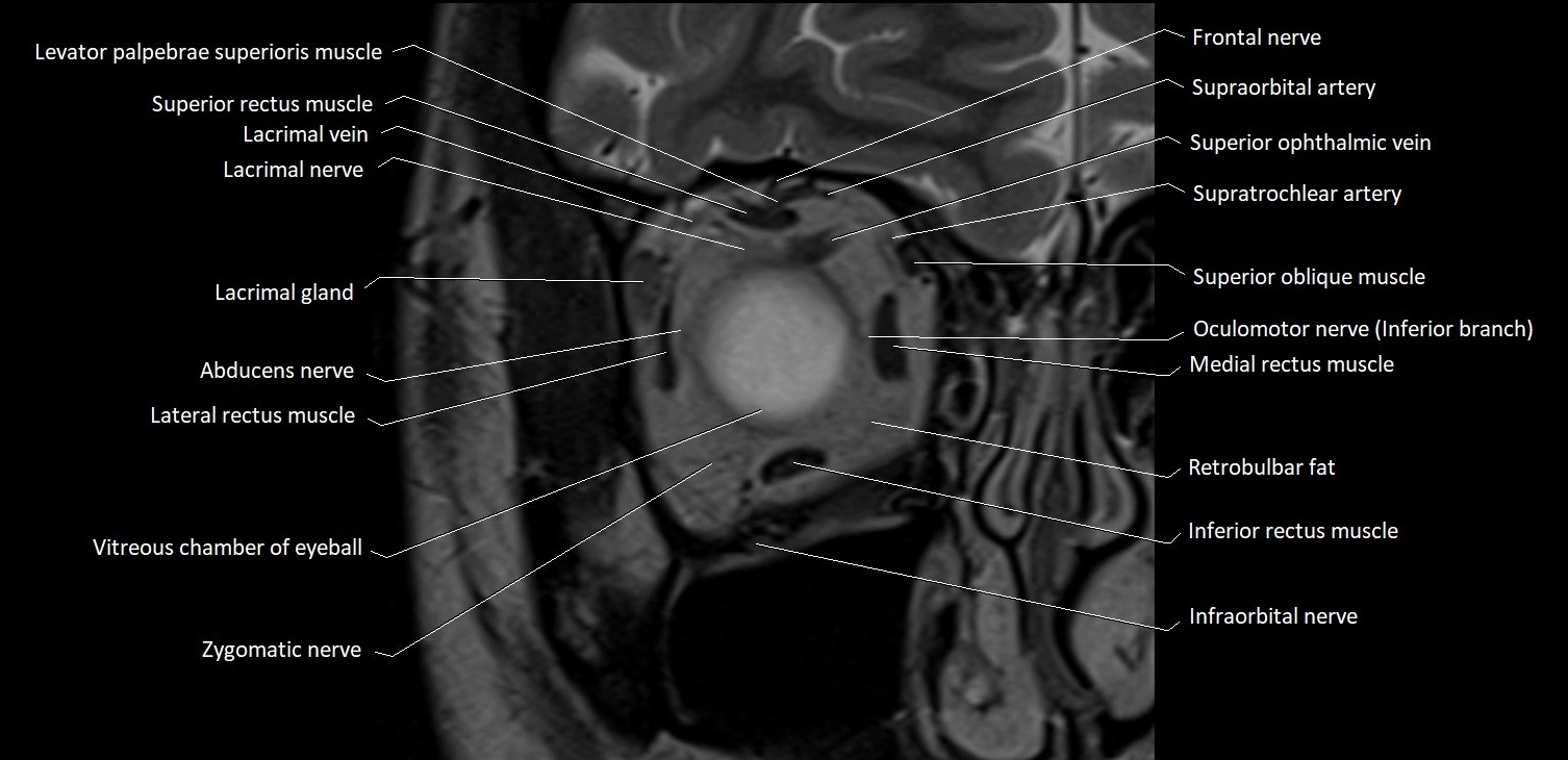
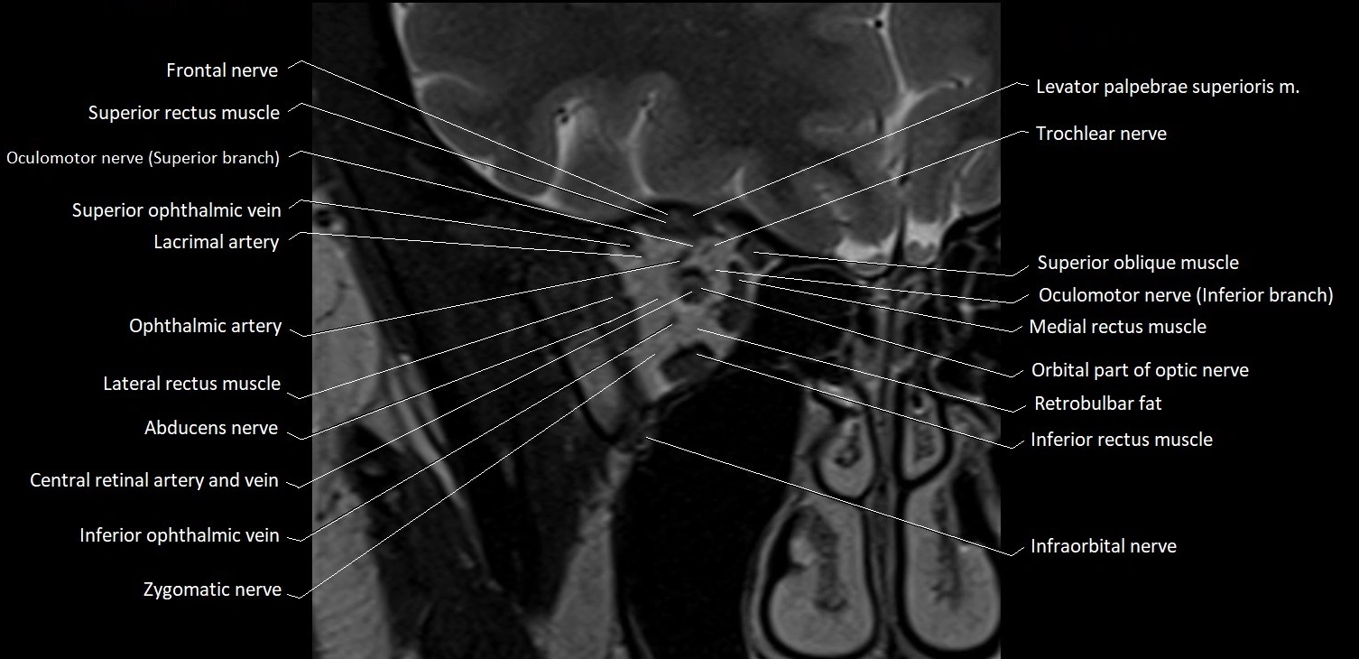
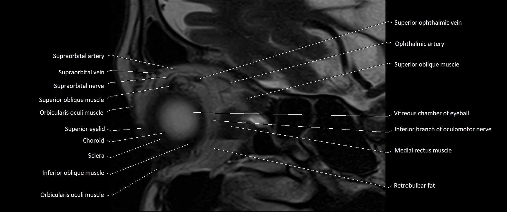
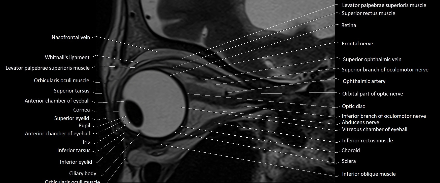
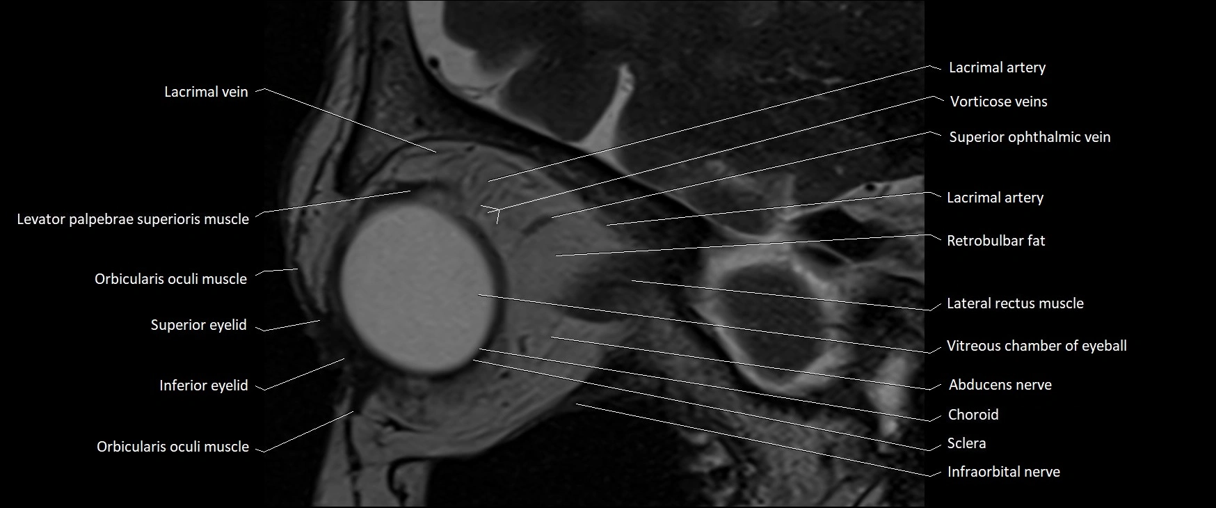
MRI images

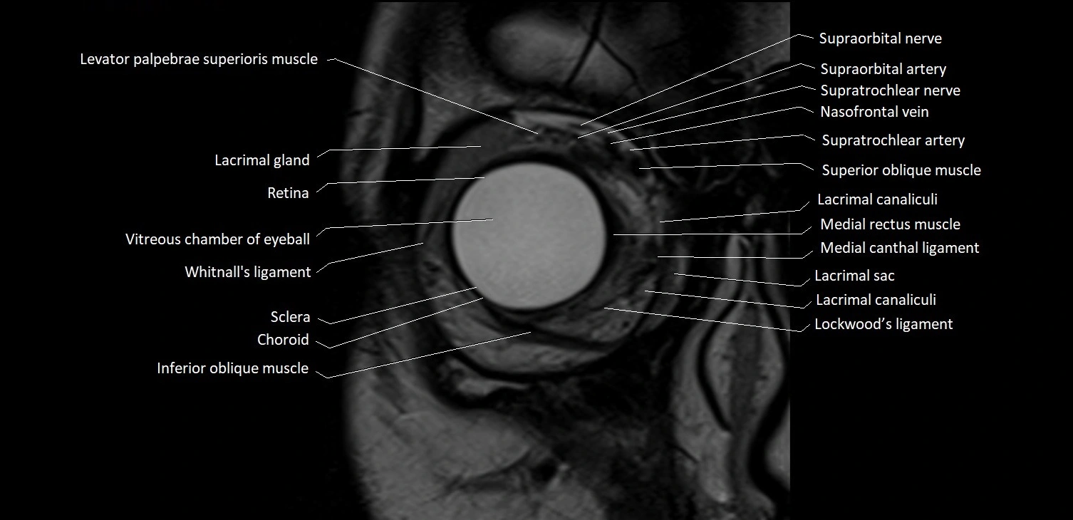
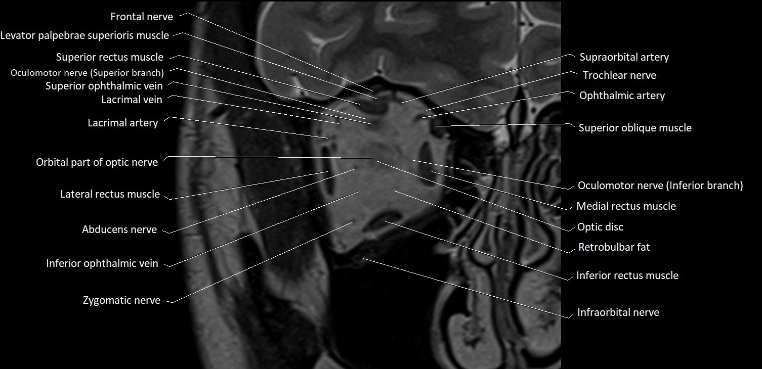
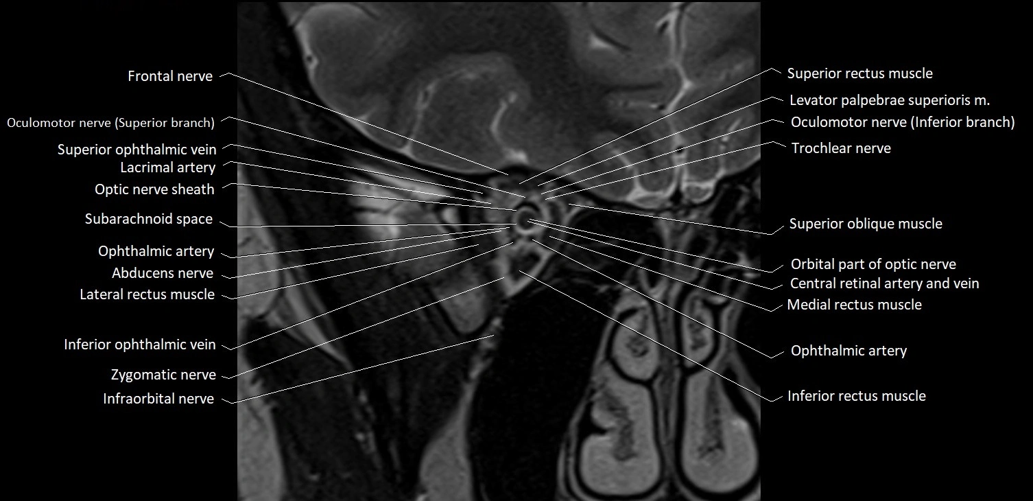
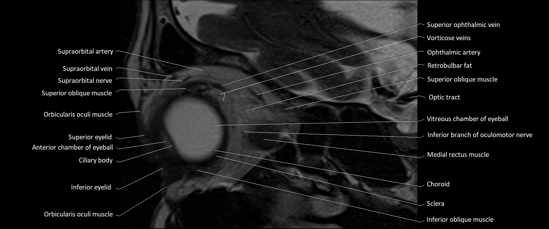
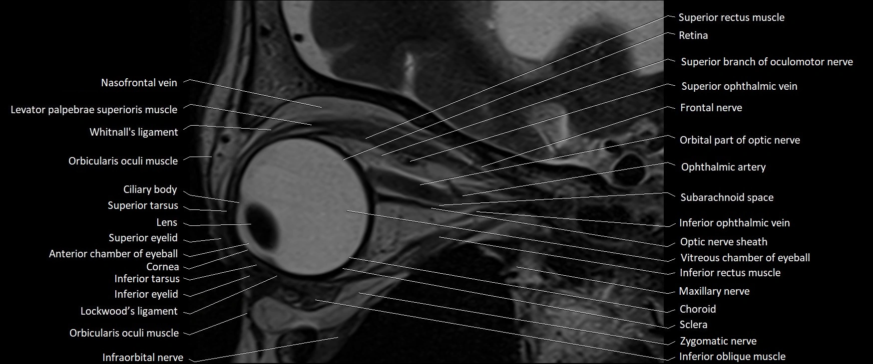
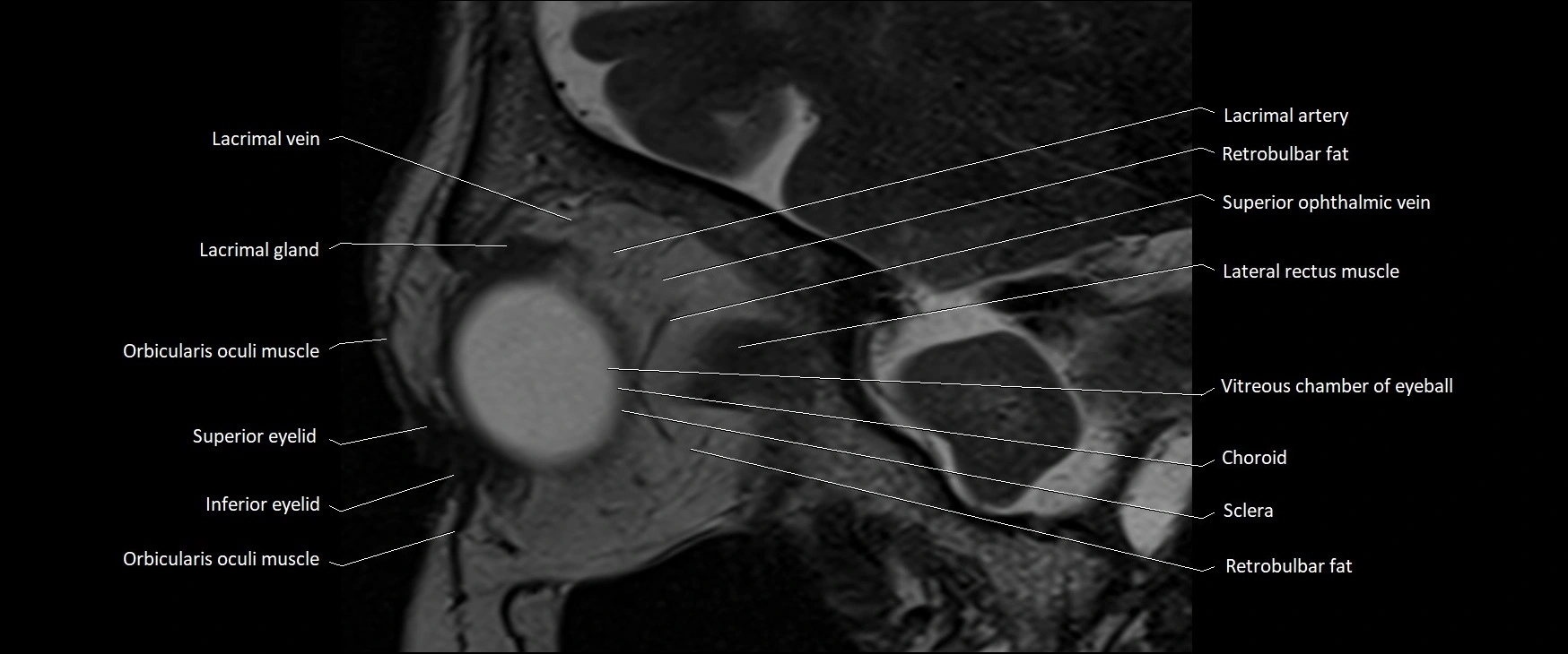
MRI images

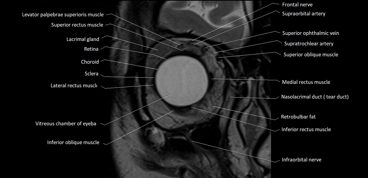
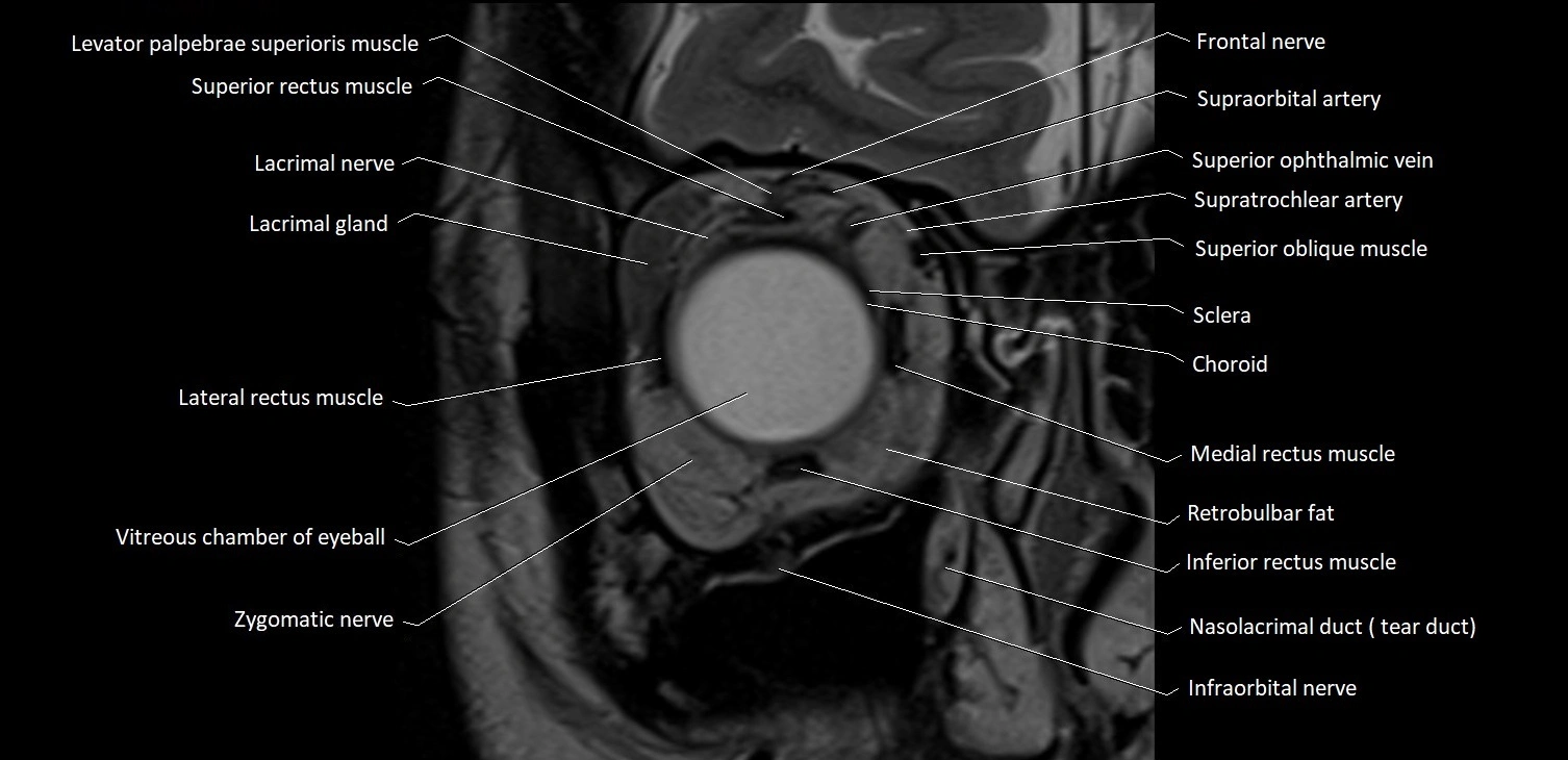
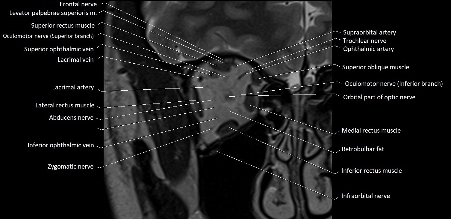
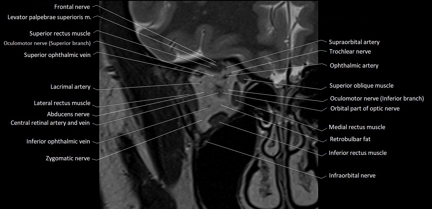
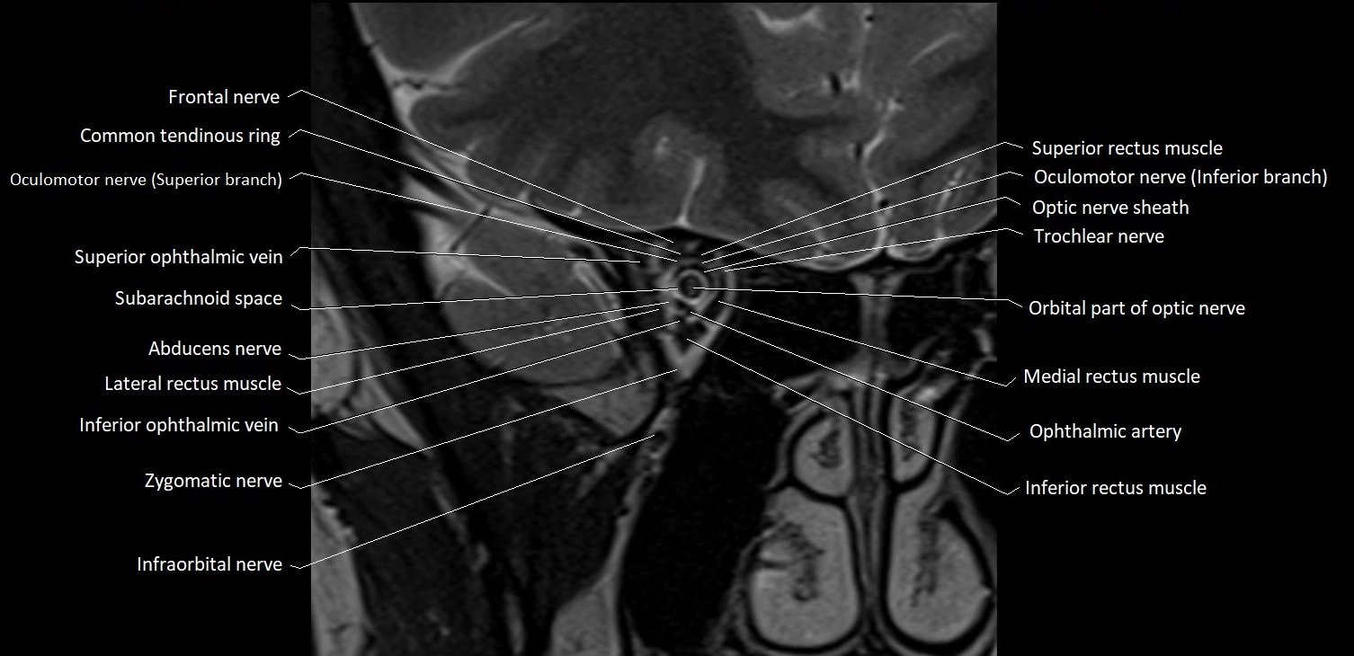
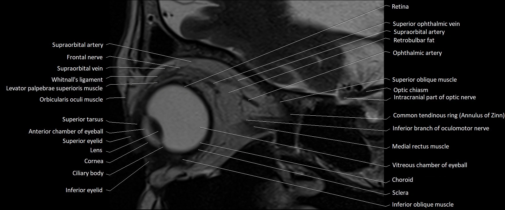
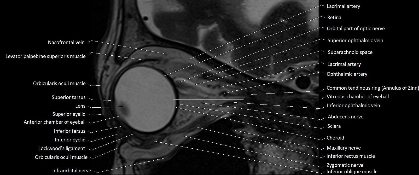
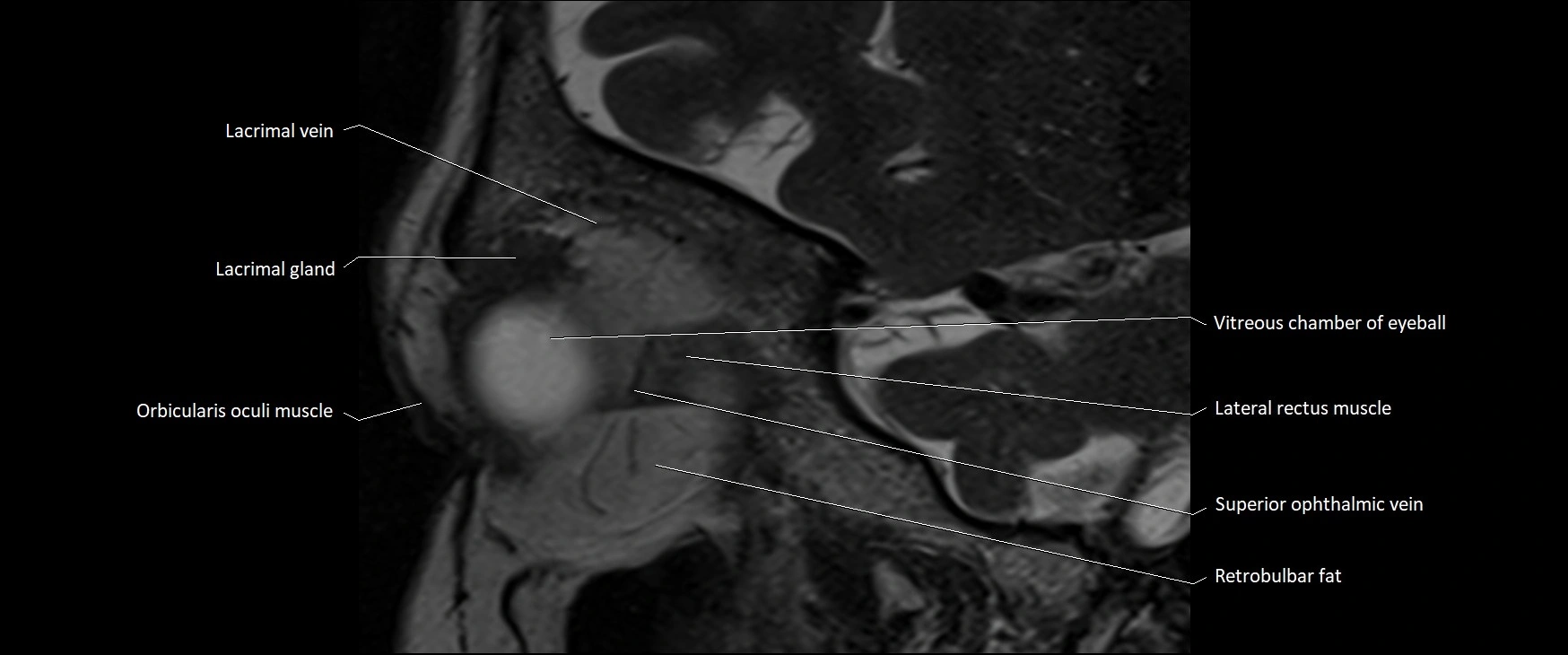
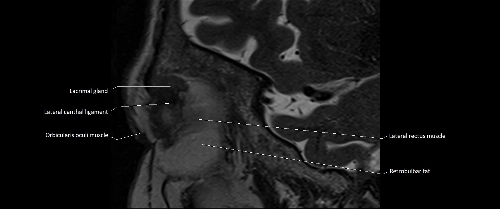
MRI images


