


Topic
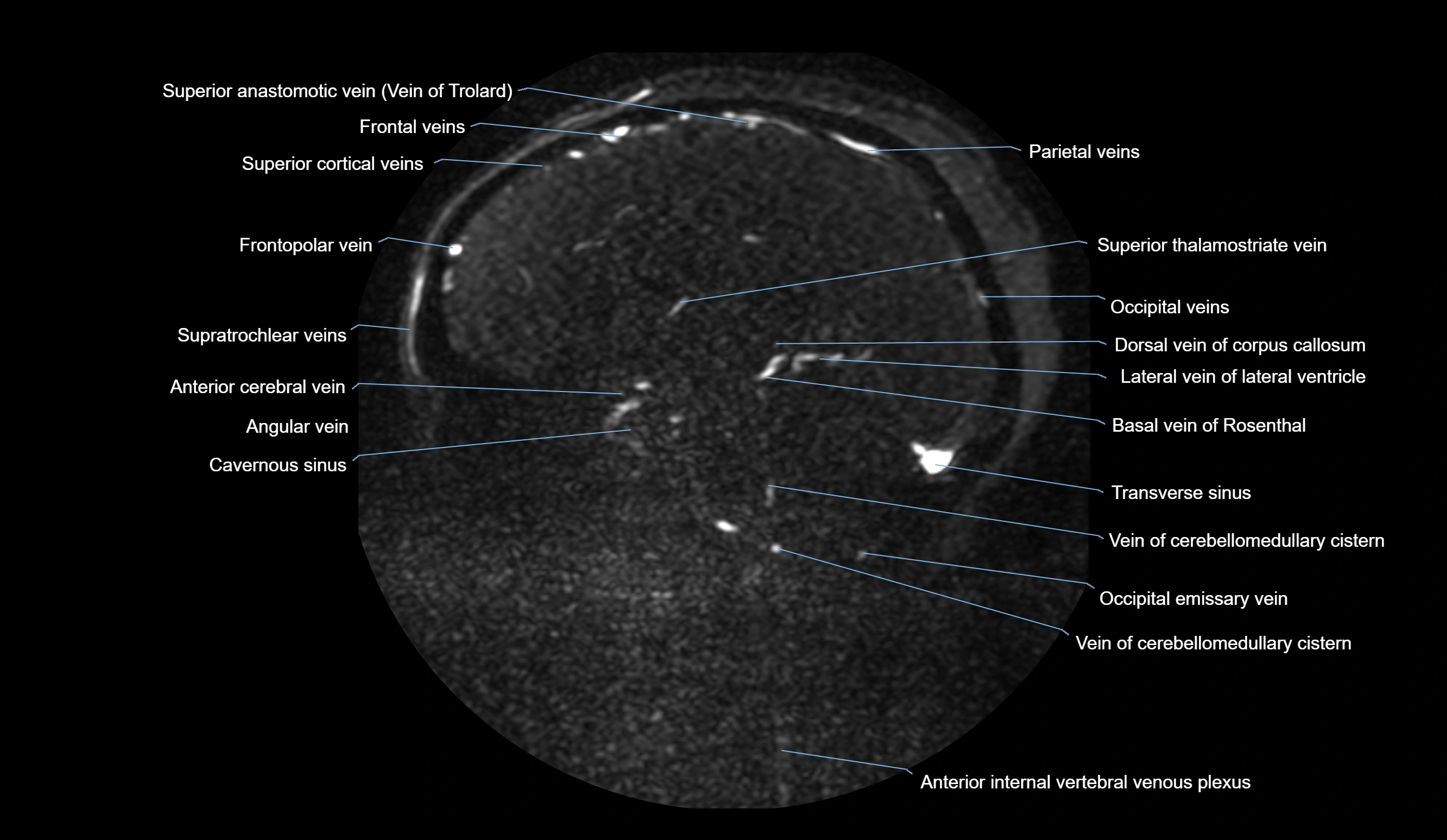
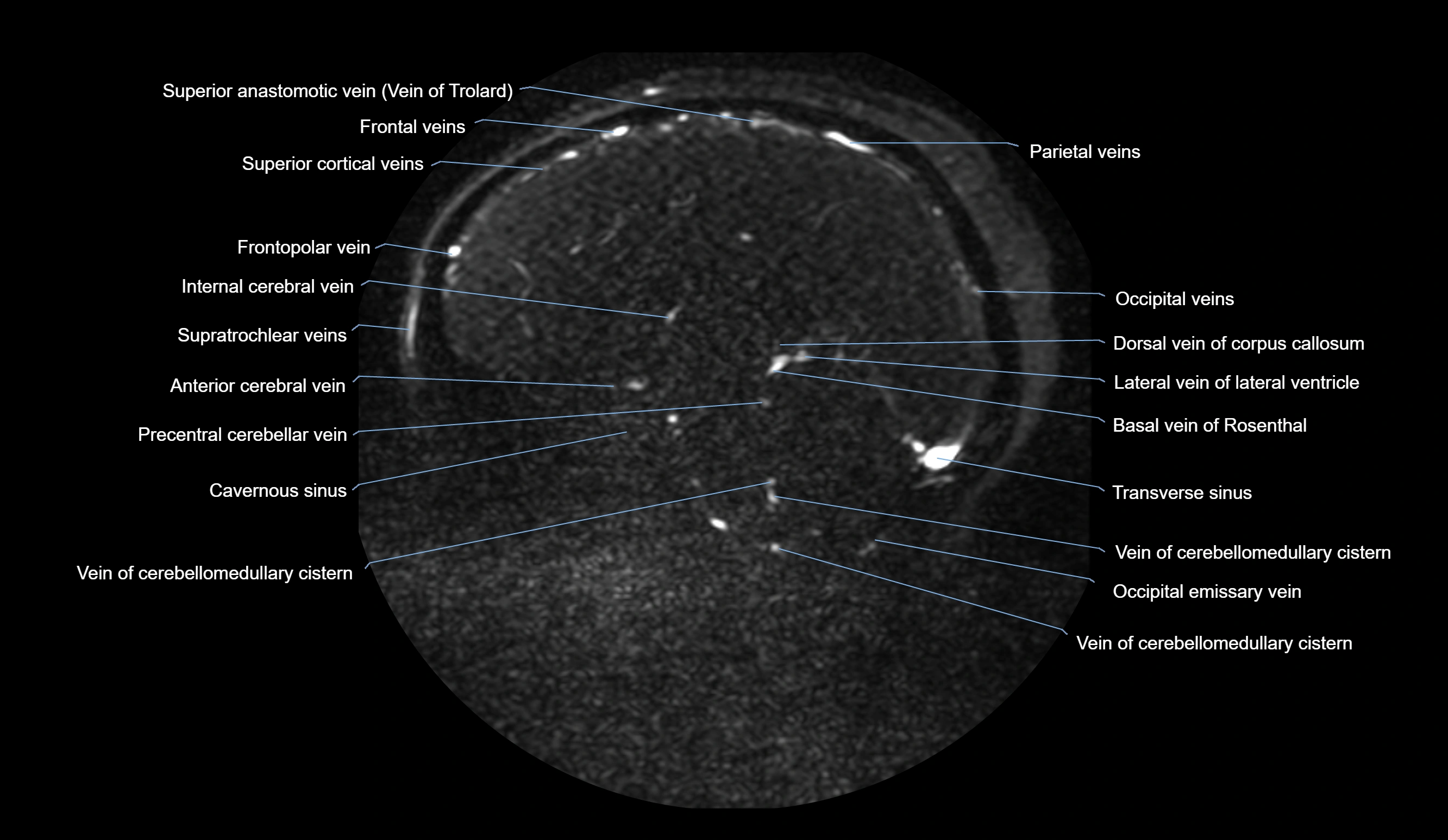
The angular vein is the superior continuation of the facial vein, formed at the medial angle of the eye by the union of the supratrochlear and supraorbital veins. It courses downward along the side of the nose to become the facial vein. Importantly, it provides a venous connection between the facial vein and the superior ophthalmic vein, thereby linking extracranial venous drainage with the cavernous sinus. This anatomical communication makes the angular vein a potential pathway for the spread of infection from the face to intracranial venous sinuses.
Synonyms
-
Medial facial vein
-
Ophthalmofacial venous connection
-
Supraorbital-supratrochlear venous trunk
Function
-
Drains venous blood from the forehead, medial canthus, nose, and upper face
-
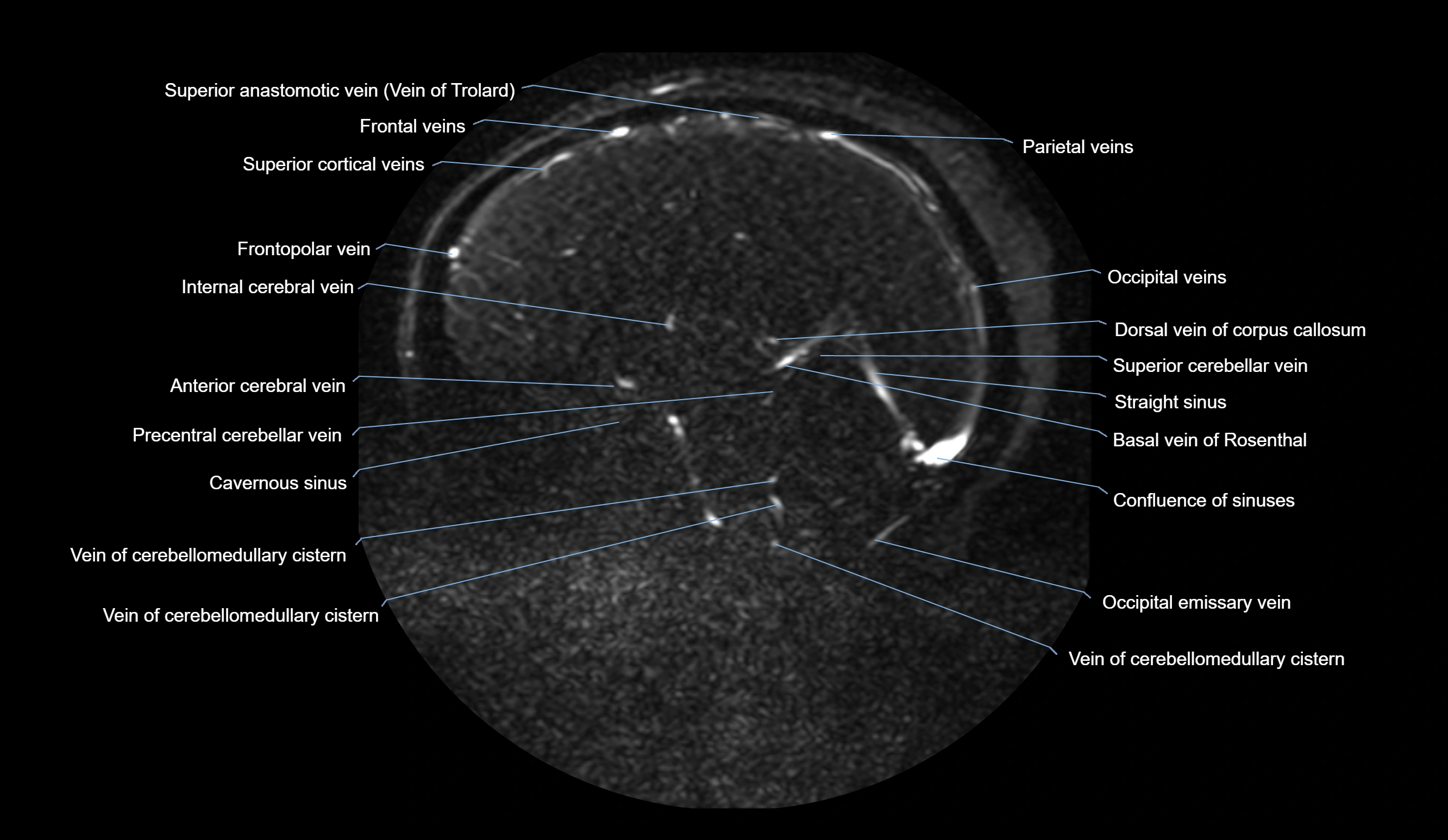
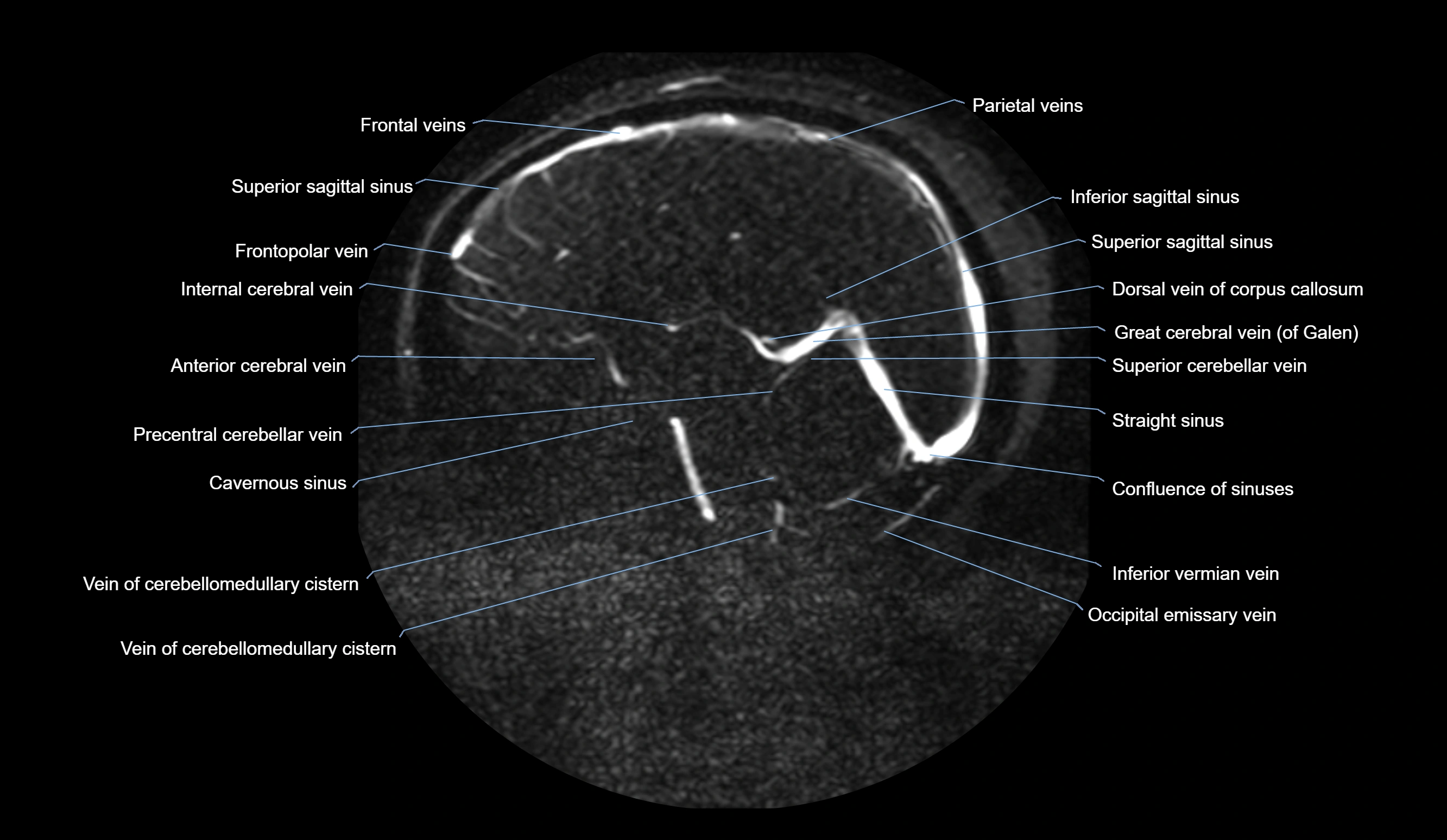
Serves as a key communication between extracranial and intracranial venous systems
-
Connects the facial vein to the superior ophthalmic vein and cavernous sinus
-
Provides collateral venous outflow in facial and orbital circulation
MRI Appearance
T1-weighted images (non-contrast):
-
Angular vein appears as a flow void (black signal) along the medial orbital angle and side of the nose
-
Surrounding soft tissue provides contrast, but vessel lumen is not well defined without venography
T2-weighted images:
-
Also demonstrates flow voids due to venous blood flow
-
In thrombosis, the vein may appear hyperintense with loss of normal flow void
MR Venography (MRV):
-
Time-of-flight (TOF) or contrast-enhanced MRV shows the angular vein as a bright enhancing venous channel
-
Clearly demonstrates its continuity with the facial vein and superior ophthalmic vein
-
MRV is highly useful in evaluating thrombosis, venous obstruction, or collateral venous drainage
T1 Post-Contrast (Gadolinium-enhanced MRI):
-
The vein lumen enhances intensely with contrast
-
Helps confirm patency or thrombus, and outlines venous communications to orbital and cavernous sinus pathways
CT Appearance
Non-contrast CT:
-
Angular vein is not directly visualized unless dilated
-
In thrombosis, may appear as a hyperdense venous structure in the medial orbital or nasal region
CT Venography (CTV):
-
Clearly visualizes the angular vein as a contrast-filled venous channel
-
Demonstrates its communication with the facial vein, superior ophthalmic vein, and cavernous sinus
-
Essential for detecting facial vein thrombosis, orbital venous involvement, and cavernous sinus pathology
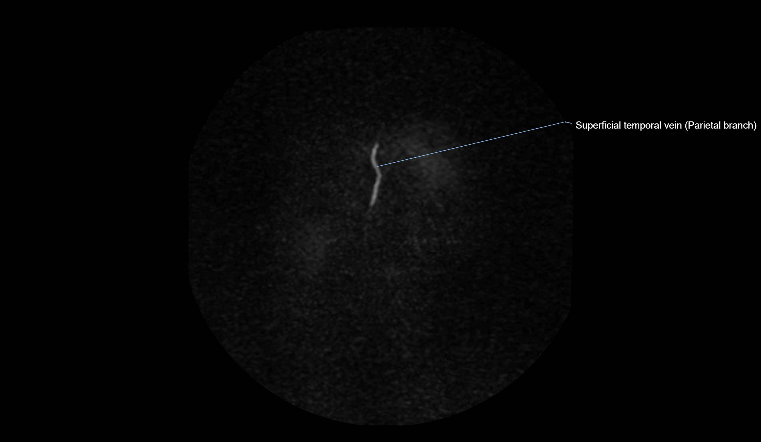
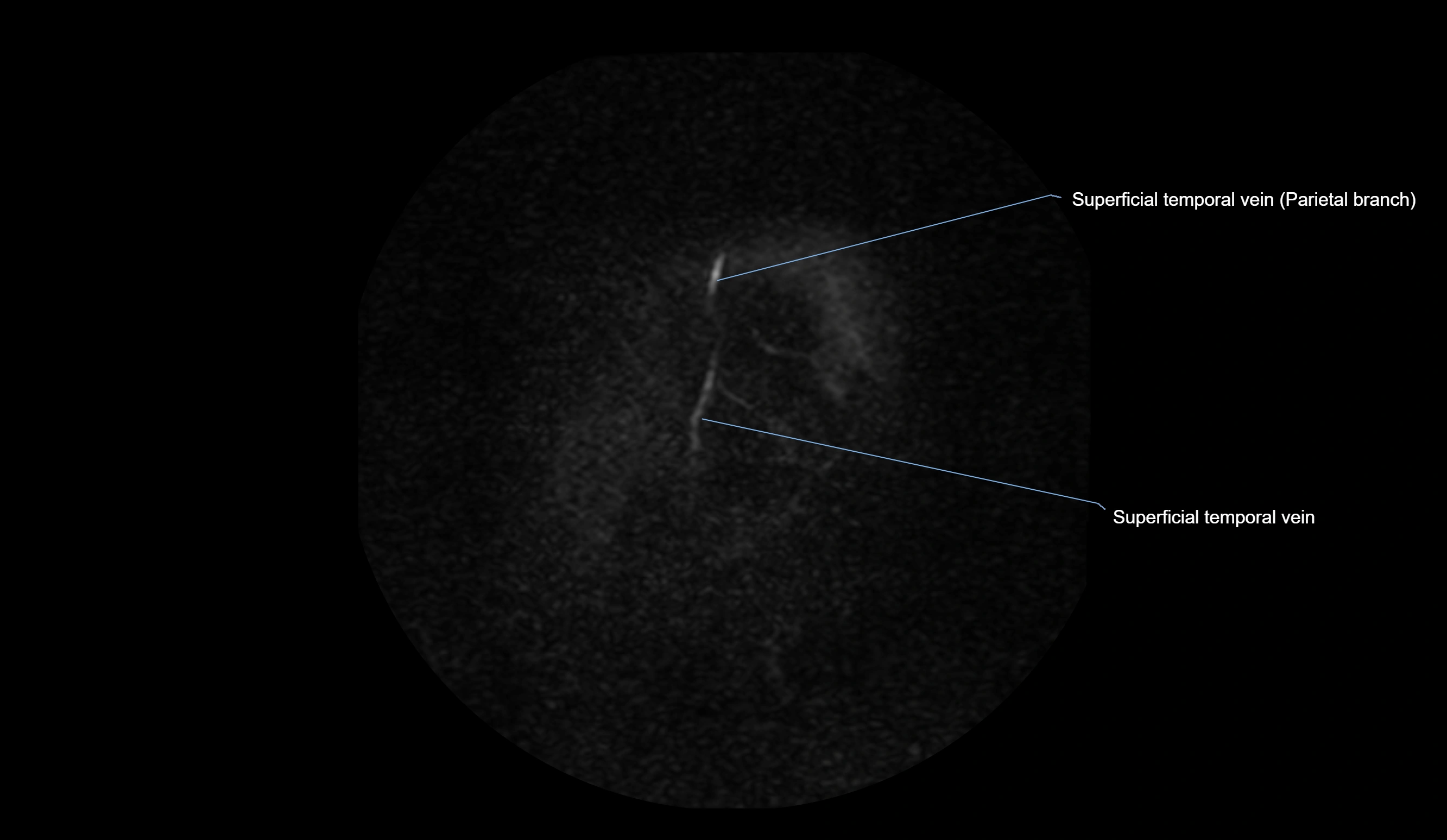
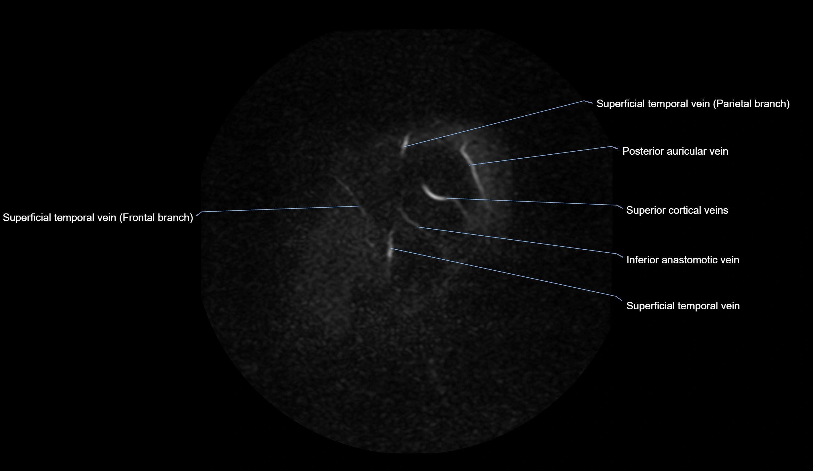
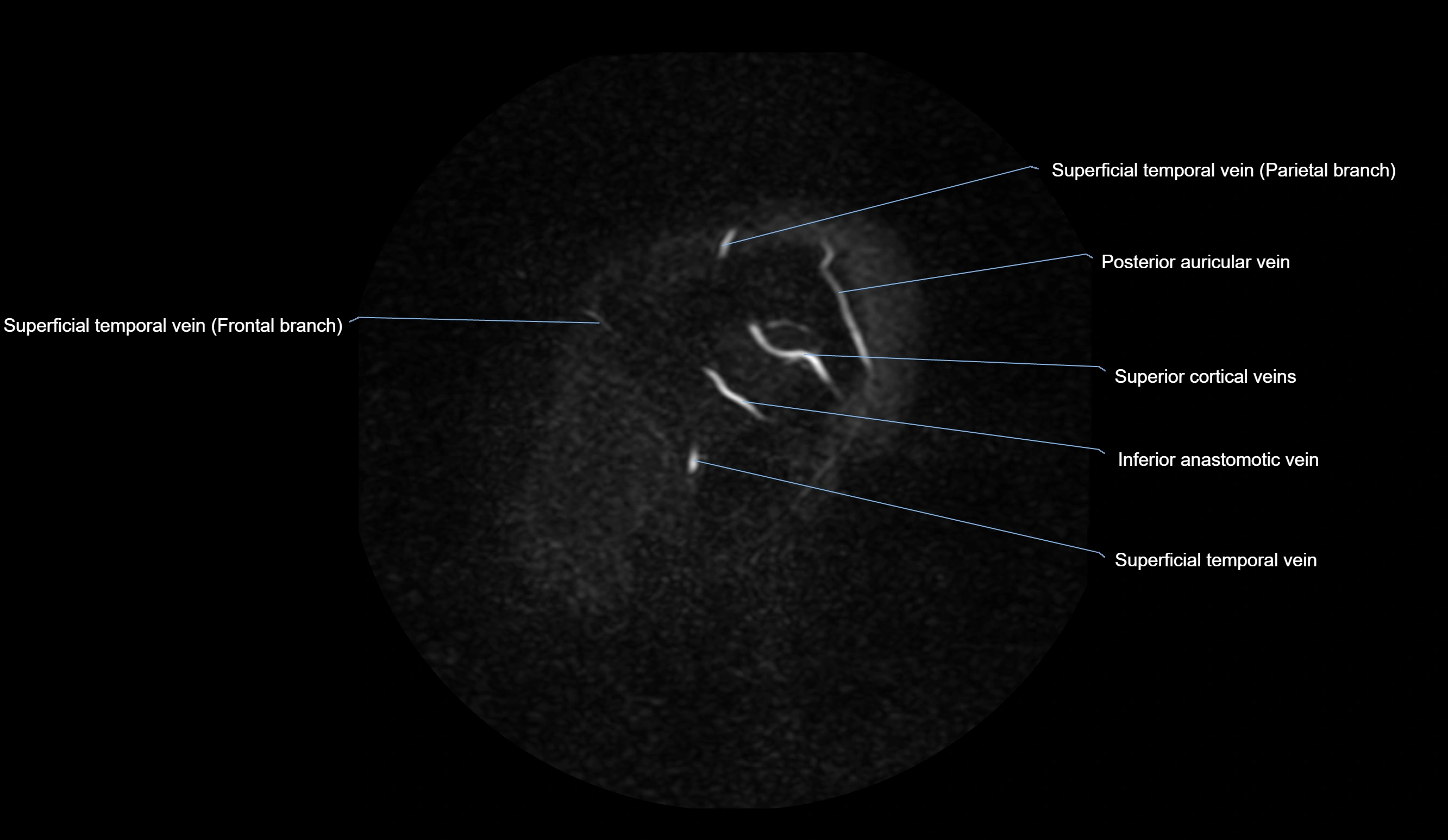
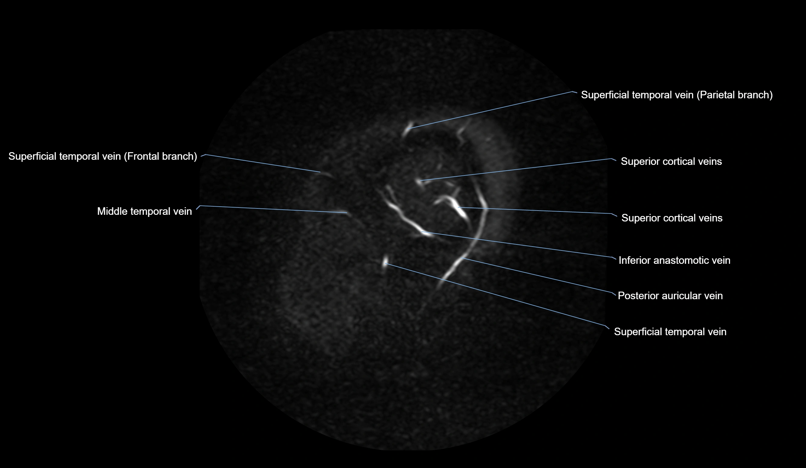
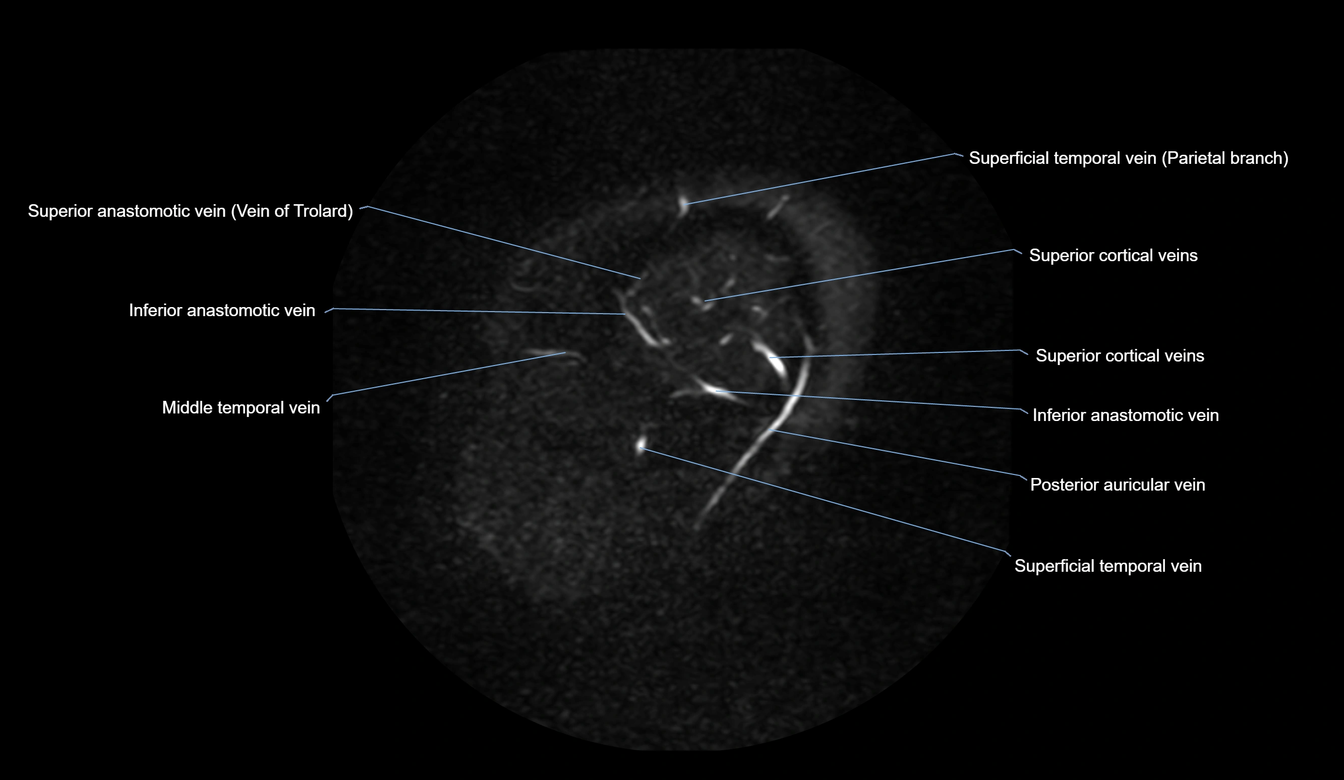
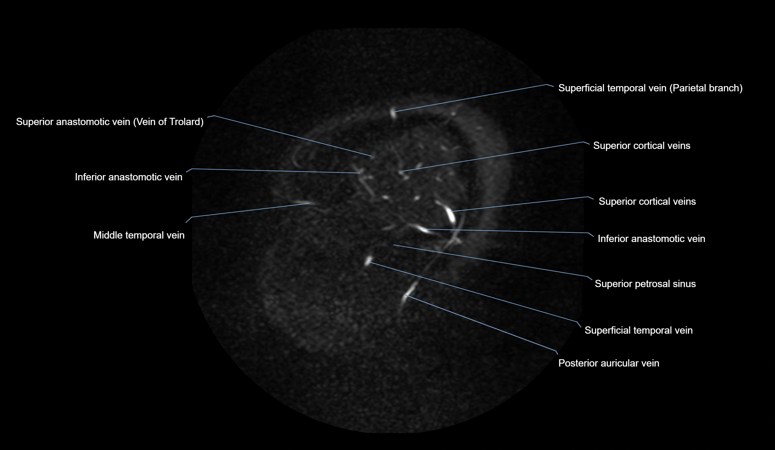
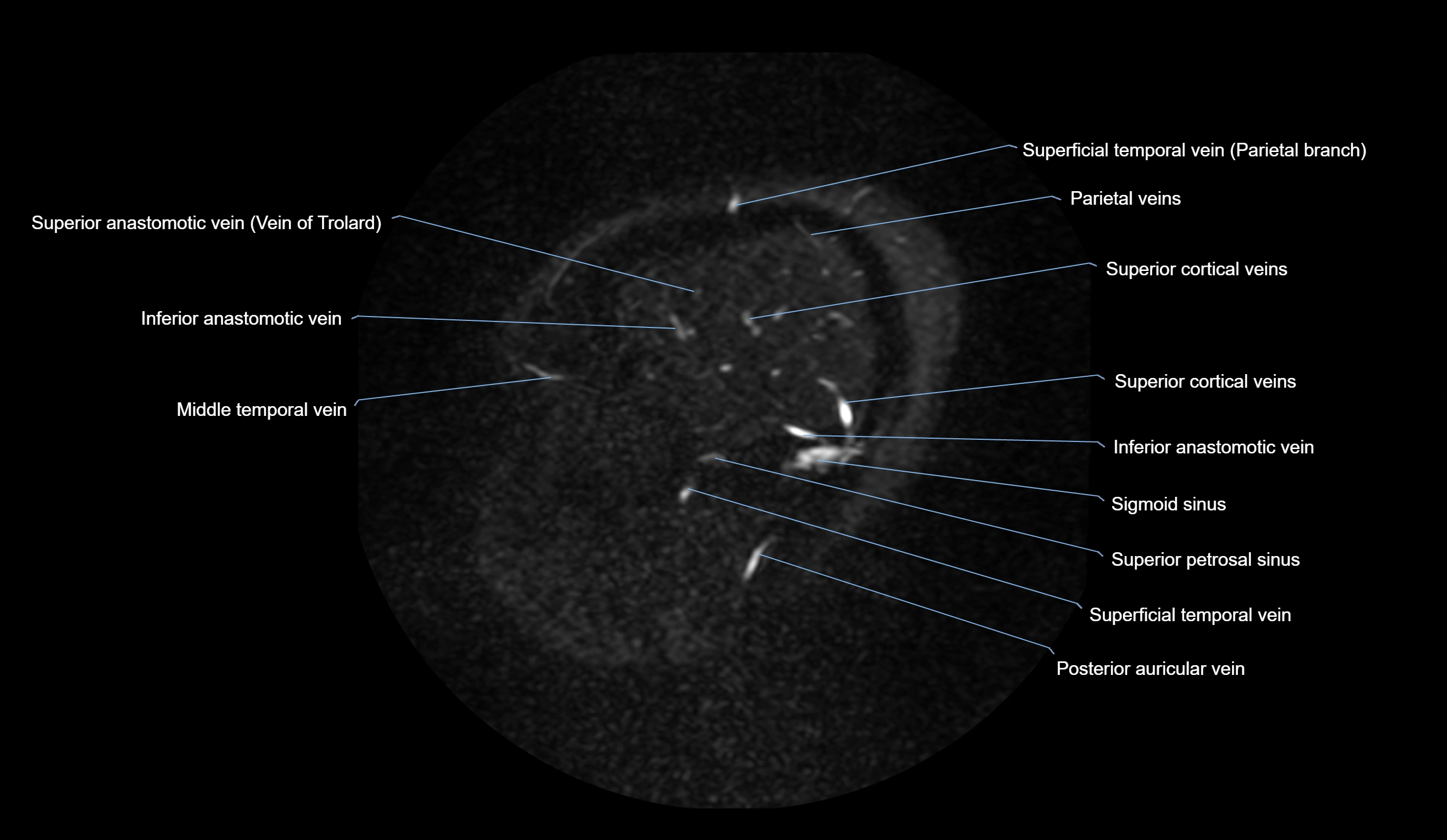
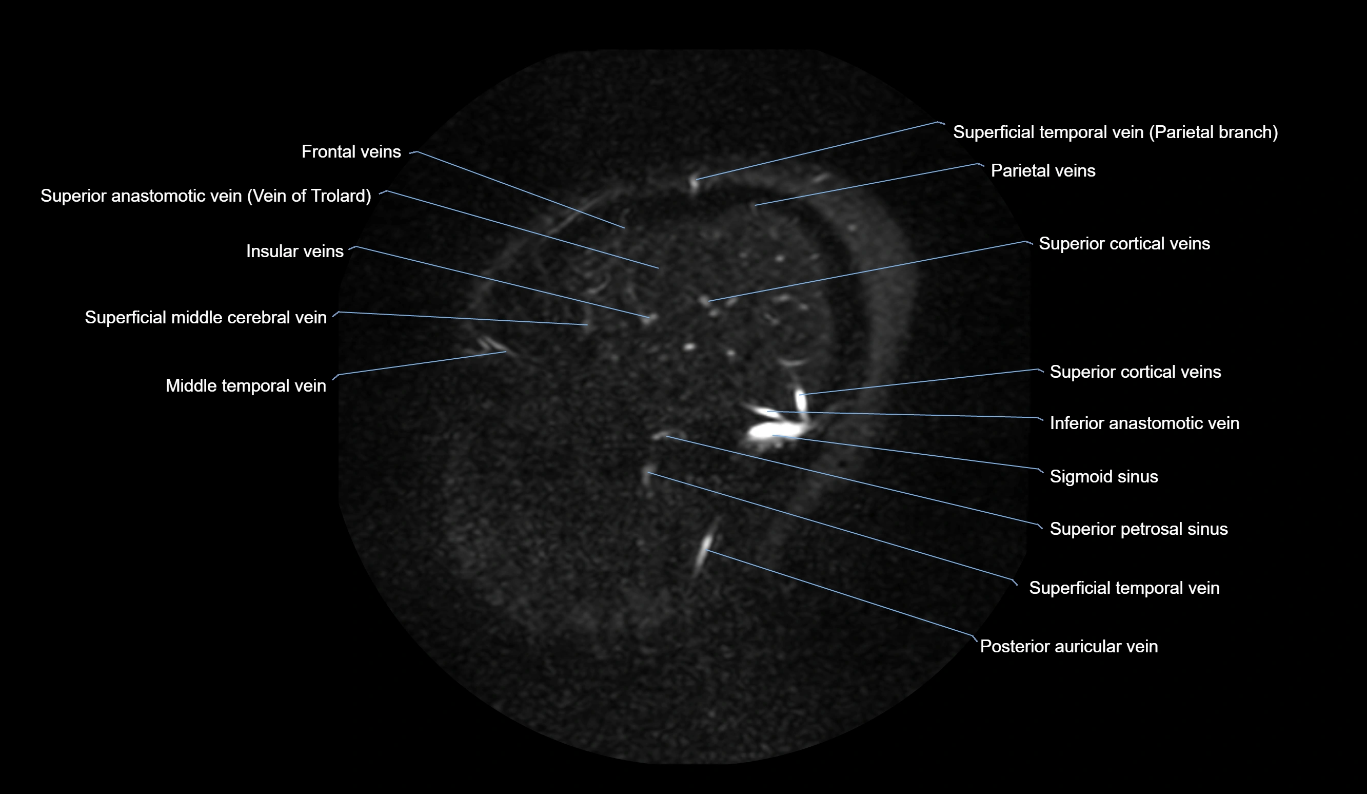
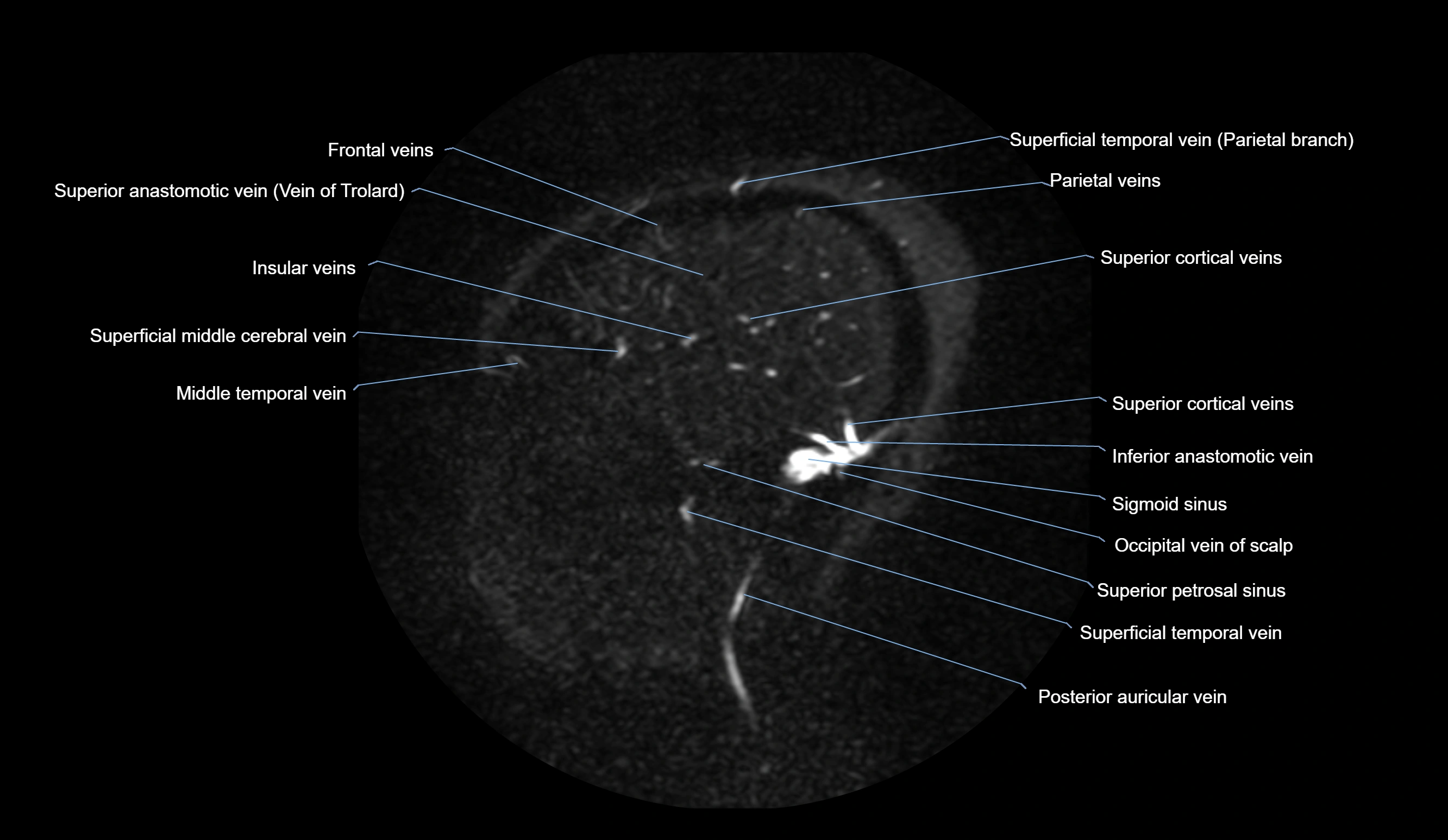
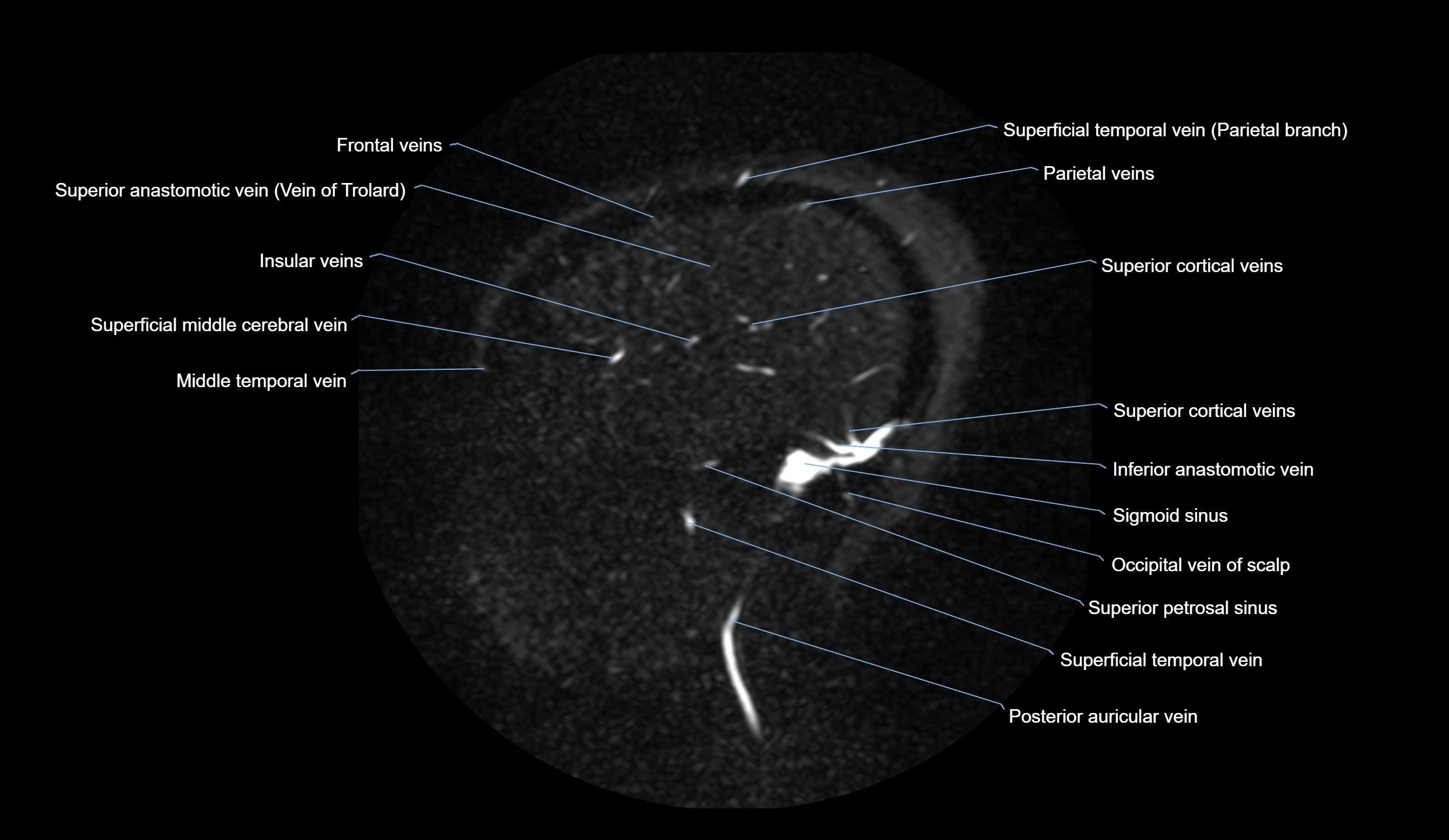
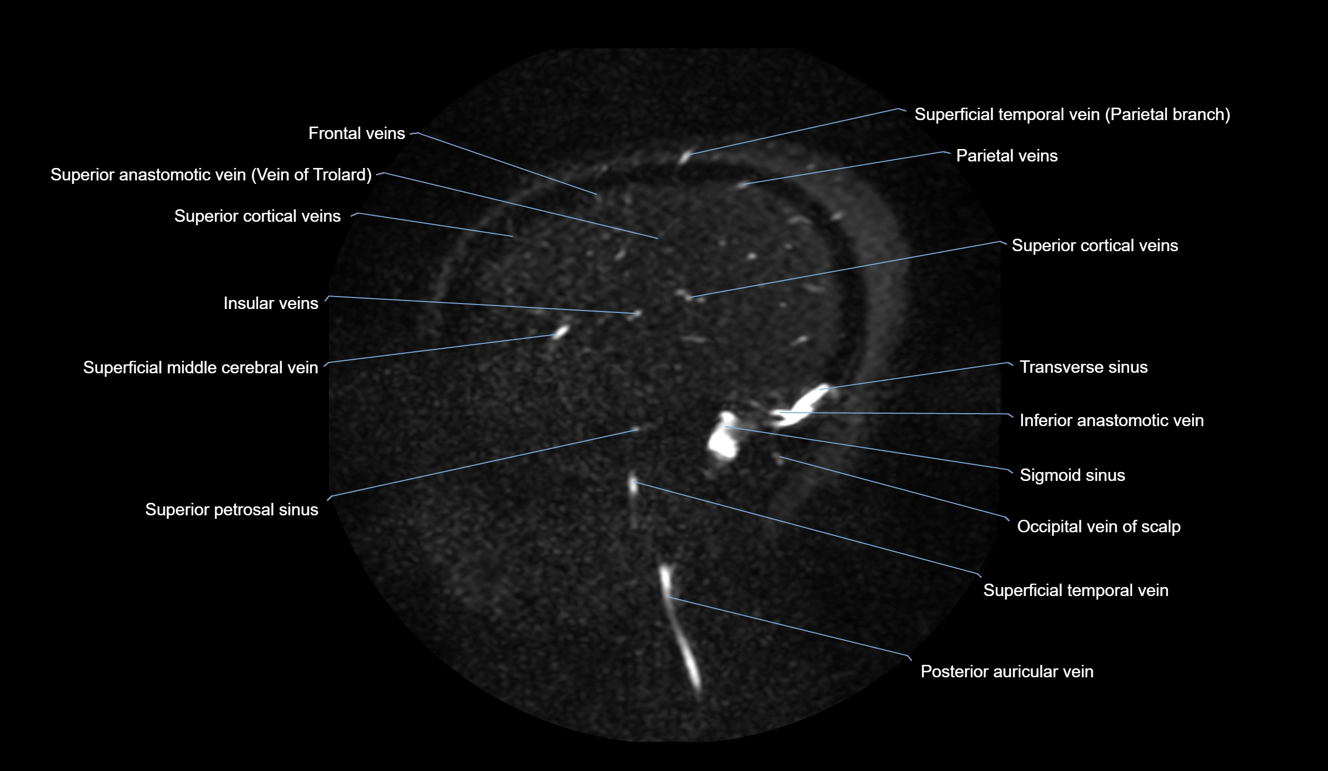
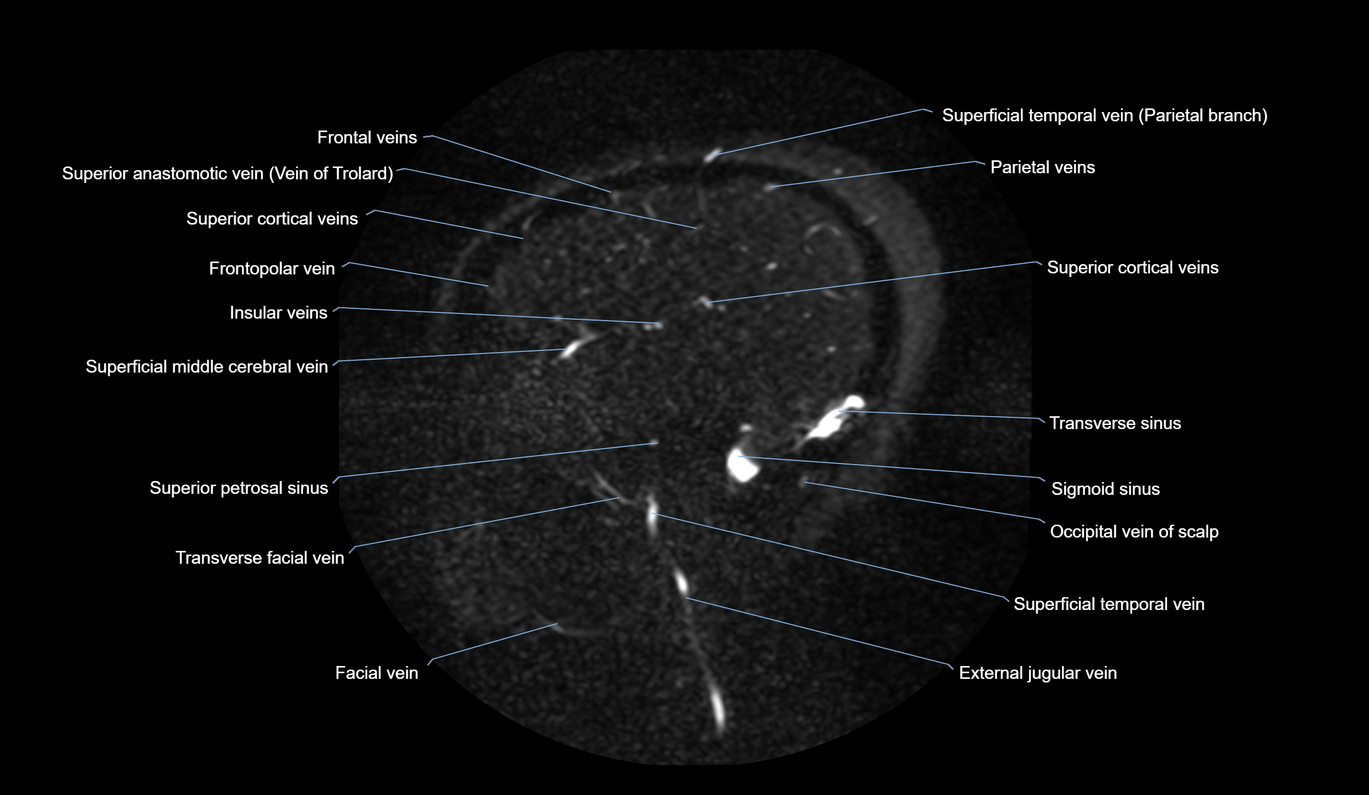
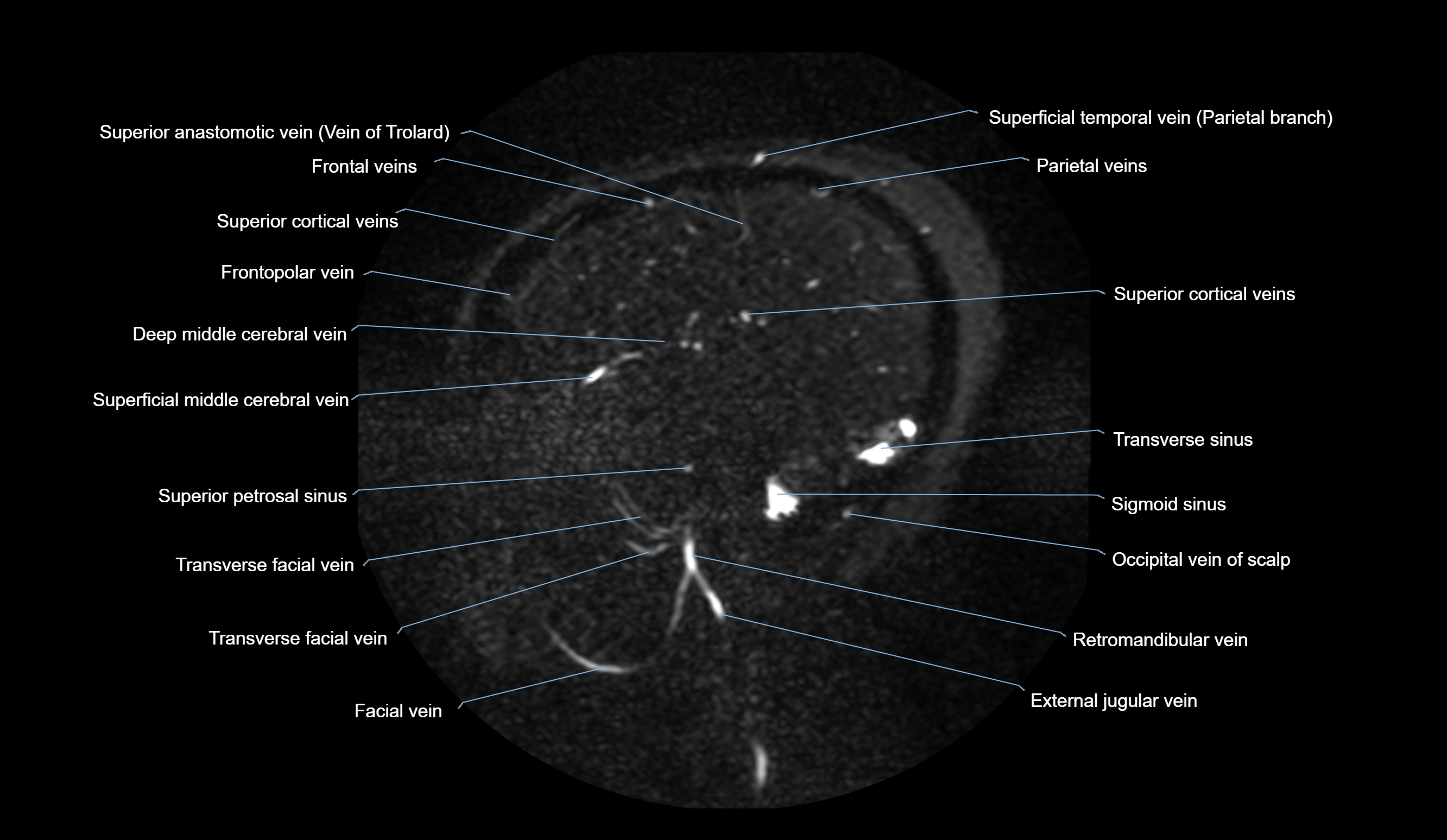
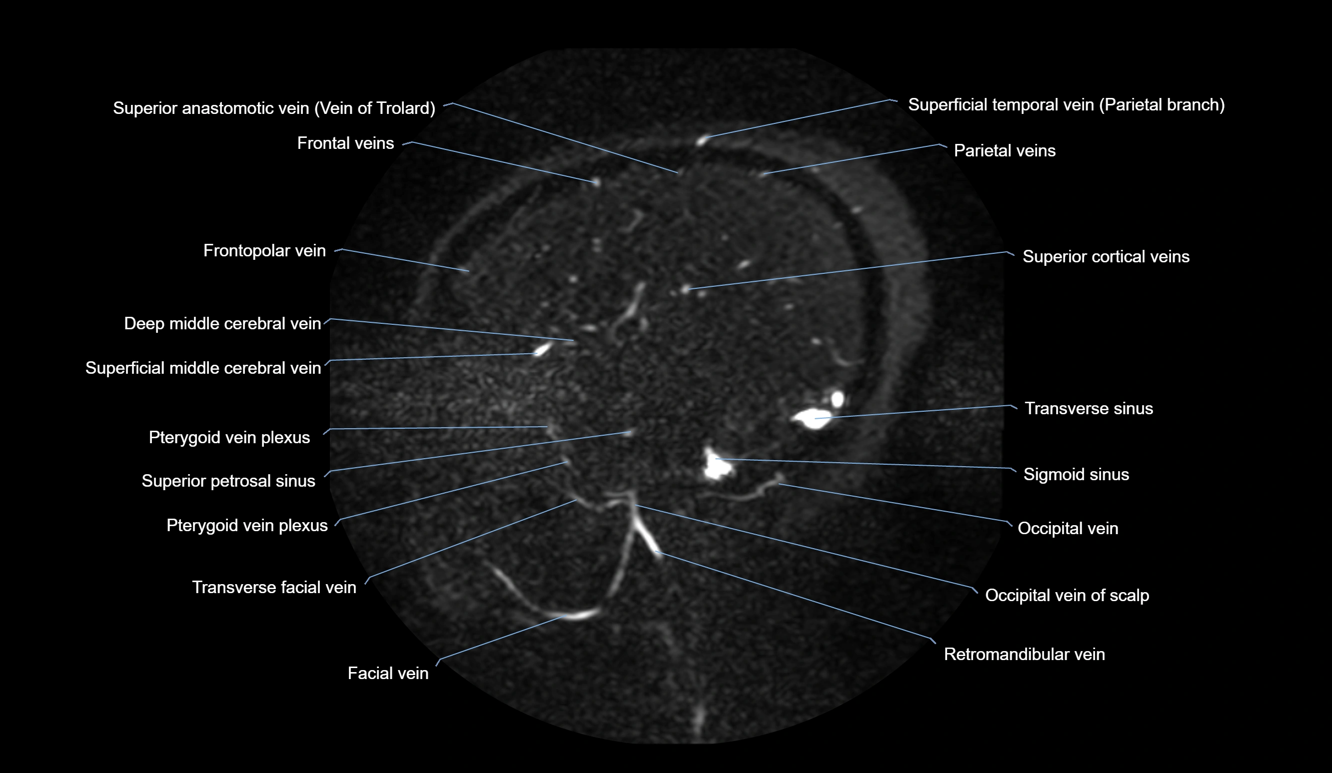
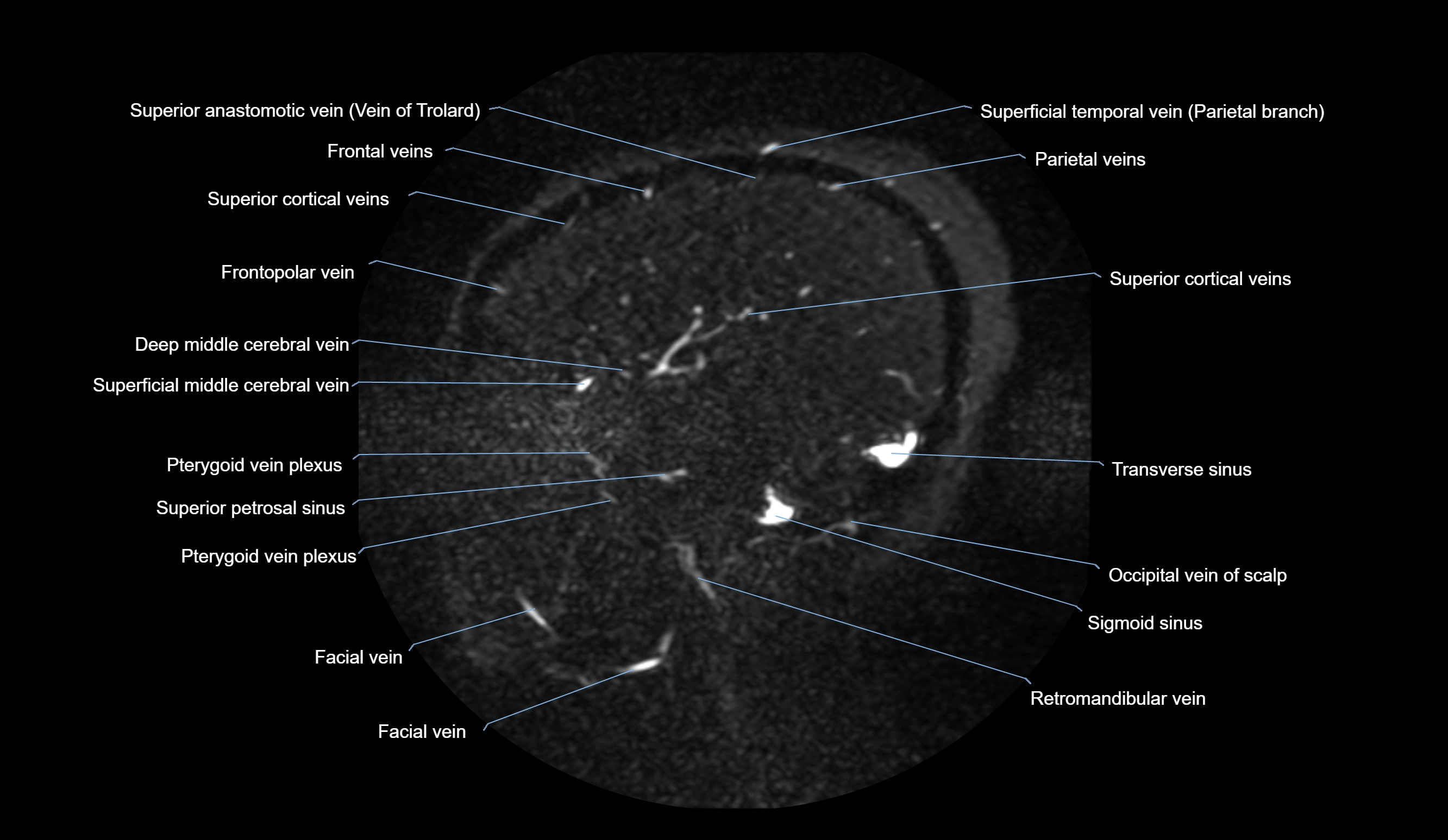
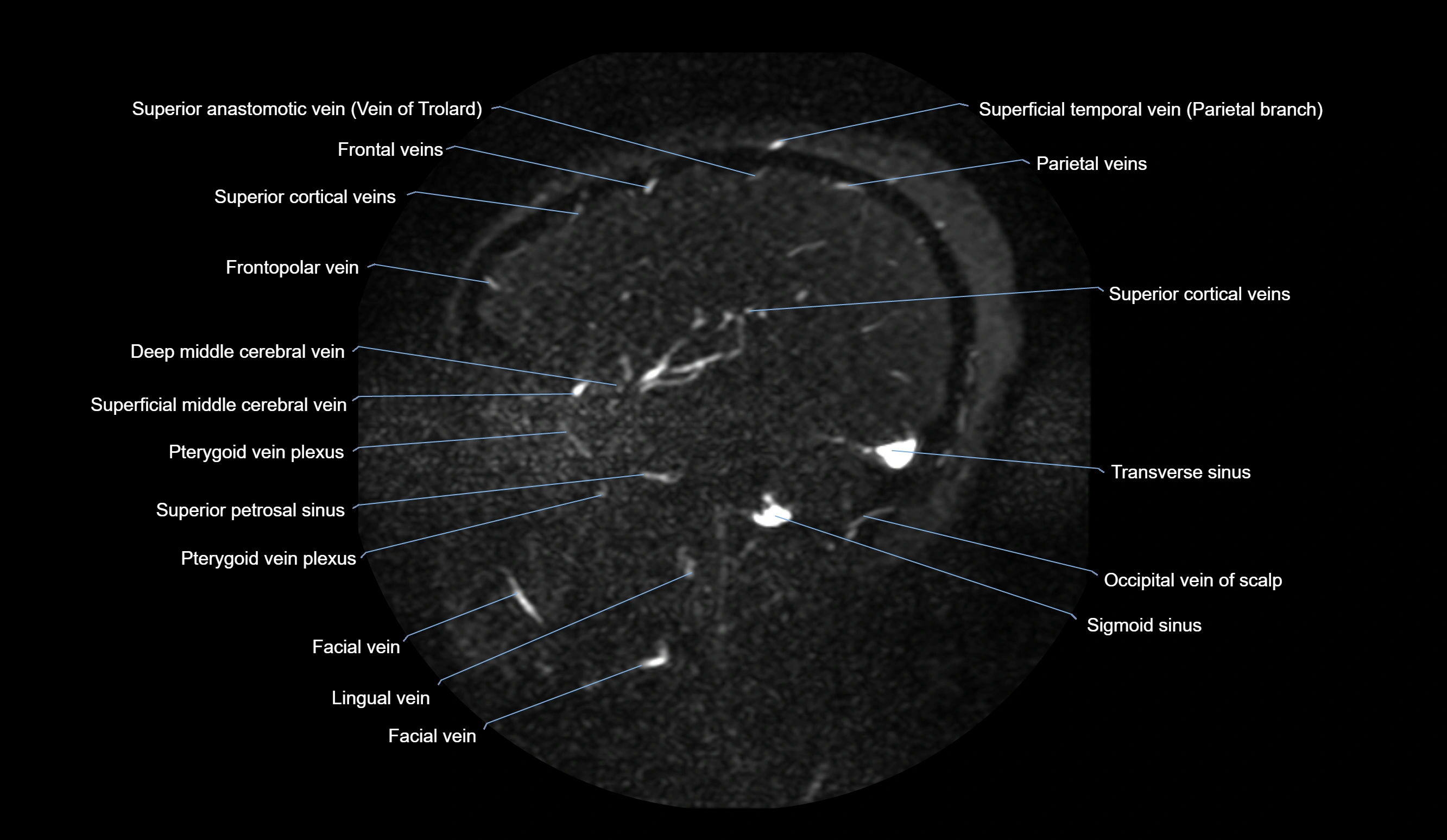
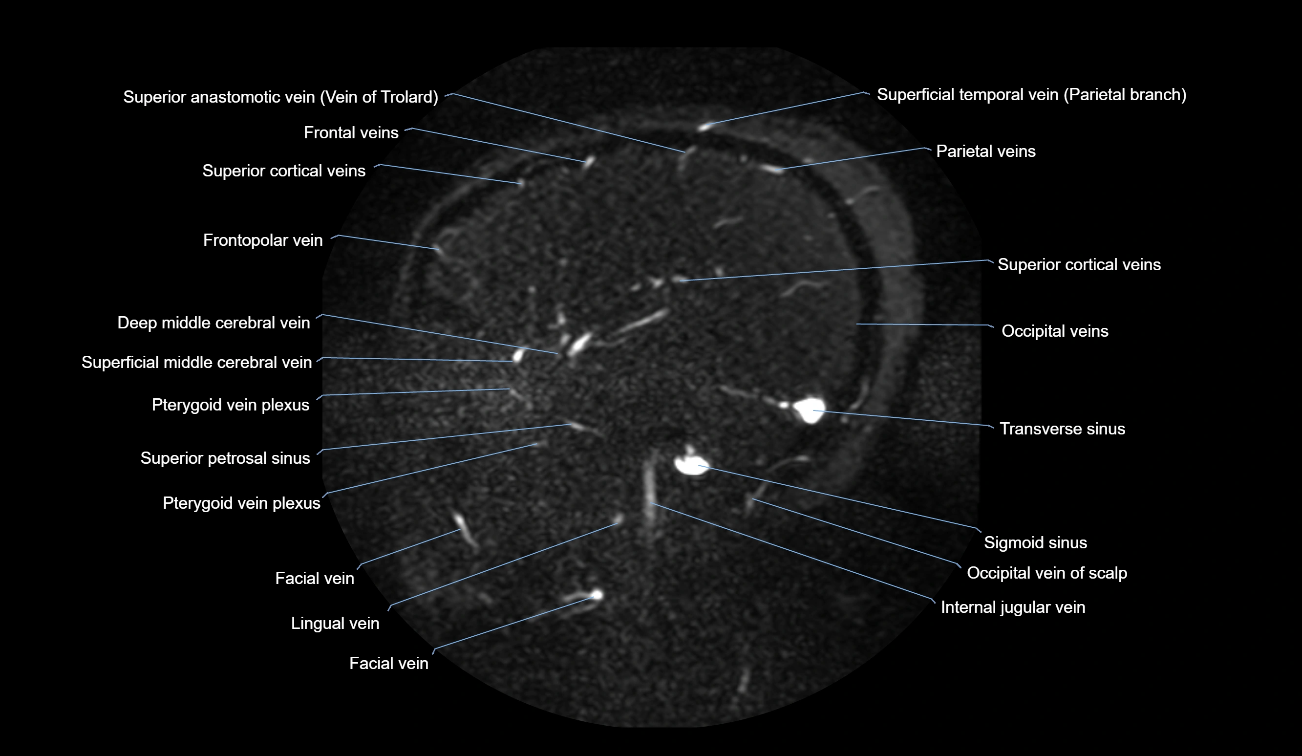
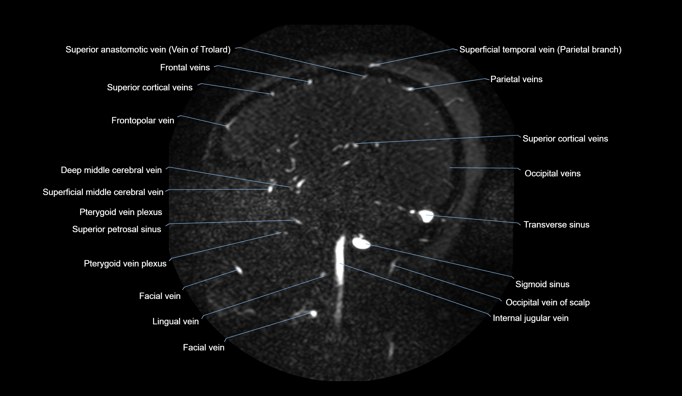
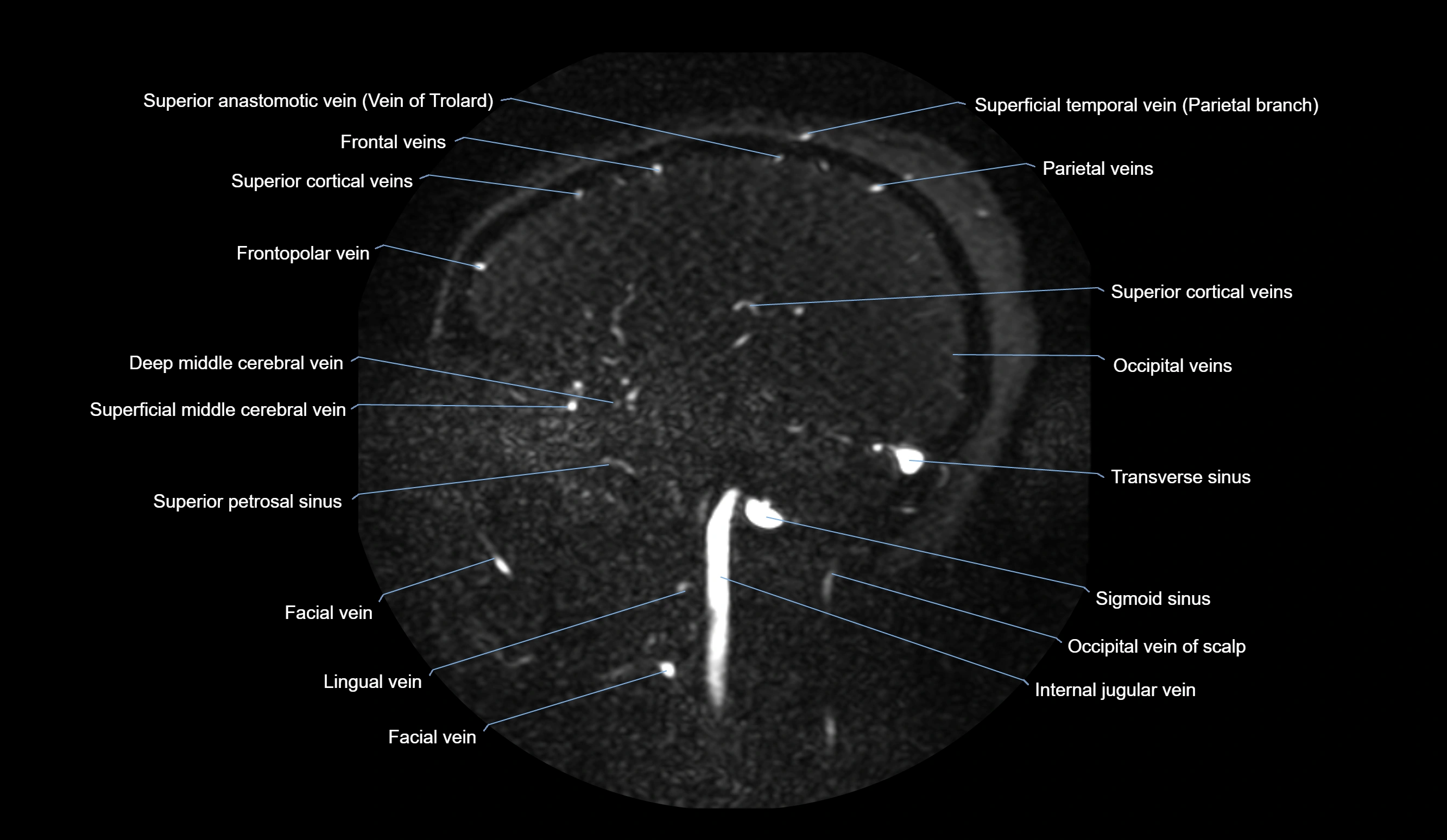
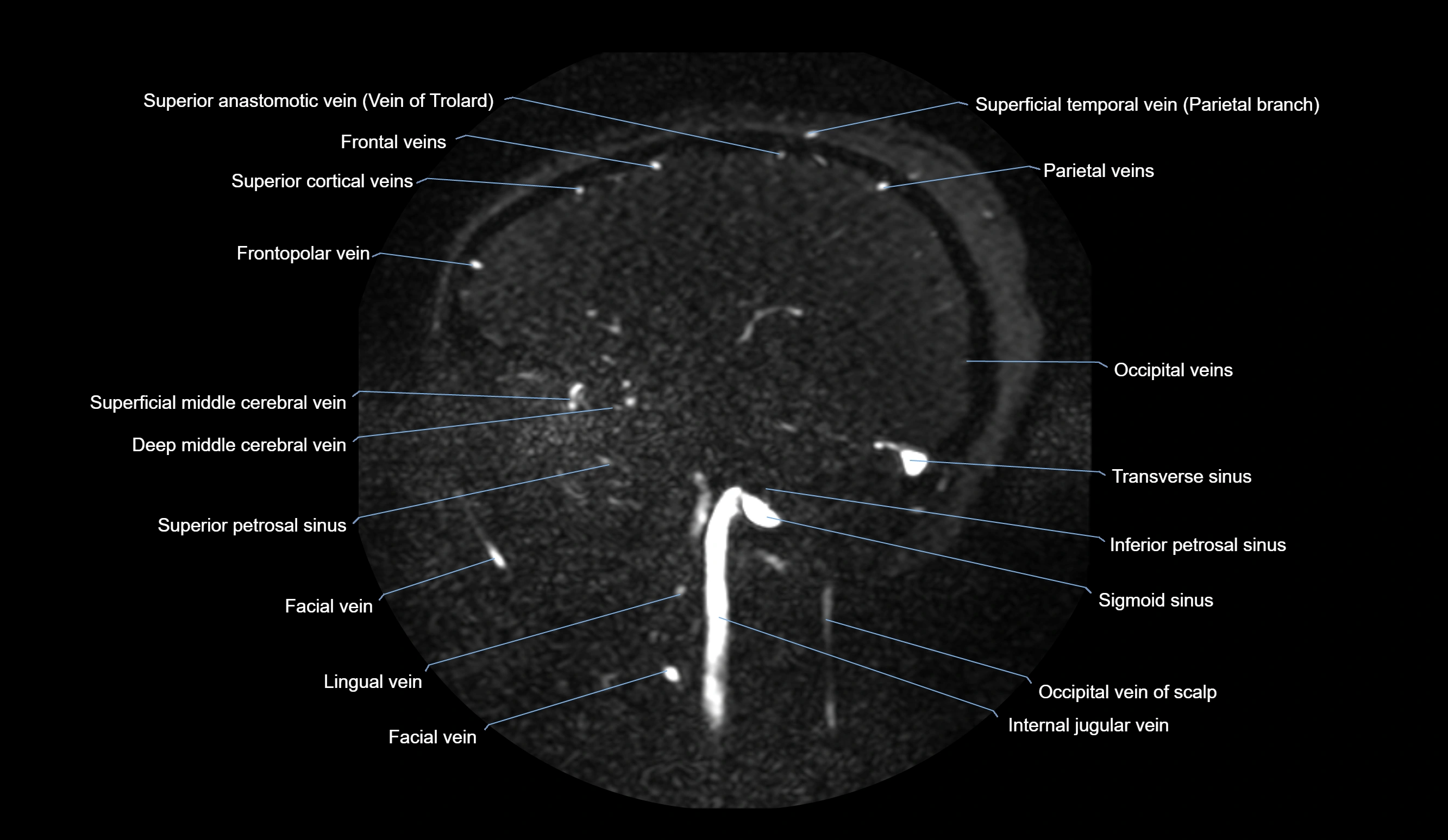
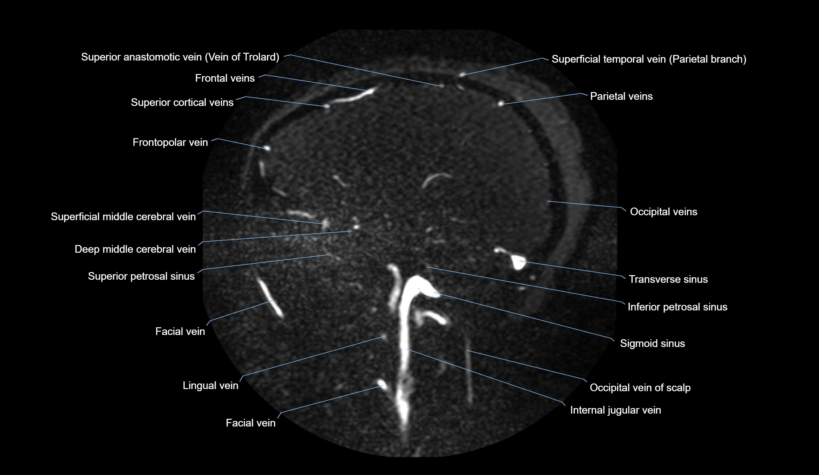
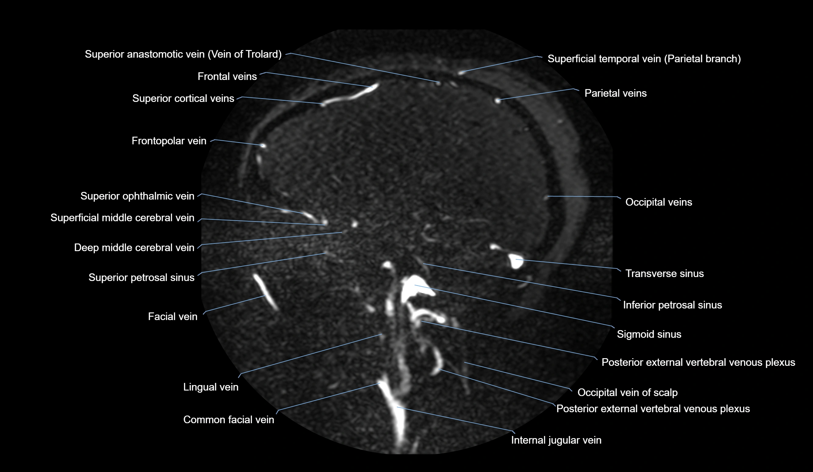
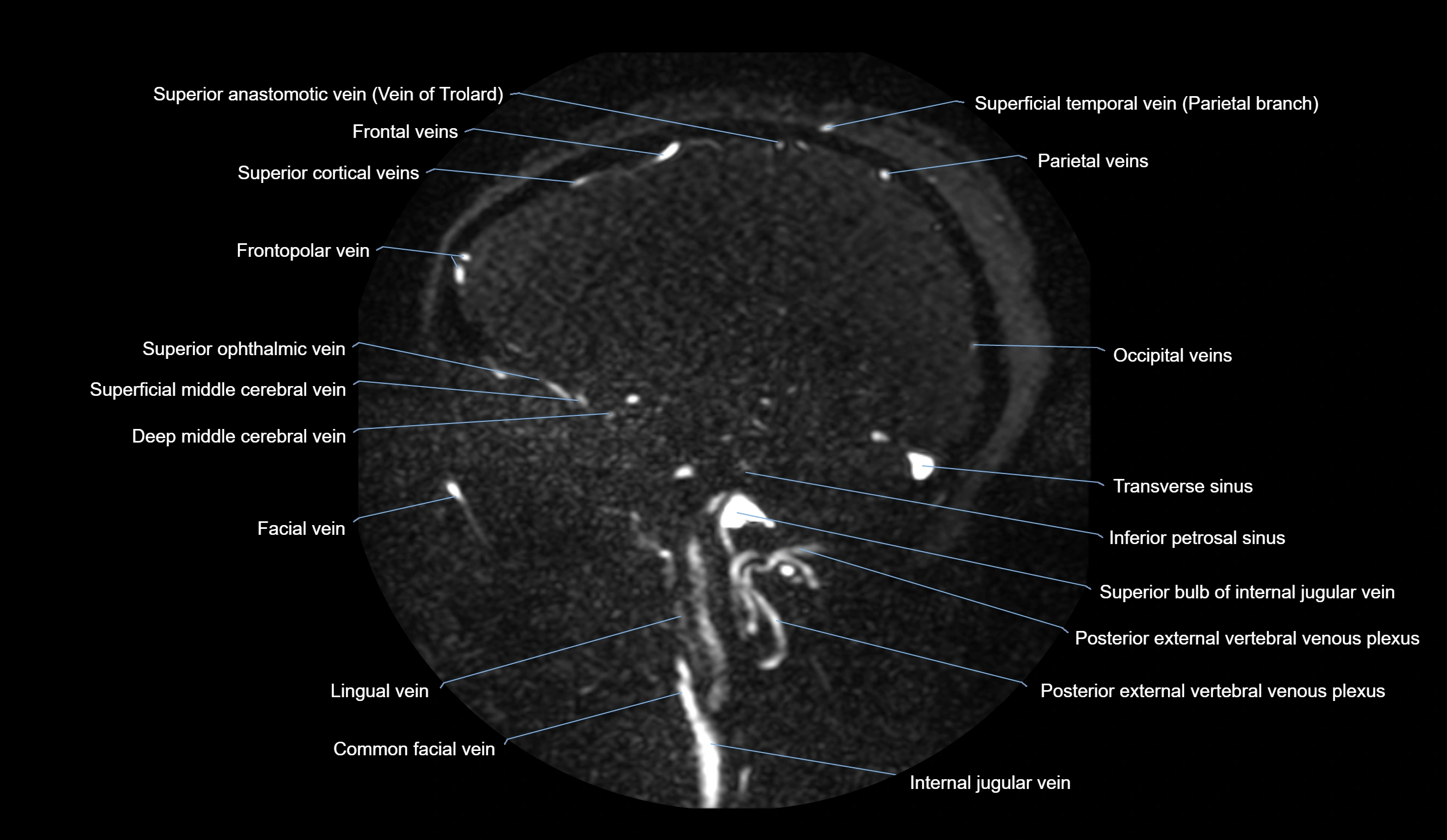
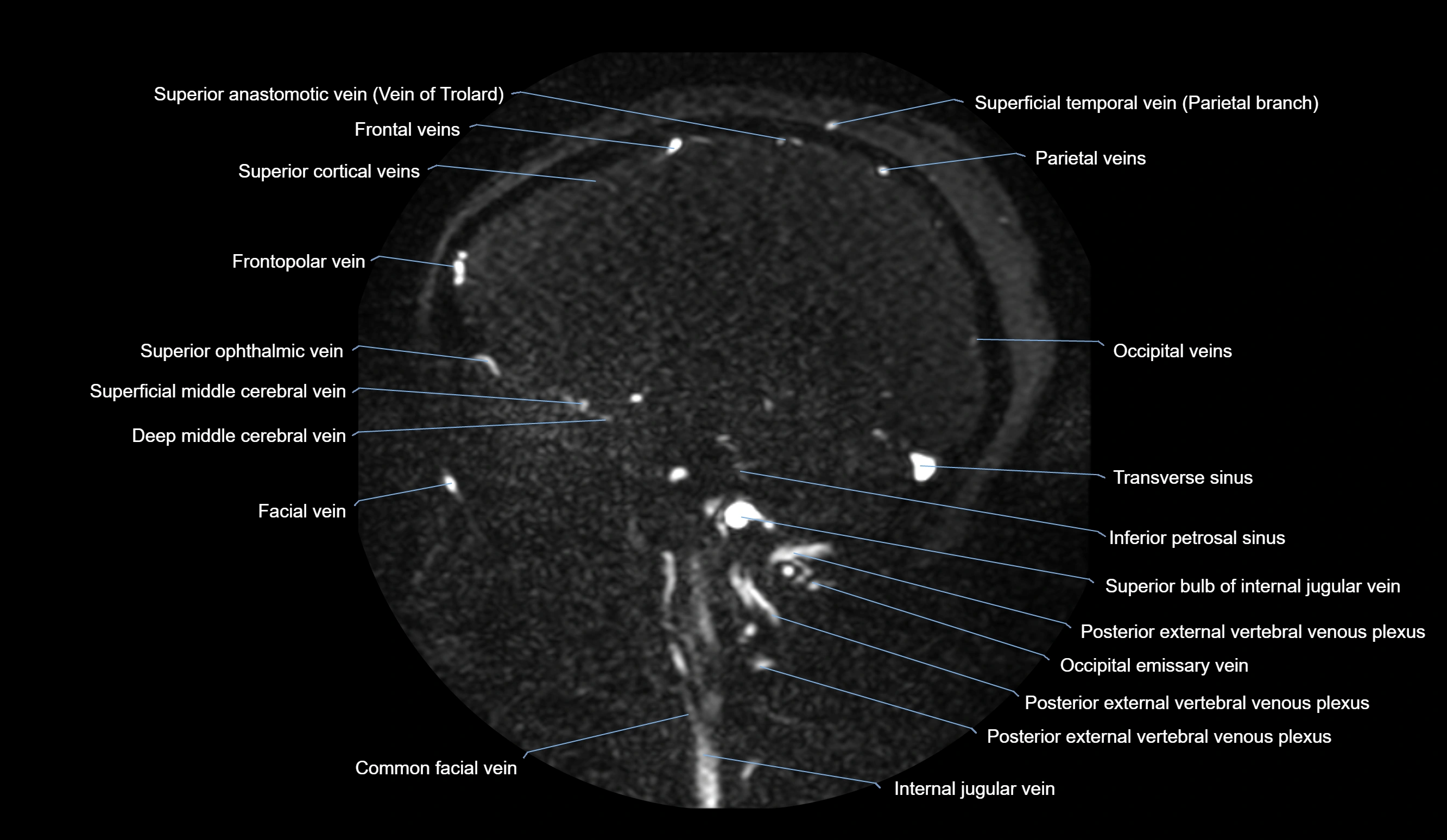
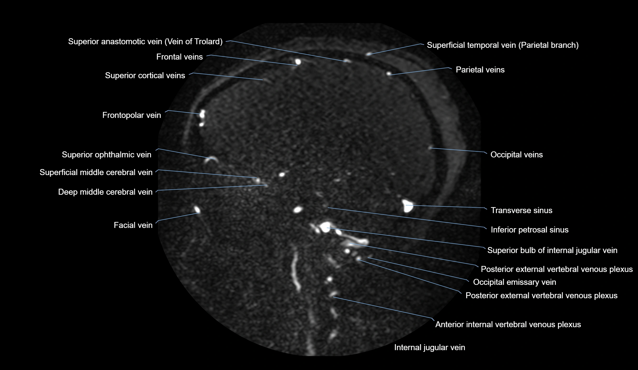
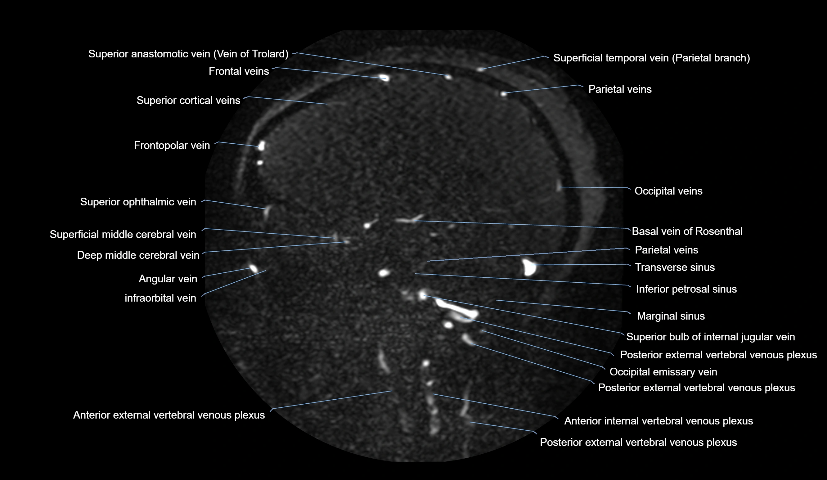
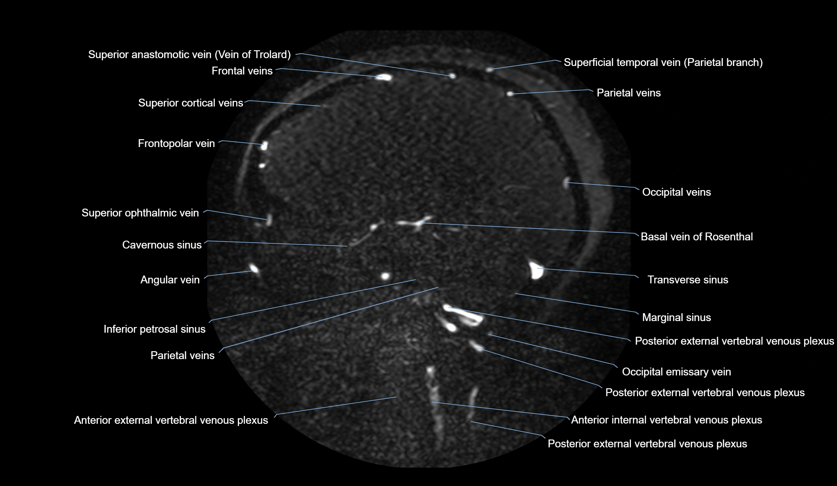
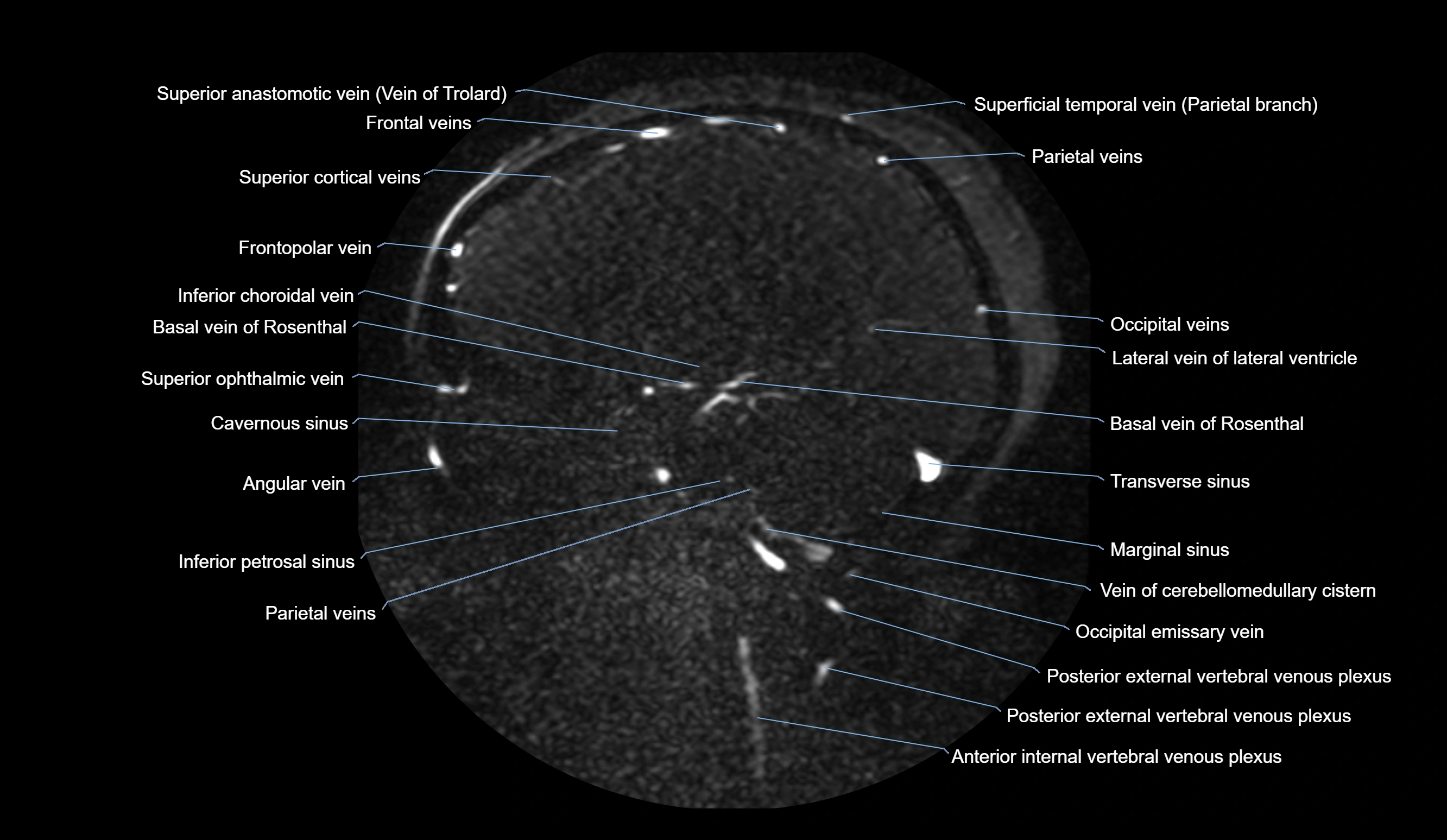
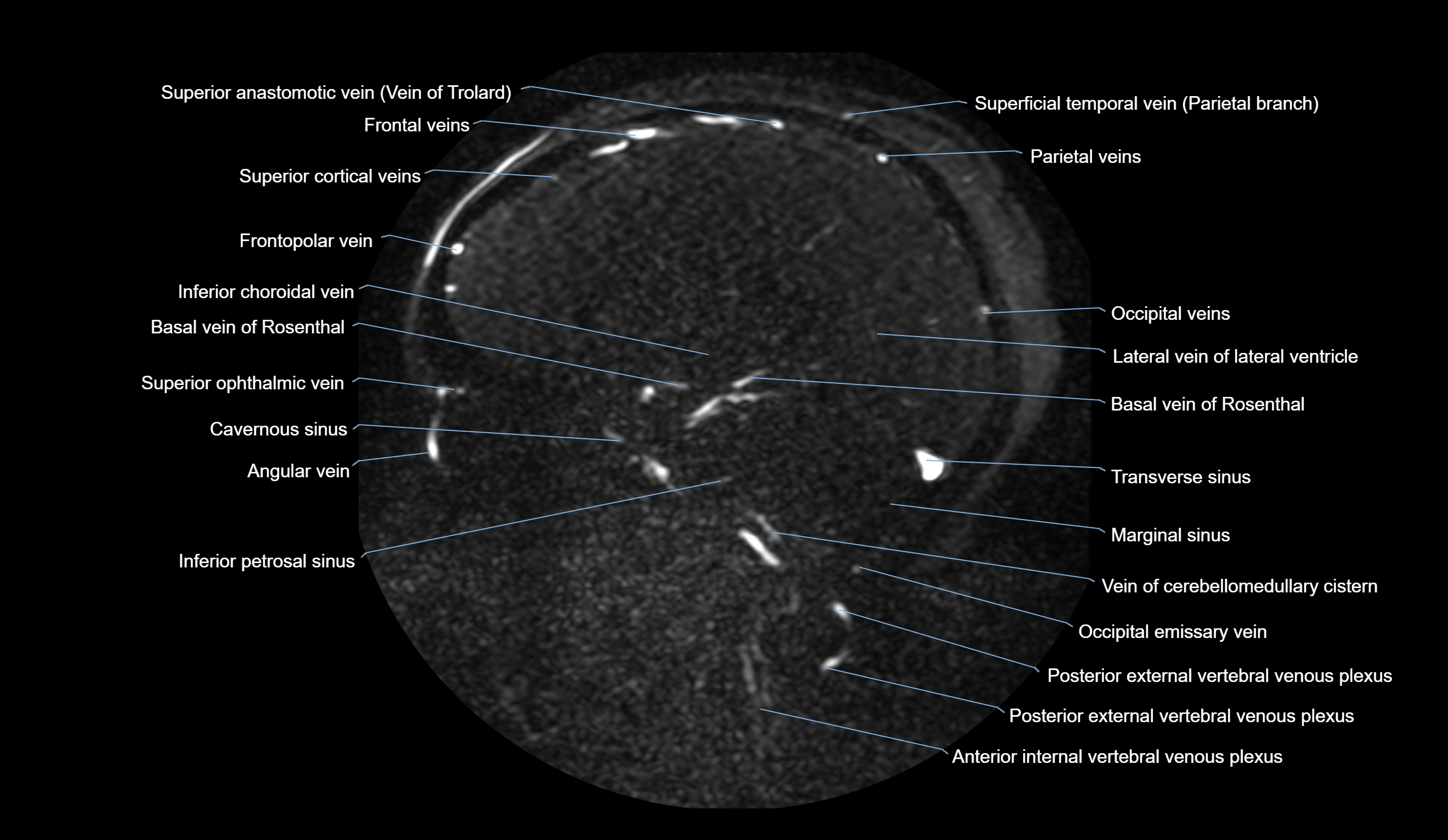
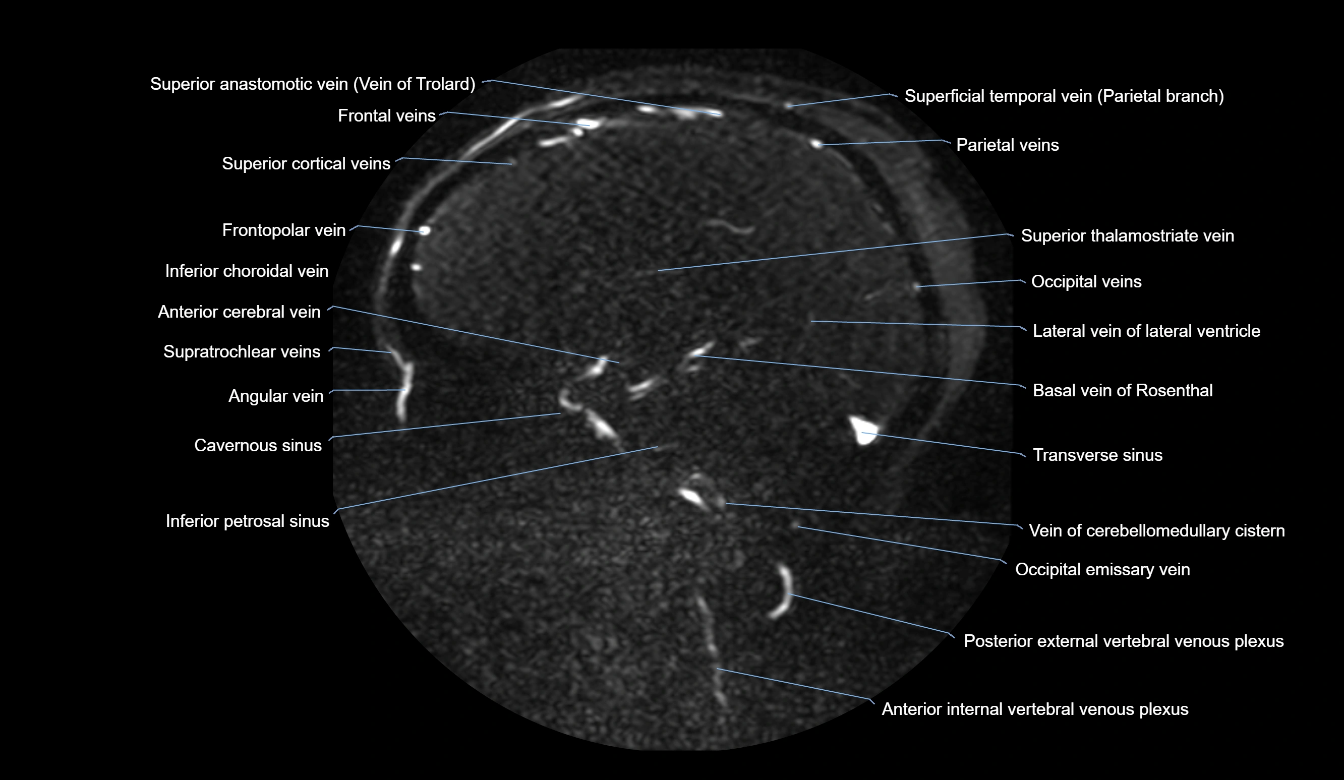
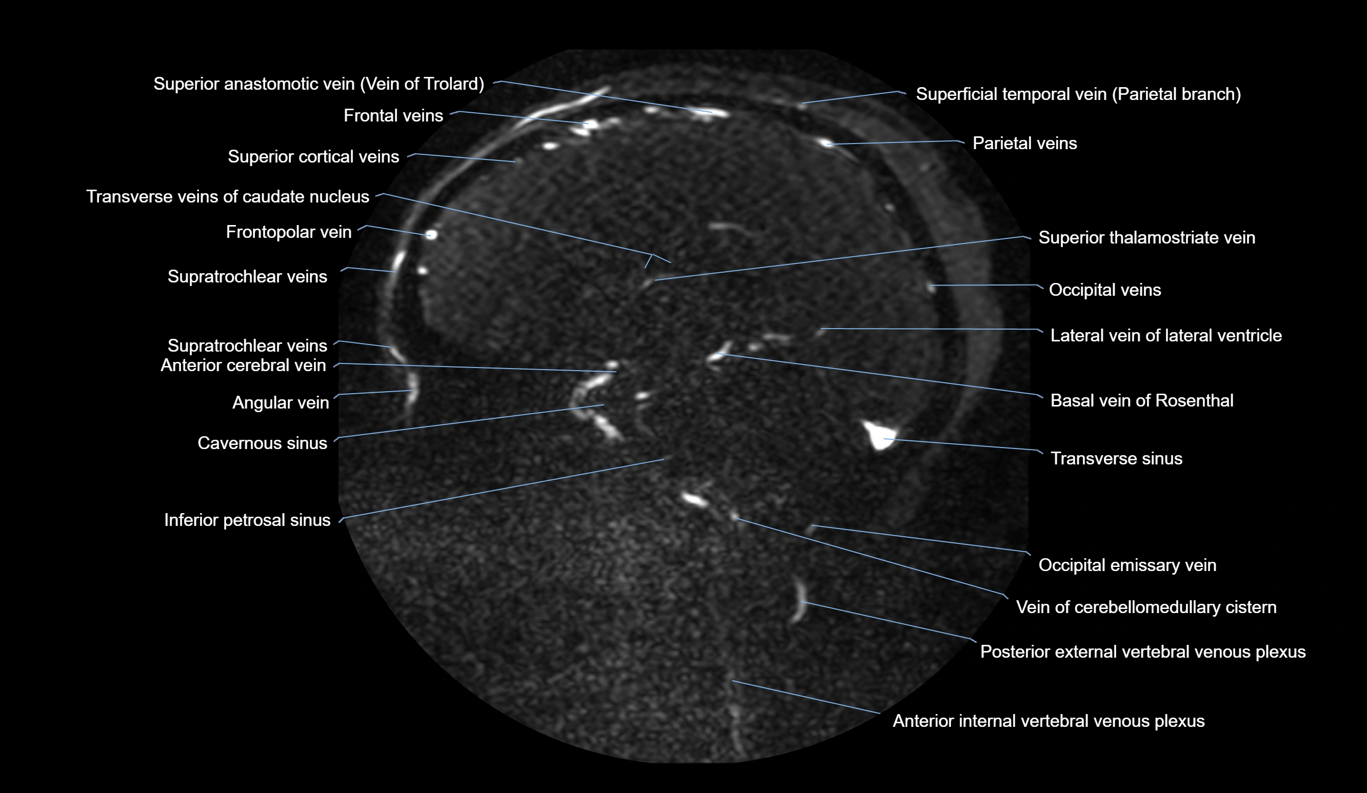
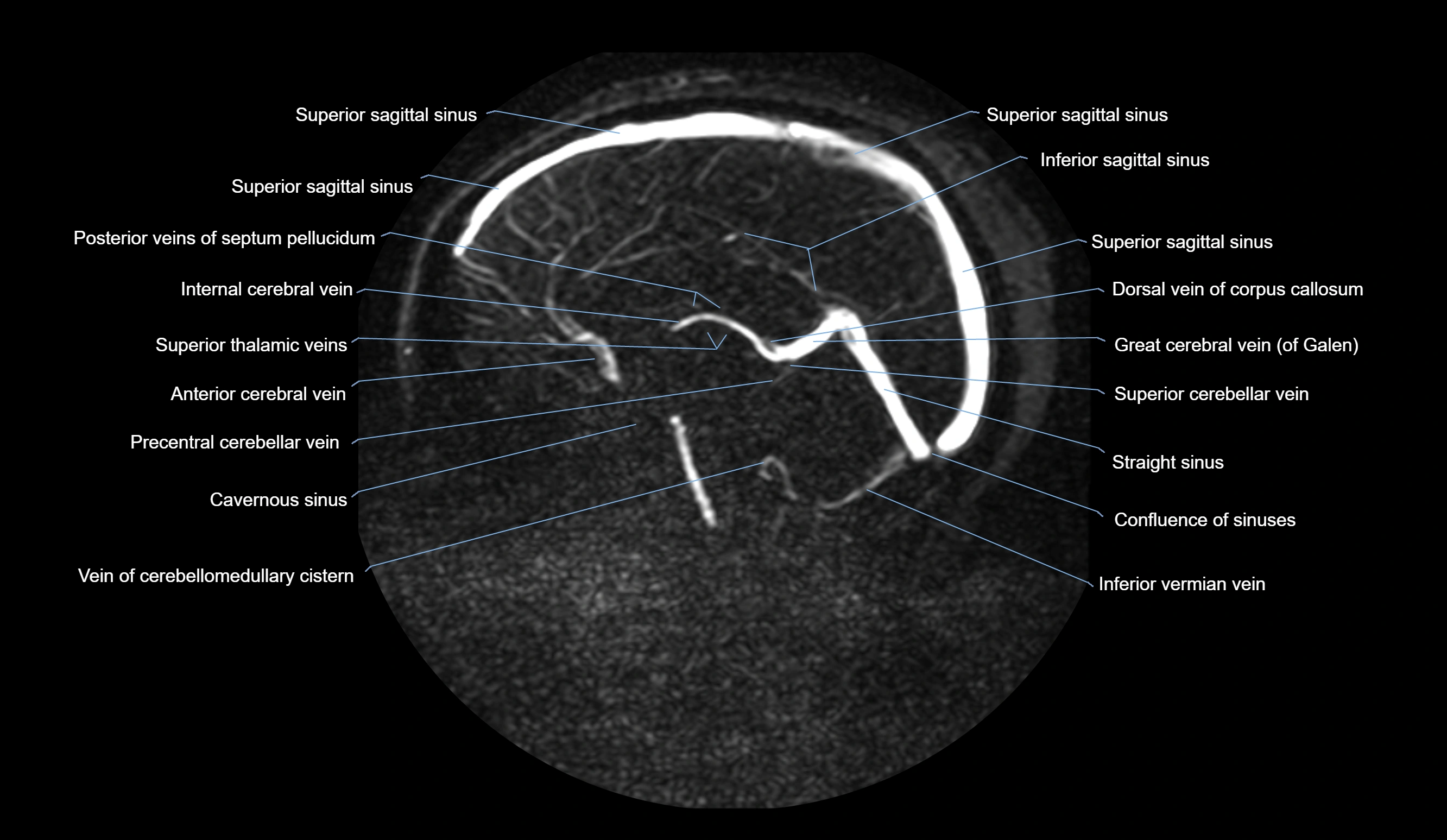
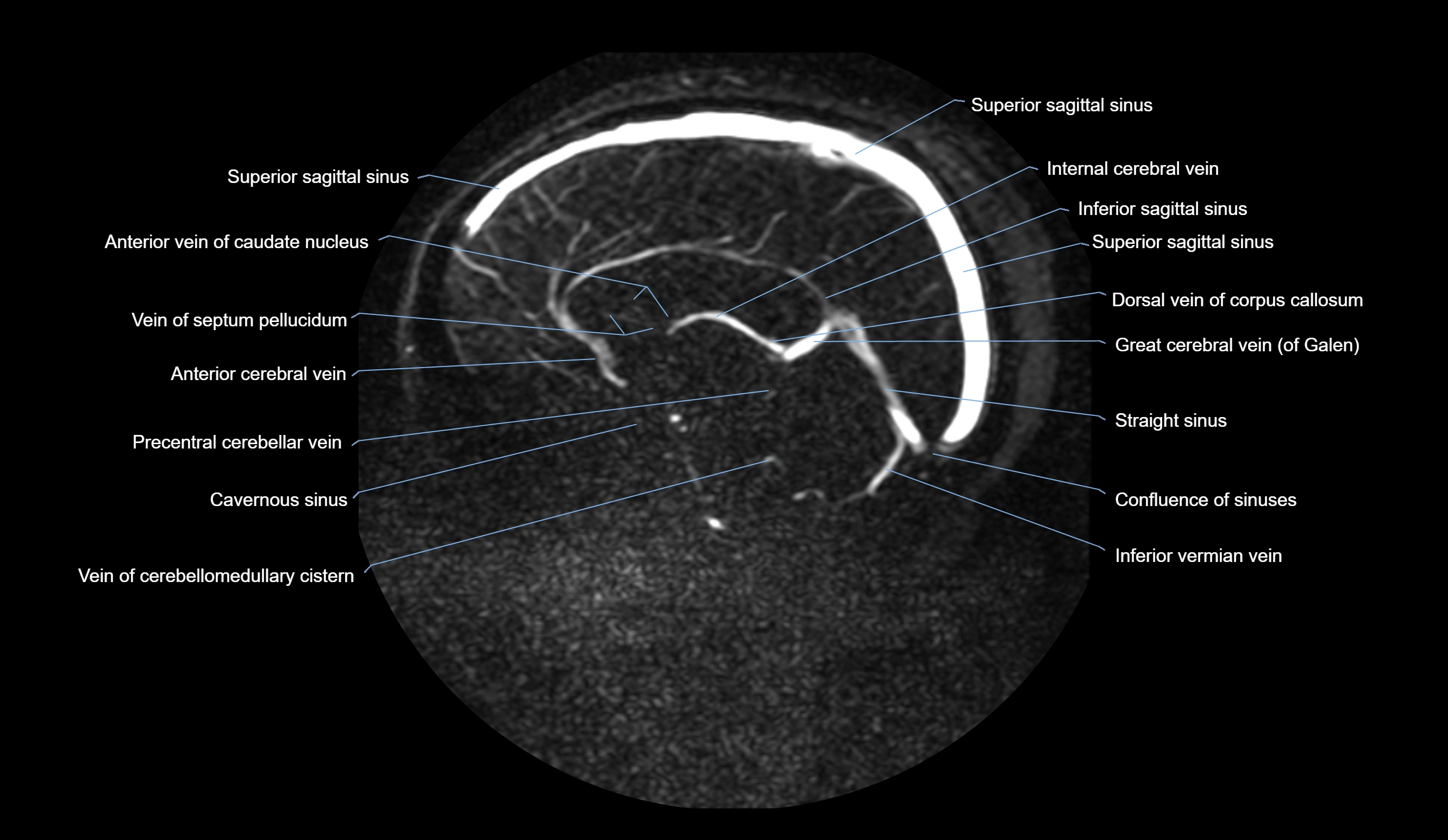
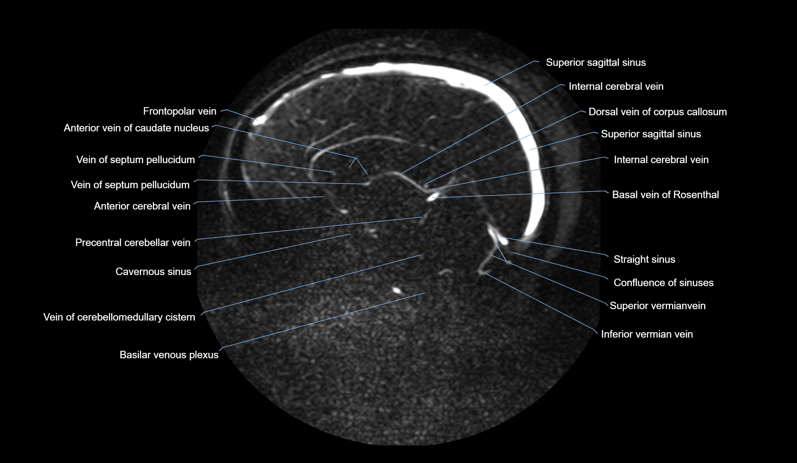
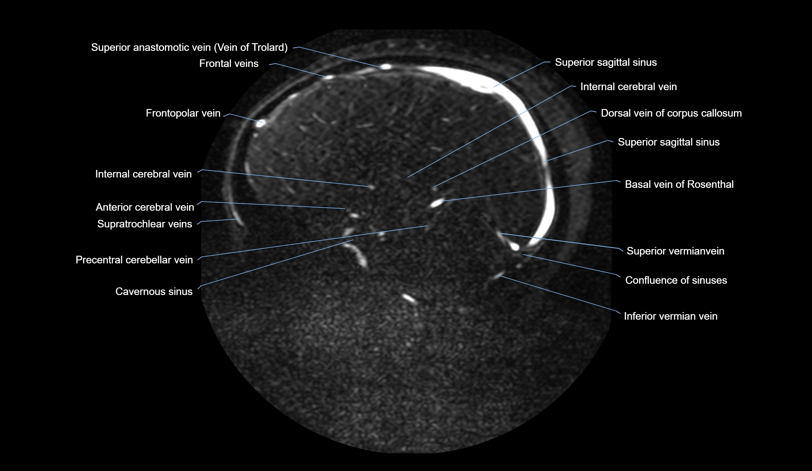
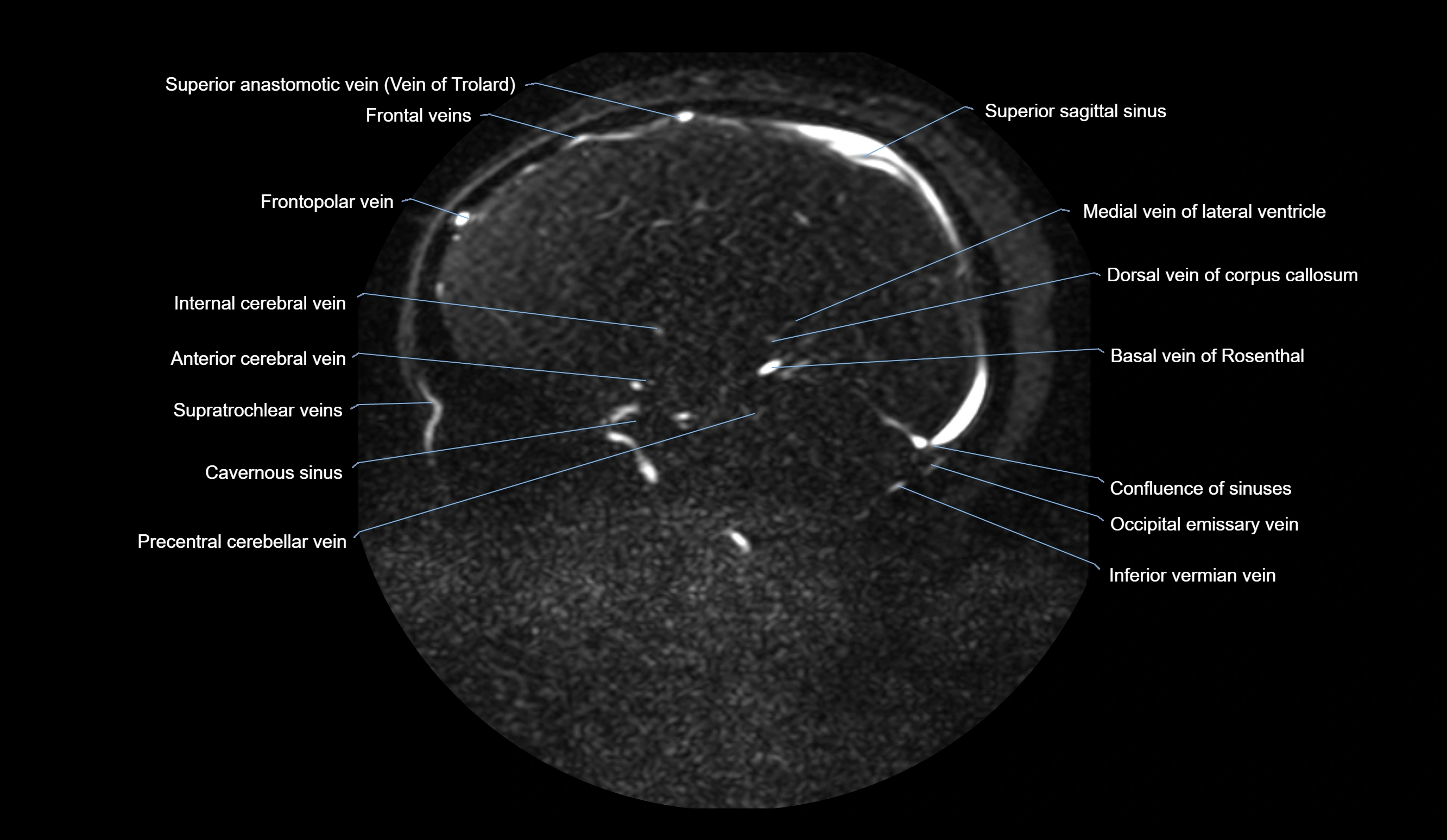
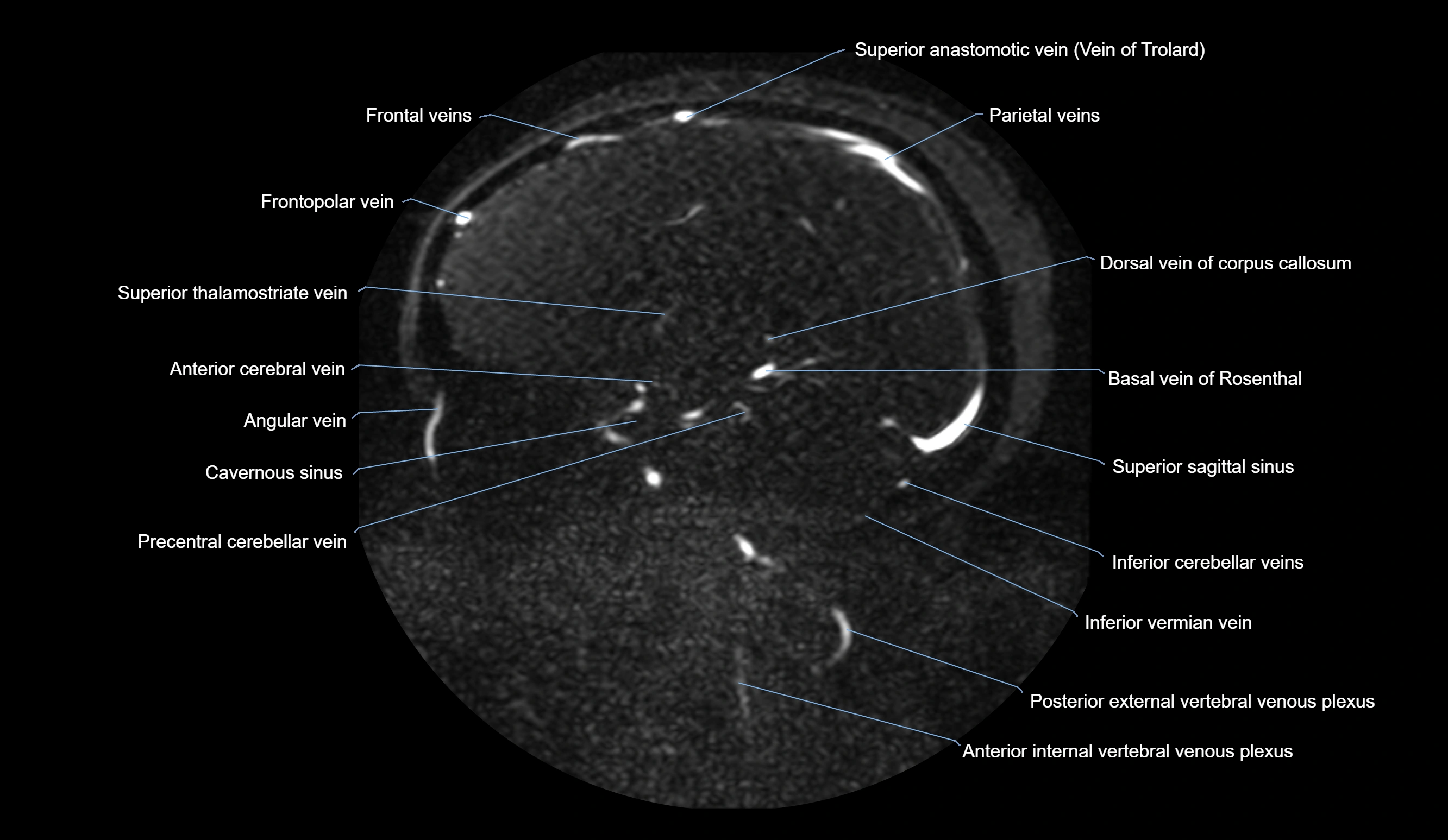
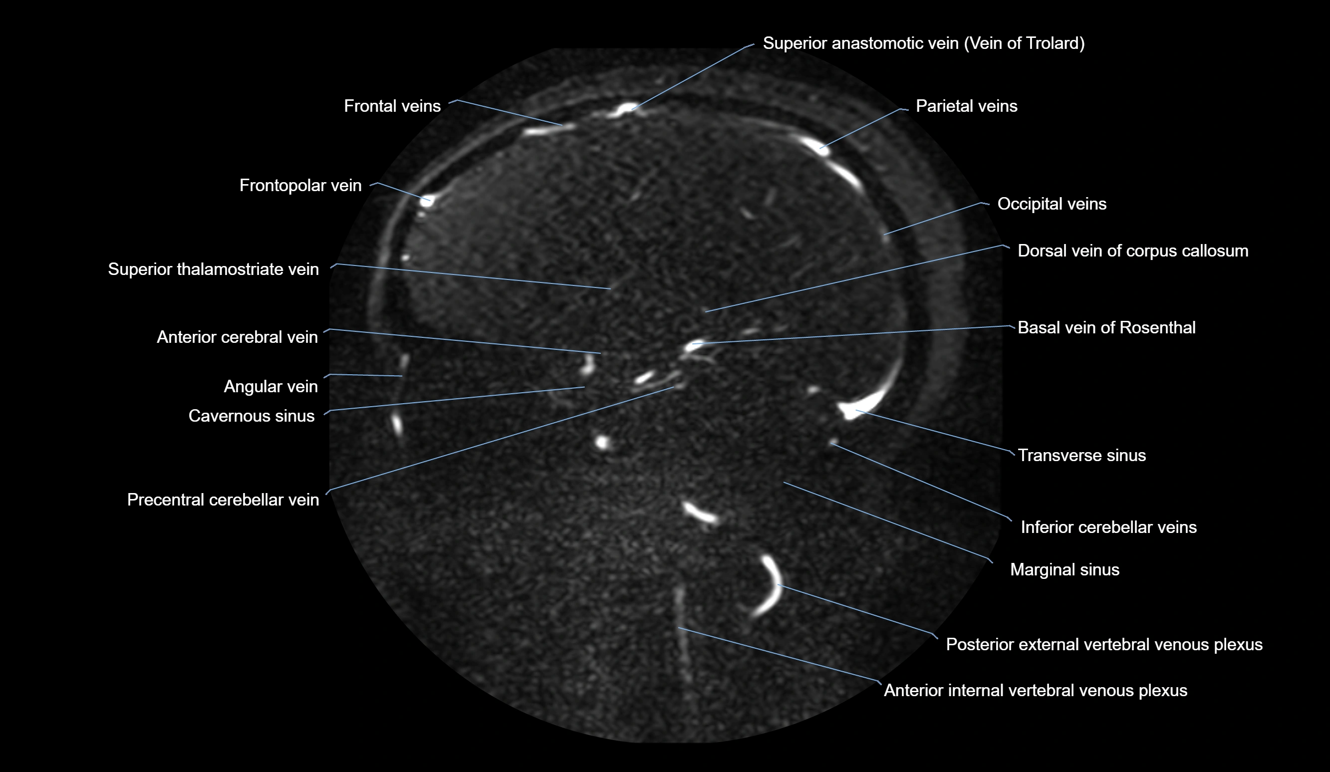
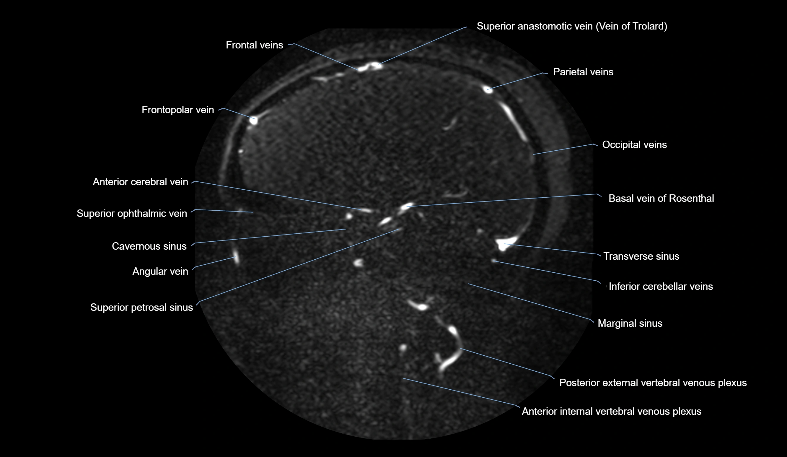
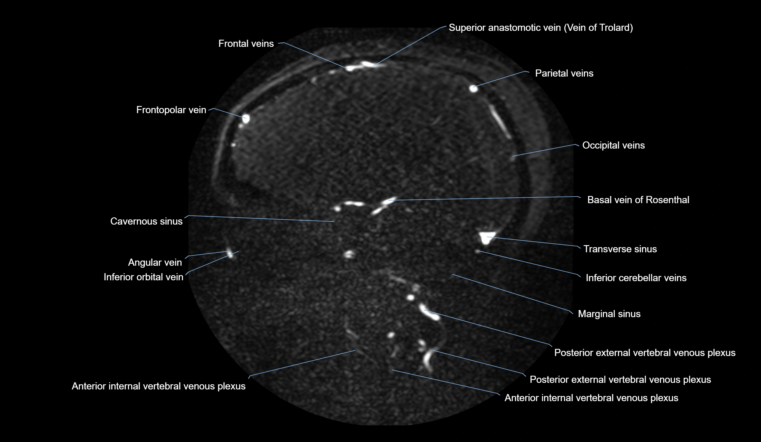
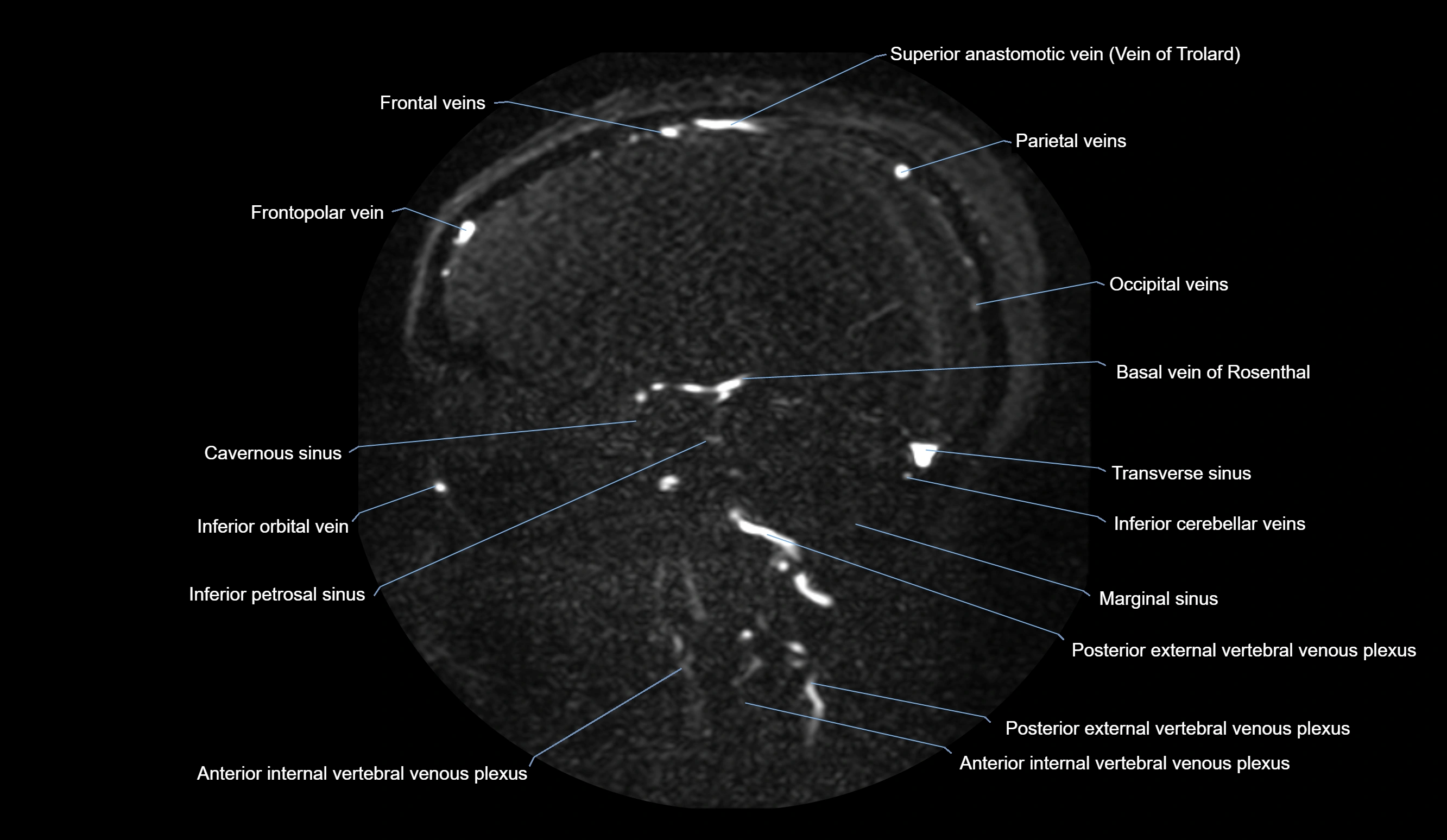
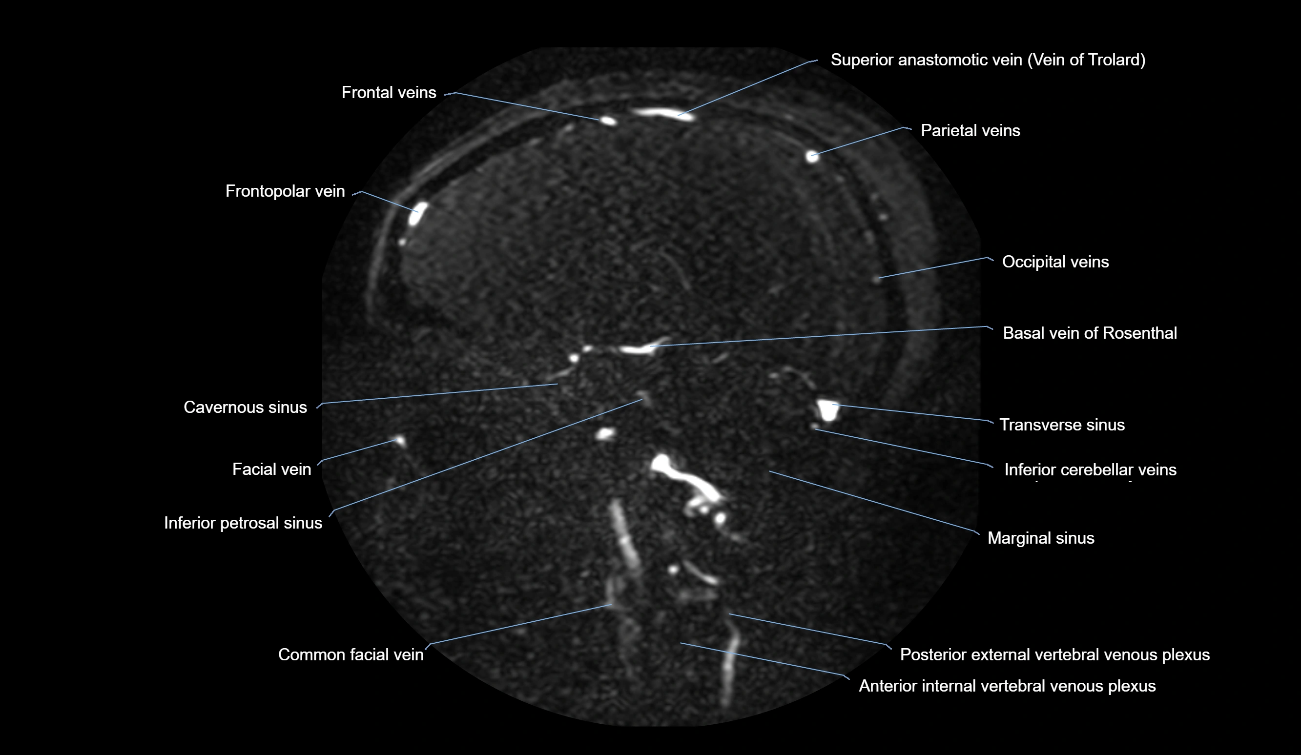
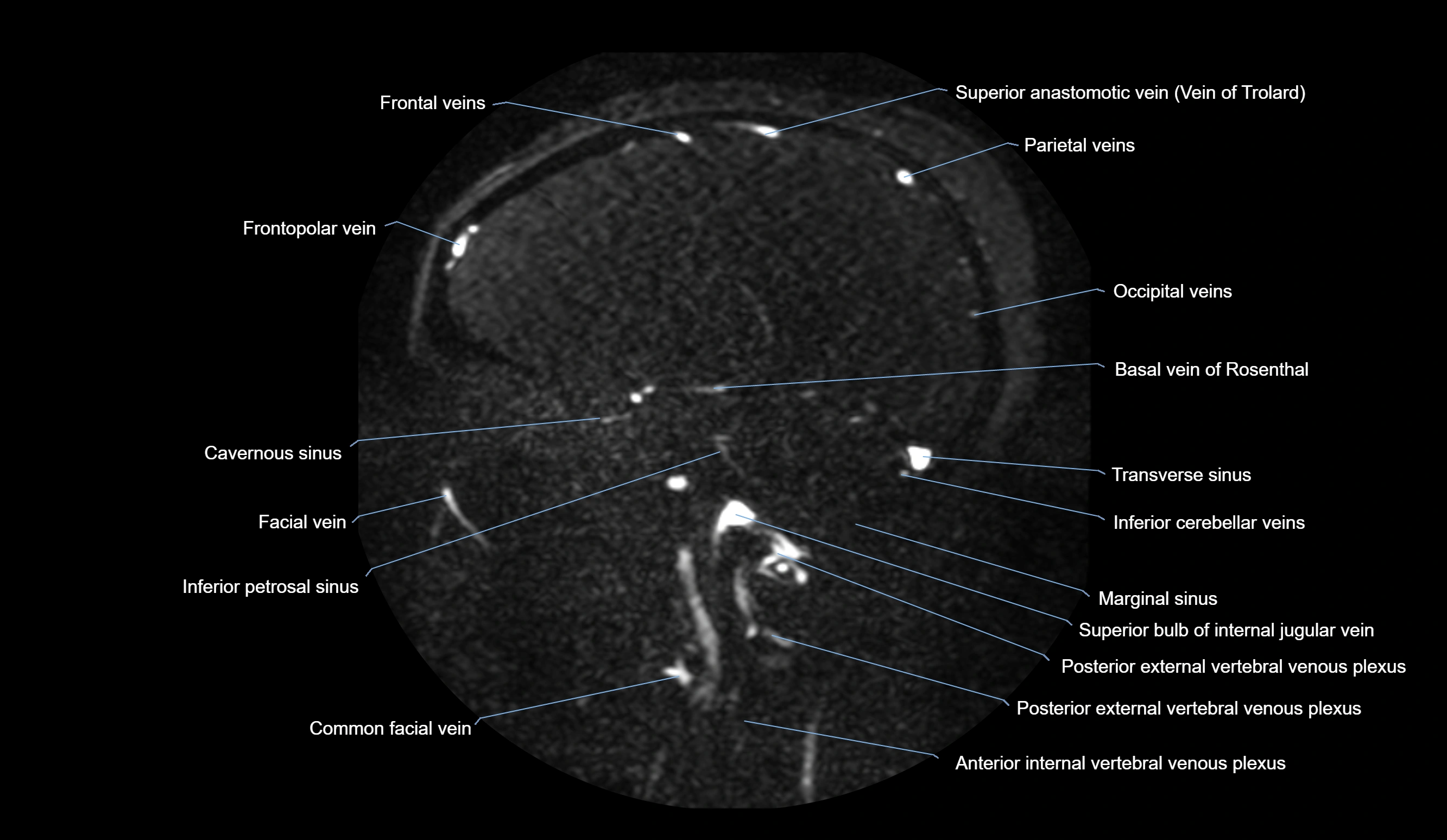
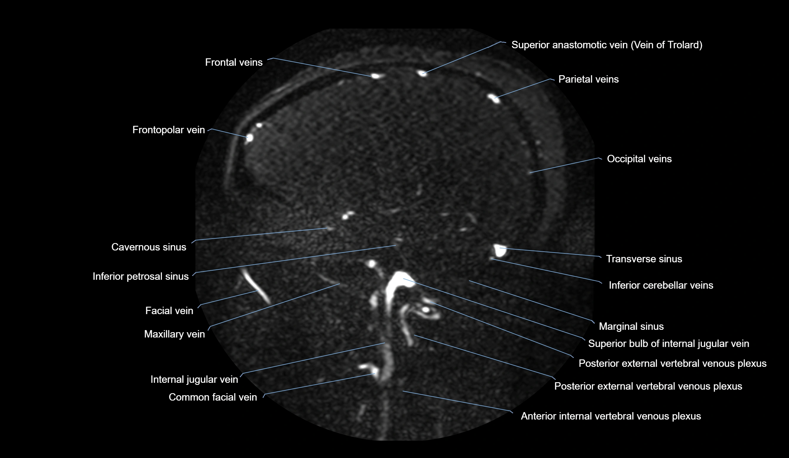
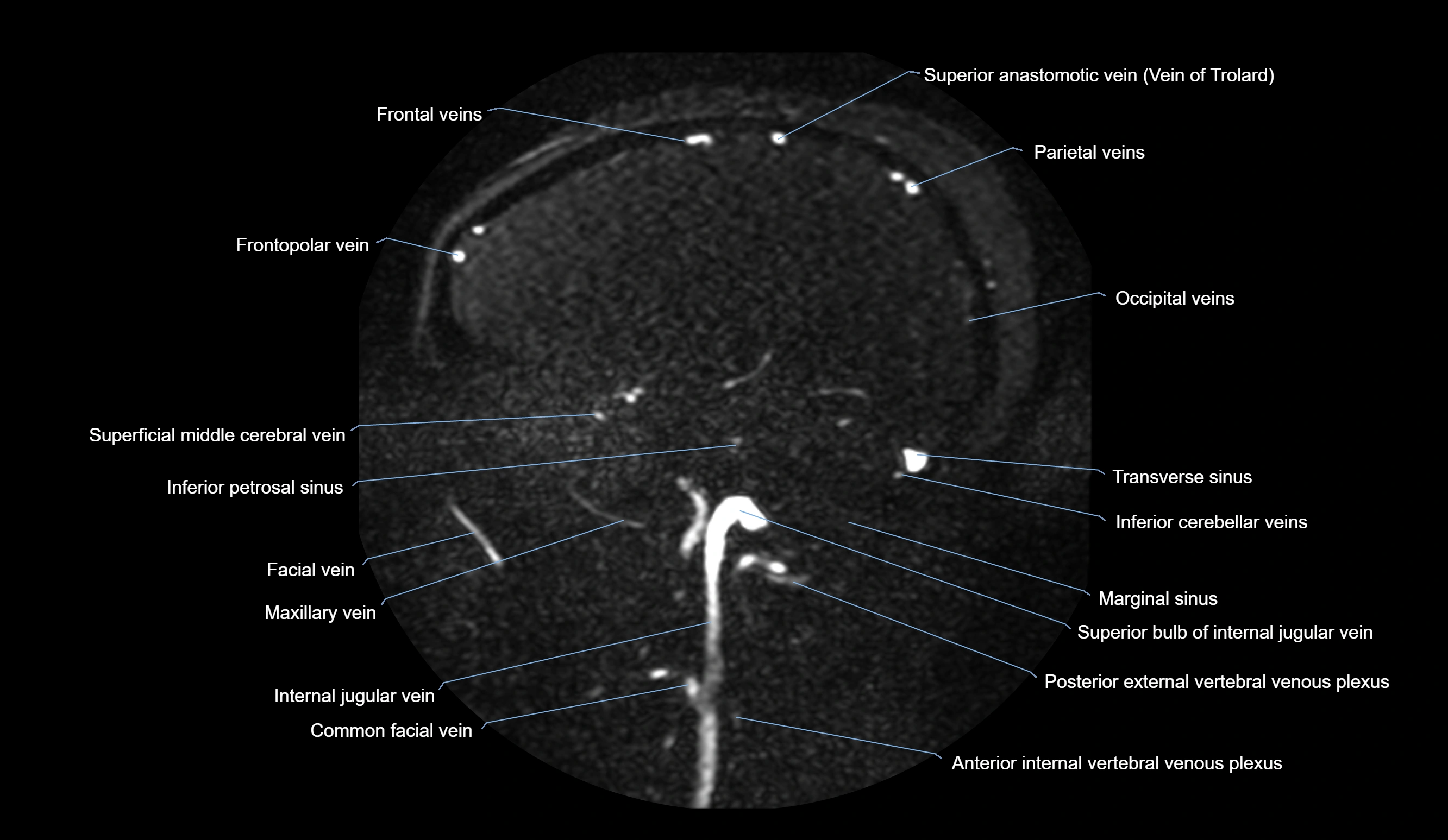
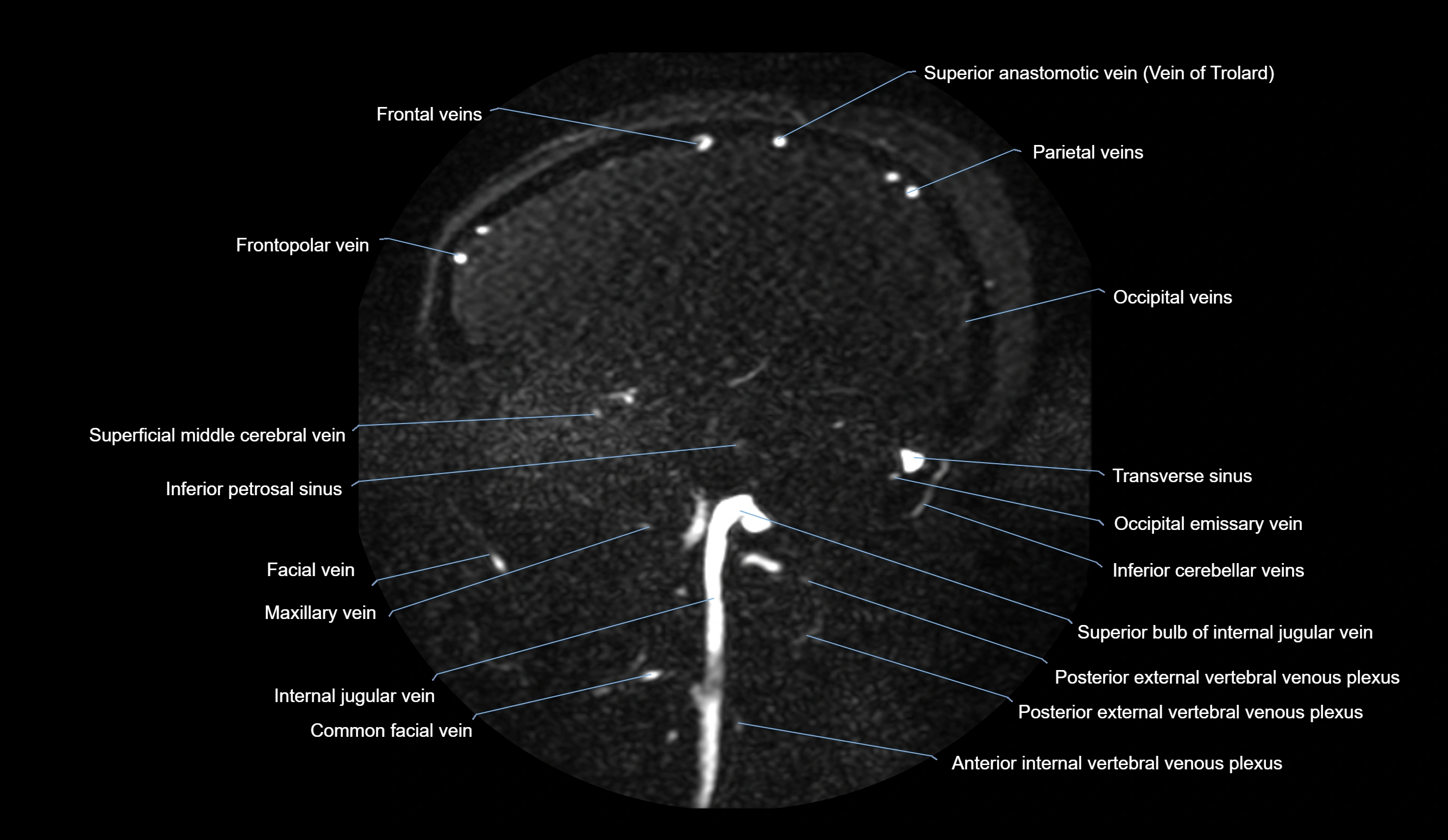
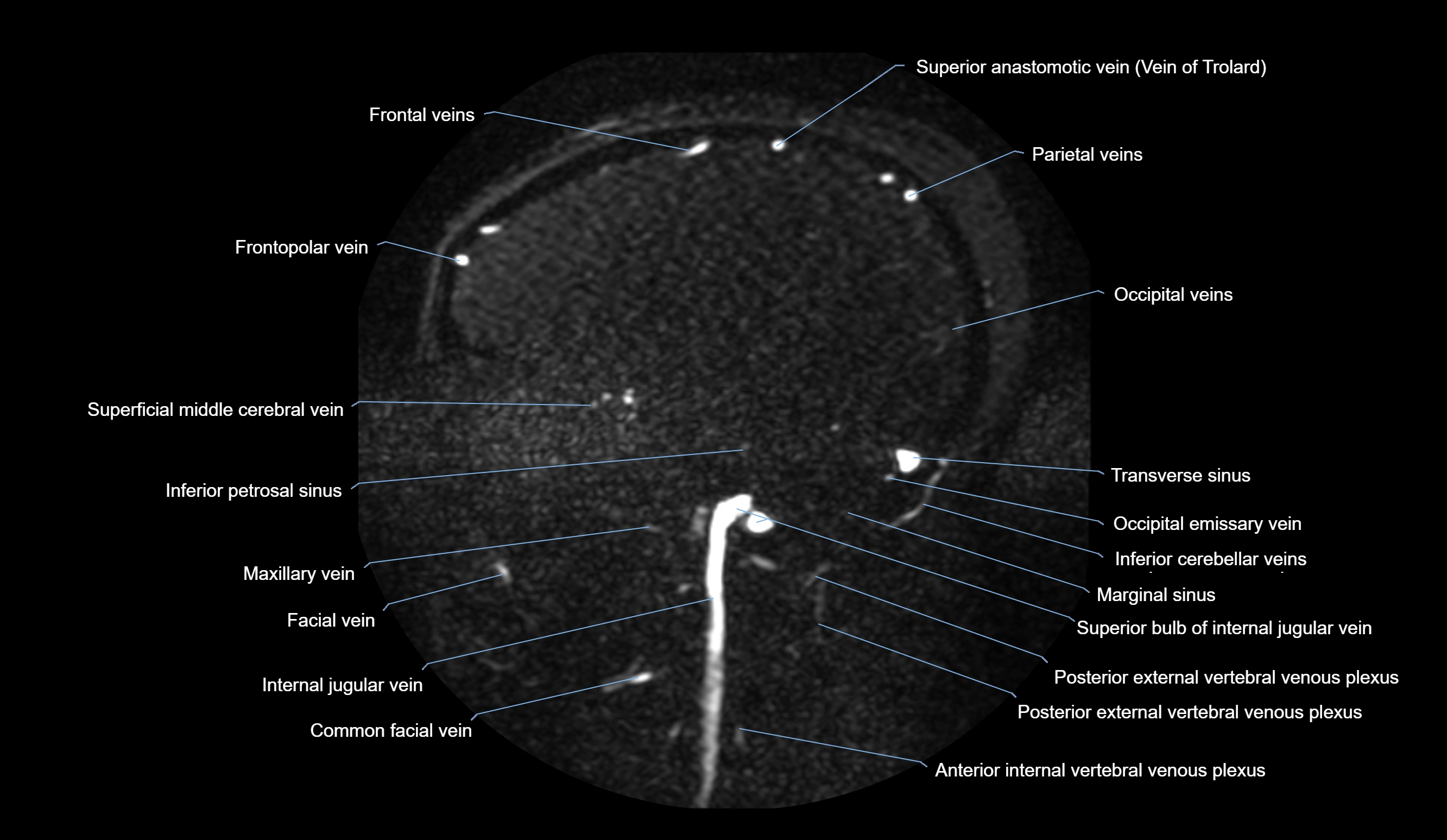
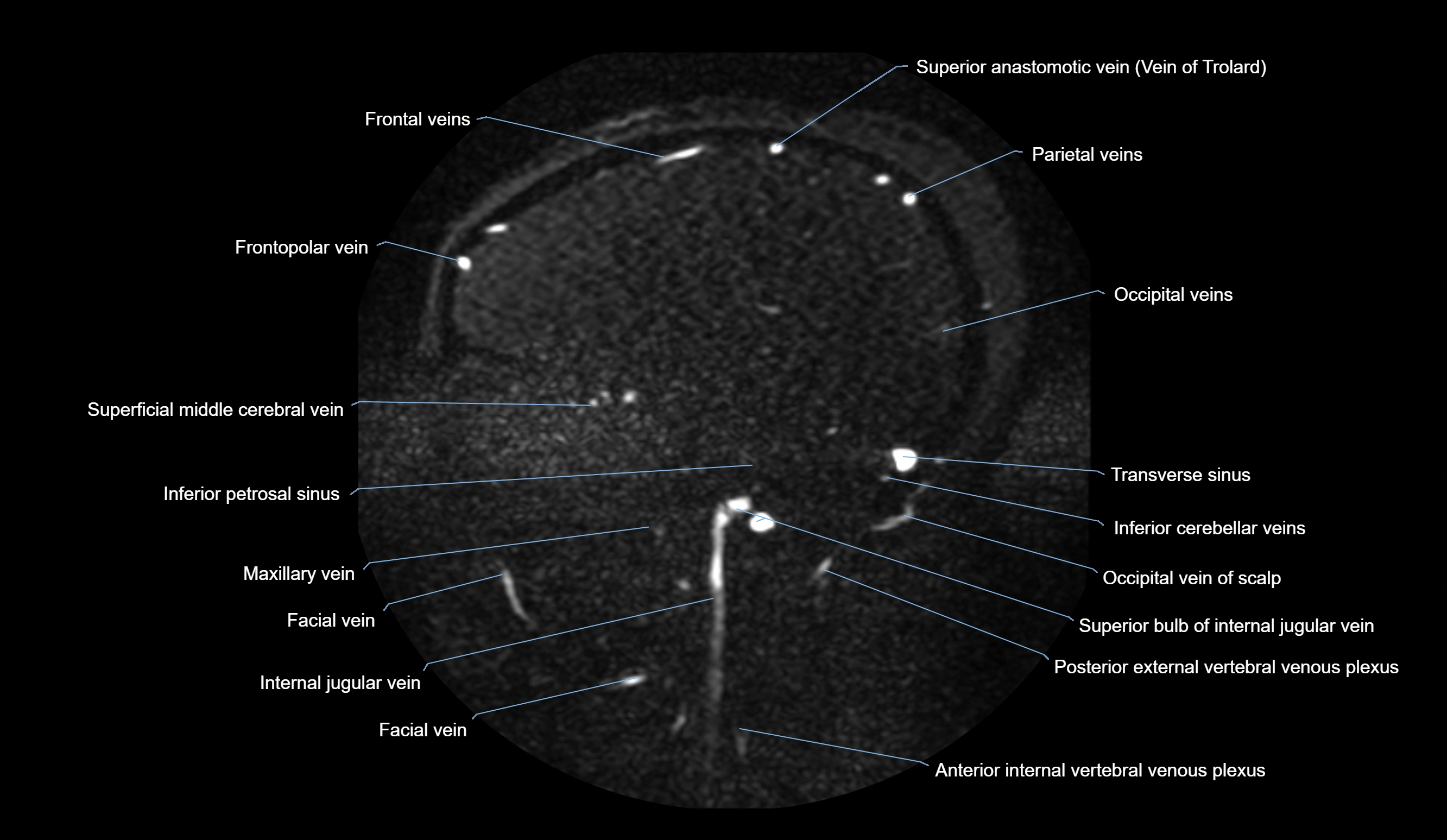
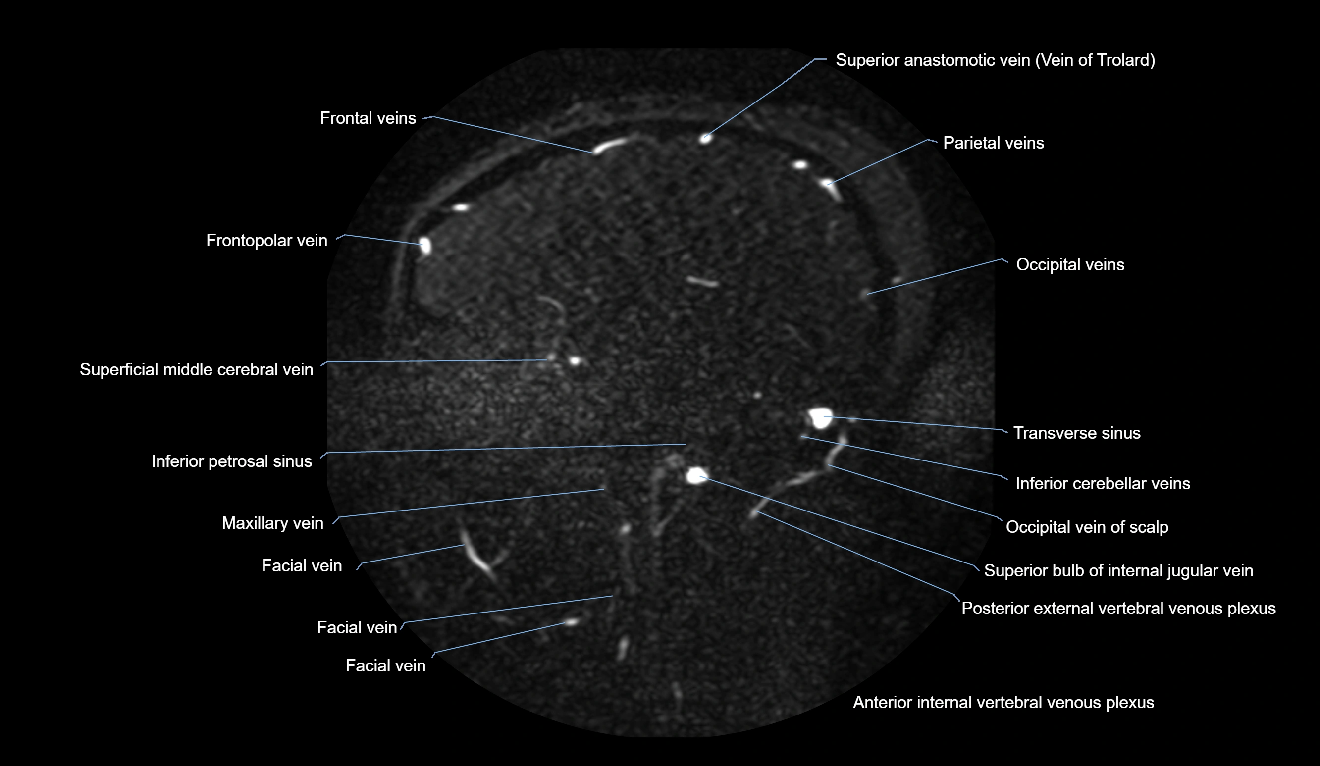
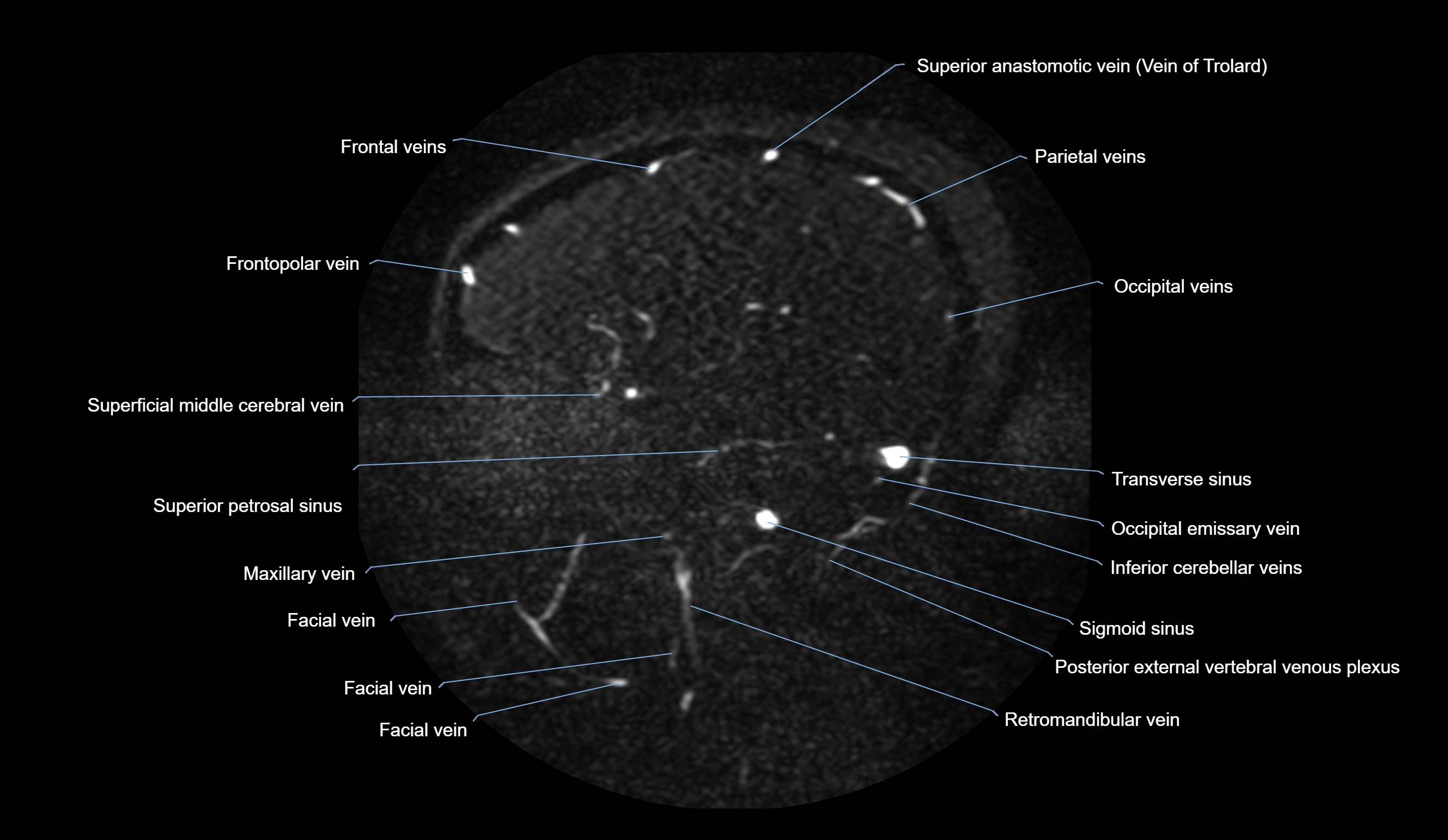
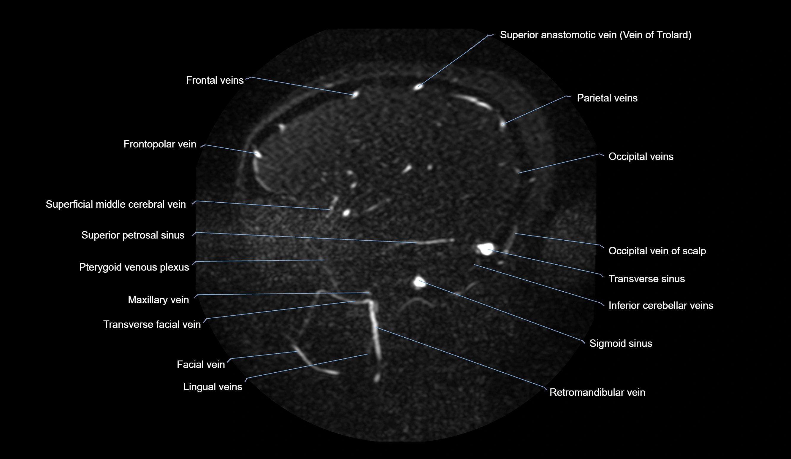
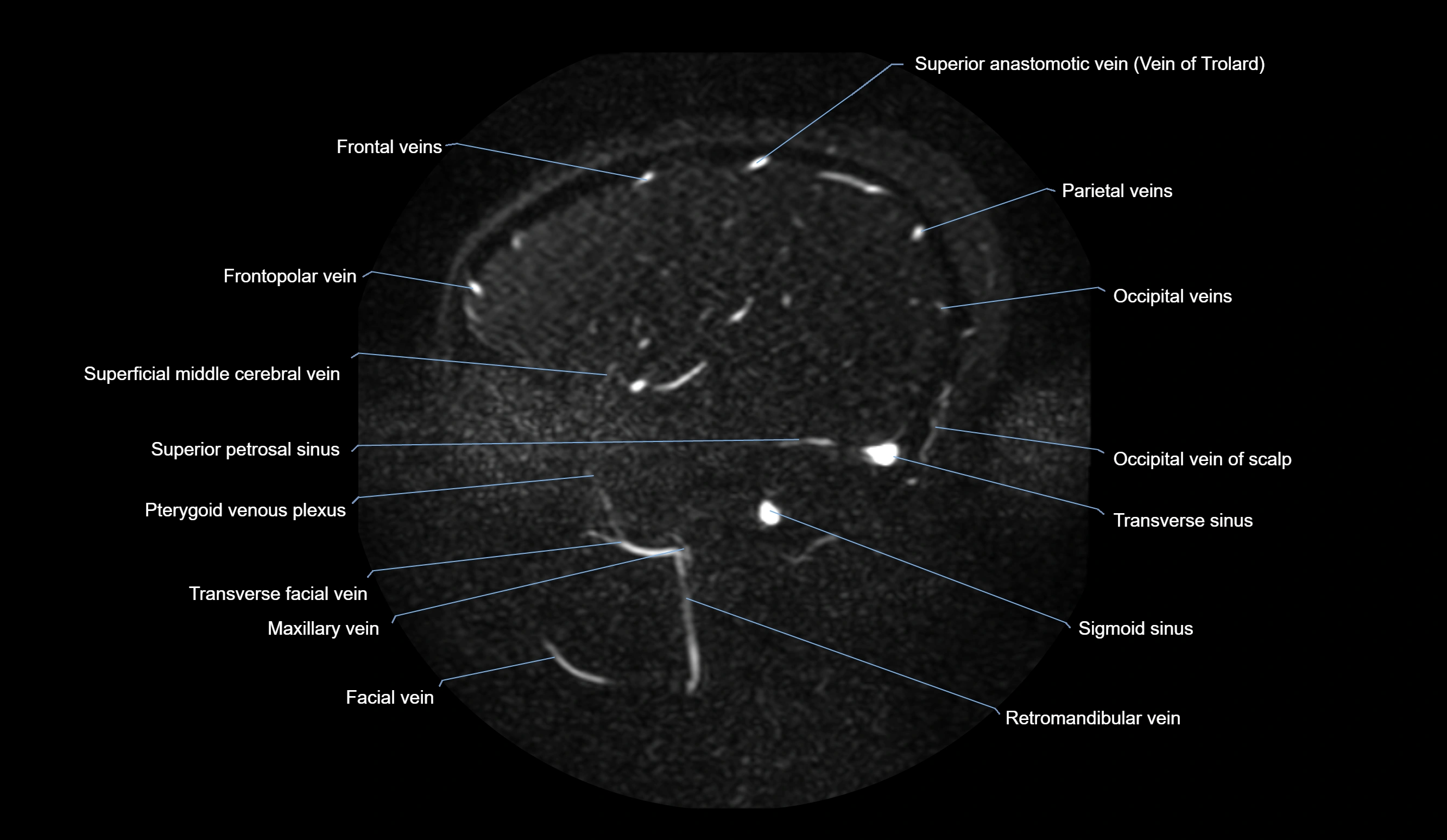
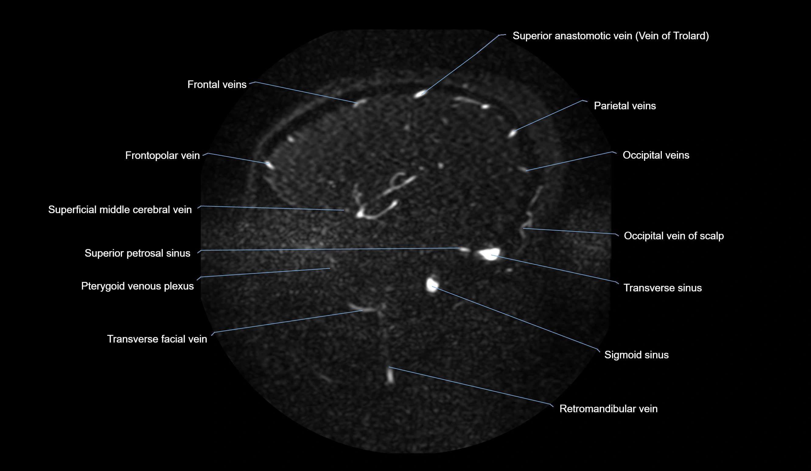
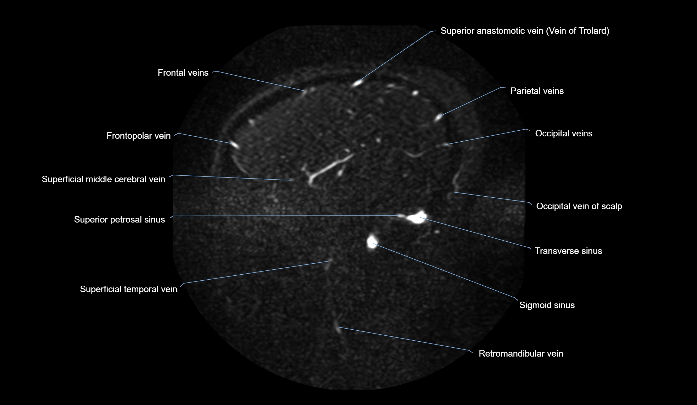
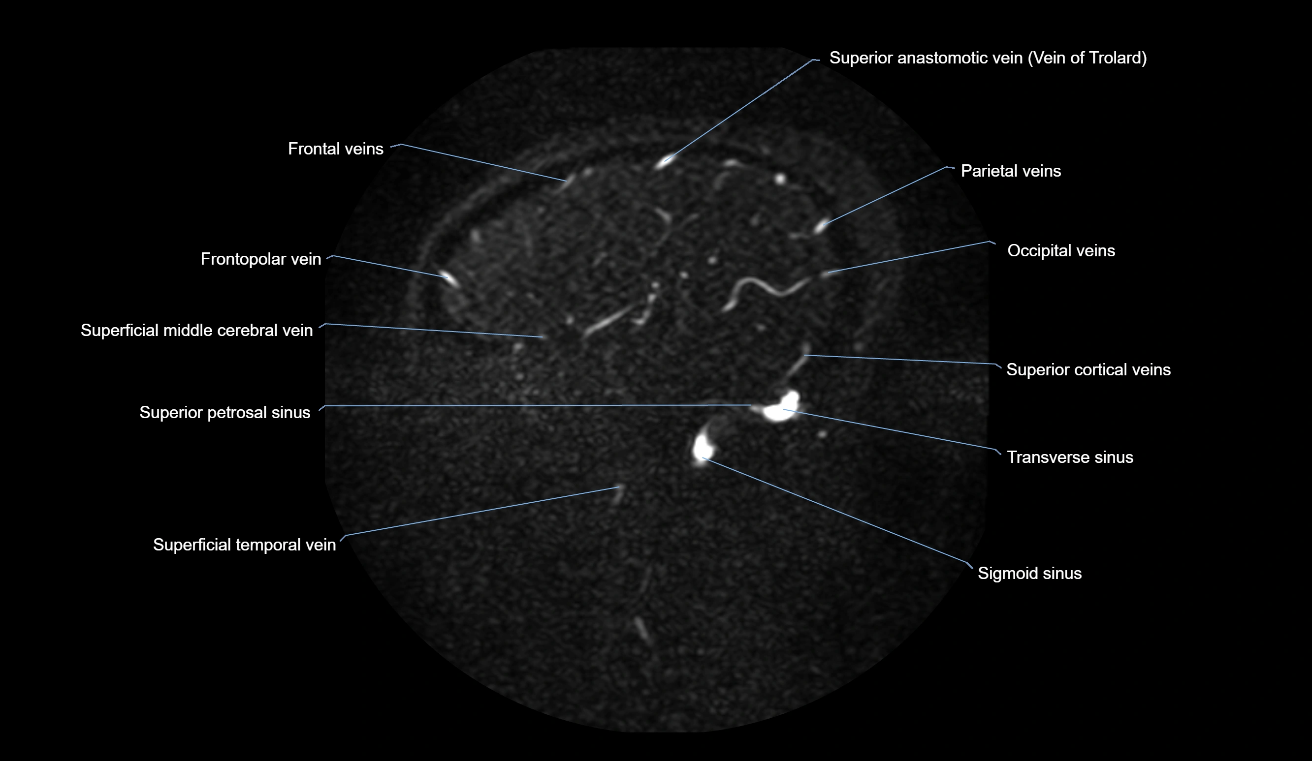
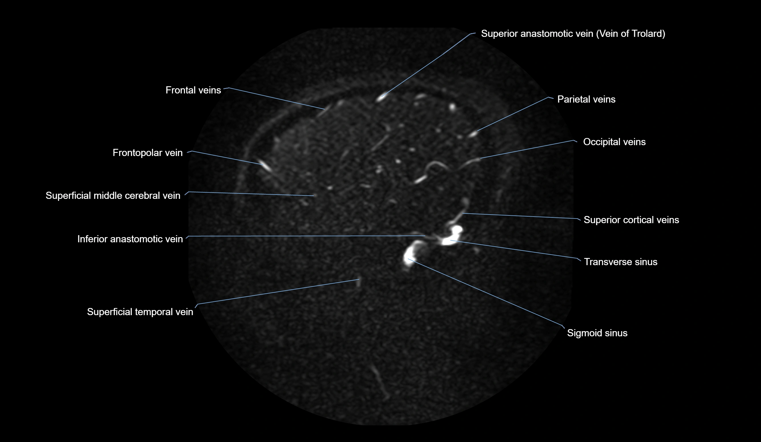
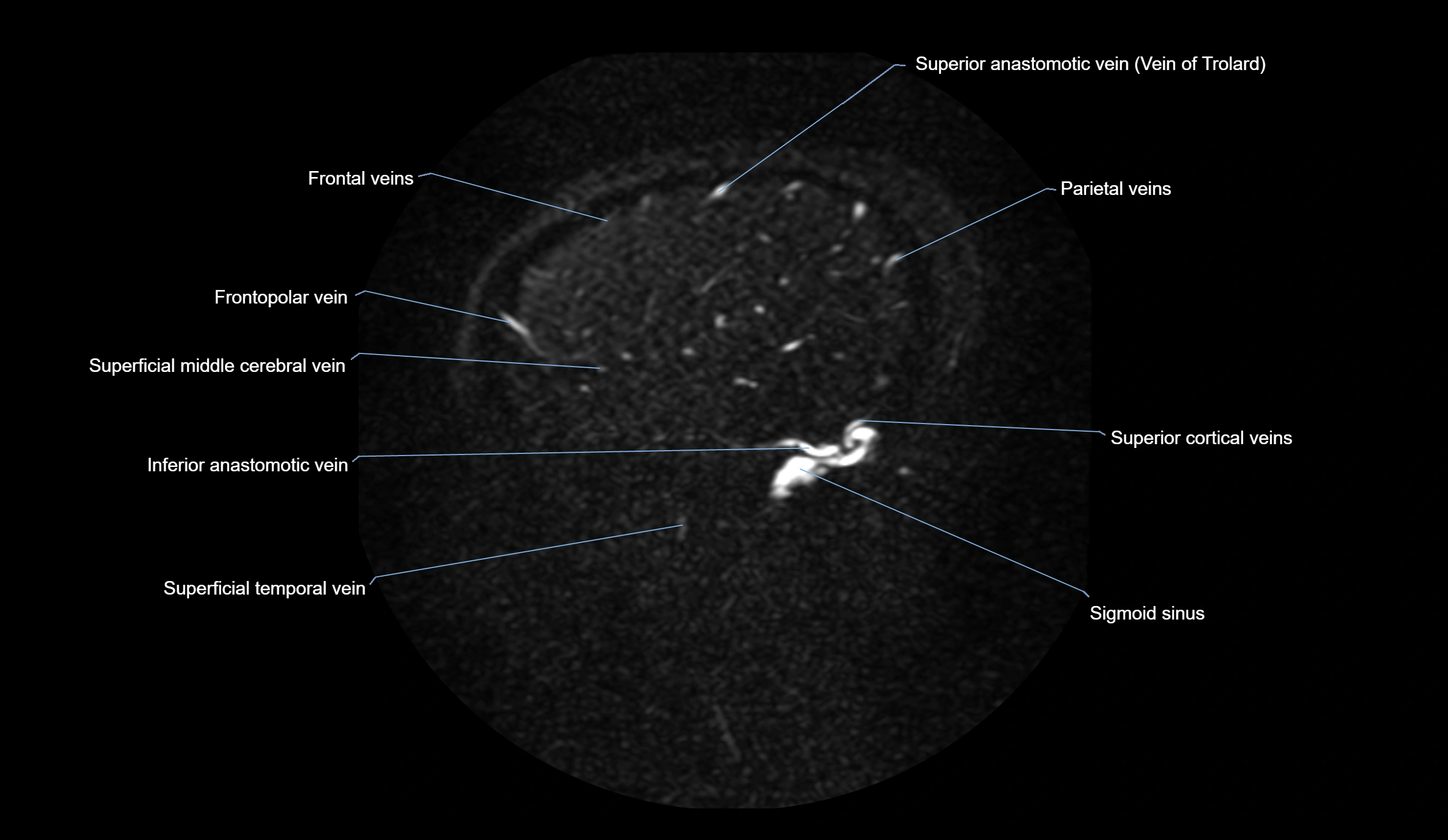
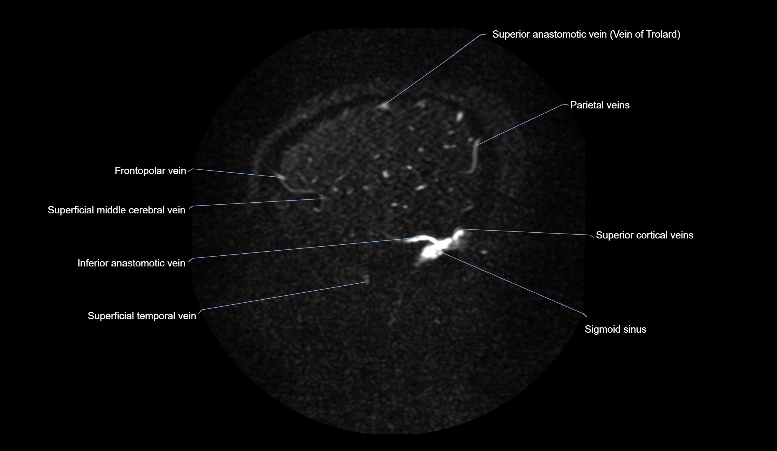
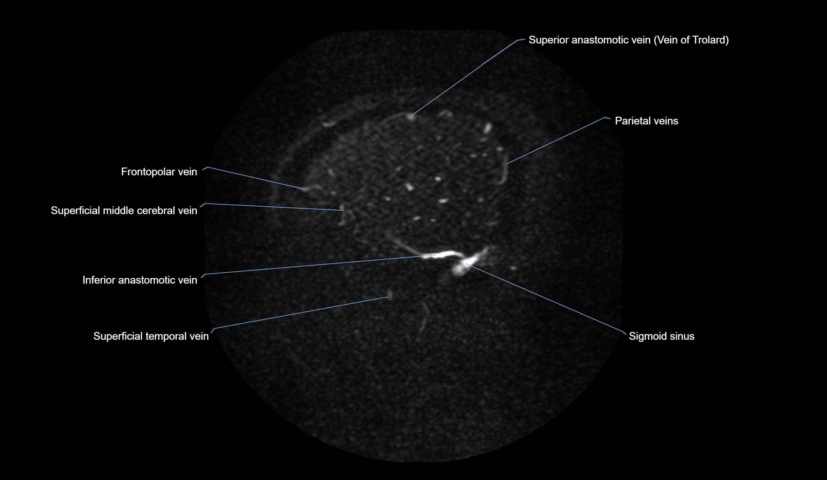
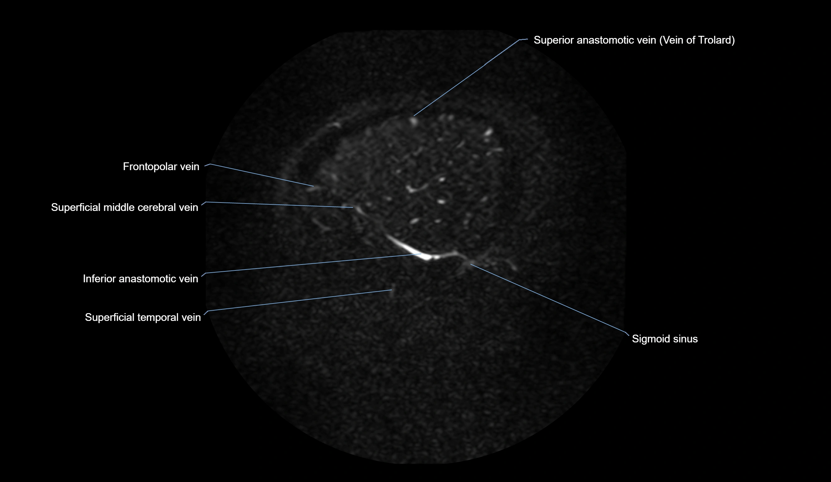
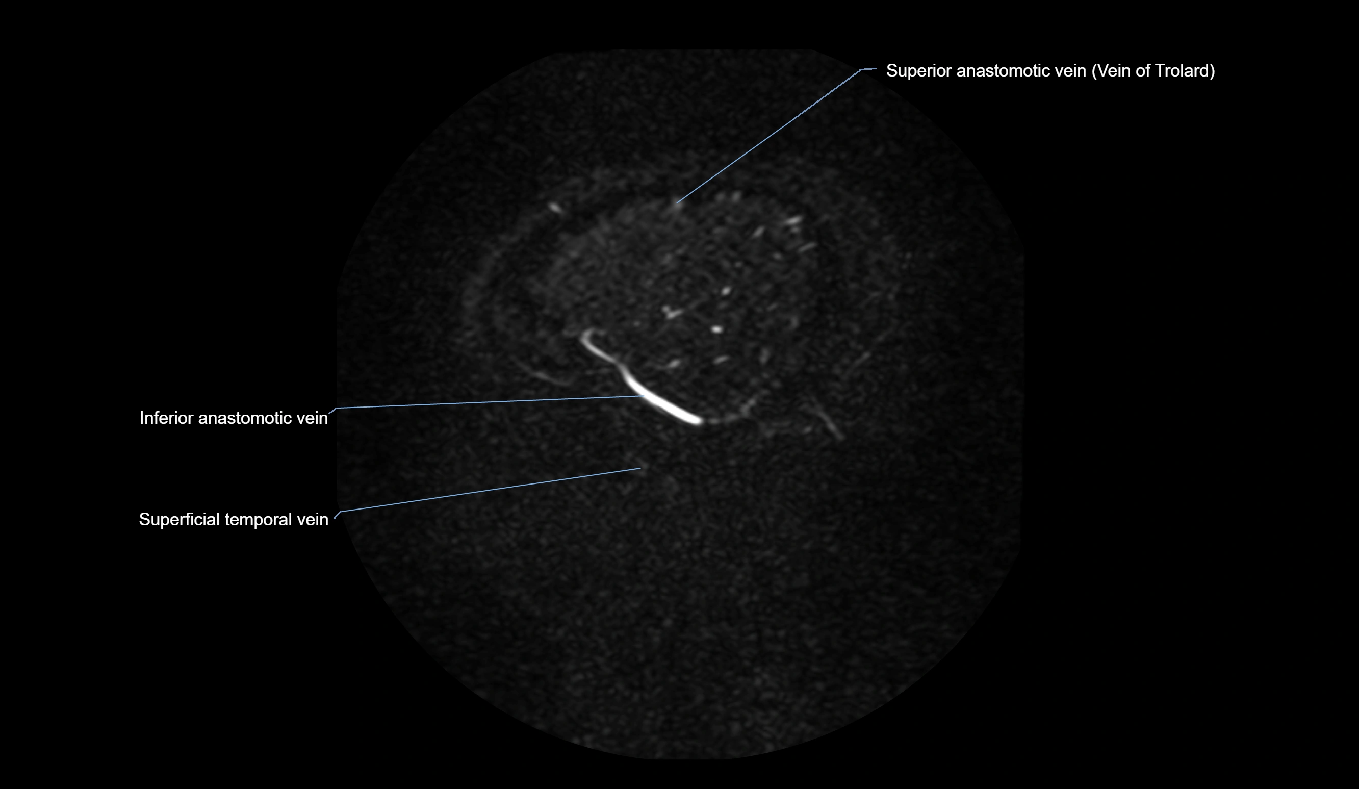
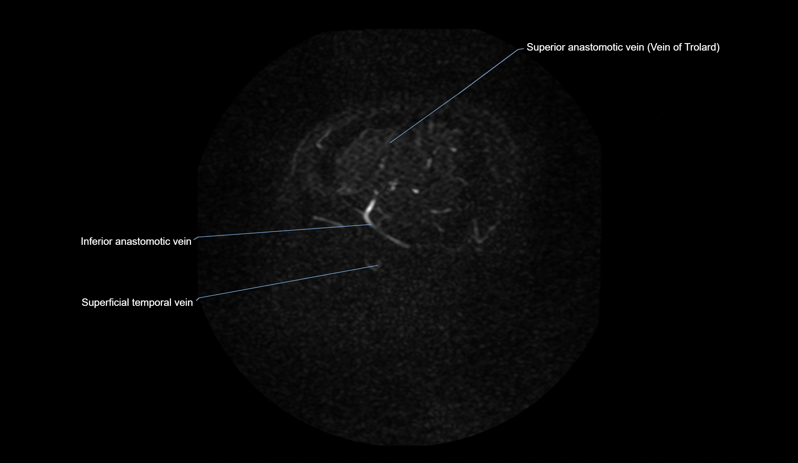
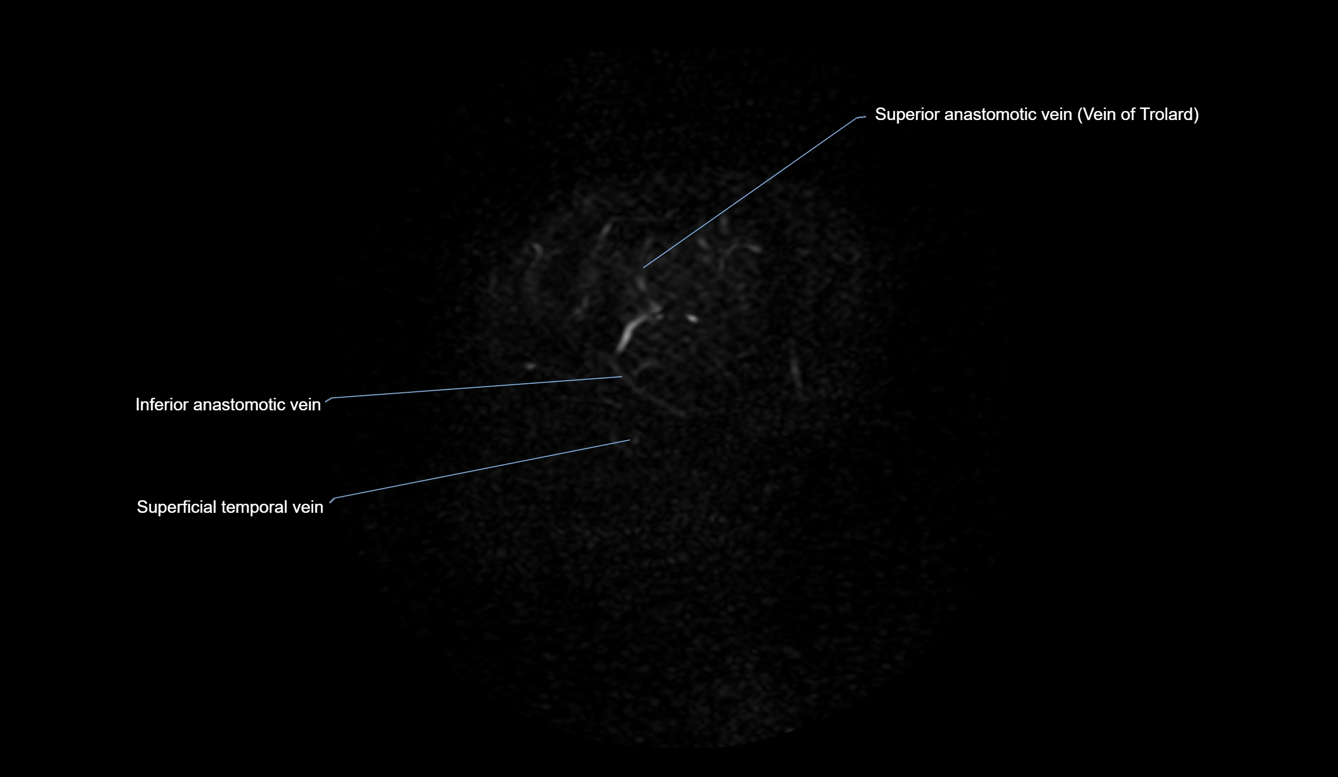
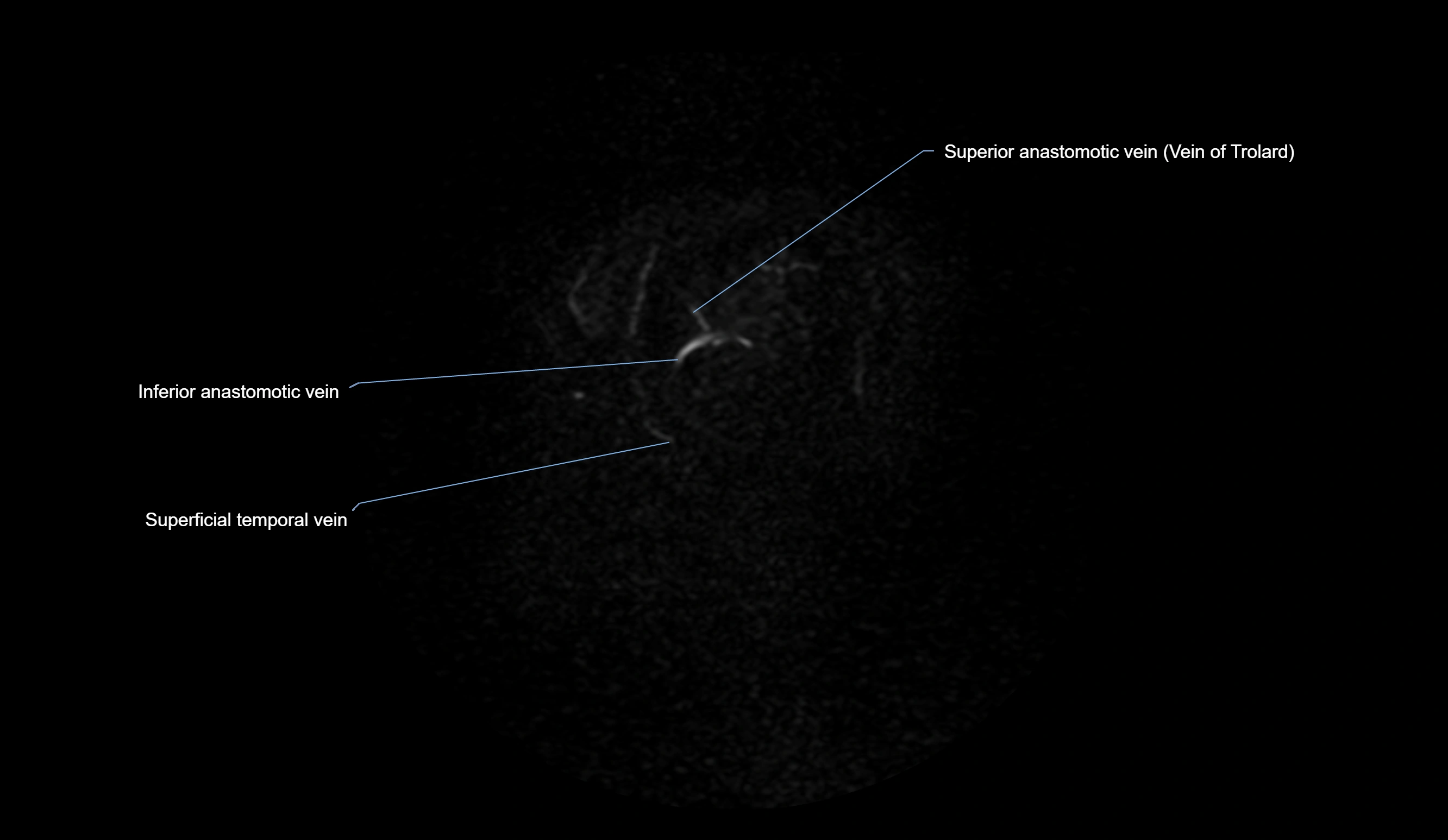
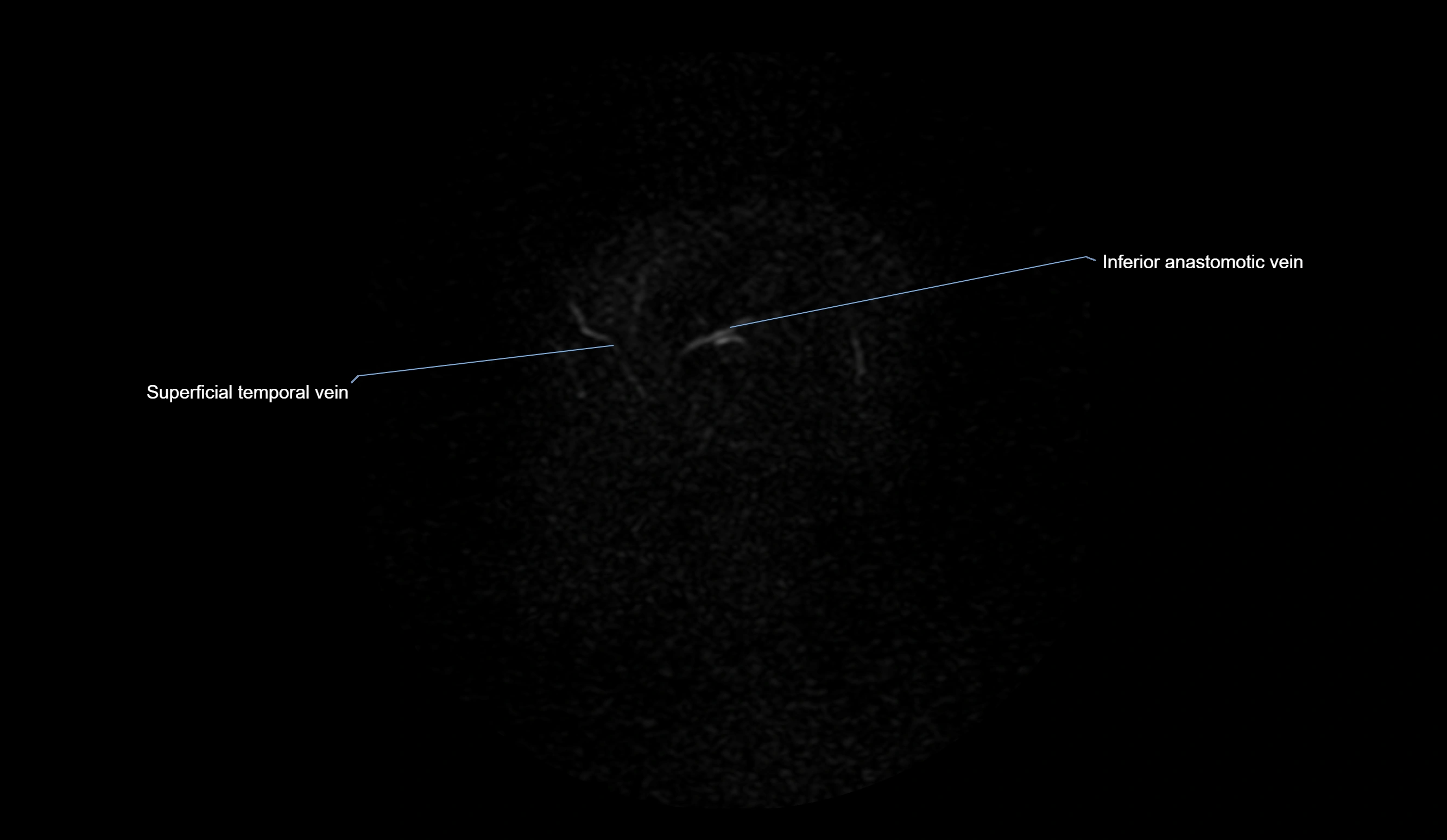
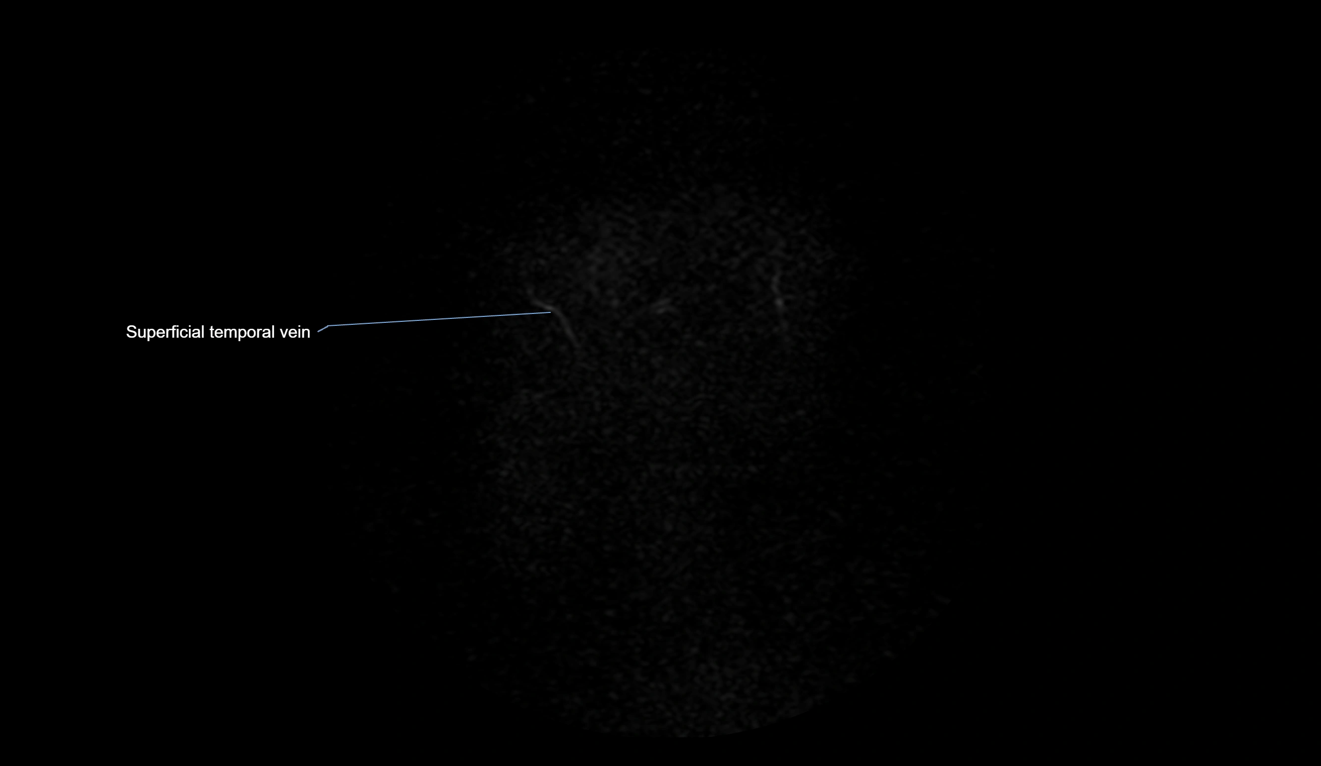
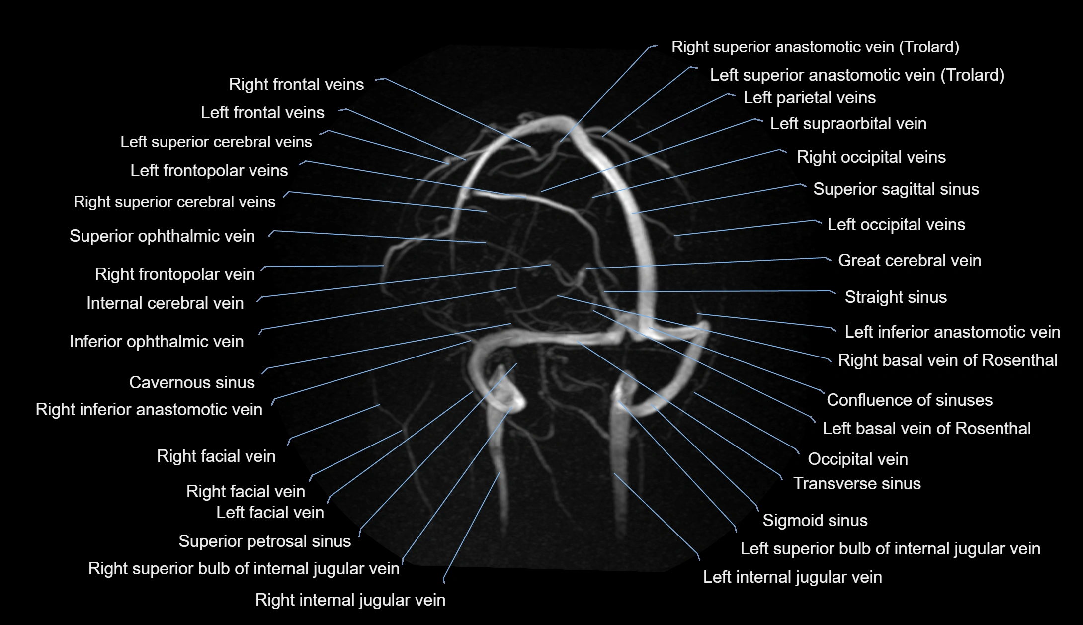
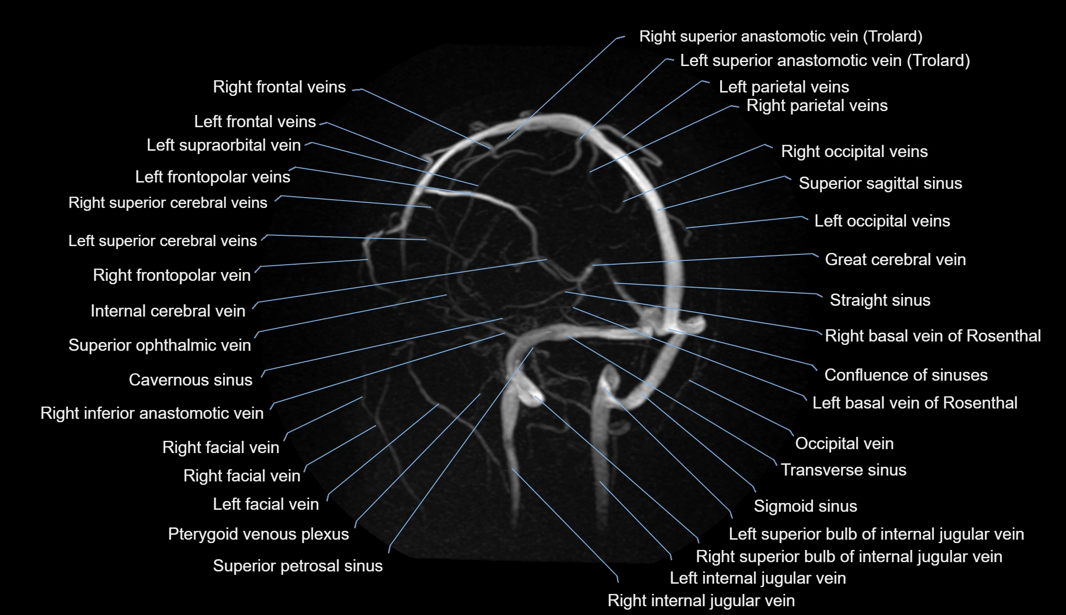
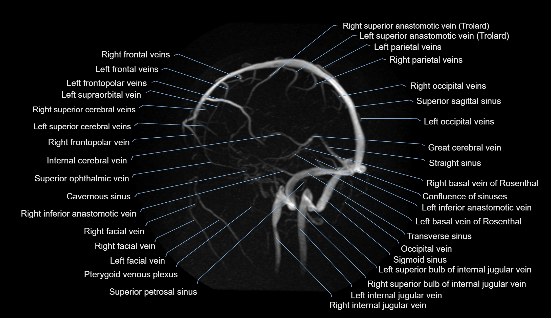
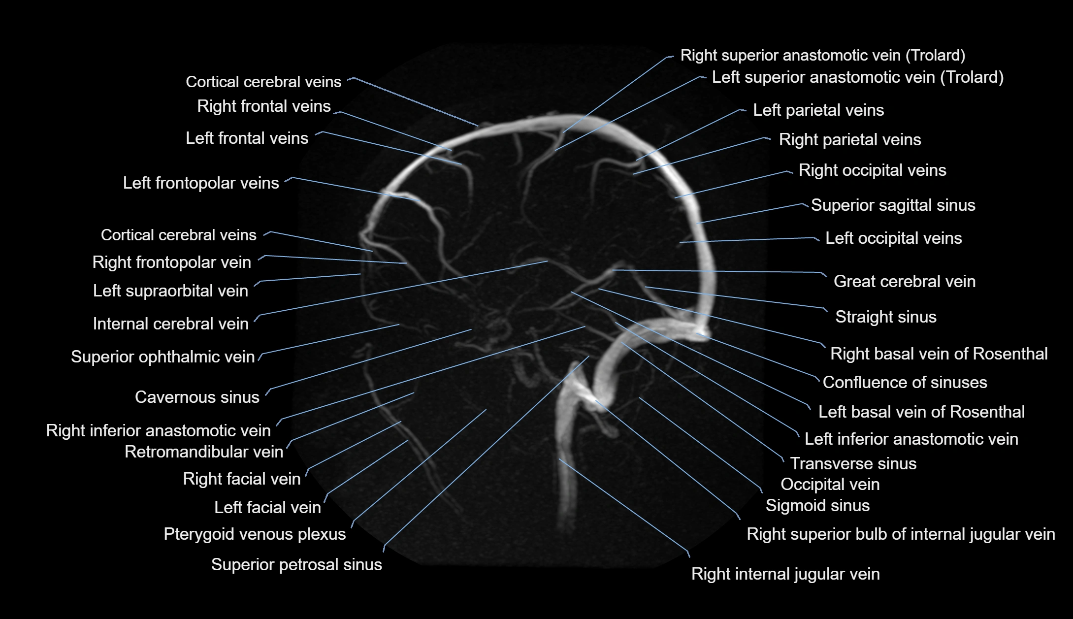
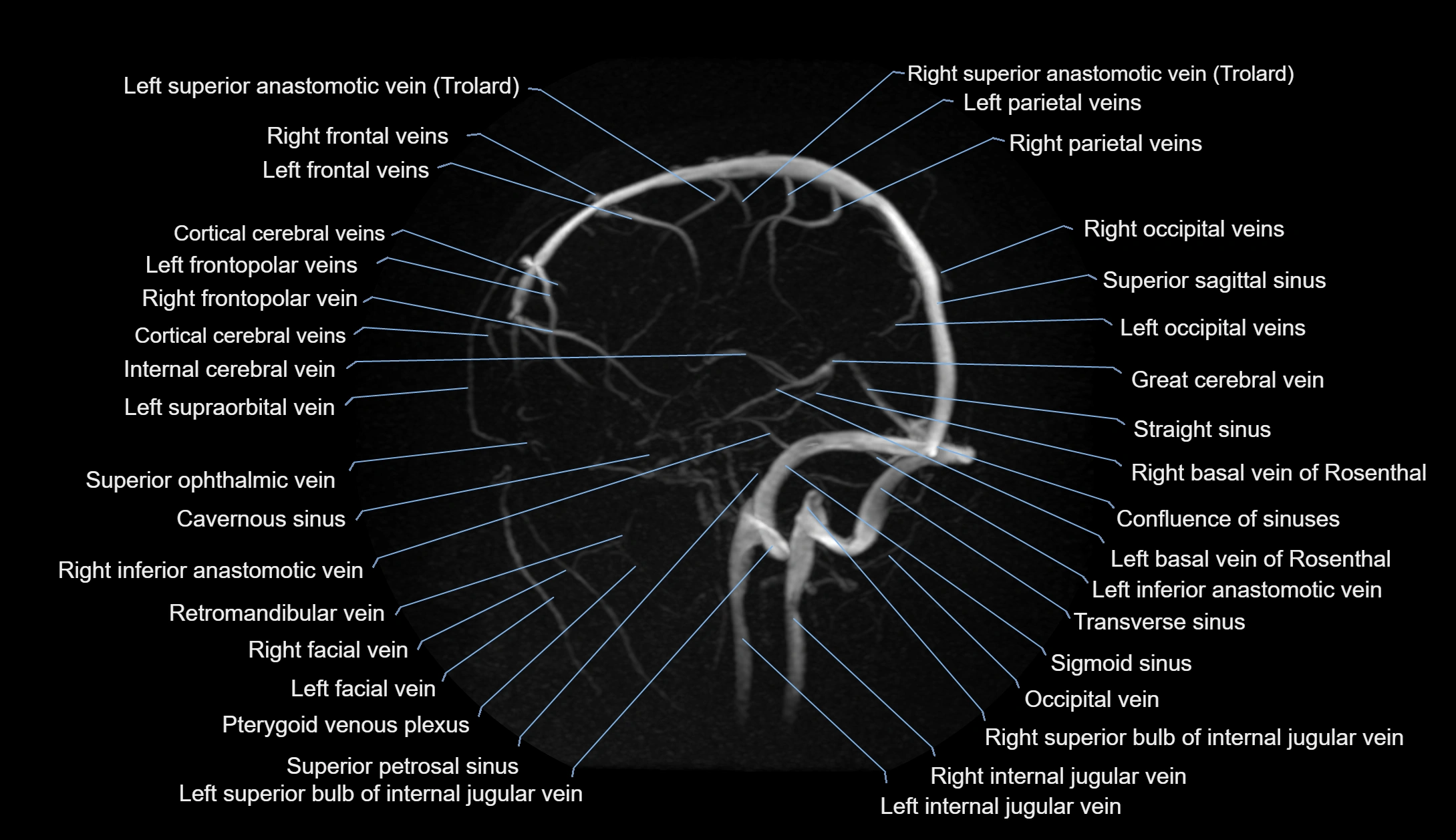
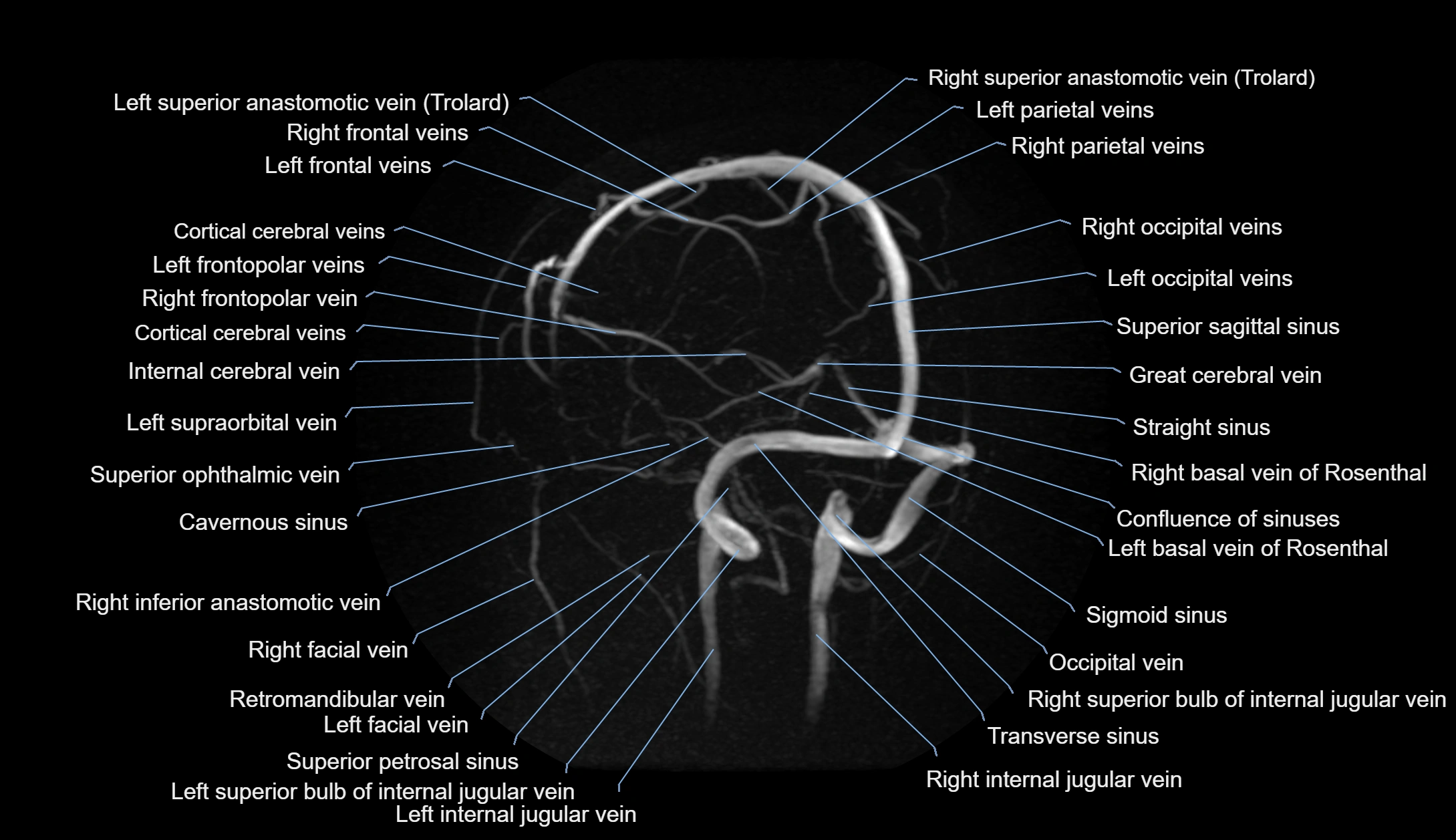
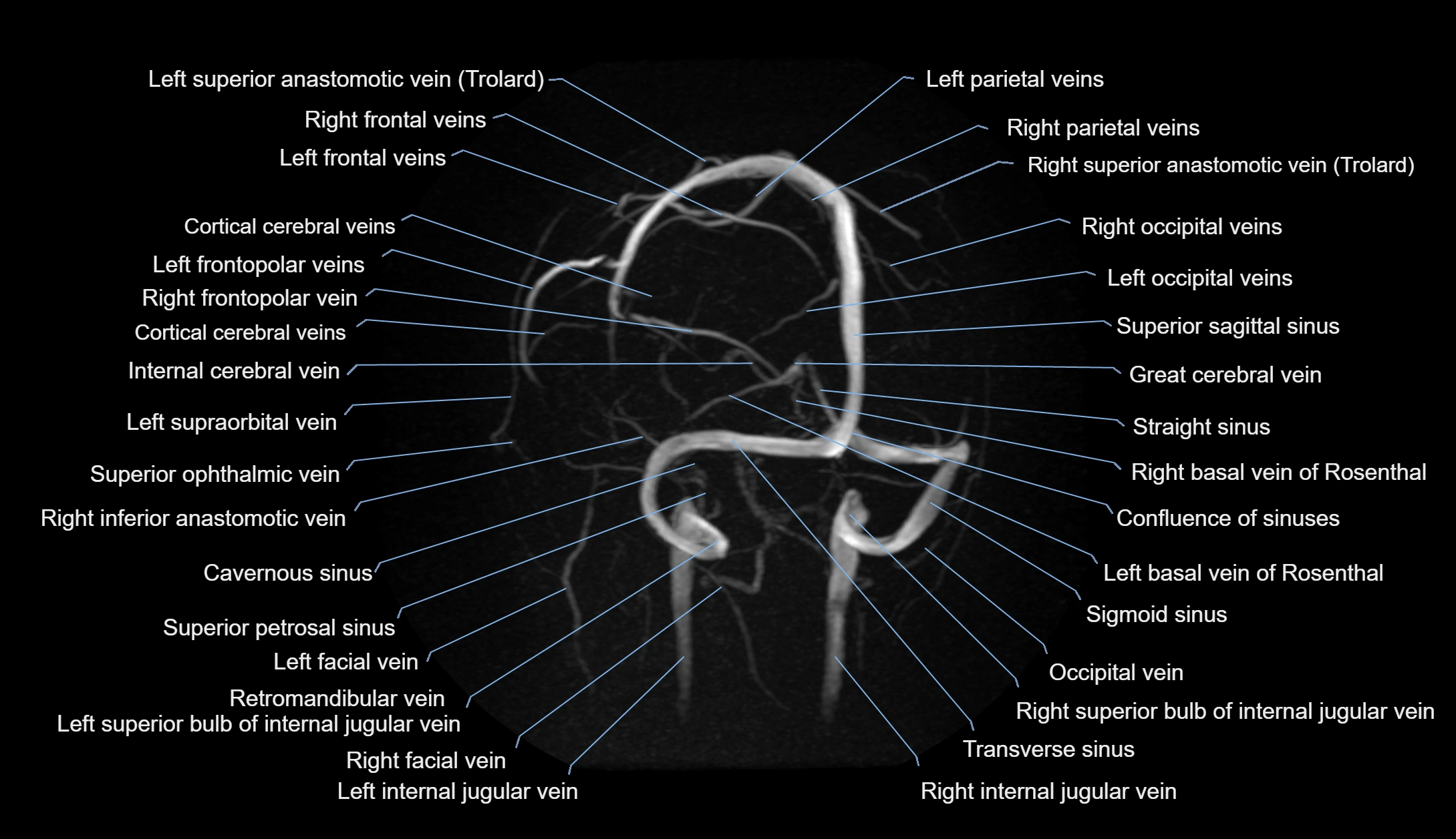
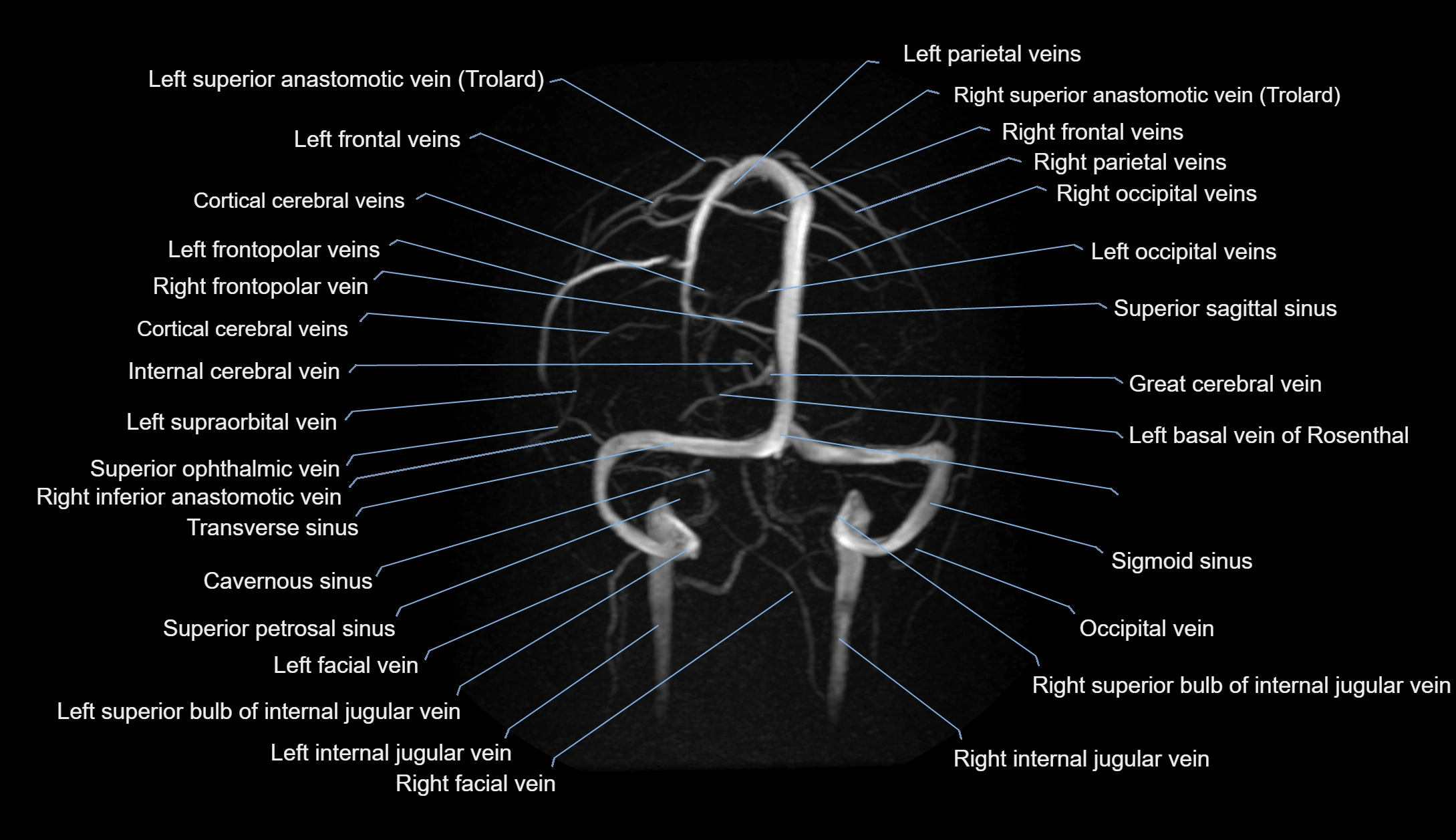
MRI images


