


Topic
The anterior jugular vein is a paired superficial vein located in the anterior aspect of the neck. It plays an important role in draining blood from the superficial structures of the anterior neck region. Its course, anatomical variations, and connections with other neck veins are clinically significant, especially in the context of central venous access, neck surgeries, and radiological imaging. Understanding its normal and variant anatomy is essential for accurate image interpretation and avoiding complications during procedures.
Synonyms:
-
Superficial anterior cervical vein
-
Vena jugularis anterior (Latin)
Function:
-
Drains blood from the submental region and anterior neck
-
Empties into the external jugular vein or directly into the subclavian vein
-
Helps maintain venous return from superficial structures of the neck
Imaging Appearance:
MRI Appearance:
-
T1-Weighted Images:
-
Appears as a tubular, hypointense (dark) structure relative to muscle
-
May show flow void if the blood flow is fast
-
-
T2-Weighted Images:
-
Typically hypointense or isointense to muscle, but can be hyperintense if slow flow or stasis is present
-
-
STIR (Short Tau Inversion Recovery):
-
Vein remains hypointense, but surrounding edema or inflammation will appear hyperintense
-
CT Appearance:
-
Appears as a small, thin-walled, contrast-enhancing vein in the anterior neck
-
Best visualized in the lower neck just lateral to the midline
-
Non-contrast CT: difficult to differentiate from surrounding soft tissue, may appear as a hypoattenuating tubular structure
CTA (CT Angiography):
-
Enhances brightly with intravenous contrast
-
Easily visualized on arterial and especially venous phase images
-
Shows continuity with external jugular vein or subclavian vein
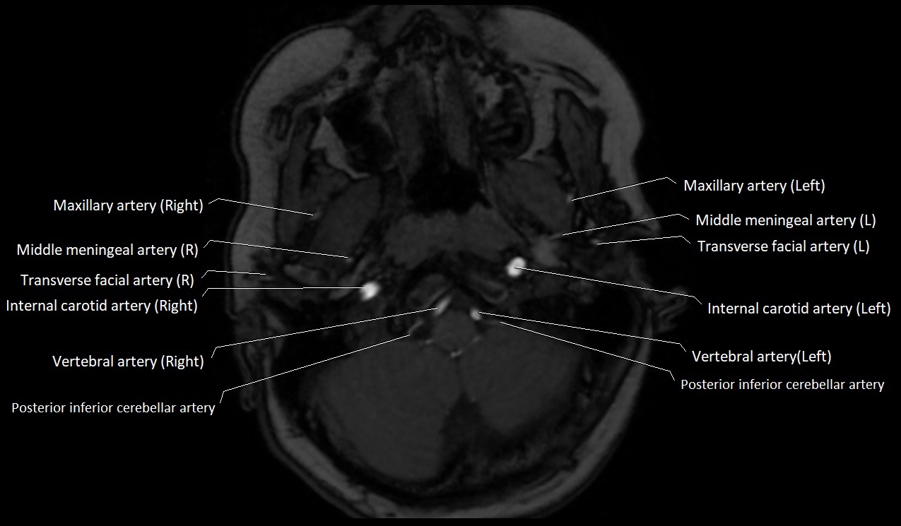
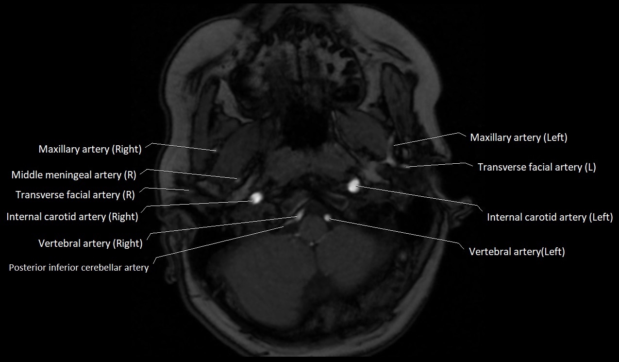
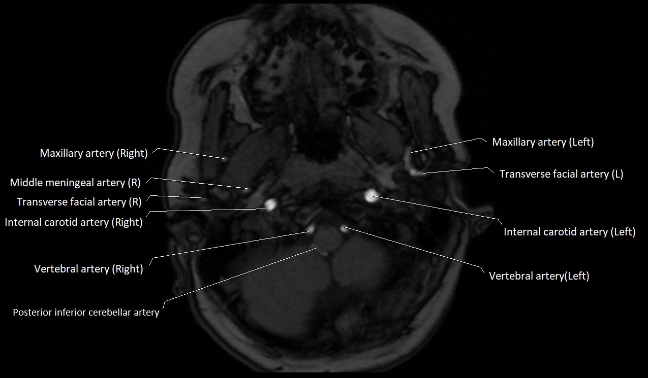
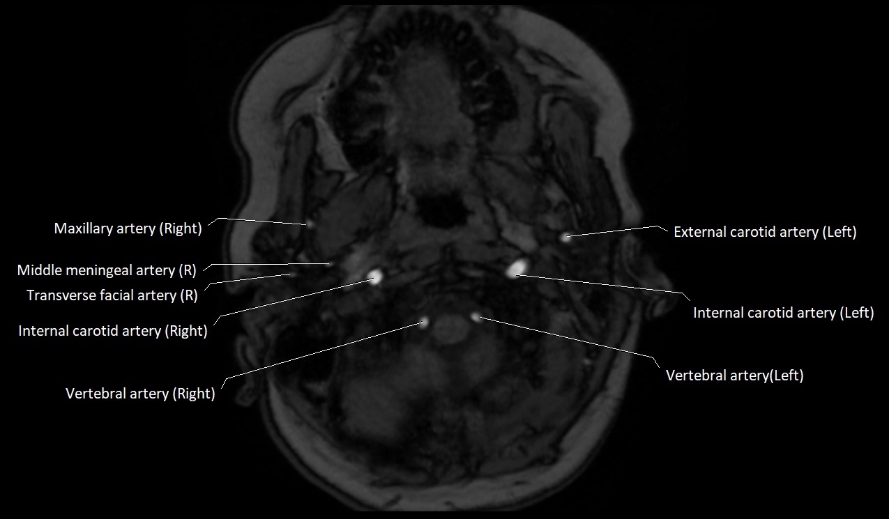
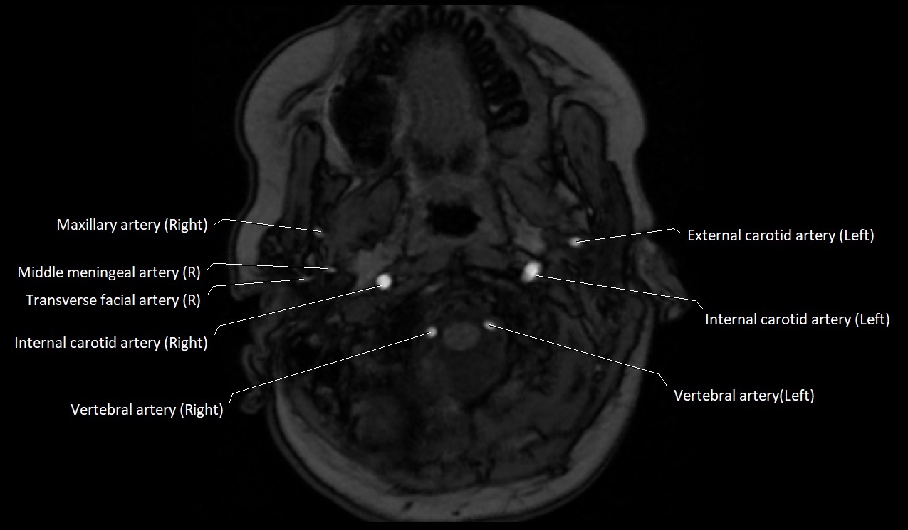
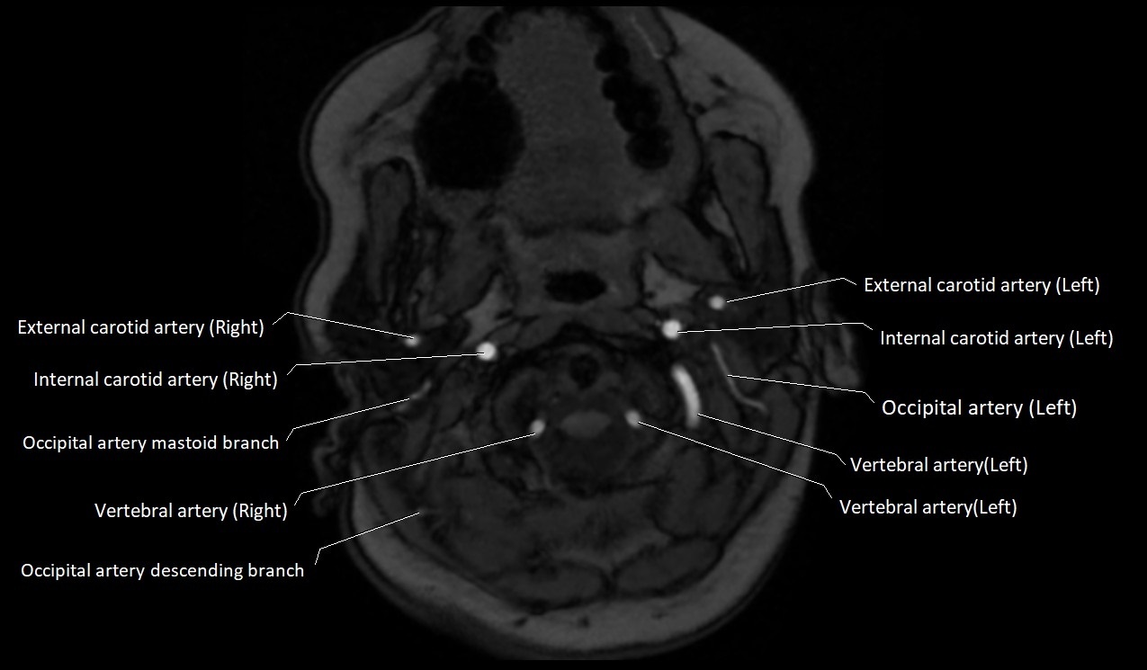
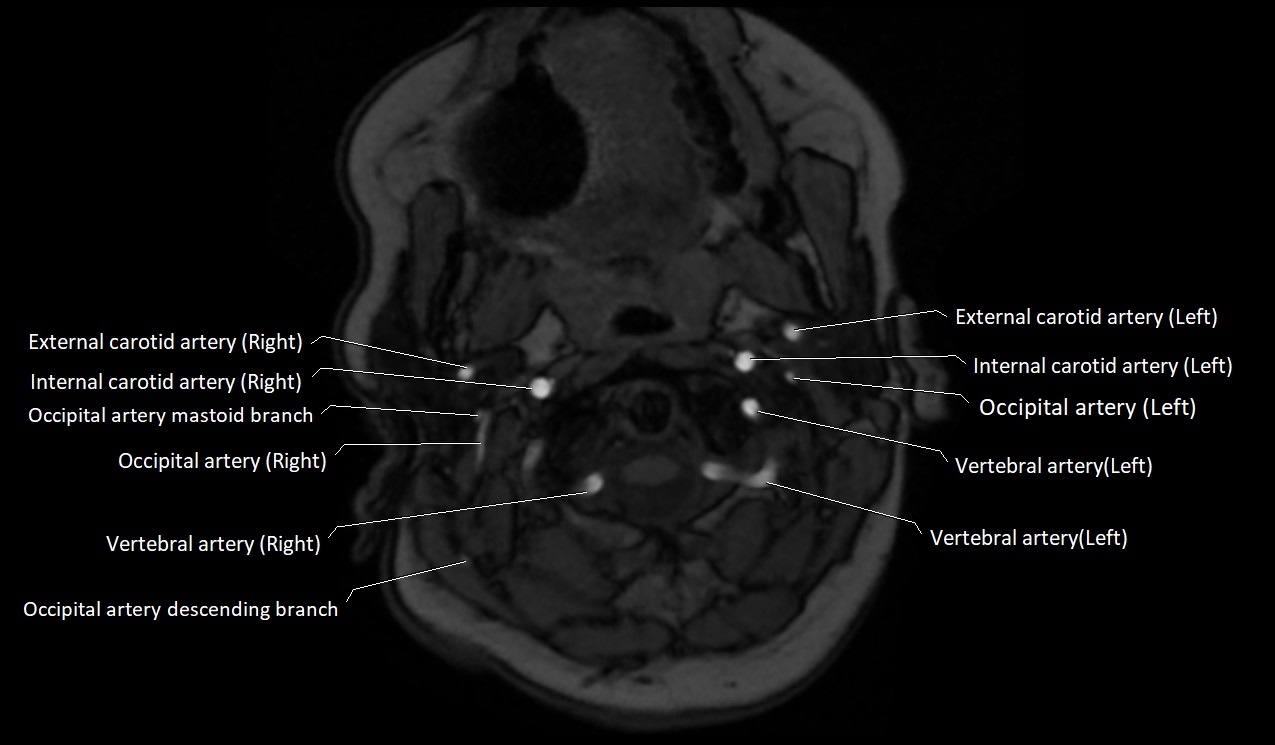
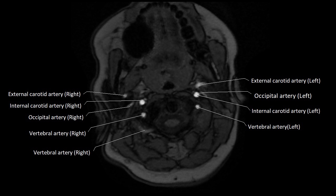
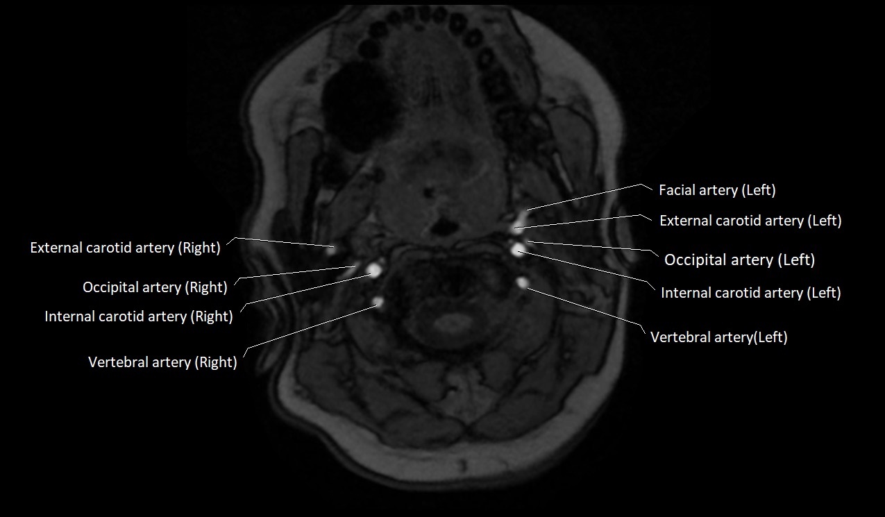
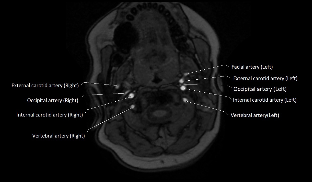
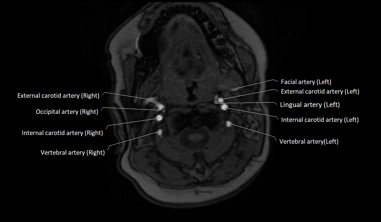
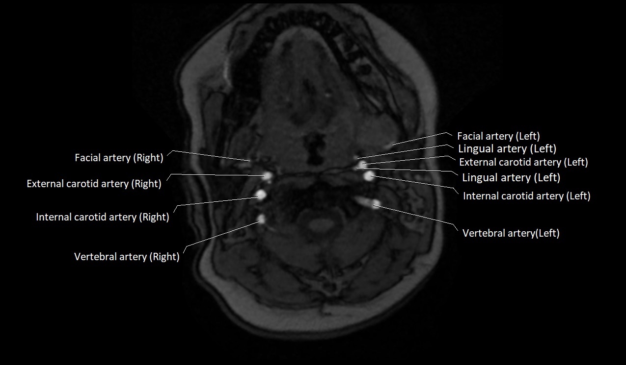
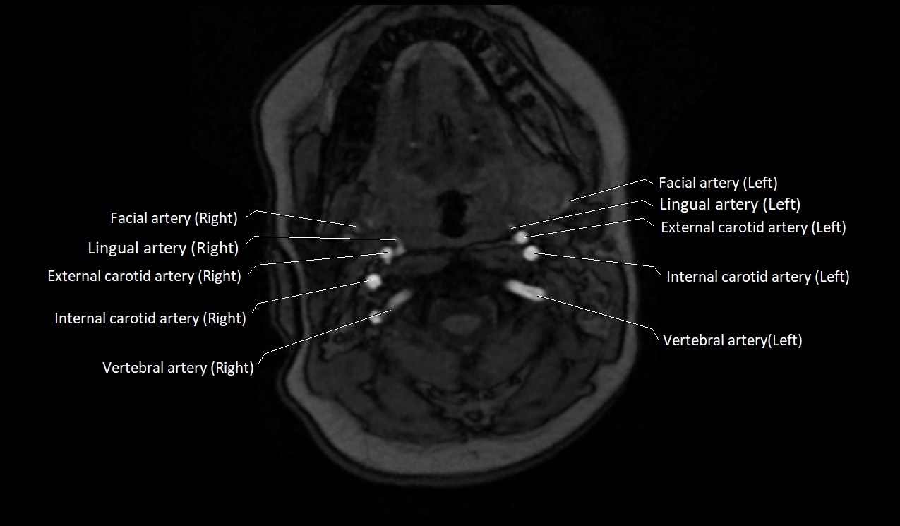
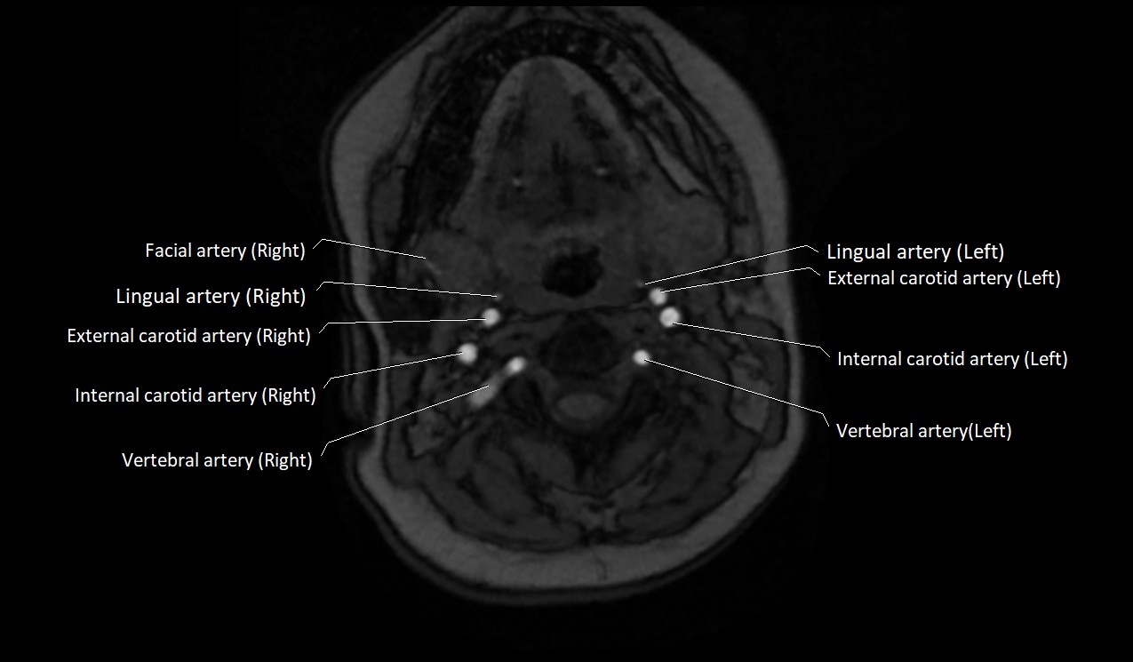
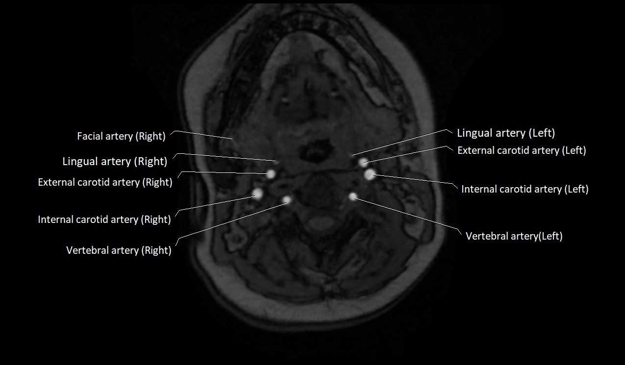
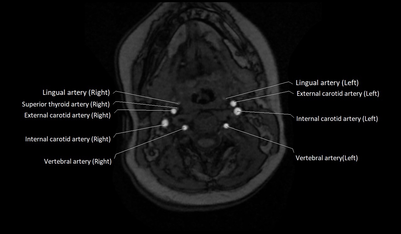
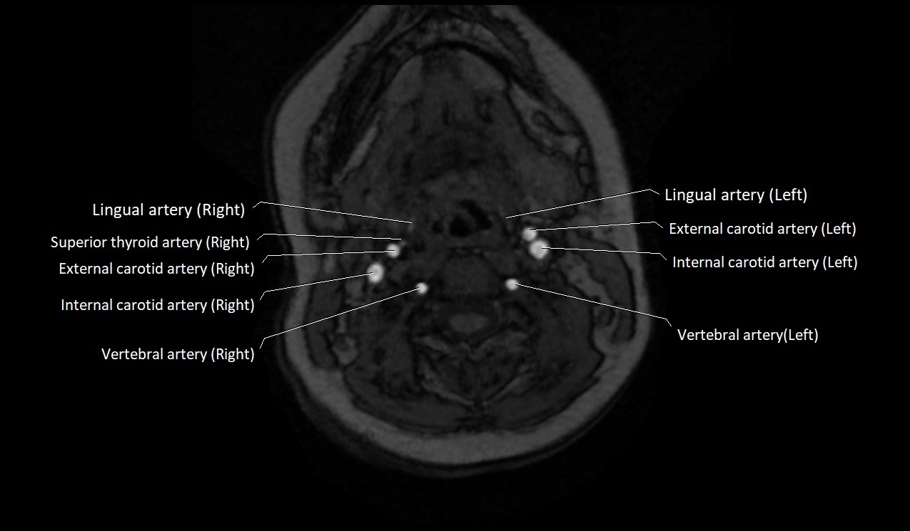
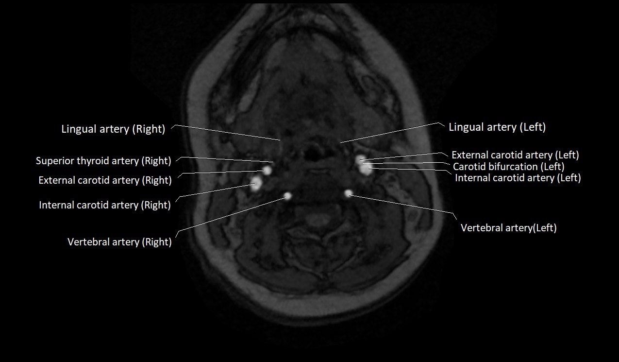
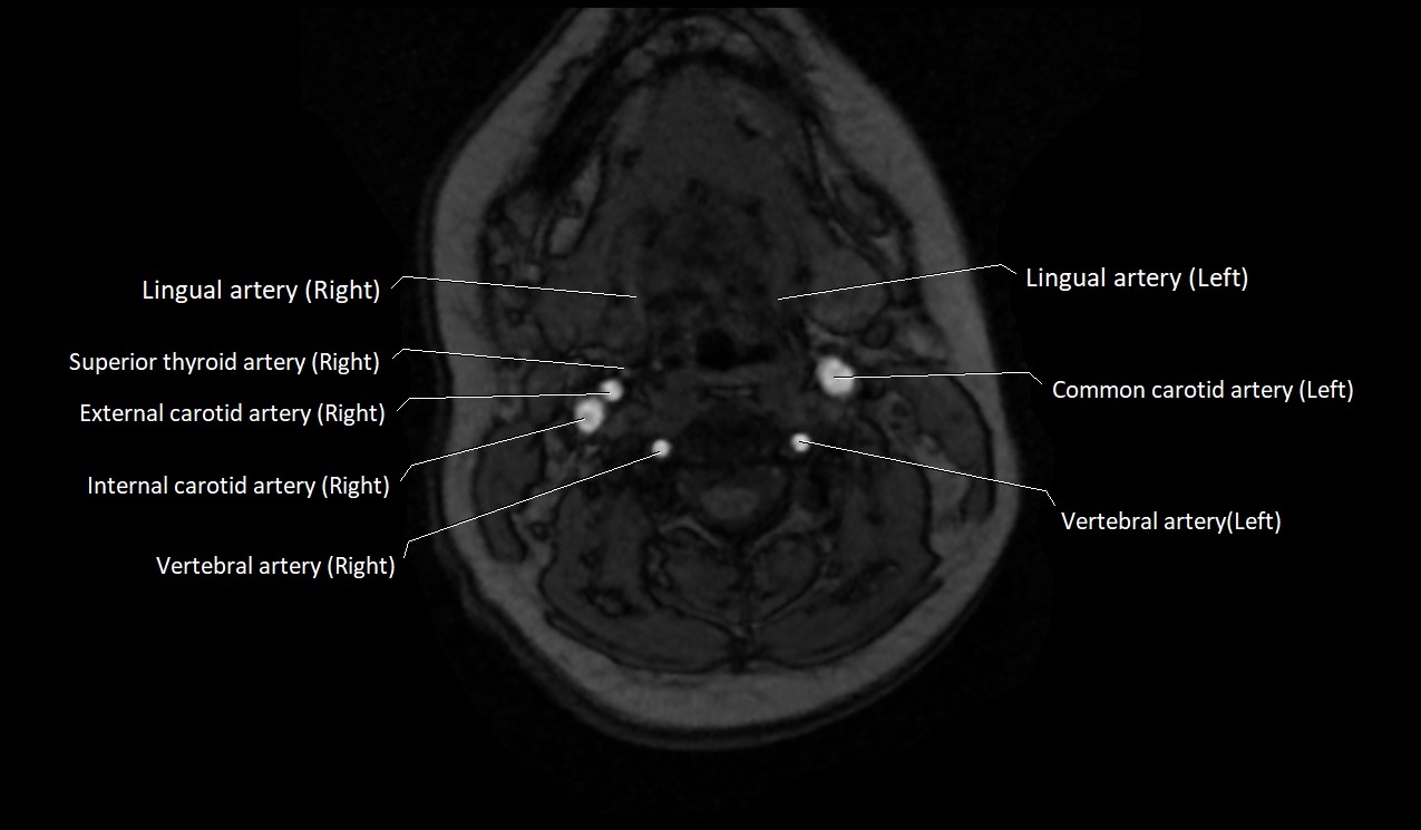
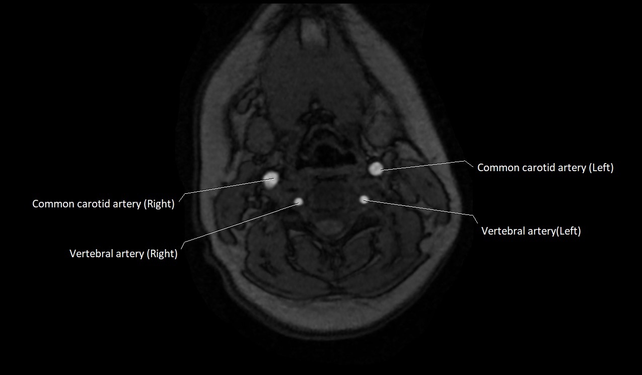
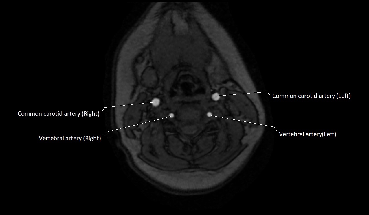
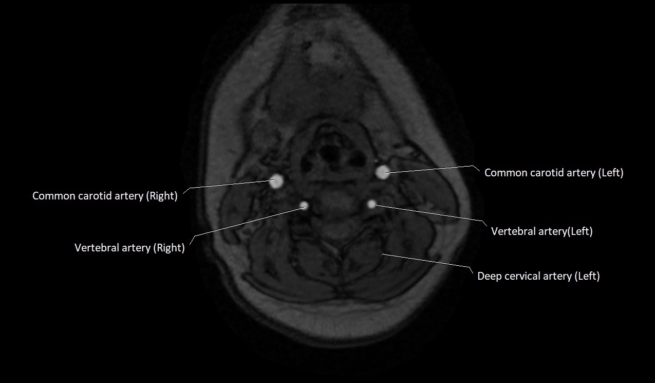
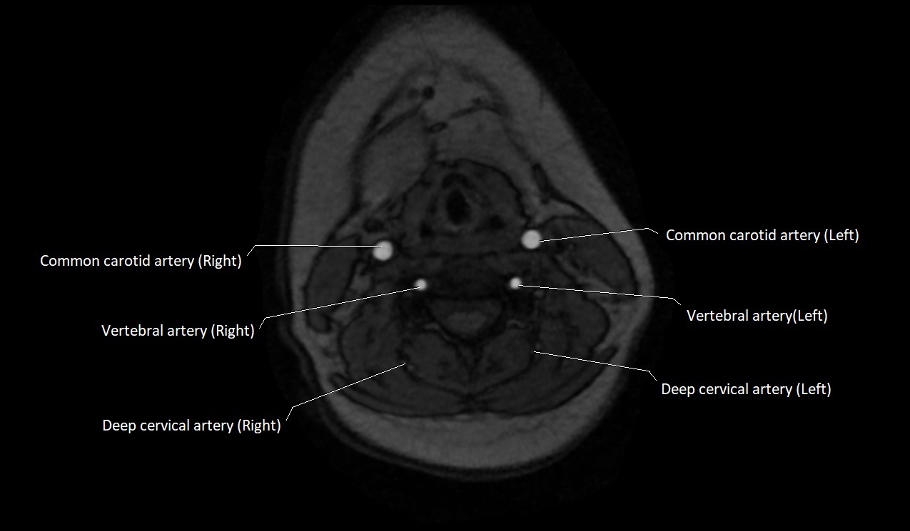
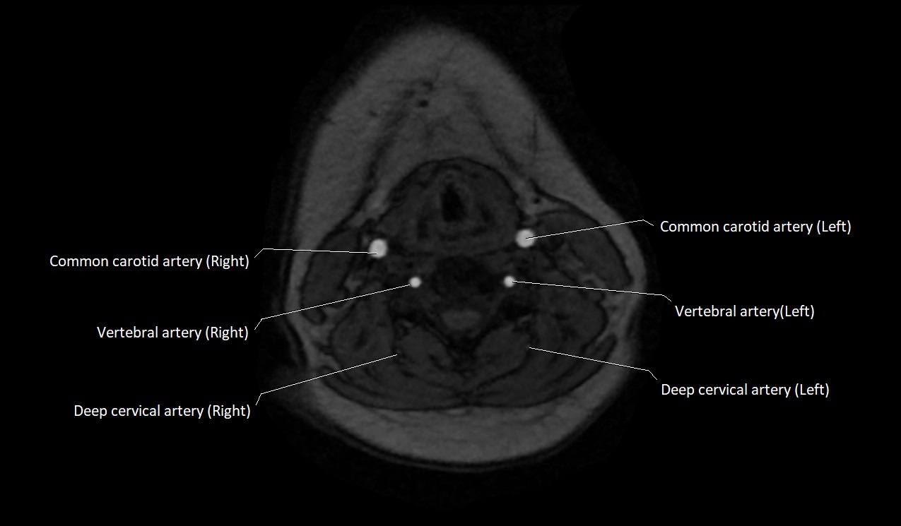
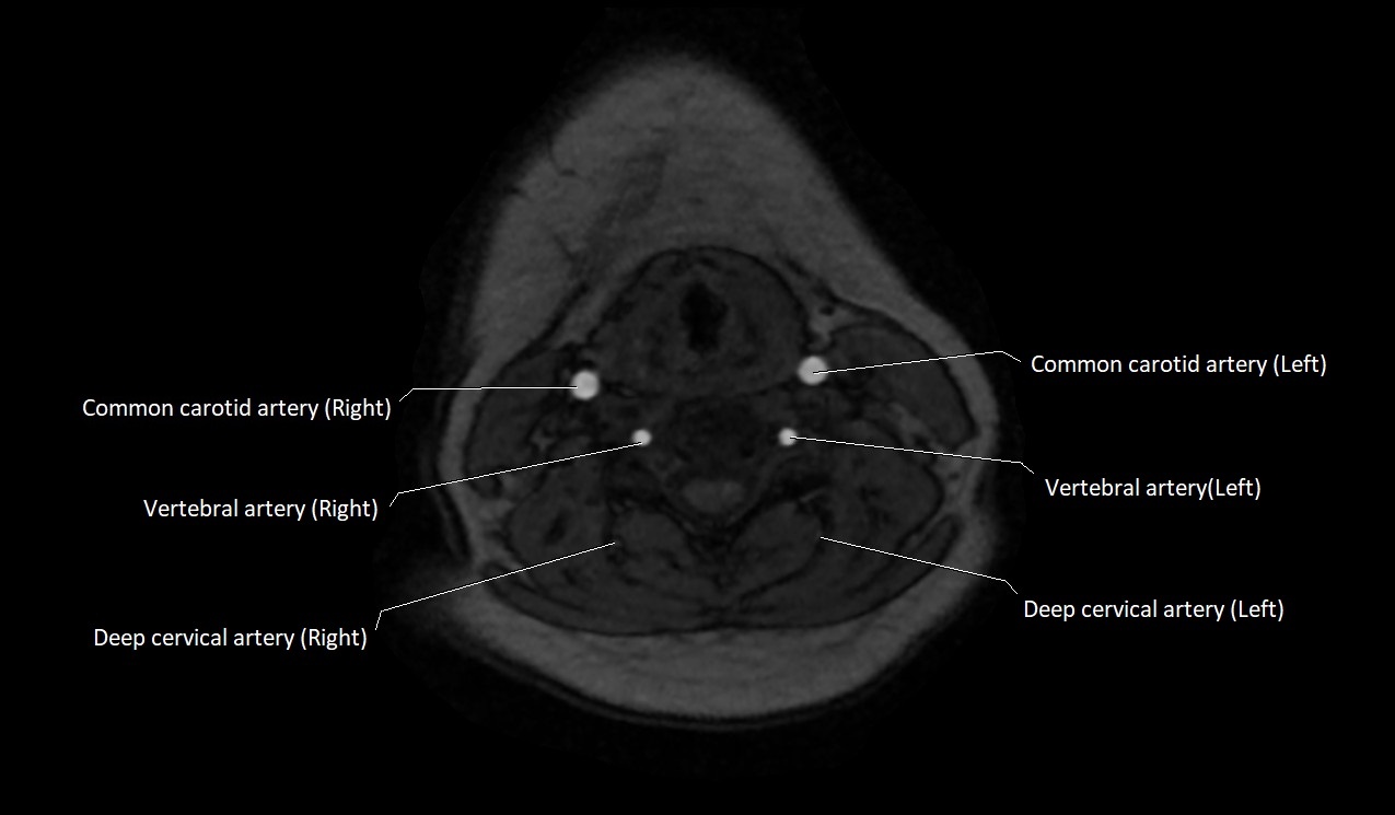
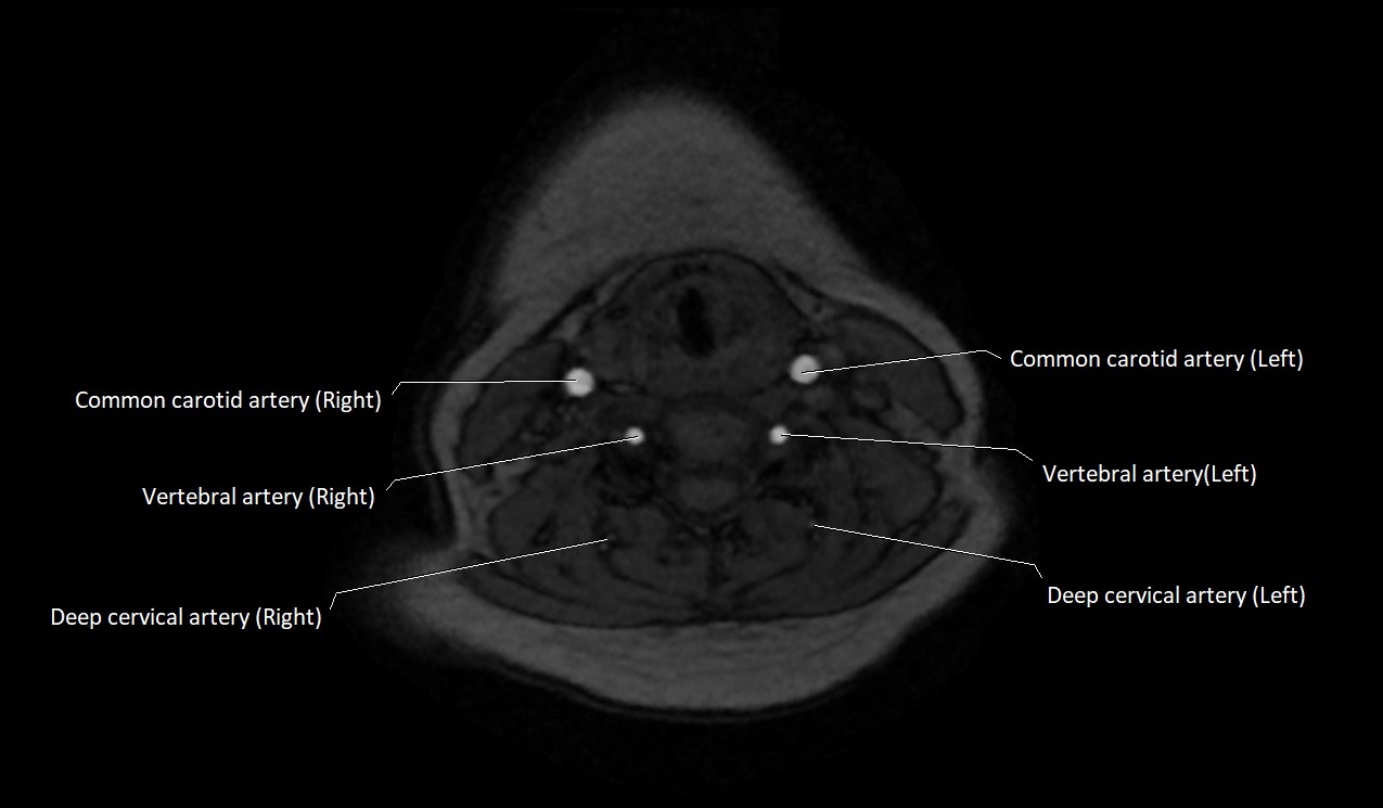
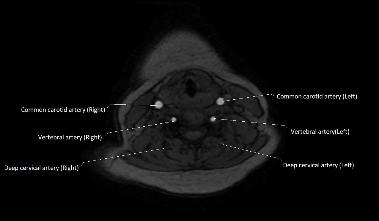
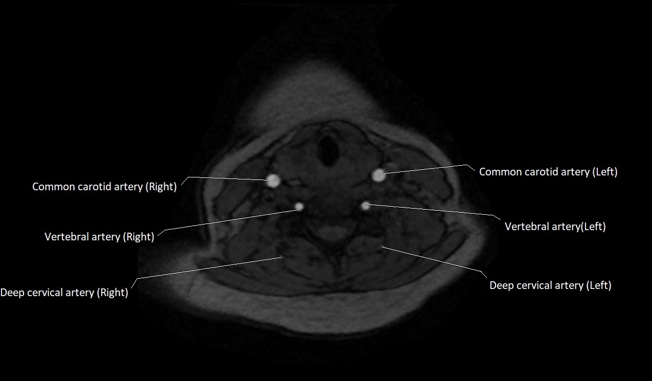
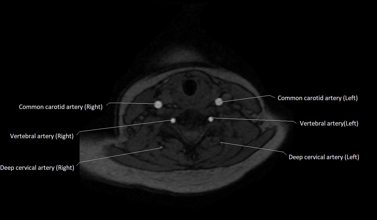
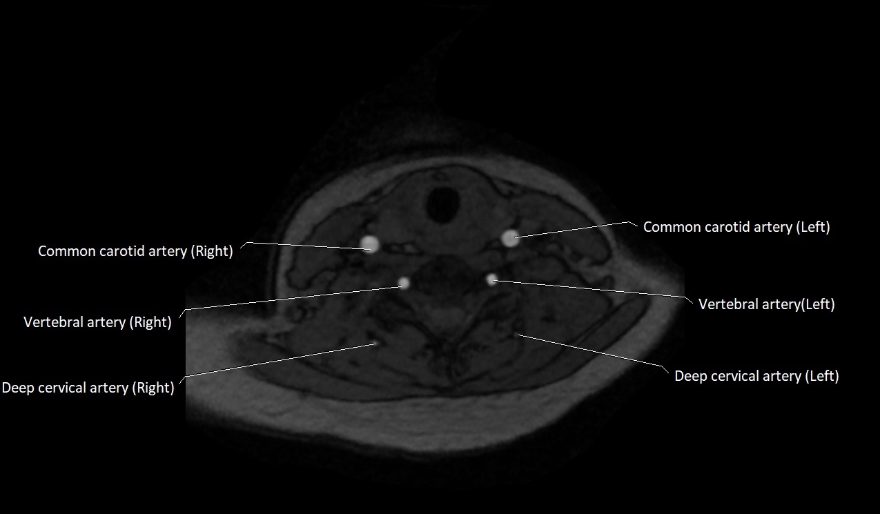
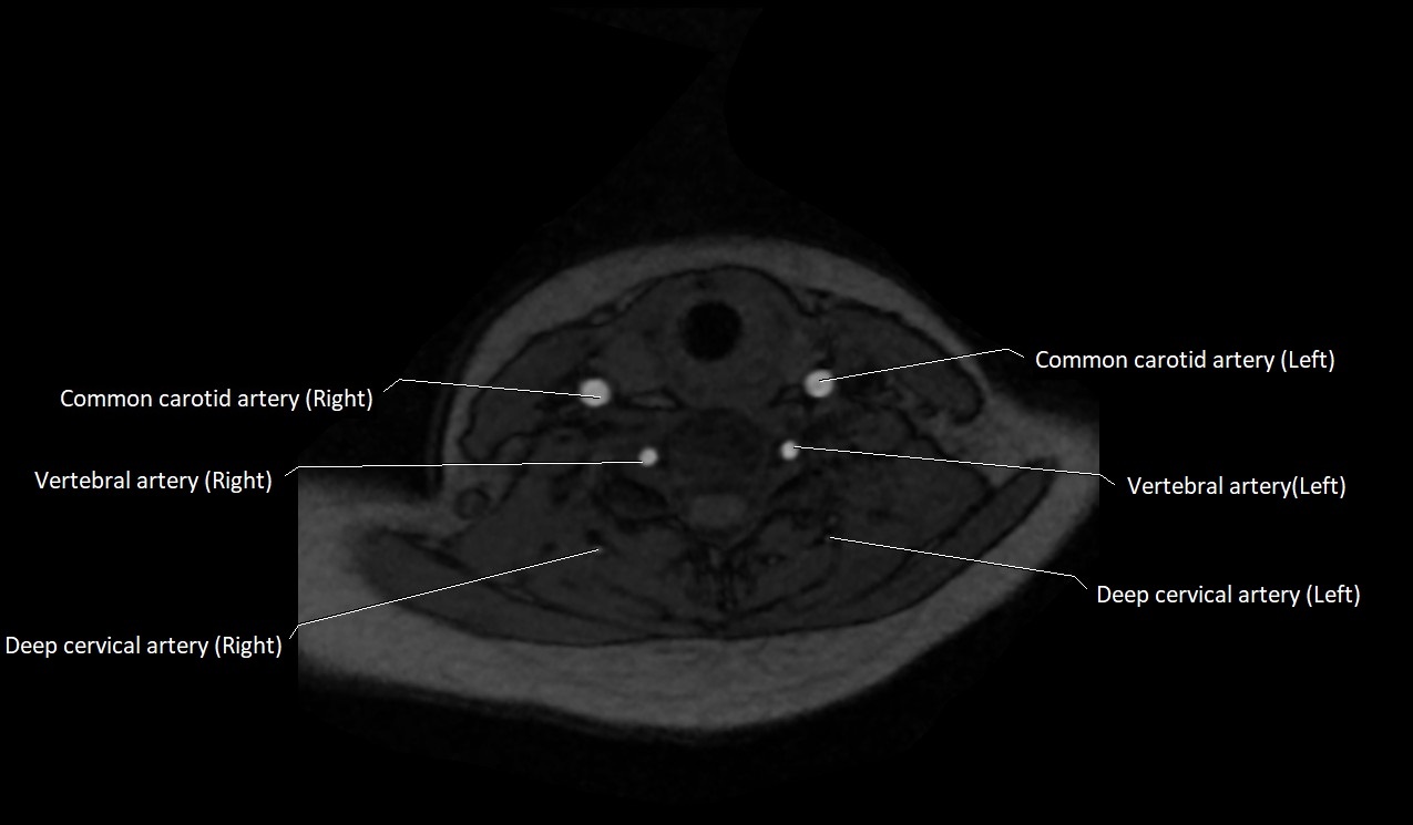
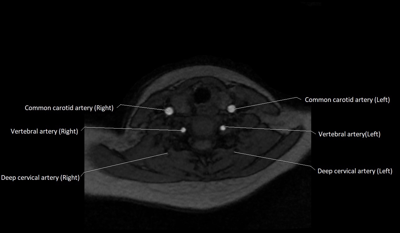
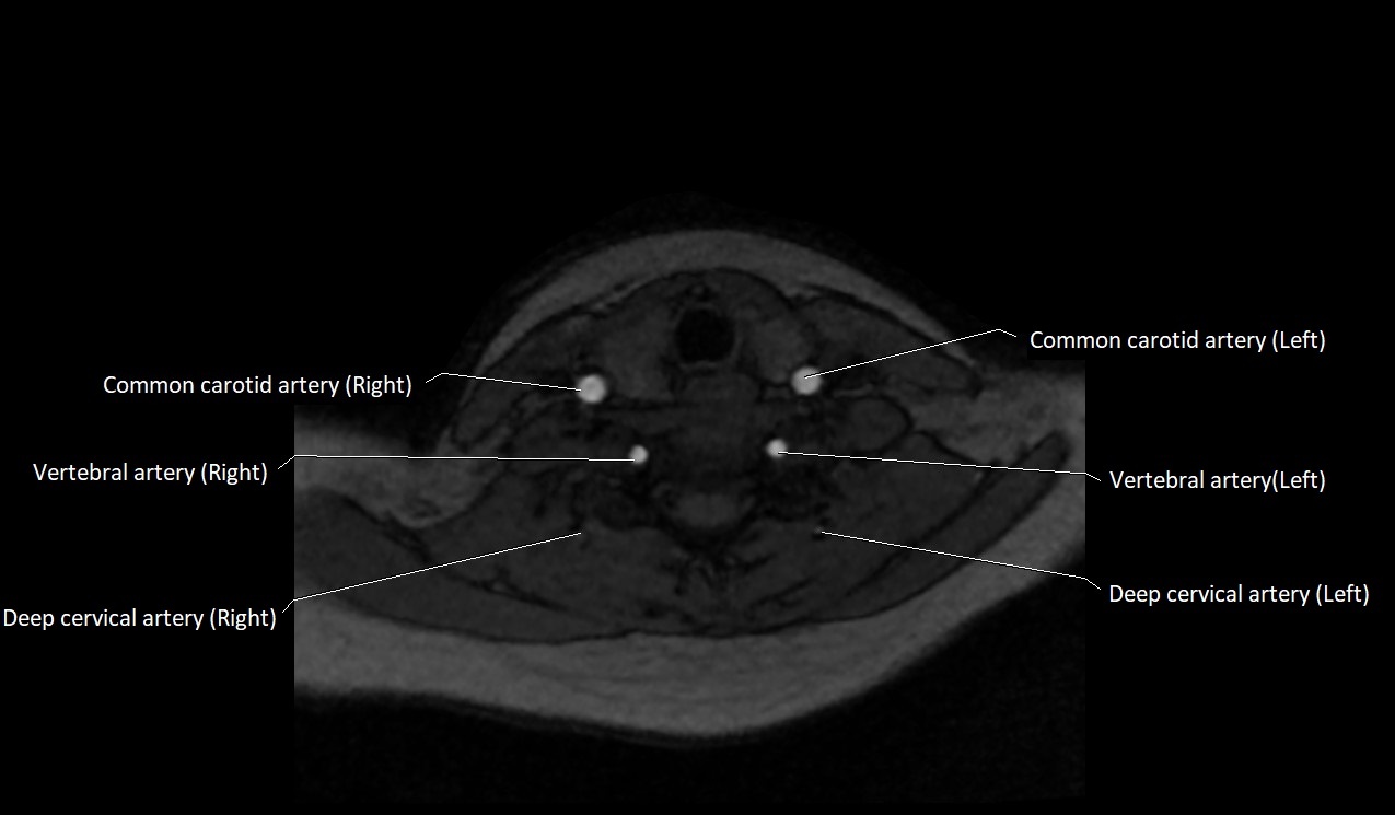
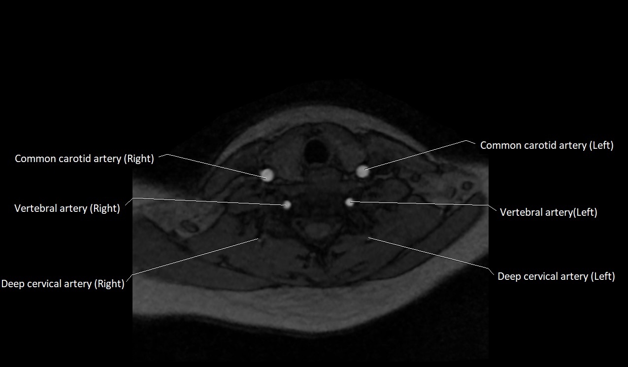
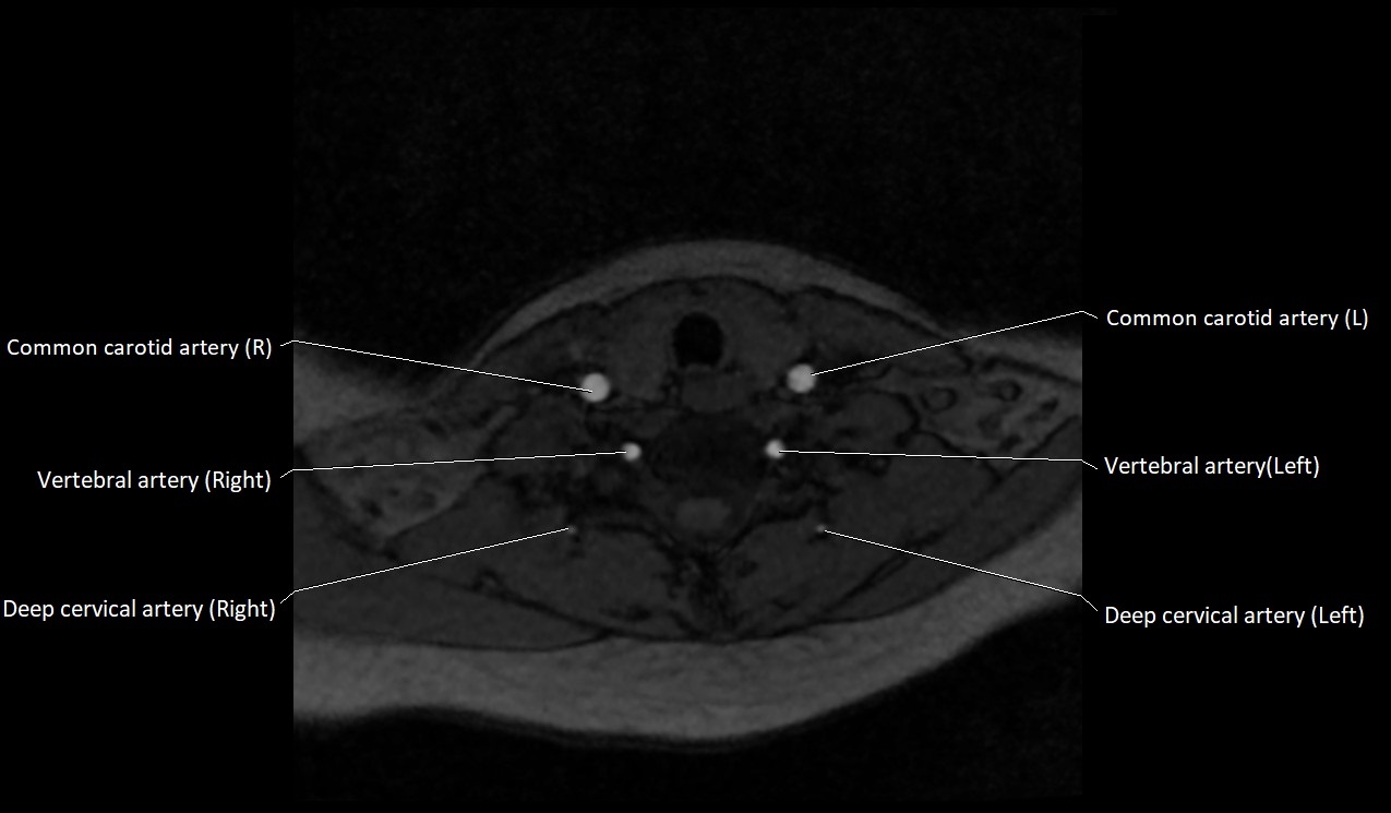
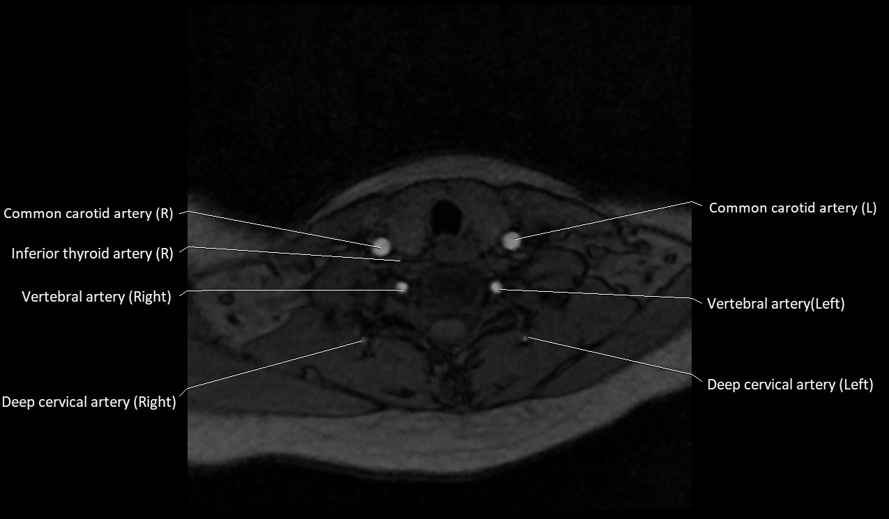
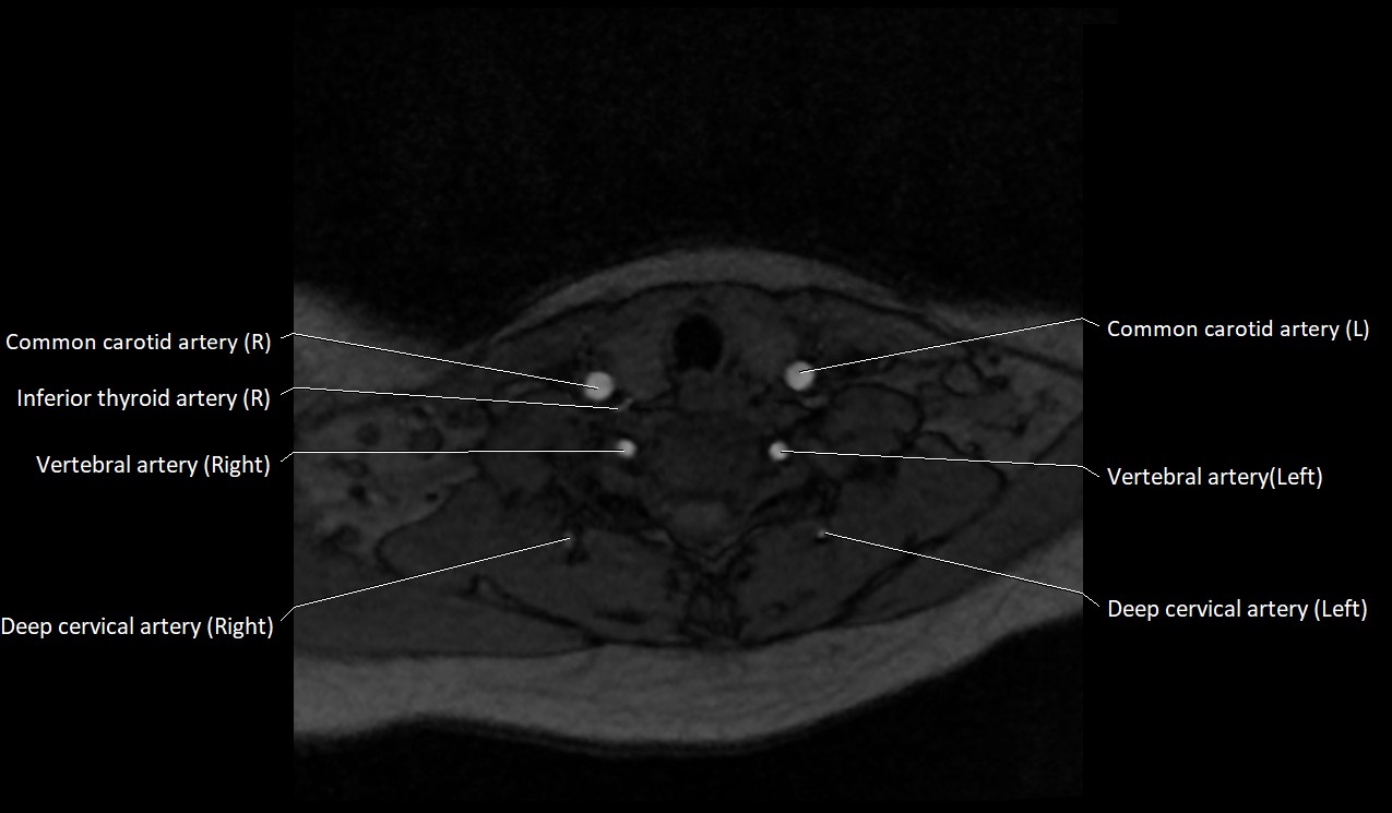
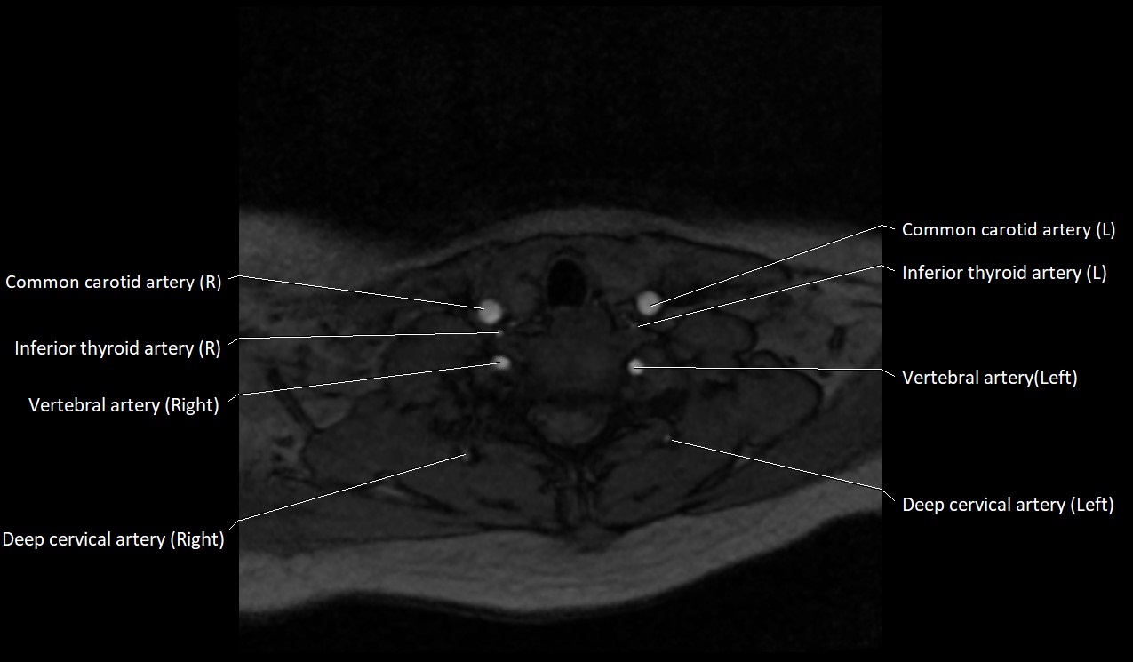
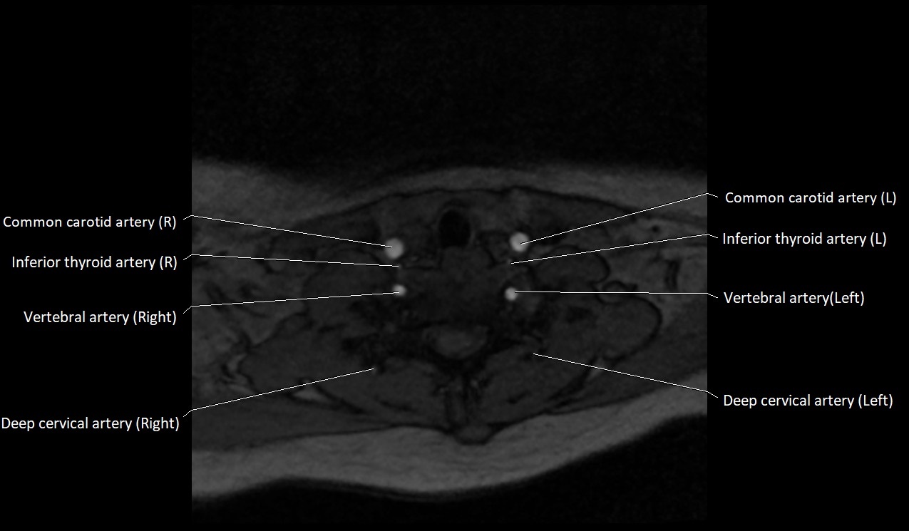
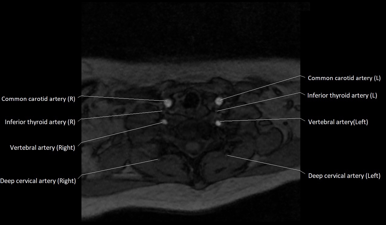
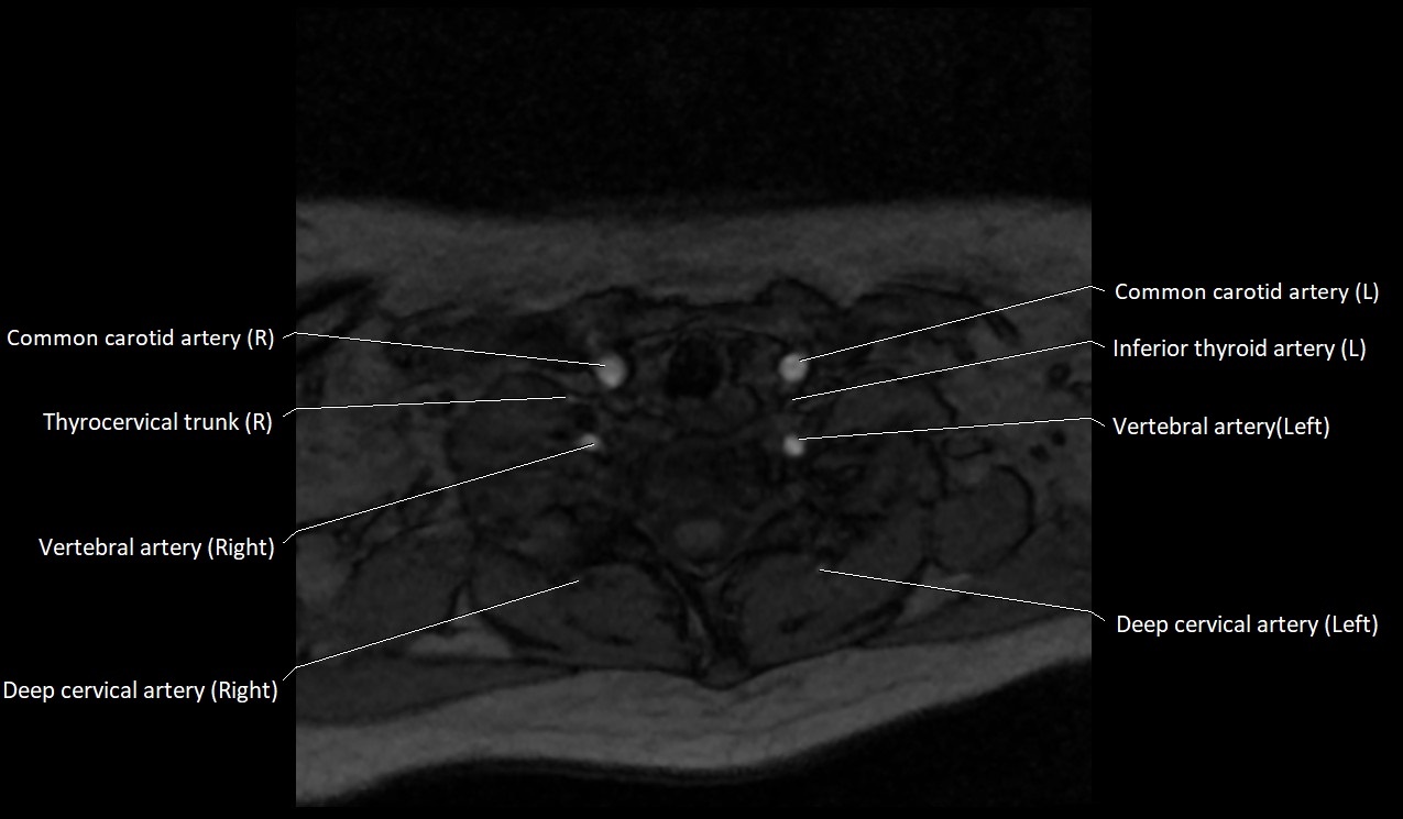
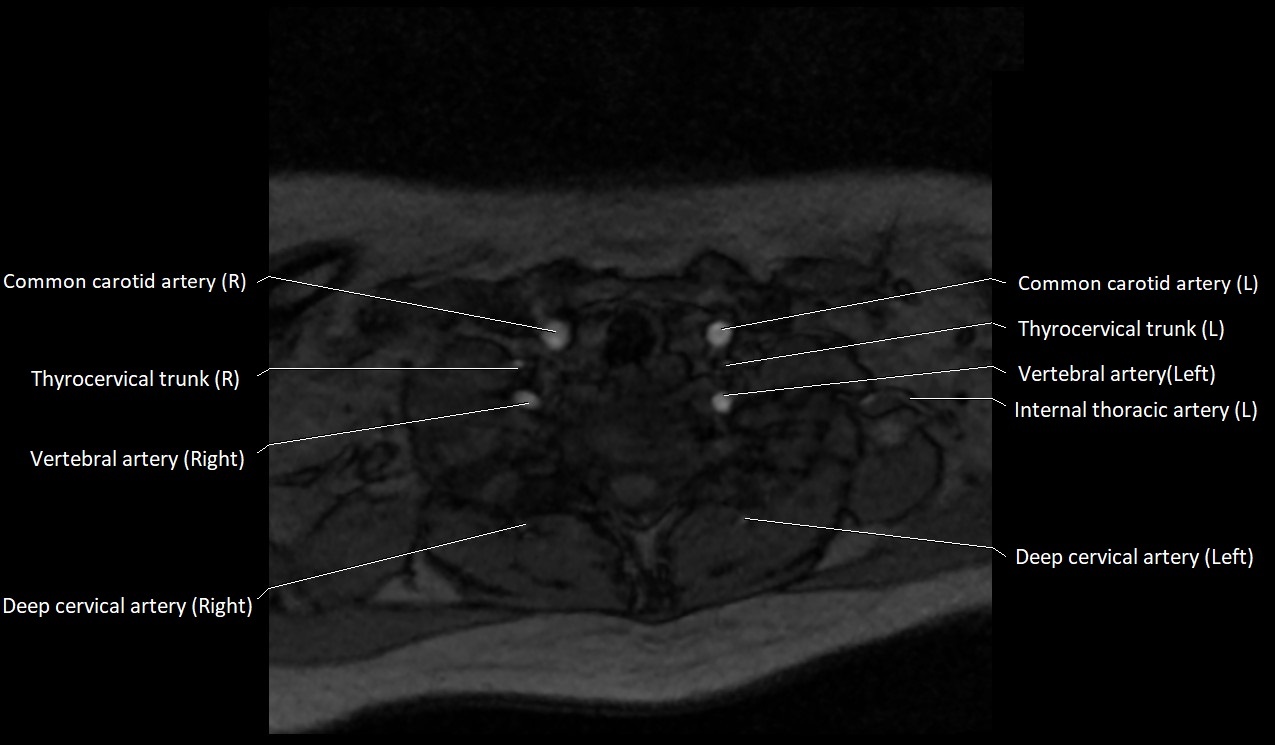
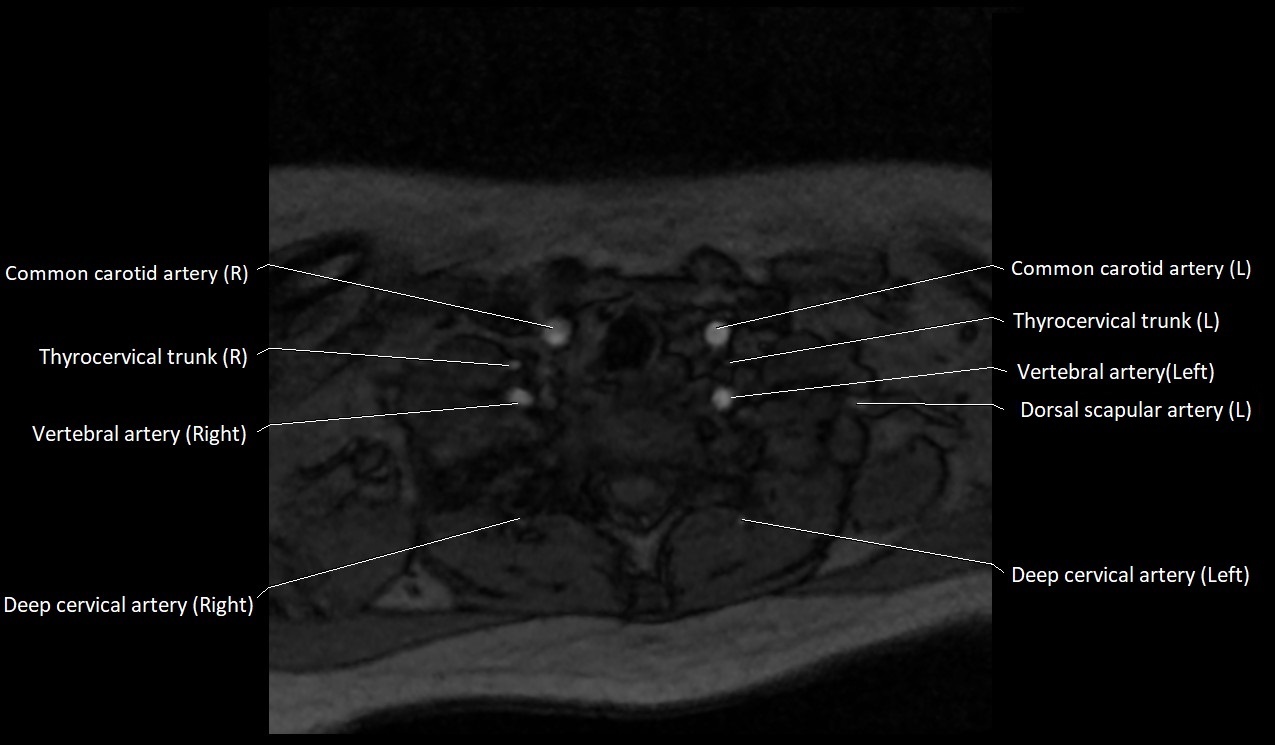
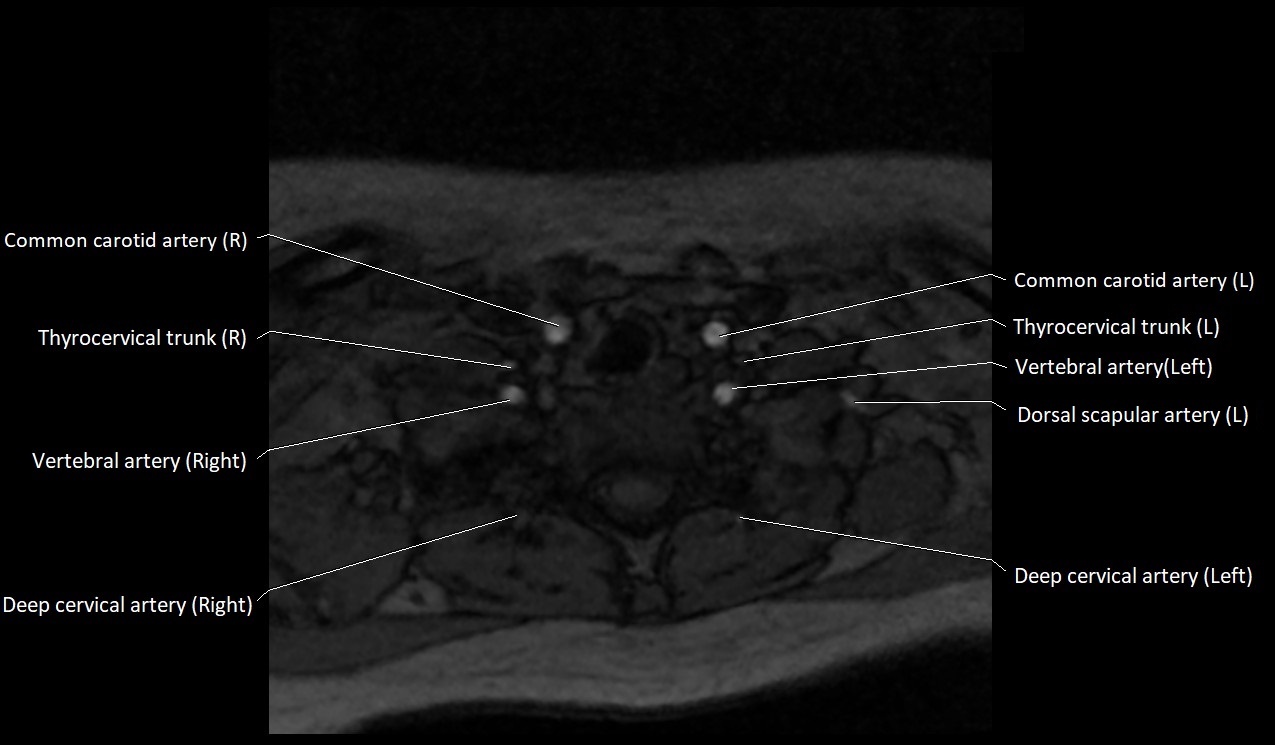
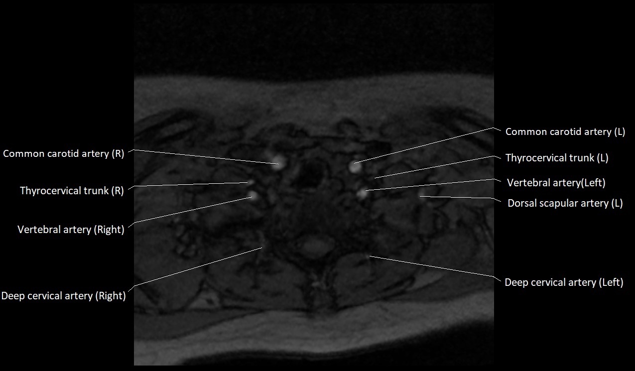
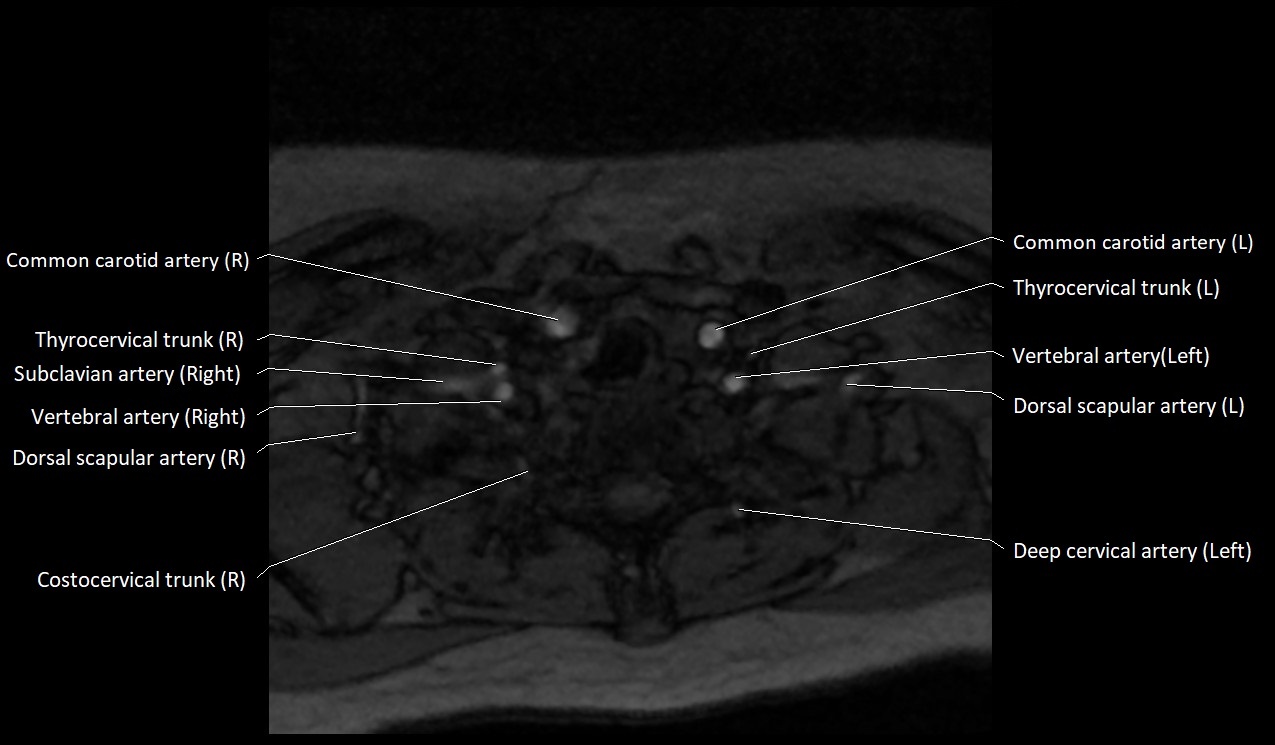
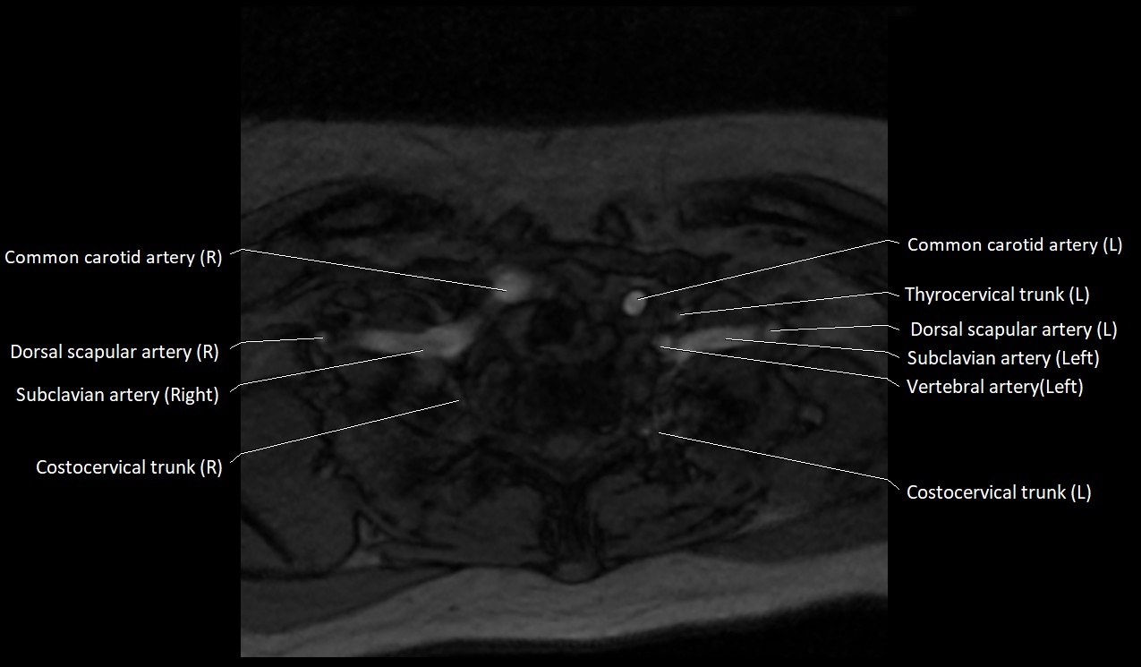
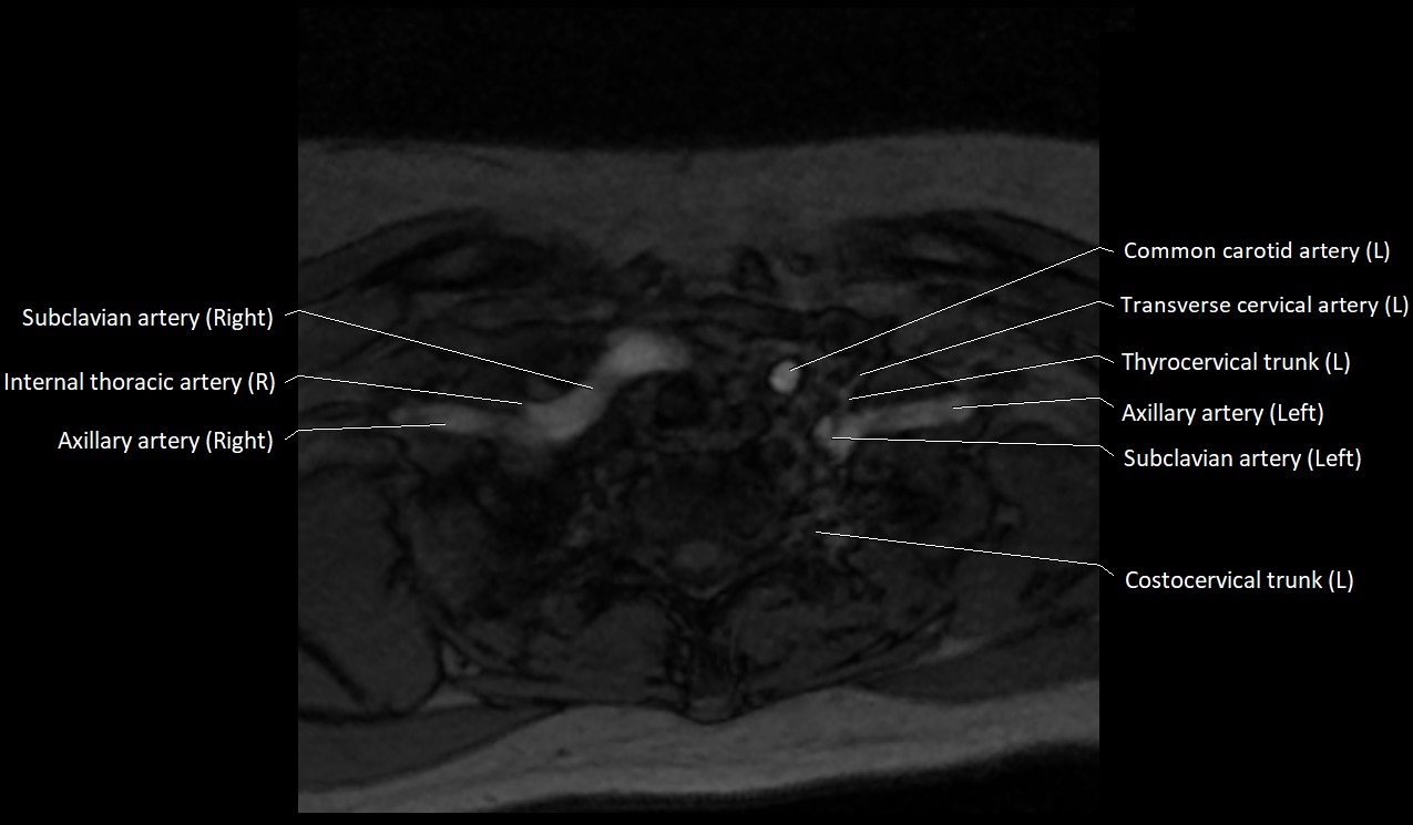
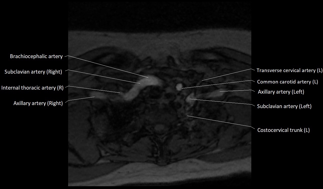
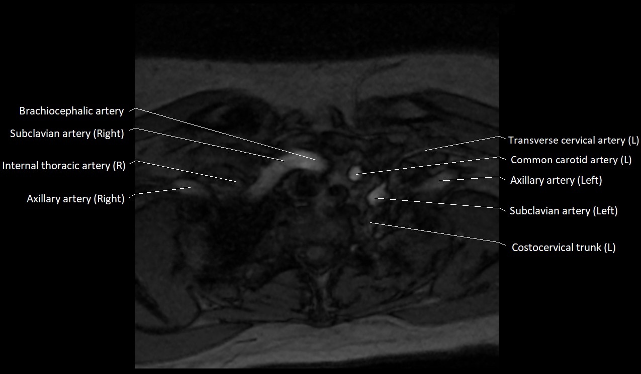
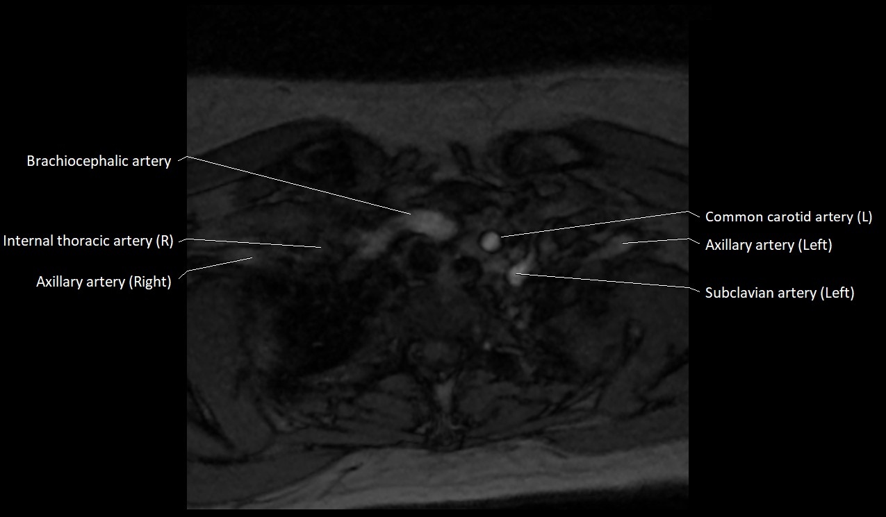
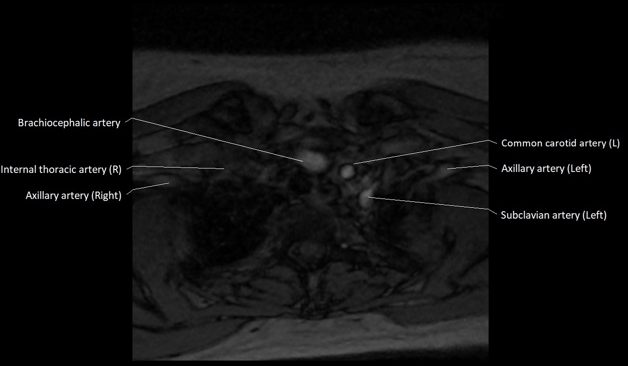
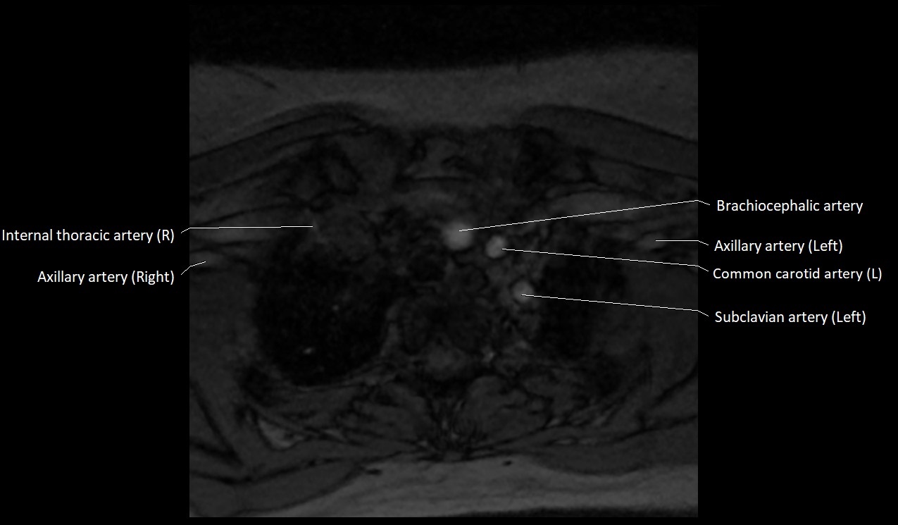
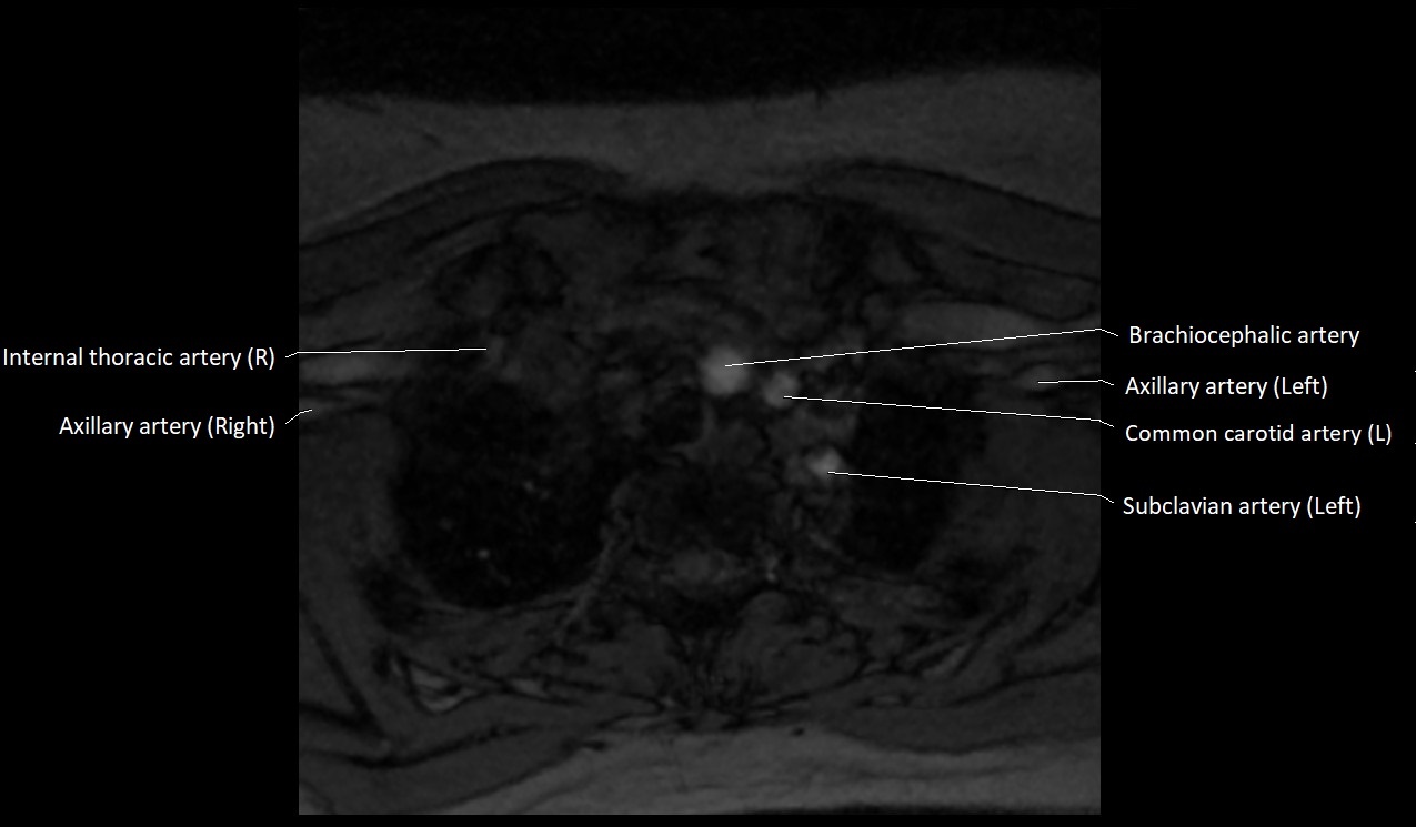
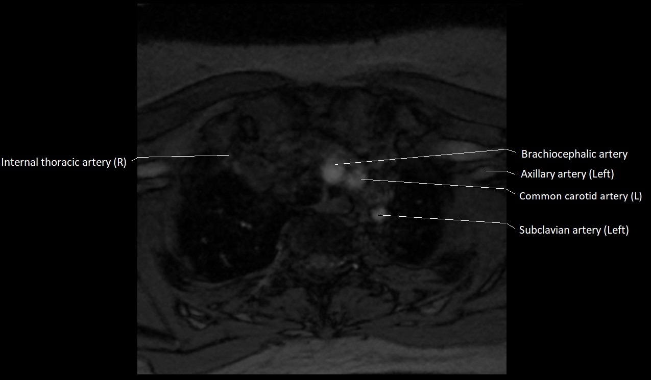
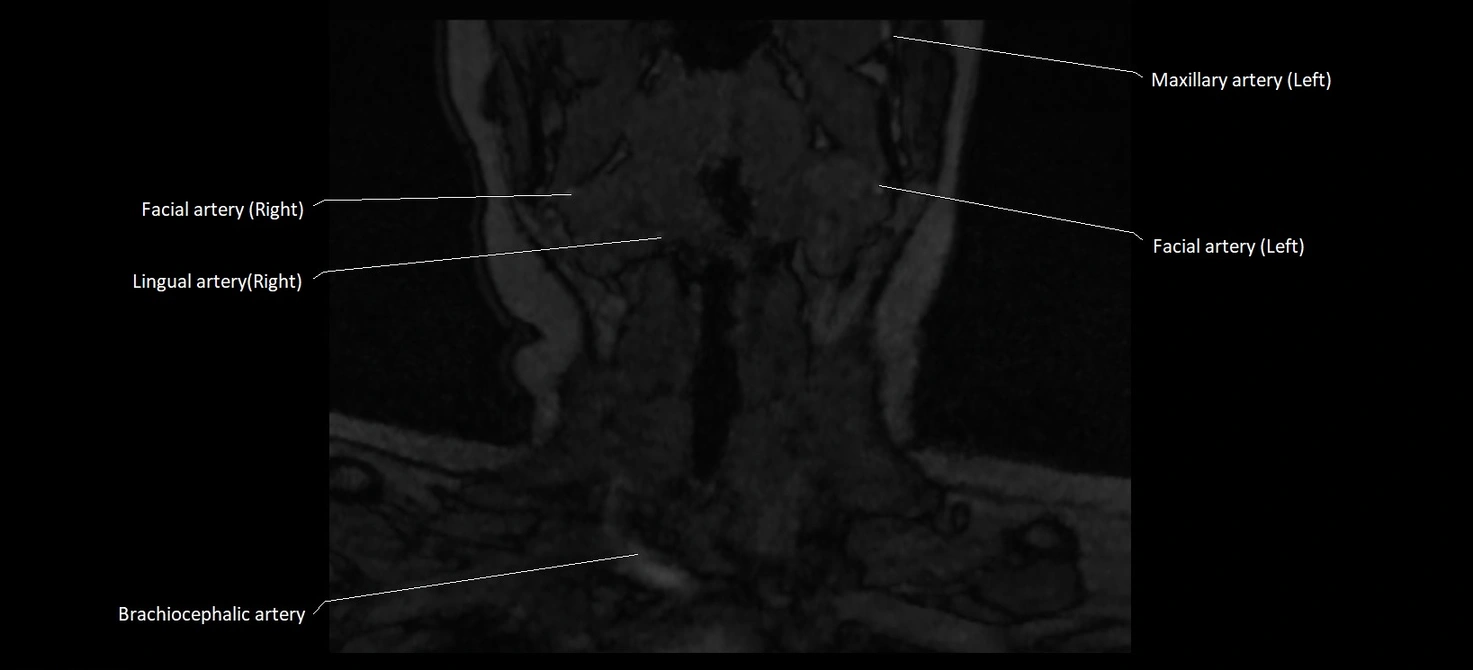
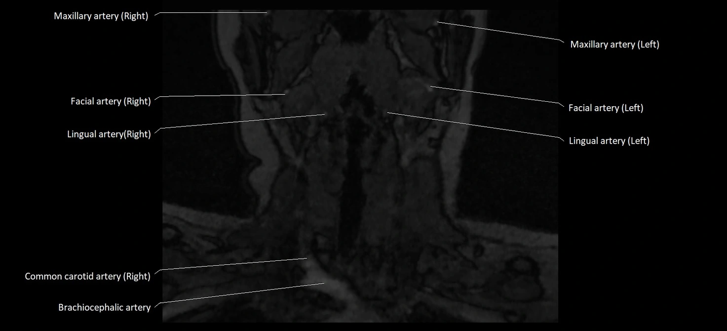
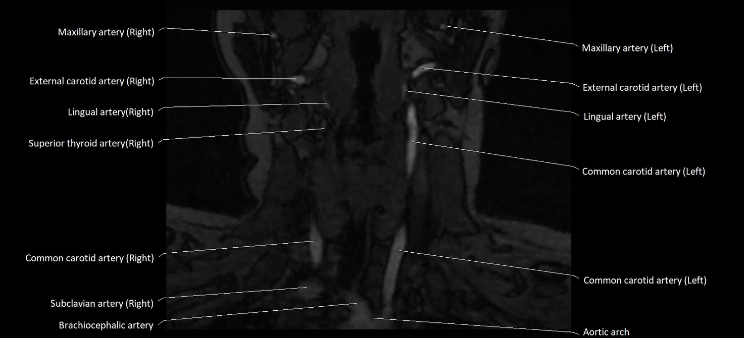
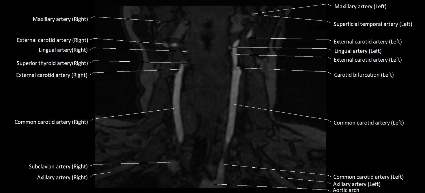
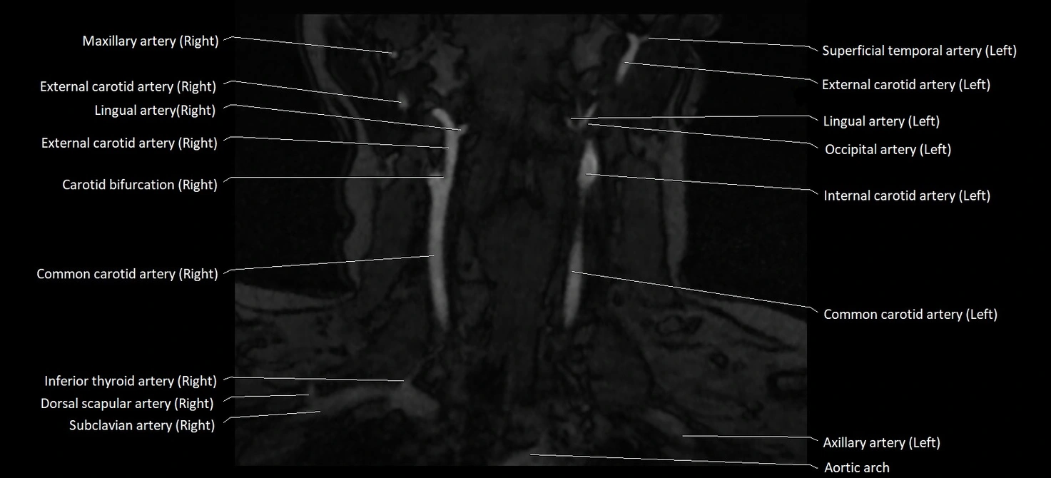
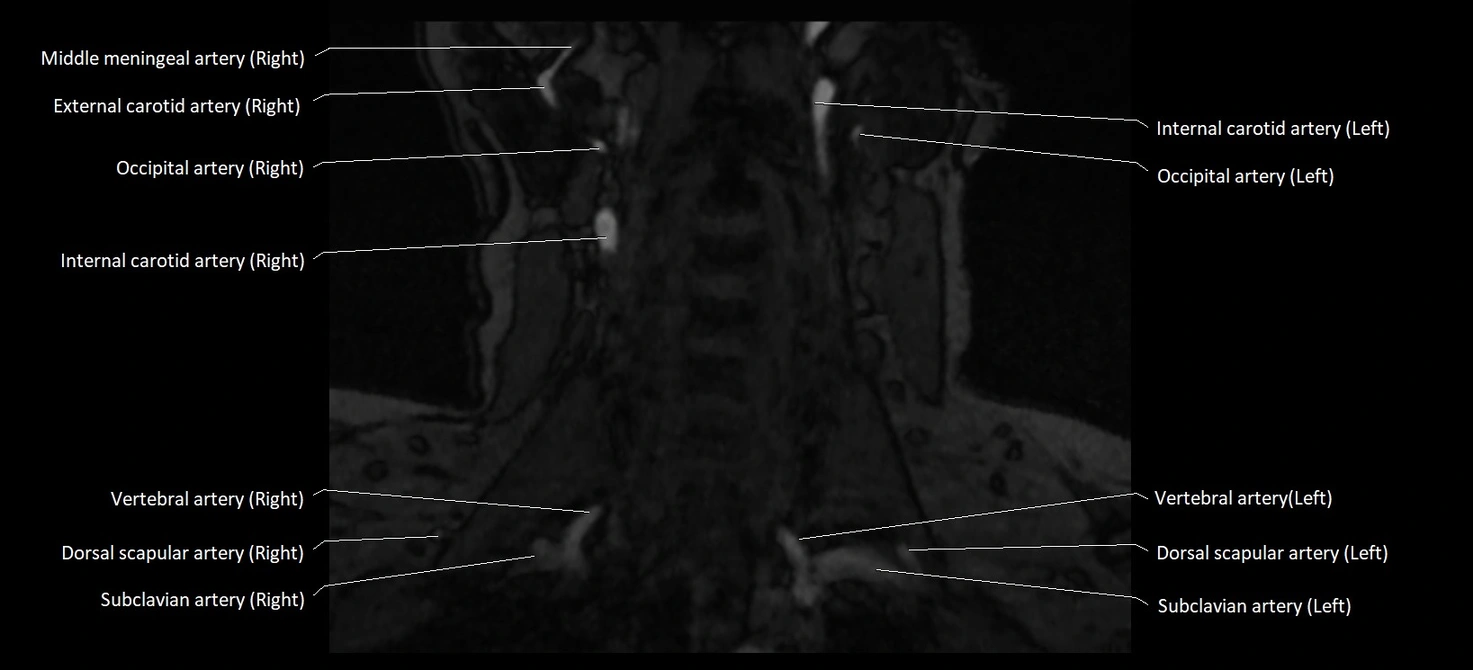
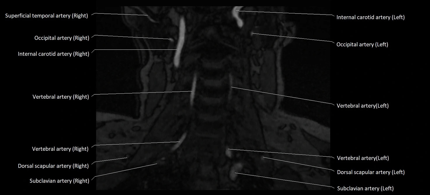
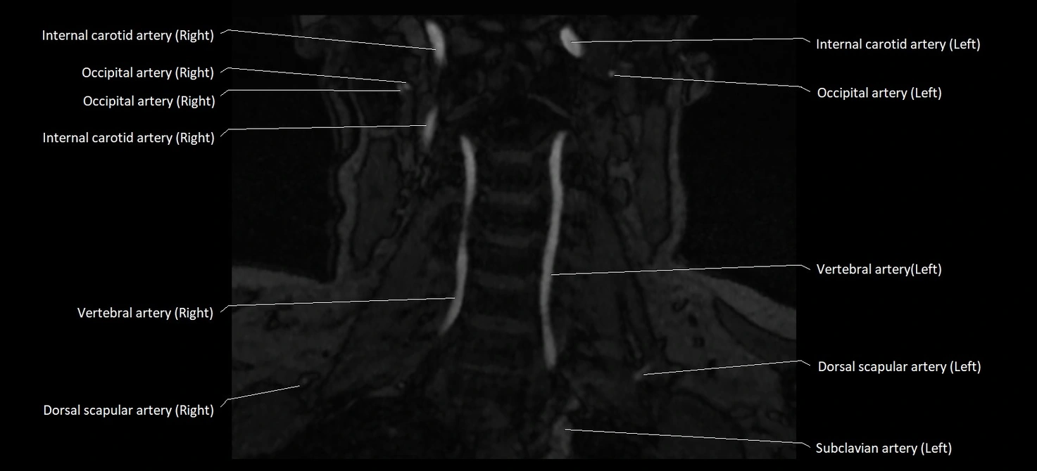
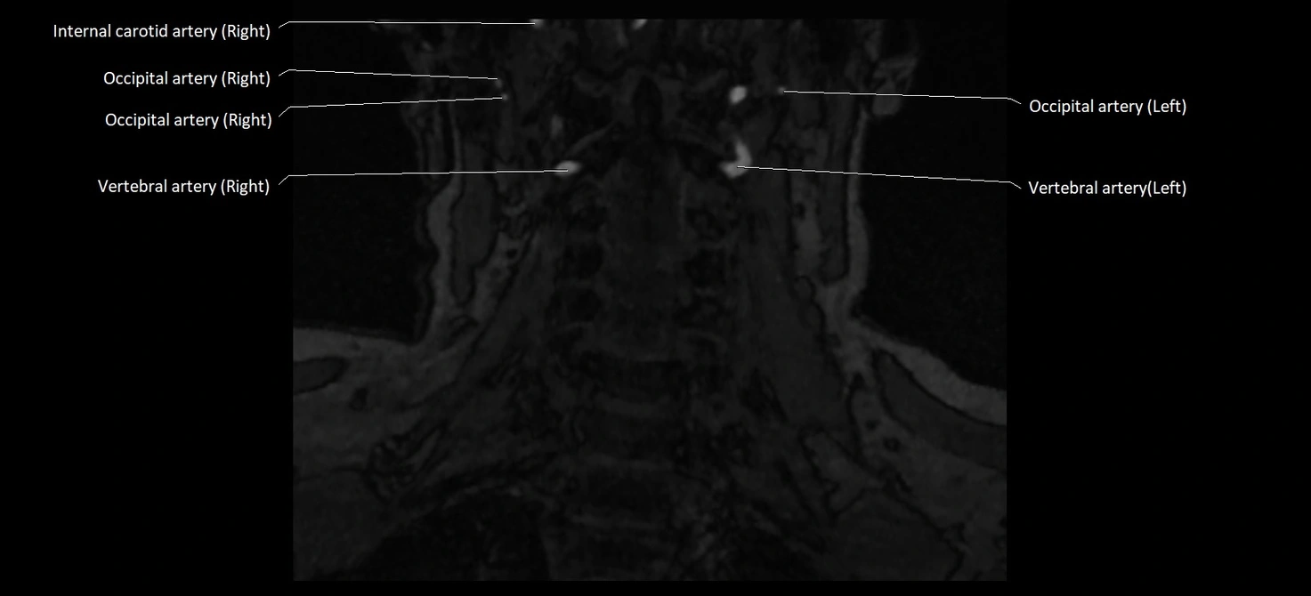
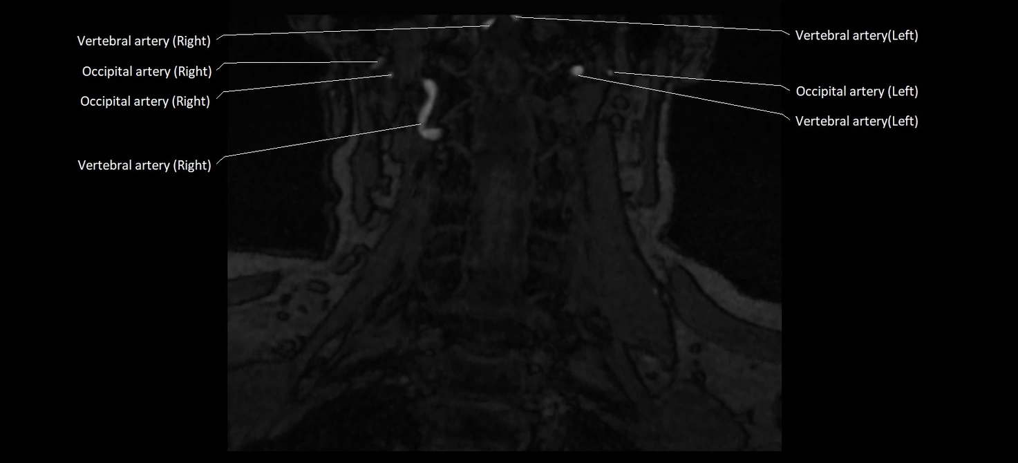
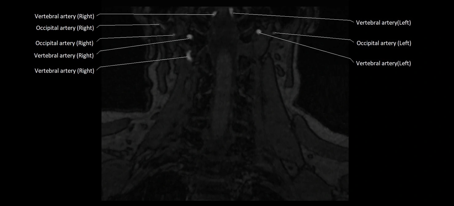
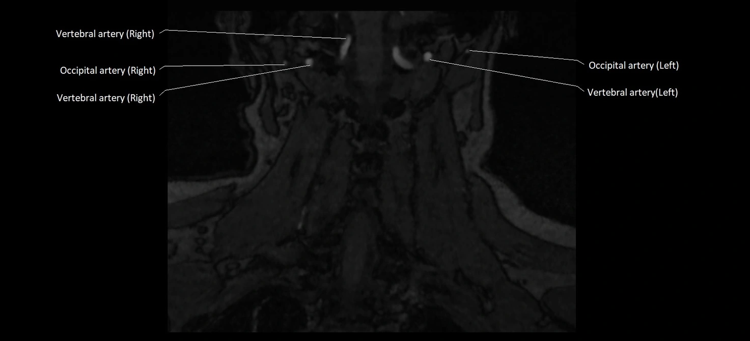
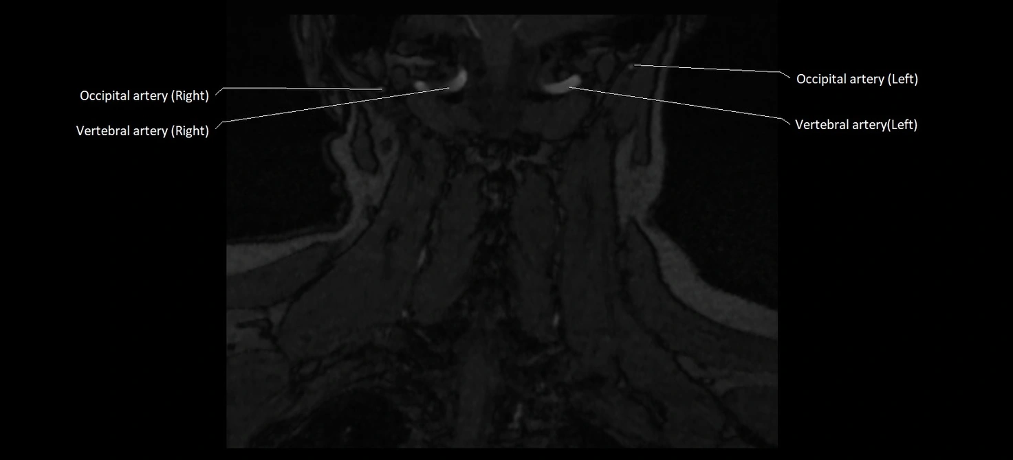
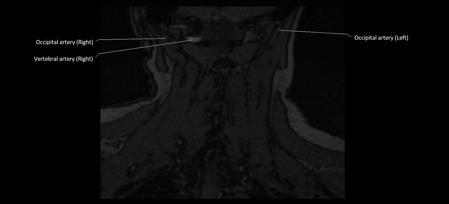
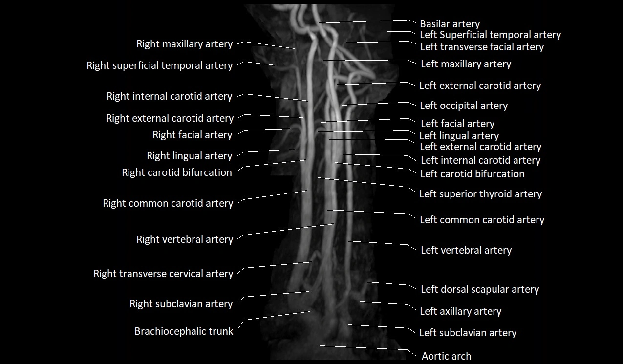
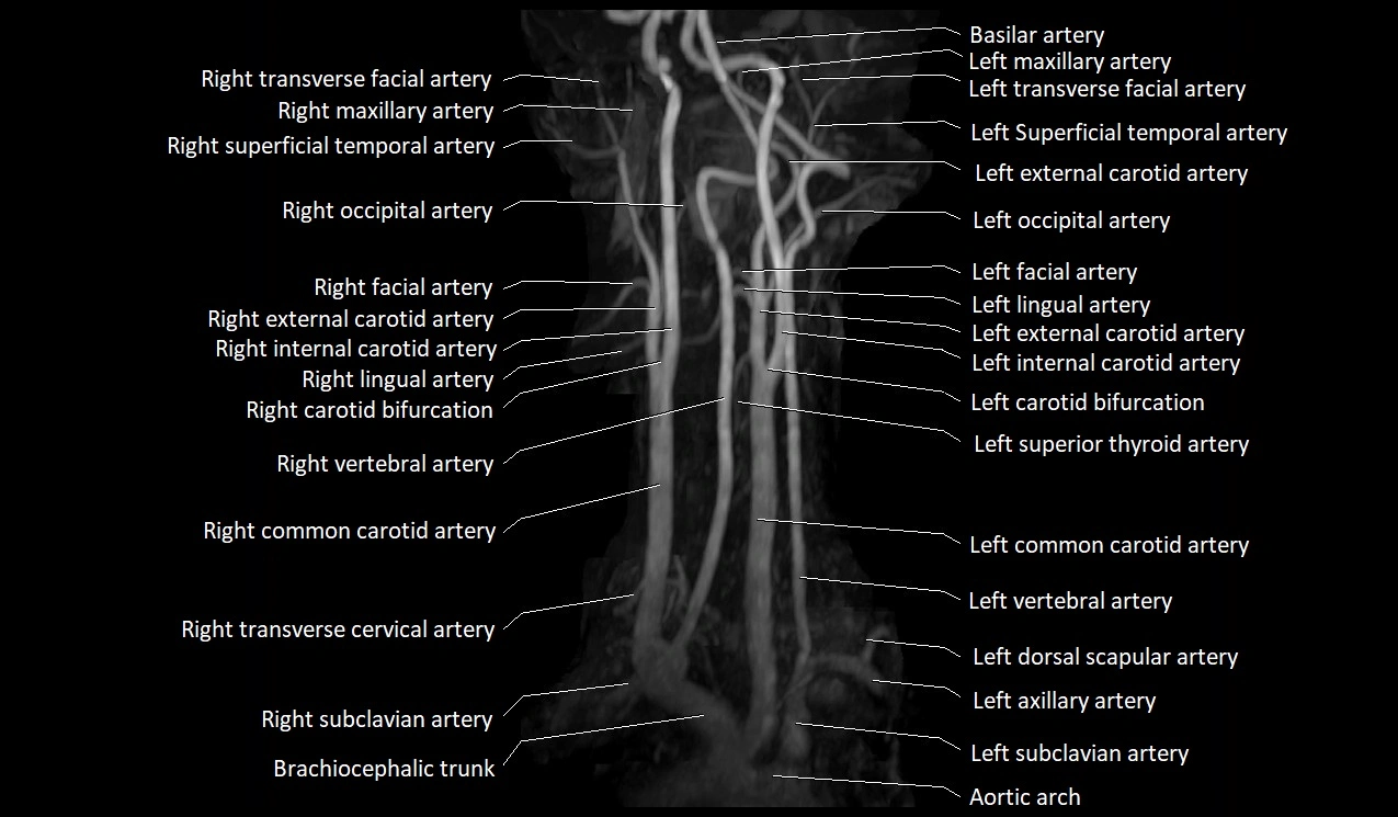
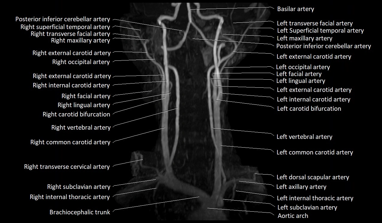
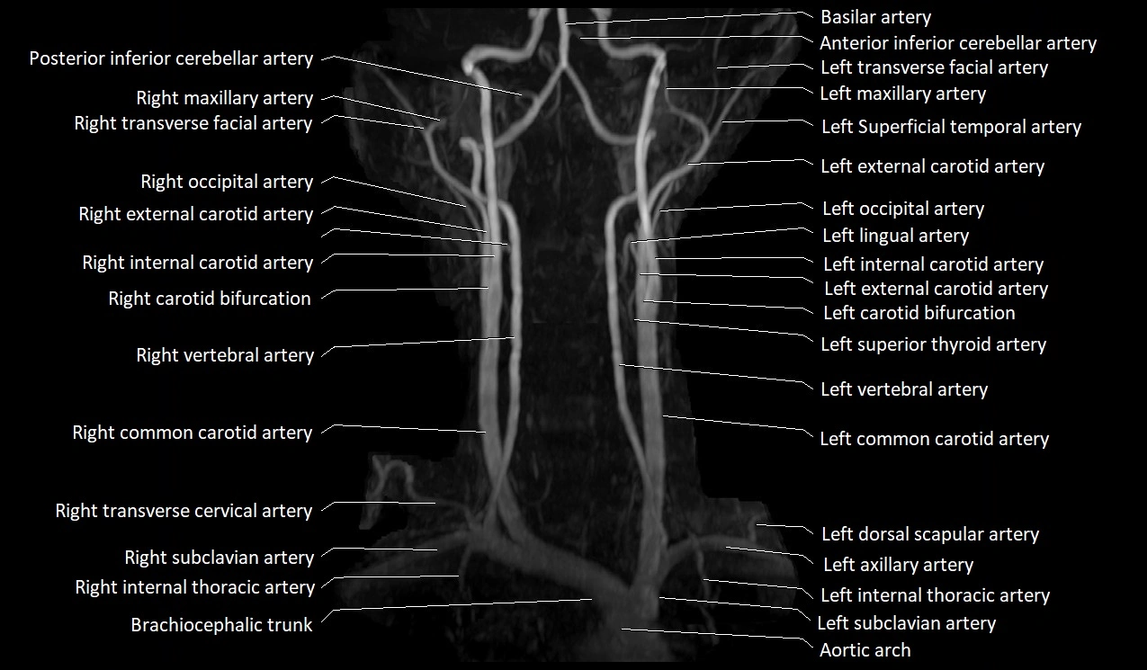
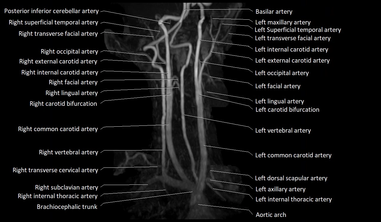
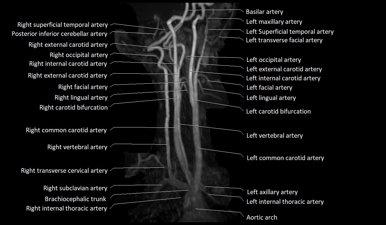
MRI images

MRI images

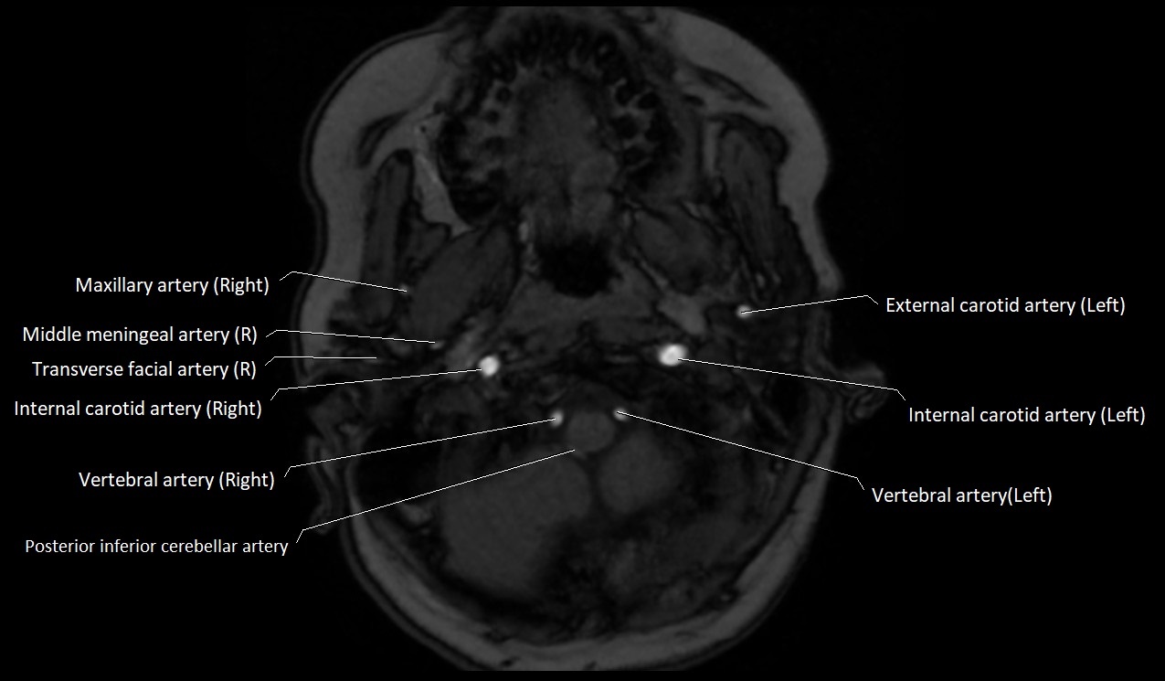
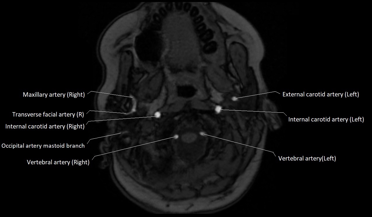
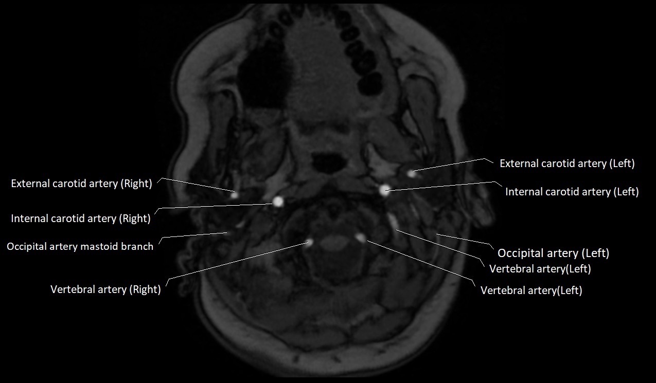
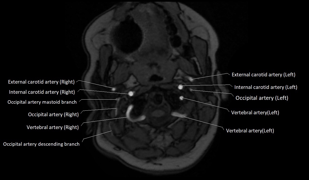
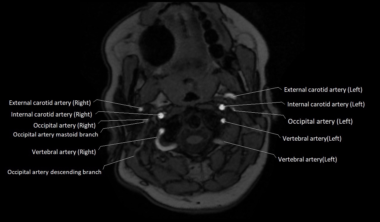
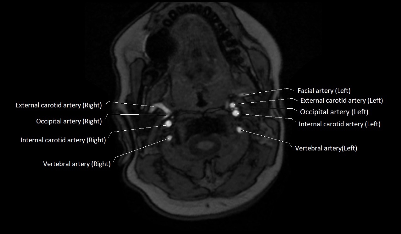
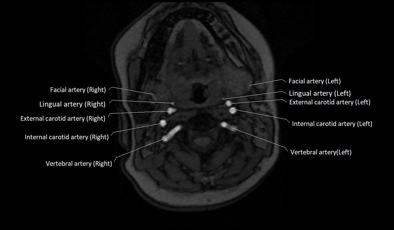
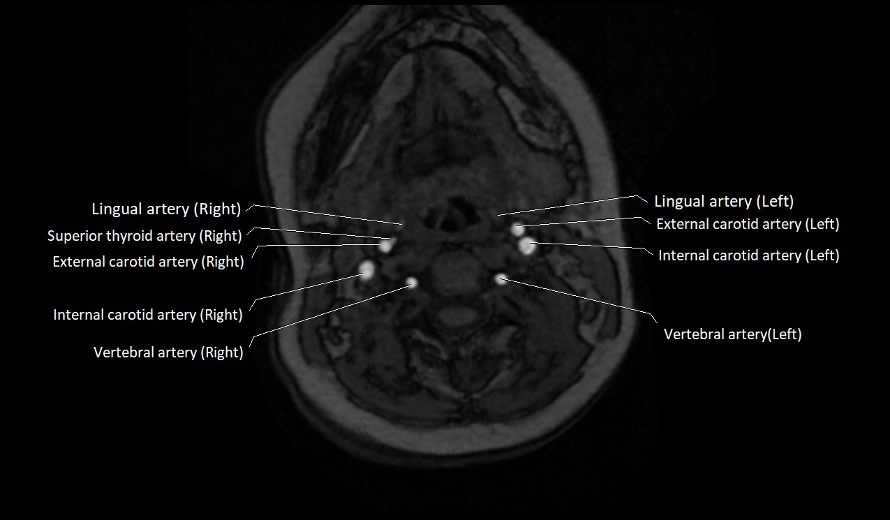
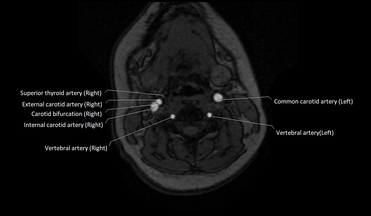
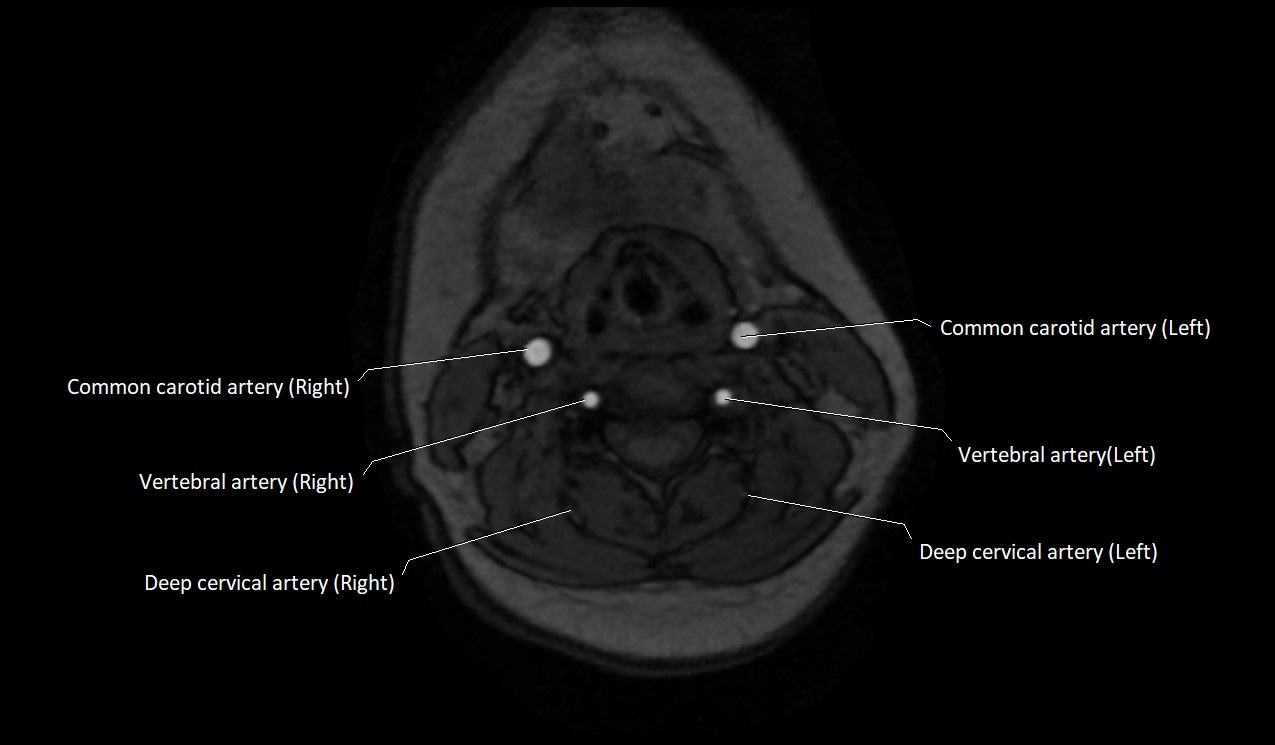
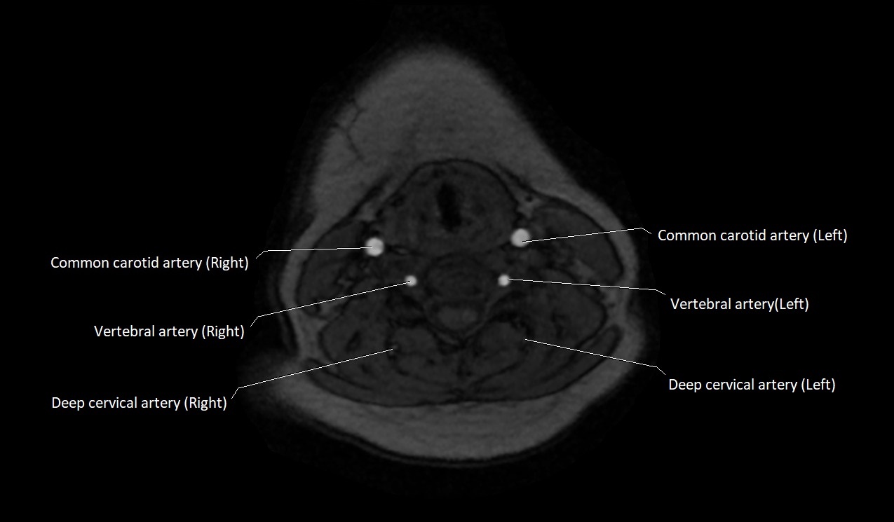
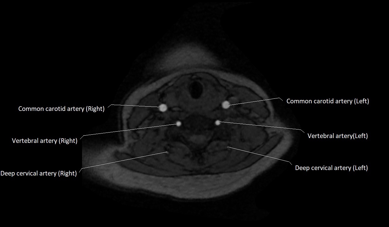
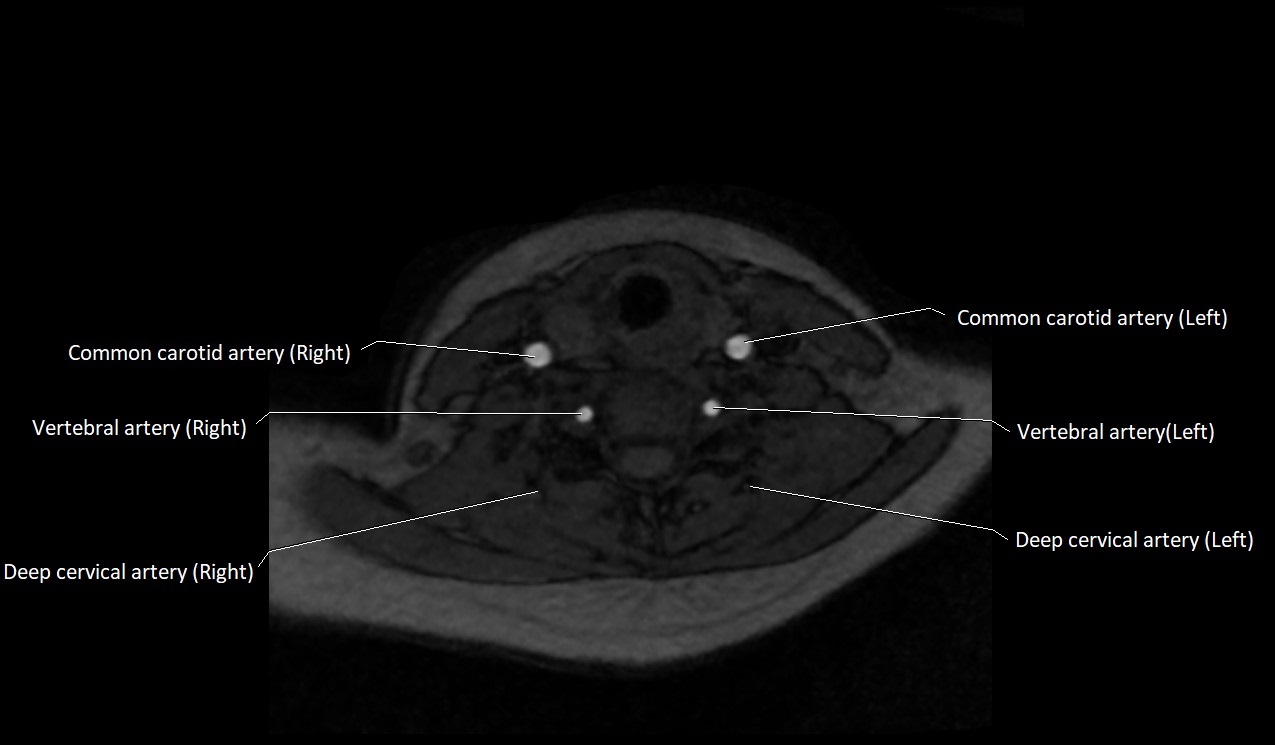
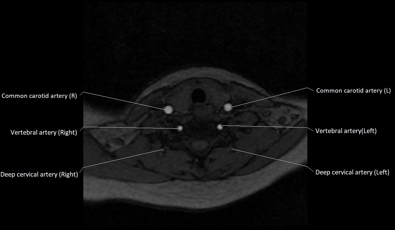
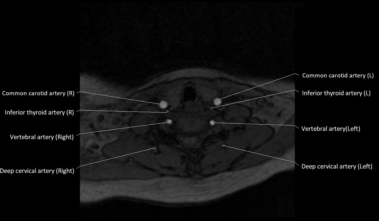
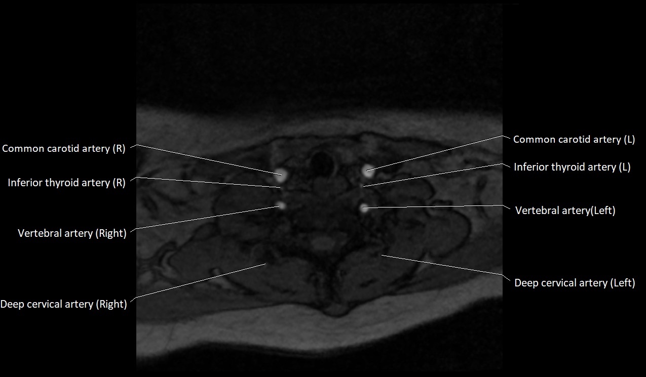
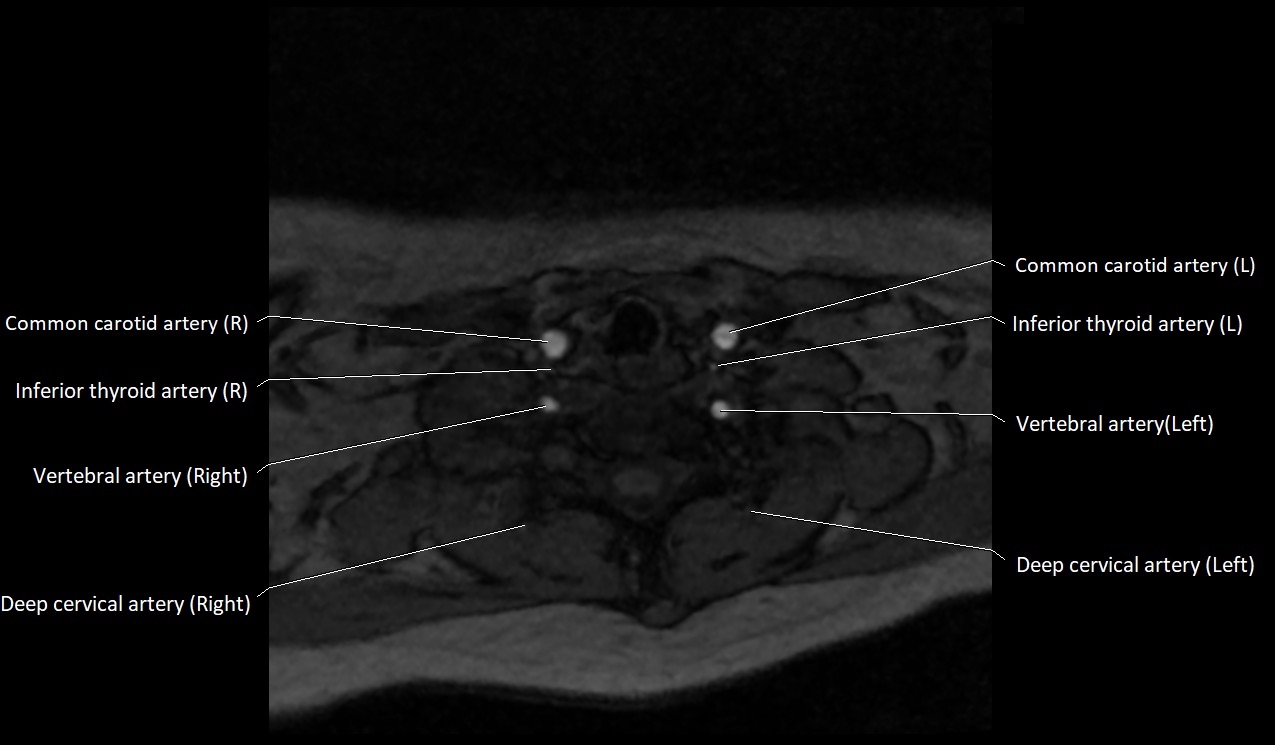
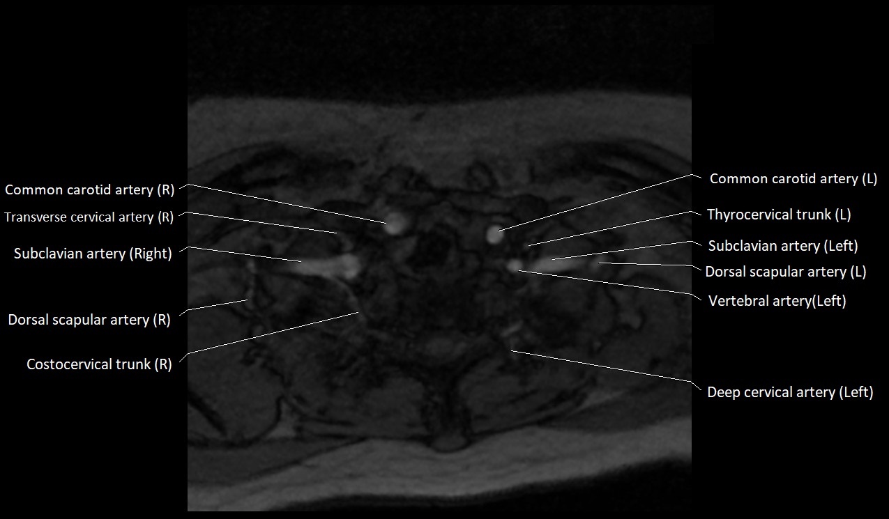
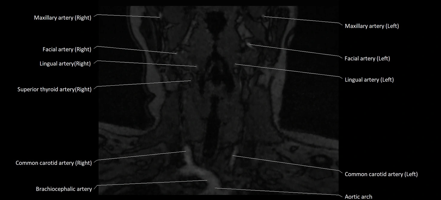
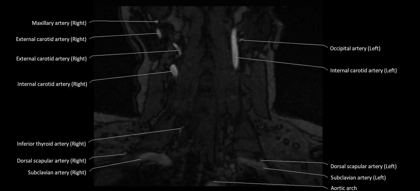
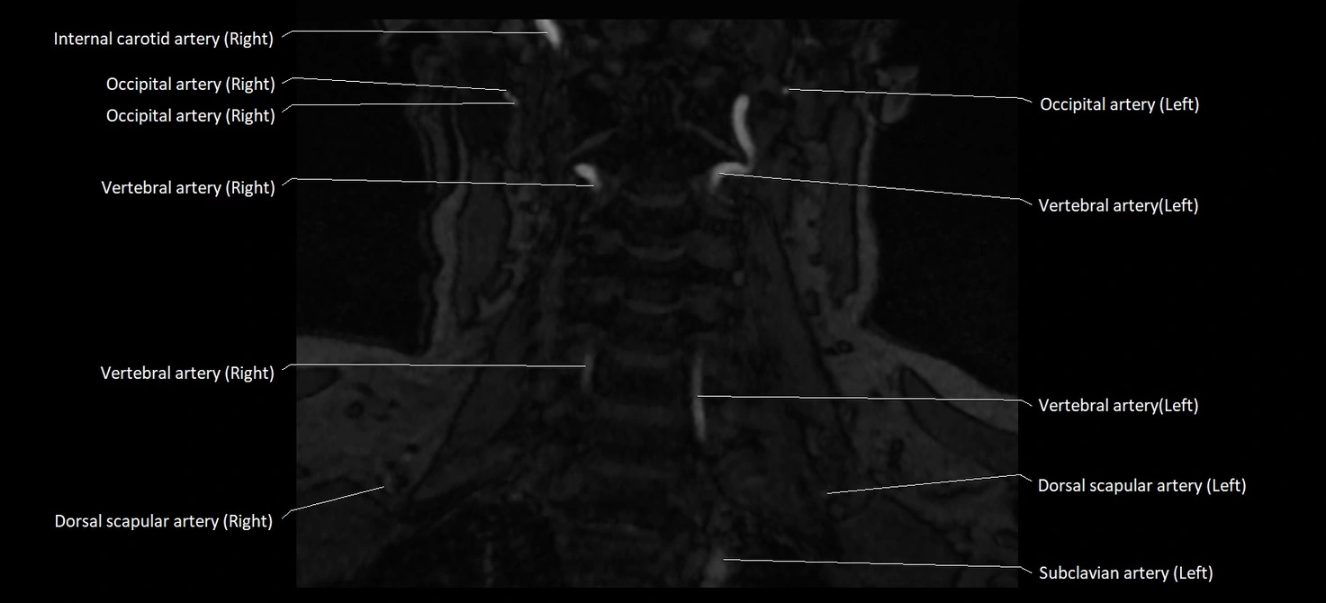
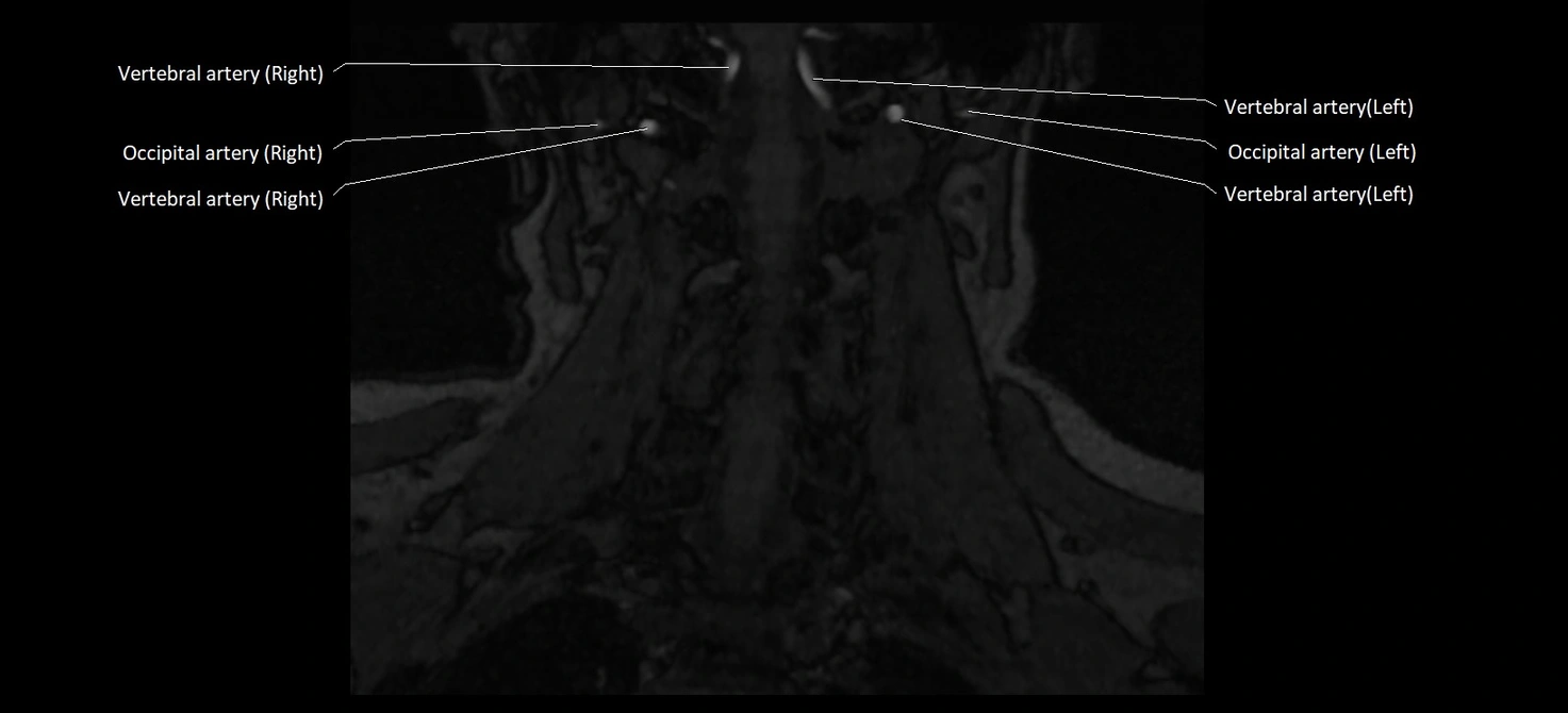
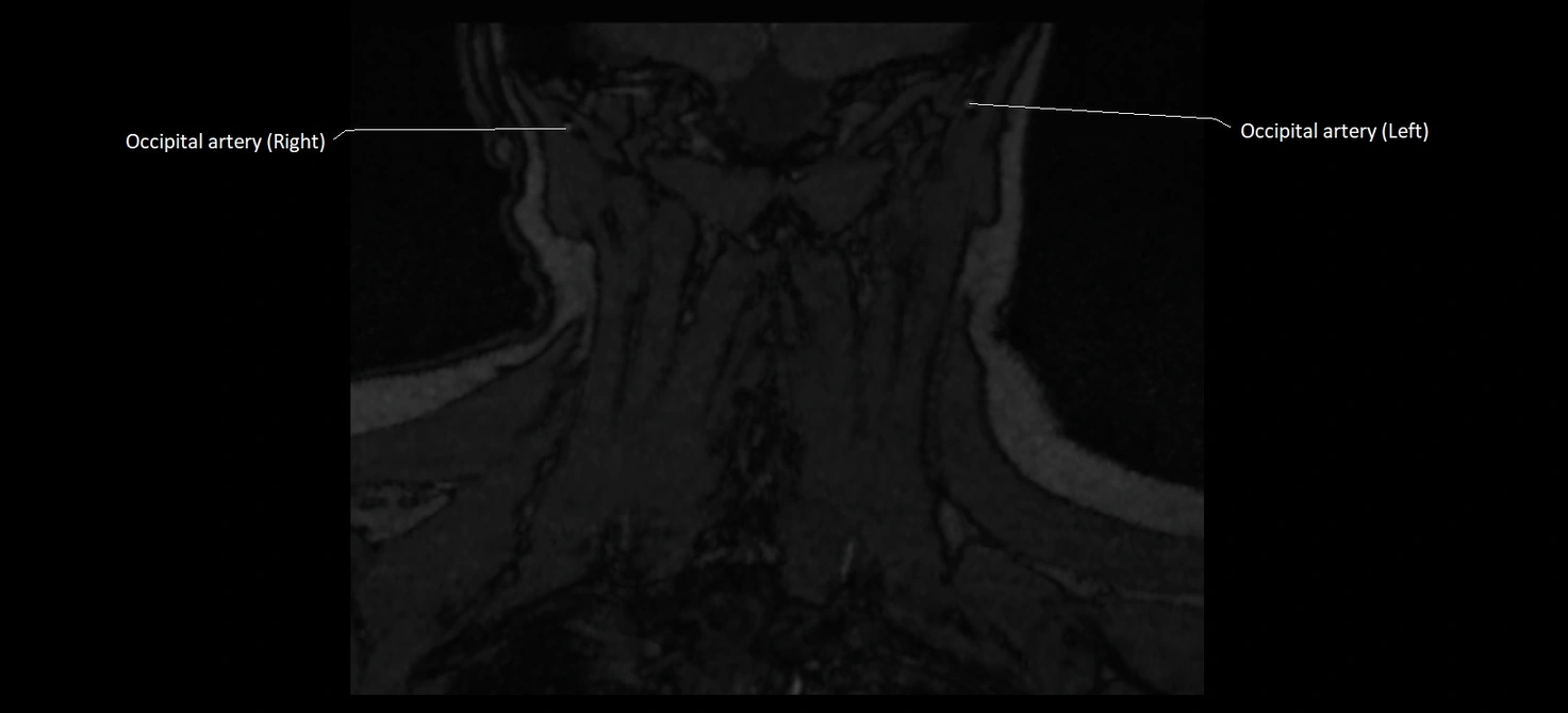
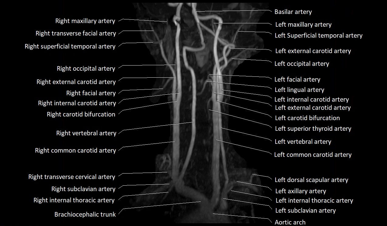
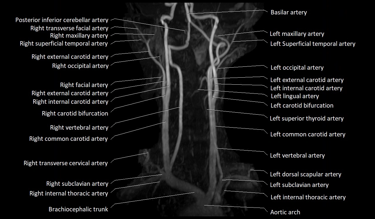
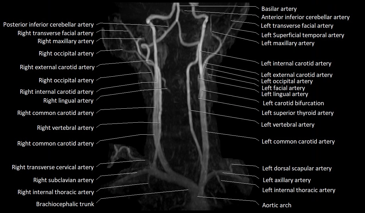
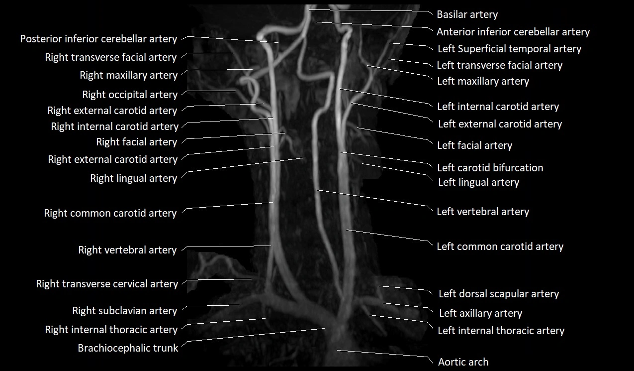
CT image


