


Topic
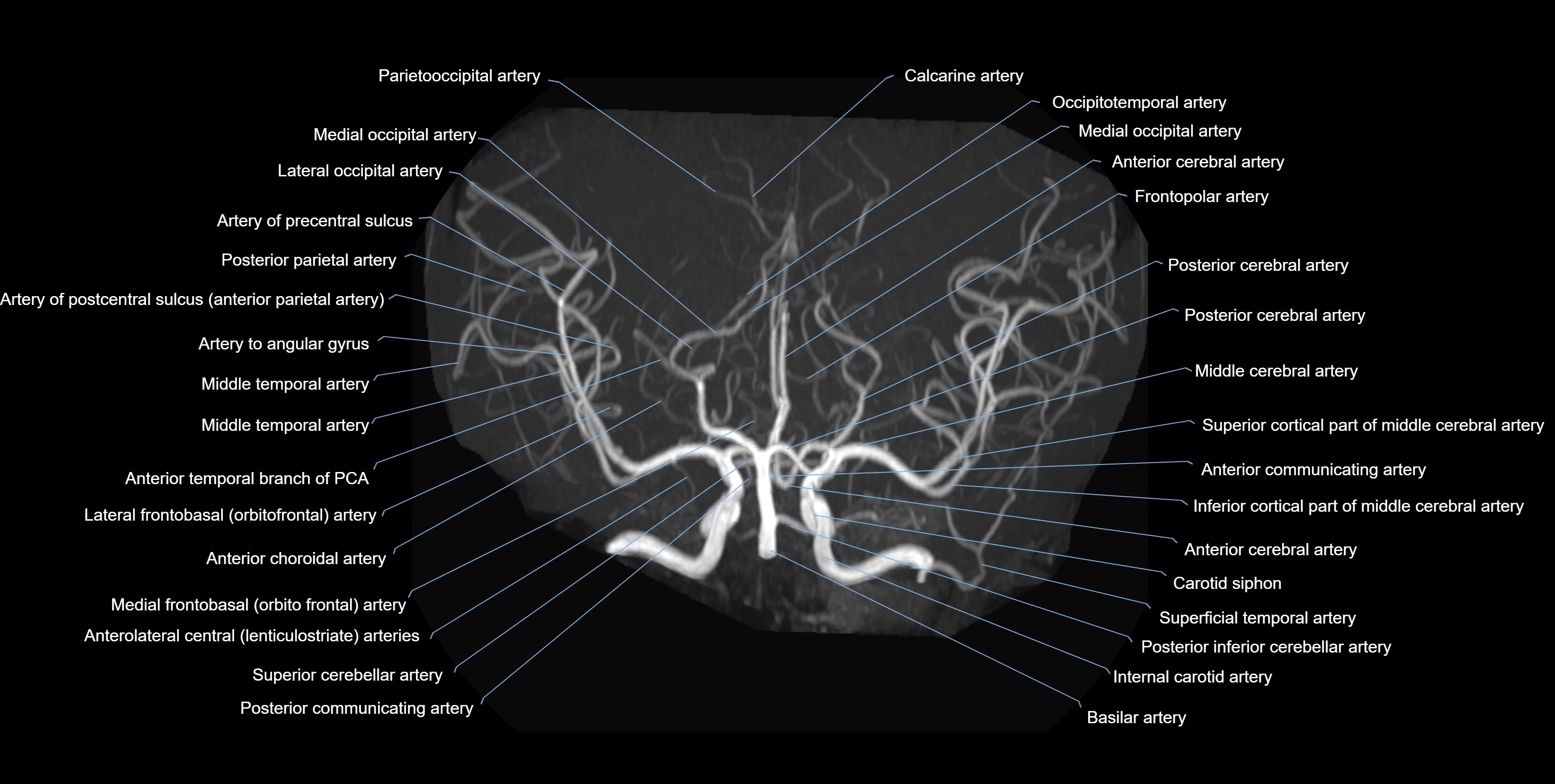
The anterior choroidal artery (AChA) is a small but clinically vital branch of the internal carotid artery (ICA). It arises distal to the posterior communicating artery and proximal to the ICA bifurcation into the middle cerebral artery (MCA) and anterior cerebral artery (ACA).
It travels posteriorly and laterally along the optic tract, giving penetrating branches to deep brain structures before terminating in the choroid plexus of the temporal horn of the lateral ventricle. Despite its size, the AChA supplies highly eloquent regions; ischemia or injury can cause devastating neurological deficits.
Territories Supplied
-
Posterior limb of internal capsule (motor/sensory pathways)
-
Optic tract and lateral geniculate body
-
Globus pallidus
-
Parts of thalamus
-
Hippocampus
-
Choroid plexus of lateral ventricle (temporal horn)
Clinical Significance
Occlusion produces the classic AChA syndrome: contralateral hemiparesis, hemisensory loss, and homonymous hemianopia. Aneurysms at its origin are also clinically important.
Synonyms
-
AChA
-
Internal carotid choroidal branch
Function
-
Supplies deep and critical structures of the brain
-
Maintains blood flow to optic, limbic, and motor pathways
-
Clinically relevant in stroke, aneurysms, epilepsy surgery, and vascular malformations
MRI Appearance
T1-weighted images:
-
Vessel appears as a flow void (dark lumen) near the optic tract and temporal horn
-
Surrounding parenchyma intermediate signal
T2-weighted images:
-
Vessel lumen shows dark flow void
-
Ischemic infarcts in its territory (internal capsule, thalamus) appear hyperintense
FLAIR:
-
Vessel not seen directly; infarcts in AChA territory appear as hyperintense cortical/subcortical changes
-
Chronic infarcts show gliotic hyperintensity
T1 Post-Gadolinium:
-
Normal AChA enhances brightly and uniformly
-
Abnormal focal enhancement indicates aneurysm, vasculitis, or AVM involvement
MRA (Magnetic Resonance Angiography):
-
Flow-related enhancement makes the AChA appear as a bright, linear vascular signal against suppressed background
-
High sensitivity for origin and proximal course; distal branches may be too small to resolve
-
Detects stenosis, occlusion, aneurysm, AVM feeders
CTA (CT Angiography):
-
Opacified with iodinated contrast, AChA appears as a bright high-attenuation vessel
-
Visualized from ICA origin along optic tract toward choroid plexus
-
3D reconstructions depict its course and relation to adjacent arteries
-
Gold standard for identifying aneurysms, occlusion, or vascular anomalies
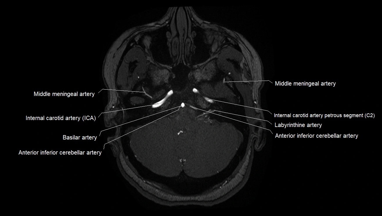
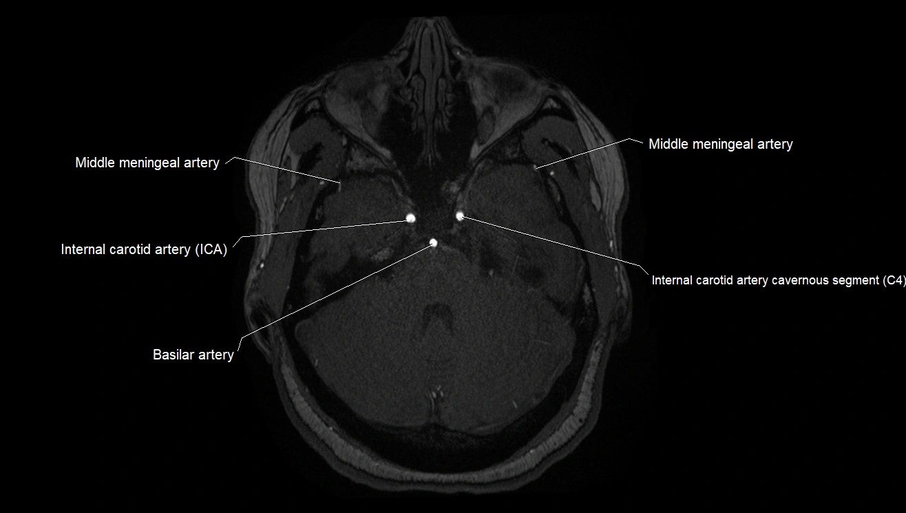
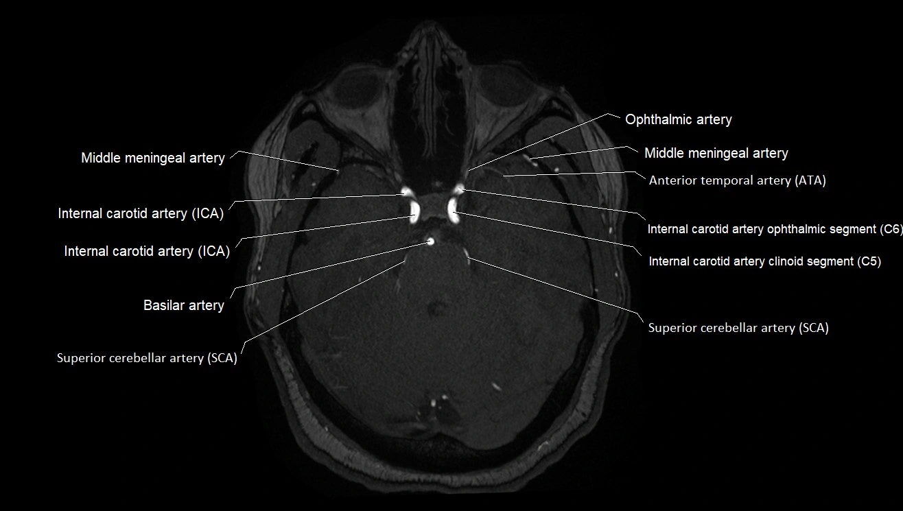
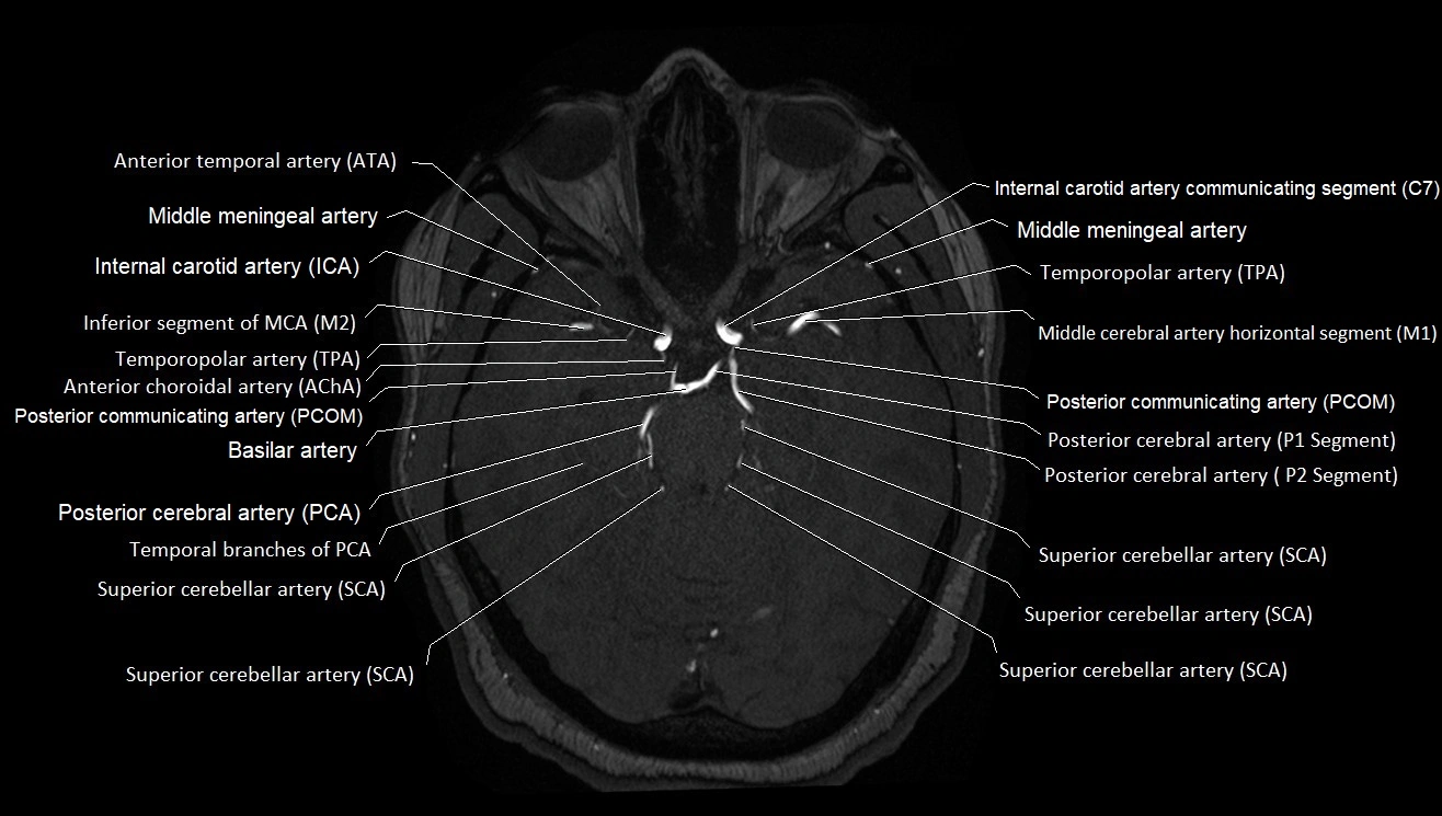
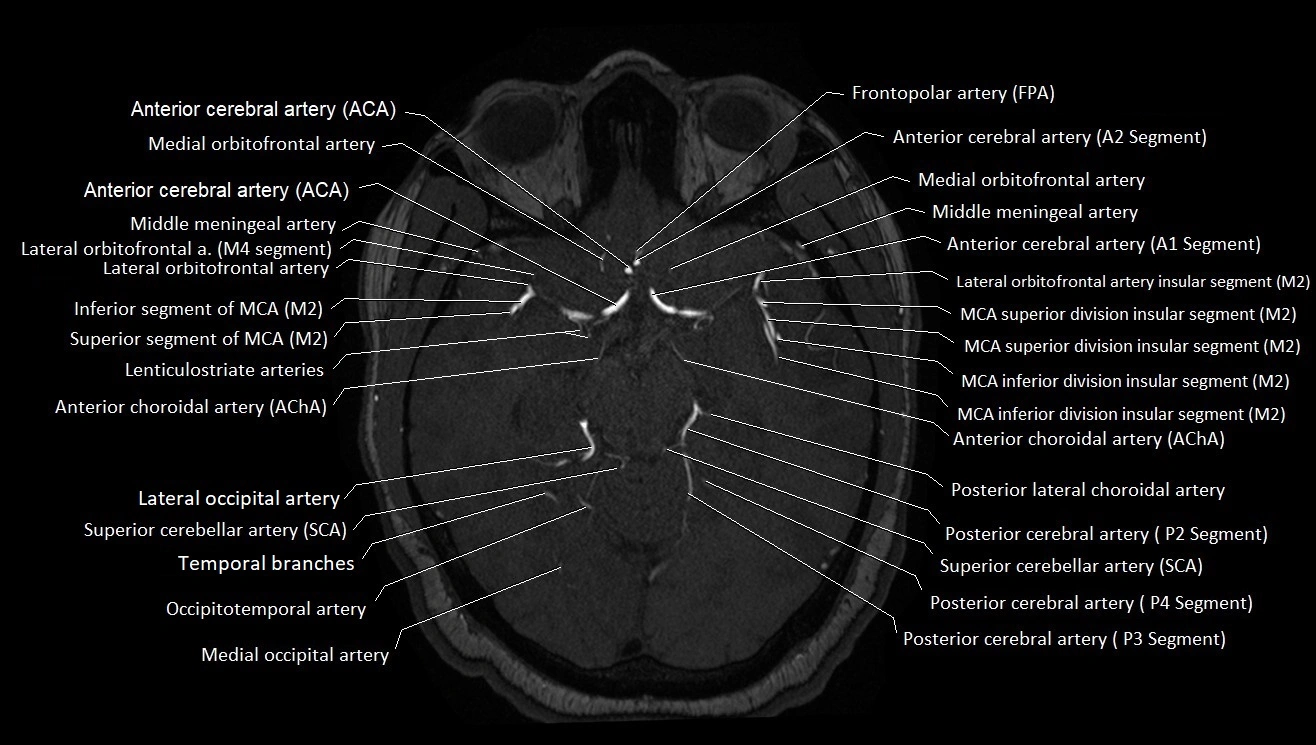
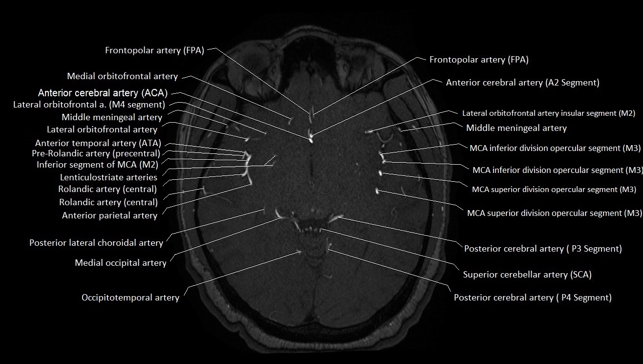
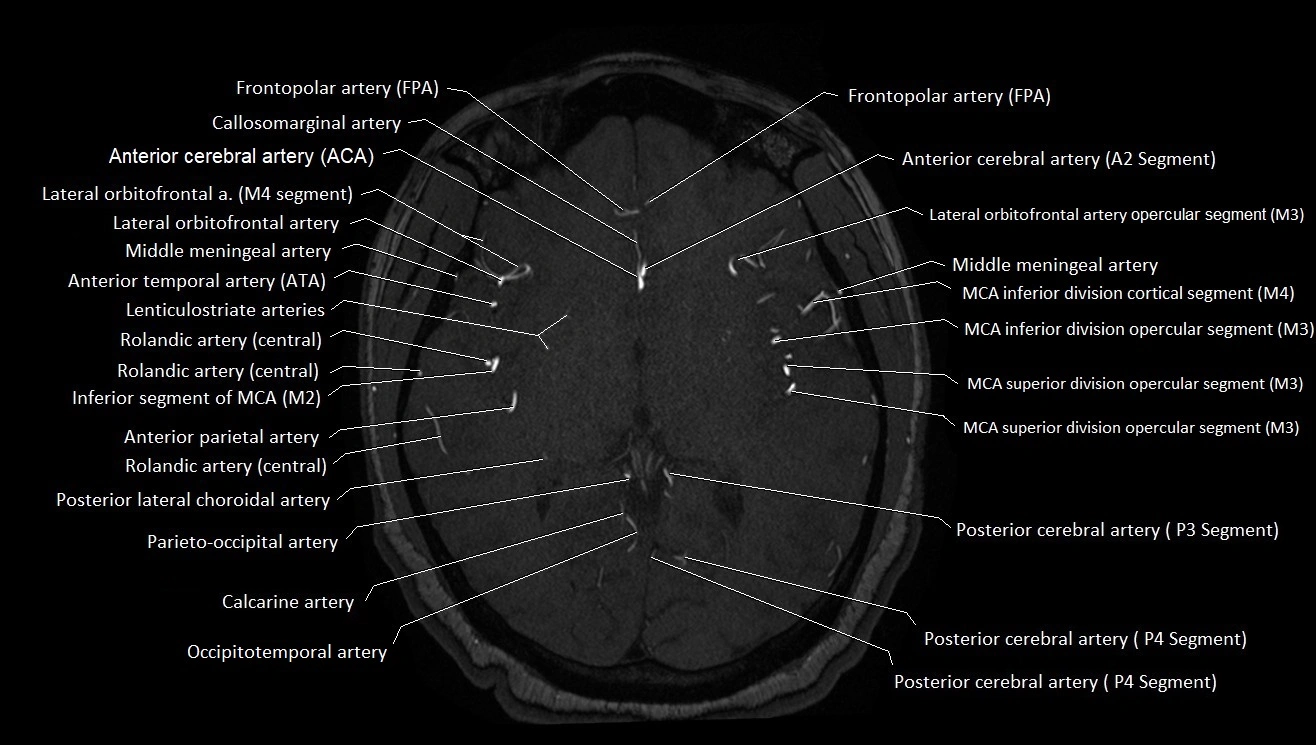
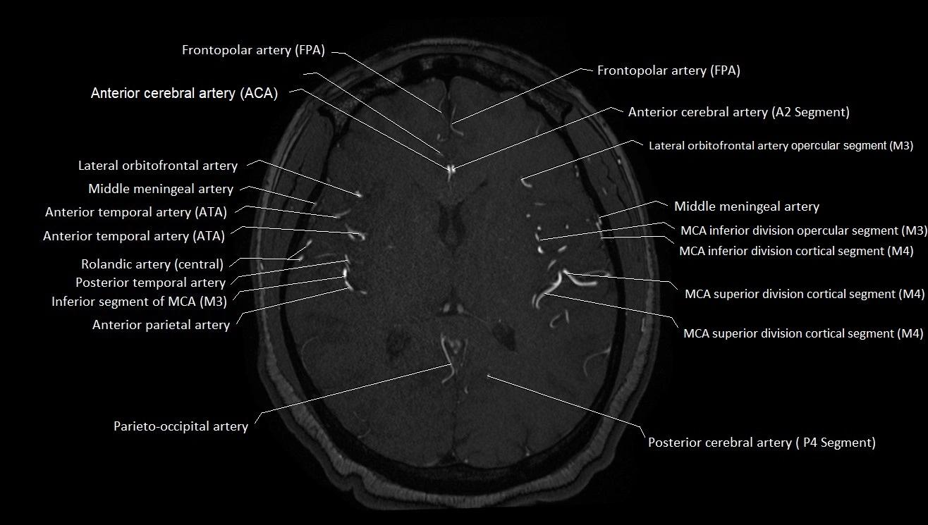
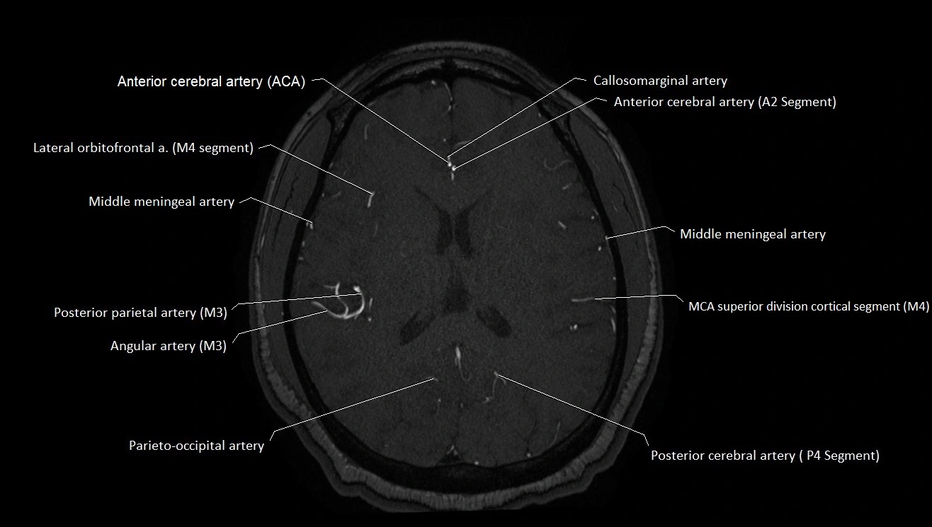
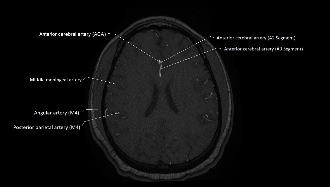
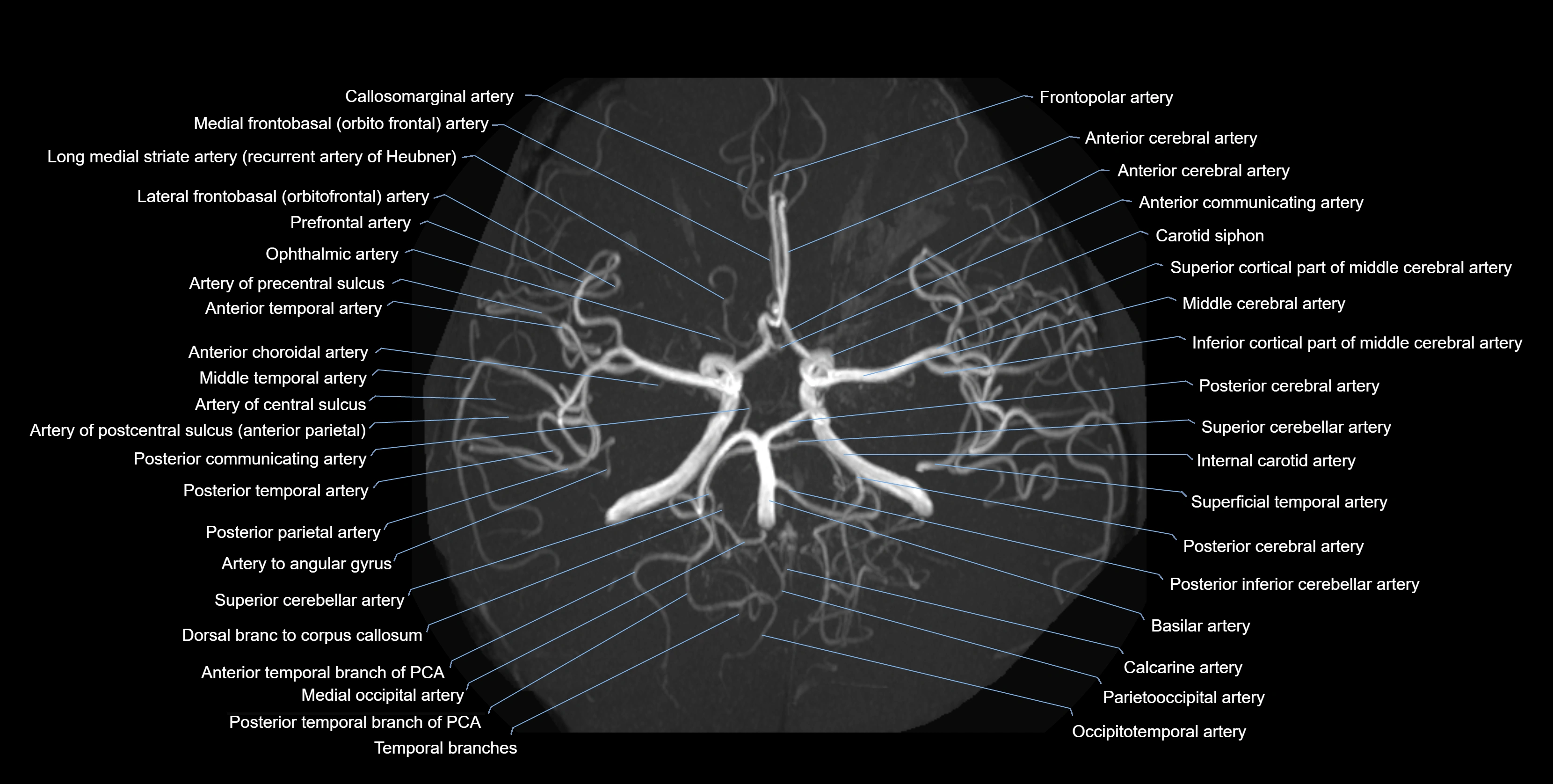
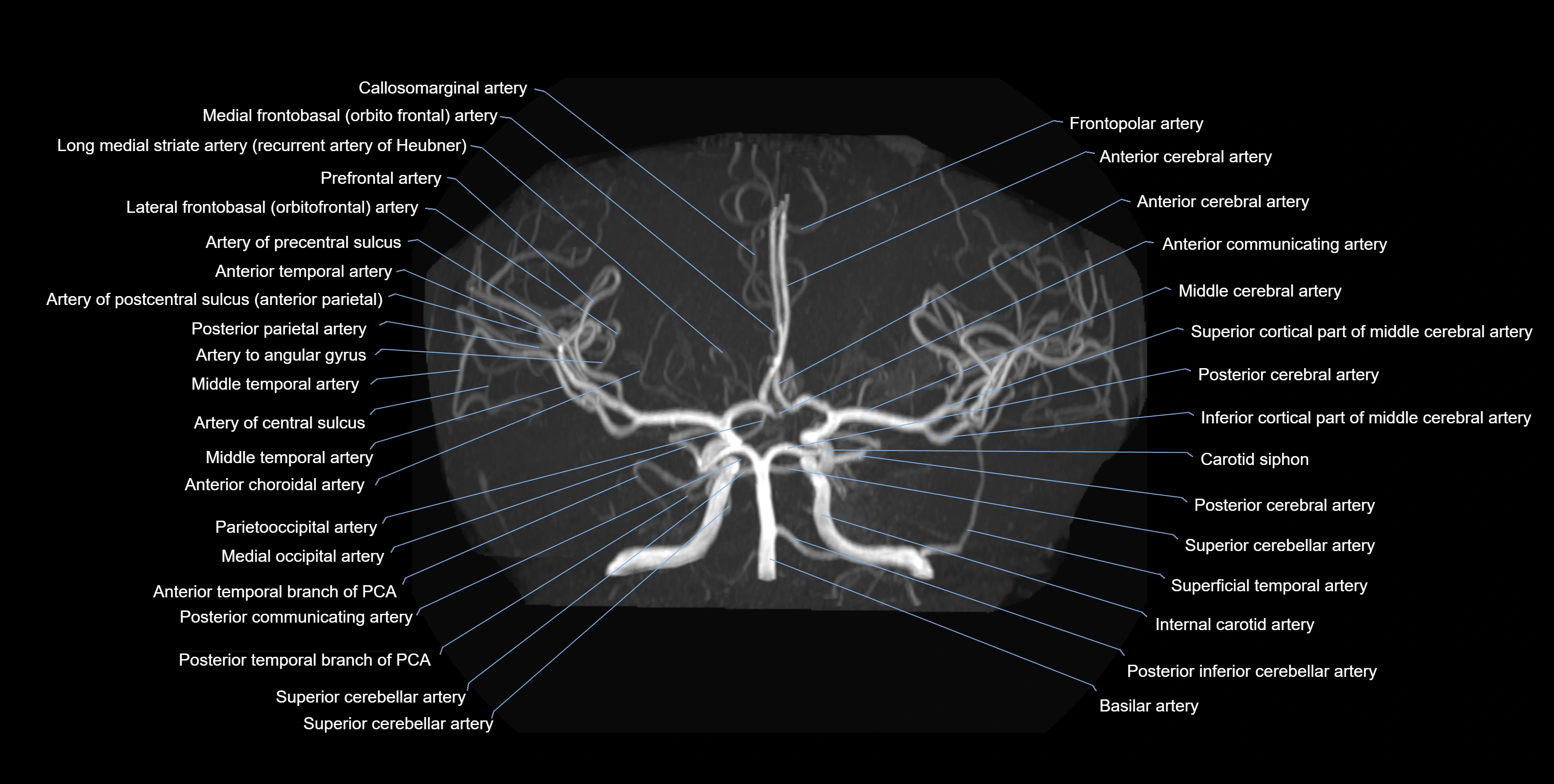
MRI images

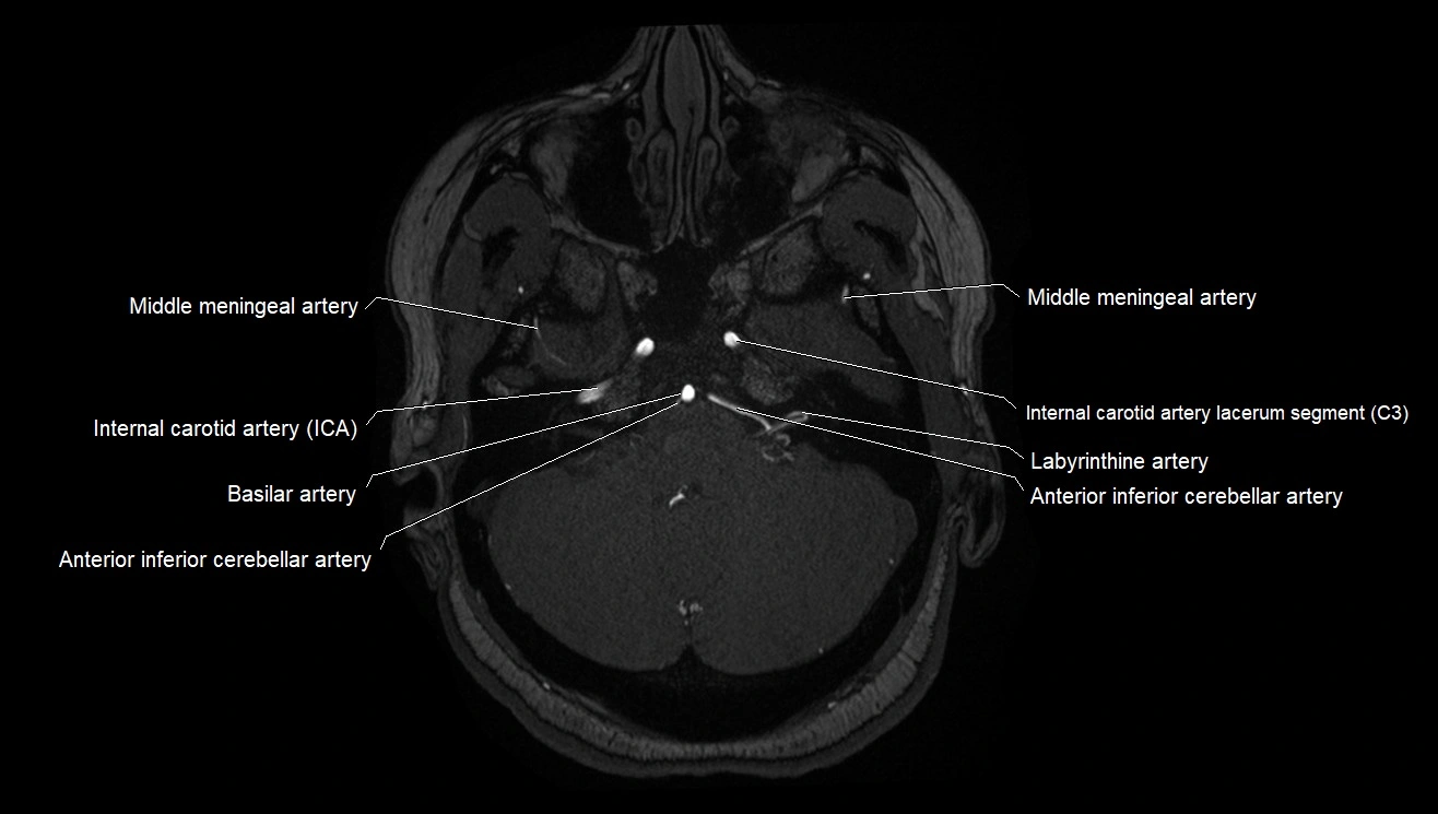
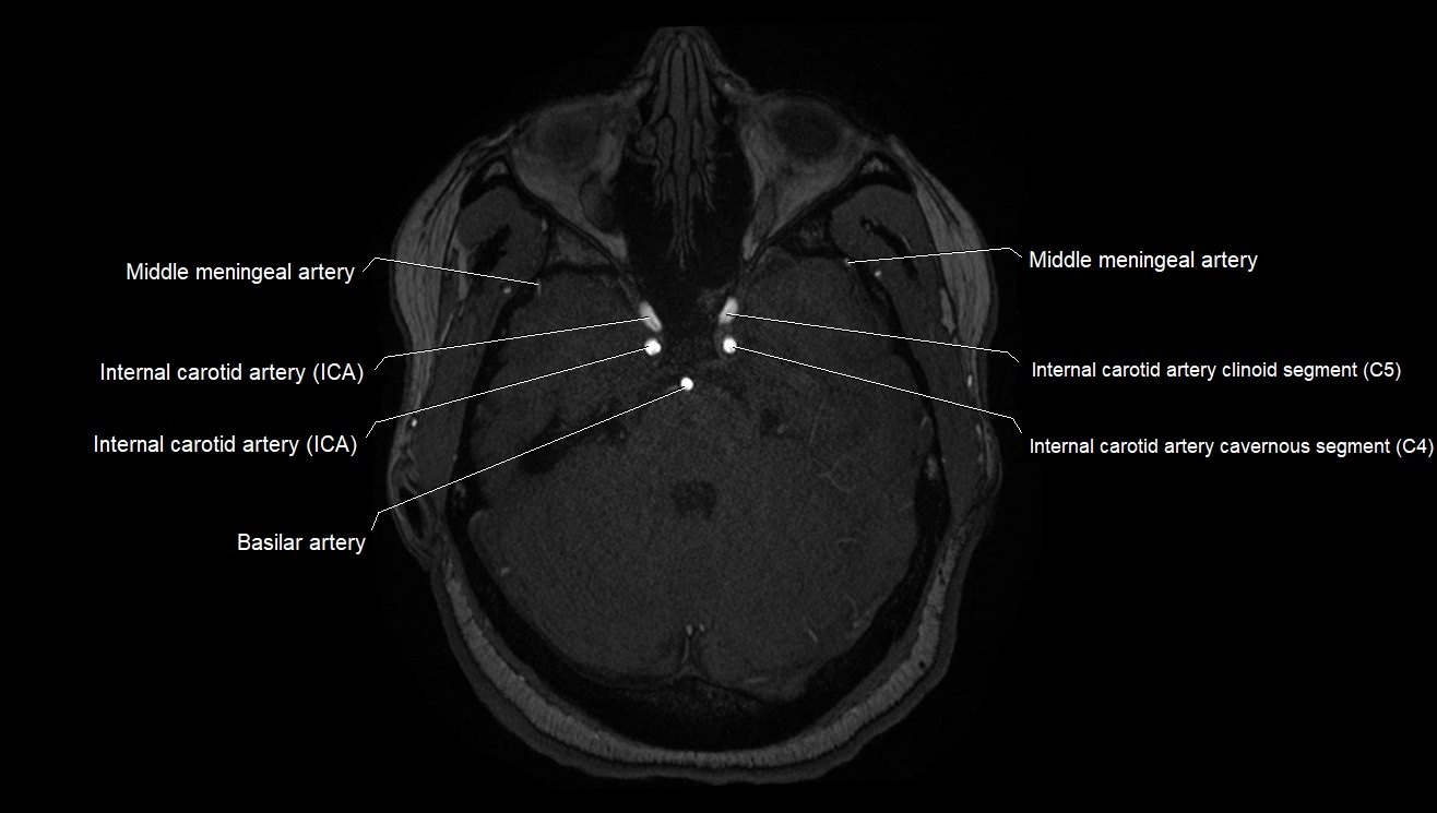
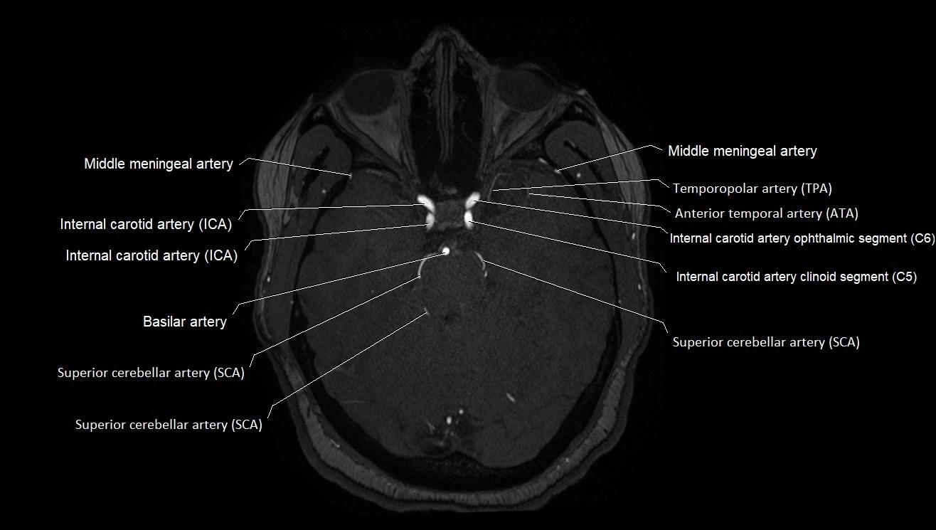
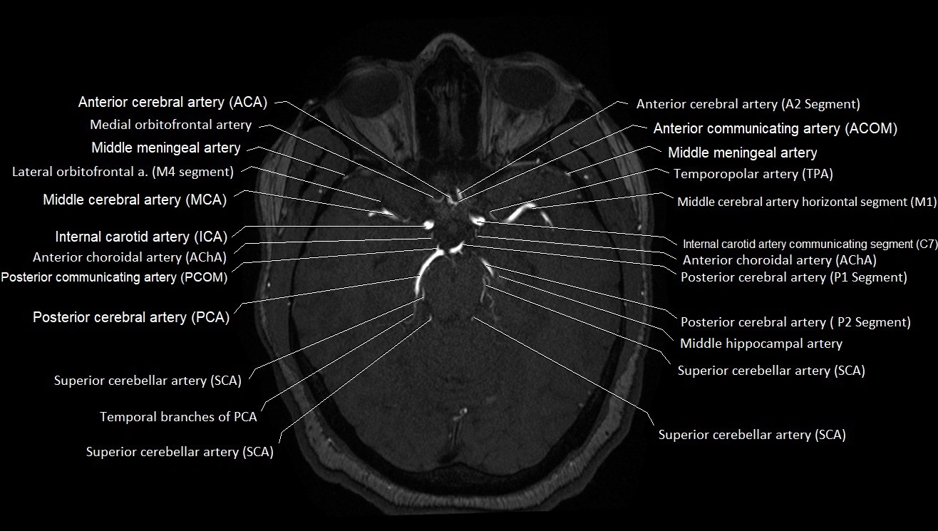
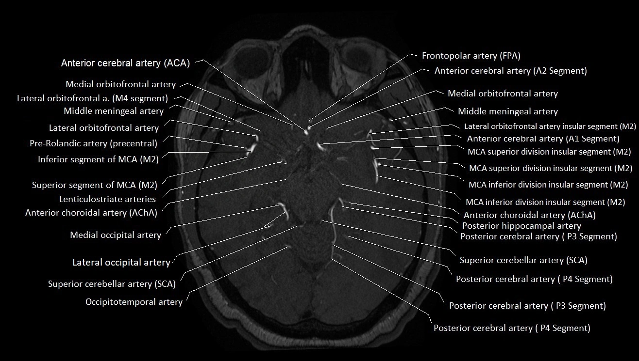
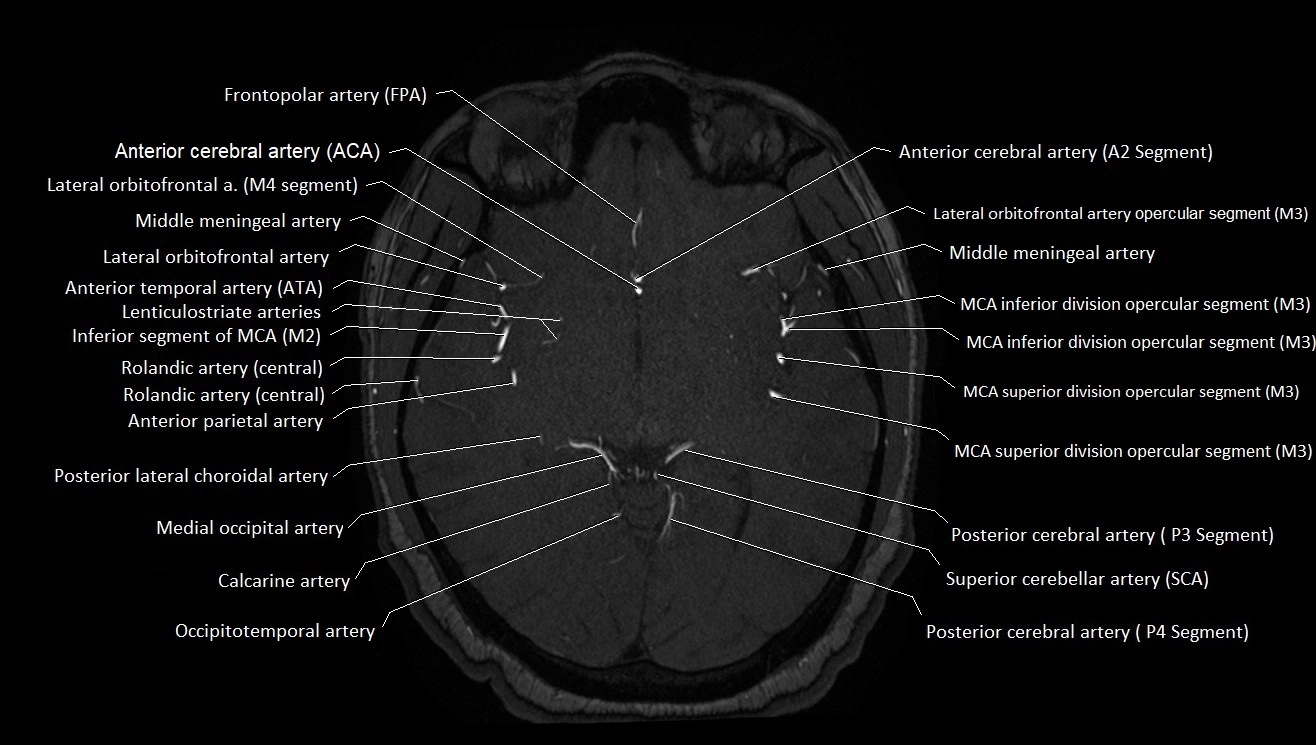
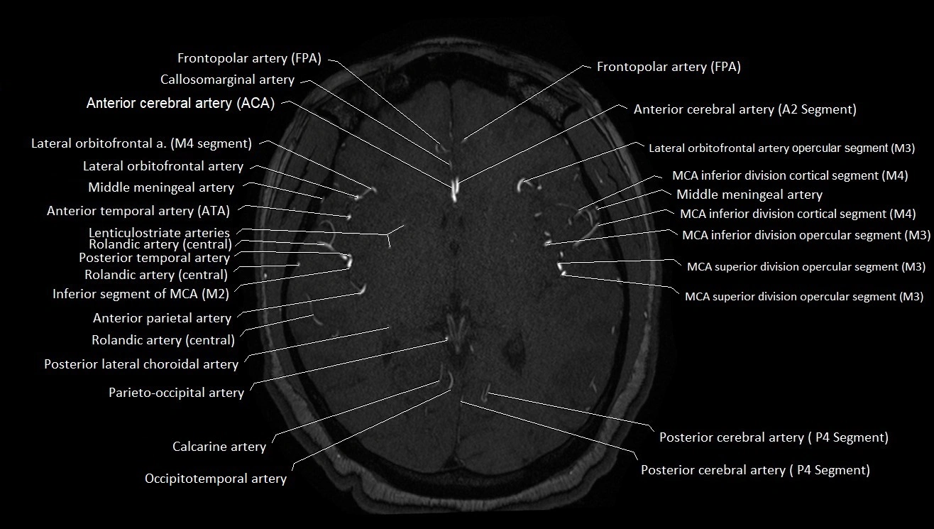
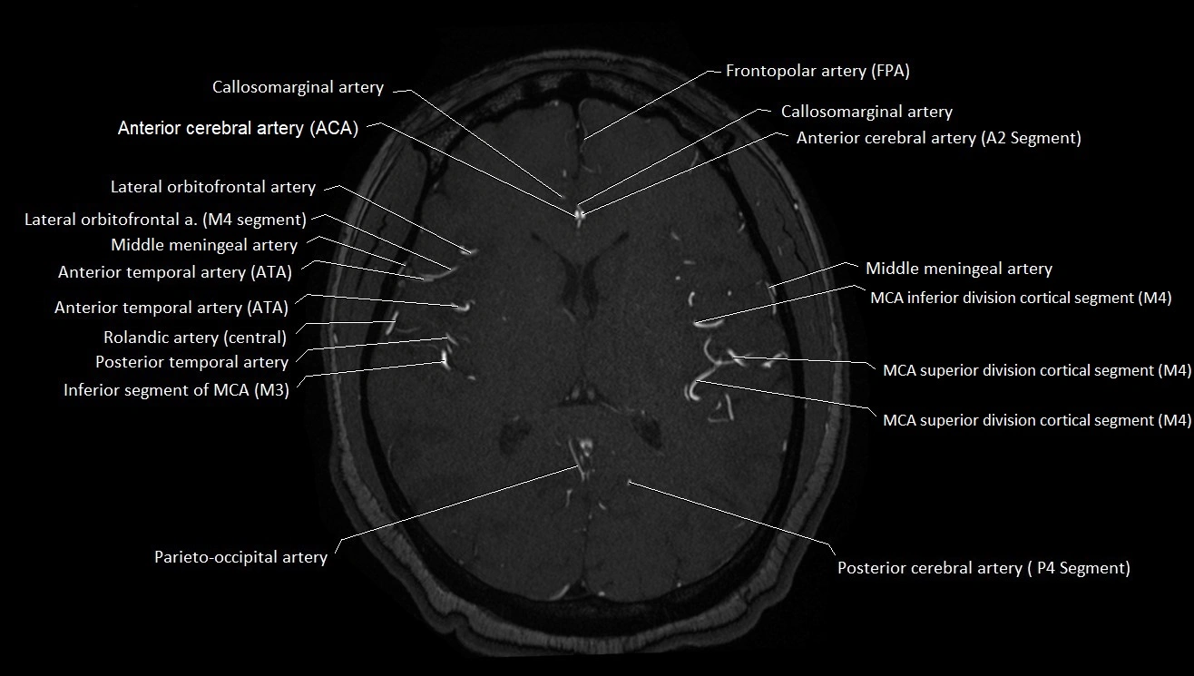
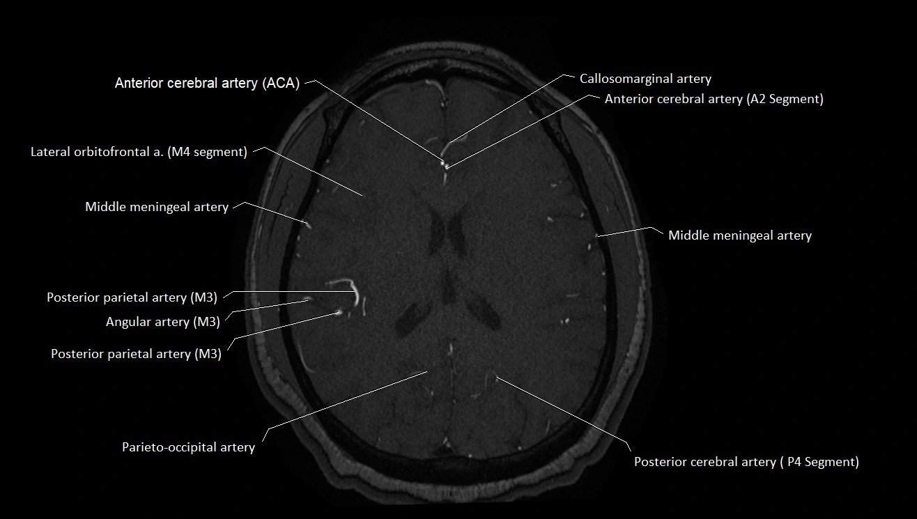
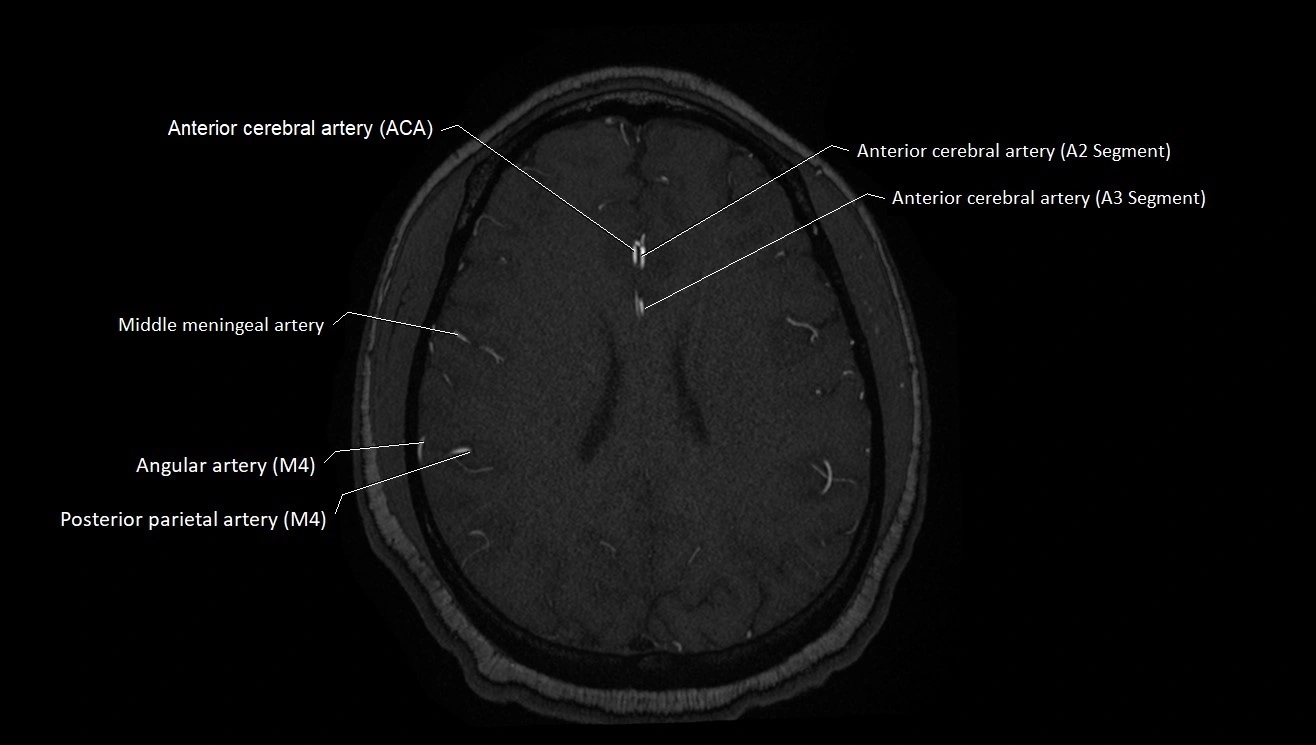
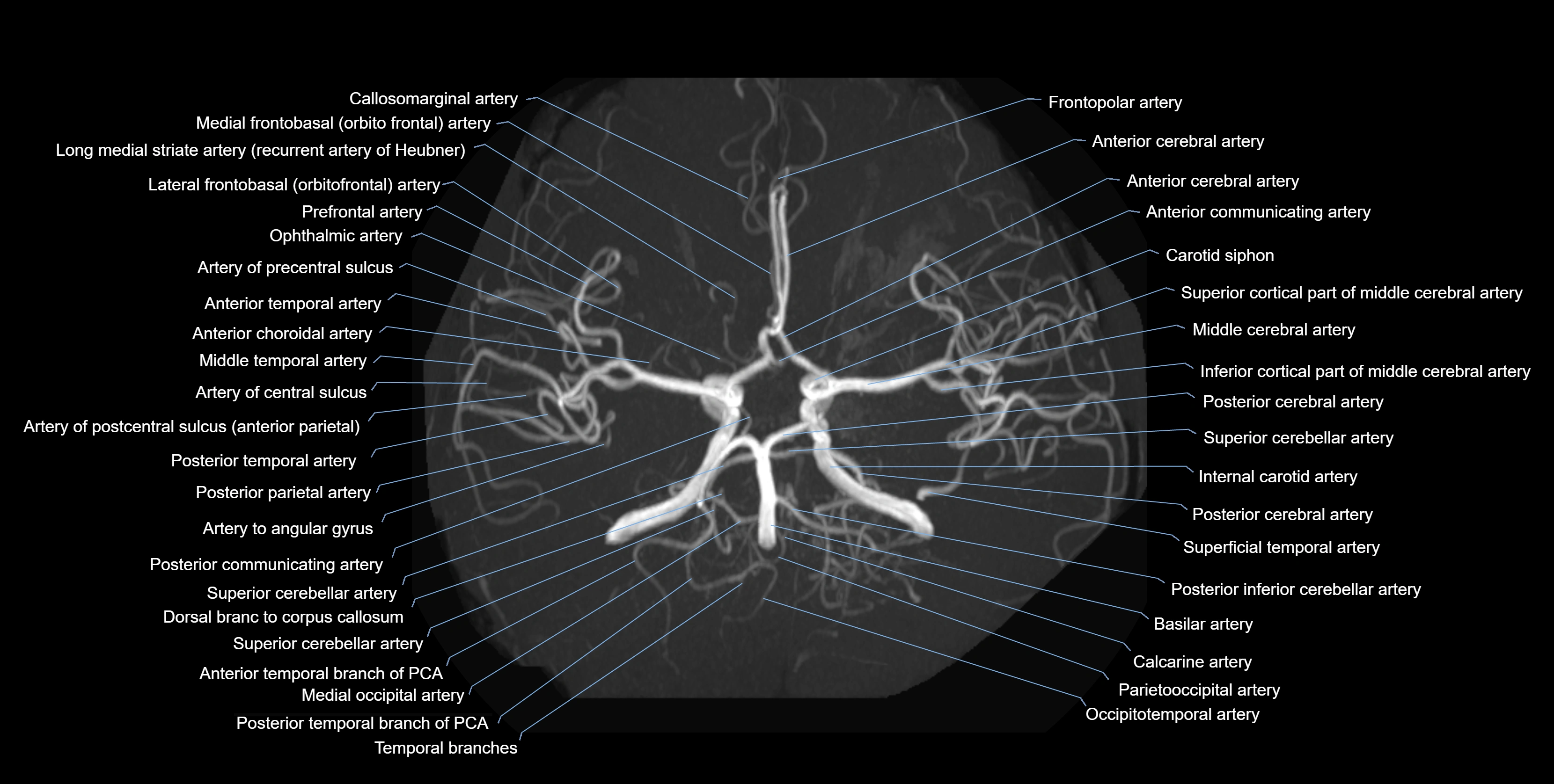
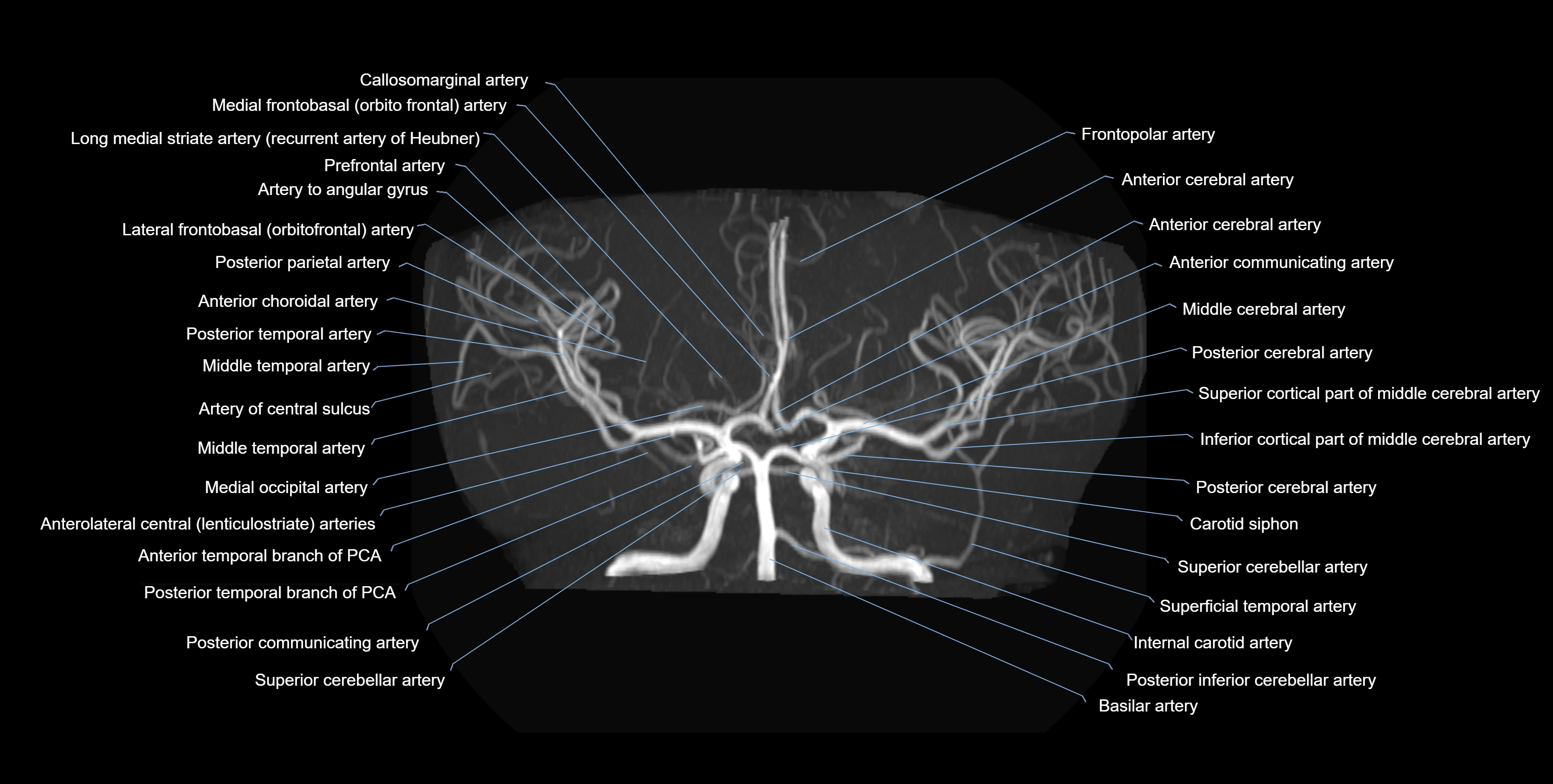
MRI images

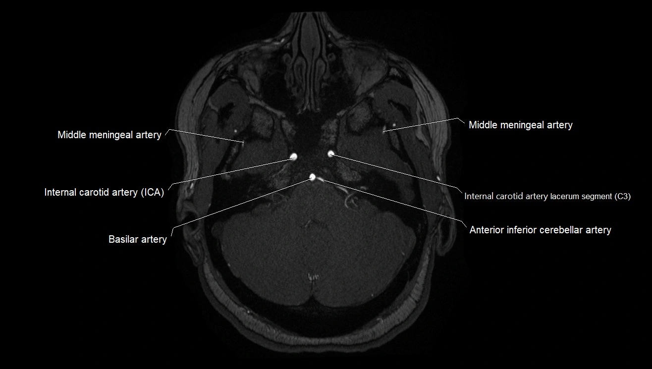
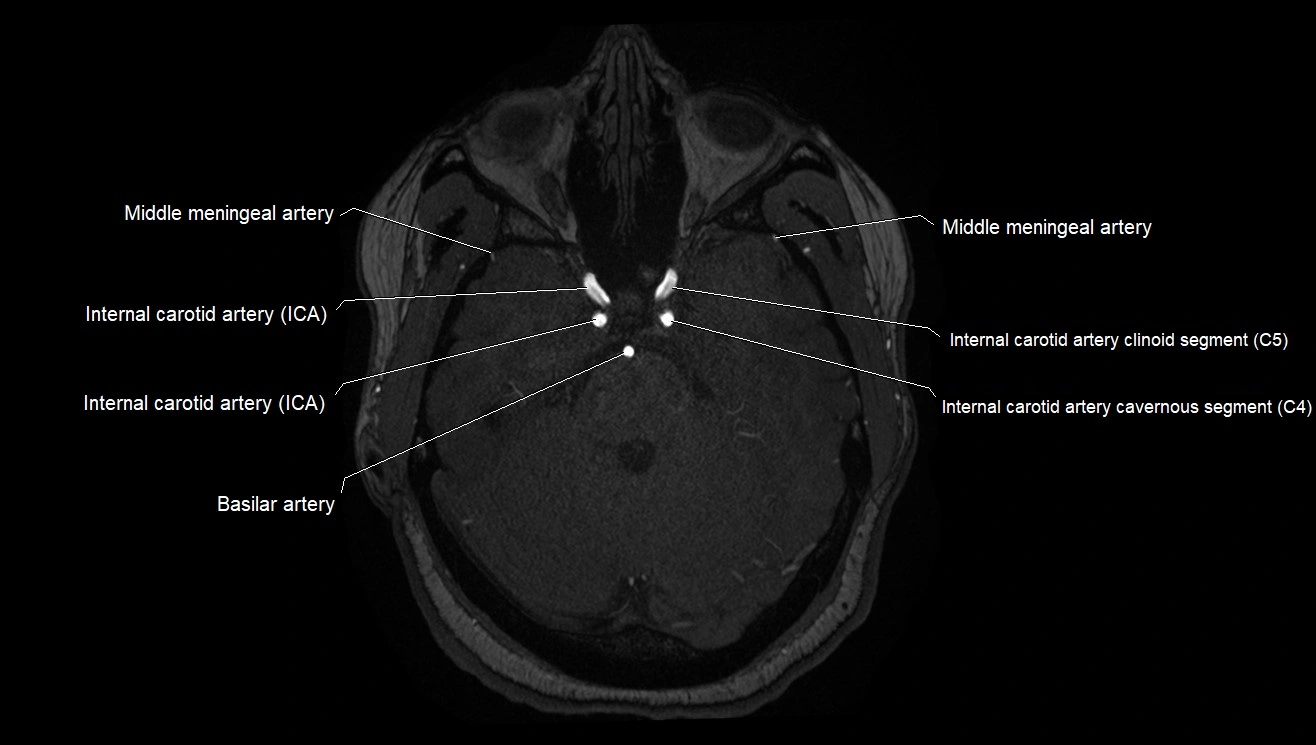
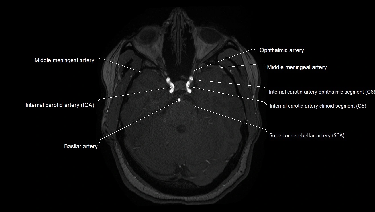
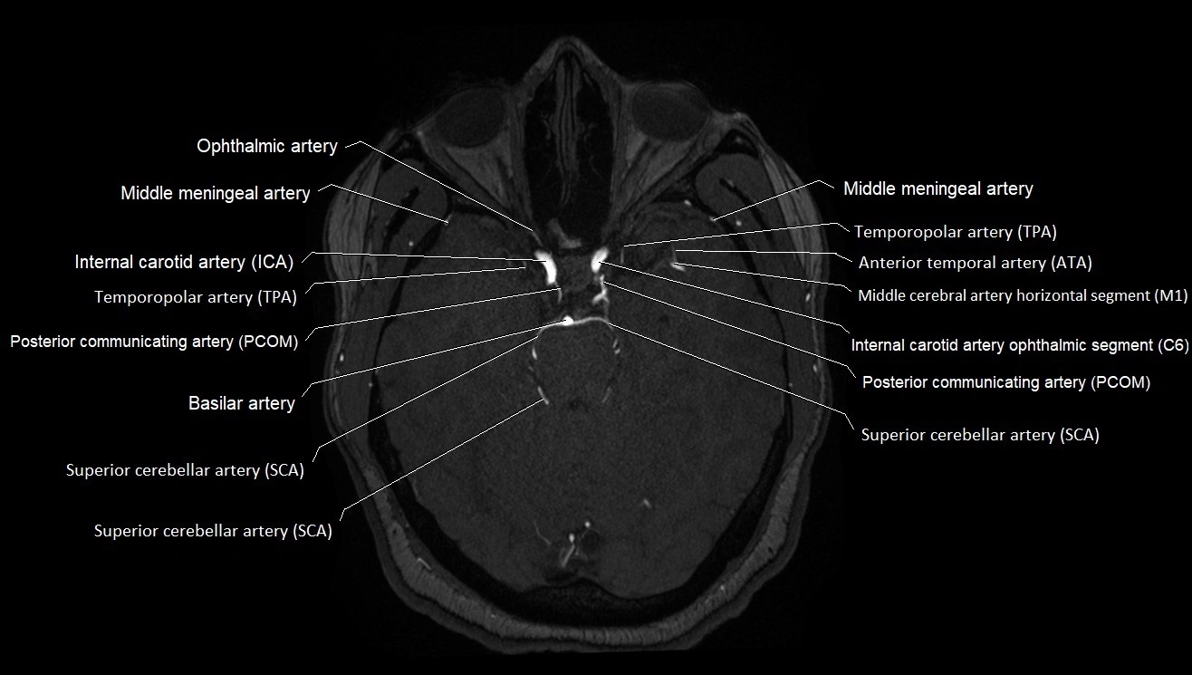
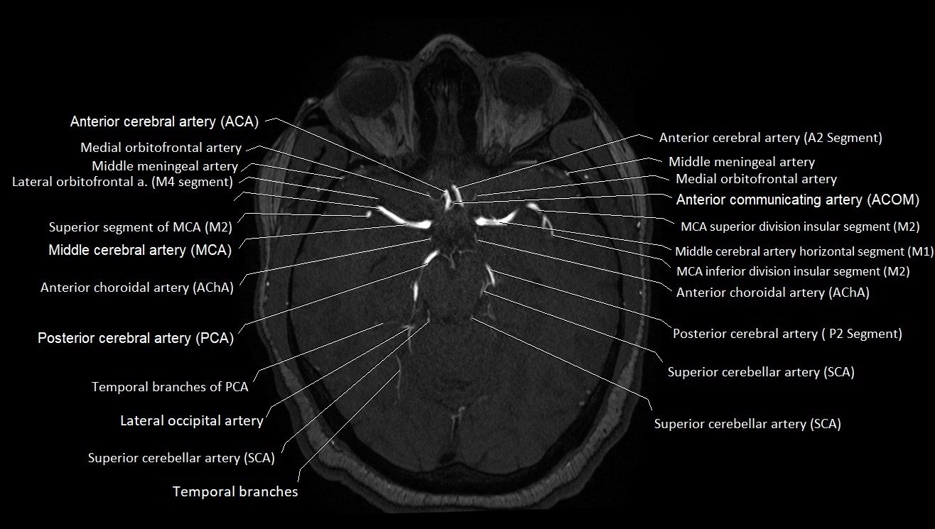
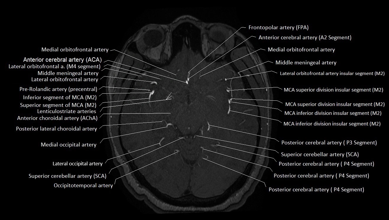
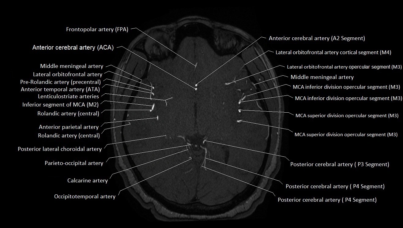
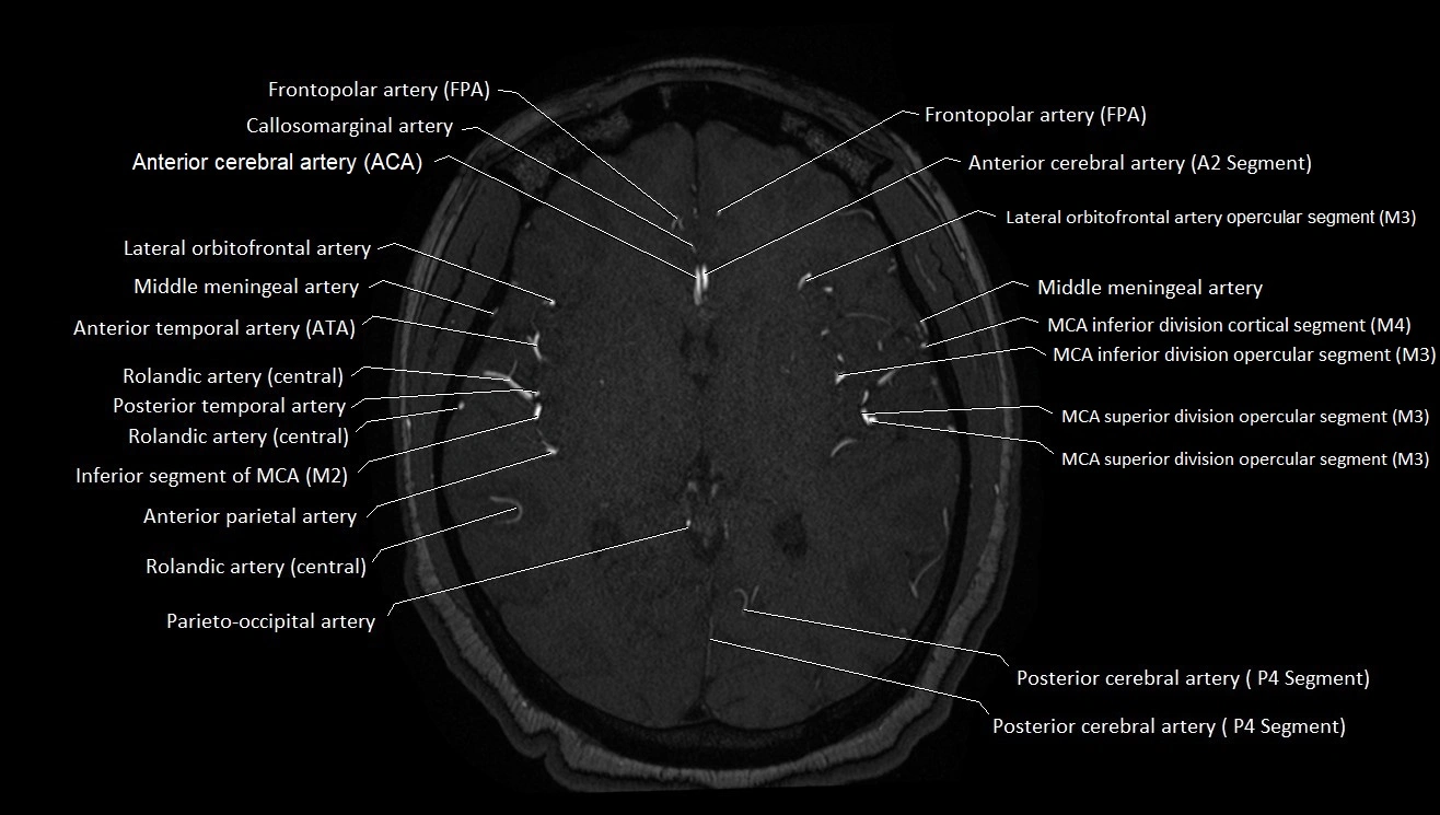
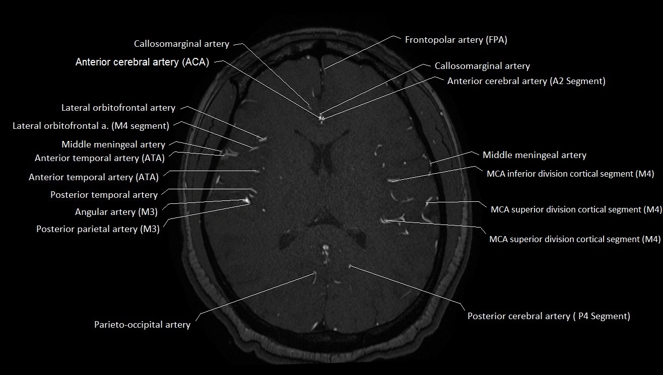
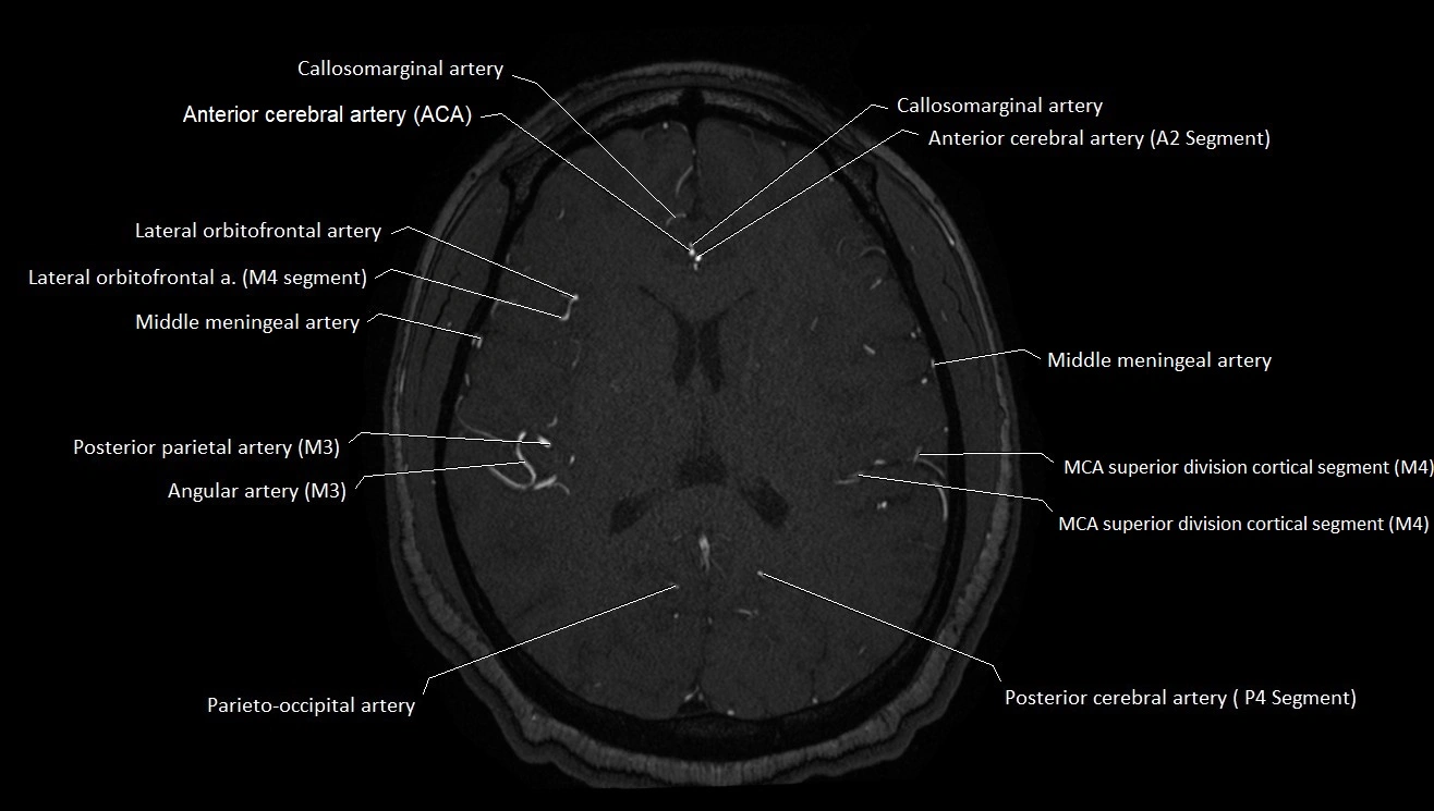
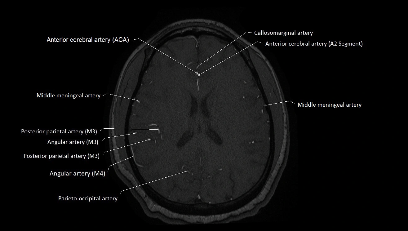
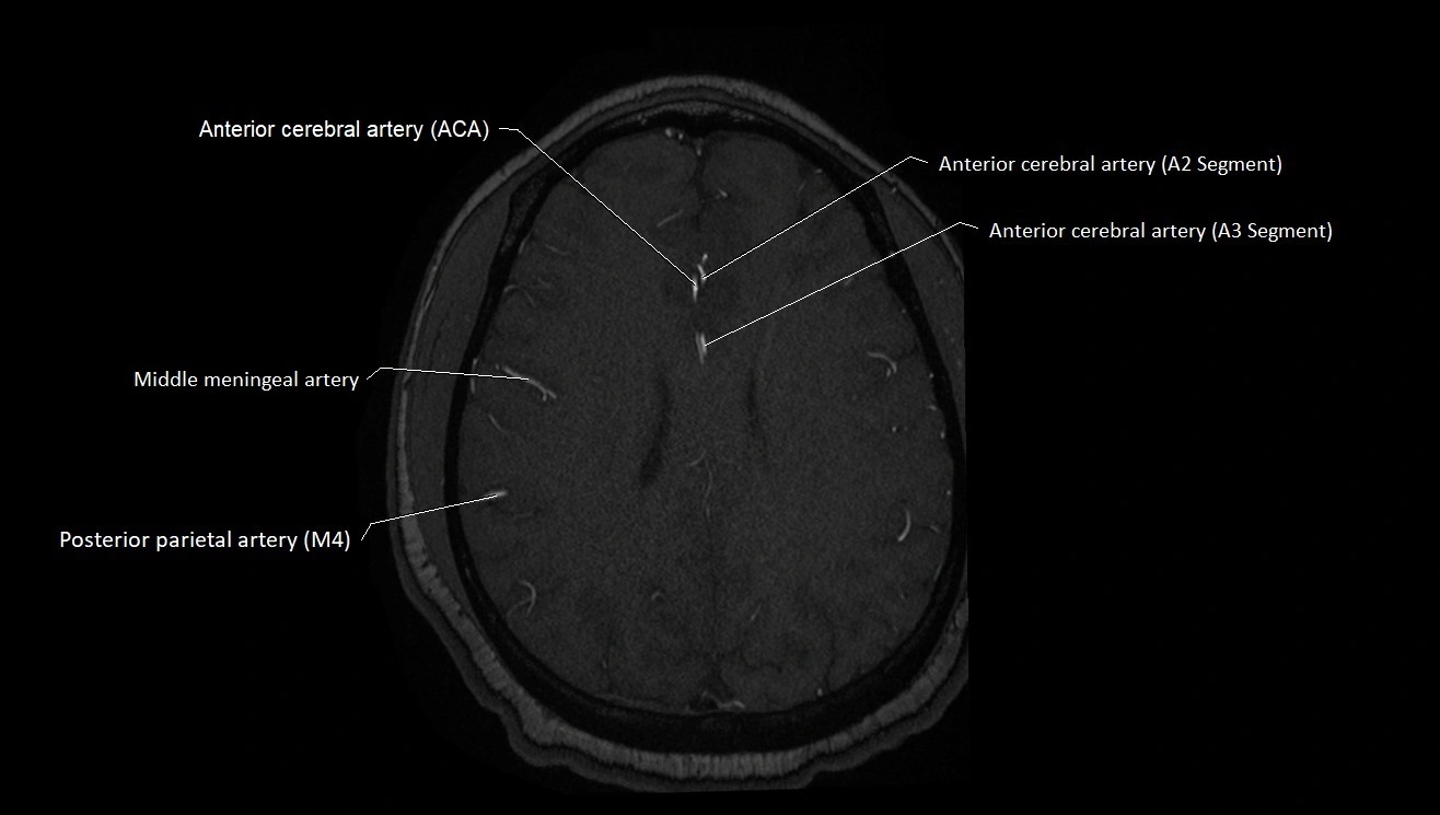
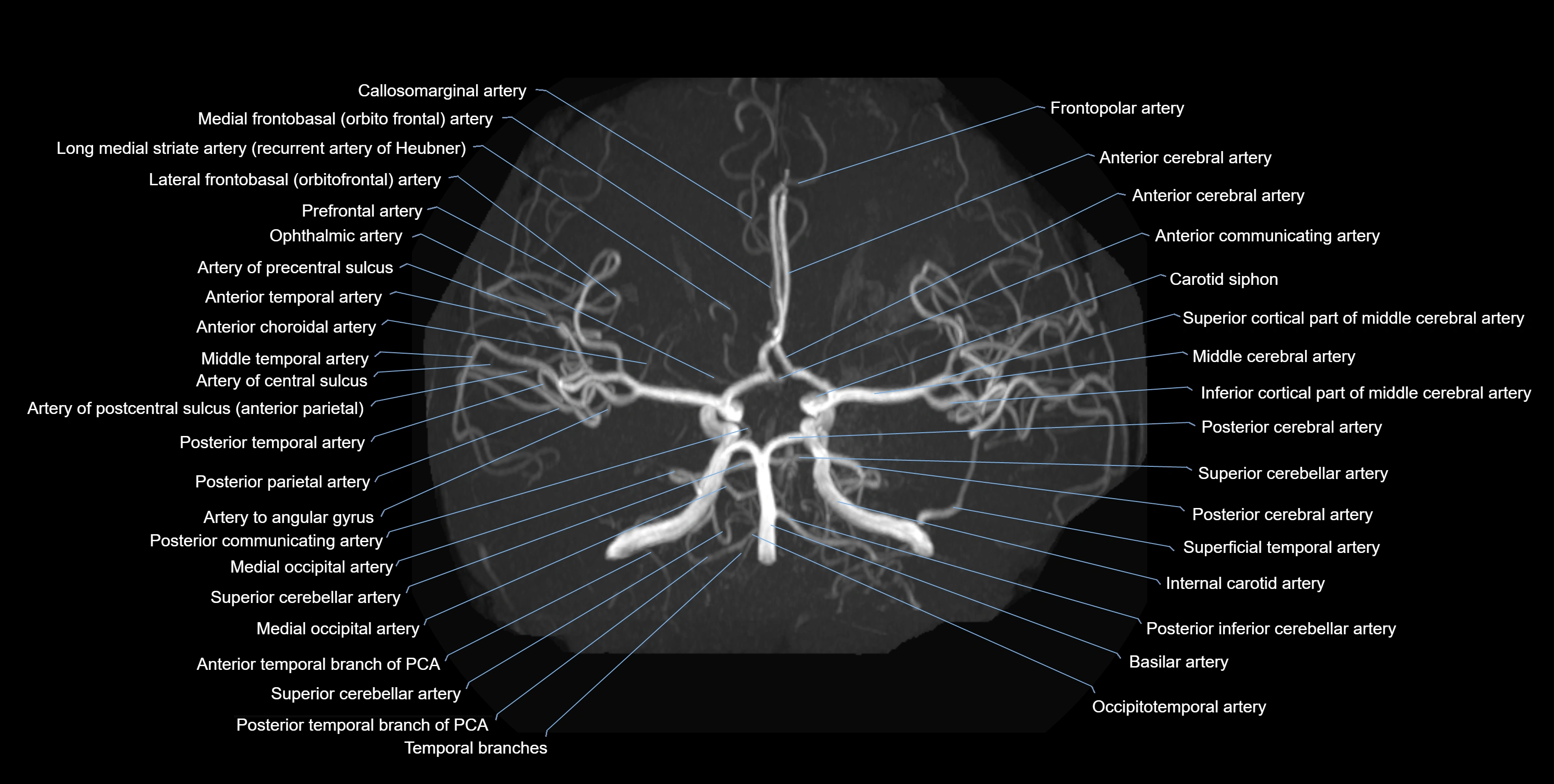
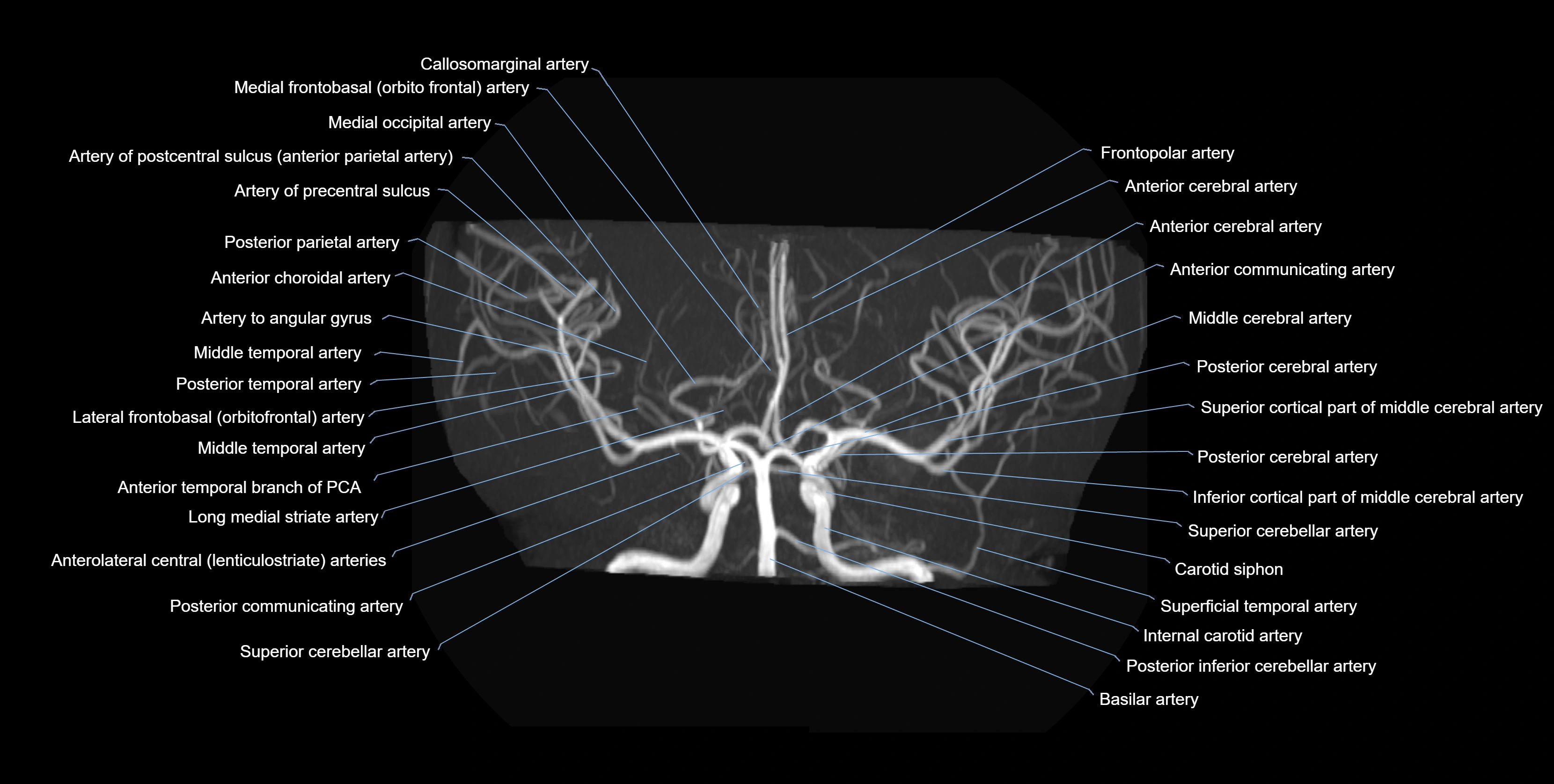
MRI images

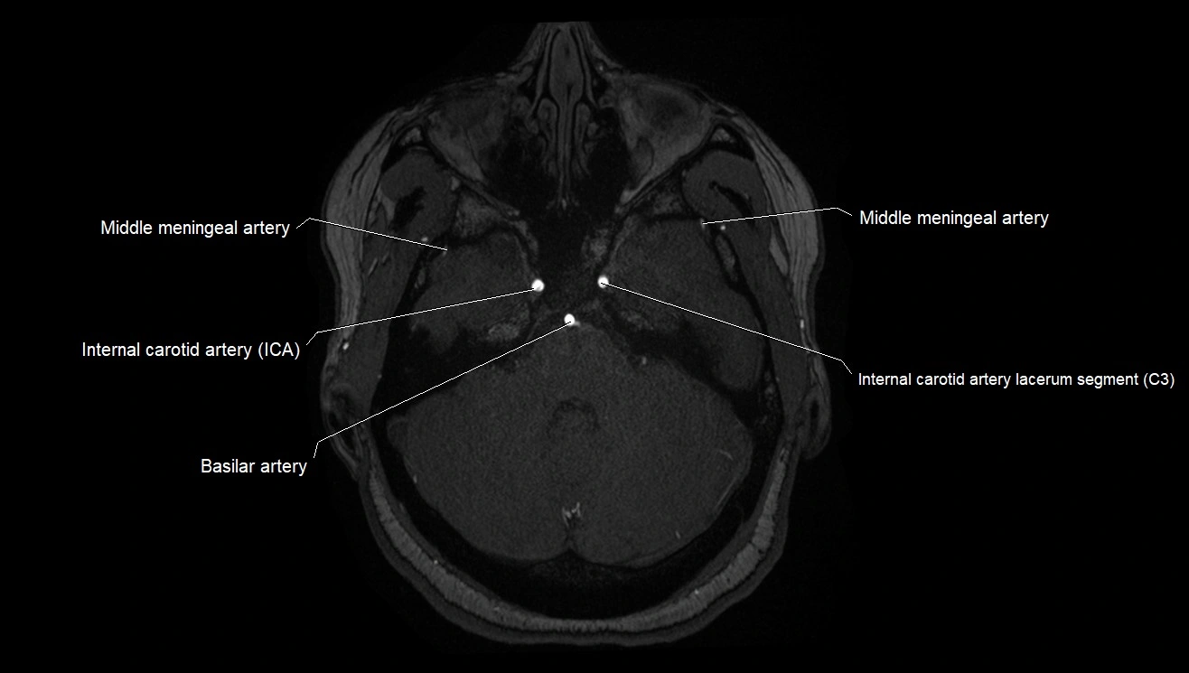
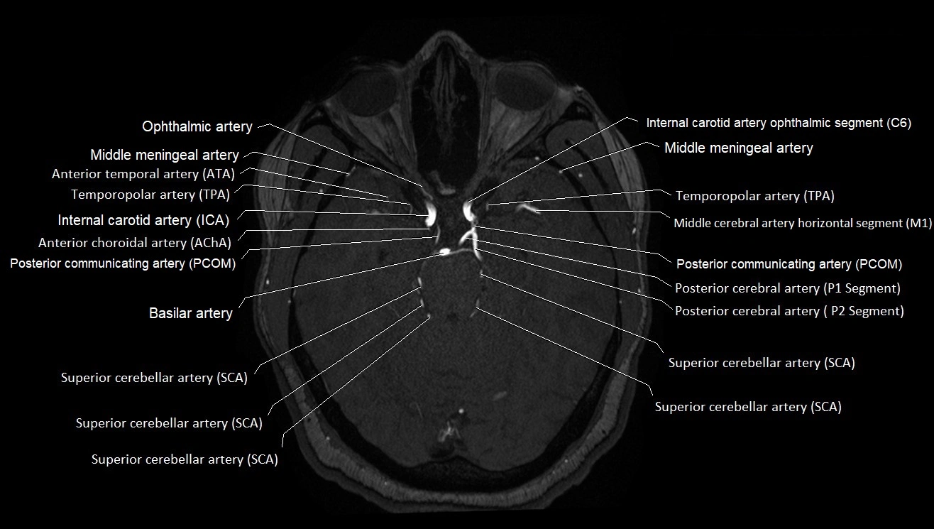
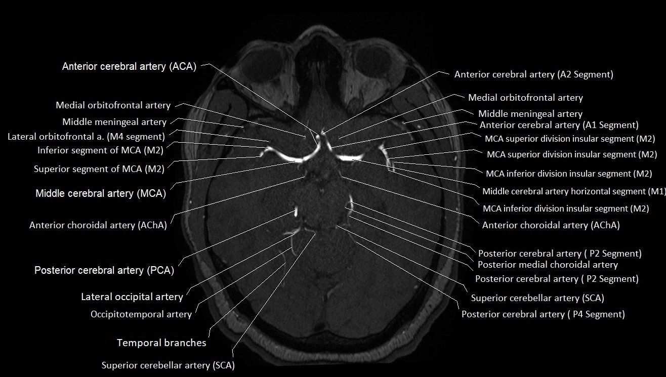
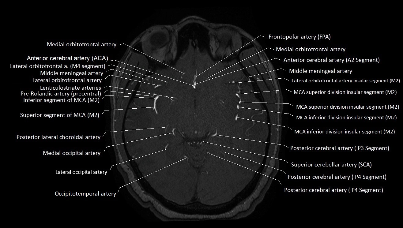
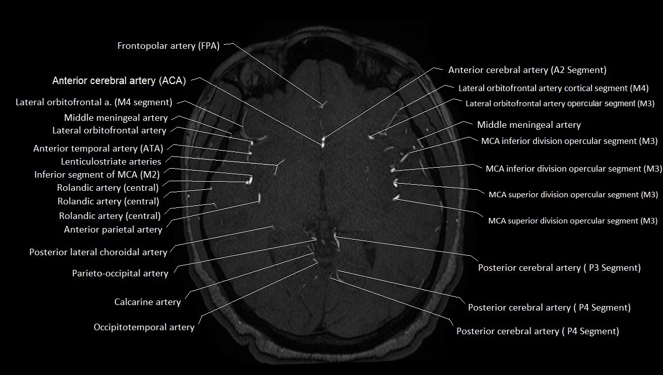
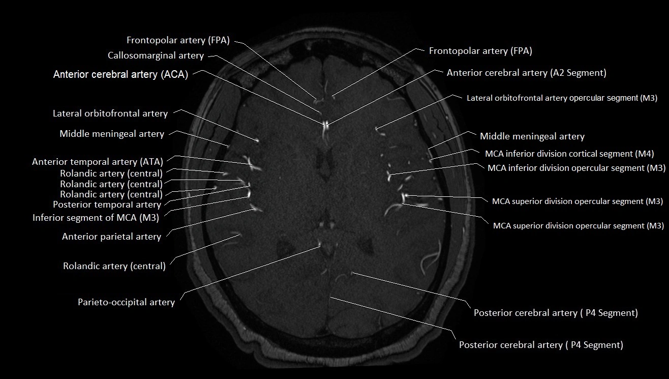
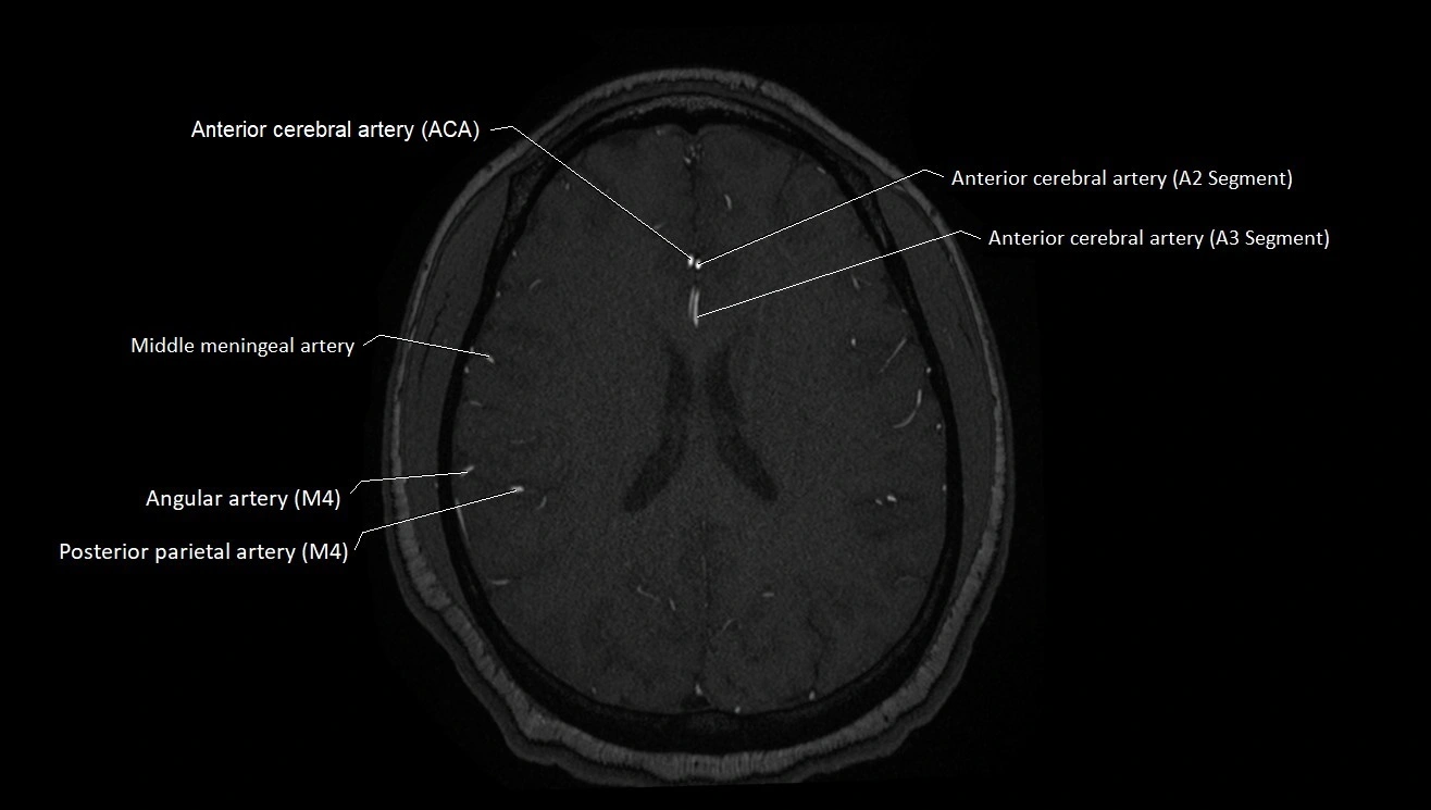
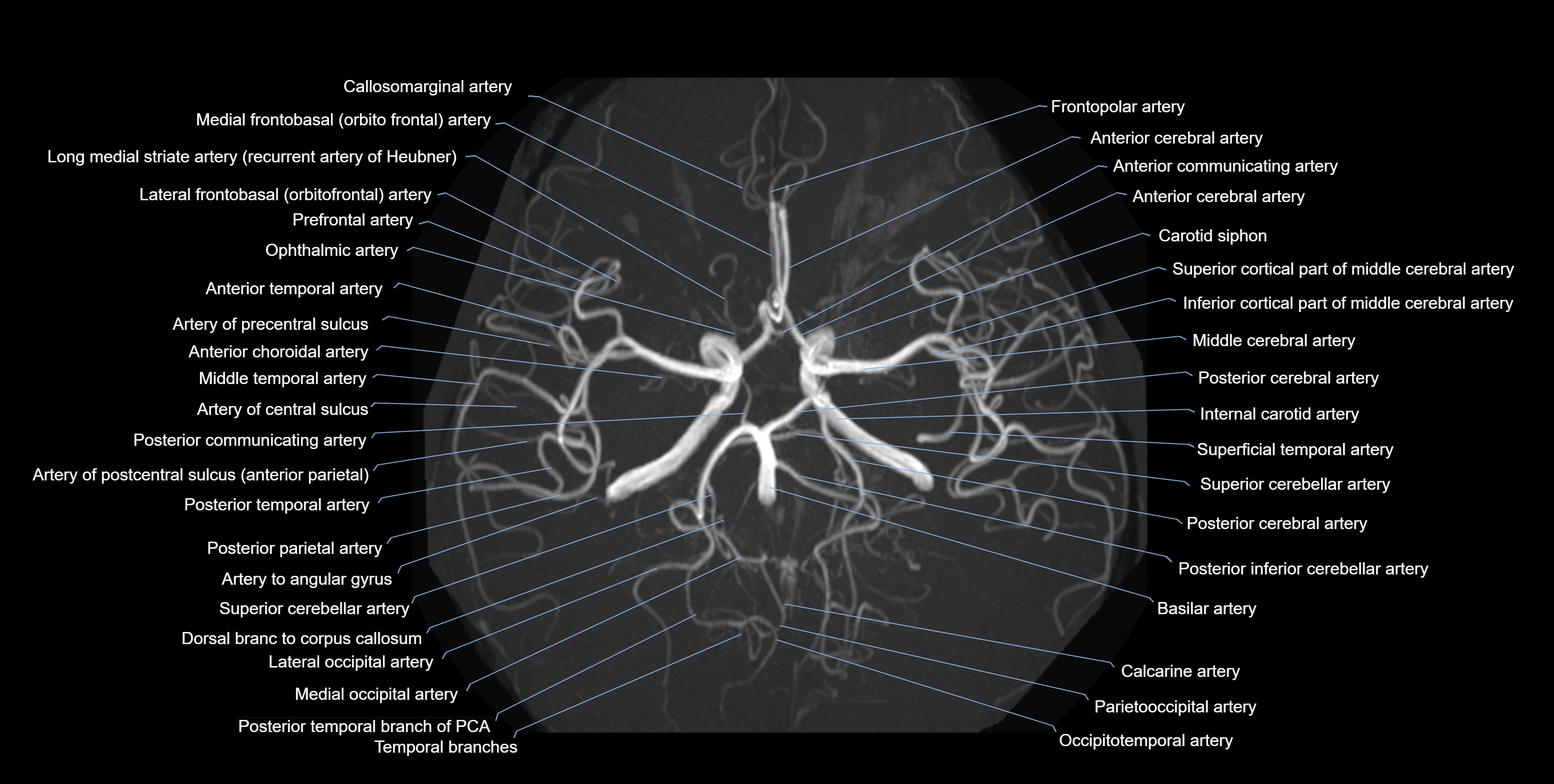
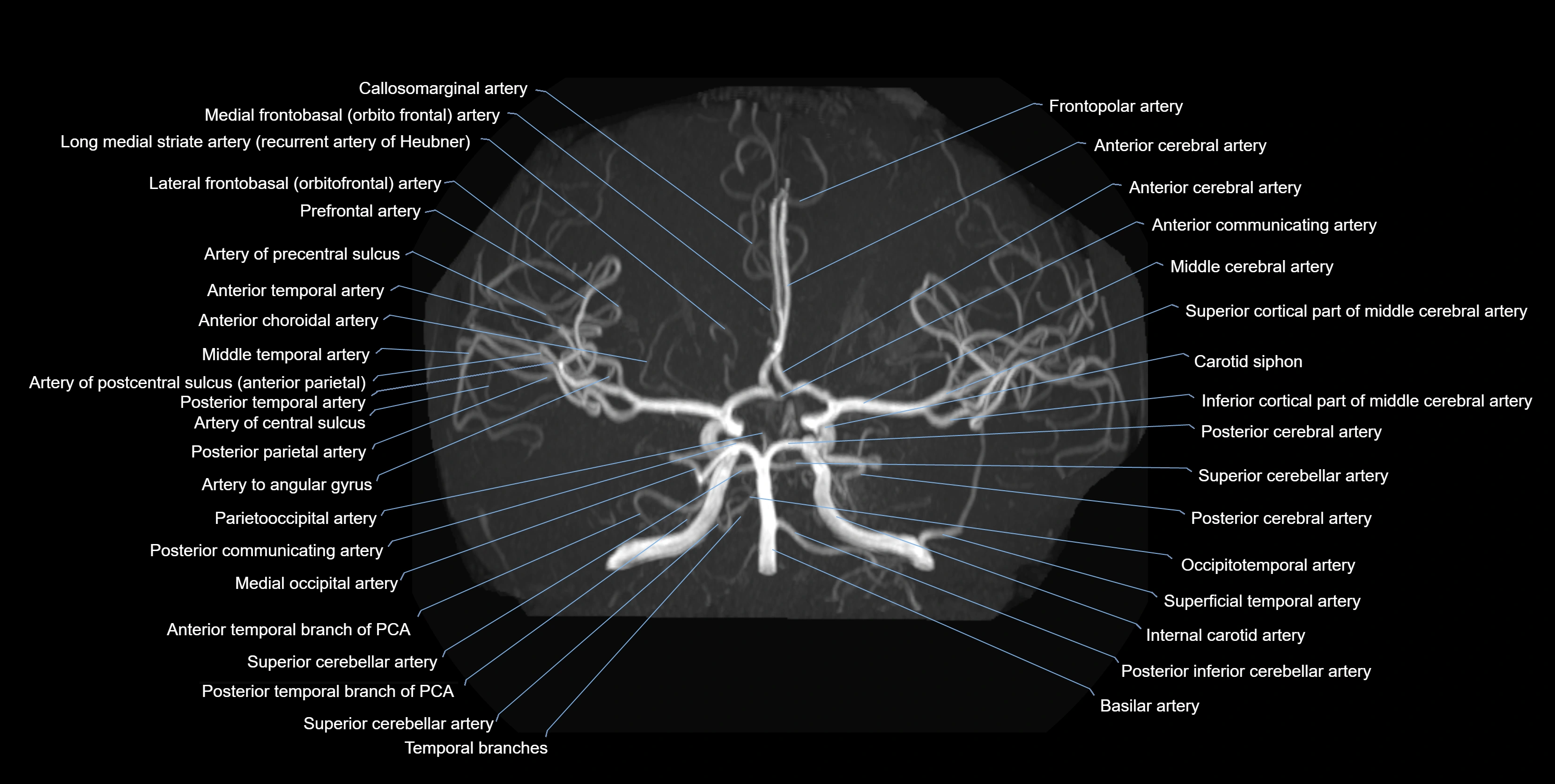
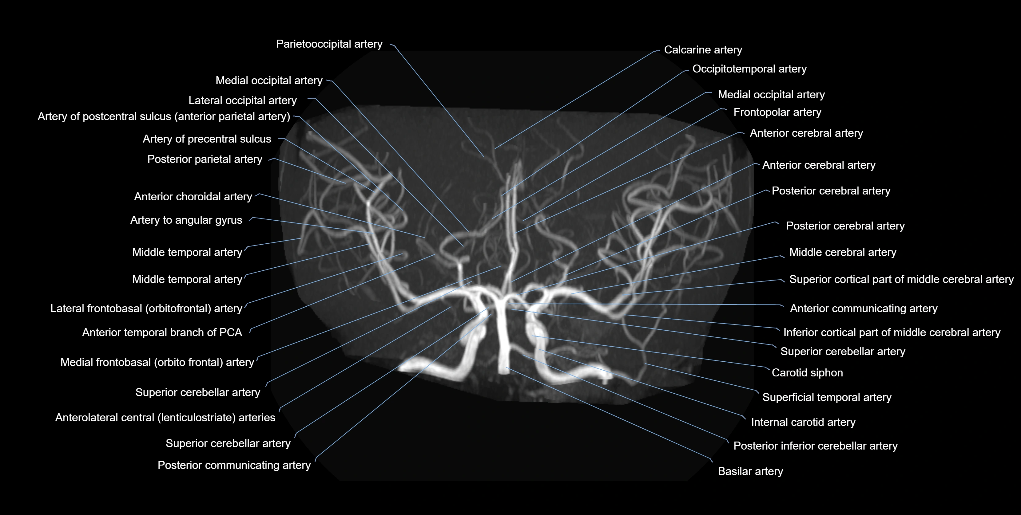
CT images


