


Topic
- Abdominal aorta
- Accessory obturator artery
- Accessory obturator vein
- Accessory process of vertebrae
- Accessory saphenous vein
- Acetabular labrum
- Acetabular margin (Acetabular rim)
- Acetabulum
- Adductor brevis muscle
- Adductor longus muscle
- Adductor magnus muscle
- Adductor minimus muscle
- Ala of ilium (wing of ilium)
- Ala of sacrum
- Anal canal
- Anococcygeal body (anococcygeal ligament)
- Anterior Fibromuscular Stroma of prostate
- Anterior acetabular wall
- Anterior cecal artery
- Anterior division of obturator nerve (Anterior branch of obturator nerve)
- Anterior fornix of cervix
- Anterior inferior iliac spine
- Anterior longitudinal ligament
- Anterior rim of acetabulum
- Anterior sacral foramina
- Anterior sacroiliac ligament
- Anterior superior iliac spine
- Aortic bifurcation
- Apex of urinary bladder
- Appendicular artery
- Articular capsule of hip joint
- Ascending colon
- Body of femur
- Body of ilium
- Body of ischium
- Body of pubis
- Body of urinary bladder
- Body of uterus
- Broad ligament of uterus
- Bulbospongiosus muscle (Female)
- Bulbospongiosus muscle (Male)
- Cecum
- Central zone of prostate
- Cervix of uterus
- Clitoris
- Co (Coccyx)
- Coccygeal nerve
- Coccygeal plexus
- Coccygeus muscle
- Coccyx
- Common iliac lymph nodes
- Common iliac vein
- Conjoint tendon of biceps femoris & semitendinosus
- Costotransverse joint of twelfth rib
- Costovertebral joint of twelfth rib
- Cystic artery
- Deep circumflex iliac artery
- Deep femoral artery (profunda femoris)
- Deep femoral vein (profunda femoris vein)
- Deep transverse perineal muscle
- Dorsal ramus of spinal nerve
- Ejaculatory duct
- Endocervical canal
- Endometrium of uterus
- Erector spinae muscles
- Exiting nerve root of spinal nerve S1
- Exiting nerve root of spinal nerve S2
- Exiting nerve root of spinal nerve S3
- Exiting nerve root of spinal nerve S4
- Exiting nerve root of spinal nerve S5
- External anal sphincter
- External iliac artery
- External iliac lymph nodes
- External iliac vein
- External os of the cervix
- External urethral orifice
- External urethral sphincter (female)
- External urethral sphincter (male)
- Fallopian tube
- Fascia of pelvic diaphragm
- Female urethra
- Femoral artery
- Femoral nerve
- Femoral shaft
- Femoral vein
- Femur
- Filum terminale internum
- Fornix of the vagina
- Fovea for ligament of head of femur
- Fundus of urinary bladder
- Fundus of uterus
- Genitofemoral nerve
- Gluteal lymph nodes
- Gluteus medius tendon
- Gluteus minimus tendon
- Gracilis muscle
- Greater sciatic notch
- Greater trochanter
- Head of femur
- Head of twelfth rib
- Hip joint
- Ileal arteries
- Ileocolic artery
- Ileocolic artery colic branches
- Ileocolic artery ileal branches
- Iliac bone
- Iliac crest
- Iliac fossa
- Iliac tubercle
- Iliac tuberosity
- Iliococcygeus muscle
- Iliocostalis lumborum muscle
- Iliofemoral Ligament inferior band (vertical band, medial band)
- Iliofemoral Ligament superior band (transverse band, lateral band)
- Iliofemoral ligament
- Iliolumbar ligament
- Iliopsoas muscle
- Iliopsoas tendon
- Iliopubic eminence
- Iliotibial tract
- Ilium bone
- Inferior articular process of L5 vertebra
- Inferior epigastric artery
- Inferior epigastric veins
- Inferior gemellus muscle
- Inferior gluteal artery
- Inferior gluteal vein
- Inferior mesenteric artery (IMA)
- Inferior mesenteric vein
- Inferior pubic ligament
- Inferior pubic ramus
- Inferior rim of acetabulum
- Inferior vesical artery
- Inguinal ligament
- Inguinal lymph nodes
- Intercommunicating branches of L3–L4 nerves
- Interlobar arteries of kidney
- Intermediate lacunar external iliac lymph nodes
- Intermediate sacral crest
- Internal anal sphincter
- Internal iliac artery
- Internal iliac lymph nodes
- Internal iliac vein
- Internal os of the cervix
- Internal pudendal artery
- Internal pudendal vein
- Internal urethral orifice
- Internal urethral sphincter (female)
- Internal urethral sphincter (male)
- Interosseous sacroiliac ligament
- Interspinales lumborum muscle
- Intertrochanteric crest
- Intertrochanteric line
- Intervertebral foramen
- Ischial spine
- Ischial tuberosity
- Ischioanal fossa
- Ischiocavernosus muscle (Female)
- Ischiocavernosus muscle (Male)
- Ischiococcygeus muscle
- Ischiofemoral ligament
- Ischiopubic ramus
- Ischium bone
- Isthmus of uterus
- Jejunal arteries
- Junctional zone of uterus
- L (Lumbar spine)
- L1–L2 Intervertebral Disc
- L2–L3 Intervertebral Disc
- L3–L4 Intervertebral Disc
- L4–L5 Intervertebral Disc
- L5–S1 Intervertebral disc
- Labia majora
- Labia minora
- Lateral circumflex femoral artery
- Lateral circumflex femoral veins
- Lateral femoral cutaneous nerve
- Lateral fornix of cervix
- Lateral part of sacrum
- Lateral sacral artery
- Lateral sacral crest
- Lateral sacral vein
- Left colic artery
- Left crus of diaphragm
- Left lumbar part of diaphragm
- Left ovarian vein
- Left ovary
- Lesser trochanter
- Levator ani muscle
- Ligamentum teres (ligament of the head of femur)
- Linea alba
- Longissimus thoracis muscle
- Lumbar triangle
- Lumbosacral joint
- Lumbosacral trunk
- Mammillary process of vertebra
- Marginal artery of Drummond
- Medial circumflex femoral artery
- Medial circumflex femoral vein
- Medial cluneal nerves
- Median sacral crest
- Median sacral vein
- Median umbilical ligament
- Membranous urethra
- Mesorectal fascia
- Mesorectal free fluid
- Mesorectum
- Mons pubis
- Muscular branches of femoral nerve
- Myometrium of uterus
- Neck of femur
- Neck of urinary bladder
- Obturator artery
- Obturator externus muscle
- Obturator externus tendon
- Obturator foramen
- Obturator internus muscle
- Obturator internus tendon
- Obturator lymph nodes
- Obturator nerve
- Obturator vein
- Obturator veins
- Ovaries
- Parietal peritoneum
- Pectineus muscle
- Pedicle of vertebra
- Penile urethra
- Perimetrium of uterus
- Perineal nerves
- Peripheral zone of prostate
- Piriformis muscle
- Posterior division of obturator nerve (Posterior branch of obturator nerve)
- Posterior femoral cutaneous nerve
- Posterior fornix of cervix
- Posterior inferior iliac spine
- Posterior rim of acetabulum
- Posterior sacral foramina
- Posterior sacroiliac ligament
- Posterior superior iliac spine
- Preperitoneal space
- Presacral fascia
- Prostatic urethra
- Psoas major muscle
- Pubic bone
- Pubic symphysis
- Pubic tubercle
- Puboanalis muscle
- Pubococcygeus muscle
- Pubofemoral ligament
- Puboprostatic ligament
- Puboprostaticus muscle
- Puborectalis muscle
- Pudendal artery
- Pudendal nerve
- Pudendal vein
- Pyramidal muscle (pyramidalis muscle)
- Quadratus femoris muscle
- Quadratus lumborum muscle
- Ramus of ischium
- Rectal proper fascia (Fascia propria of the rectum)
- Rectococcygeal muscle
- Rectoprostatic fascia (Denonvilliers' fascia)
- Rectosacral fascia (Waldeyer's fascia)
- Rectouterine pouch (pouch of Douglas)
- Rectovaginal septum (rectovaginal fascia)
- Rectum
- Rectus femoris muscle
- Rectus femoris tendon (Proximal tendon of rectus femoris)
- Retropubic space
- Right lumbar part of diaphragm
- Right ovary
- Rotatores lumborum muscles
- Rotatores thoracis muscles
- Round ligament of uterus
- S (Sacral spine)
- Sacral canal
- Sacral cornu (sacral horn)
- Sacral hiatus
- Sacral lymph nodes
- Sacral plexus
- Sacral splanchnic nerves
- Sacroiliac joint
- Sacrospinous ligament
- Sacrotuberous ligament
- Sacrum
- Saphenous nerve
- Sartorius Tendon (Proximal)
- Sartorius muscle
- Sciatic nerve
- Semimembranosus tendon (proximal)
- Seminal vesicle
- Sigmoid colon
- Skene’s gland (paraurethral glands)
- Spinal nerve Co1
- Spinal nerve L2
- Spinal nerve L3
- Spinal nerve L5
- Spinal nerve S2
- Spinal nerve S3
- Spinal nerve S5
- Spinalis thoracis muscle
- Stroma of the cervix
- Superficial circumflex iliac artery
- Superficial circumflex iliac vein
- Superficial femoral artery
- Superficial inguinal lymph nodes
- Superficial transverse perineal muscle
- Superior articular process of S1 vertebra
- Superior articular process of sacrum
- Superior articular process of vertebra
- Superior gemellus muscle
- Superior gluteal artery
- Superior gluteal veins
- Superior pubic ligament
- Superior pubic ramus
- Superior rectal artery
- Superior rim of acetabulum
- Superior vesical artery
- Suspensory ligament of ovary
- T (Thoracic spine)
- T12–L1 Intervertebral Disc
- Tensor fasciae latae muscle
- Tensor fasciae latae tendon
- Testicular artery
- Thoracic aorta
- Thoracolumbar fascia (anterior layer)
- Thoracolumbar fascia (middle layer)
- Thoracolumbar fascia (posterior layer)
- Transitional zone of prostate
- Transverse abdominal muscle
- Transverse acetabular ligament
- Transverse colon
- Transverse process of vertebra
- Transverse processes
- Transverse ridges
- Traversing nerve root of spinal nerve
- Traversing nerve root of spinal nerve S1
- Traversing nerve root of spinal nerve S2
- Traversing nerve root of spinal nerve S3
- Traversing nerve root of spinal nerve S4
- Trigone of urinary bladder
- Umbilical artery
- Union of common iliac veins
- Ureteric Orifice
- Urethrovaginal space
- Urinary bladder
- Uterine artery
- Uterine horn
- Uterine veins
- Uterine venous plexus
- Uterosacral ligament
- Uterus
- Vagina
- Vaginal venous plexus
- Vas deferens
- Vastus intermedius muscle
- Vastus lateralis muscle
- Vastus medialis muscle
- Ventral exiting nerve root
- Ventral traversing nerve root
- Vertebrae
- Vertebral venous plexus
- Vesical veins
- Vesical venous plexus
- Vesicouterine pouch
- Vesicovaginal space
- Vestibular fossa
- Zona orbicularis ligament
- Zygapophyseal joint
- common iliac artery
- great saphenous vein
The abdominal aorta is the continuation of the thoracic aorta, beginning at the level of the aortic hiatus of the diaphragm (T12 vertebra) and terminating at the level of the L4 vertebra where it bifurcates into the right and left common iliac arteries. It lies slightly to the left of the midline and courses anterior to the vertebral bodies, surrounded by the retroperitoneal structures of the abdomen.
The abdominal aorta gives off numerous visceral and parietal branches, supplying the abdominal organs, pelvic structures, and lower limbs. It is the main conduit of oxygenated blood from the heart to the abdomen and lower body. The aorta is clinically significant as the common site of aneurysm, dissection, atherosclerosis, and traumatic injury.
Synonyms
-
Aorta abdominalis
-
Infradiaphragmatic aorta
-
Abdominal portion of aorta
Function
-
Conducts oxygenated blood from the thoracic aorta to abdominal, pelvic, and lower limb structures
-
Provides direct arterial supply to major abdominal organs (liver, spleen, kidneys, intestines)
-
Maintains systemic blood flow and hemodynamic regulation
-
Plays a central role in surgical and interventional procedures (aneurysm repair, stent grafts)
Branches
-
Unpaired visceral branches: celiac trunk, superior mesenteric artery (SMA), inferior mesenteric artery (IMA)
-
Paired visceral branches: middle suprarenal arteries, renal arteries, gonadal arteries (testicular or ovarian)
-
Parietal branches: inferior phrenic arteries, lumbar arteries, median sacral artery
-
Terminal branches: right and left common iliac arteries
MRI Appearance
T1-weighted images:
-
Flowing blood appears as a signal void (black lumen)
-
Vessel wall appears as a thin hypointense rim; retroperitoneal fat enhances contrast
T2-weighted images:
-
Lumen remains a signal void due to flow
-
Adjacent edema, hematoma, or aneurysm wall thrombus may appear hyperintense
STIR (Short Tau Inversion Recovery):
-
Fat suppression improves visualization of the aortic wall and periaortic tissues
-
Wall edema, inflammation, or periaortic hematoma appears hyperintense
-
Useful in vasculitis, dissection, or trauma
T1 Post-Contrast (Gadolinium-enhanced):
-
Aortic lumen enhances brightly and homogeneously
-
Clearly demonstrates aneurysm, stenosis, dissection, mural thrombus, or aortic wall enhancement in vasculitis
MRA (Magnetic Resonance Angiography):
-
Contrast-enhanced MRA provides high-resolution imaging of the aorta and its branches
-
Allows 3D reconstruction of visceral, parietal, and terminal branches
-
Excellent for evaluating aneurysm size, dissection flap, stenosis, or preoperative planning
-
Non-invasive alternative to conventional angiography
CT Appearance
Non-contrast CT:
-
Appears as a tubular soft tissue structure anterior to vertebral bodies
-
Calcified atherosclerotic plaques appear as hyperdense foci along the wall
-
Useful for screening abdominal aortic aneurysm (AAA) size and mural calcification
Contrast-enhanced CT (CTA):
-
Gold standard for abdominal aortic imaging
-
Provides excellent detail of lumen, wall, aneurysm, thrombus, and branch vessels
-
Multiplanar and 3D reconstructions help in aneurysm measurement, stent graft planning, and dissection evaluation
-
Detects acute rupture, traumatic injury, or occlusion with high sensitivity
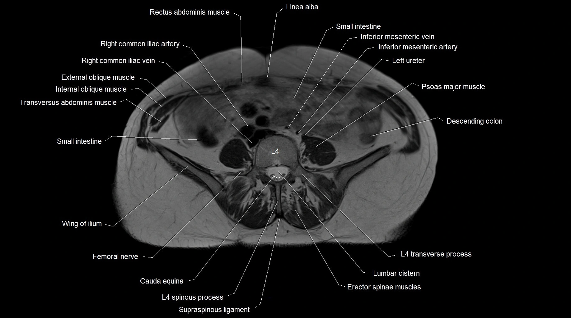
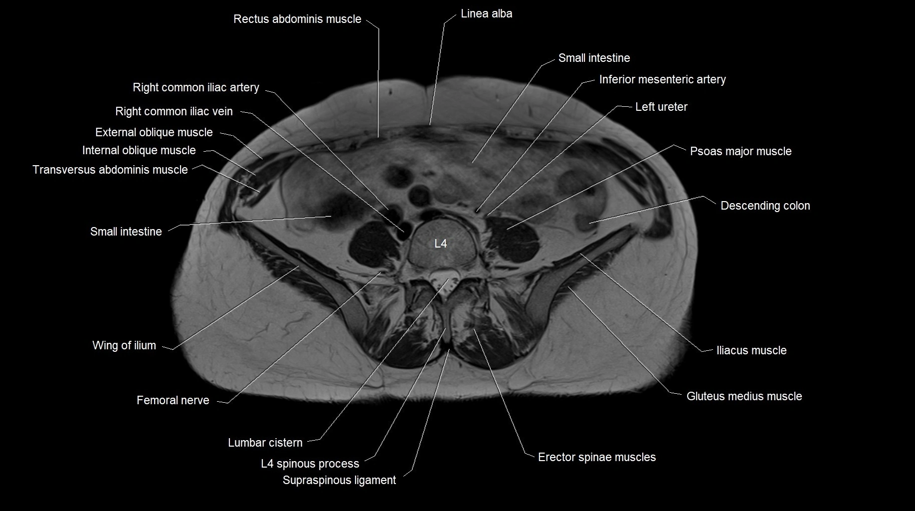
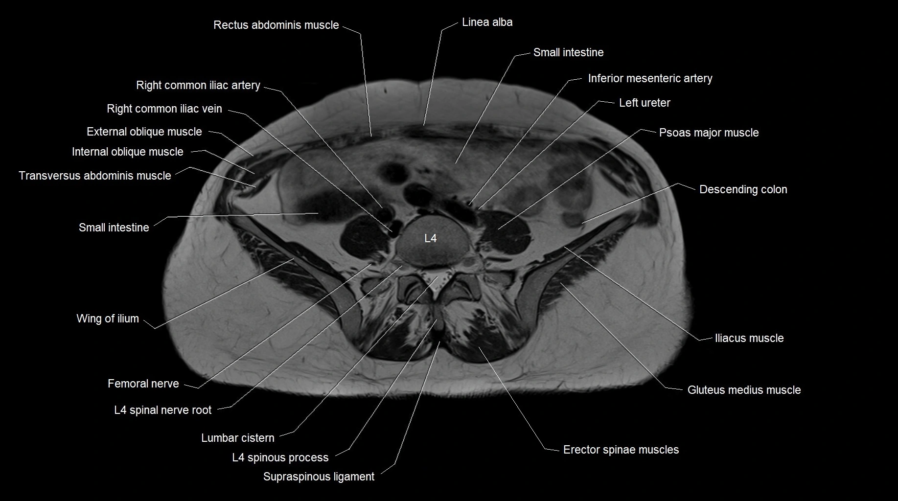
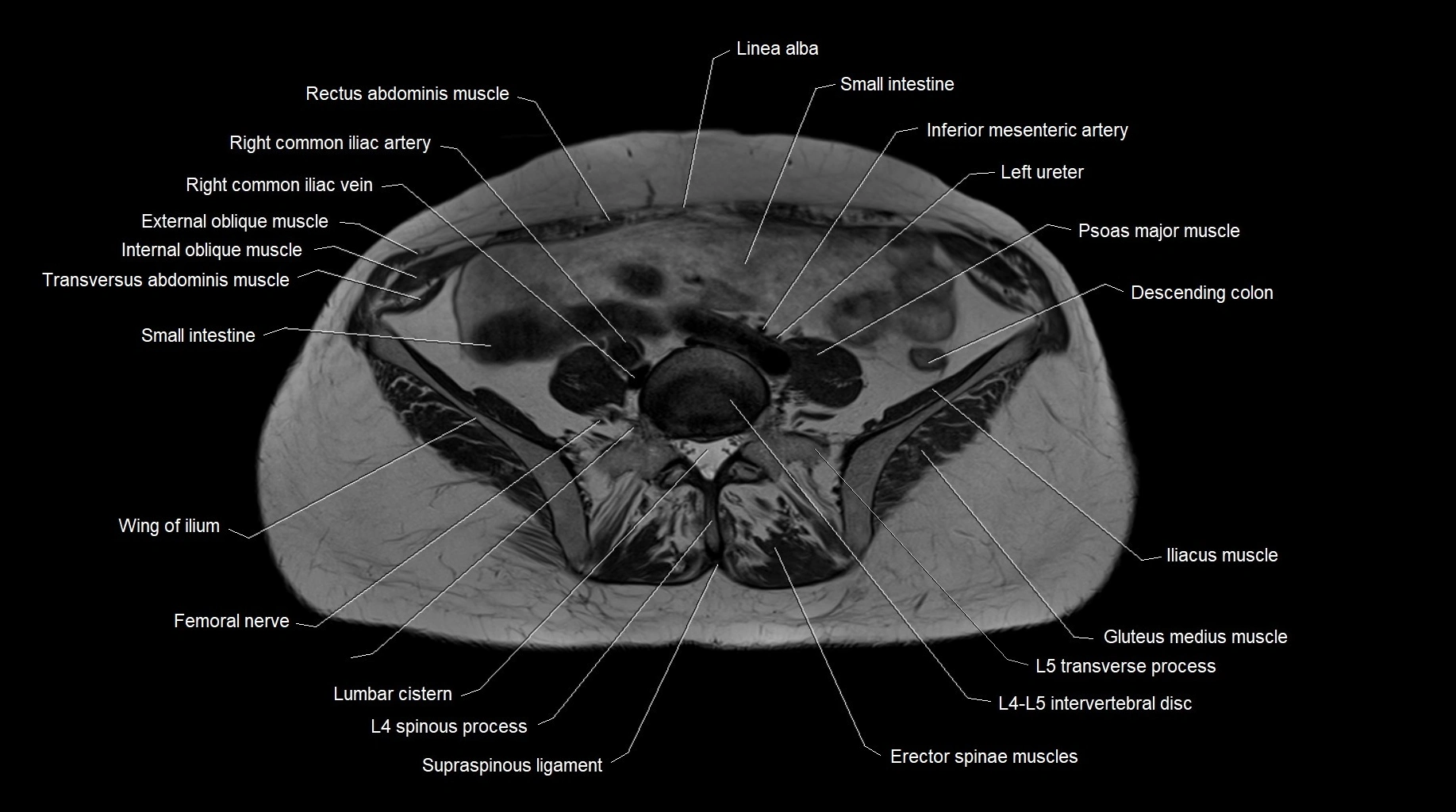
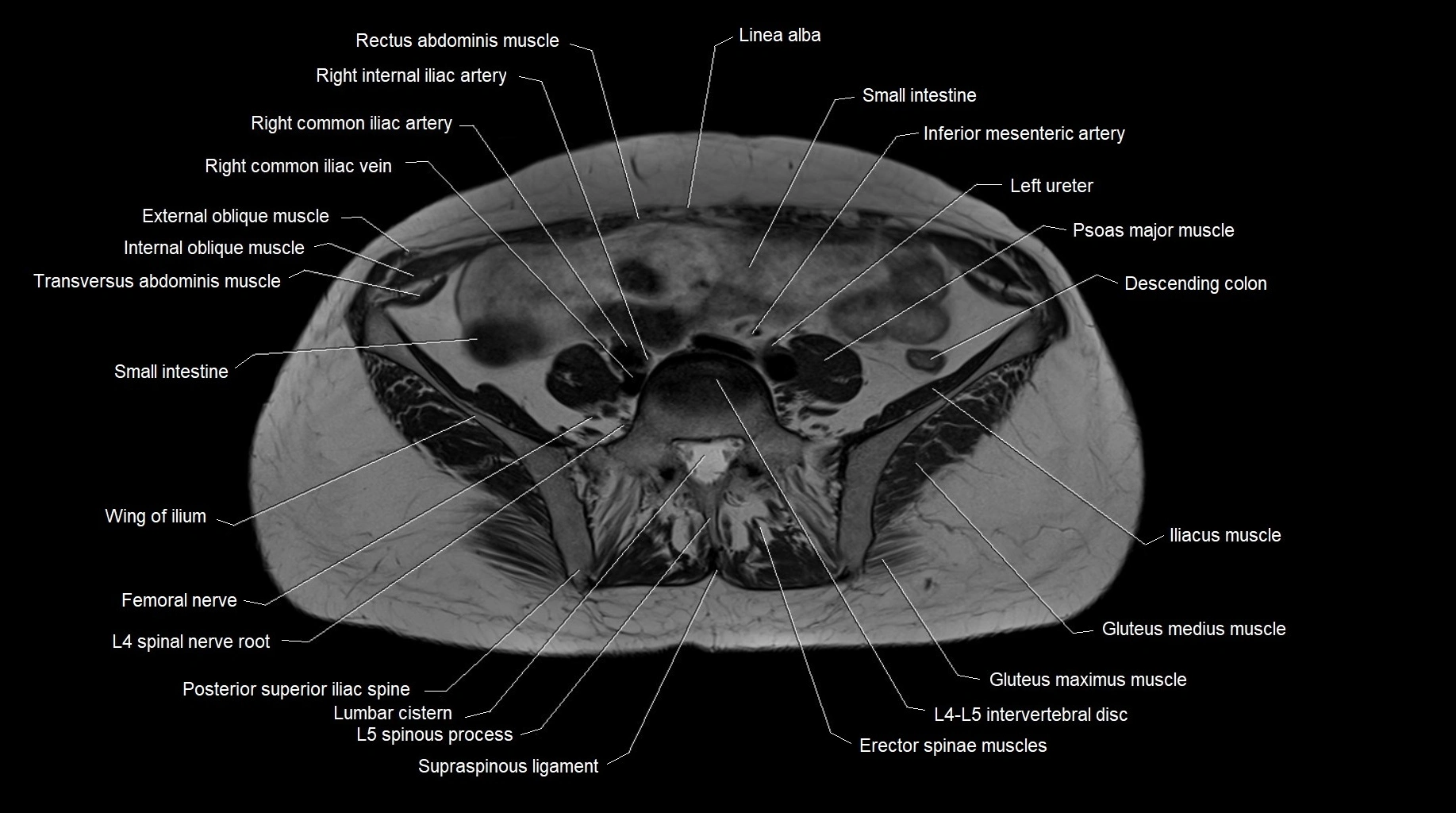
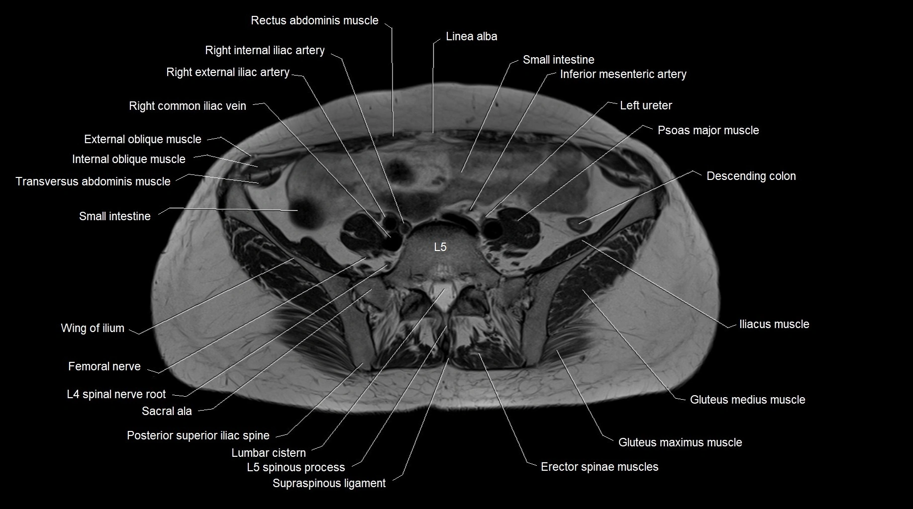
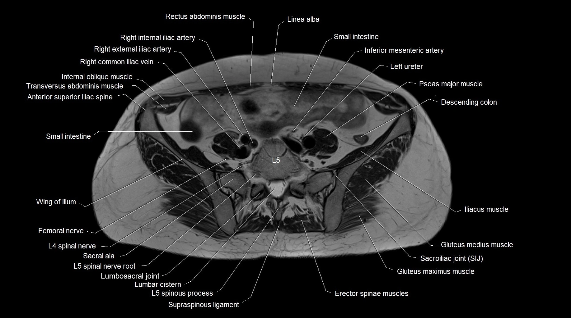
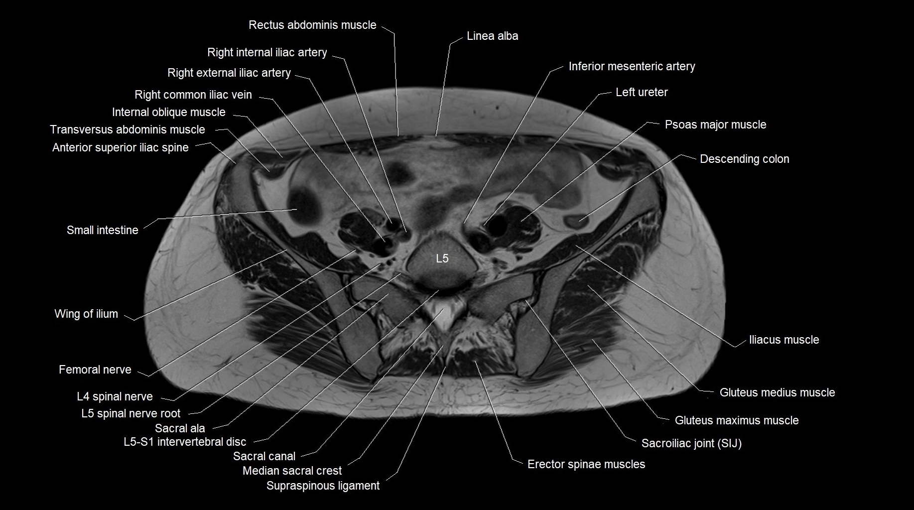
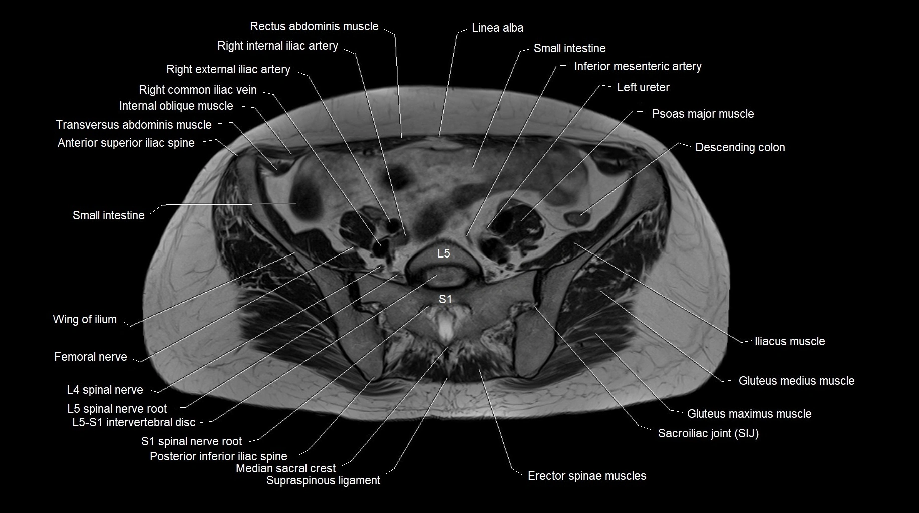
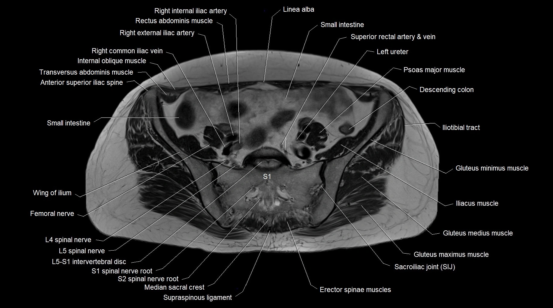
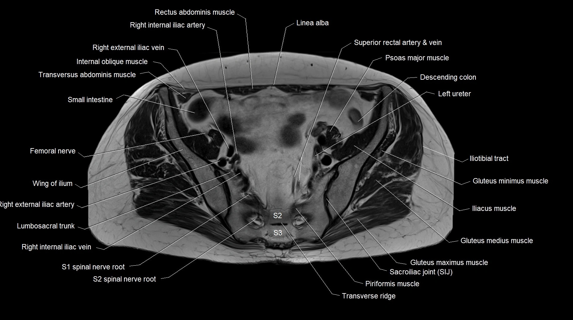
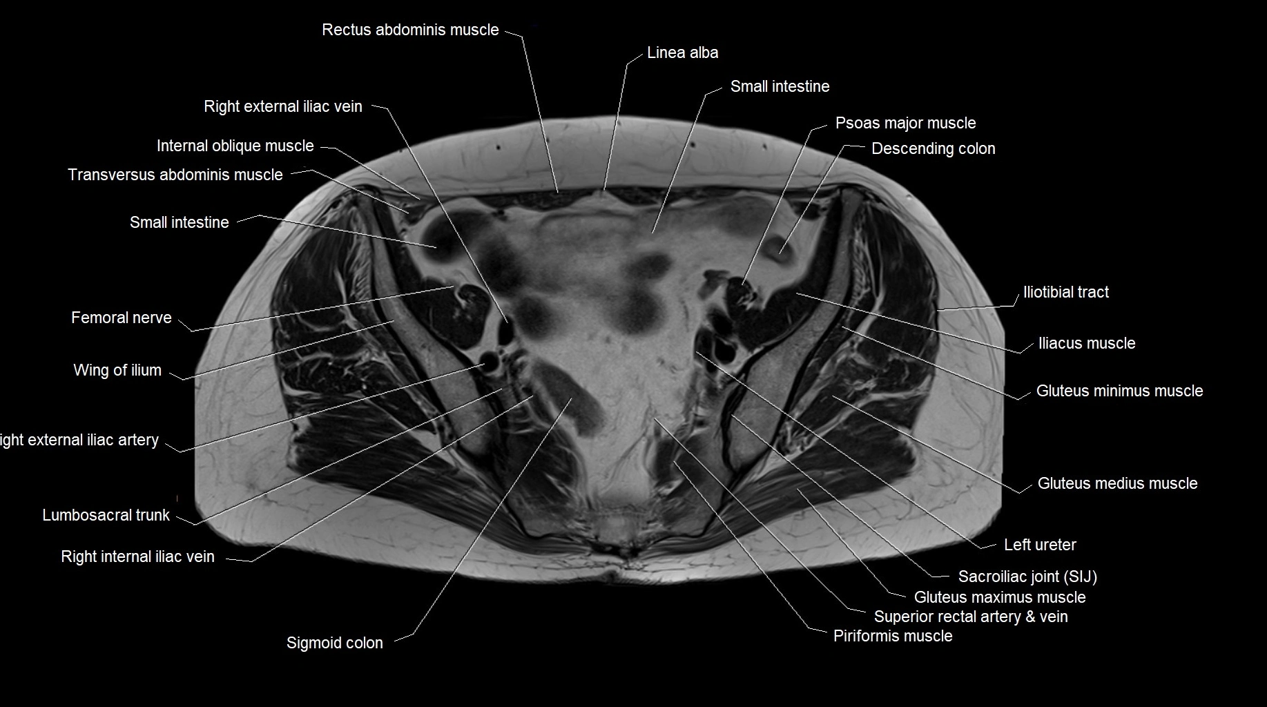
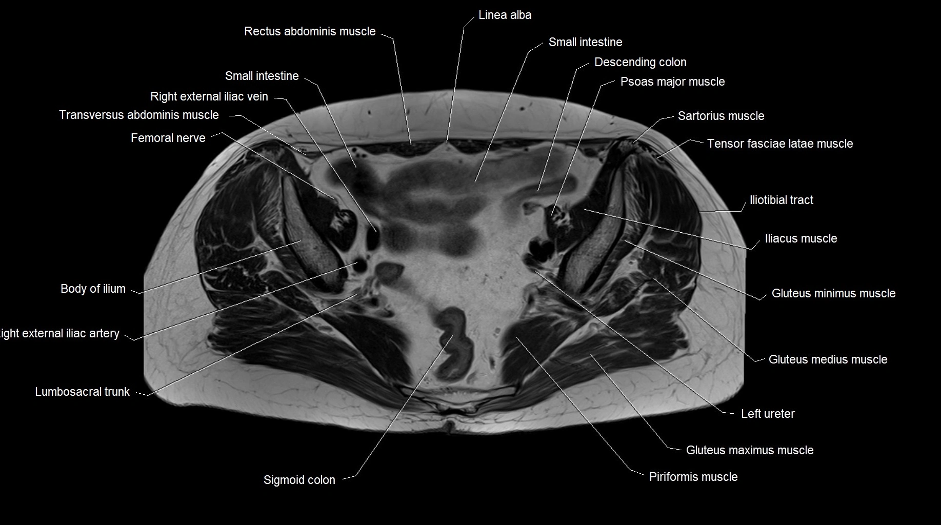
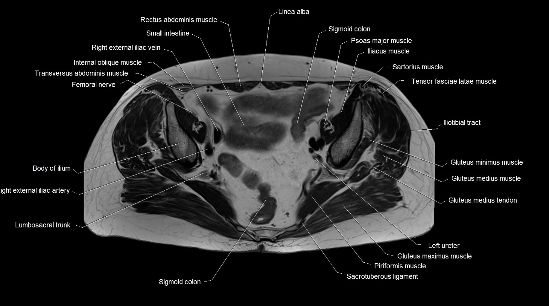
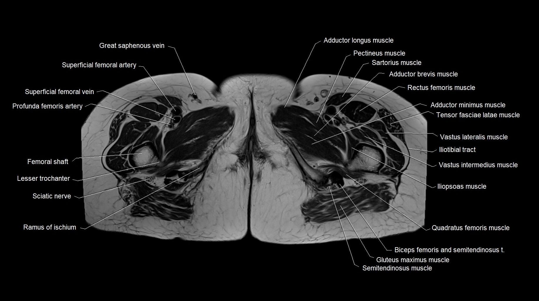
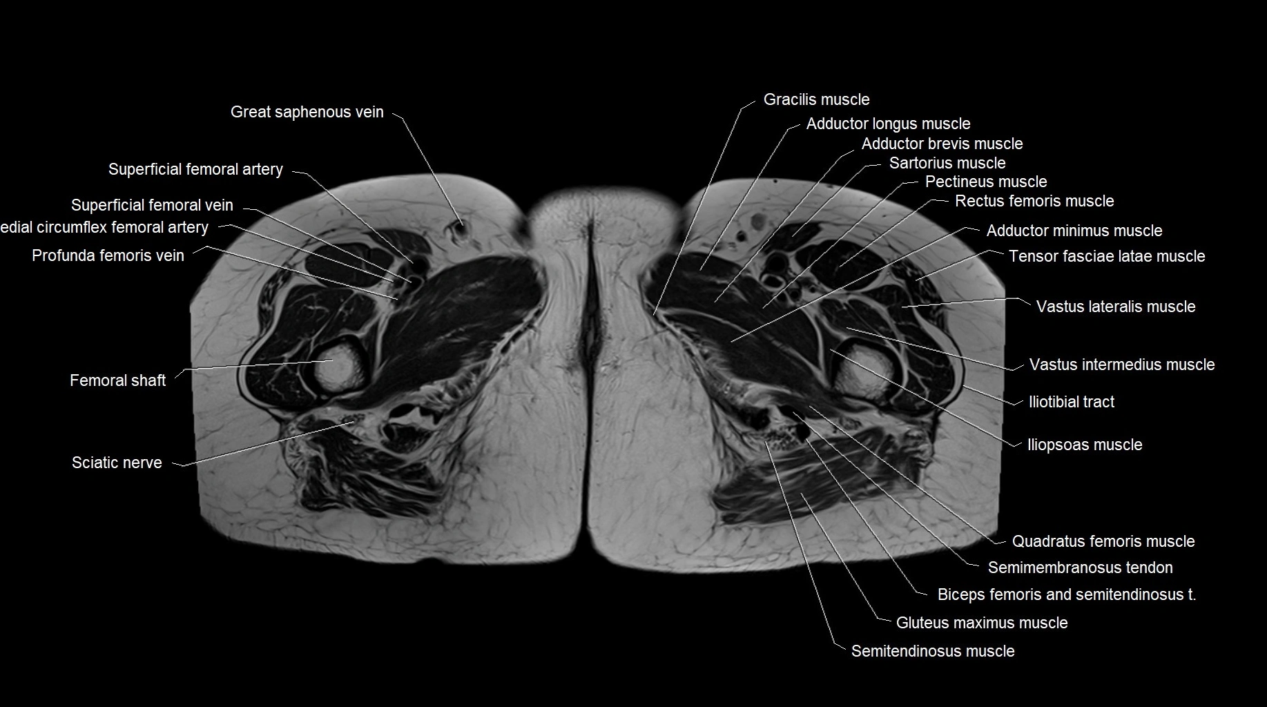
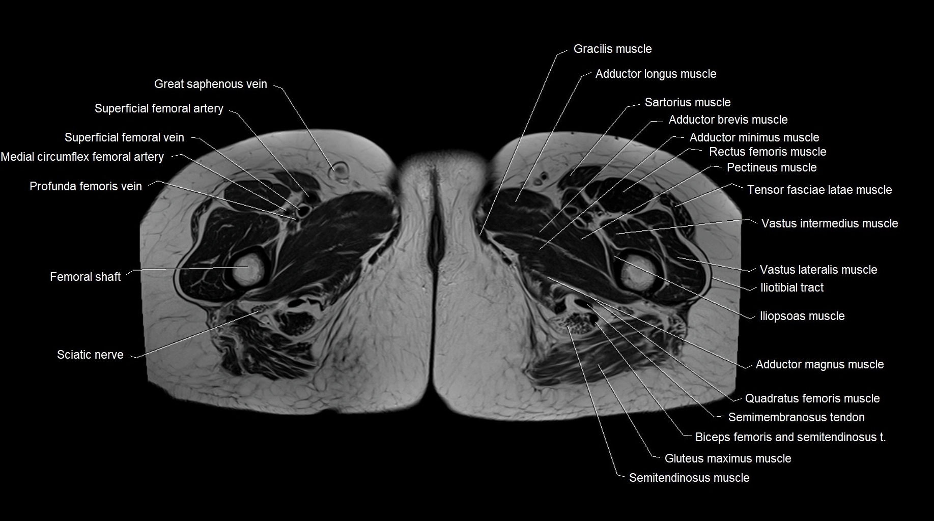
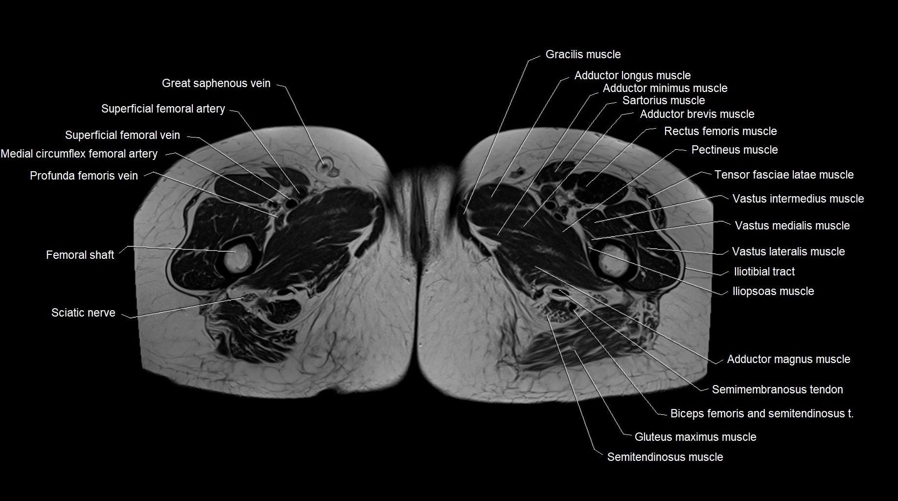
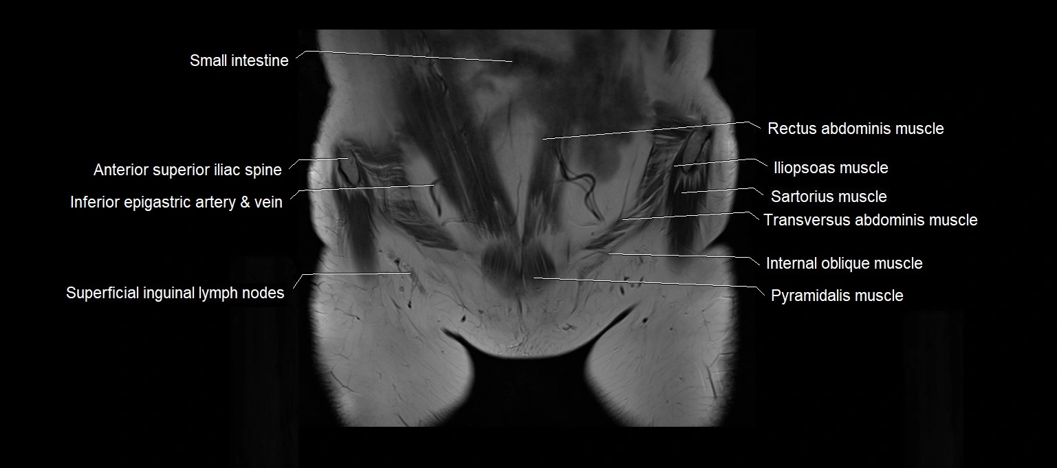
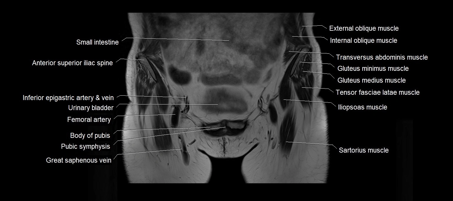
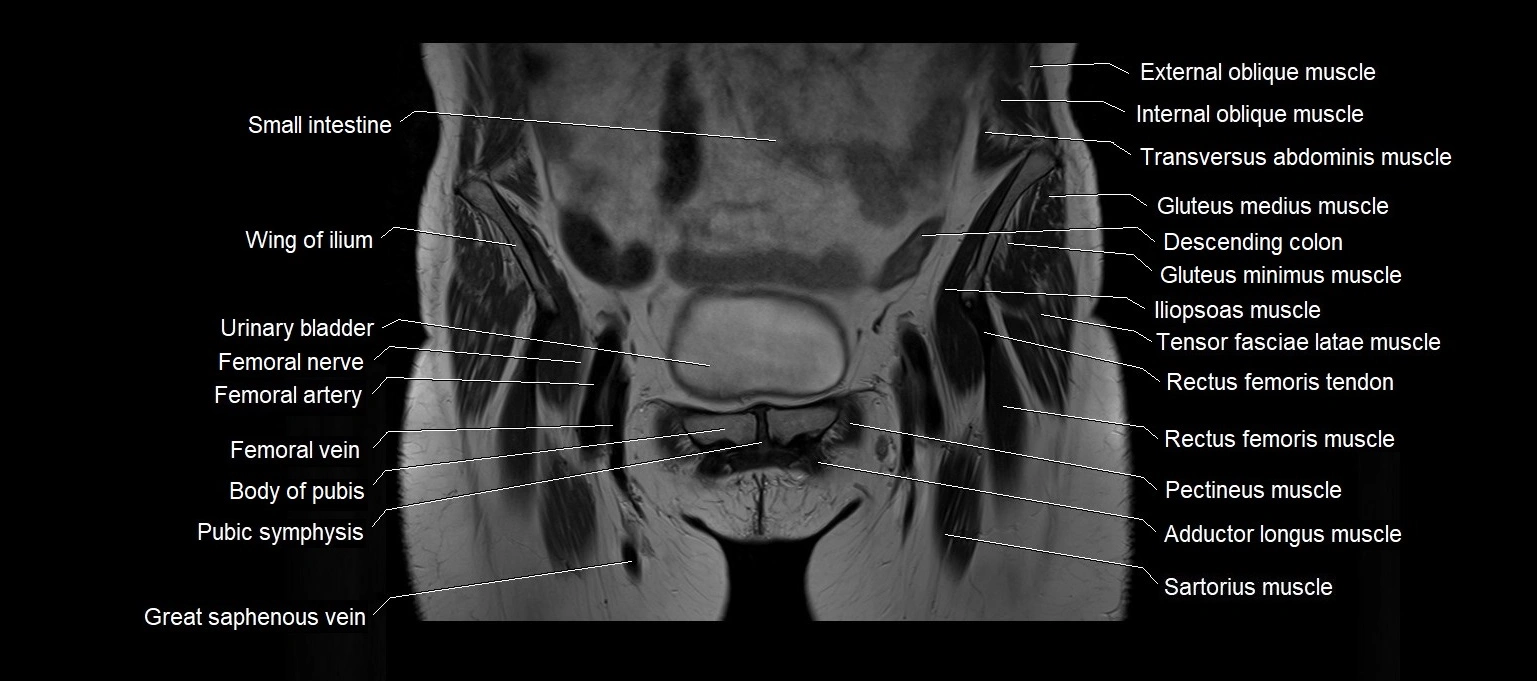
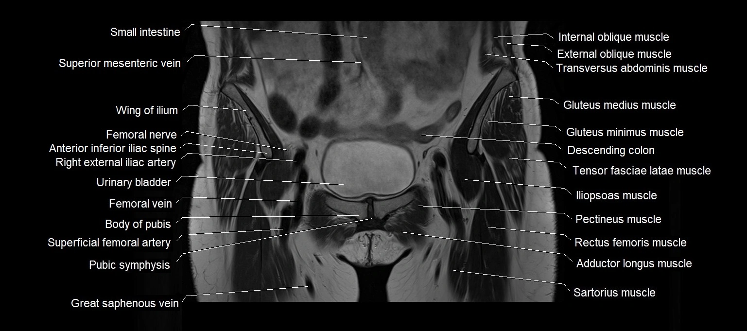
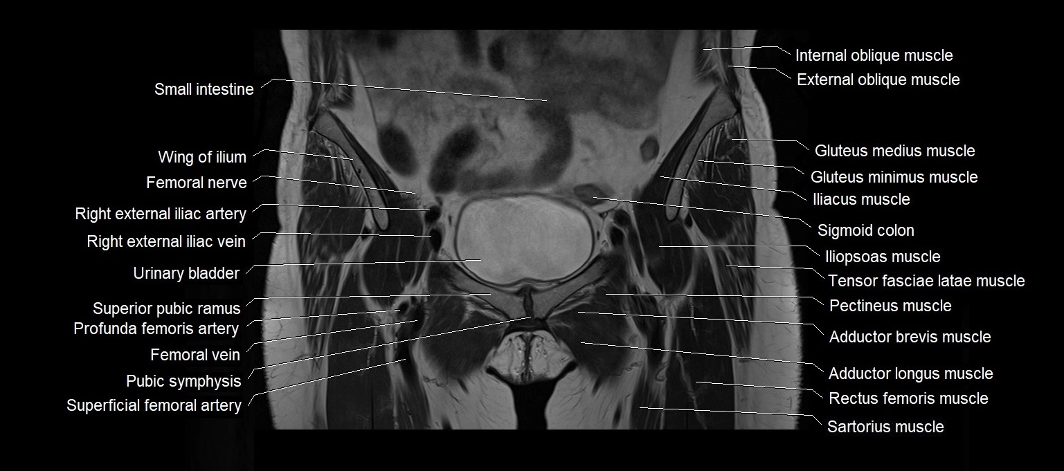
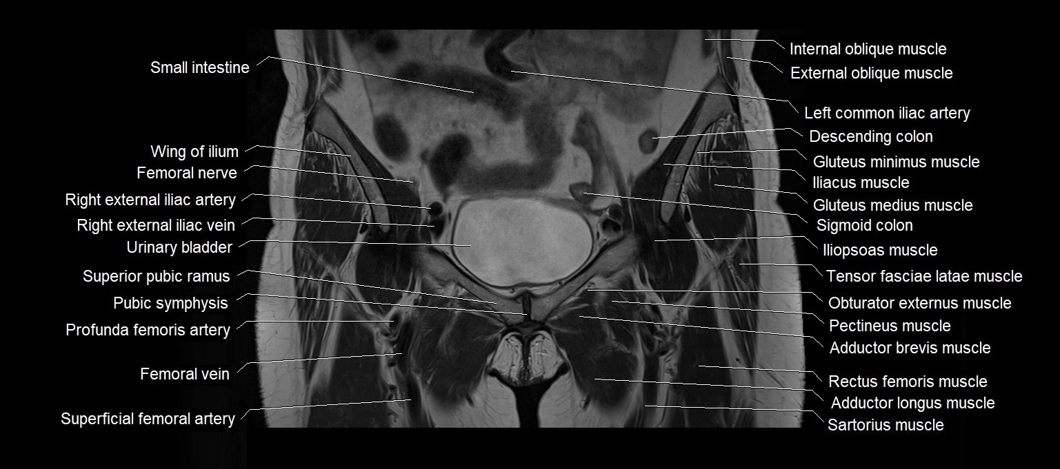
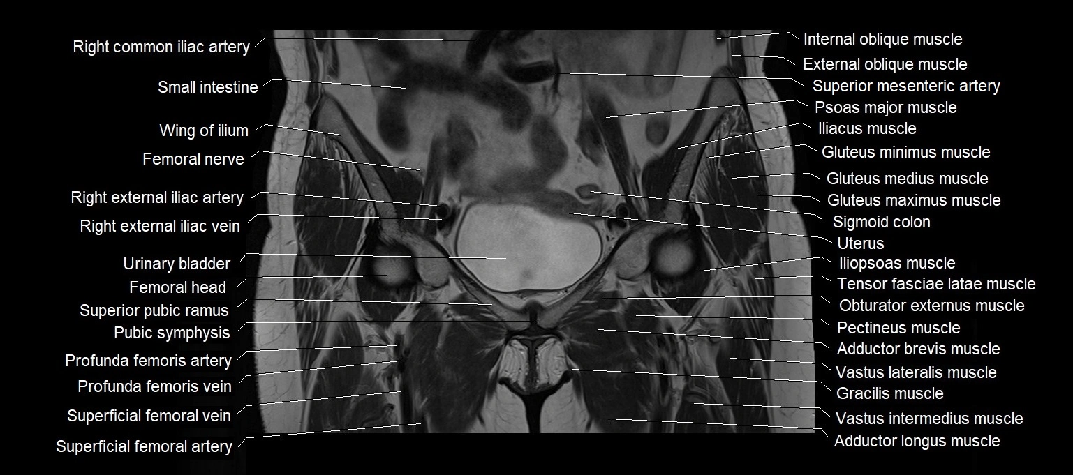
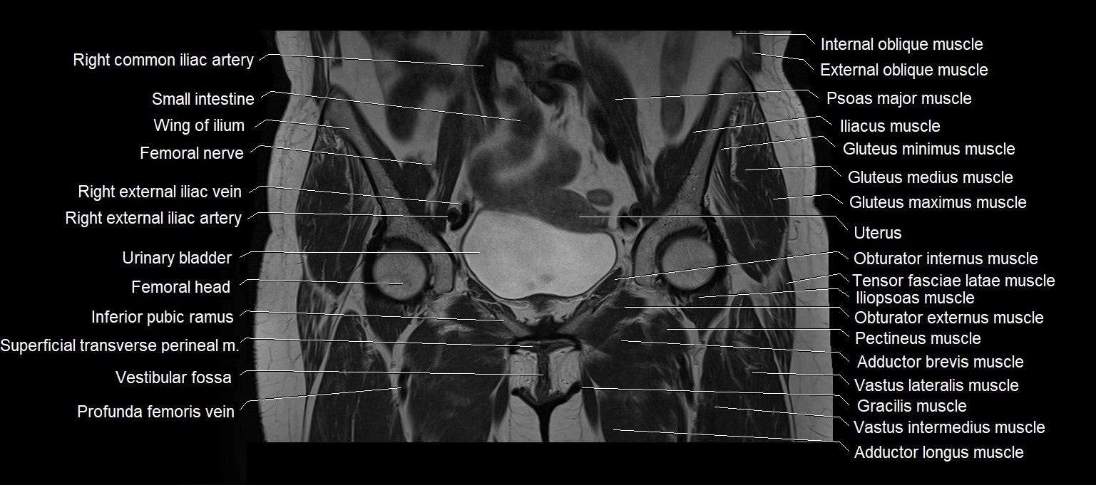
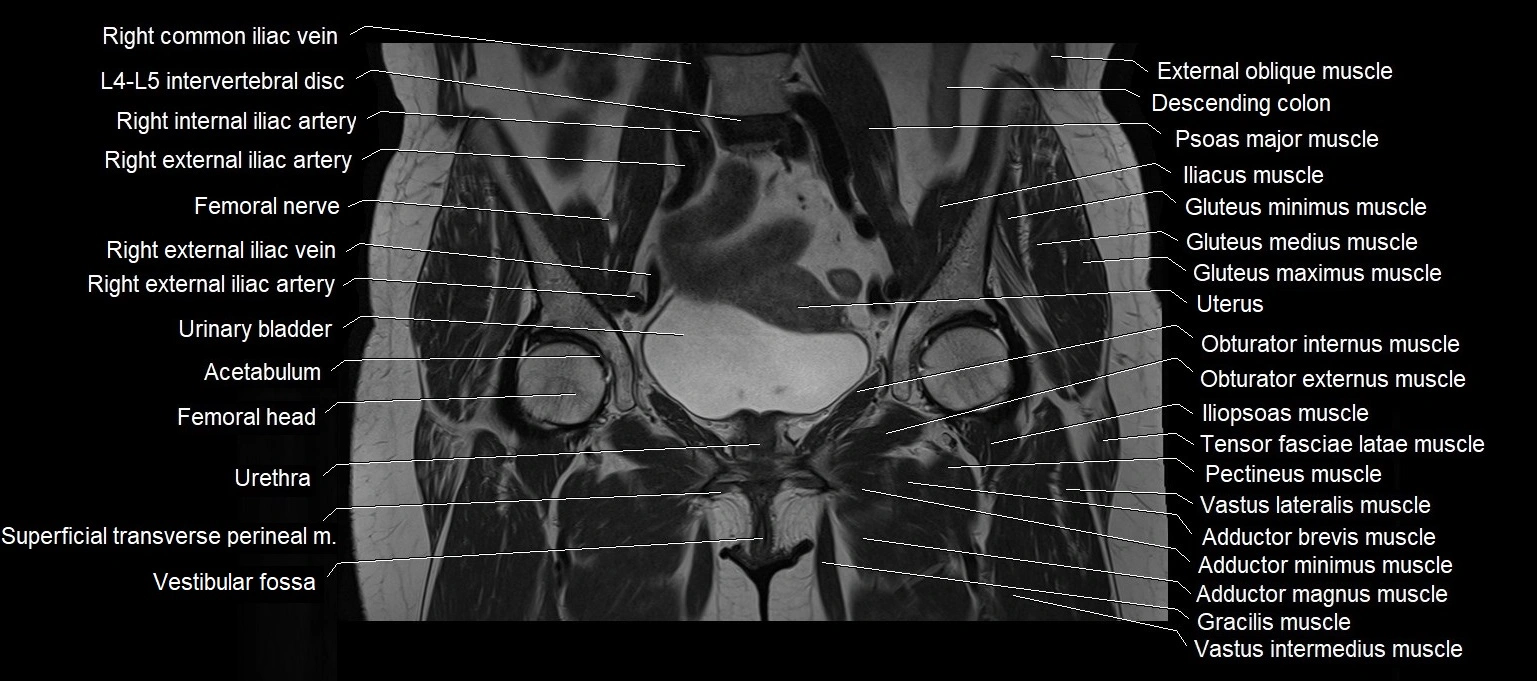
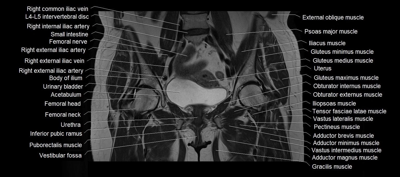
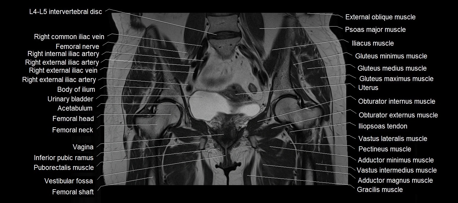
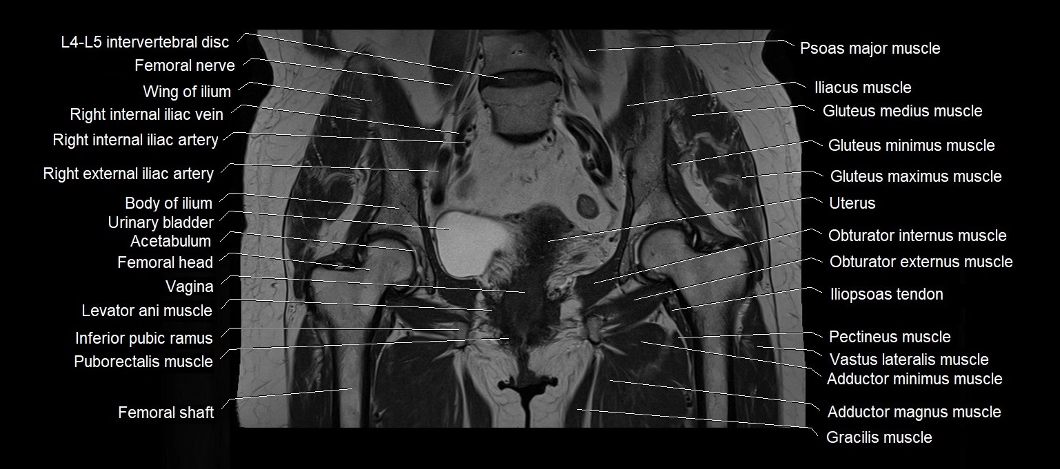
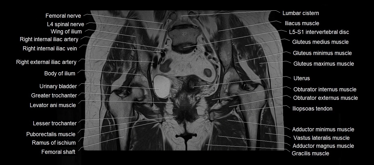
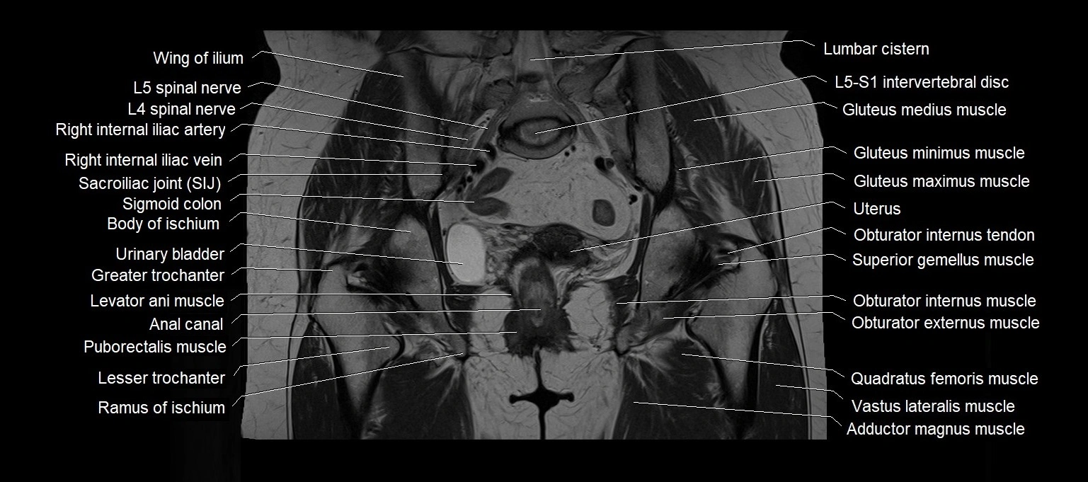
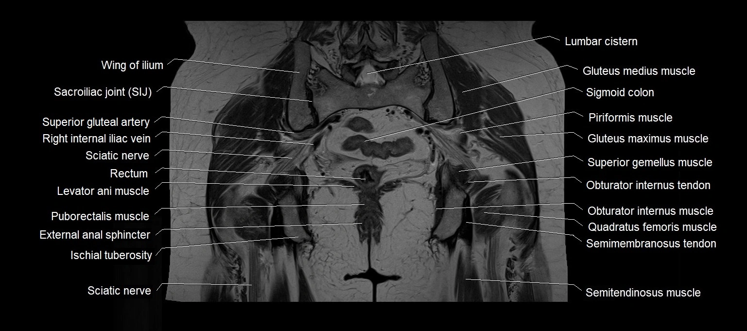
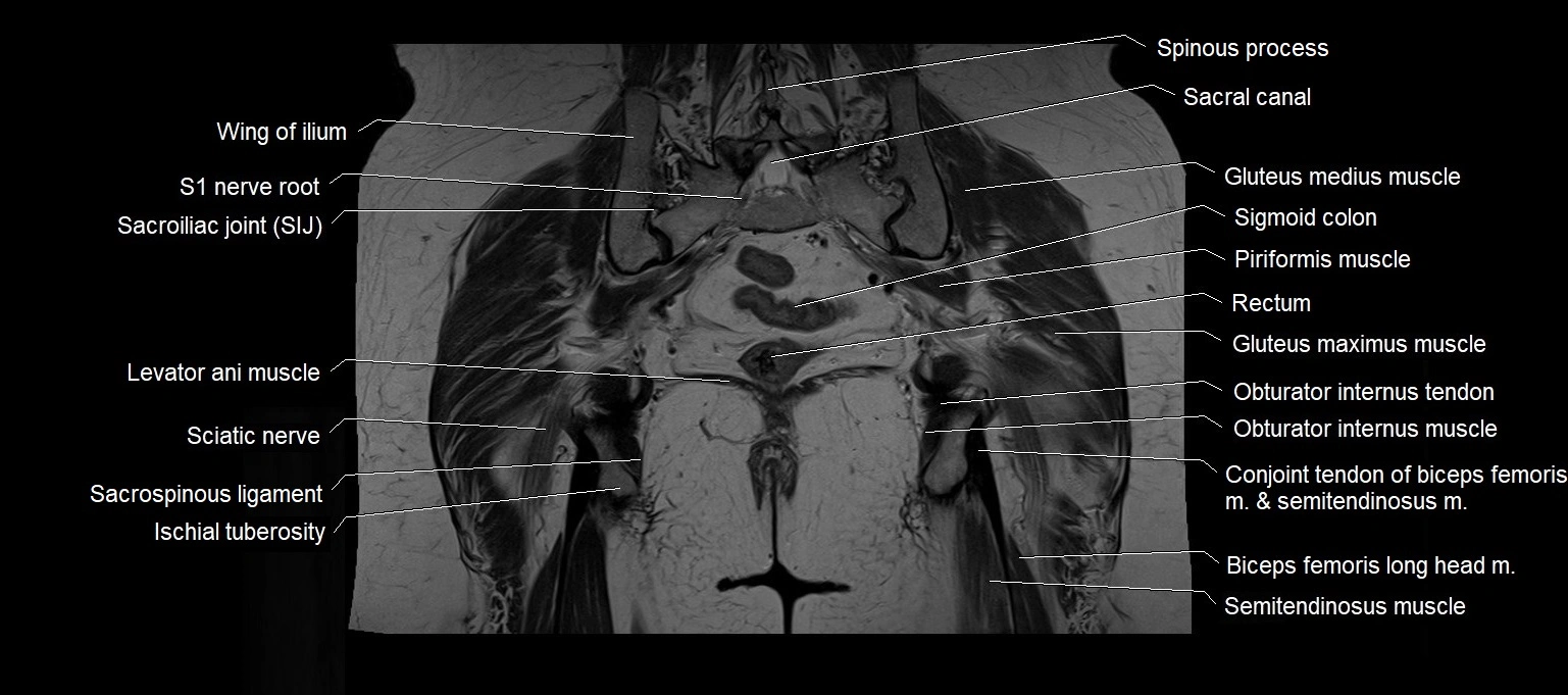
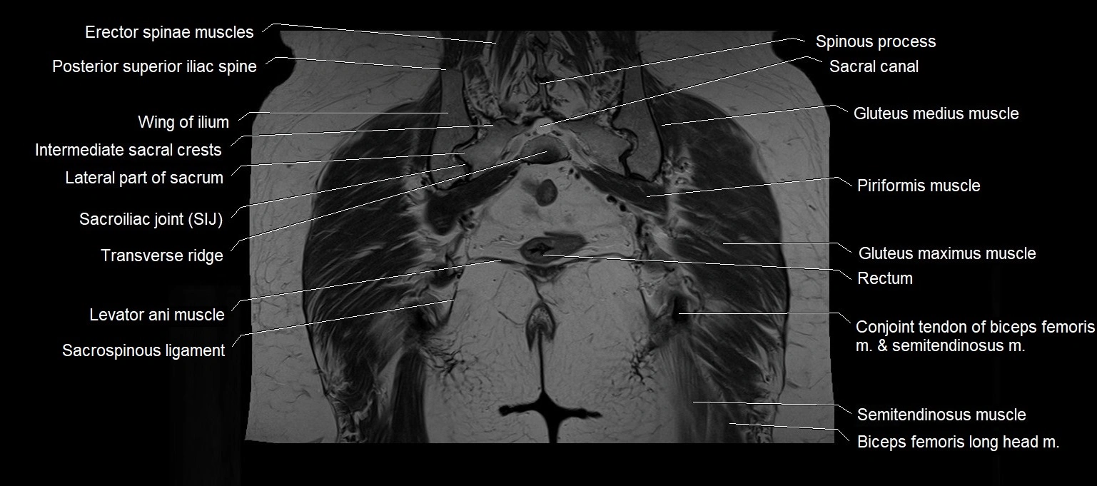
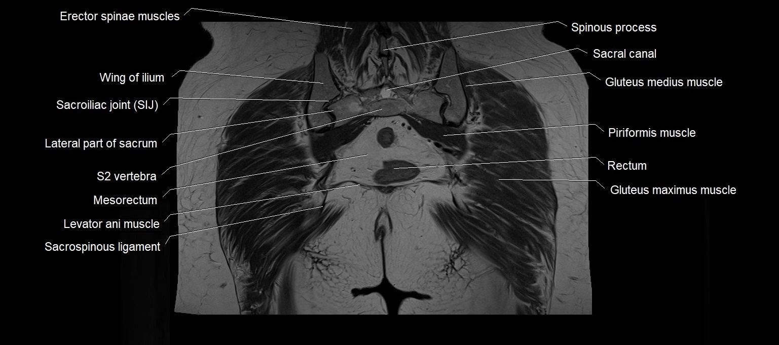
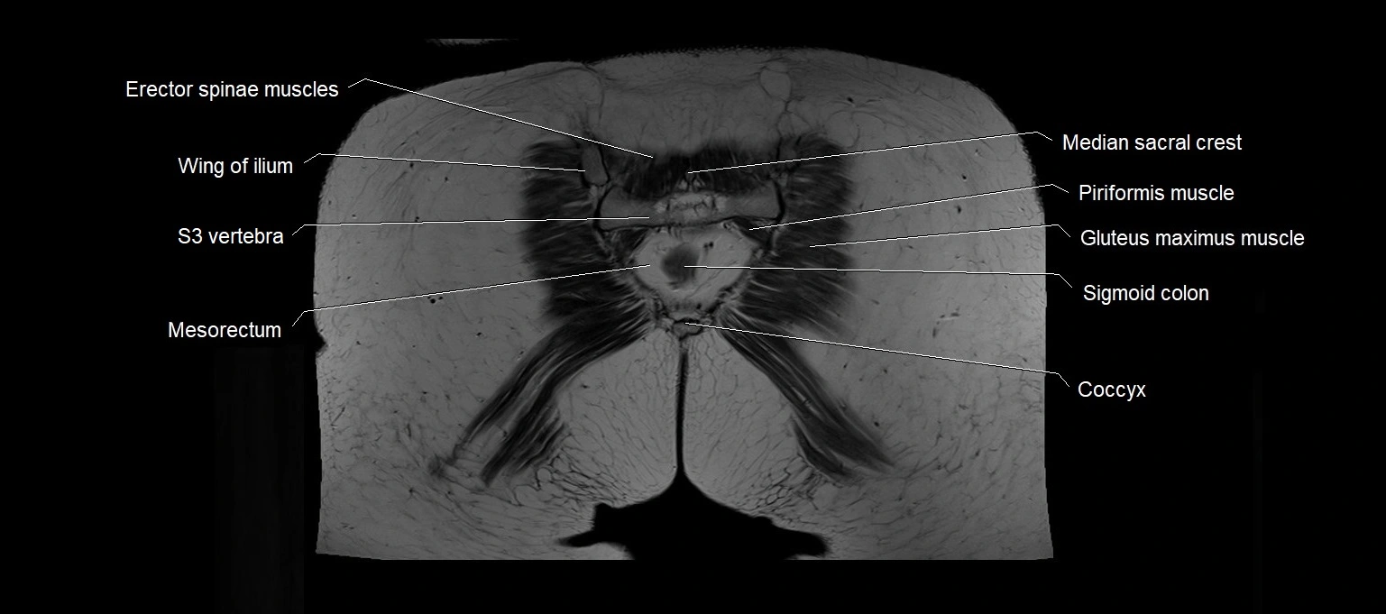
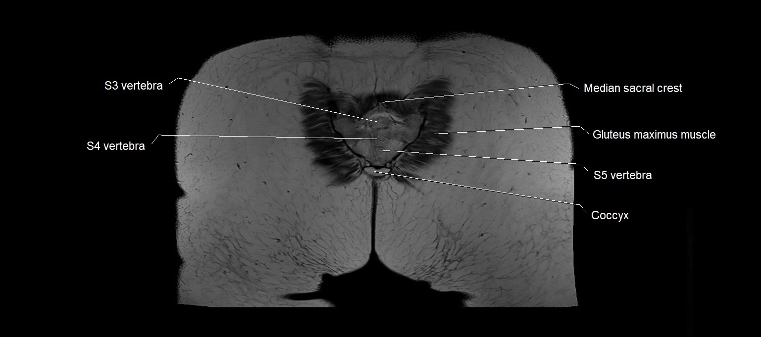
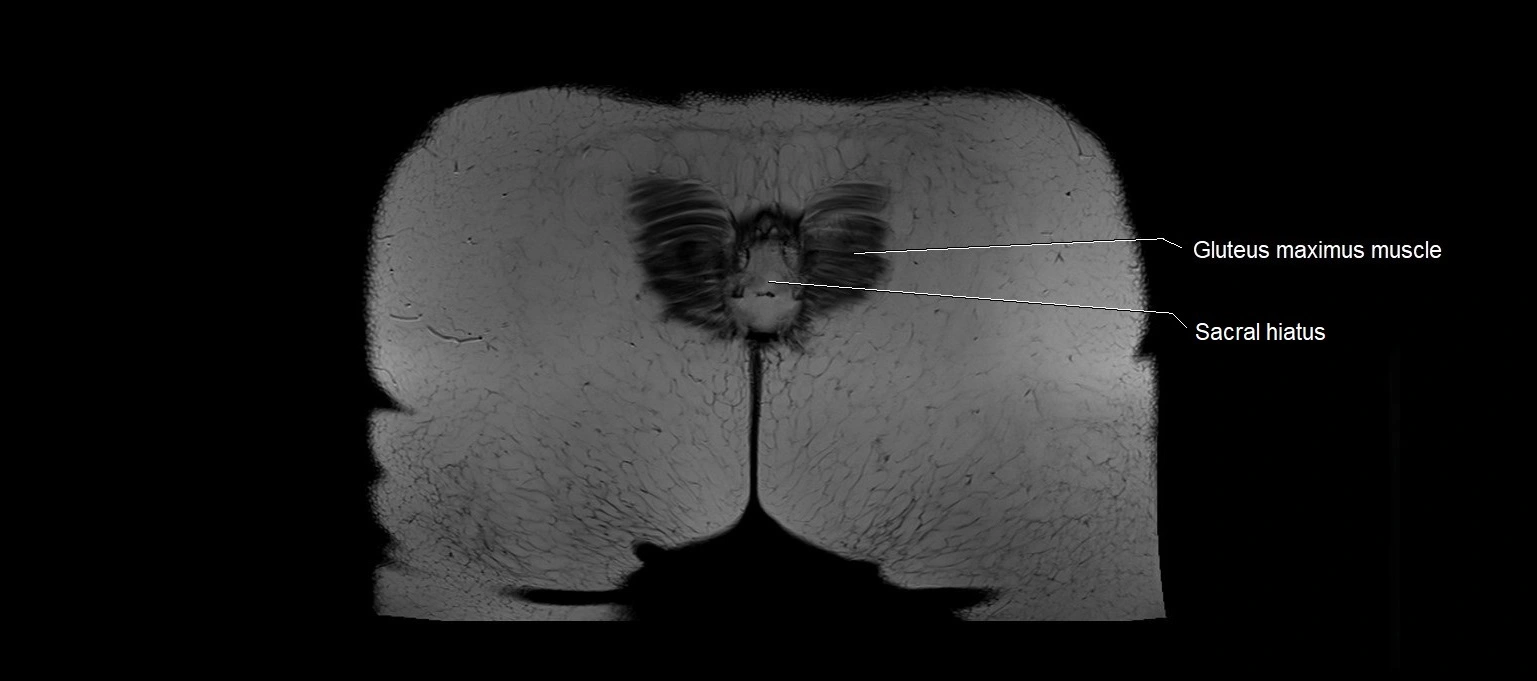
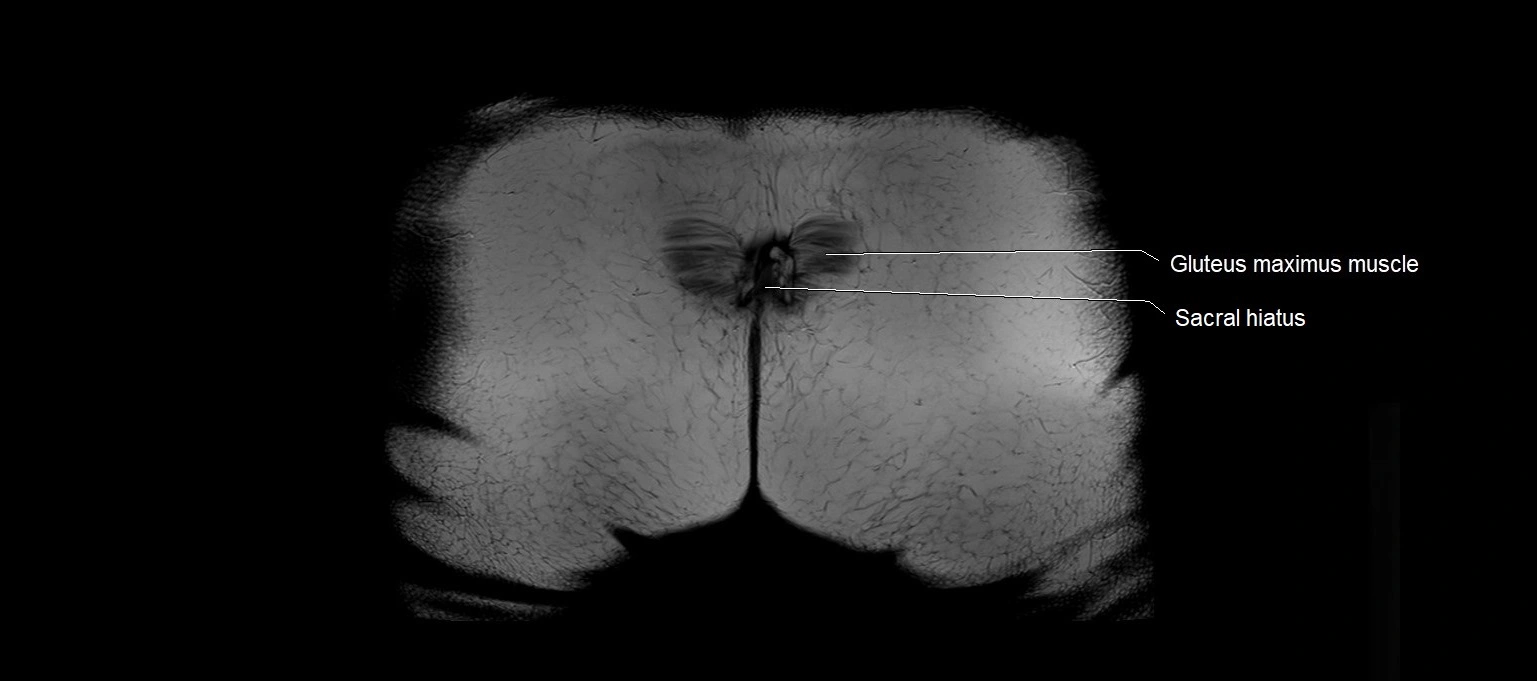
MRI images

MRI images

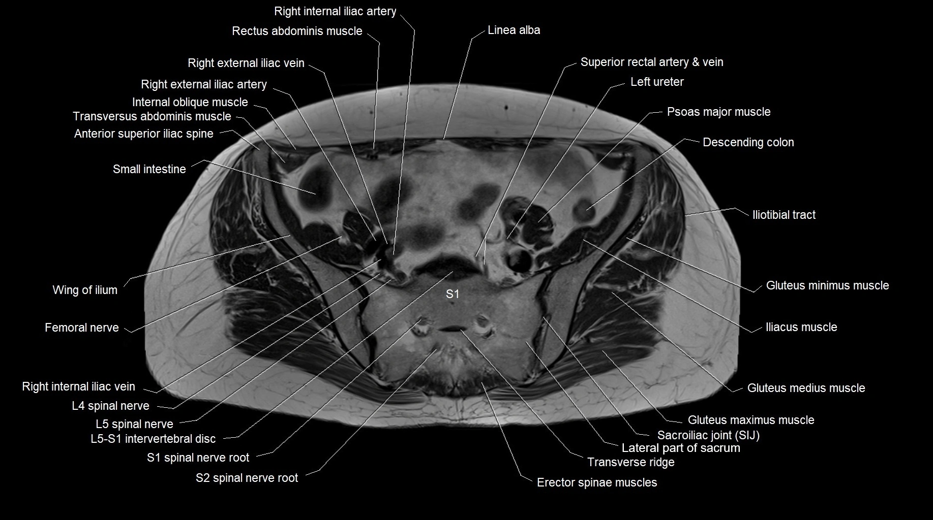
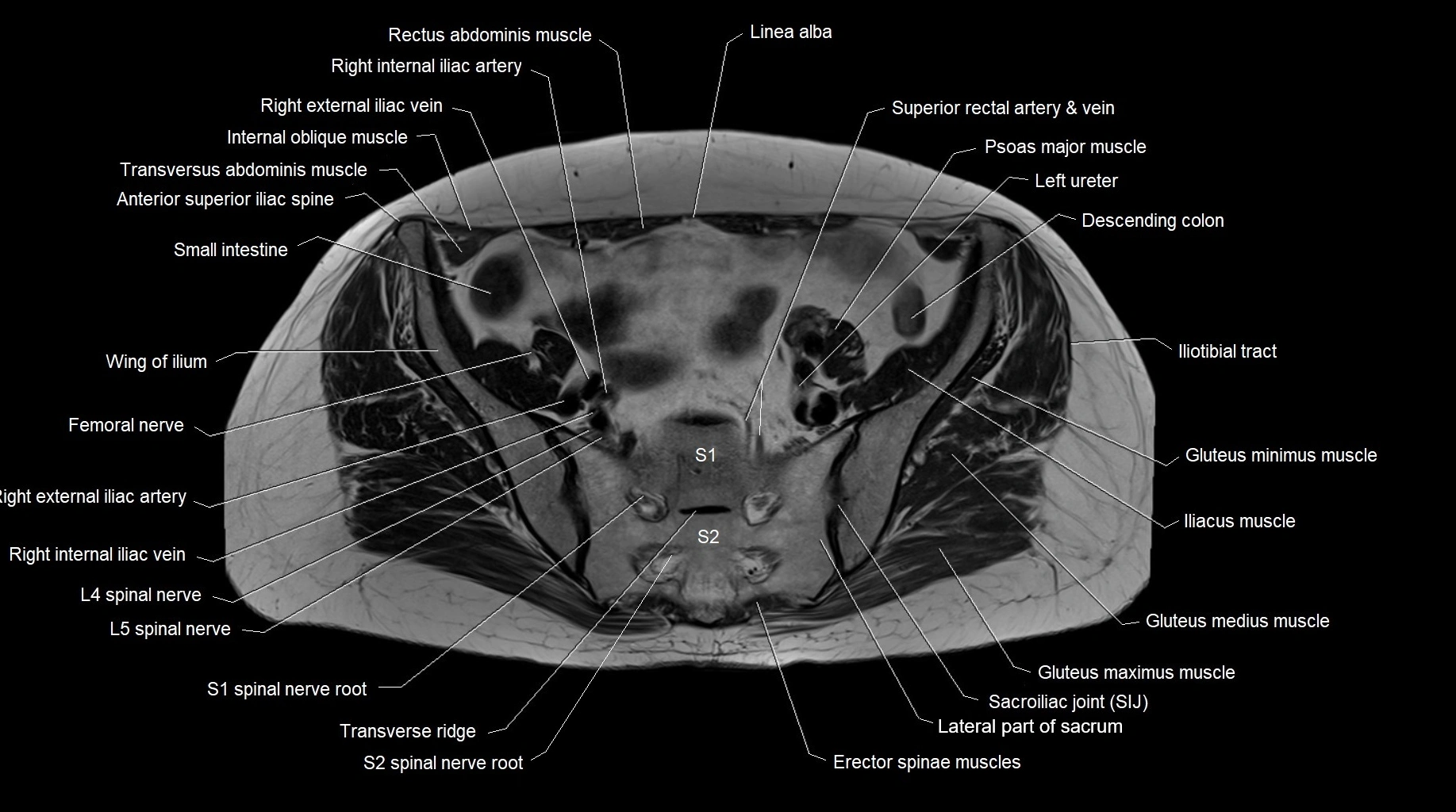
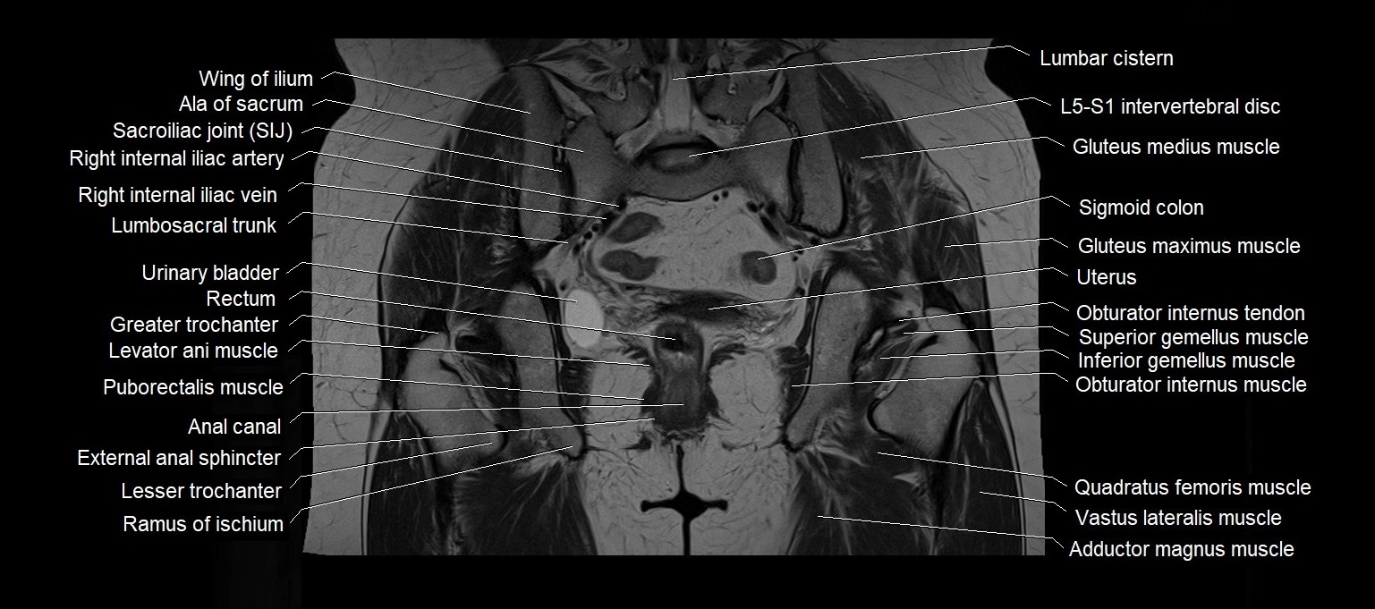
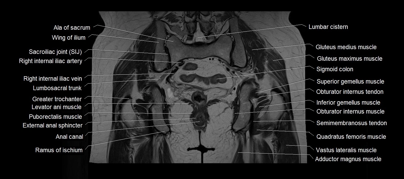
CT images

CT images


