


Topic
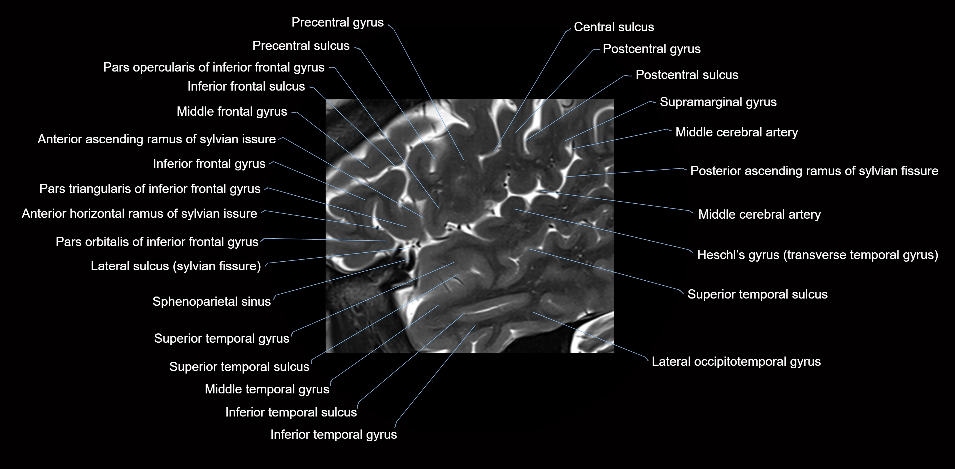
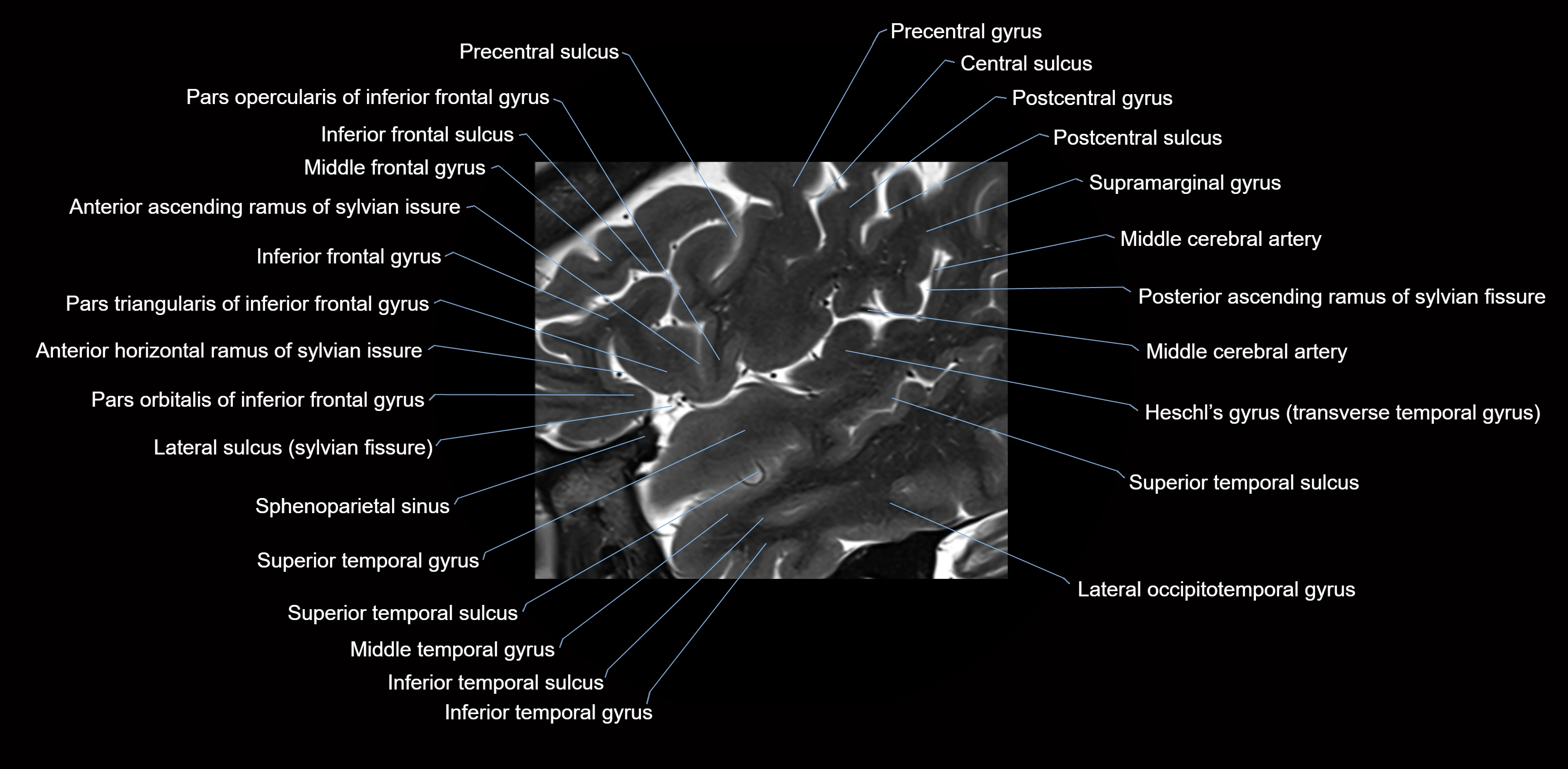
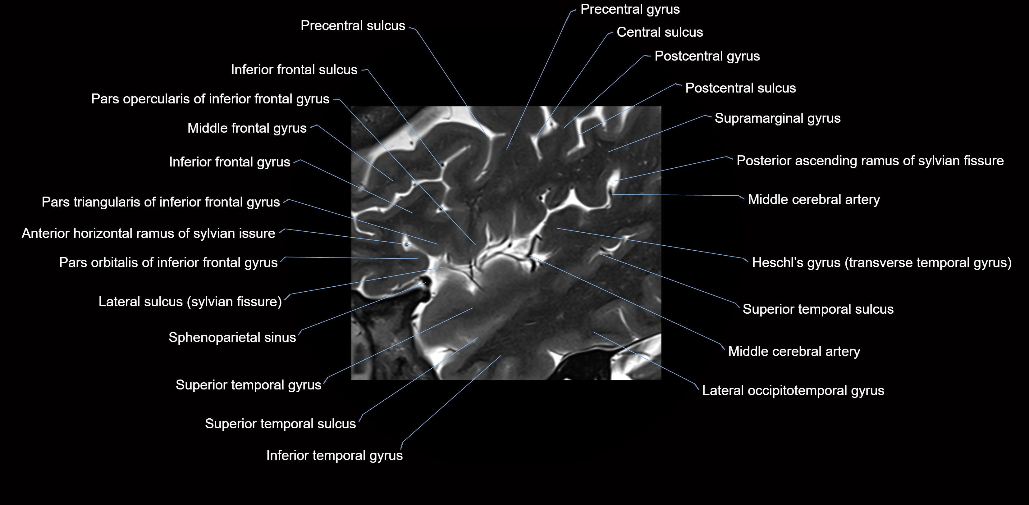
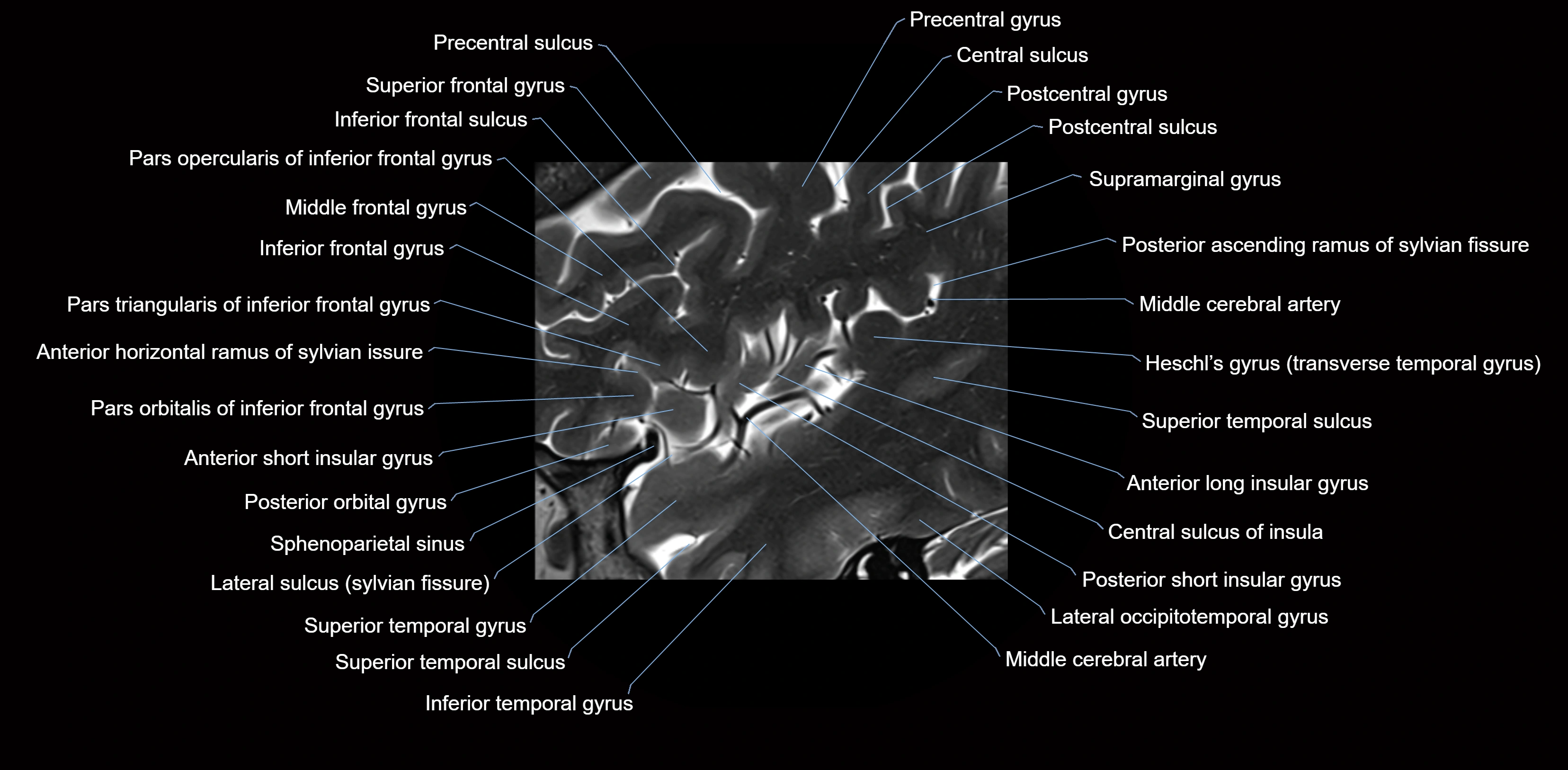
The anterior ascending ramus of the Sylvian fissure is a significant anatomical landmark in the lateral surface of the cerebral hemisphere. It represents one of the key branches of the Sylvian fissure (also known as the lateral sulcus) and plays an essential role in demarcating the boundaries between important cortical regions, notably within the frontal and parietal lobes. Understanding its anatomy and imaging appearance is crucial in neuroradiology, neurosurgery, and neuroanatomy for accurate localization and identification of adjacent brain structures.
Description
-
The anterior ascending ramus is a short, superiorly oriented branch that arises from the main stem of the Sylvian fissure.
-
It projects upward (anteriorly and slightly dorsally) from the lateral sulcus into the inferior frontal gyrus.
-
This ramus separates the pars opercularis (opercular part) from the pars triangularis (triangular part) of the inferior frontal gyrus.
-
The anterior ascending ramus marks the boundary between Broca's area and adjacent cortical areas in the dominant hemisphere.
Synonyms
-
Ascending ramus of the lateral sulcus
-
Ascending branch of the Sylvian fissure
-
Ascending ramus of the Sylvian sulcus
Function
-
Serves as an anatomical landmark for neurosurgical planning and neuroimaging.
-
Helps define Broca’s area (important for speech production) in the dominant hemisphere.
-
Aids in the localization of cortical regions during functional mapping and epilepsy surgery.
Arterial Supply
-
Supplied predominantly by branches of the middle cerebral artery (MCA), particularly its opercular branches.
-
The MCA runs within the Sylvian fissure and provides small cortical branches to adjacent gyri and sulci.
Venous Drainage
-
Drained by superficial Sylvian veins (also called superficial middle cerebral veins), which follow the course of the Sylvian fissure.
-
Venous blood ultimately drains into the sphenoparietal sinus and cavernous sinus.
MRI Appearance
-
T1-weighted imaging:
-
The Sylvian fissure and its anterior ascending ramus appear as low-signal intensity (dark) CSF-filled clefts between the gyri.
-
Clear demarcation between adjacent gray and white matter.
-
-
T2-weighted imaging:
-
The fissure, including the anterior ascending ramus, is hyperintense (bright) due to CSF signal.
-
Better visualization of the separation between opercular and triangular parts of the inferior frontal gyrus.
-
-
FLAIR imaging:
-
The ramus is visualized as a linear dark space (suppressed CSF signal) between the surrounding cortex.
-
Adjacent cortical and subcortical abnormalities (e.g., edema or gliosis) can be assessed relative to the ramus.
-
CT Appearance
-
The anterior ascending ramus of the Sylvian fissure appears as a hypodense (dark) linear cleft on non-contrast CT.
-
Separates the inferior frontal gyrus segments; used as a reference point for identifying frontal operculum and insula.
-
May not be as clearly defined as on MRI, but serves as a crucial landmark, especially in acute stroke and trauma imaging.
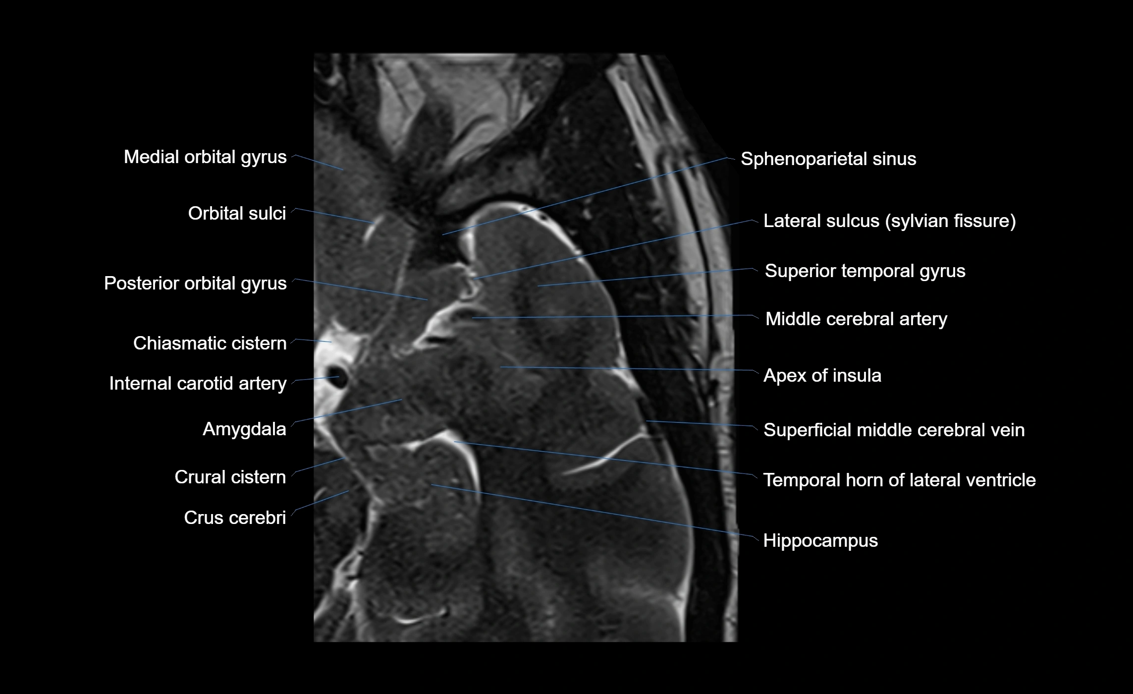
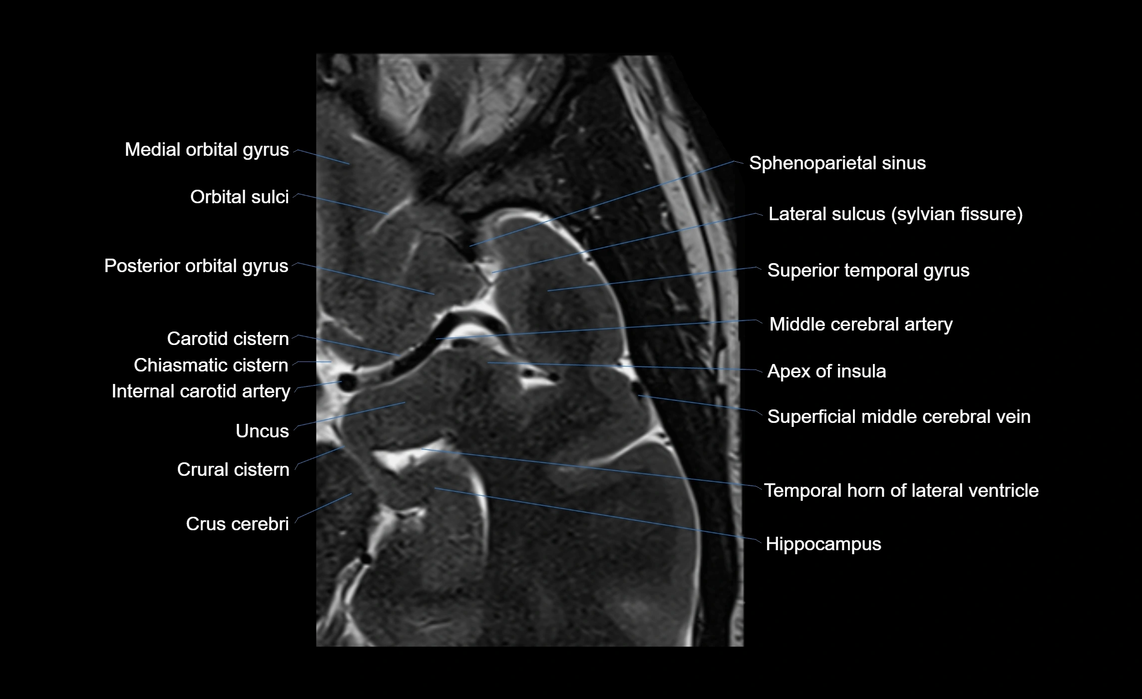
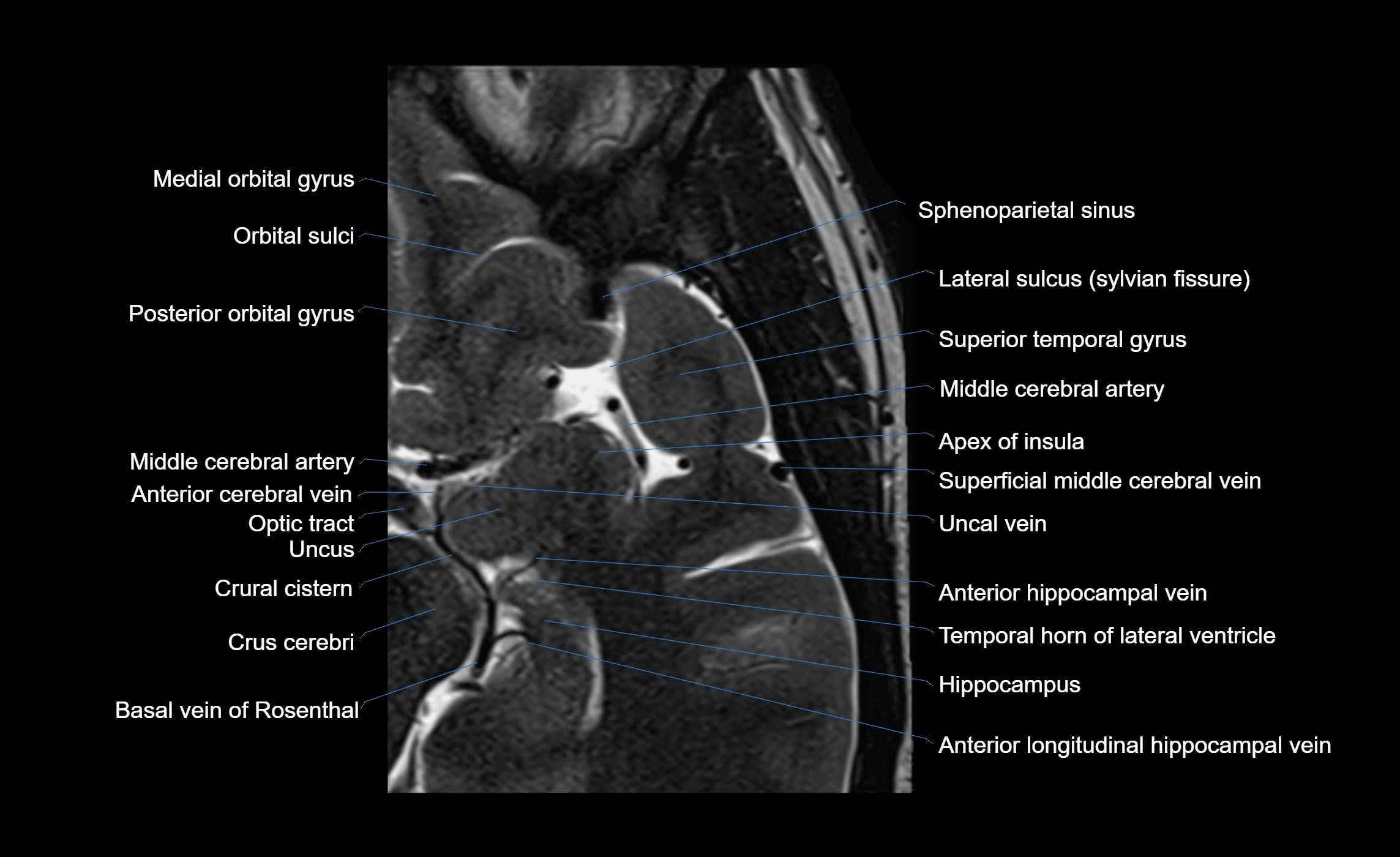
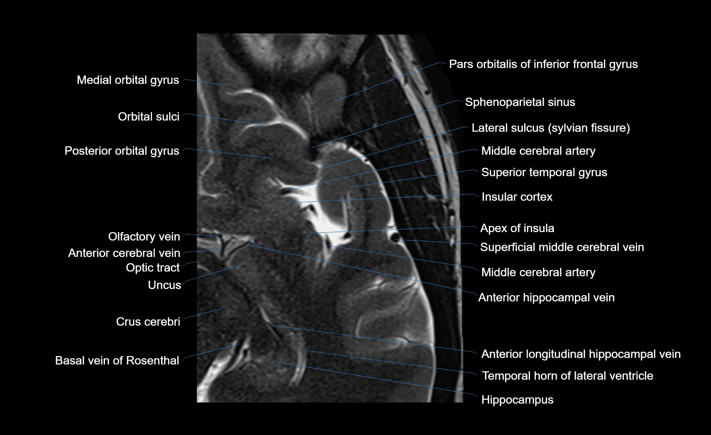
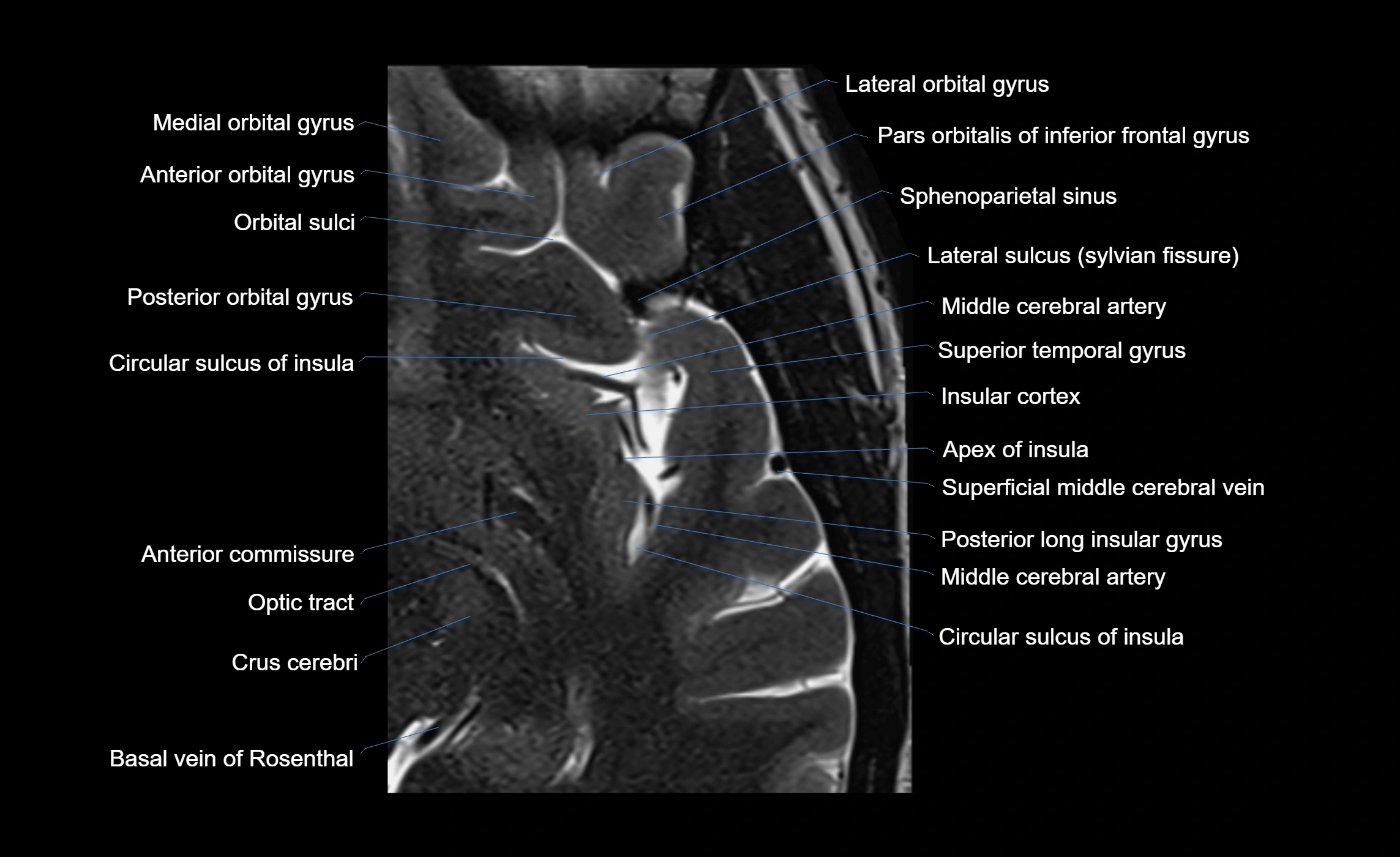
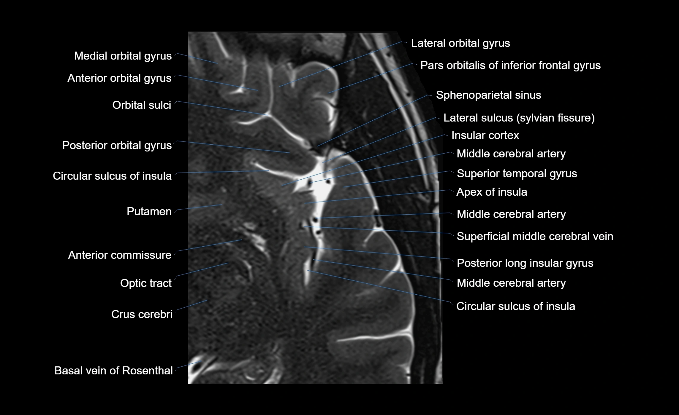
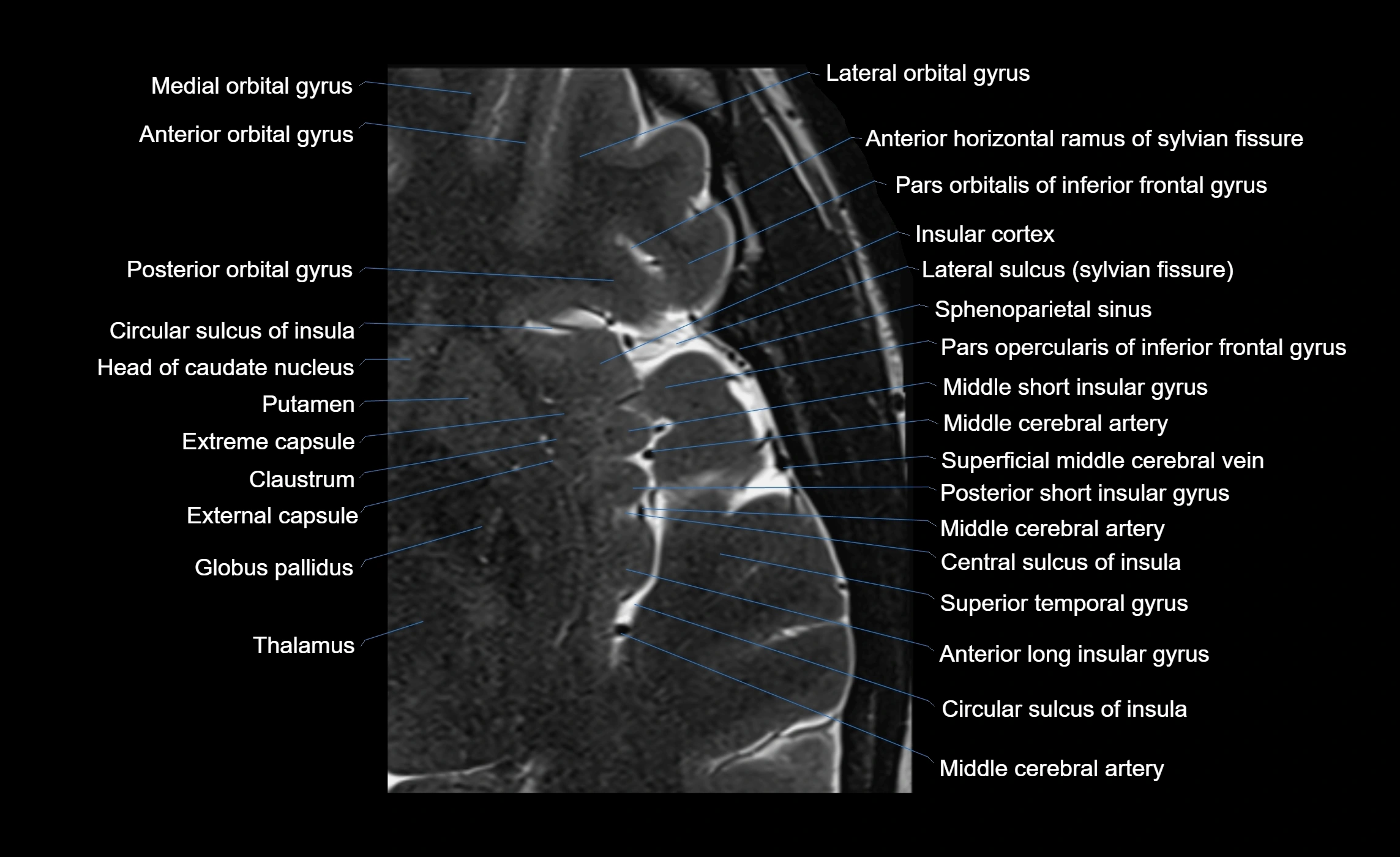
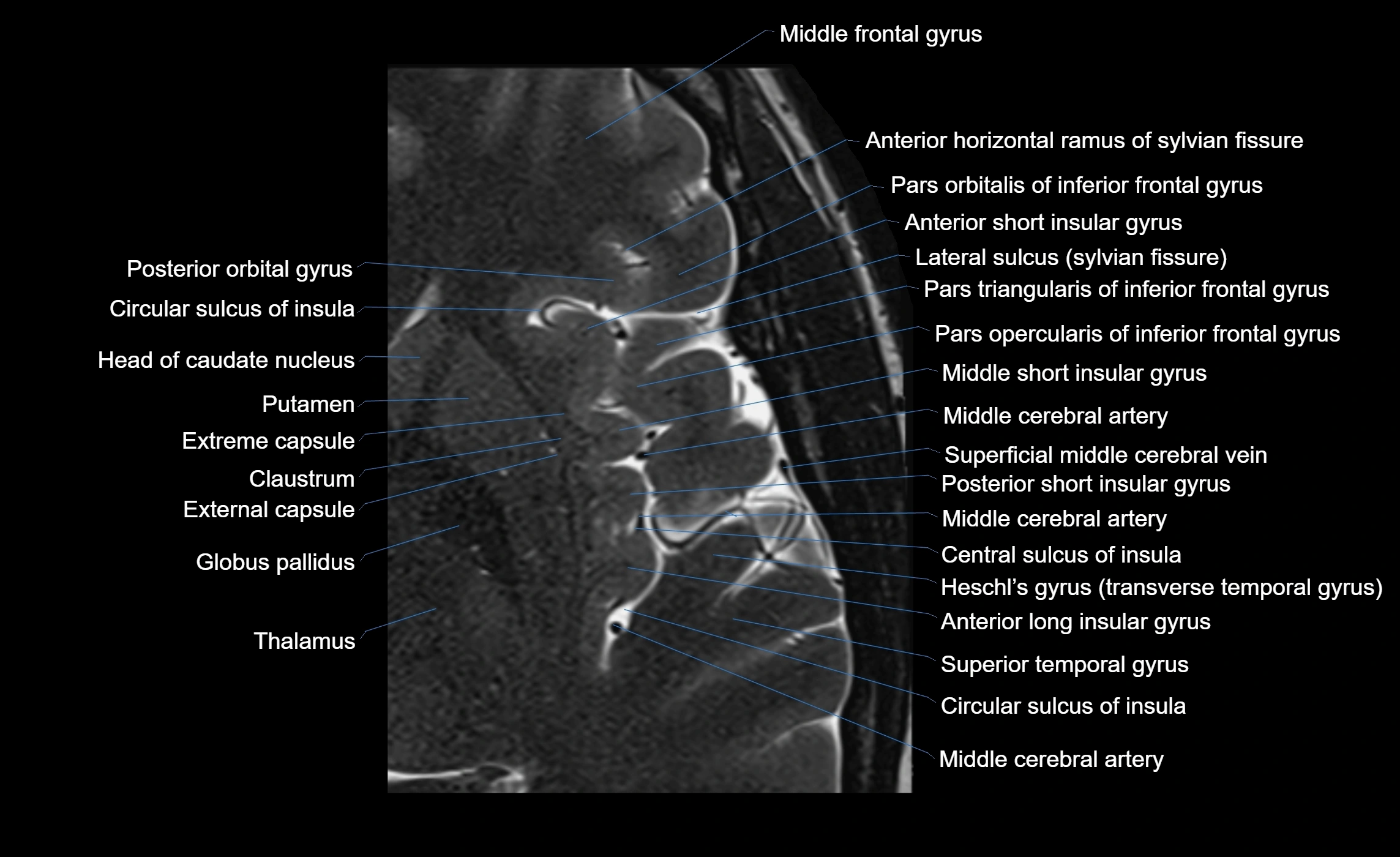
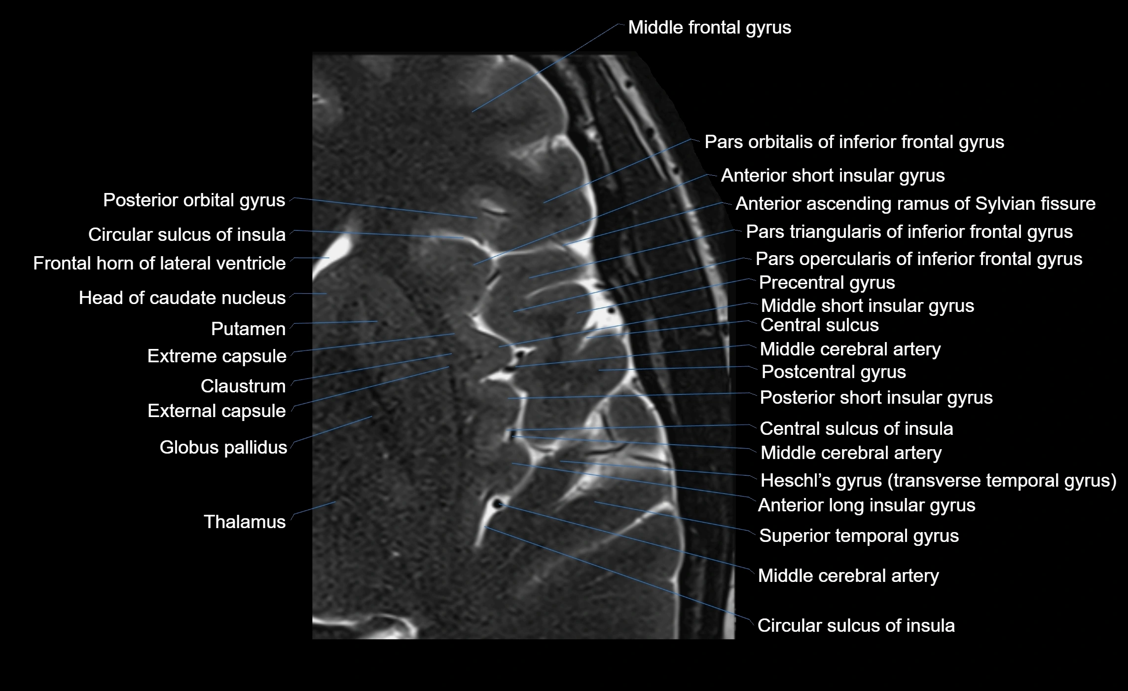
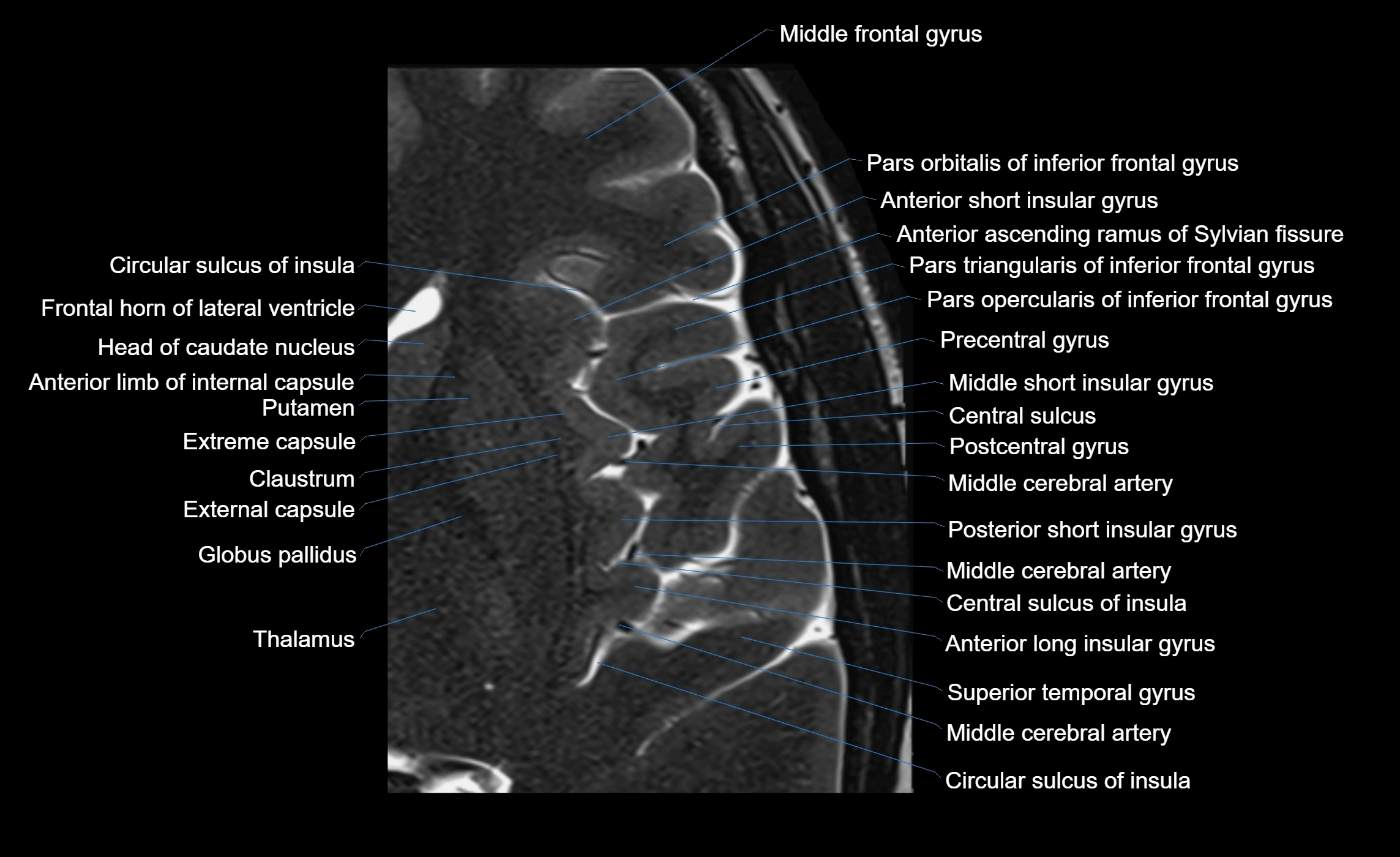
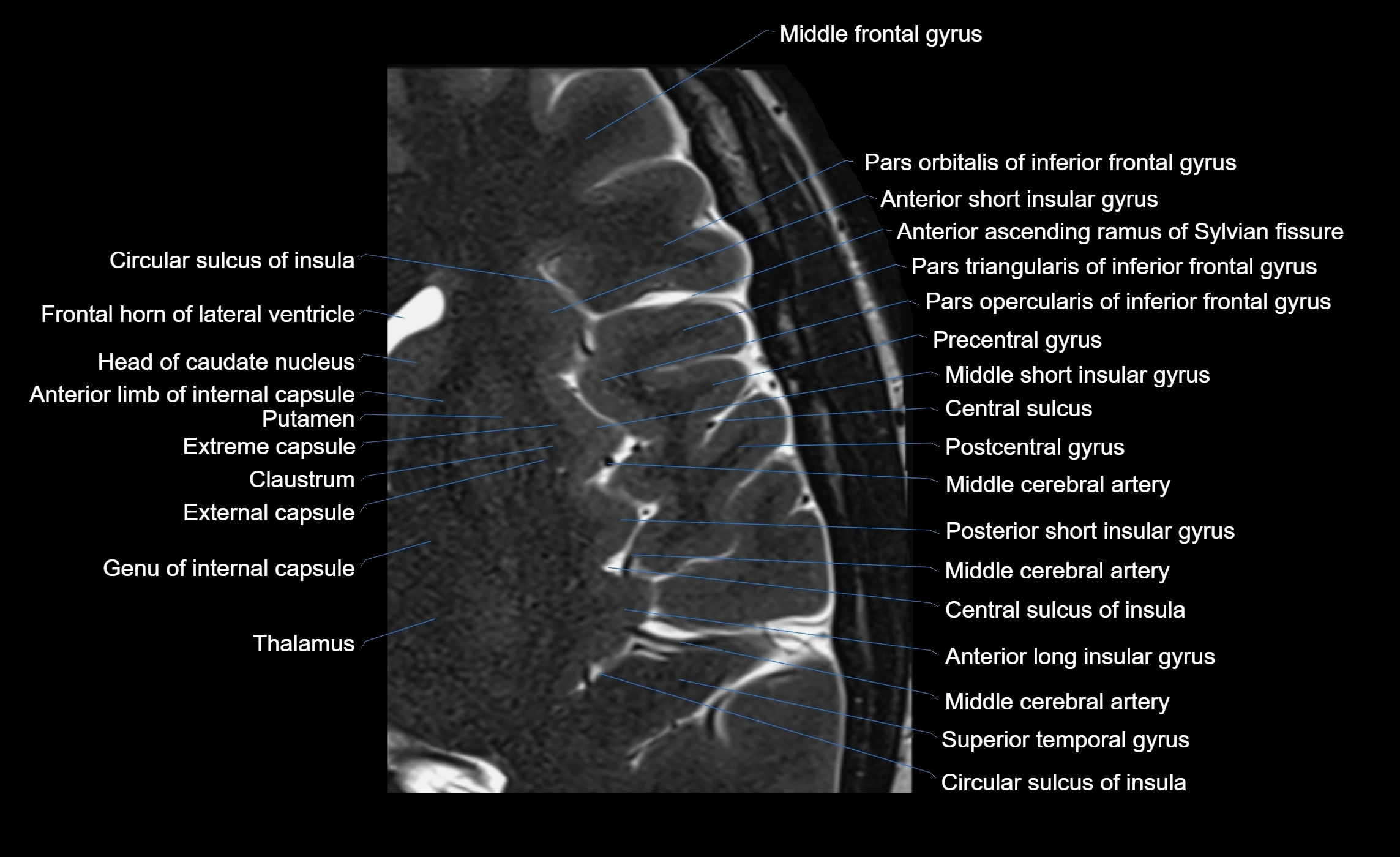
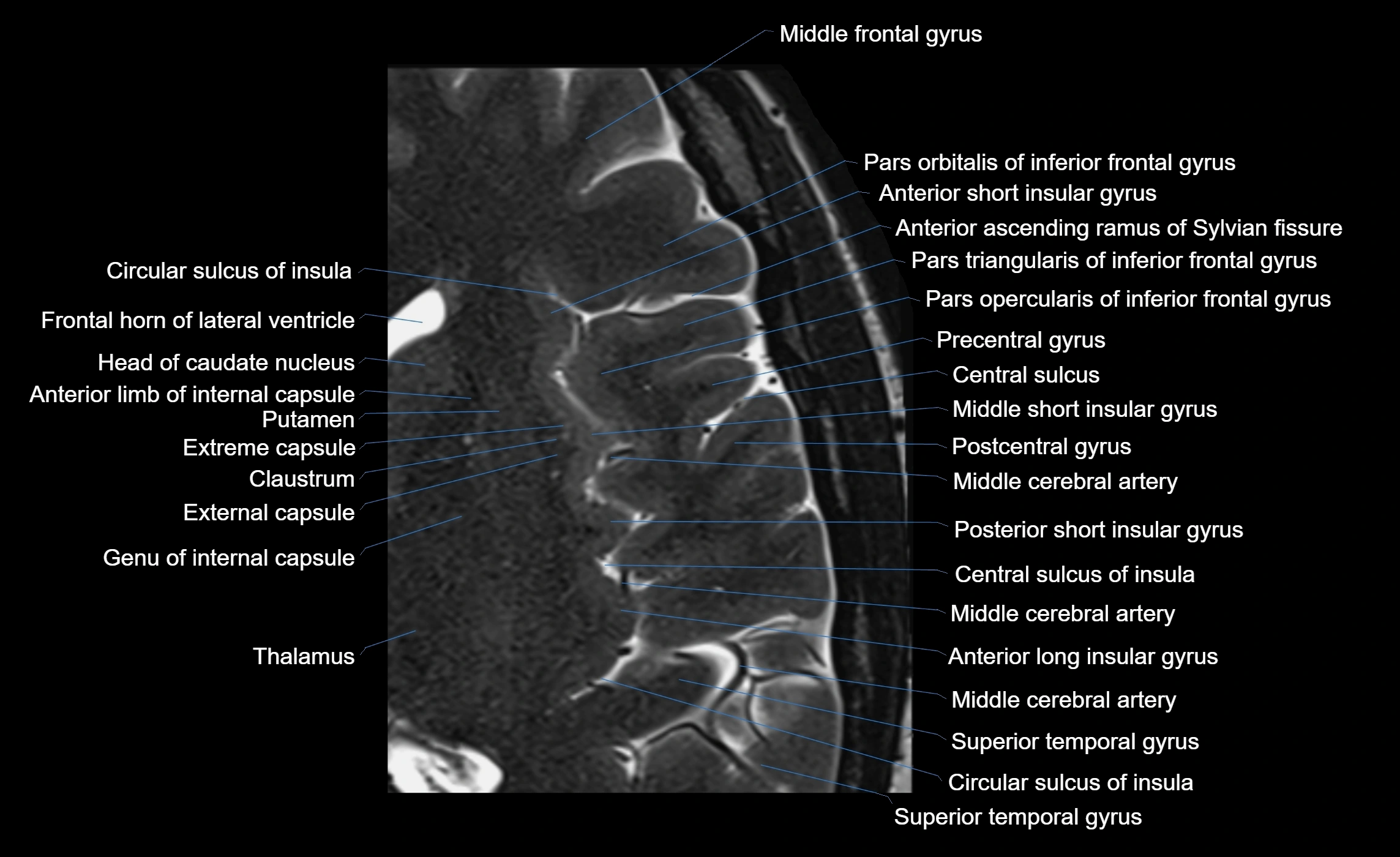
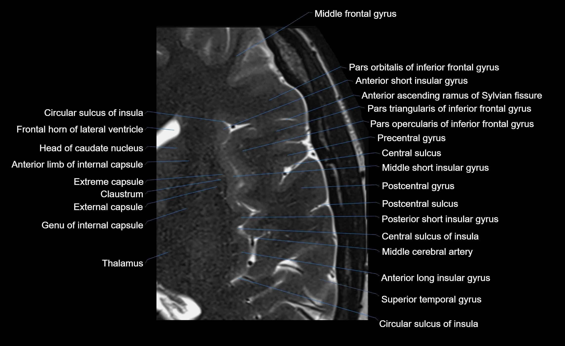
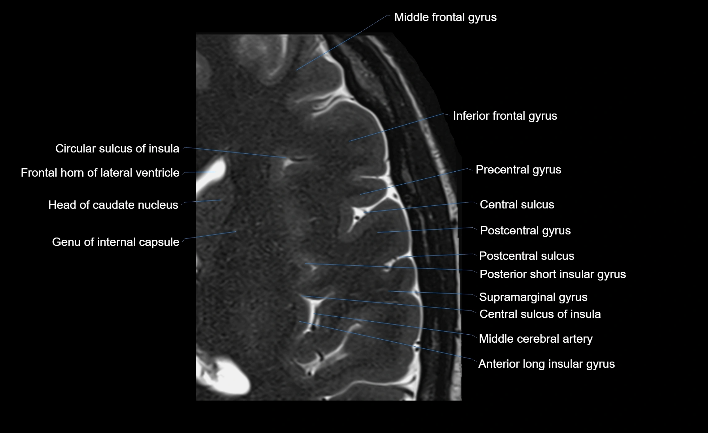
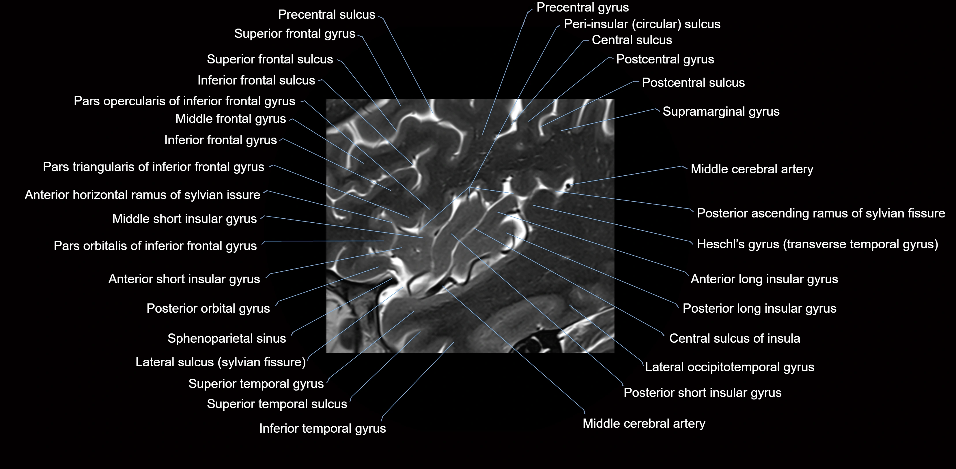
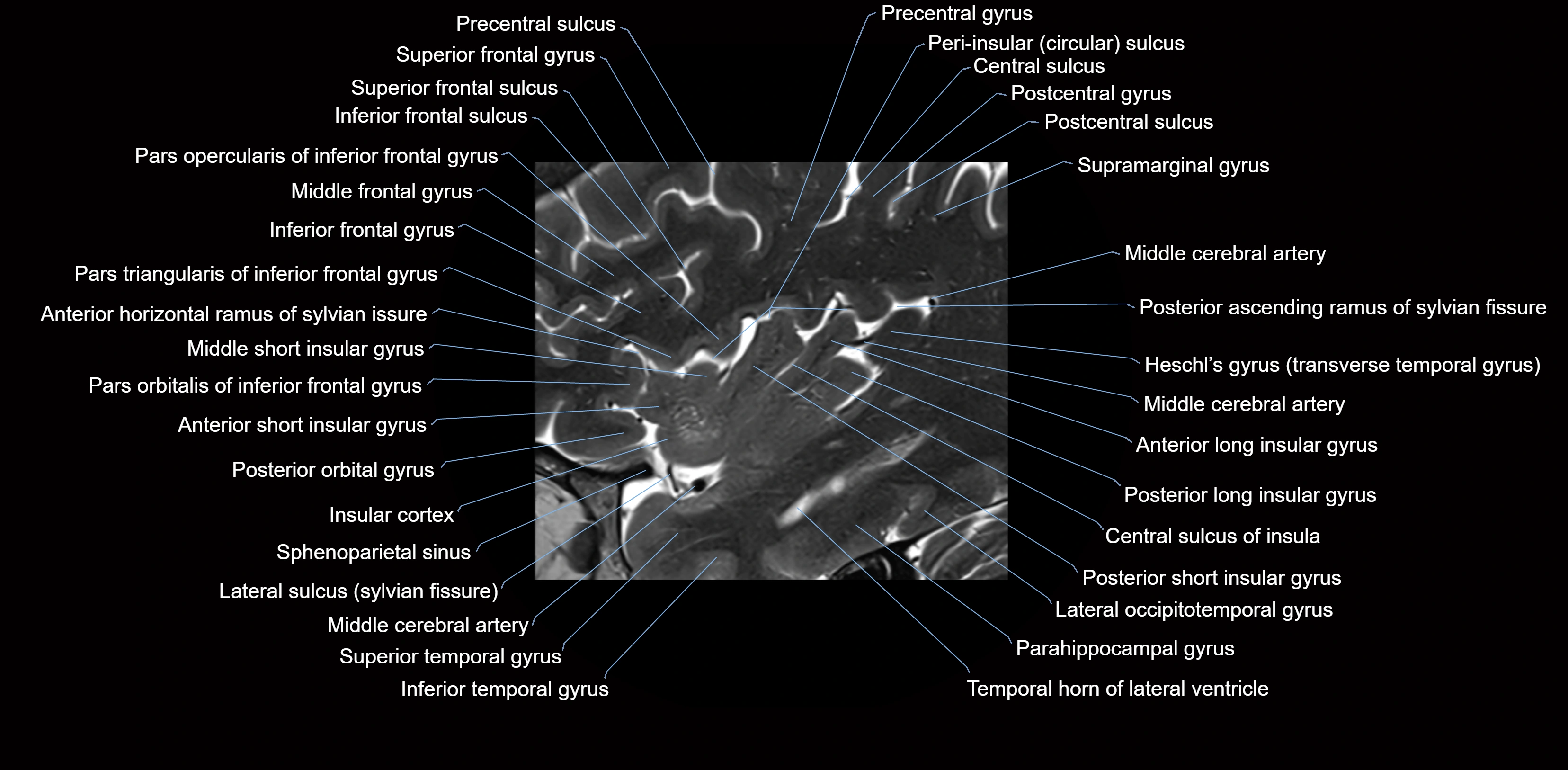
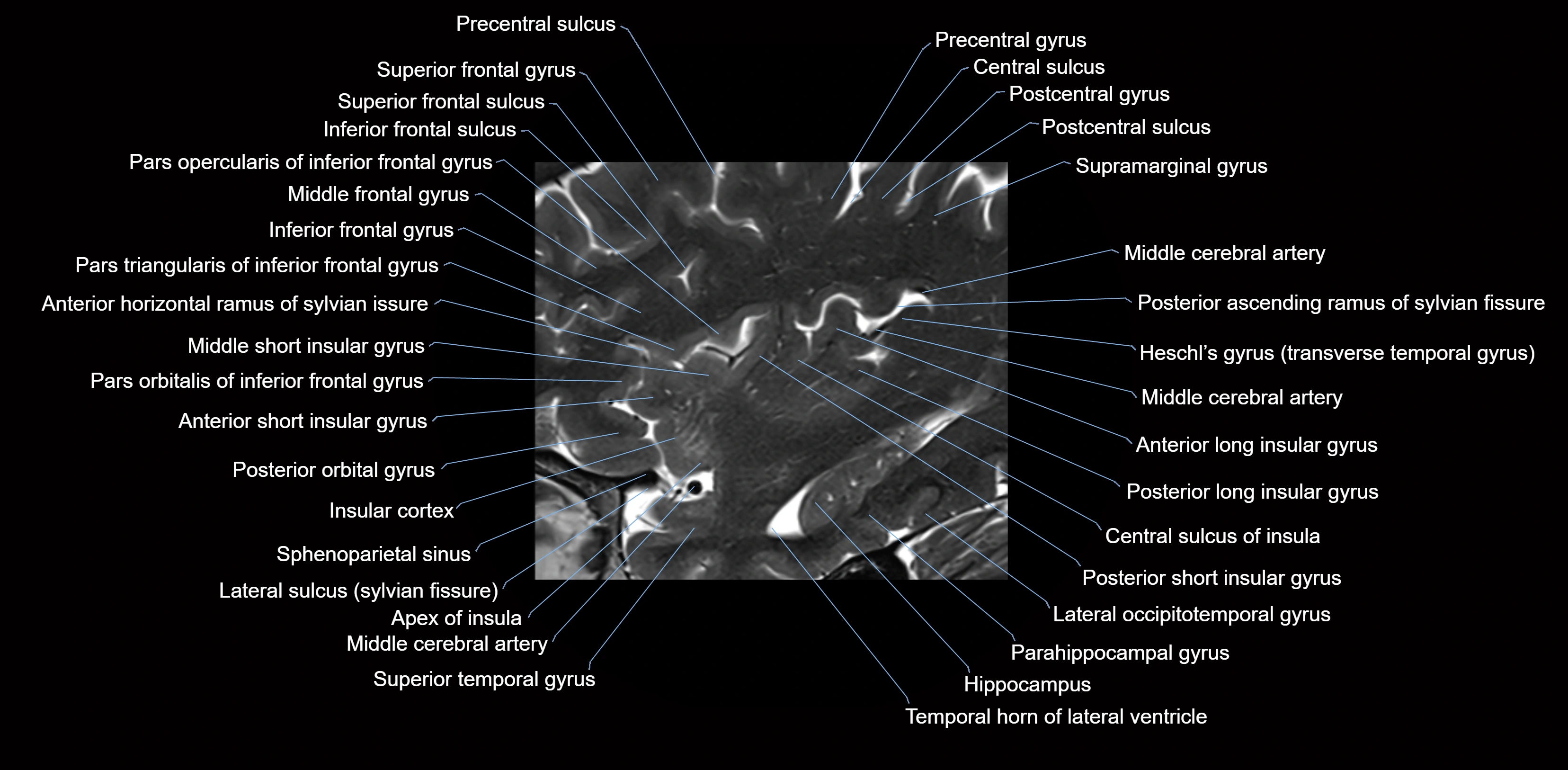
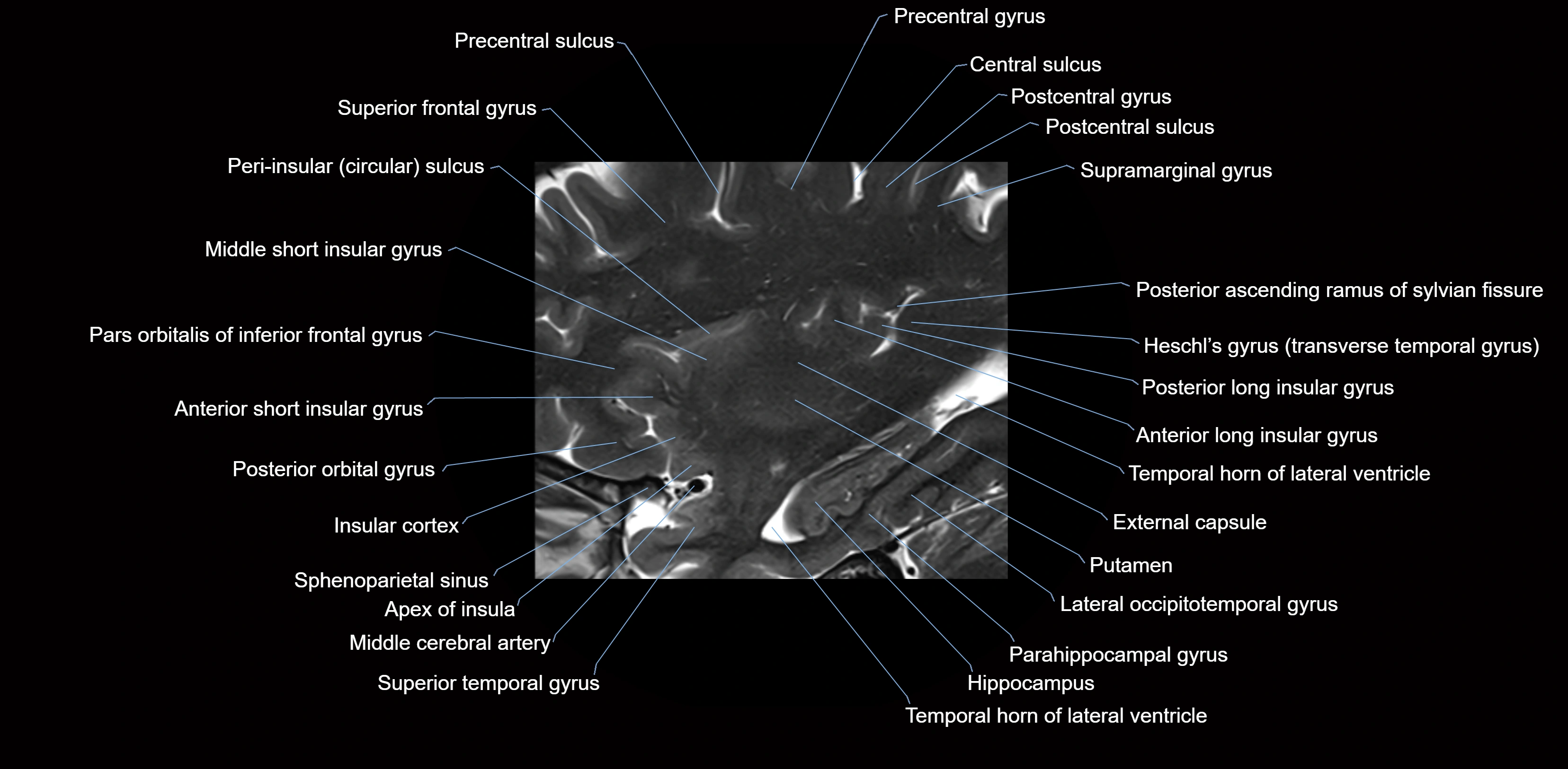
MRI images

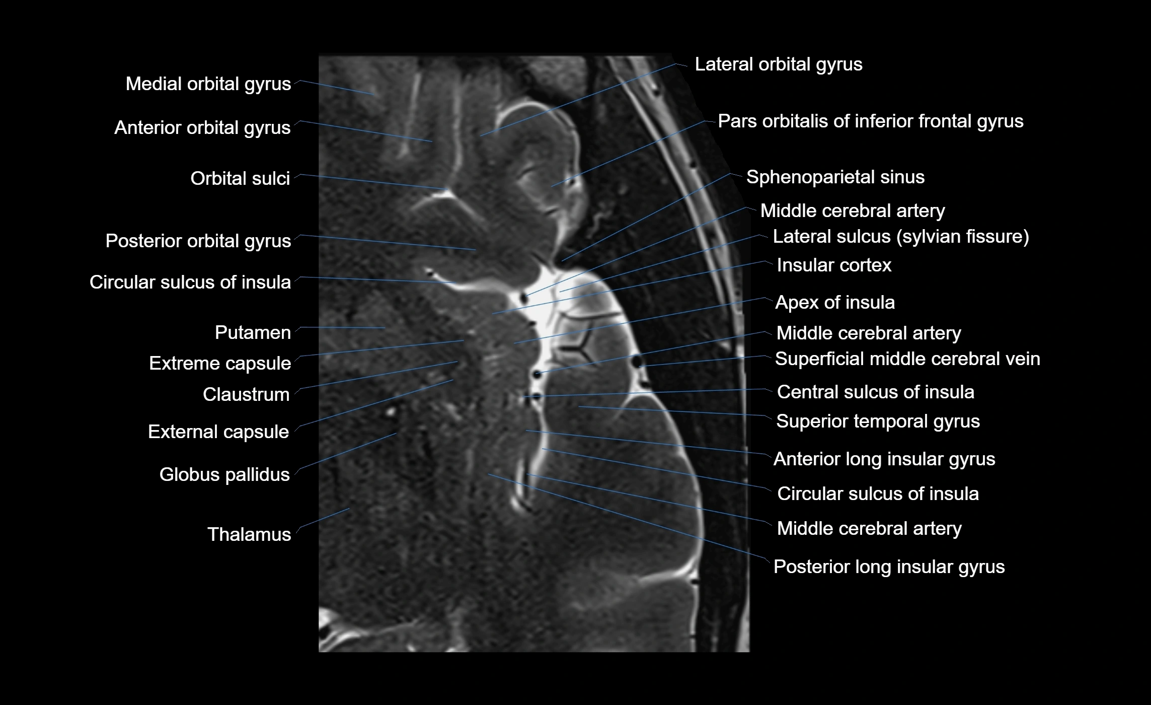
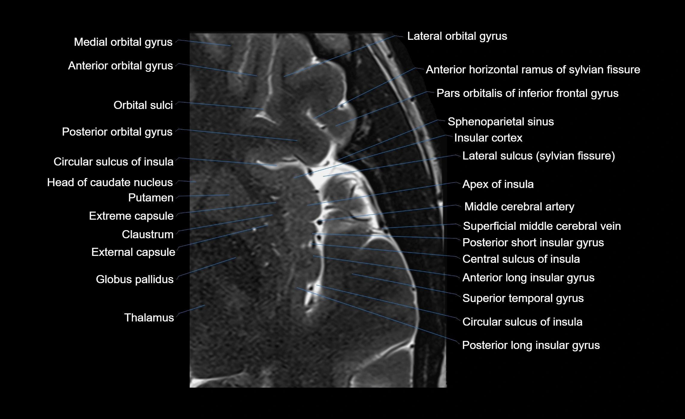
MRI images


