


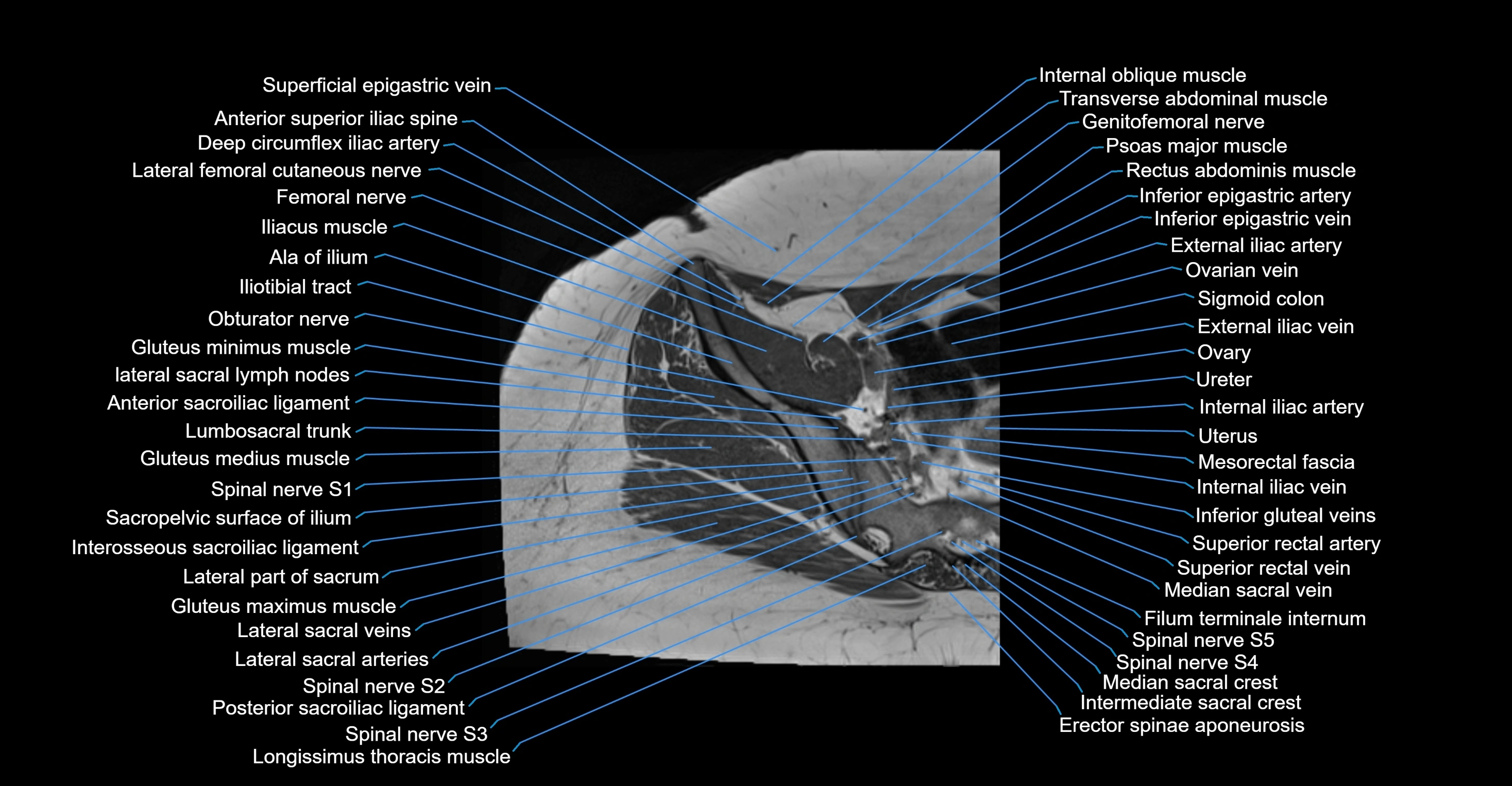
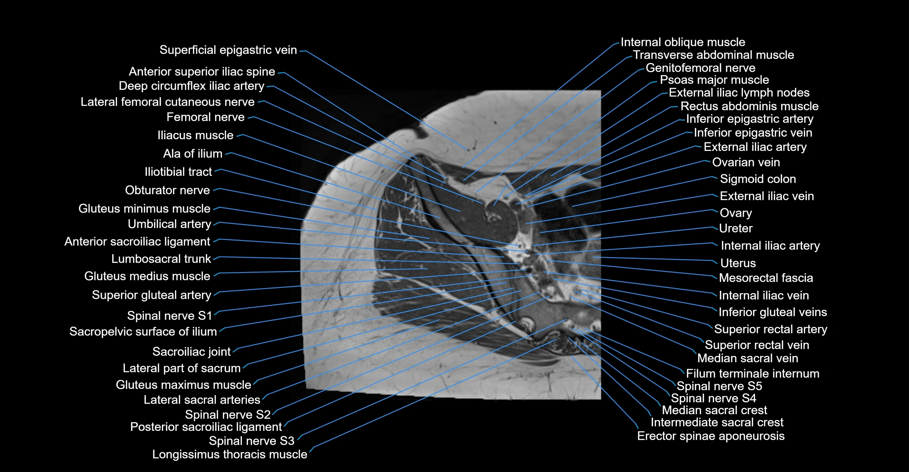
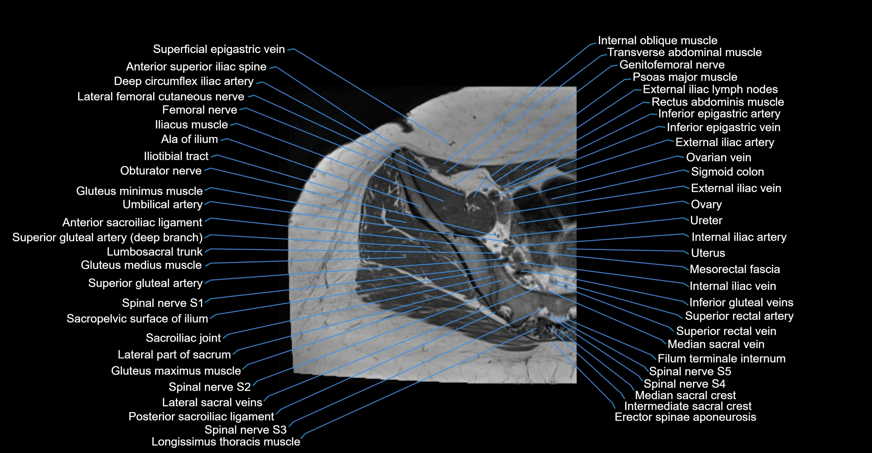
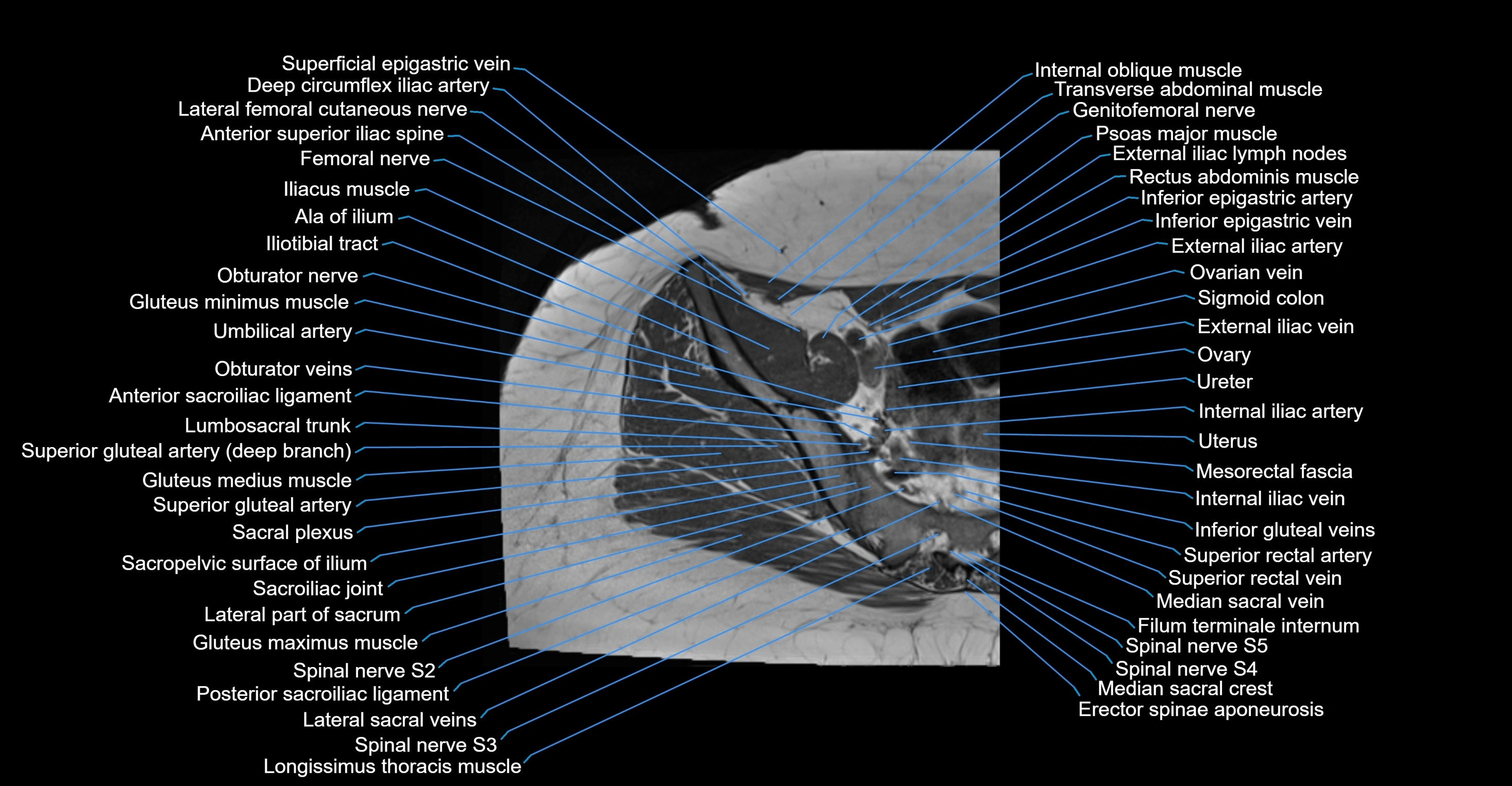
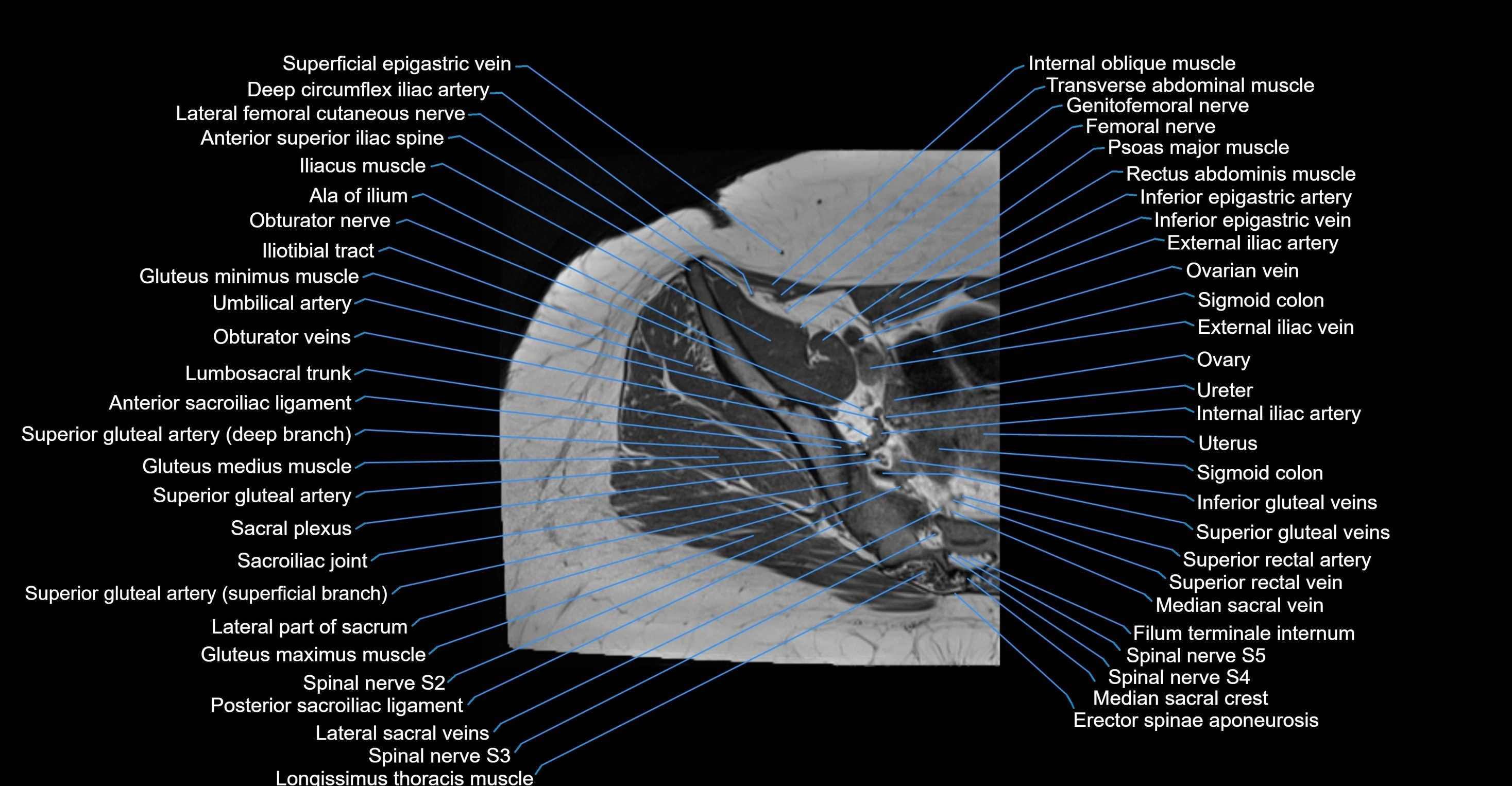
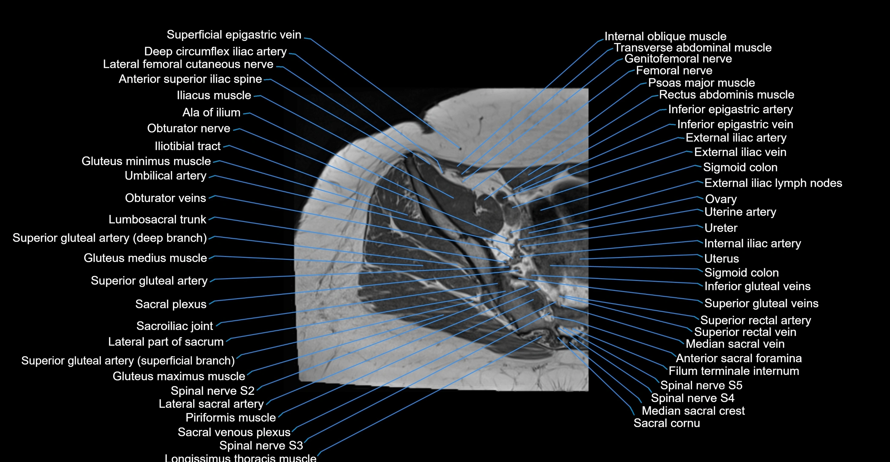
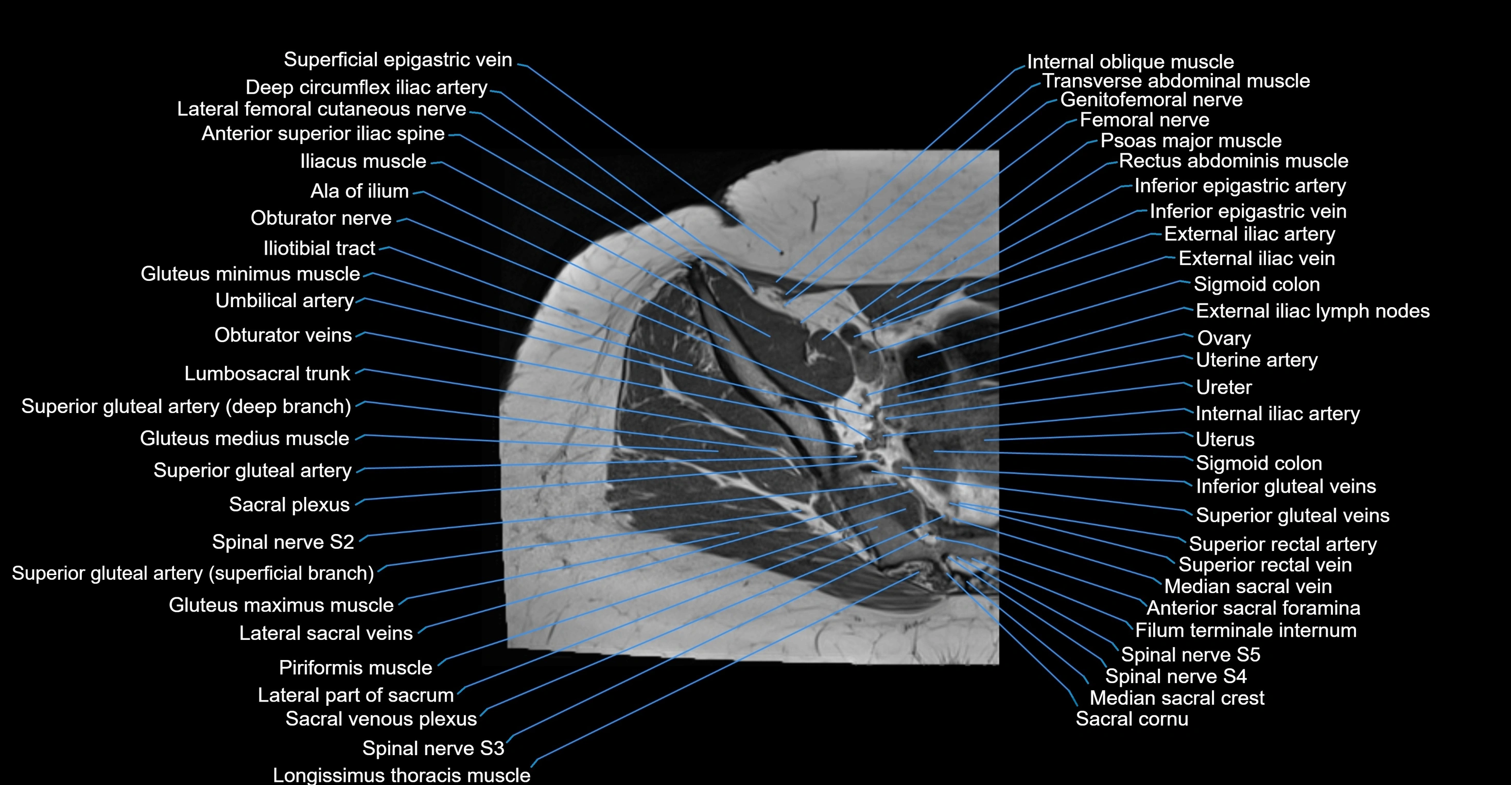
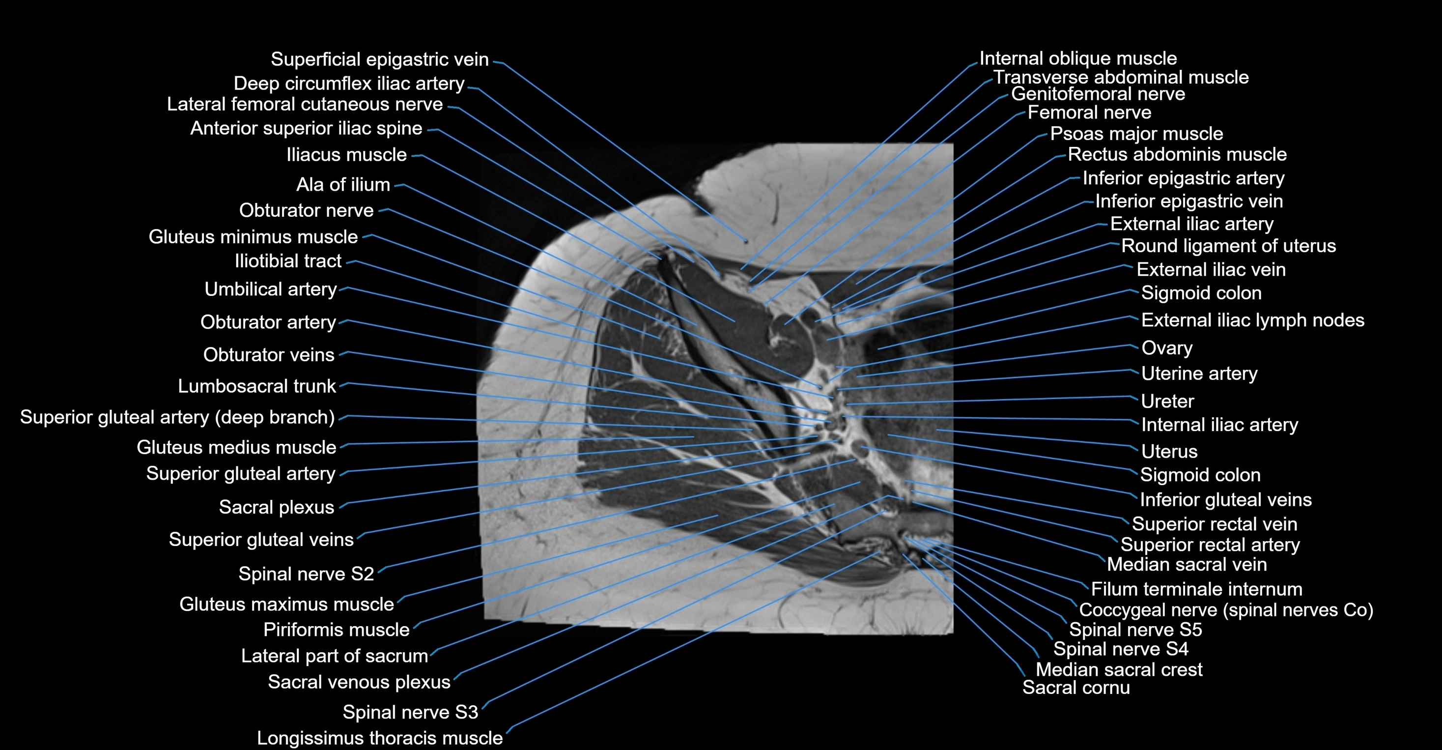
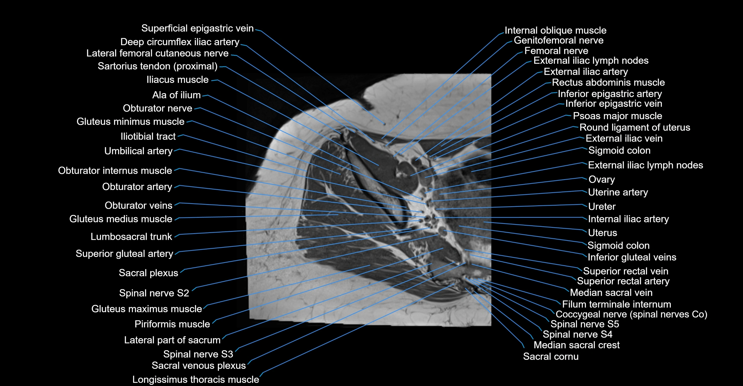
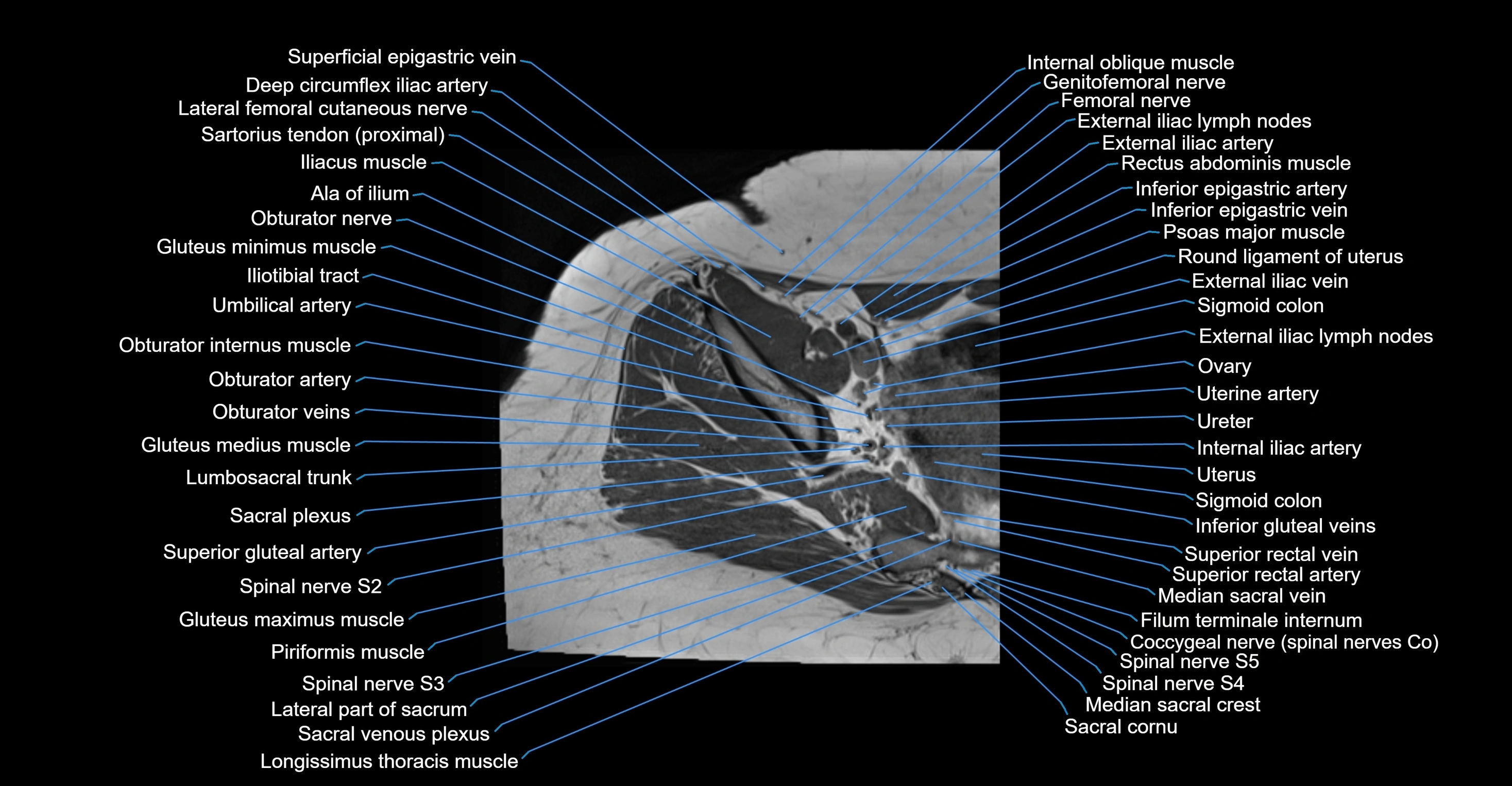
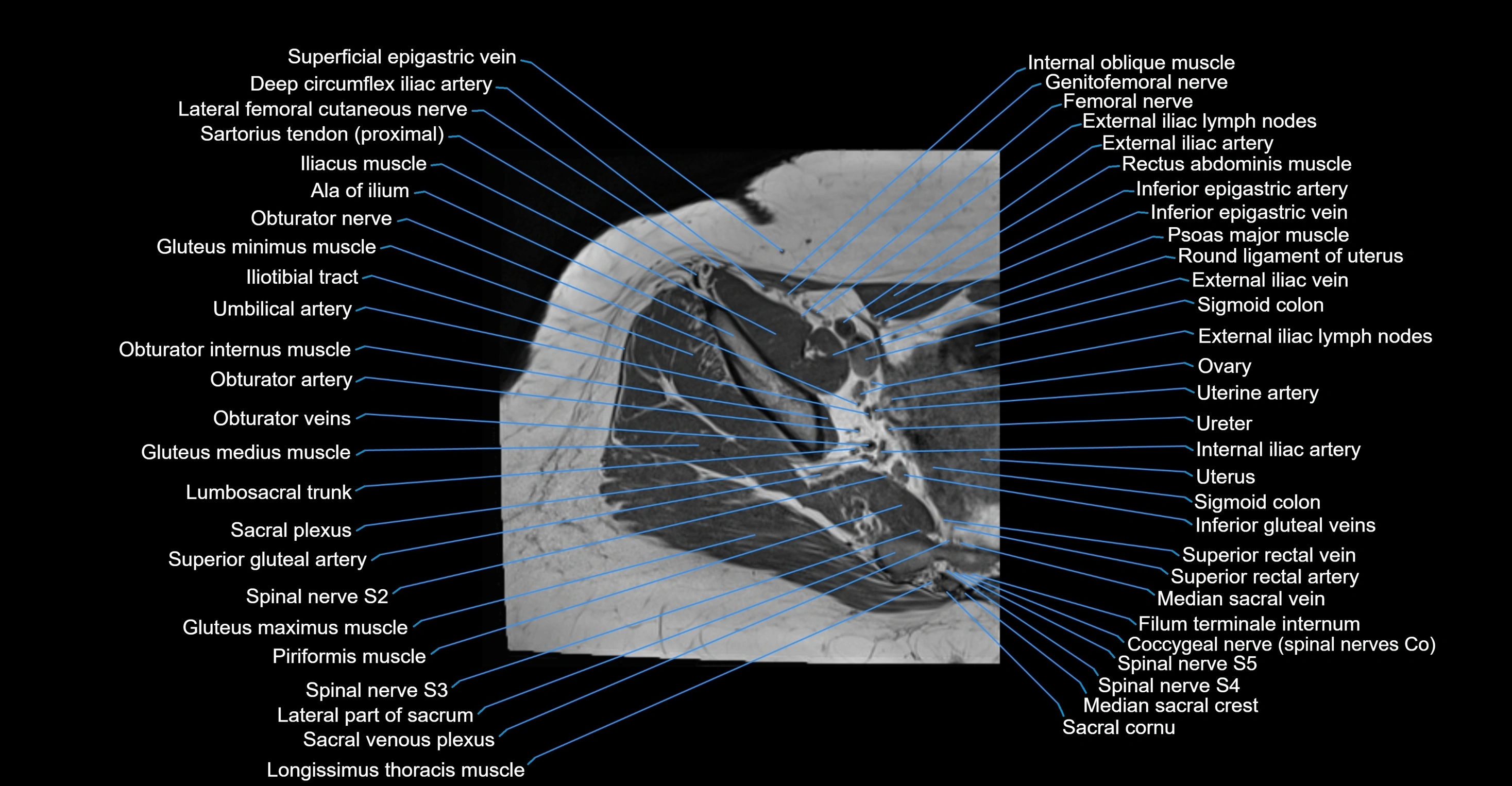
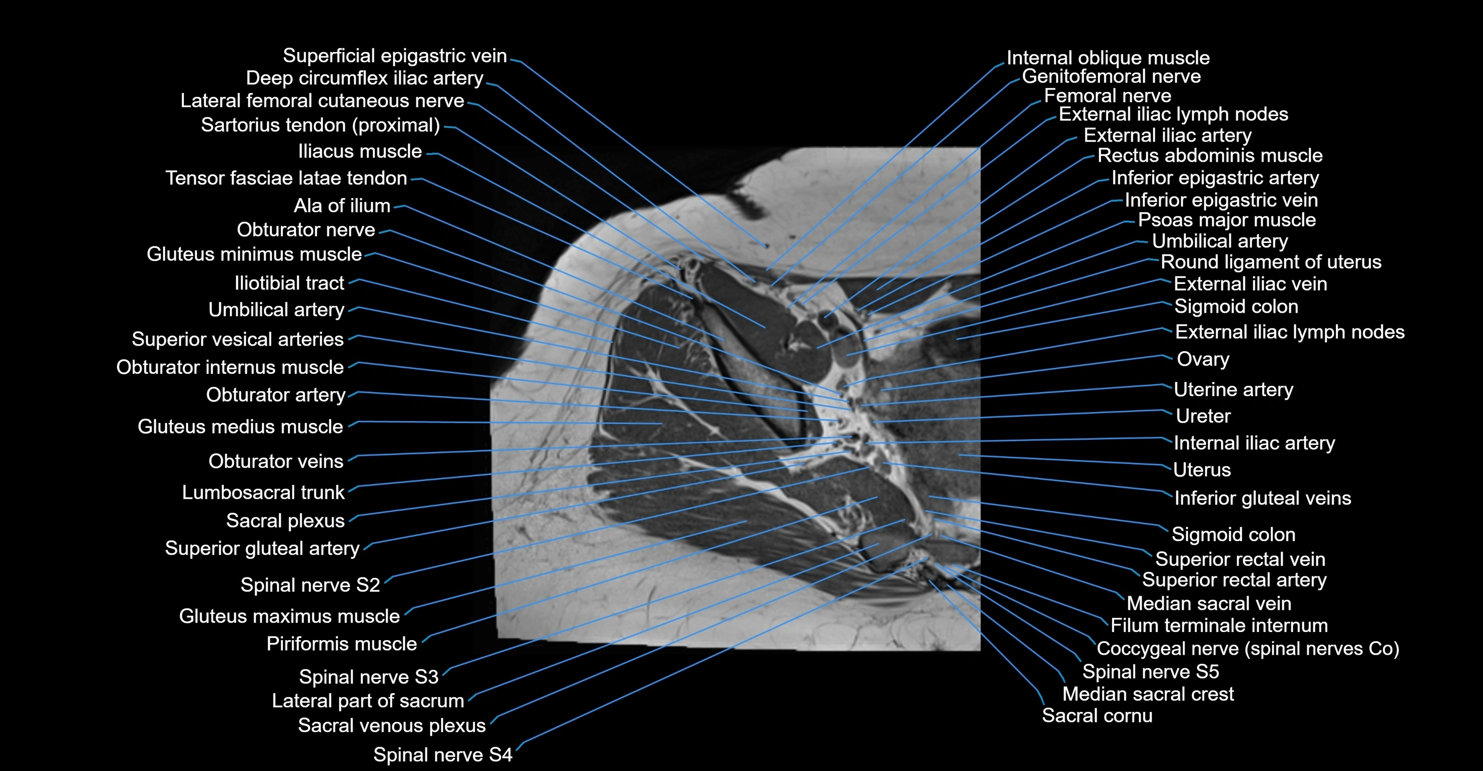
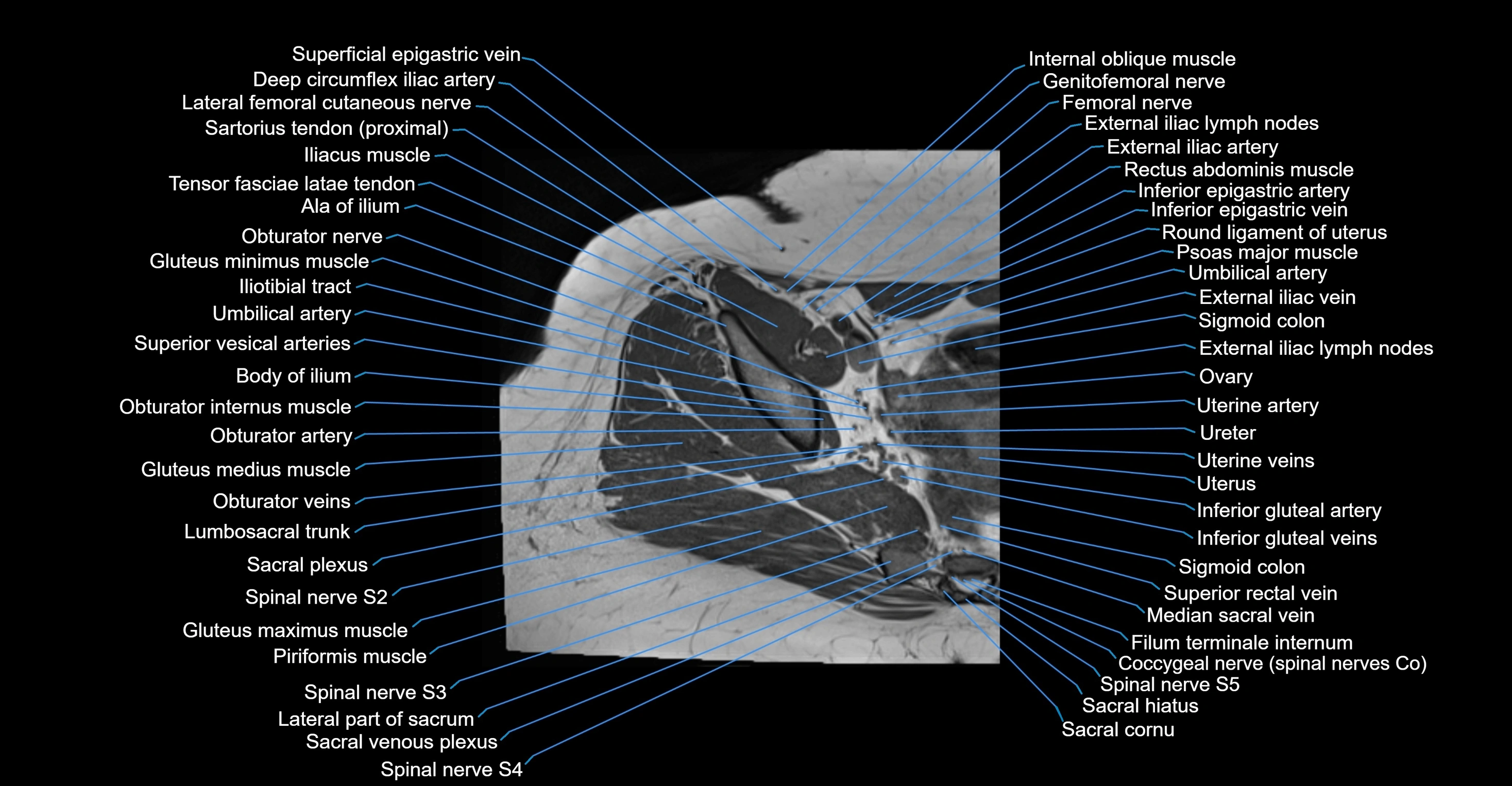

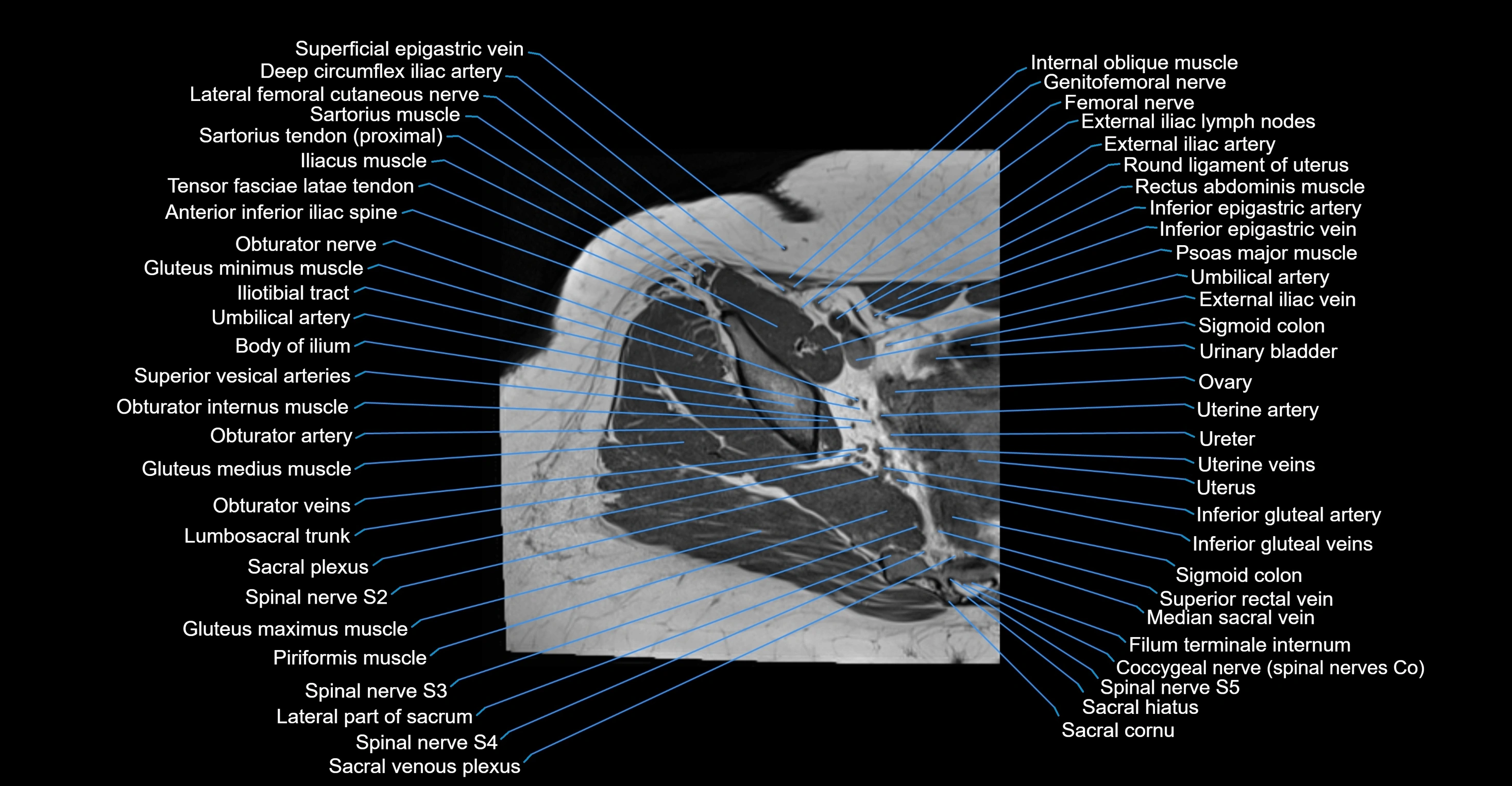
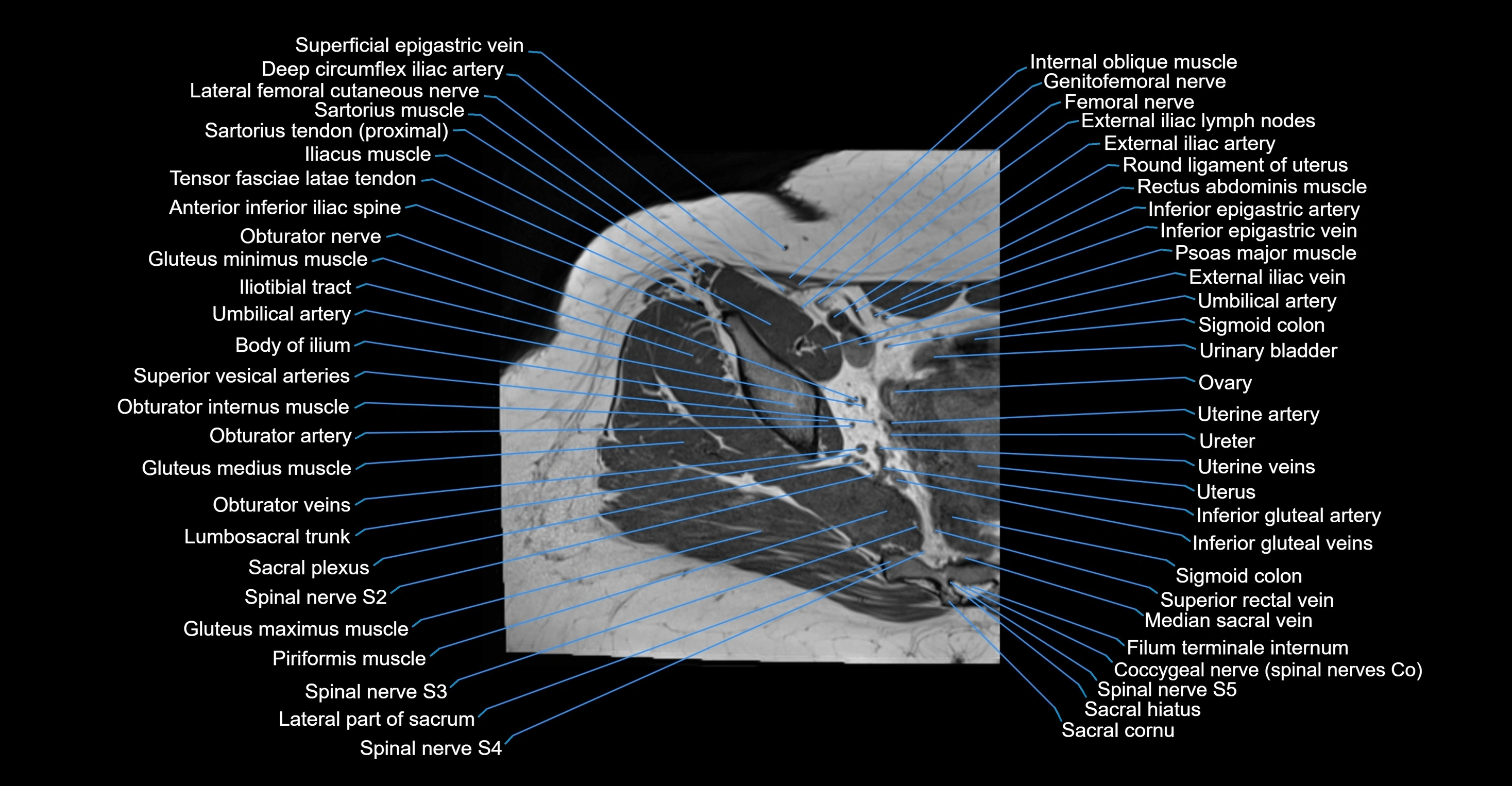
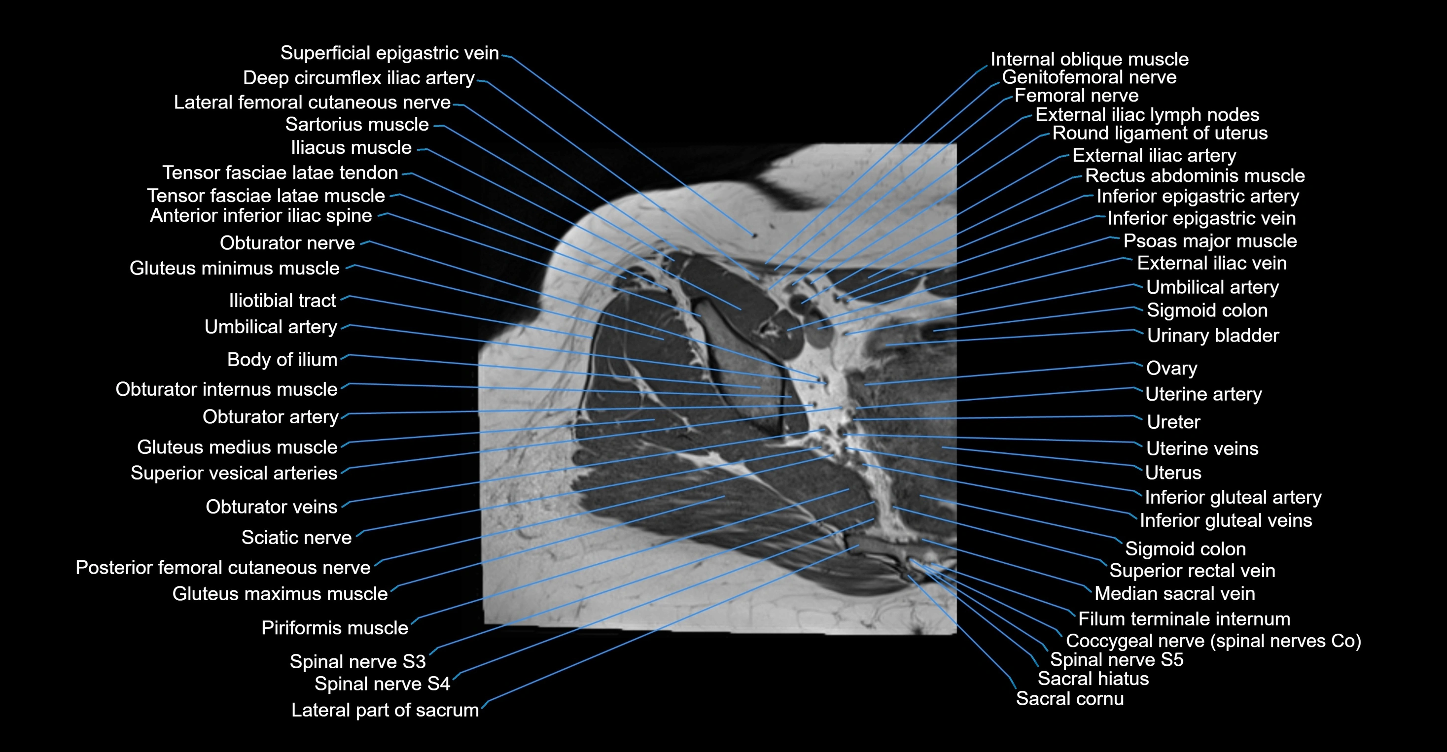
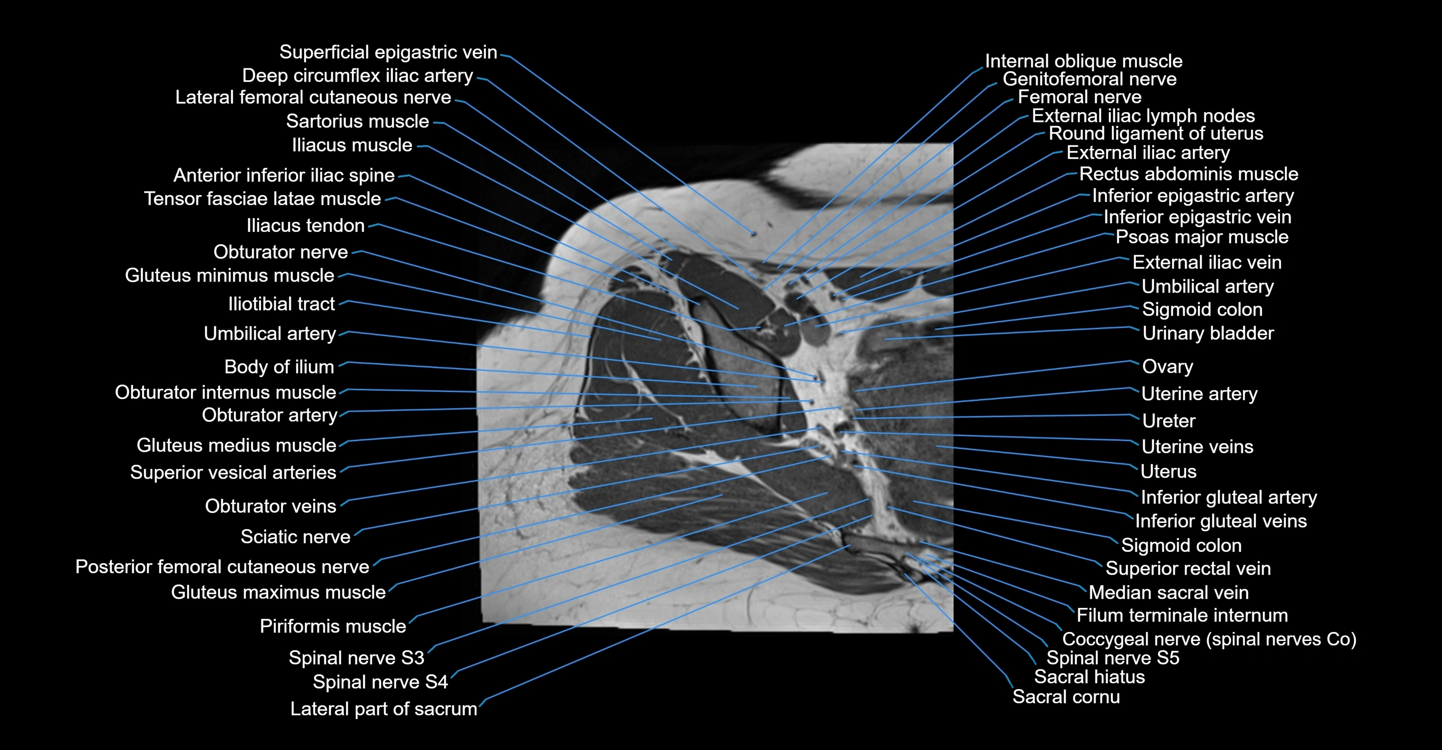
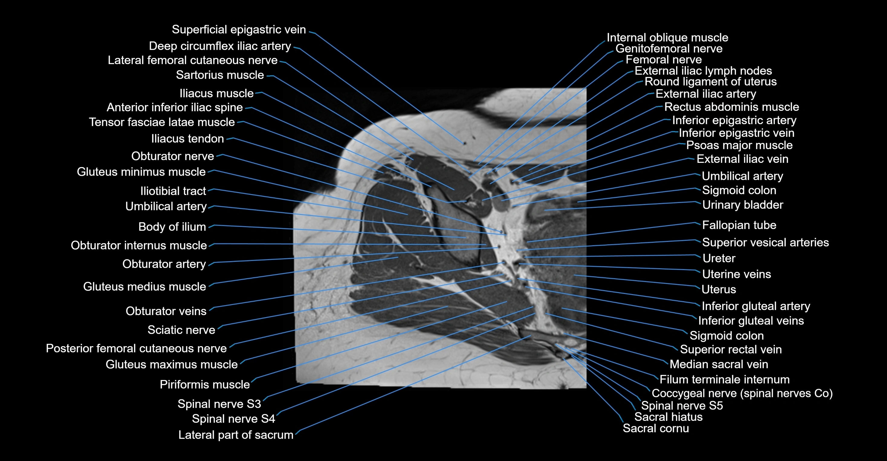
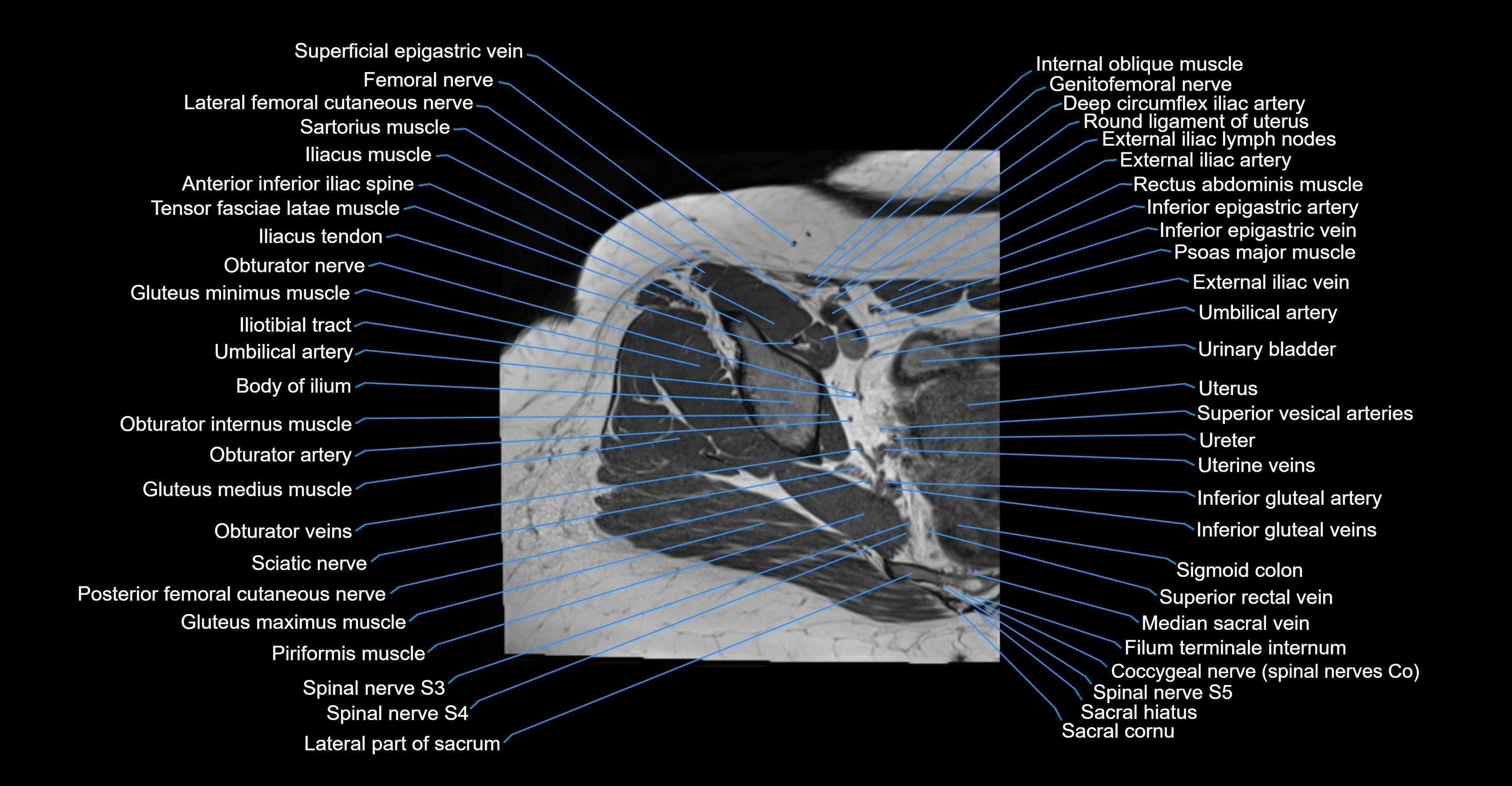
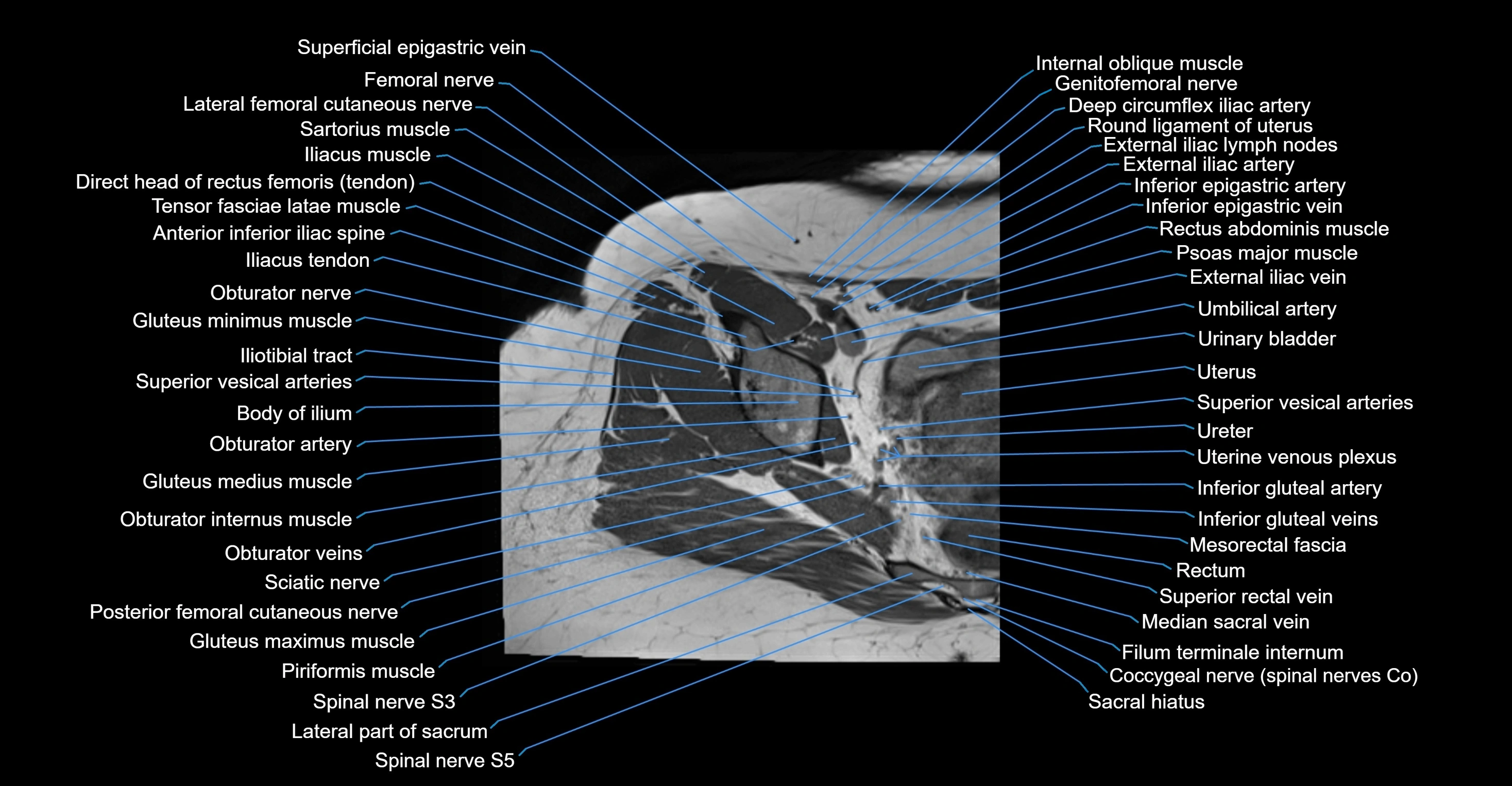
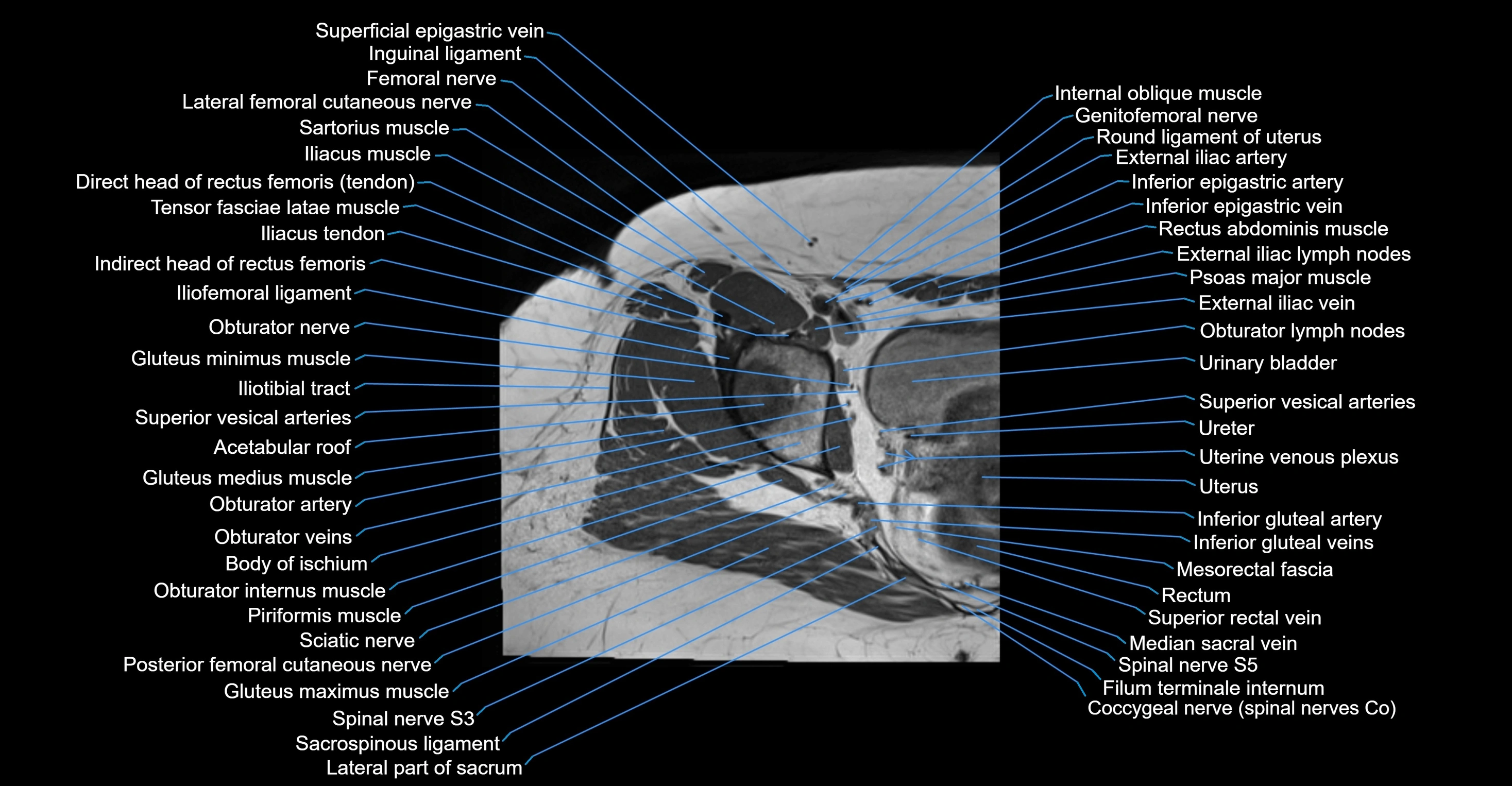
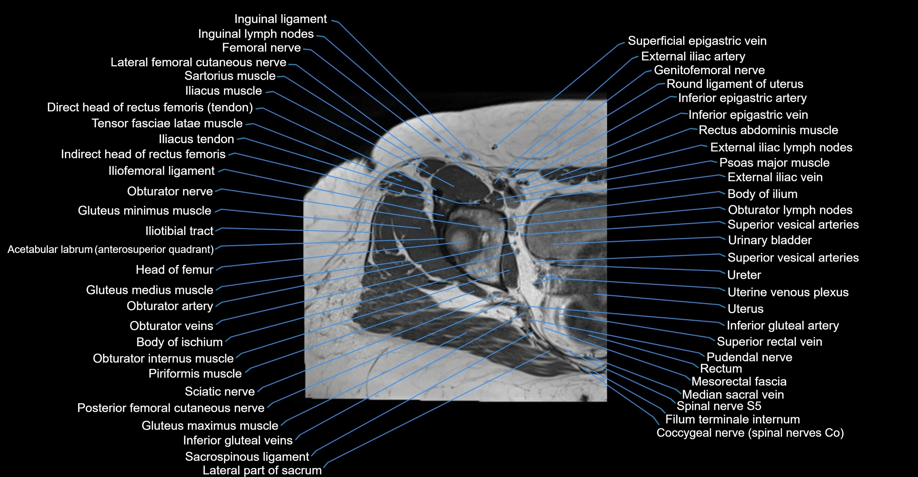
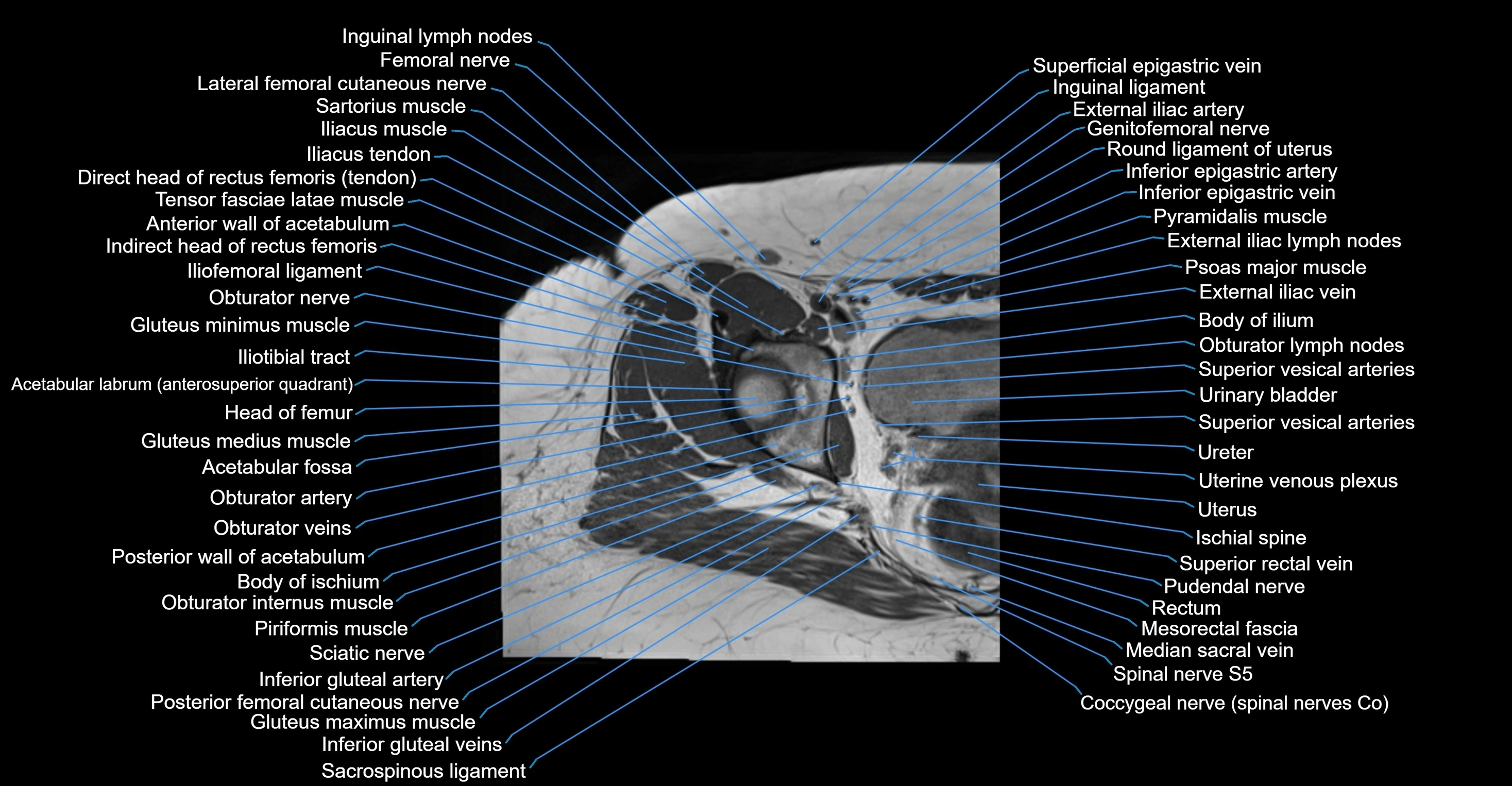
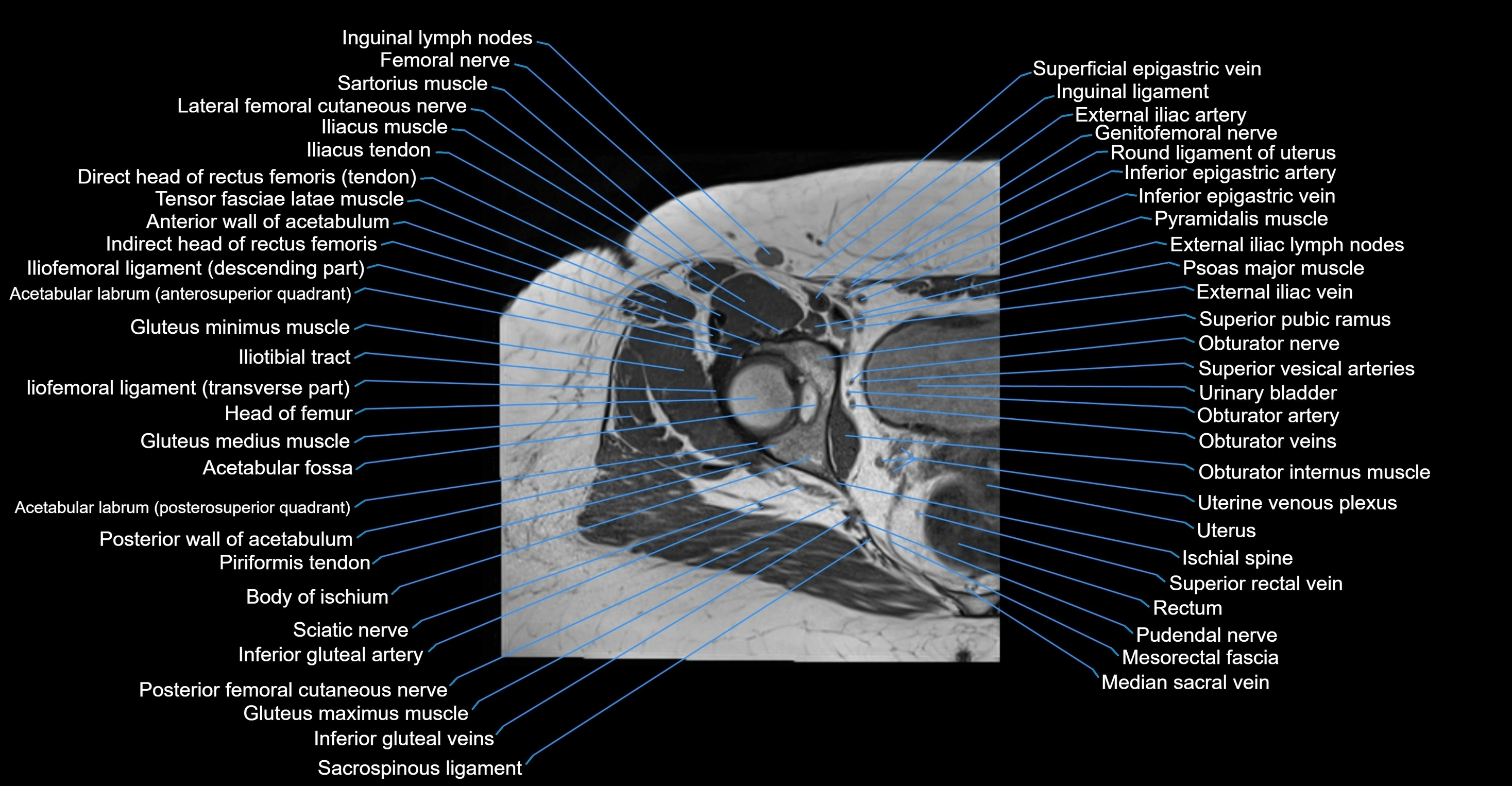
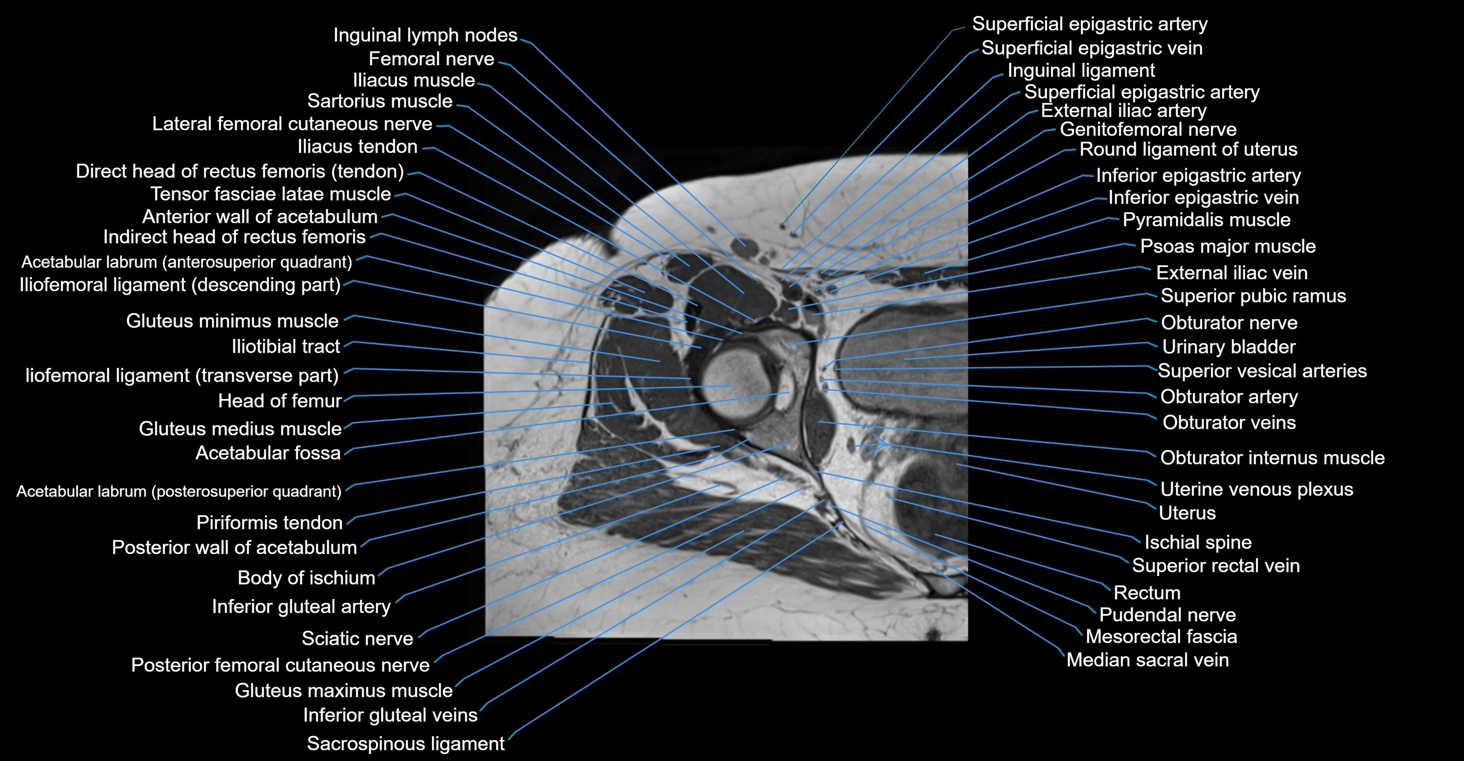
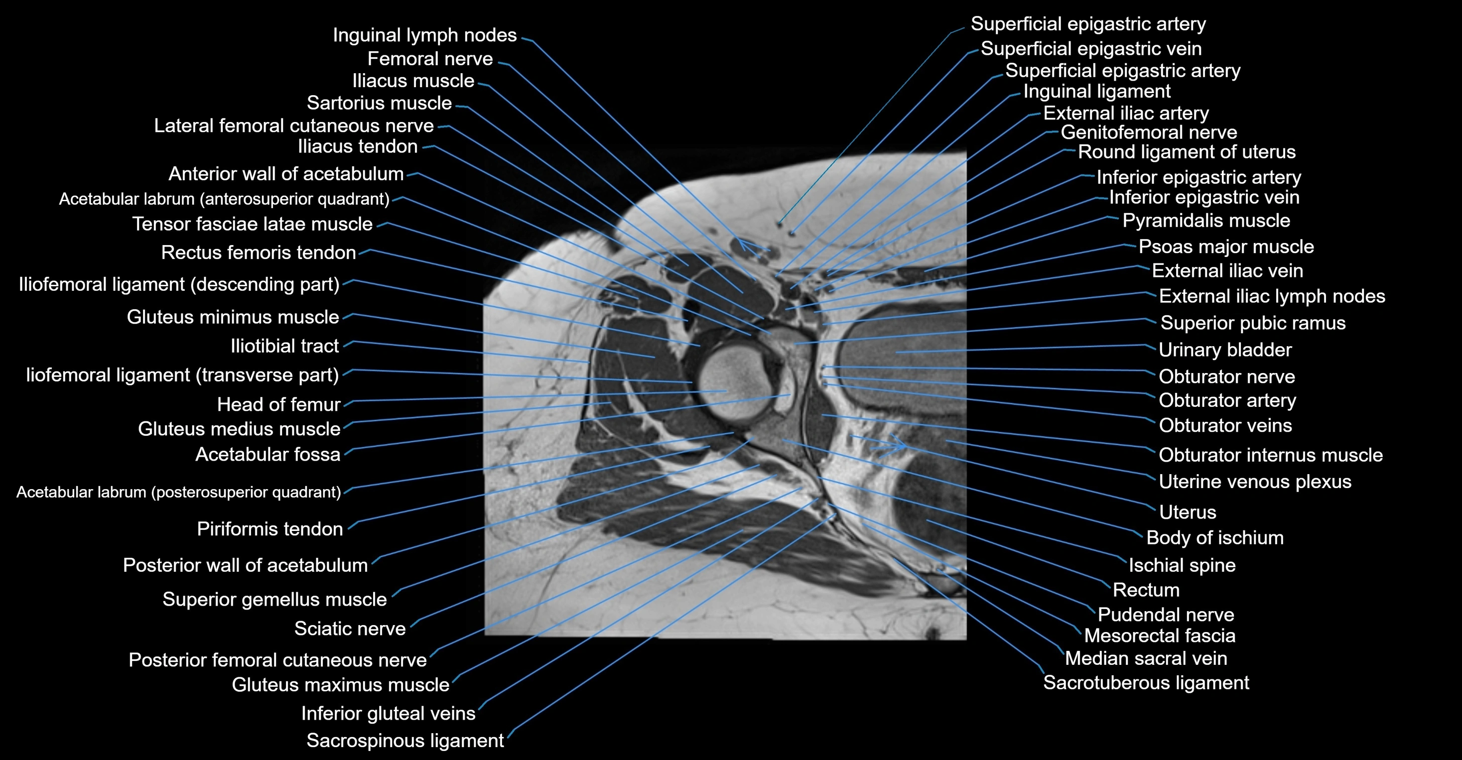
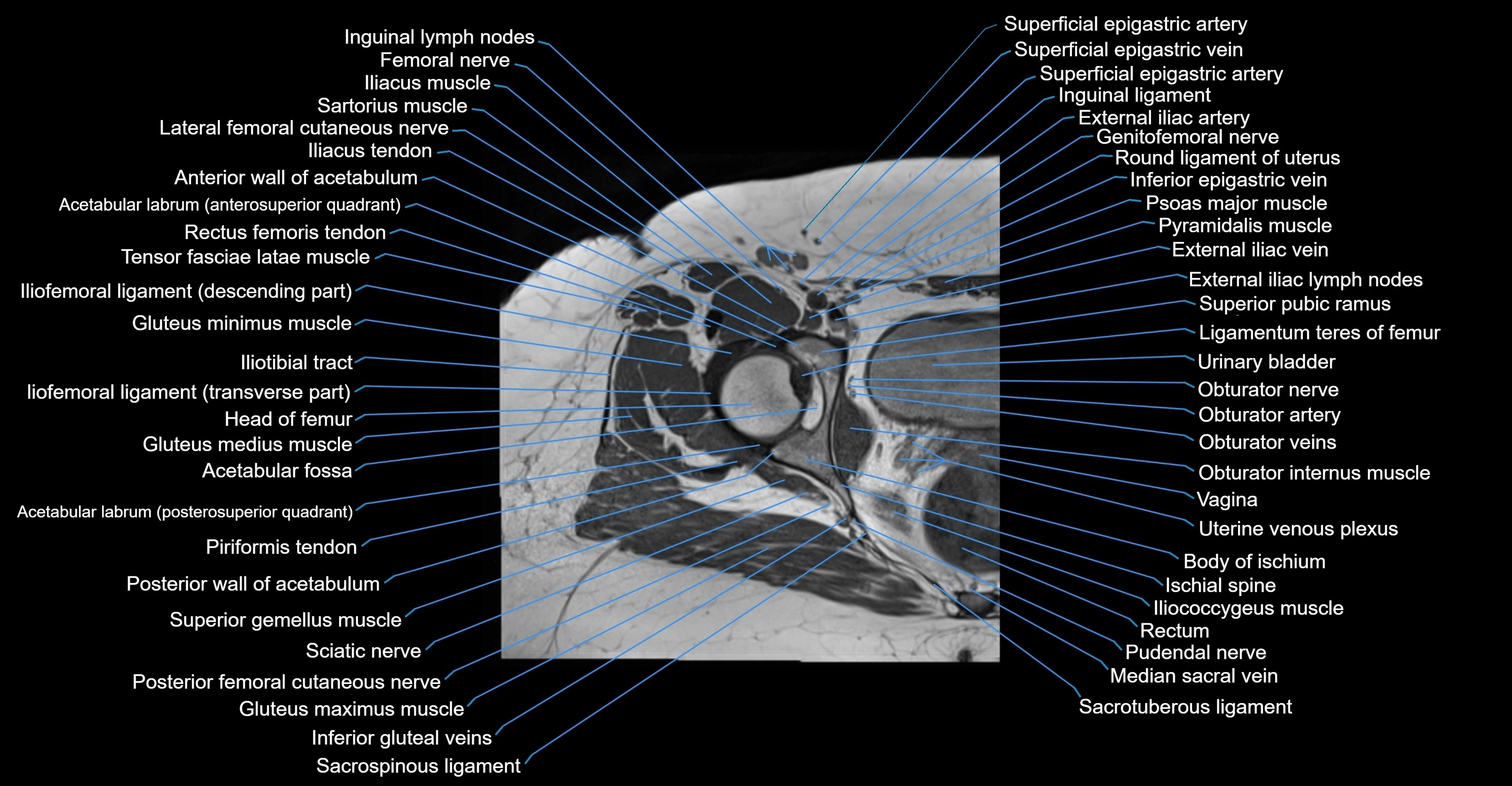

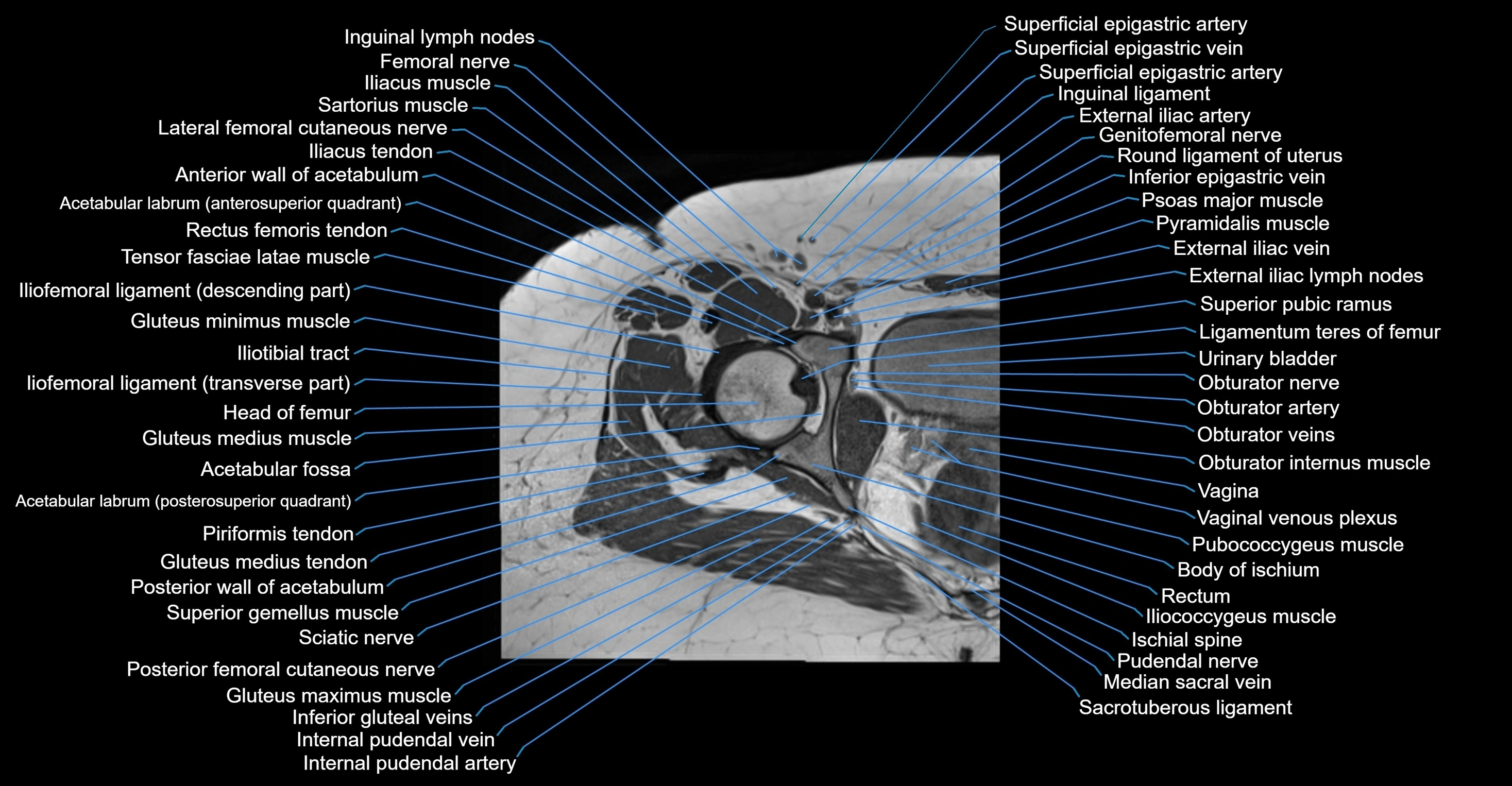
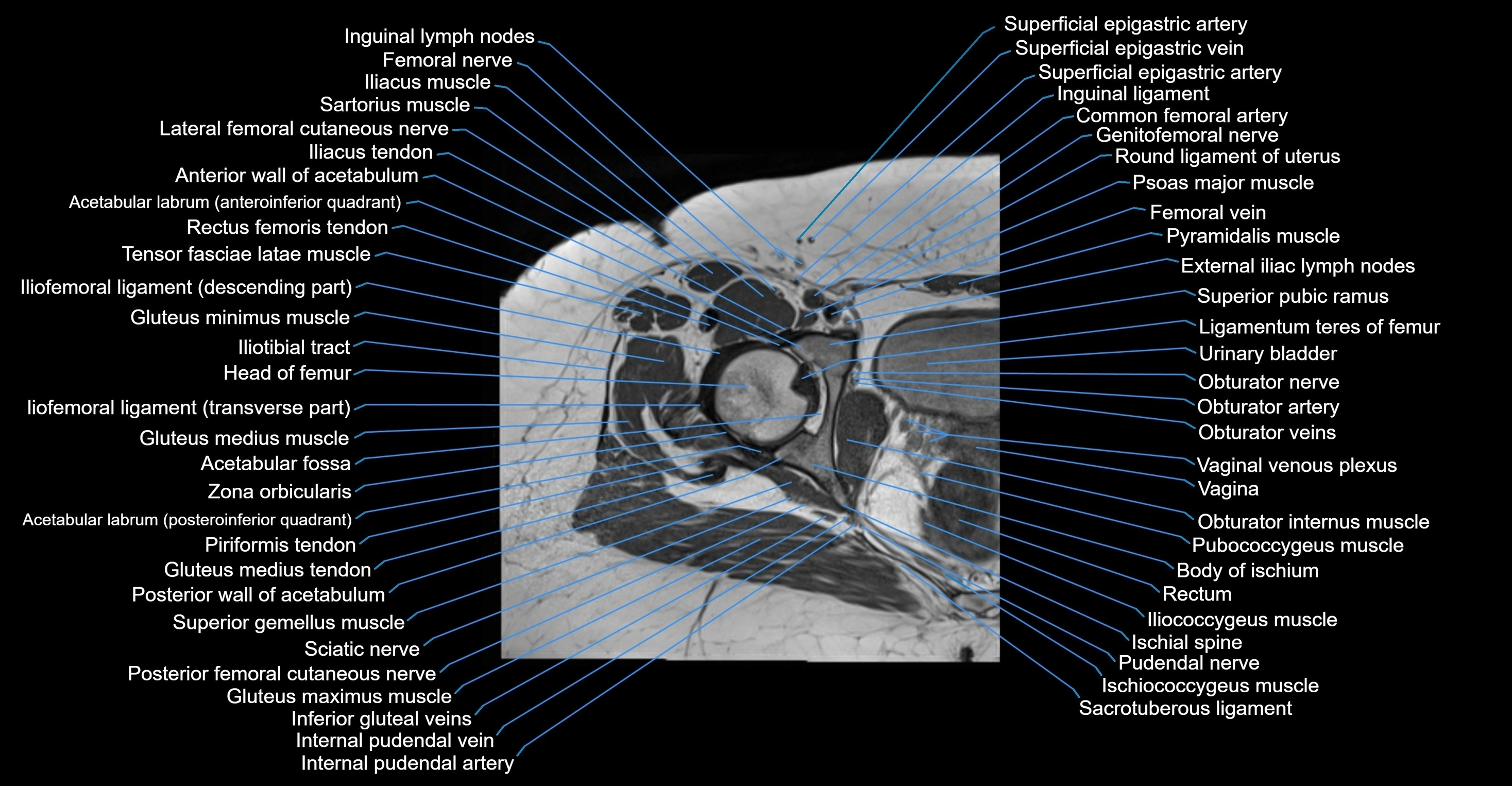
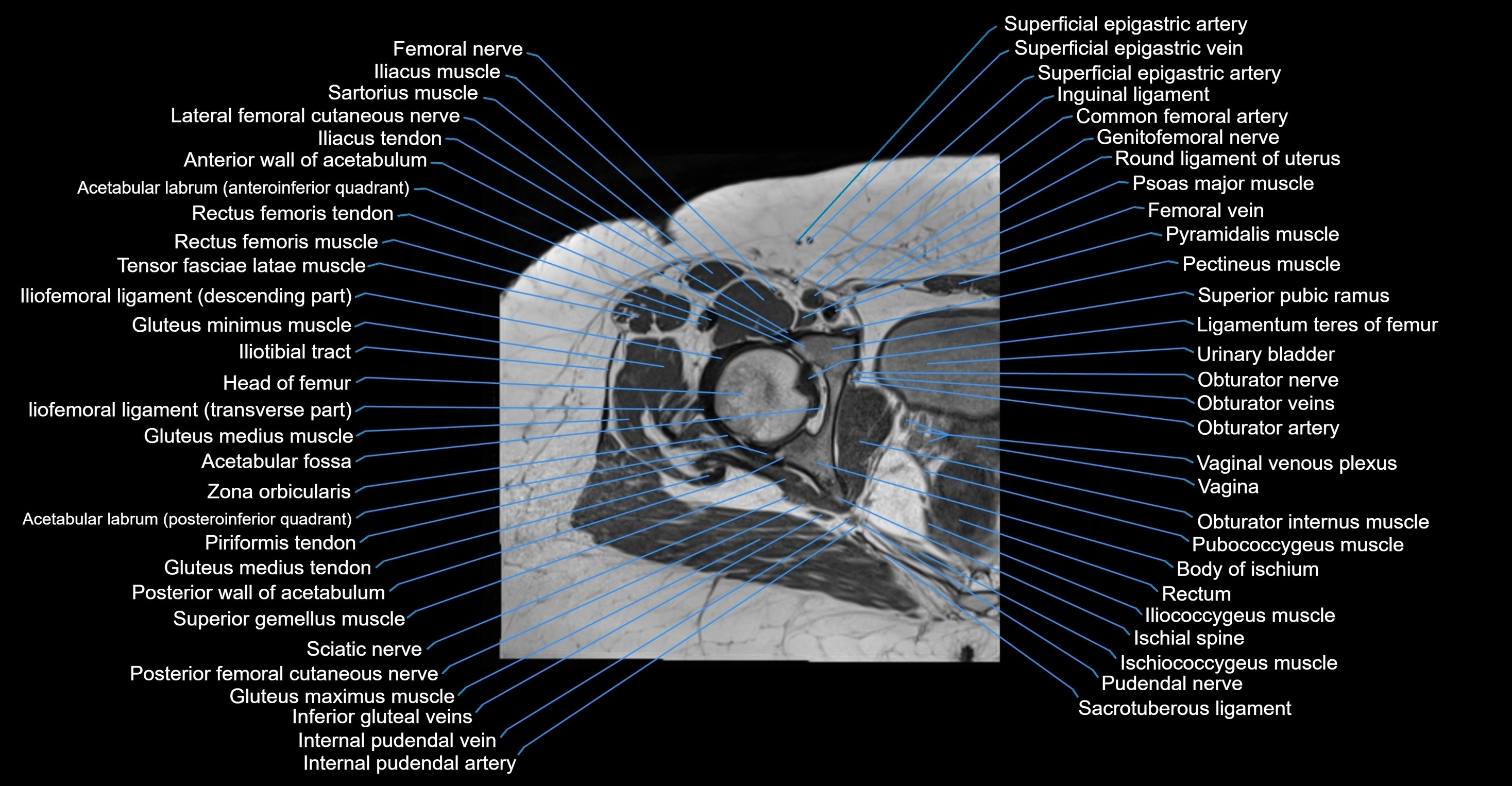
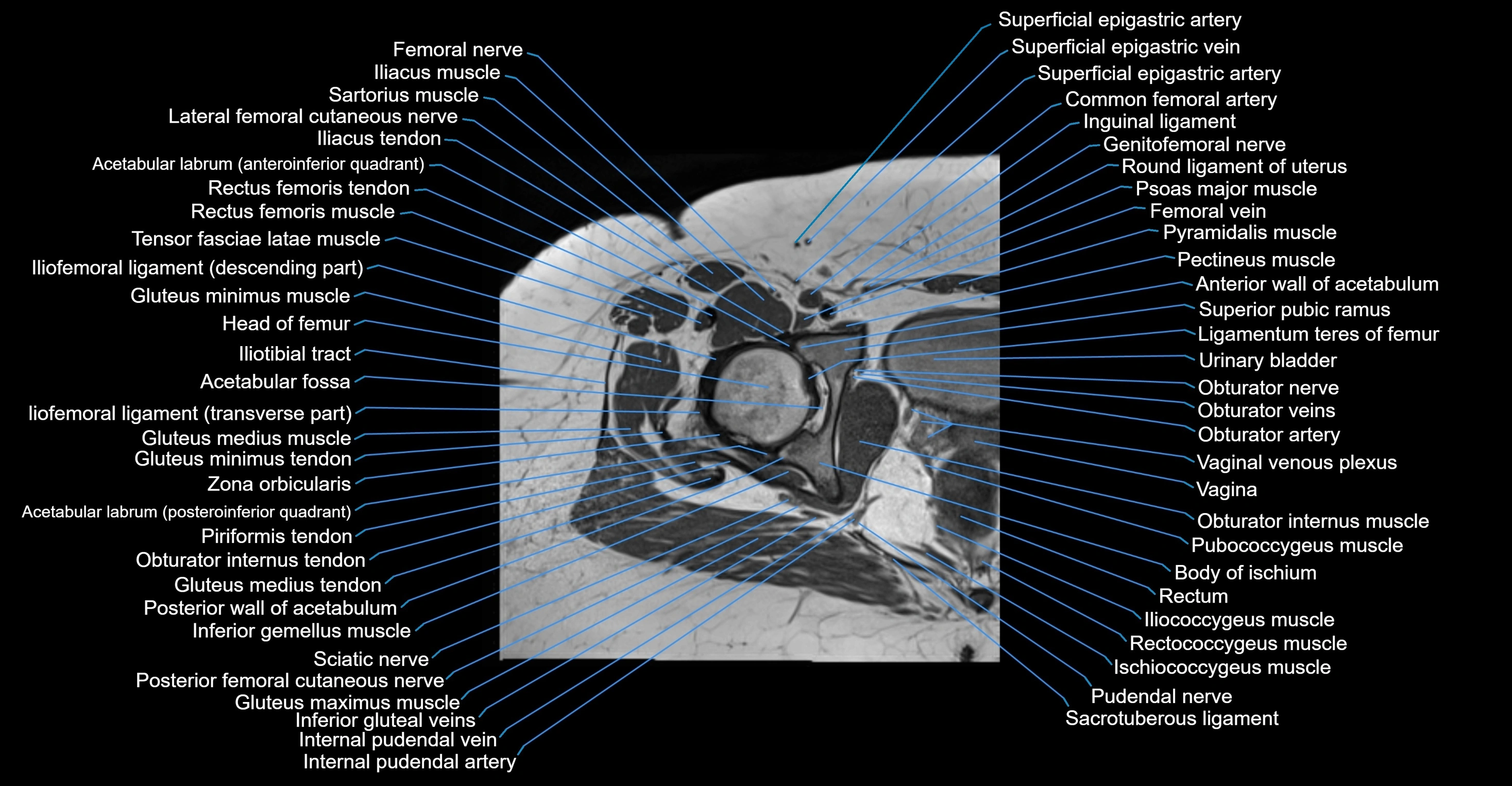
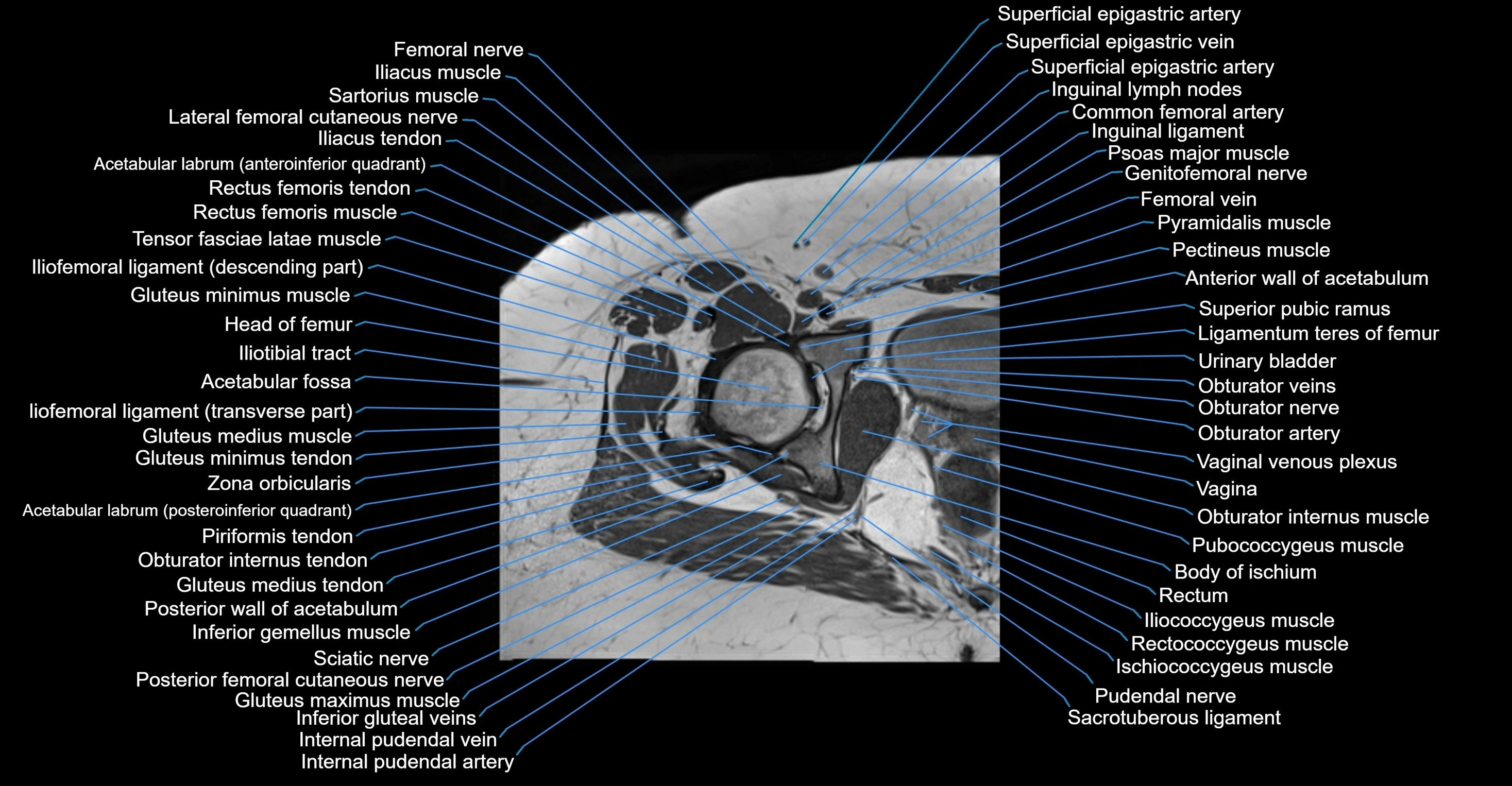

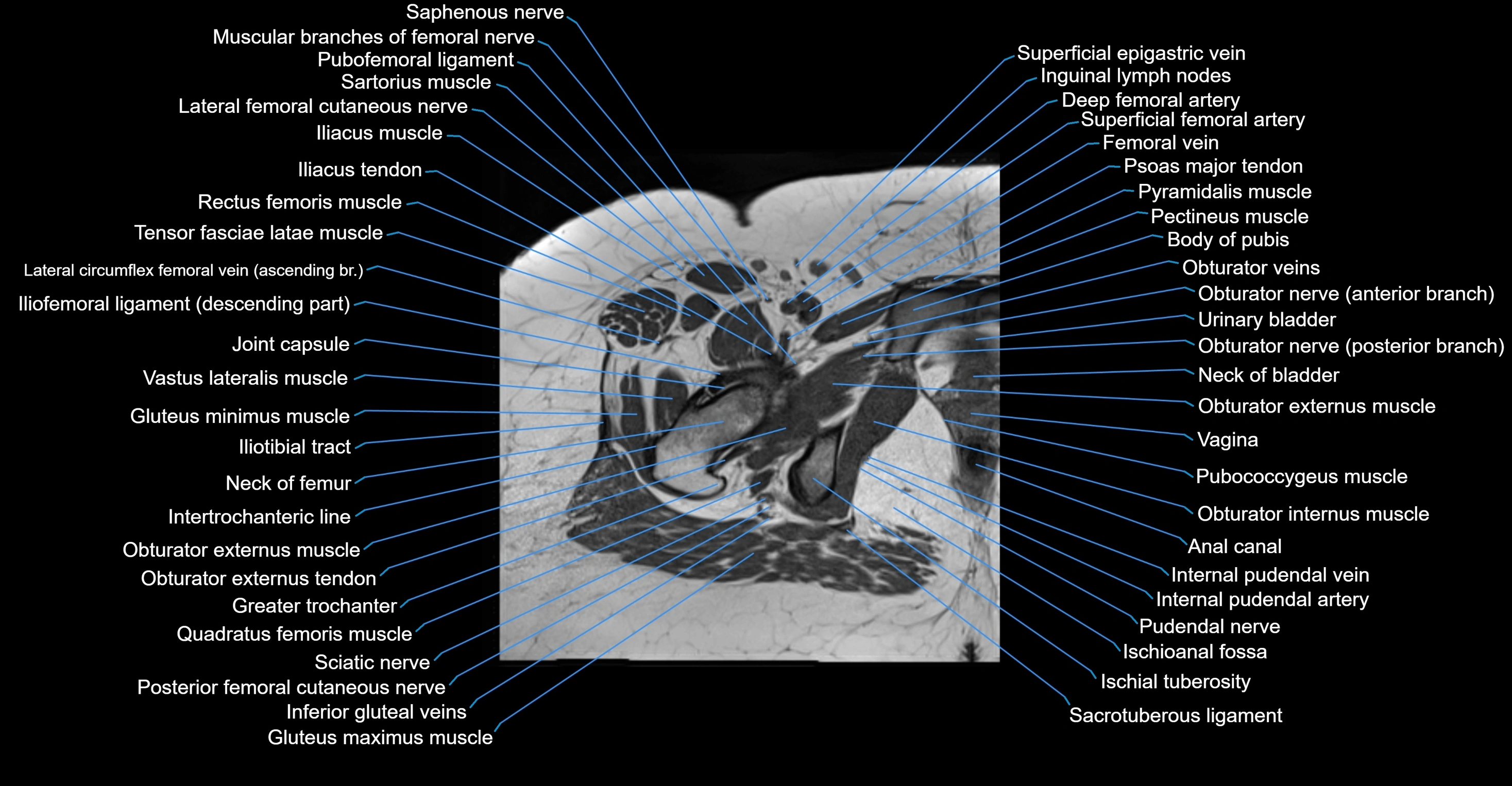
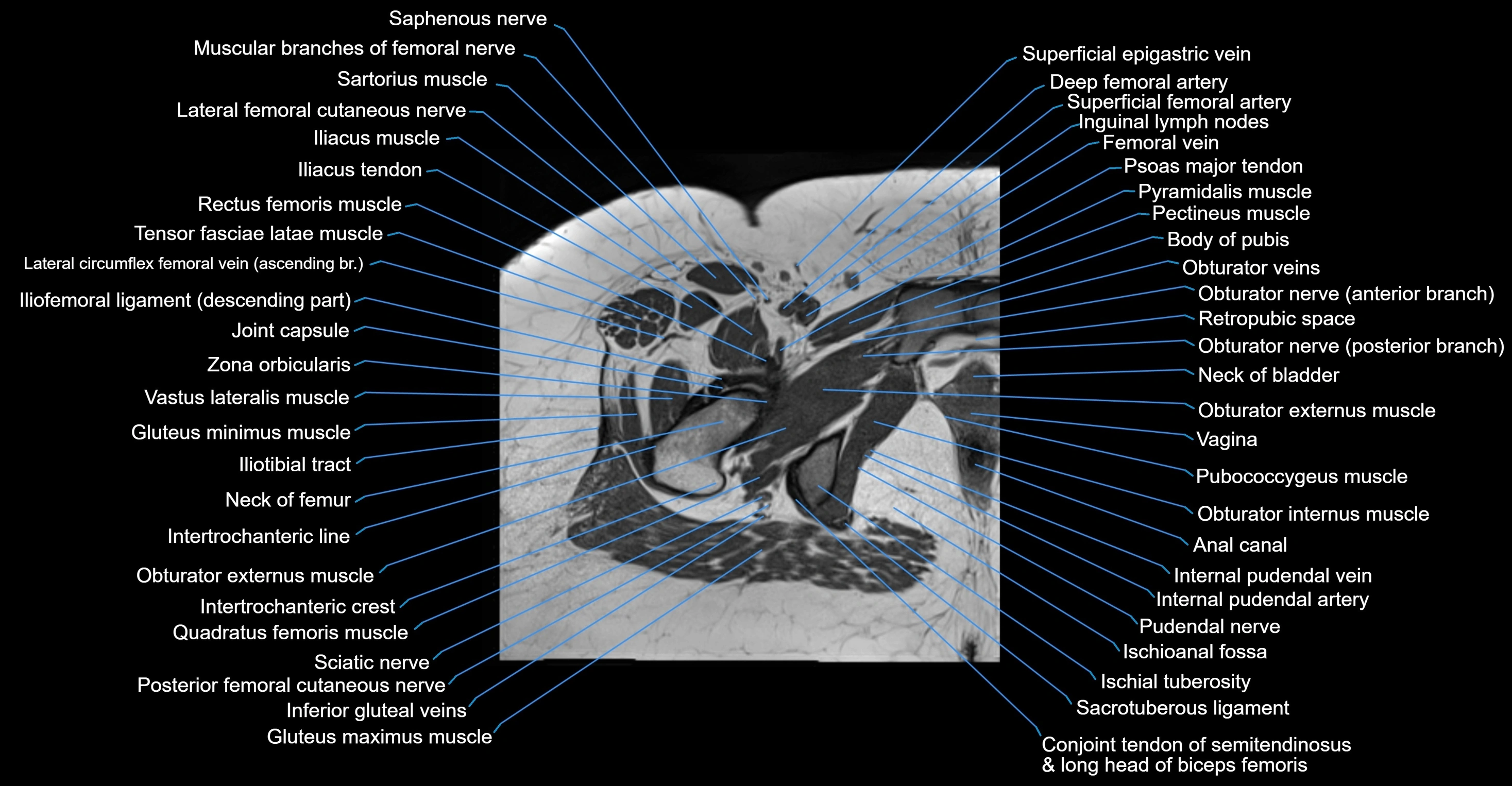
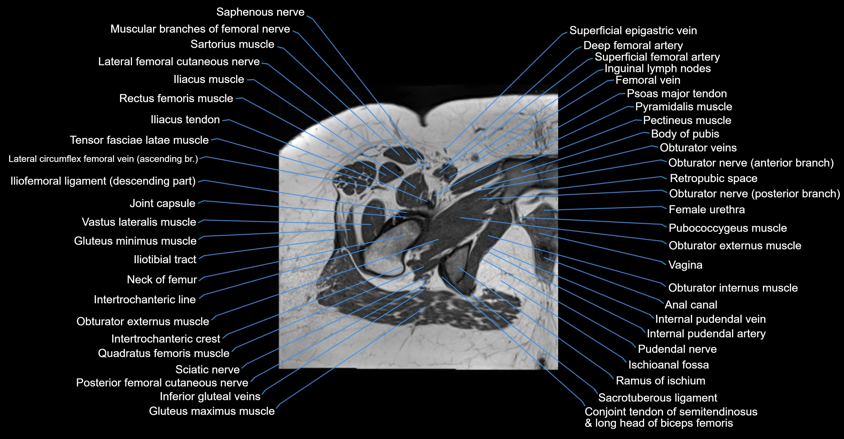
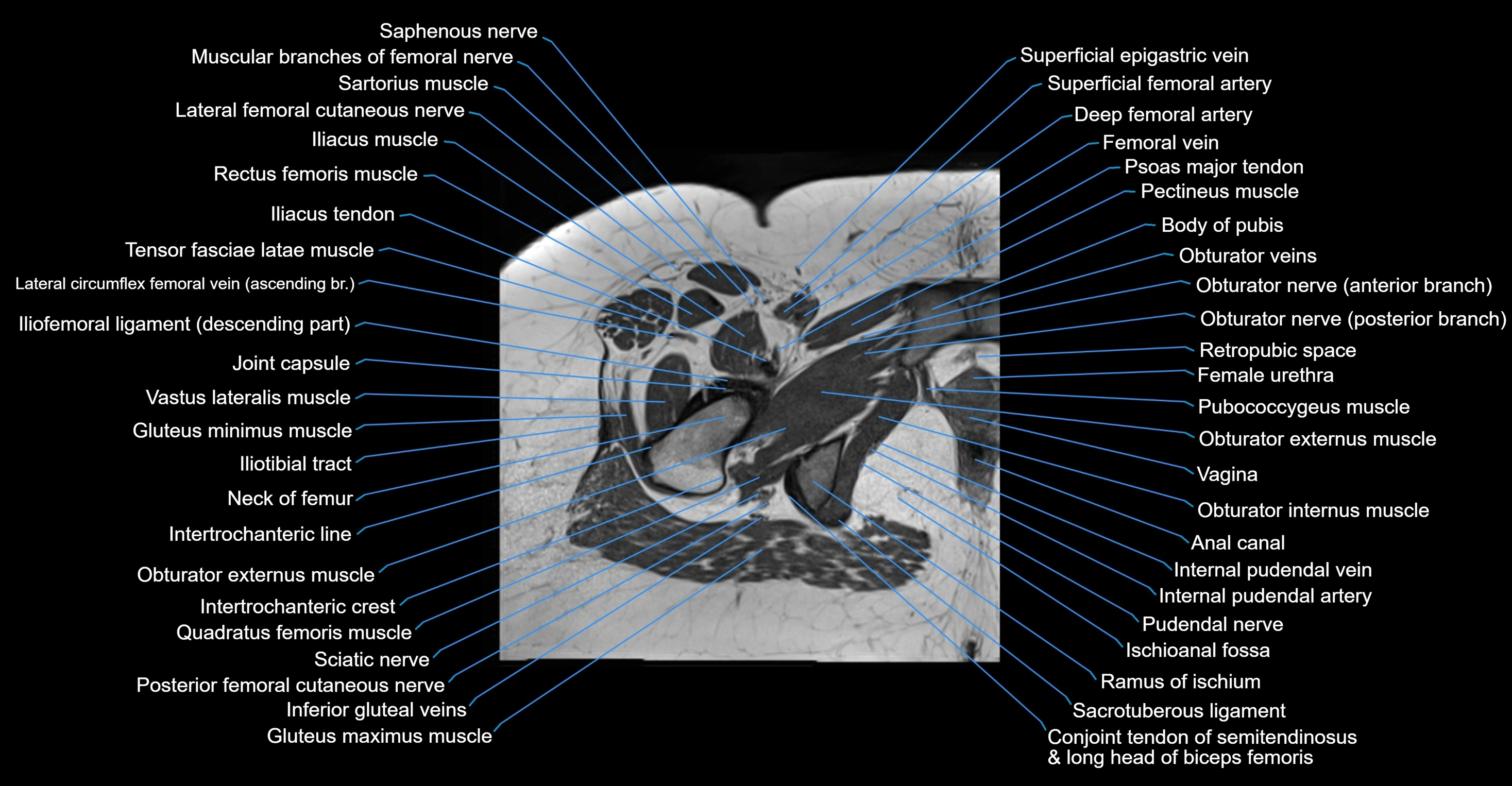

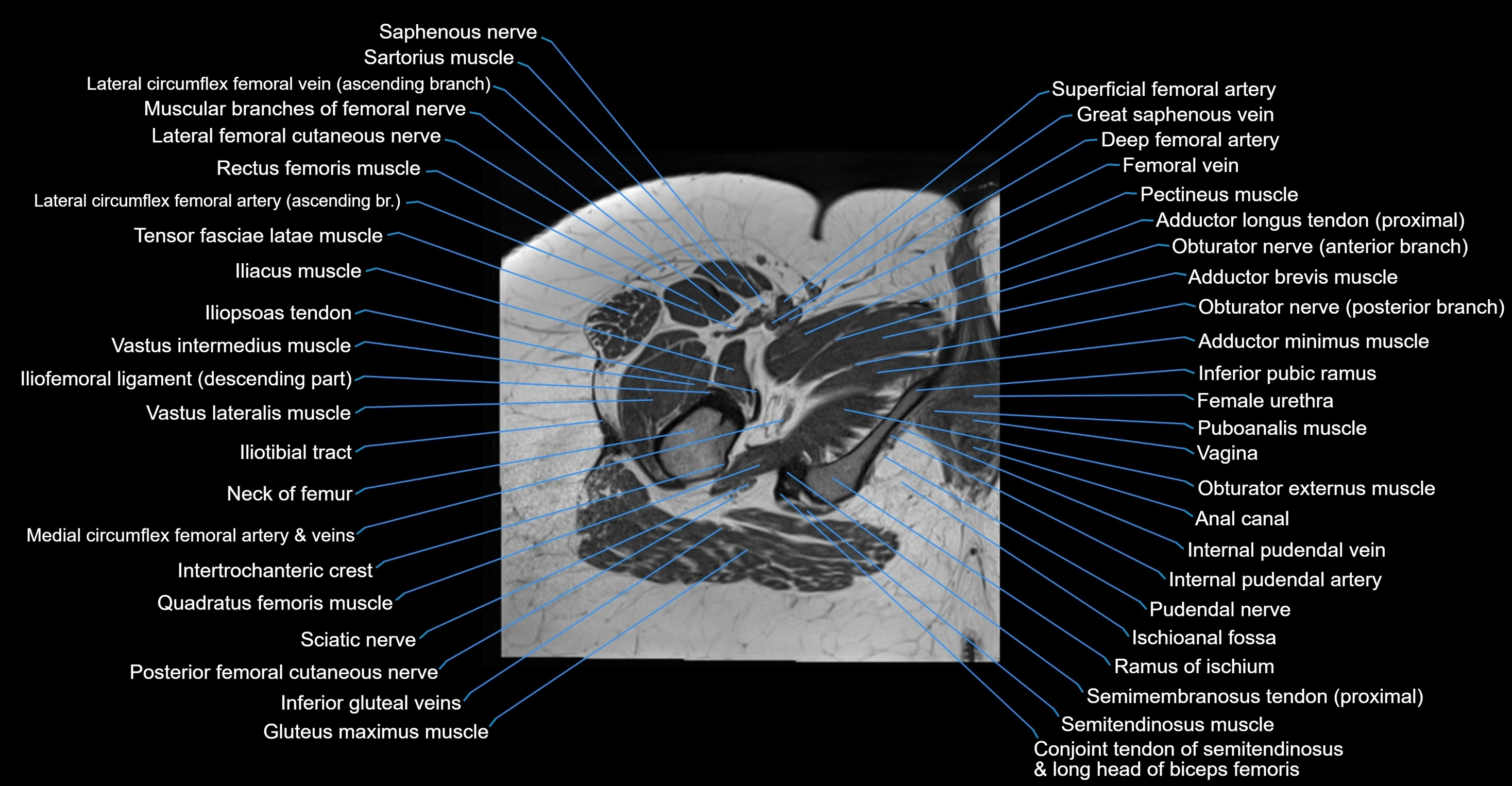
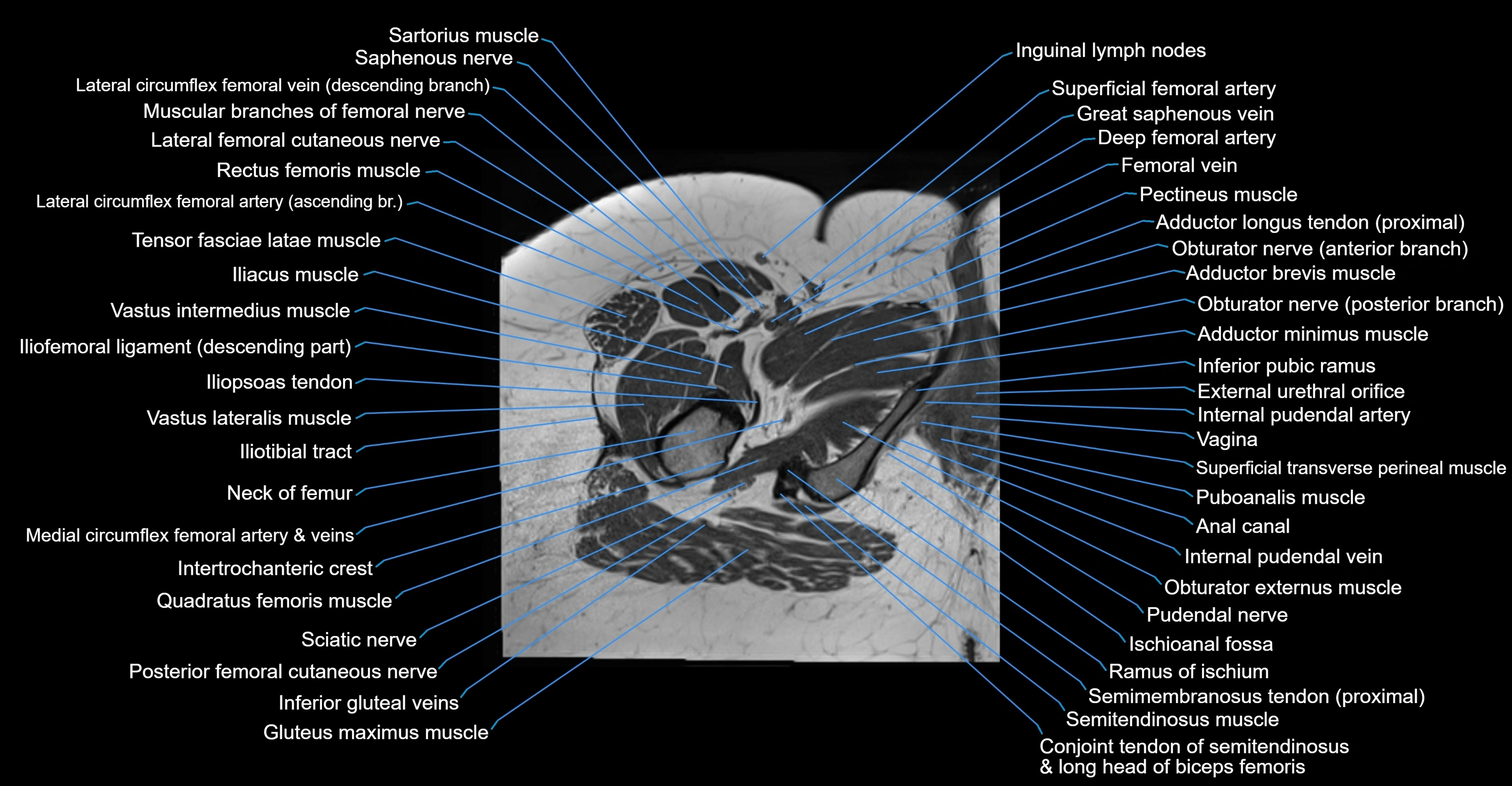
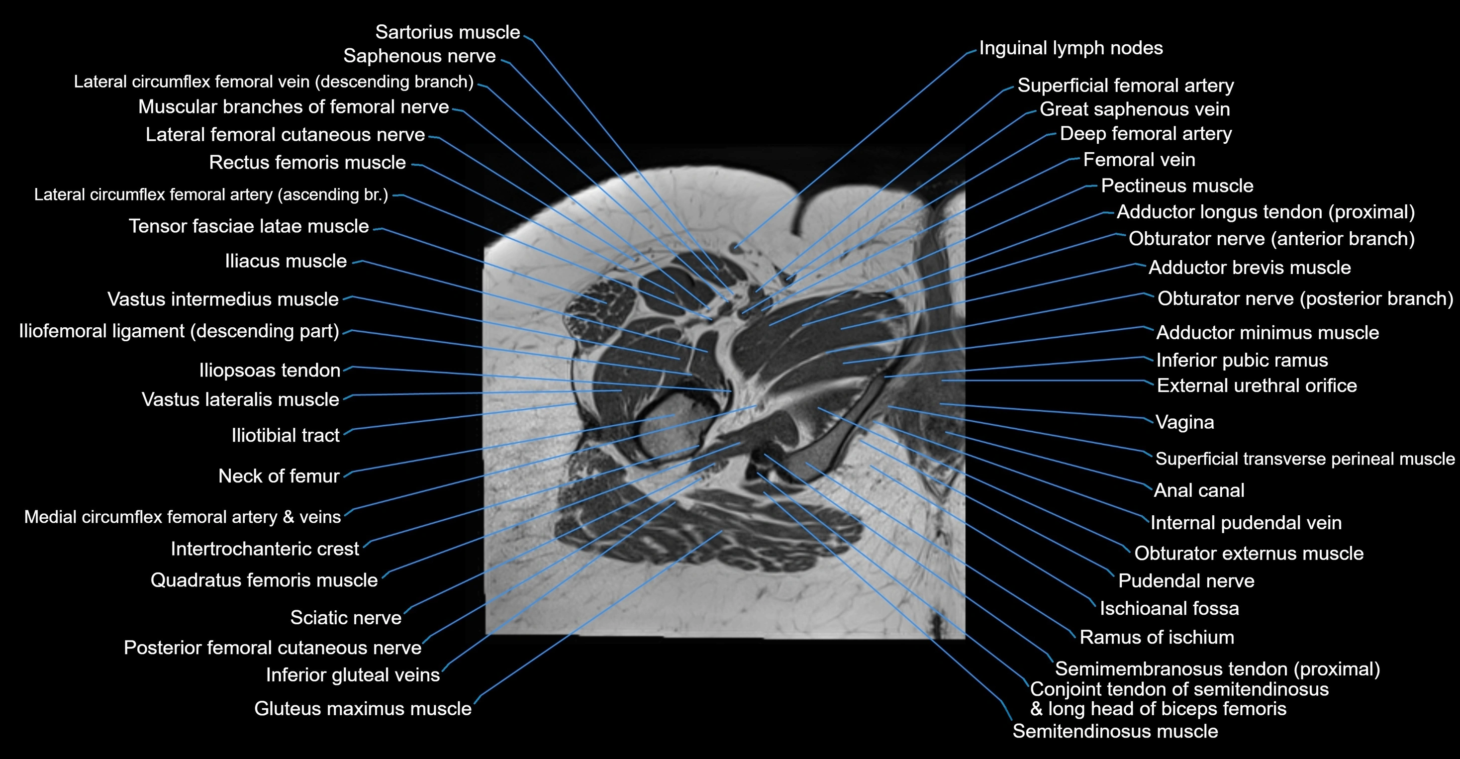
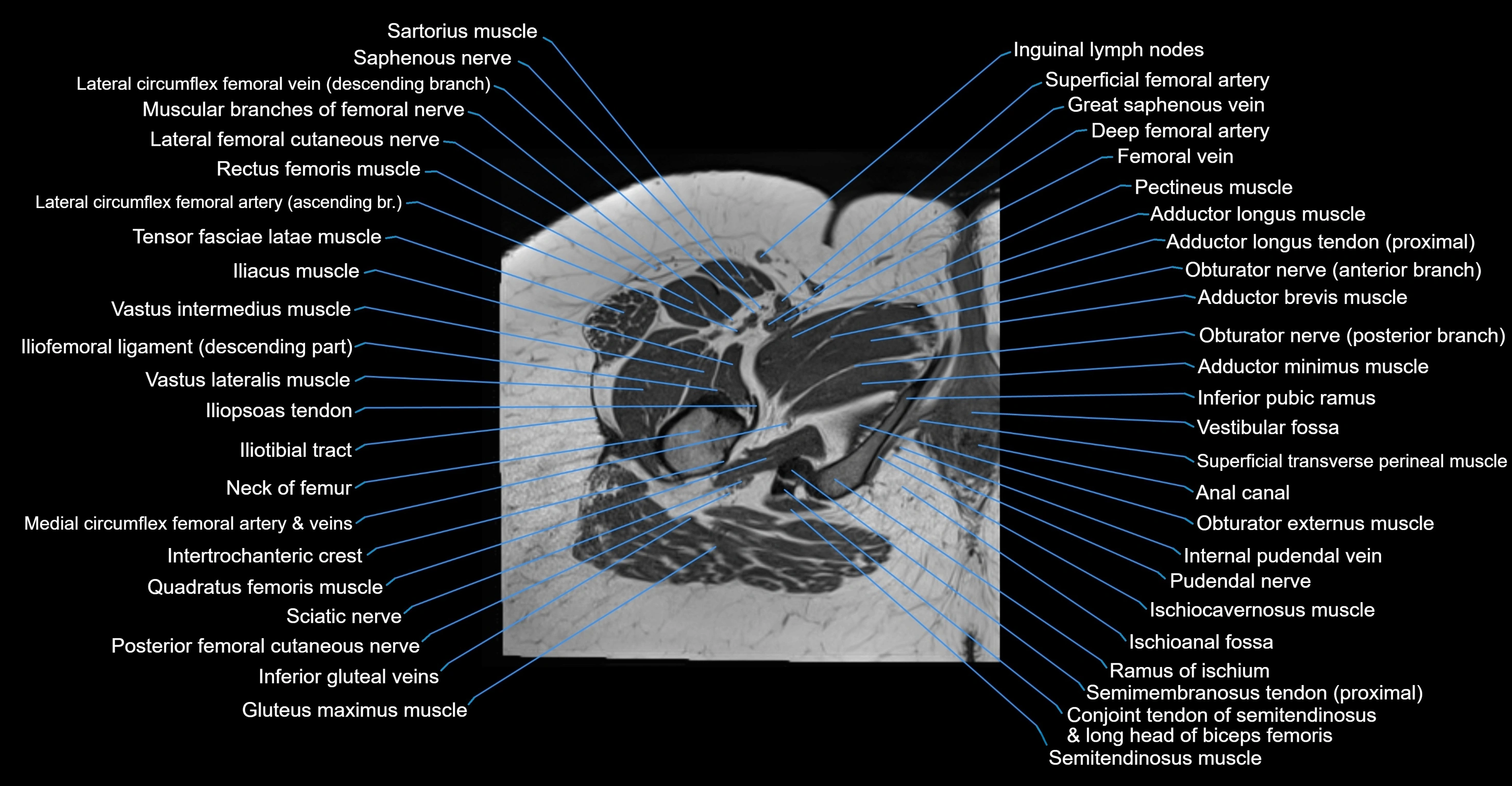


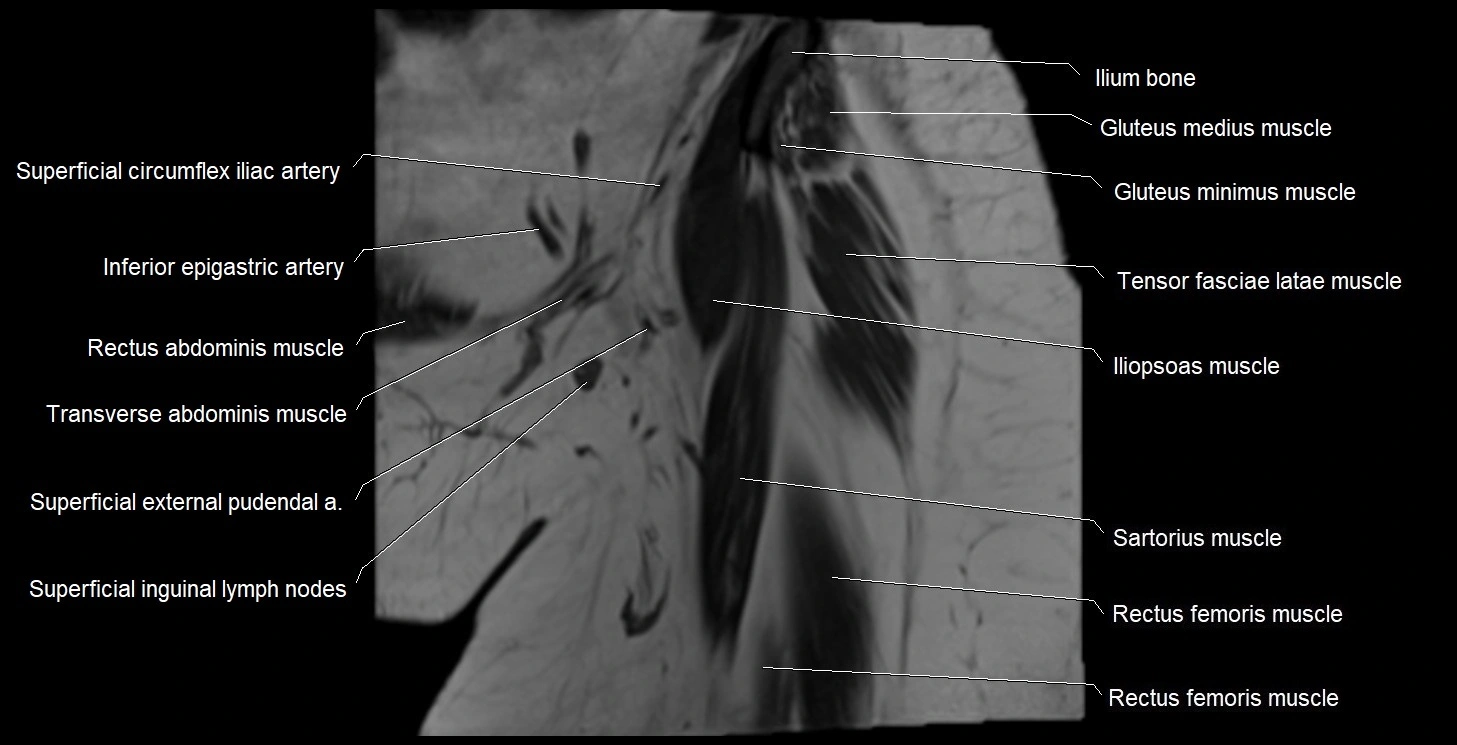
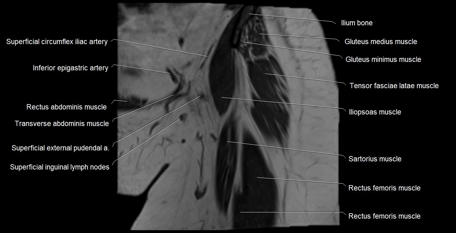
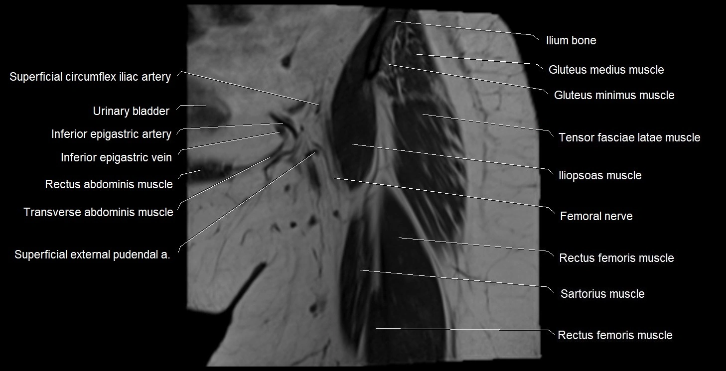
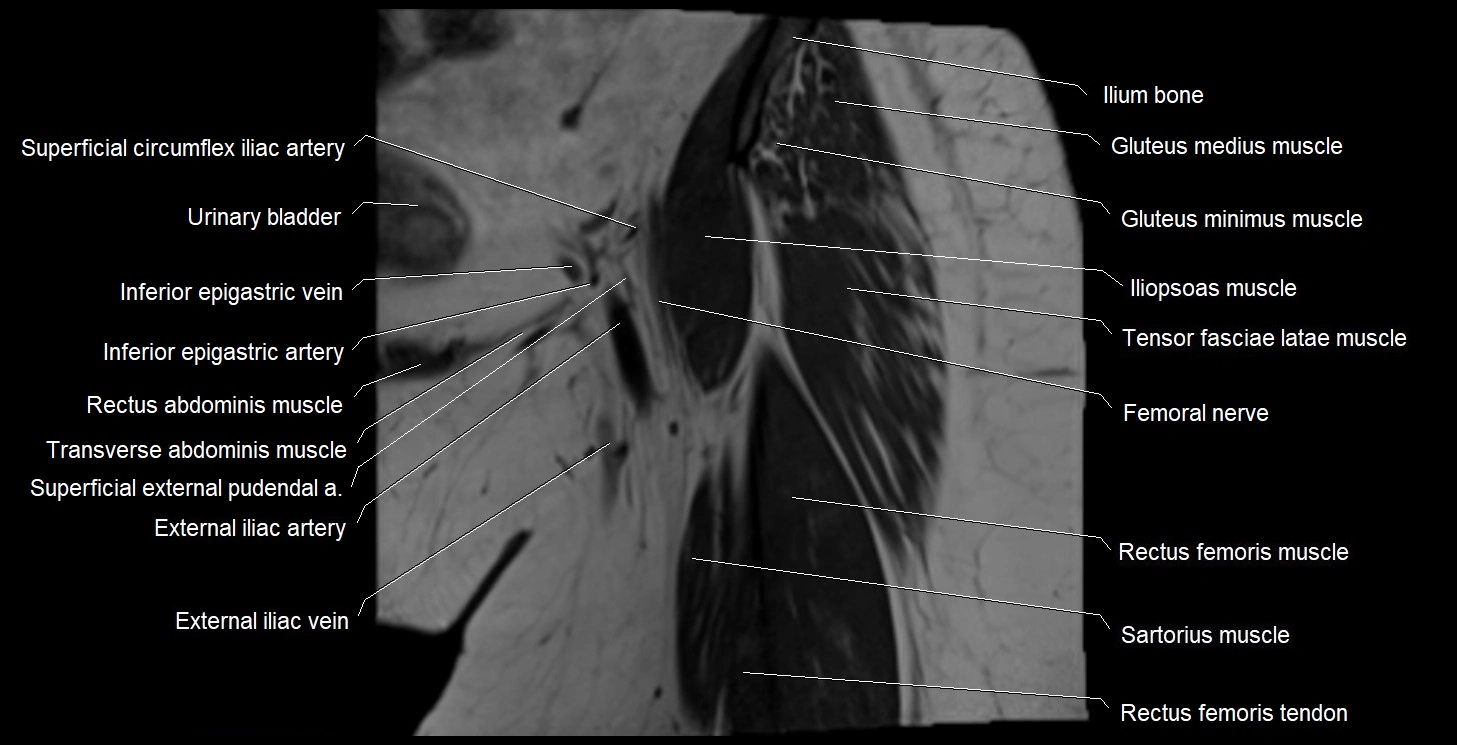
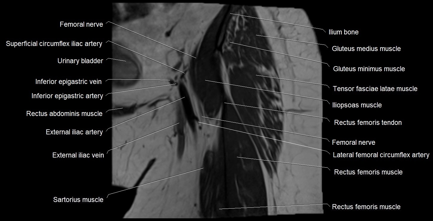
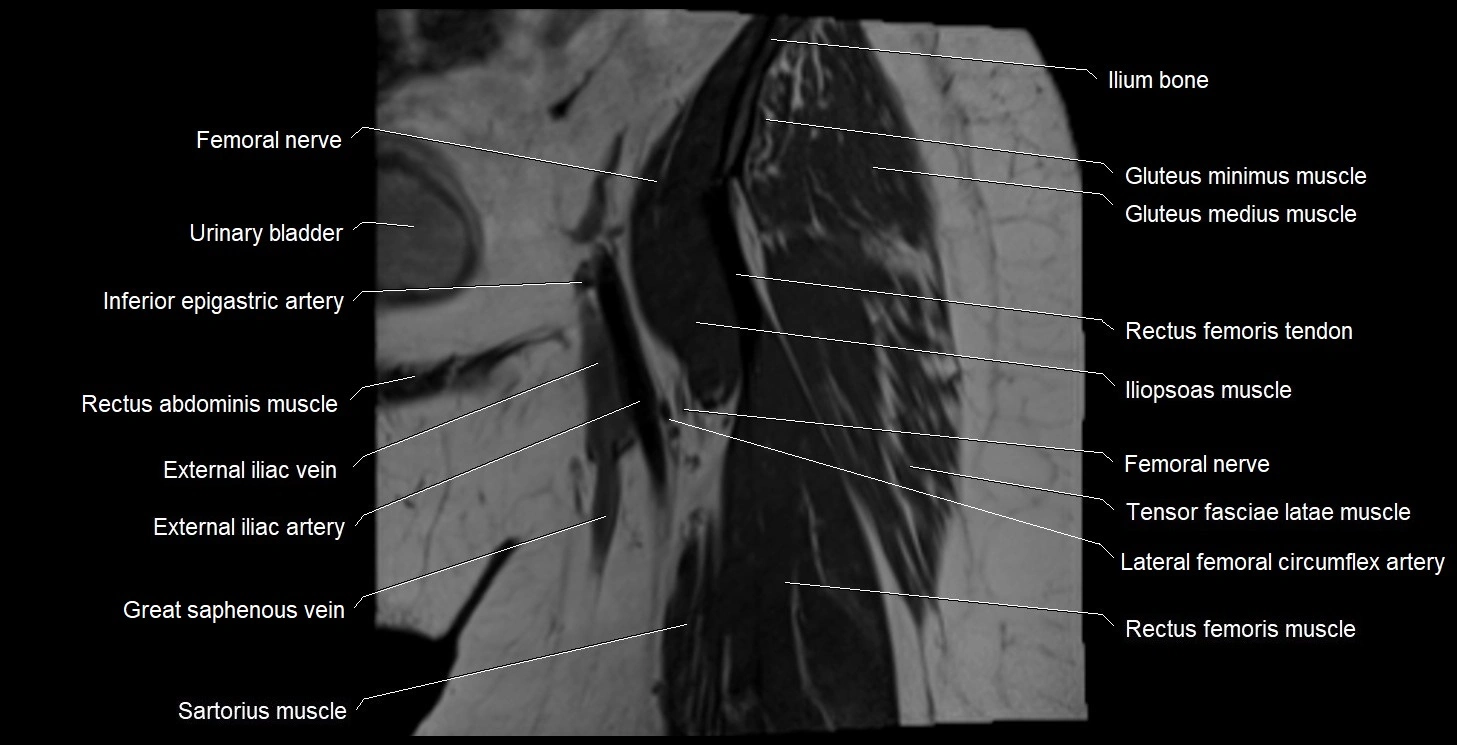
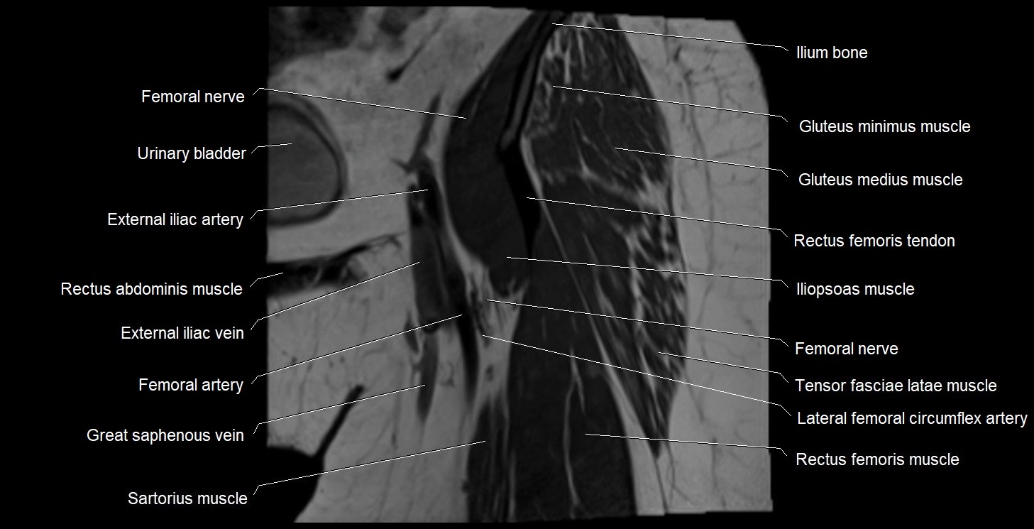
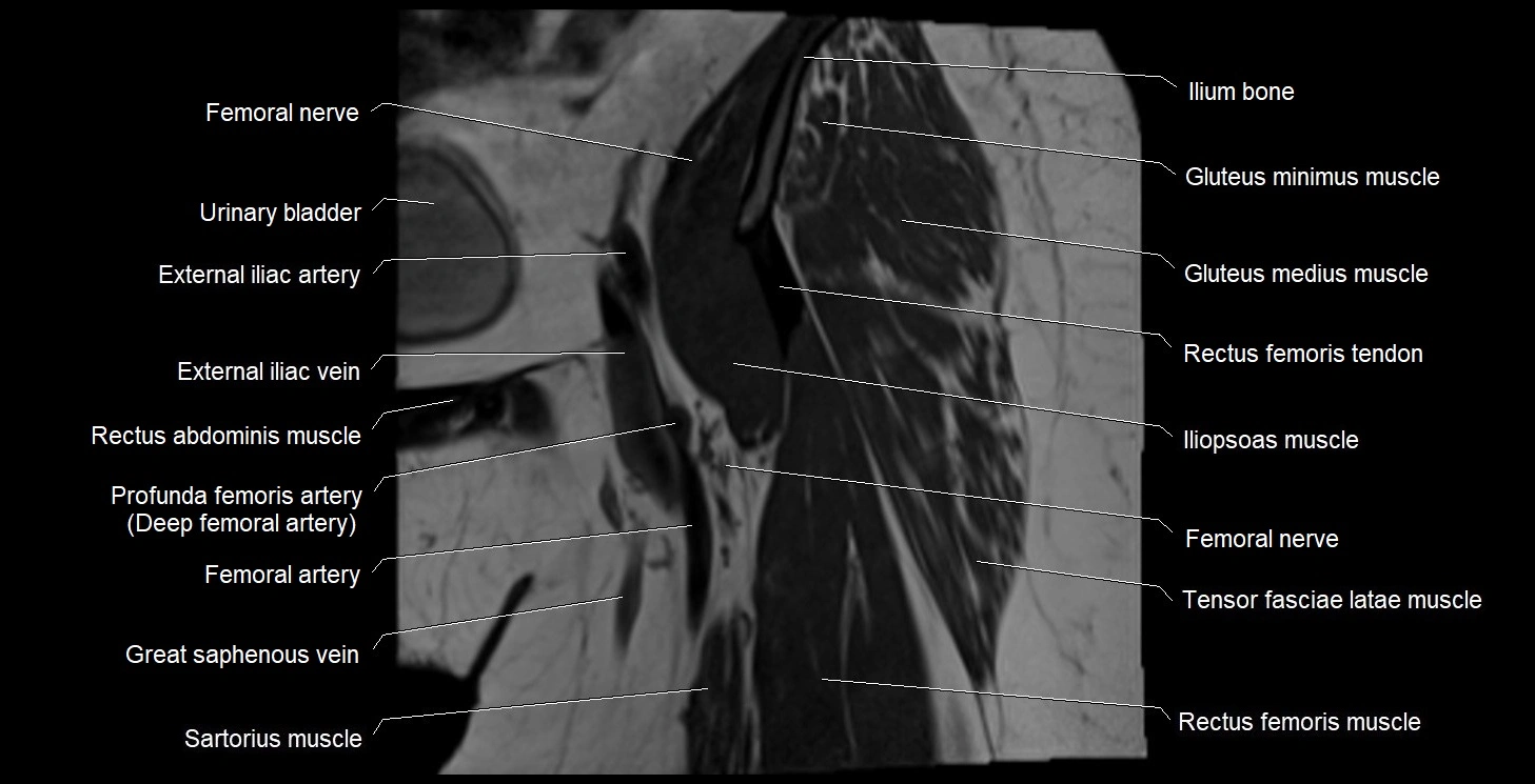
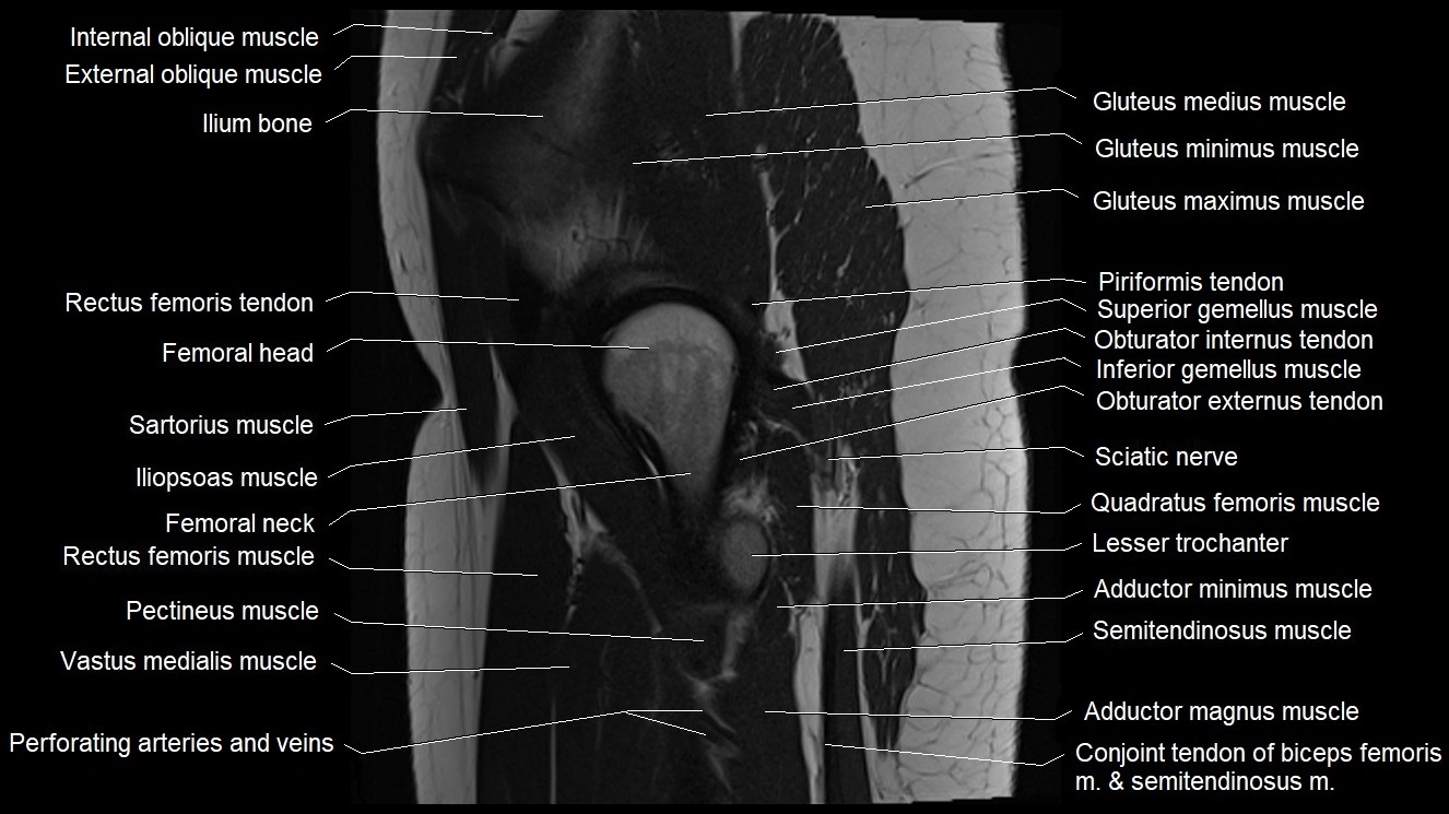
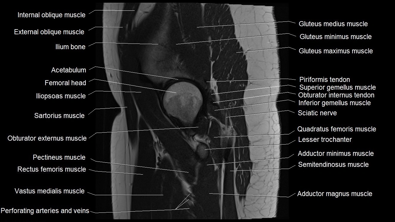
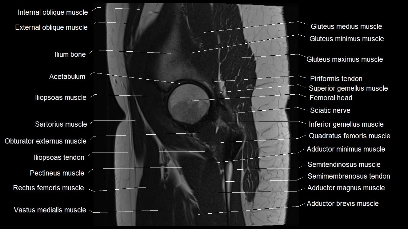
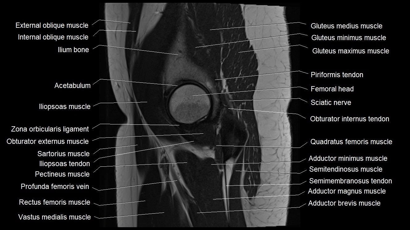
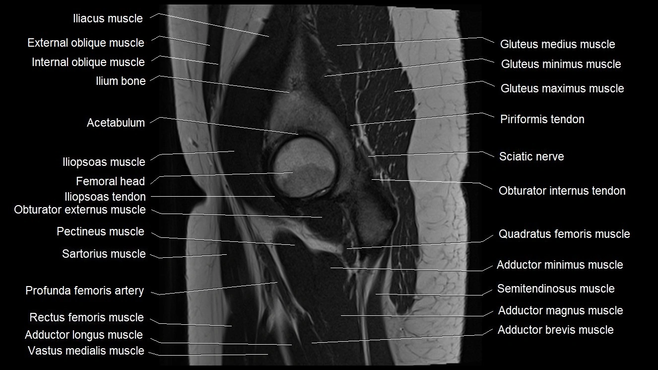
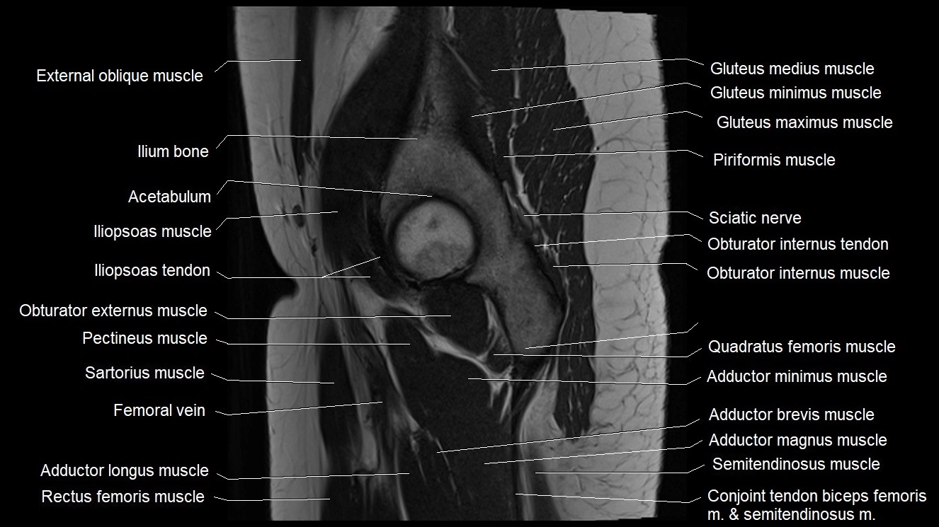
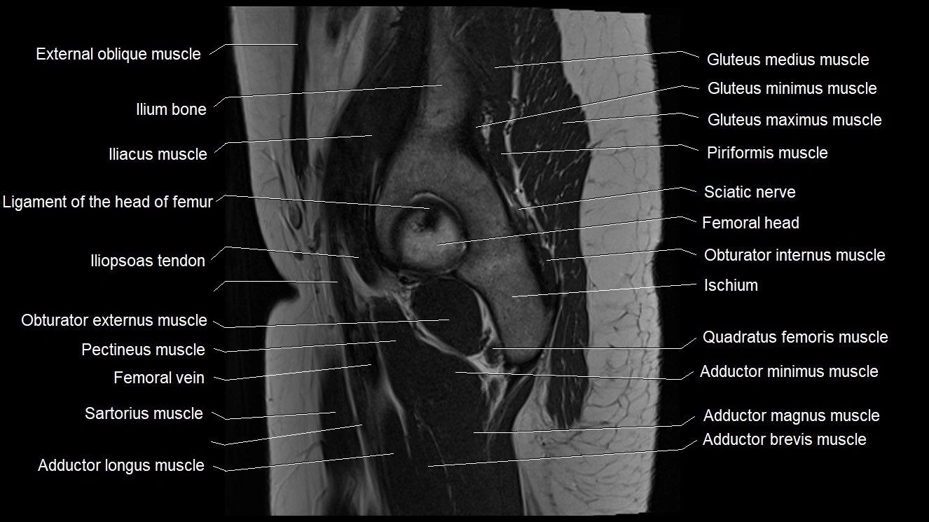
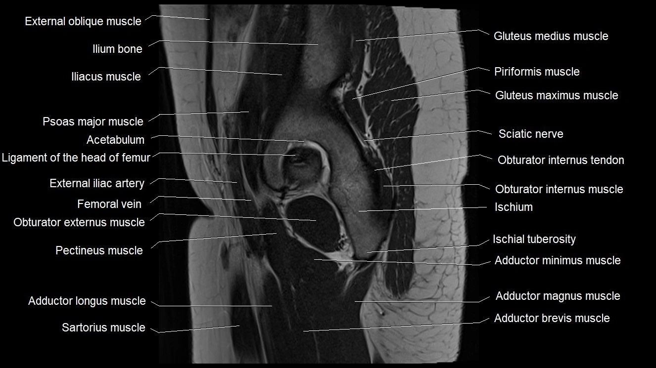
Topic
- Accessory obturator artery
- Accessory obturator vein
- Accessory saphenous vein
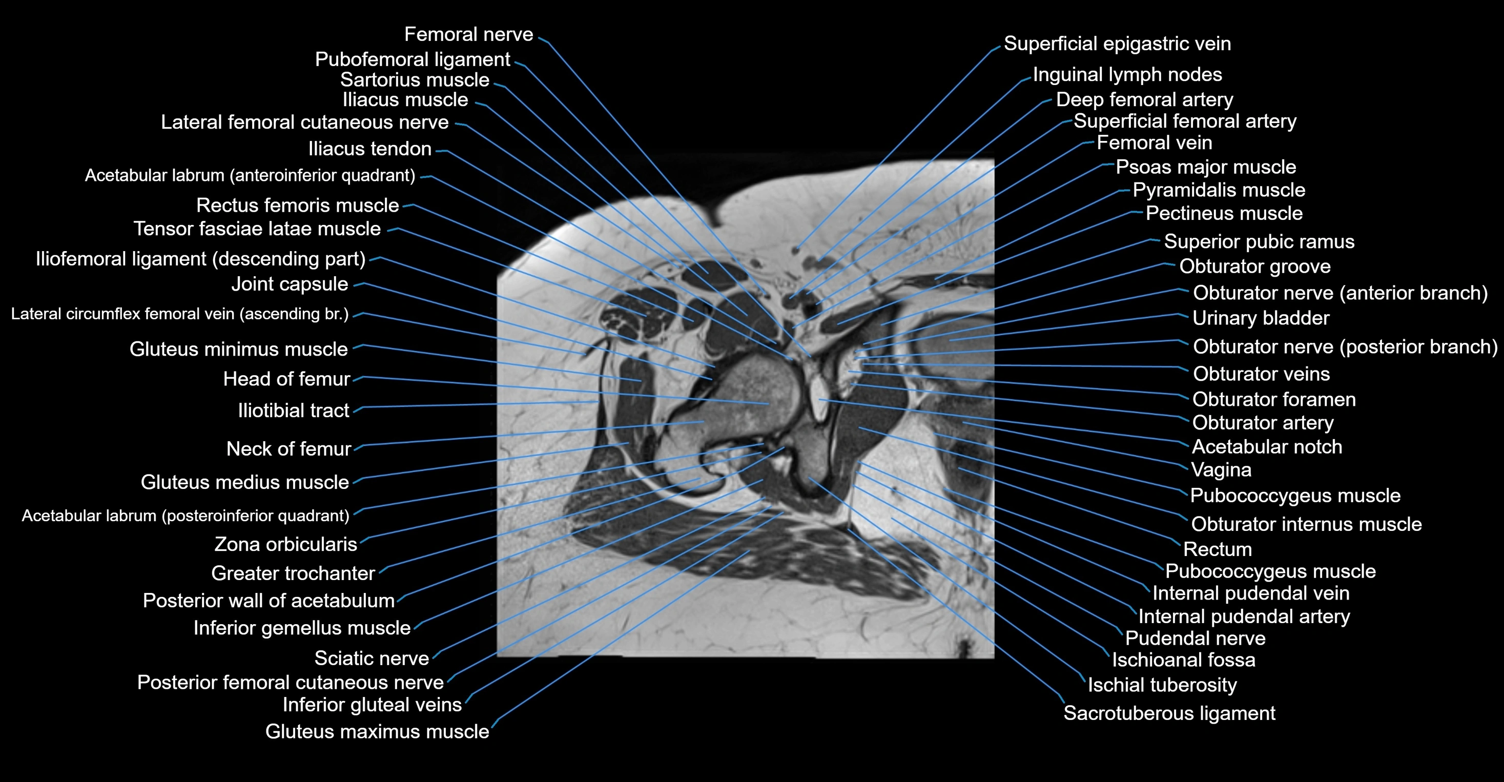
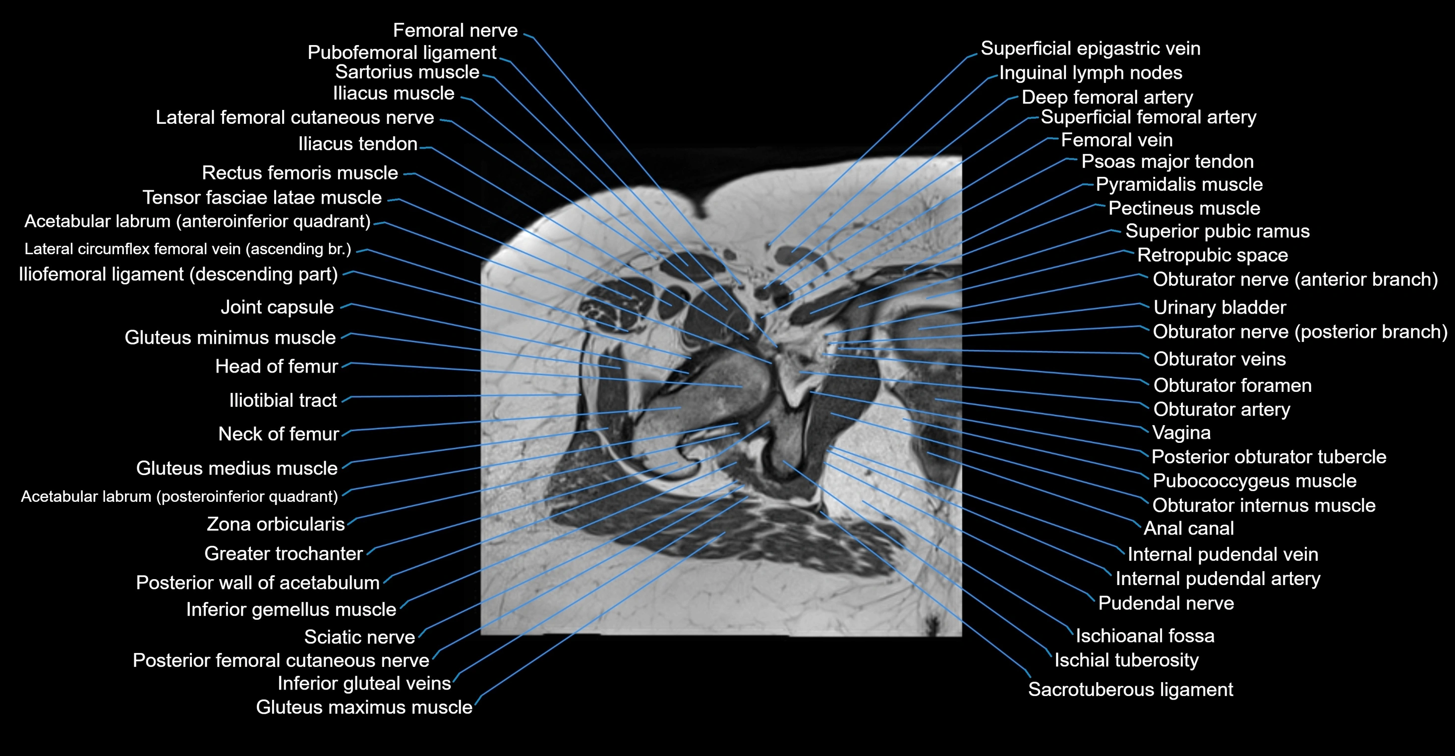
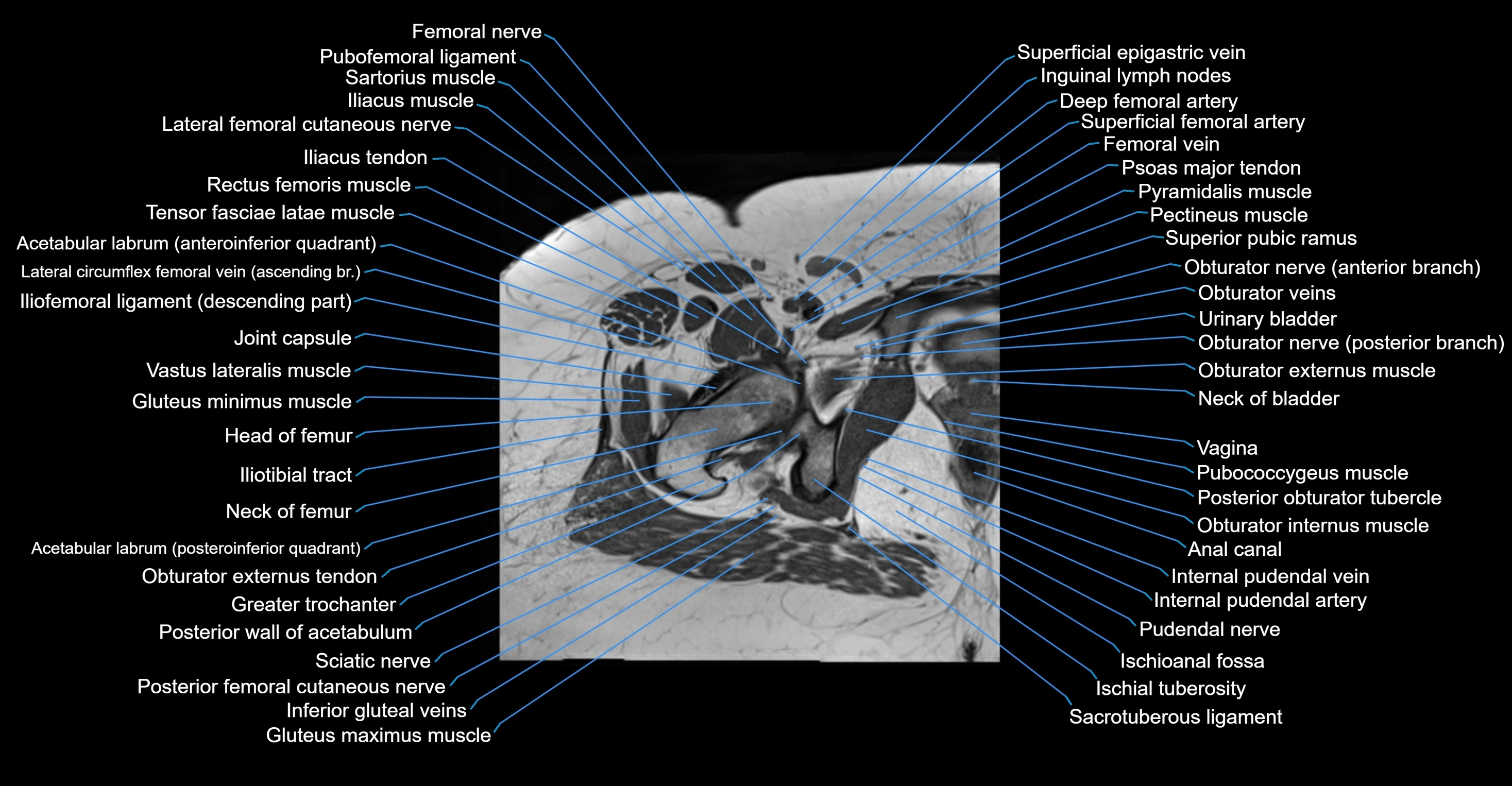
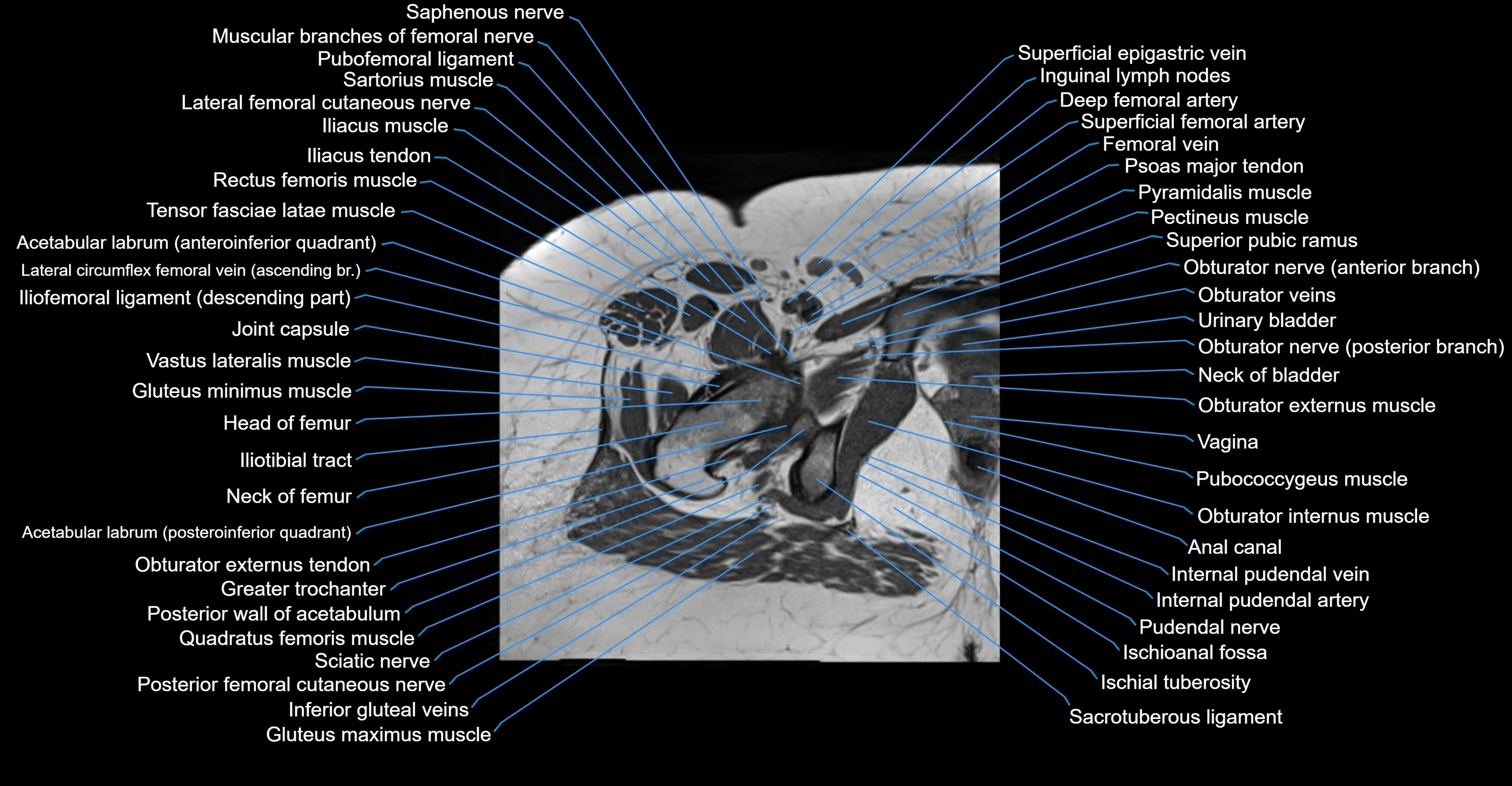
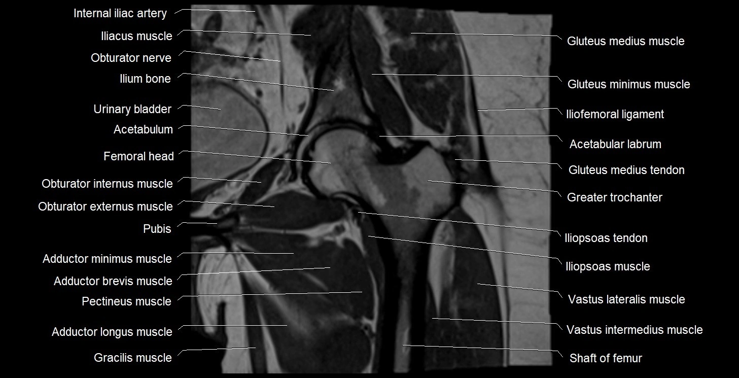
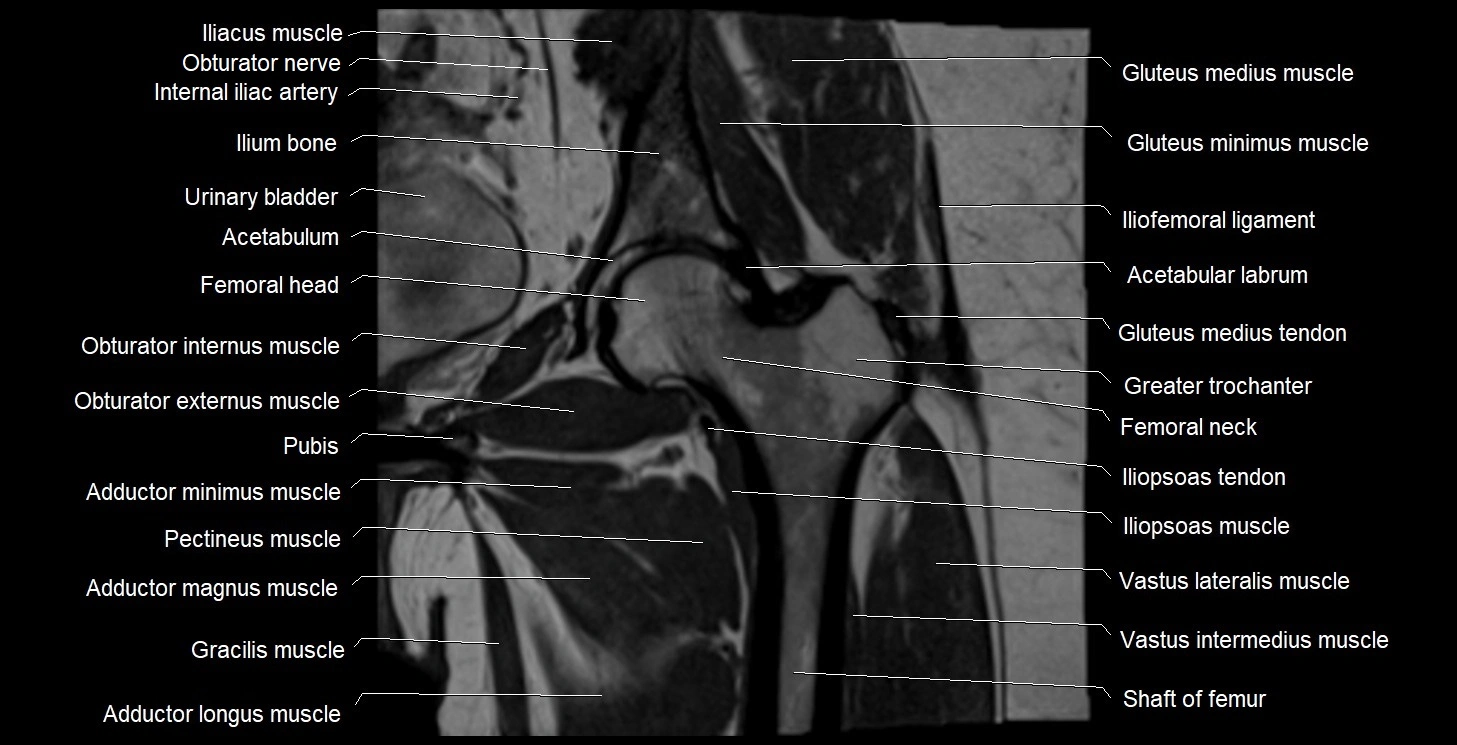
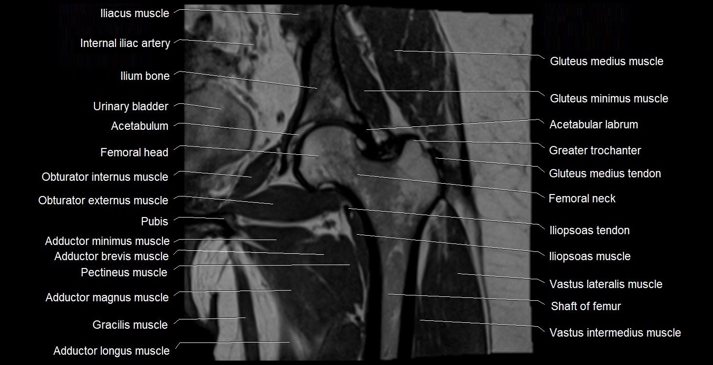
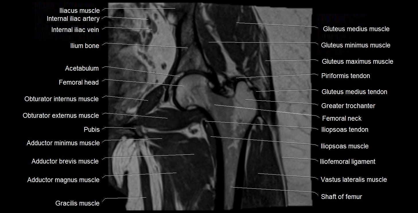
- Acetabular labrum
- Acetabular margin (Acetabular rim)
- Acetabular notch
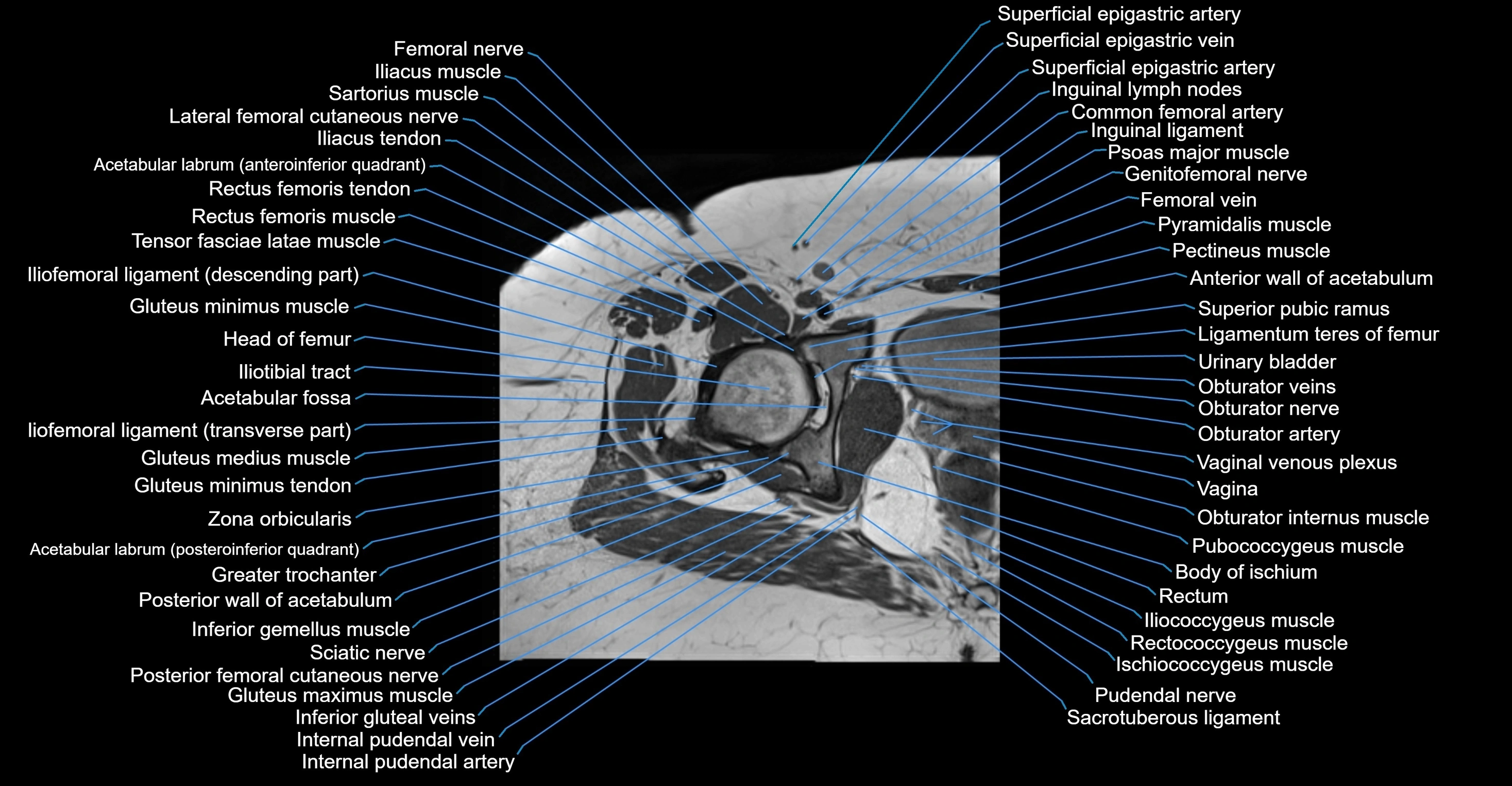
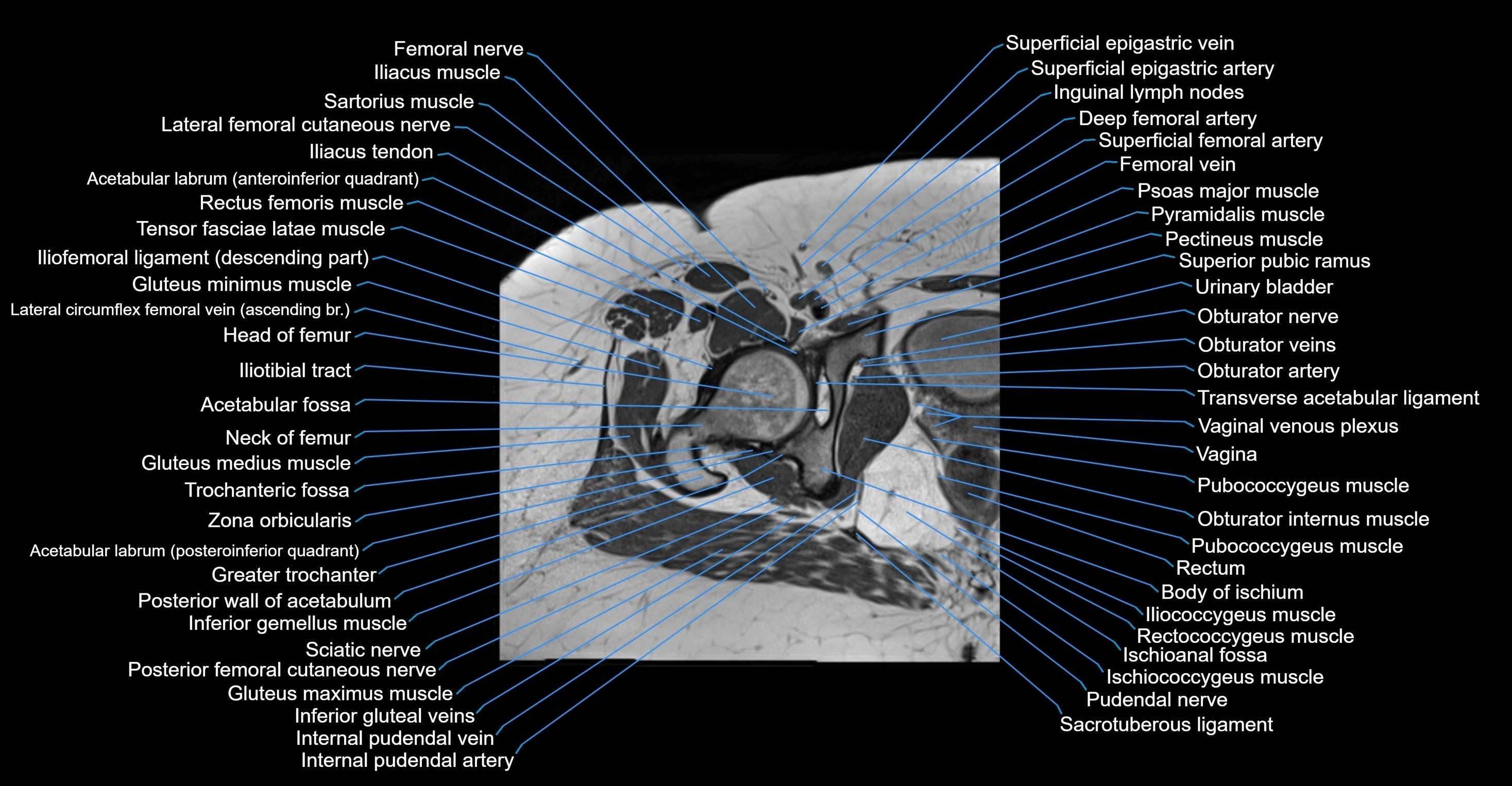
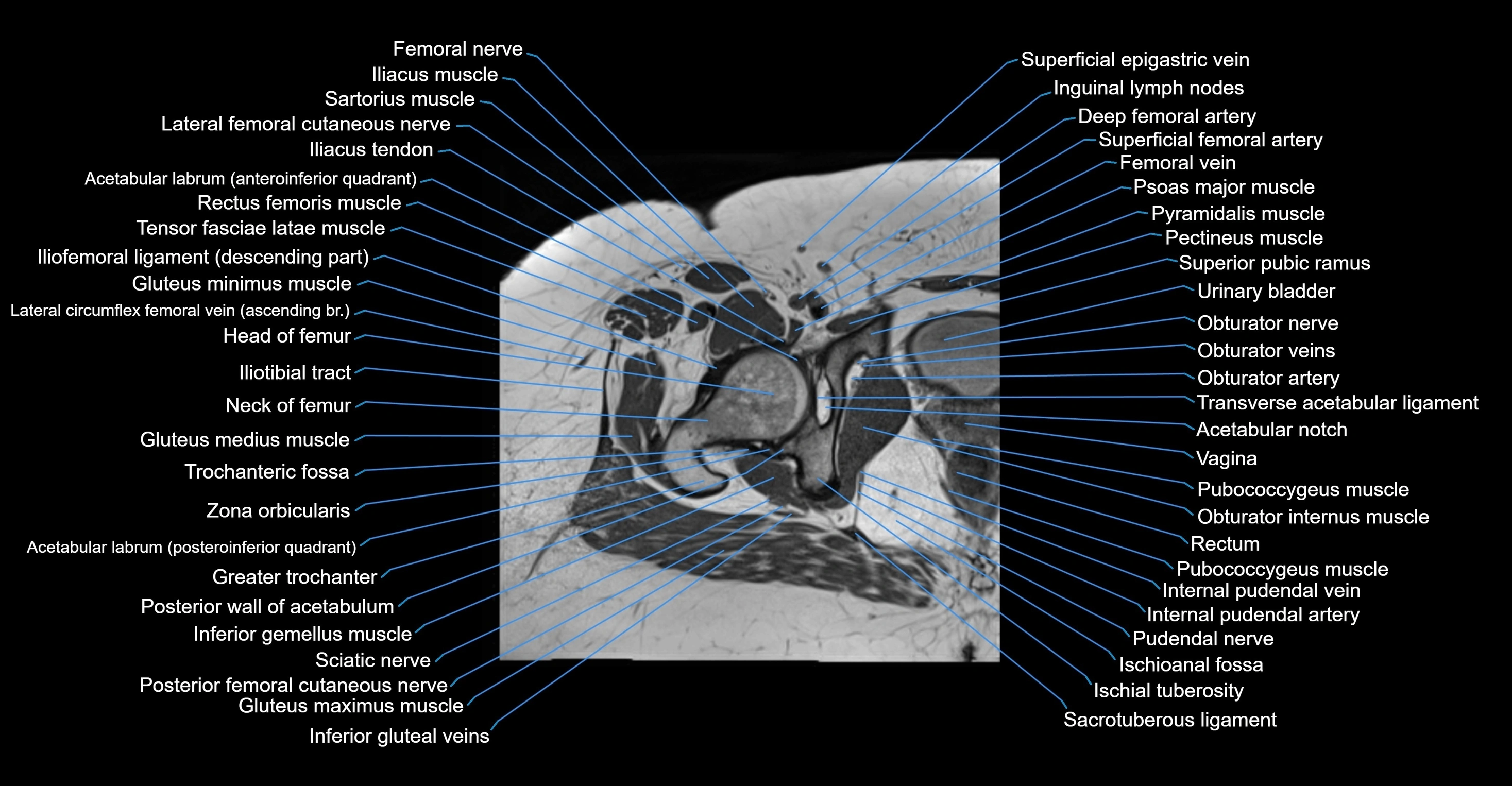
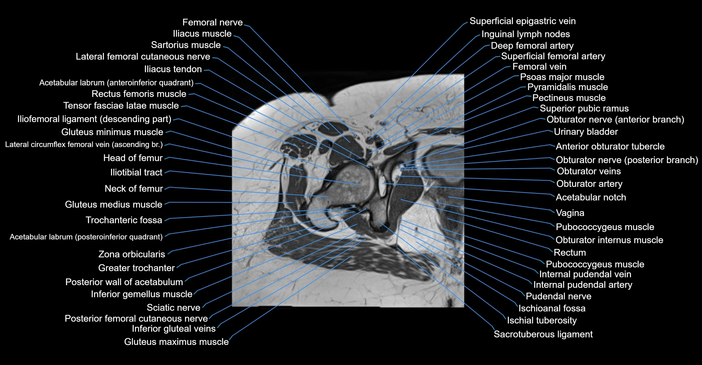
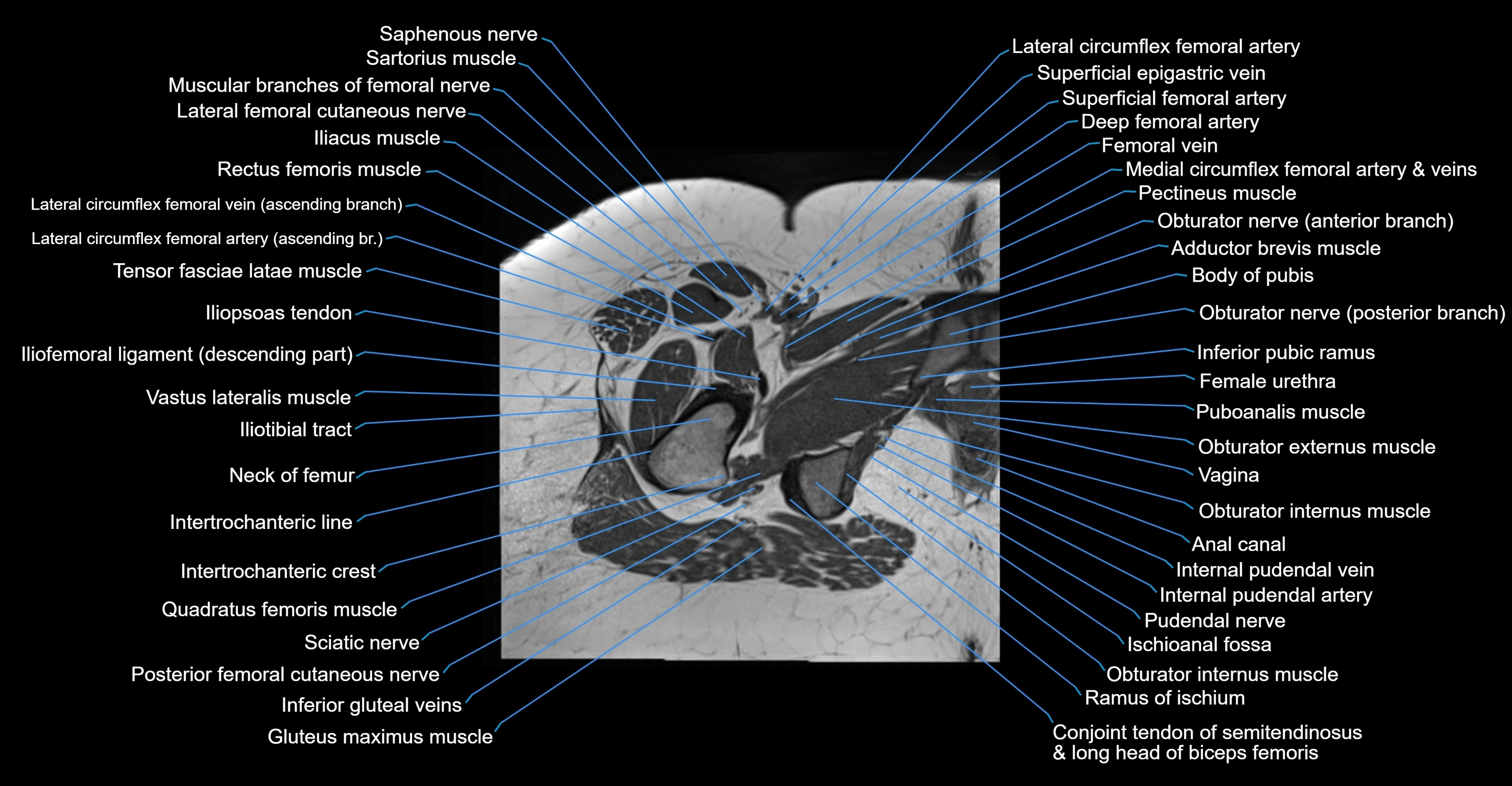
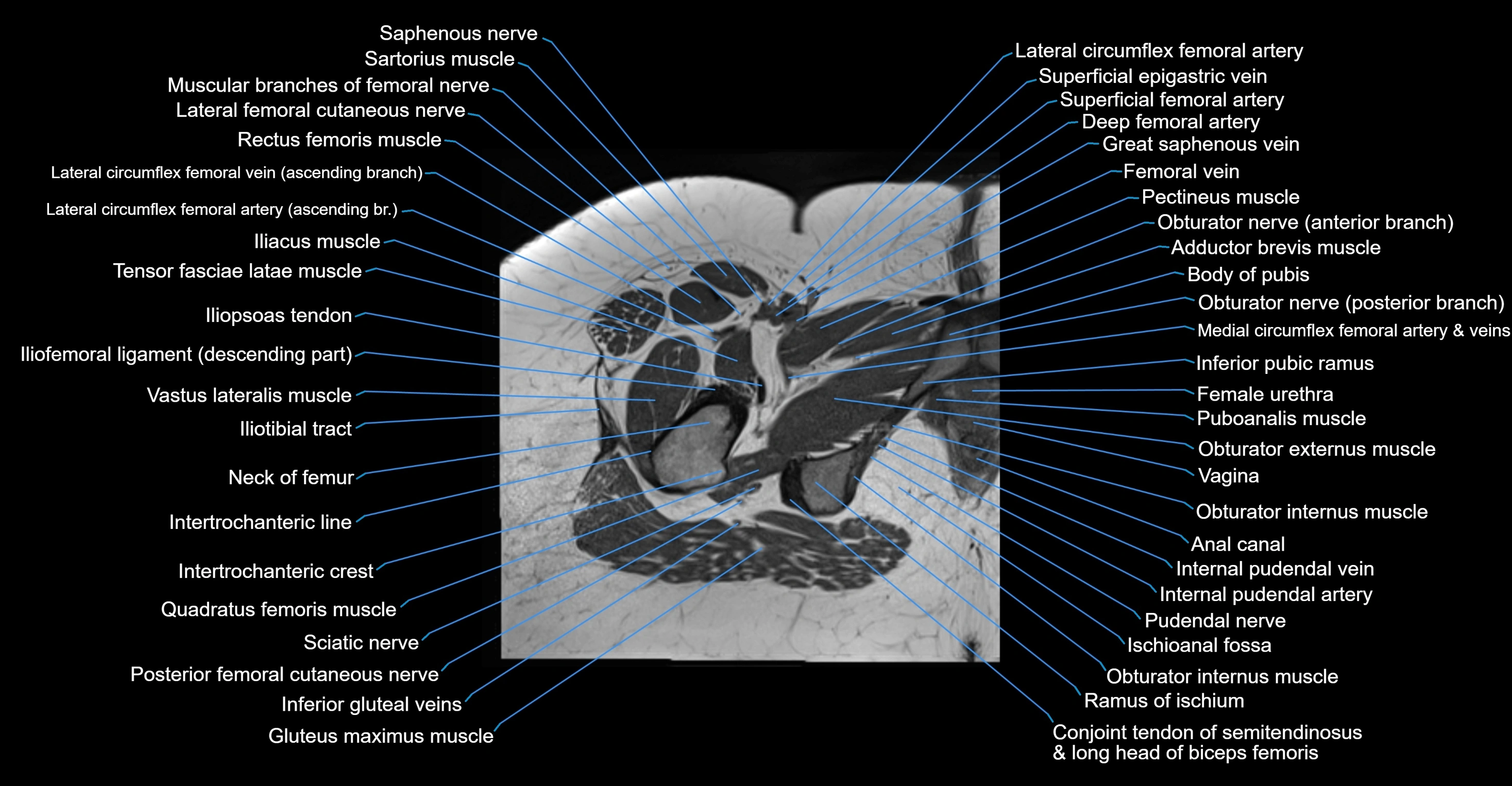
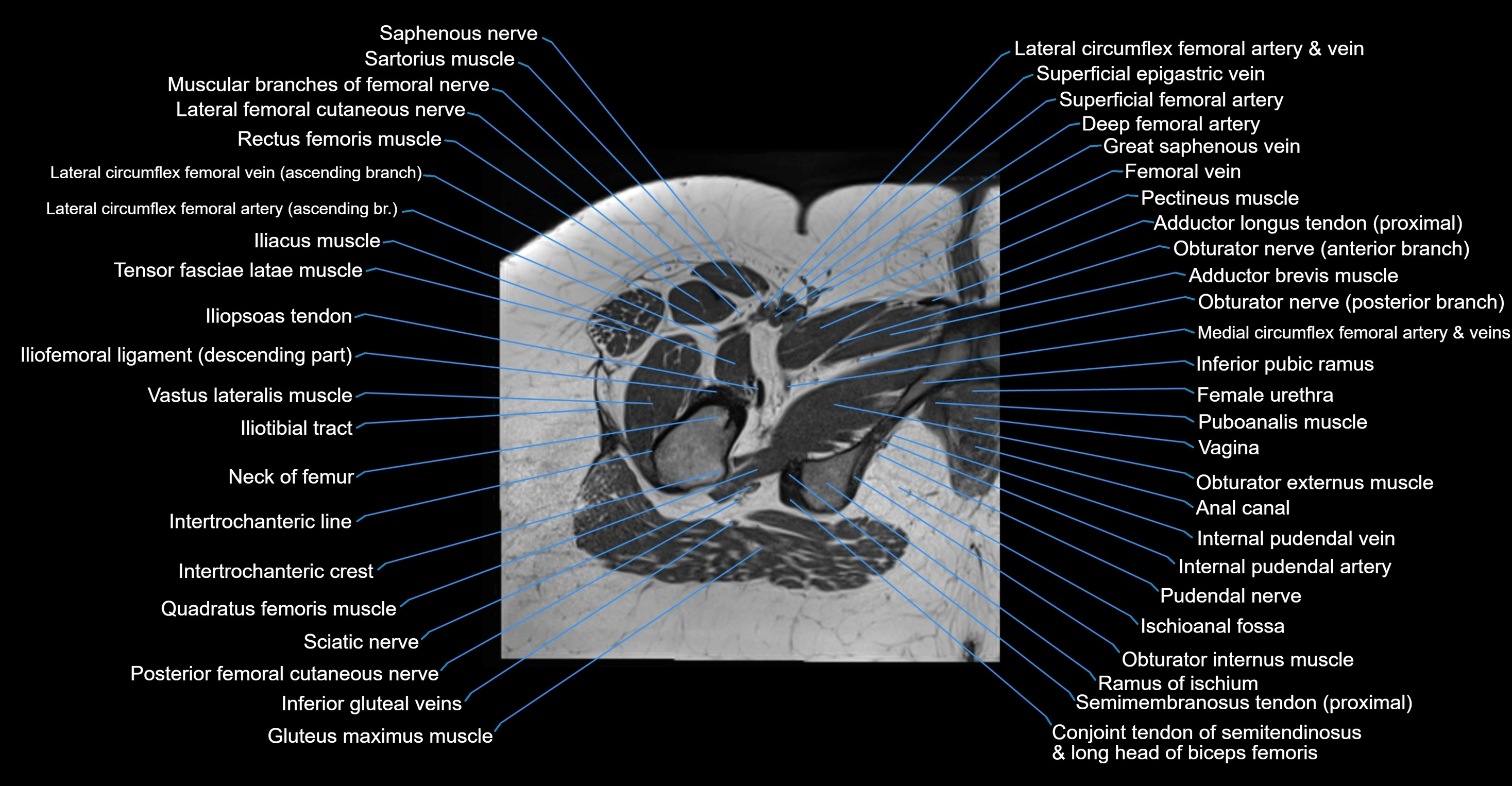
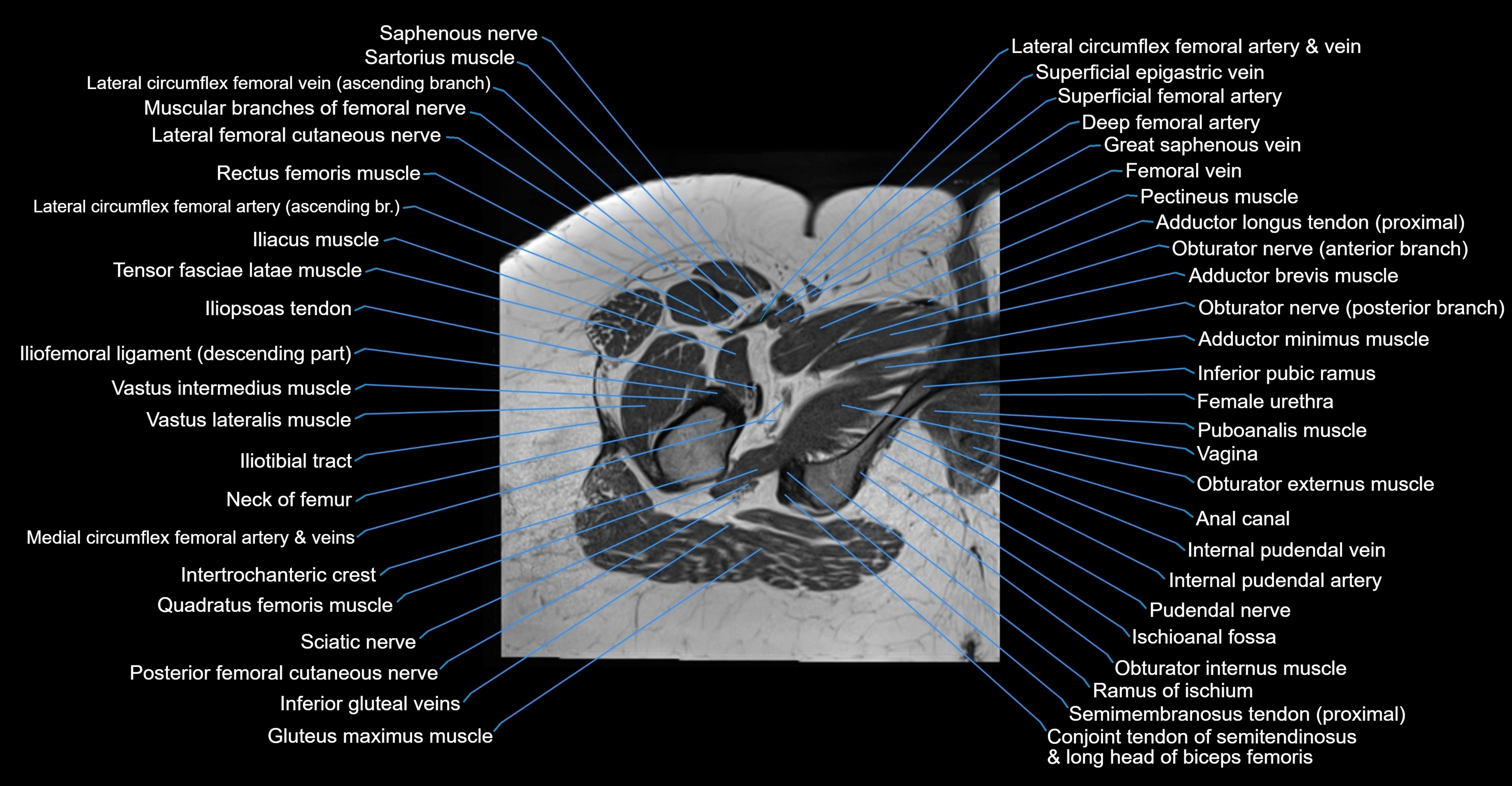
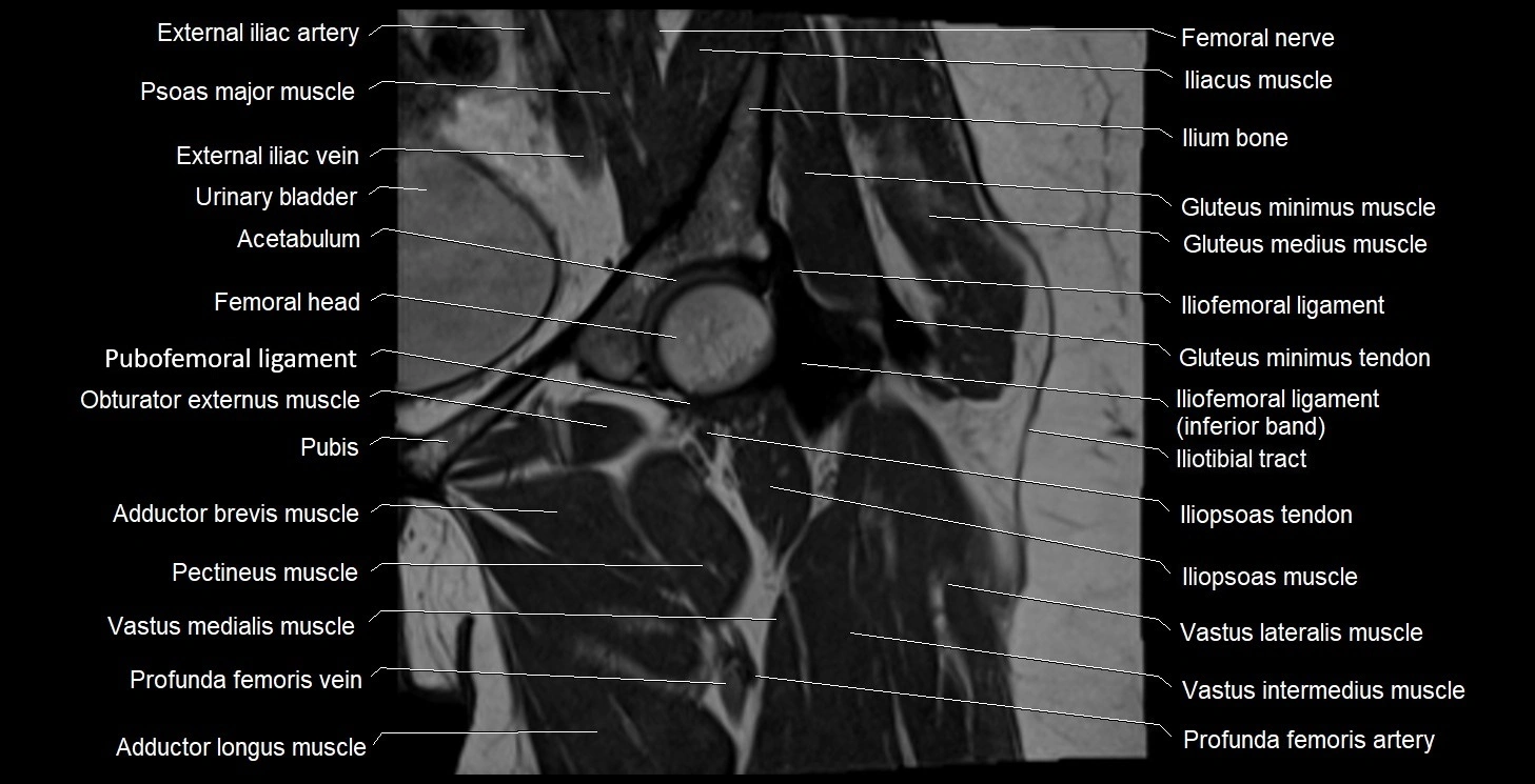
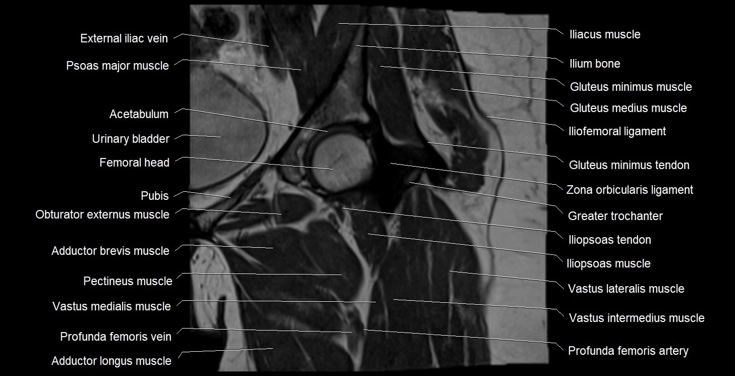
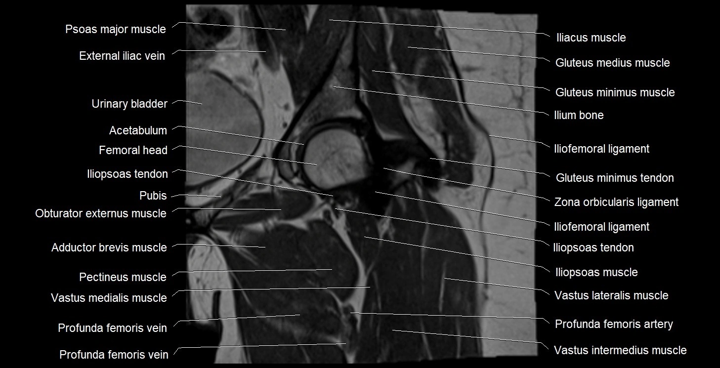
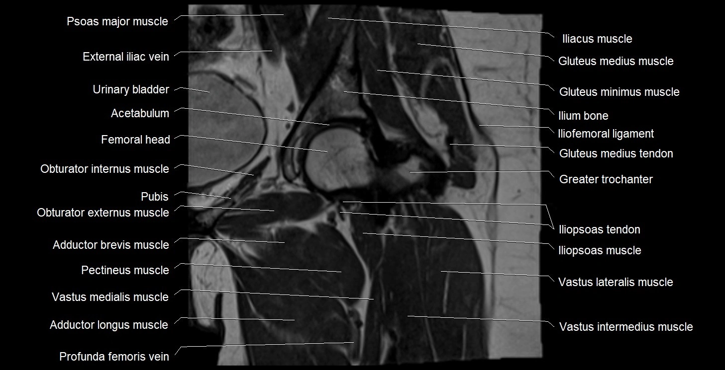
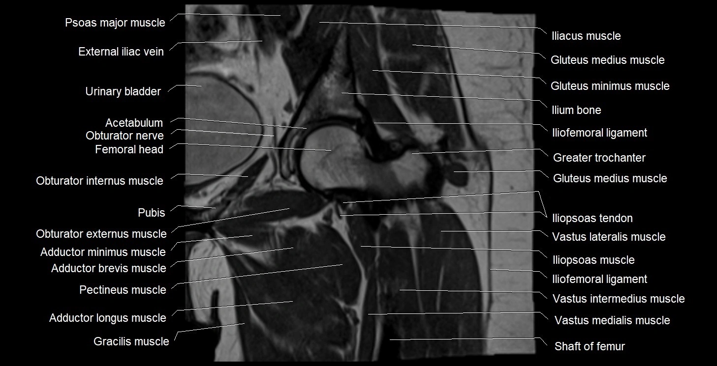
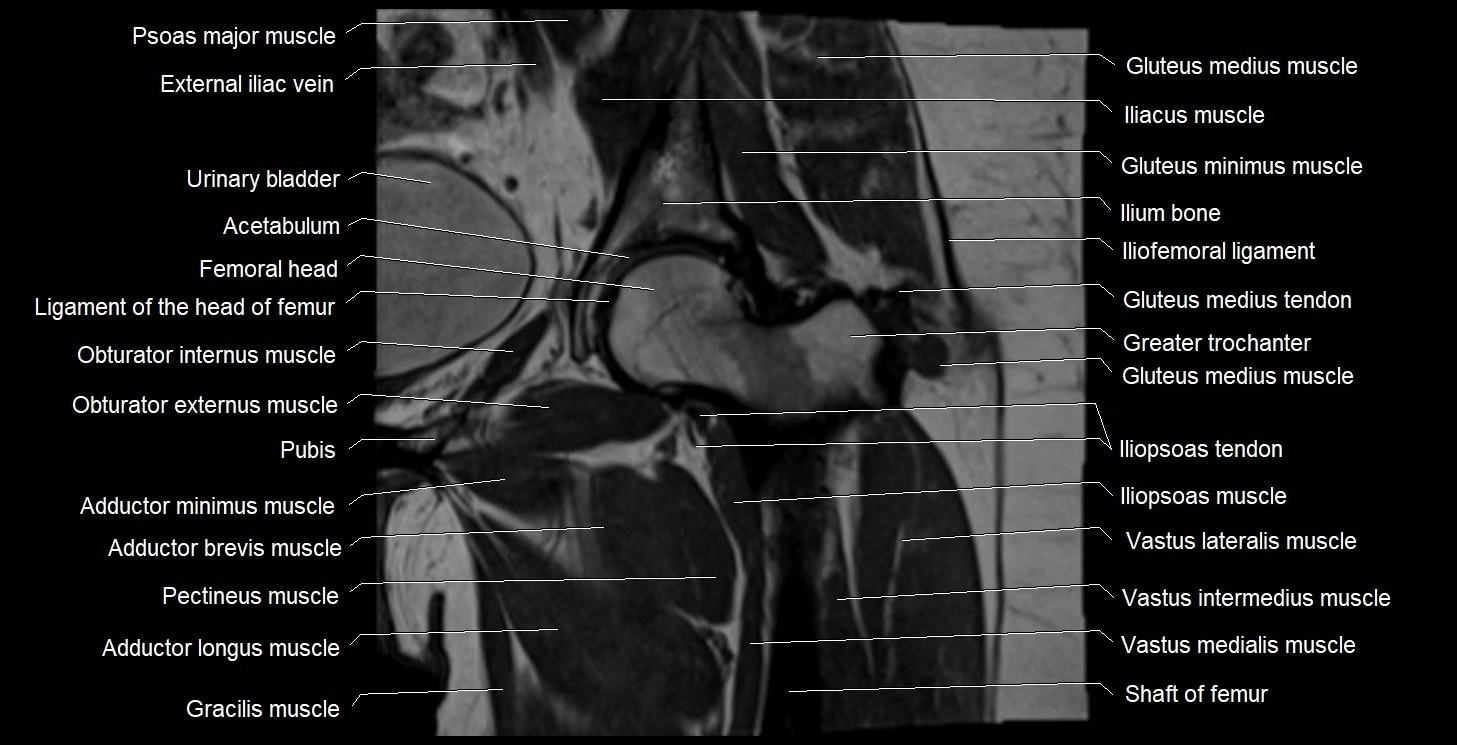
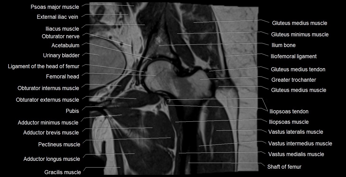
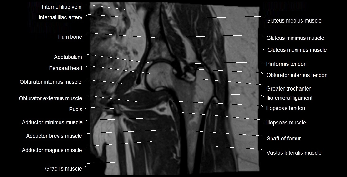
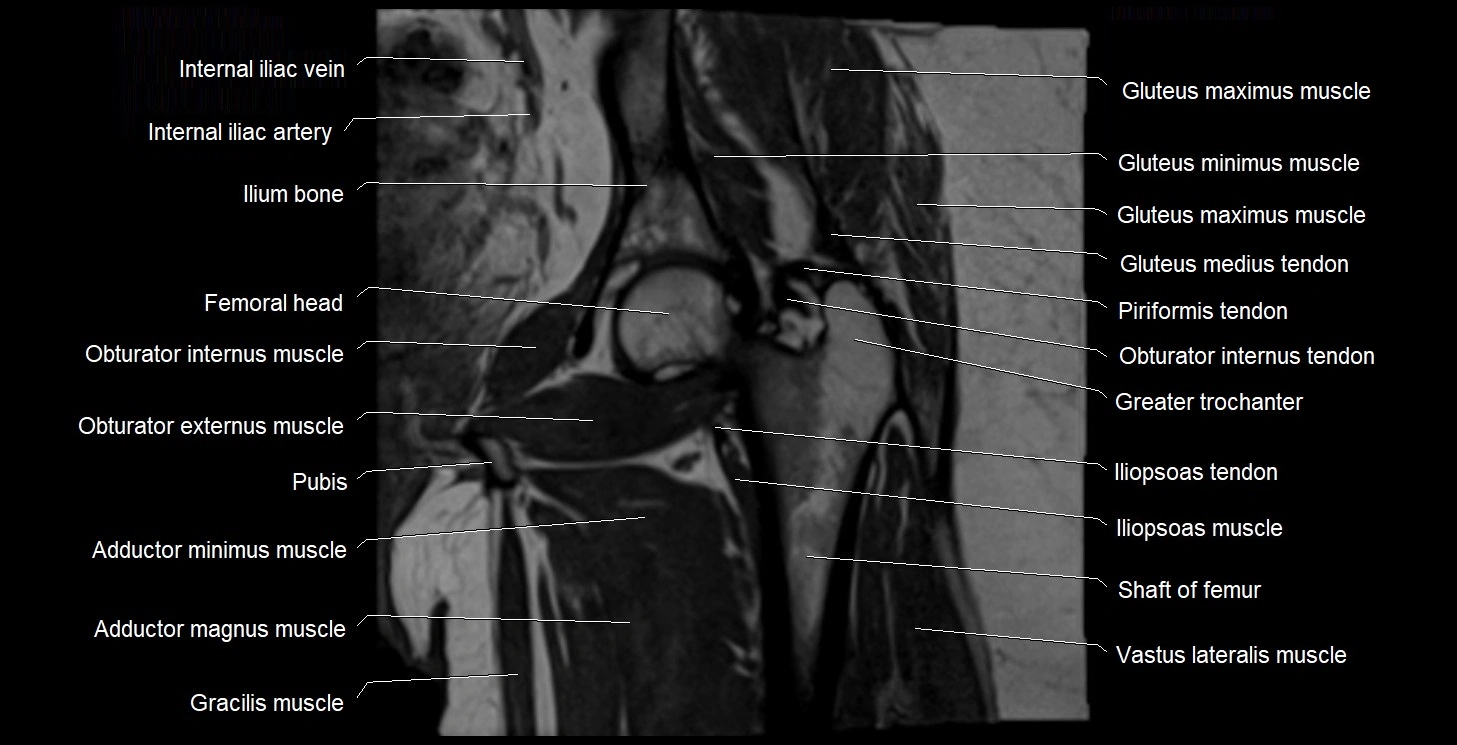
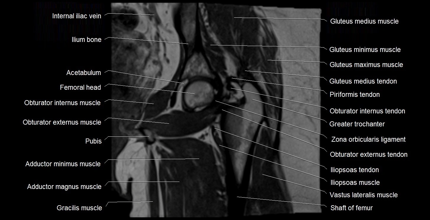
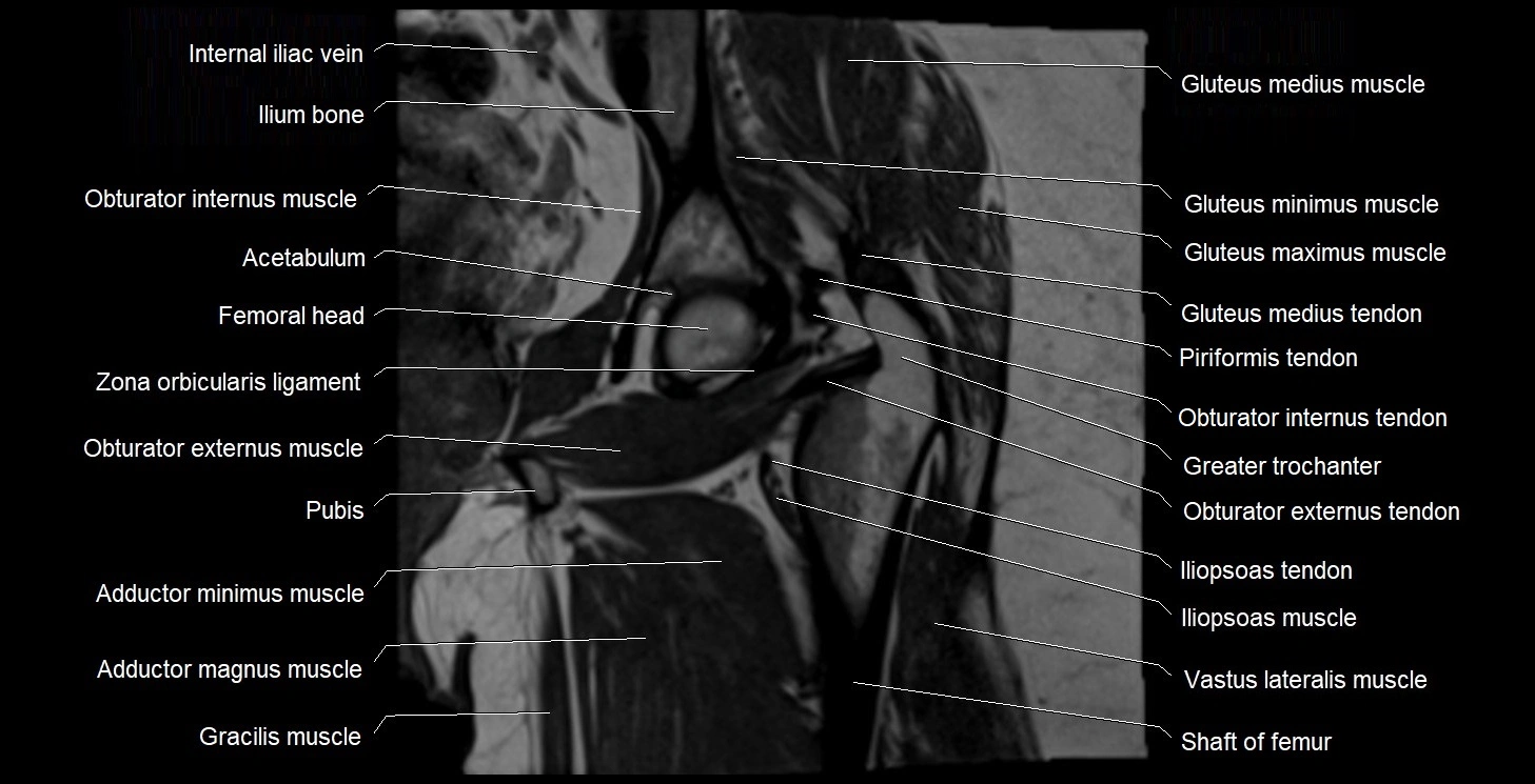
- Acetabulum
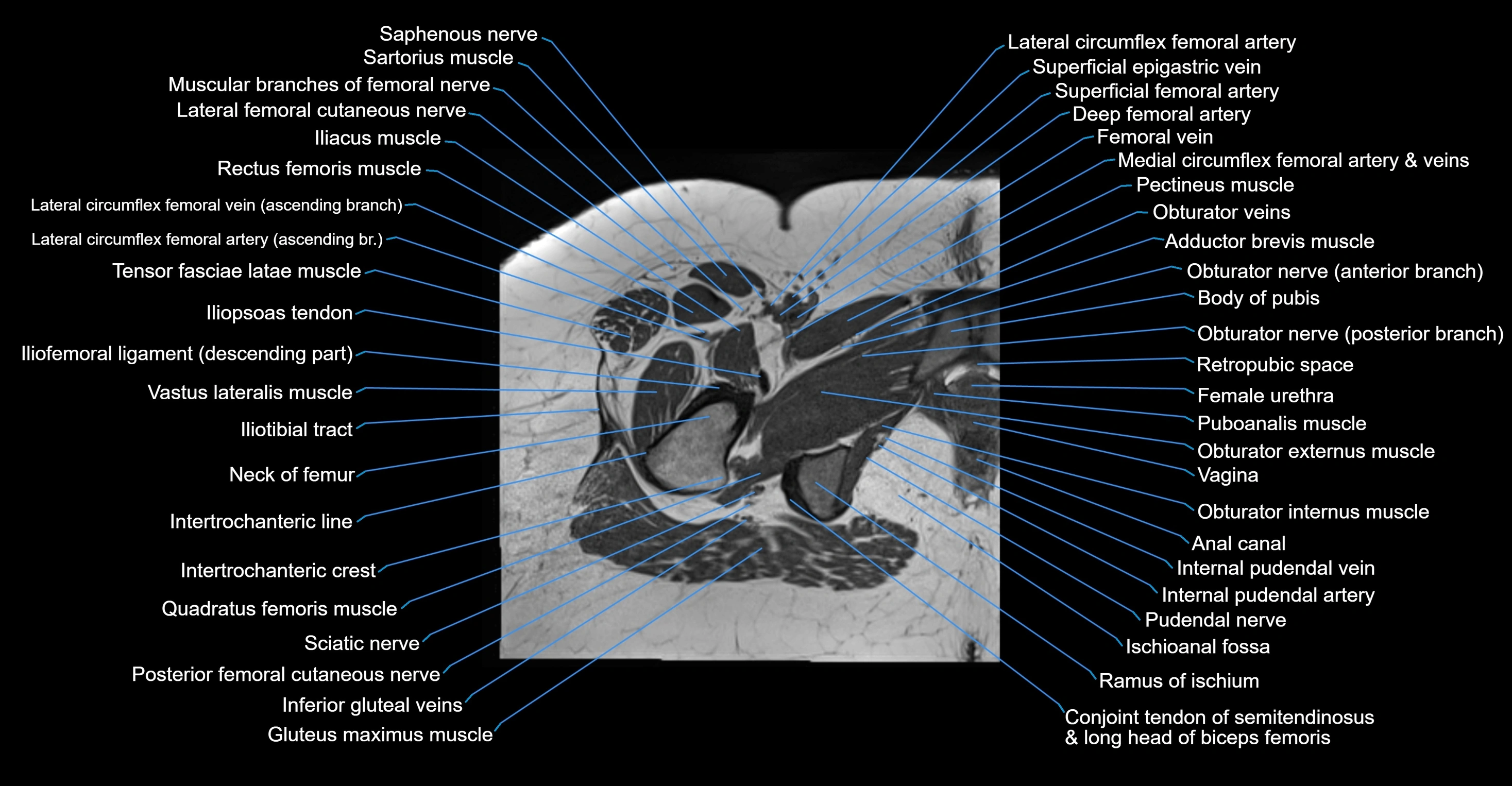
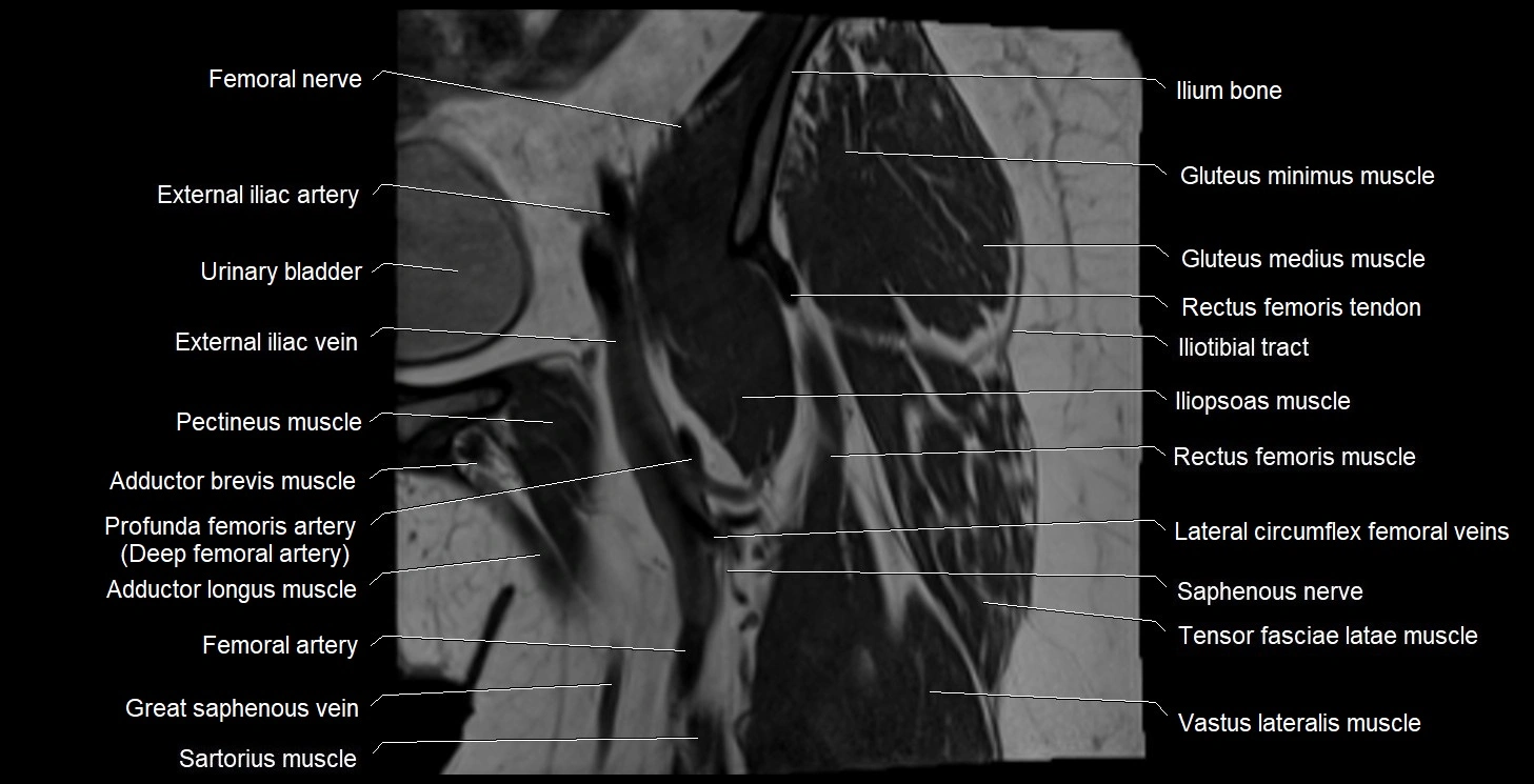
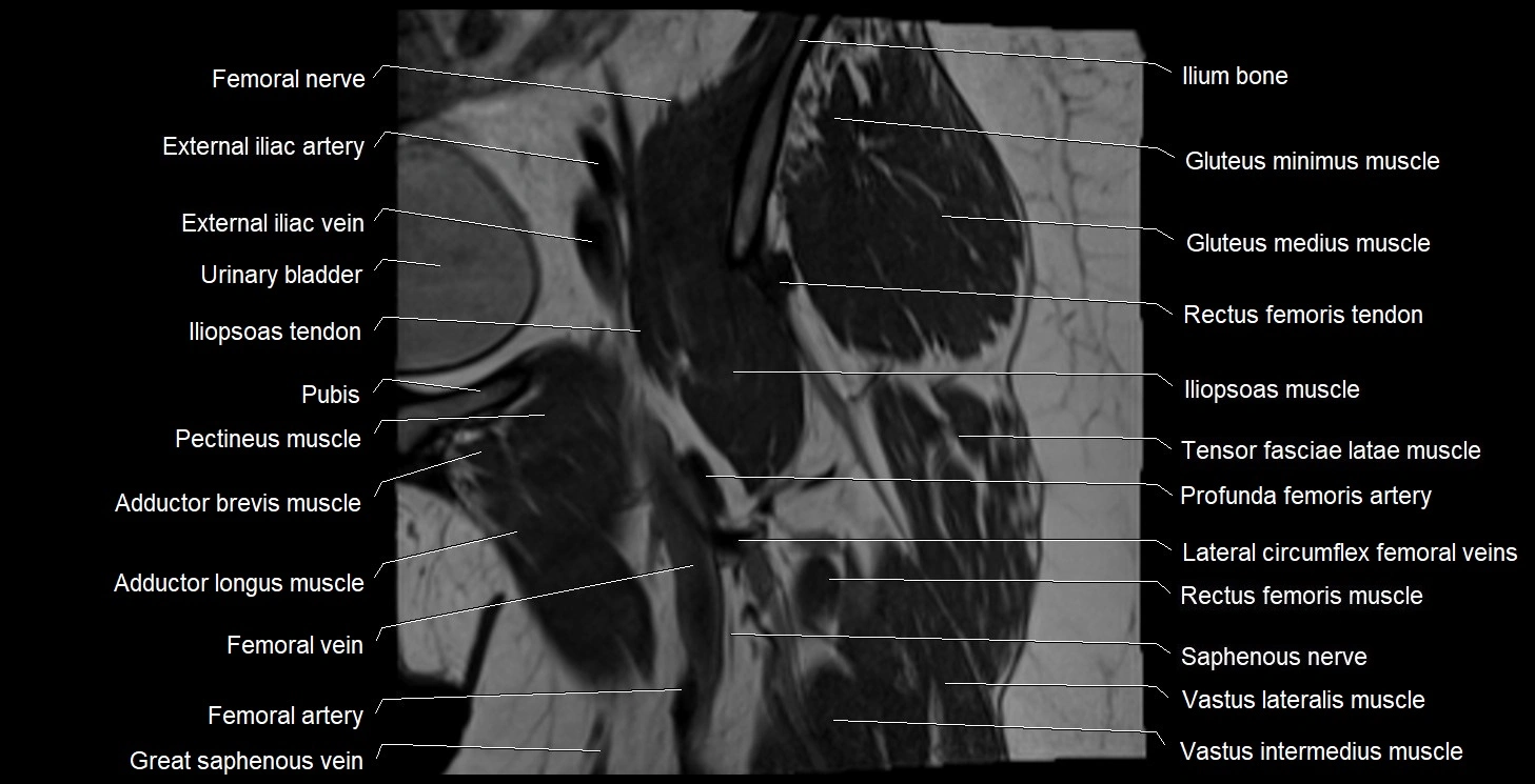
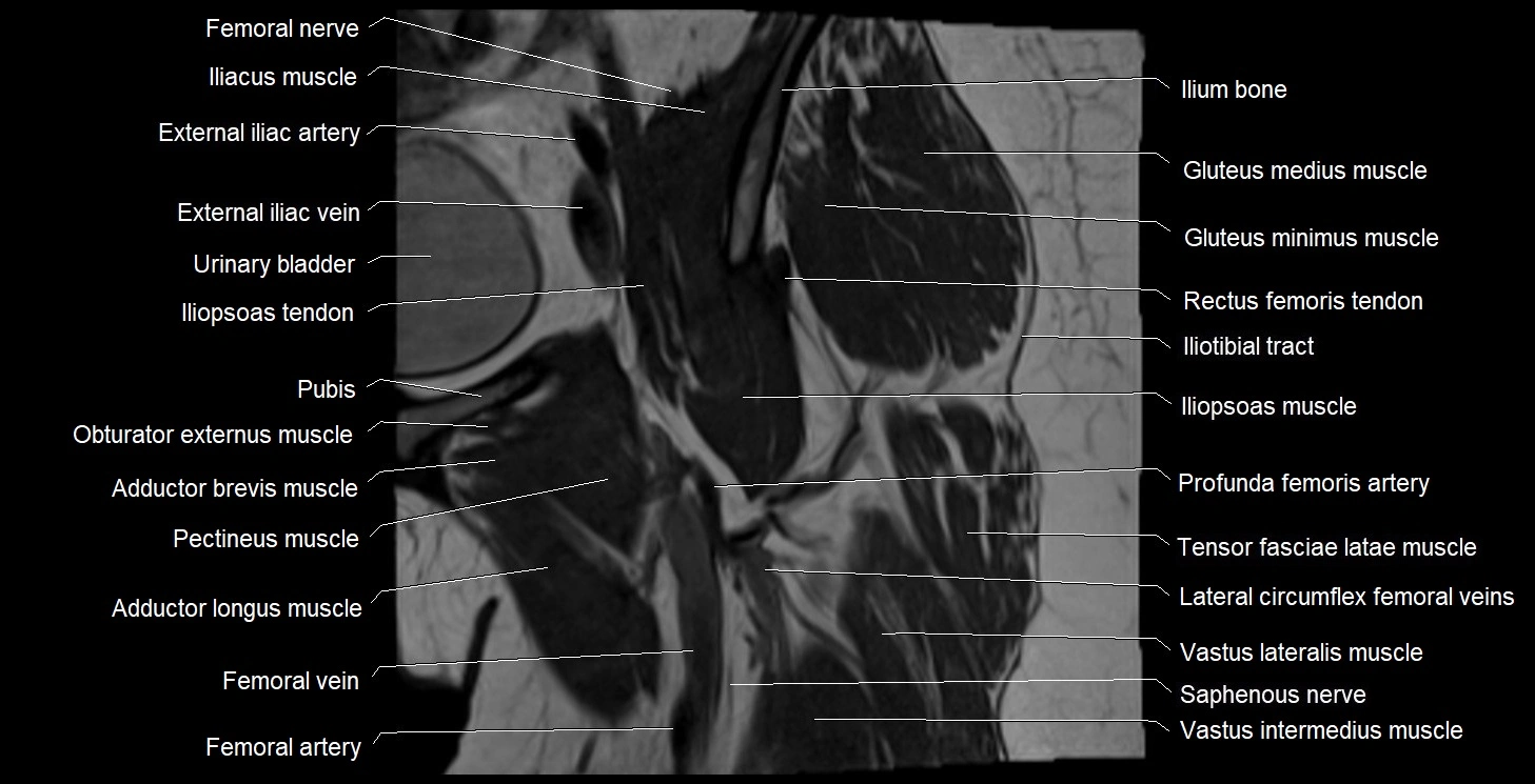
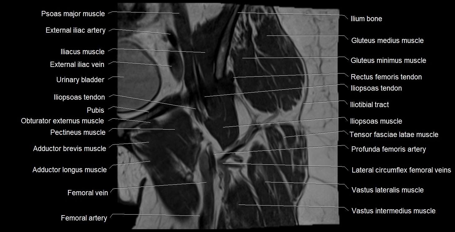
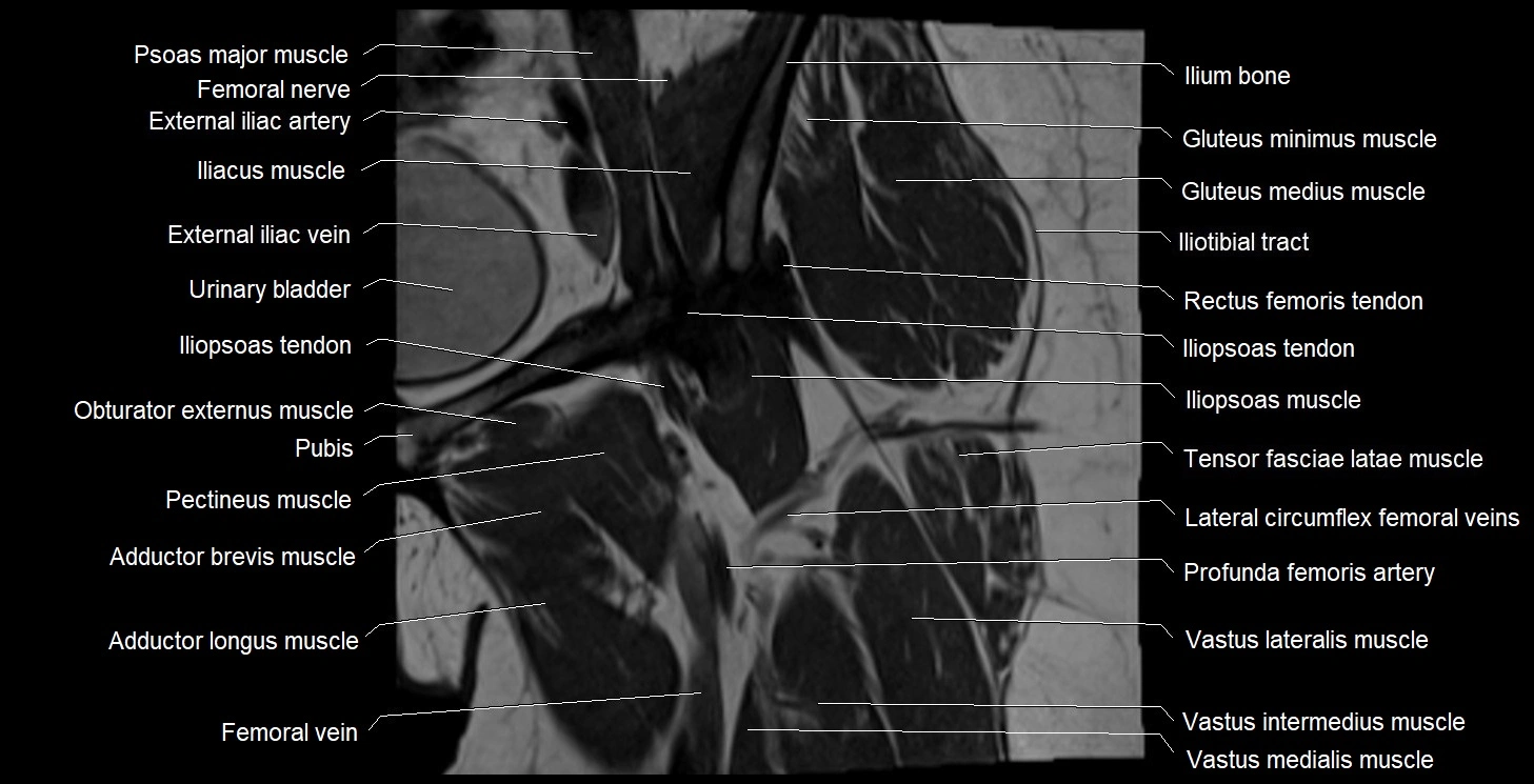
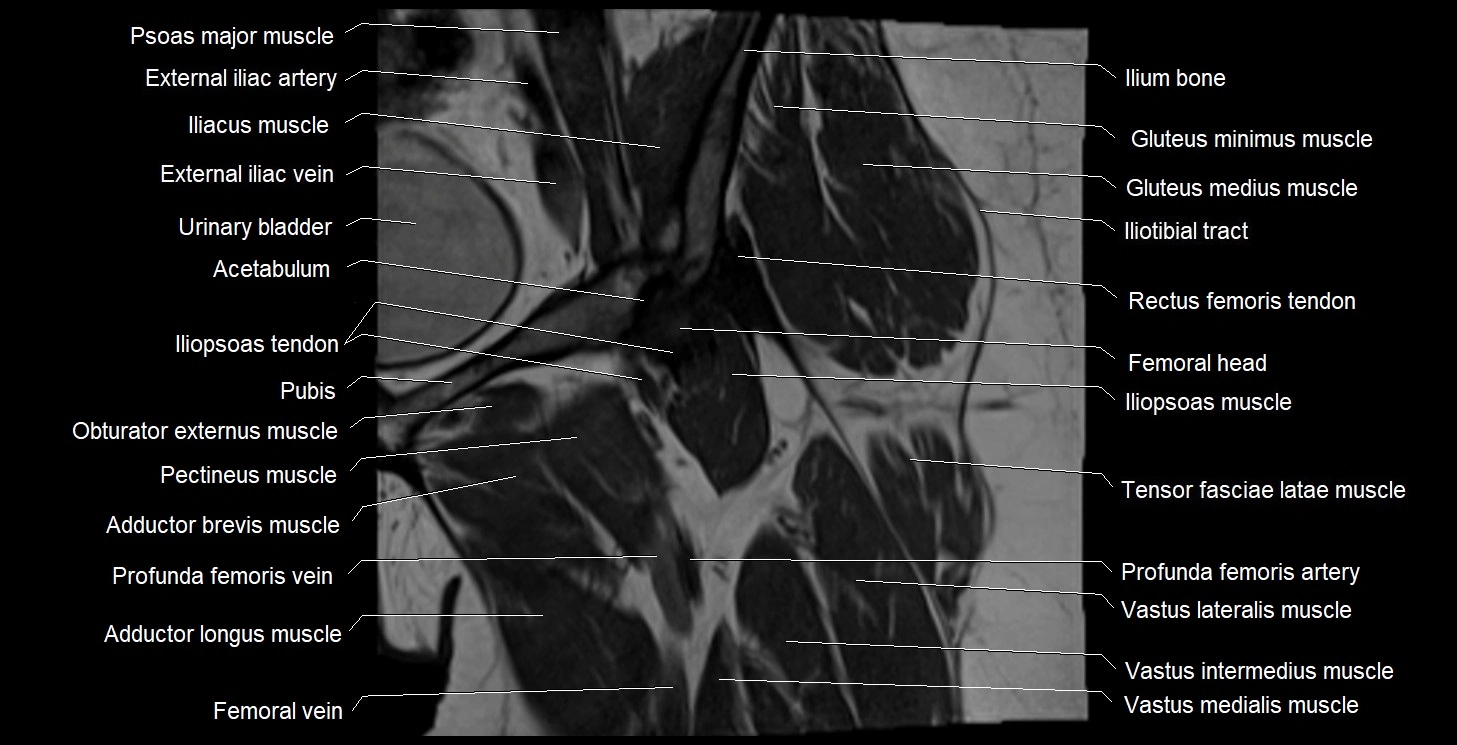
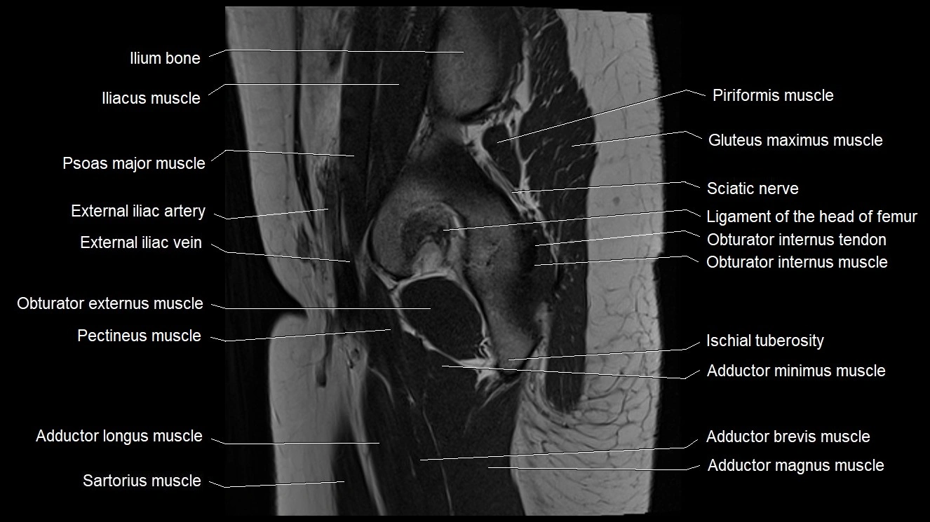
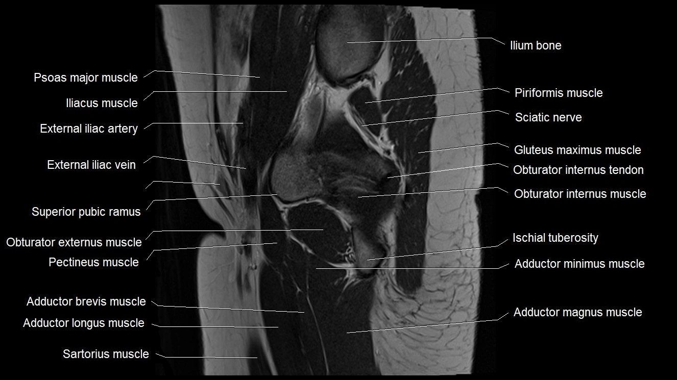
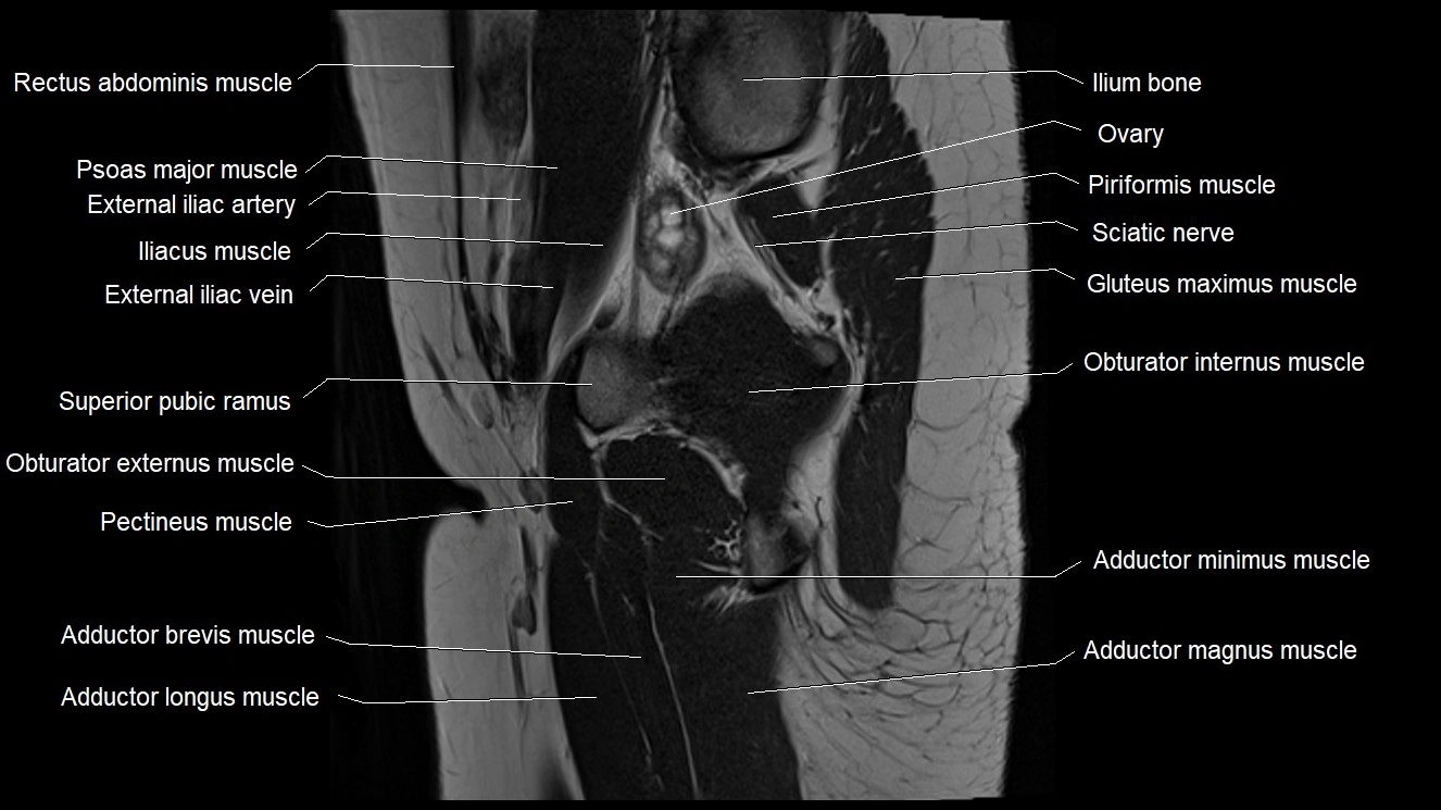
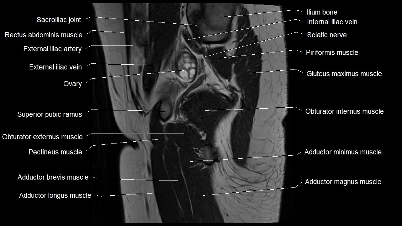
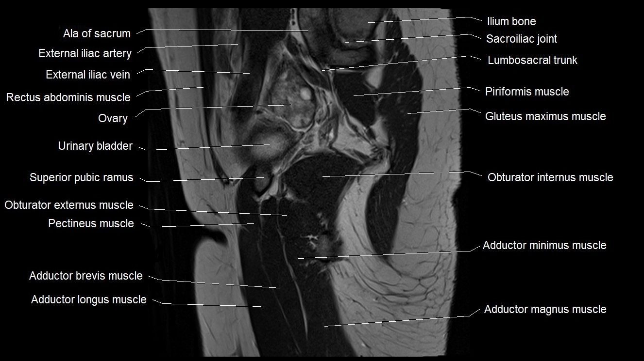
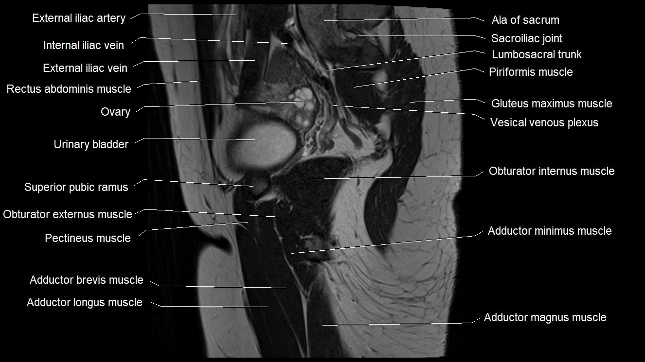
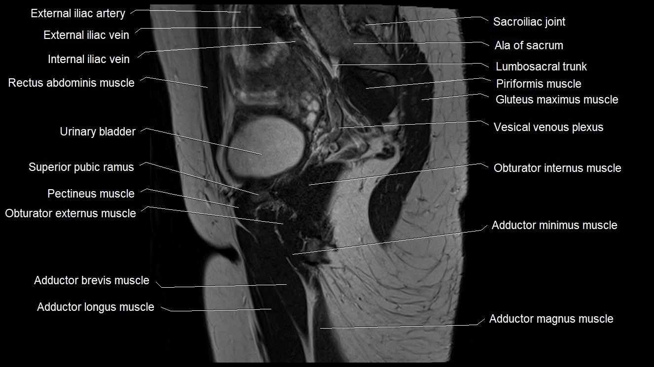
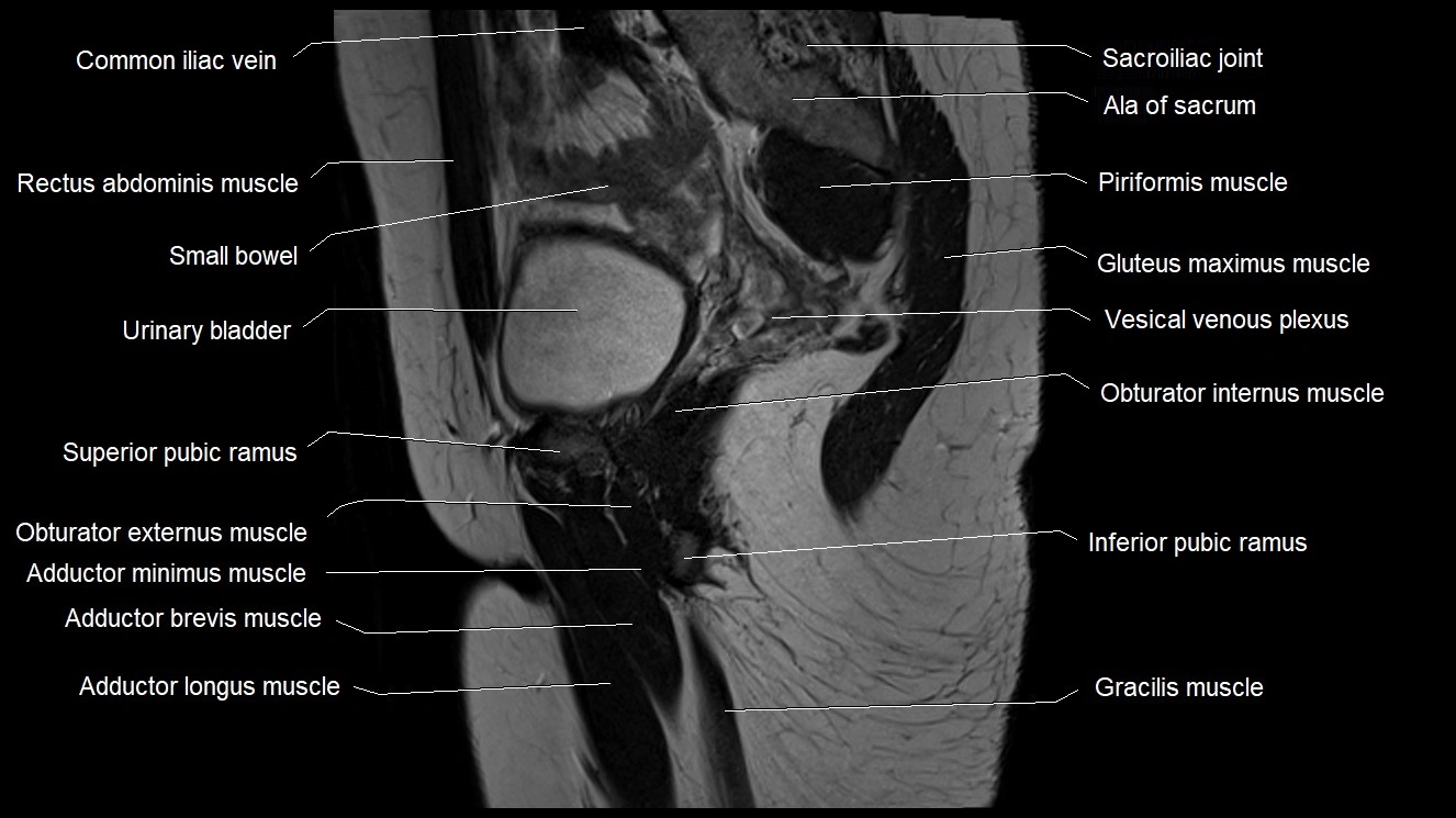
- Adductor brevis muscle
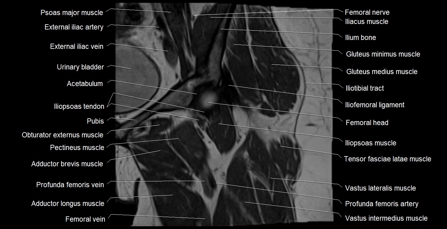
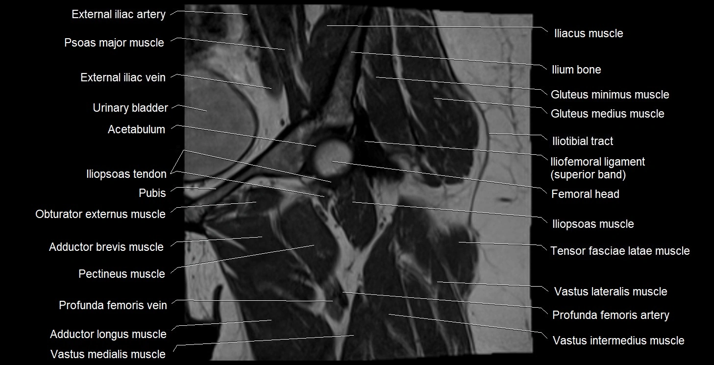
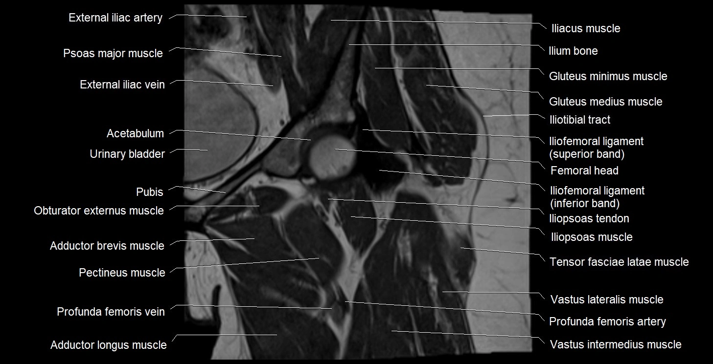
- Adductor longus muscle
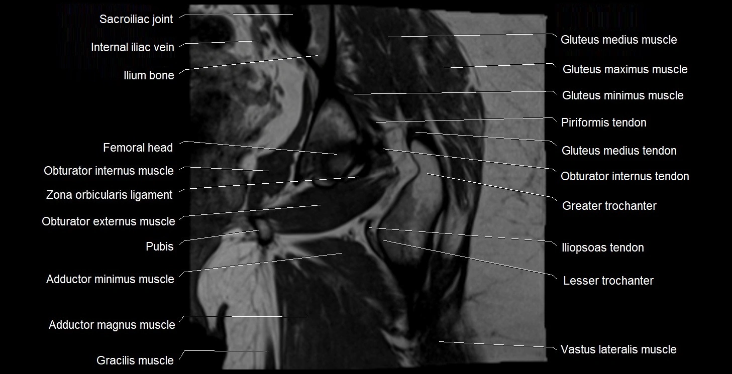
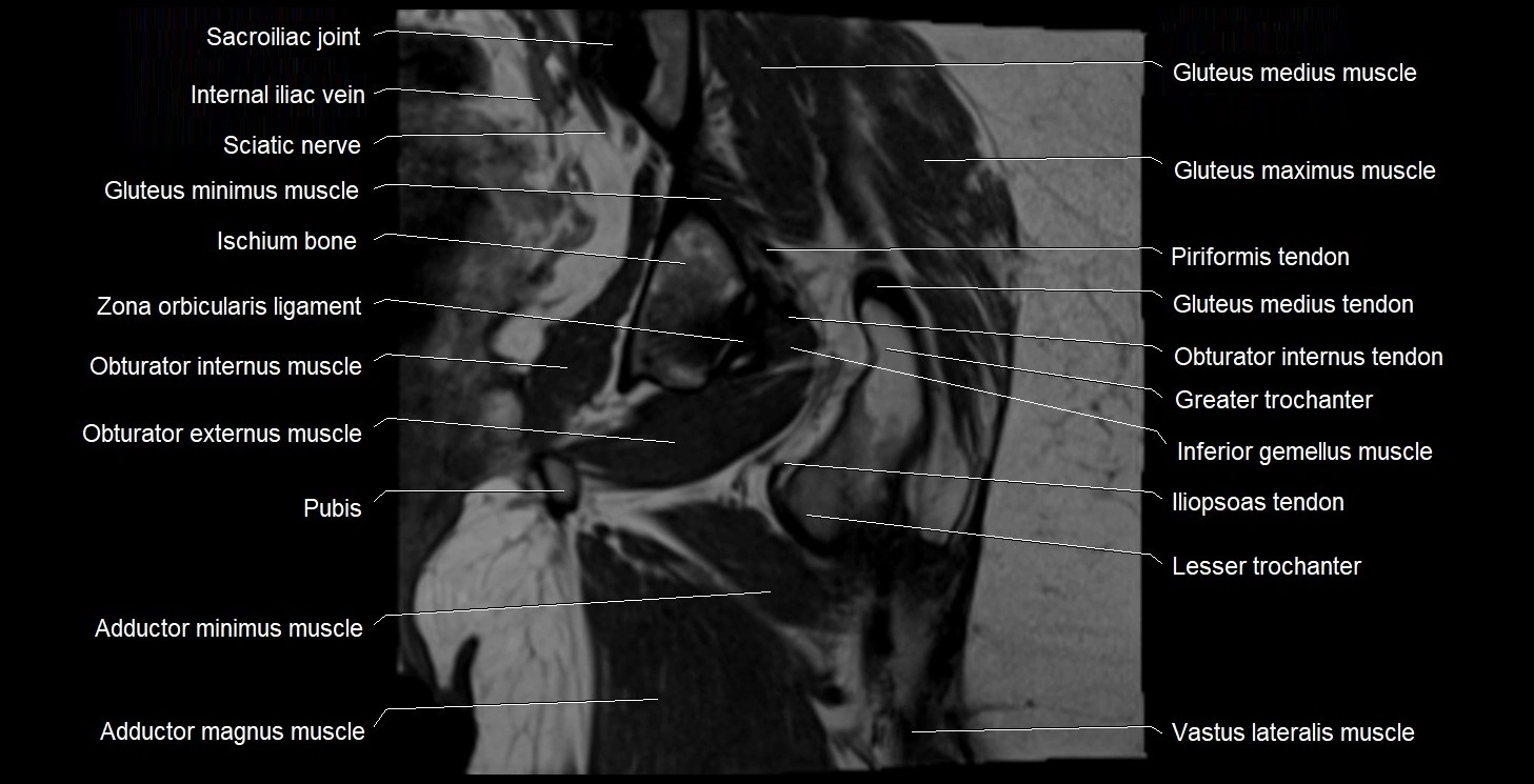
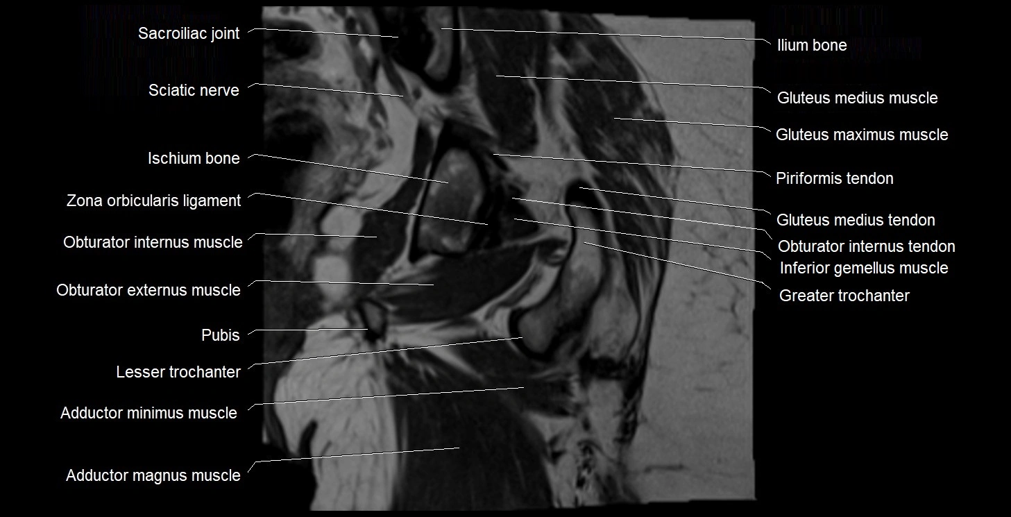
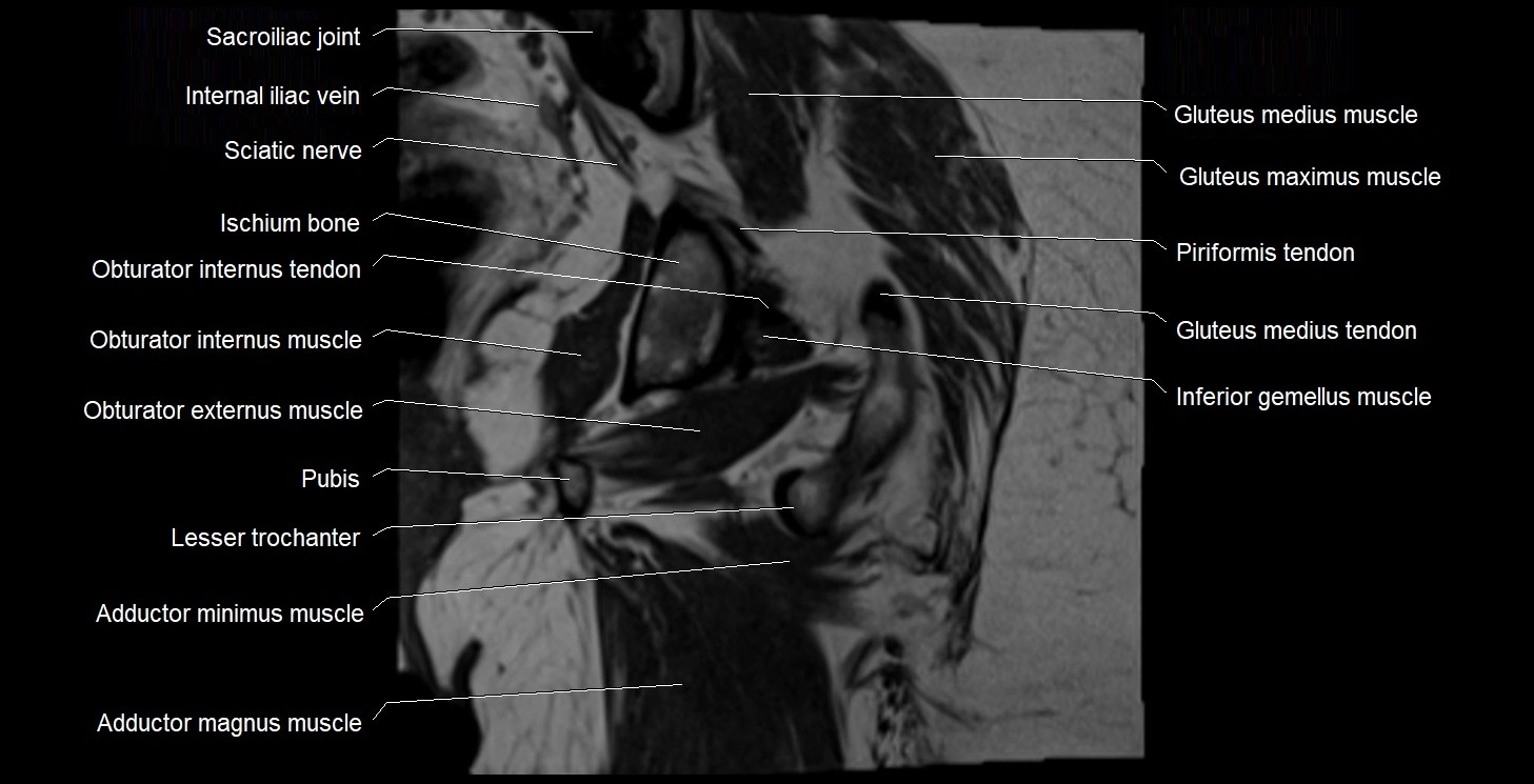
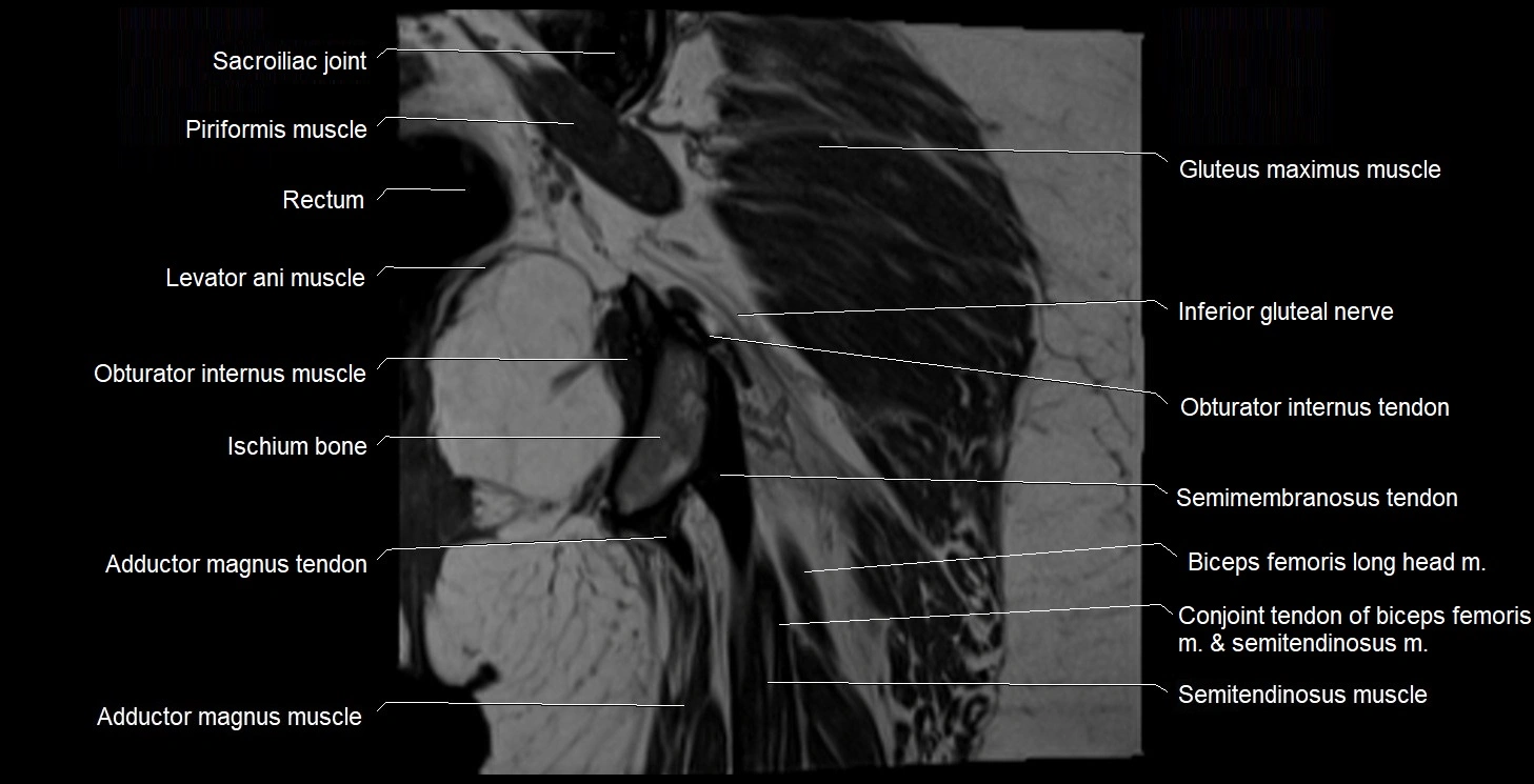
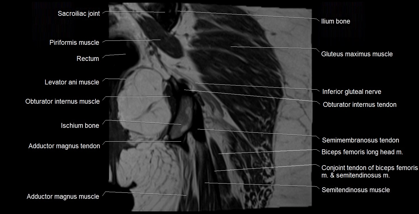
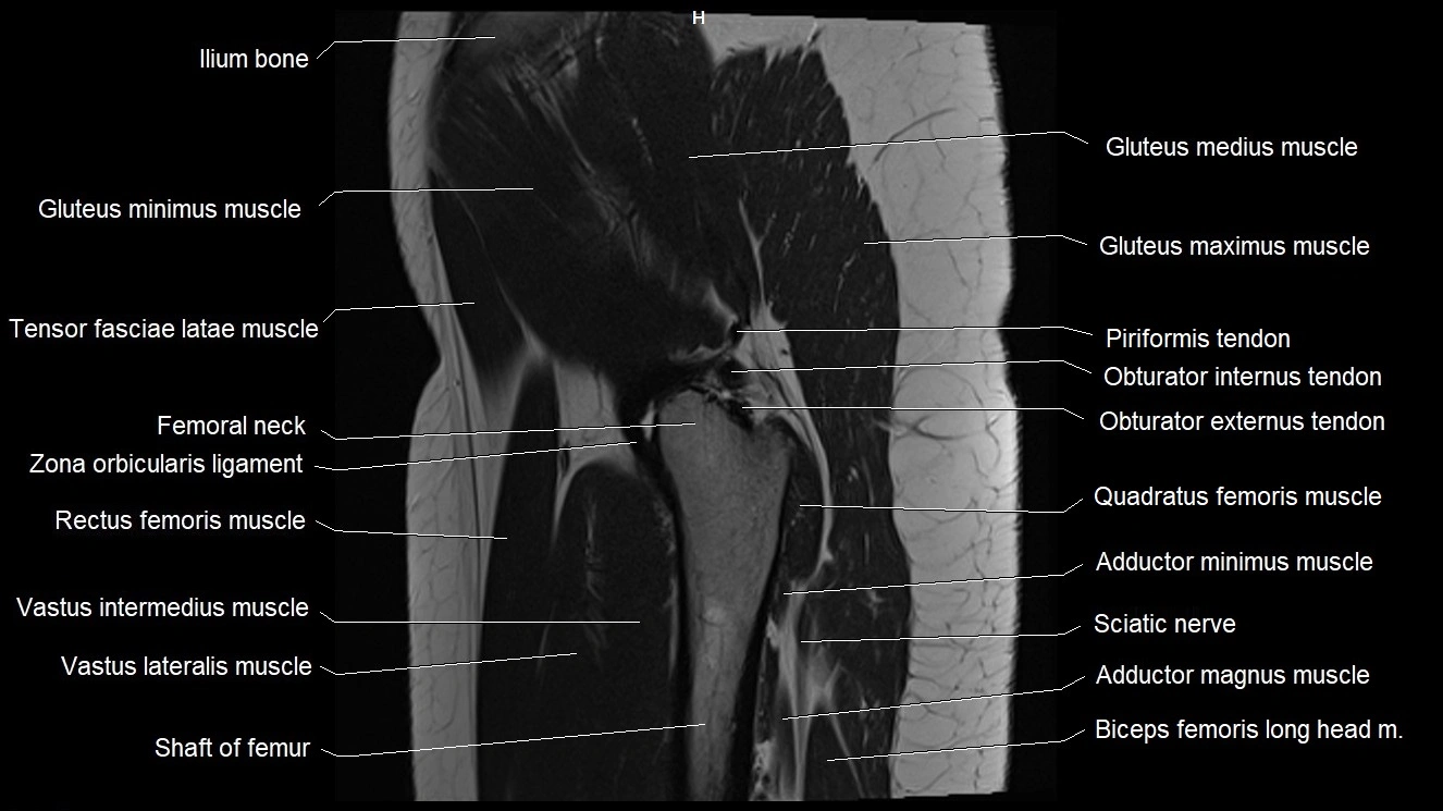
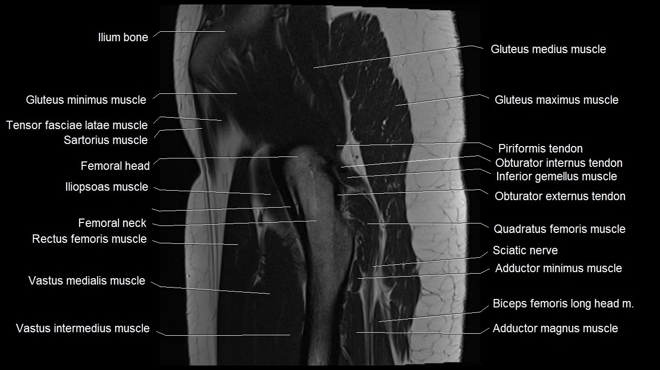
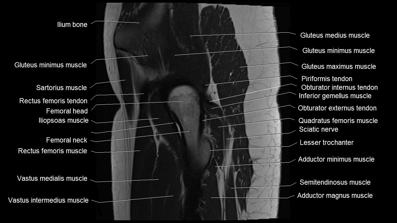
- Adductor magnus muscle
- Adductor minimus muscle
- Ala of ilium (wing of ilium)
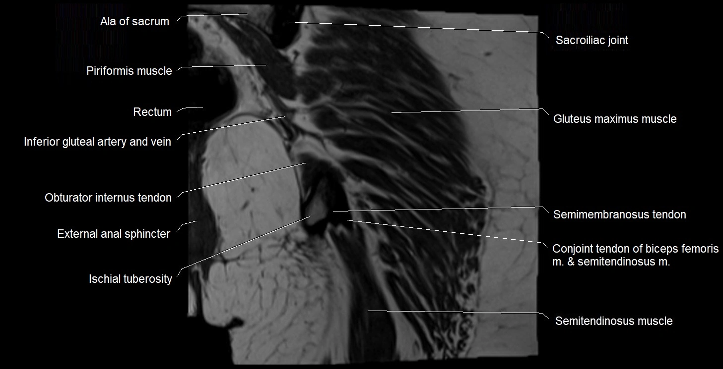
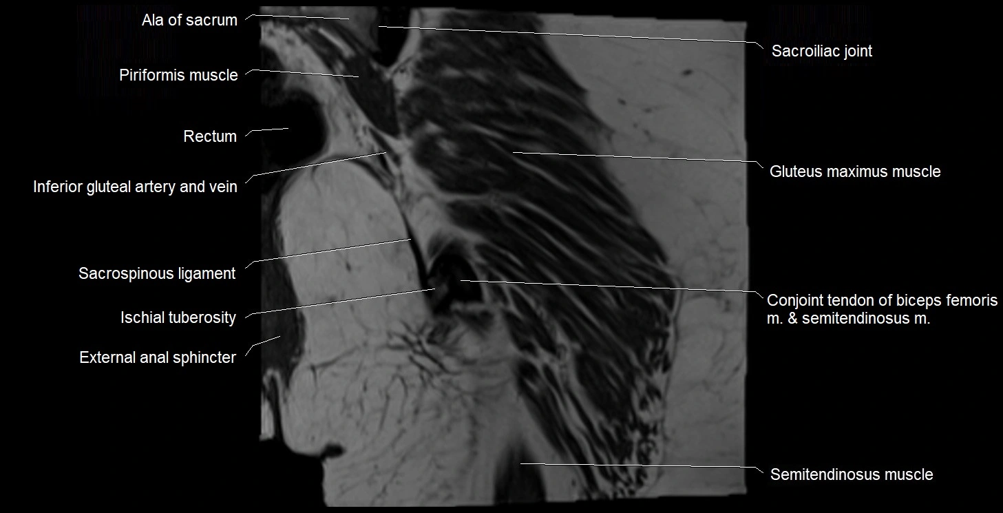
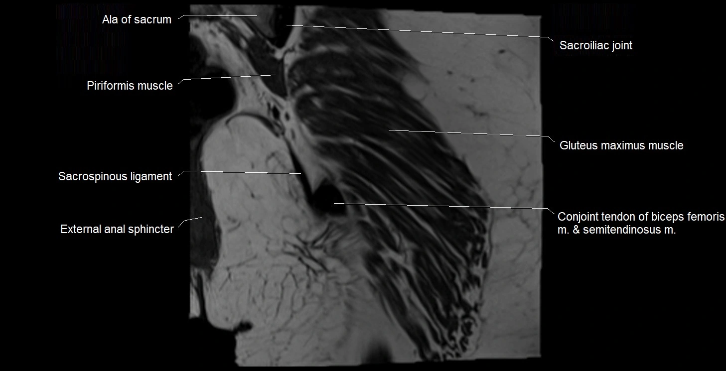
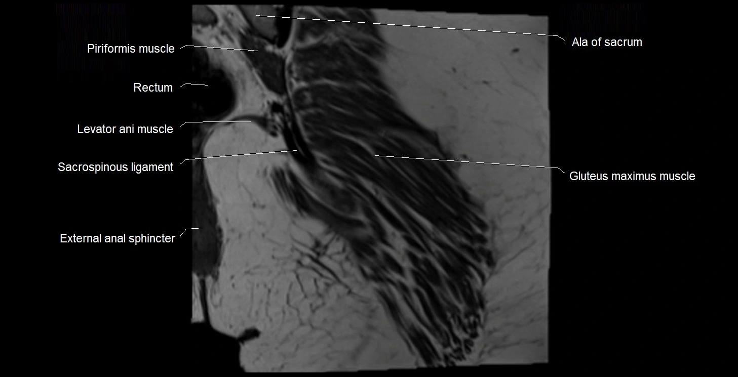
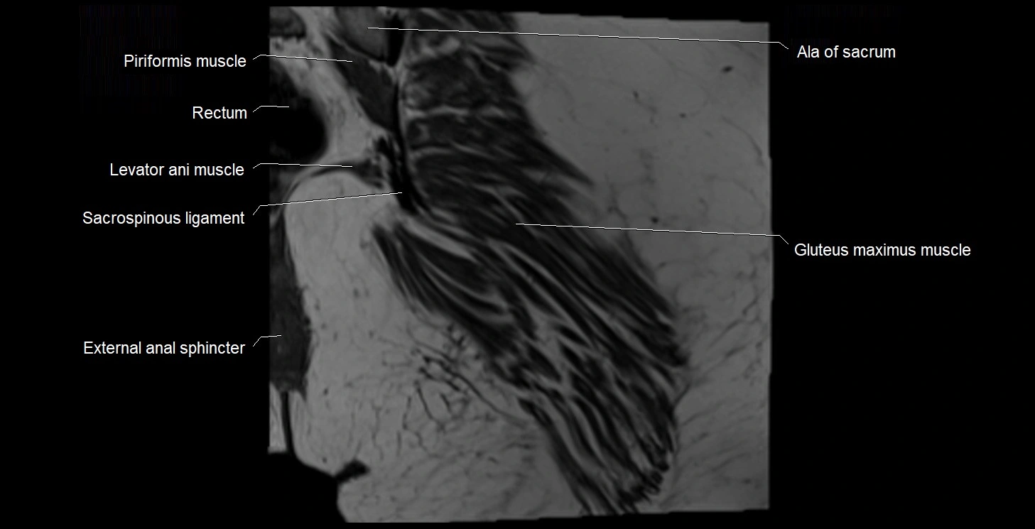
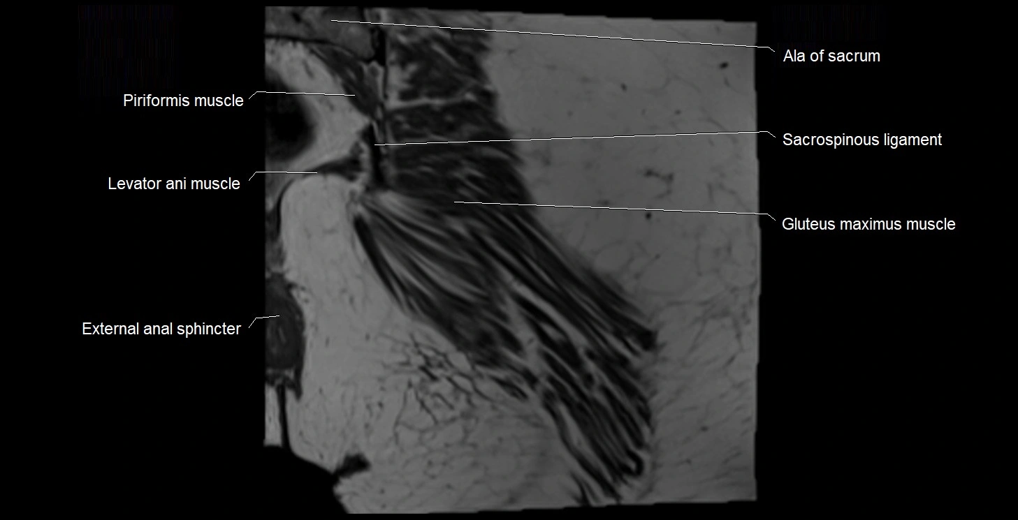
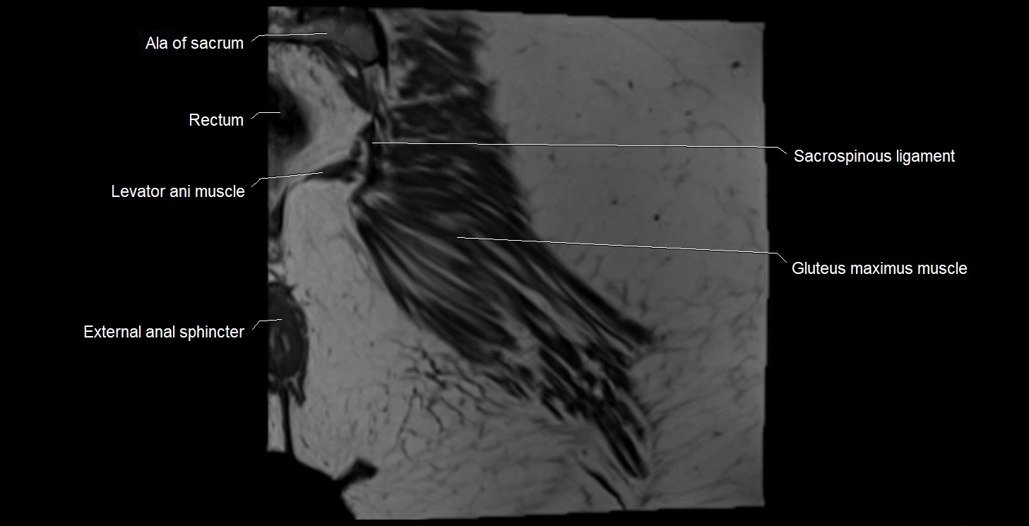
- Ala of sacrum
- Anal canal
- Anococcygeal body (anococcygeal ligament)
- Anococcygeal nerve
- Anterior Fibromuscular Stroma of prostate
- Anterior acetabular wall
- Anterior division of obturator nerve (Anterior branch of obturator nerve)
- Anterior inferior iliac spine
- Anterior lateral femoral cutaneous nerve
- Anterior rim of acetabulum
- Anterior sacral foramina
- Anterior sacroiliac ligament
- Anterior superior iliac spine
- Anterior wall of acetabulum
- Apex of urinary bladder
- Articular capsule of hip joint
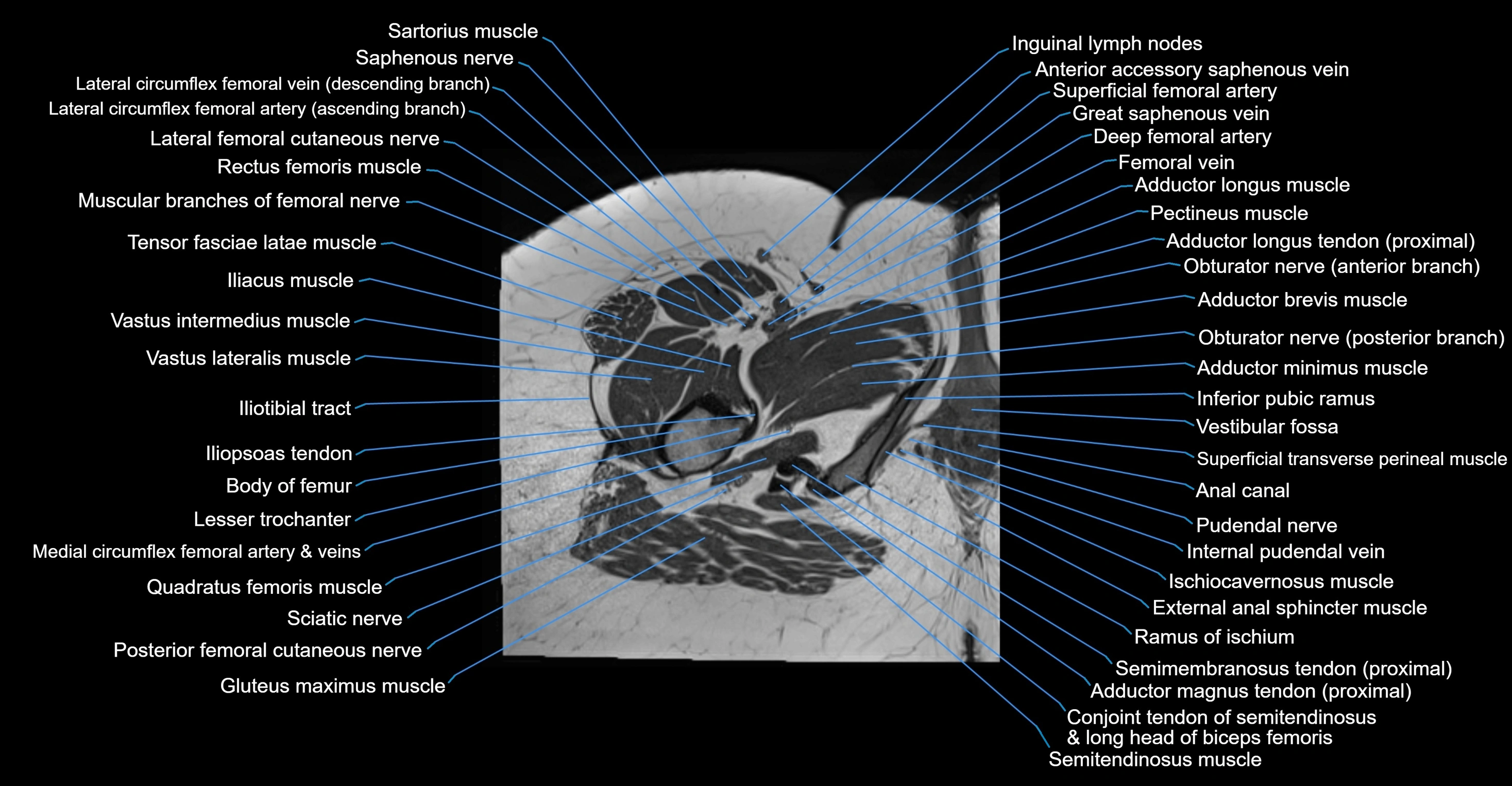
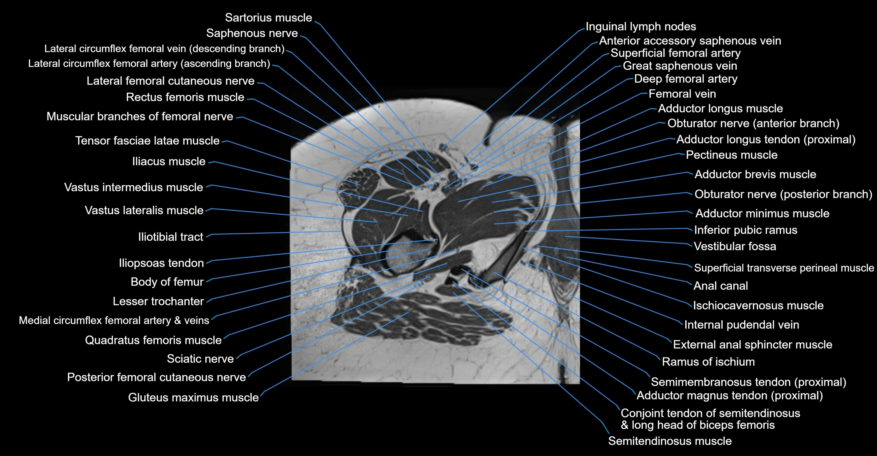
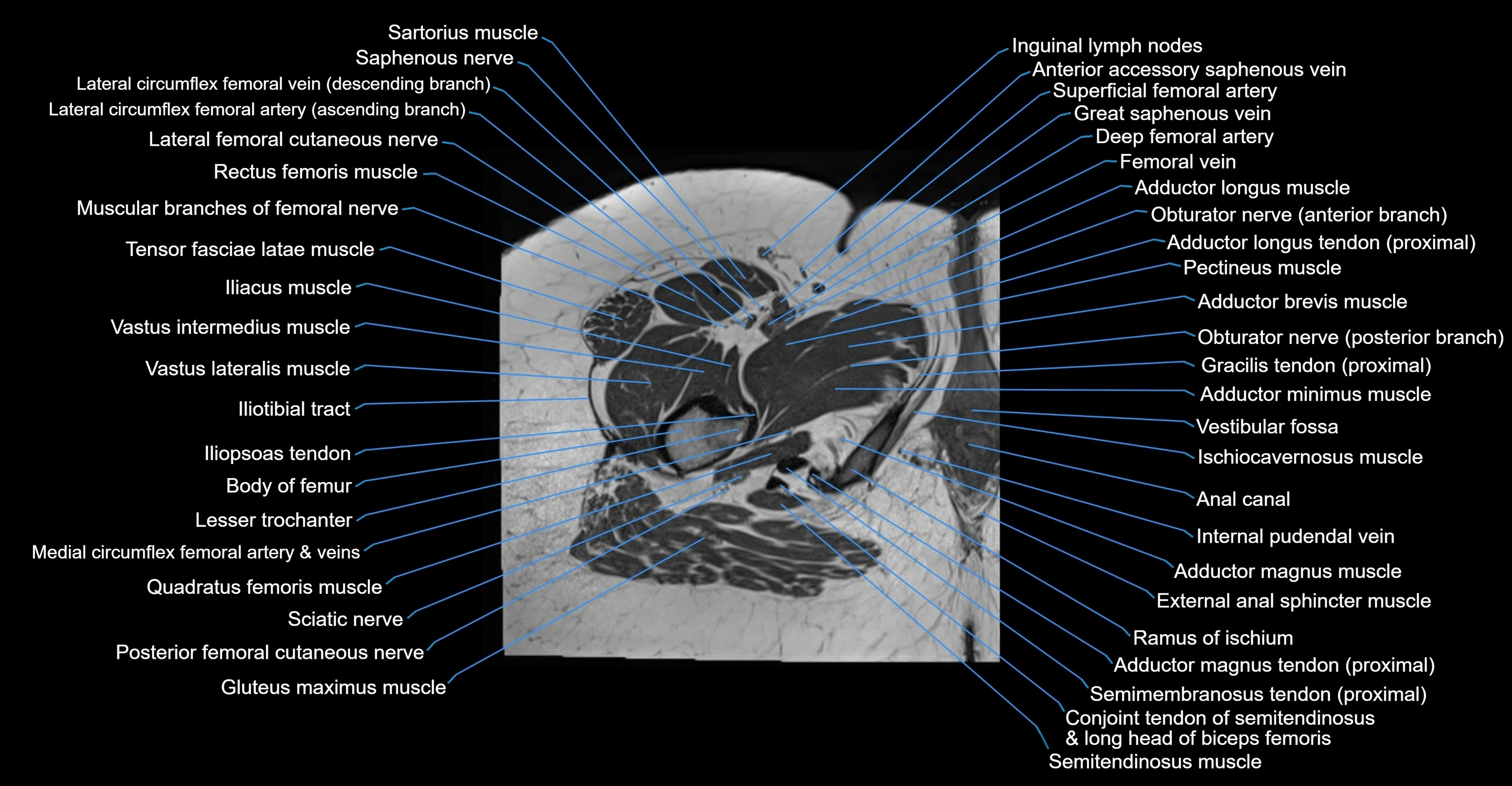
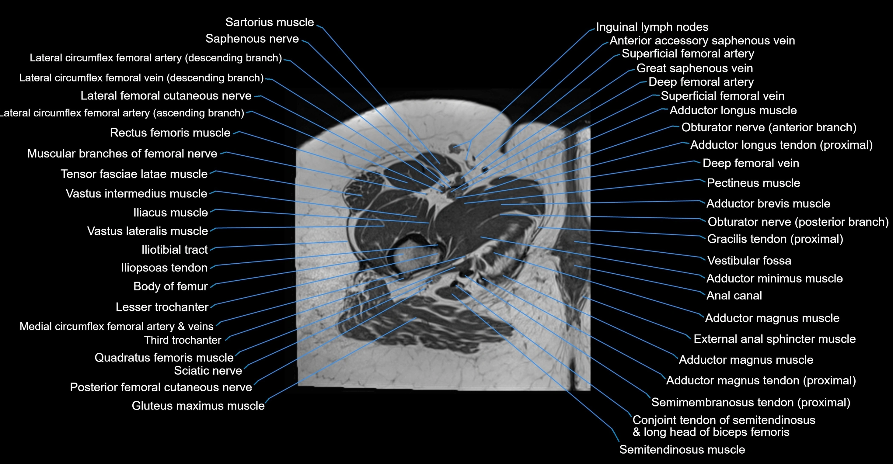
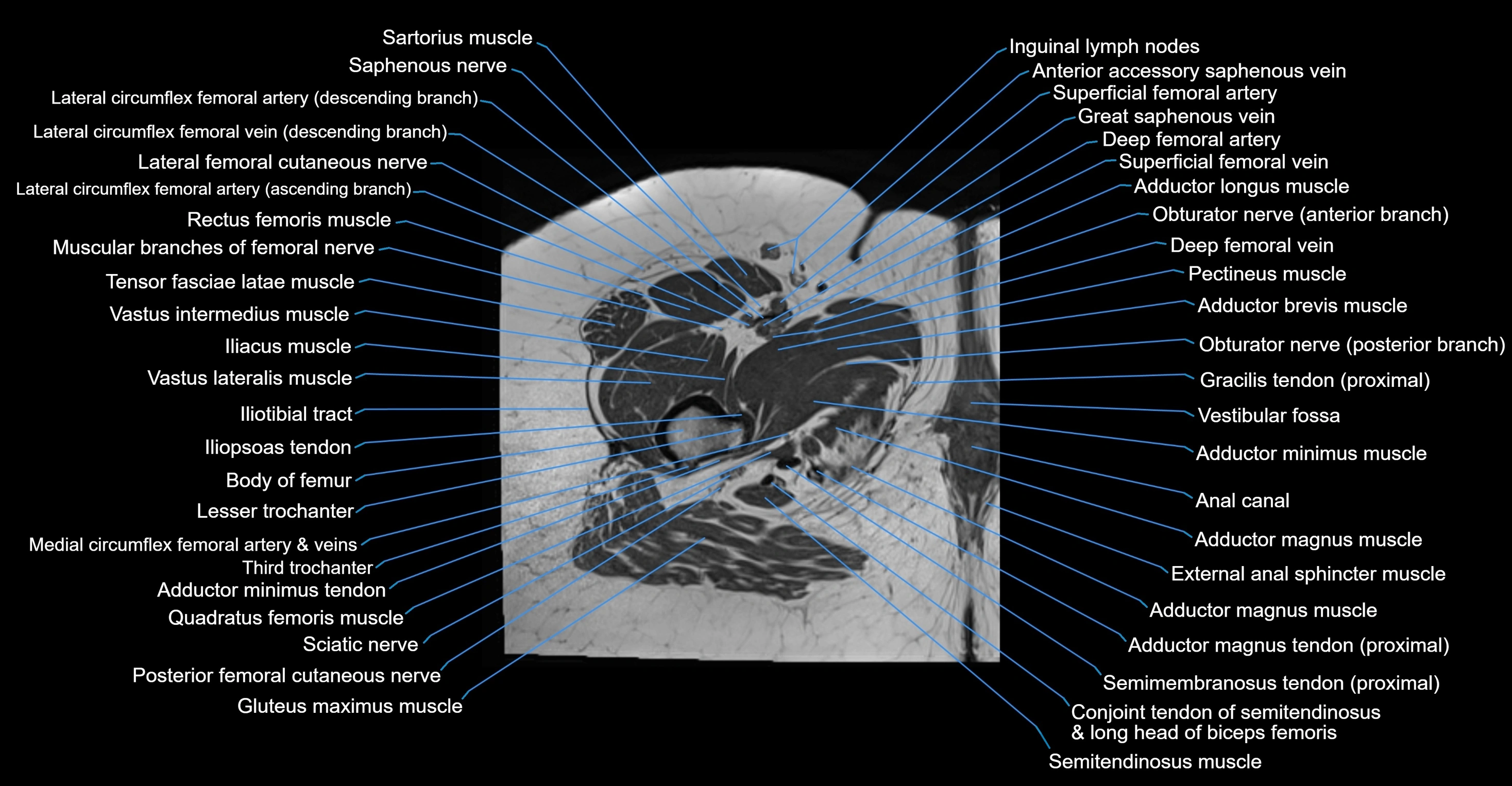
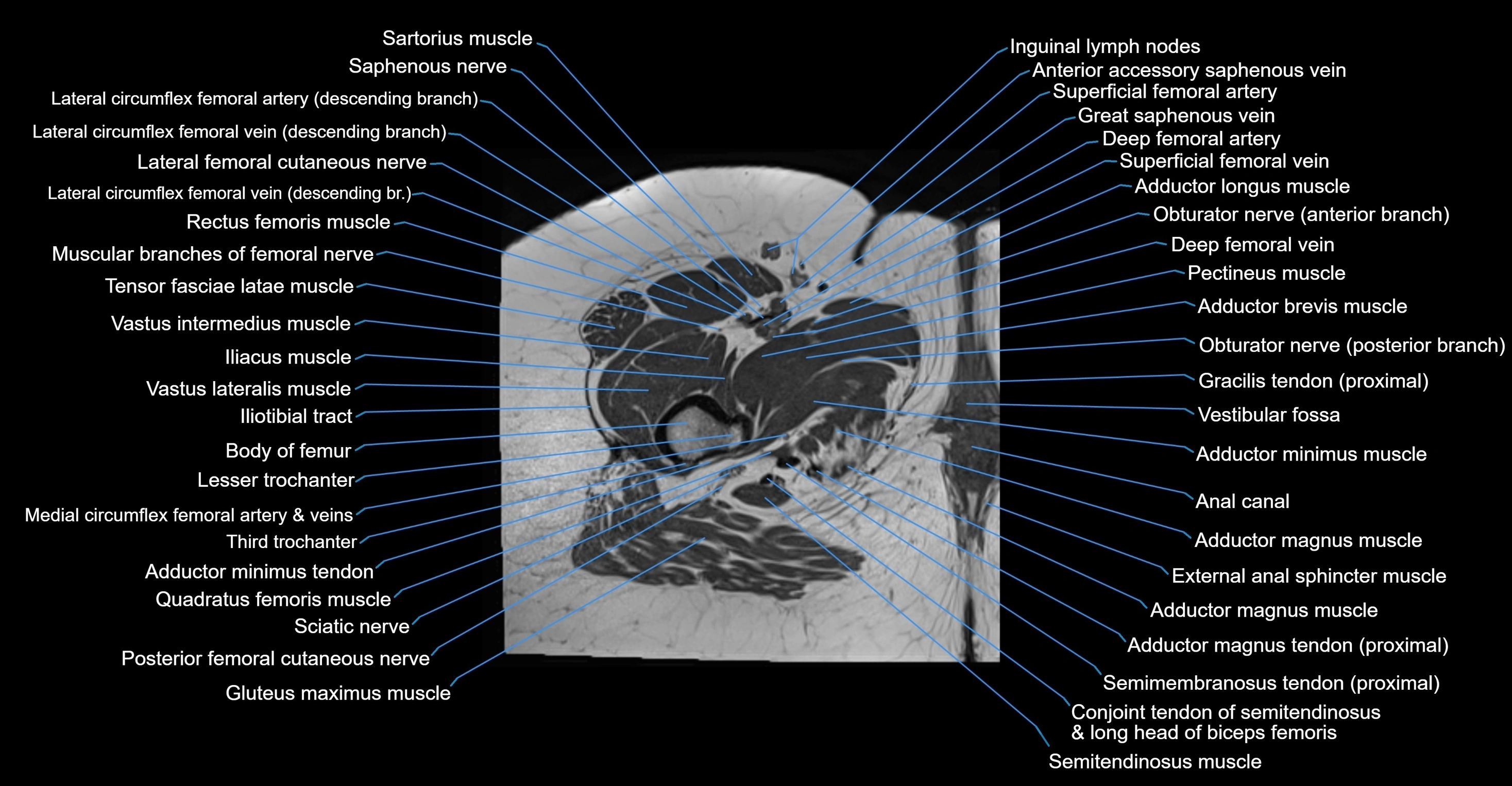
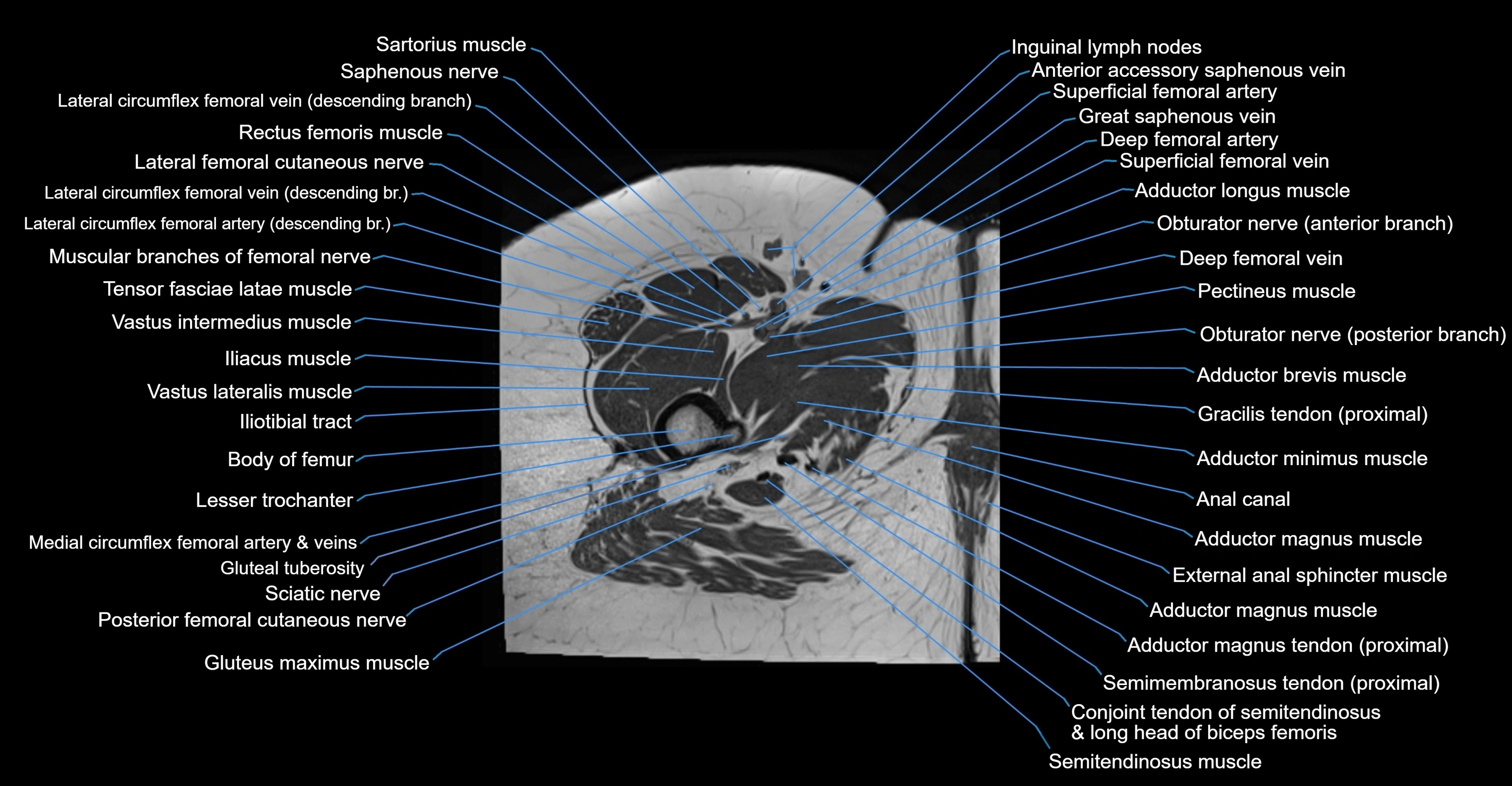
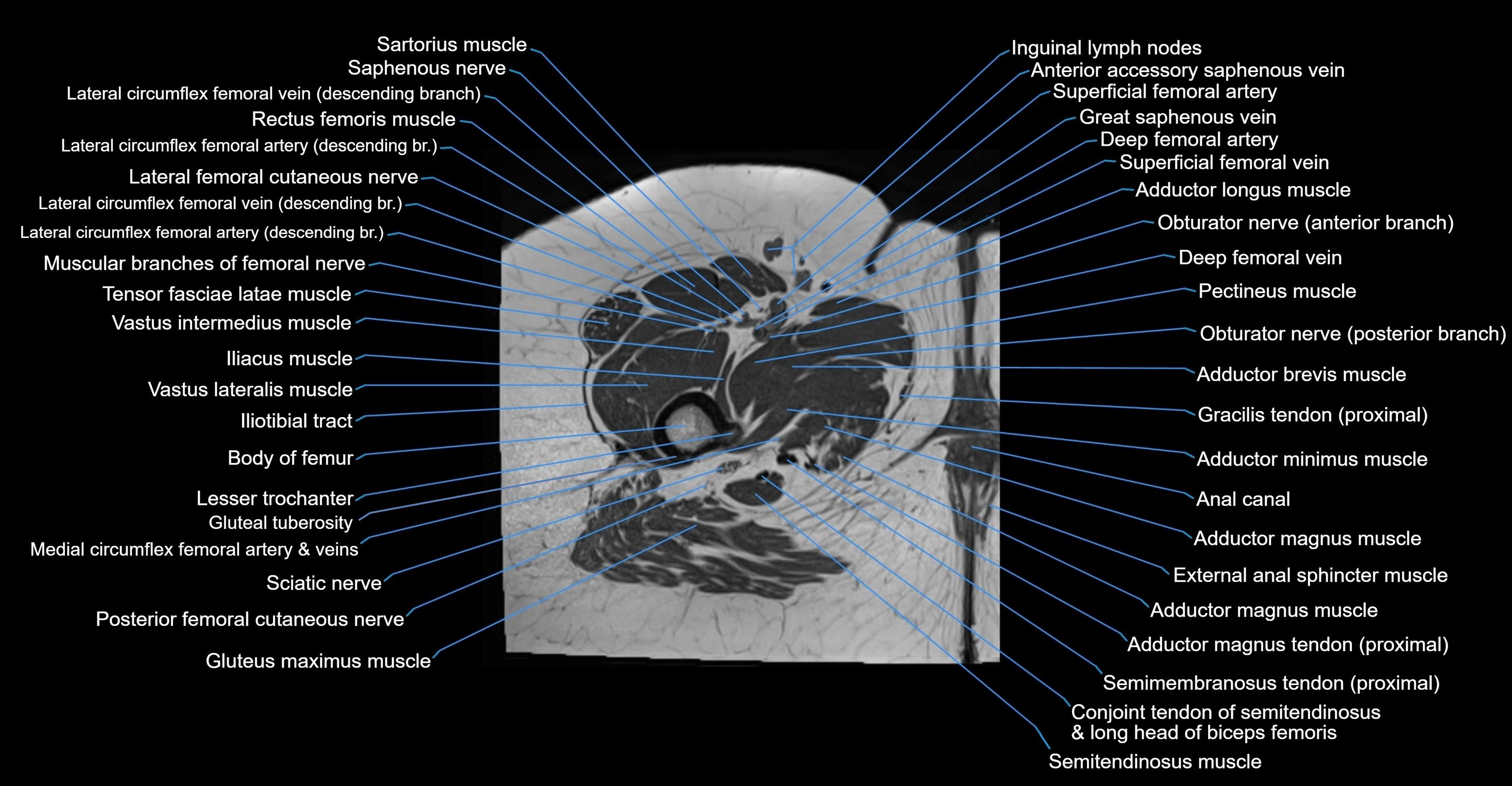
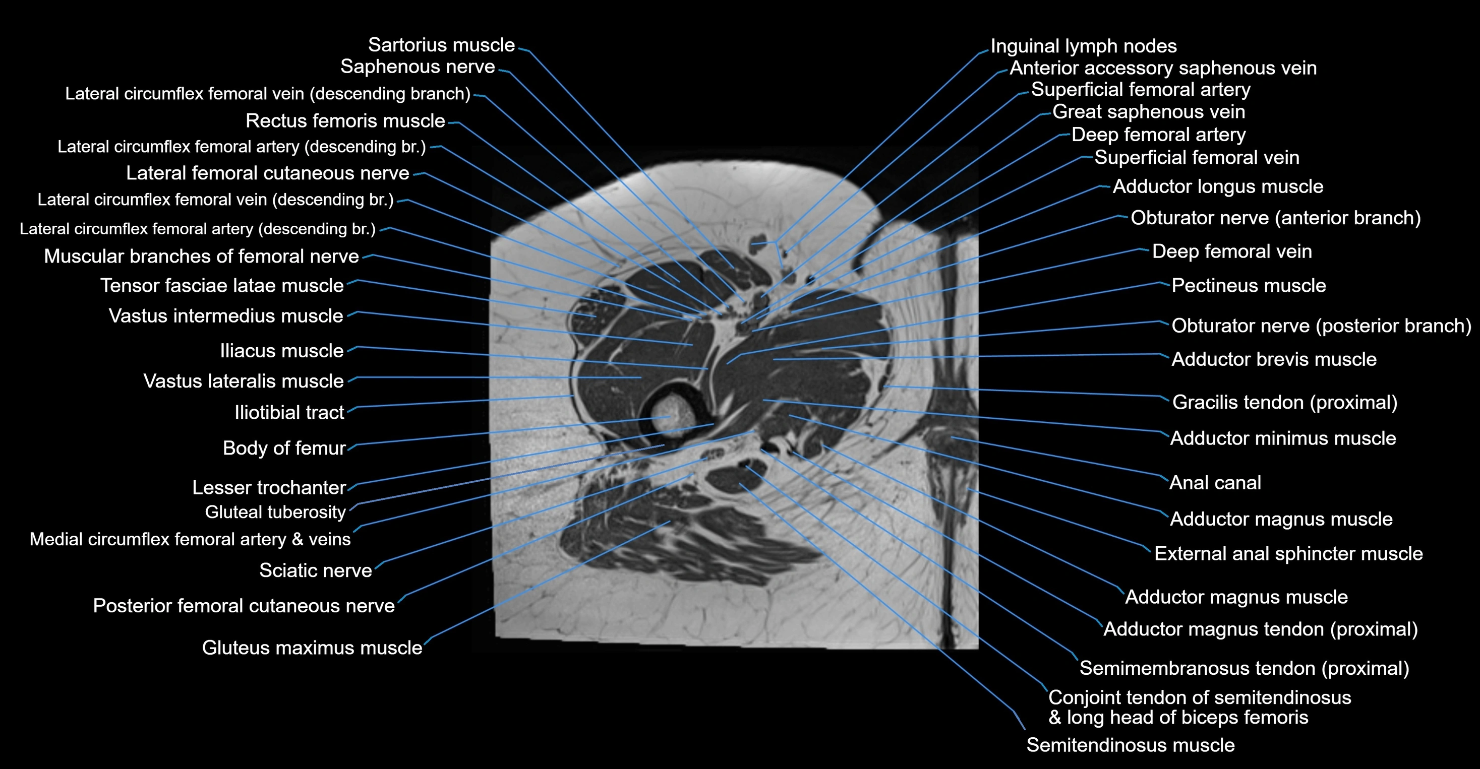
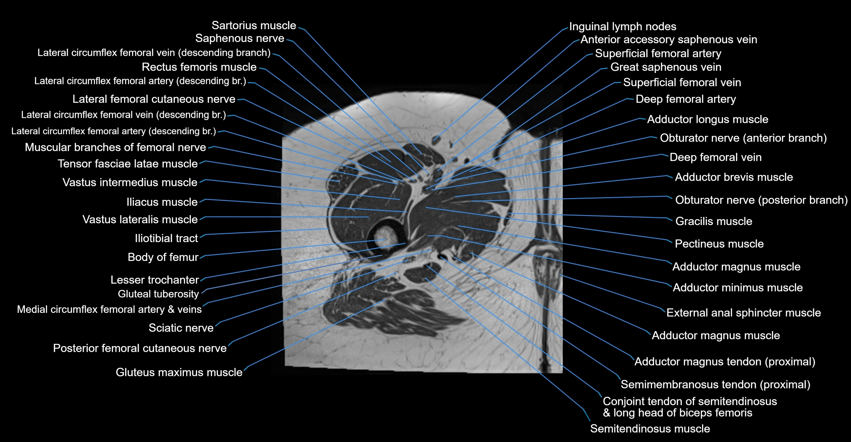
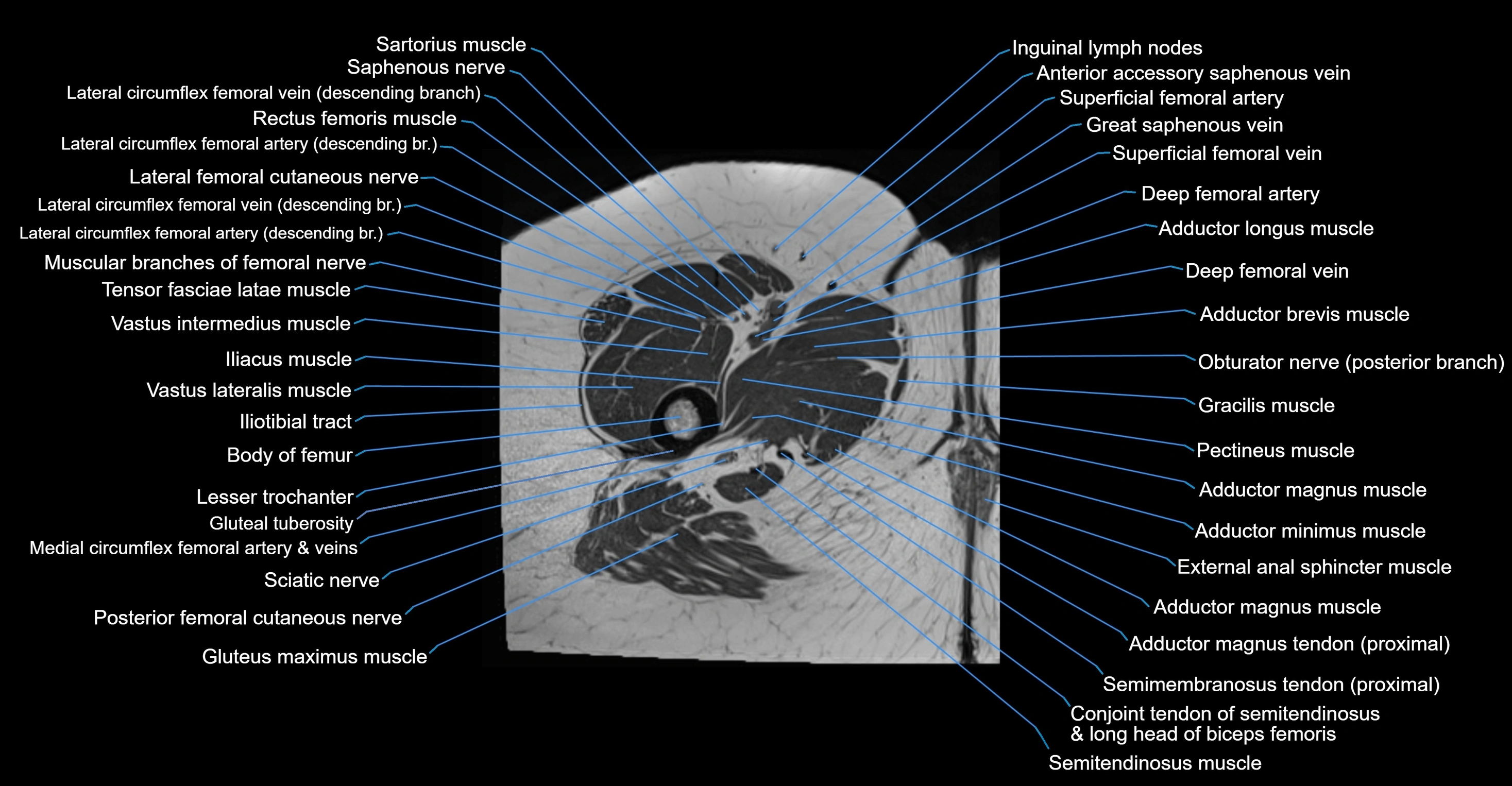
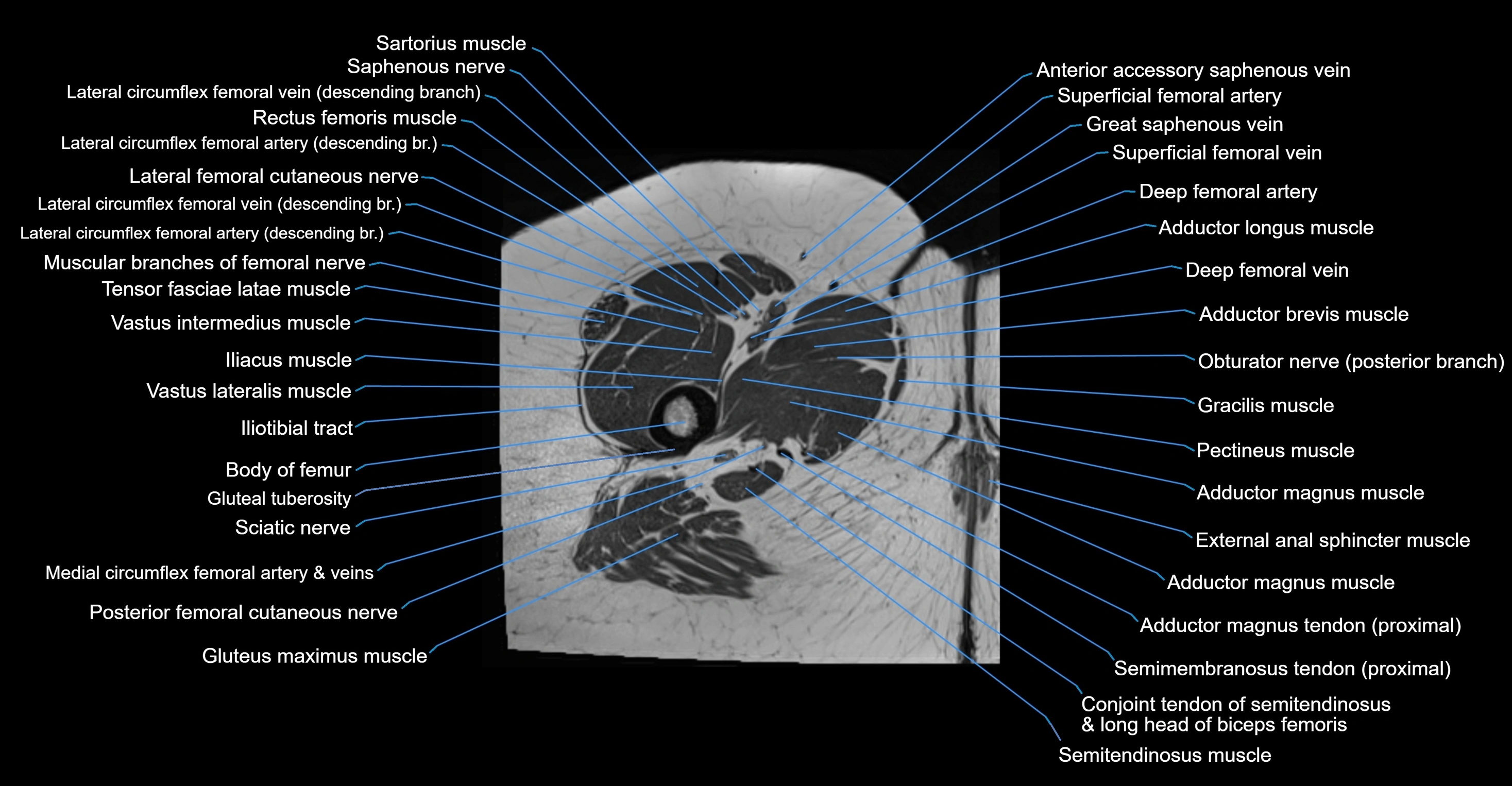
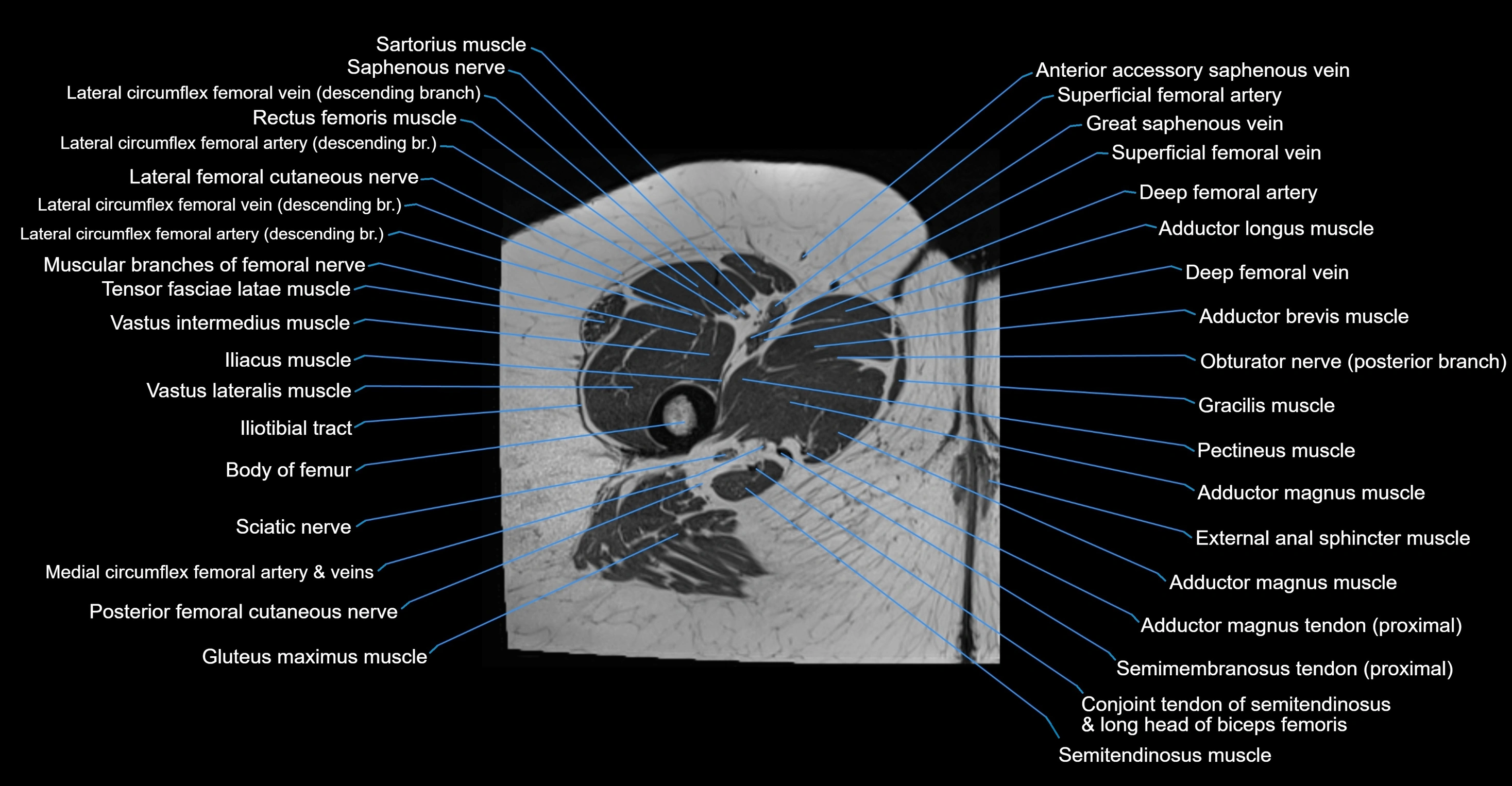
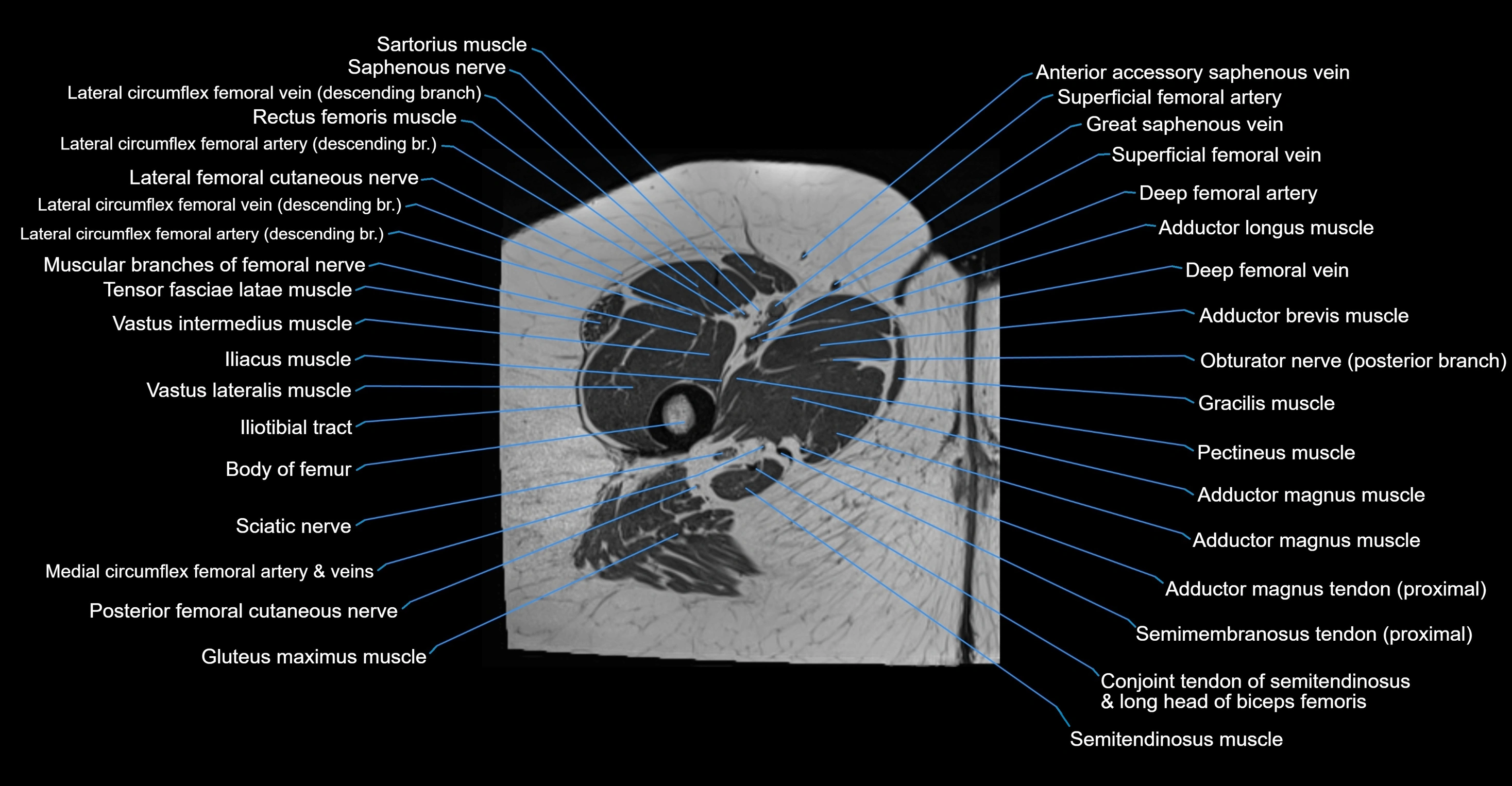
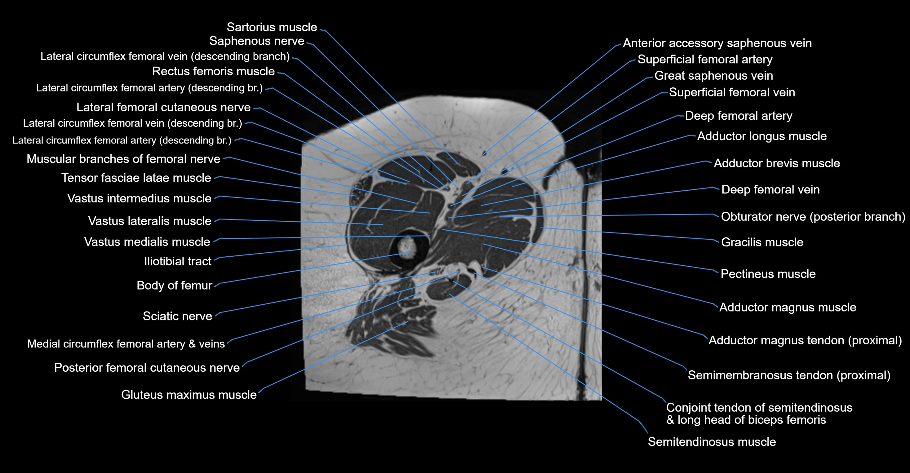
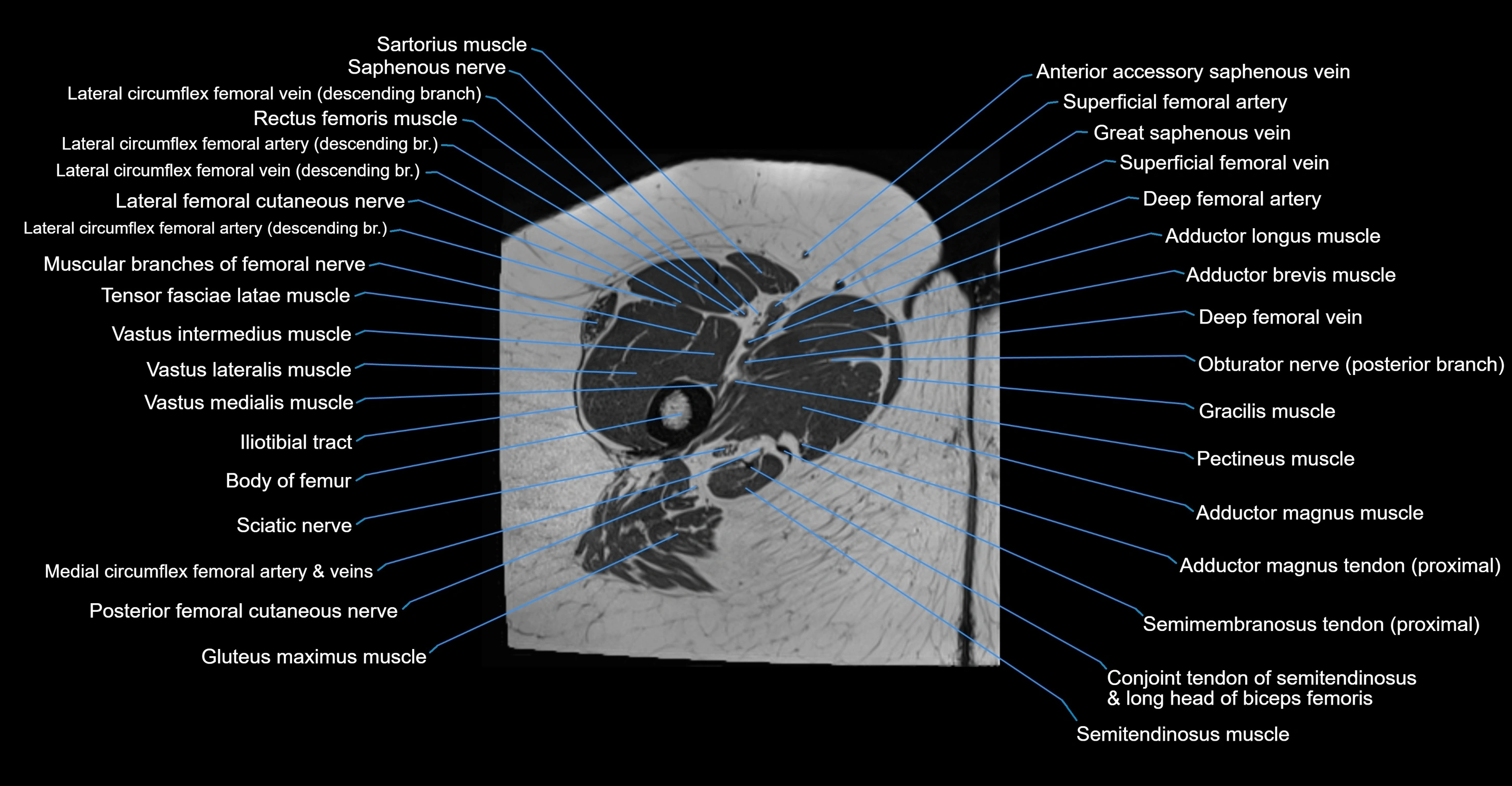
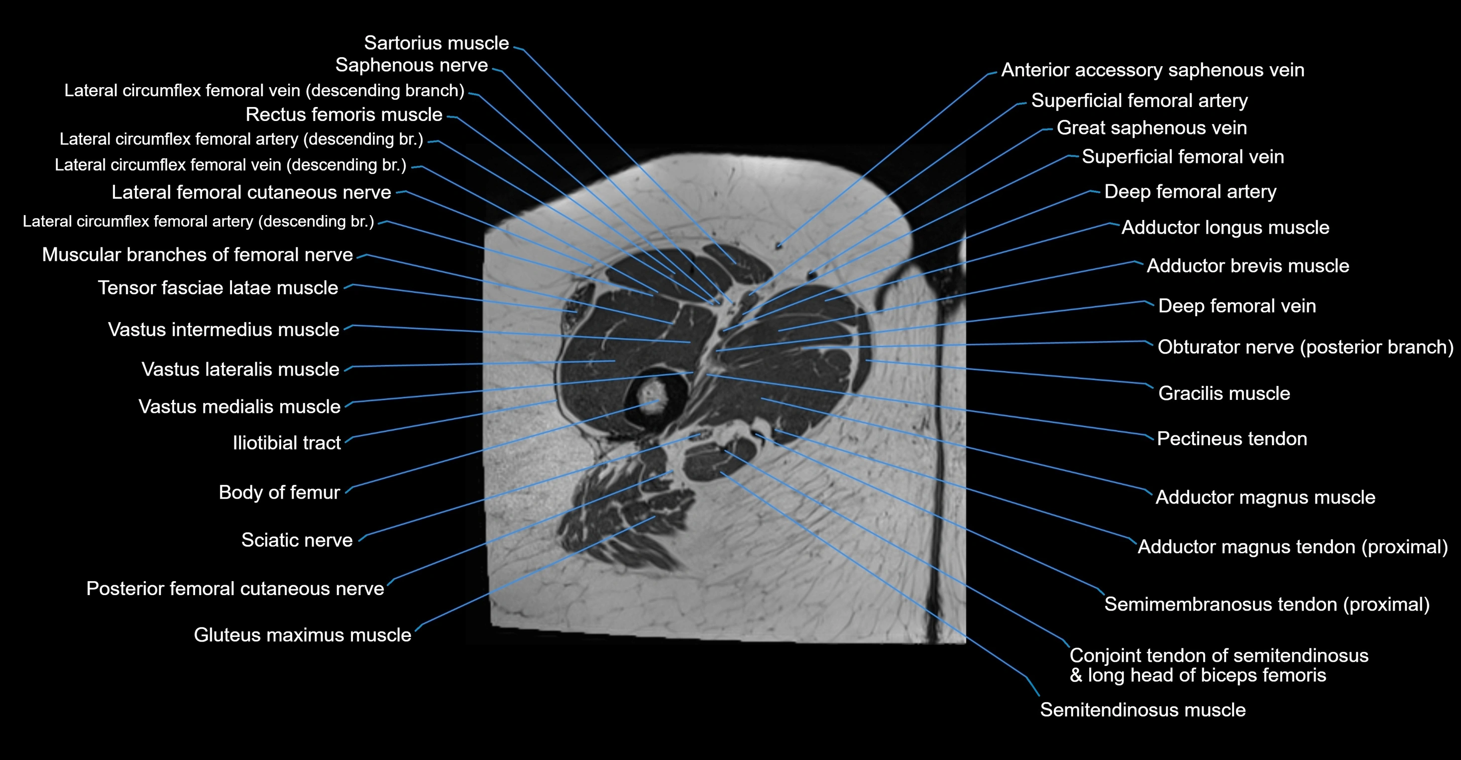
- Body of femur
- Body of ilium
- Body of ischium
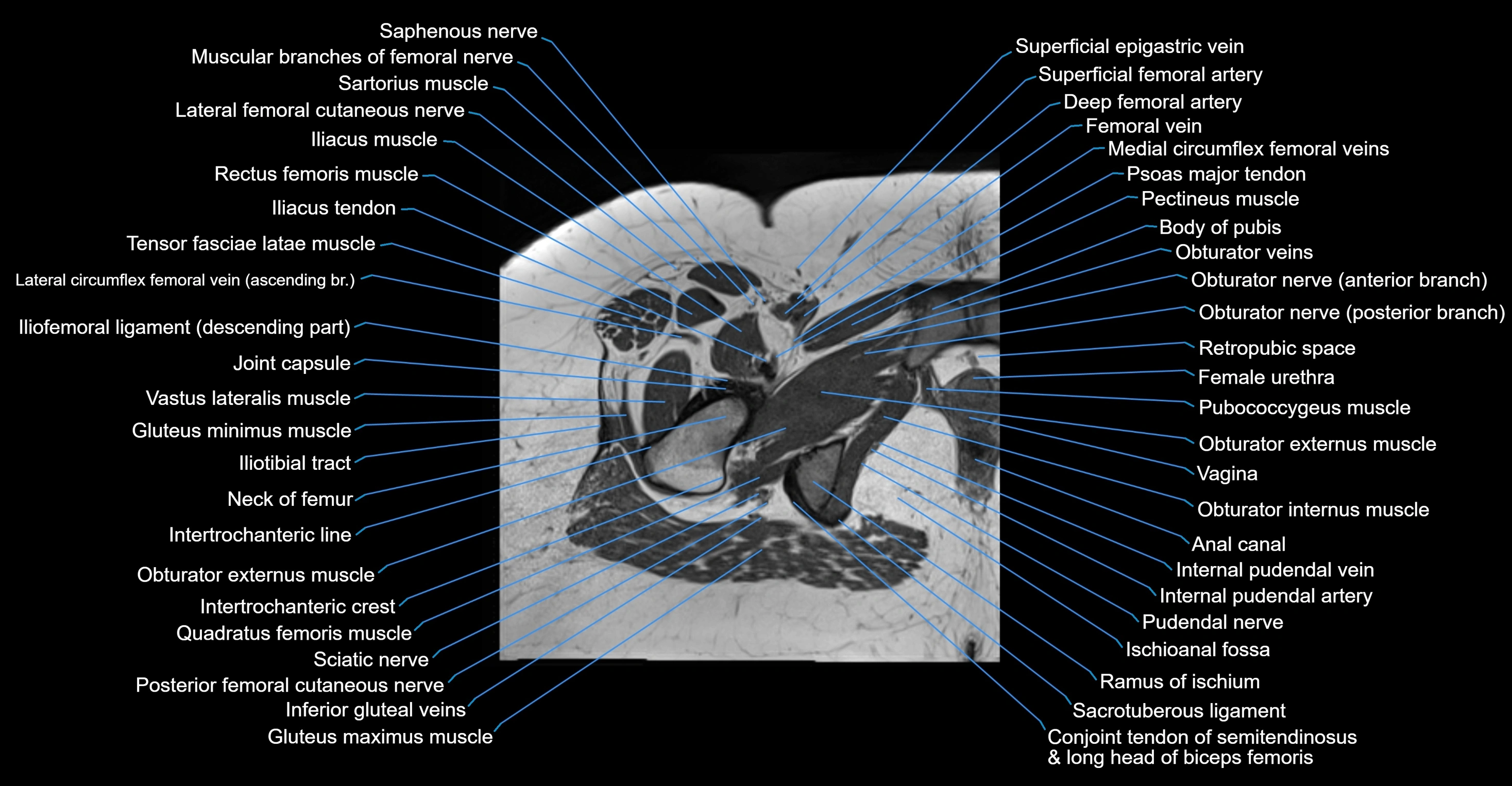
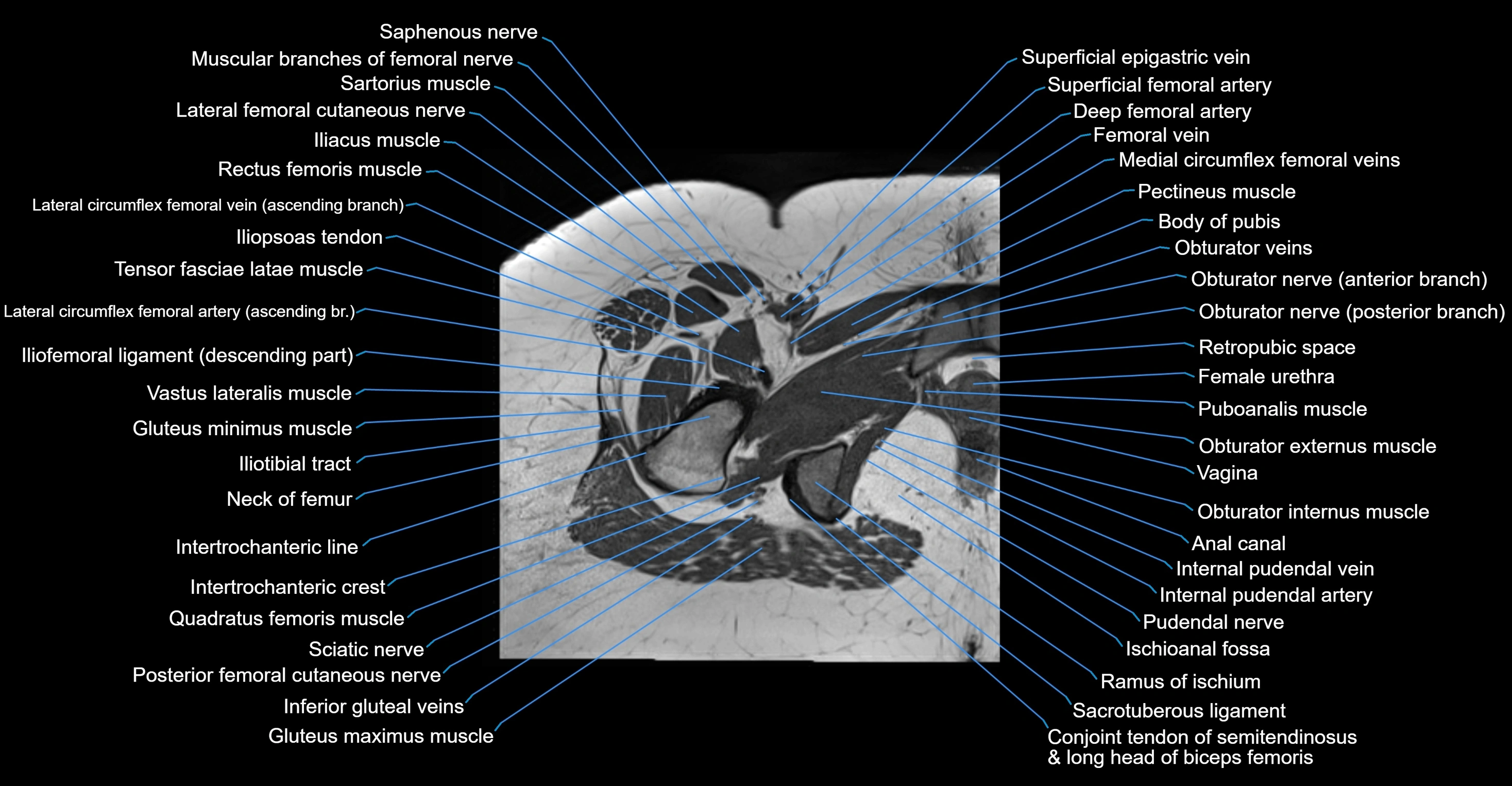
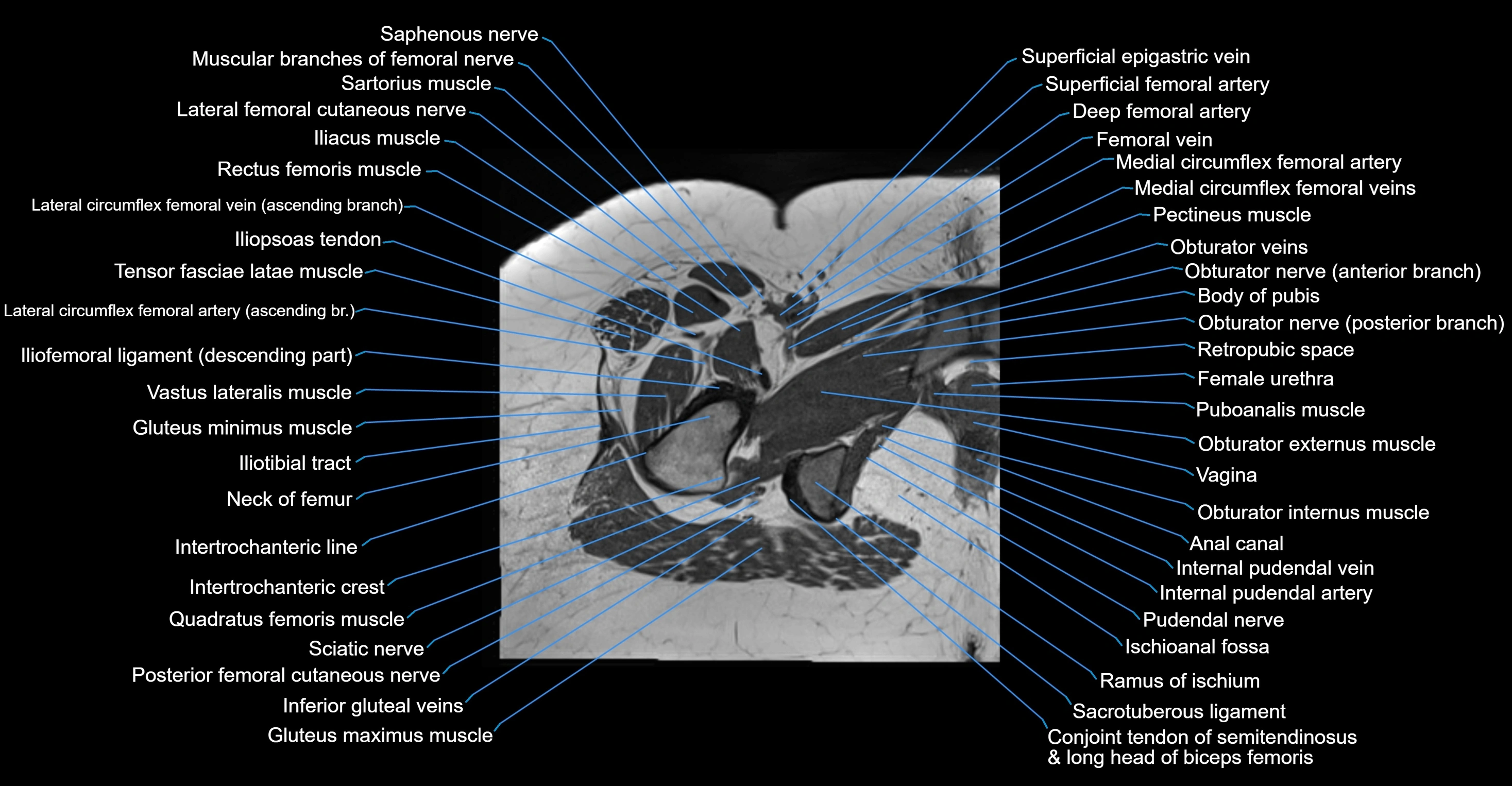
- Body of pubis
- Body of urinary bladder
- Bulbospongiosus muscle (Female)
- Bulbospongiosus muscle (Male)
- Central zone of prostate
- Clitoris
- Coccygeal nerve
- Coccygeal plexus
- Coccygeus muscle
- Coccyx
- Common iliac lymph nodes
- Common iliac vein
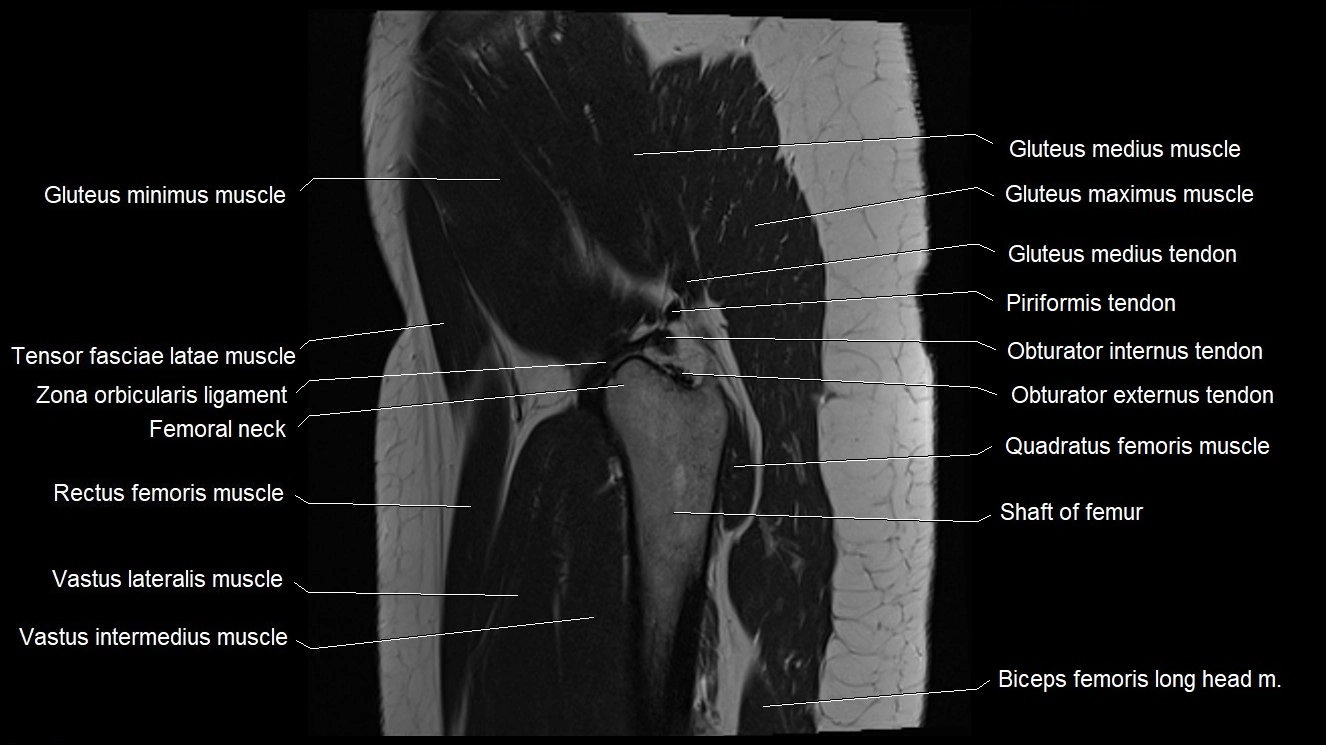
- Conjoint tendon of biceps femoris & semitendinosus
- Deep circumflex iliac artery
- Deep femoral artery (profunda femoris)
- Deep femoral vein (profunda femoris vein)
- Deep transverse perineal muscle
- Dermis of skin
- Dorsal ramus of spinal nerve
- Ejaculatory duct
- Endocervical canal
- Epidermis
- Erector spinae muscles
- Exiting nerve root of spinal nerve S1
- Exiting nerve root of spinal nerve S2
- Extensor hood of foot
- External anal sphincter
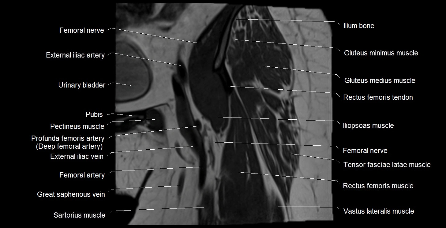
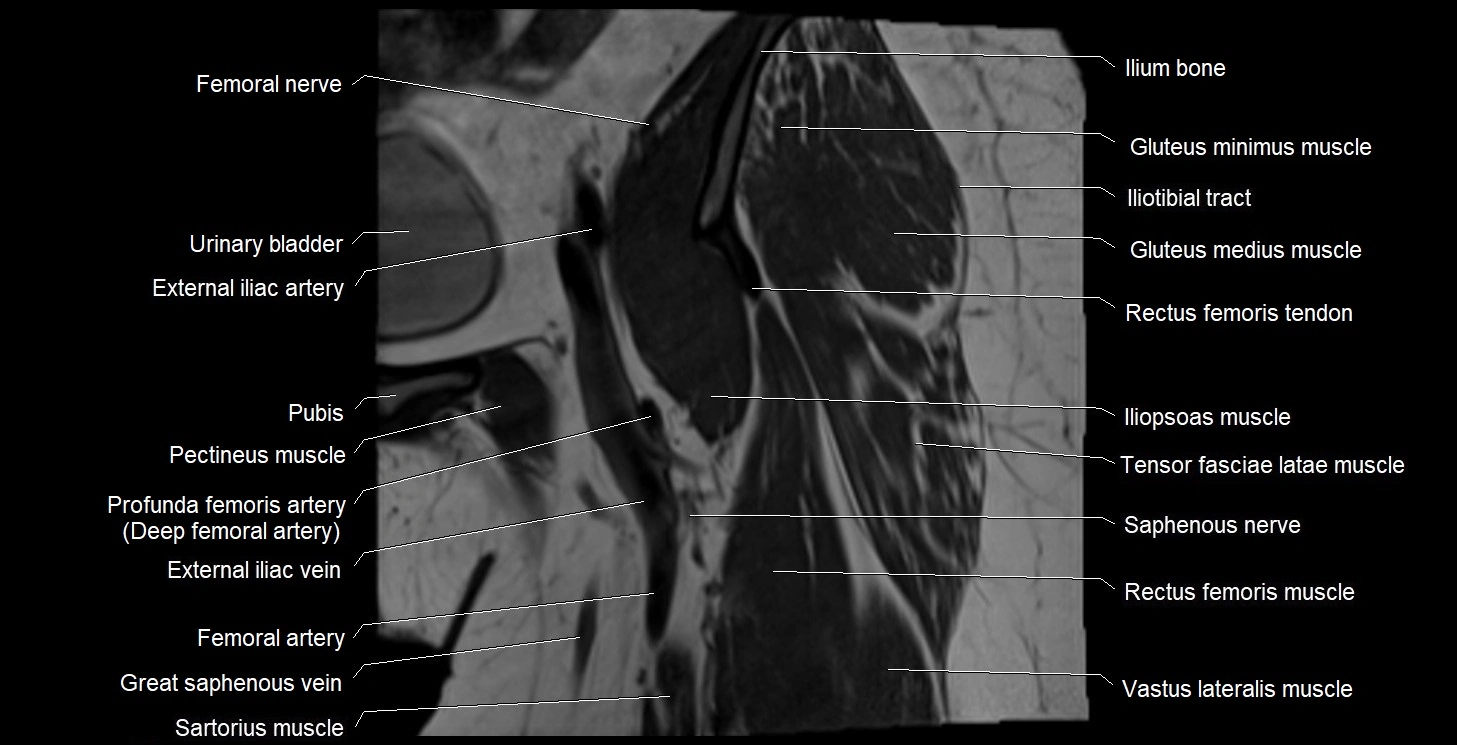
- External iliac artery
- External iliac lymph nodes
- External iliac vein
- External os of the cervix
- External urethral orifice
- External urethral sphincter (female)
- External urethral sphincter (male)
- Fascia of pelvic diaphragm
- Female urethra
- Femoral artery
- Femoral nerve
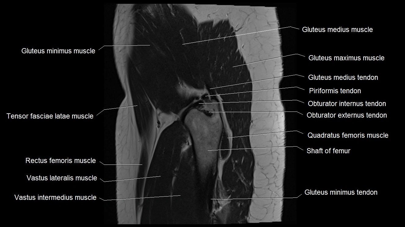
- Femoral shaft
- Femoral vein
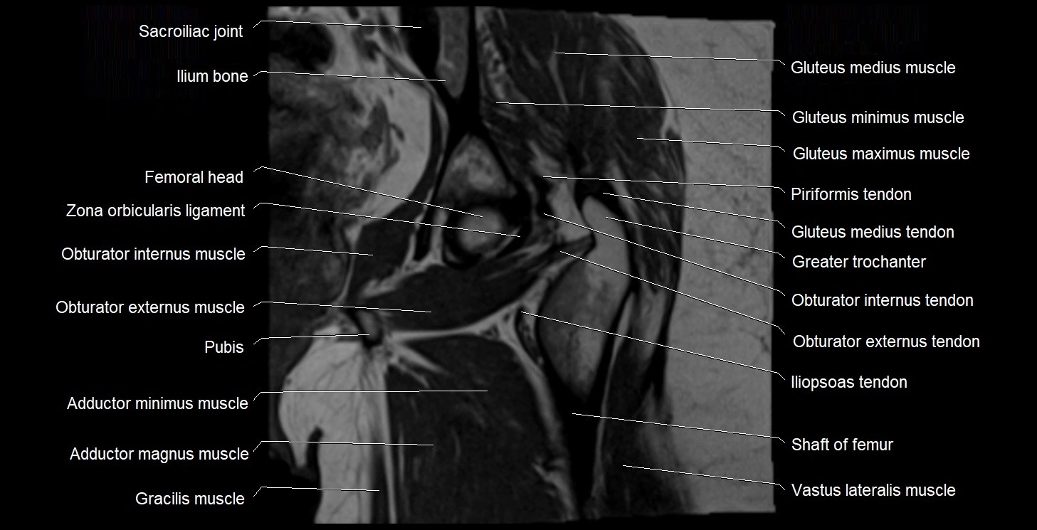
- Femur
- Filum terminale internum
- Fovea for ligament of head of femur
- Fundus of urinary bladder
- Genitofemoral nerve
- Gluteal lymph nodes
- Gluteal tuberosity
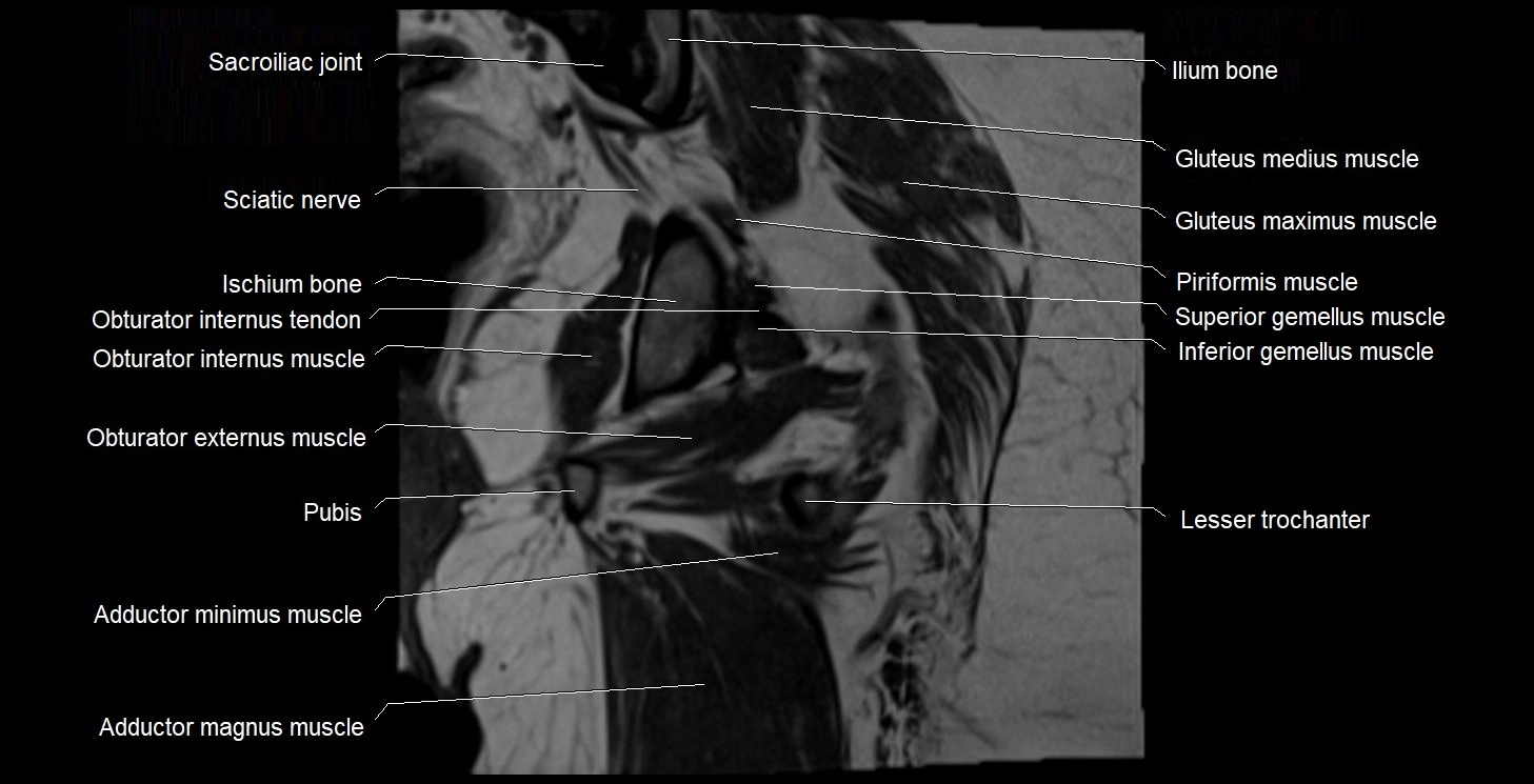
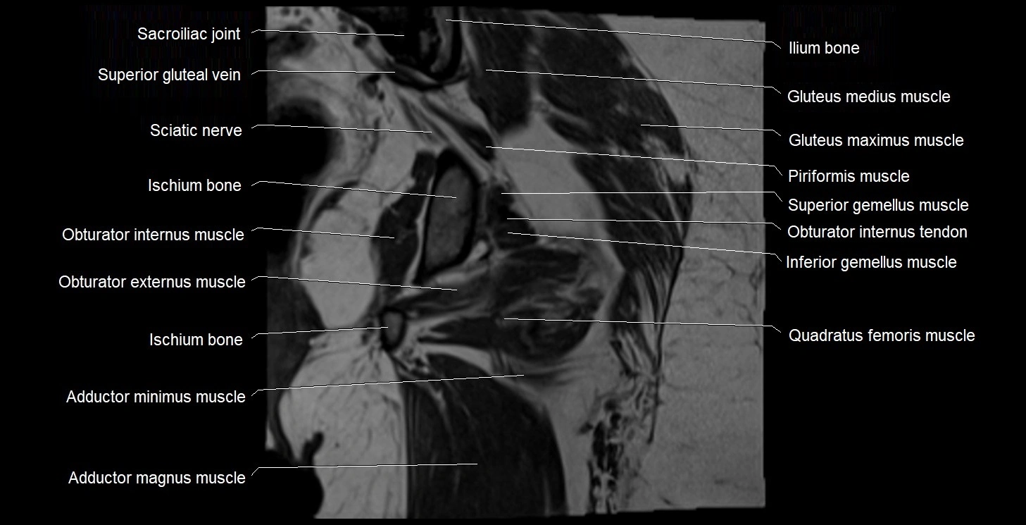
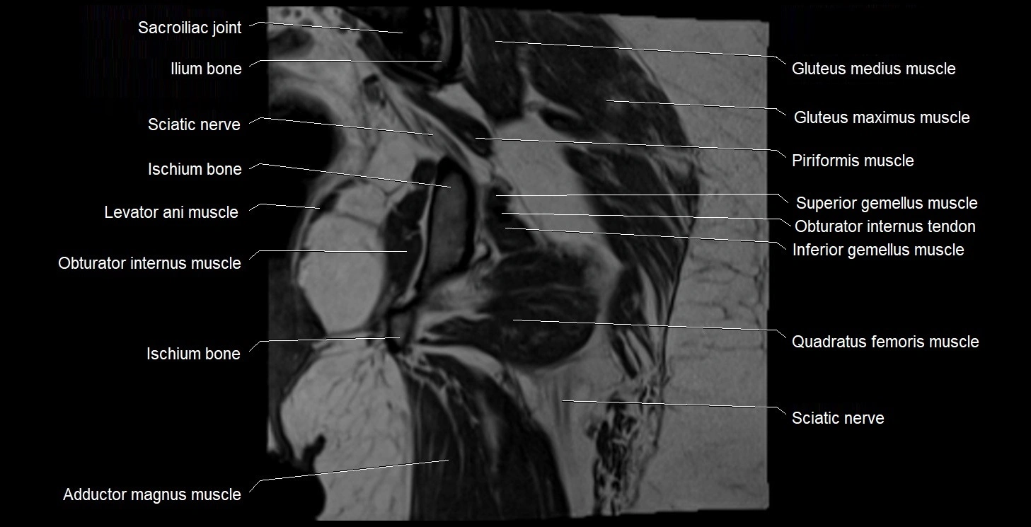
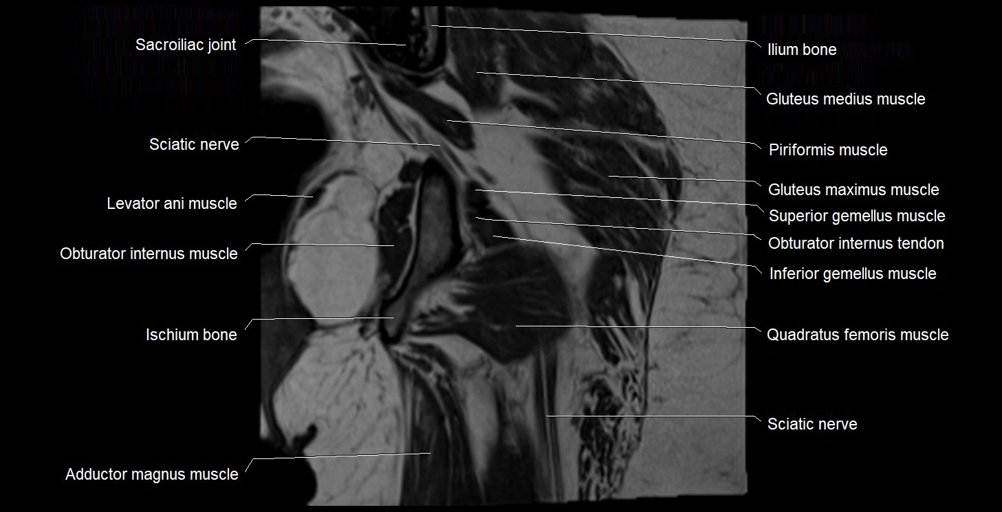
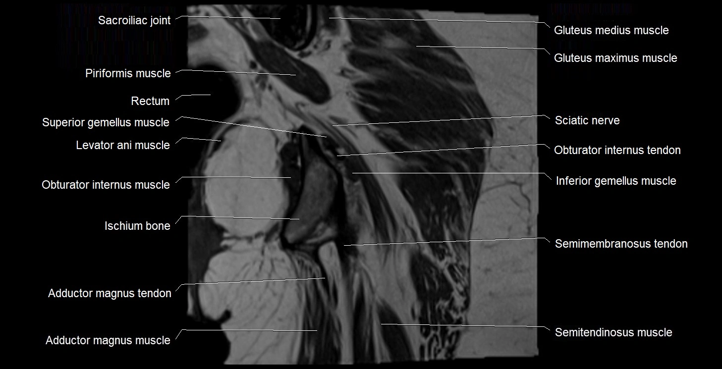
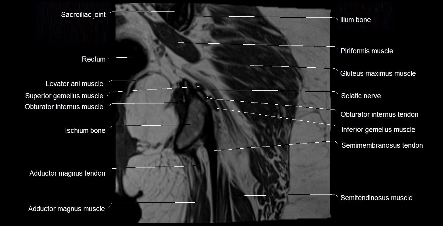
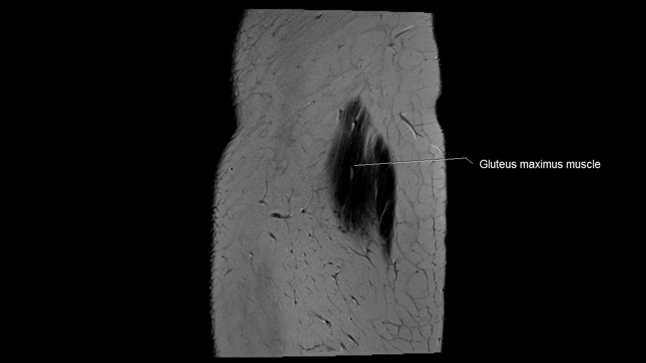
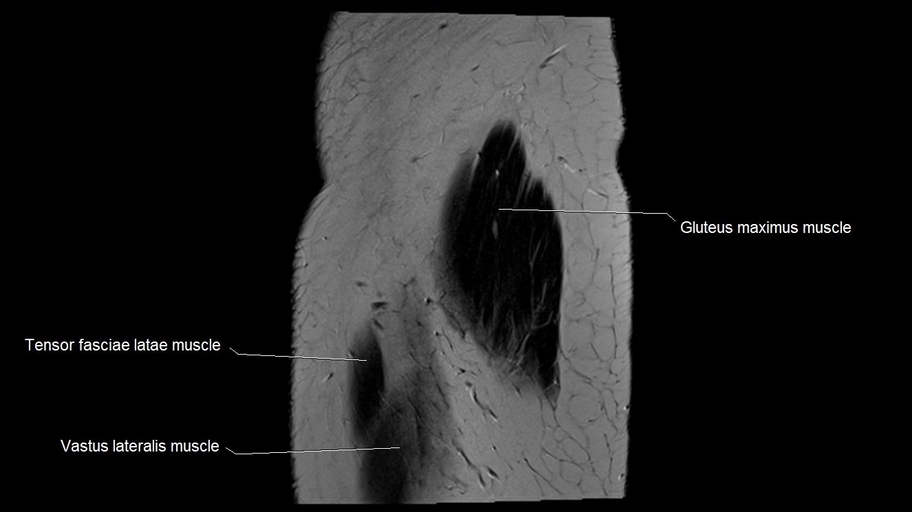
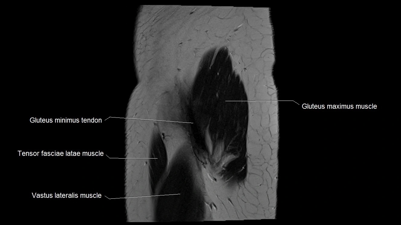
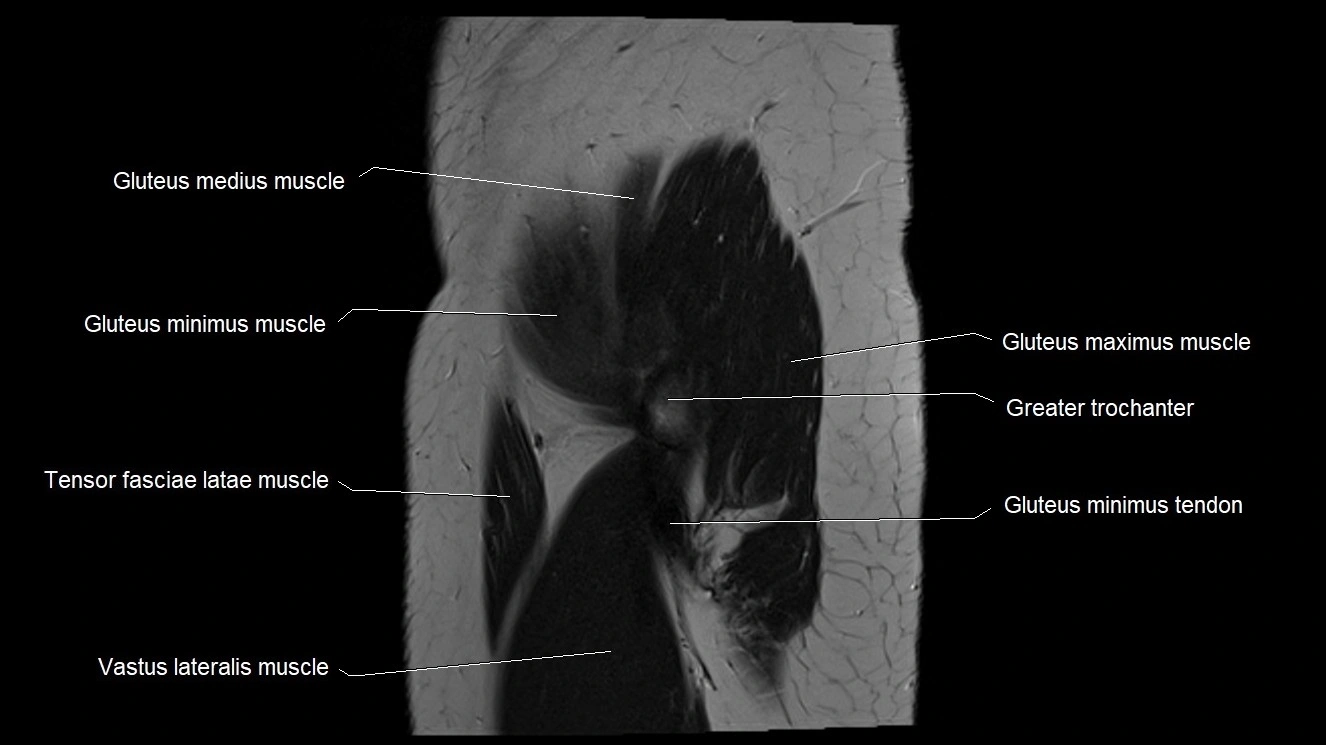
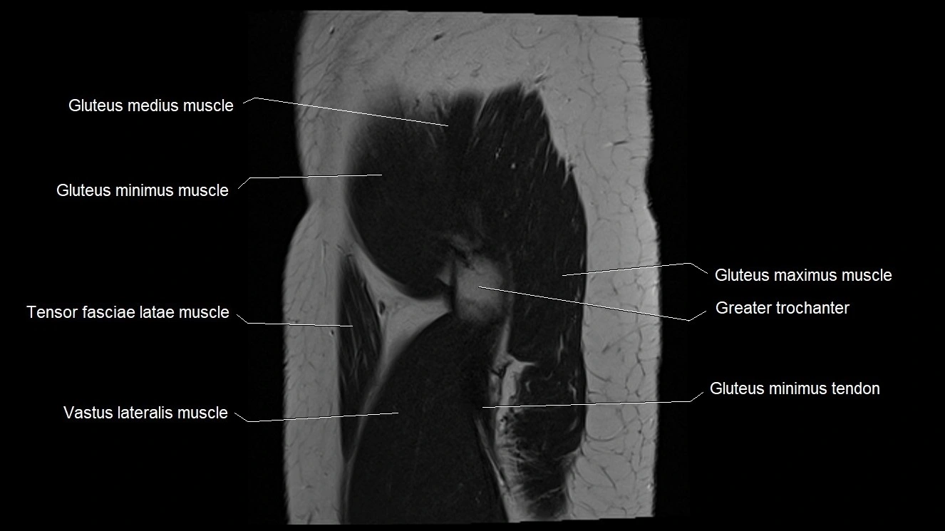
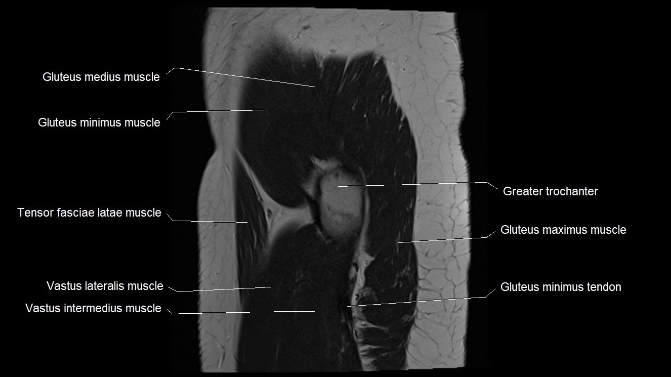
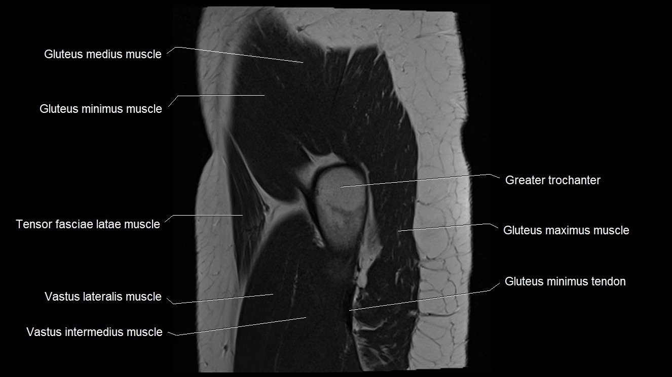
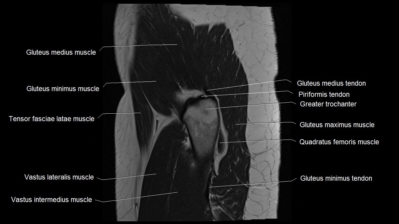
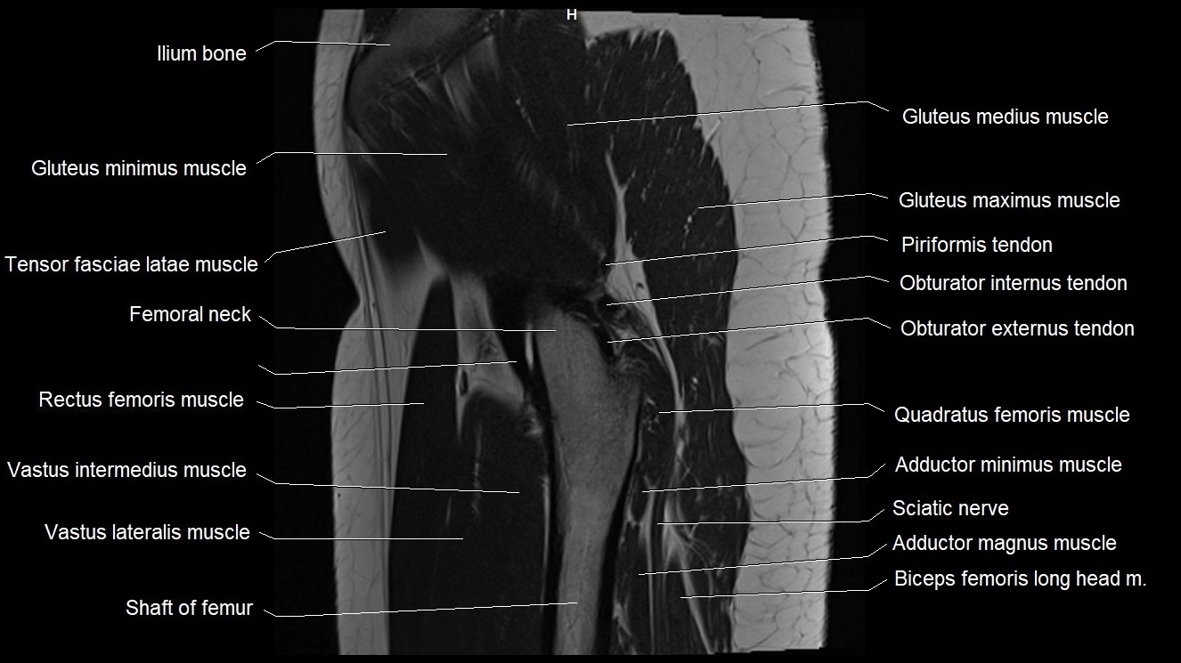
- Gluteus maximus muscle
- Gluteus medius muscle
- Gluteus medius tendon
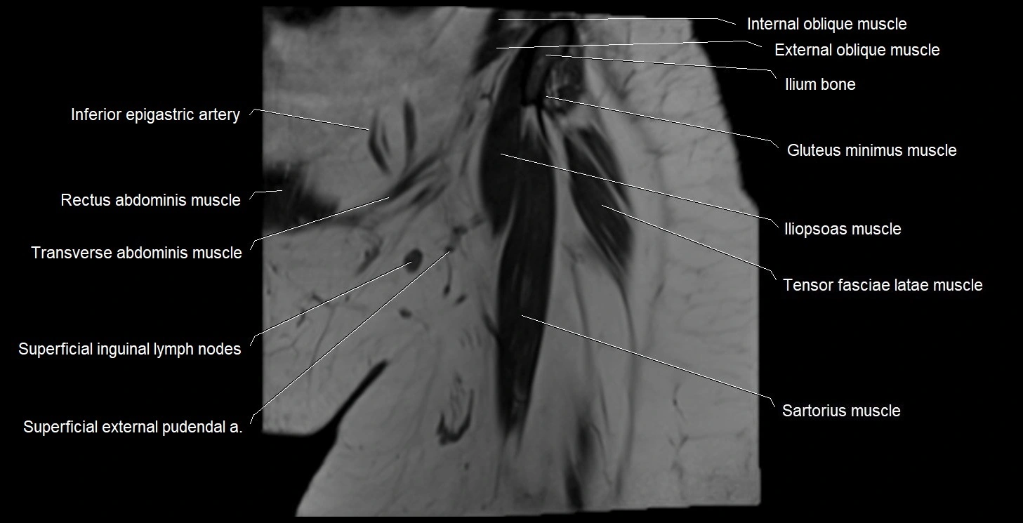
- Gluteus minimus muscle
- Gluteus minimus tendon
- Gracilis muscle
- Greater sciatic notch
- Greater trochanter
- Head of femur
- Hip joint
- Hypodermis of skin
- Iliac crest
- Iliac tubercle
- Iliococcygeus muscle
- Iliofemoral Ligament inferior band (vertical band, medial band)
- Iliofemoral Ligament superior band (transverse band, lateral band)
- Iliofemoral ligament
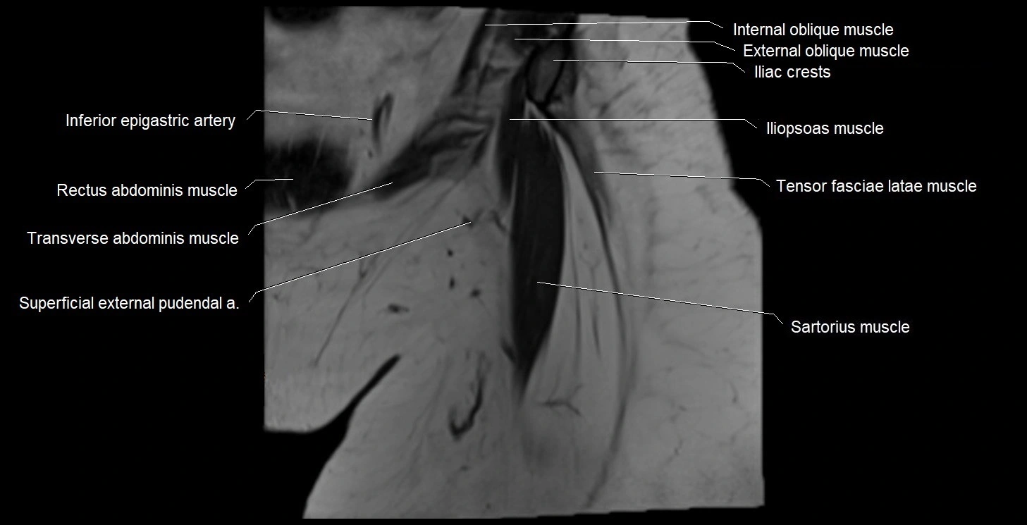
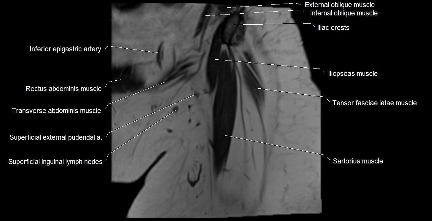
- Iliopsoas muscle
- Iliopsoas tendon
- Iliopubic eminence
- Iliotibial tract
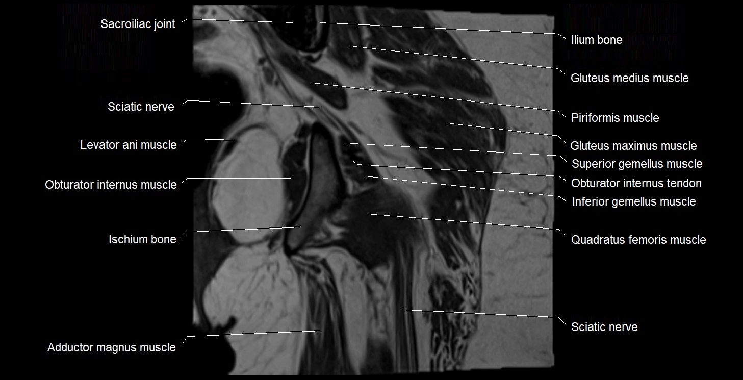
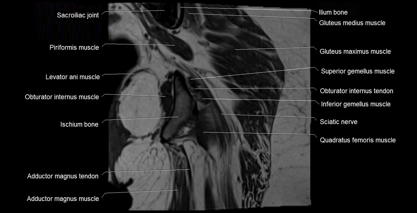
- Ilium bone
- Inferior epigastric artery
- Inferior epigastric veins
- Inferior gemellus muscle
- Inferior gluteal artery
- Inferior gluteal vein
- Inferior pubic ligament
- Inferior pubic ramus
- Inferior rectal nerve
- Inferior rim of acetabulum
- Inferior vesical artery
- Inguinal ligament
- Inguinal lymph nodes
- Intercommunicating branches of L3–L4 nerves
- Intermediate lacunar external iliac lymph nodes
- Intermediate sacral crest
- Internal anal sphincter
- Internal iliac artery
- Internal iliac lymph nodes
- Internal os of the cervix
- Internal urethral orifice
- Internal urethral sphincter (female)
- Internal urethral sphincter (male)
- Interosseous sacroiliac ligament
- Intertrochanteric crest
- Intertrochanteric line
- Ischial spine
- Ischial tuberosity
- Ischioanal fossa
- Ischiocavernosus muscle (Female)
- Ischiocavernosus muscle (Male)
- Ischiococcygeus muscle
- Ischiofemoral ligament
- Ischiopubic ramus
- Ischium bone
- Labia majora
- Labia minora
- Lateral circumflex femoral artery
- Lateral circumflex femoral veins
- Lateral femoral cutaneous nerve
- Lateral nail fold (toe)
- Lateral part of sacrum
- Lateral sacral artery
- Lateral sacral crest
- Lateral sacral vein
- Lesser trochanter
- Levator ani muscle
- Ligamentum teres (ligament of the head of femur)
- Lumbosacral trunk
- Medial circumflex femoral artery
- Medial circumflex femoral vein
- Median sacral crest
- Median sacral vein
- Median umbilical ligament
- Membranous urethra
- Mesorectal fascia
- Mesorectal free fluid
- Mesorectum
- Mons pubis
- Muscular branches of femoral nerve
- Nail (toe)
- Nail bed (toe)
- Nail root (toe)
- Neck of femur
- Neck of urinary bladder
- Obturator artery
- Obturator externus muscle
- Obturator externus tendon
- Obturator foramen
- Obturator internus muscle
- Obturator internus tendon
- Obturator lymph nodes
- Obturator nerve
- Obturator vein
- Obturator veins
- Ovaries
- Pectineus muscle
- Penile urethra
- Perineal nerves
- Peripheral zone of prostate
- Posterior acetabular wall
- Posterior division of obturator nerve (Posterior branch of obturator nerve)
- Posterior femoral cutaneous nerve
- Posterior inferior iliac spine
- Posterior lateral femoral cutaneous nerve
- Posterior rim of acetabulum
- Posterior sacral foramina
- Posterior superior iliac spine
- Posterior wall of acetabulum
- Presacral fascia
- Prostatic urethra
- Proximal nail fold
- Psoas major muscle
- Pubic bone
- Pubic symphysis
- Pubic tubercle
- Puboanalis muscle
- Pubococcygeus muscle
- Pubofemoral ligament
- Puboprostatic ligament
- Puboprostaticus muscle
- Puborectalis muscle
- Pudendal artery
- Pudendal nerve
- Pudendal vein
- Pyramidal muscle (pyramidalis muscle)
- Quadratus femoris muscle
- Ramus of ischium
- Rectal proper fascia (Fascia propria of the rectum)
- Rectococcygeal muscle
- Rectoprostatic fascia (Denonvilliers' fascia)
- Rectosacral fascia (Waldeyer's fascia)
- Rectouterine pouch (pouch of Douglas)
- Rectovaginal septum (rectovaginal fascia)
- Rectum
- Rectus femoris muscle
- Rectus femoris tendon (Proximal tendon of rectus femoris)
- Retropubic space
- Round ligament of uterus
- Sacral canal
- Sacral cornu (sacral horn)
- Sacral hiatus
- Sacral lymph nodes
- Sacral plexus
- Sacral splanchnic nerves
- Sacroiliac joint
- Sacrospinous ligament
- Sacrotuberous ligament
- Sacrum
- Saphenous nerve
- Sartorius Tendon (Proximal)
- Sartorius muscle
- Semimembranosus tendon (proximal)
- Seminal vesicle
- Sigmoid colon
- Skene’s gland (paraurethral glands)
- Spinal nerve Co1
- Spinal nerve L4
- Spinal nerve L5
- Spinal nerve S1
- Spinal nerve S2
- Spinal nerve S3
- Spinal nerve S4
- Spinal nerve S5
- Stroma of the cervix
- Superficial circumflex iliac artery
- Superficial circumflex iliac vein
- Superficial femoral artery
- Superficial inguinal lymph nodes
- Superficial transverse perineal muscle
- Superior gemellus muscle
- Superior gluteal artery
- Superior gluteal nerve
- Superior pubic ligament
- Superior pubic ramus
- Superior rectal vein
- Superior rim of acetabulum
- Superior vesical artery
- Synovial fluid
- Tendon sheath
- Tensor fasciae latae muscle
- Tensor fasciae latae tendon
- Testicular artery
- Third trochanter
- Transitional zone of prostate
- Transverse acetabular ligament
- Transverse processes
- Transverse ridges
- Traversing nerve root of spinal nerve
- Trigone of urinary bladder
- Trochanteric fossa
- Umbilical artery
- Union of common iliac veins
- Urethrovaginal space
- Urinary bladder
- Uterine artery
- Uterine horn
- Uterine venous plexus
- Uterosacral ligament
- Uterus
- Vagina
- Vaginal venous plexus
- Vas deferens
- Vastus intermedius muscle
- Vastus lateralis muscle
- Vastus medialis muscle
- Vesical veins
- Vesical venous plexus
- Vesicouterine pouch
- Vesicovaginal space
- Vestibular fossa
- Zona orbicularis ligament
- common iliac artery
- great saphenous vein
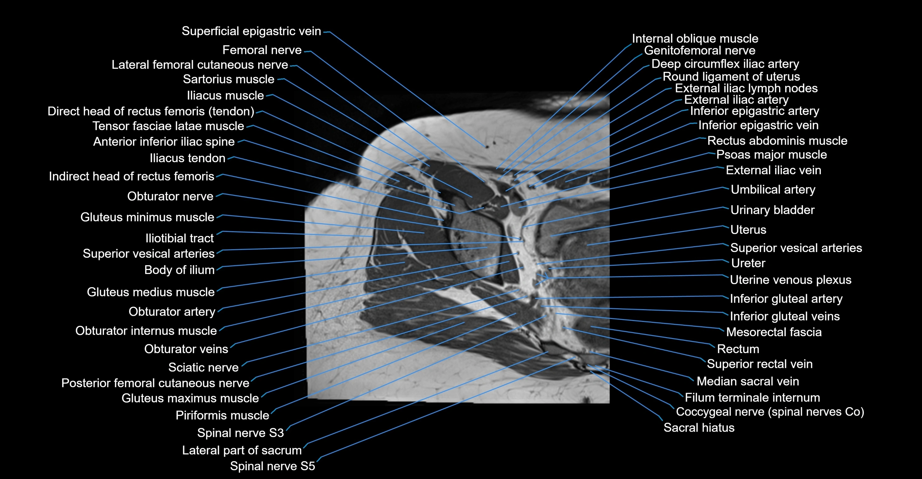
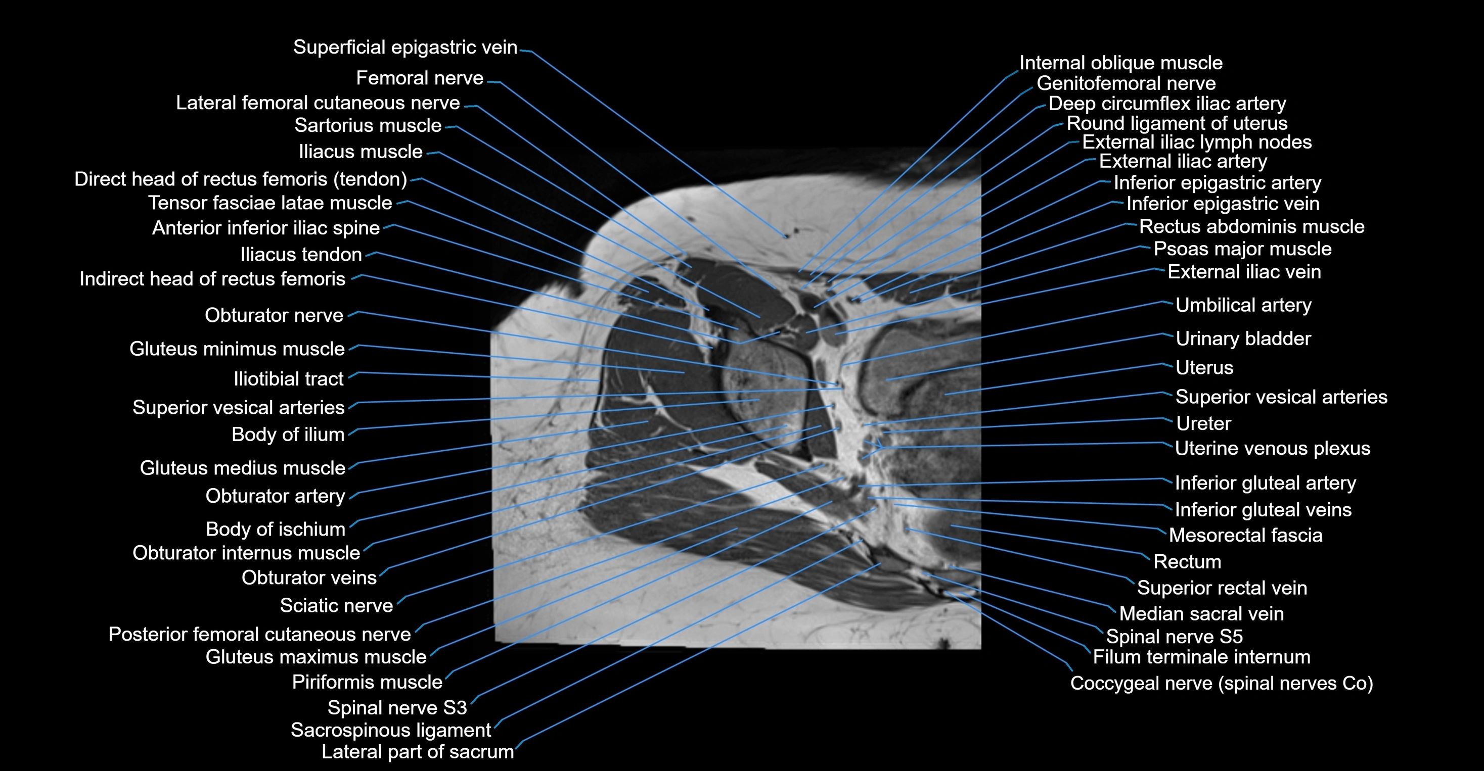
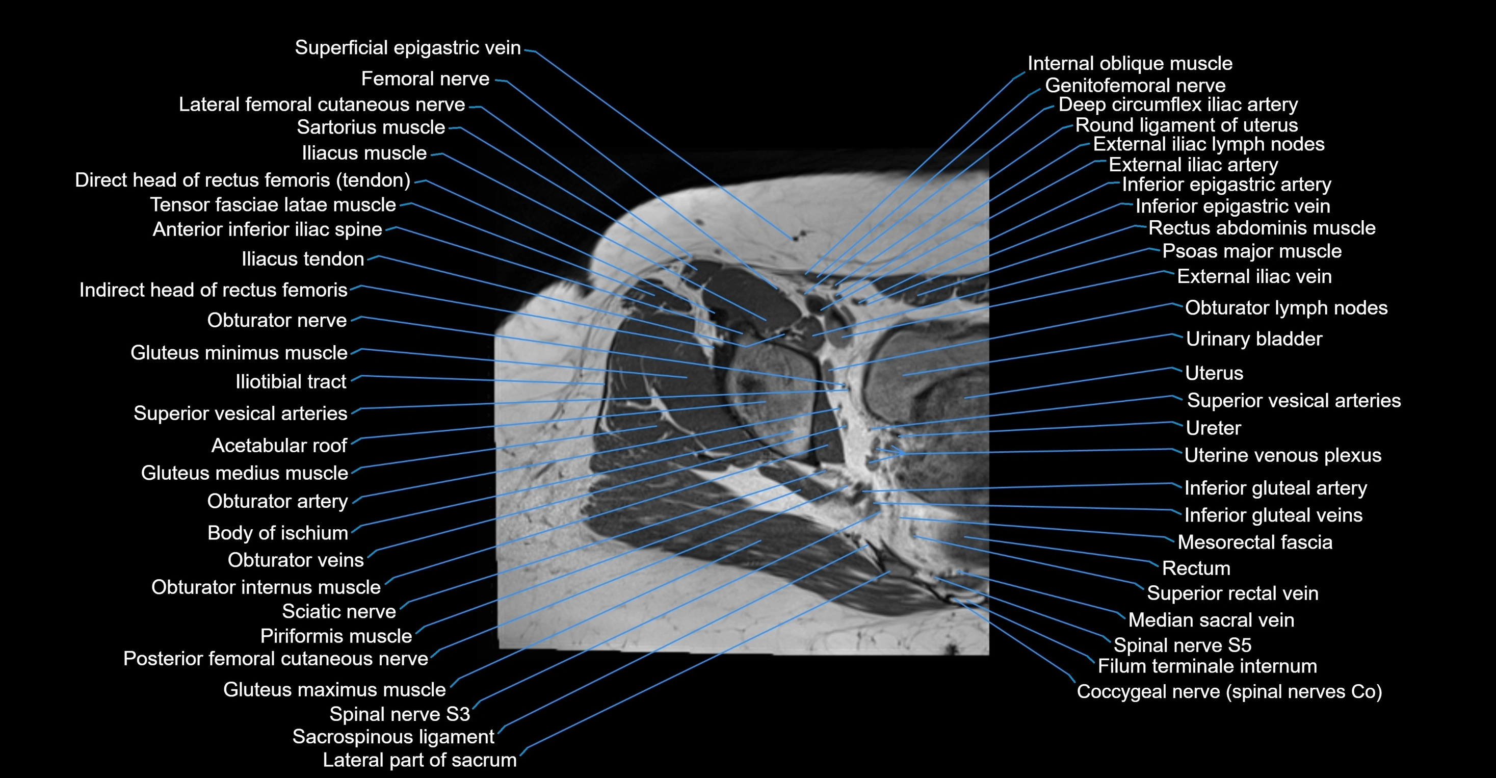
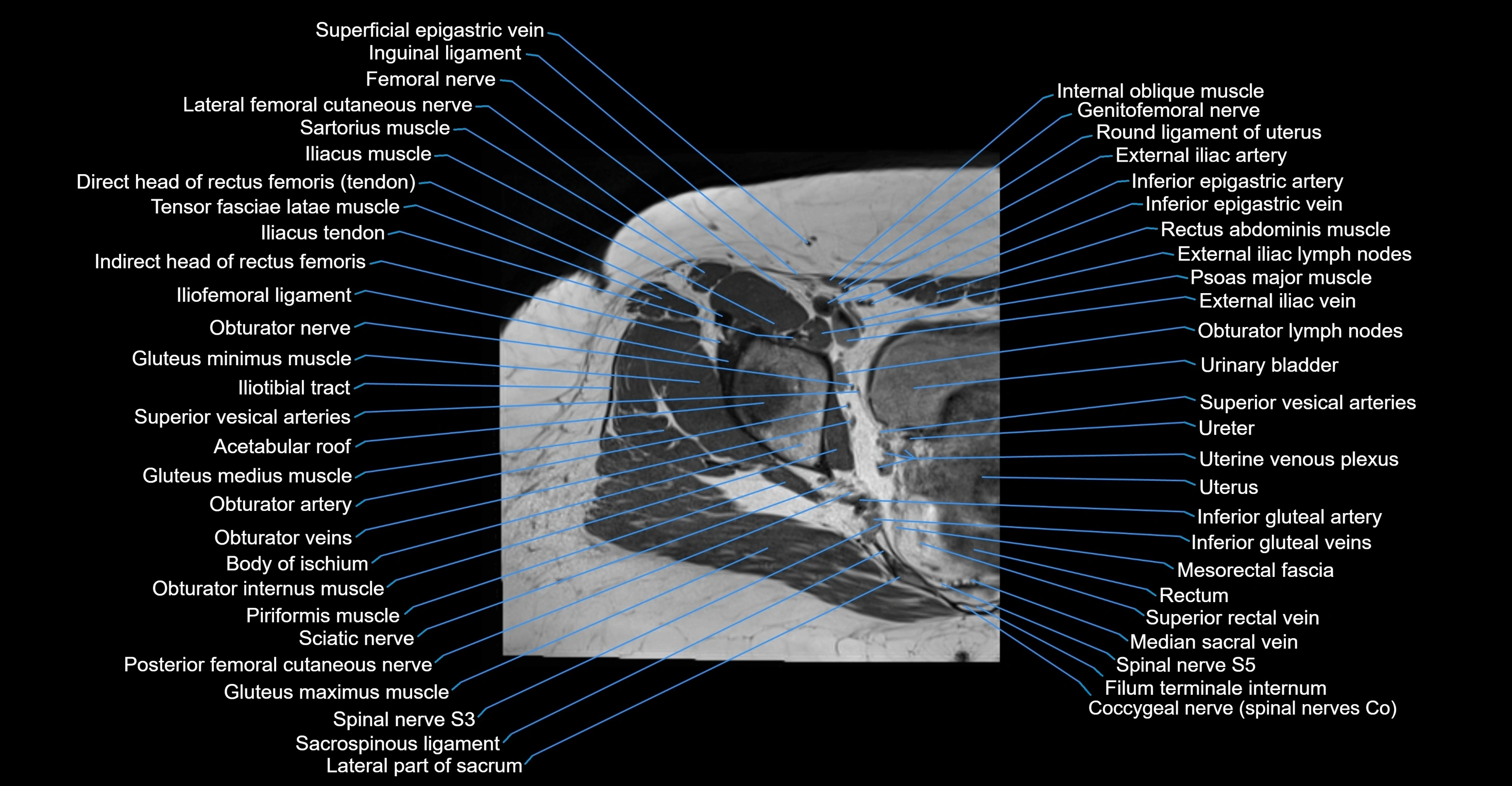
The accessory obturator artery (AOA) is an anatomical variant present in approximately 10–30% of individuals. It typically arises from the external iliac artery or inferior epigastric artery, rather than the internal iliac system. When present, it runs along the superior pubic ramus toward the obturator canal, often forming an anastomosis with the obturator artery.
This artery is clinically significant because it may contribute to the vascularization of the obturator region, pubic bone, and medial thigh, and can create a corona mortis (Latin for "crown of death") when it forms a large pubic anastomosis between the external and internal iliac systems. Injury to this artery during pelvic or hernia surgery can result in life-threatening hemorrhage.
Function
-
Provides collateral circulation to the obturator territory when the main obturator artery is absent, small, or compromised
-
Supplies branches to the pubic bone, hip joint capsule, and adductor muscles
-
Clinically important in pelvic trauma, hernia repairs, orthopedic and gynecological surgery
MRI Appearance
T1-weighted images:
-
Artery appears as a small linear hypointense flow void coursing over the superior pubic ramus
-
Seen within bright perivascular fat of pelvis
T2-weighted images:
-
Artery lumen is a signal void
-
In thrombosed or diseased variants, lumen may appear hyperintense relative to surrounding fat
STIR:
-
Fat suppression makes the artery more visible within pelvic fat
-
Helps identify perivascular edema, hematoma, or inflammatory changes
T1 Post-Gadolinium (with fat suppression):
-
Artery enhances brightly and homogeneously
-
Useful for tracing the course, anastomoses, and presence of corona mortis
-
Highlights arterial wall thickening or tumor encasement if present
MRA Pelvis with Gadolinium:
-
Clearly delineates the origin, course, and anastomoses of the accessory obturator artery
-
Identifies connection with inferior epigastric artery, external iliac artery, or obturator artery
-
Excellent for detecting vascular variants prior to surgery
-
Useful in mapping pelvic vasculature in trauma, tumor embolization, or preoperative planning
CT Appearance
Non-contrast CT:
-
Artery not well seen without contrast
-
Can suggest its location along the superior pubic ramus by small vessel density
CT Post-Contrast:
-
Vessel opacifies clearly along superior pubic ramus
-
Detects vascular variants, aneurysms, or active bleeding
-
Important in trauma imaging when pelvic fractures are associated with hemorrhage


