


Topic
- Alveolar arch of maxilla
- Alveolar process of maxilla
- Alveolar ridge
- Angle of mandible
- Anterior atlanto-occipital membrane
- Anterior belly of digastric muscle
- Anterior ethmoidal air cells
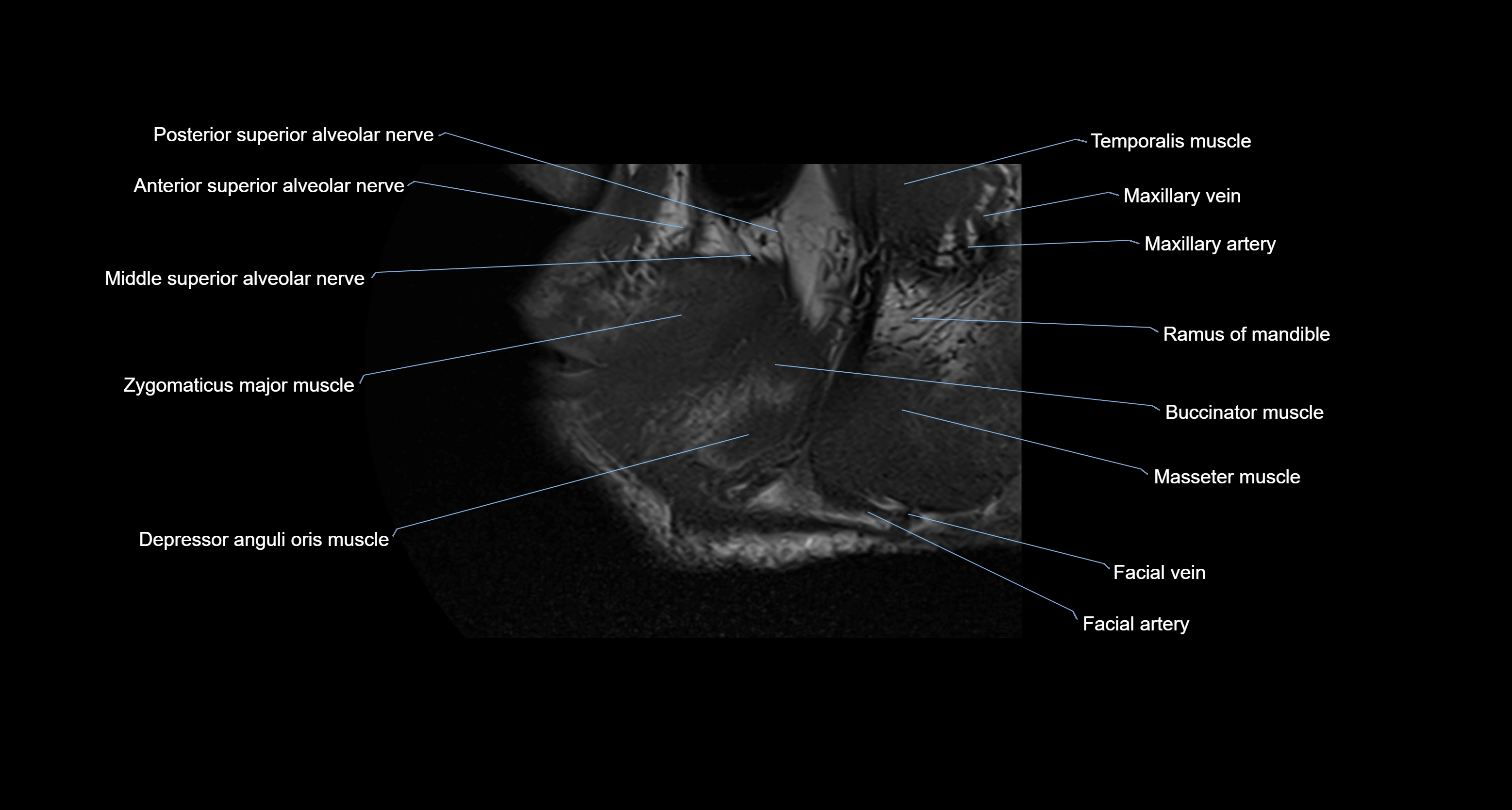
- Anterior superior alveolar nerve
- Apex of nose
- Auricularis anterior muscle
- Auricularis posterior muscle
- Body of mandible
- Body of tongue
- Buccinator muscle
- Cartilaginous part of nasal septum
- Central inferior incisor tooth
- Central superior incisor tooth
- Coronoid process of mandible
- Cricothyroid muscle
- Cruciate ligament of the atlas
- Dental branches of inferior alveolar artery, vein, & nerve
- Dental pulp of upper molar tooth
- Dental pulp of upper premolar tooth
- Dental pulp of lower molar tooth
- Depressor anguli oris muscle
- Depressor labii inferioris muscle
- Depressor septi nasi muscle
- Dorsum of tongue
- Enamel of lower molar tooth
- Enamel of upper molar tooth
- Enamel of canines tooth
- Enamel of lower incisor tooth
- Enamel of lower canines tooth
- Enamel of lower premolar tooth
- Enamel of upper incisor tooth
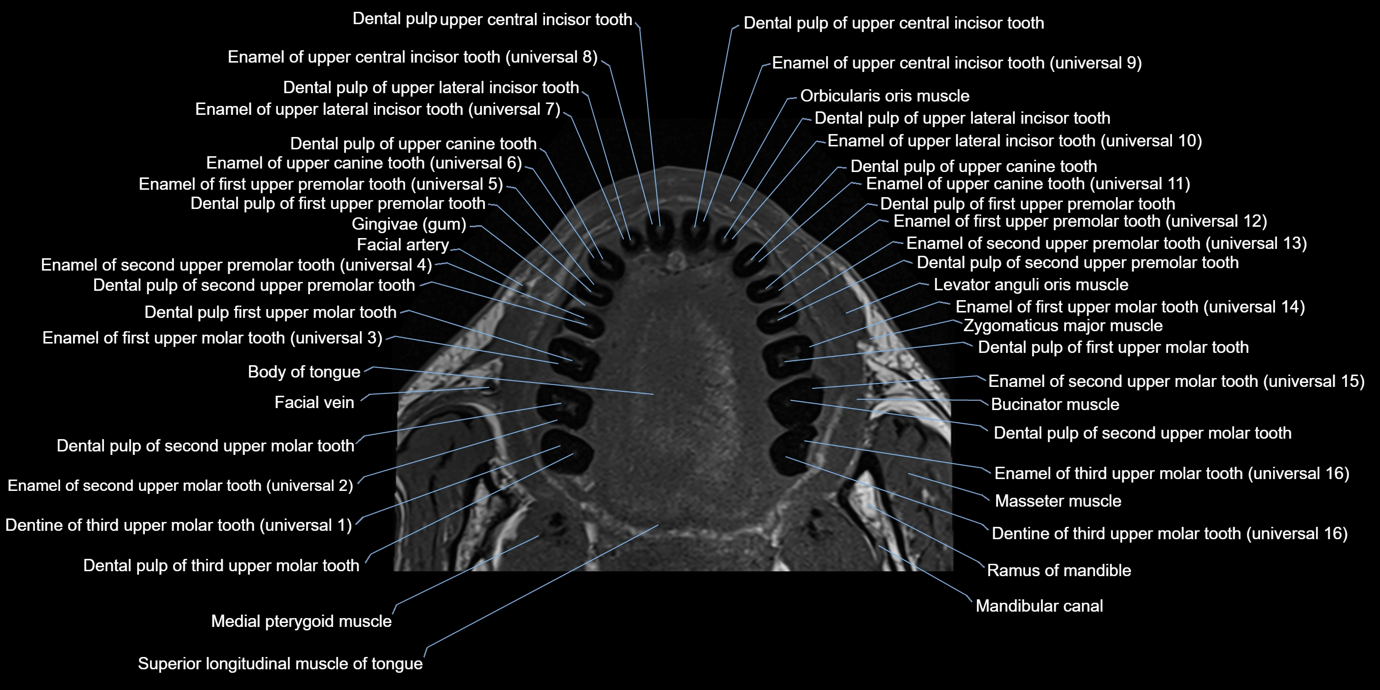
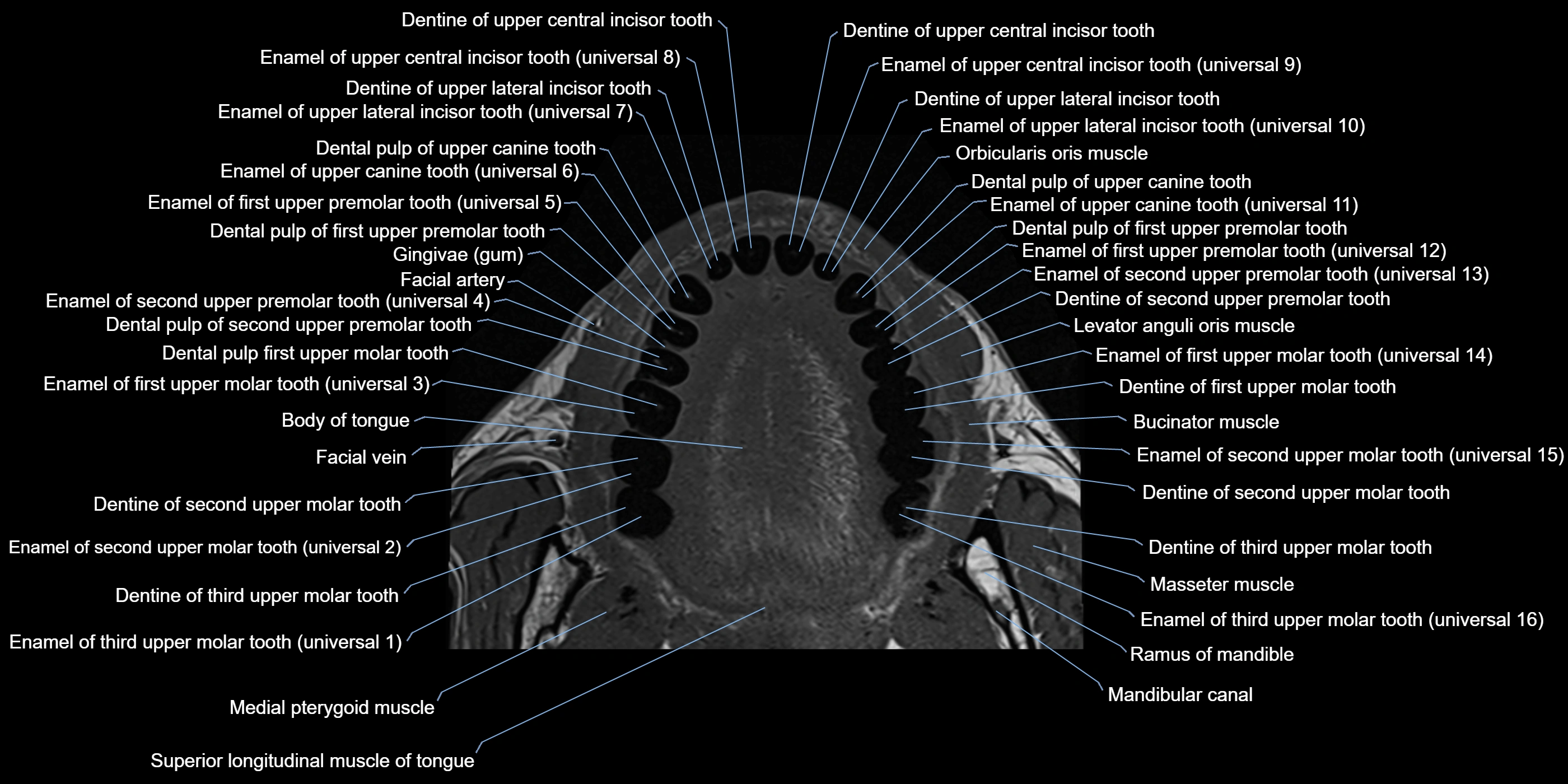
- Gum (gingiva)
- Hyoepiglottic ligament
- Hyoglossus
- Hyoglossus muscle
- Iliocostalis cervicis muscle
- Incisive duct
- Inferior alveolar foramen (mandibular foramen)
- Inferior alveolar nerve
- Inferior belly of omohyoid muscle
- Inferior canine tooth
- Inferior first premolar tooth
- Inferior longitudinal lingual muscle
- Inferior longitudinal muscle of tongue
- Inferior second molar tooth
- Inferior second premolar tooth
- Inferior third molar tooth
- Infraglottic cavity
- Internal occipital crest
- Intracanalicular part of optic nerve
- Jugular foramen
- Jugular foramen pars vascularis
- Lateral cricoarytenoid muscle
- Lateral inferior incisor tooth
- Lateral nasal cartilage
- Lateral superior incisor tooth
- Levator anguli oris muscle
- Levator labii superioris alaeque nasi muscle
- Levator labii superioris muscle
- Lingual Septum
- Lingual tonsil
- Longissimus capitis muscle
- Longissimus cervicis muscle
- Longus capitis muscle
- Longus colli muscle
- Lower molar apical foramen
- Lower premolar apical foramen
- Major alar cartilage
- Mandibular canal
- Mandibular condyle
- Mandibular foramen
- Mandibular nerve
- Masseter muscle (Deep part)
- Masseter muscle (Superficial part)
- Mastoid air cells
- Mental foramen
- Mental nerve
- Mentalis muscle
- Middle constrictor muscle of pharynx
- Middle superior alveolar nerve
- Minor alar cartilage
- Multifidus muscles
- Mylohyoid muscle
- Nasal spine of frontal bone
- Nasalis muscle
- Neck of mandible
- Nuchal ligament
- Obliquus inferior capitis muscle
- Obliquus superior capitis muscle
- Orbicularis oculi muscle (Orbital part)
- Orbicularis oculi muscle (Preseptal part)
- Orbicularis oris muscle
- Palatoglossus muscle
- Palatopharyngeus muscle
- Platysma muscle
- Posterior belly of digastric muscle
- Posterior cricoarytenoid muscle
- Posterior superior alveolar nerve
- Ramus of mandible
- Rectus capitis anterior muscle
- Rectus capitis lateralis muscle
- Rectus capitis posterior major muscle
- Rectus capitis posterior minor muscle
- Risorius muscle
- Root canal of lower canines tooth
- Root canal of lower premolar tooth
- Root canal of upper canines tooth
- Root canal of upper molar tooth
- Root canal of upper premolar tooth
- Root of lower canines tooth
- Root of lower molar tooth
- Root of upper molar tooth
- Rotatores cervicis muscle
- Semispinalis capitis muscle
- Semispinalis cervicis muscle
- Serratus anterior muscle
- Spinalis cervicis muscle
- Splenius capitis muscle
- Splenius cervicis muscle
- Sternohyoid muscle
- Sternothyroid muscle
- Styloglossus muscle
- Stylohyoid muscle
- Stylopharyngeus muscle
- Subclavius muscle
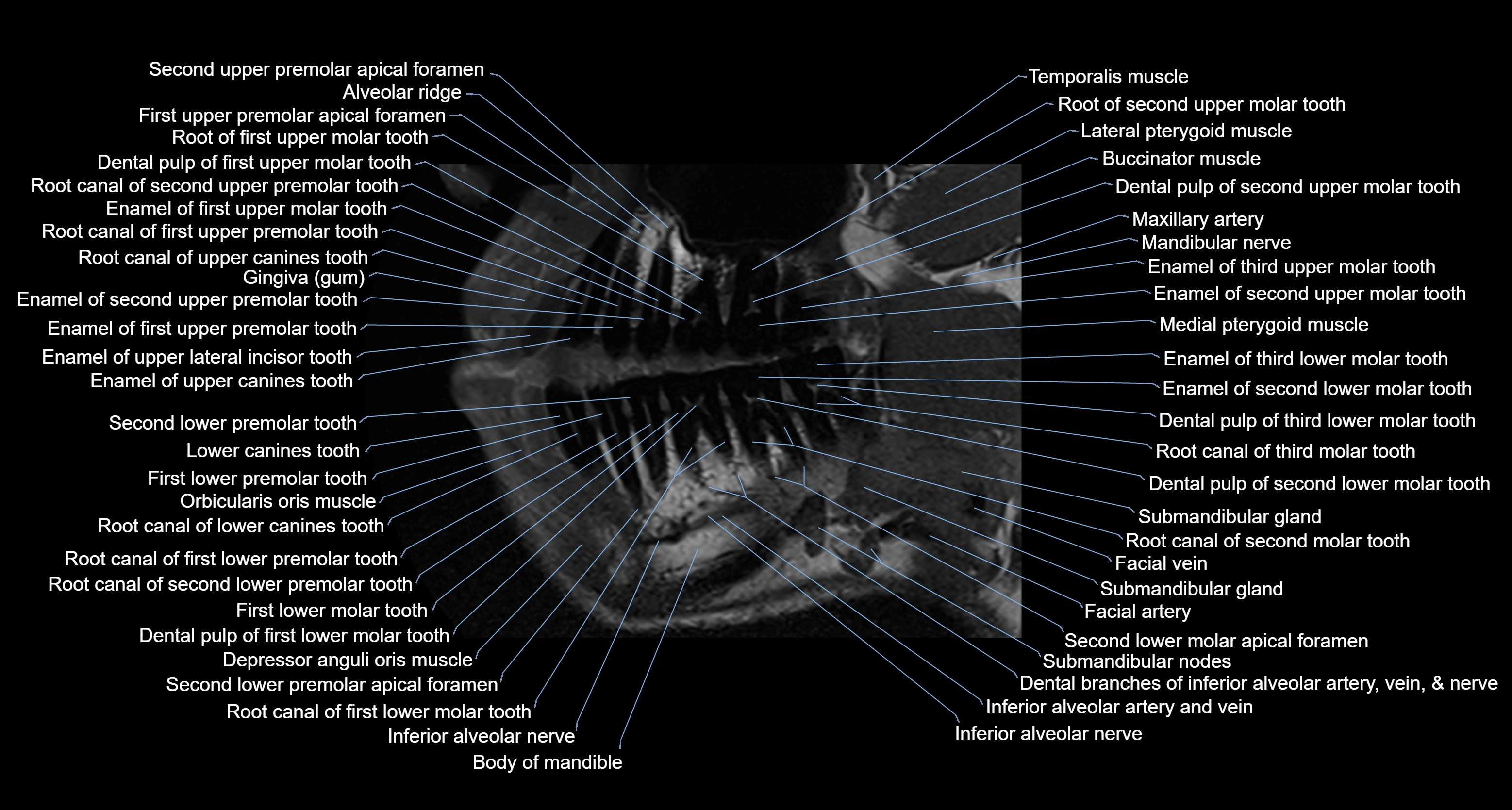
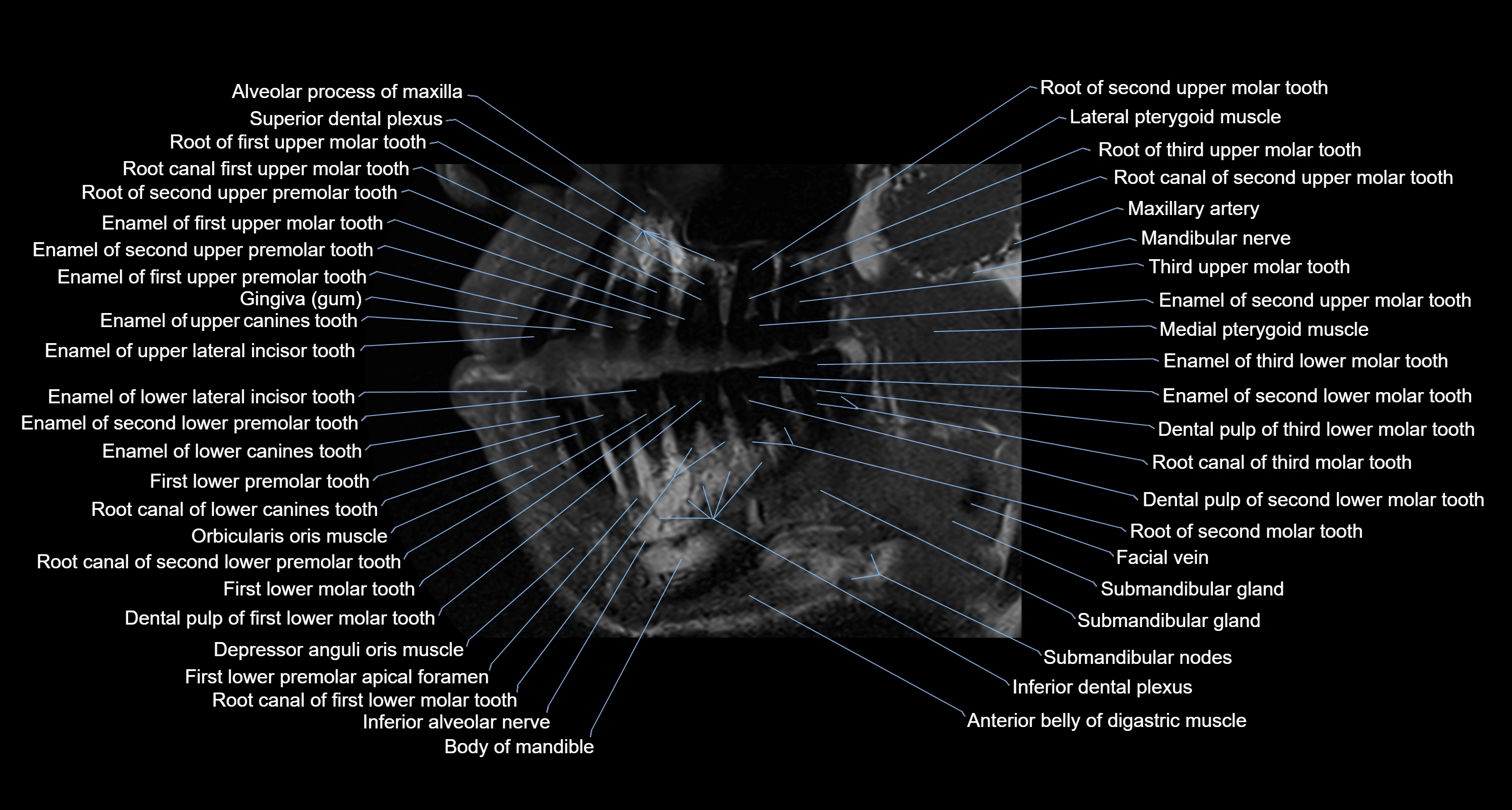
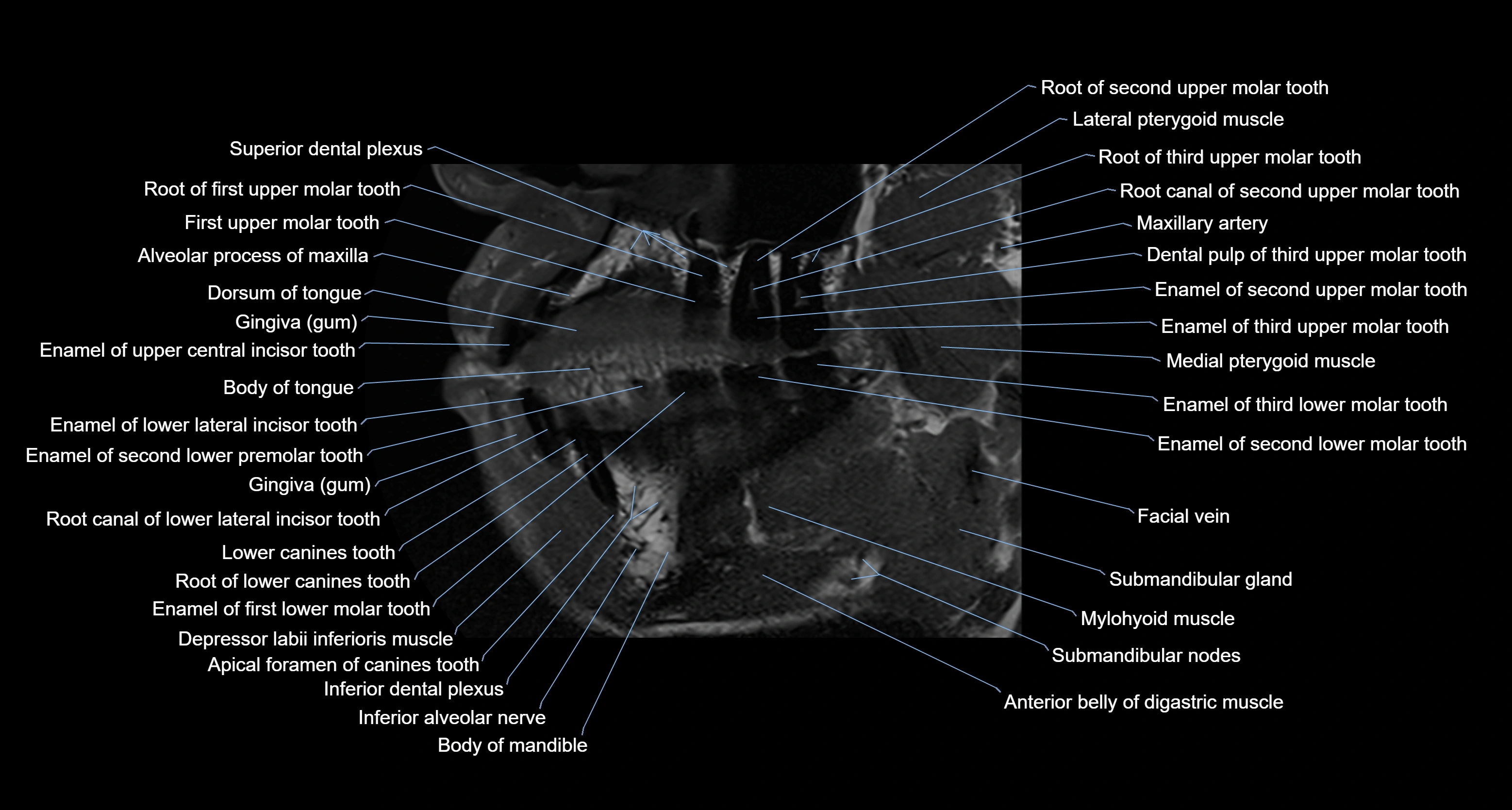
- Submandibular lymph nodes
- Superciliary arch
- Superficial head of medial pterygoid muscle
- Superior belly of omohyoid muscle
- Superior constrictor muscle of pharynx
- Superior dental plexus
- Superior first molar tooth
- Superior first premolar tooth
- Superior longitudinal lingual muscle
- Superior longitudinal muscle of tongue
- Superior second molar tooth
- Superior second premolar tooth
- Superior third molar tooth
- Teeth
- Temporomandibular joint
- Transverse muscle of the tongue
- Trapezius muscle
- Upper premolar apical foramen
- Zygomatic bone
- inferior alveolar artery
- jugular foramen pars nervosa
- superior canine tooth
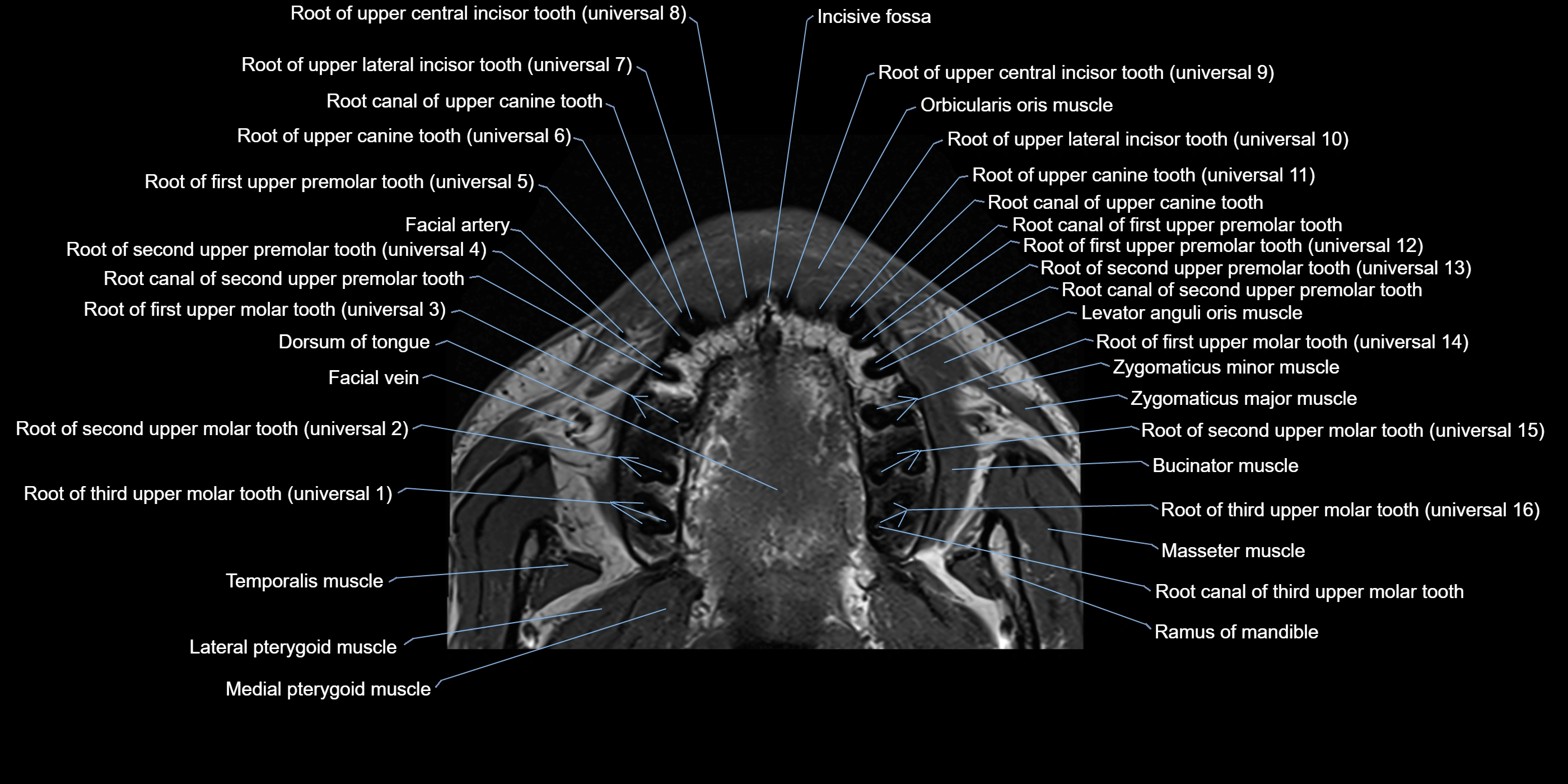
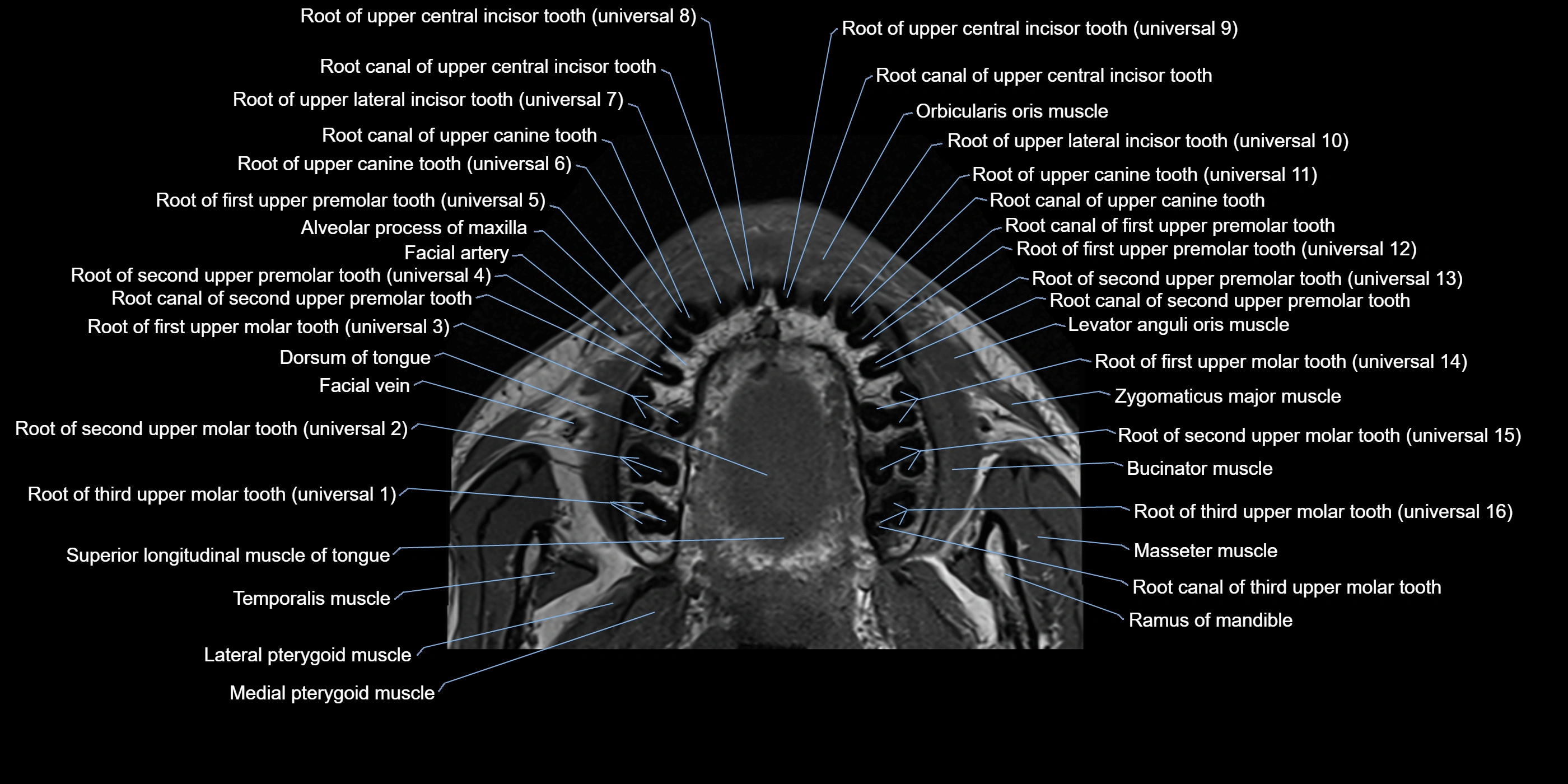
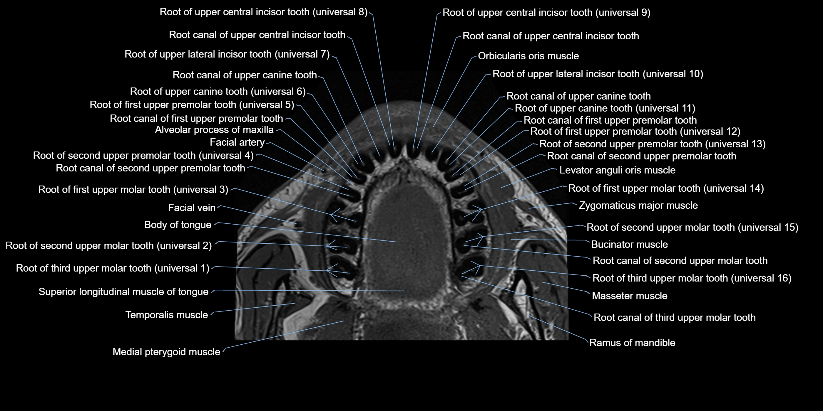
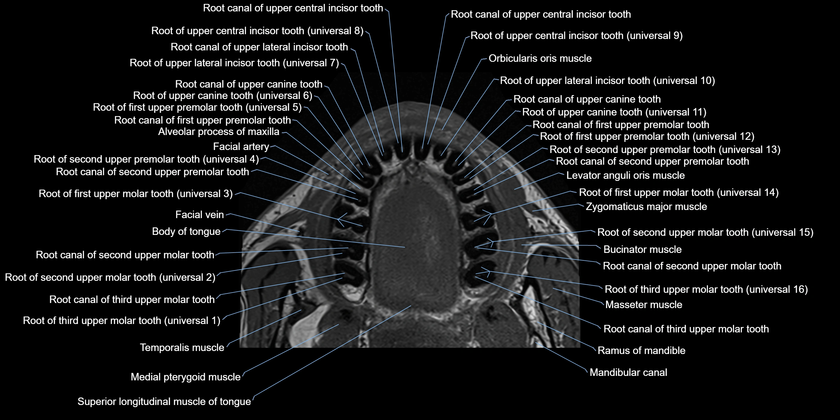
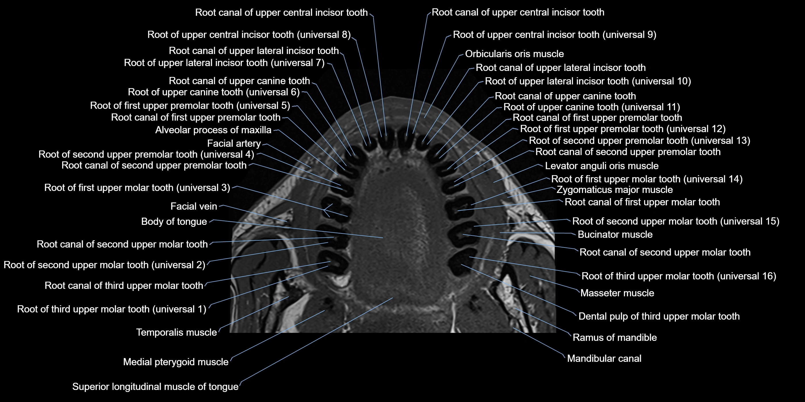
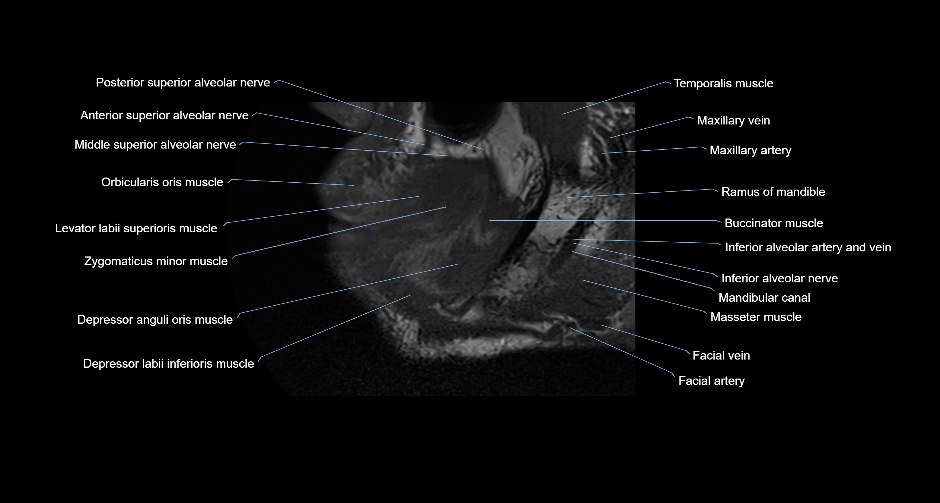
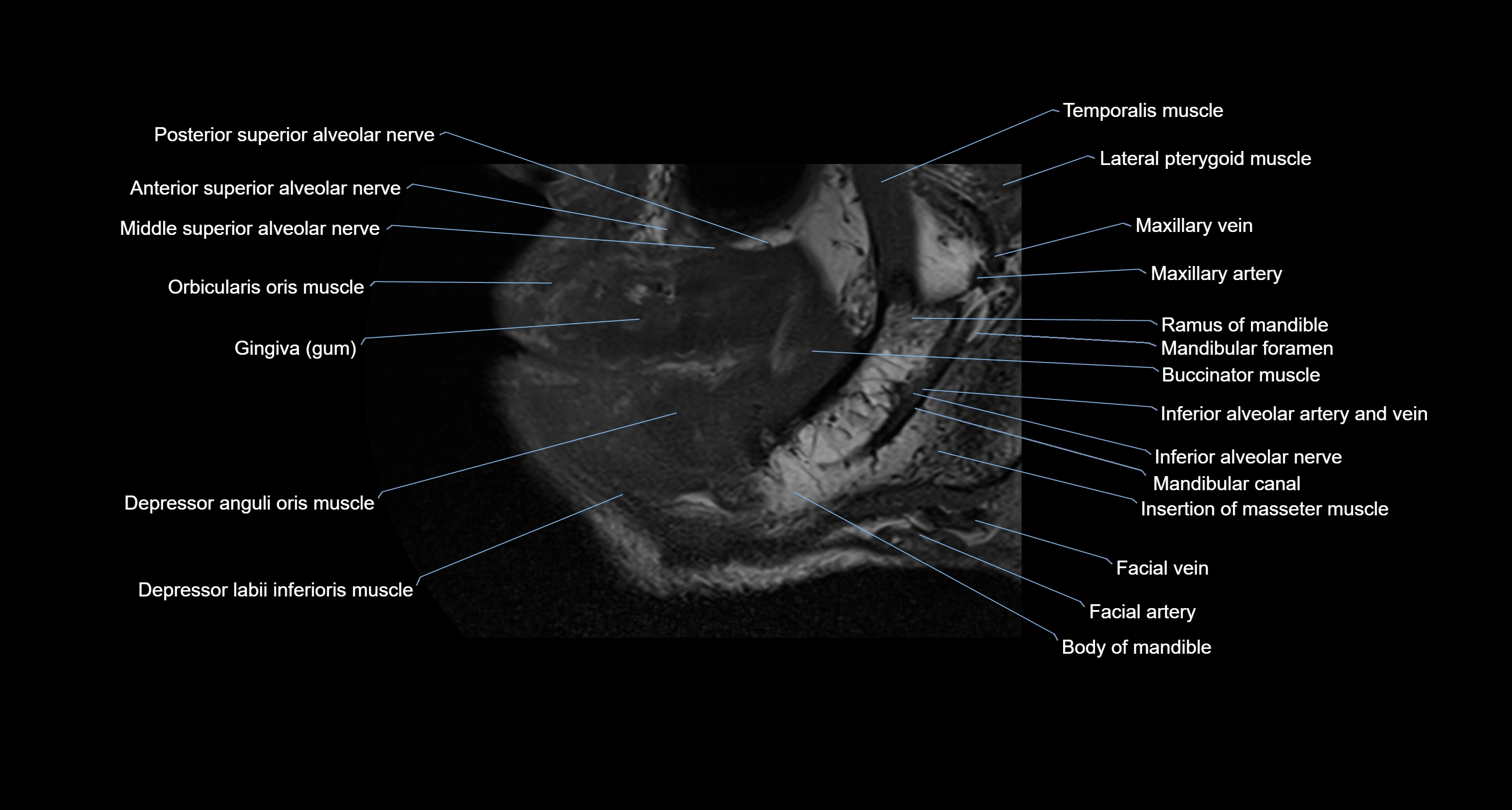
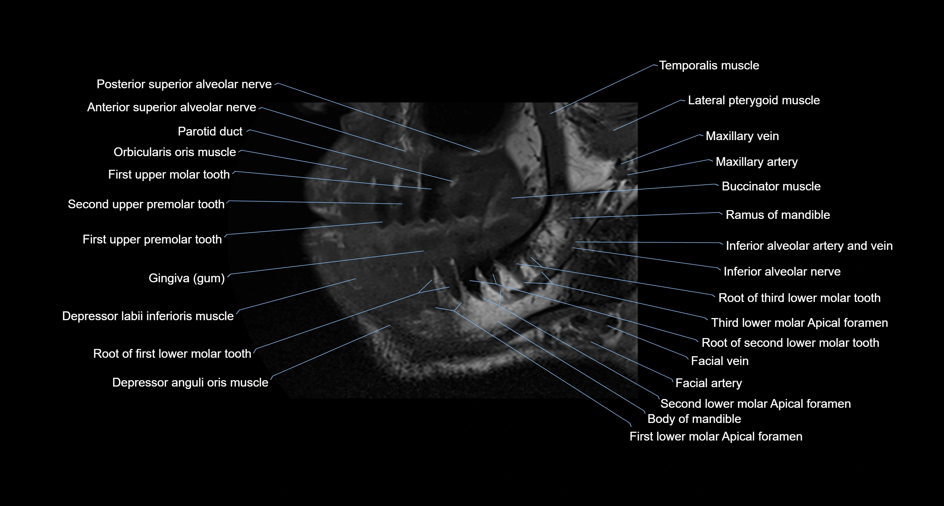
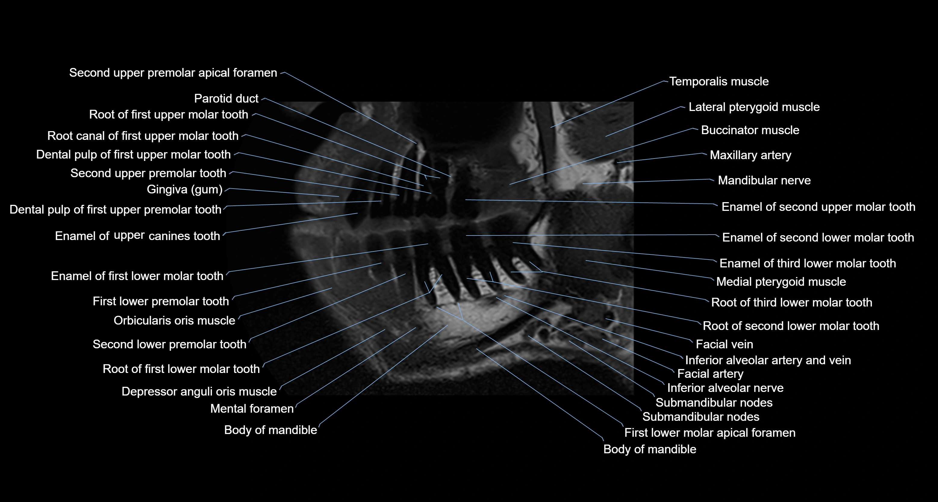
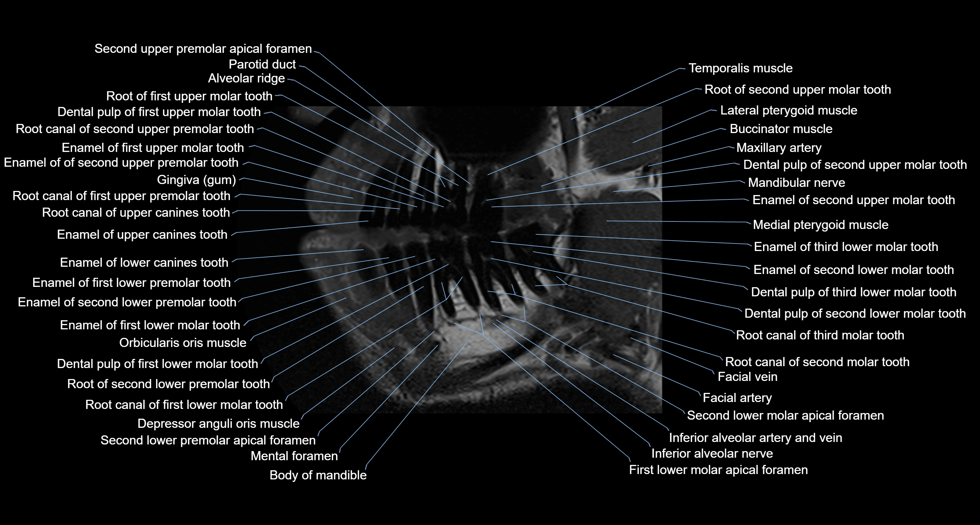
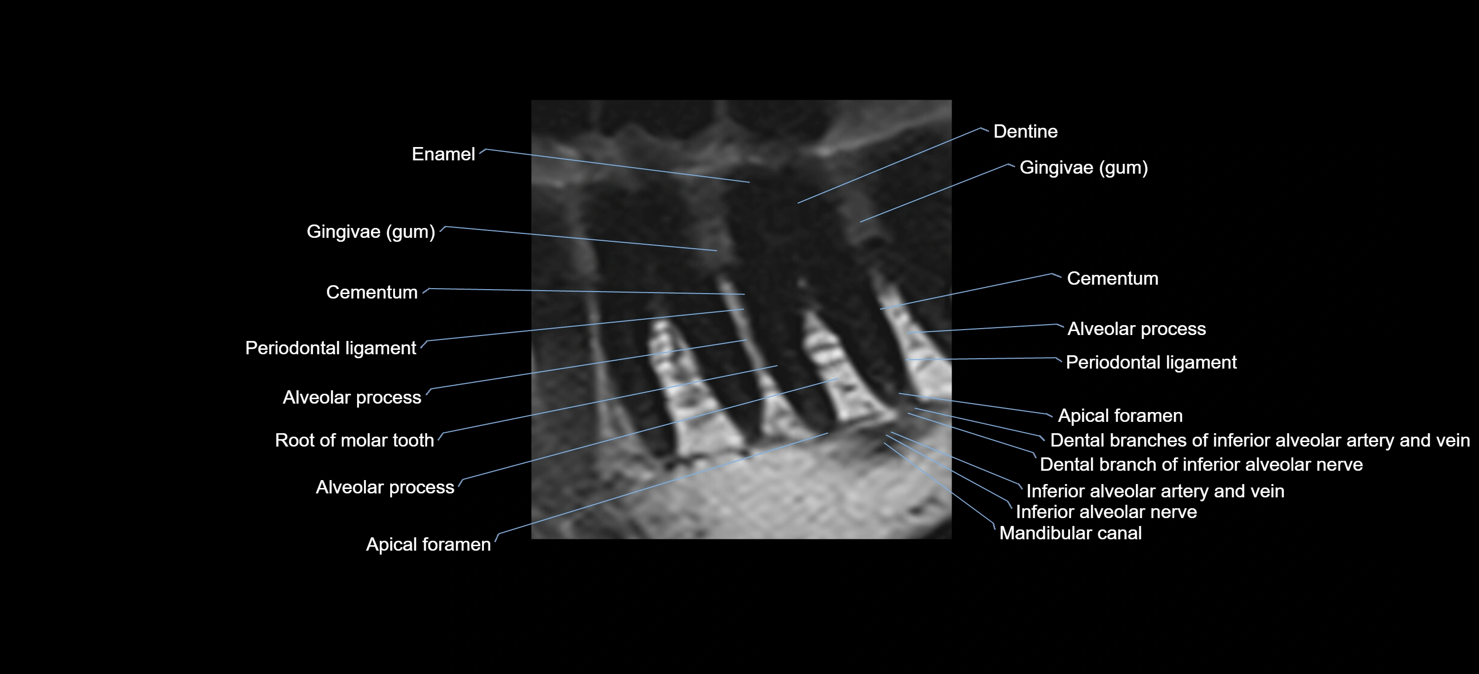
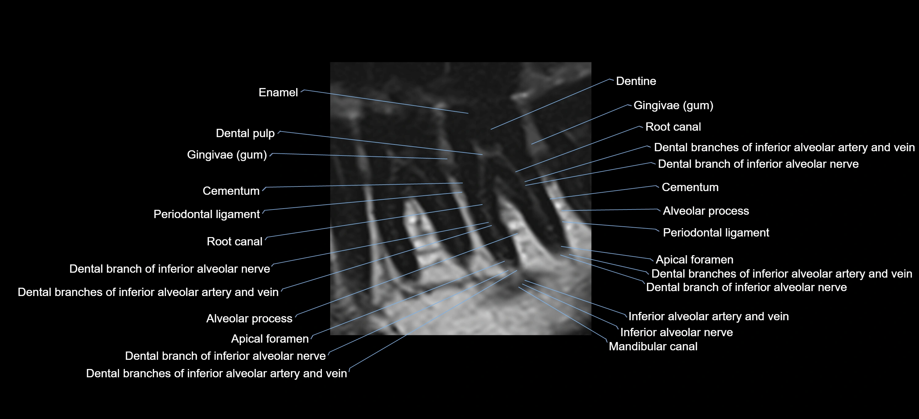
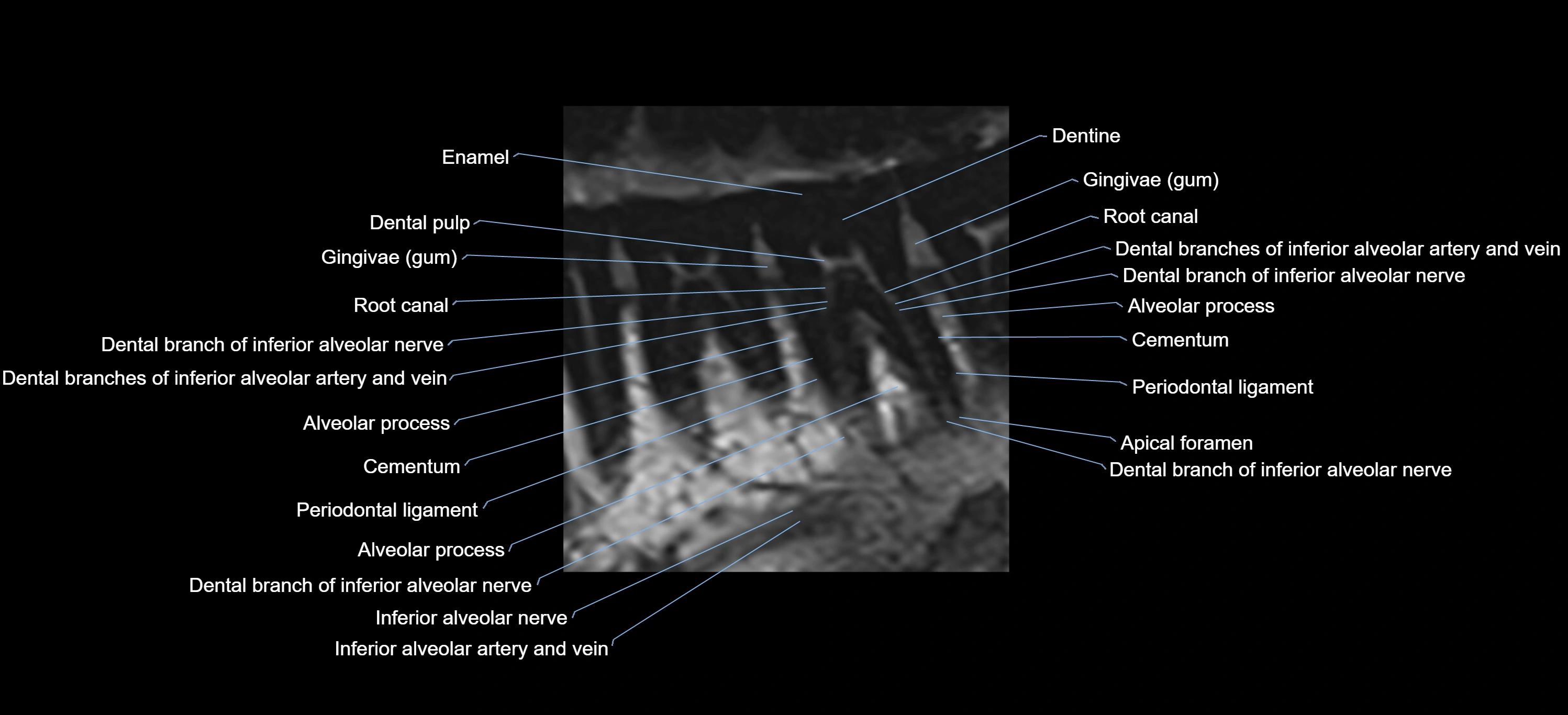
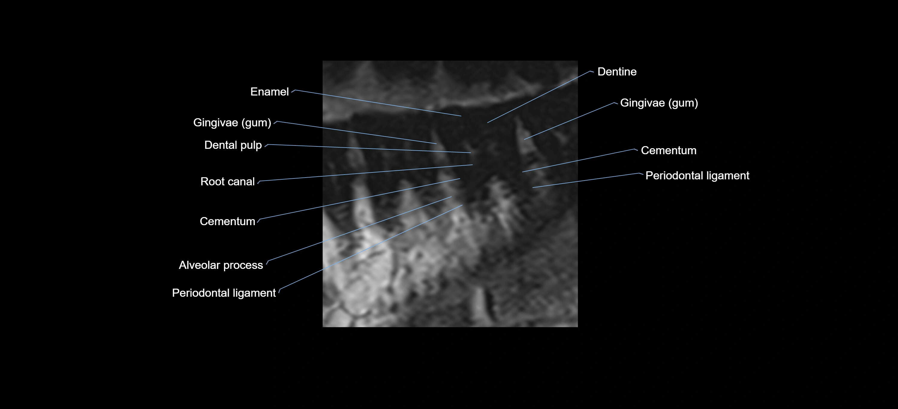
The alveolar arch of the maxilla is the curved, horseshoe-shaped bony structure forming the inferior portion of the maxilla. It contains the alveoli (tooth sockets) for the maxillary teeth and plays a central role in dental anatomy, occlusion, facial contour, and maxillofacial imaging.
The alveolar arch is dynamic, undergoing structural changes with tooth eruption, loss, and age, and is a key region assessed in dentistry, orthodontics, implant planning, trauma evaluation, and head and neck imaging.
Synonyms
-
Maxillary alveolar arch
-
Alveolar process of maxilla
-
Maxillary dental arch
Location
-
Forms the inferior margin of the maxilla
-
Extends bilaterally from the maxillary tuberosity on each side
-
Curves anteriorly to form the upper dental arch
-
Located inferior to the maxillary sinus
-
Superior to the oral cavity
-
Anterior to the hard palate
Anatomical components
-
Alveolar process:
-
Vertical bony ridge of the maxilla
-
-
Dental alveoli:
-
Sockets for incisors, canines, premolars, and molars
-
-
Interalveolar septa:
-
Bone between adjacent tooth sockets
-
-
Interradicular septa:
-
Bone between roots of multirooted teeth
-
-
Alveolar crest:
-
Superior margin of the alveolar bone between teeth
-
Relations
Superiorly:
-
Maxillary sinus floor
-
Body of the maxilla
Inferiorly:
-
Oral cavity
-
Gingiva and maxillary teeth
Anteriorly:
-
Anterior nasal spine (near midline)
-
Upper lip soft tissues
Posteriorly:
-
Maxillary tuberosity
-
Pterygopalatine region (posterior relation)
Medially:
-
Hard palate
-
Nasal cavity (via palatal process)
Laterally:
-
Buccal cortex of the maxilla
-
Buccal soft tissues
Developmental anatomy
-
Develops in association with tooth eruption
-
Alveolar height increases during mixed and permanent dentition
-
Tooth loss leads to alveolar resorption, most pronounced in the vertical dimension
-
Degree of pneumatization of maxillary sinus influences alveolar bone thickness
X-ray appearance
Dental and skull radiographs (periapical / panoramic / occlusal views):
-
Alveolar arch: Curved radiopaque bony ridge
-
Dental sockets: Radiolucent spaces surrounded by radiopaque lamina dura
-
Alveolar crest: Thin radiopaque line between teeth
-
Relationship to teeth: Clearly delineated tooth roots within alveoli
CT appearance
Non-contrast CT:
-
Cortical bone: Hyperdense buccal and palatal cortices
-
Cancellous bone: Lower-density trabecular pattern
-
Dental alveoli: Well-defined socket contours
-
Maxillary sinus floor: Closely related to posterior alveolar arch
Post-contrast CT:
-
Alveolar bone: No intrinsic enhancement
MRI appearance
T1-weighted images:
-
Cortical bone: Low signal intensity
-
Cancellous marrow: Intermediate to high signal depending on fatty content
-
Teeth: Signal void structures
-
Adjacent soft tissues: Normal gingiva and oral mucosa signal
T2-weighted images:
-
Cortical bone and teeth: Low signal
-
Marrow: Intermediate signal
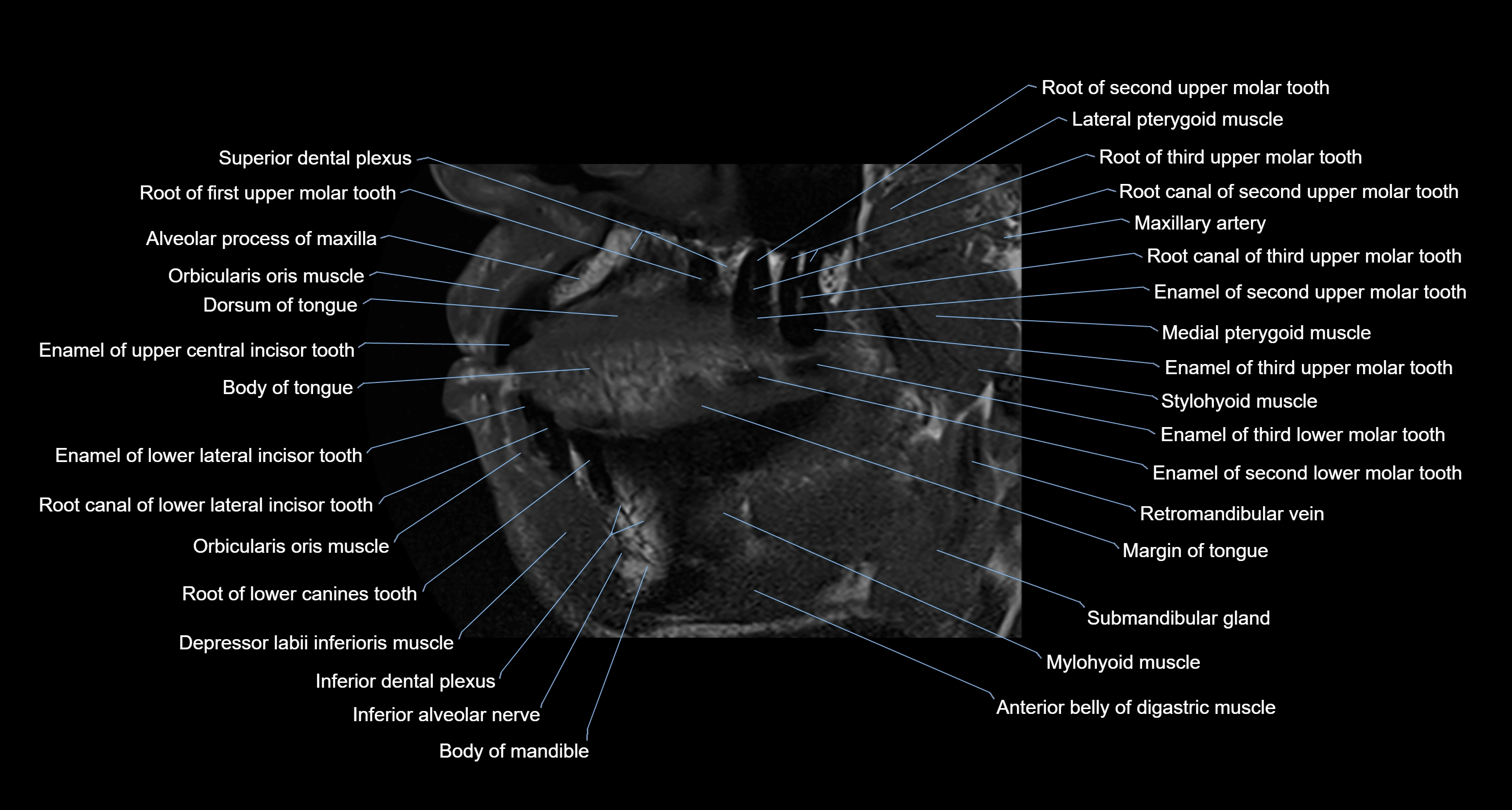
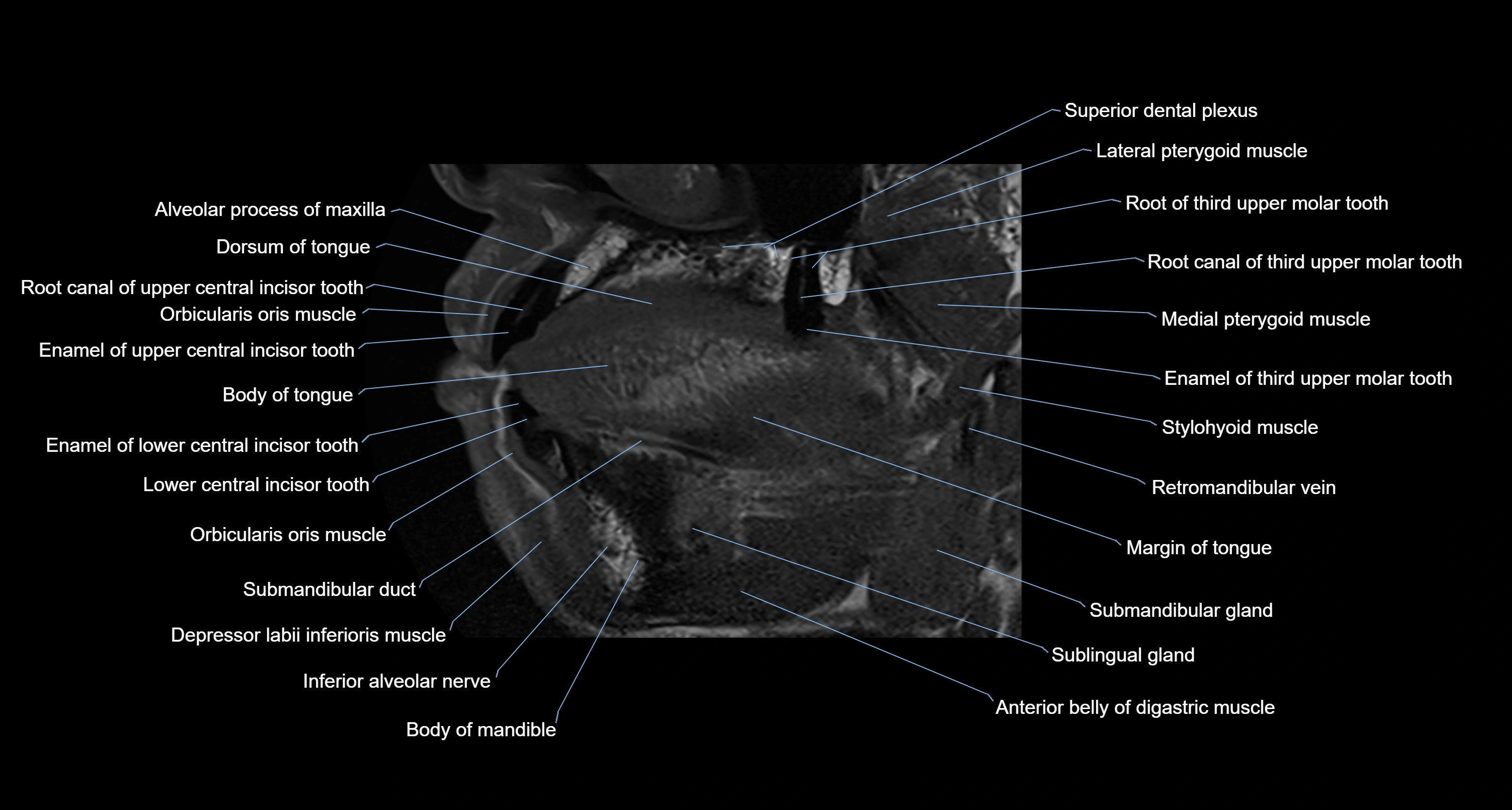
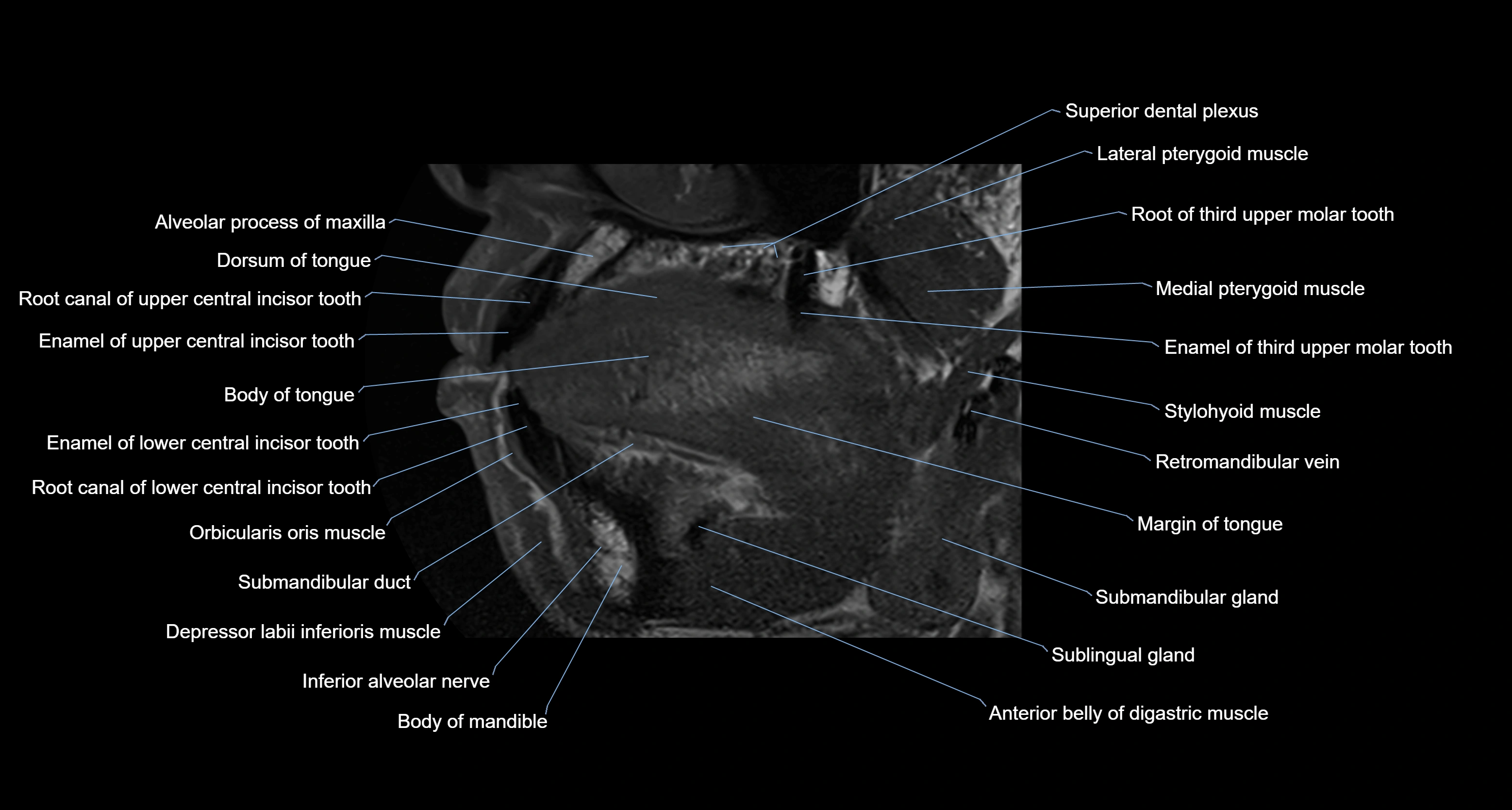
CT VRT 3D image

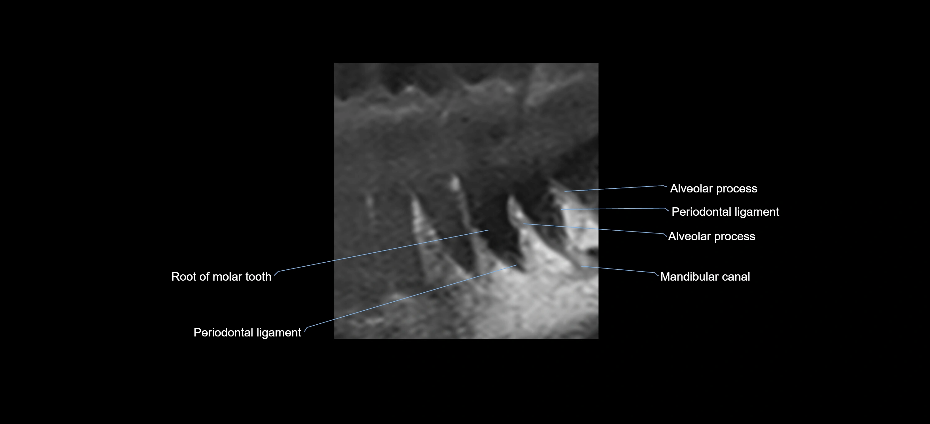
X-Ray image


