


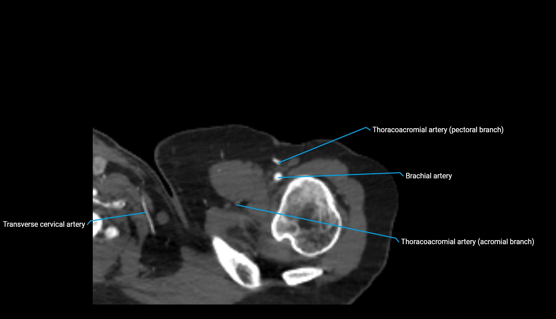
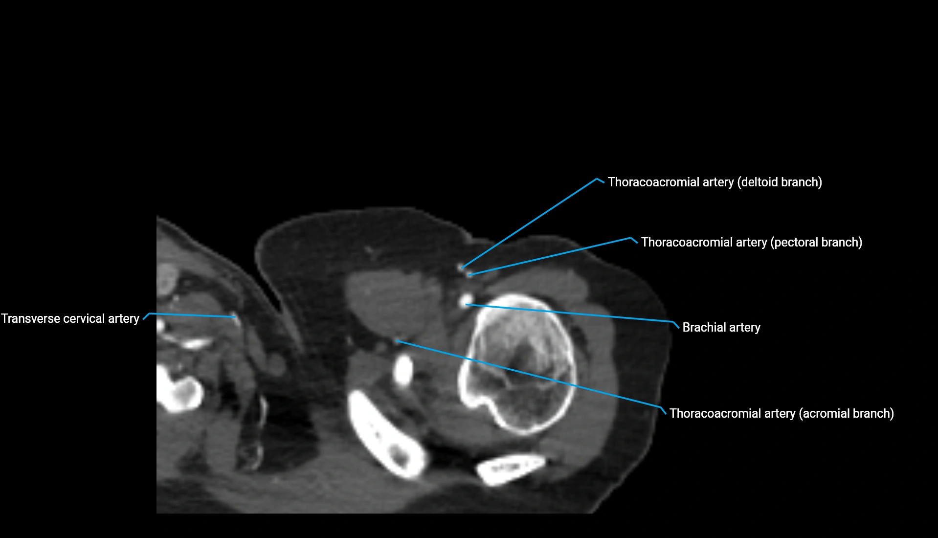
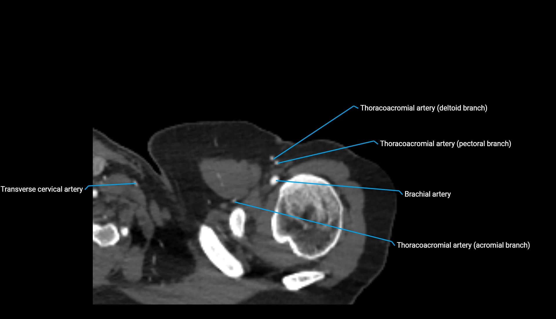
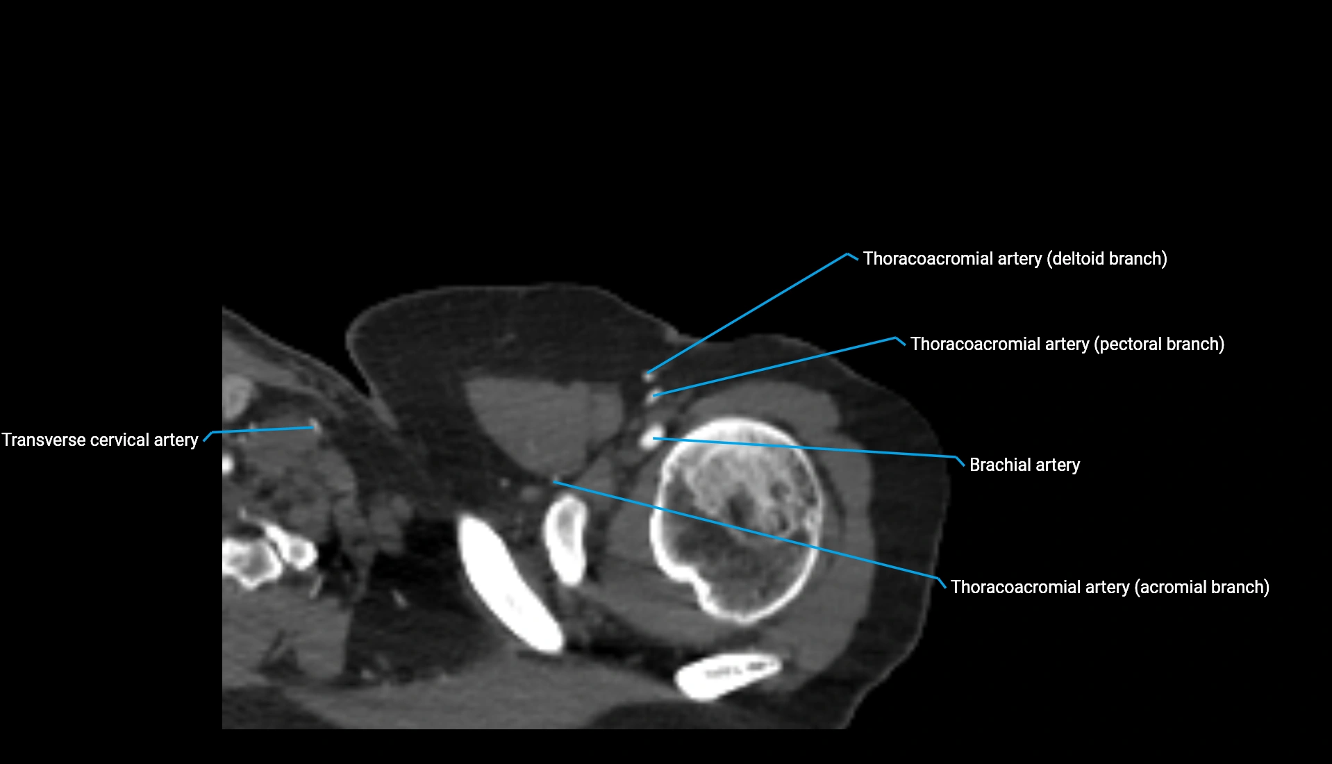
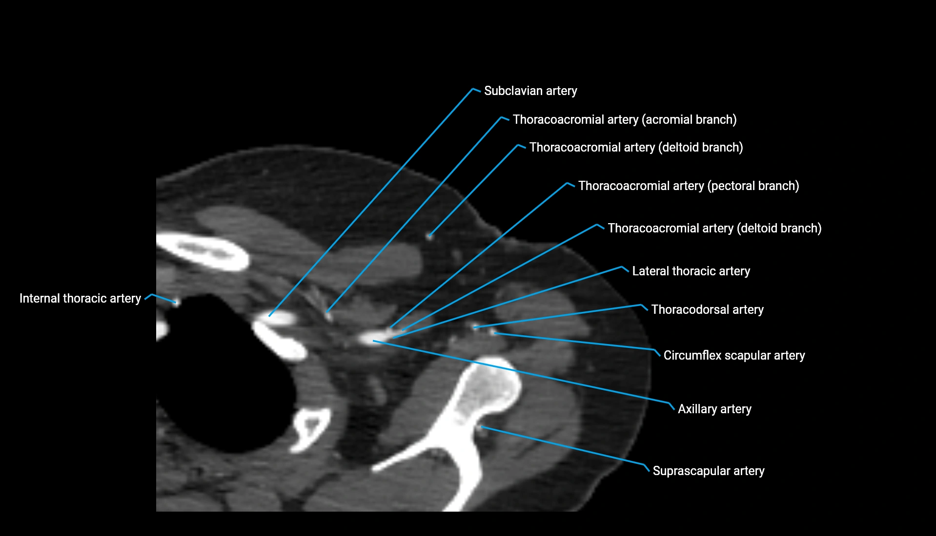
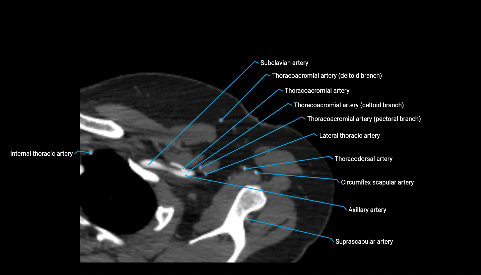
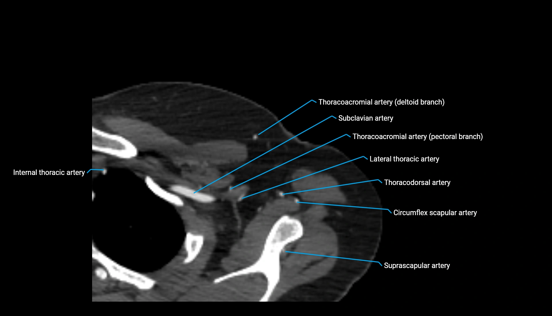
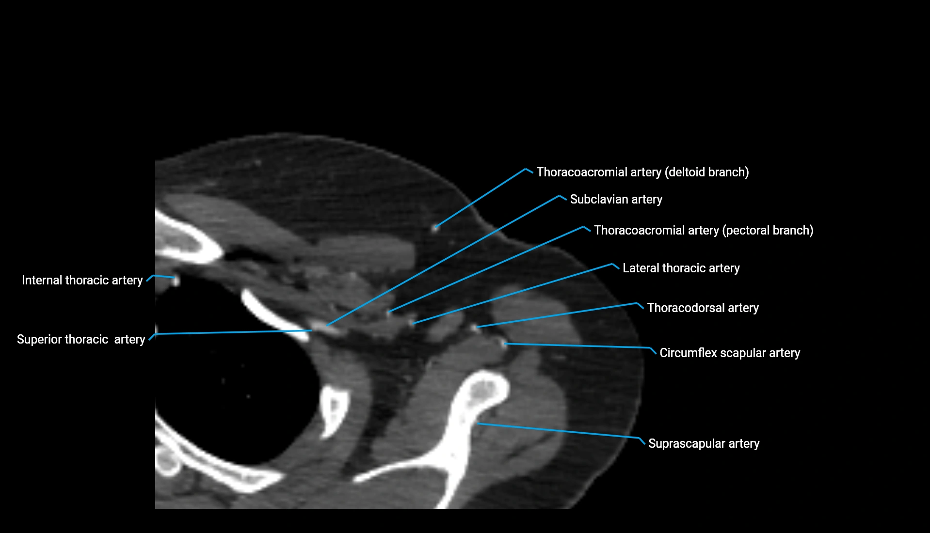
Topic
The articular facet of the head of the radius is the smooth, concave superior surface of the radial head that articulates with the capitellum of the humerus. This articulation forms the radiocapitellar joint, essential for elbow flexion–extension and forearm pronation–supination.
The facet is covered with hyaline cartilage, allowing low-friction movement during rotation of the radius. Inferiorly, the radial head also has a circumferential articular surface that interacts with the radial notch of the ulna, contributing to the proximal radioulnar joint.
Synonyms
-
Superior radial articular surface
-
Radiocapitellar articular facet
-
Radial head joint surface
Location and Structure
-
Position: Superior end of the radius, forming the proximal part of the forearm.
-
Shape: Shallow, concave articular depression matching the convex capitellum.
-
Cartilage: Thick hyaline cartilage covering the entire surface to permit smooth rotation.
-
Continuity: Merges inferiorly with the annular ligament-covered radial head circumference.
-
Joint associations:
-
Forms the radiocapitellar joint superiorly
-
Contributes to proximal radioulnar joint medially (via radial head rim)
-
Relations
-
Superiorly: Capitellum of humerus
-
Inferiorly: Neck of radius
-
Medially: Annular ligament and radial notch of ulna
-
Laterally: Joint capsule and lateral collateral ligament complex
-
Anteriorly/Posteriorly: Joint capsule and surrounding synovium
Attachments
-
No direct muscular attachments
-
Articular cartilage anchored to subchondral bone
-
Margins reinforced by the annular ligament surrounding the radial head
-
Continuity with the joint capsule of the elbow
Function
-
Provides smooth articulation with the capitellum for elbow flexion and extension
-
Enables rotation of the radial head during pronation and supination
-
Distributes load from the hand and forearm to the humerus
-
Contributes to joint congruency and elbow stability
Clinical Significance
-
Frequent site of radial head fractures, especially from FOOSH injuries
-
Important evaluation point in radiocapitellar arthritis
-
Articular cartilage defects may cause mechanical symptoms
-
Key landmark in assessing elbow alignment and stability
MRI Appearance
T1-weighted images:
-
Cortex: Very low signal (dark)
-
Subchondral marrow: Bright due to fatty content
-
Articular cartilage: Smooth intermediate-to-low signal covering the facet
-
Joint fluid: Dark
-
Contours: Well-defined curvature matching capitellum
T2-weighted images:
-
Cortex: Dark
-
Marrow: Bright (fat + fluid sensitivity)
-
Cartilage: Intermediate-to-brightly outlined, allowing evaluation of thickness
-
Joint fluid: Bright
-
Distinct visualization of the radiocapitellar interface
STIR:
-
Bone marrow: Intermediate-to-dark signal
-
Cartilage: Dark against suppressed fat
-
Joint fluid: Bright
-
Highlights subtle fluid around joint margins
T1 Fat-Saturated Post-Contrast:
-
Bone: Mild homogeneous enhancement of marrow
-
Cartilage: No intrinsic enhancement
-
Synovium and capsule: Smooth, mild enhancement
-
Provides clear delineation of radiocapitellar joint surfaces
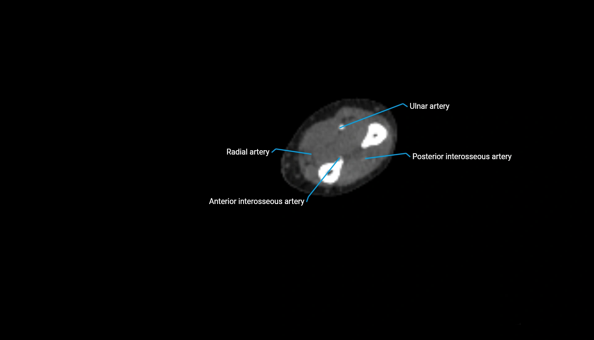
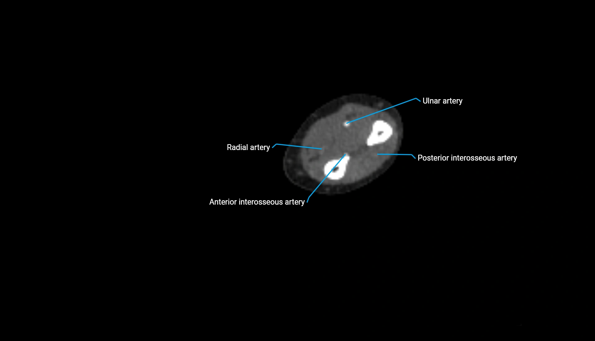
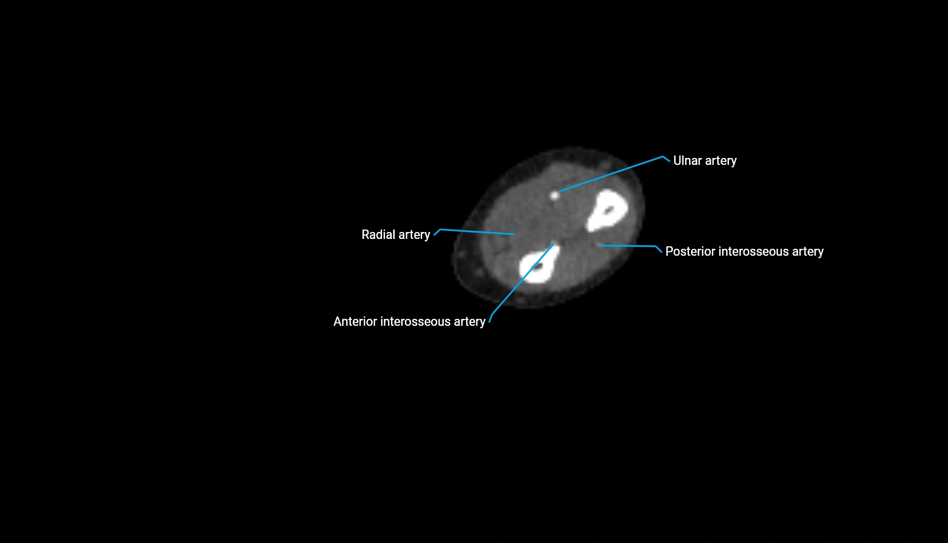
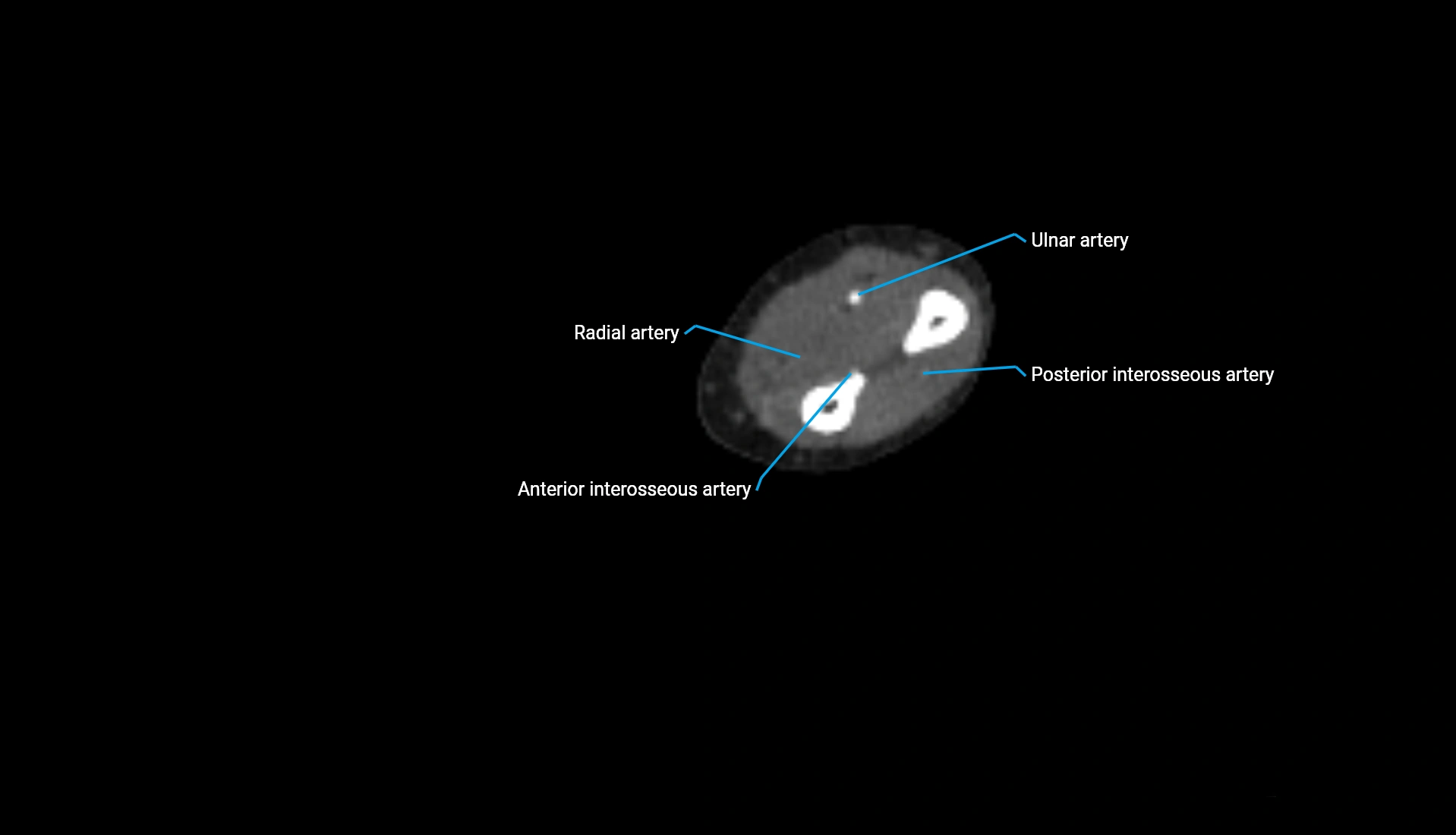
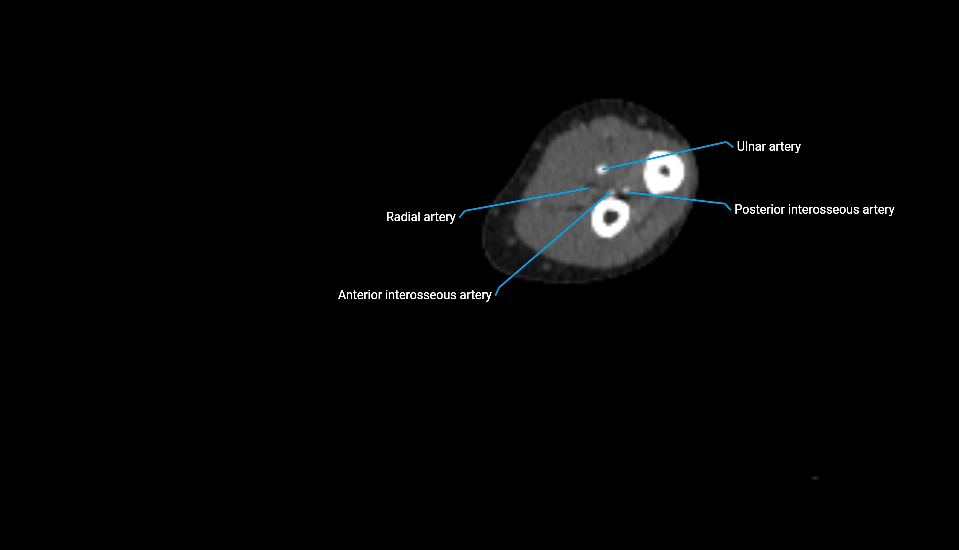
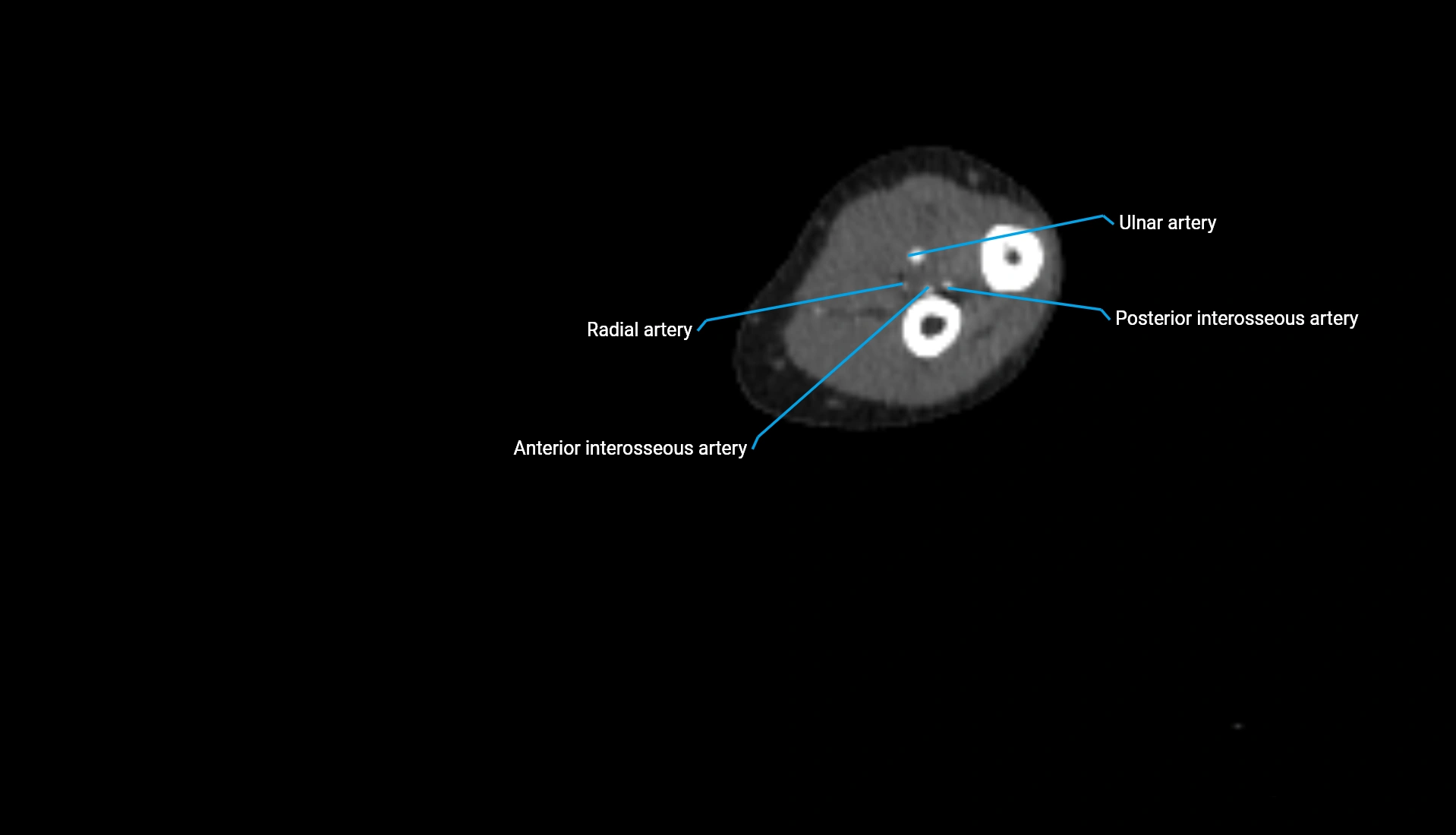
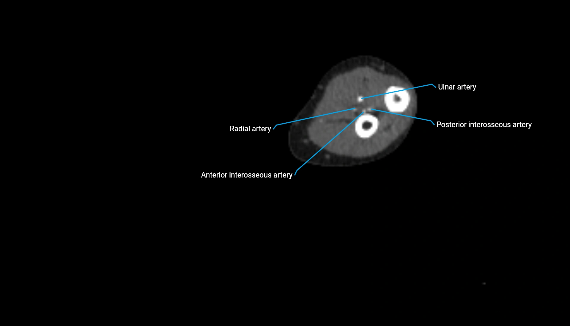
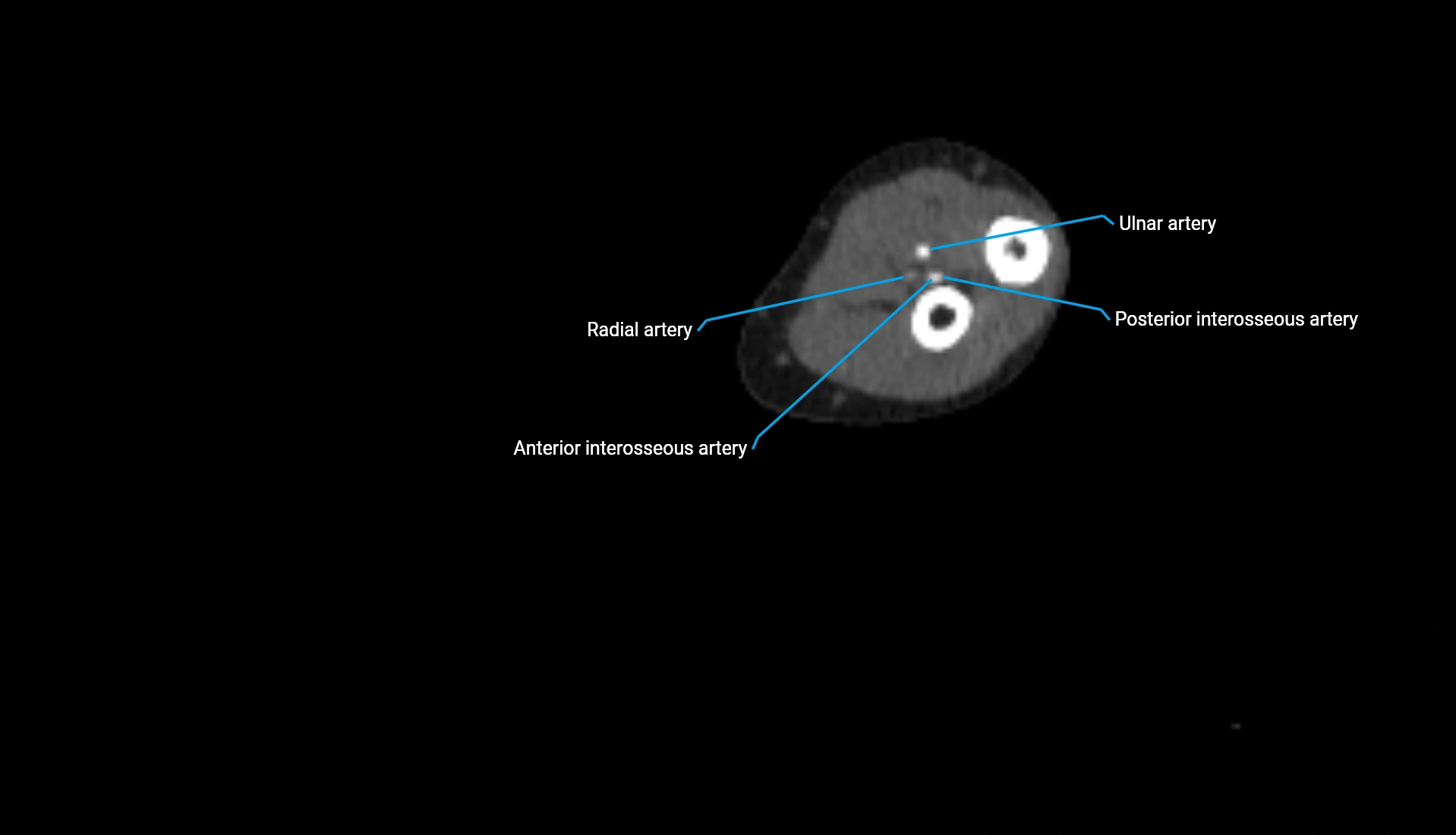
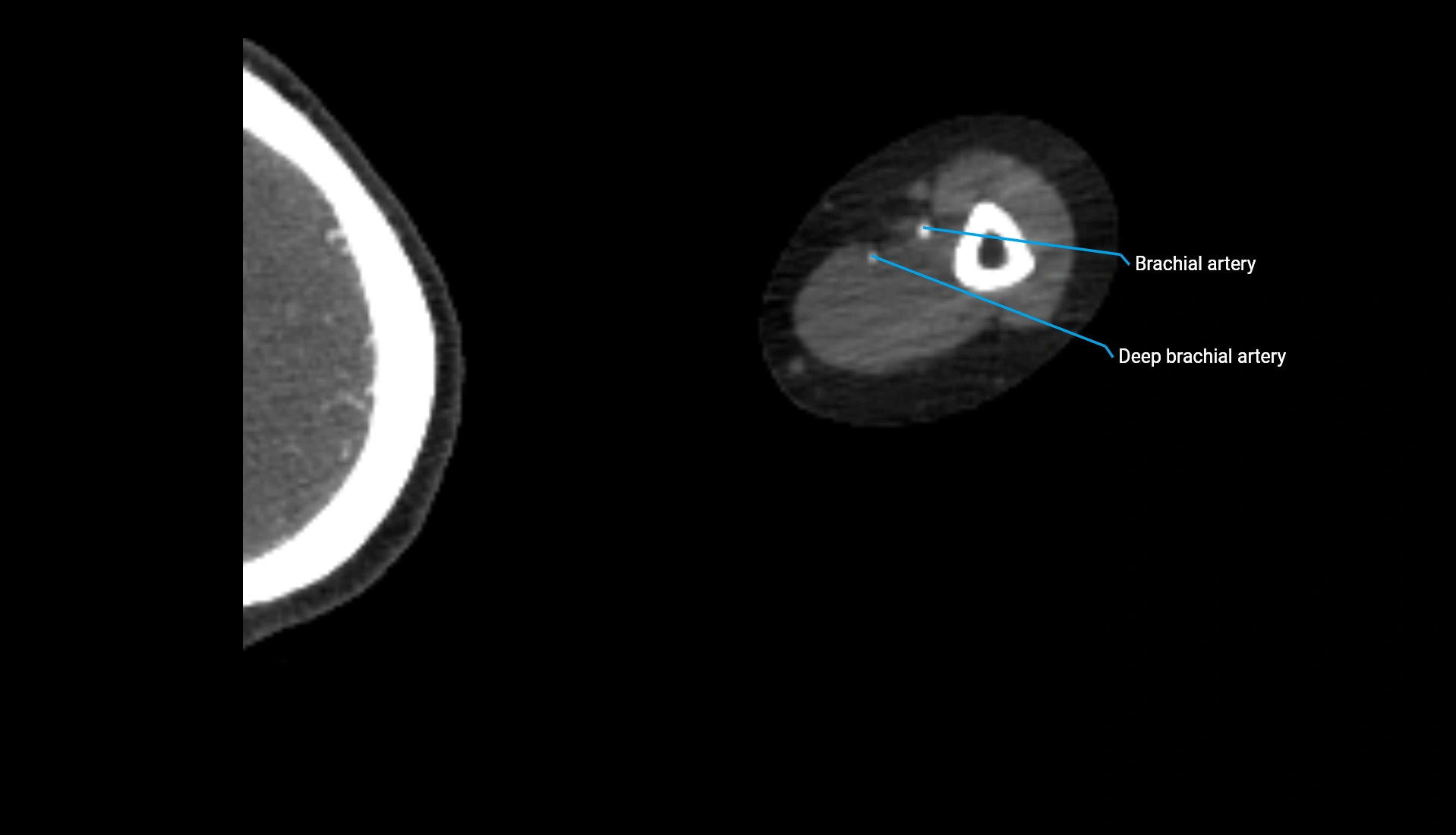
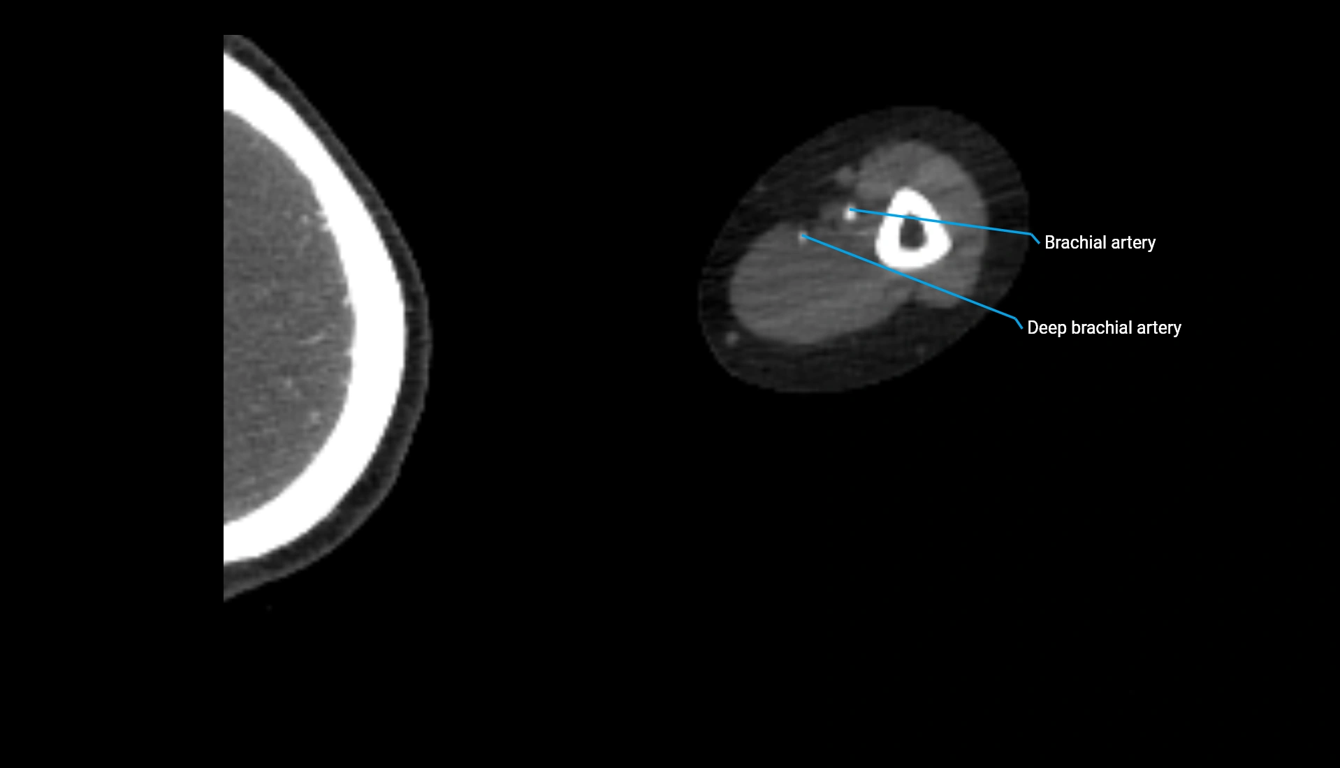
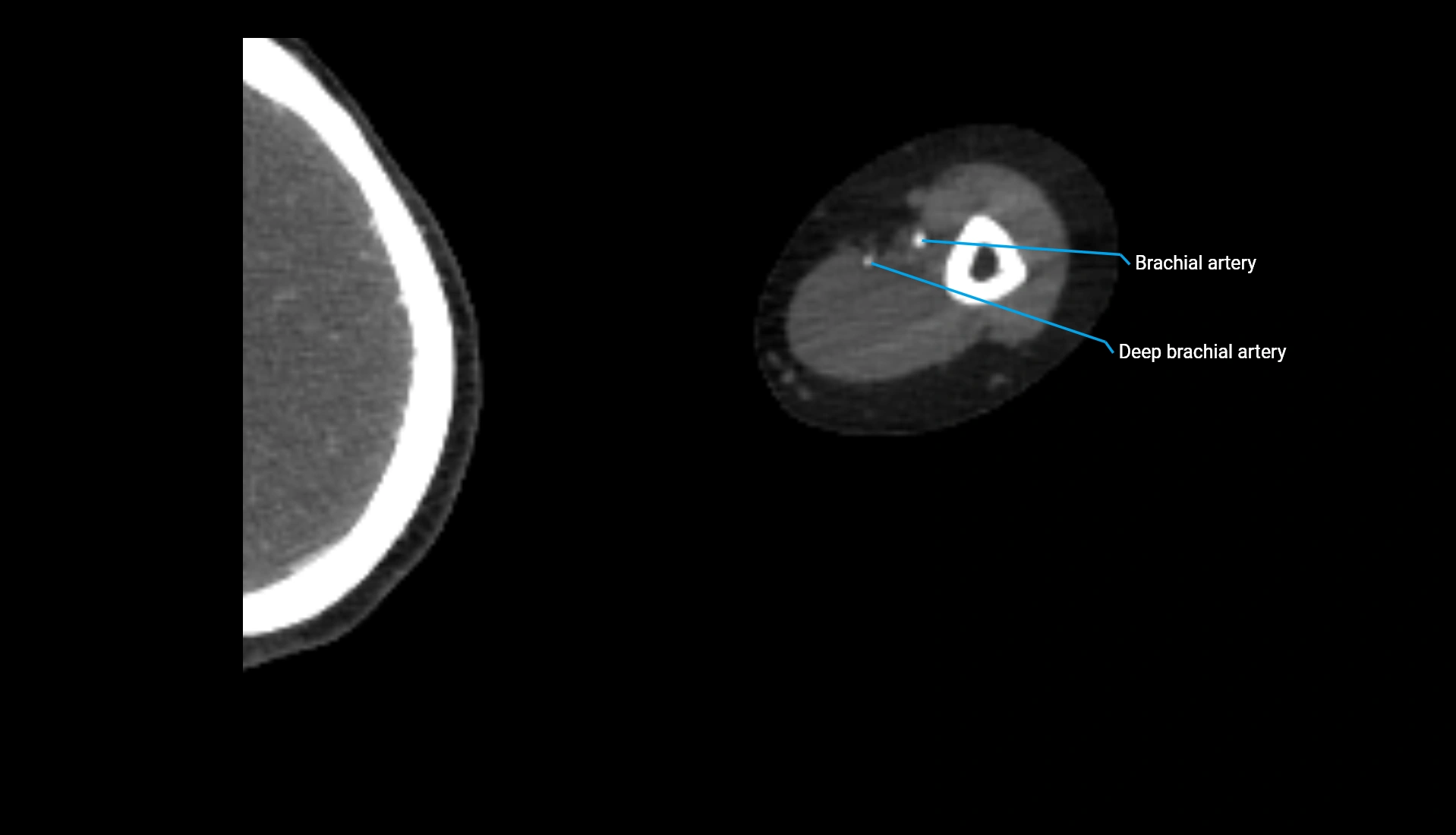
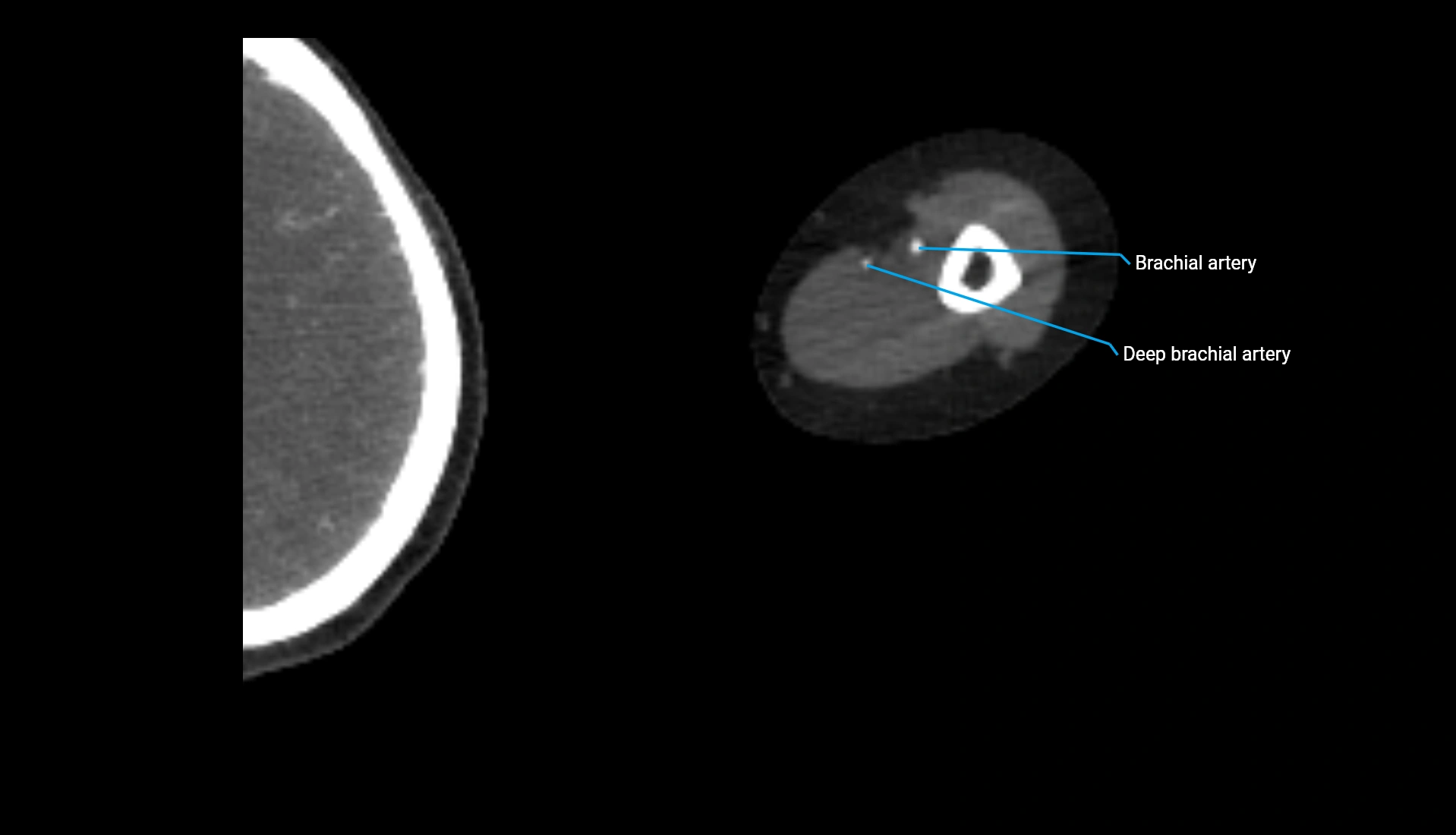
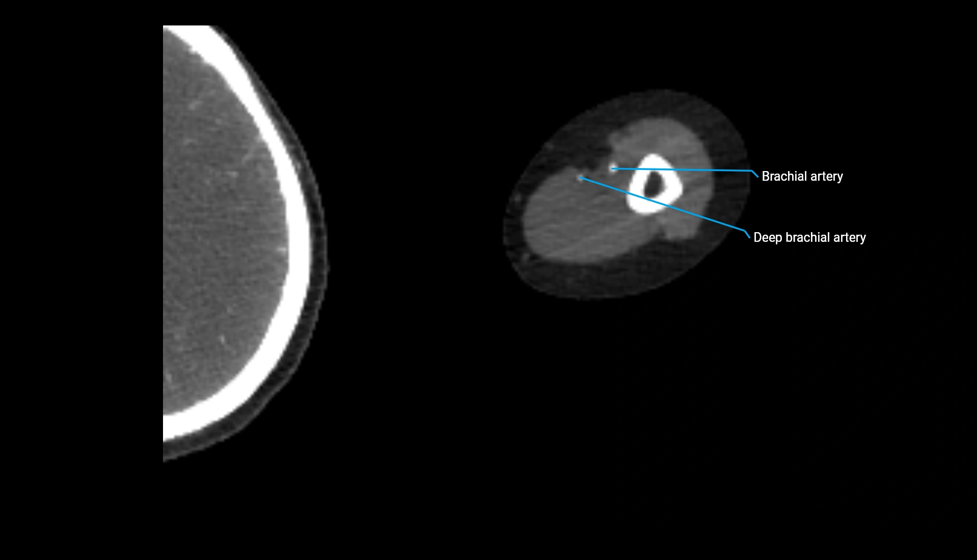
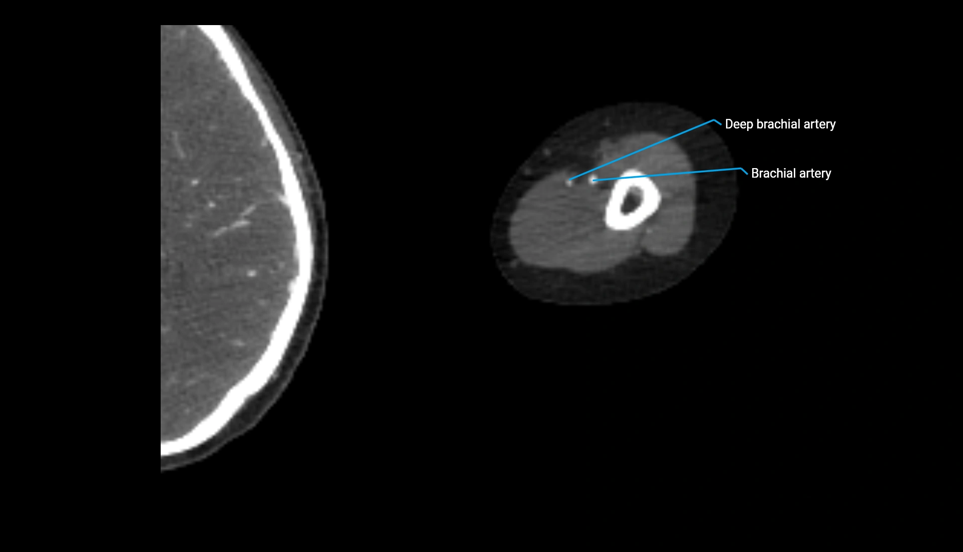
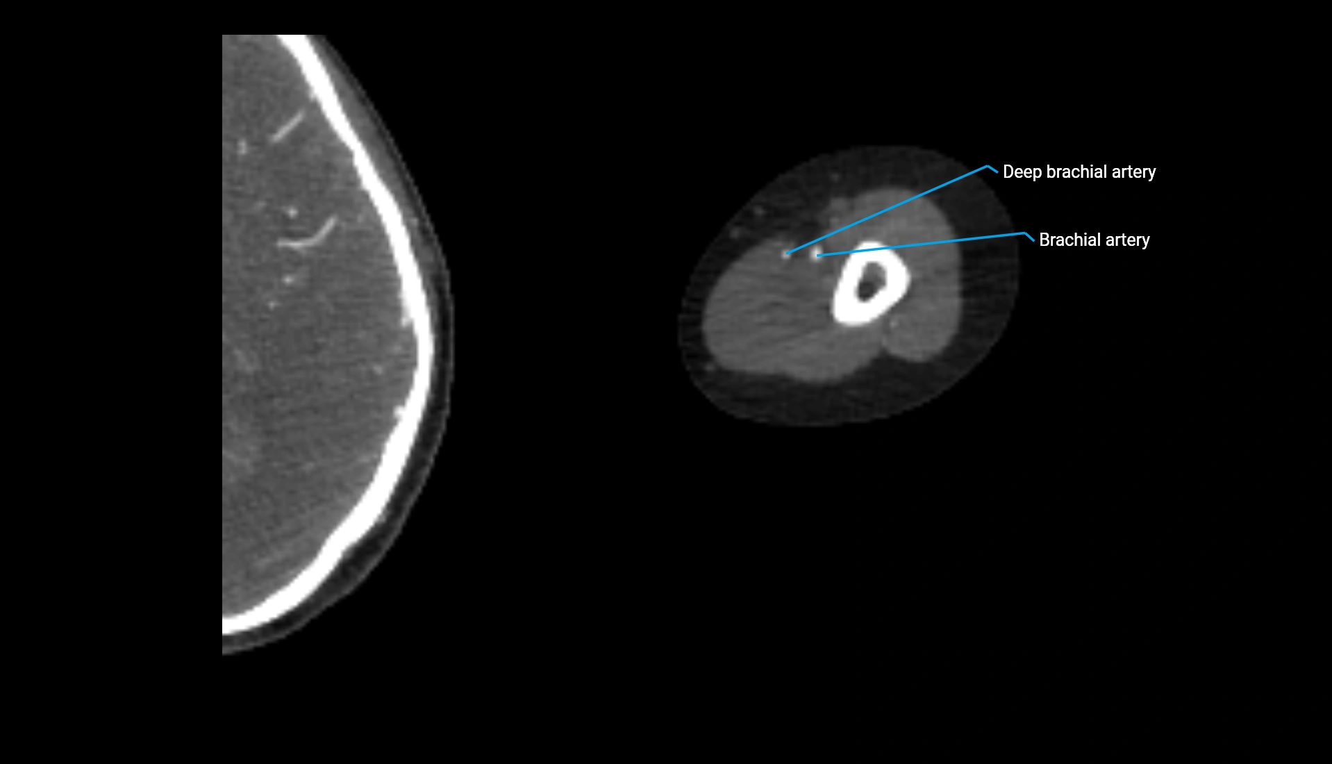
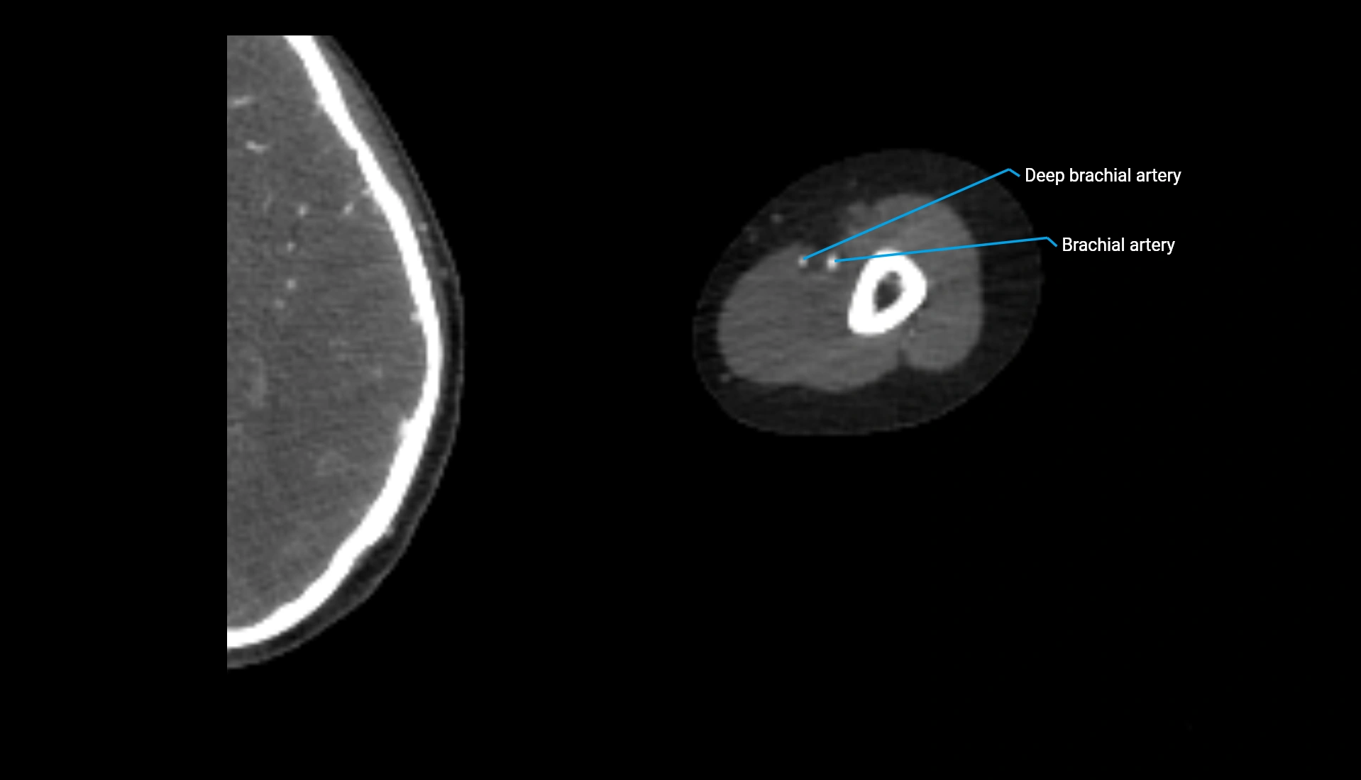
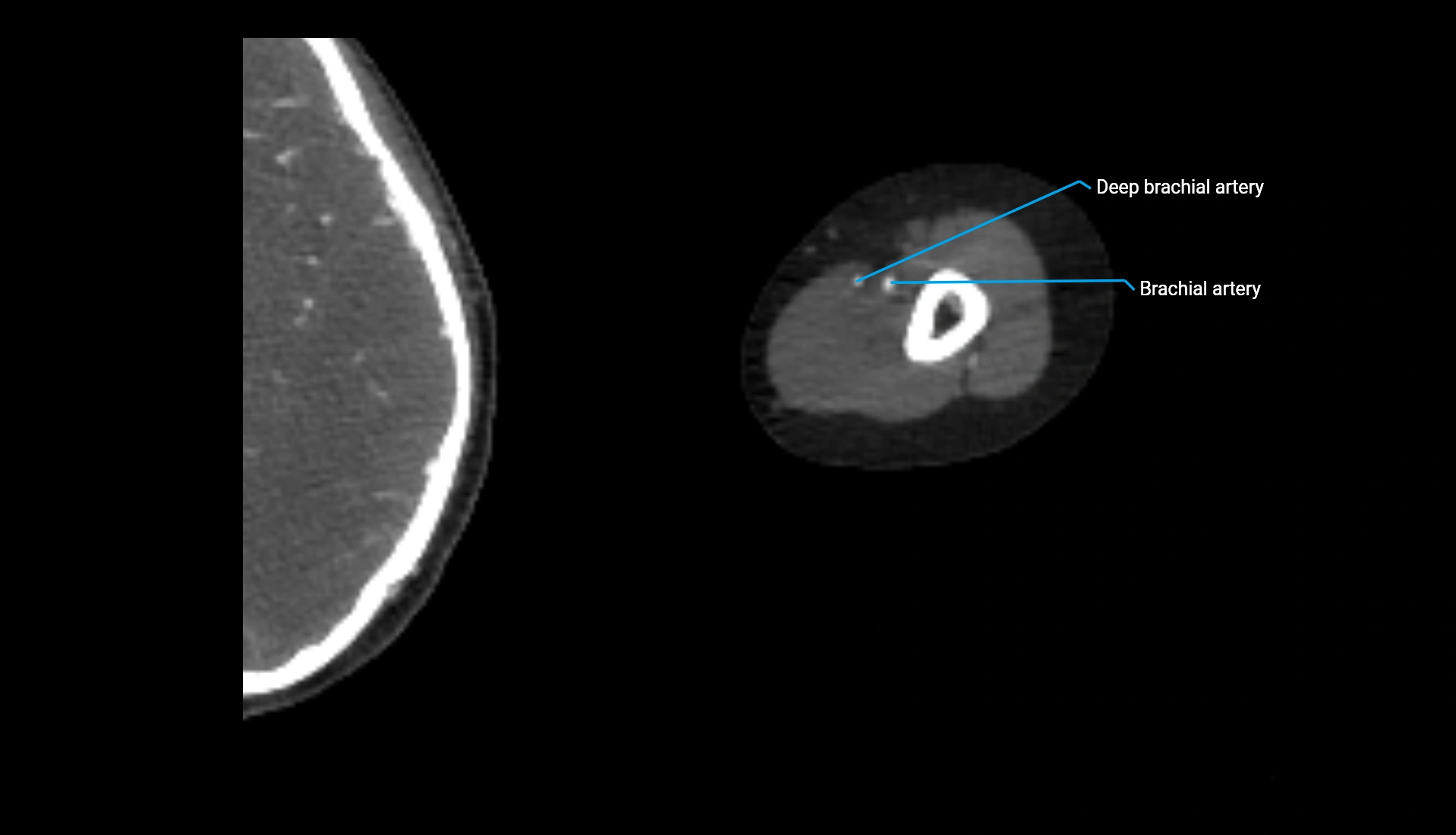
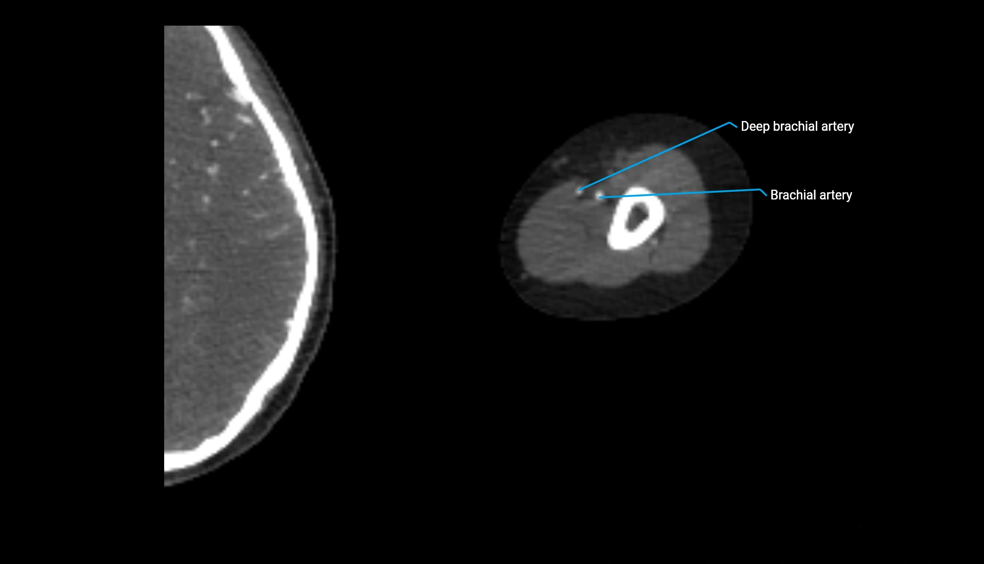
CT Appearance
Non-Contrast CT:
-
Cortex: High-density, sharply defined
-
Subchondral bone: Dense cancellous matrix
-
Articular surface: Smooth concave contour articulating with the capitellum
-
Excellent for evaluating bone integrity, alignment, and subtle fractures
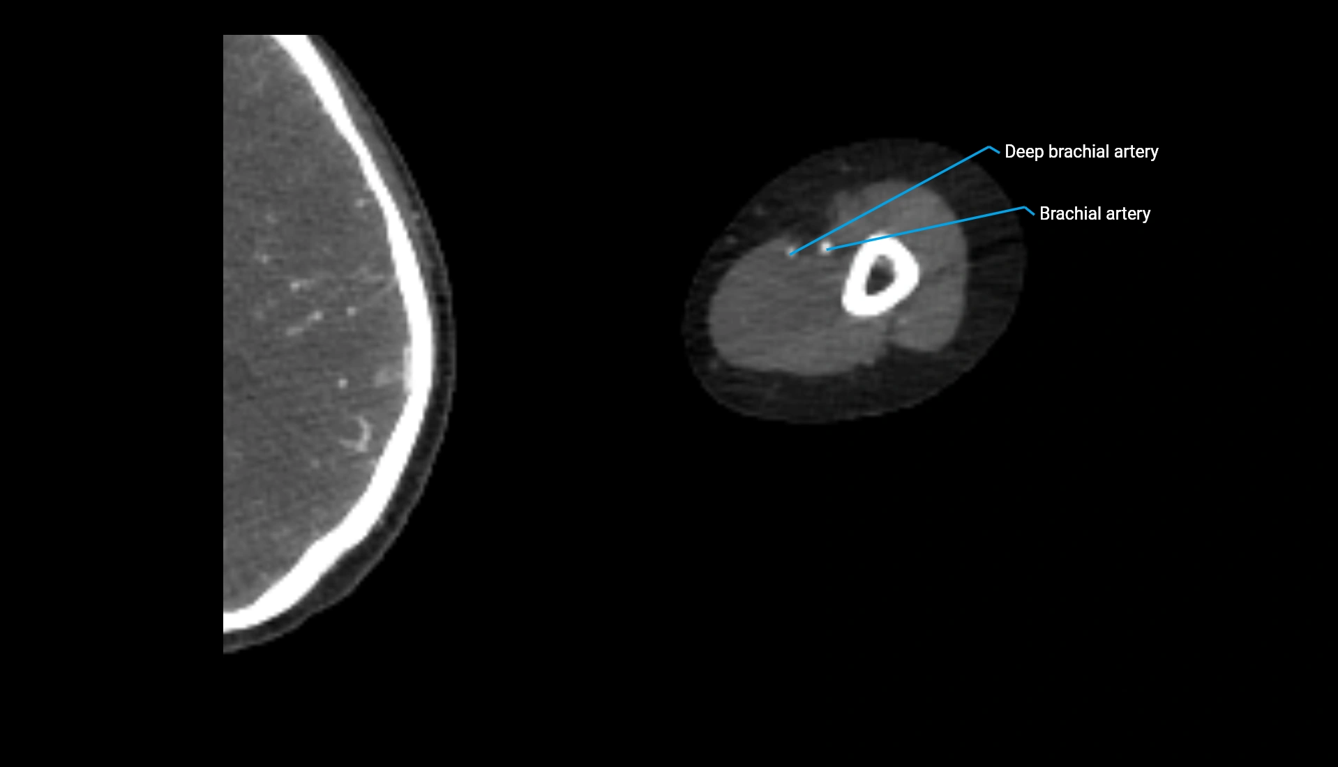
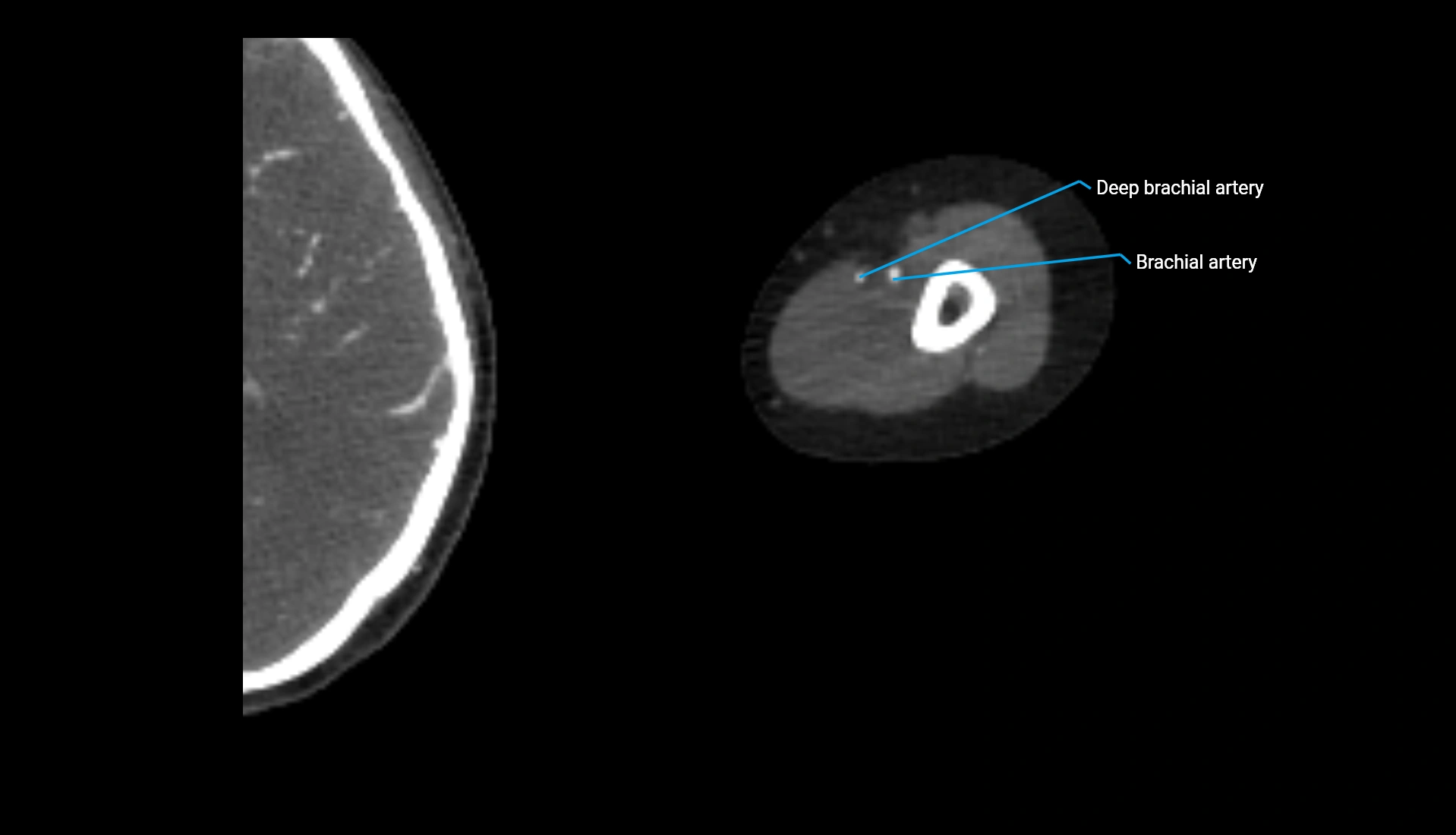
Post-Contrast CT:
-
Bone: No enhancement
-
Joint capsule and synovium: Mild enhancement outlining the joint
-
Improves contrast between soft tissues and bony margins
-
Useful in detecting subtle joint abnormalities or postoperative changes
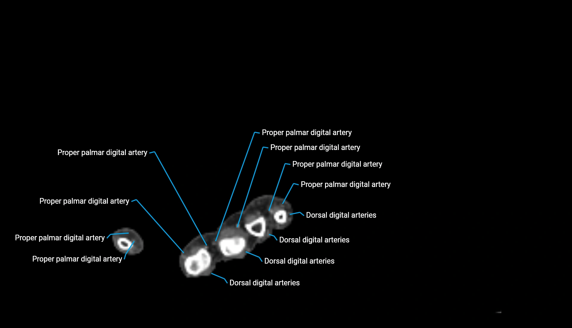
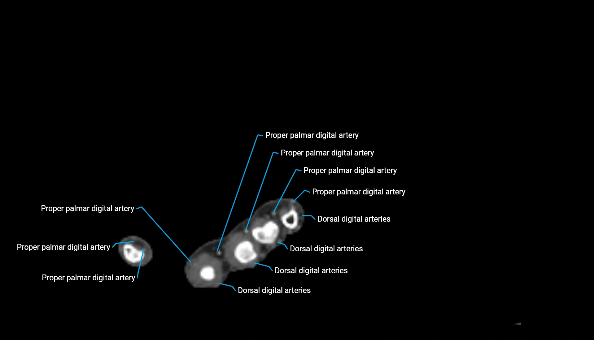
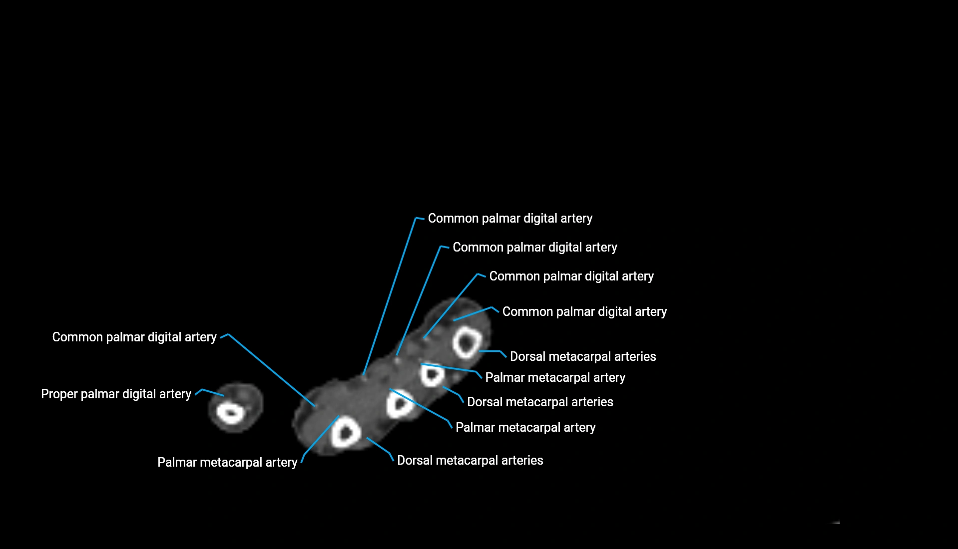
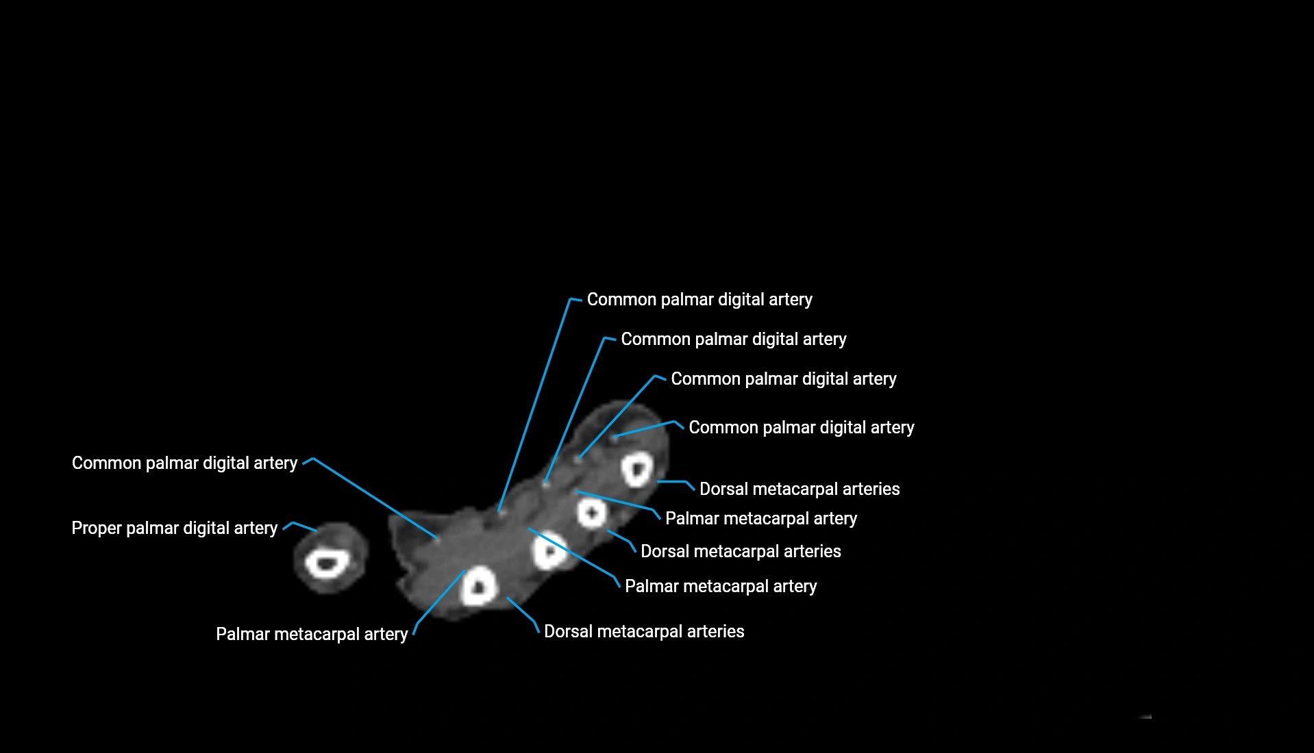
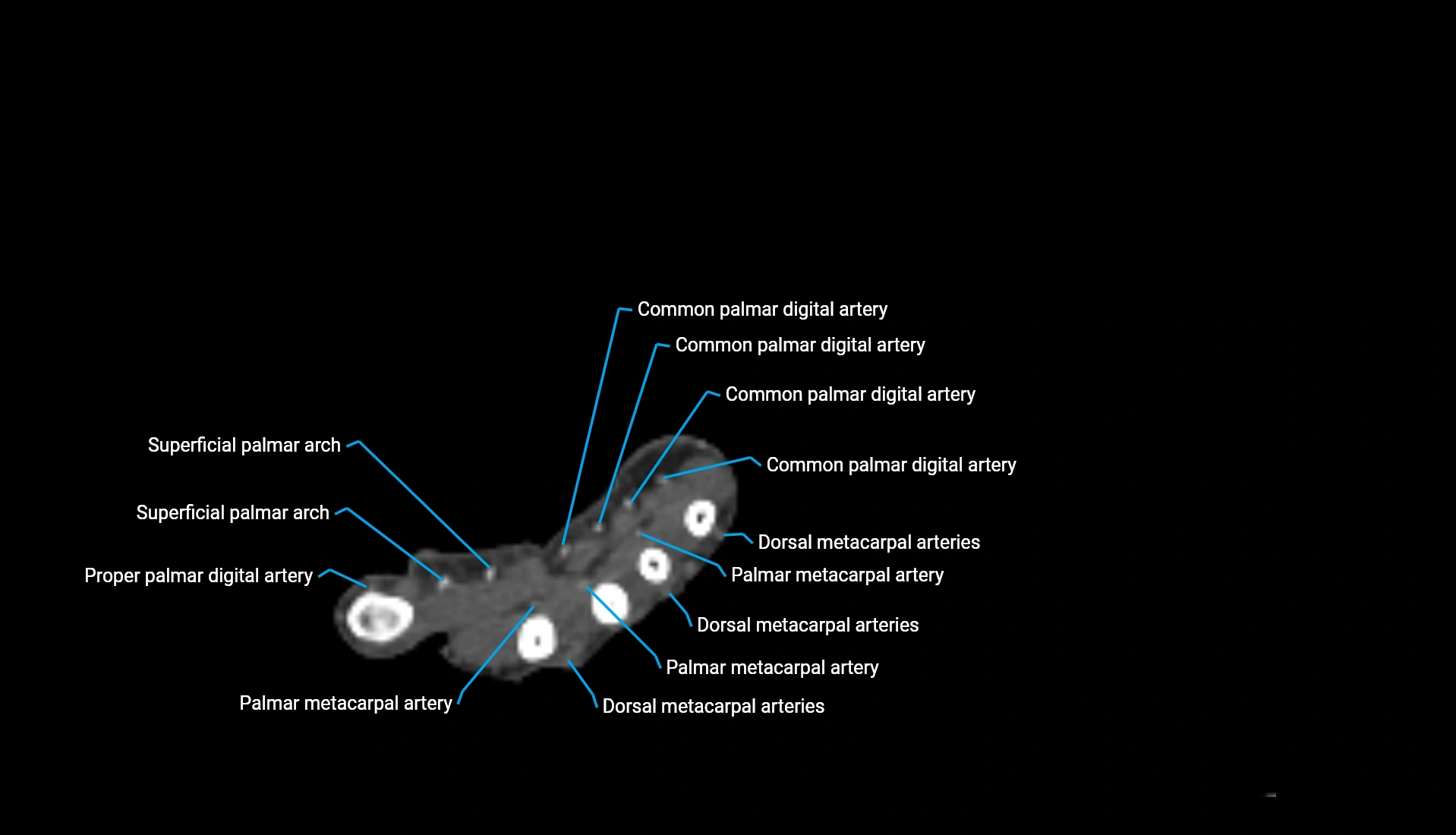
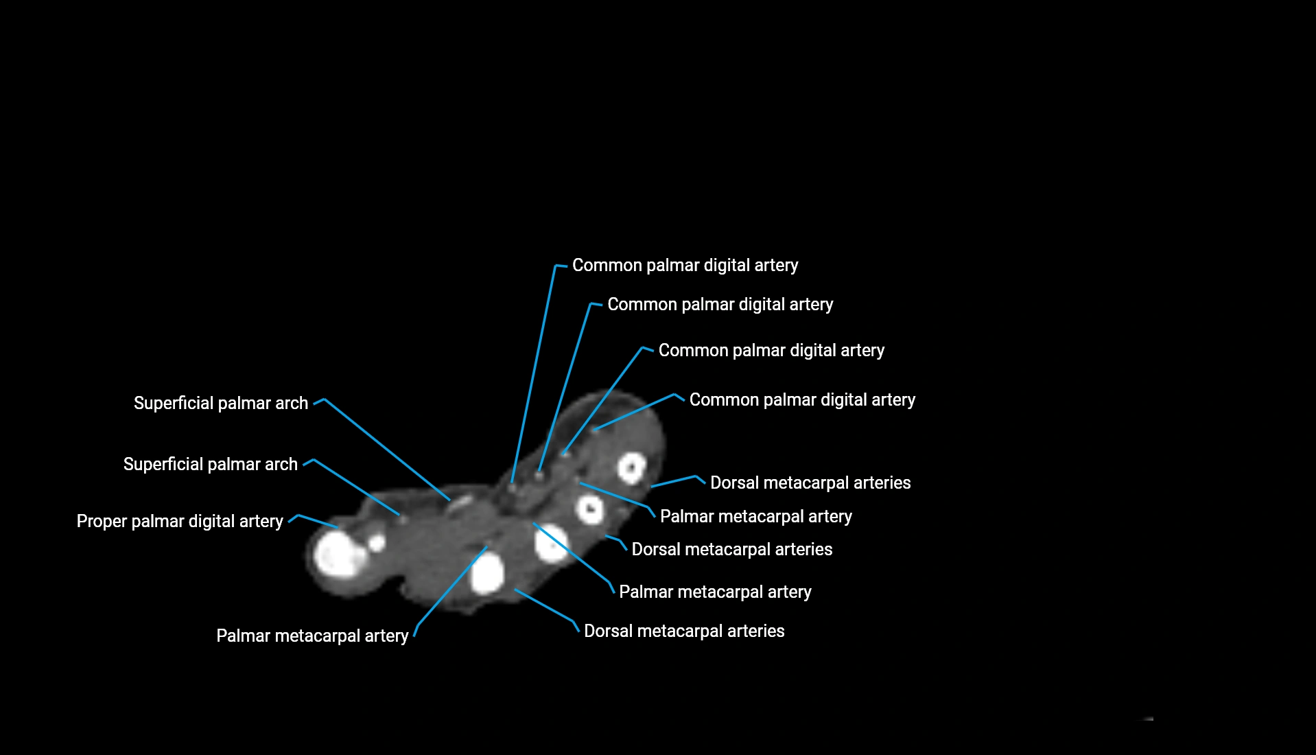
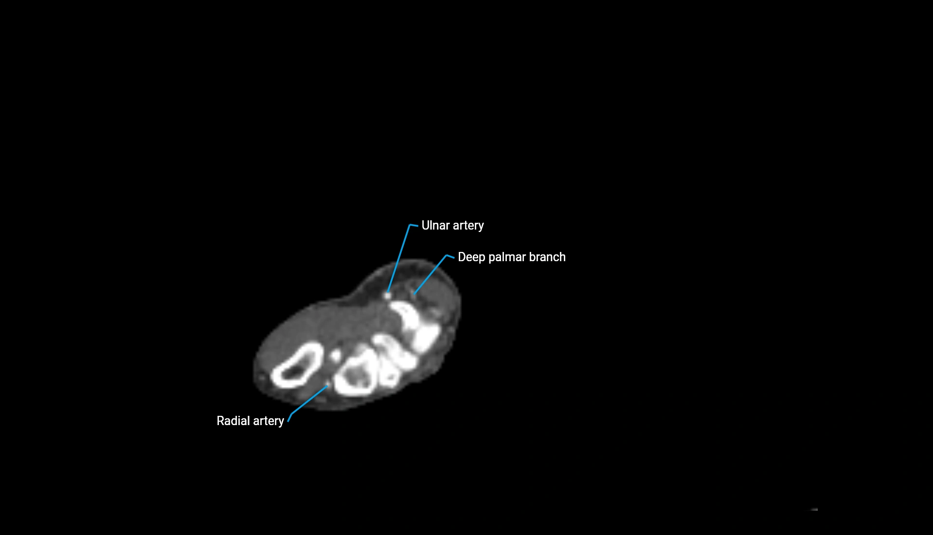
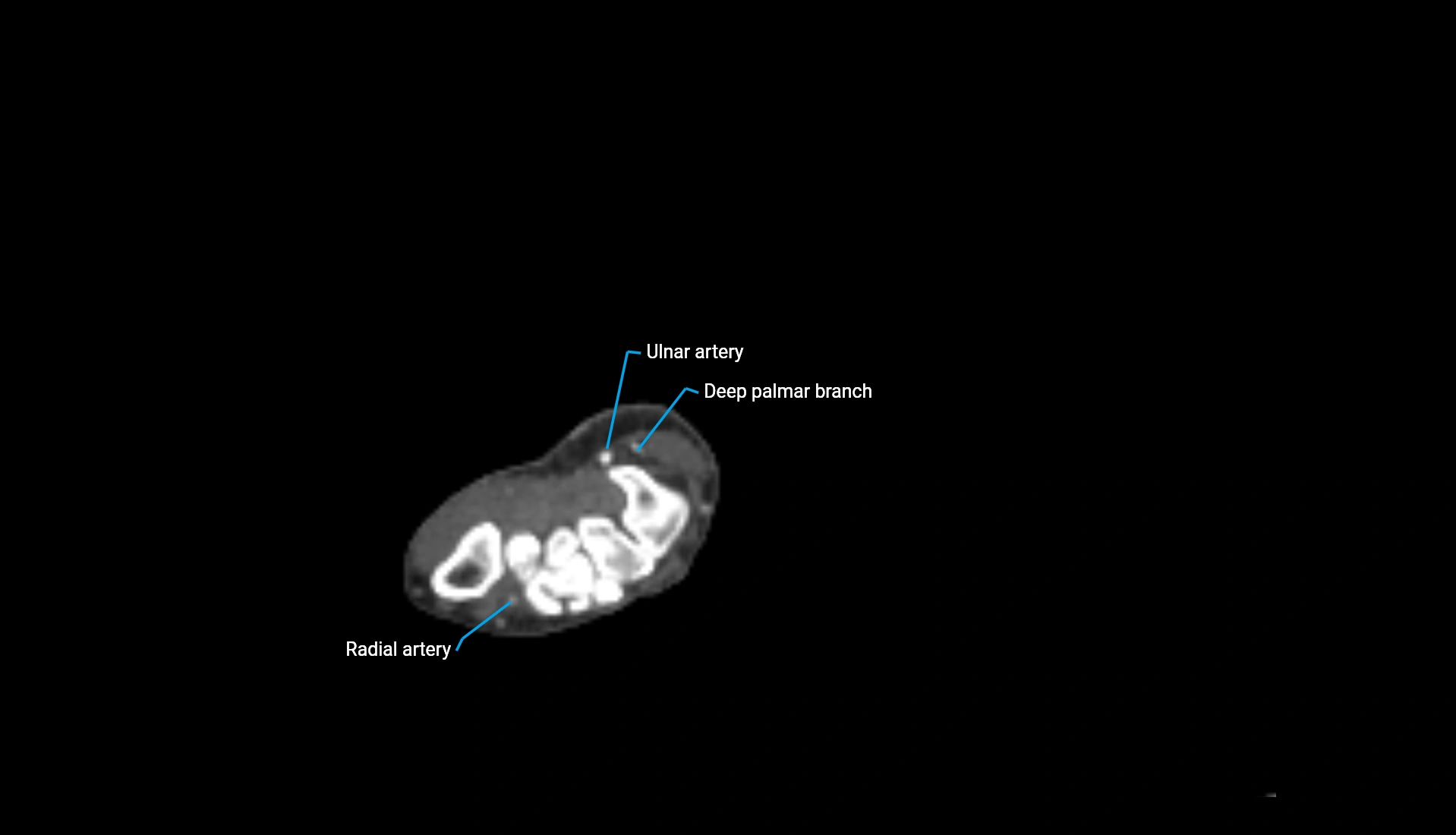
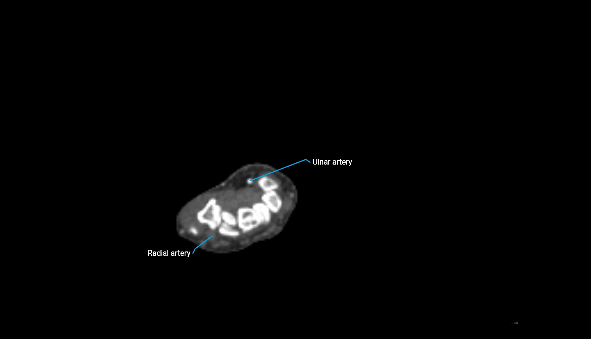
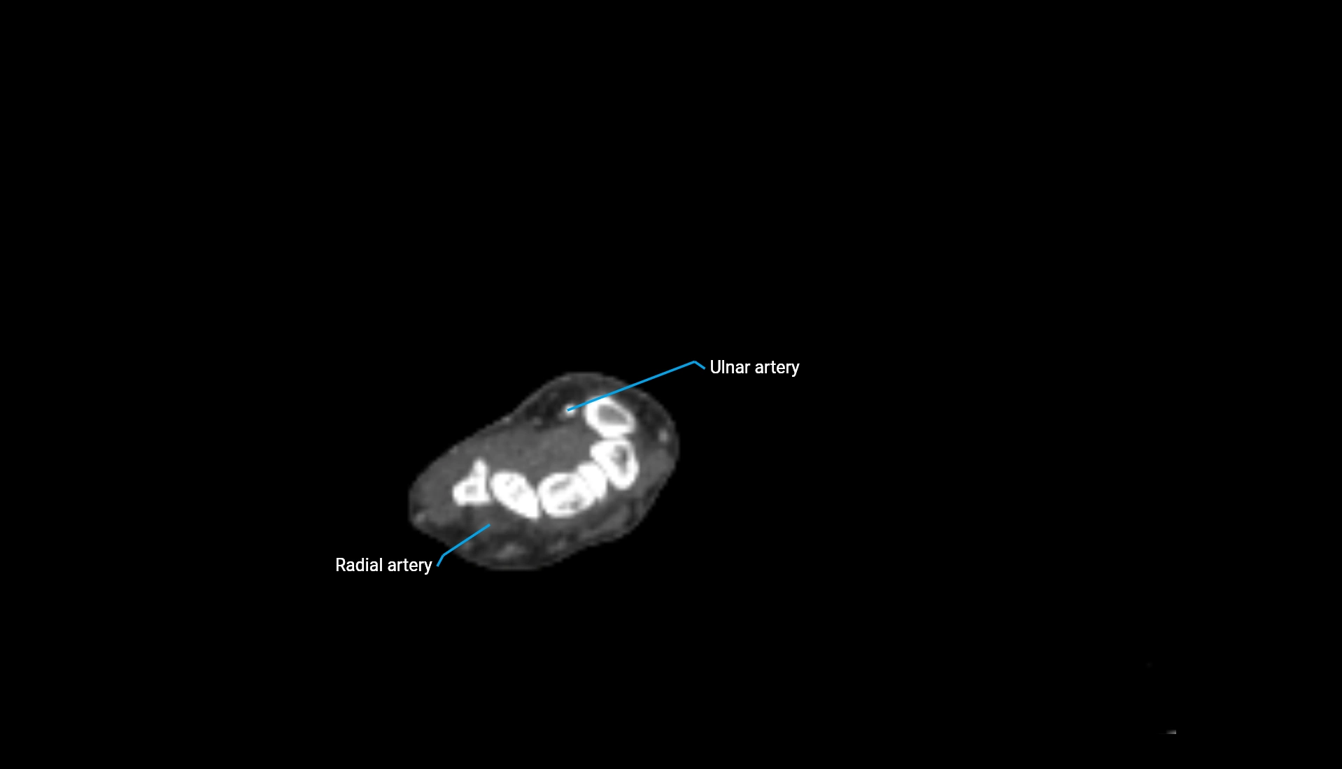
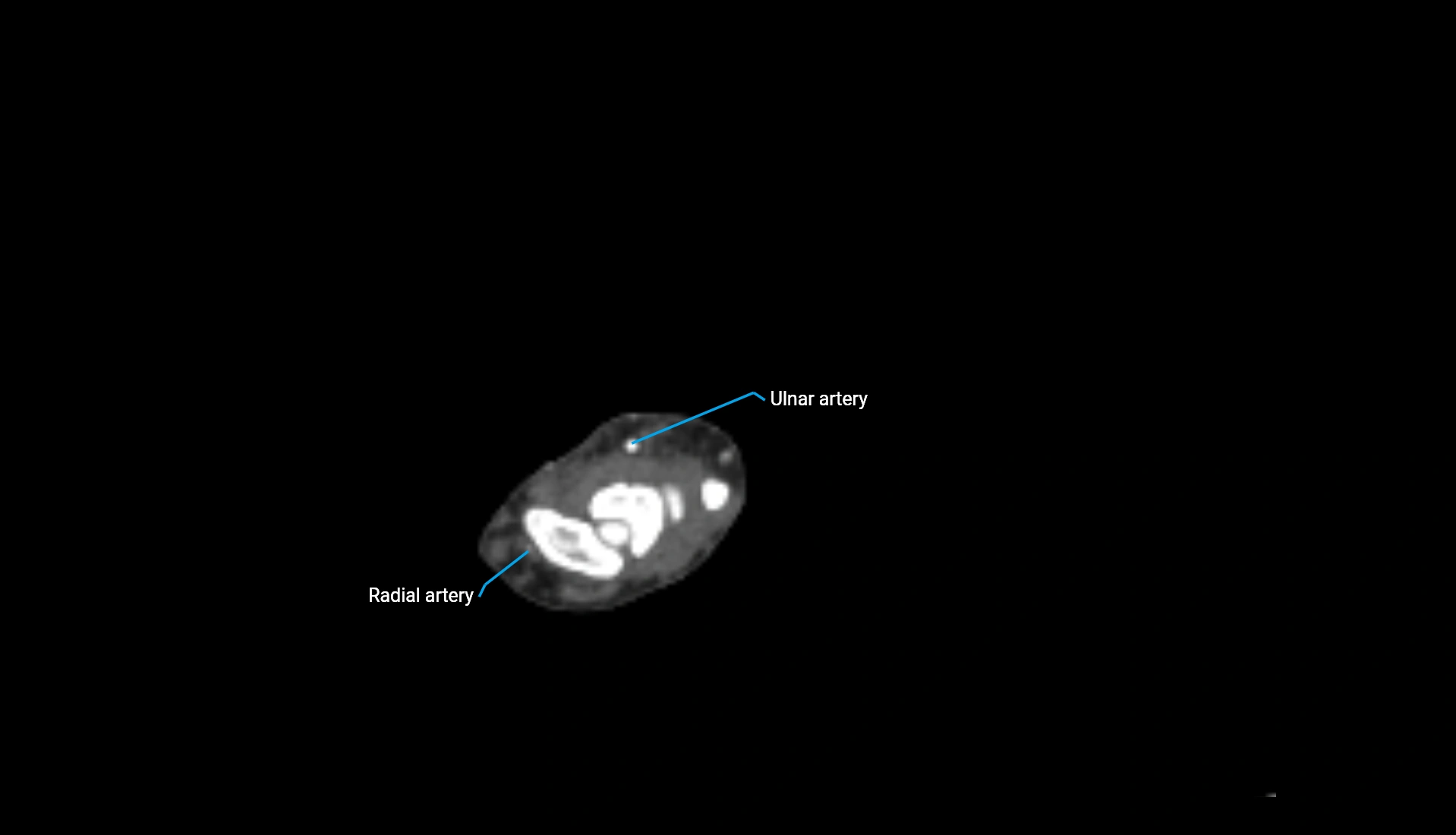
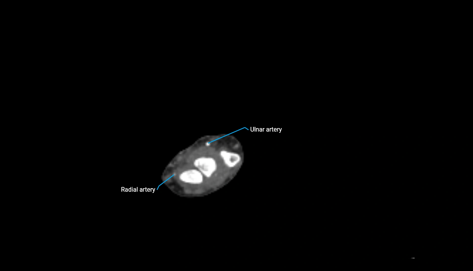
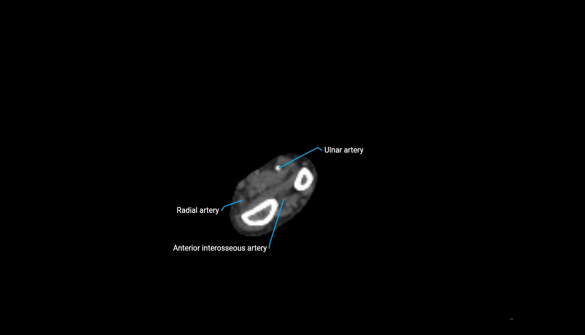
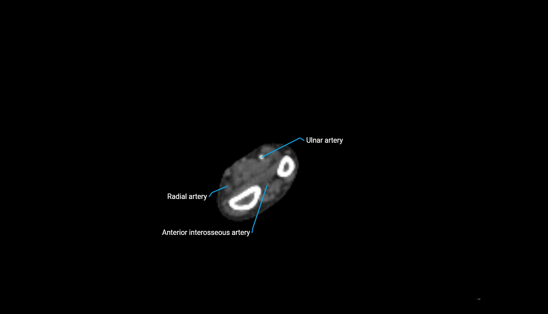
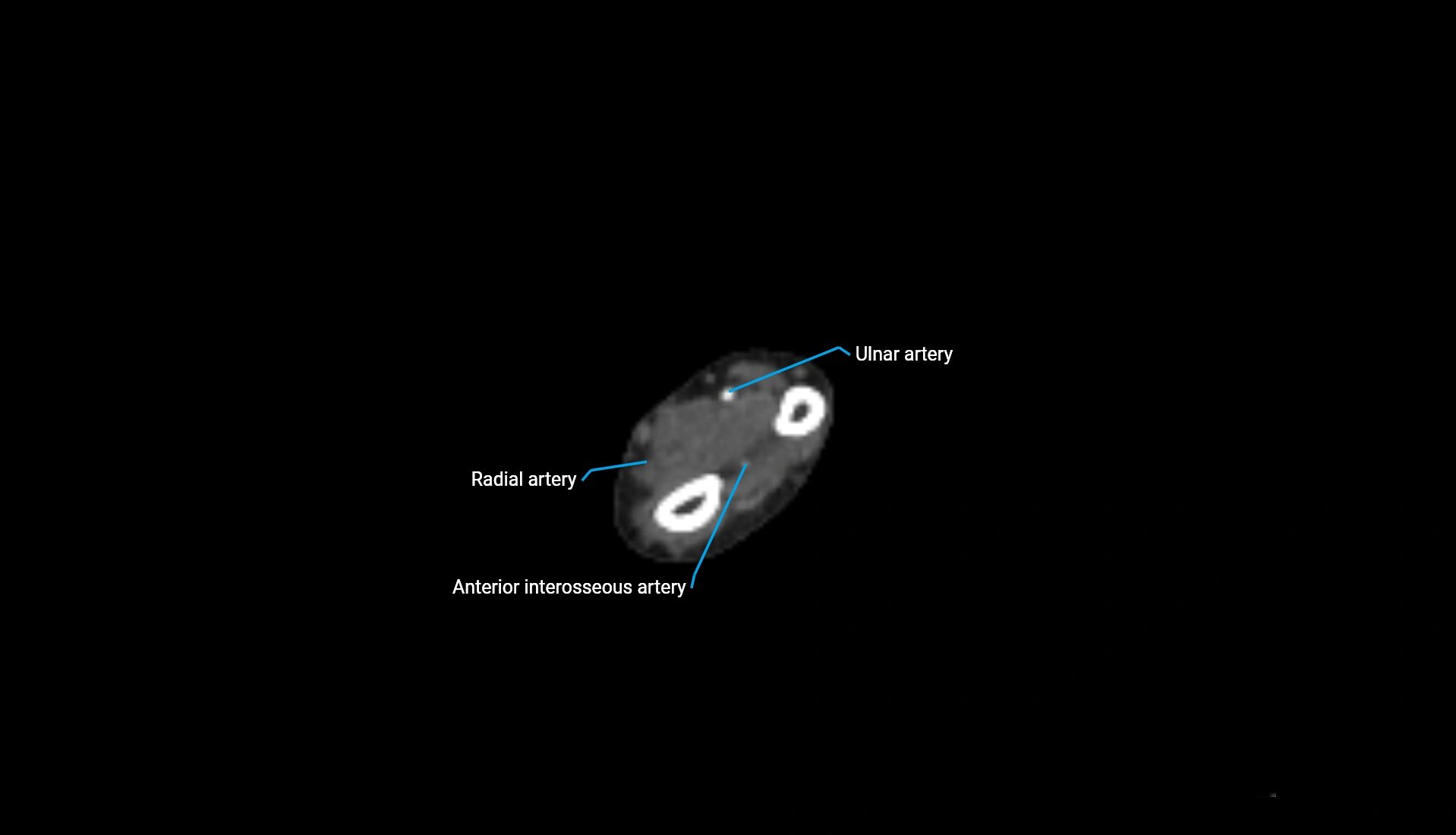
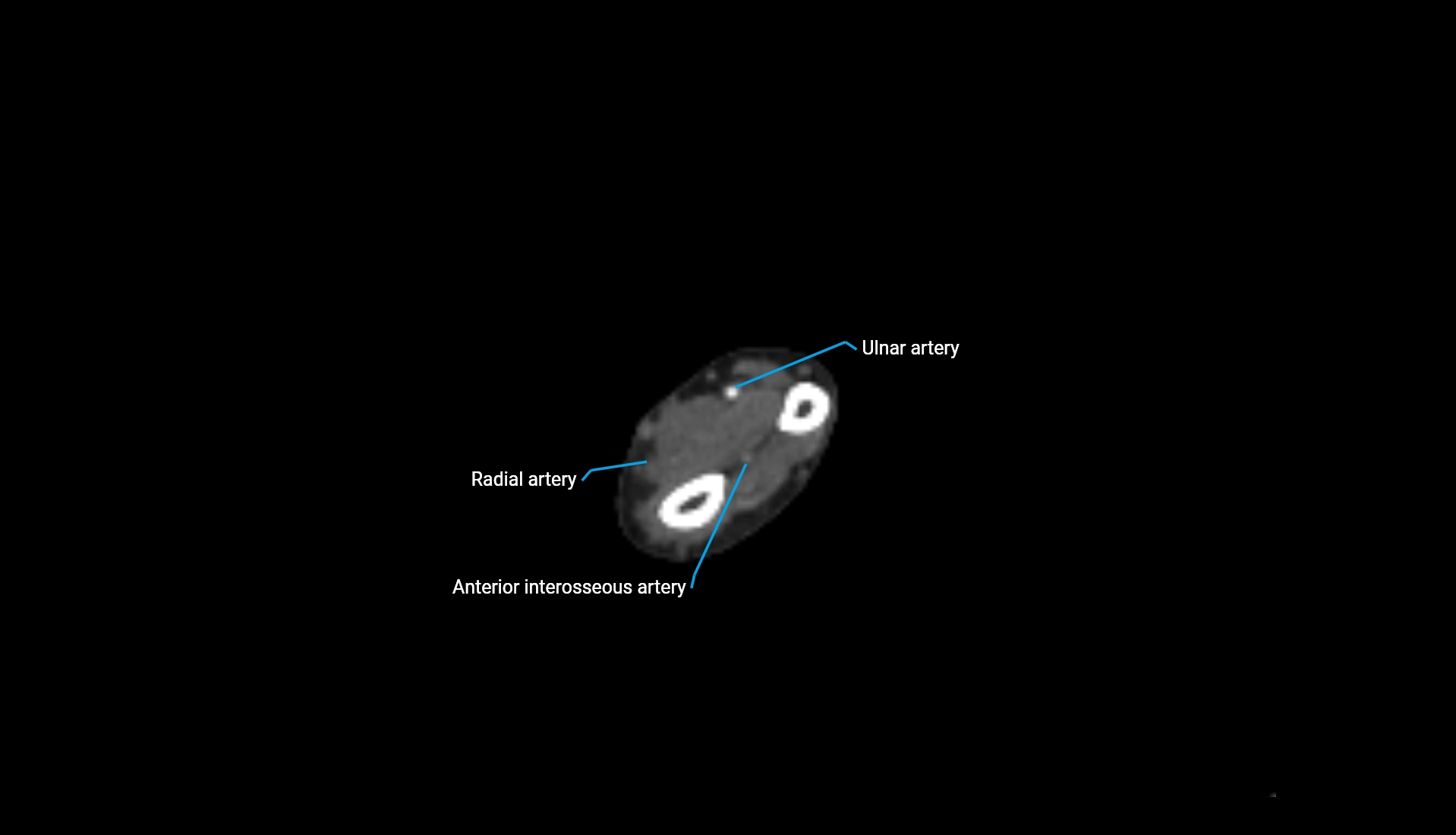
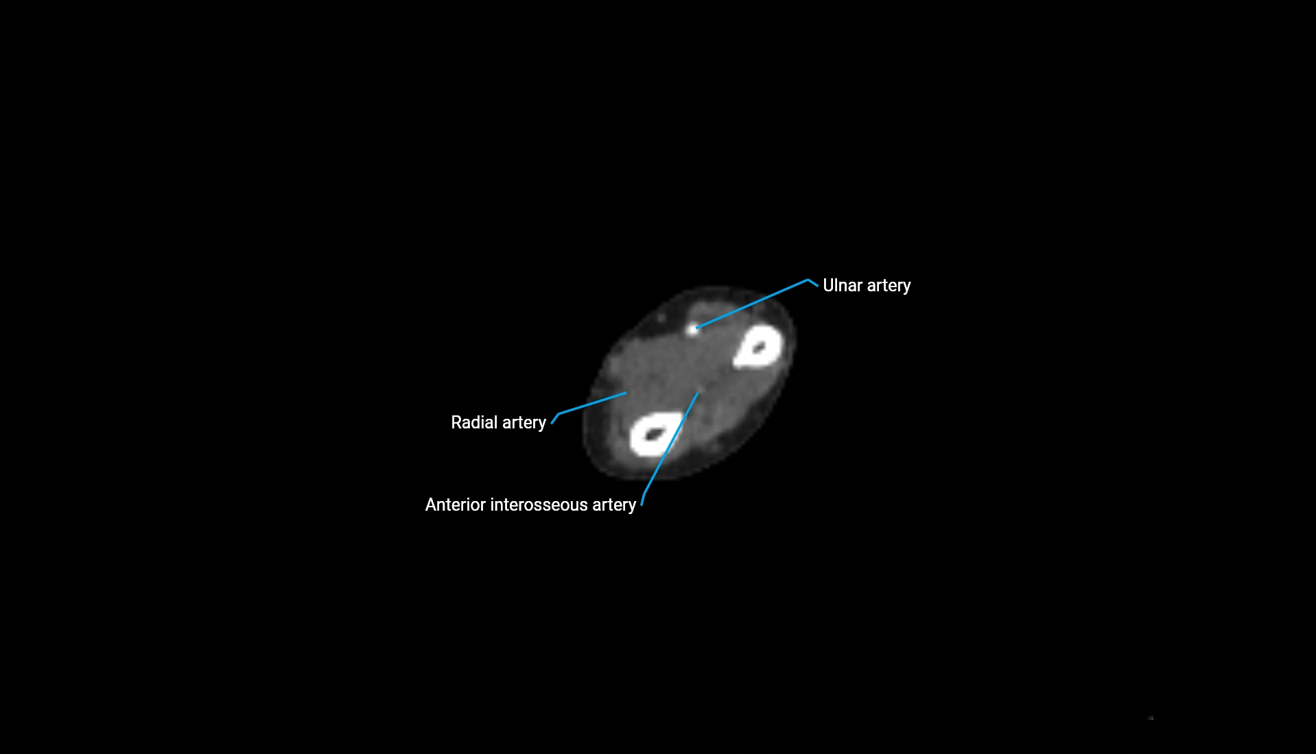
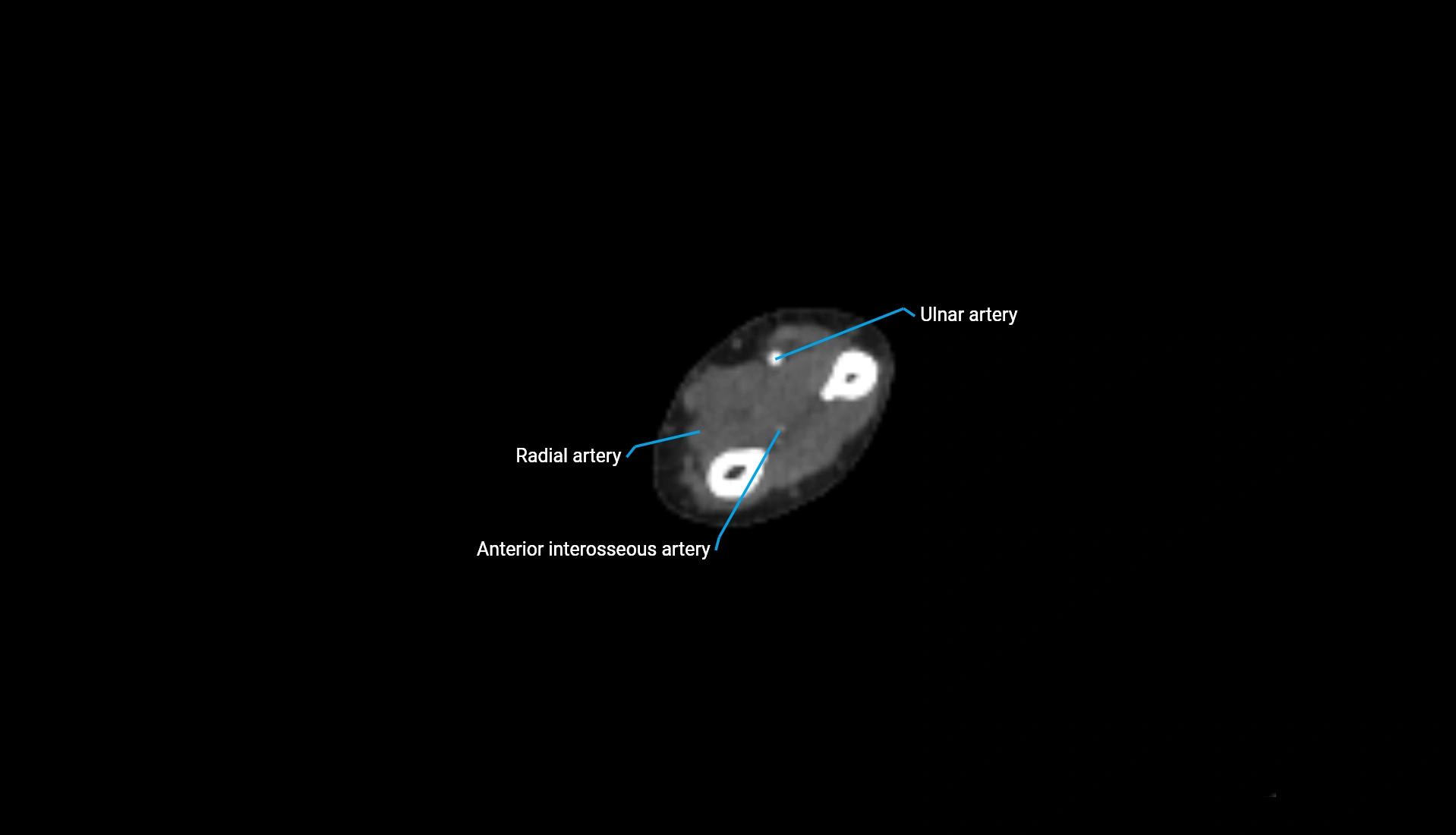
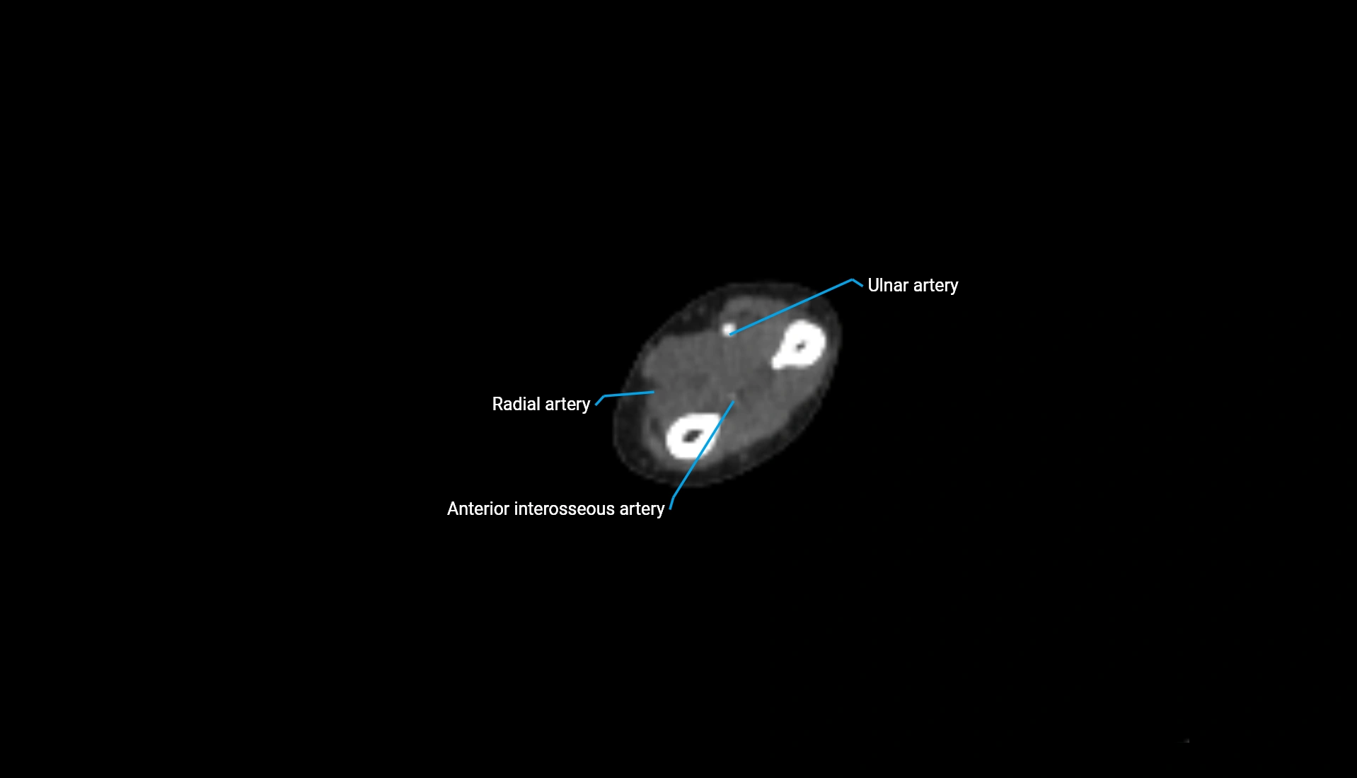
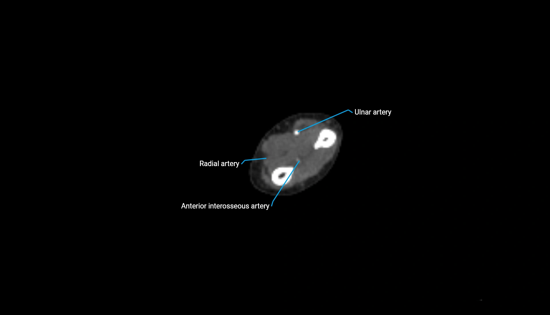
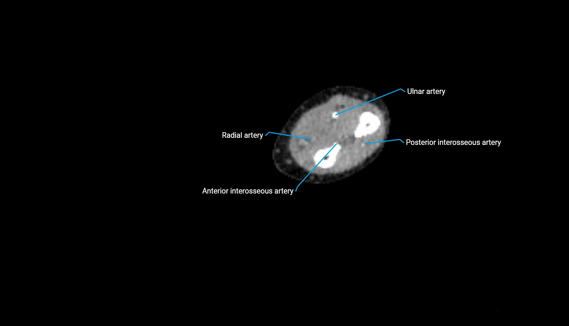
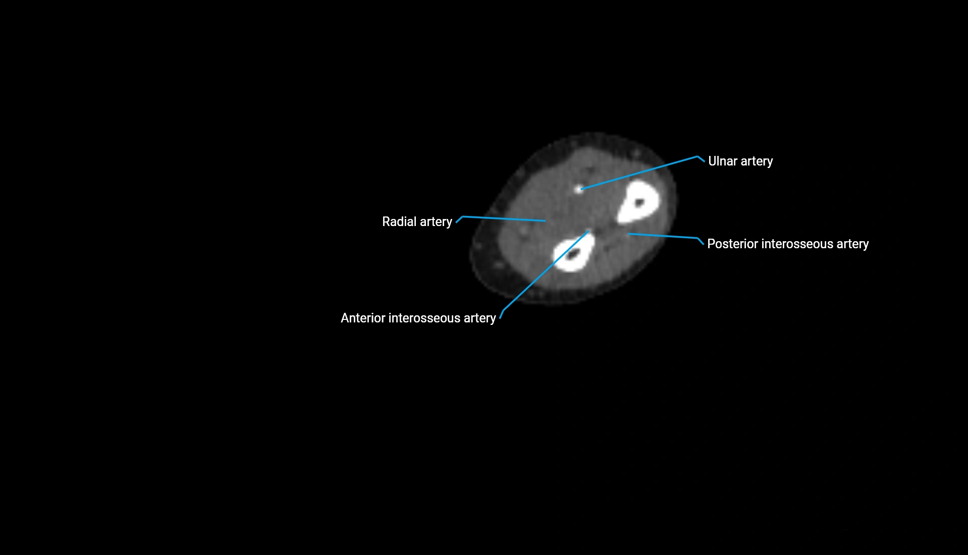
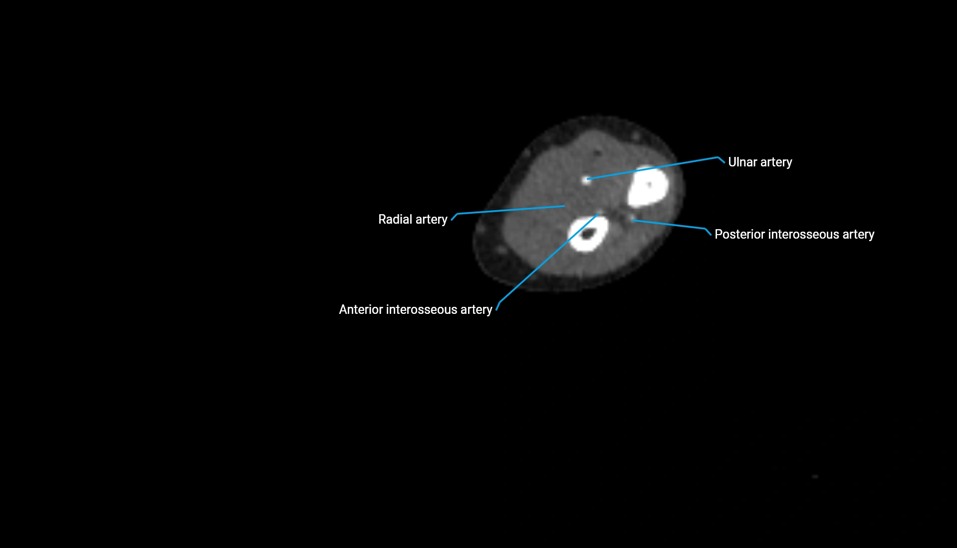
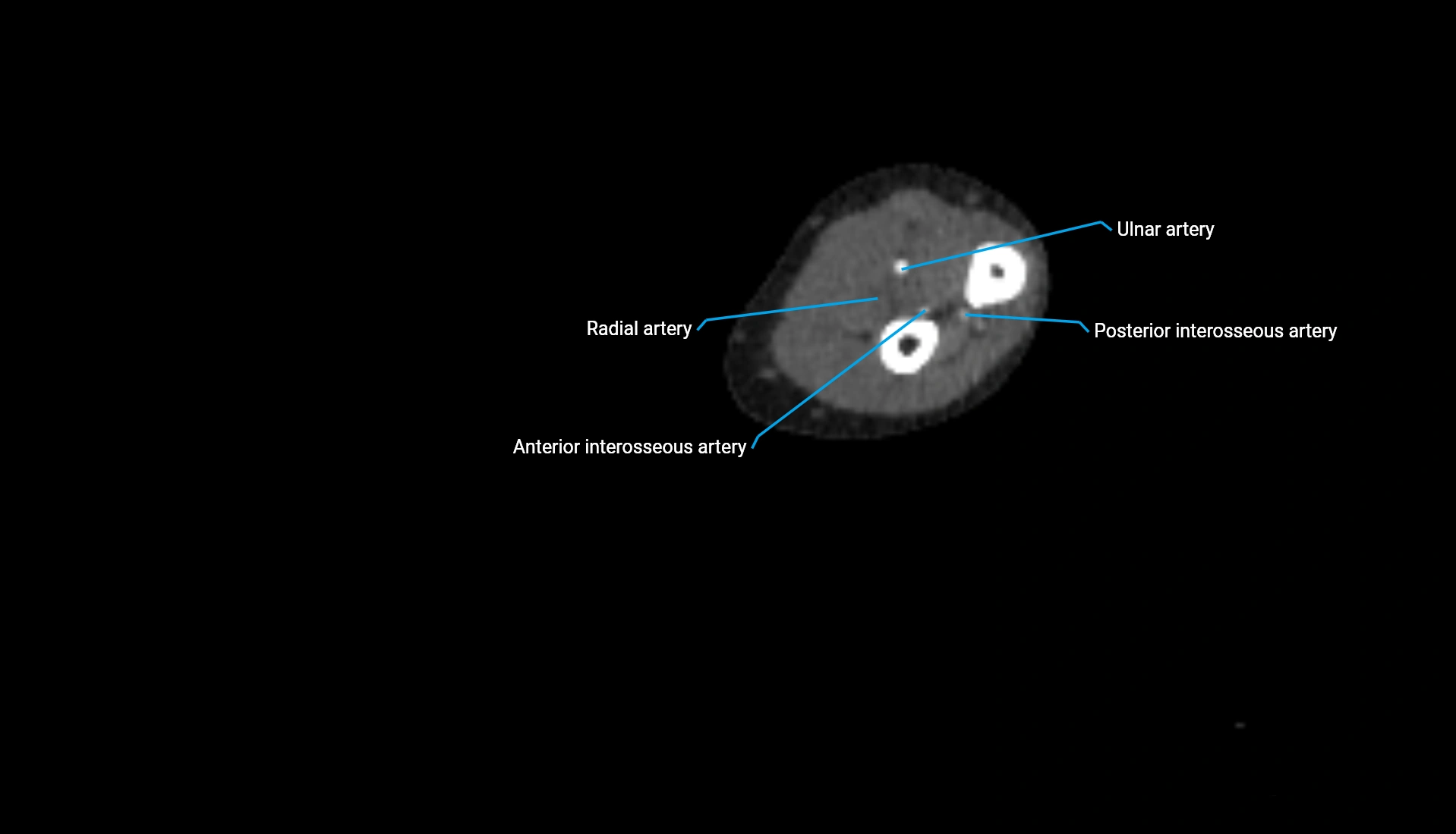
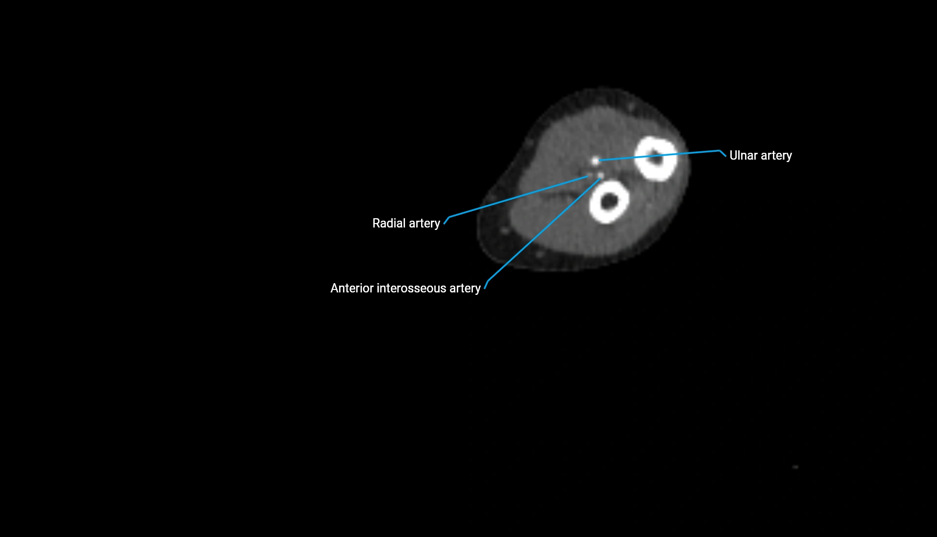
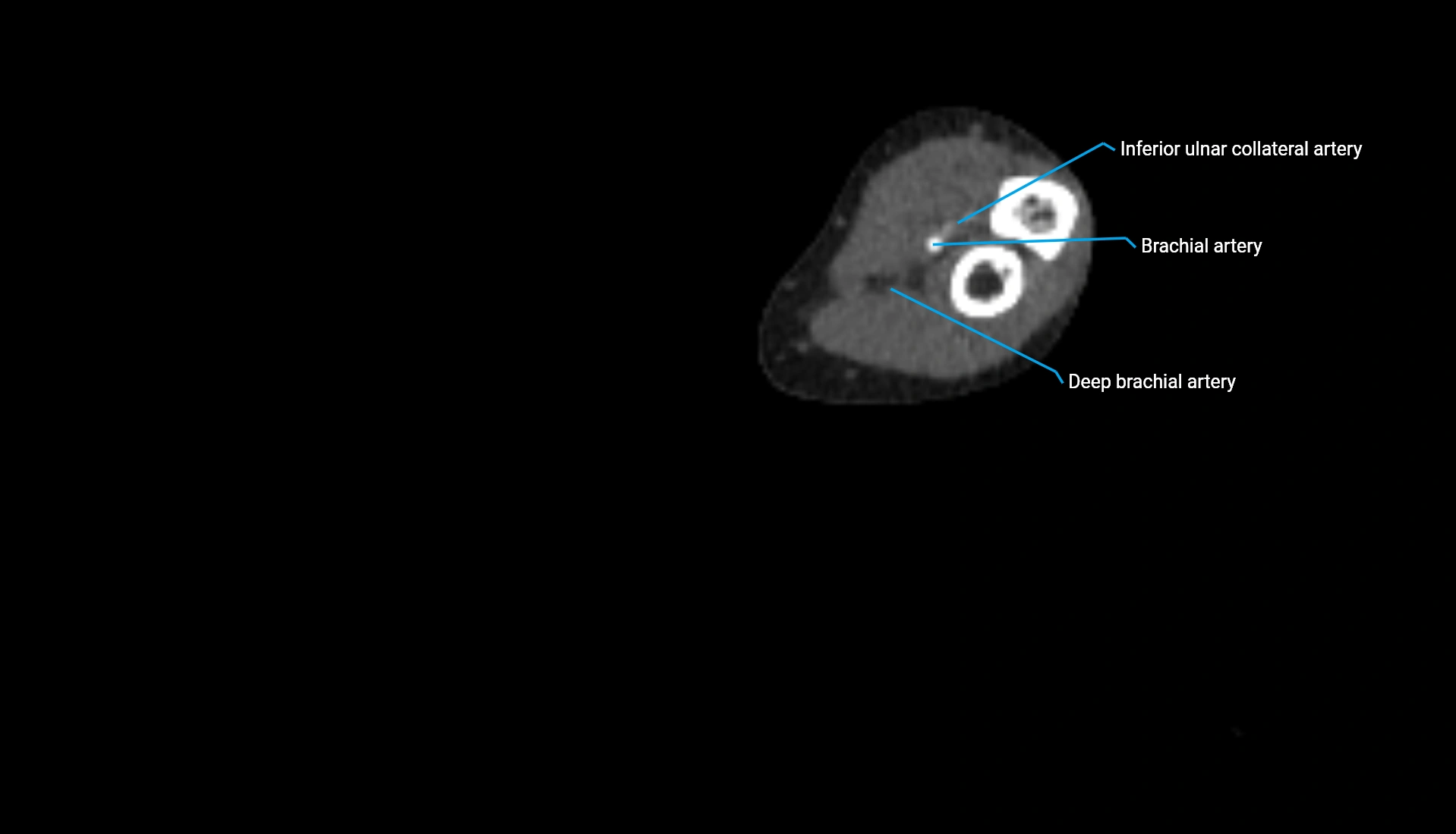
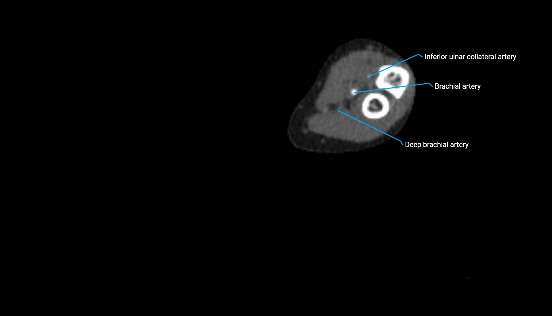
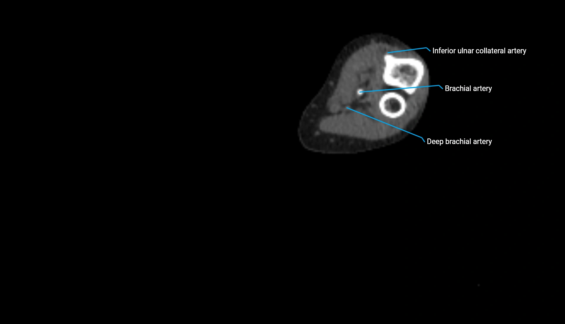
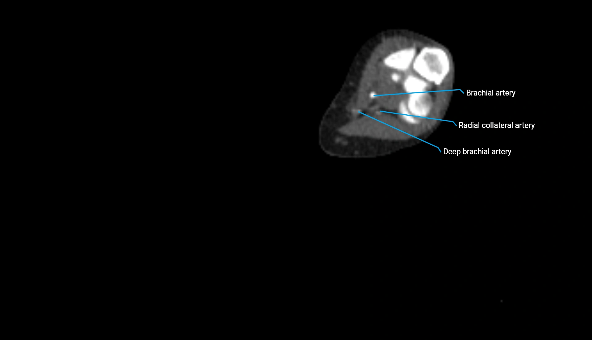
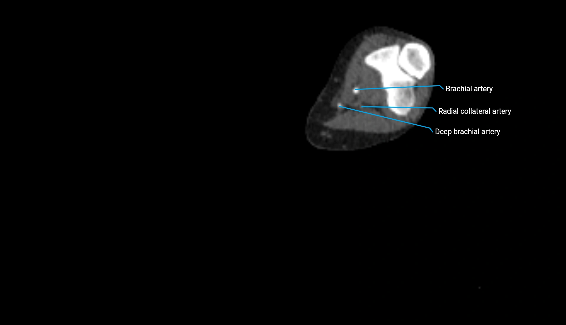
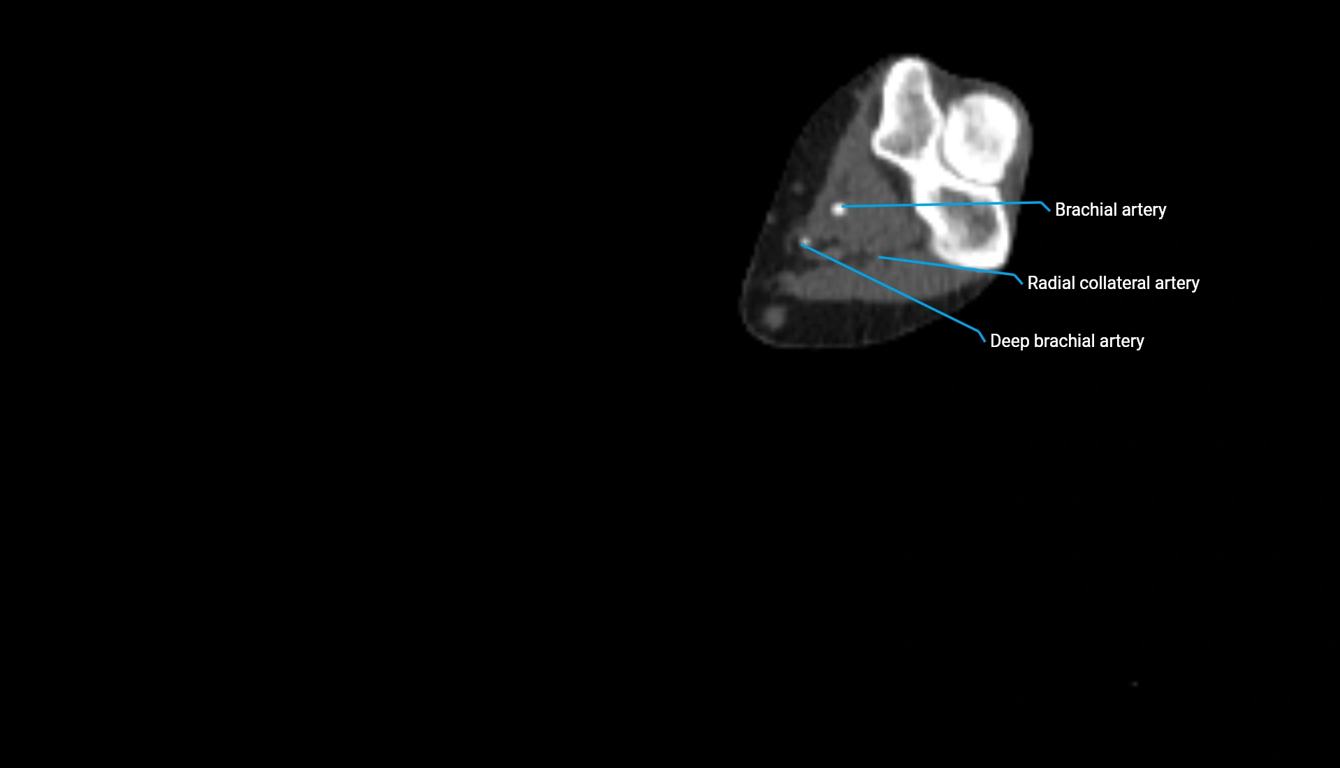
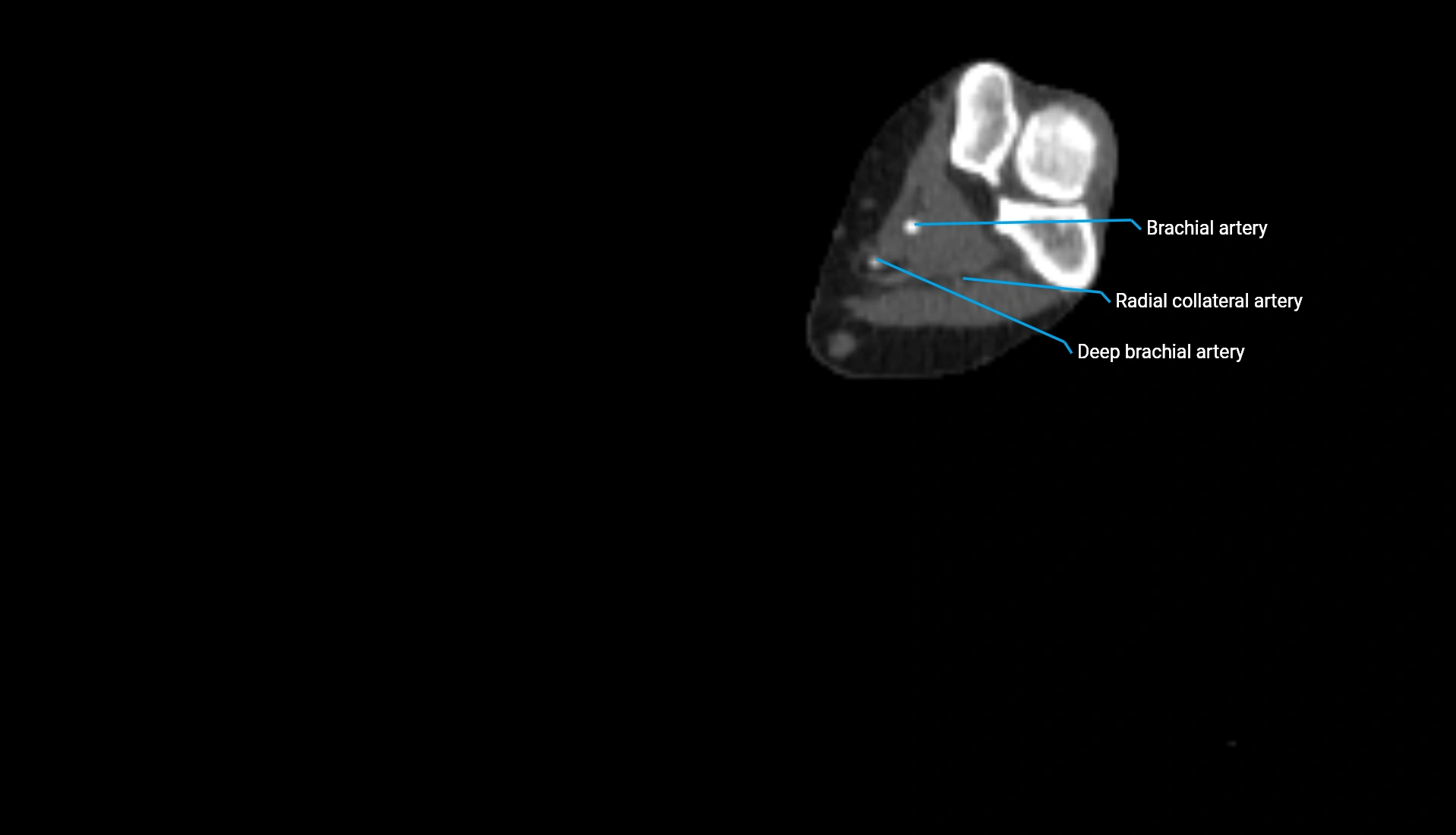
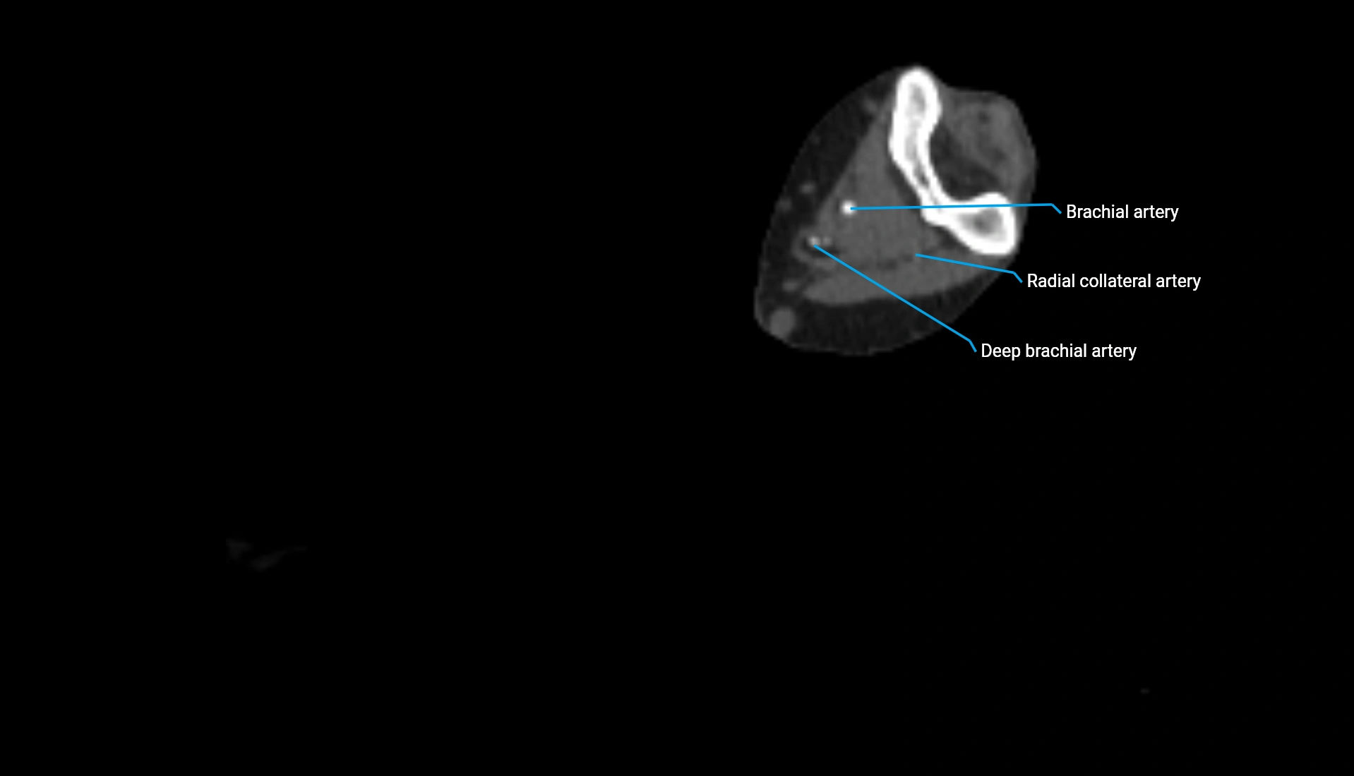
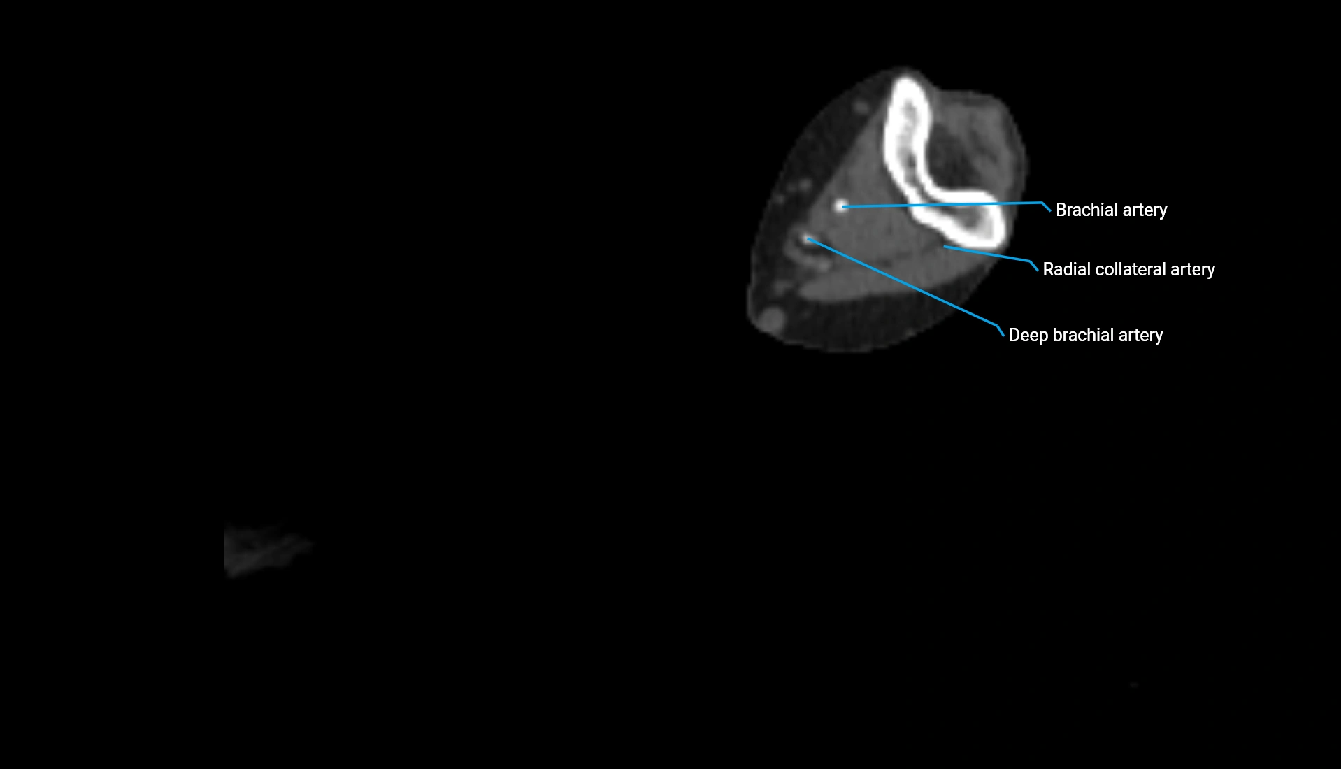
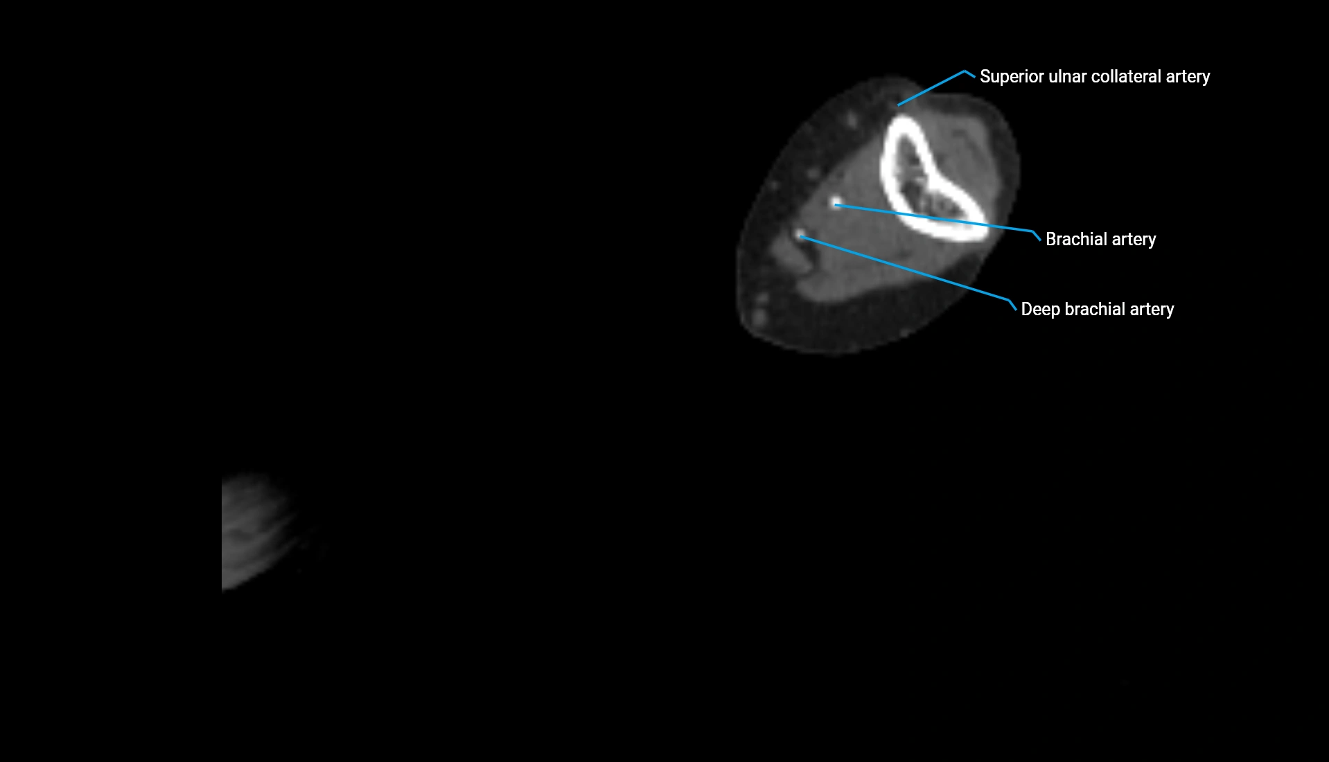
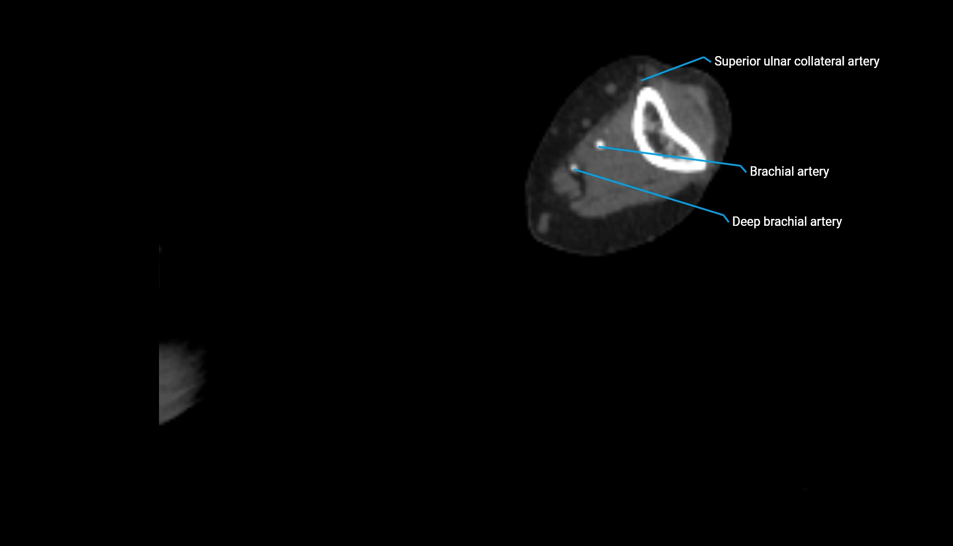
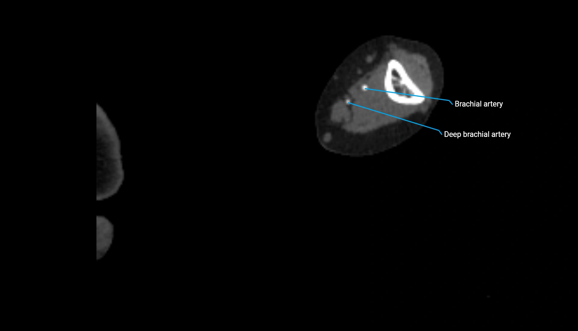
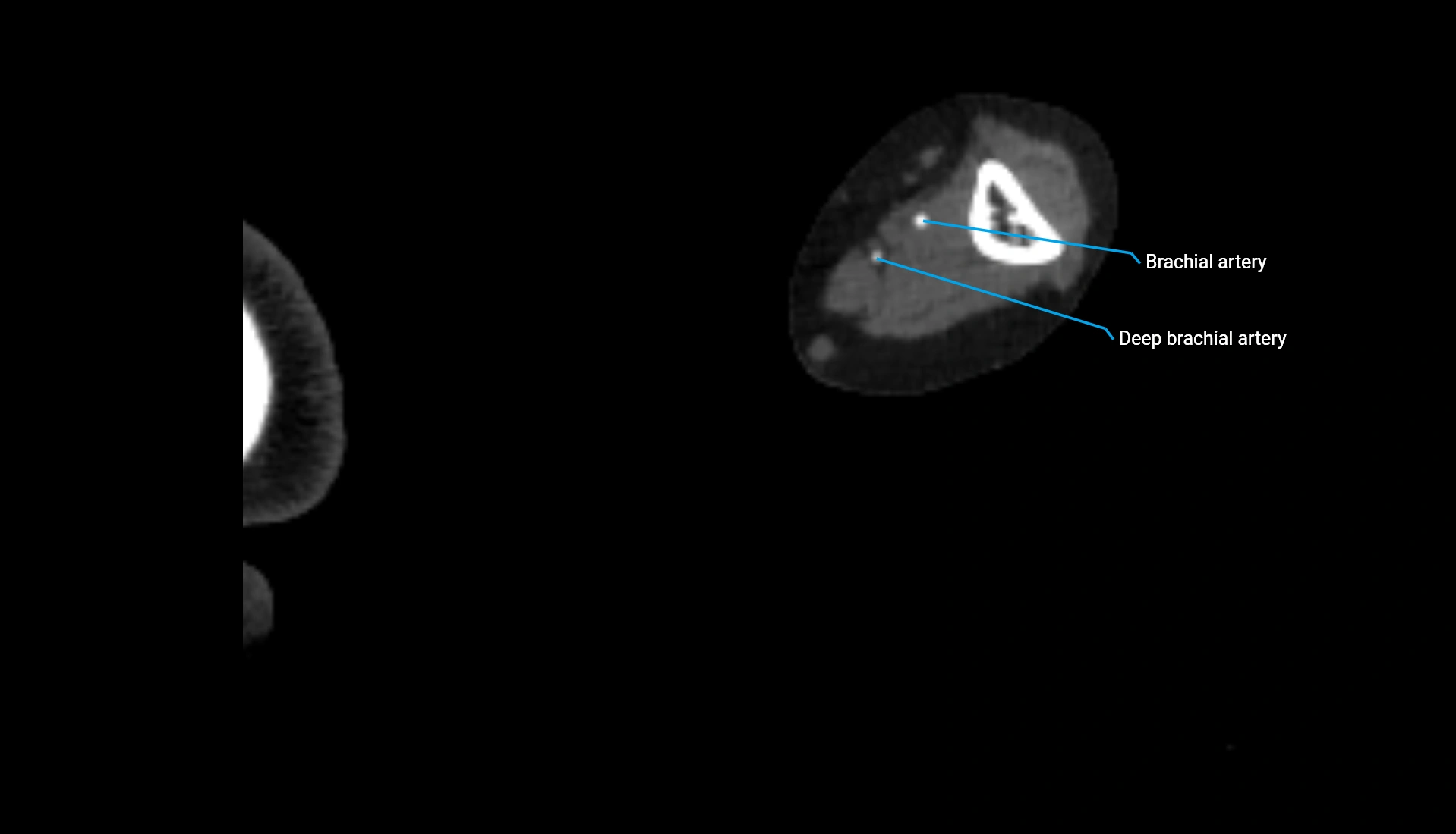
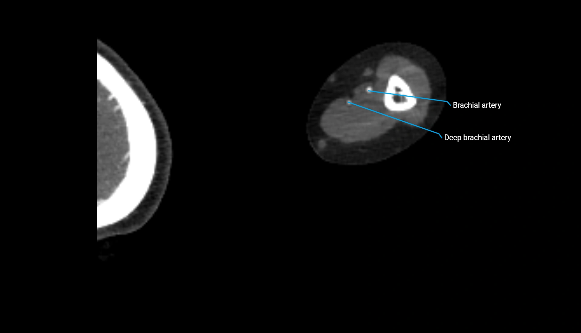
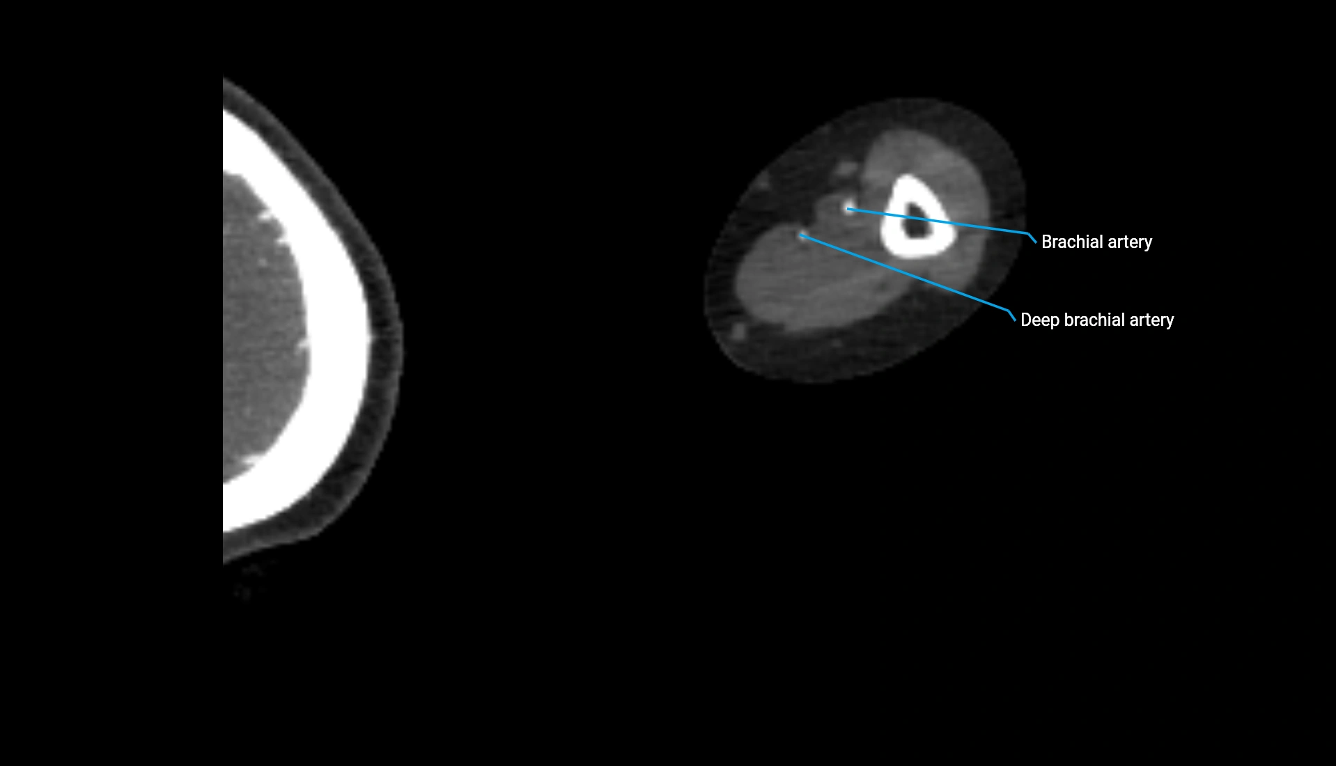
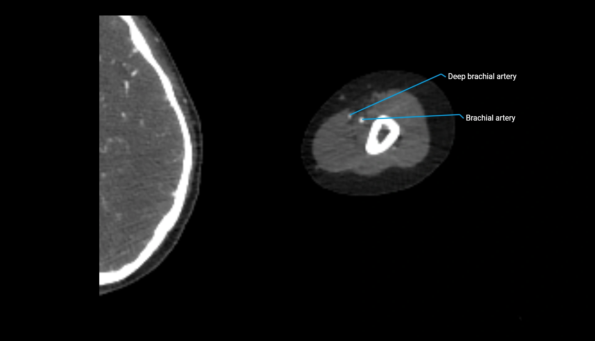
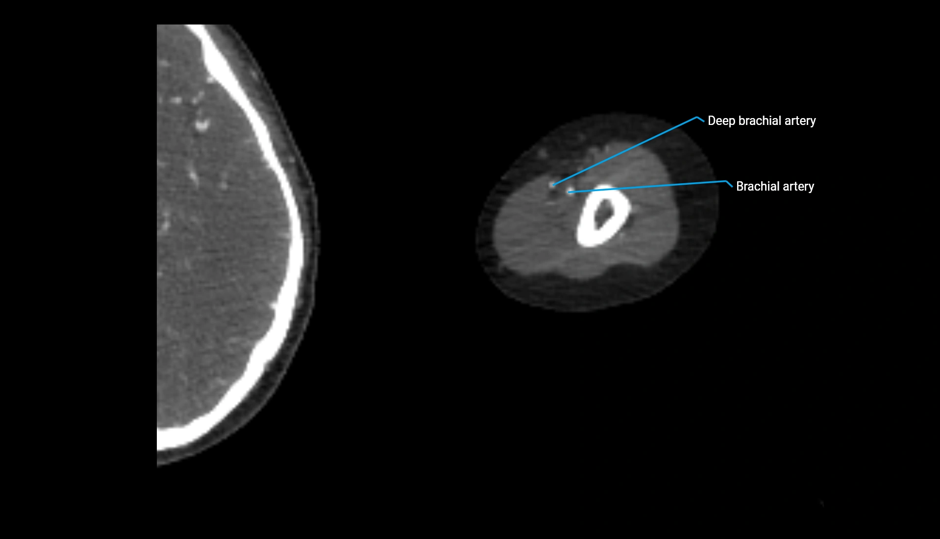
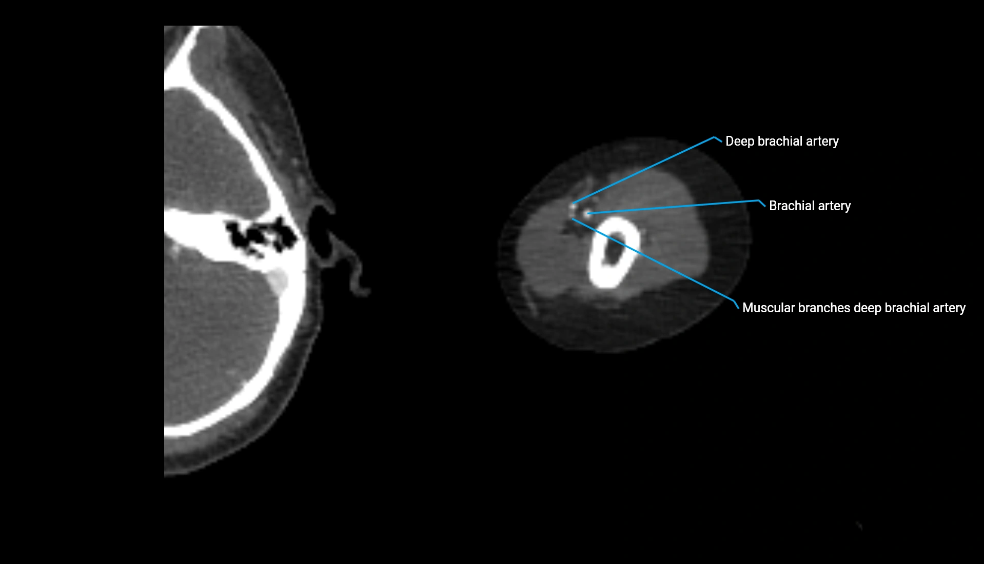
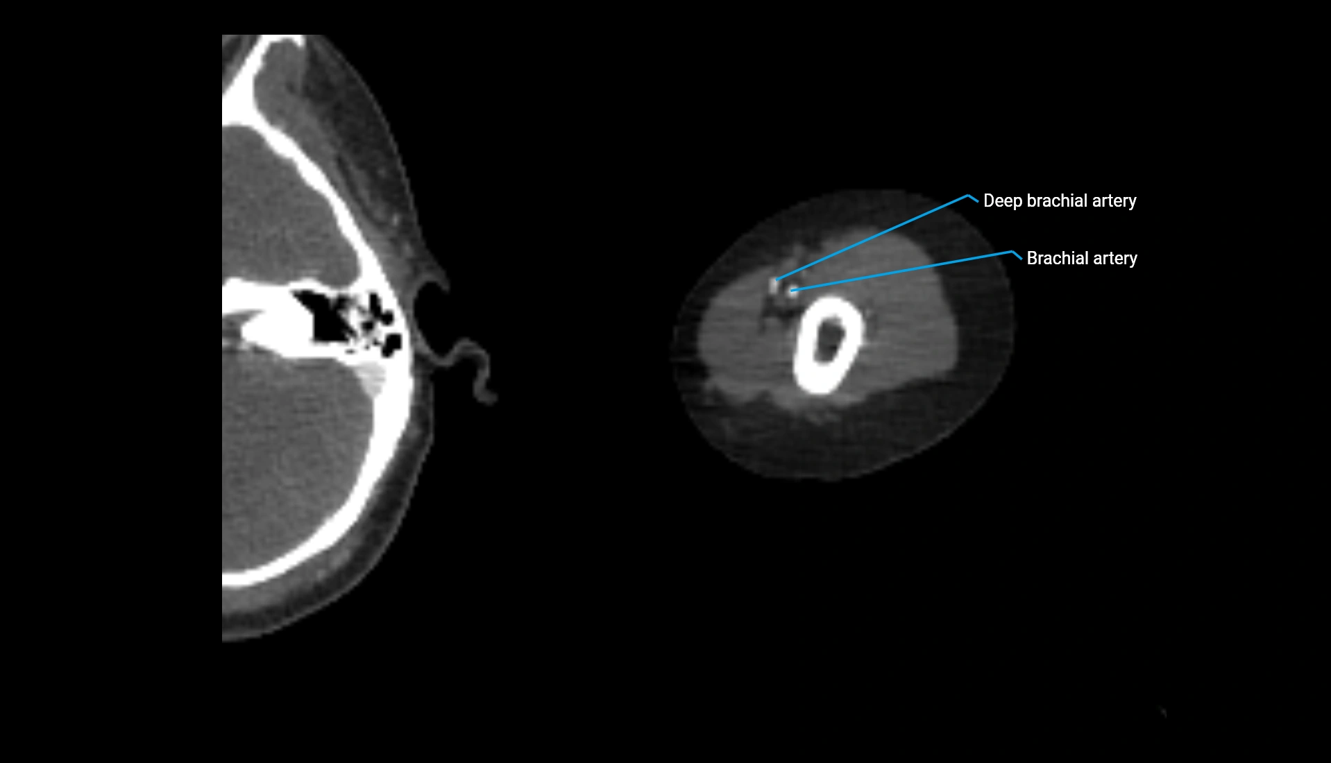
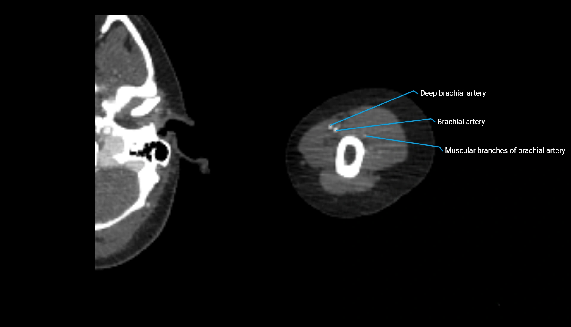
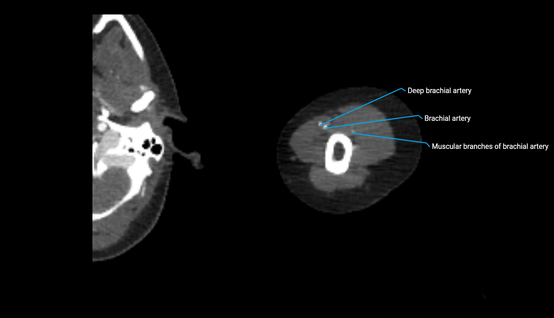
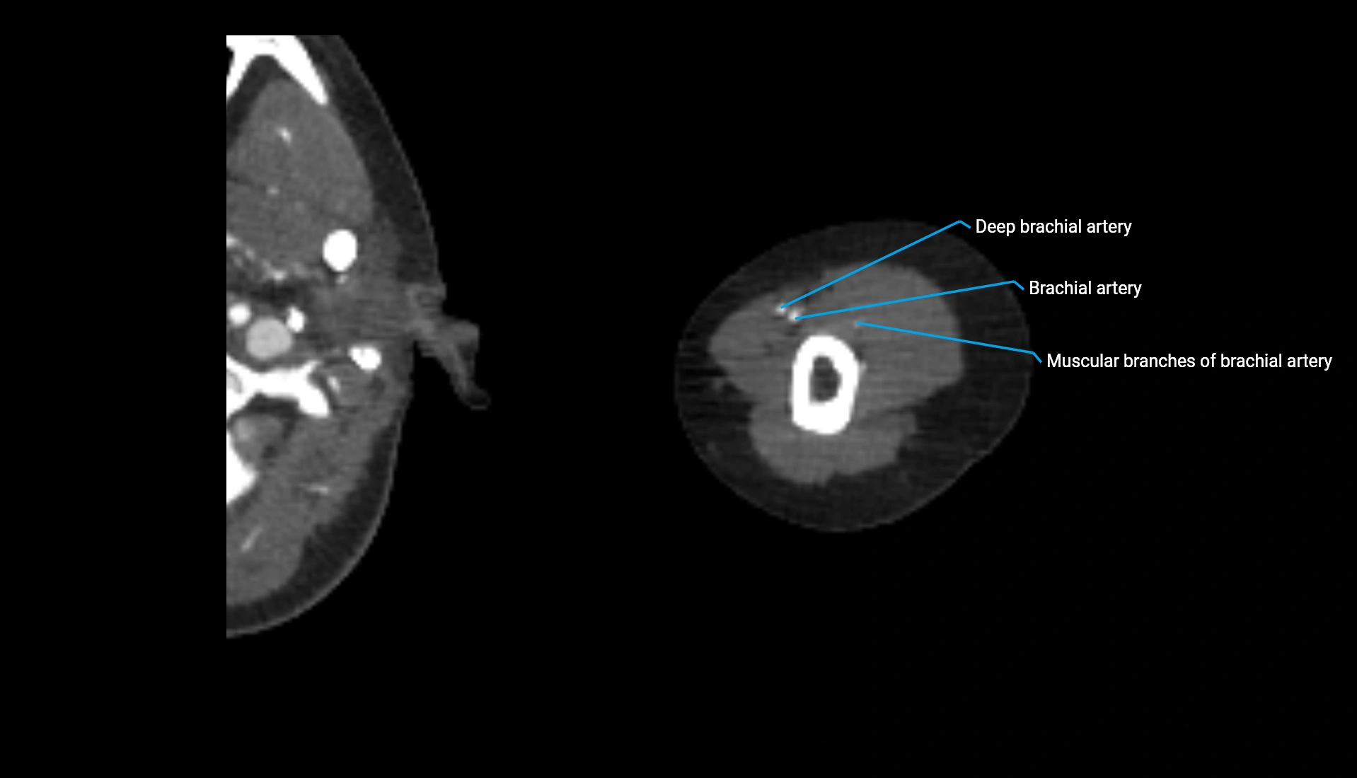
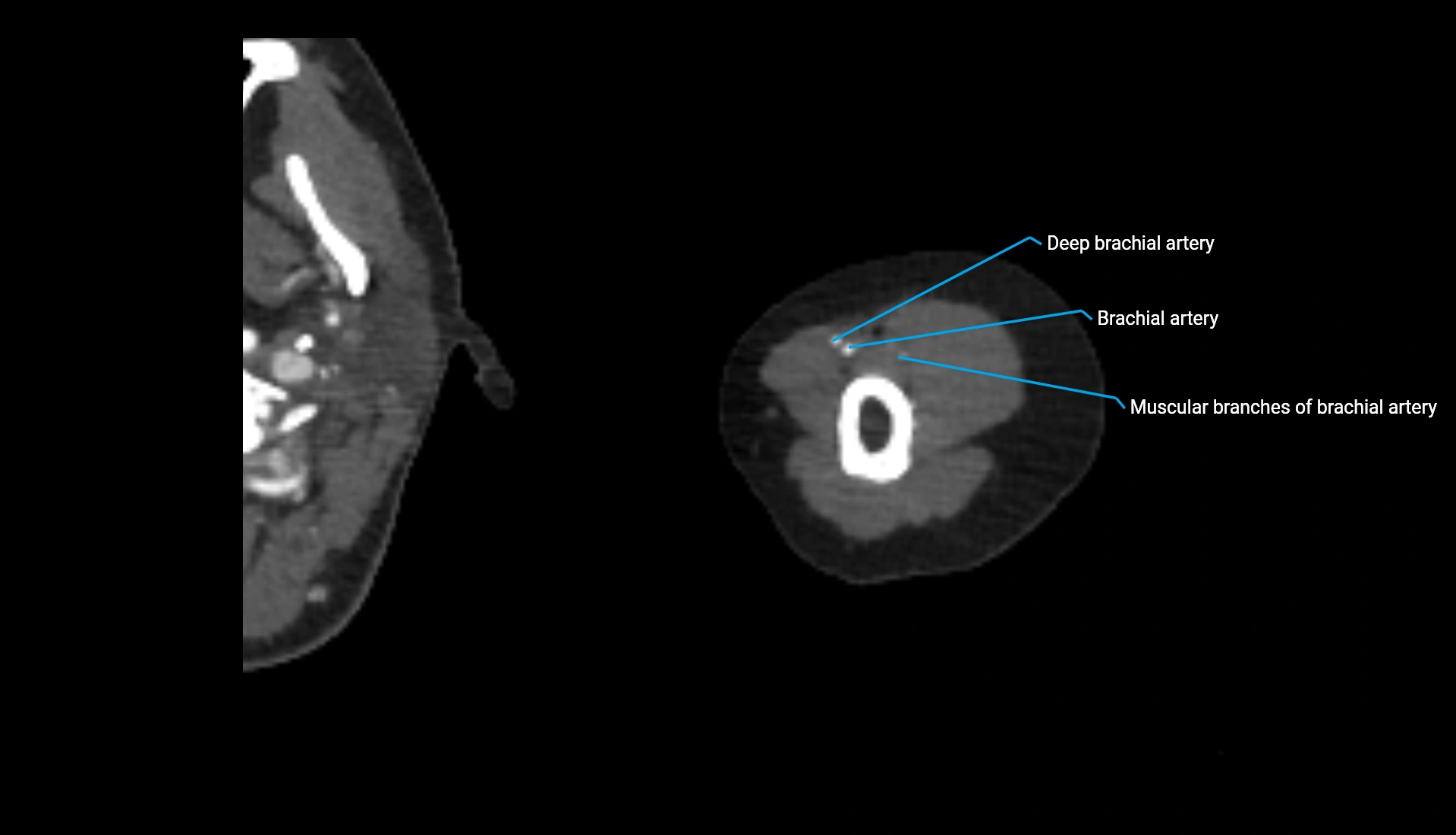
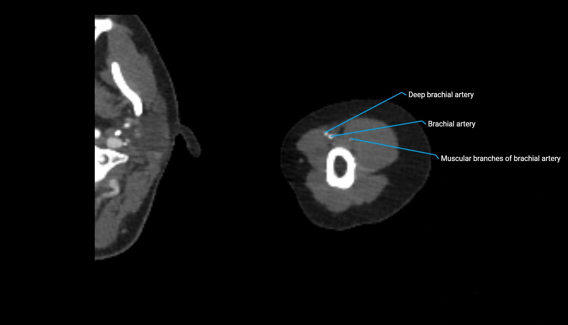
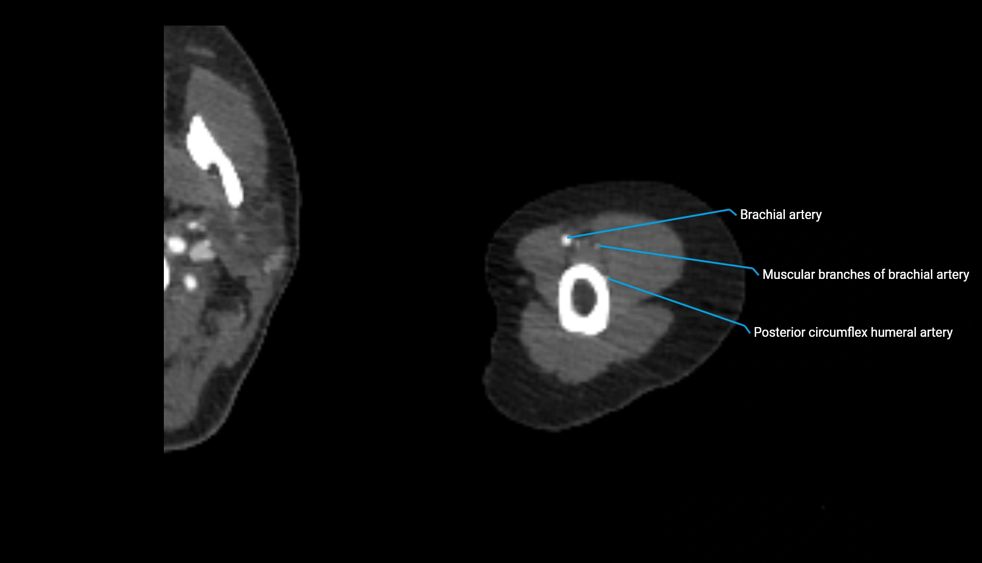
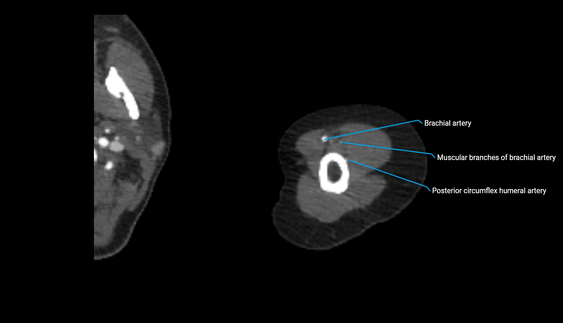
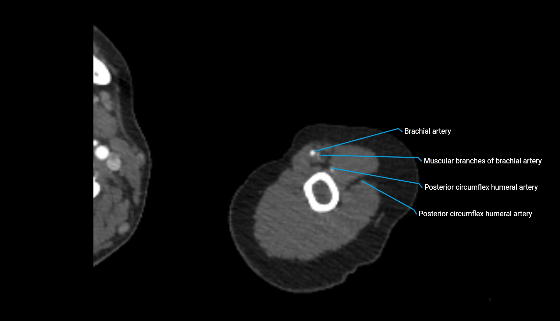
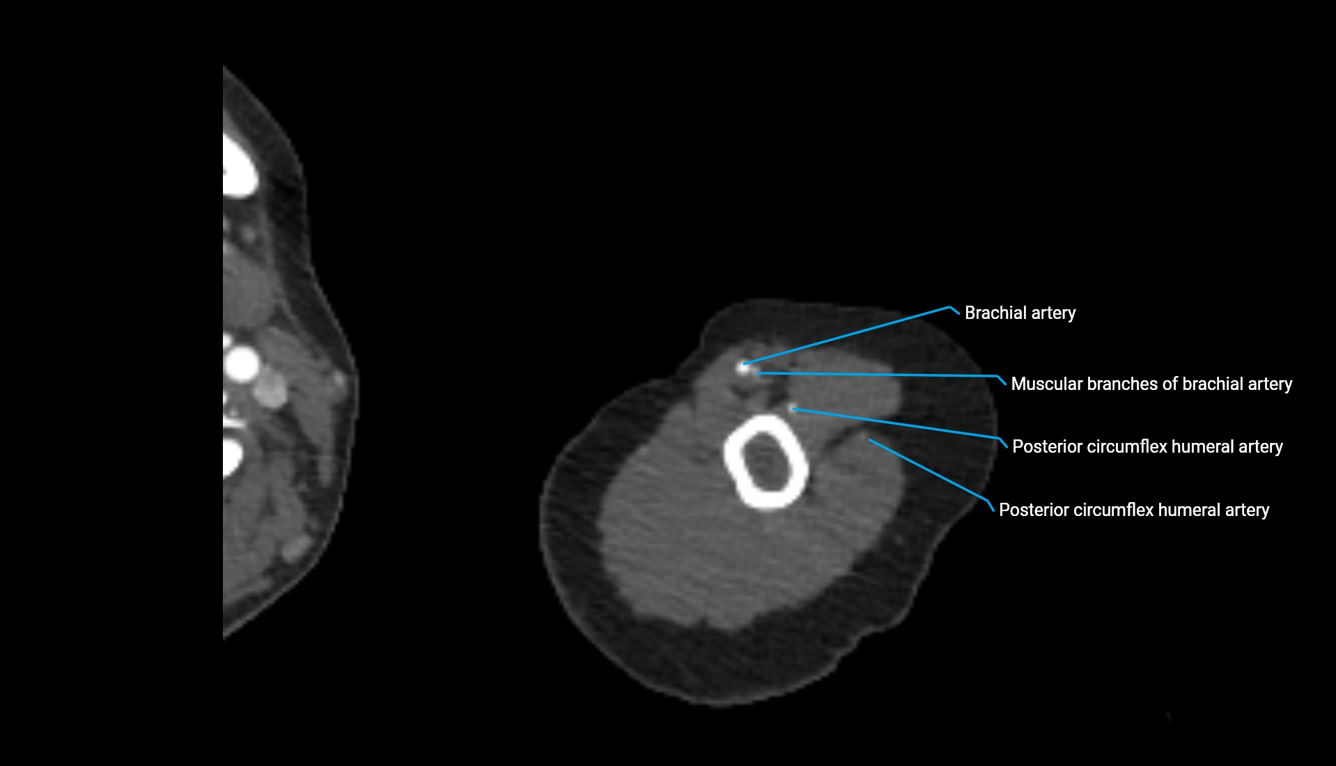
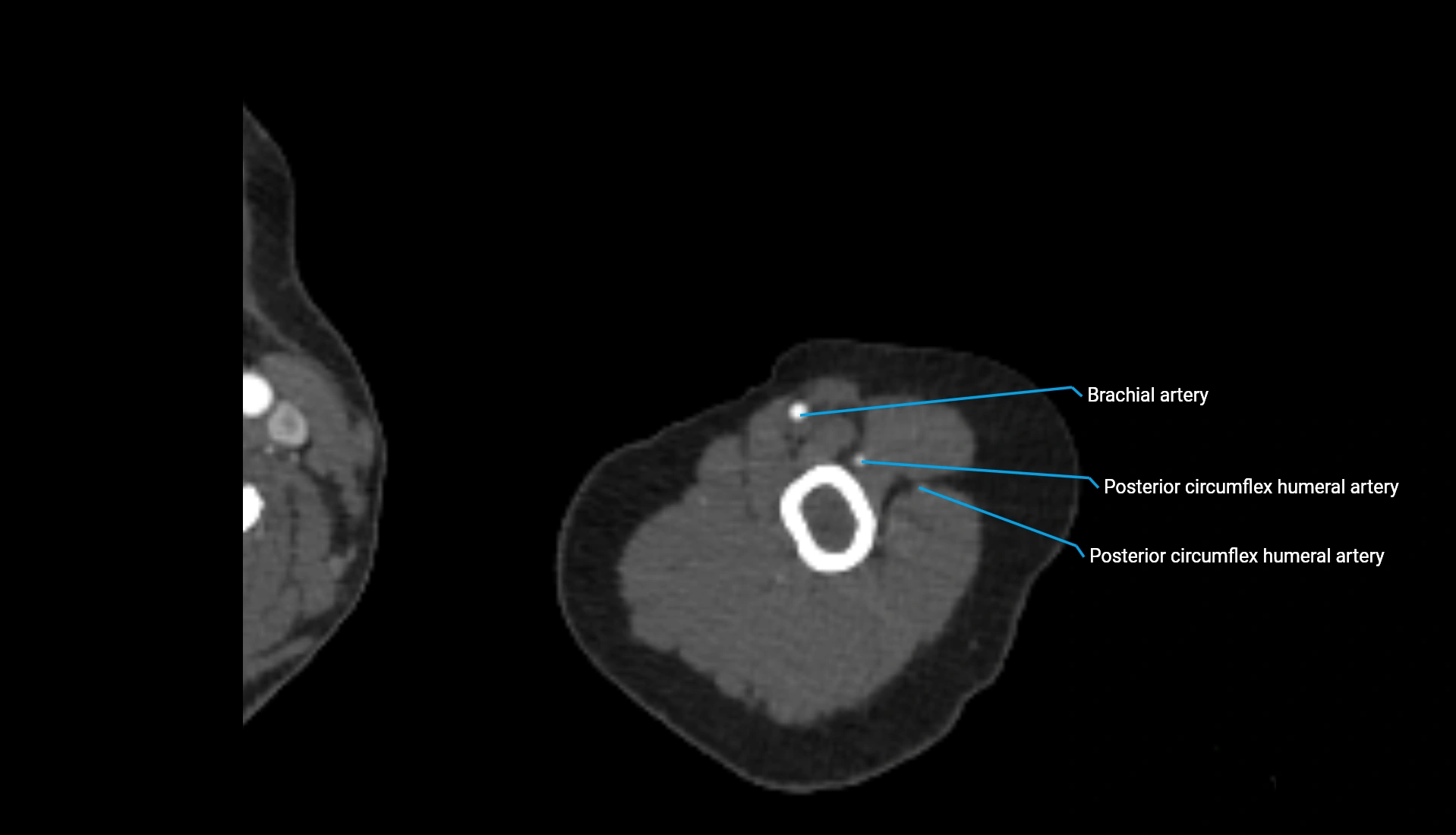
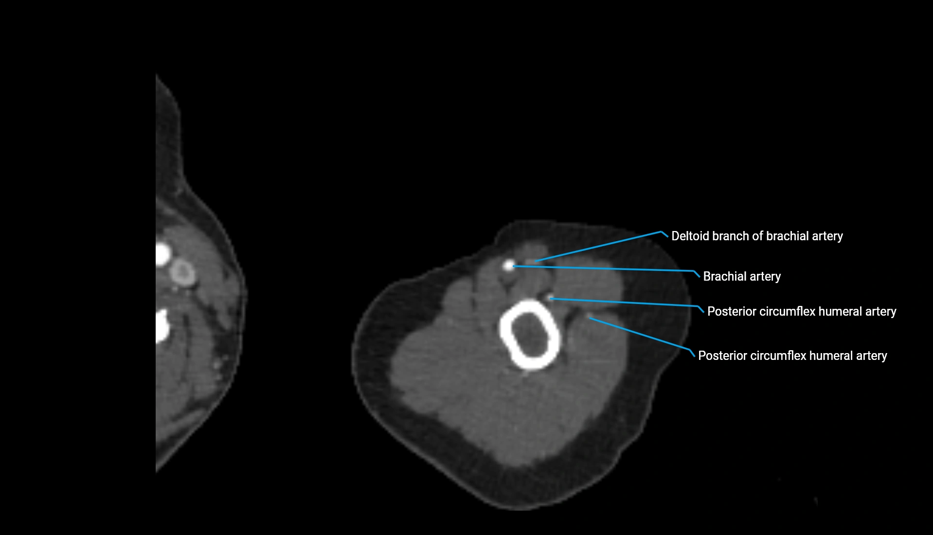
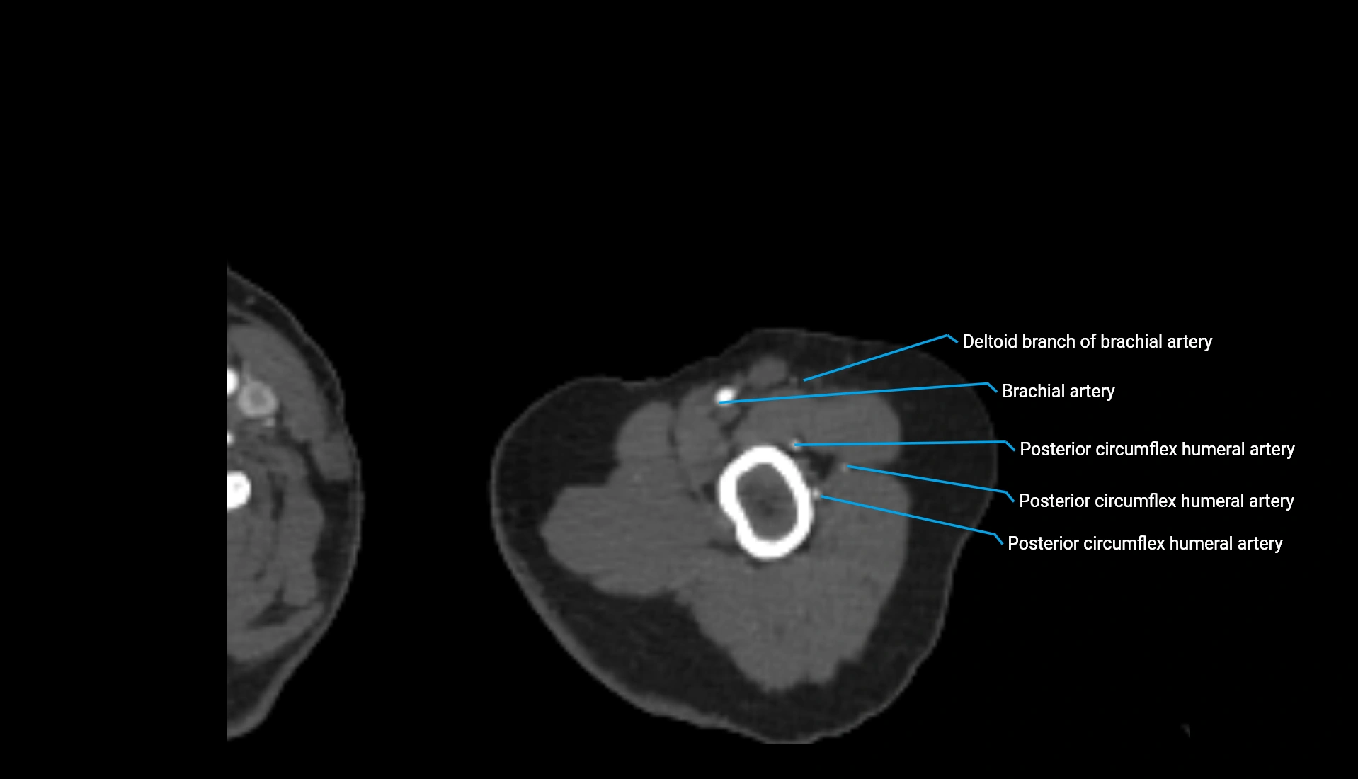
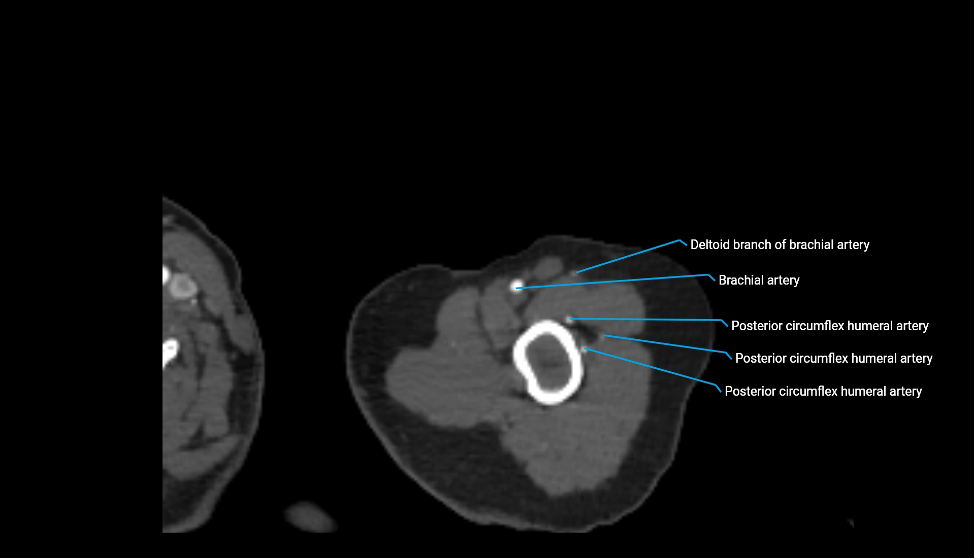
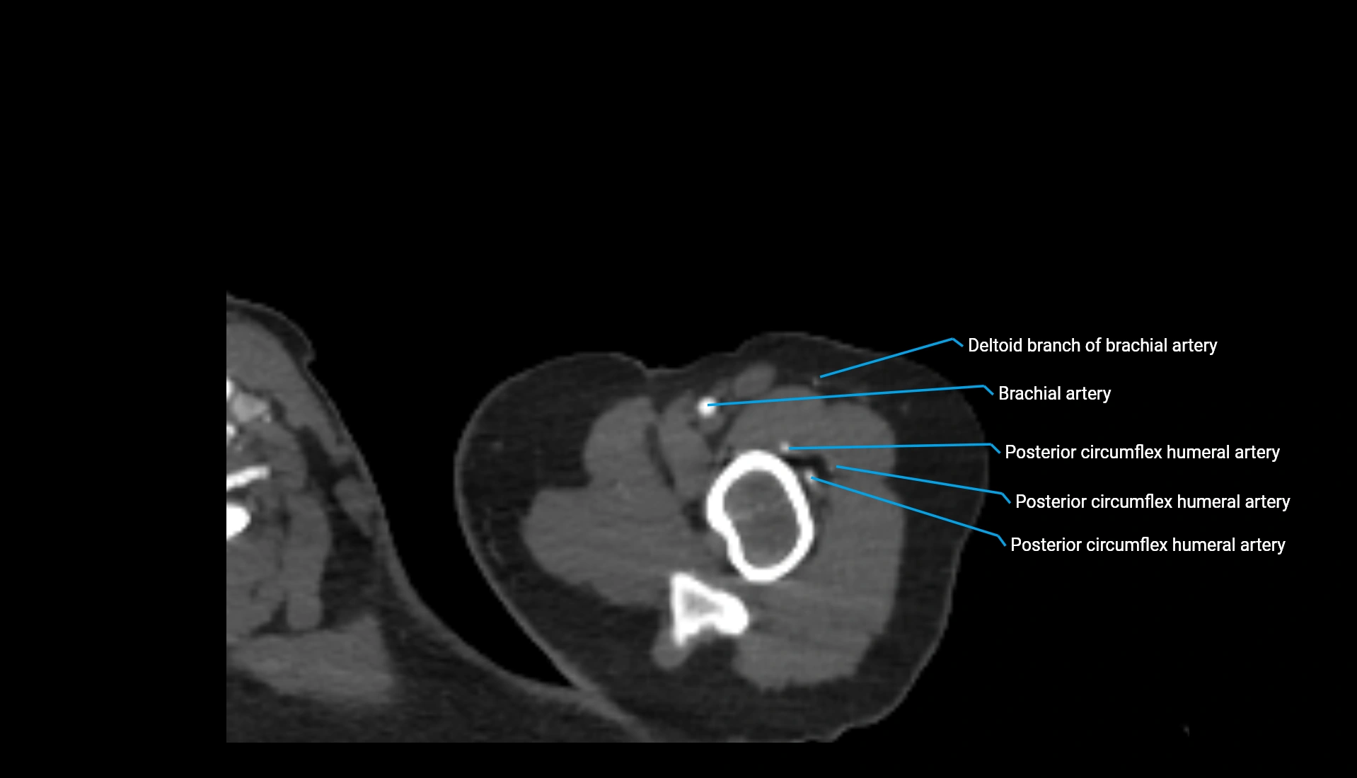
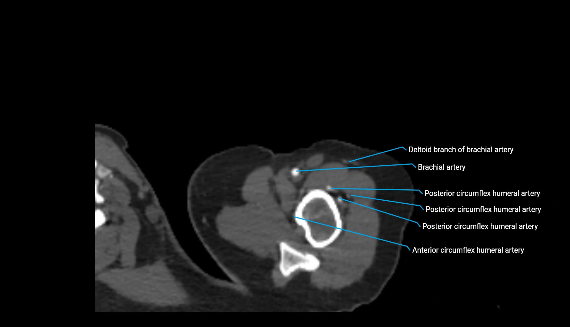
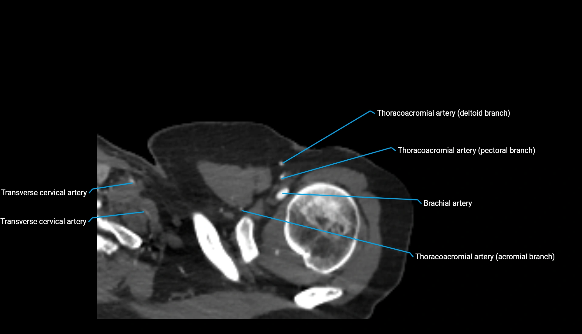
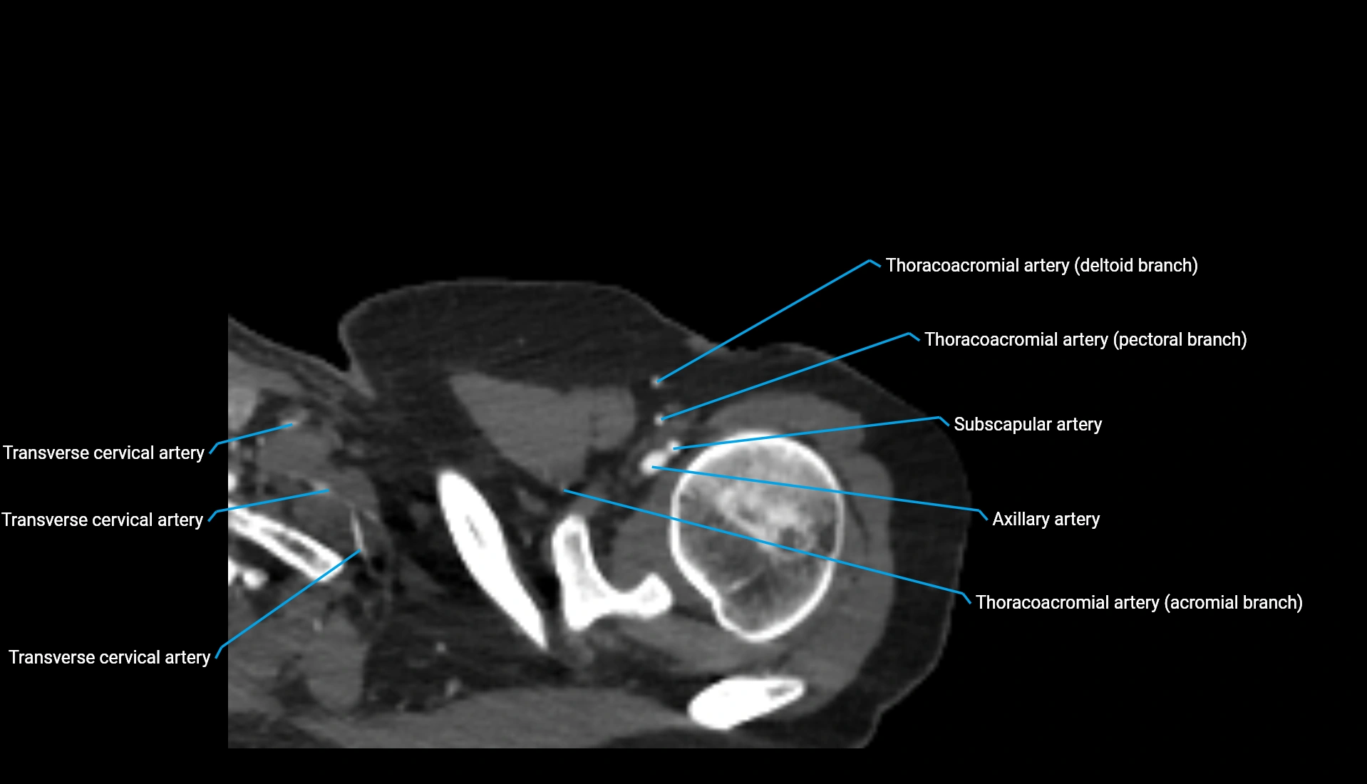
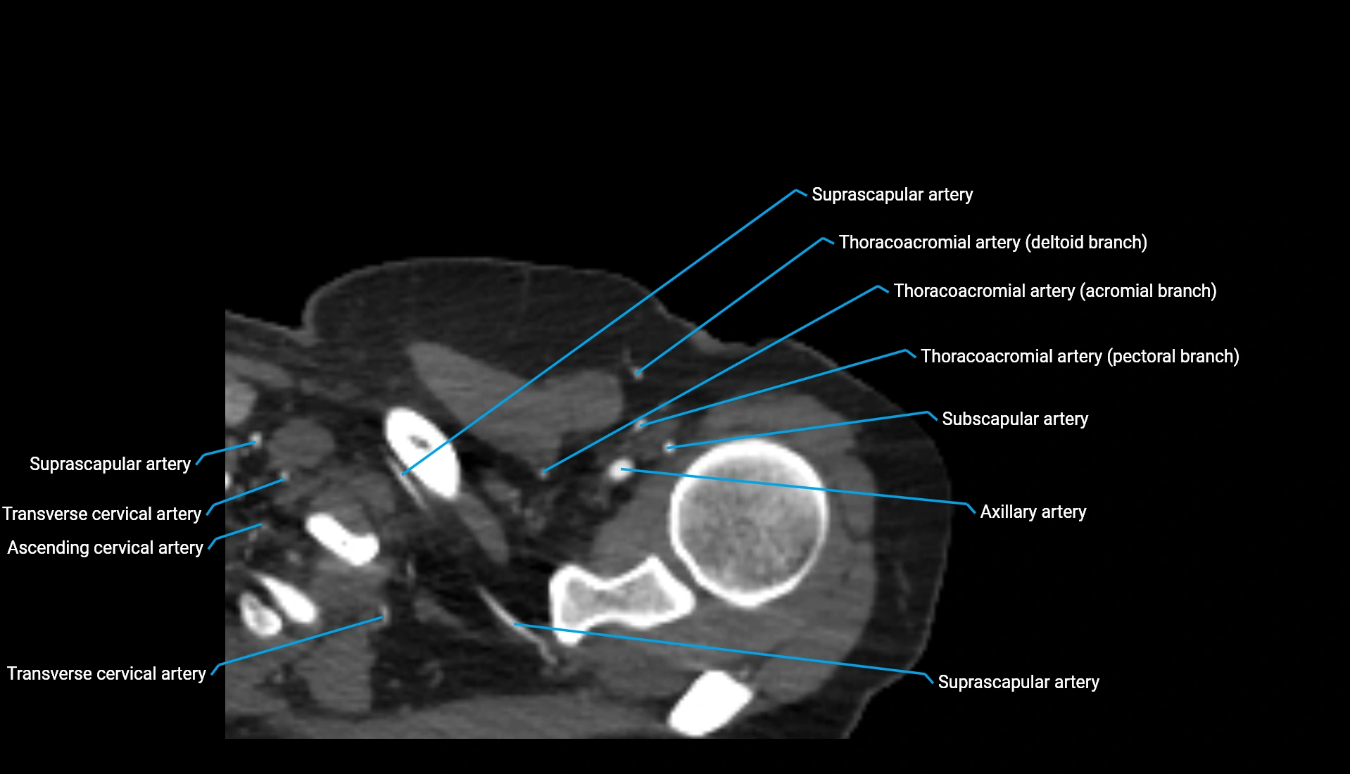
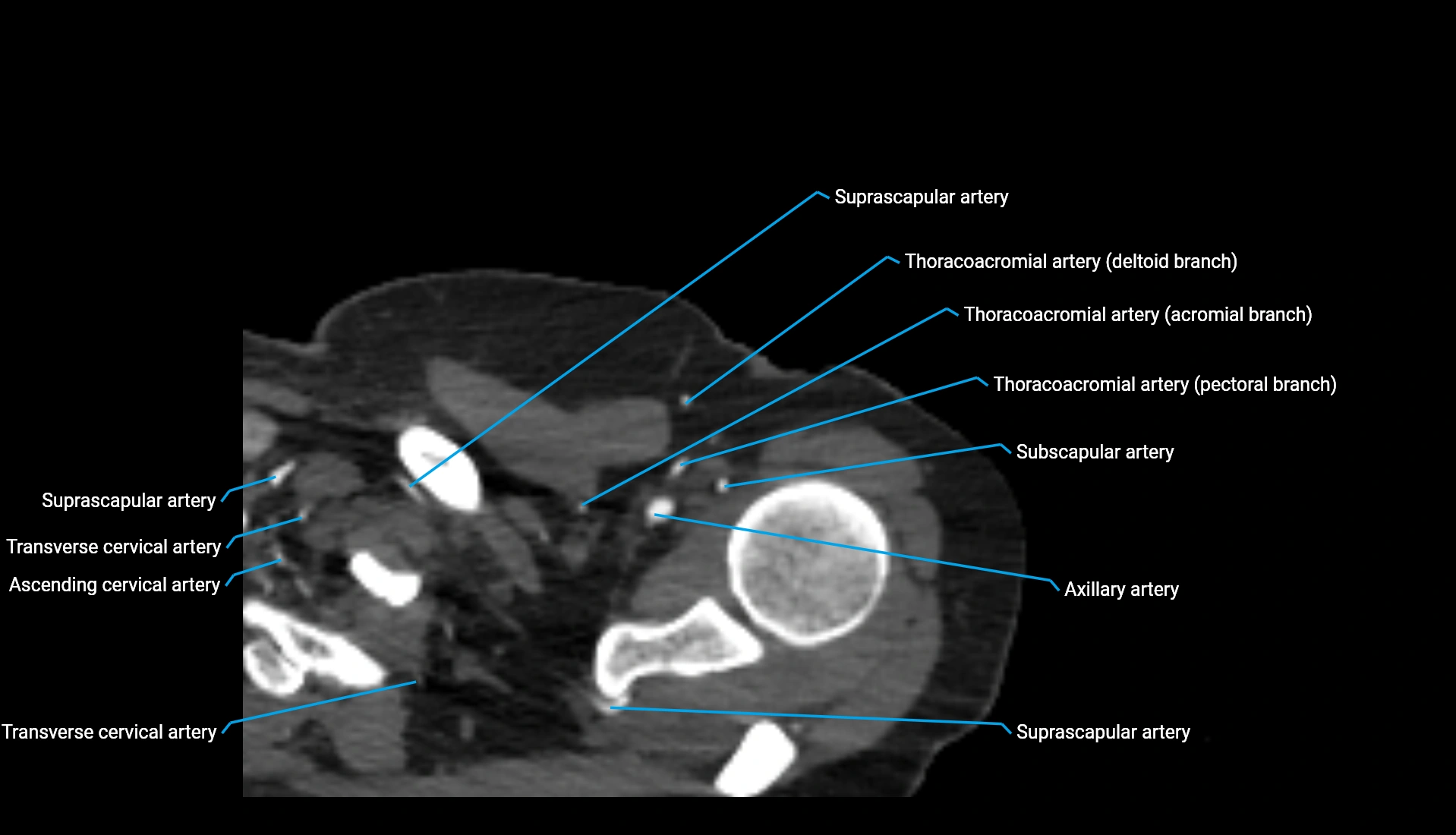
MRI image

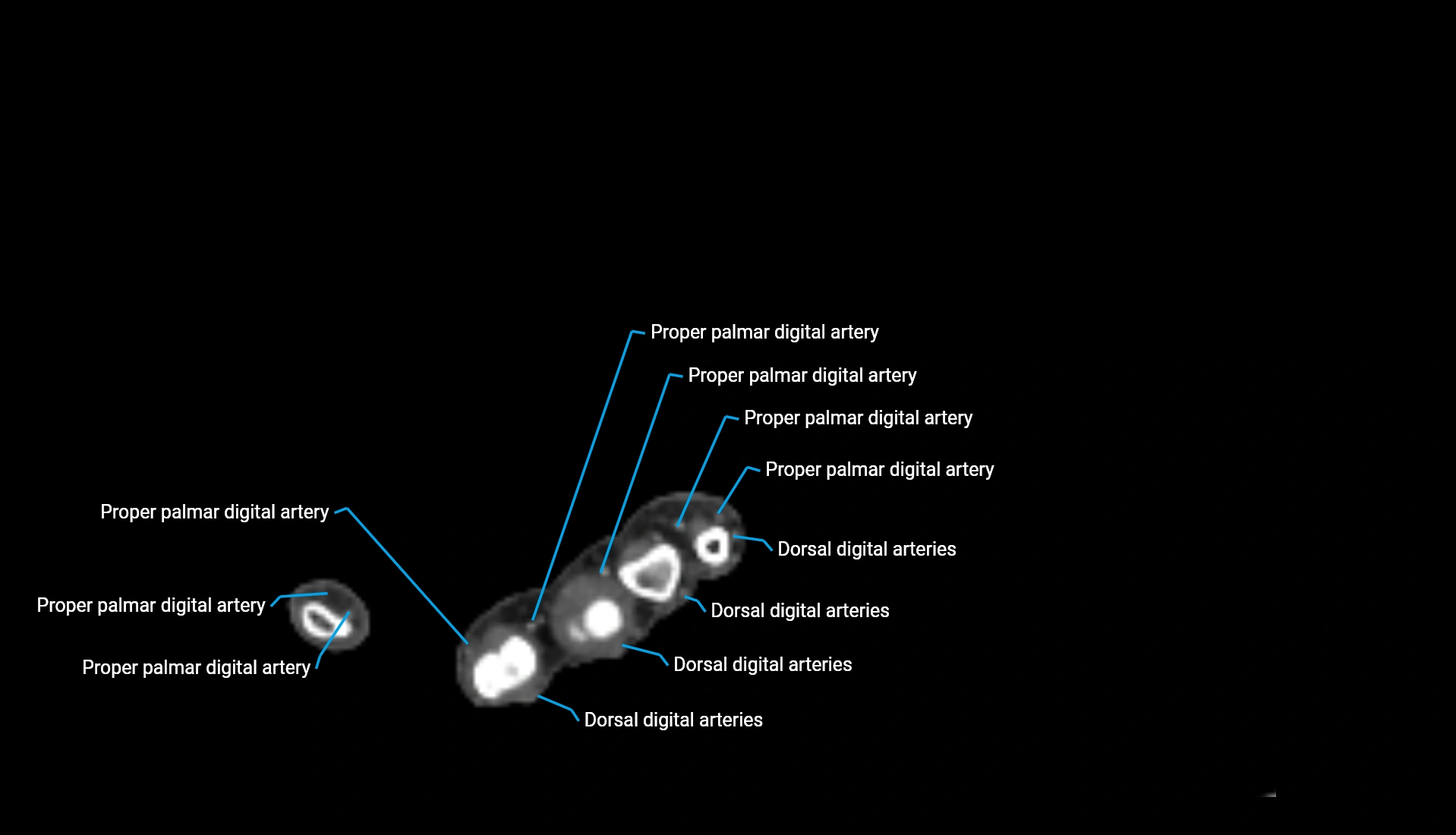
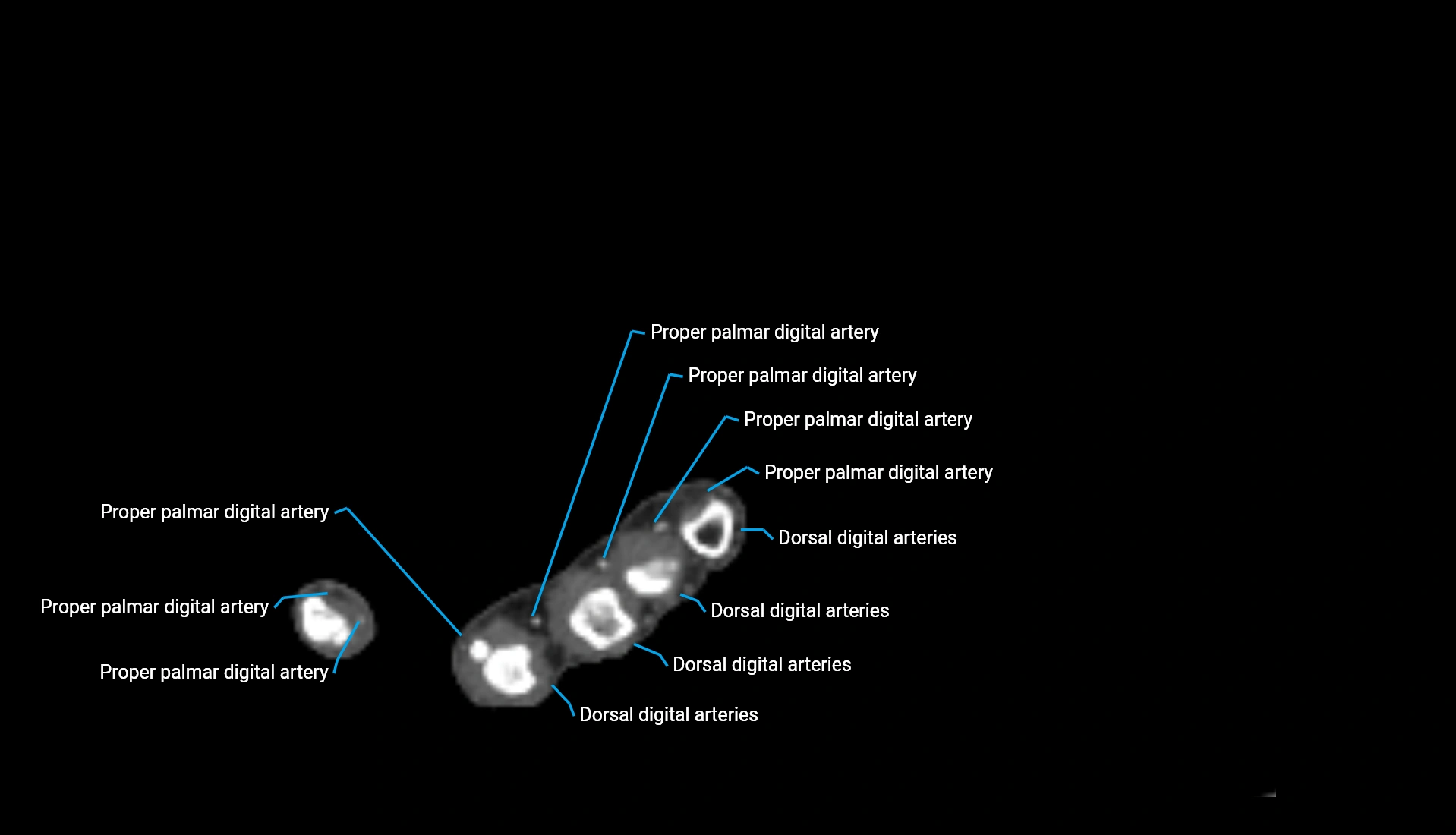
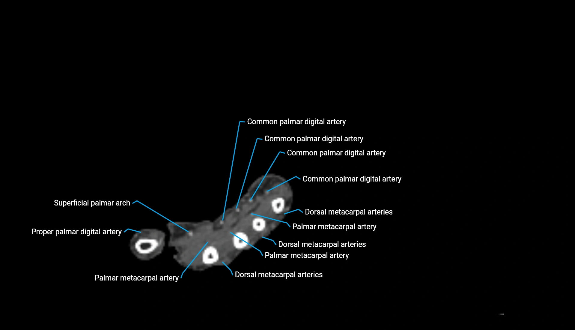
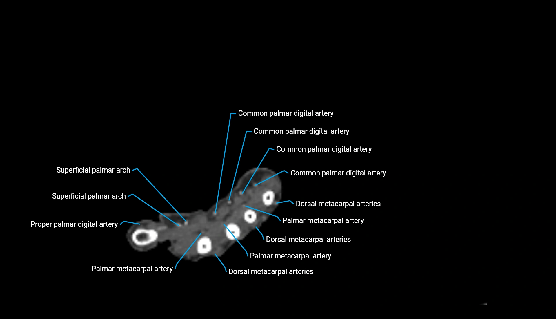
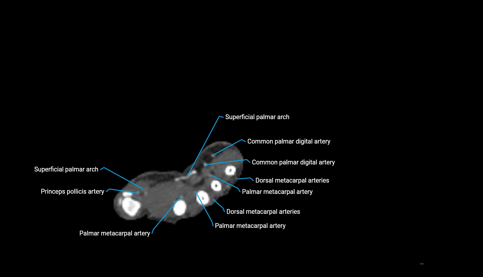
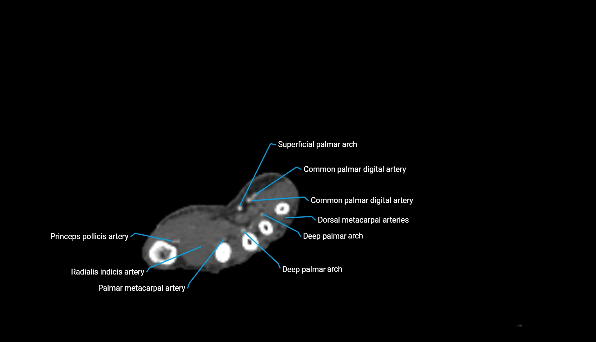
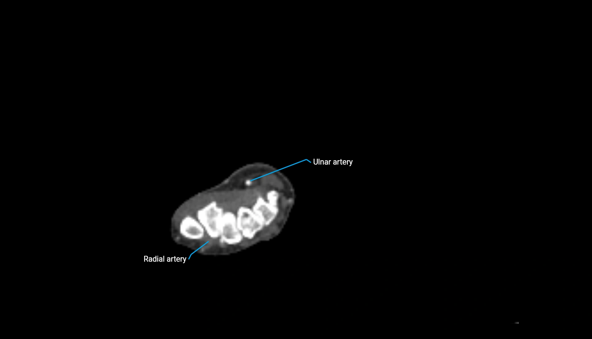
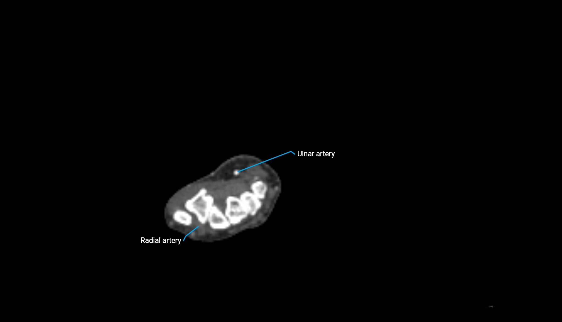
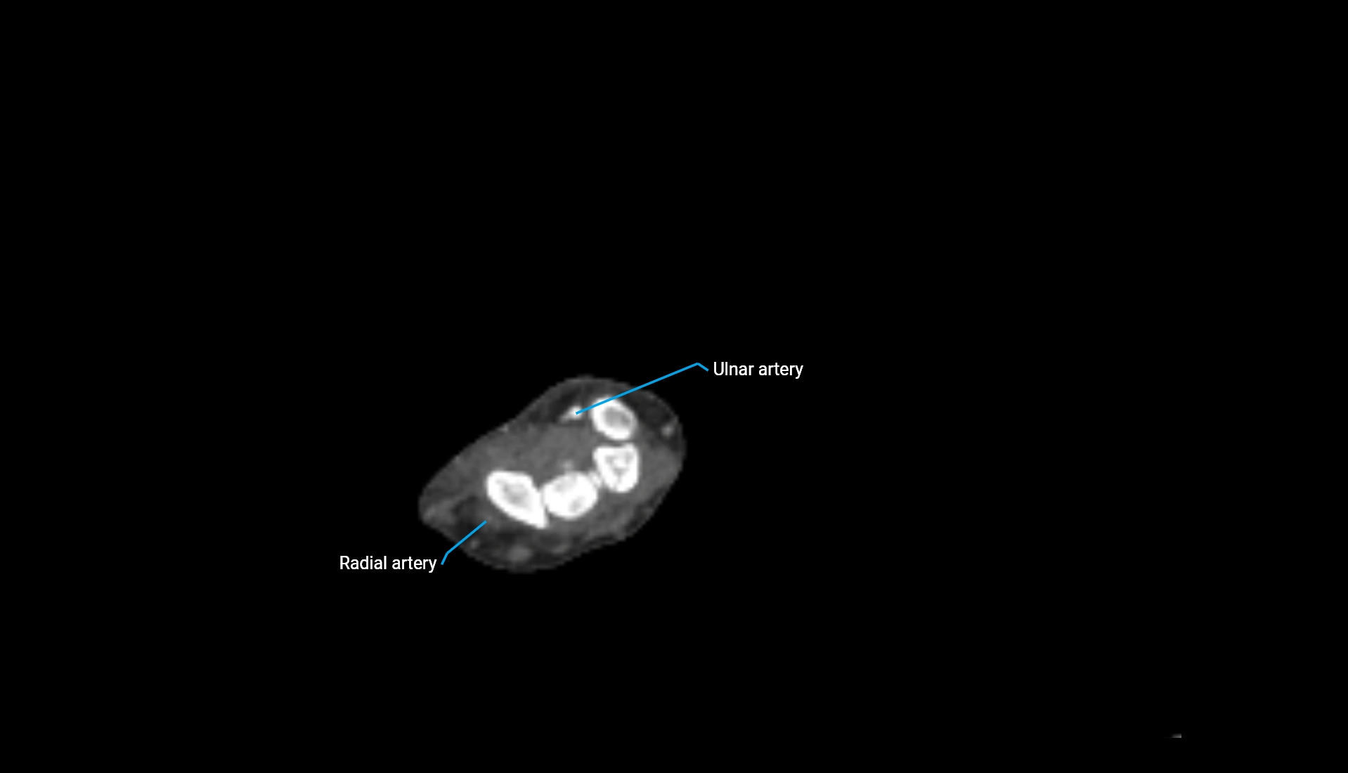
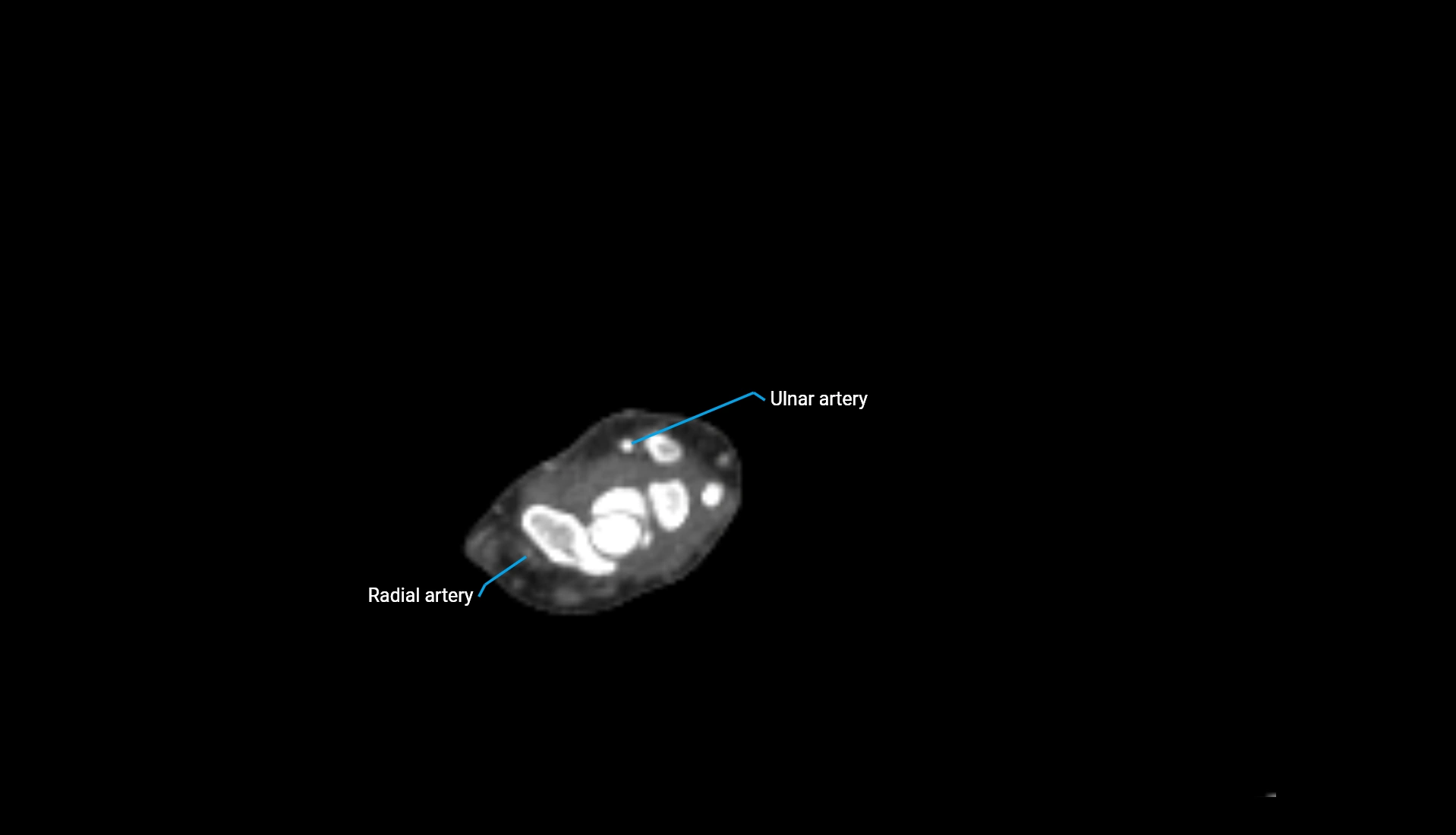
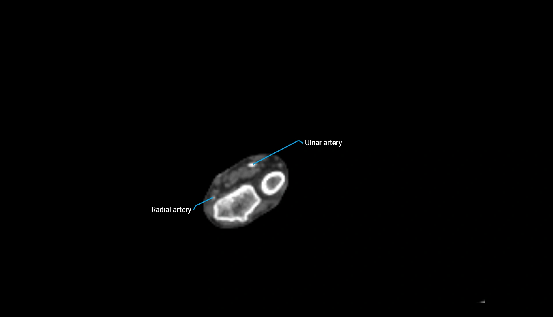
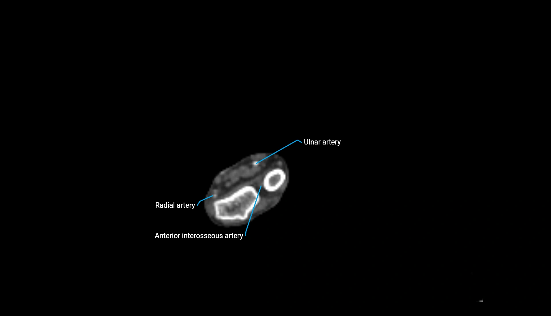
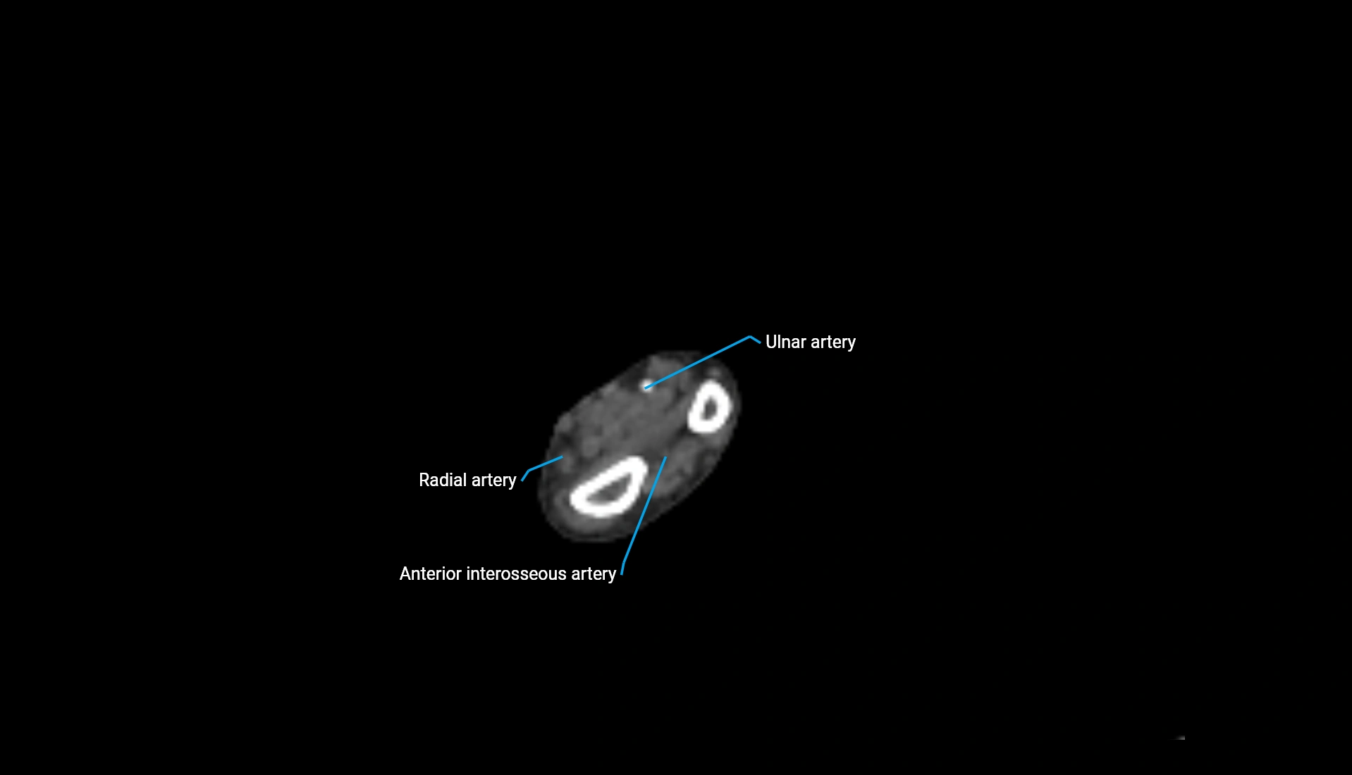
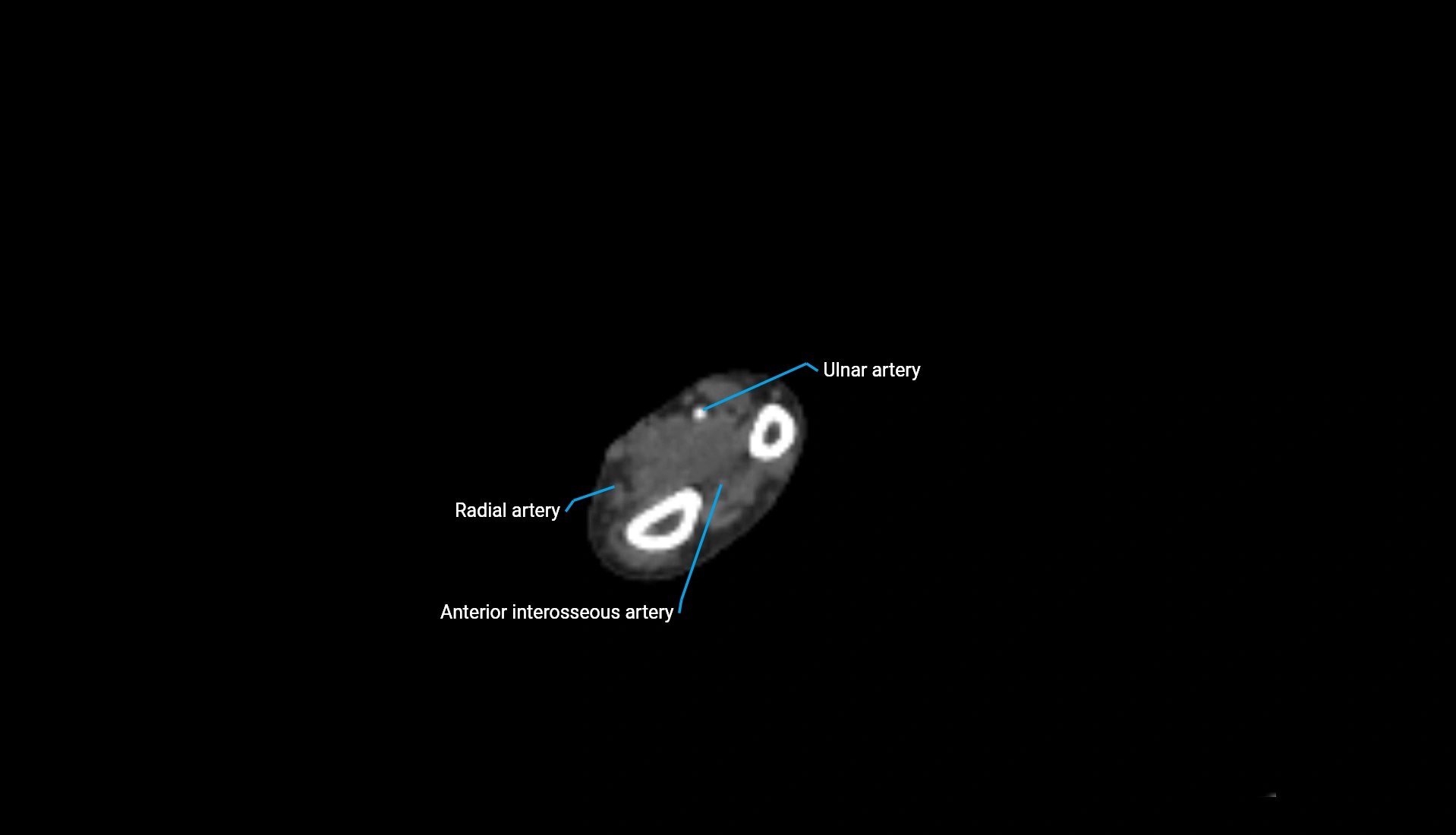
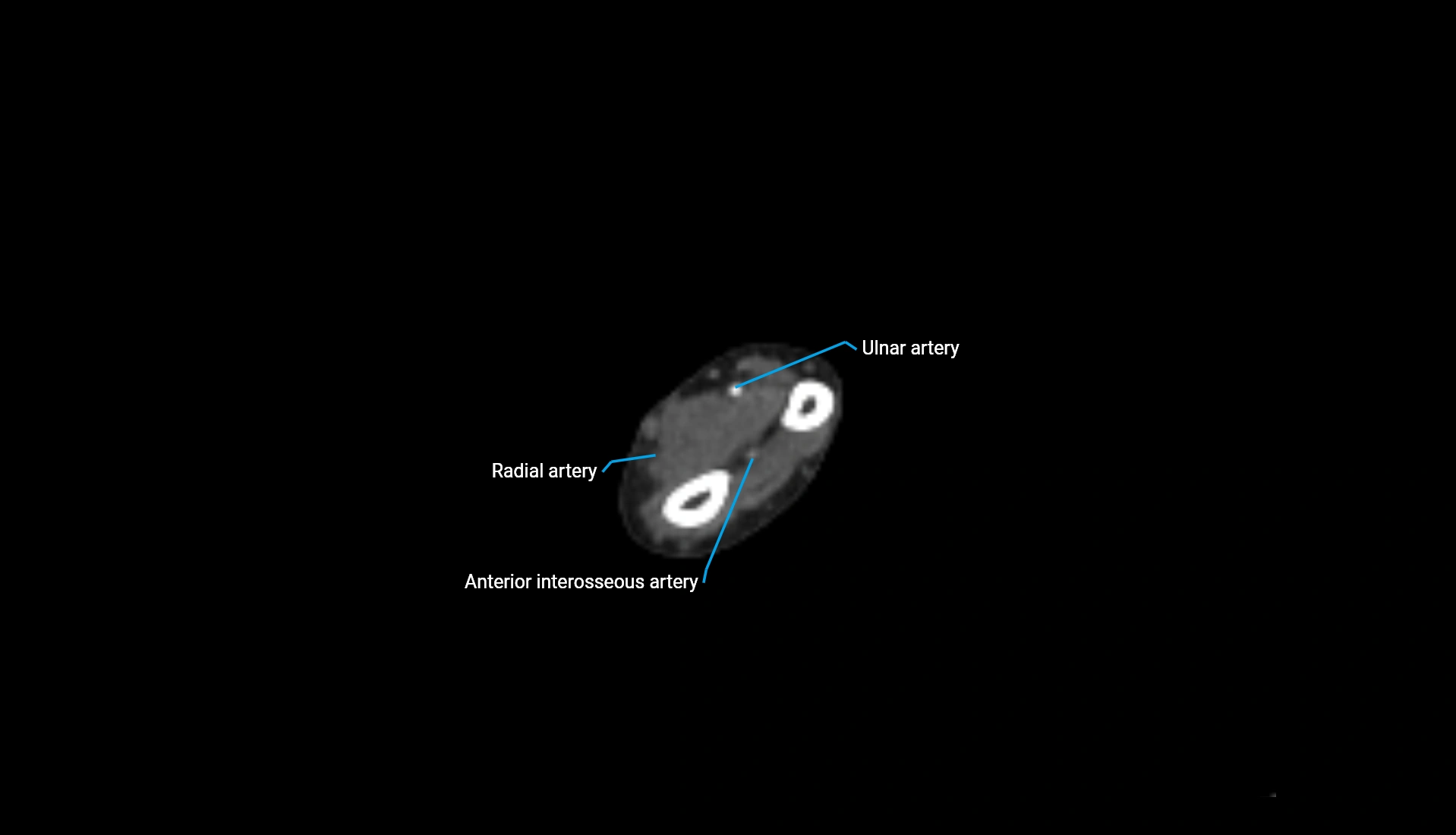
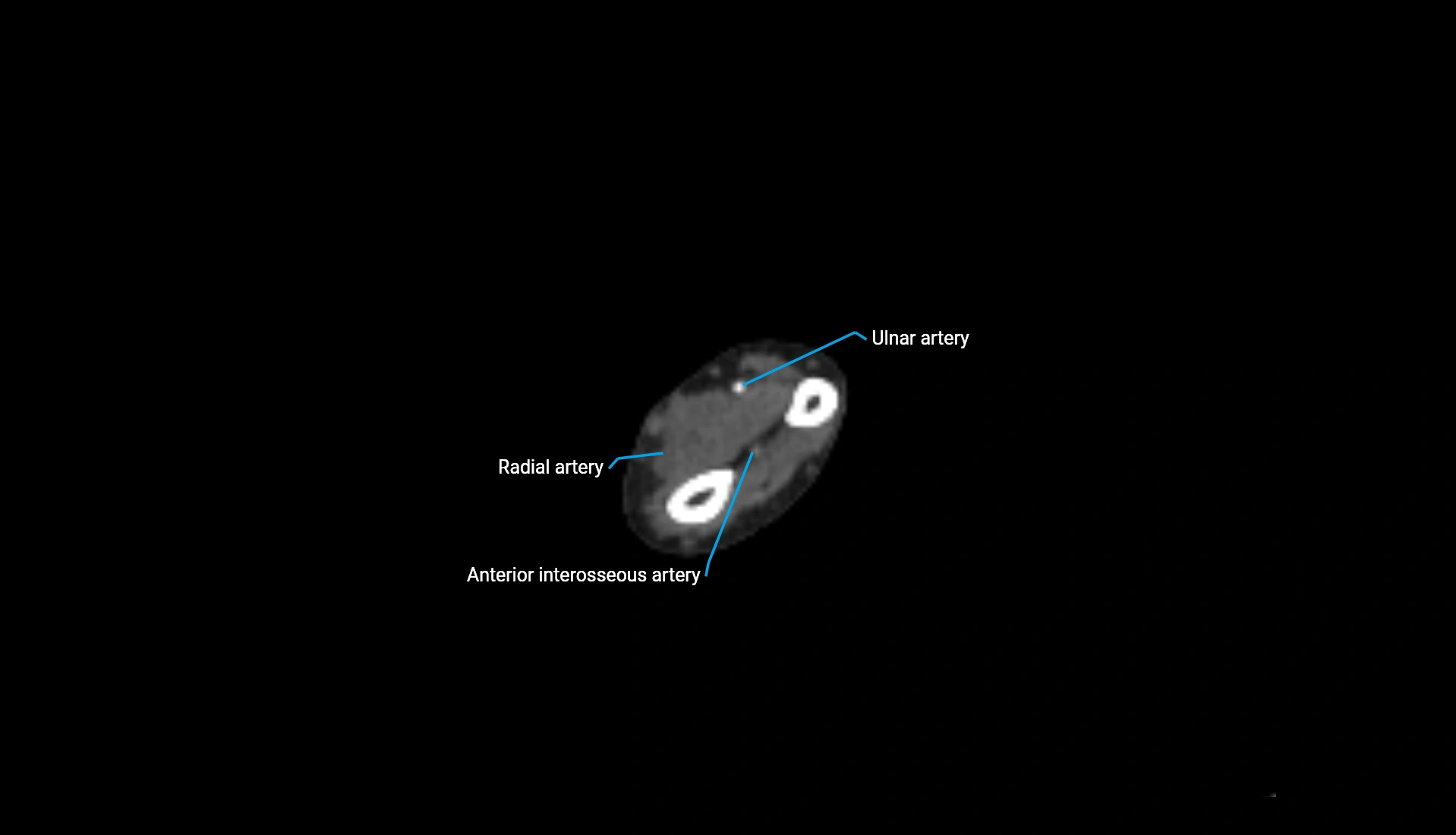
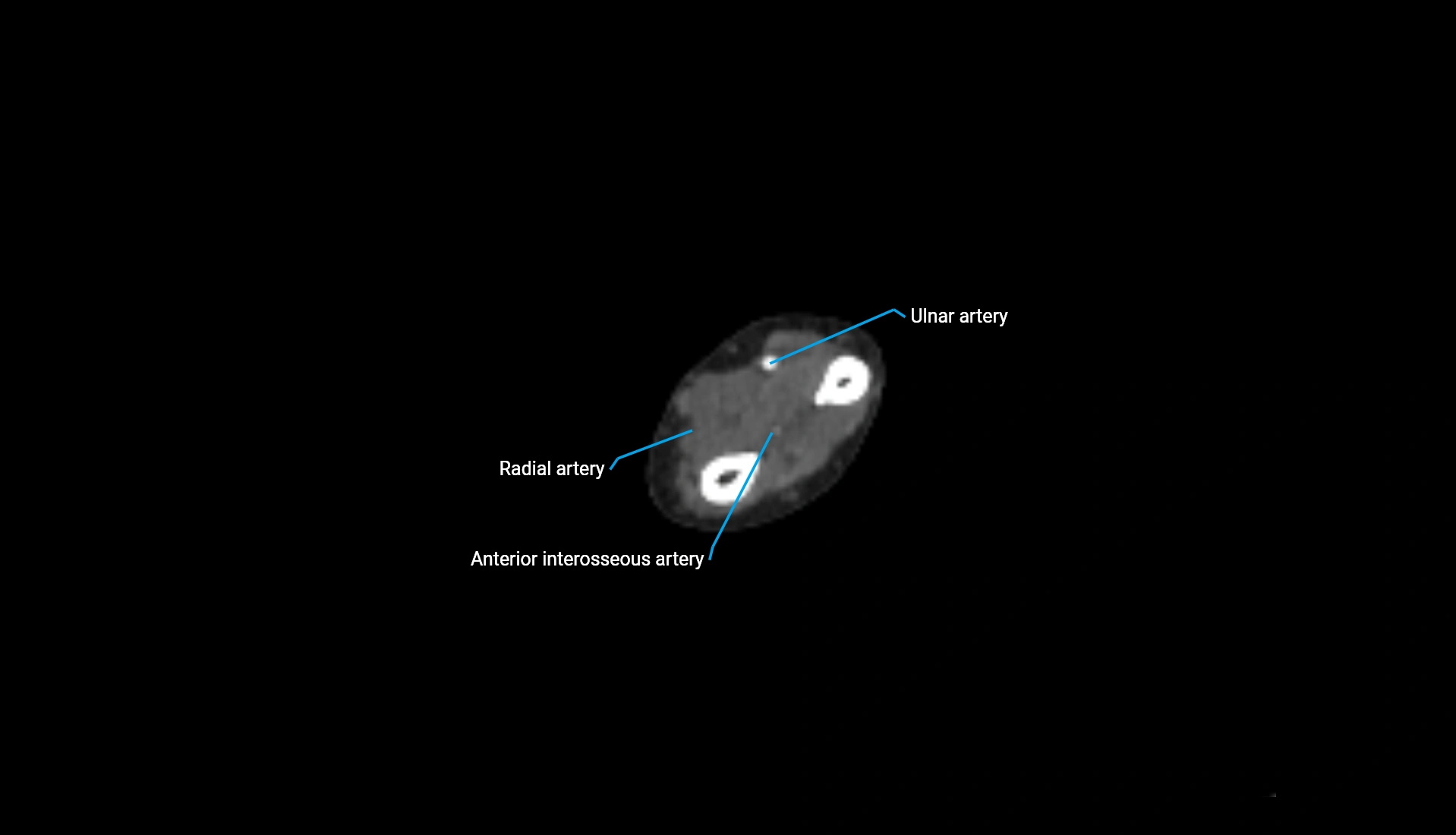
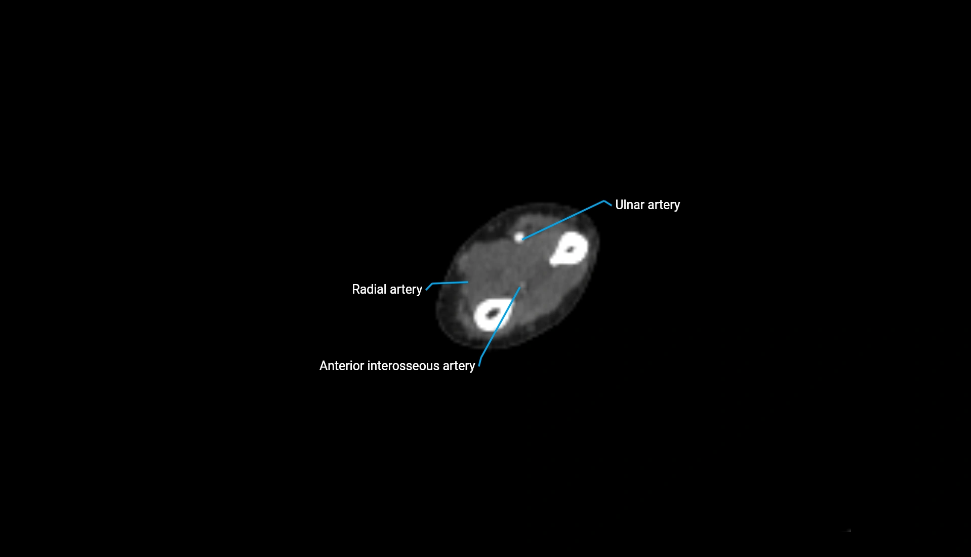
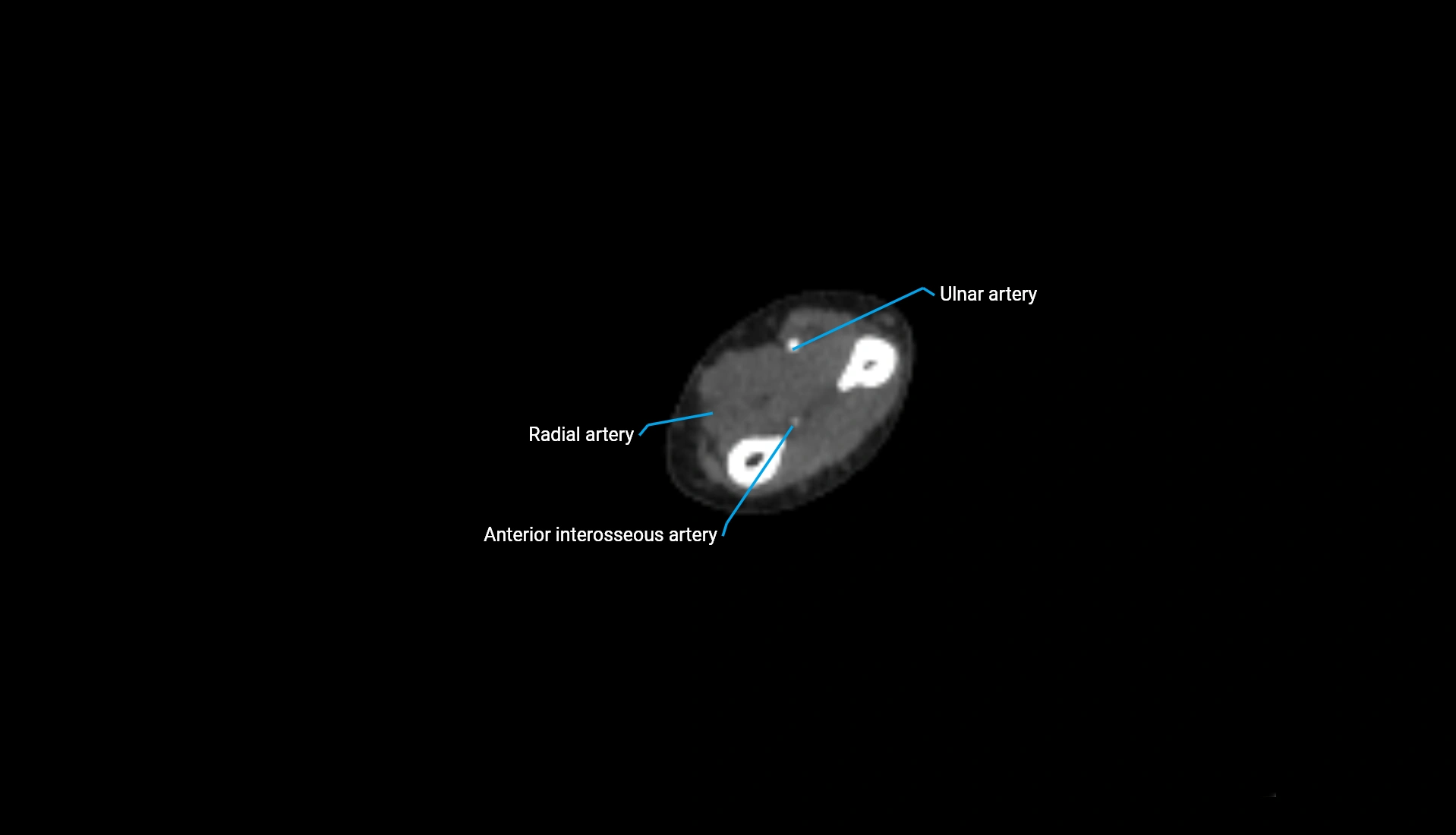
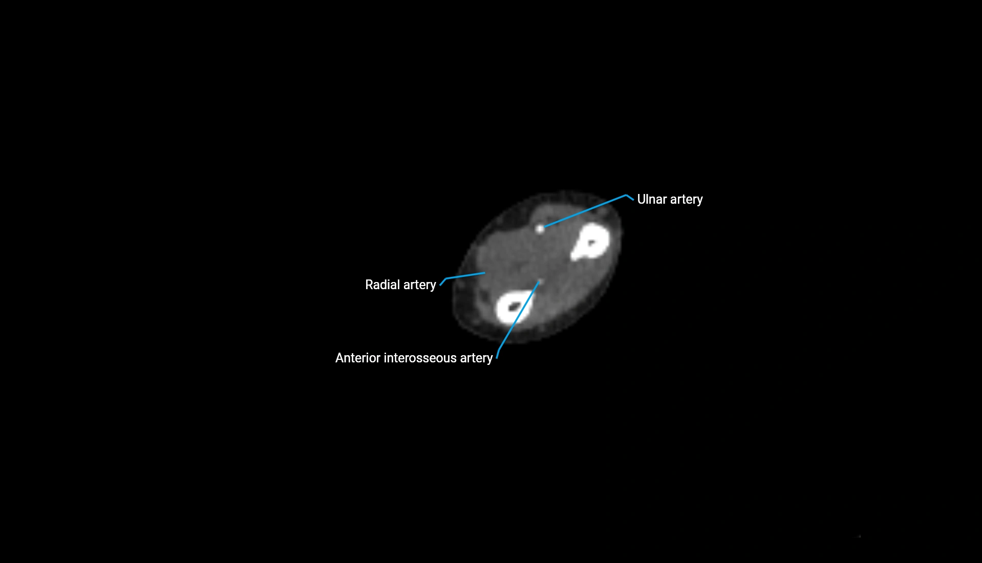
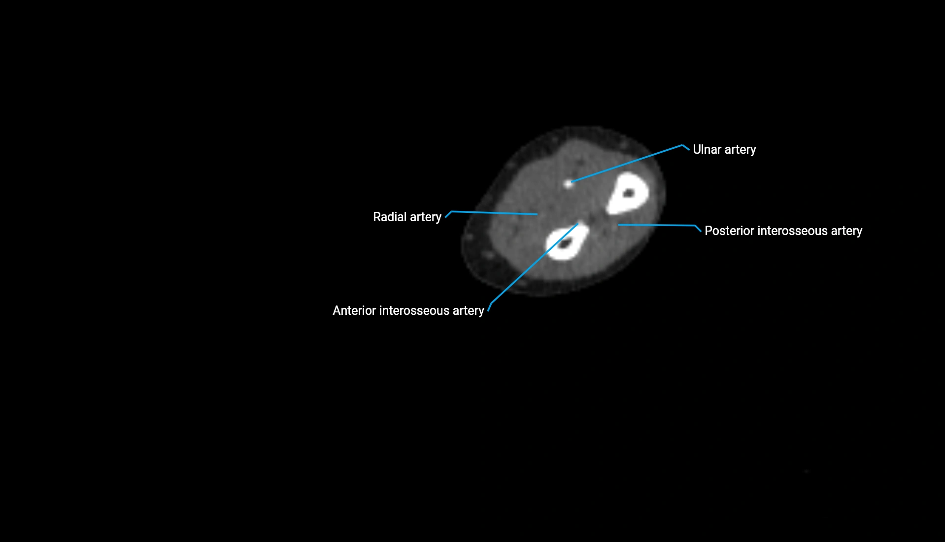
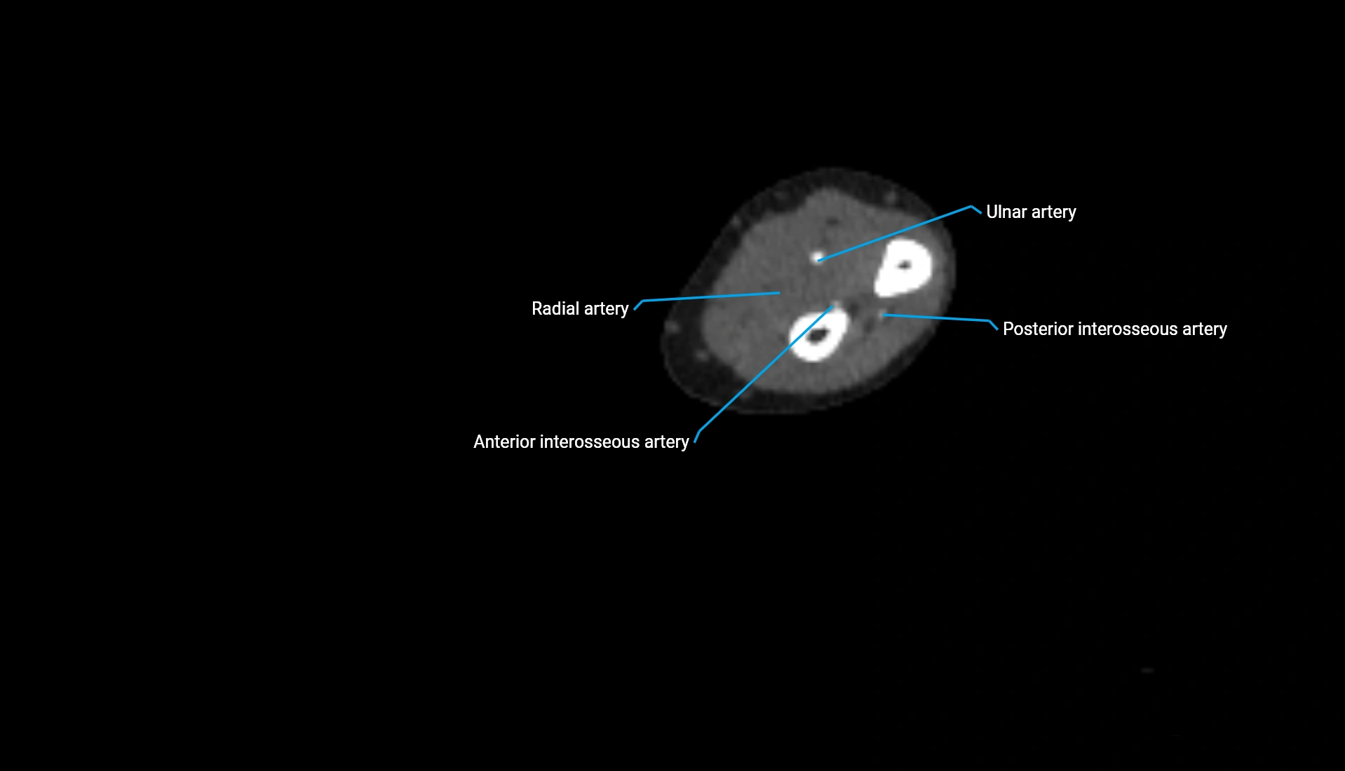
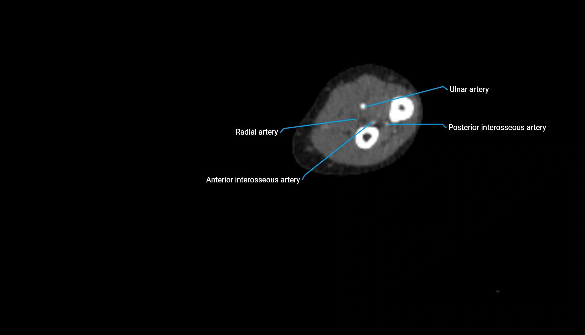
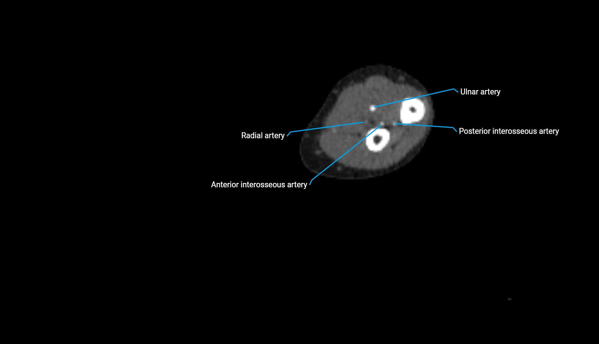
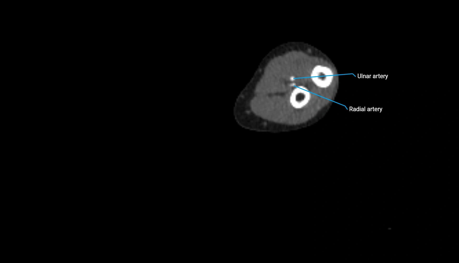
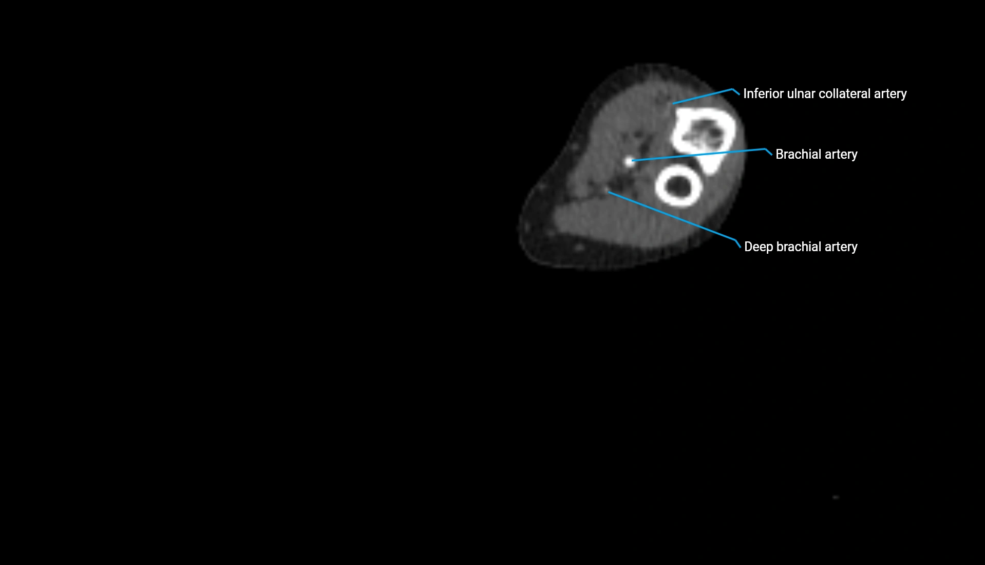
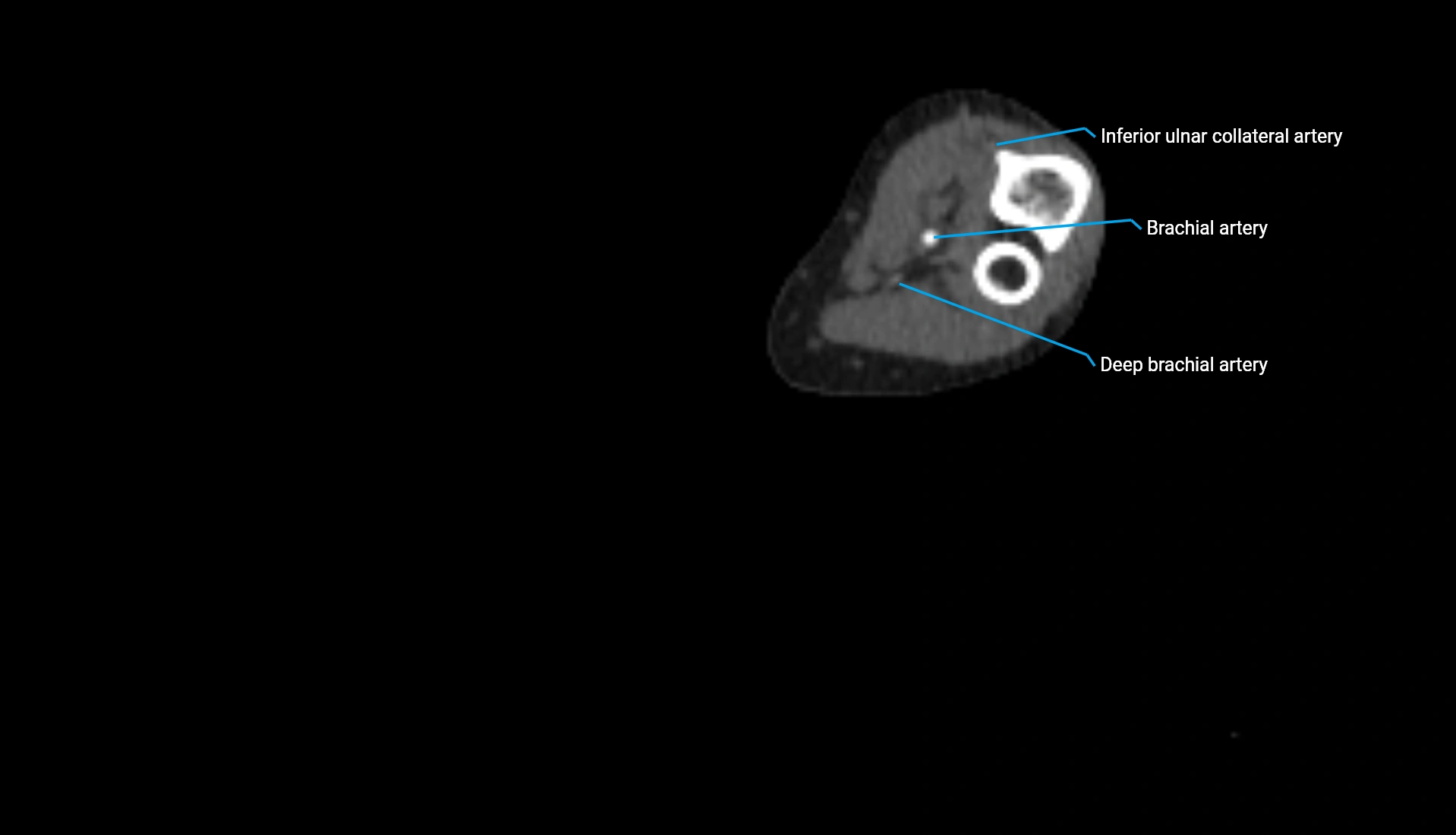
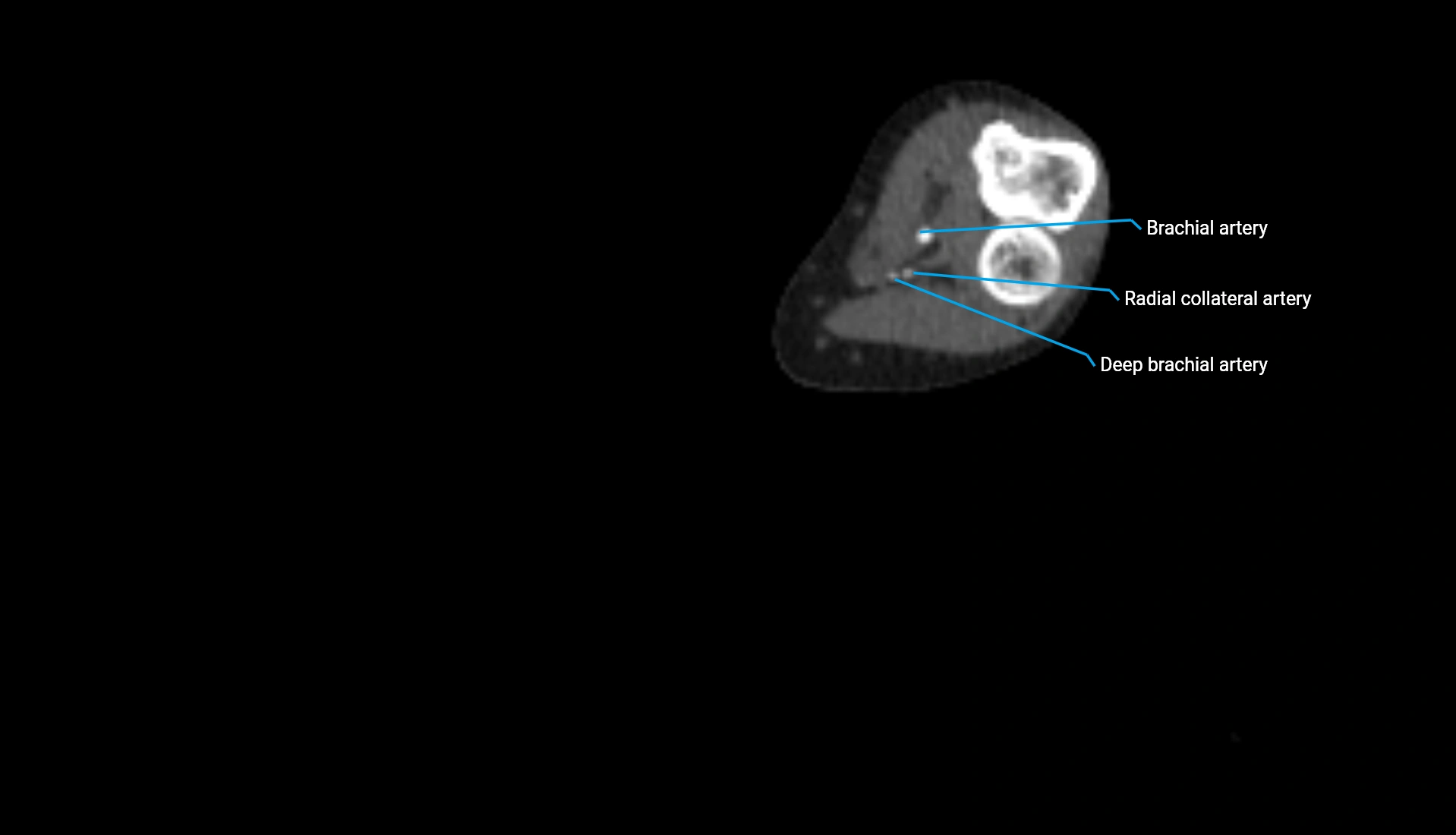
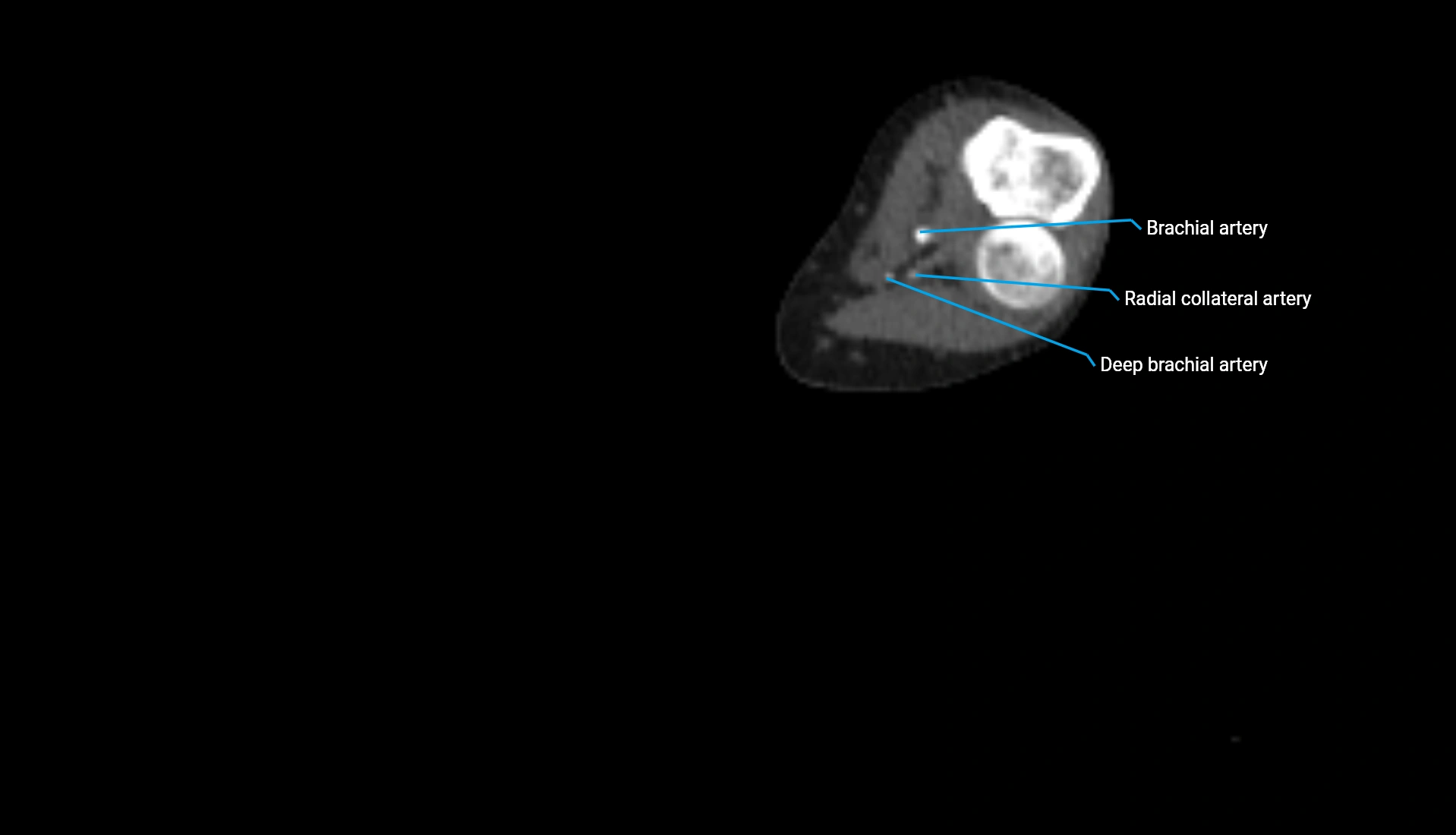
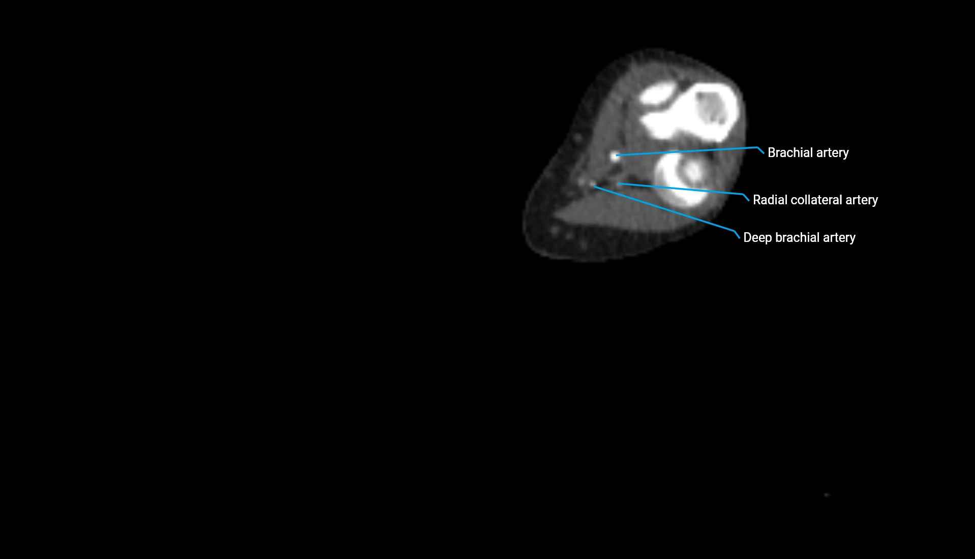
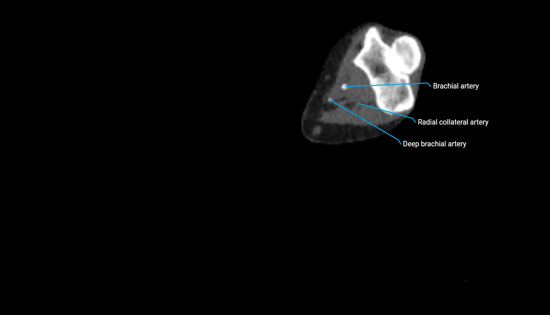
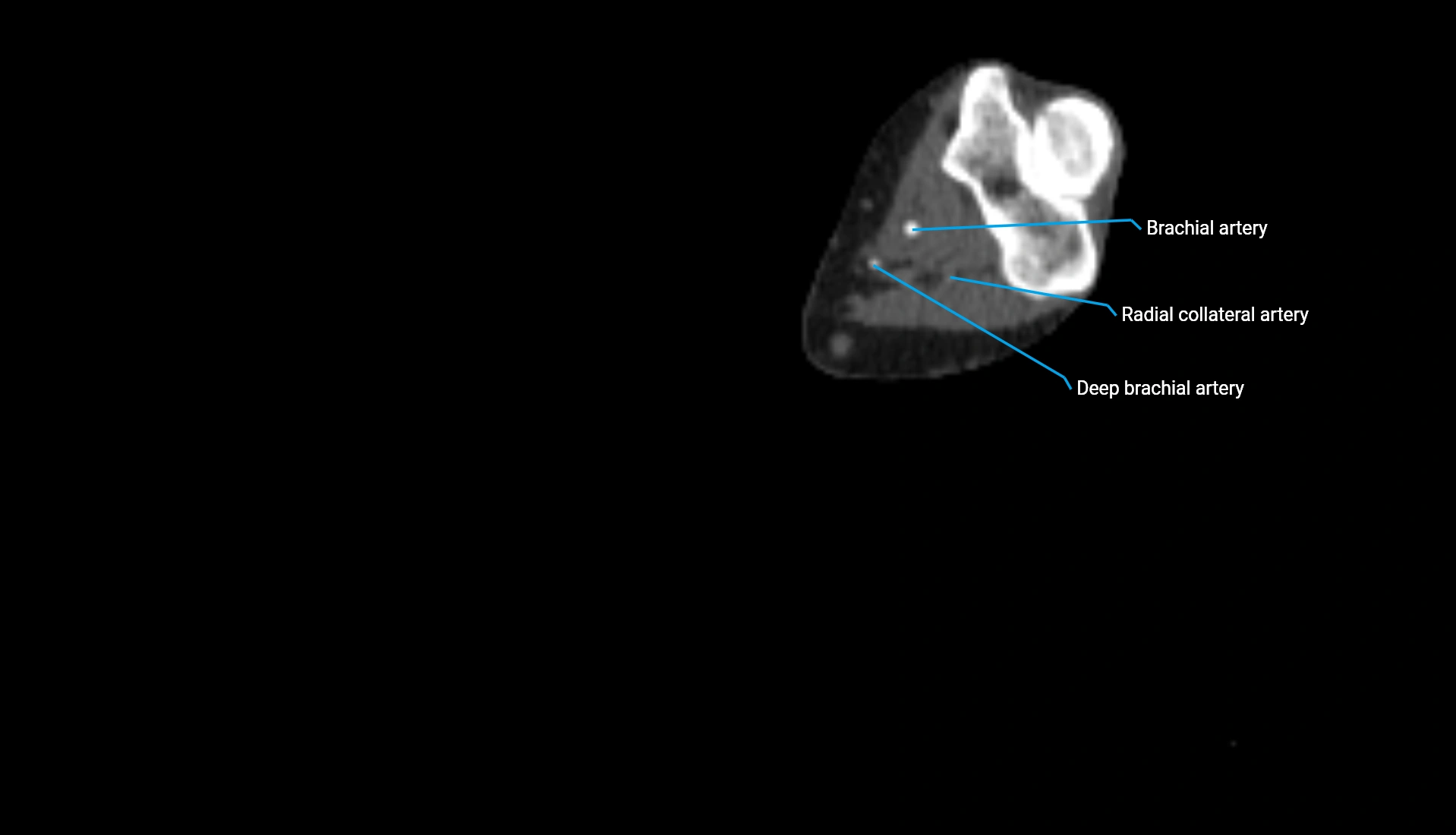
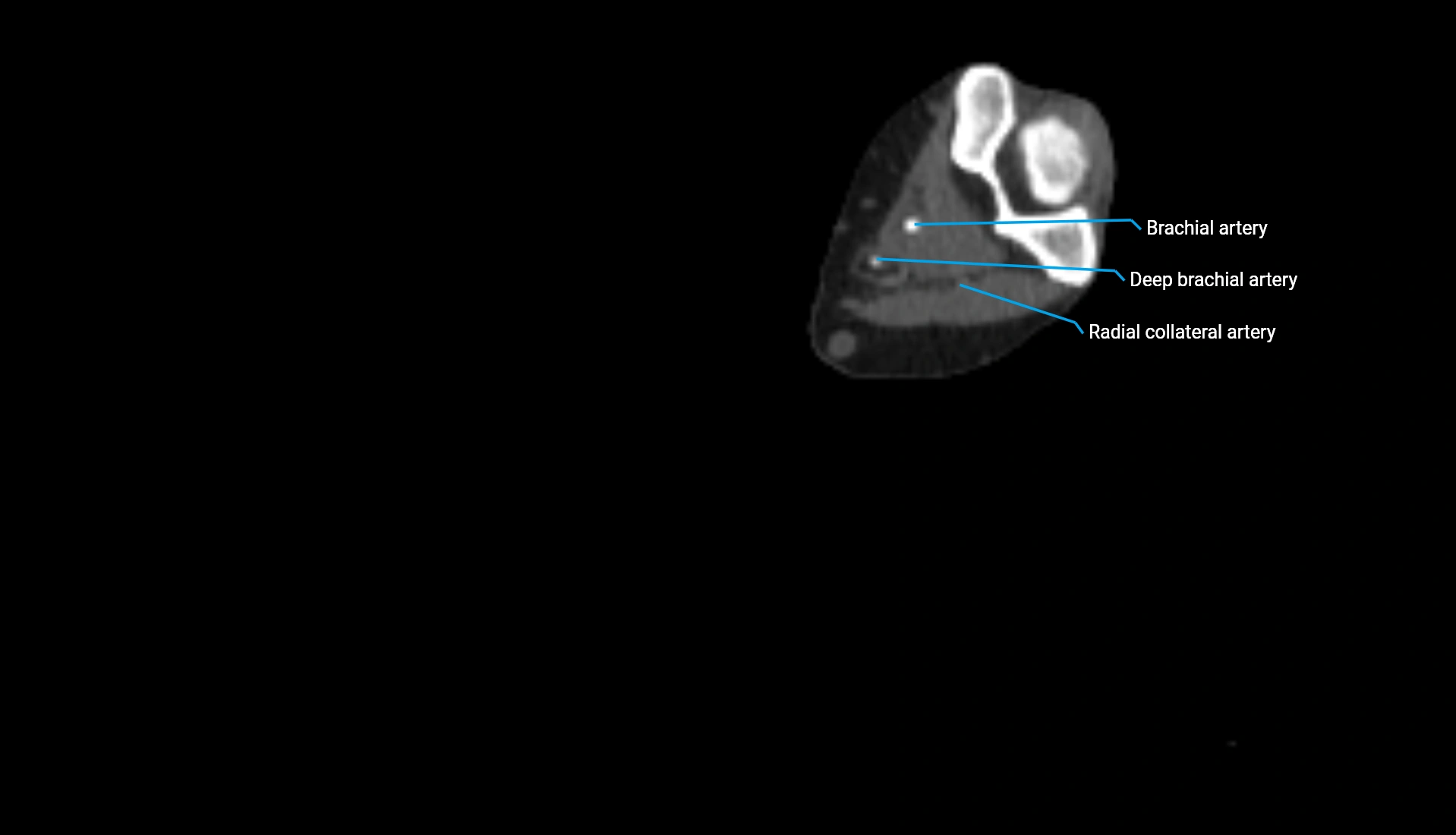
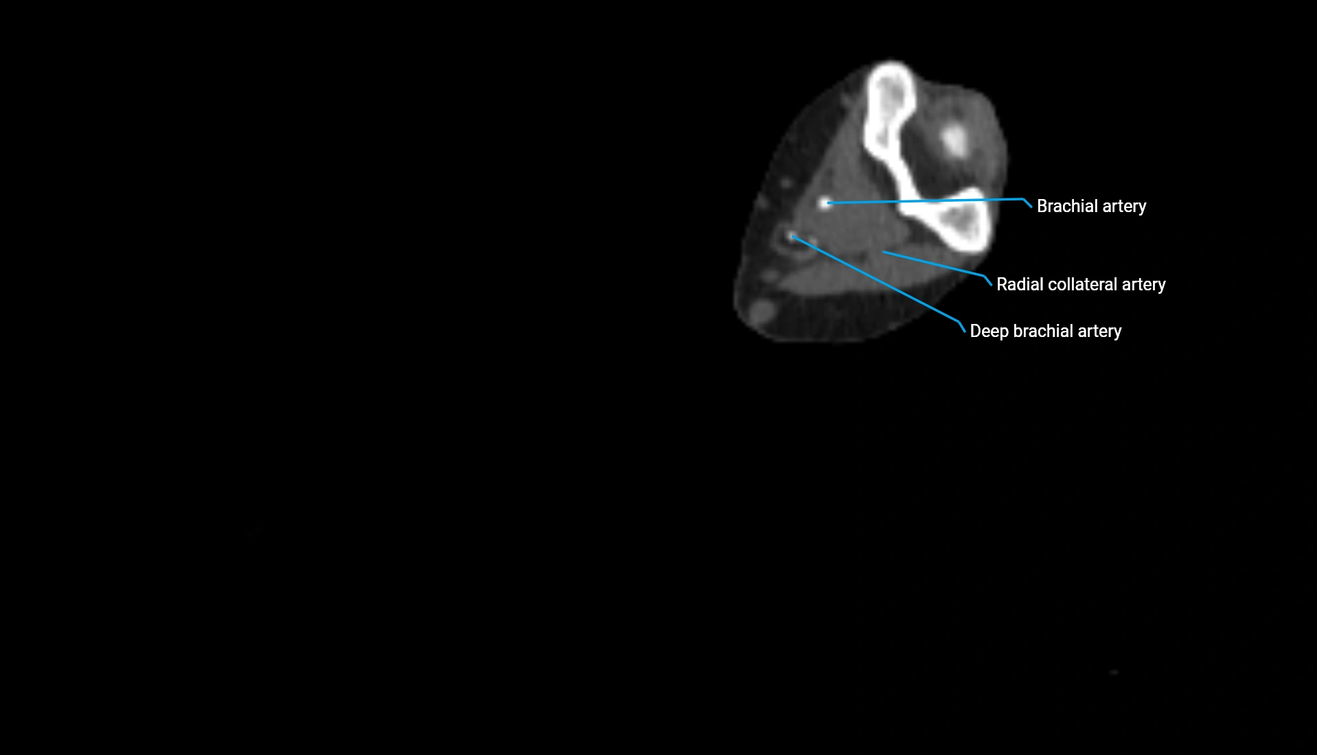
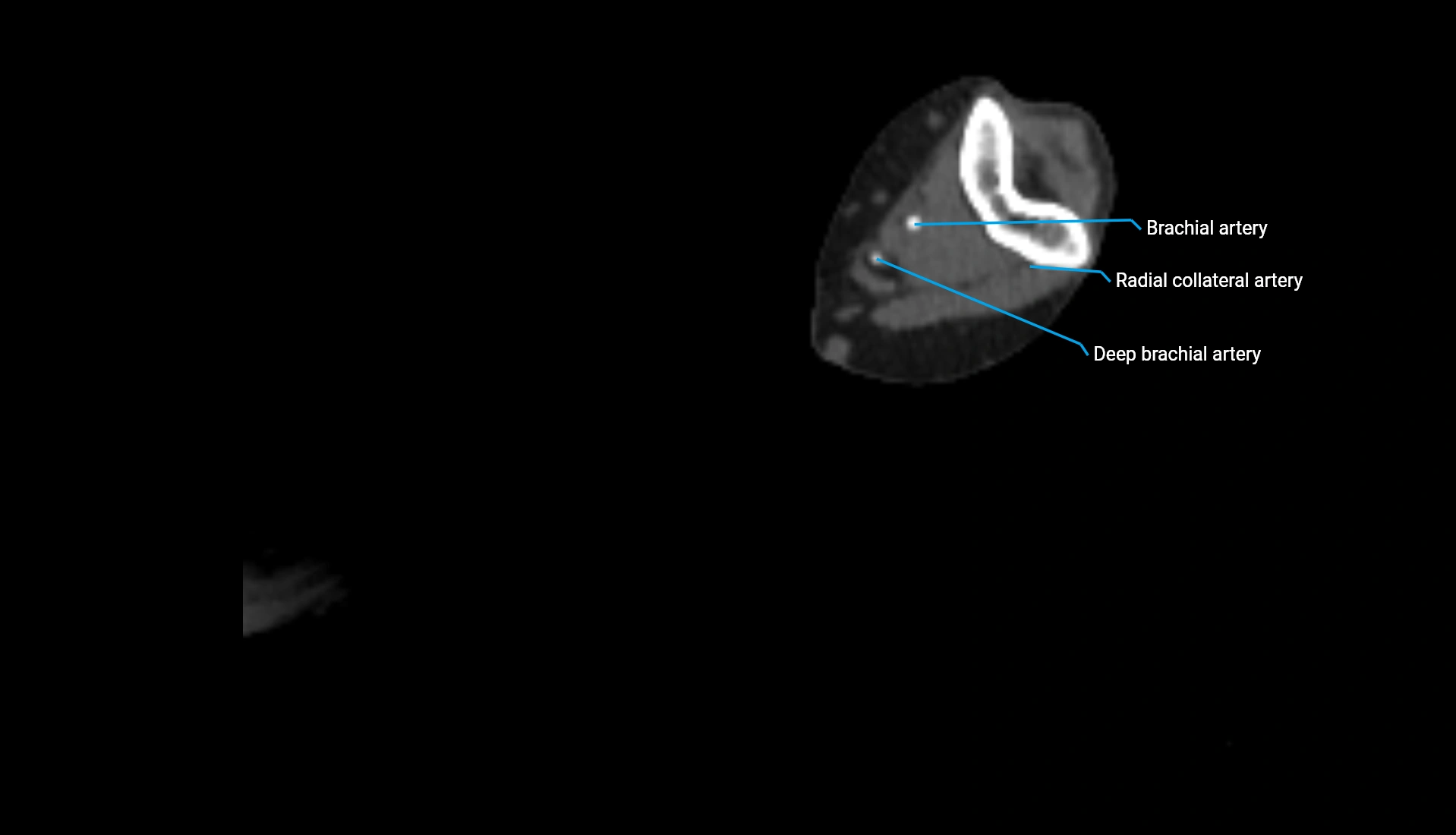
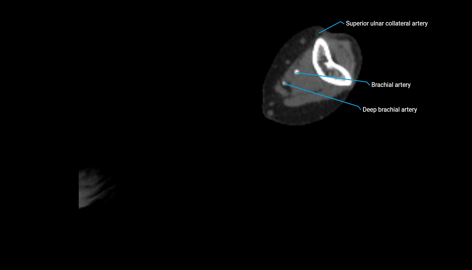
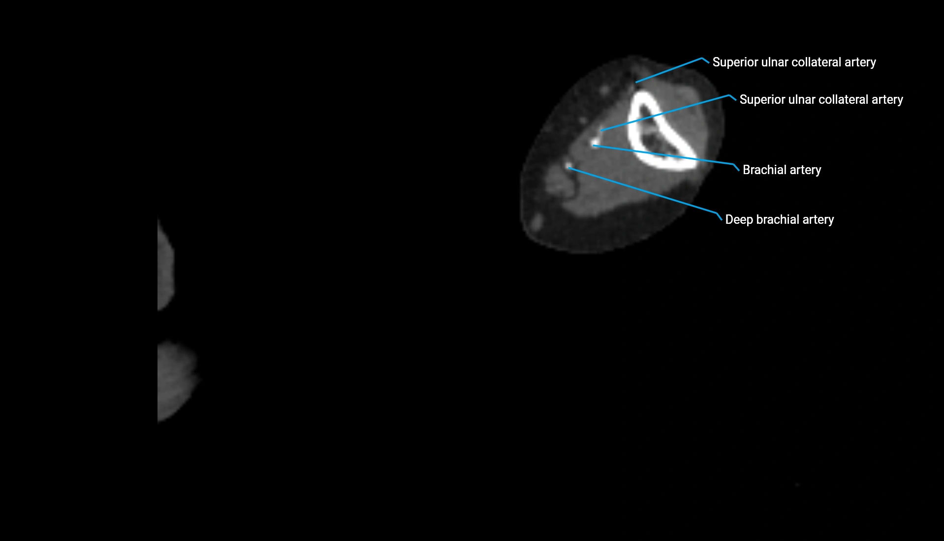
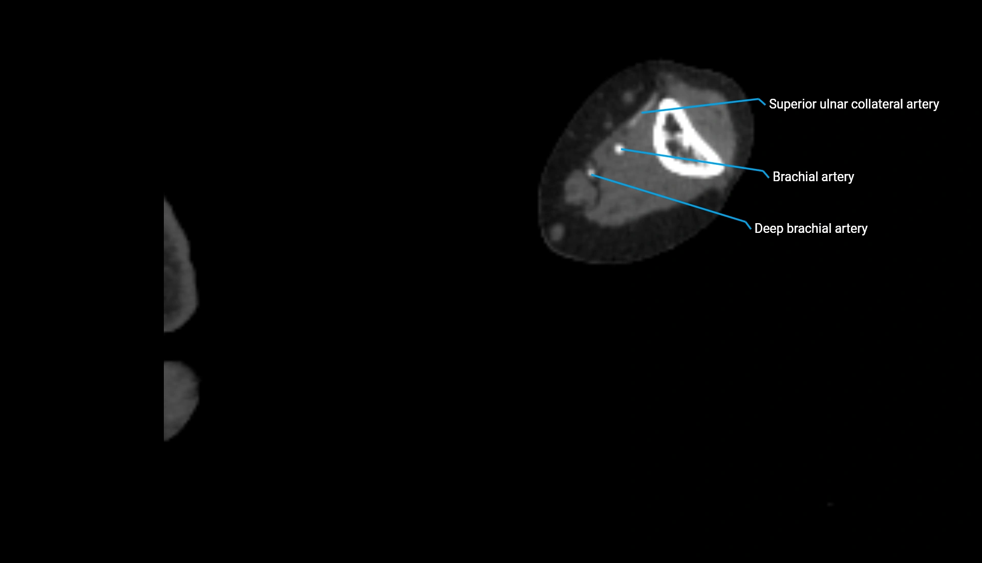
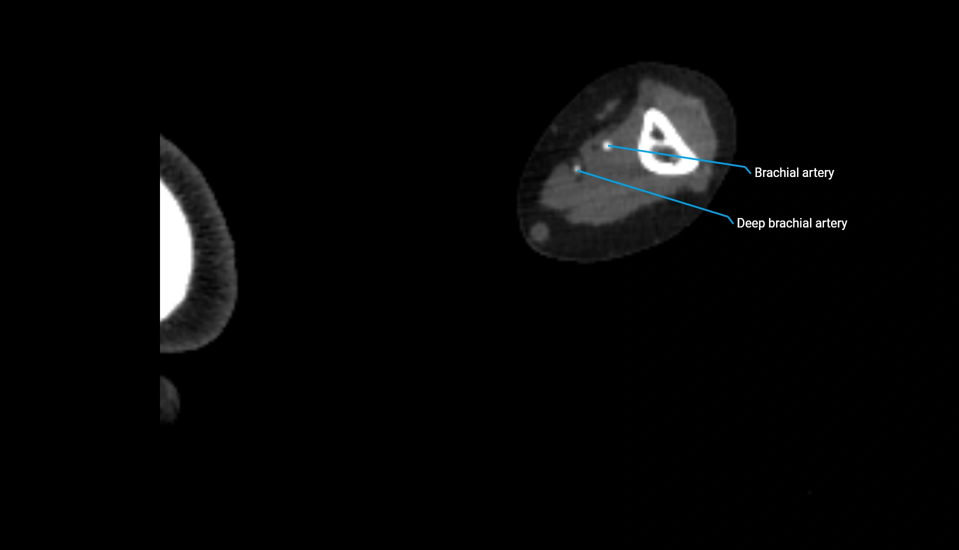
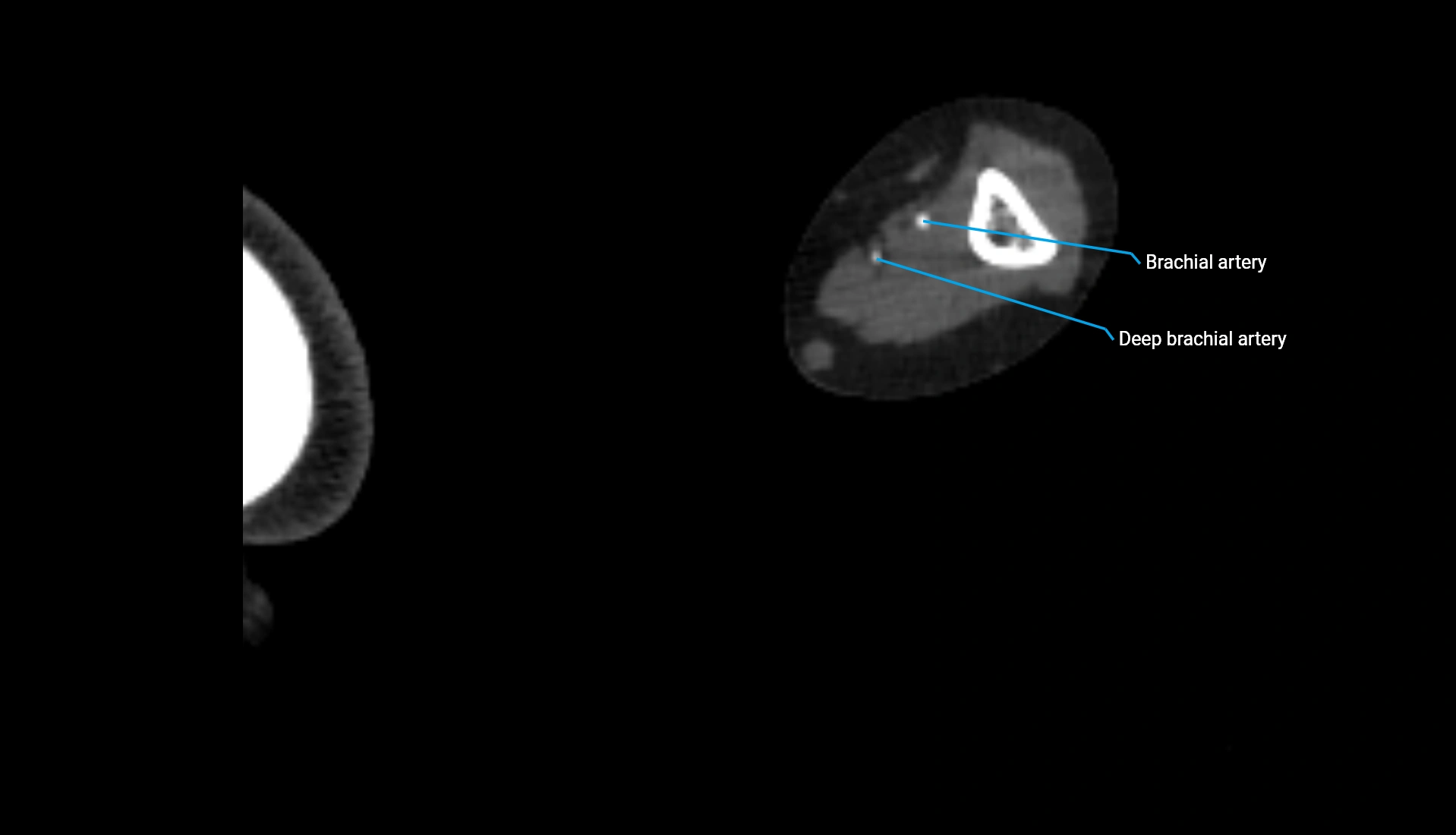
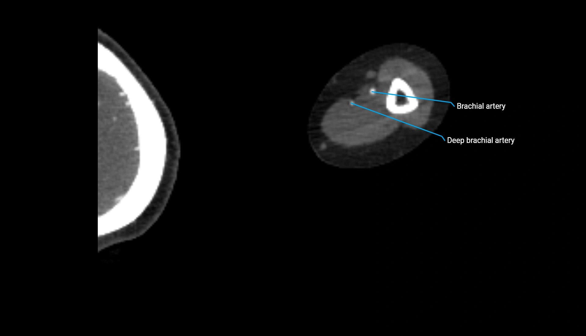
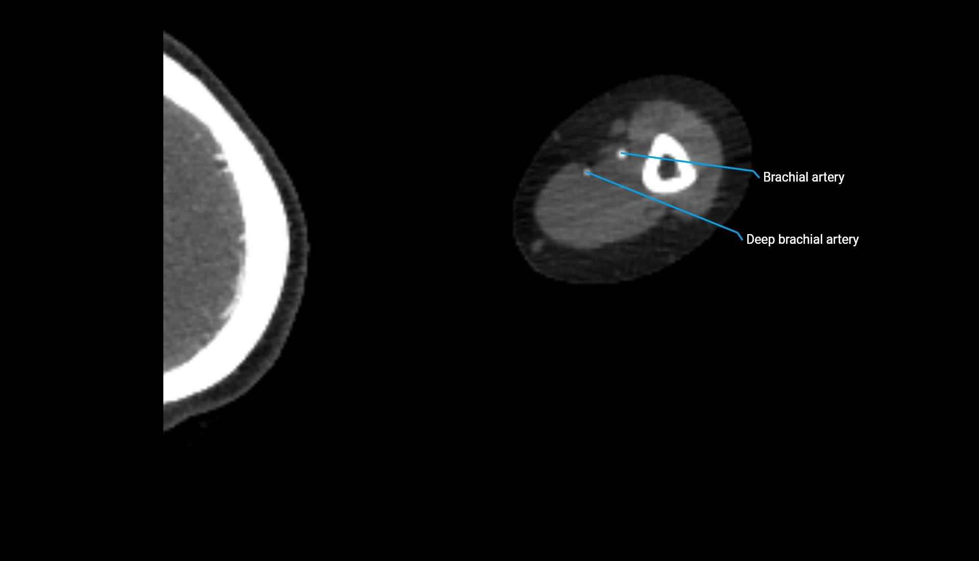
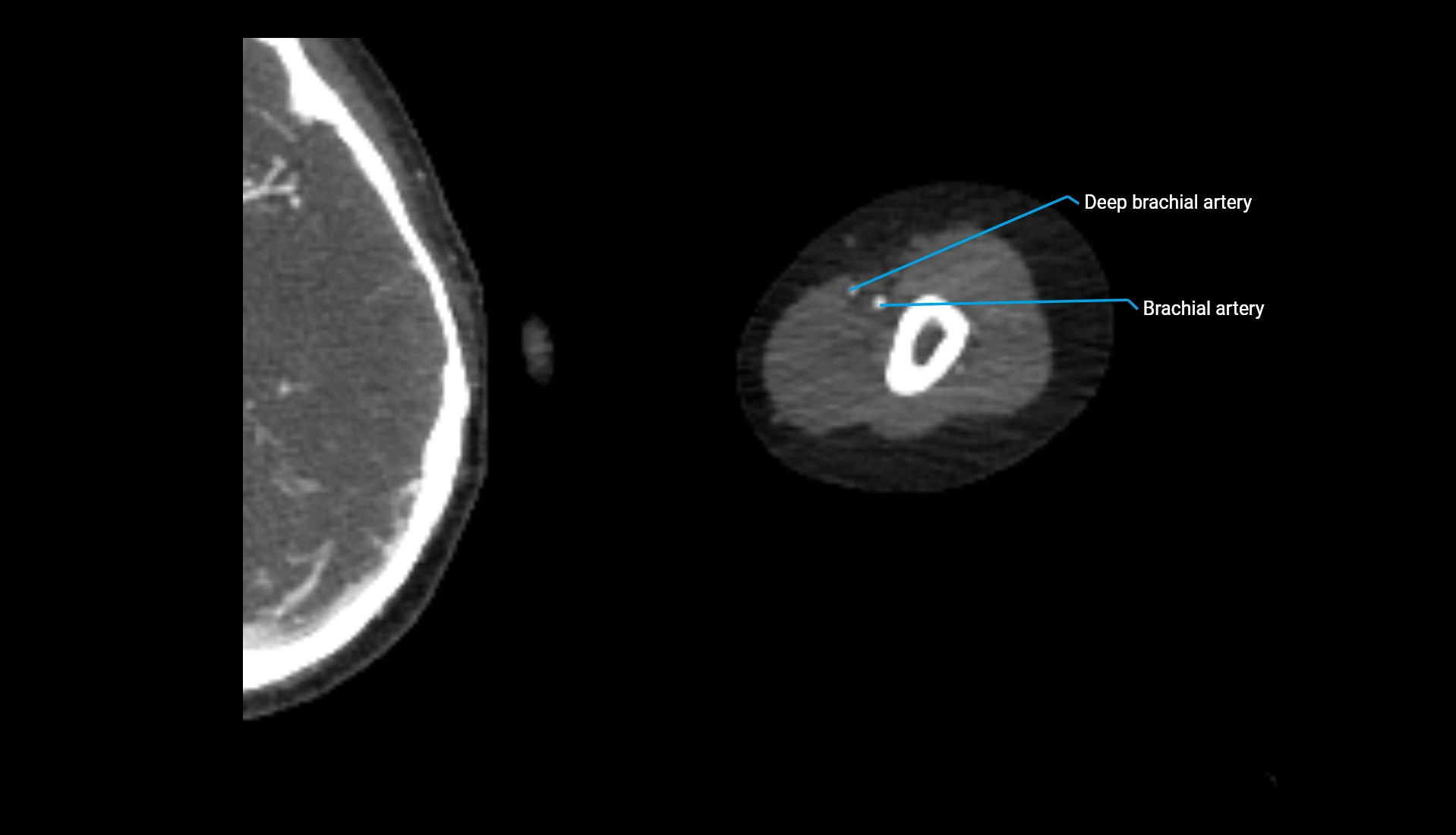
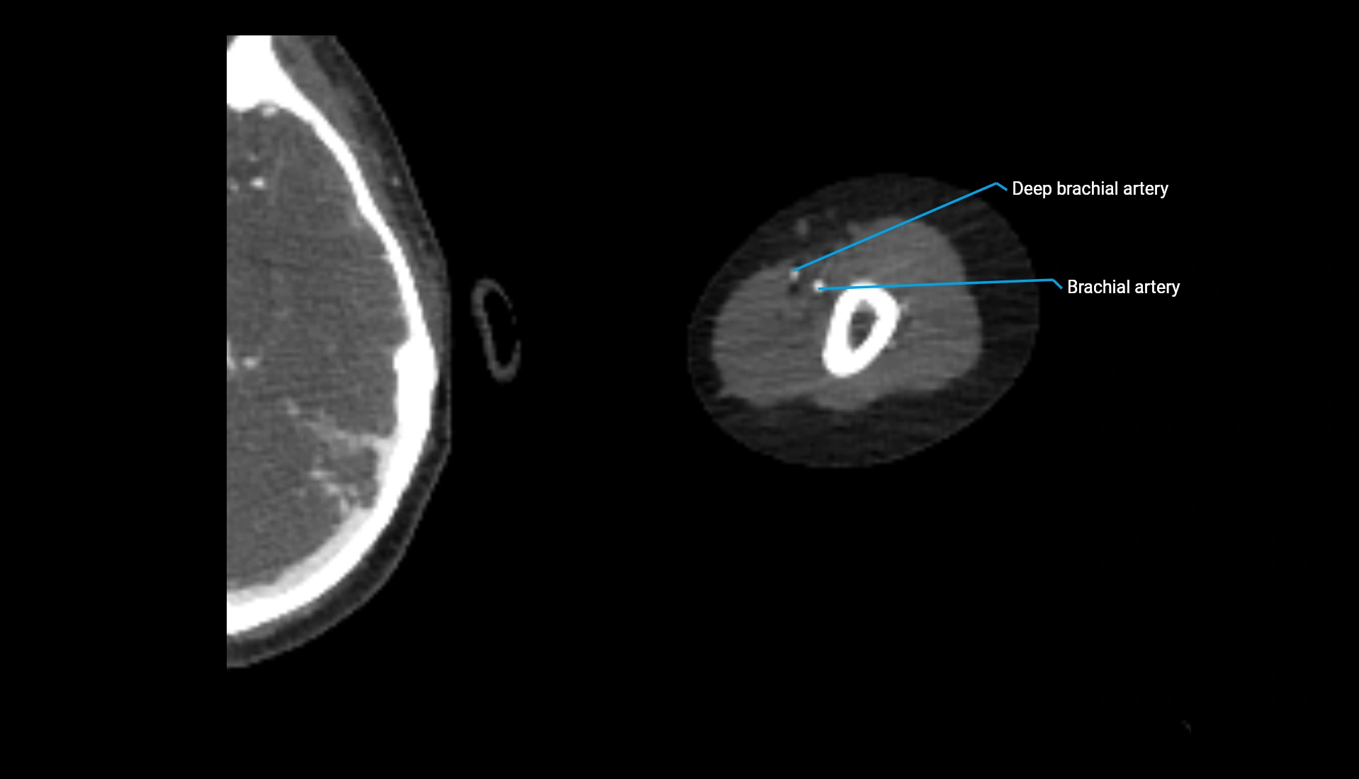
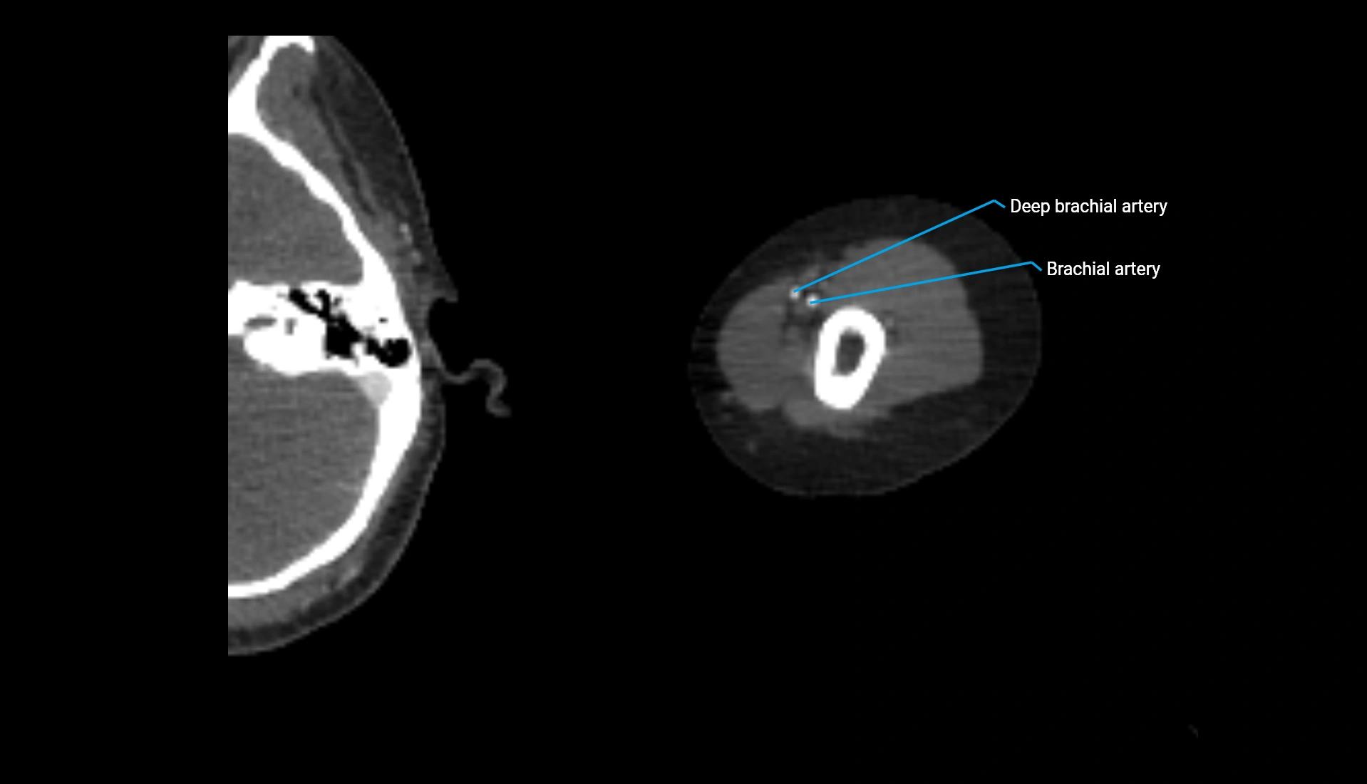
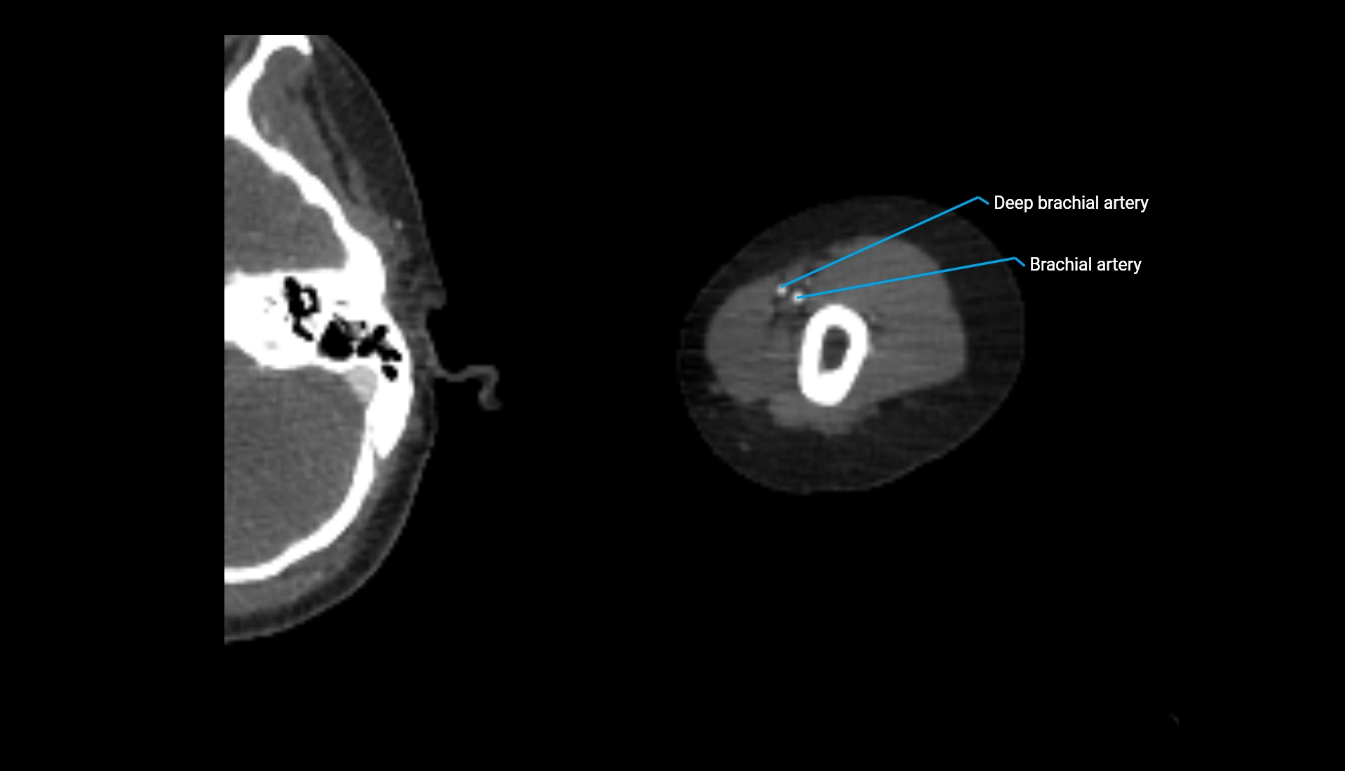
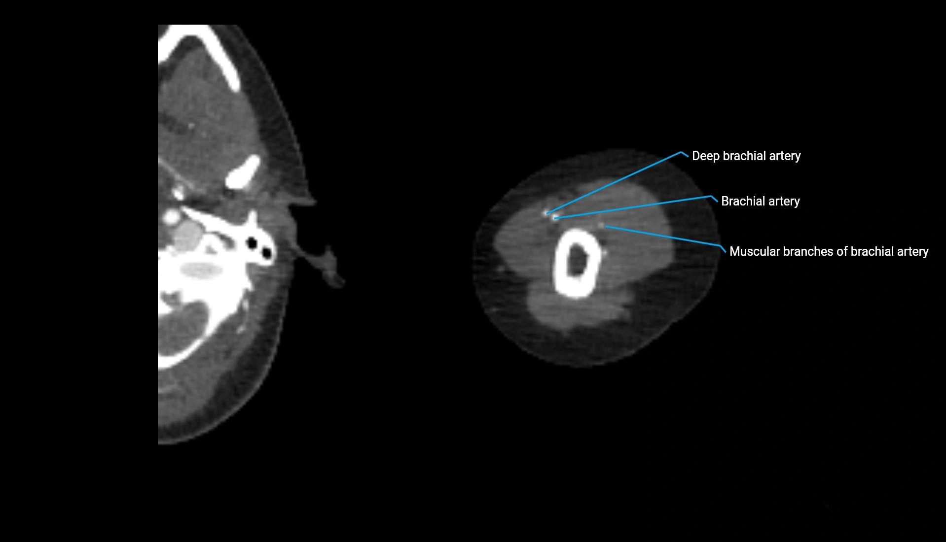
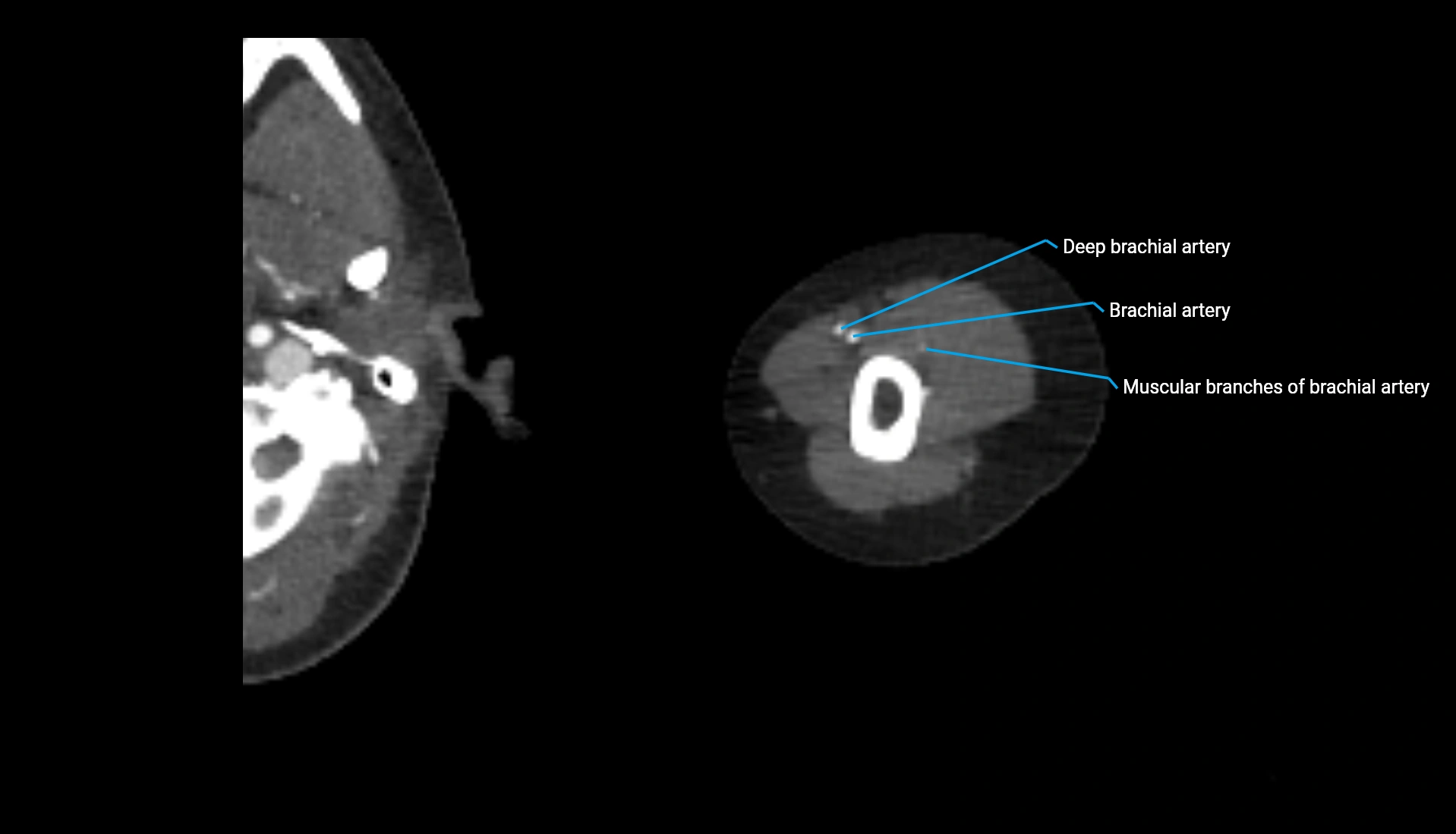
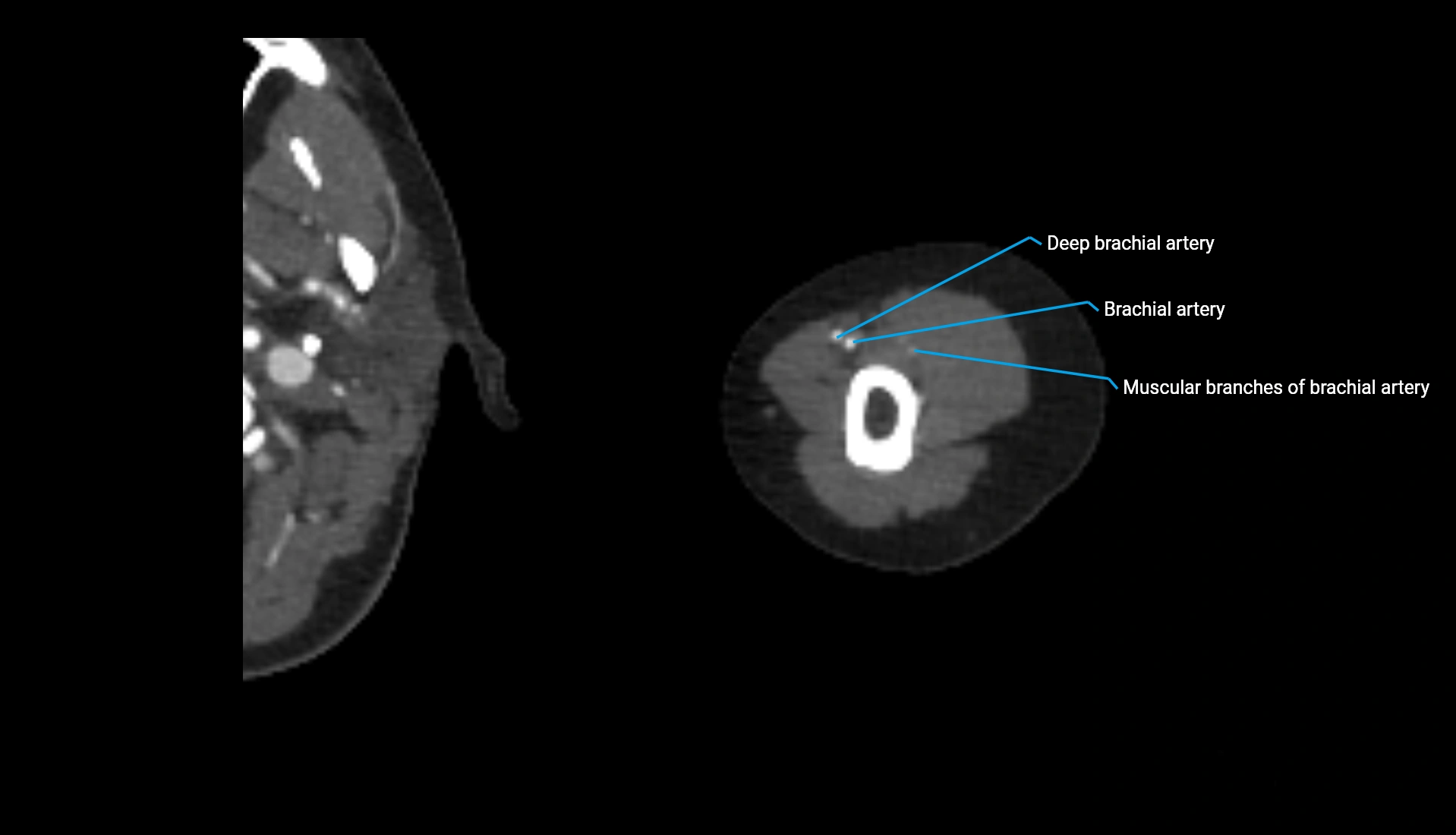
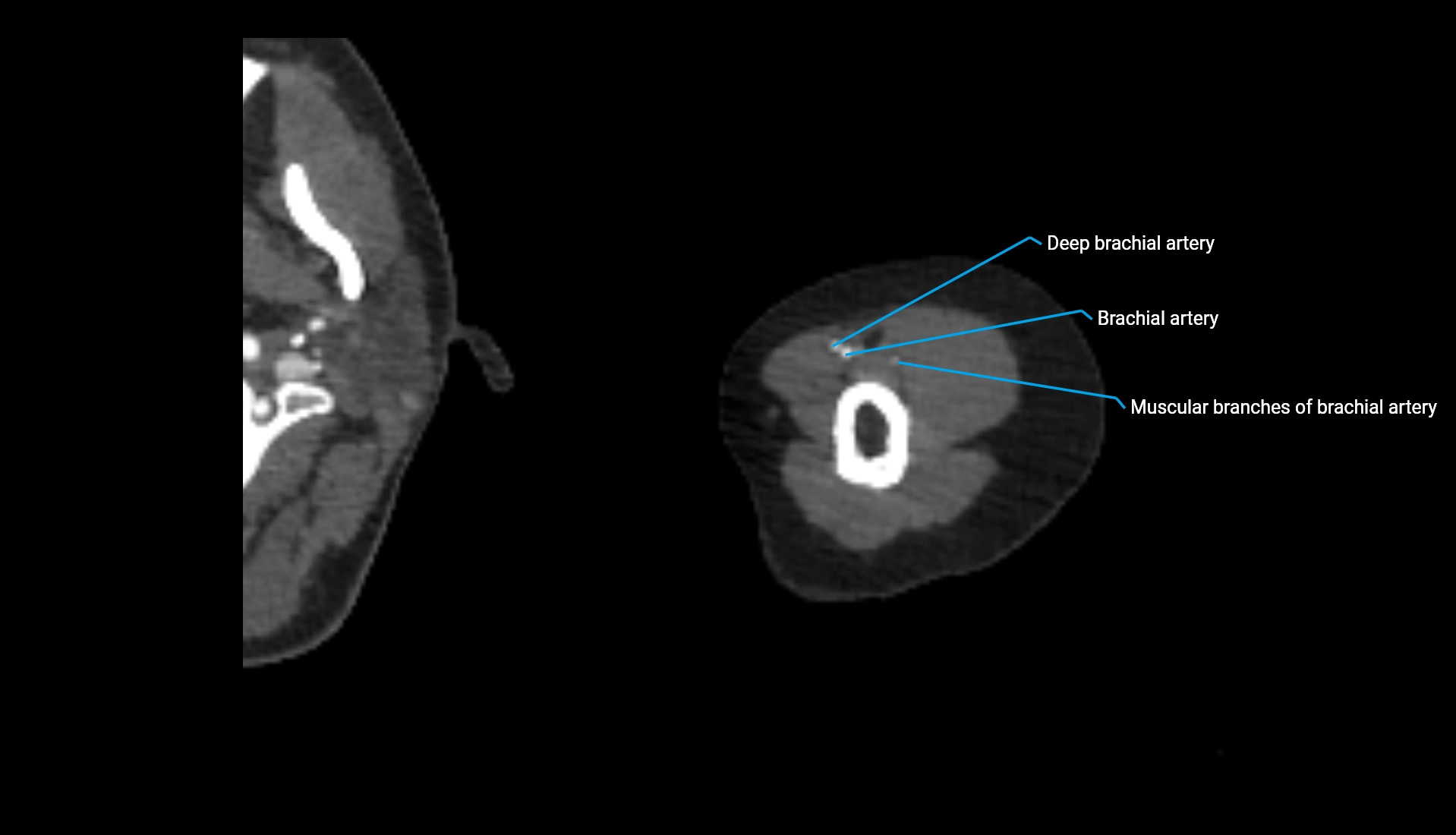
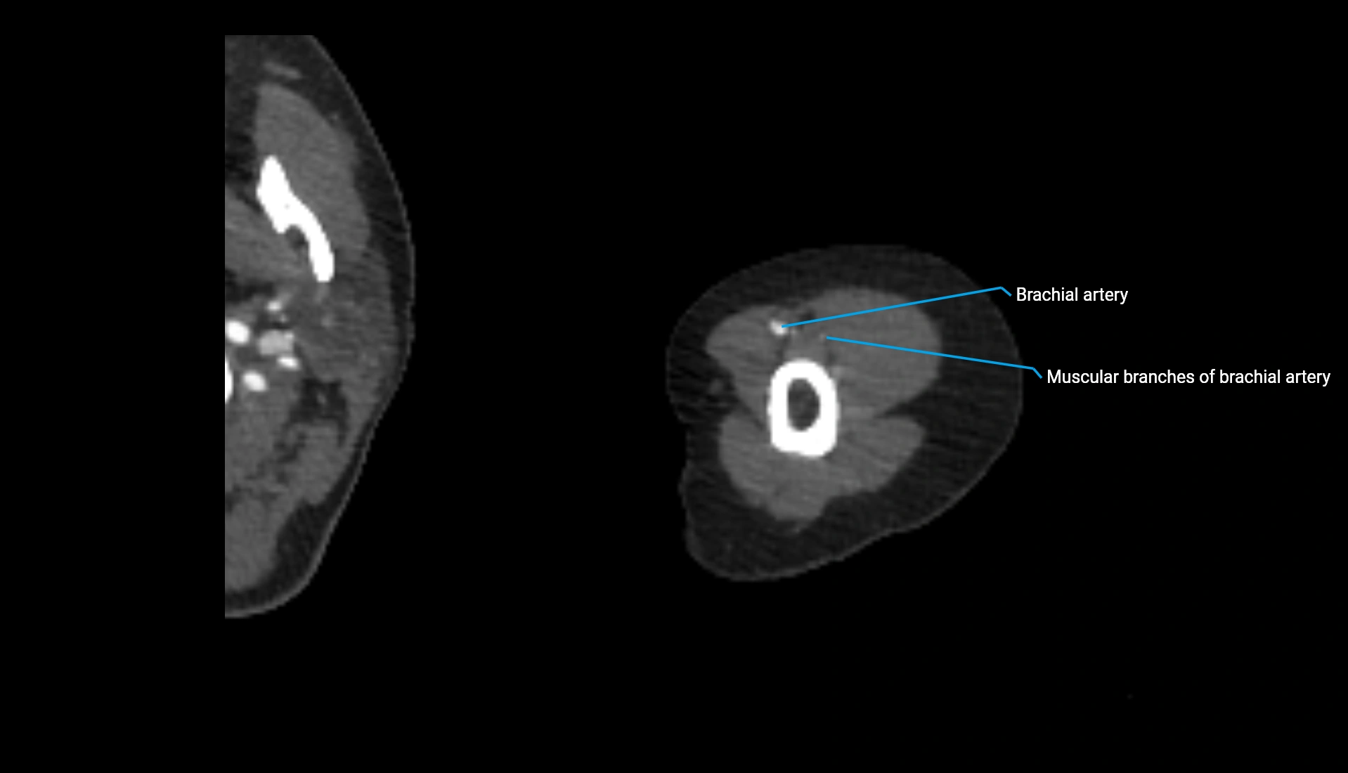
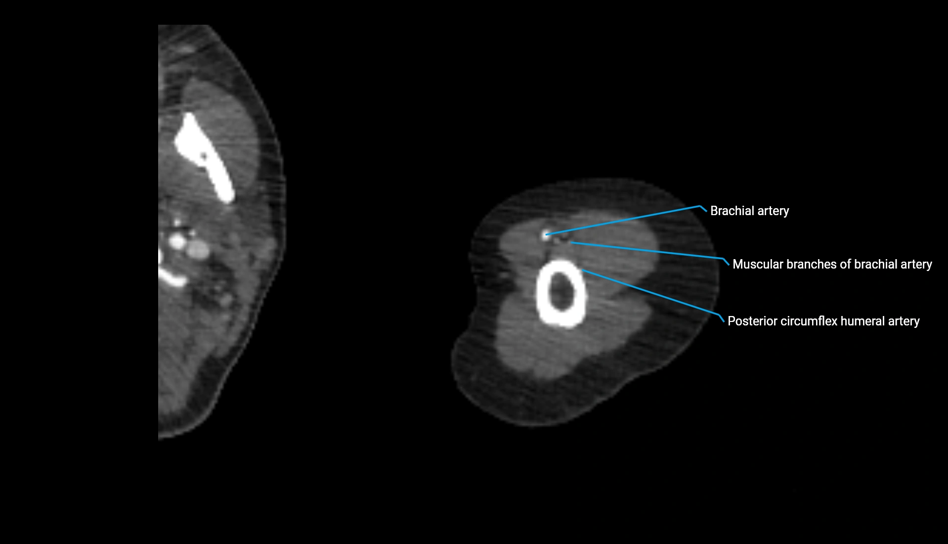
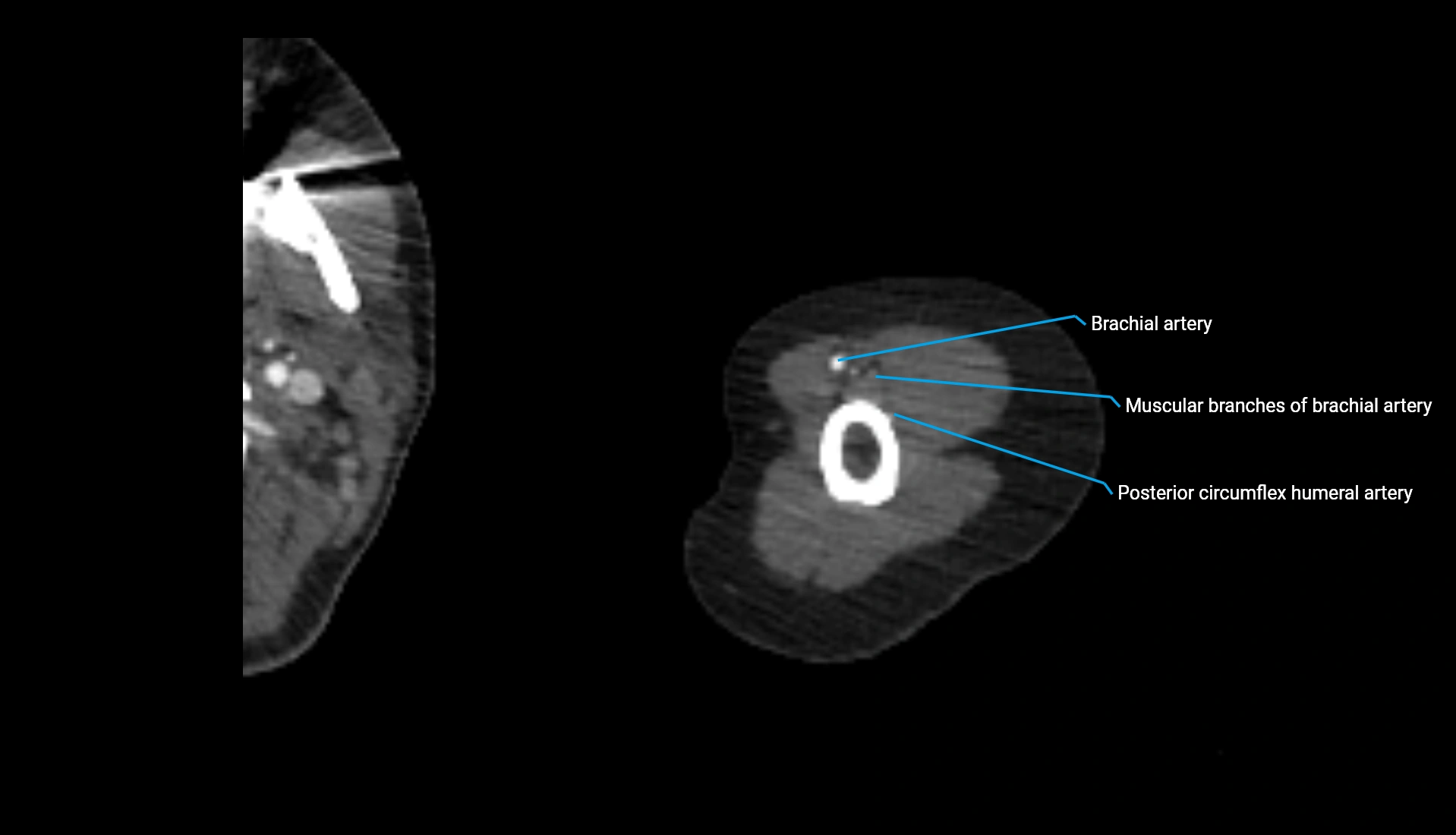
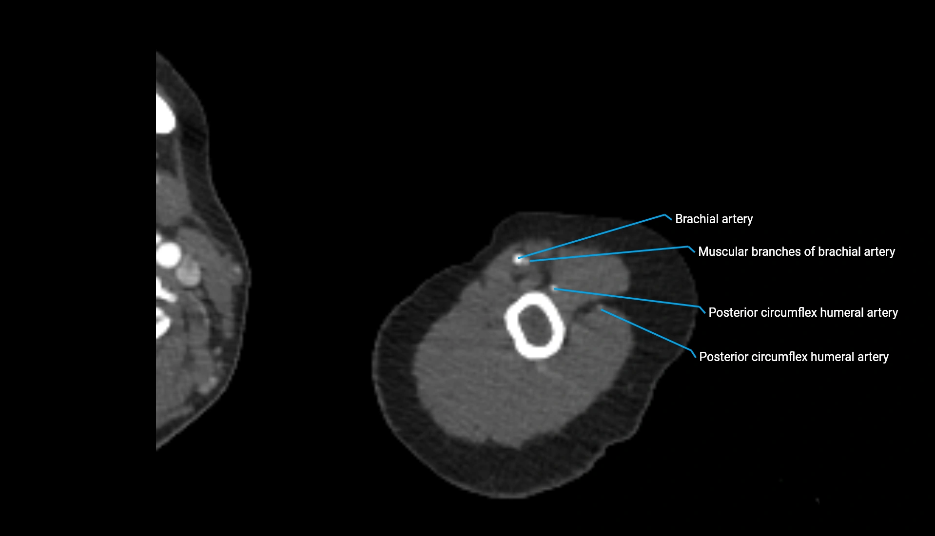
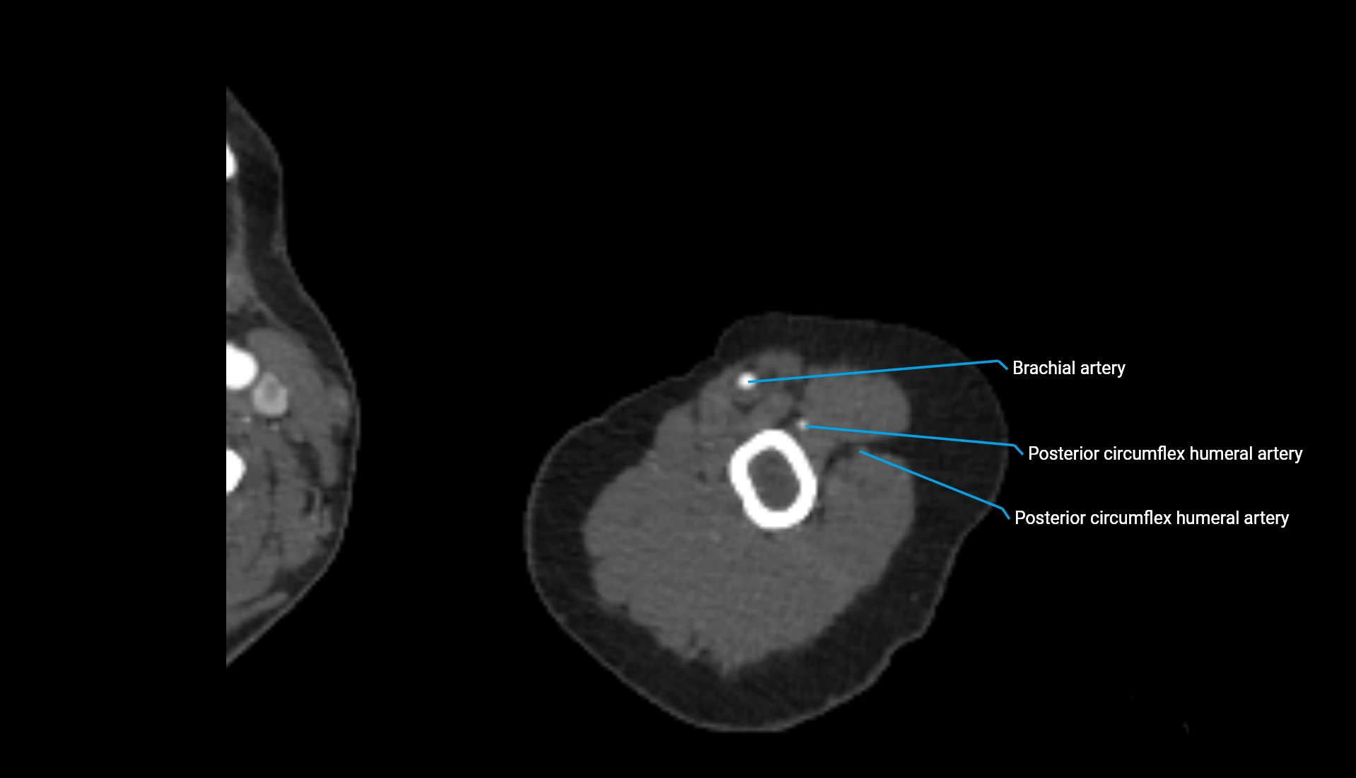
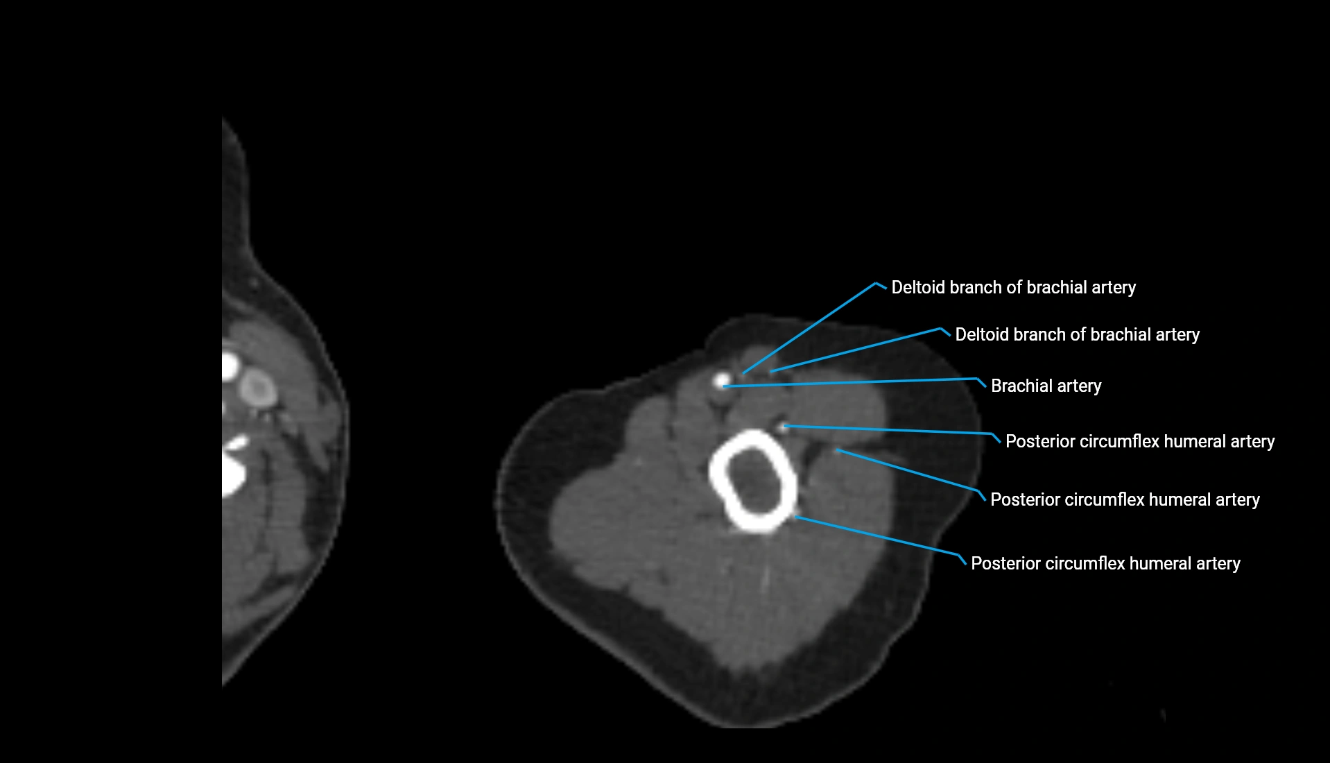
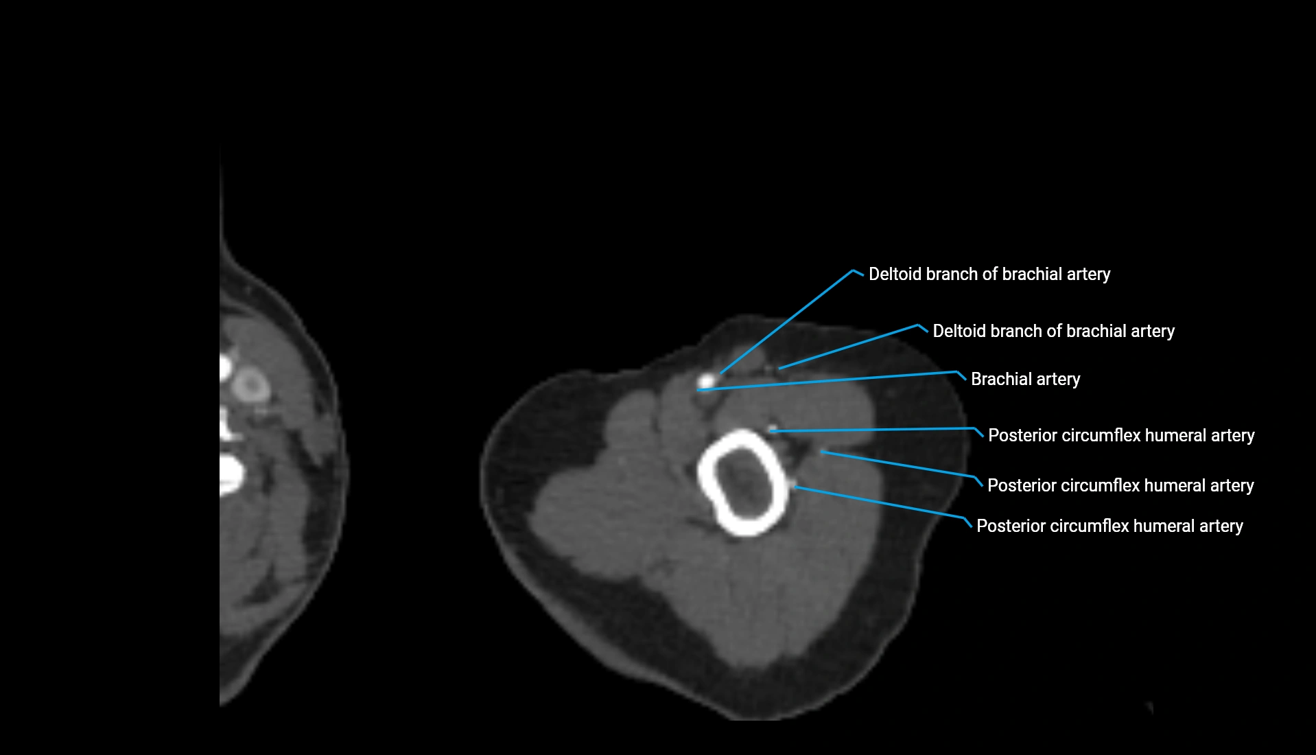
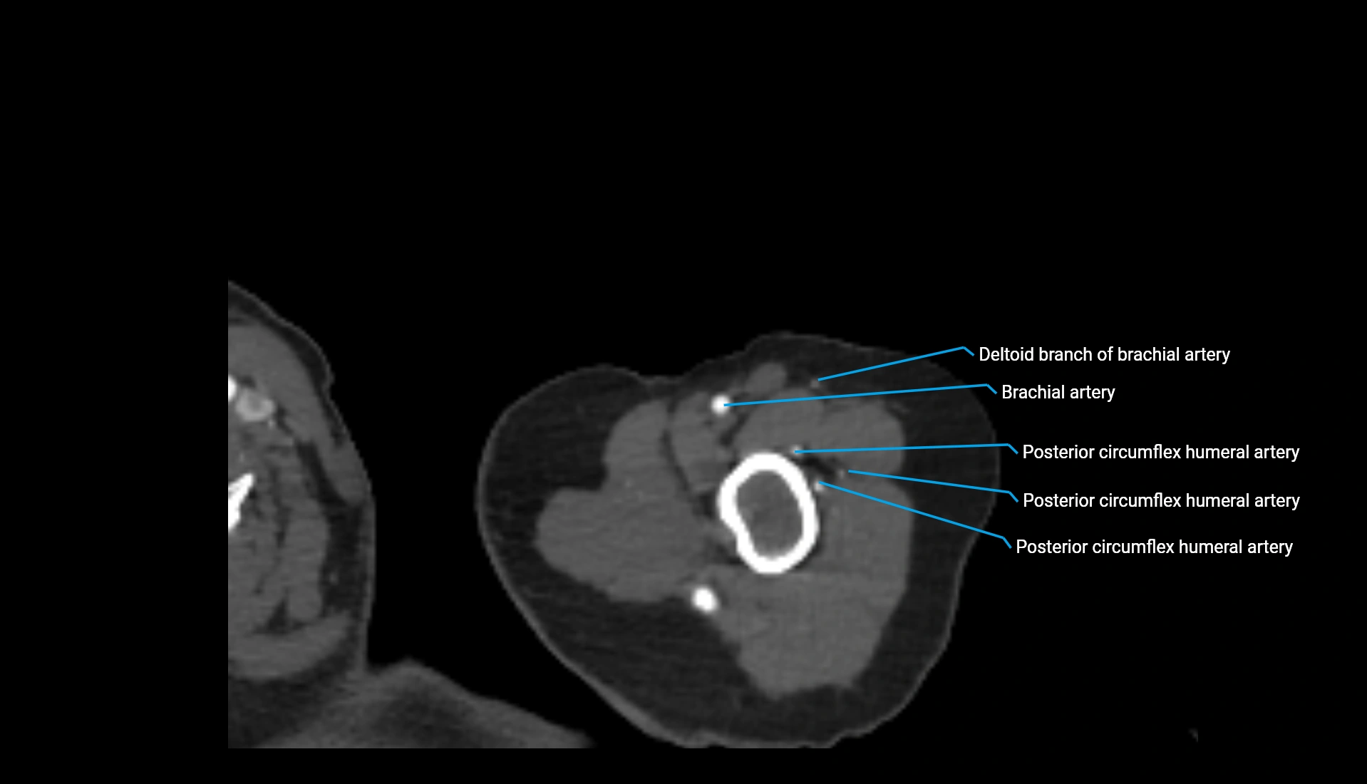
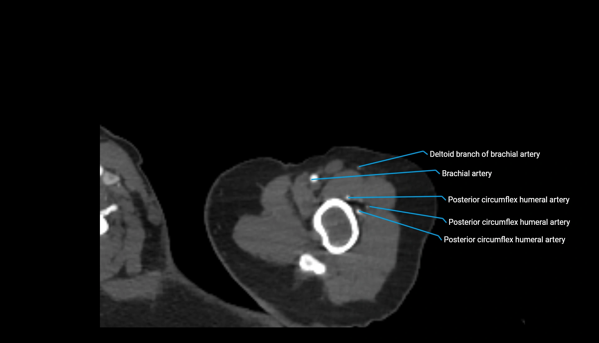
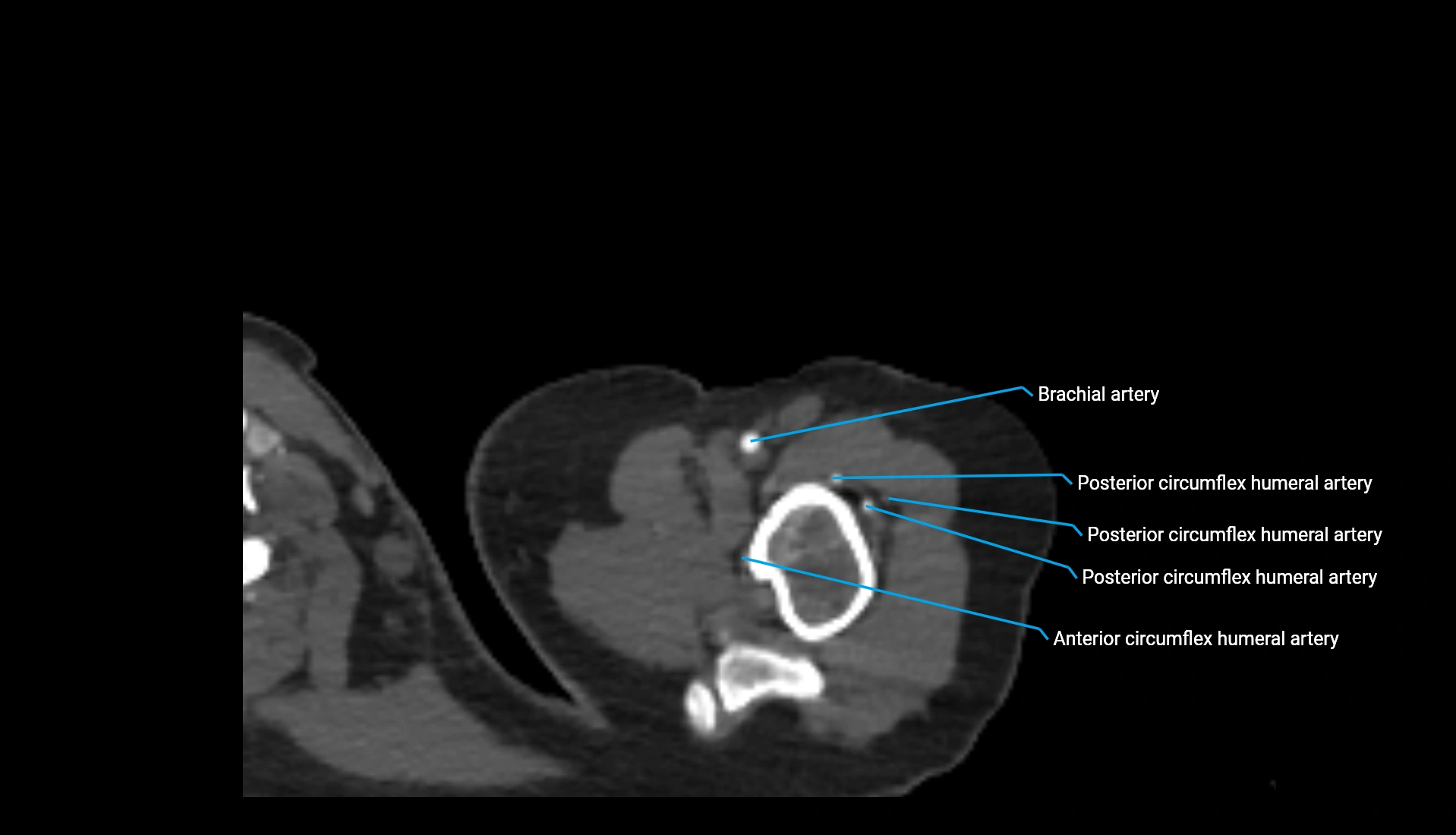
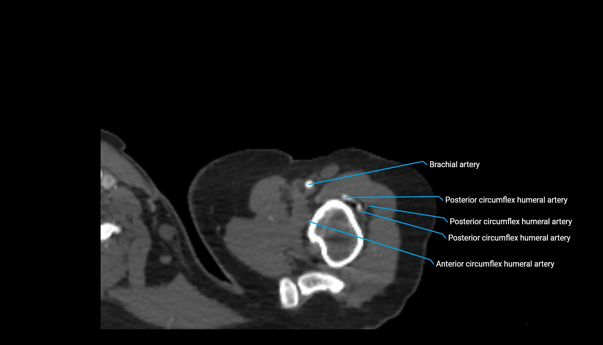
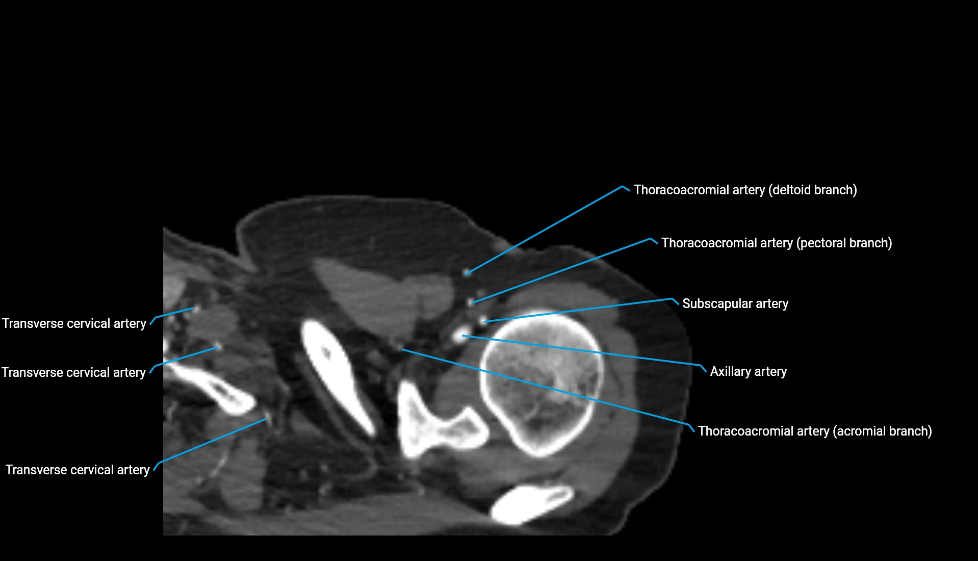
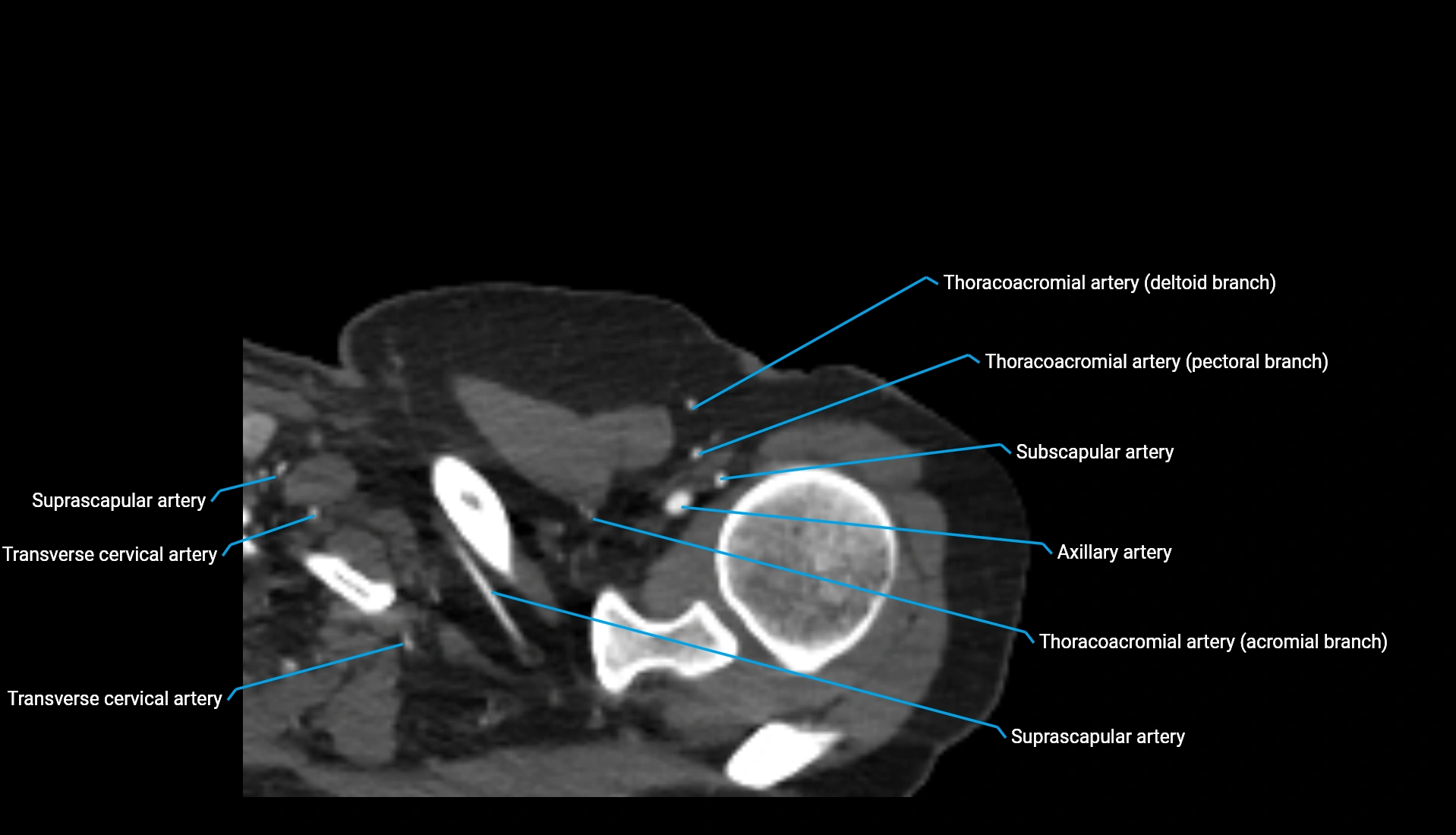
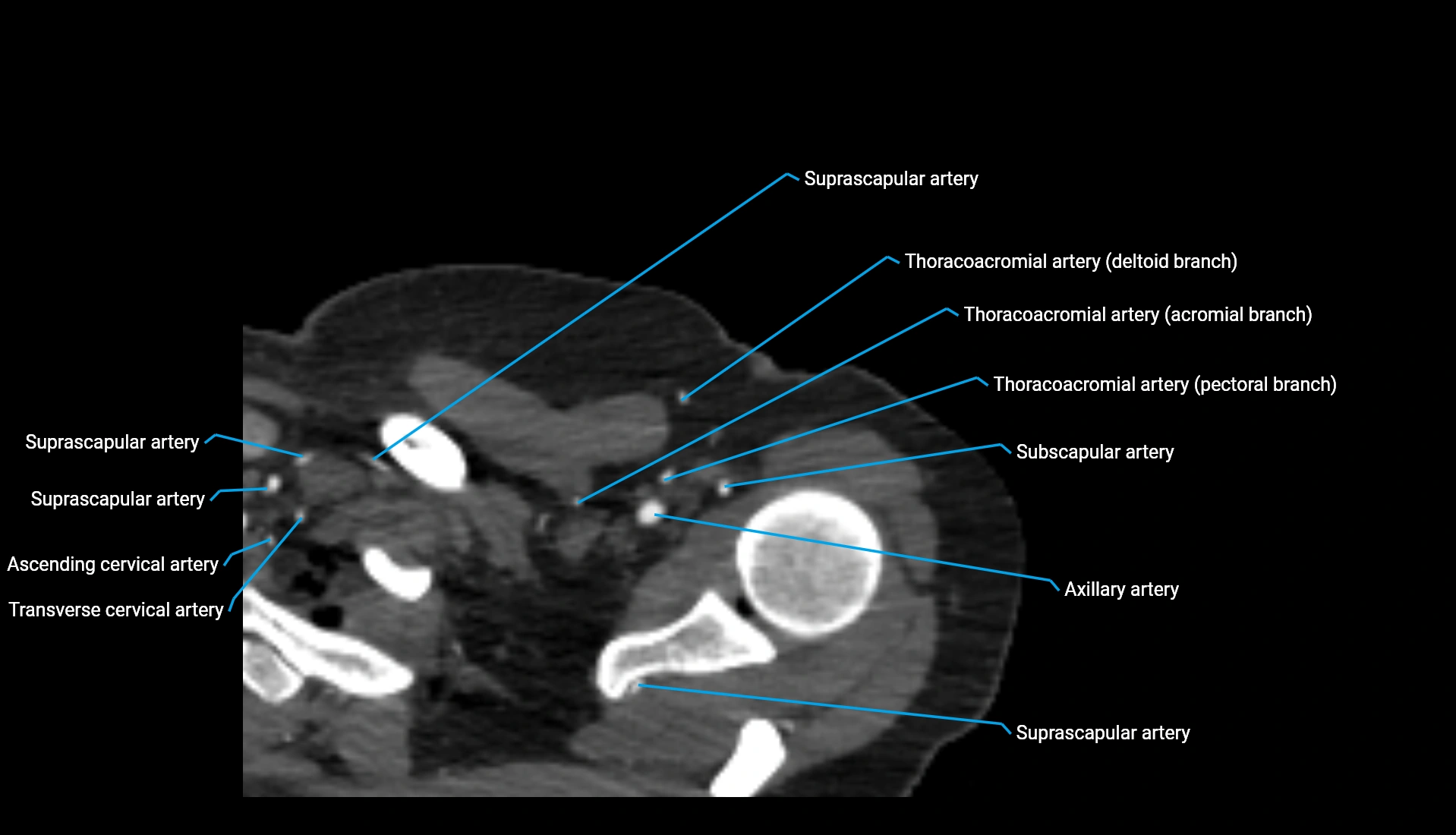
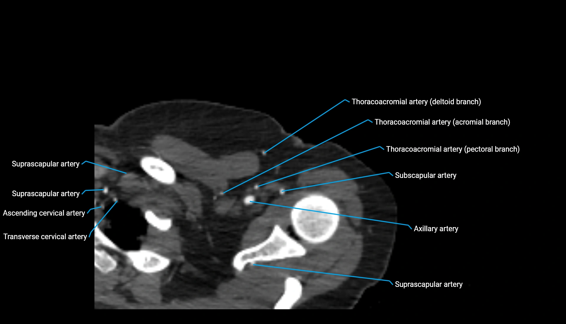
MRI image
