


Topic
The anterior inferior iliac spine (AIIS) is a bony prominence of the ilium, located just inferior to the anterior superior iliac spine (ASIS), separated by the interspinous groove. It projects anteriorly from the ilium above the acetabulum and serves as an important attachment site for muscles and ligaments.
The AIIS provides the proximal origin for the direct (straight) head of the rectus femoris muscle and attachment for the iliofemoral ligament, one of the strongest ligaments in the body, which stabilizes the hip joint. The reflected (indirect) head of rectus femoris arises from the acetabular rim, in close relation to the AIIS.
It is a critical surgical and imaging landmark. Avulsion fractures of the AIIS are common in adolescents and athletes due to strong traction forces of the rectus femoris. Overuse or abnormal morphology of the AIIS may contribute to subspine impingement, a recognized subtype of femoroacetabular impingement (FAI).
Attachments
-
Muscle attachment: Direct head of rectus femoris
-
Ligament attachment: Iliofemoral ligament (superior band)
-
Capsular attachment: Part of the anterior hip capsule
Relations
-
Lies superior to the acetabular rim and anterior to the hip joint
-
Located inferior to the ASIS, separated by the interspinous groove
-
Closely related to rectus femoris tendon and anterior joint capsule
Synonyms
-
AIIS
-
Spina iliaca anterior inferior
Function
-
Provides origin for rectus femoris (straight head), aiding hip flexion and knee extension
-
Serves as an attachment site for iliofemoral ligament, contributing to hip stability
-
Acts as a landmark in imaging and surgical approaches to the hip and pelvis
Nerve Supply (related structures)
-
Rectus femoris muscle: innervated by femoral nerve (L2–L4)
Arterial Supply
-
Ascending branch of lateral circumflex femoral artery (primary supply to rectus femoris origin)
-
Contributions from superior gluteal artery and iliolumbar artery
Venous Drainage
-
Lateral circumflex femoral vein → femoral vein
-
Anastomotic drainage via iliac and gluteal venous networks
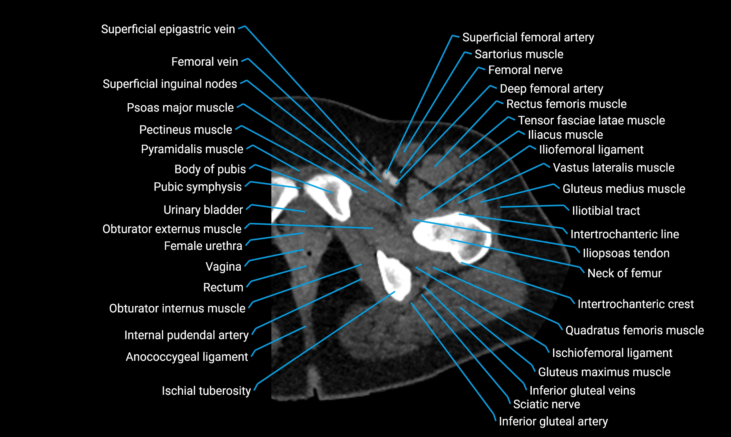
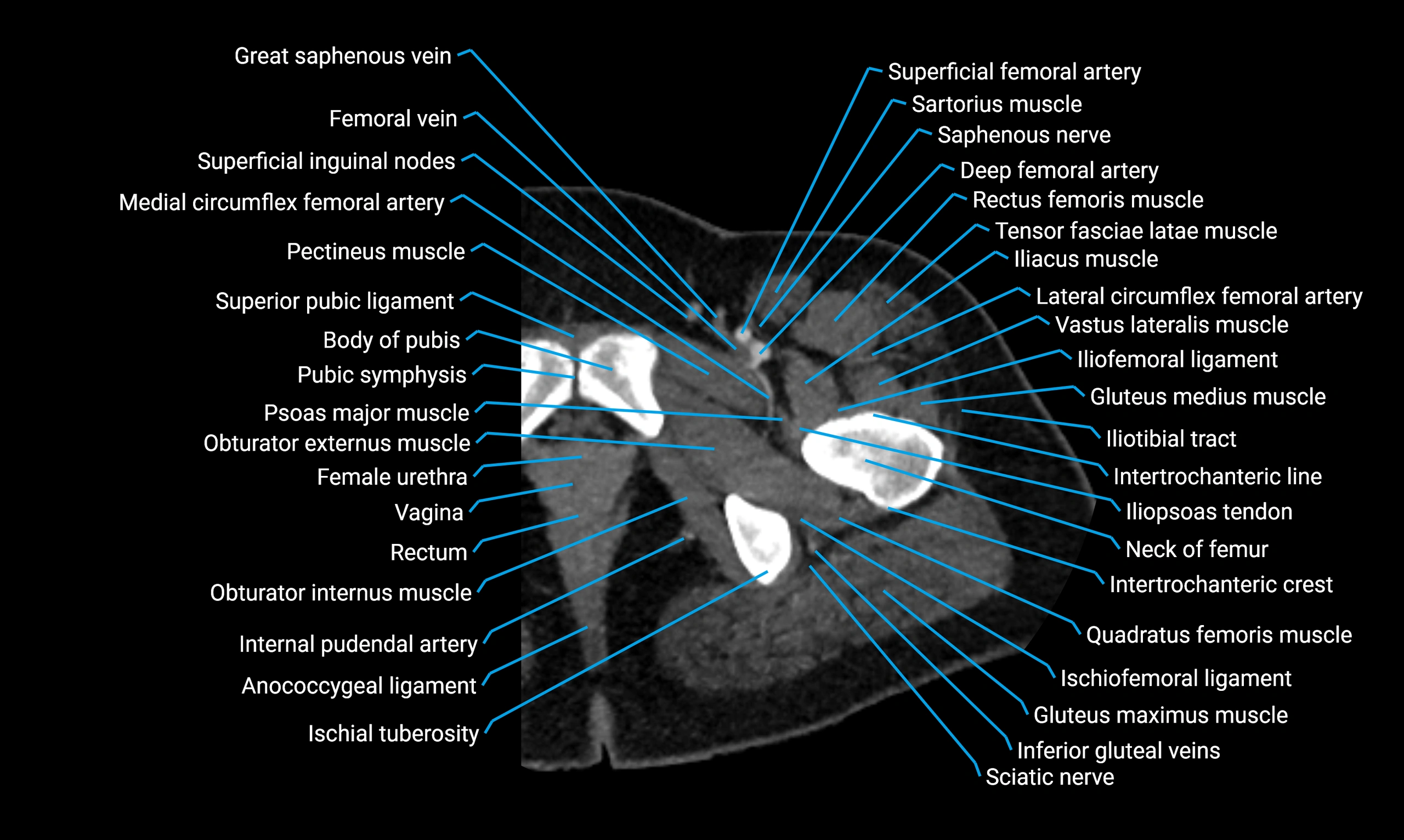
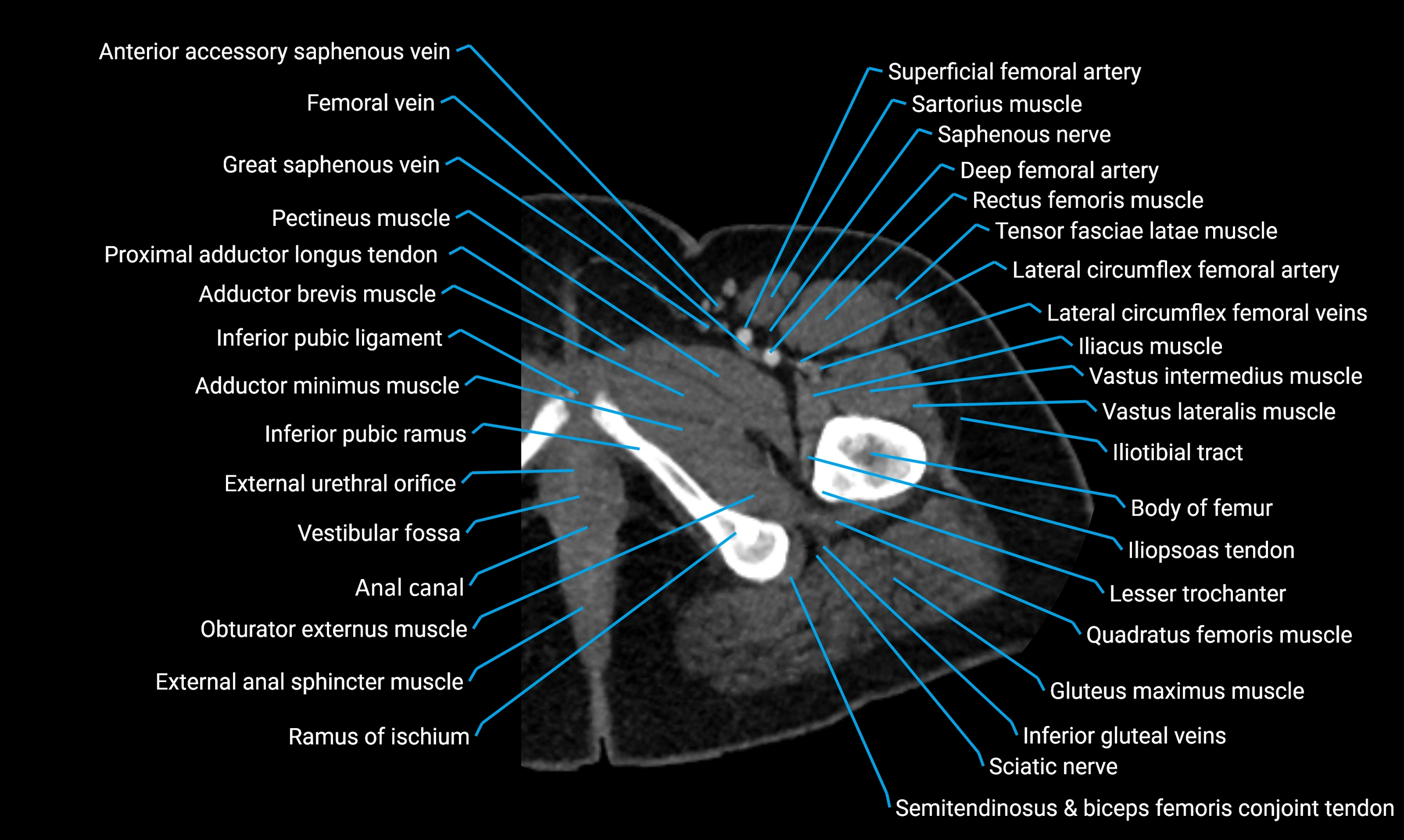
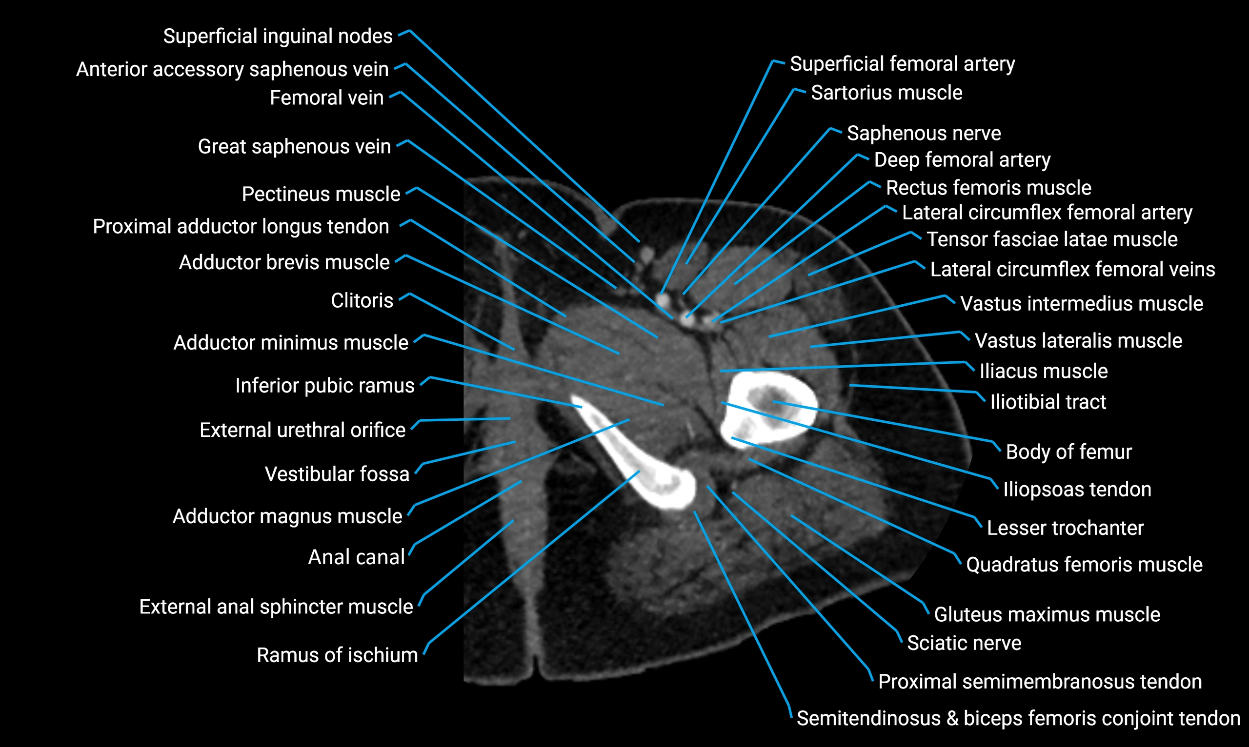
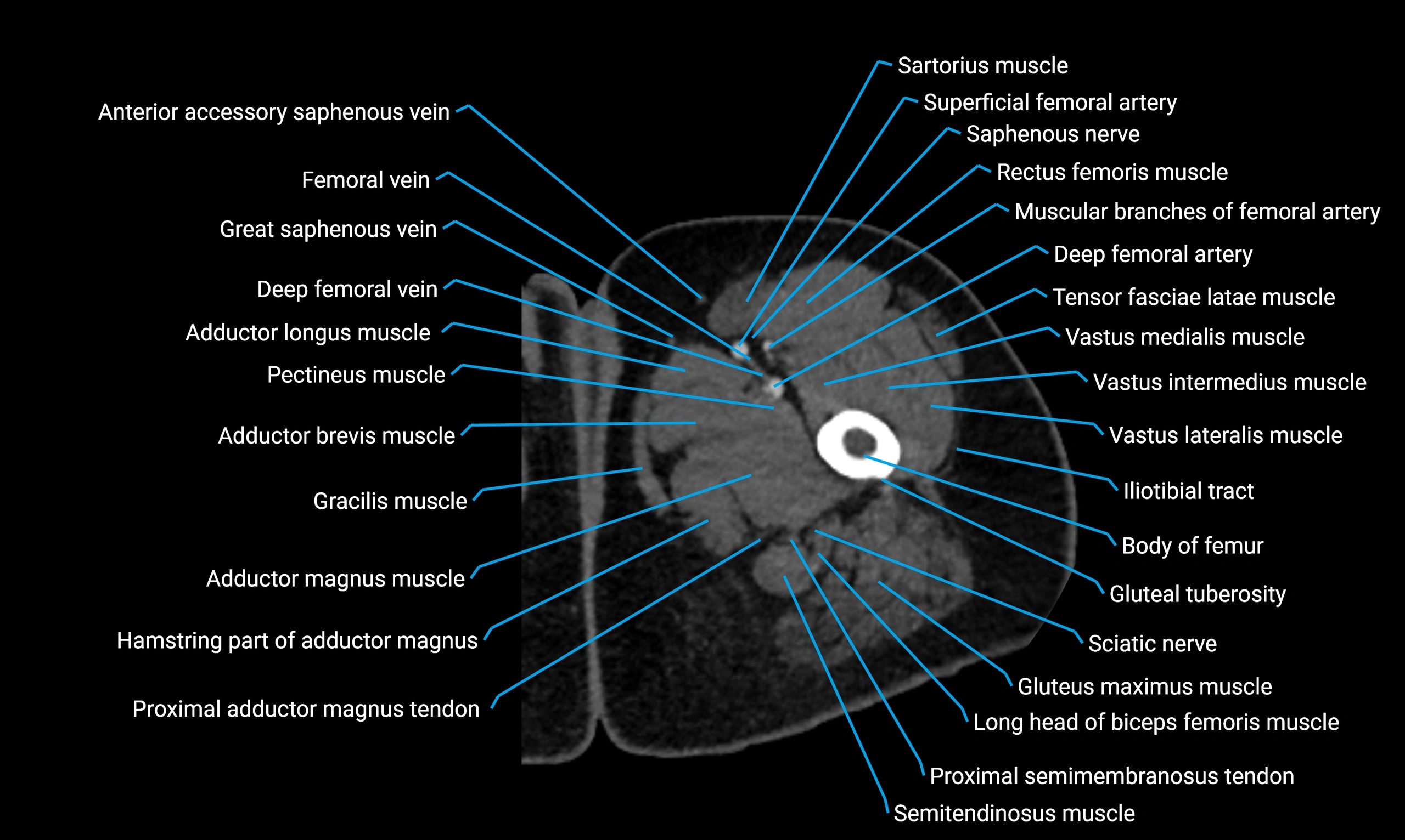
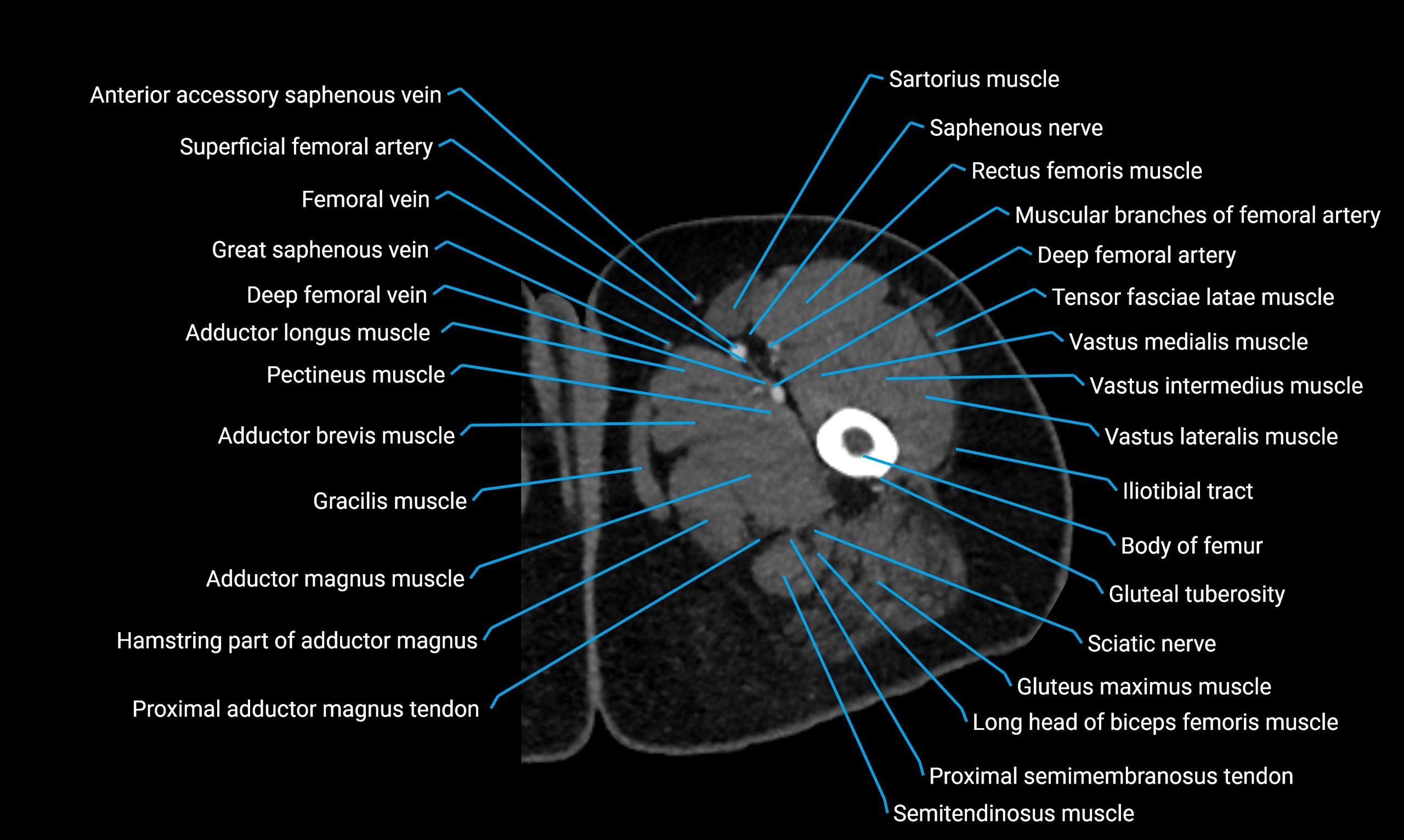
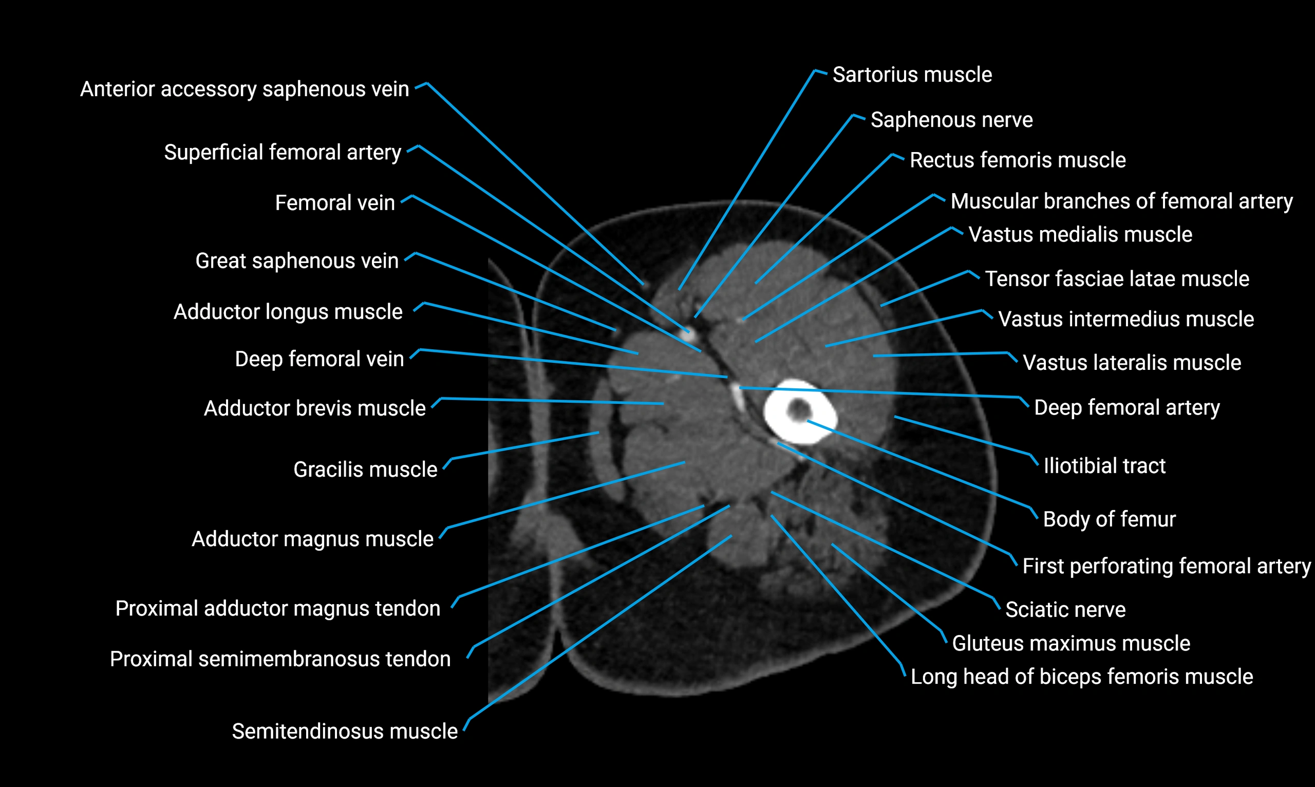
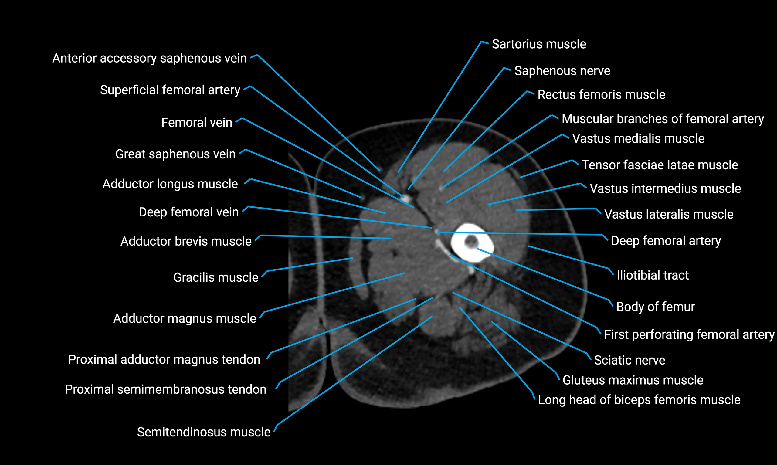
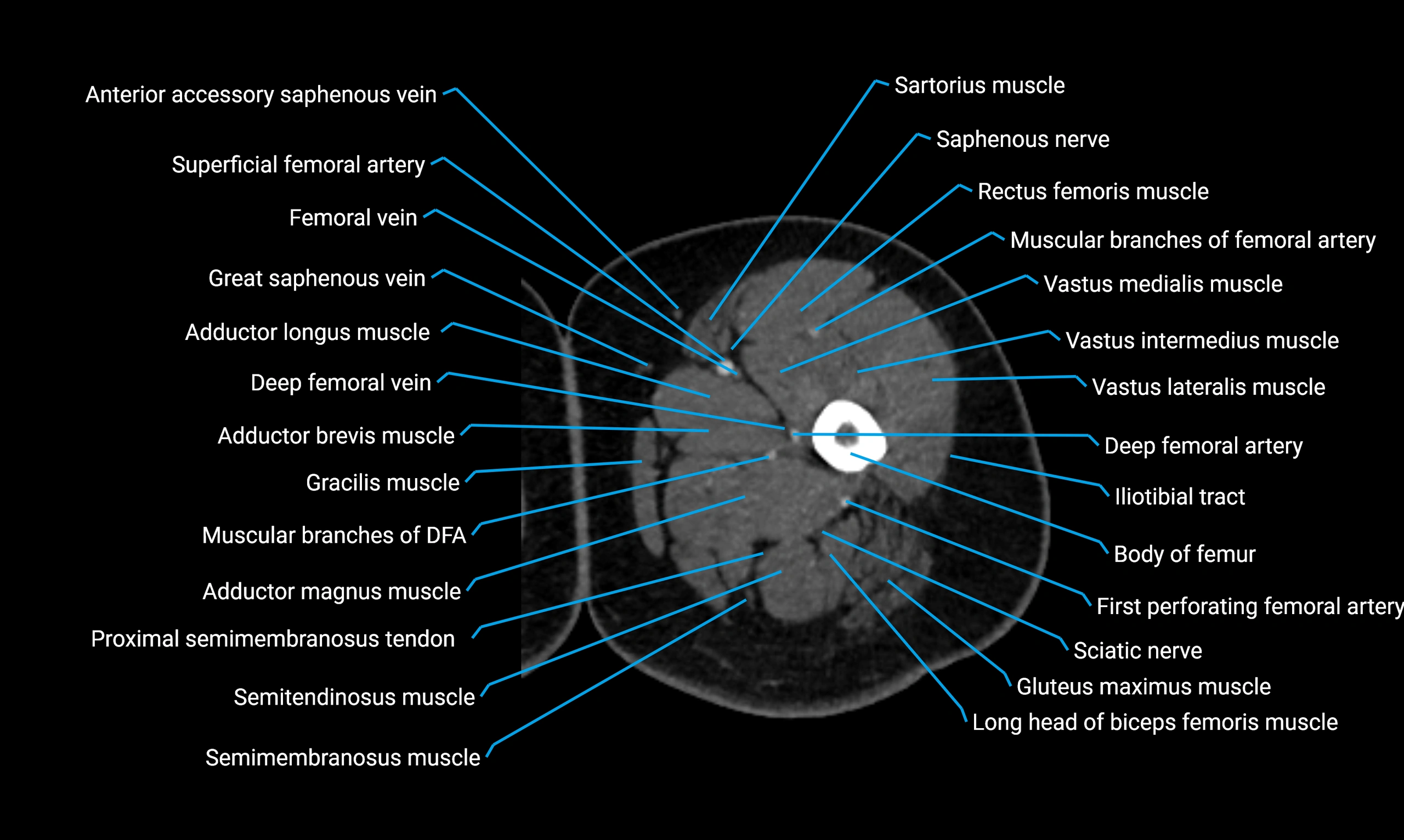
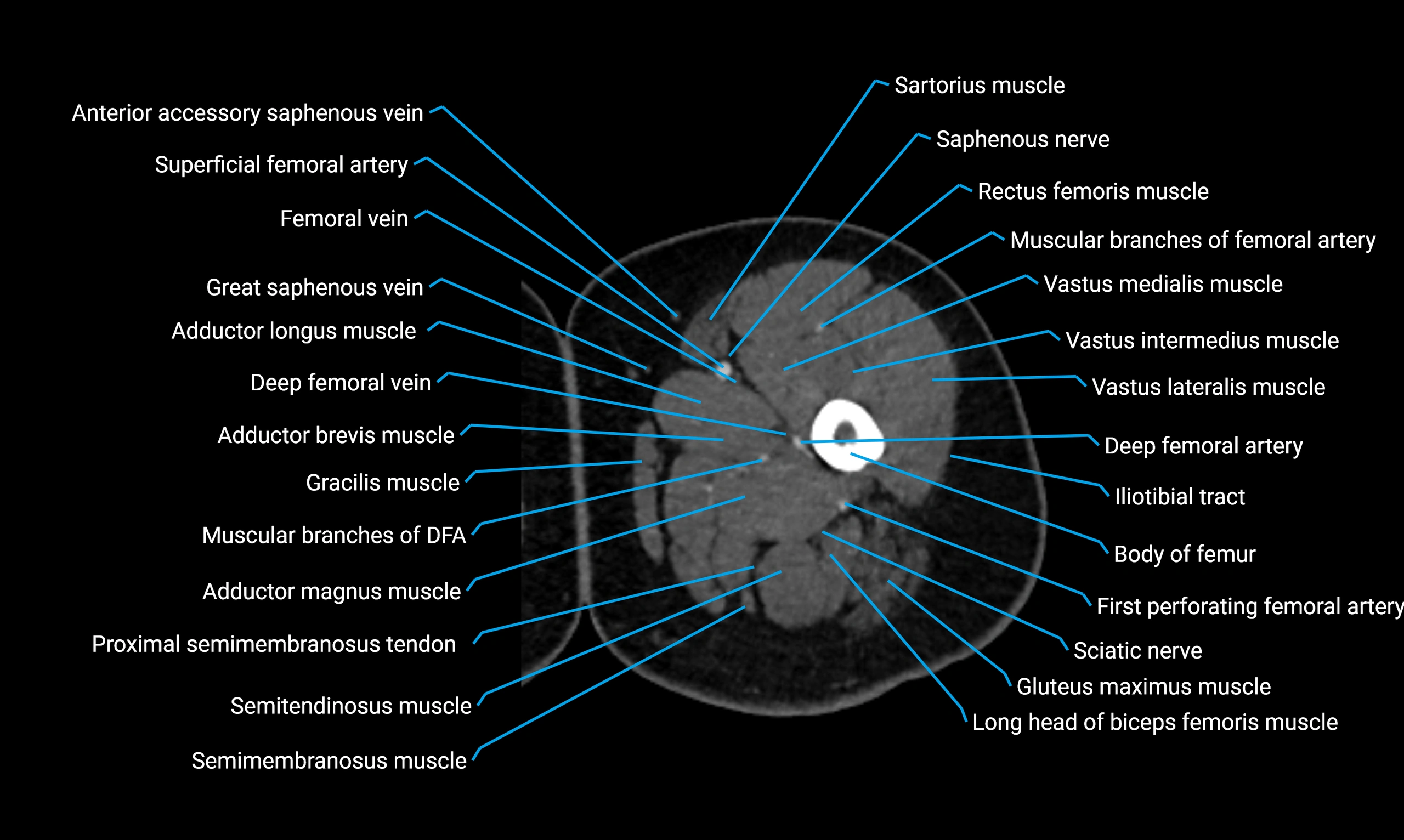
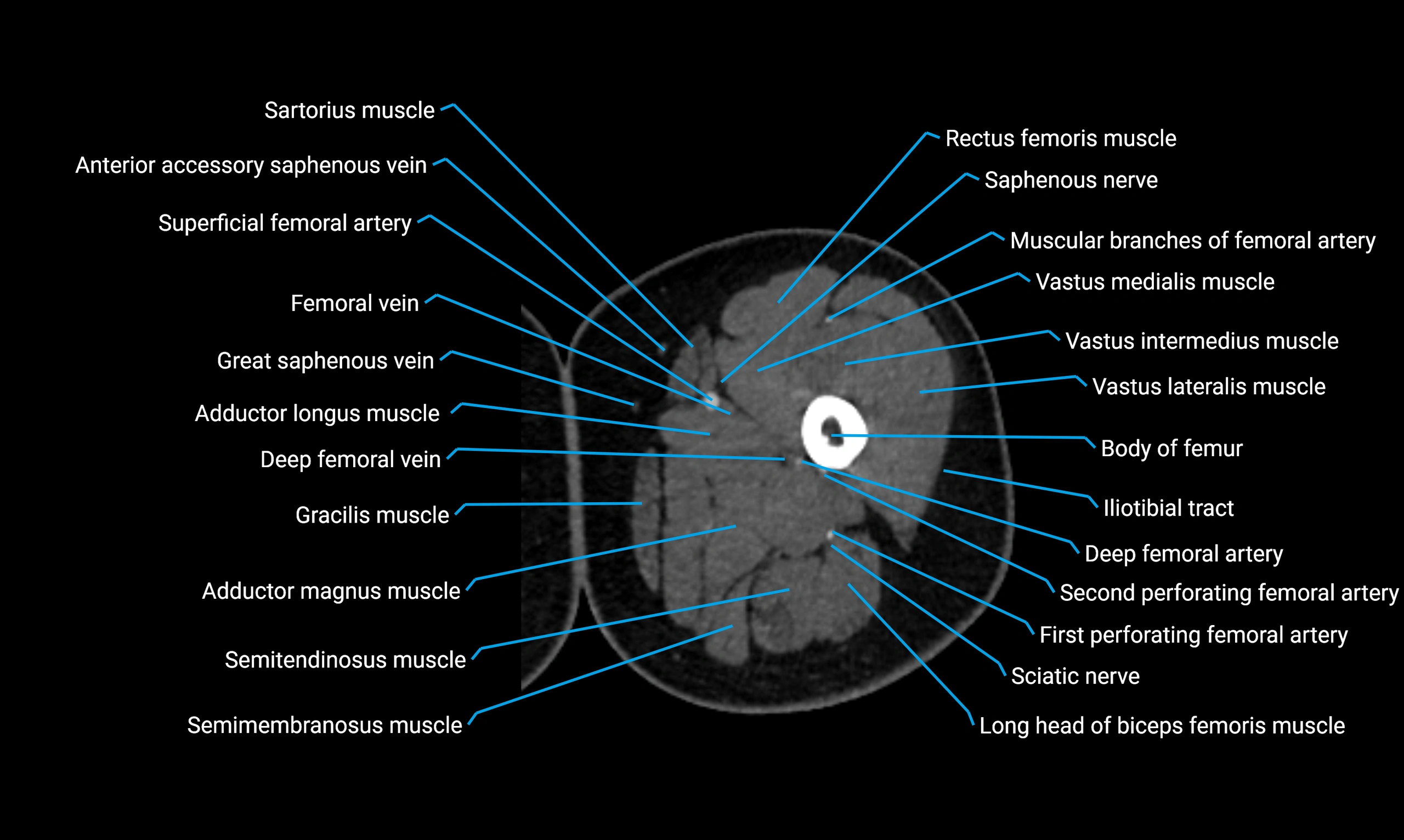
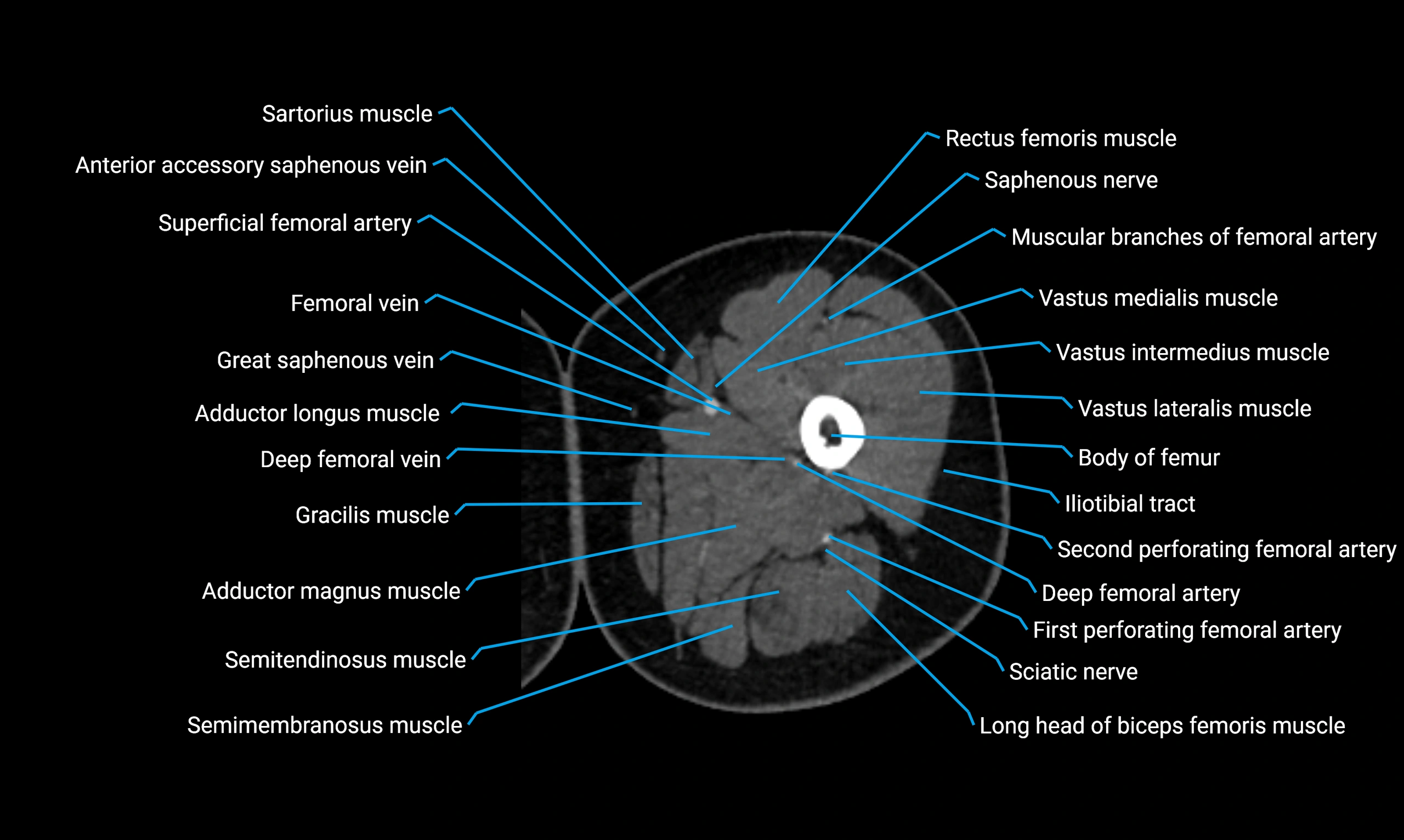
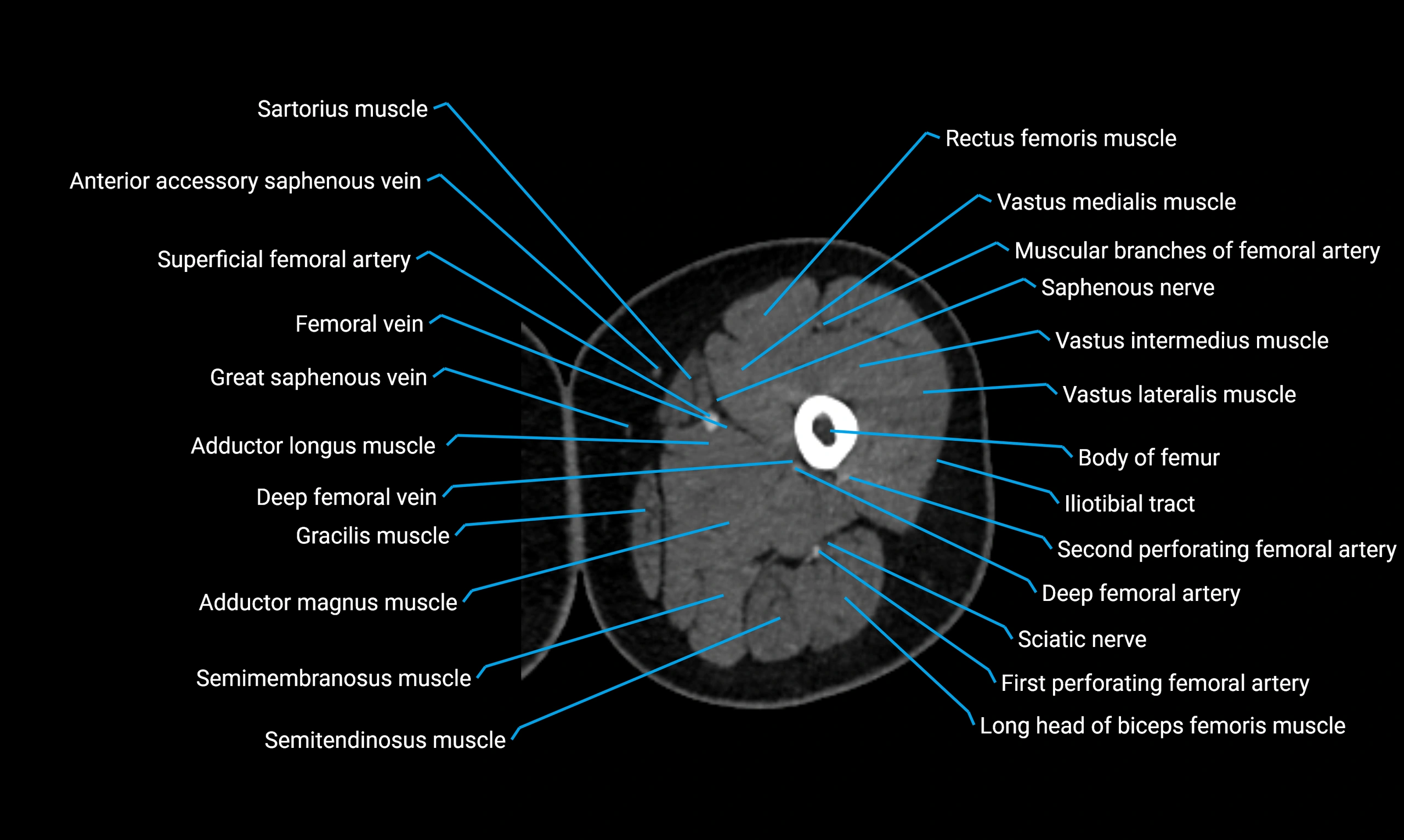
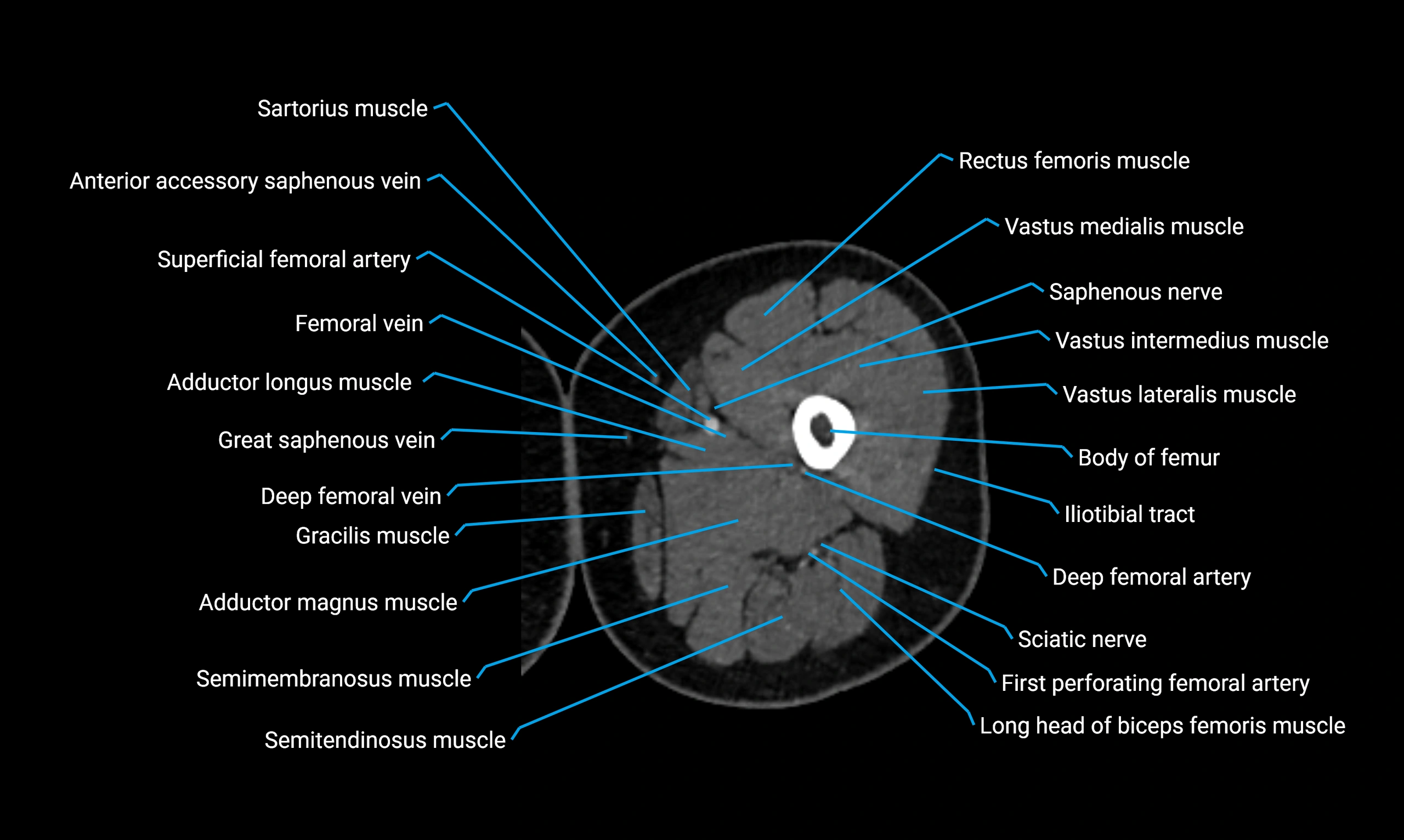
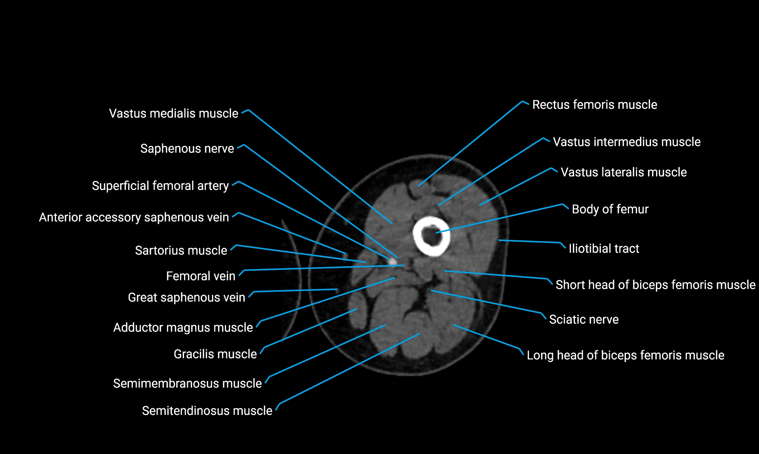
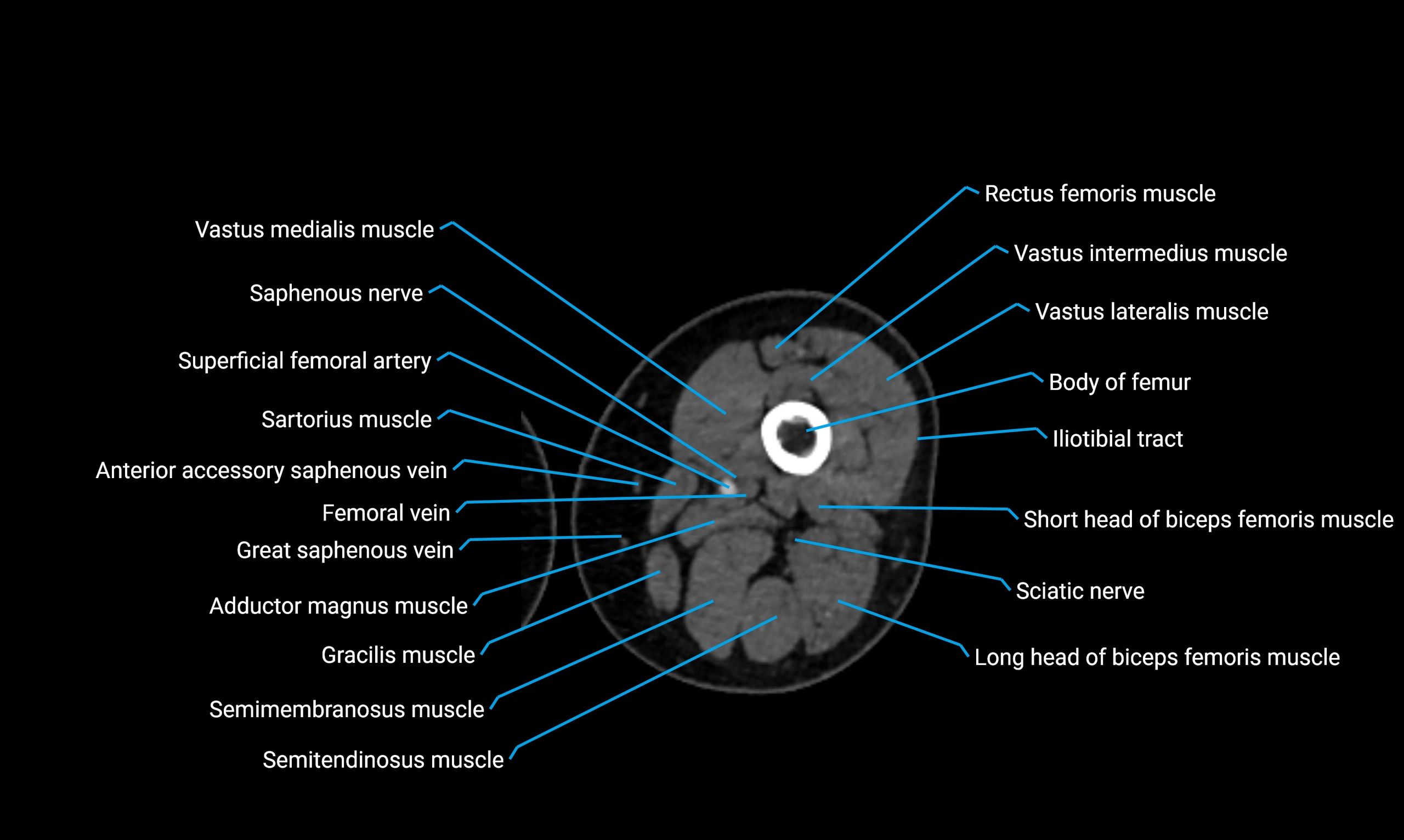
MRI Appearance
T1-weighted images:
-
Bone cortex: hypointense
-
Marrow: intermediate signal
-
Identifies avulsion fragments or marrow infiltration
T2-weighted images:
-
Bone cortex: hypointense
-
Marrow: intermediate to hyperintense if edema is present
-
Useful for detecting stress changes, avulsions, or cortical irregularity
PD Fat-Saturated (Proton Density FS):
-
Bone cortex: hypointense
-
Marrow edema, tendon pathology, or avulsions: hyperintense
-
Very sensitive for rectus femoris origin injuries
STIR:
-
Highlights acute avulsion injuries, bone marrow edema, or enthesitis
-
Suppresses fat for clear visualization of inflammatory changes
T1 Post-Gadolinium (with fat saturation):
-
Bone cortex: remains hypointense
-
Inflammation, tumors, or infection: abnormal enhancement
-
Useful for evaluating enthesitis, neoplasms, or post-surgical changes
3D T2-weighted Imaging:
-
Bone cortex: sharply hypointense
-
Provides multiplanar reconstruction of AIIS morphology
-
Essential in FAI evaluation and preoperative planning for hip surgery
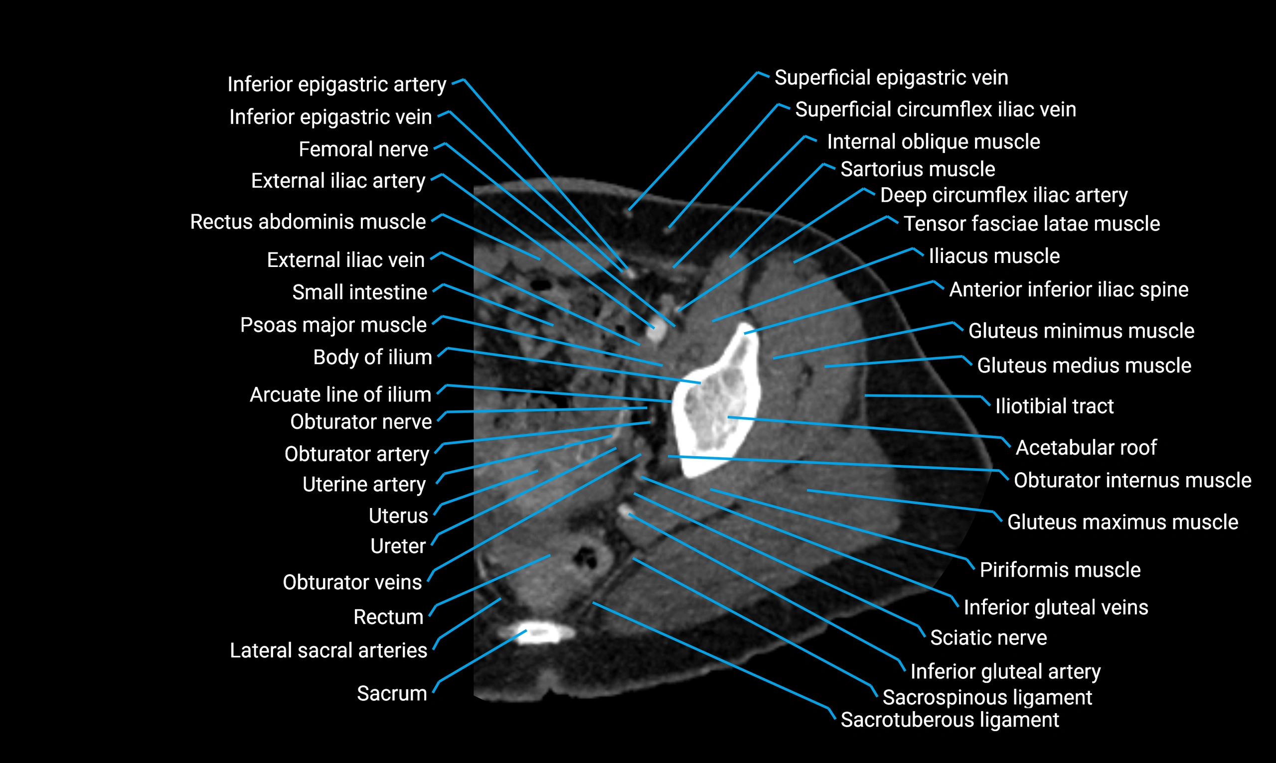
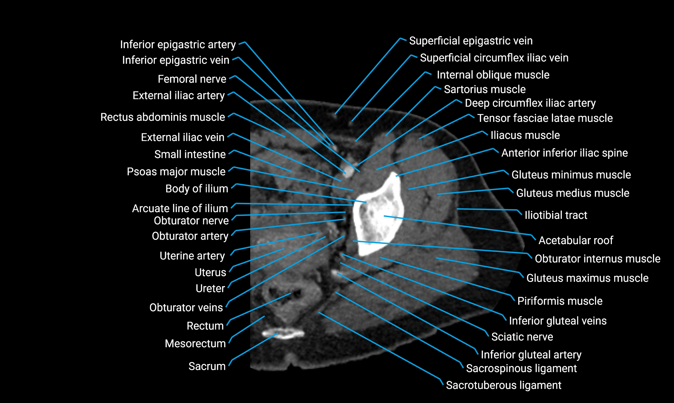
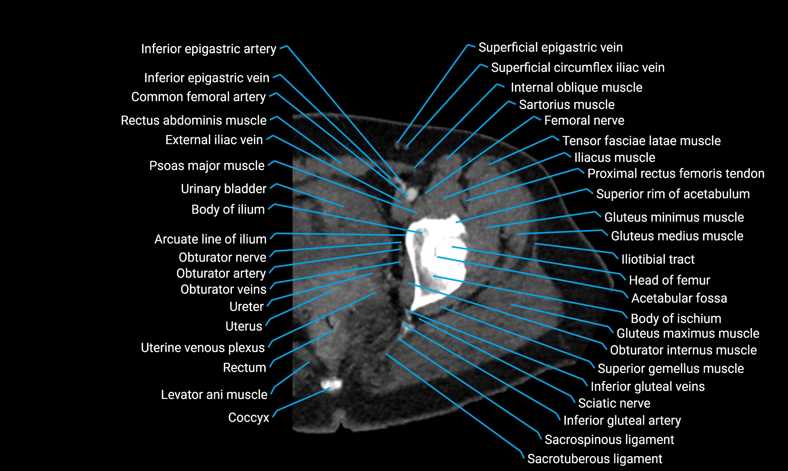
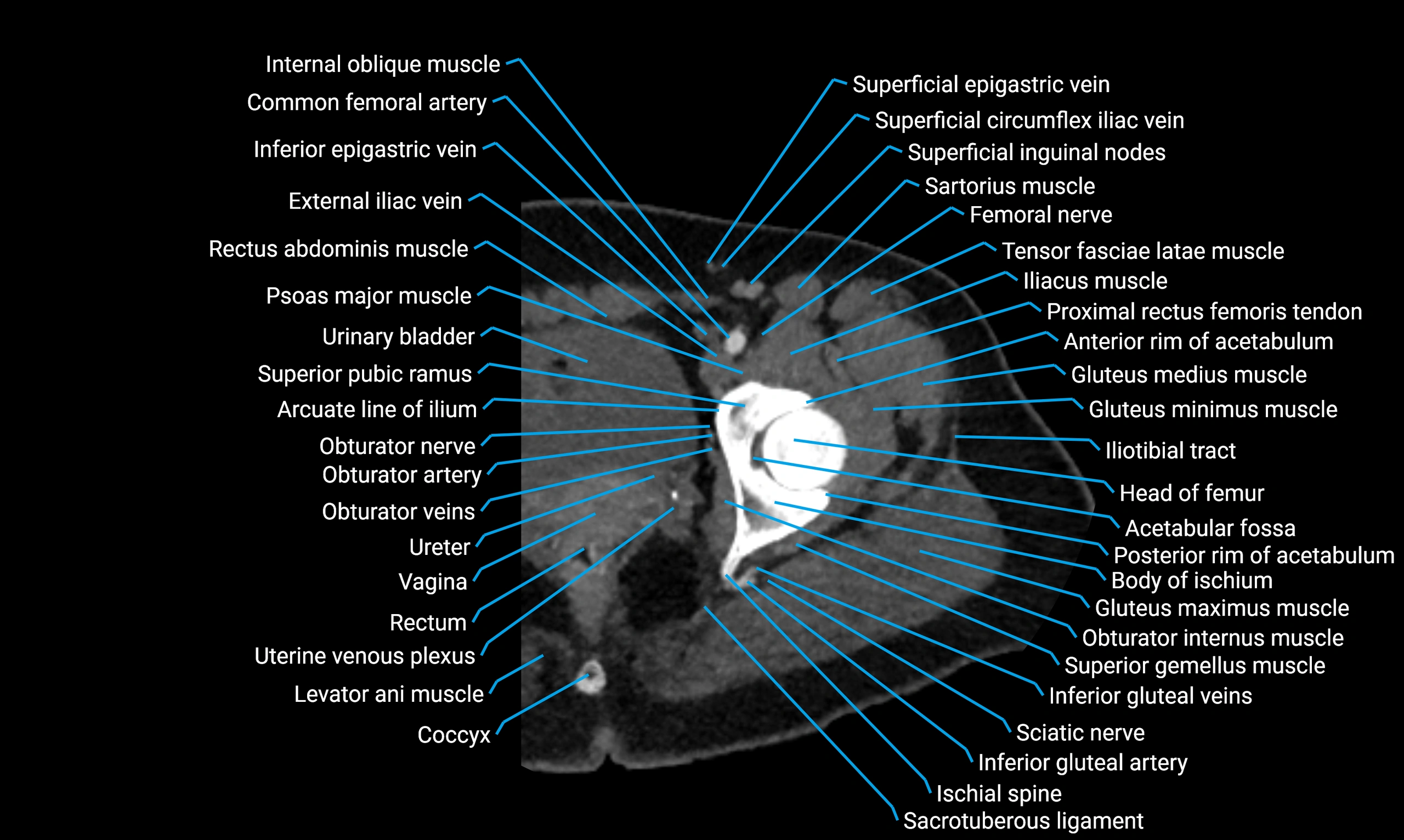
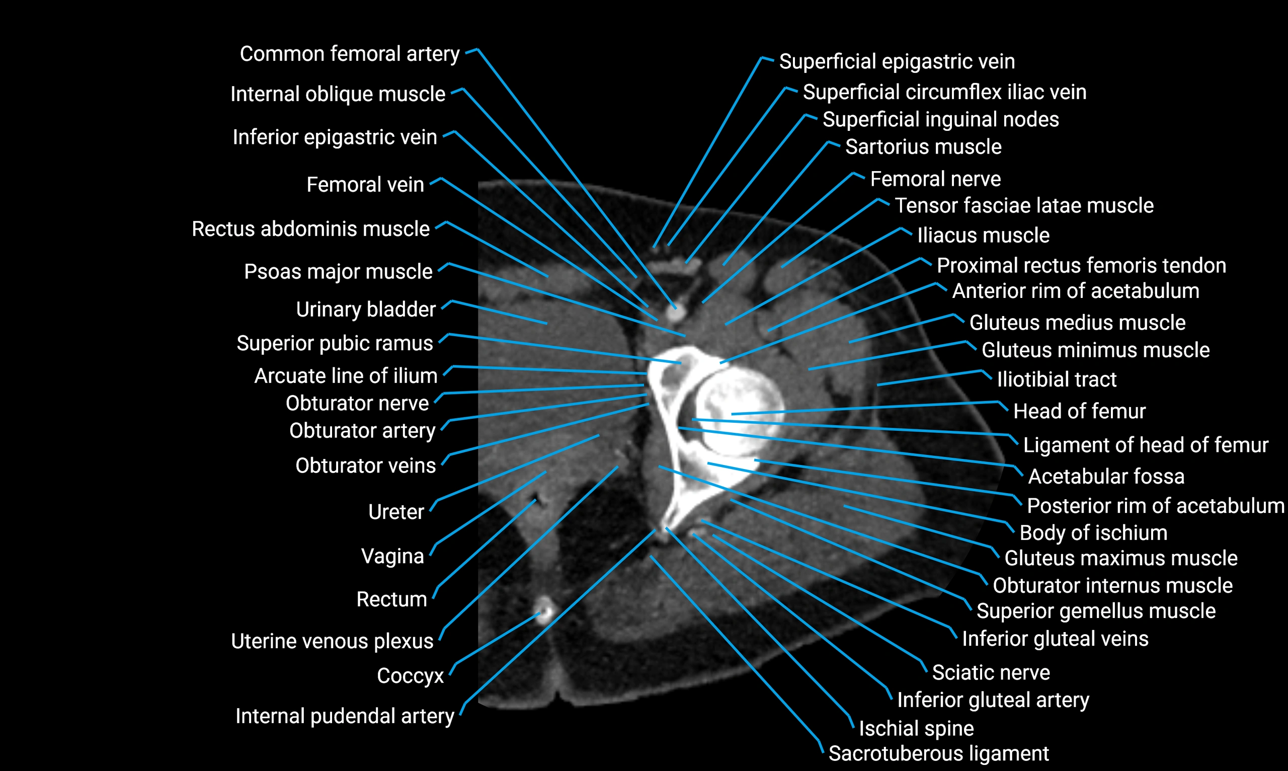
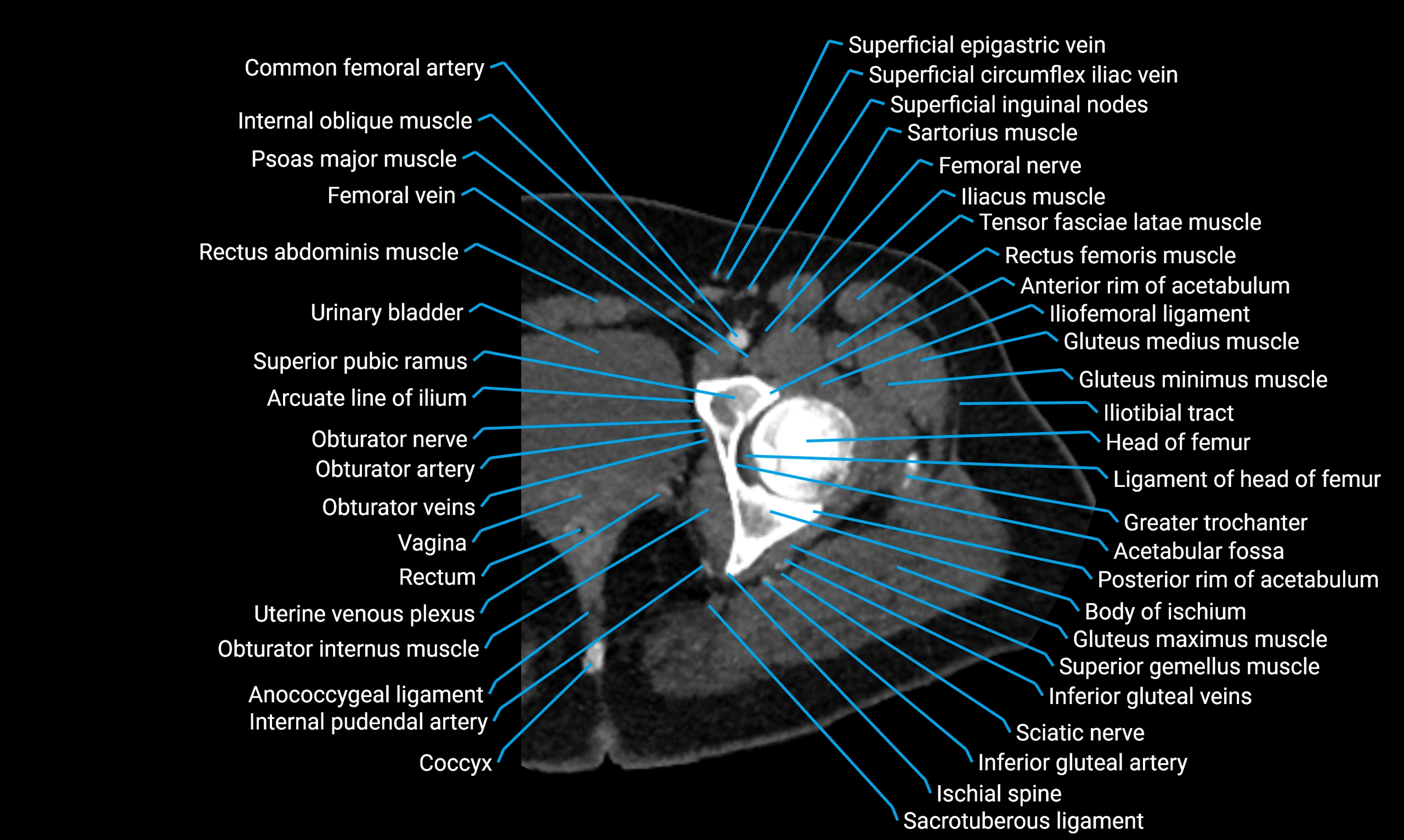
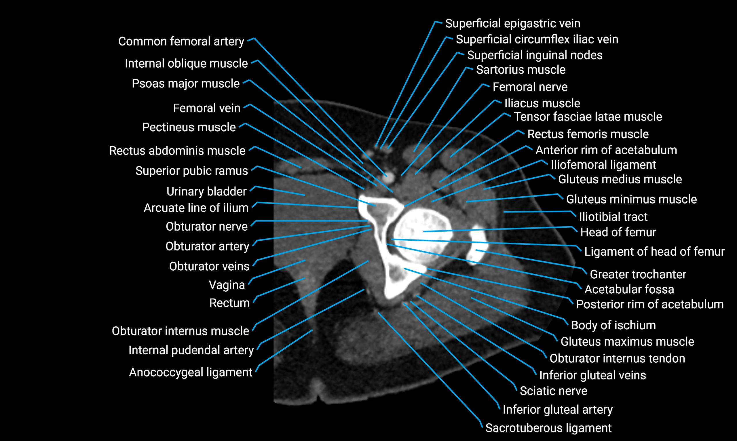
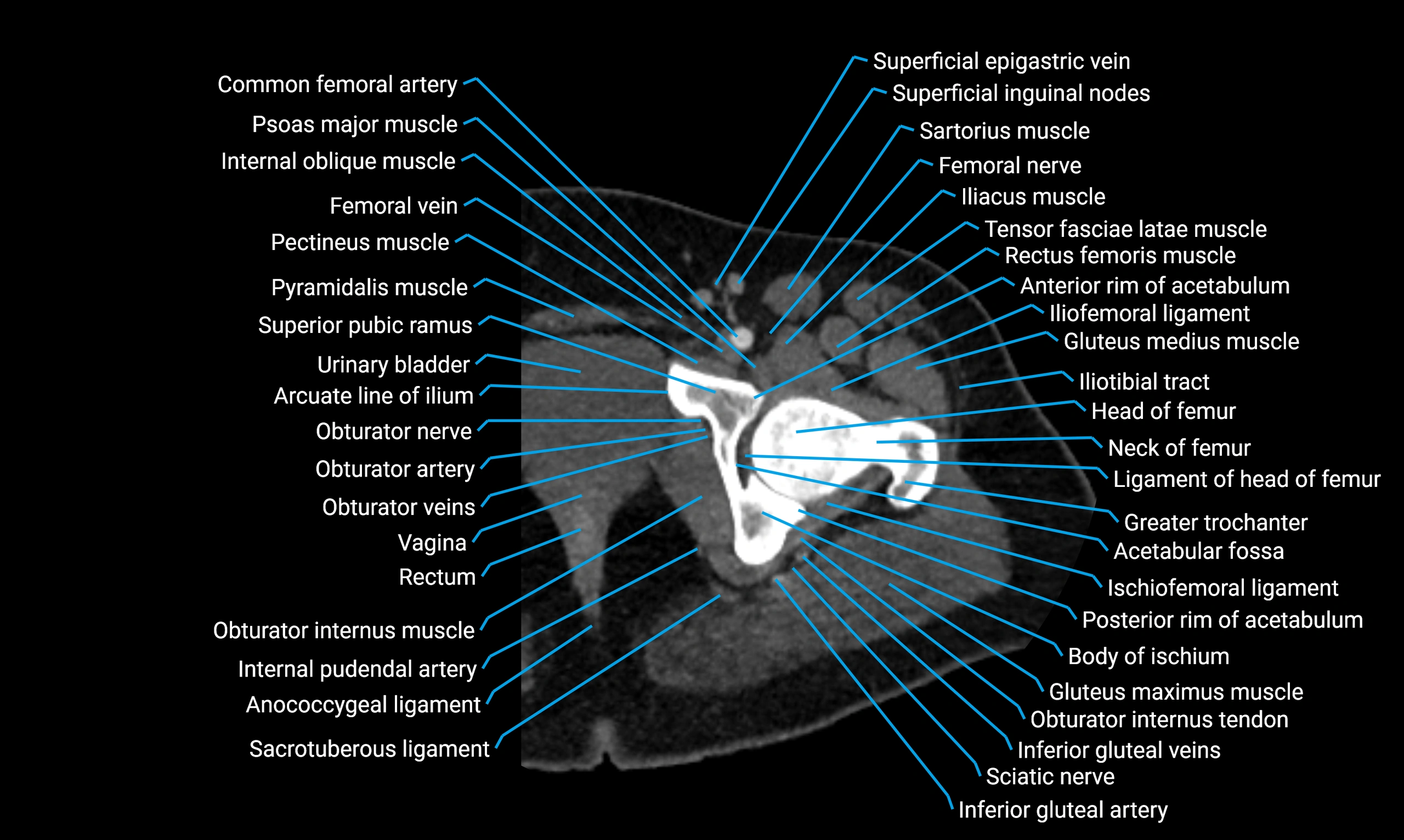
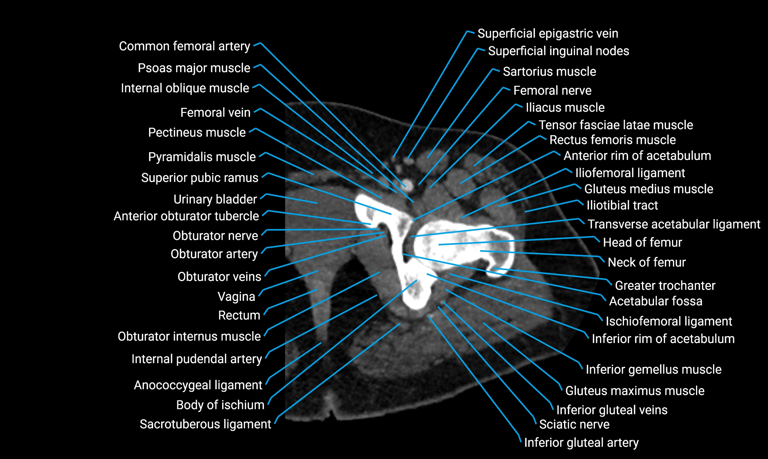
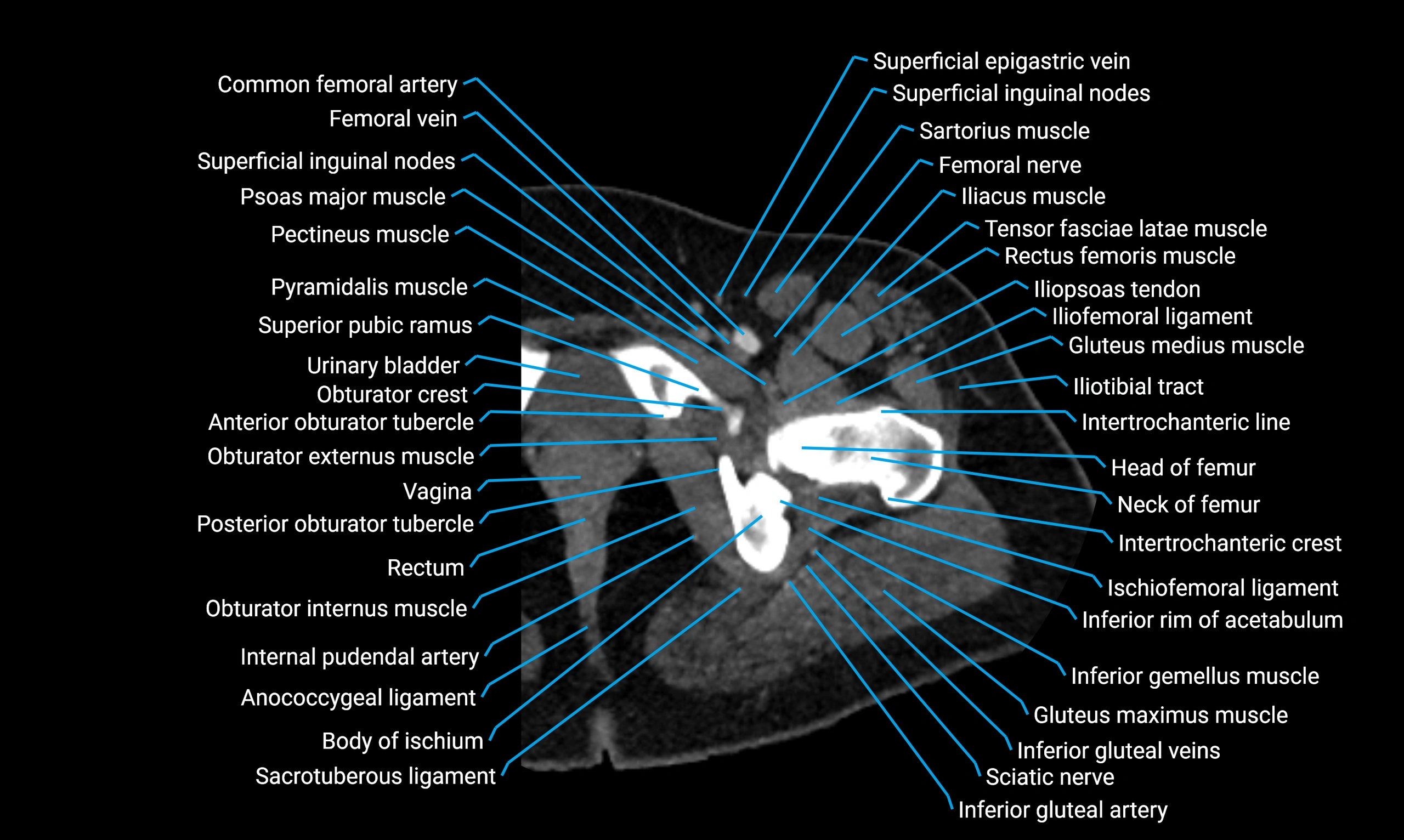
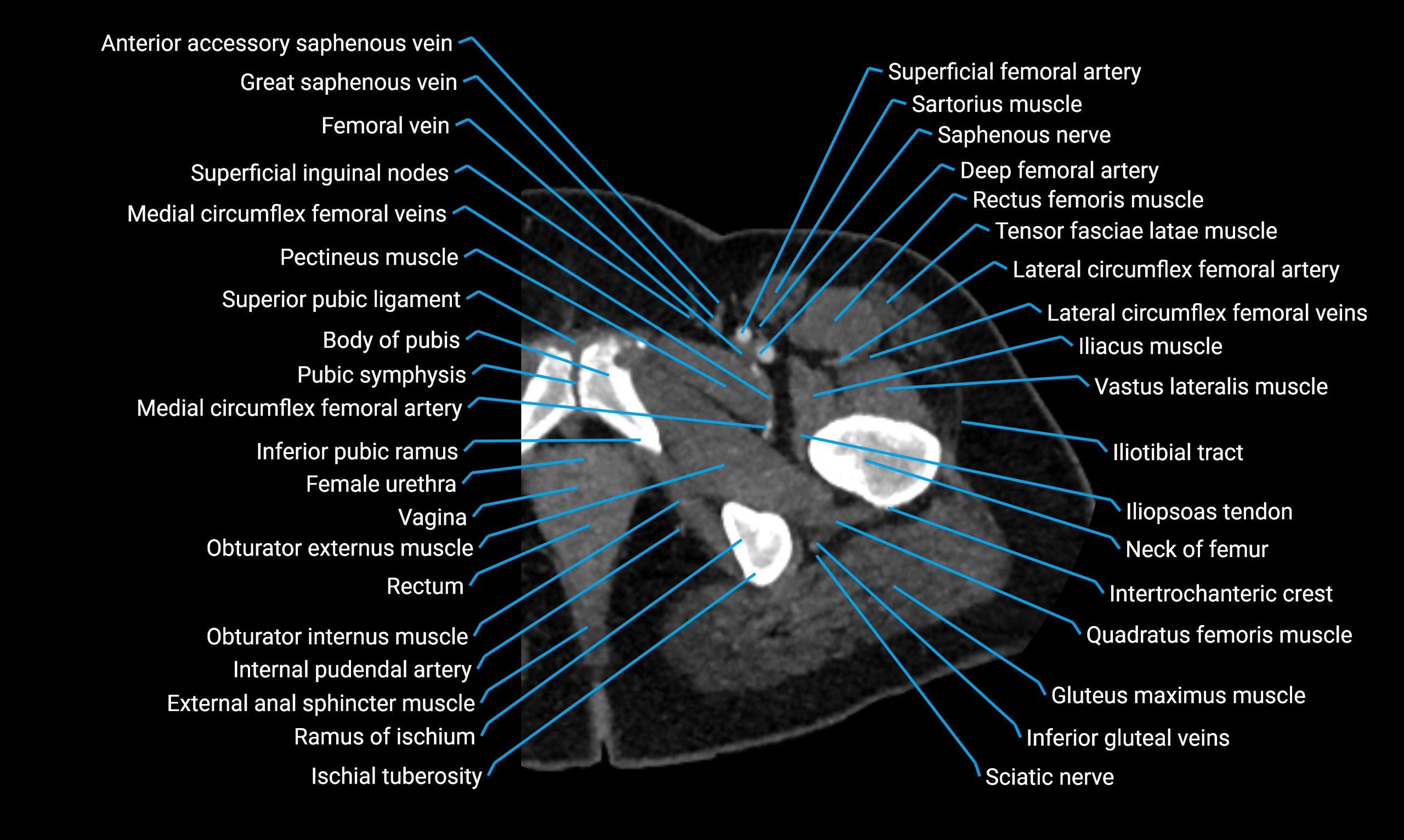
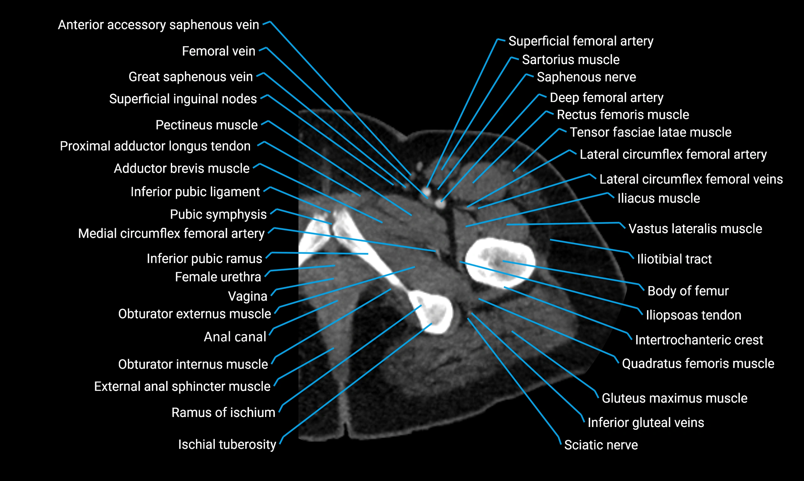
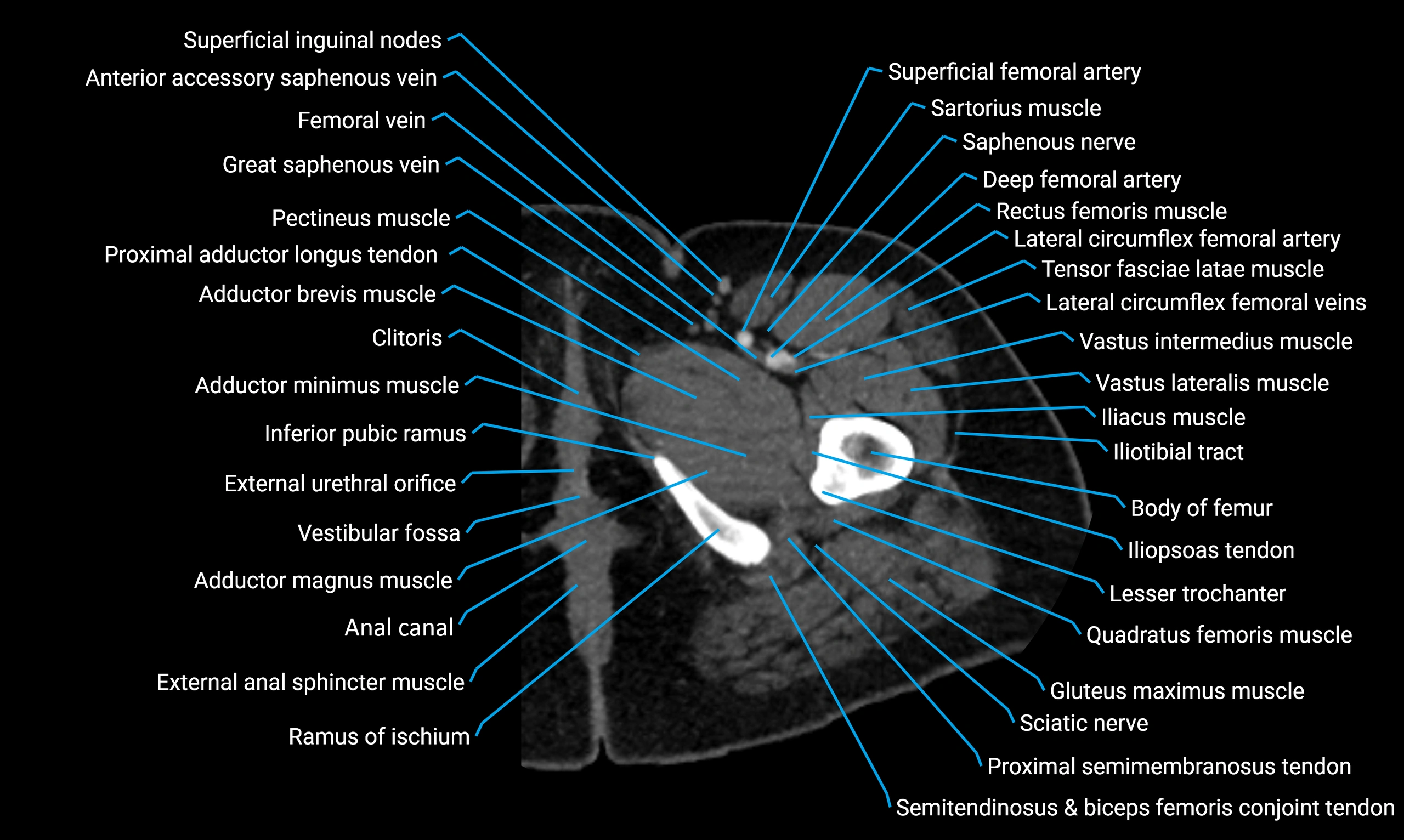
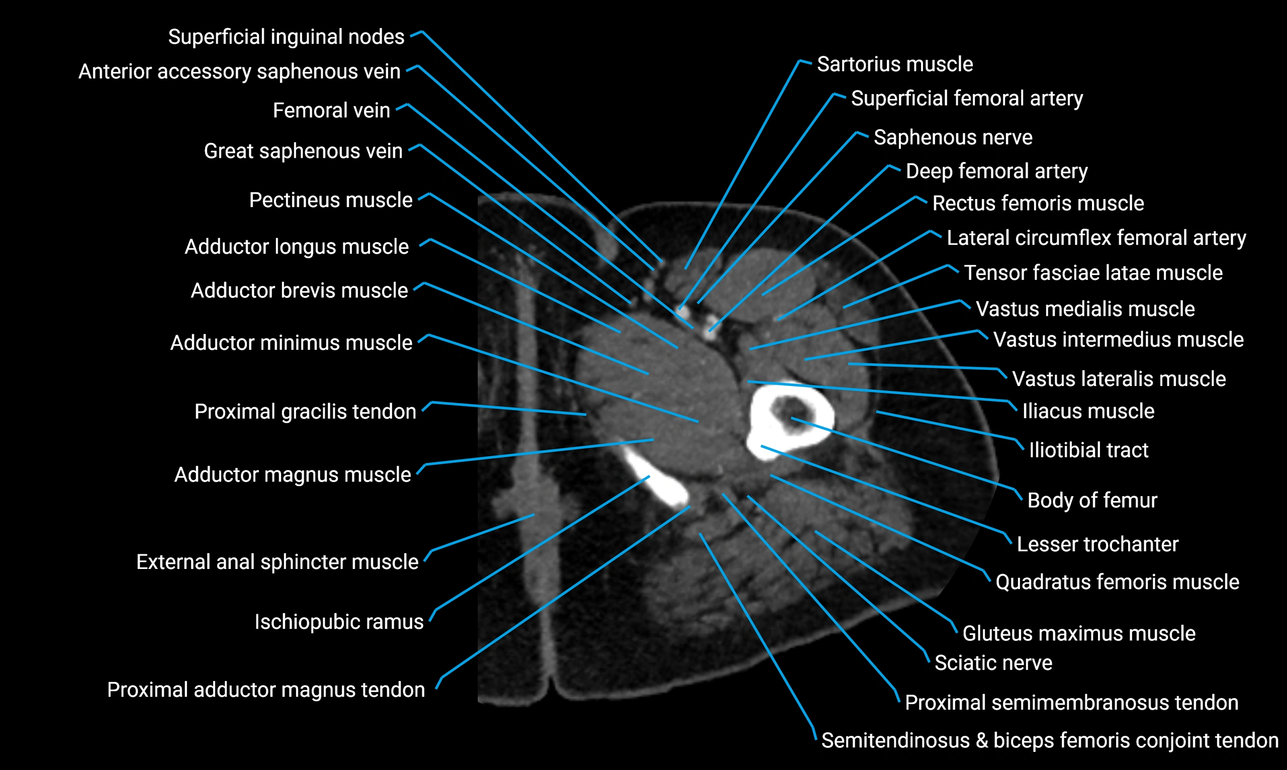
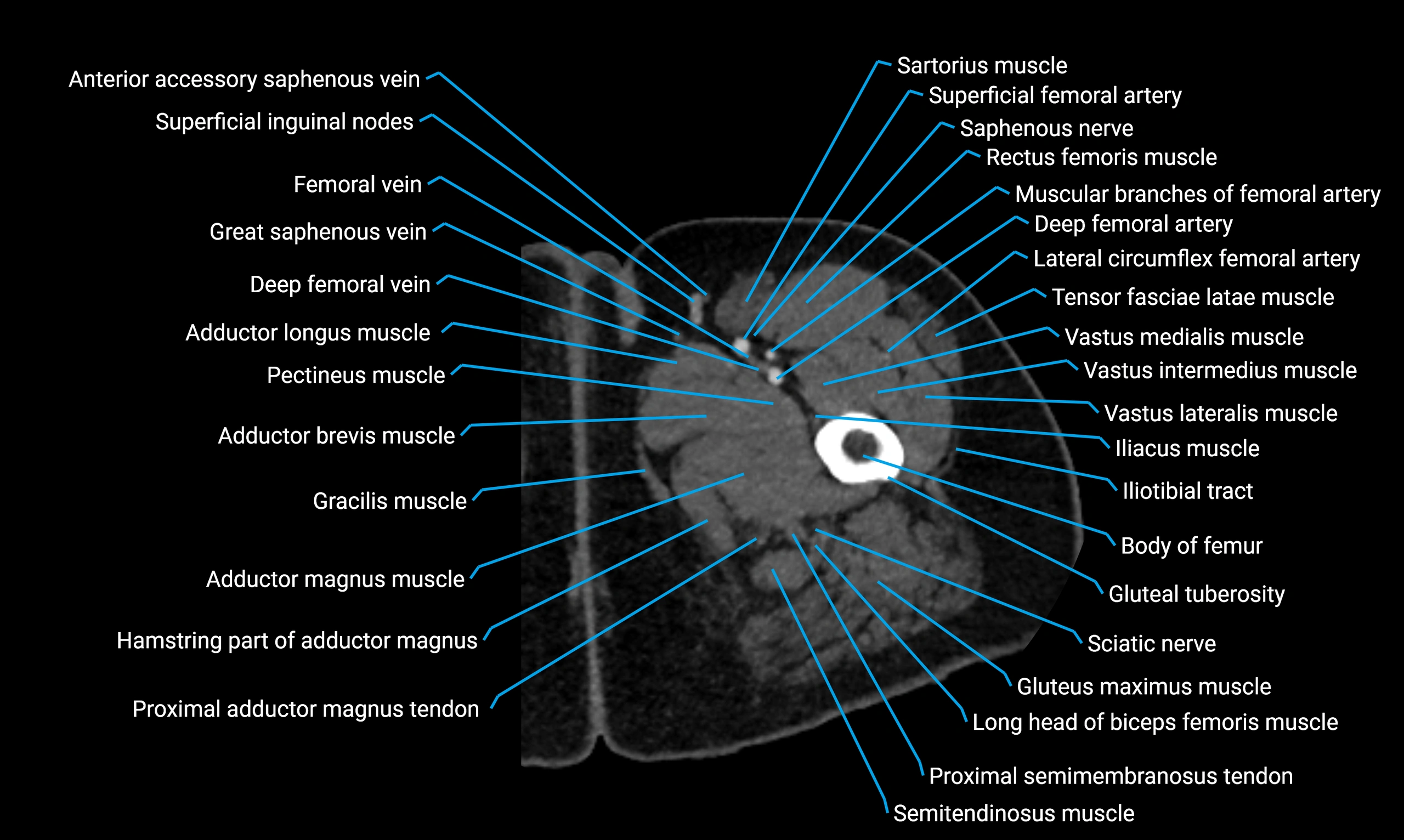
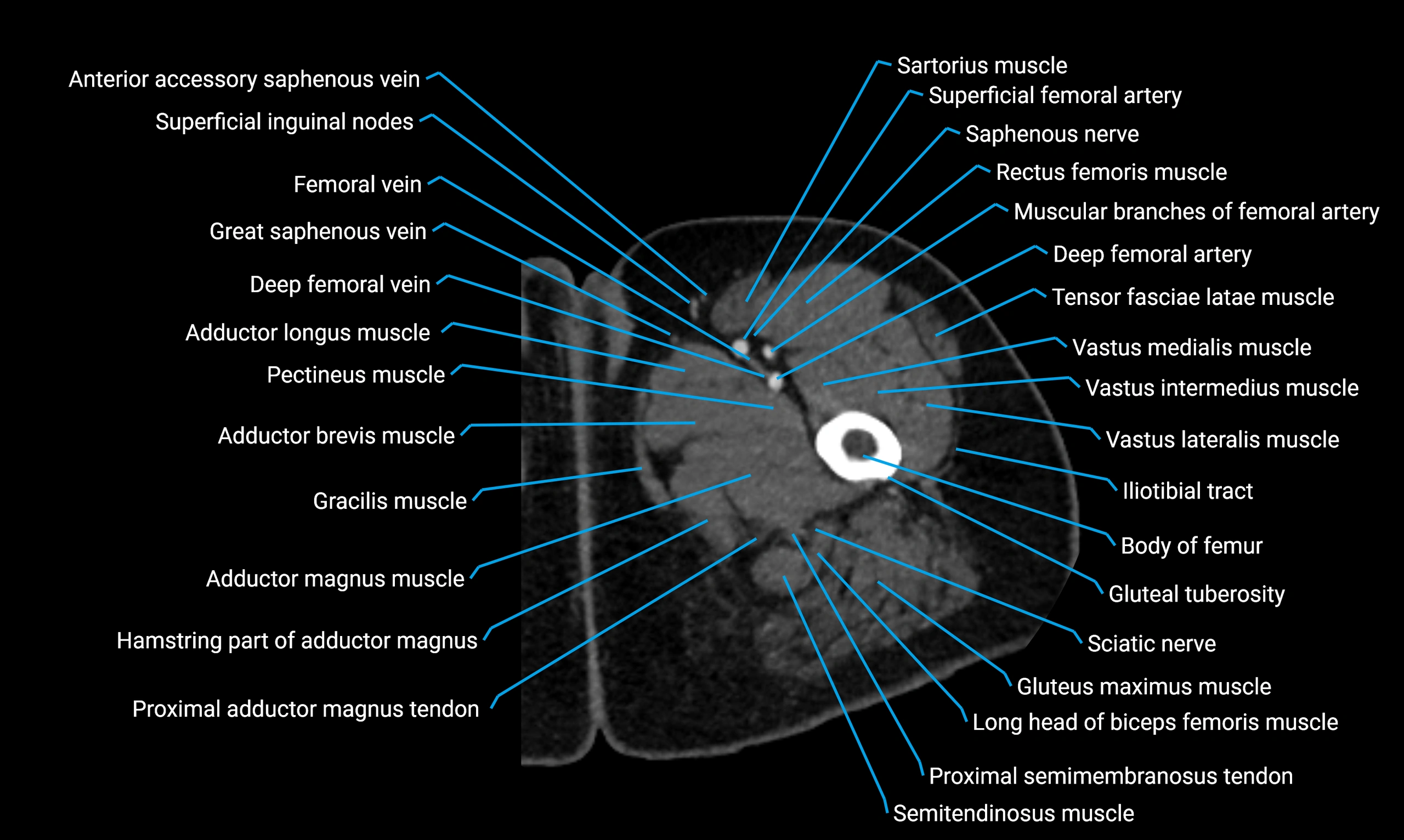
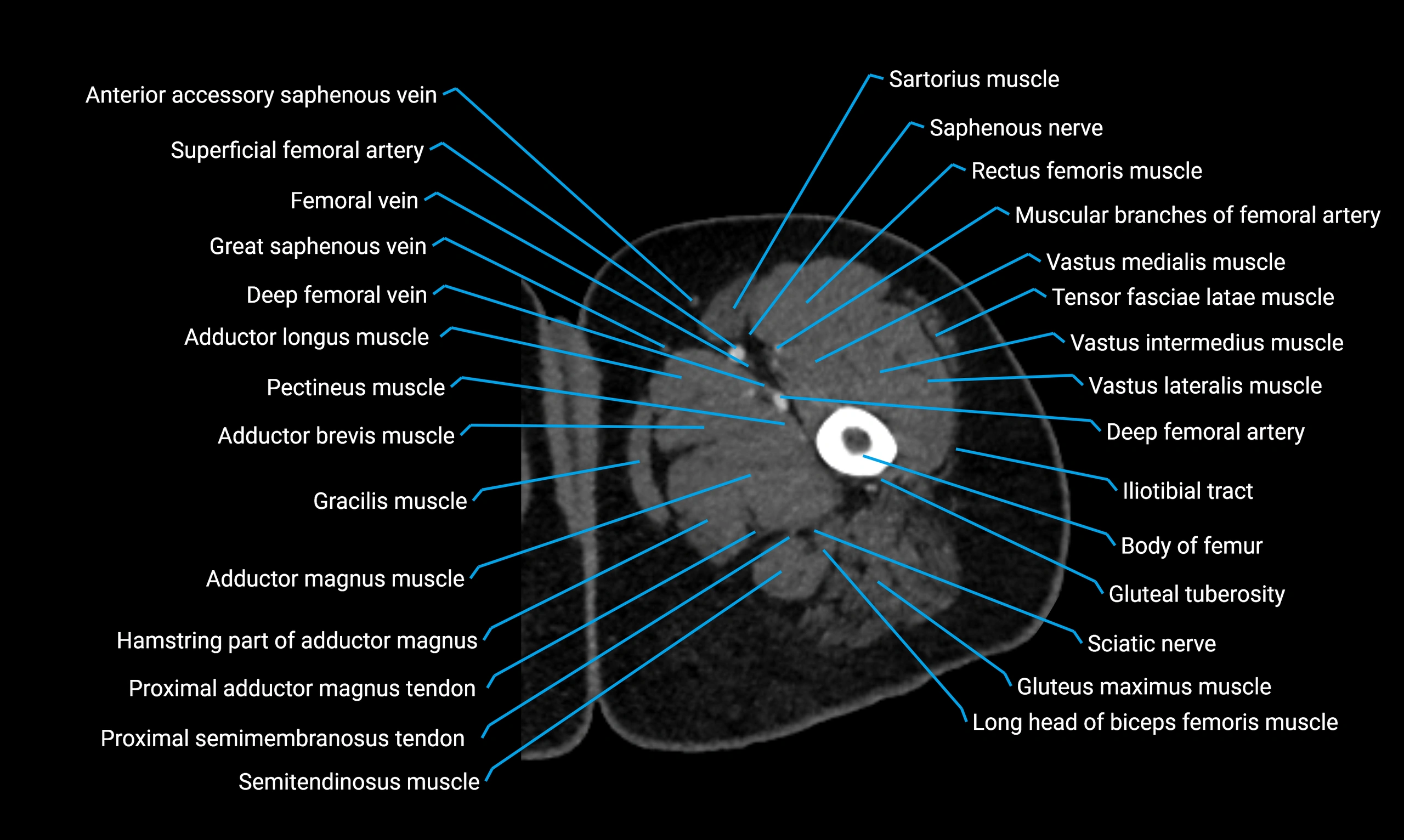
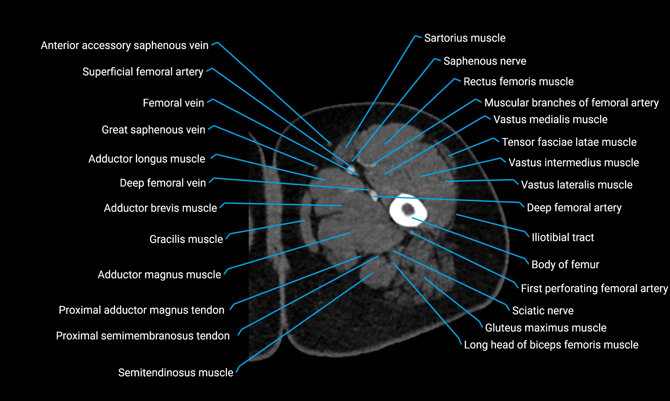
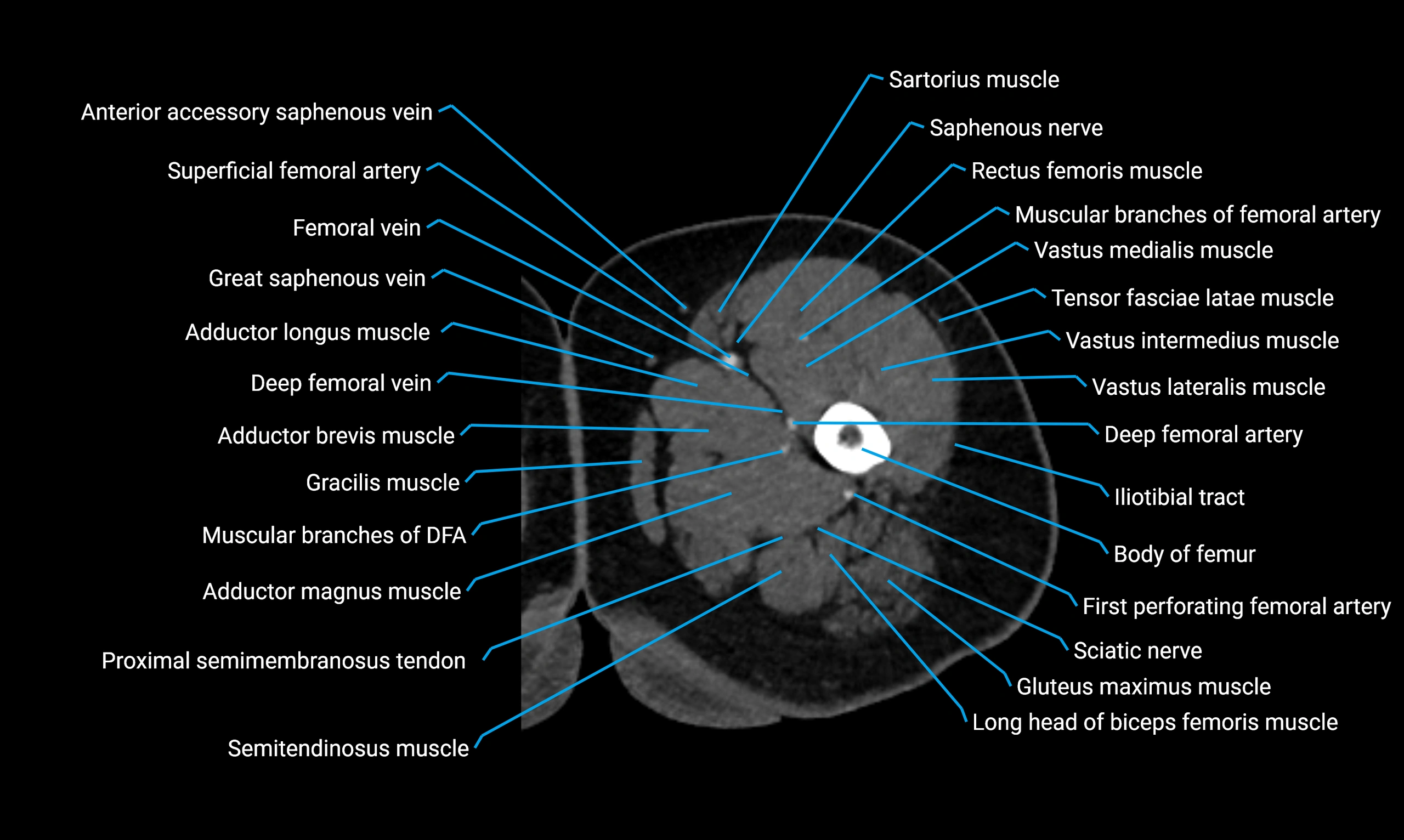
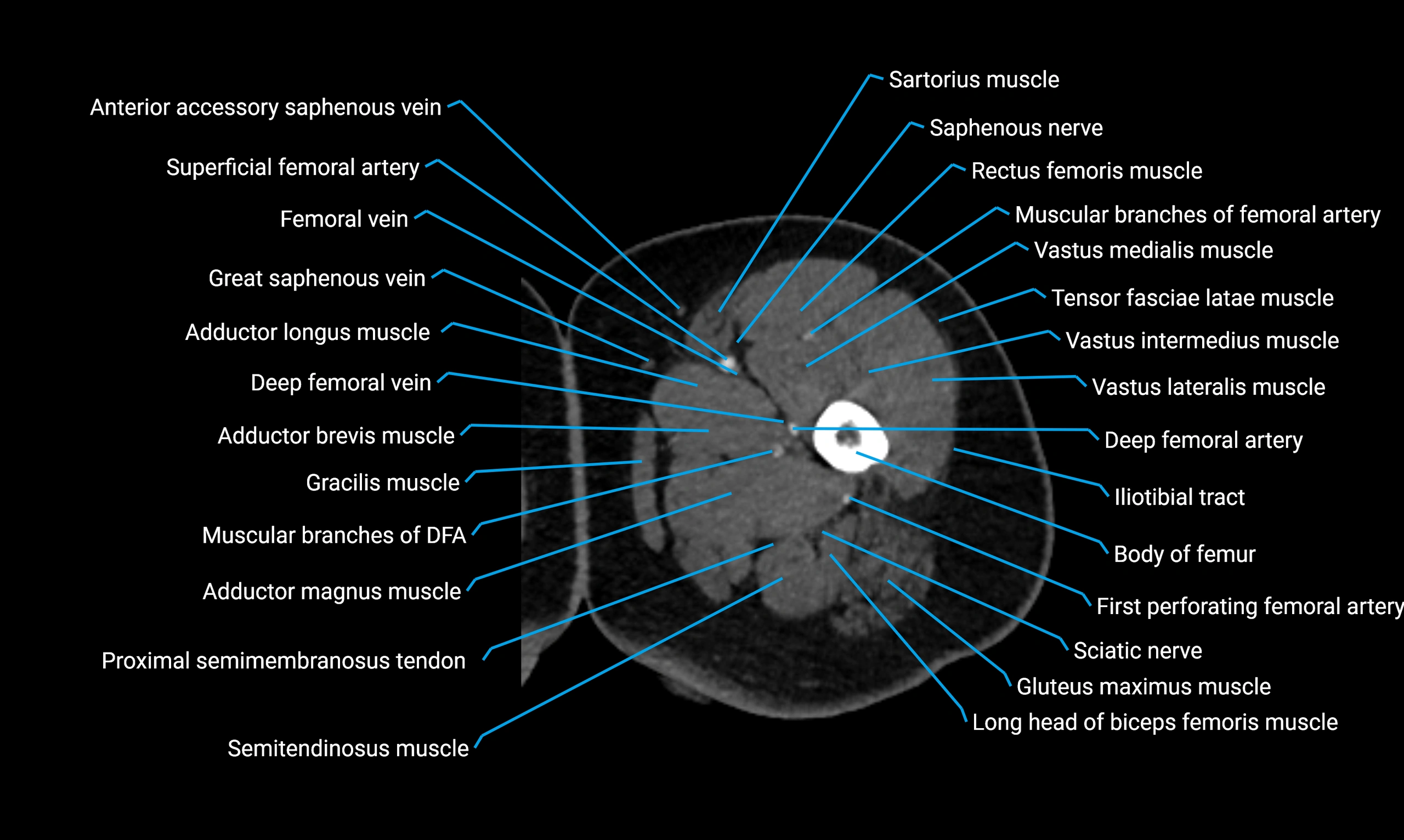
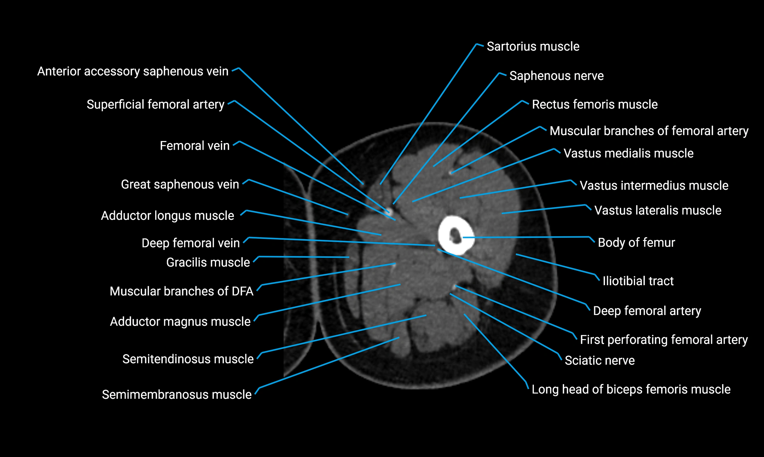
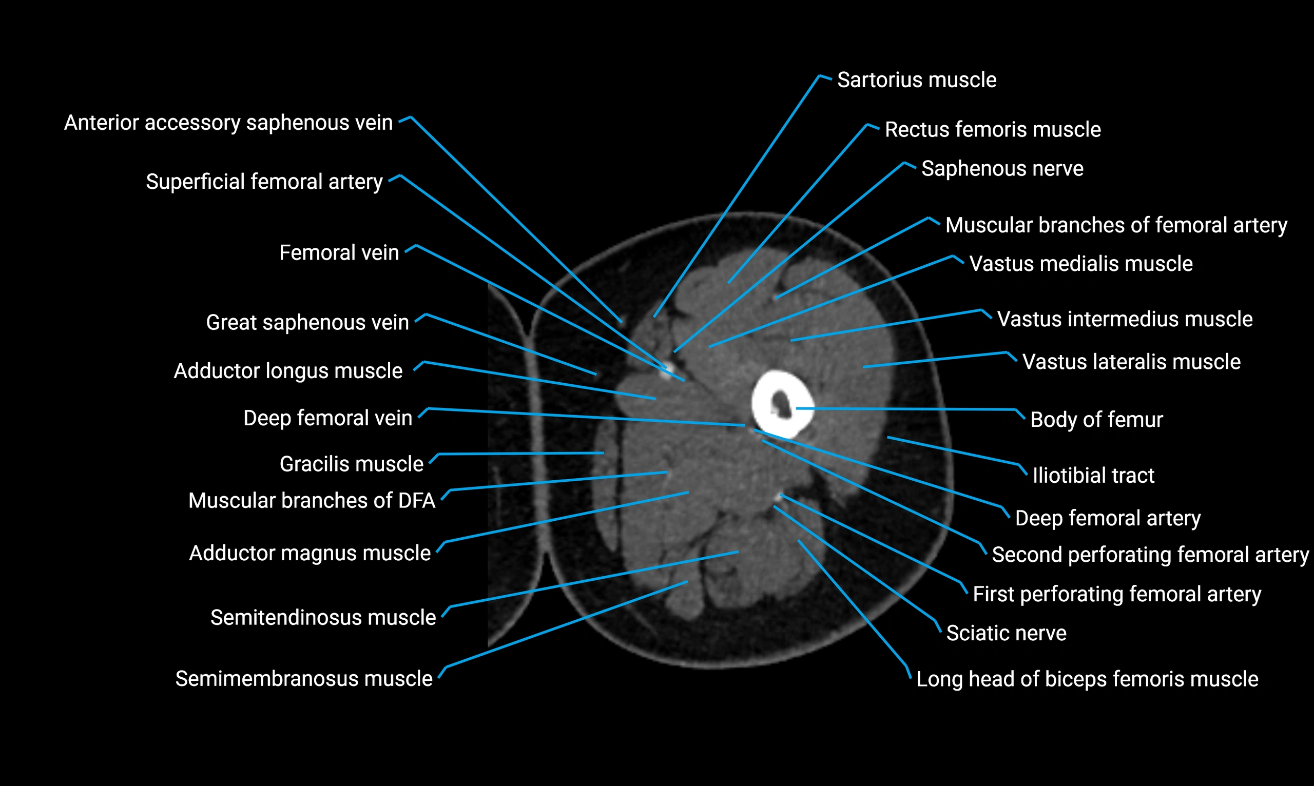
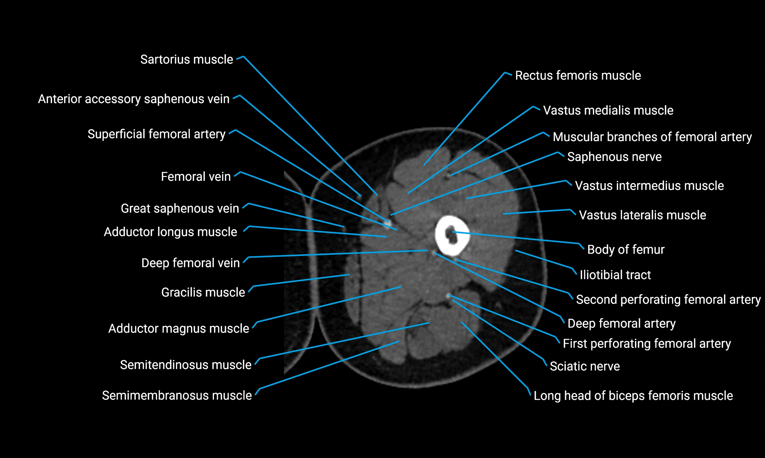
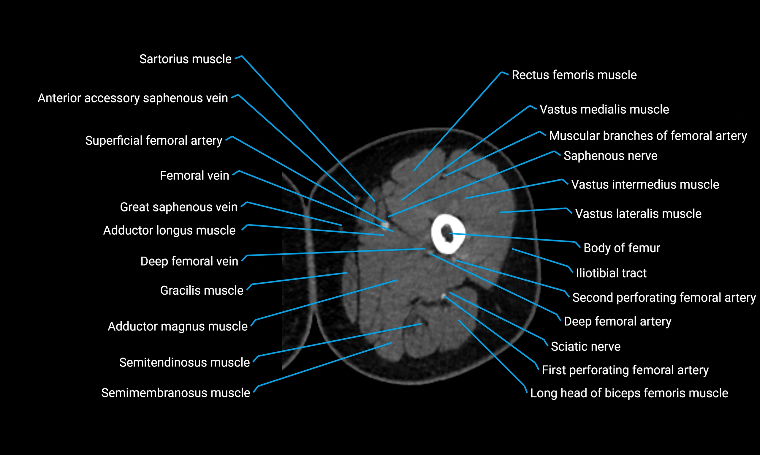
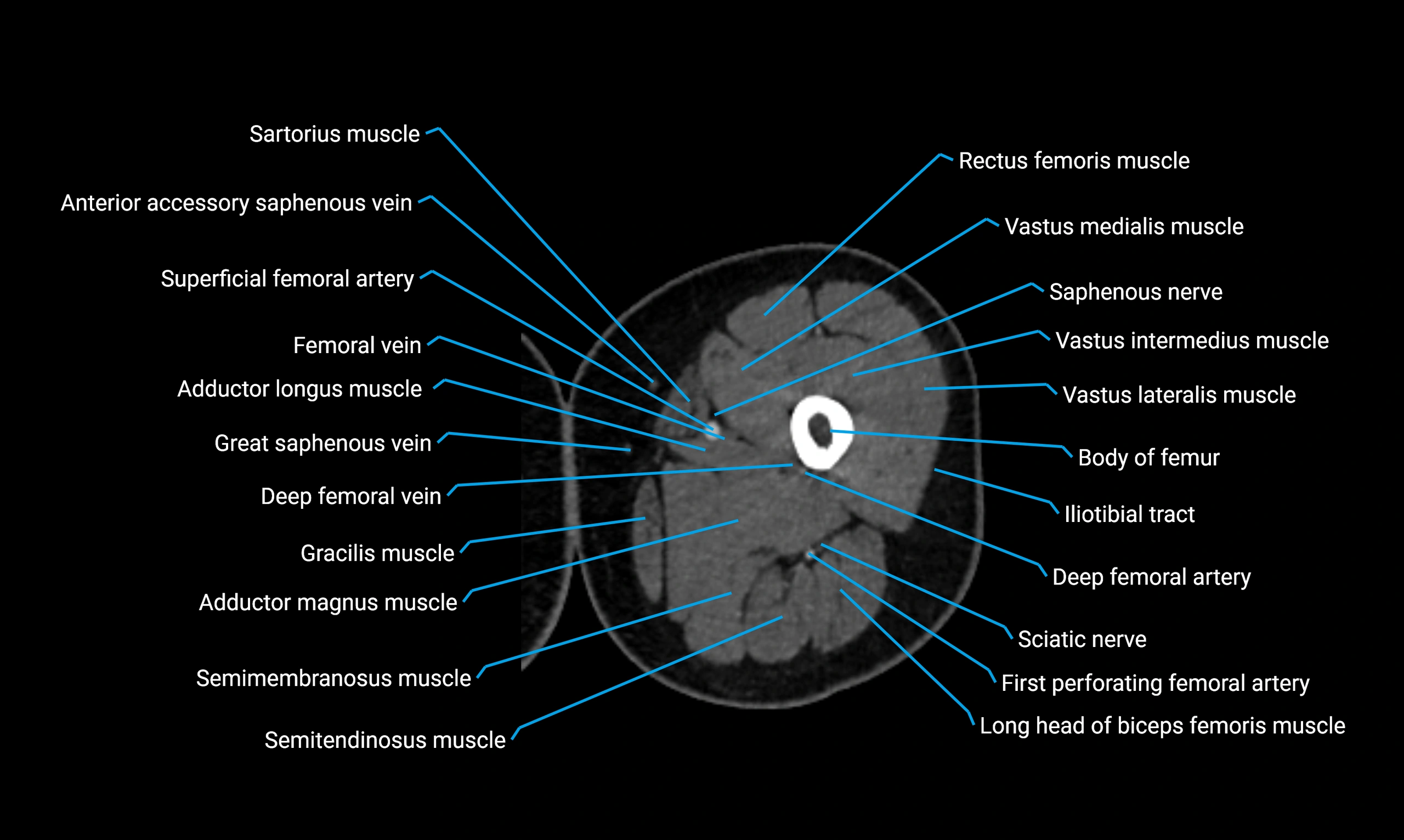
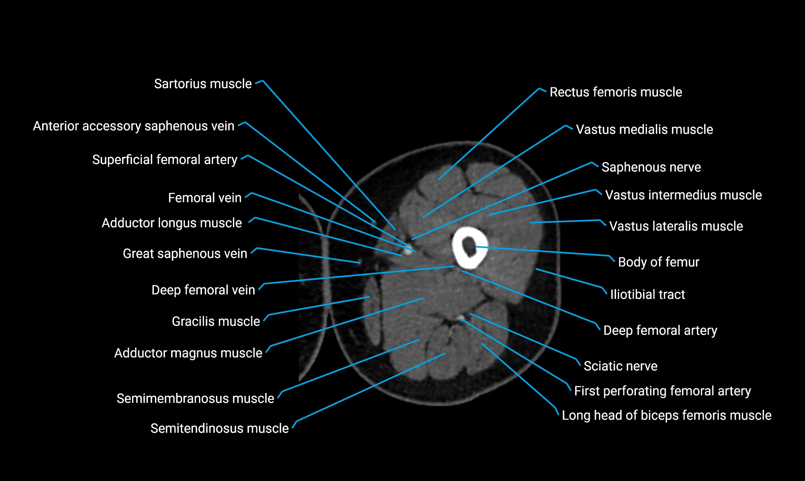
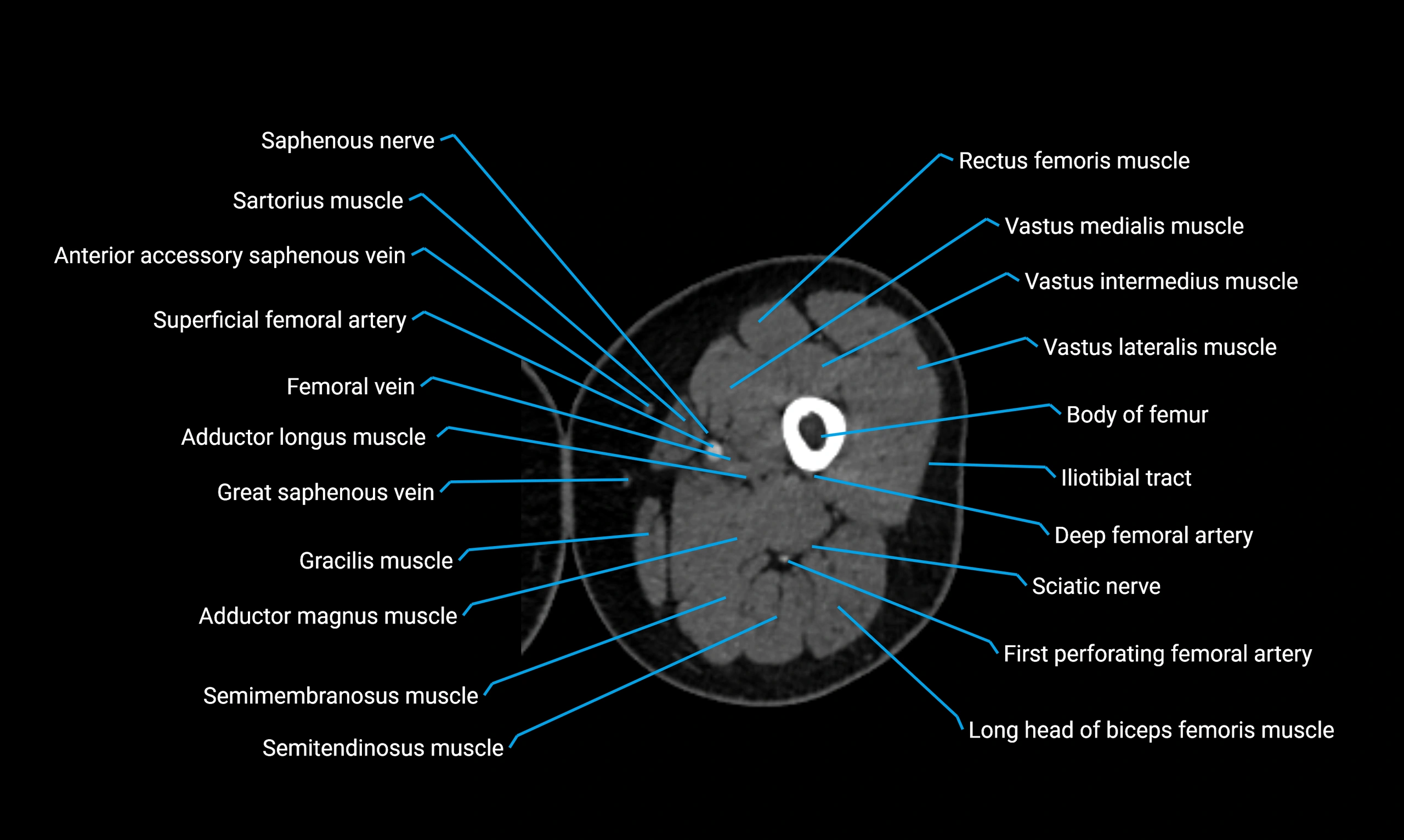
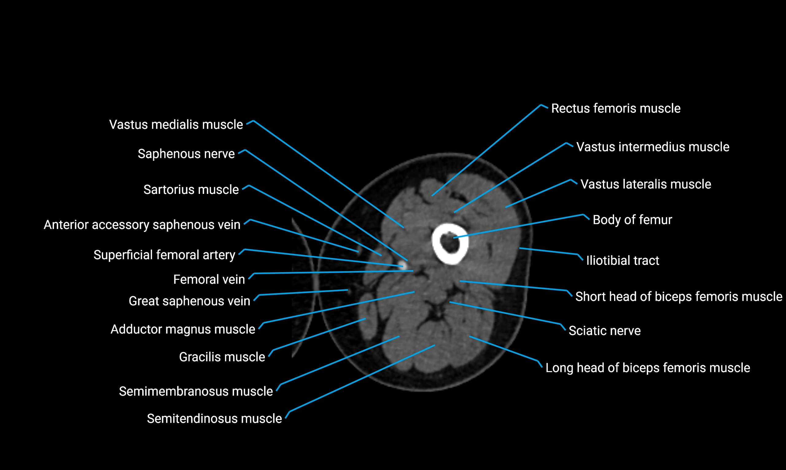
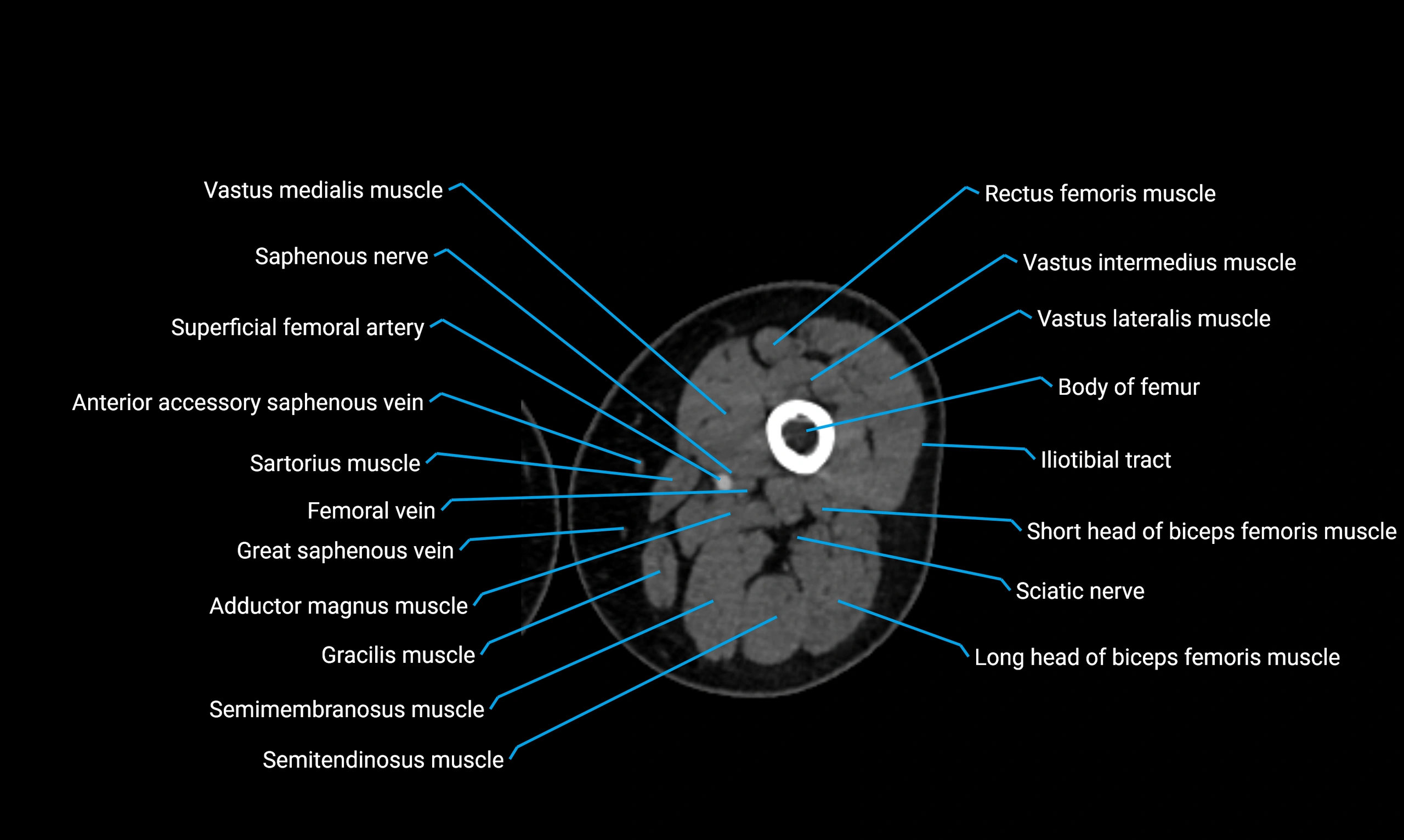
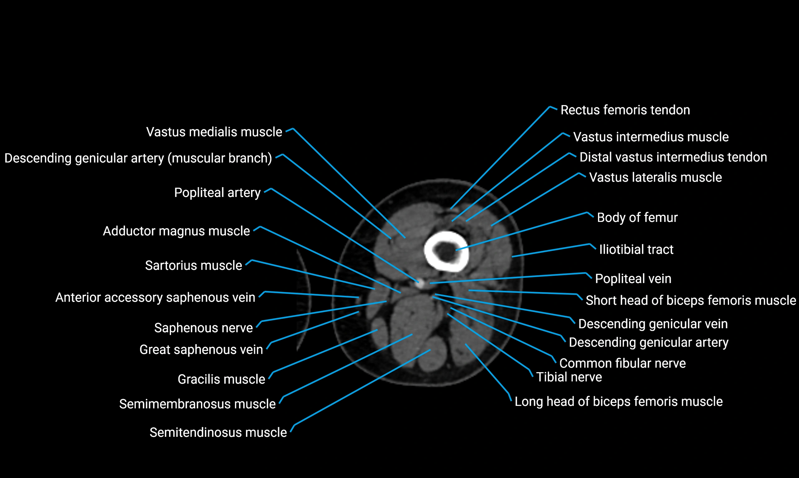
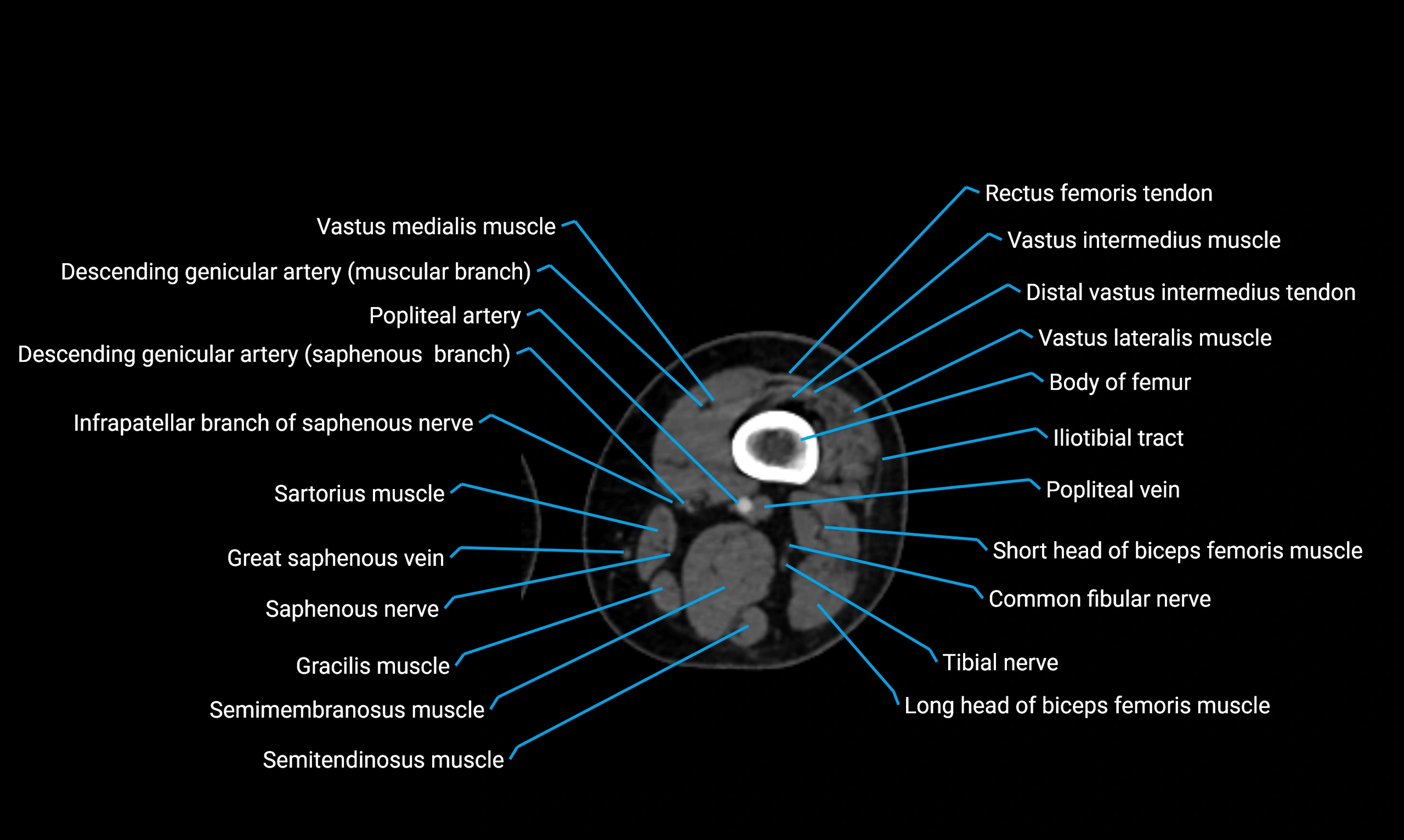
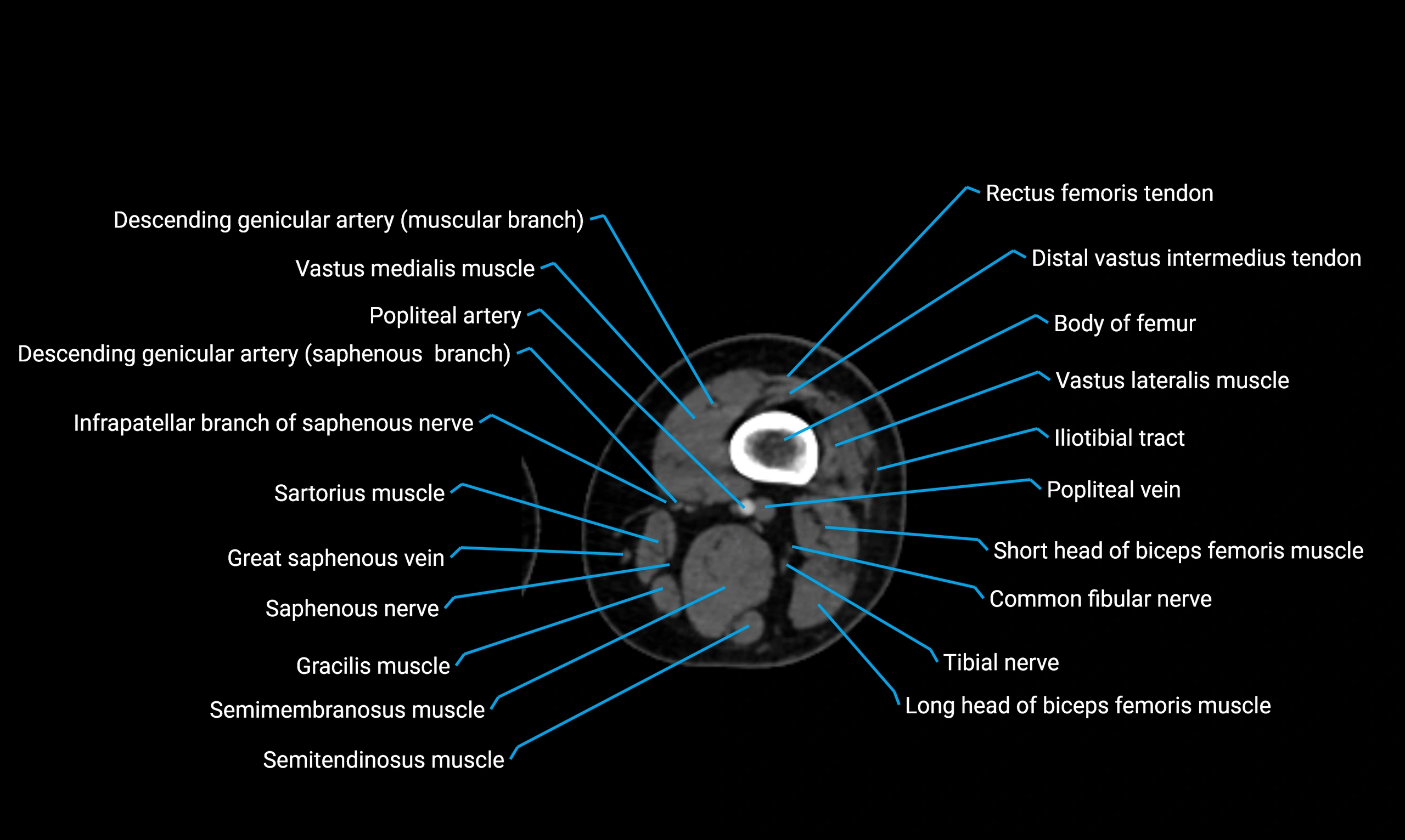
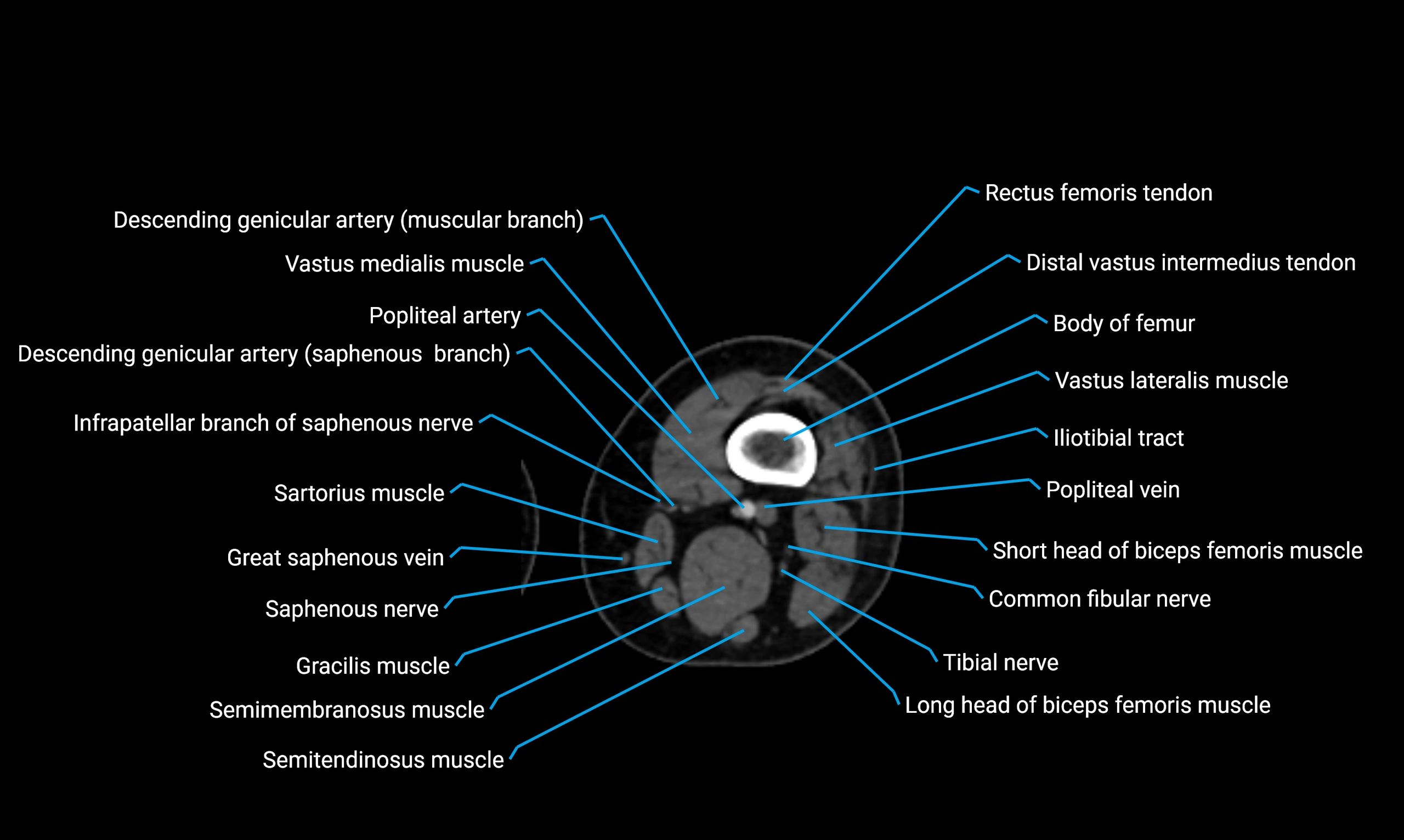
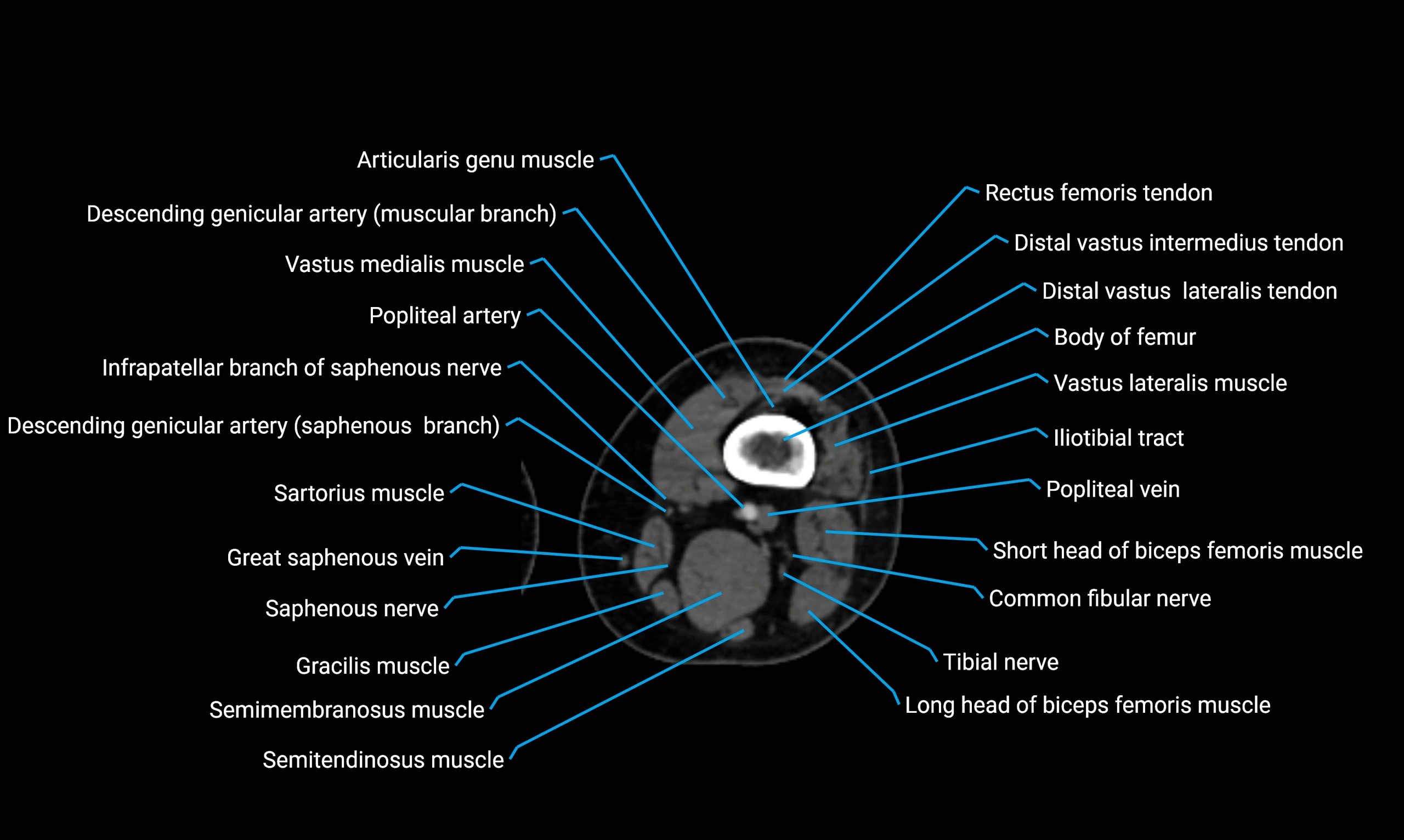
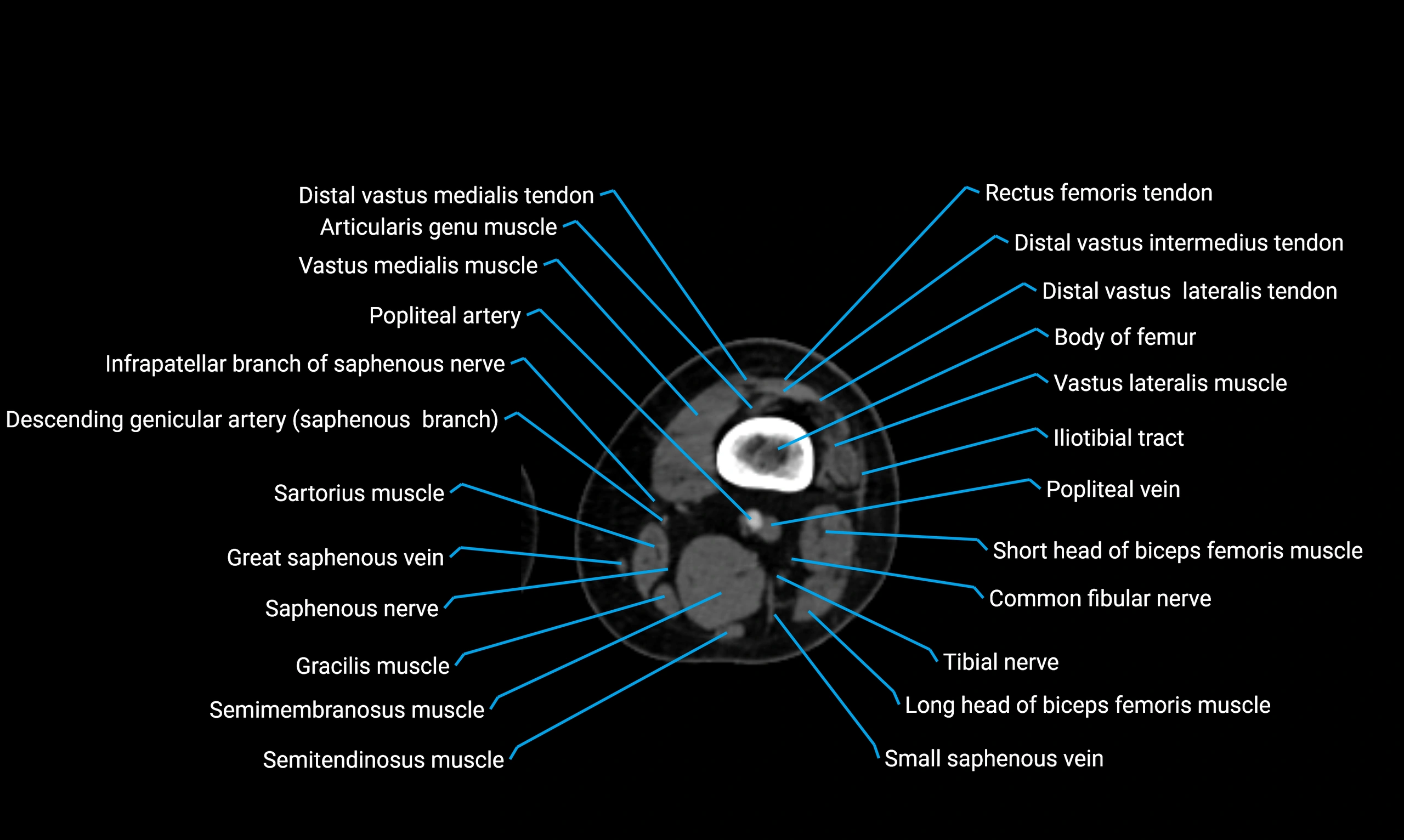
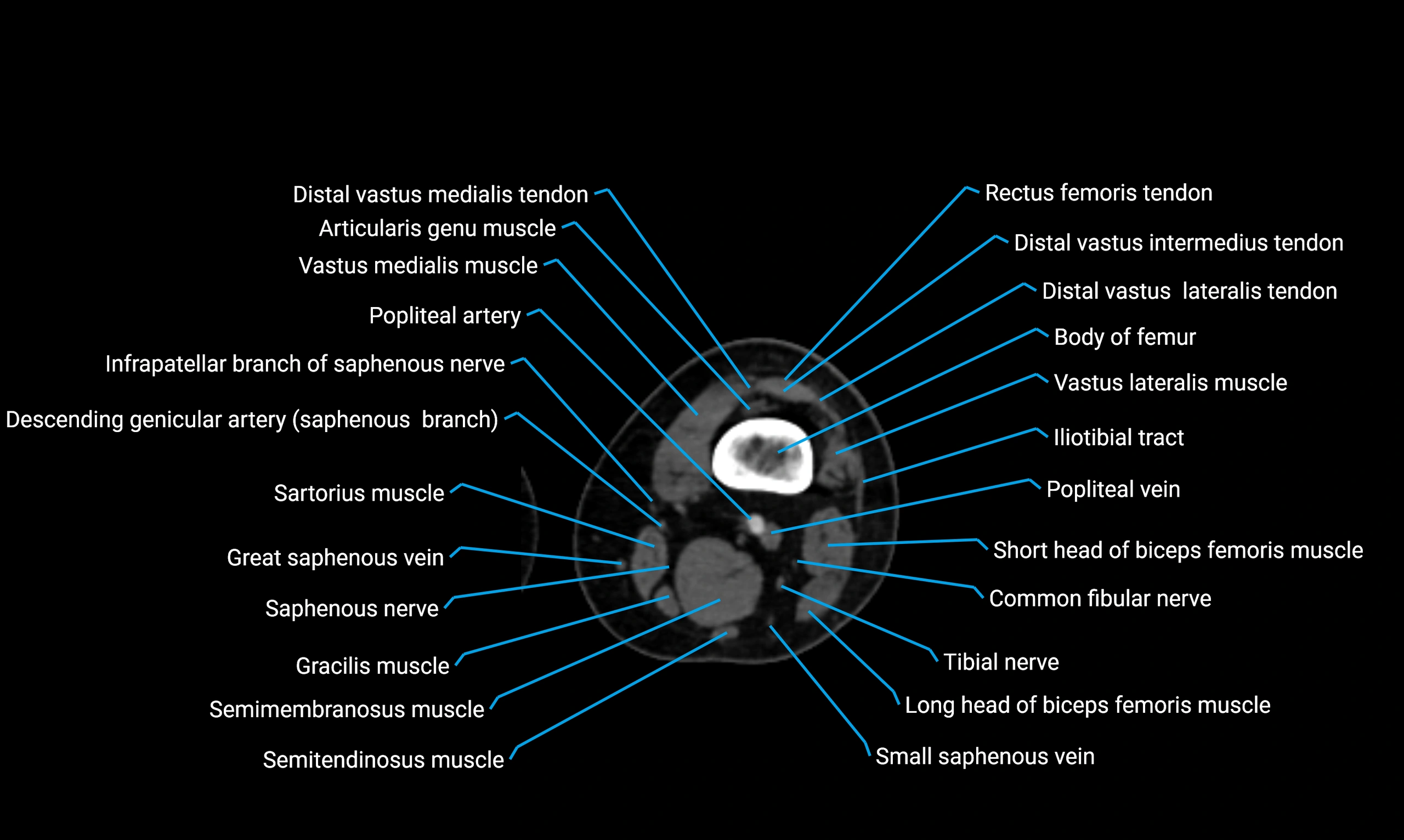
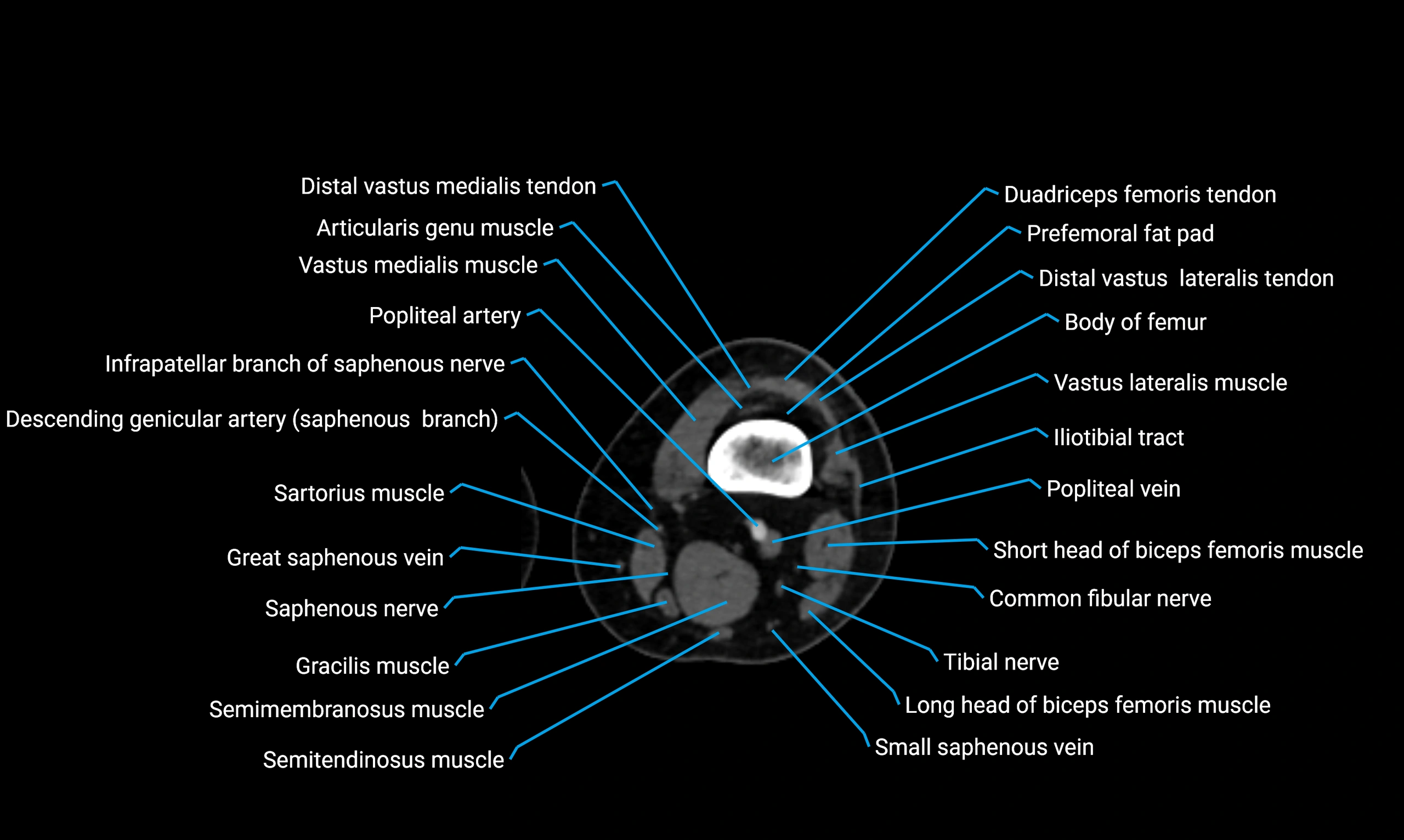
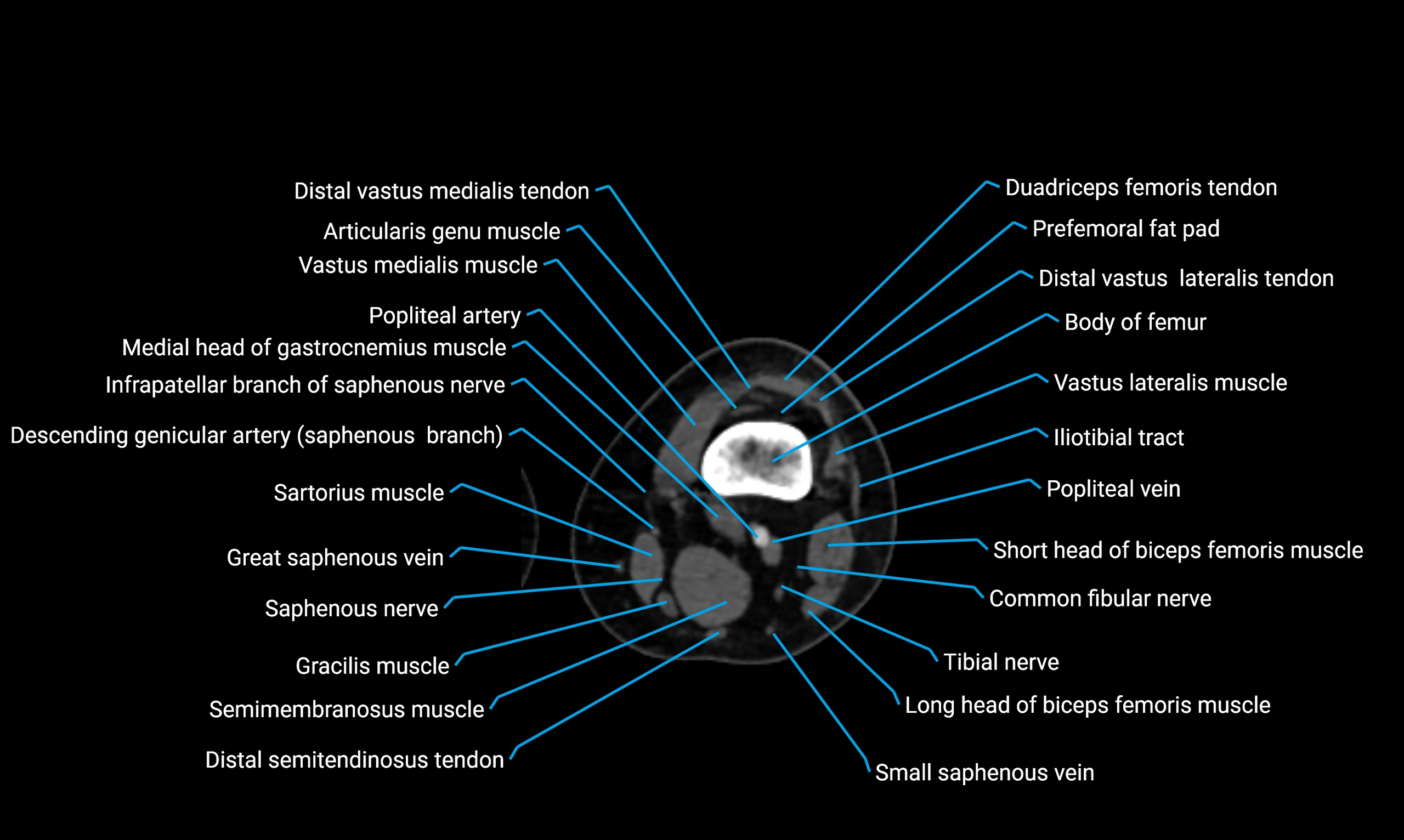
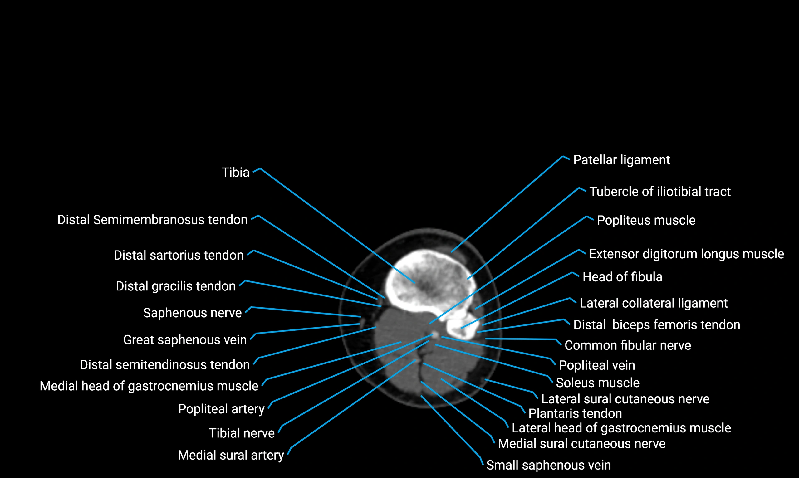
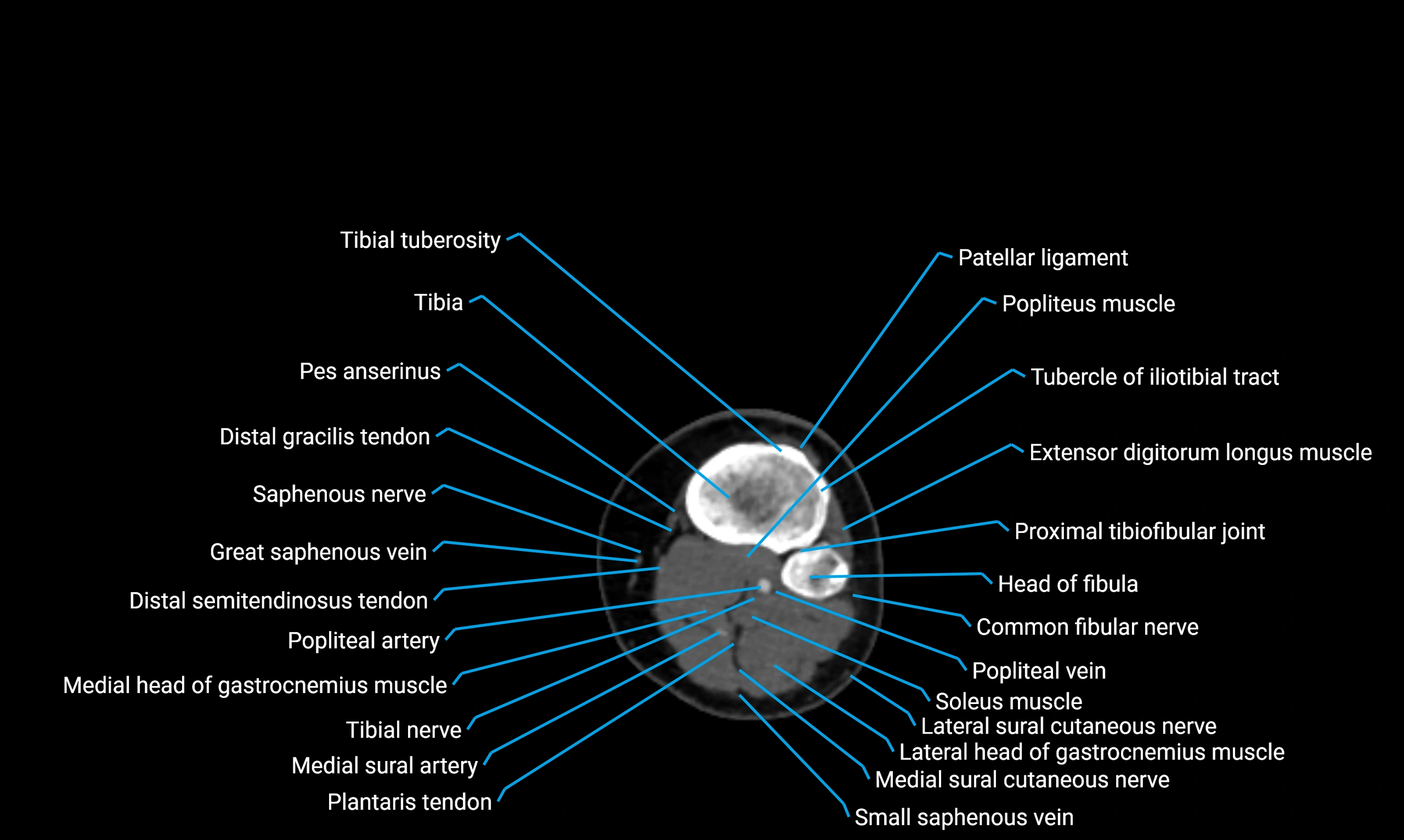
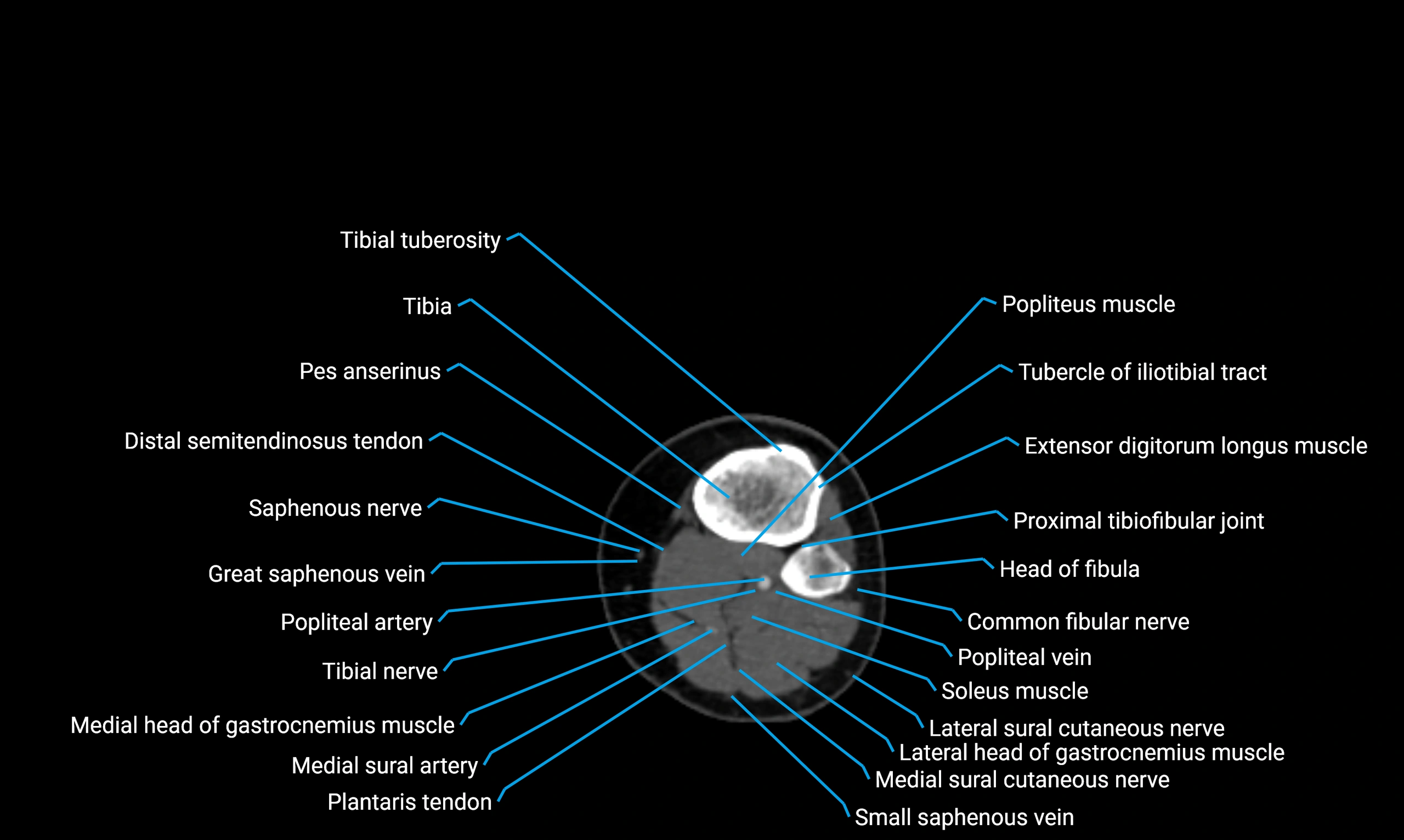
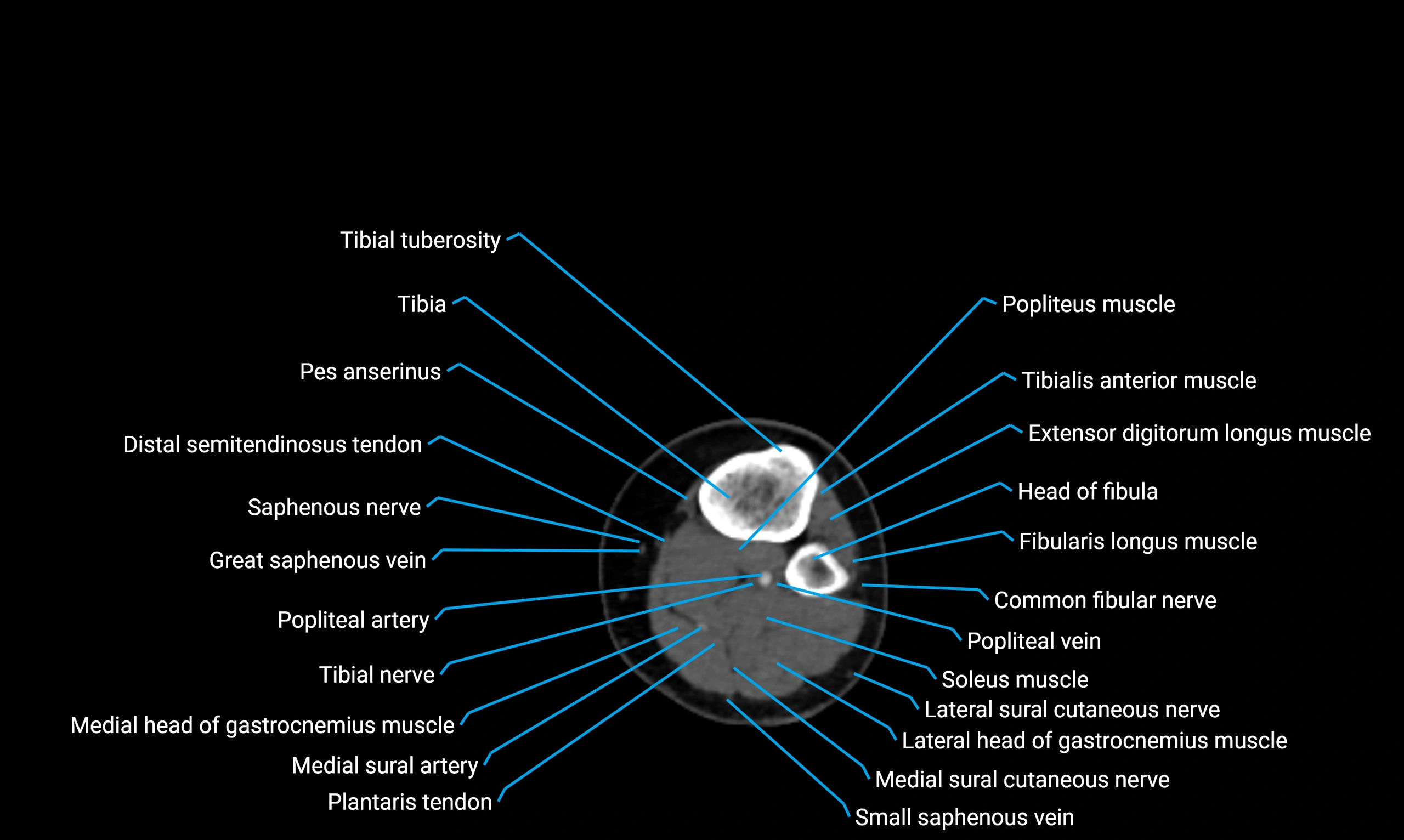
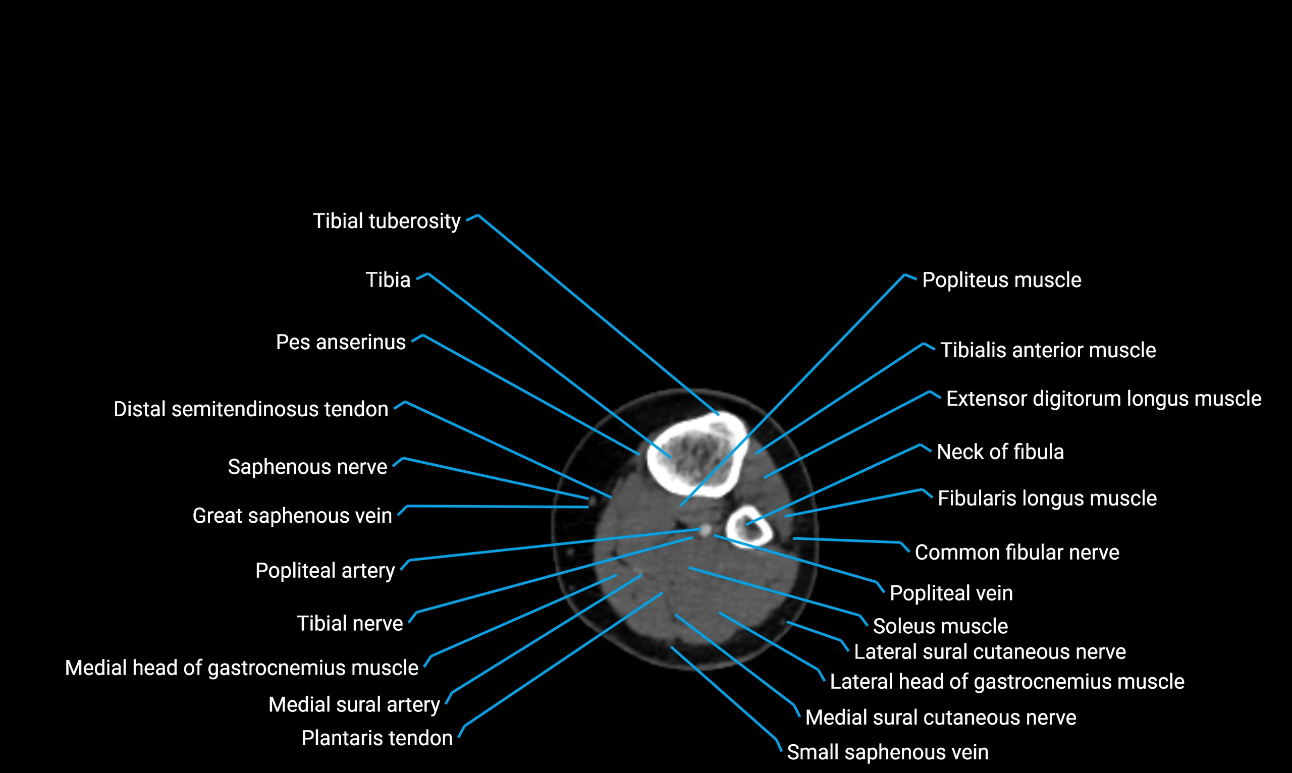
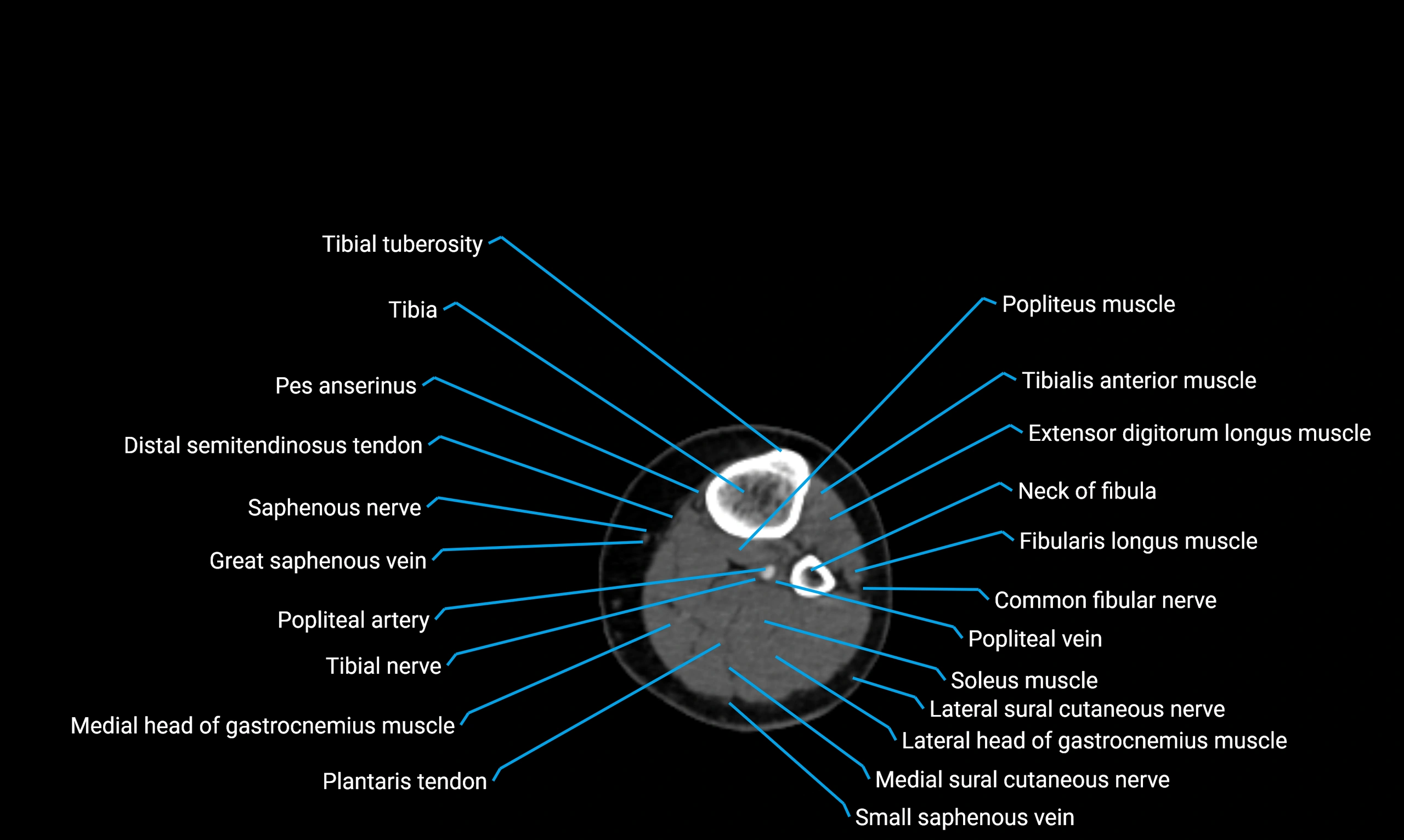
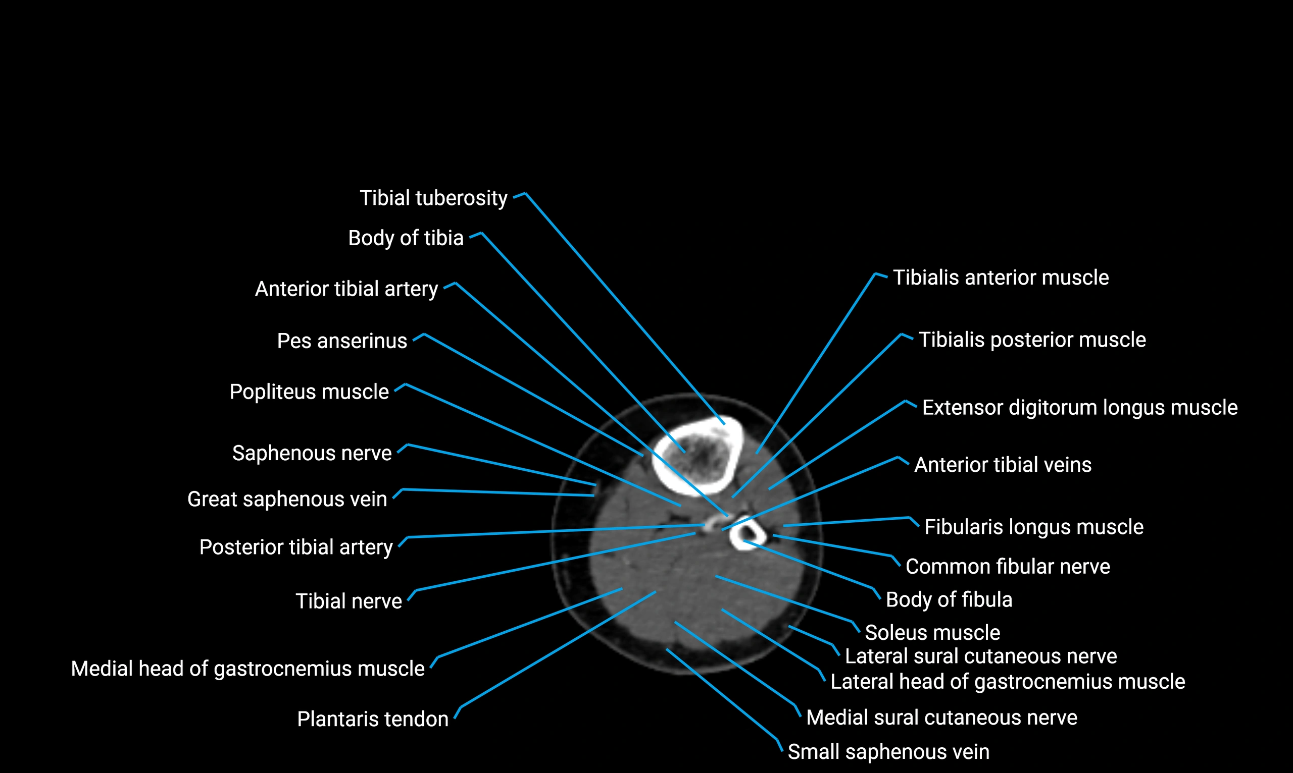
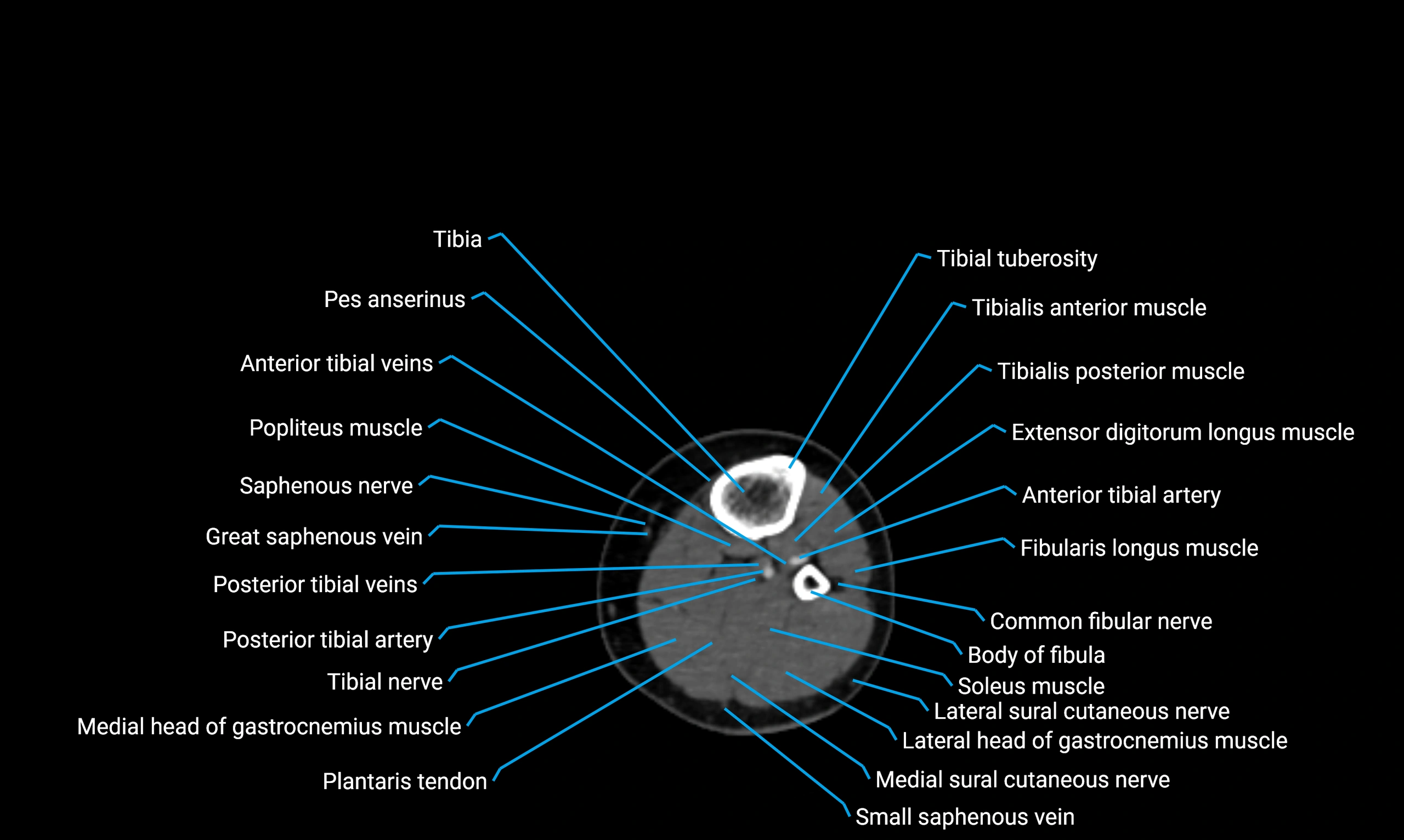
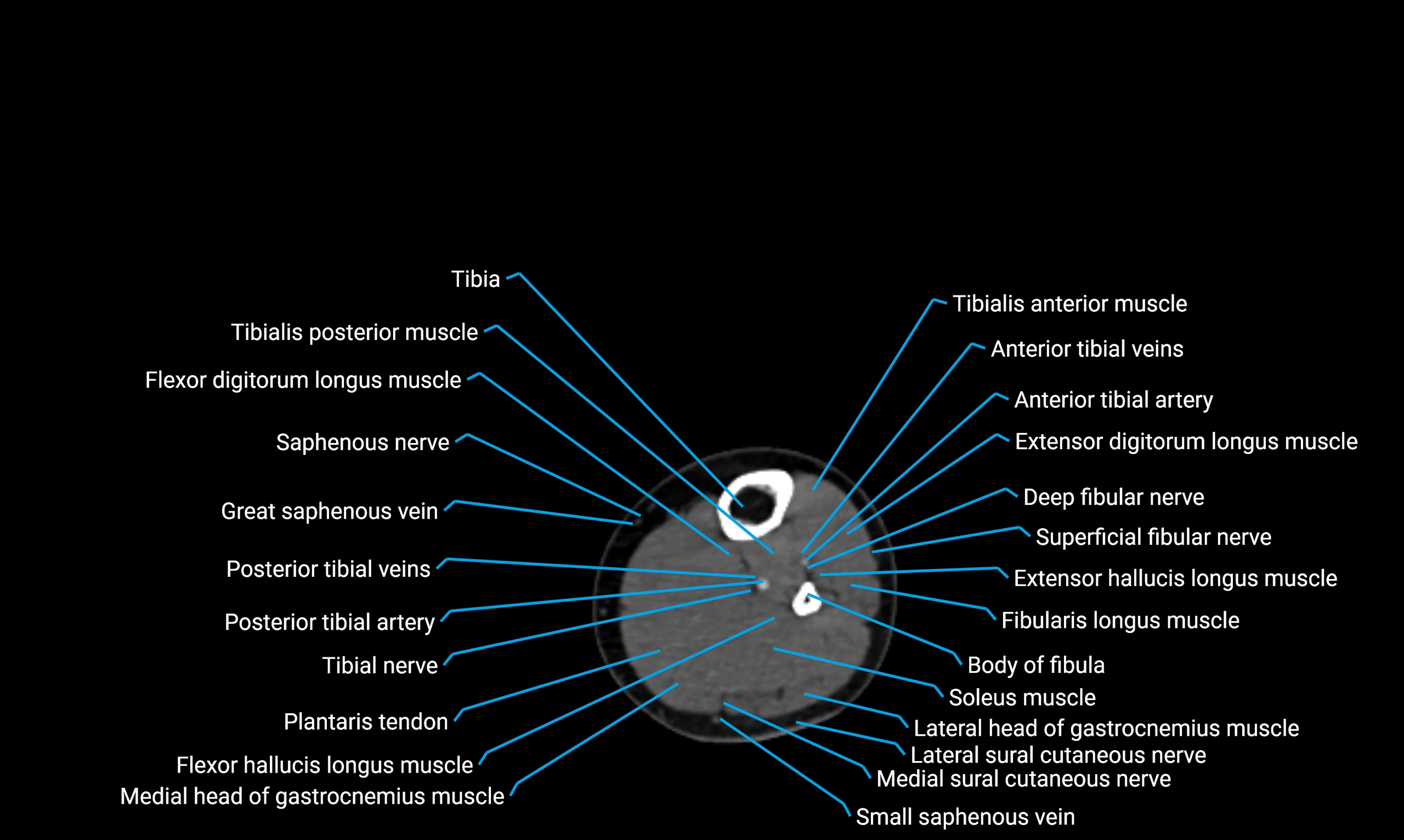
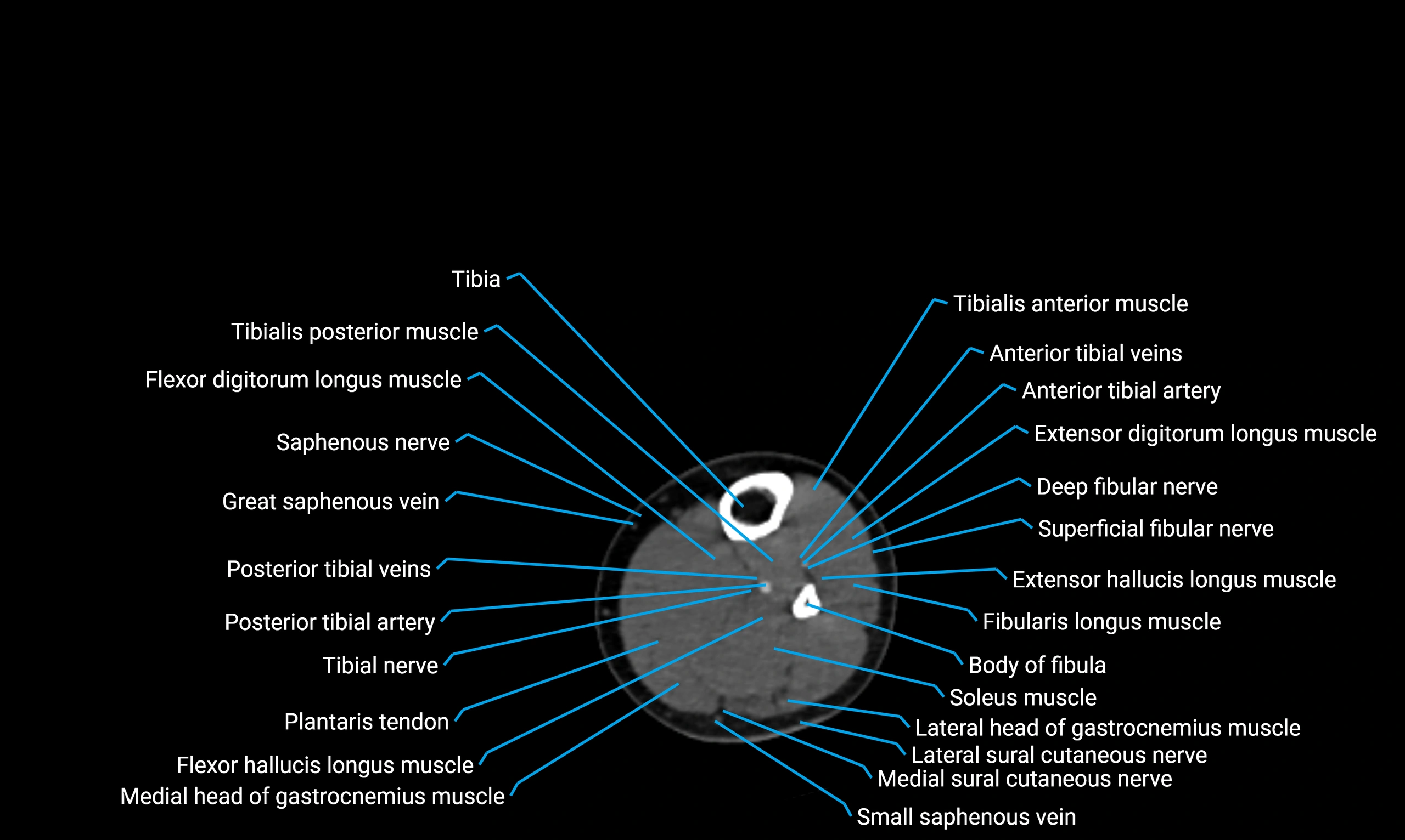
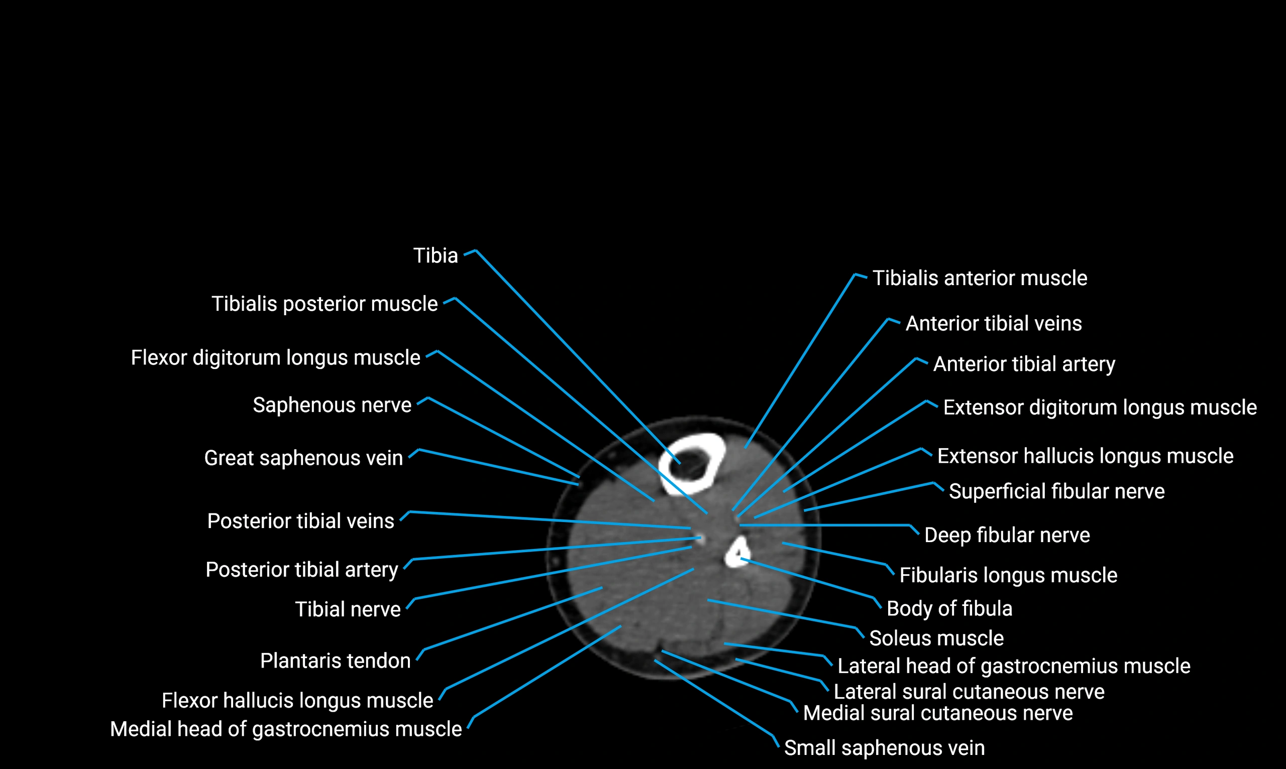
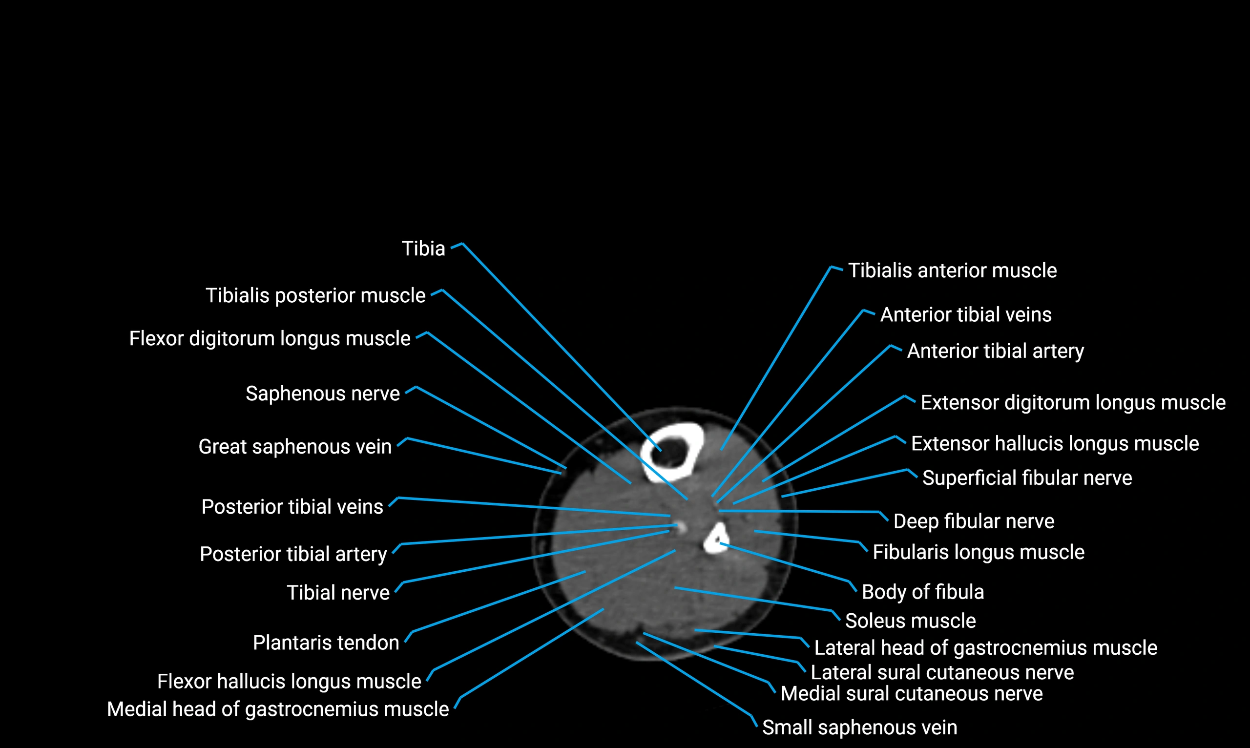
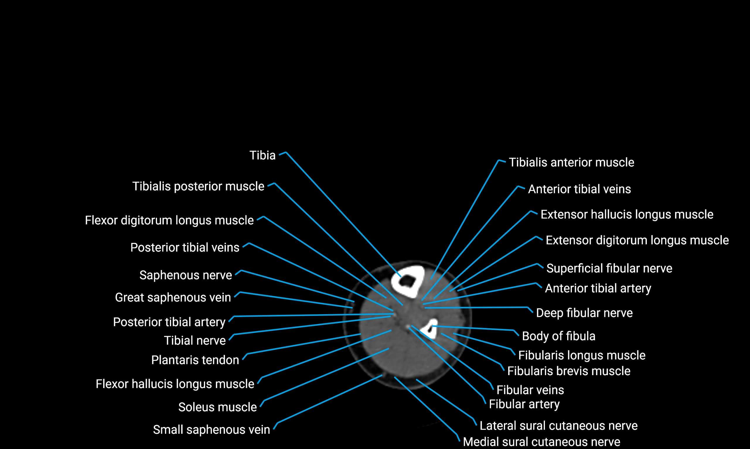
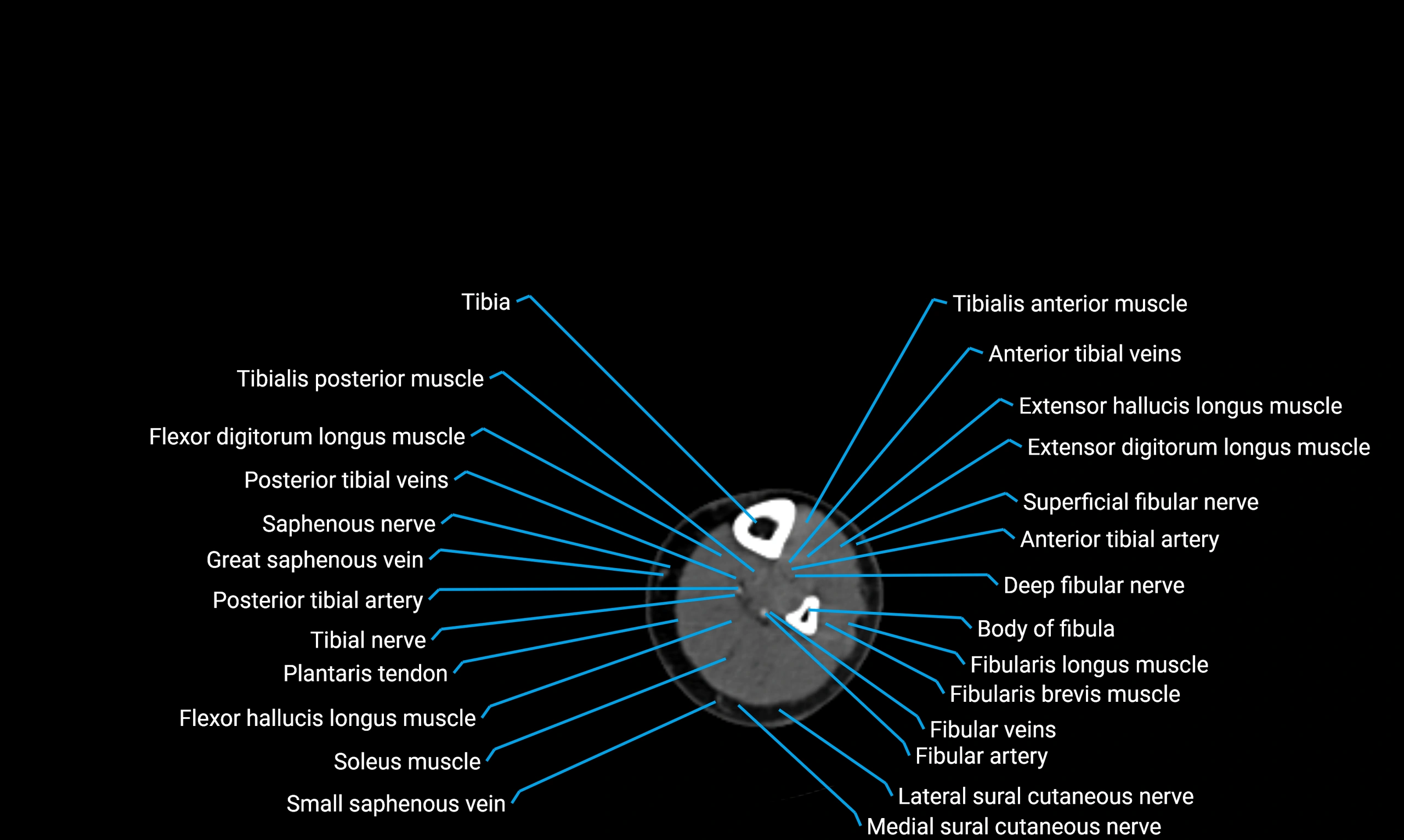
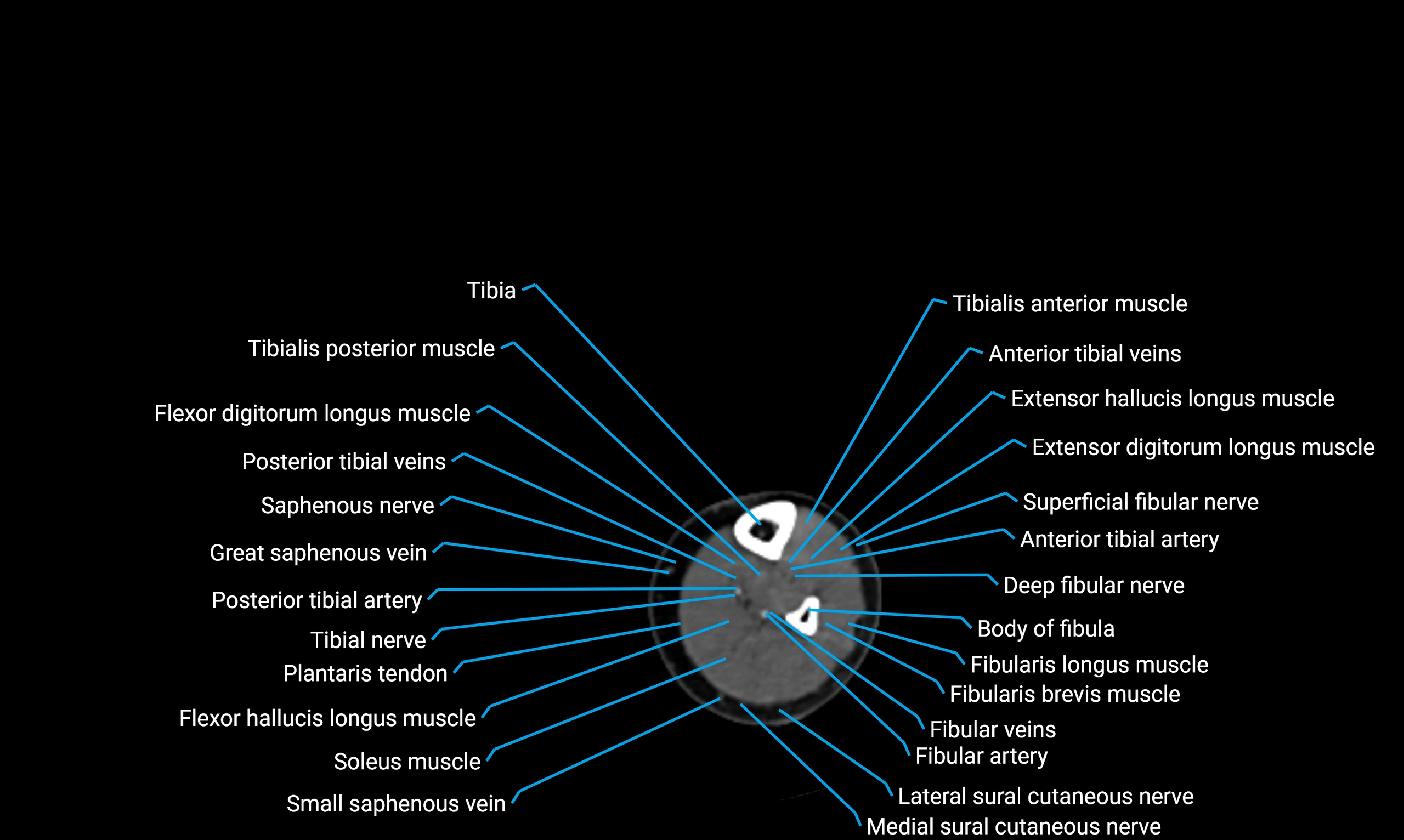
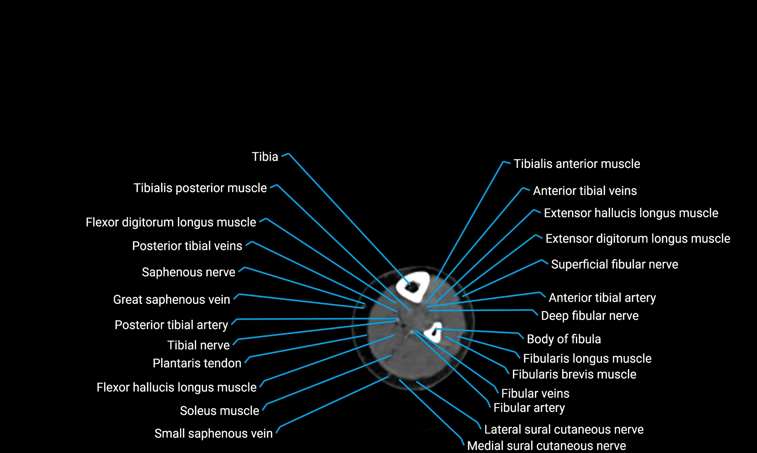
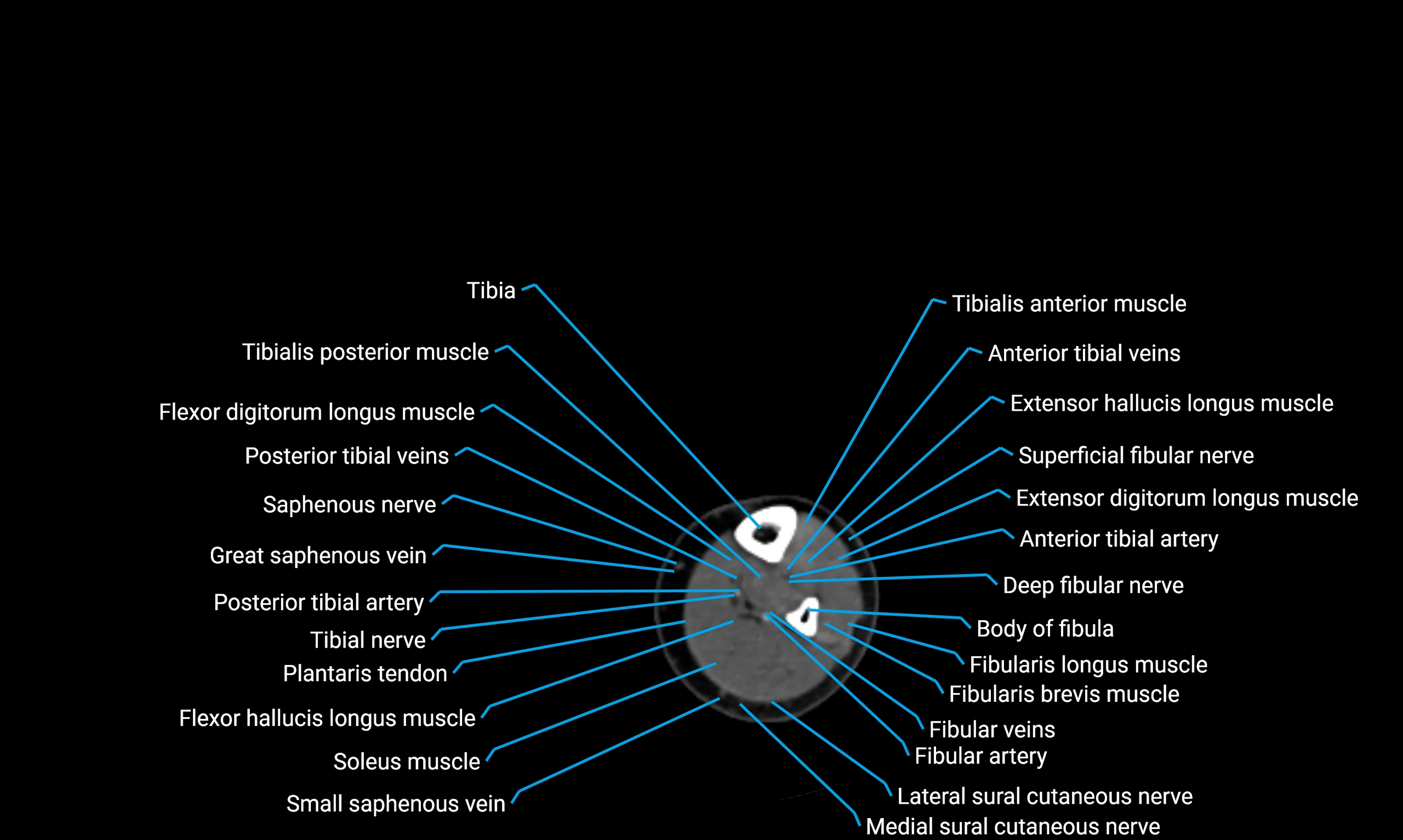
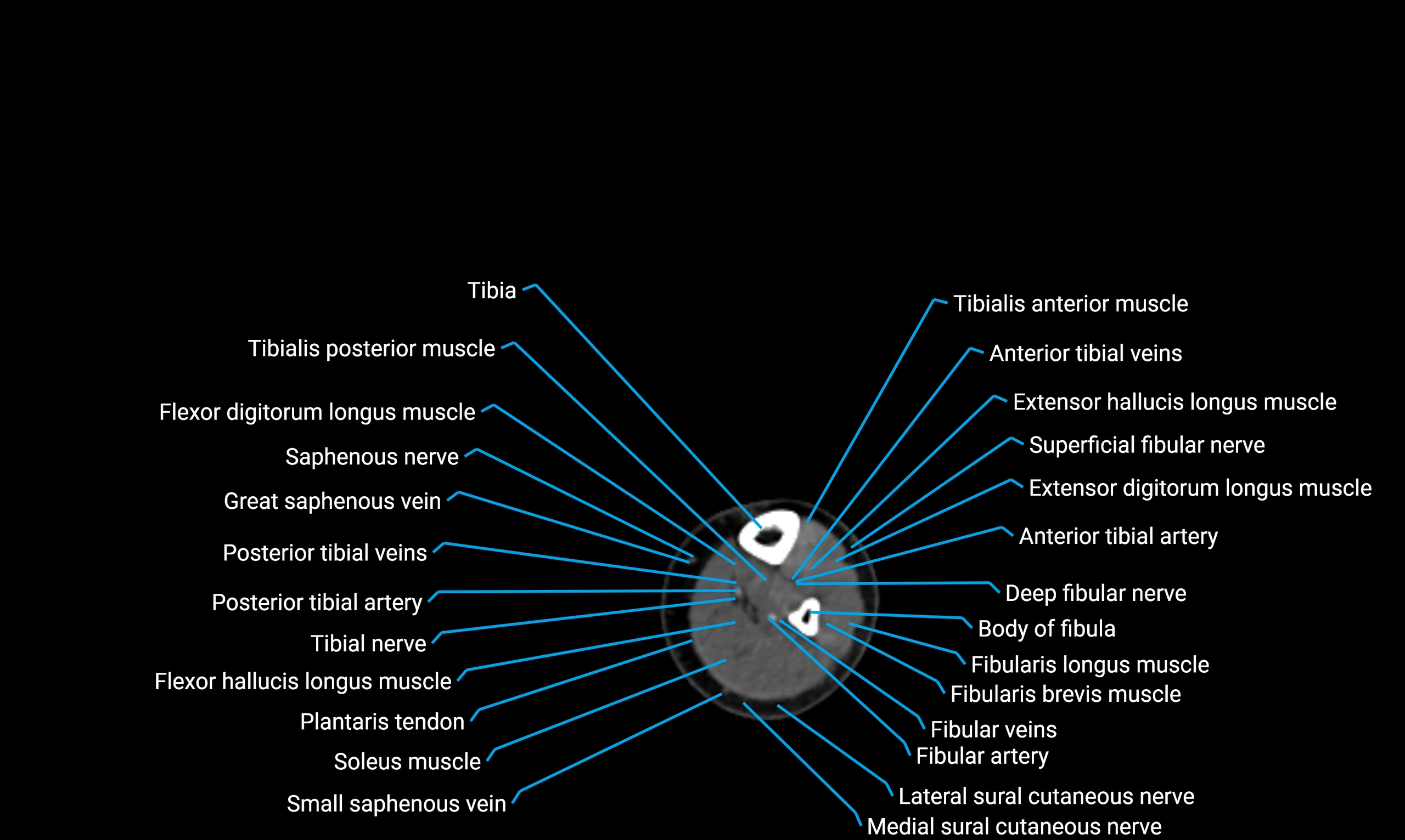
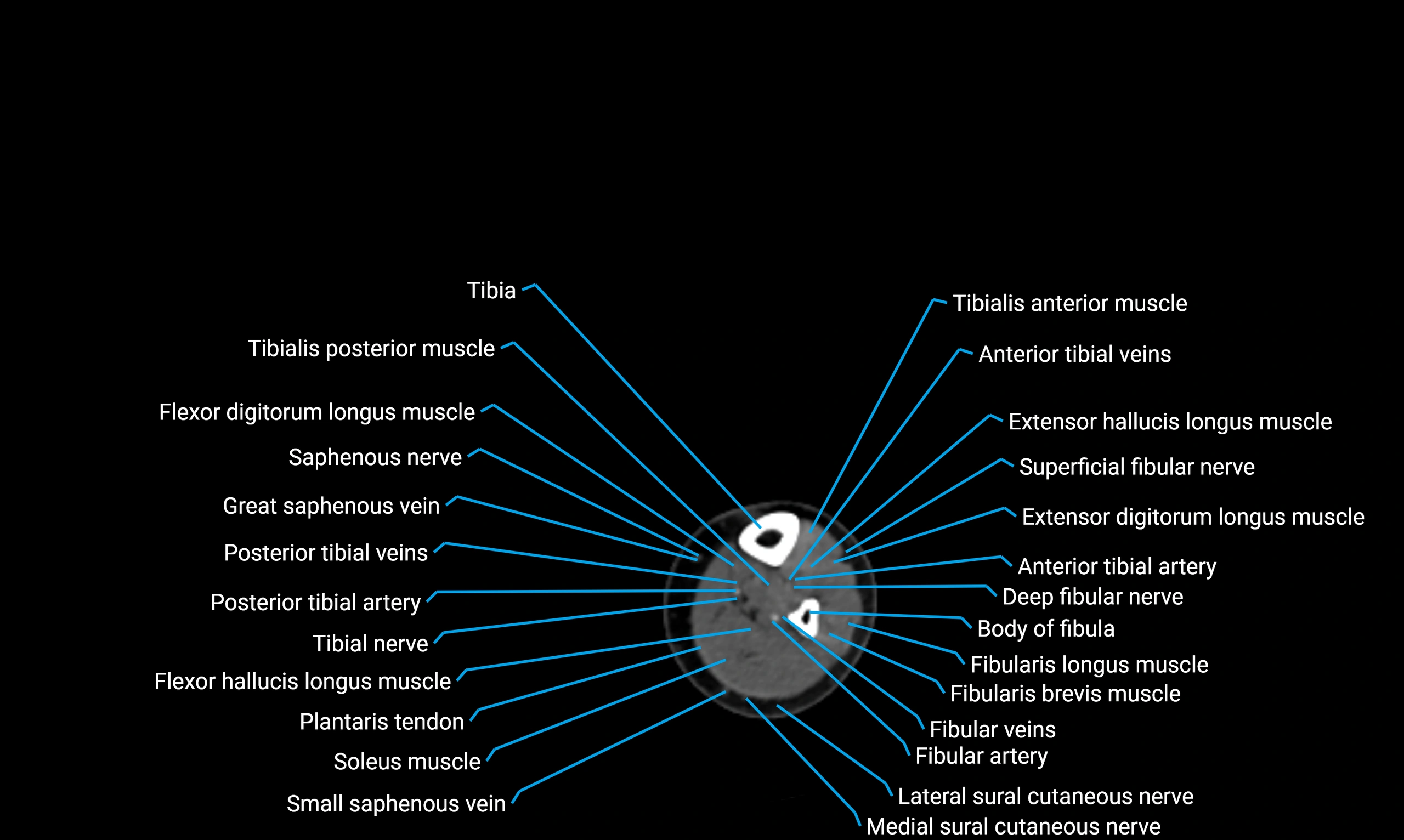
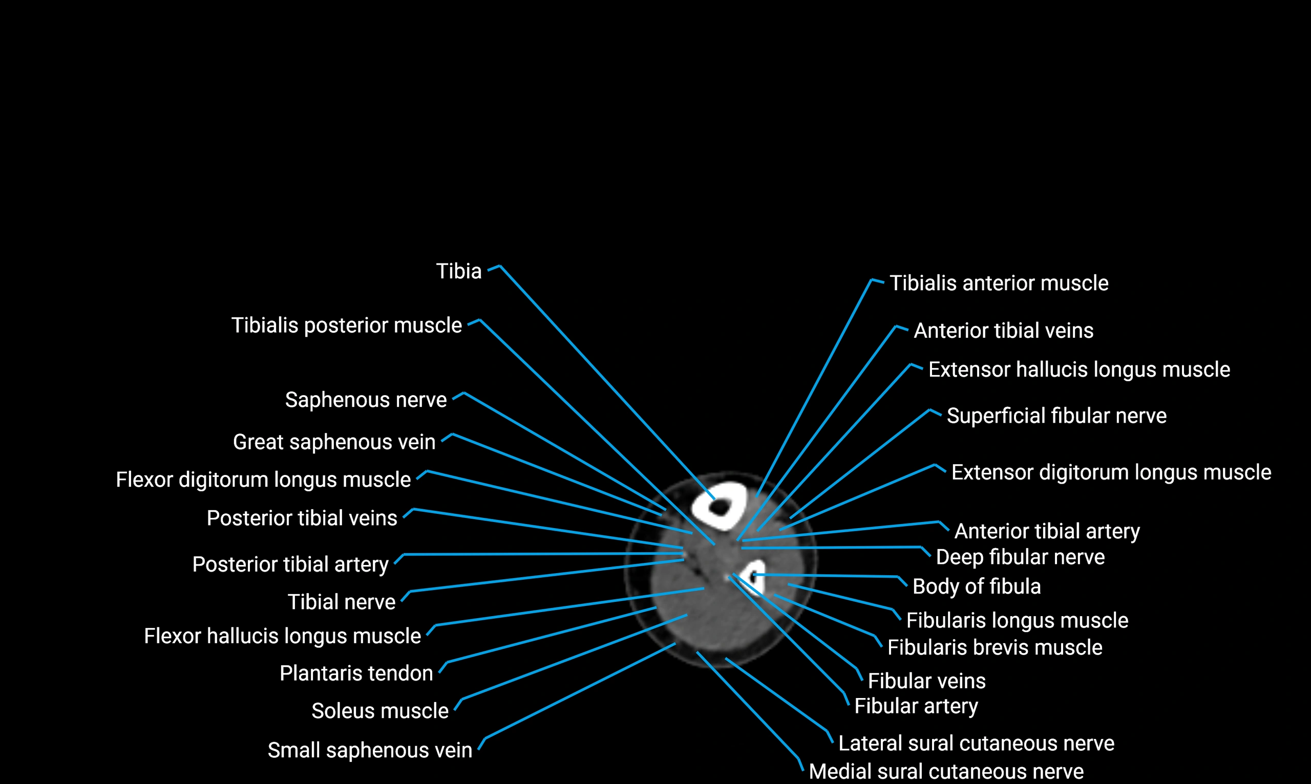
CT Appearance
Non-contrast CT:
-
Bone cortex: hyperdense and sharply outlined
-
Marrow cavity: hypodense relative to cortex
-
Gold standard for detecting fractures, avulsion injuries, and cortical morphology (subspine prominence)
CT Post-Contrast:
-
Bone cortex: unchanged
-
Surrounding soft tissues: enhancement indicates inflammation, hematoma, or tumor
-
3D CT reconstructions valuable in orthopedic planning and pelvic trauma evaluation
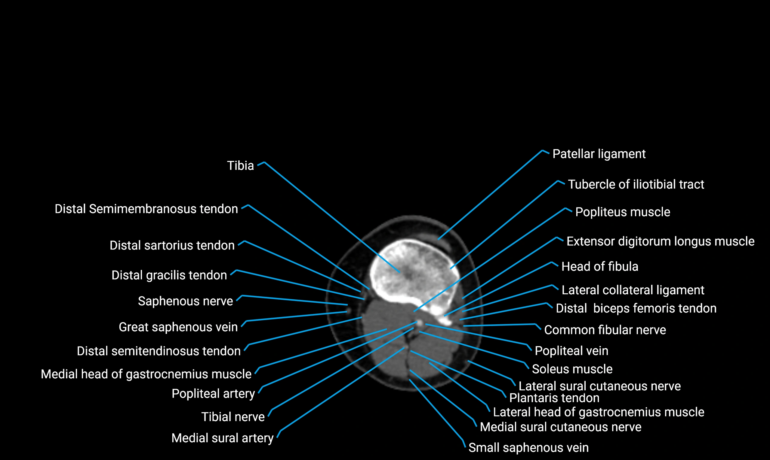
CT VRT 3D image

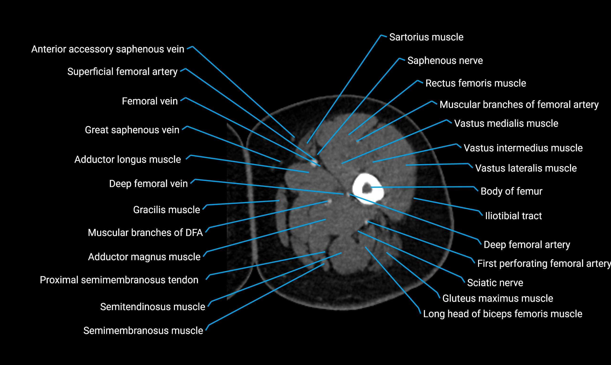
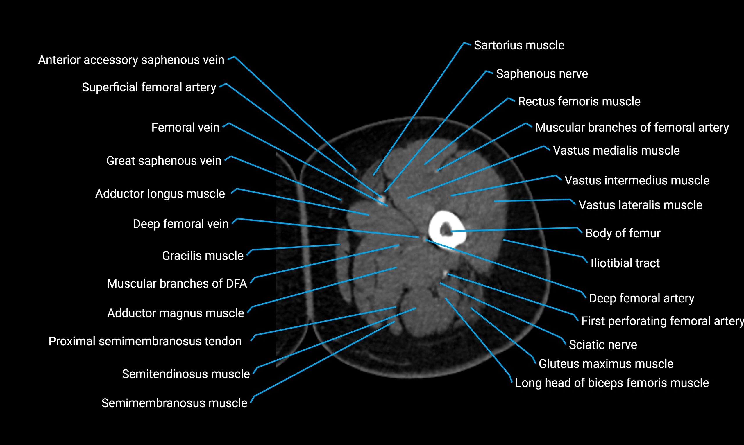
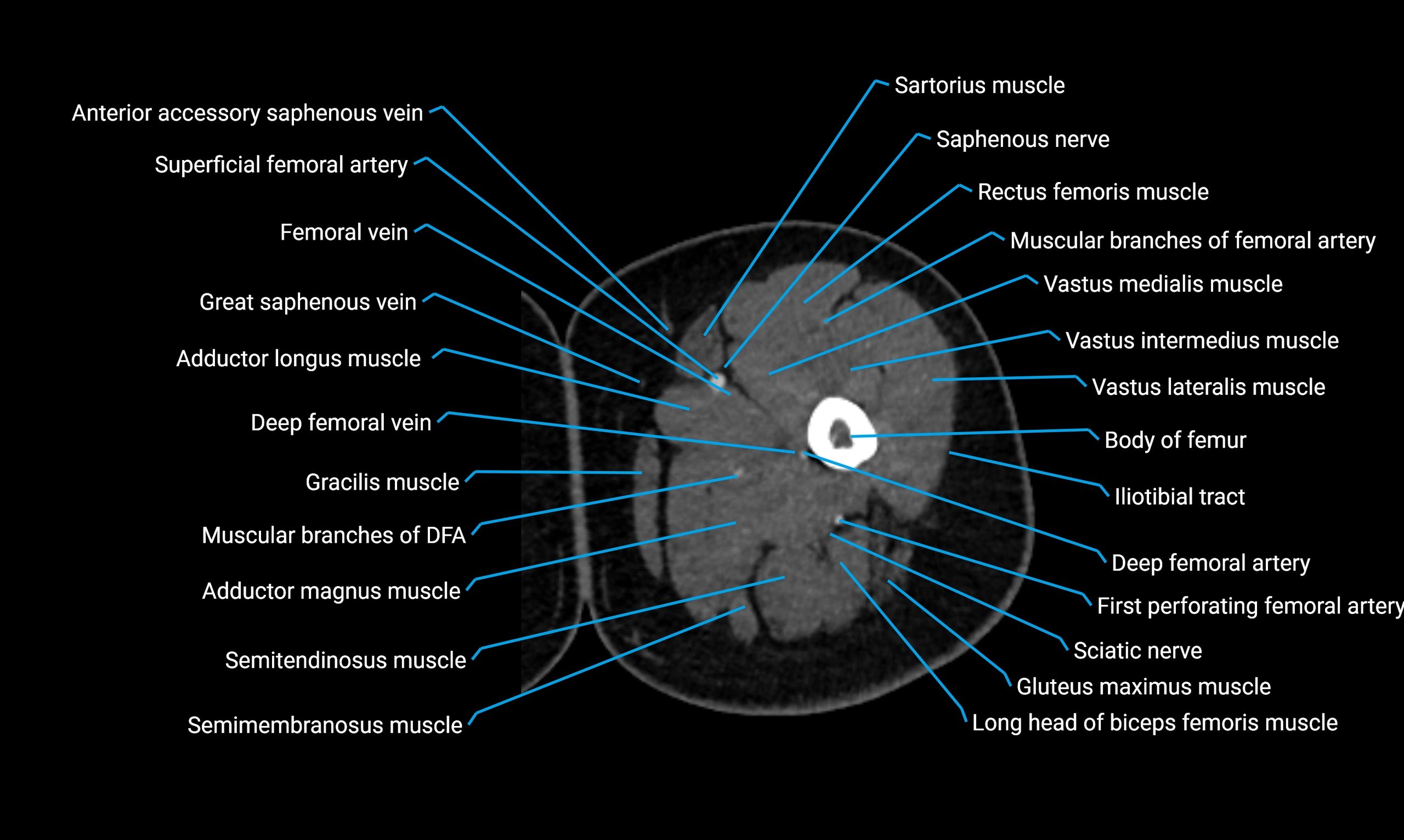
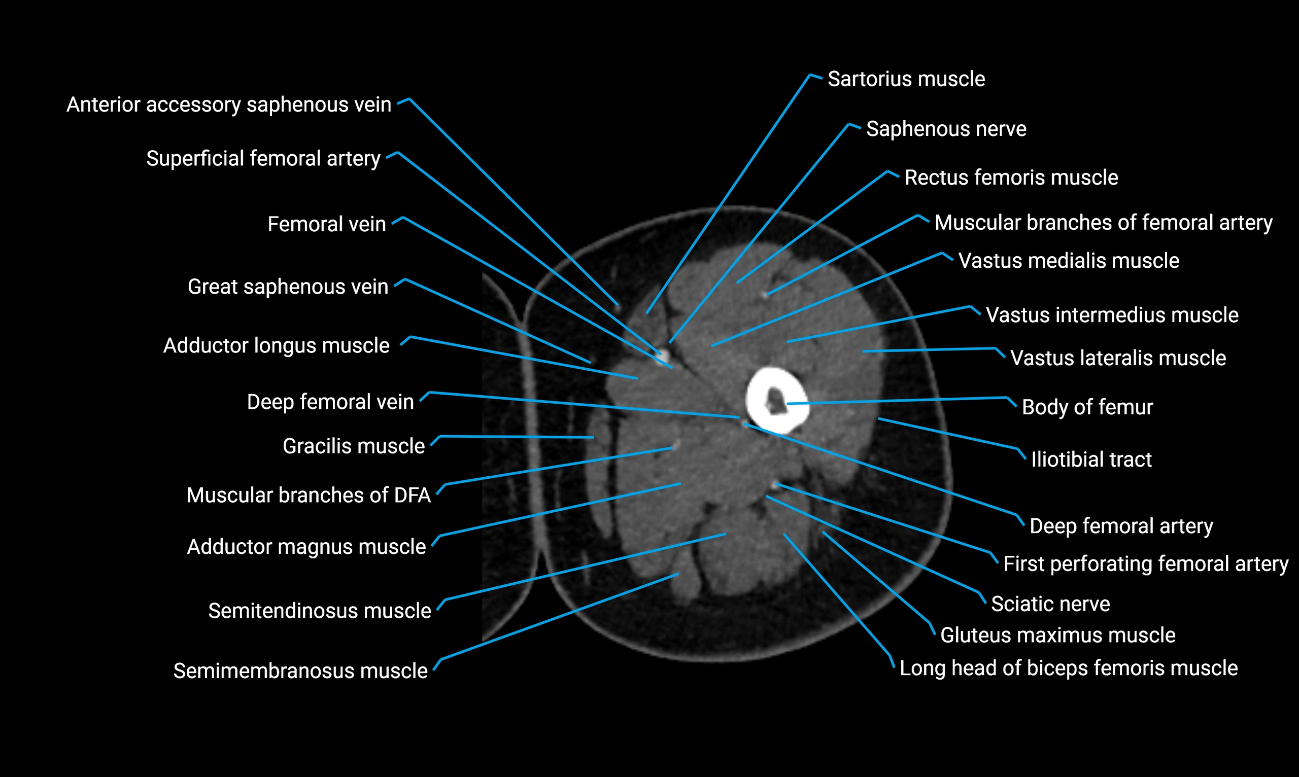
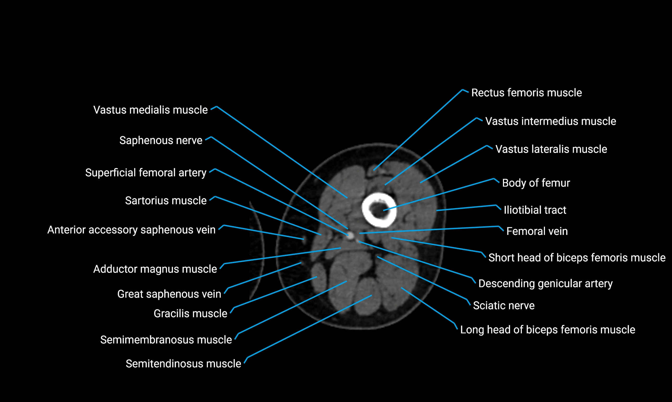
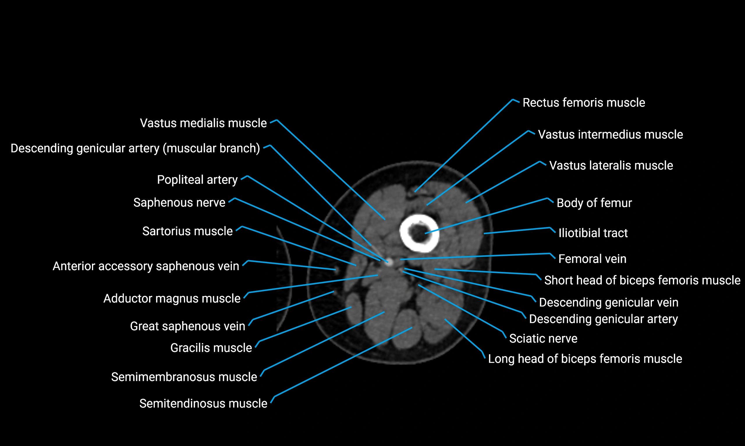
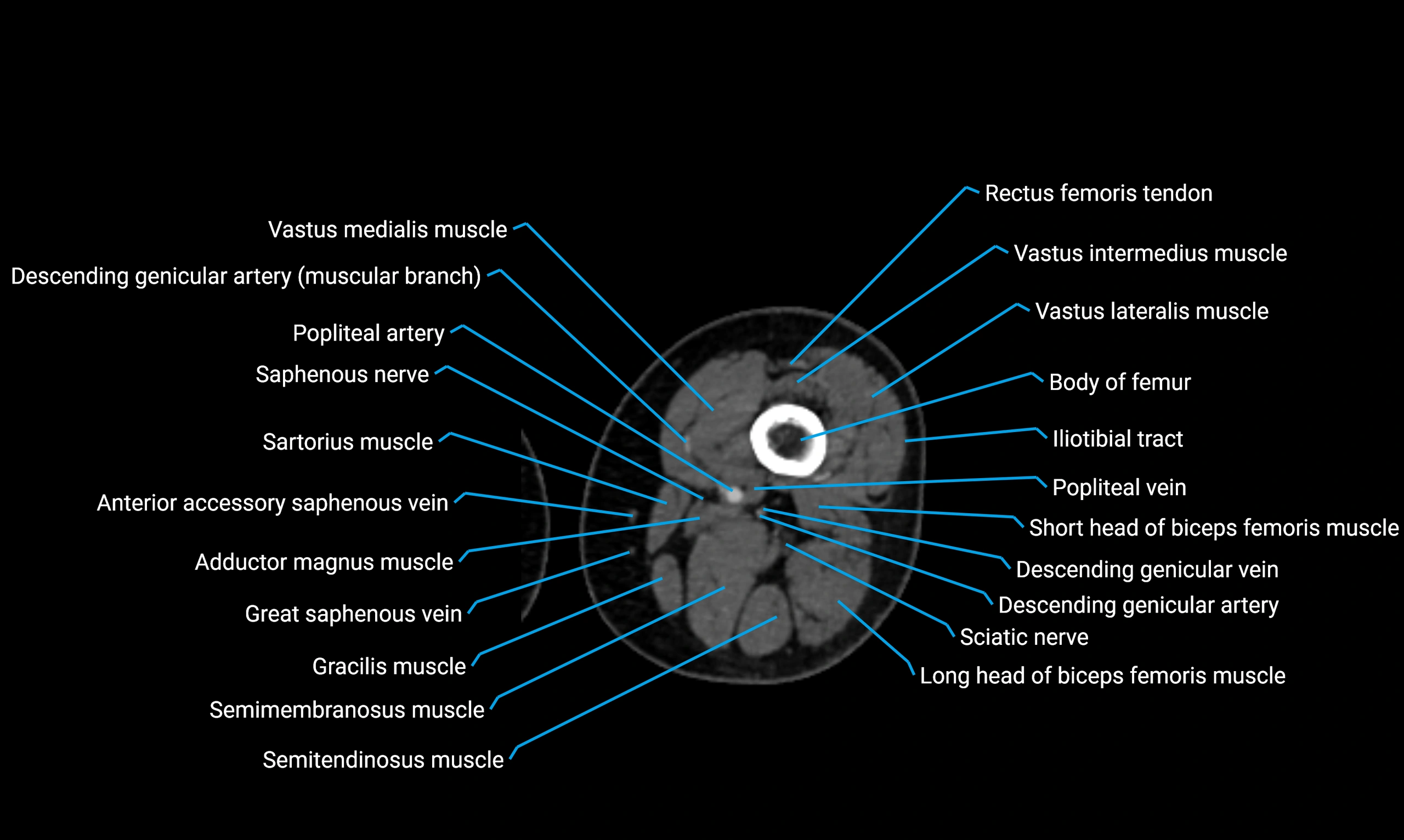
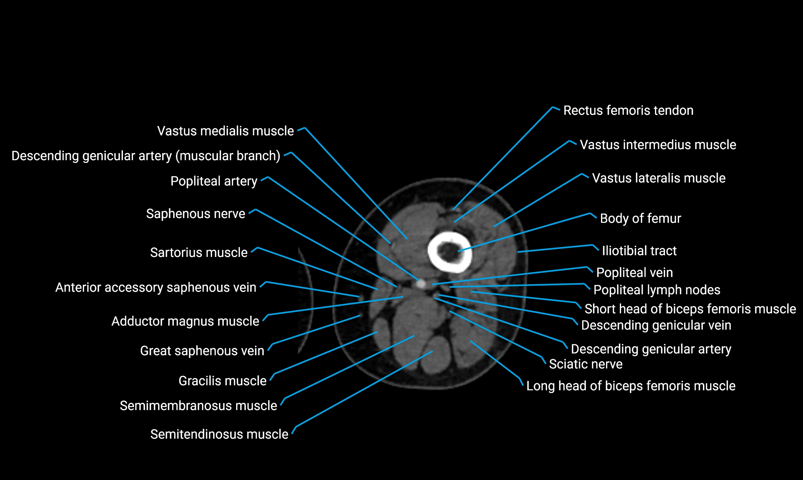
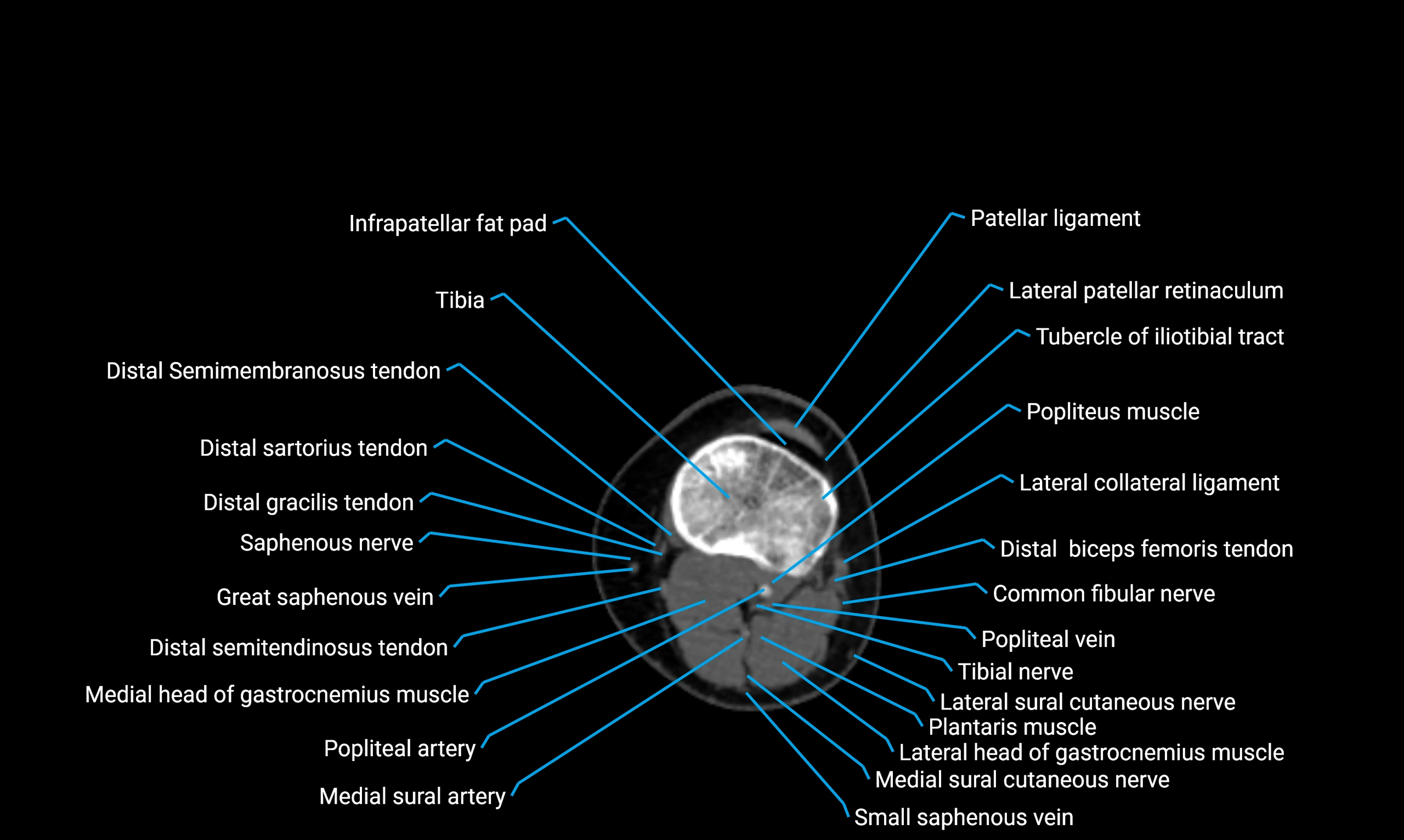
CT image

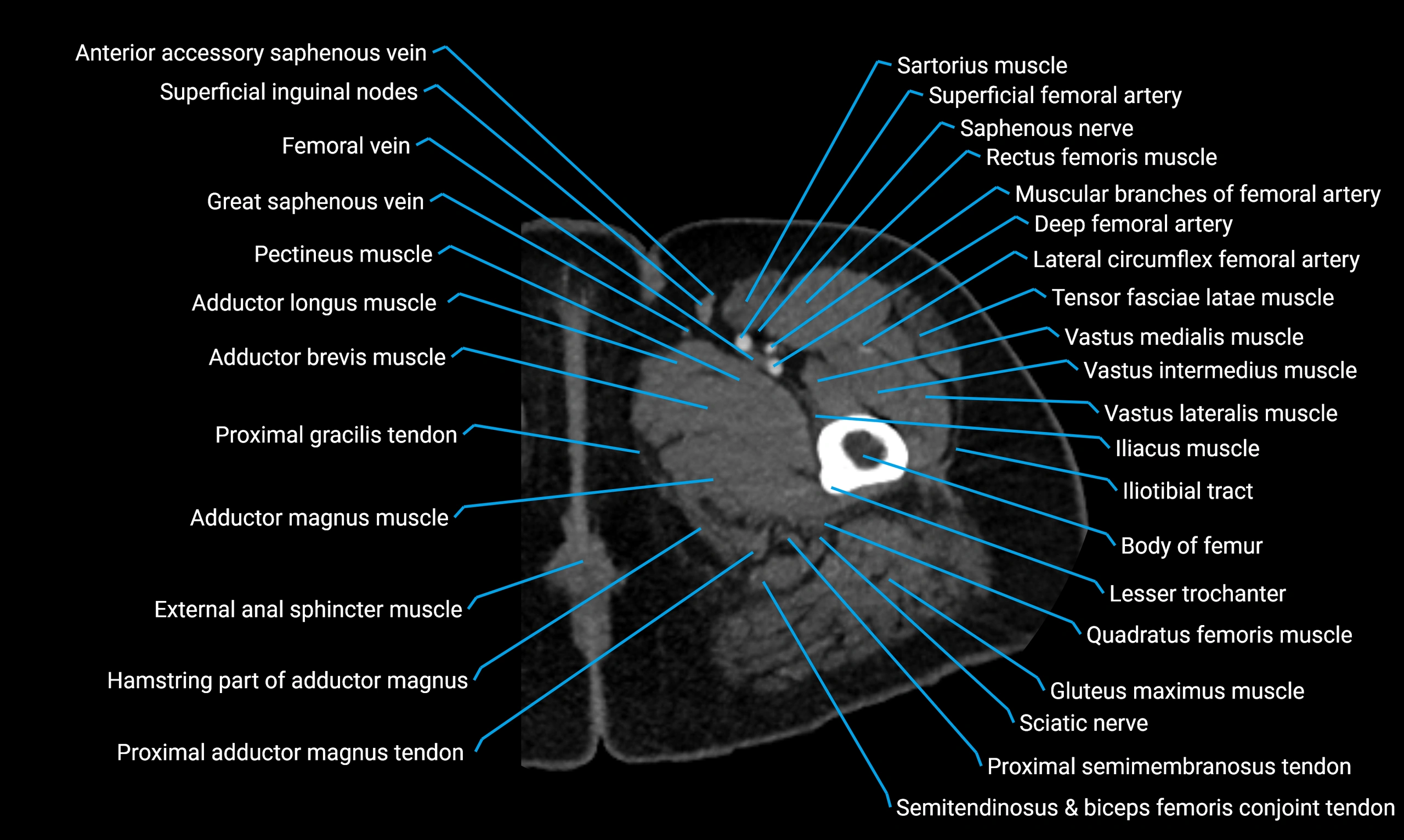
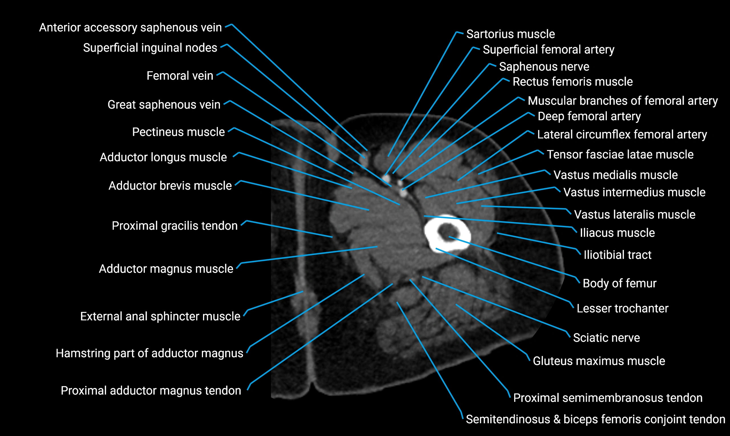
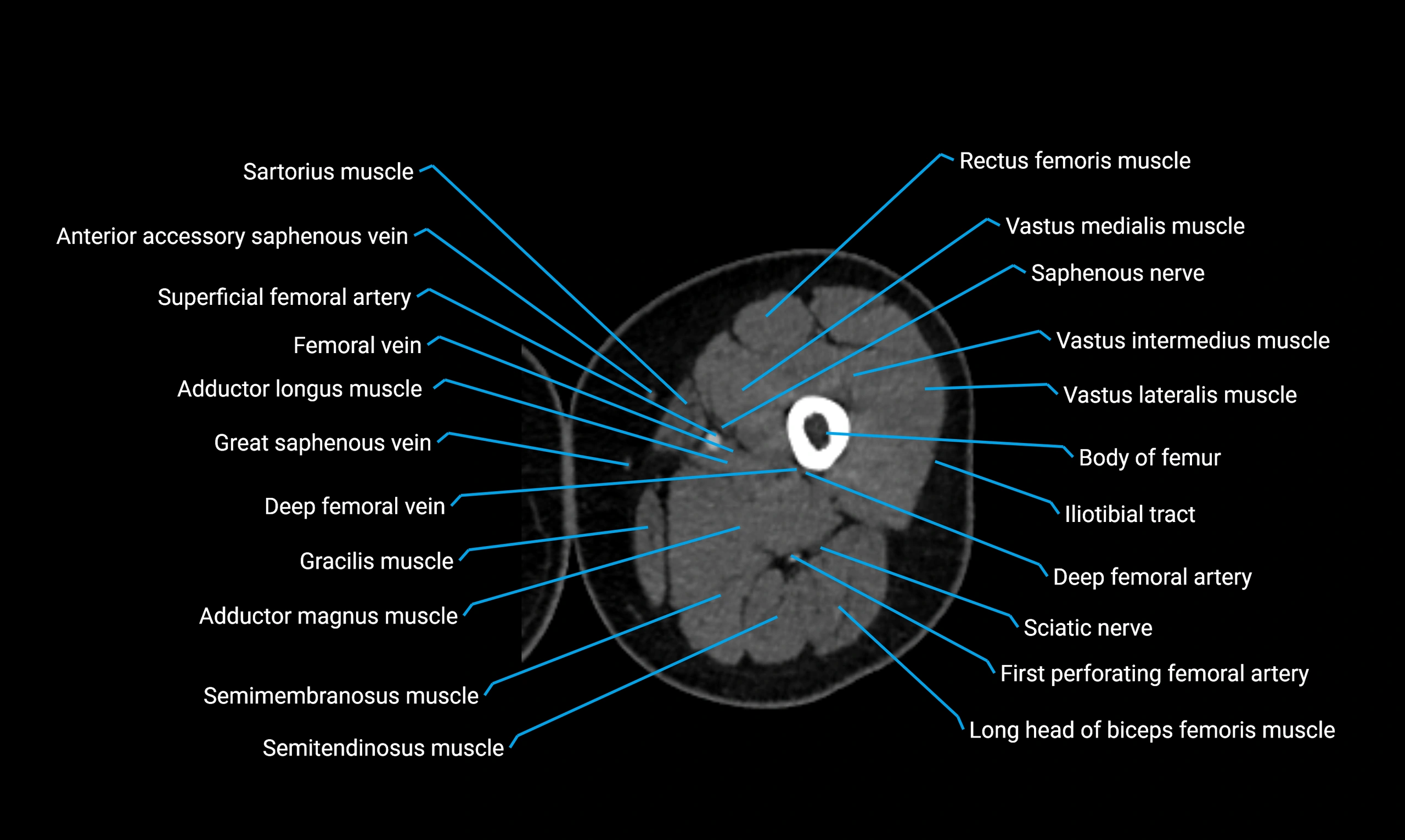
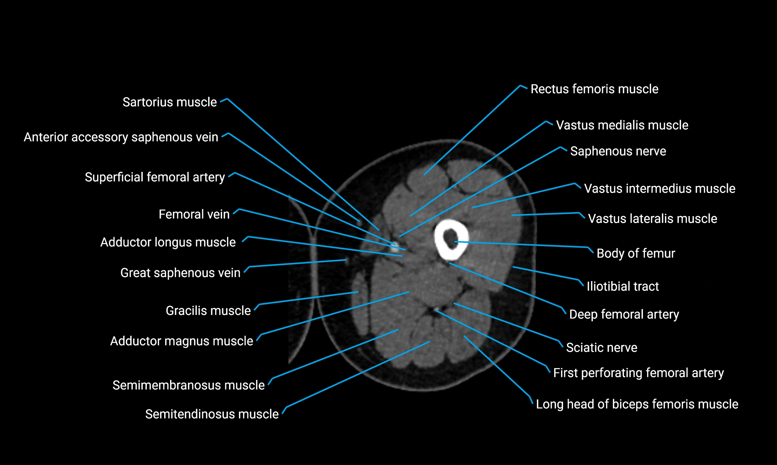
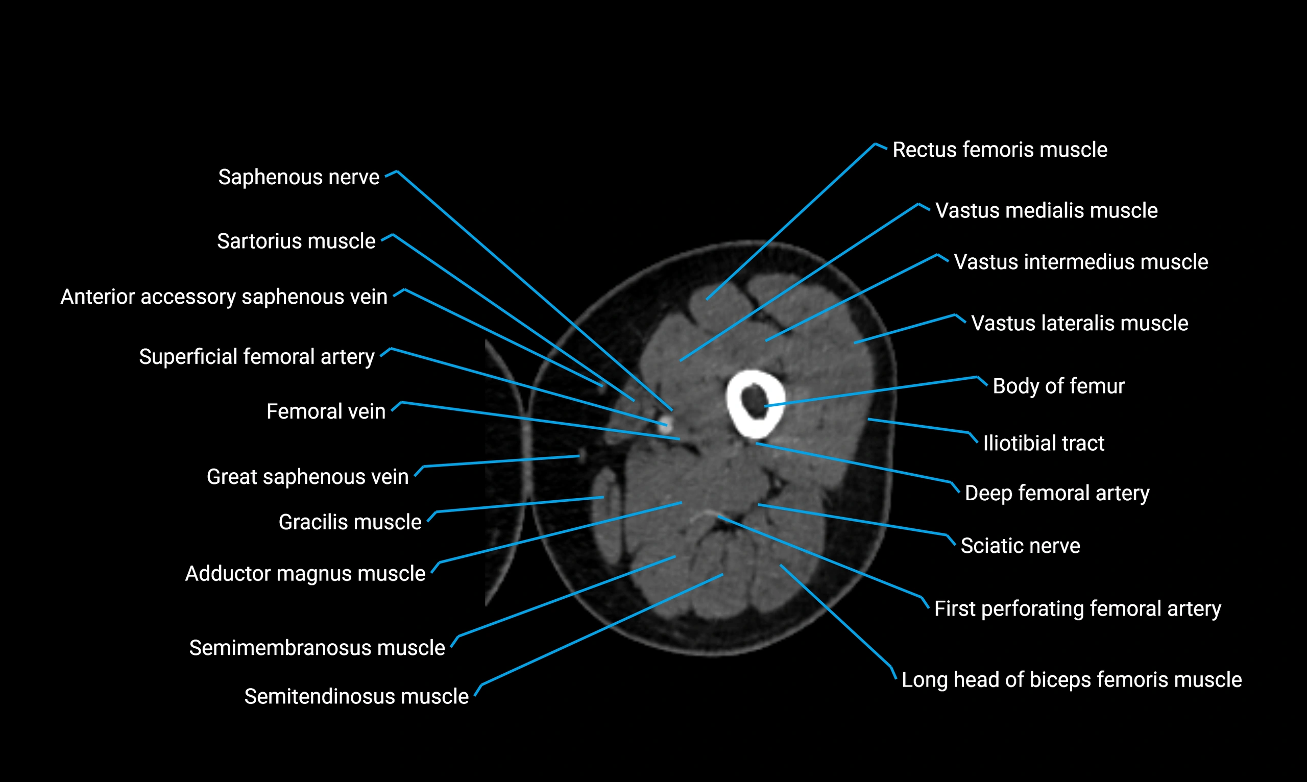
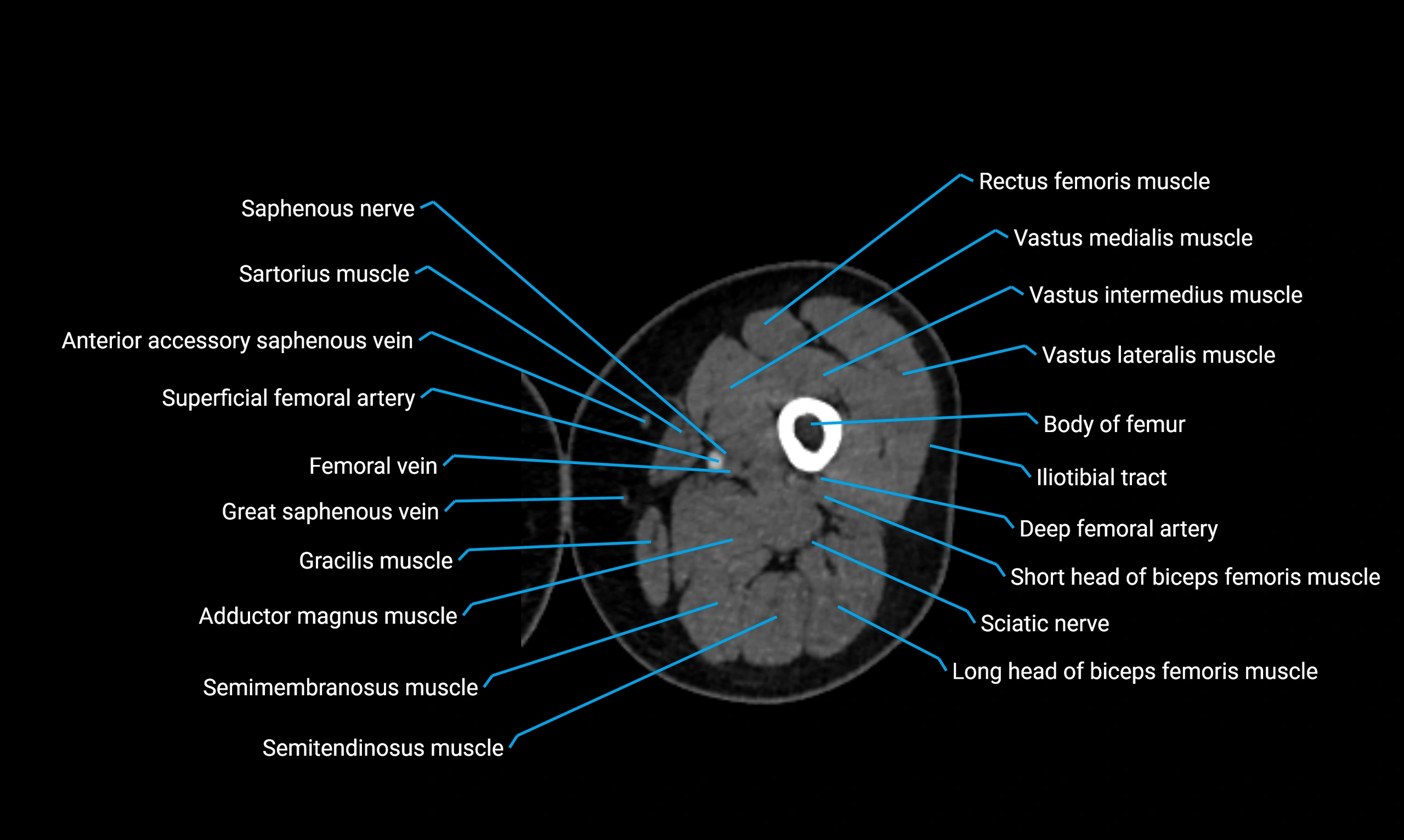
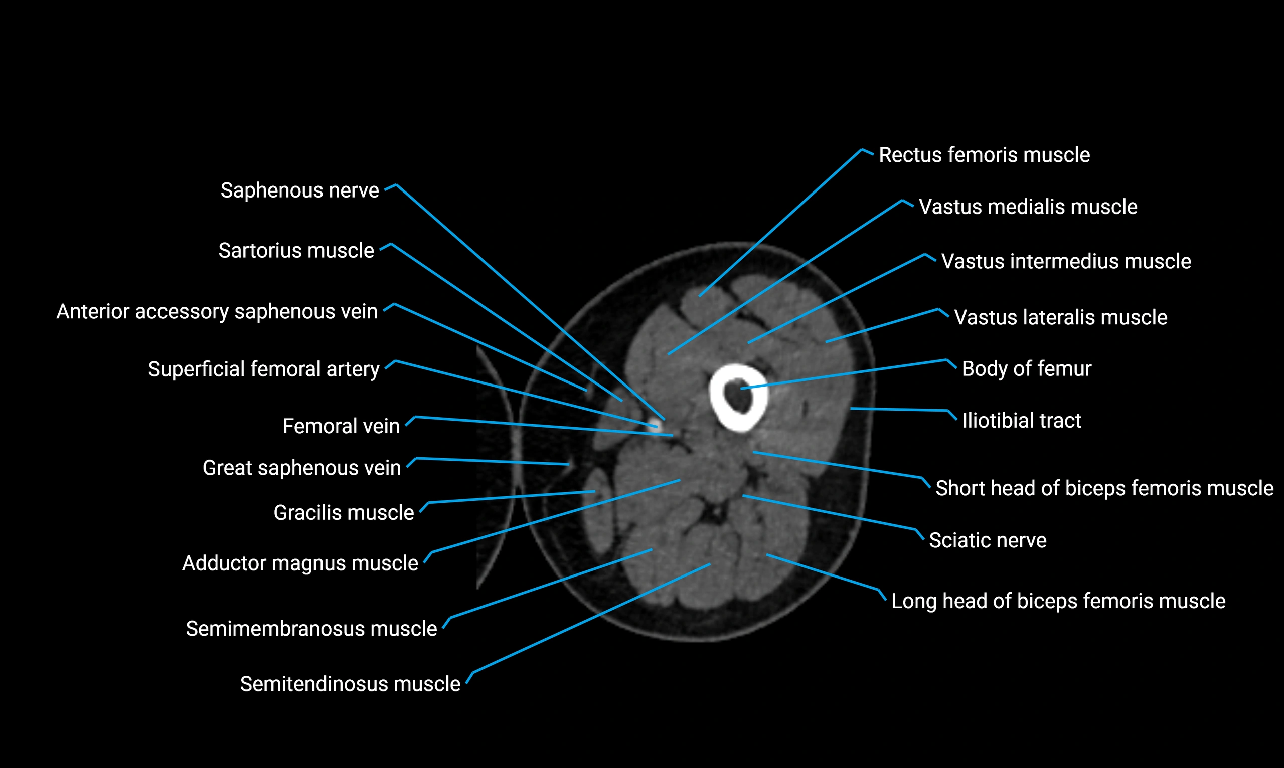
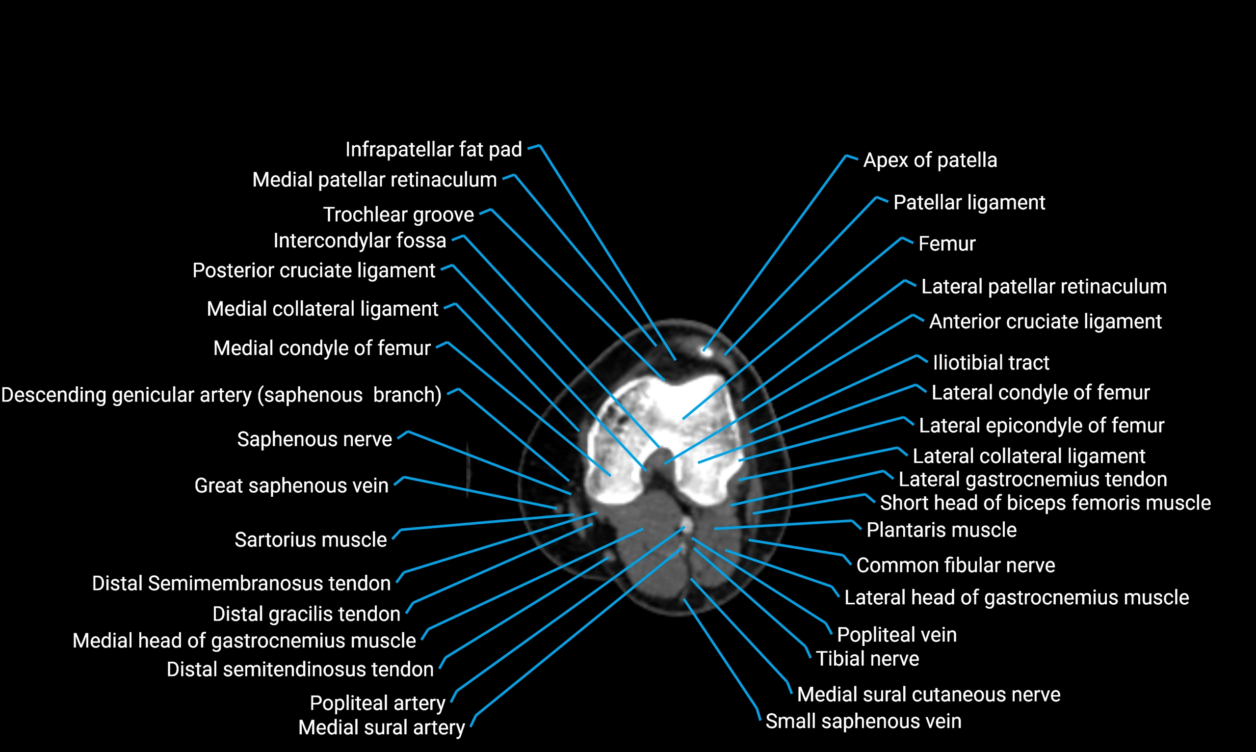
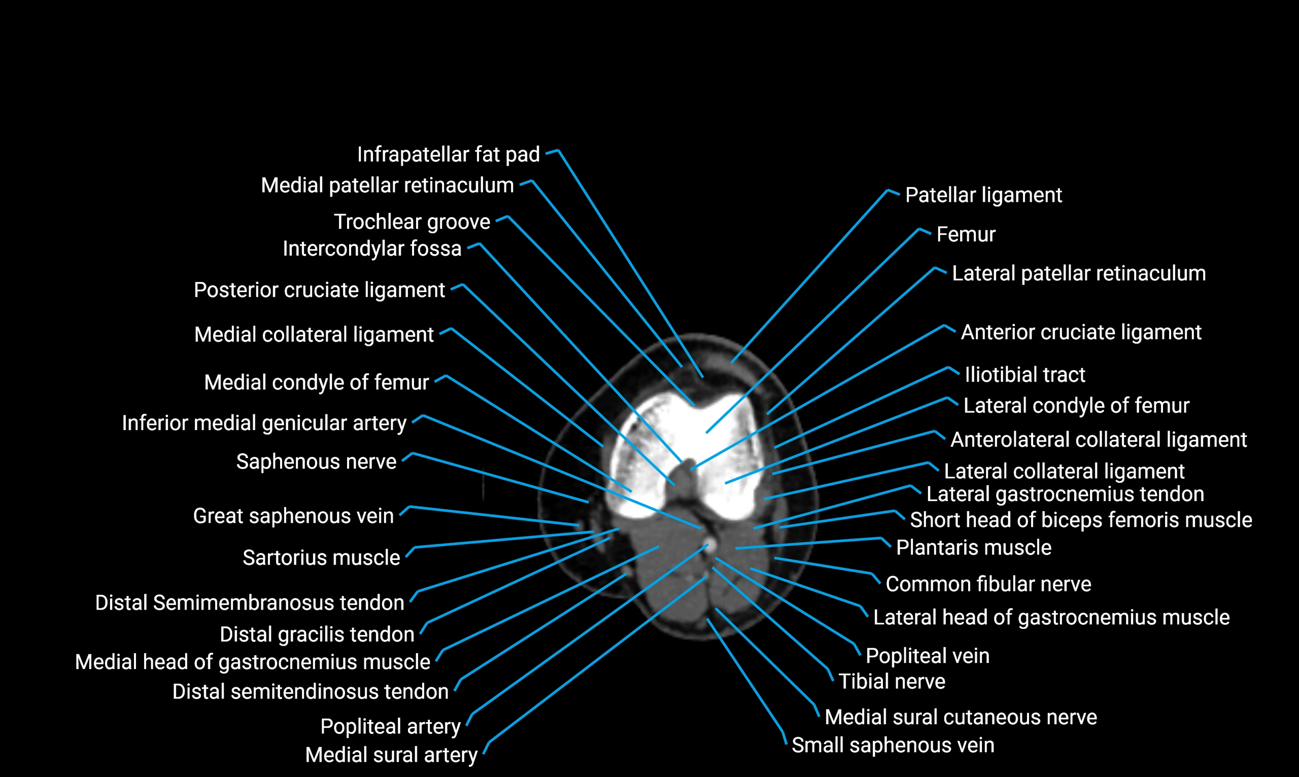
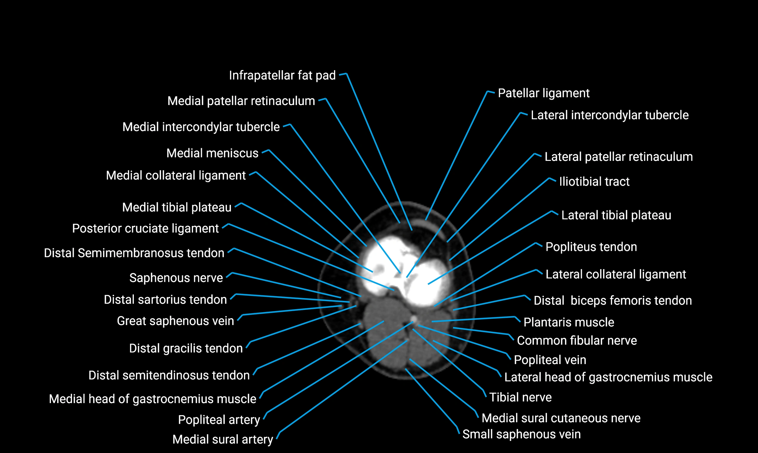
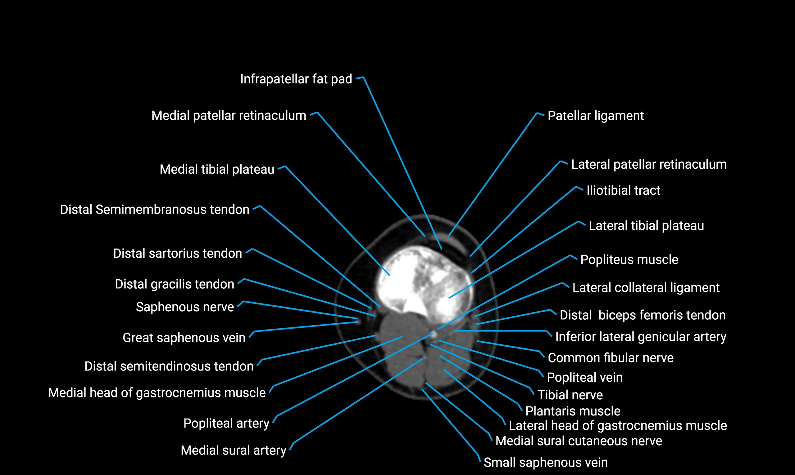
MRI image

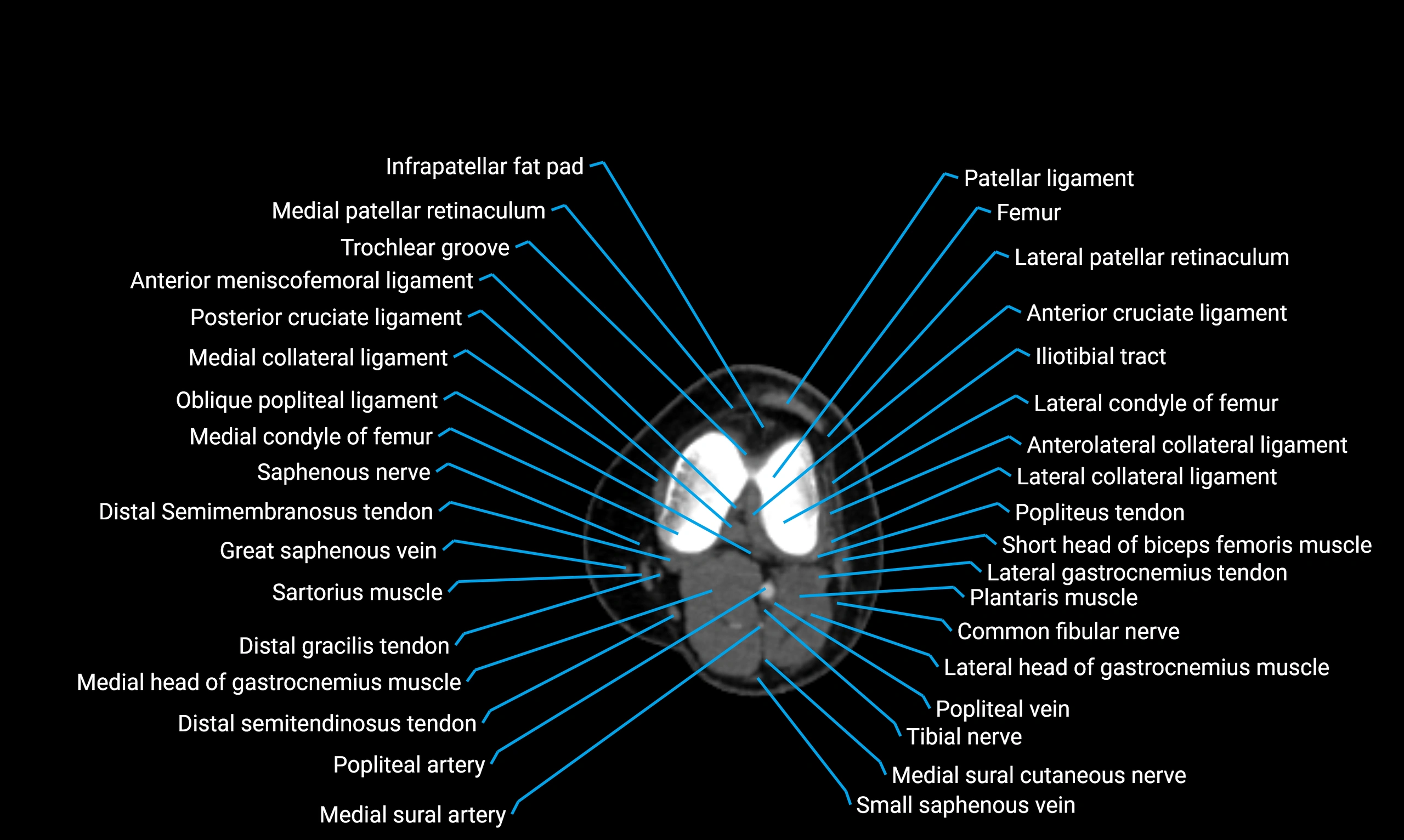
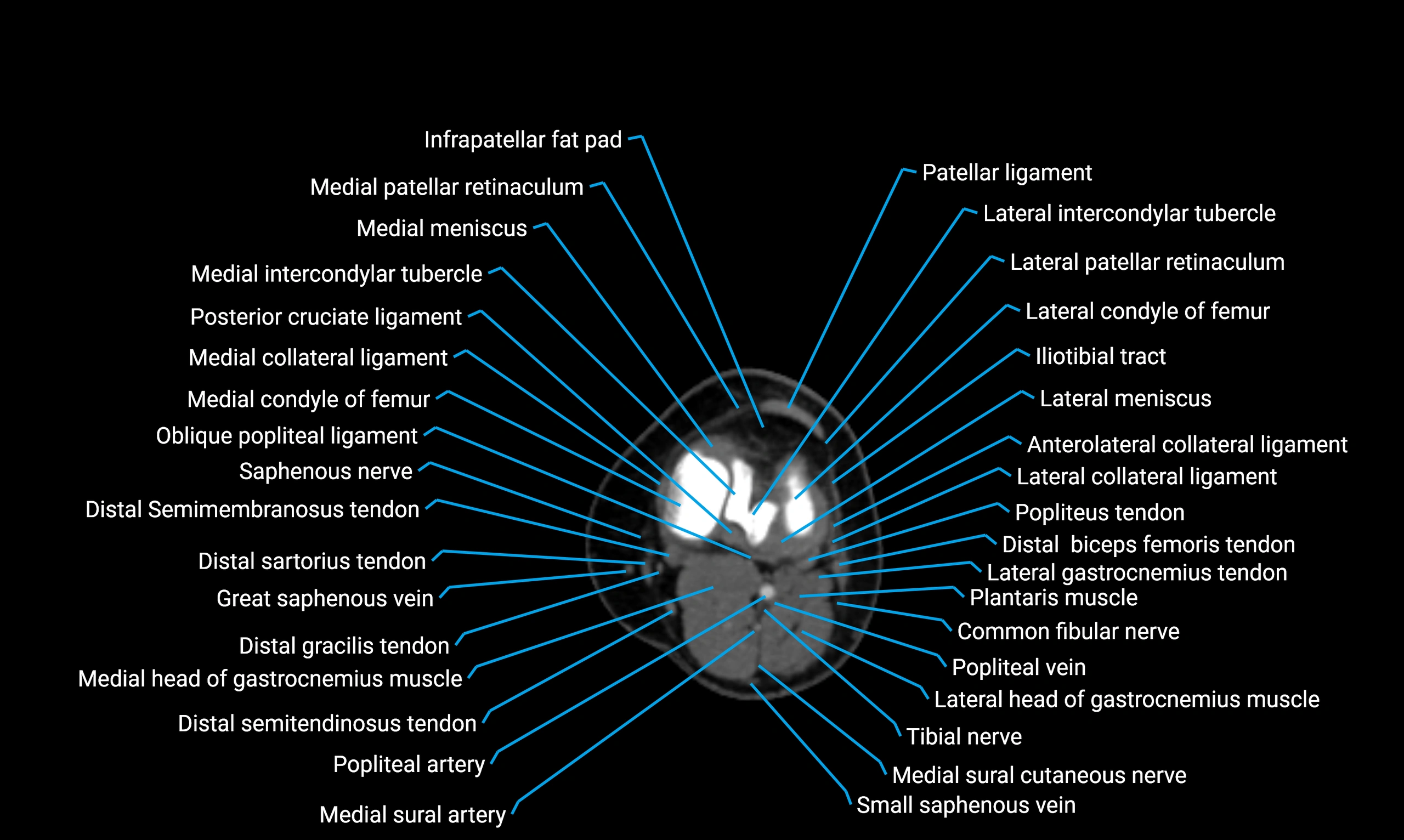
MRI image
