


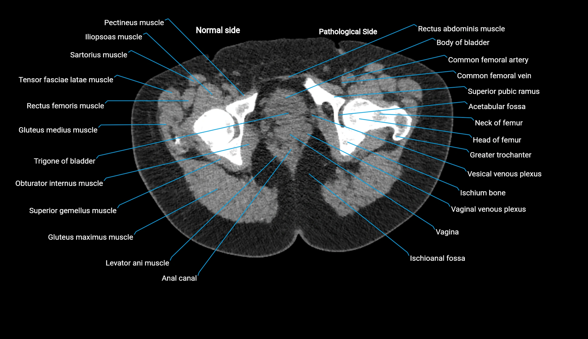

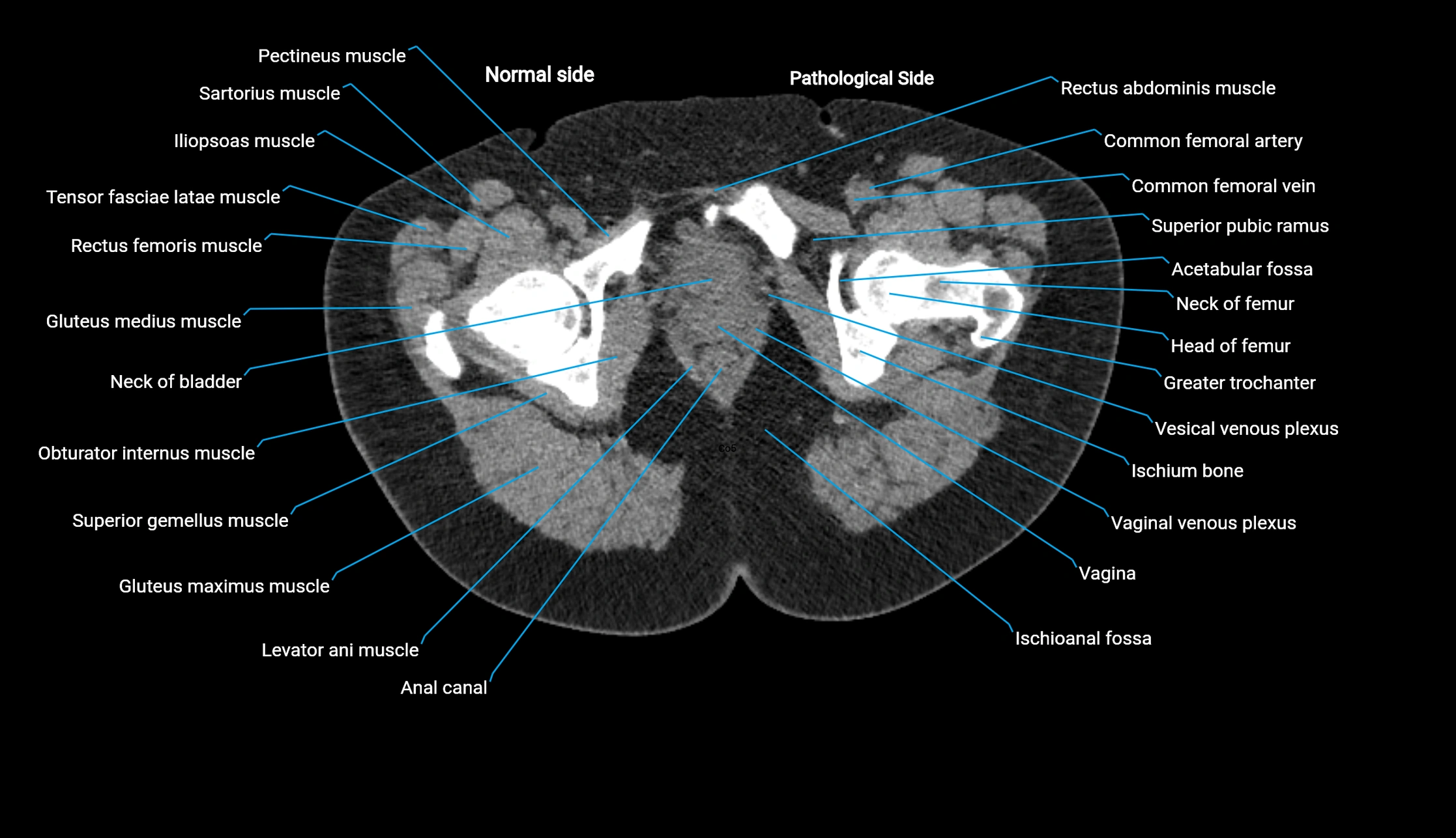
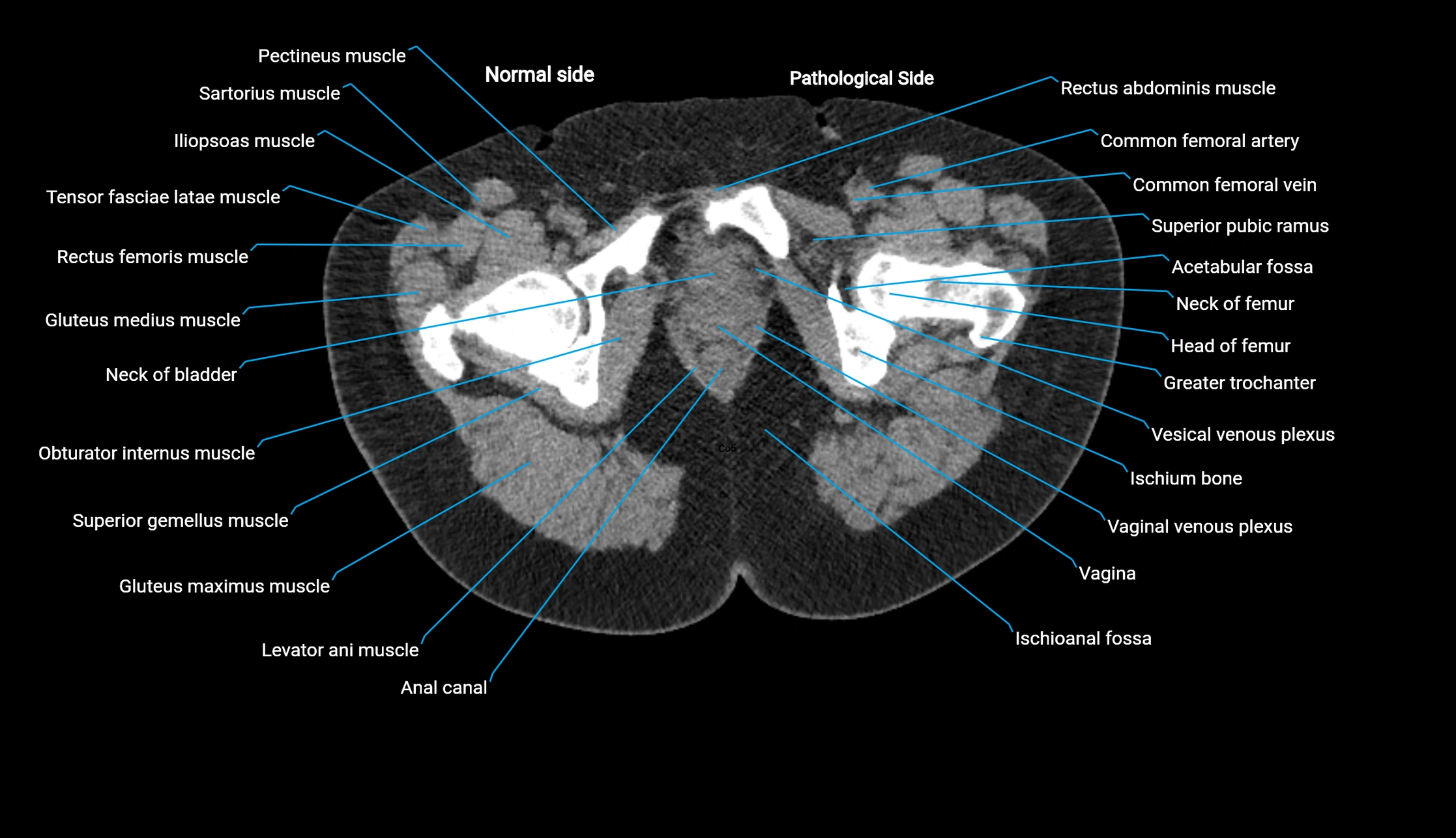
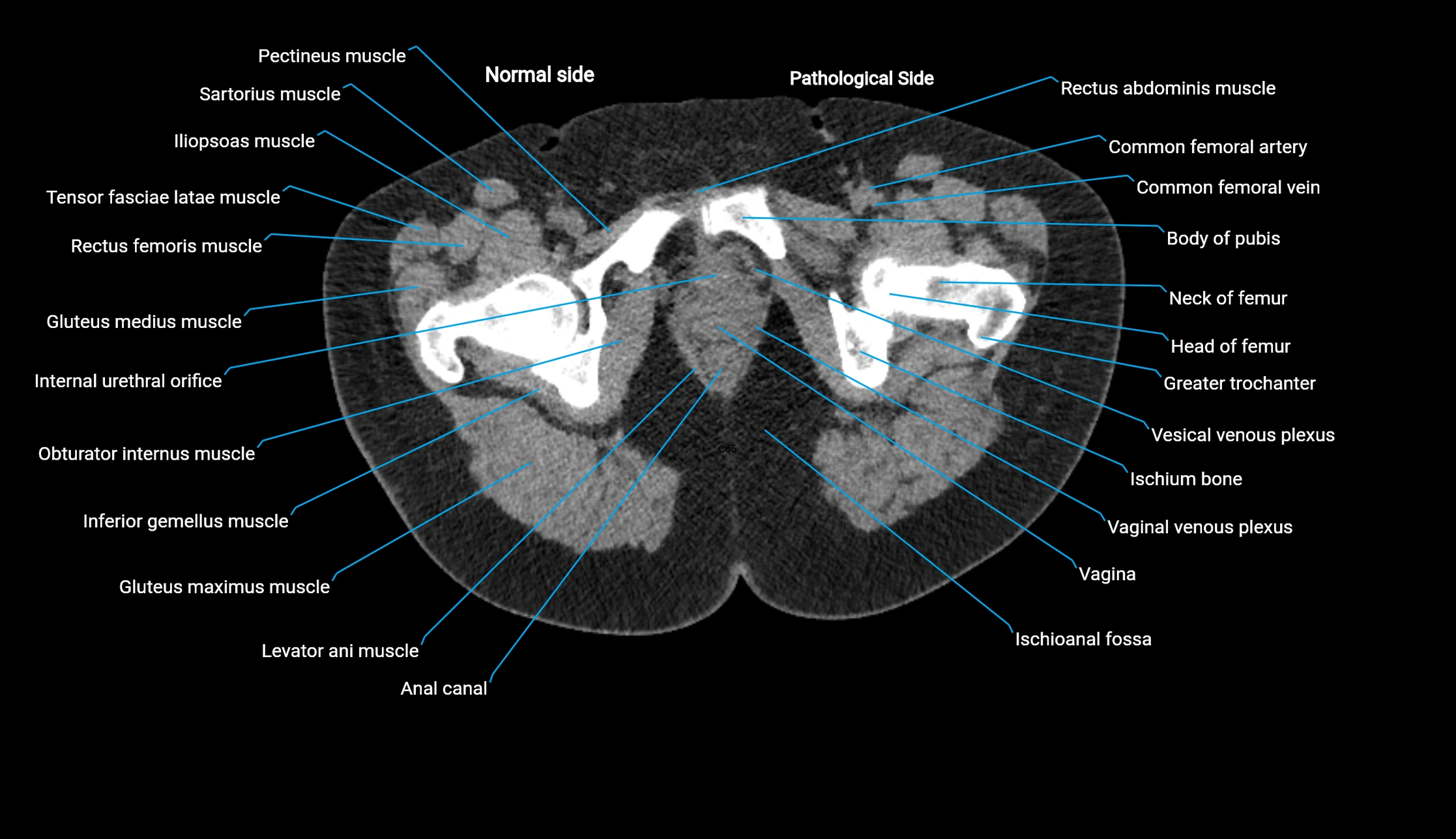
Topic
The acetabular margin, also called the acetabular rim, is the bony edge of the acetabulum, the cup-shaped cavity on the lateral aspect of the pelvis that articulates with the head of the femur to form the hip joint. The acetabular margin consists of the superior, anterior, and posterior borders of the acetabulum and is interrupted inferiorly by the acetabular notch.
The rim provides attachment for the acetabular labrum, a fibrocartilaginous structure that deepens the acetabulum, increasing hip joint stability. The transverse acetabular ligament bridges the acetabular notch, completing the bony ring. Superiorly, the margin bears the greatest load during standing and gait, making it the most common site of degenerative changes.
The acetabular margin is clinically significant in femoroacetabular impingement (FAI), acetabular fractures, hip dysplasia, and osteoarthritis. Its morphology (depth, coverage, and orientation) is a key factor in hip biomechanics and surgical planning, especially in arthroscopy and hip preservation surgery.
Synonyms
-
Acetabular rim
-
Acetabular border
-
Margin of acetabulum
Function
-
Forms the boundary of the acetabulum, contributing to the hip joint socket
-
Provides attachment for the acetabular labrum
-
Serves as a load-bearing structure during locomotion
-
Landmark for orthopedic surgery, hip arthroscopy, and imaging evaluation
MRI Appearance
T1-weighted images:
-
Acetabular bone cortex: low signal intensity
-
Marrow within acetabular margin: intermediate signal
-
Labrum appears hypointense, cartilage intermediate
T2-weighted images:
-
Subchondral bone: hypointense
-
Articular cartilage: bright hyperintense
-
Acetabular labrum: dark hypointense triangle at margin
-
Labral tears appear as hyperintense fluid clefts
PD Fat-Saturated (Proton Density with Fat Suppression):
-
Suppresses marrow fat, highlighting cartilage, labrum, and soft tissue pathology
-
Labral tears, cartilage defects, and periacetabular edema appear bright hyperintense
-
Highly sensitive for subtle labral pathology and chondral damage
STIR:
-
Fat suppression highlights bone marrow edema, fractures, or periacetabular inflammation
-
Useful in trauma and early arthritis detection
T1 Post-Gadolinium (MR Arthrography):
-
Contrast fills joint space and extends into labral or chondral tears
-
Enhances delineation of acetabular margin–labrum interface
-
Detects subtle labral detachments and capsular pathology
3D T2-weighted Imaging:
-
Provides isotropic voxels for multiplanar reconstructions of the acetabulum
-
Excellent for visualizing labrum, cartilage surface, and acetabular morphology
-
Essential for arthroscopic planning and evaluation of femoroacetabular impingement
CT Appearance
Non-contrast CT:
-
Demonstrates cortical bone of acetabular rim in excellent detail
-
Detects fractures, dysplasia, retroversion, or bony overcoverage (pincer impingement)
-
3D reconstructions used in preoperative hip surgery planning
CT Post-Contrast (CT Arthrography):
-
Joint contrast outlines the acetabular labrum, cartilage, and margin
-
Demonstrates labral tears, cartilage defects, and subtle bony abnormalities
-
Alternative to MR arthrography in patients with MRI contraindications
CT VRT 3D image

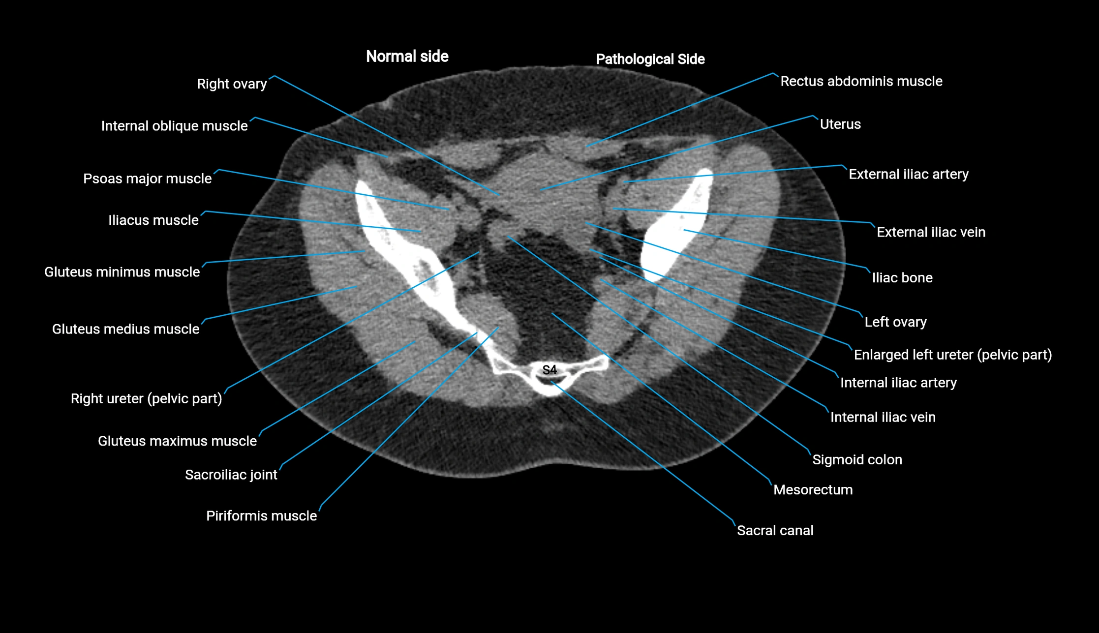
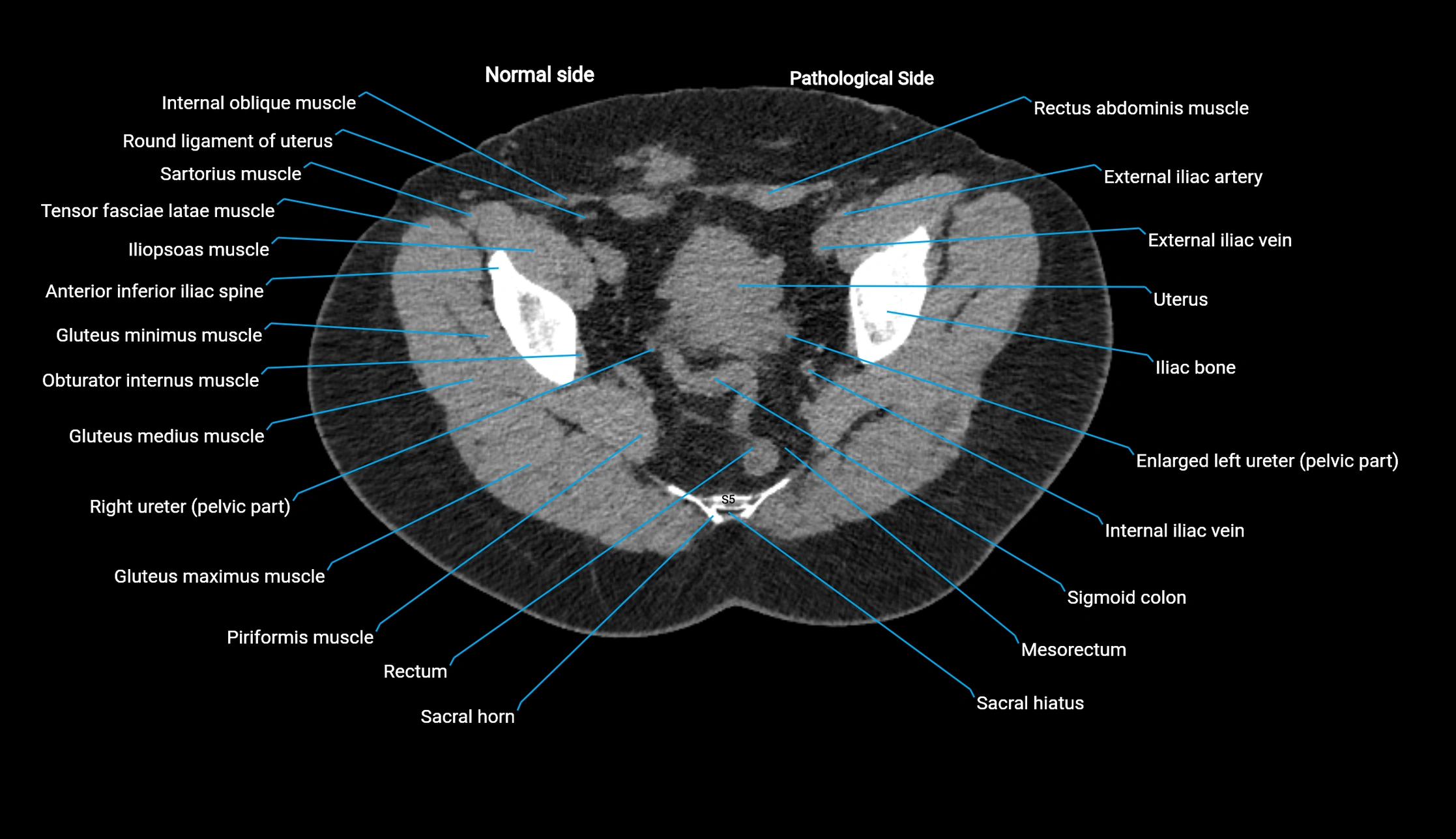
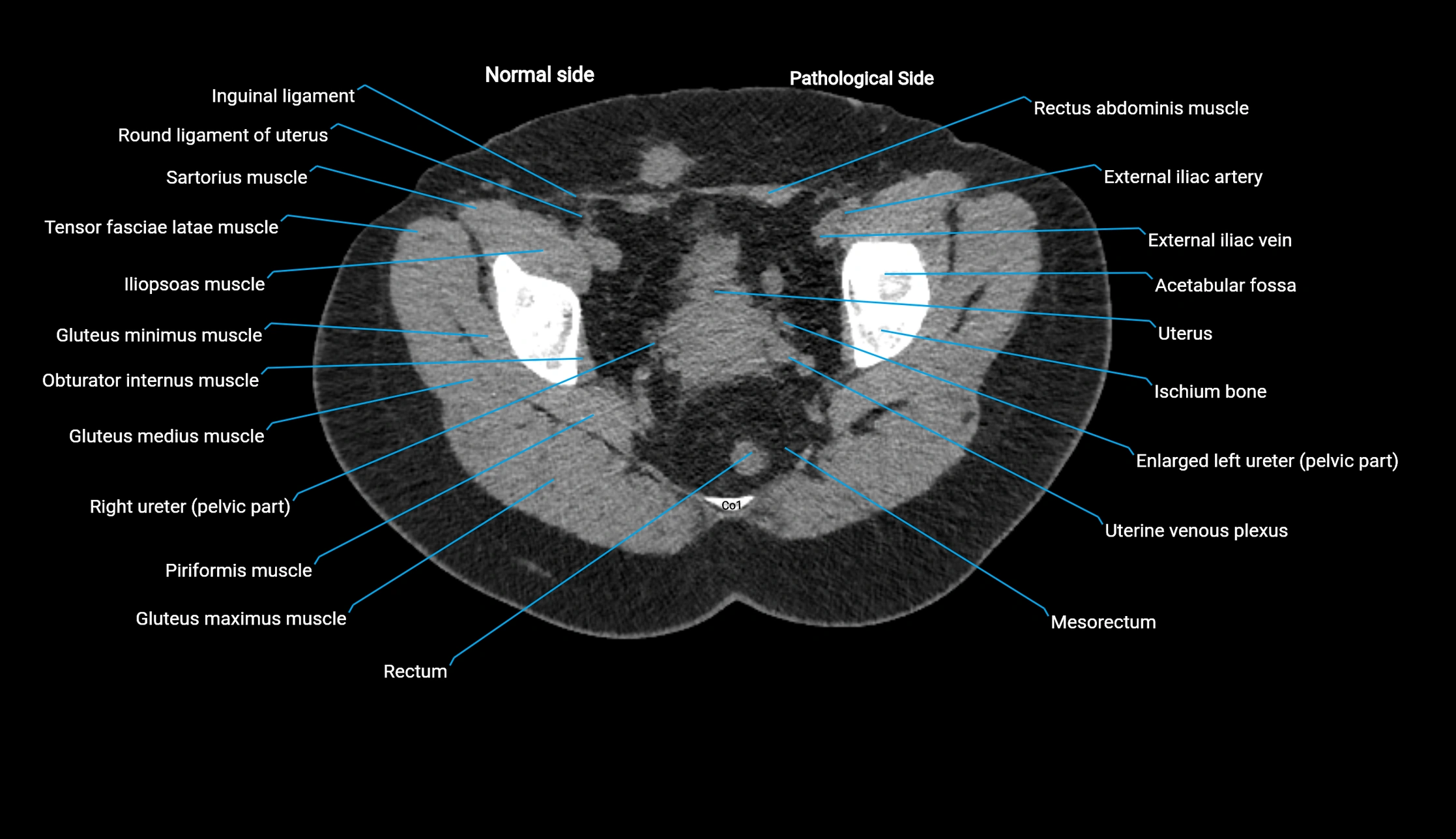
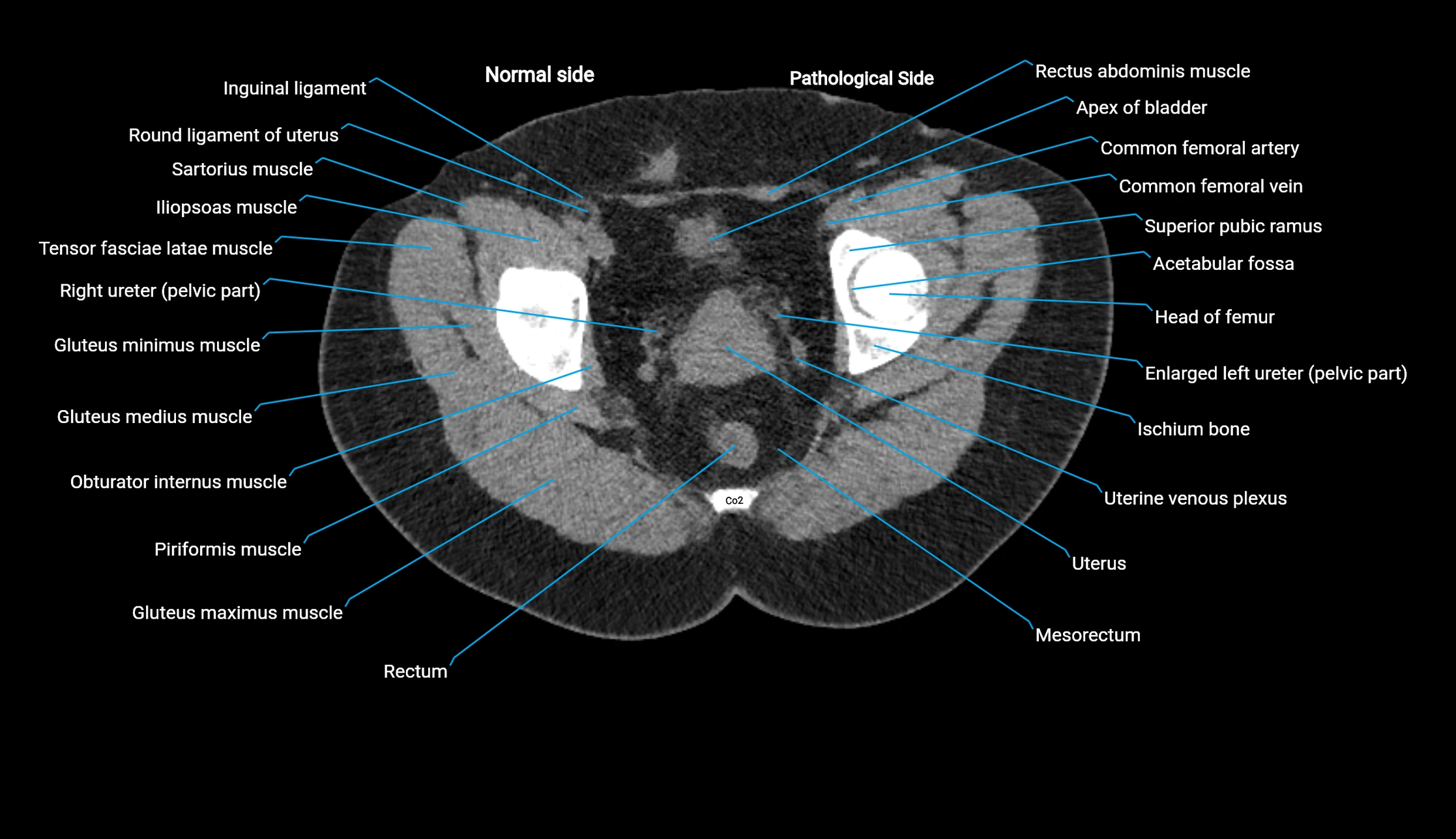
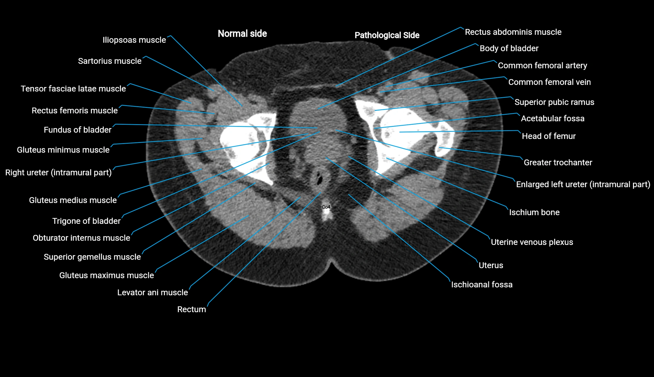
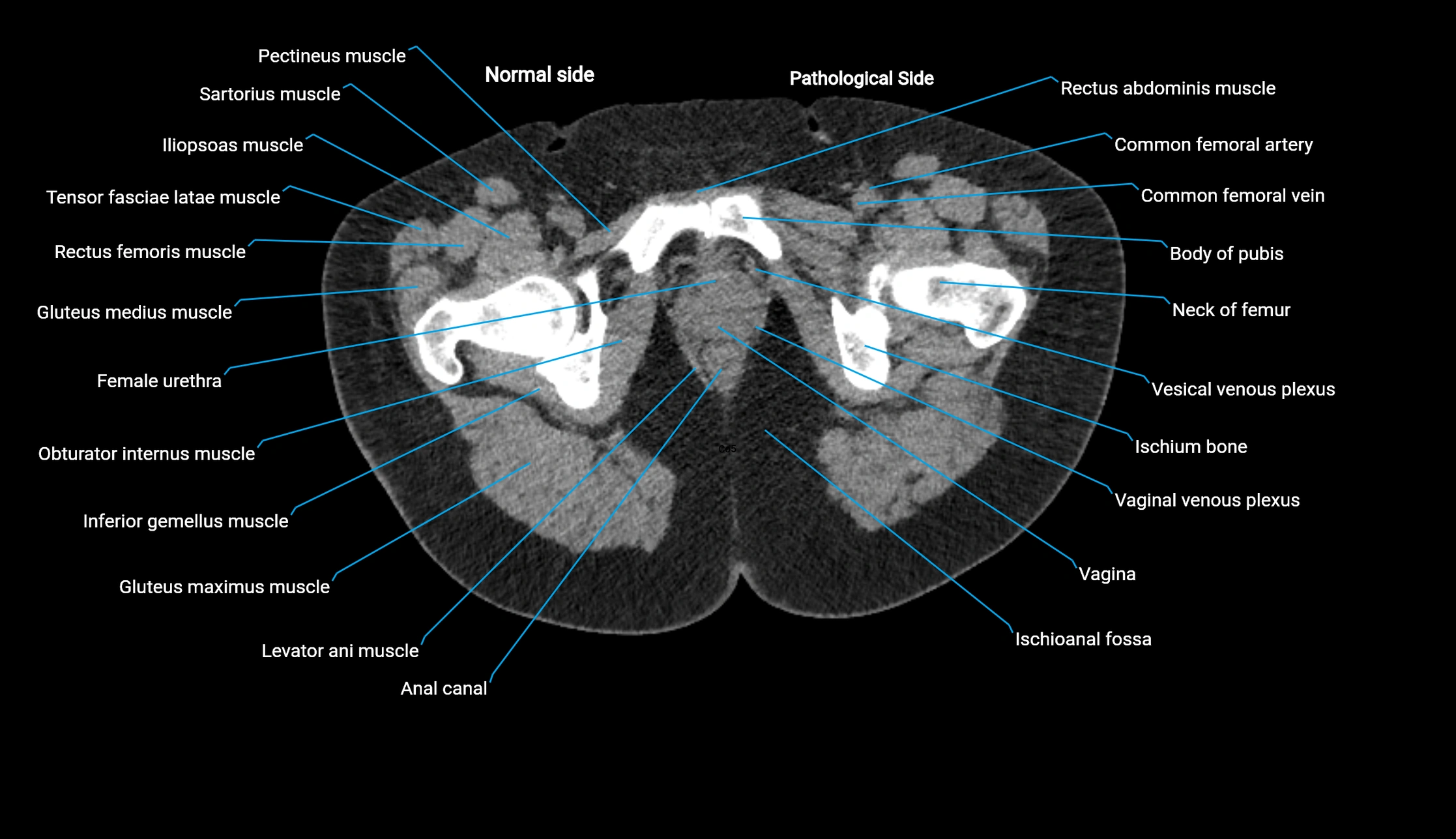
CT image

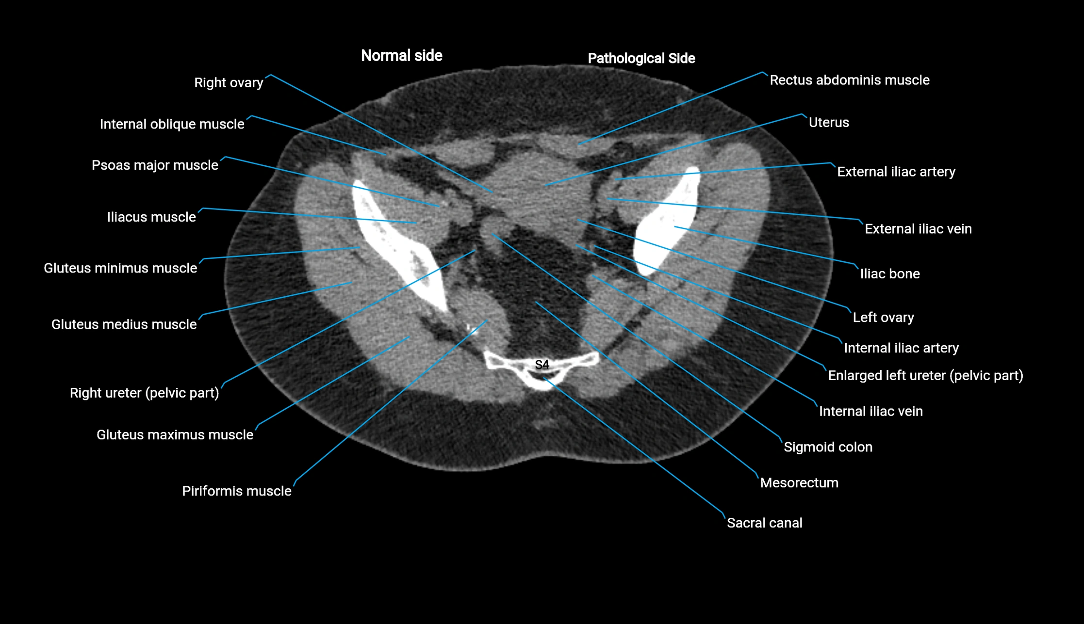
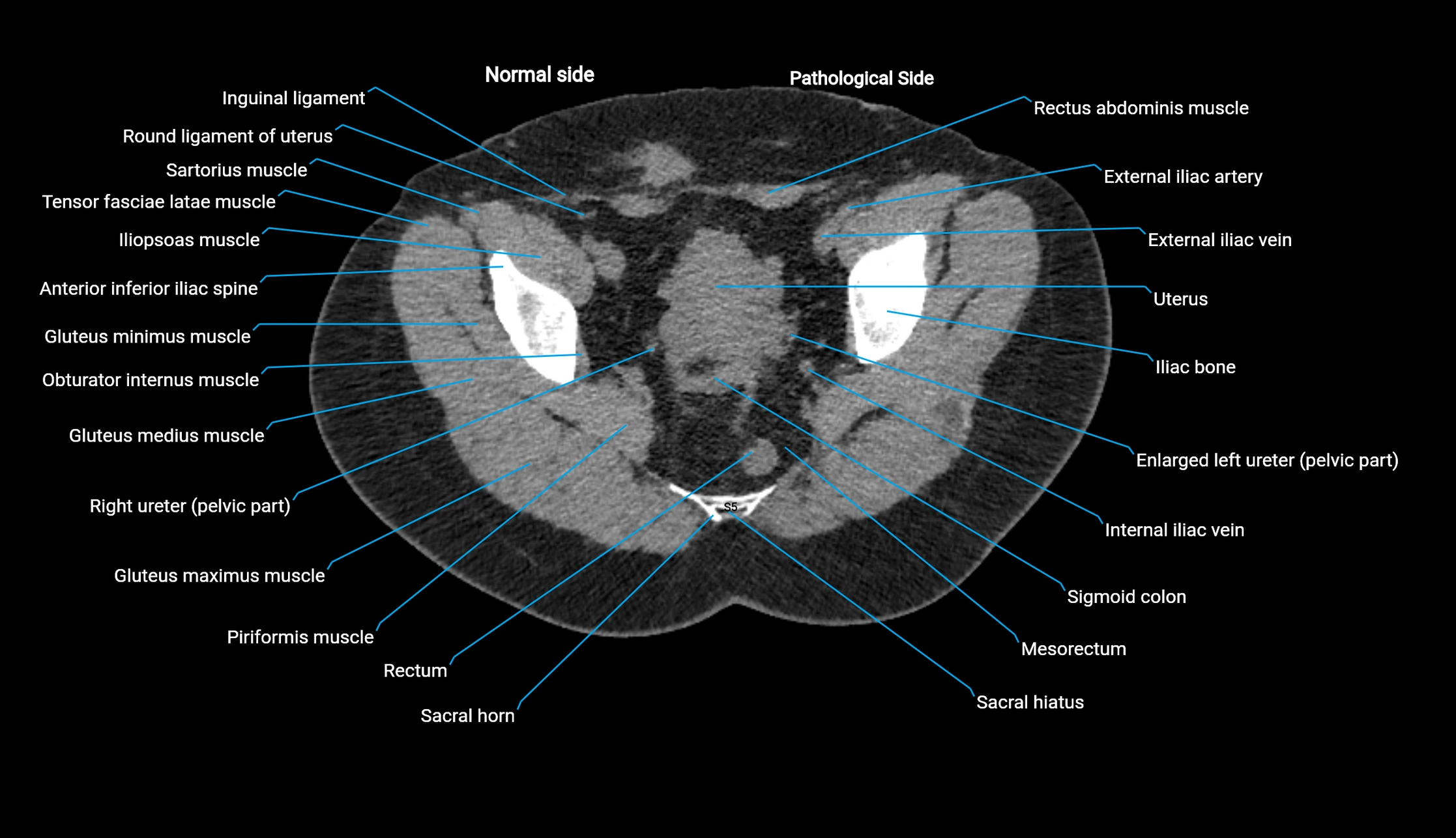
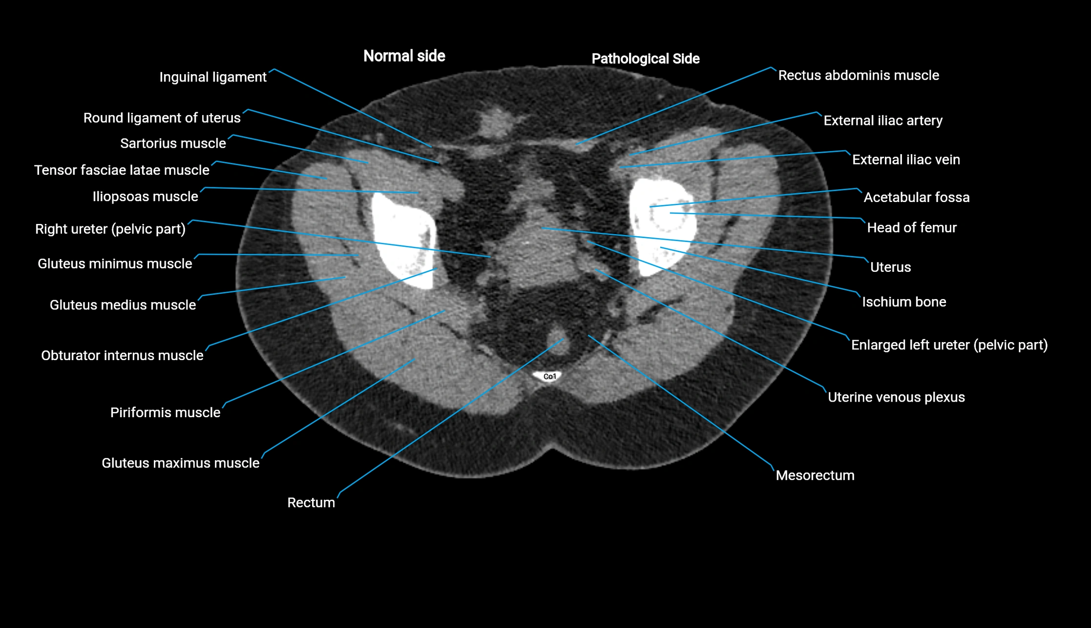
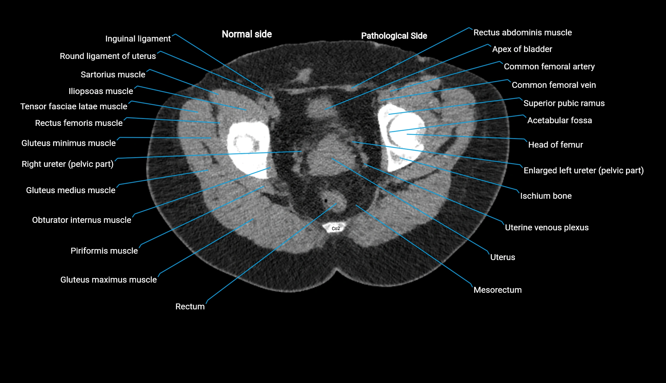
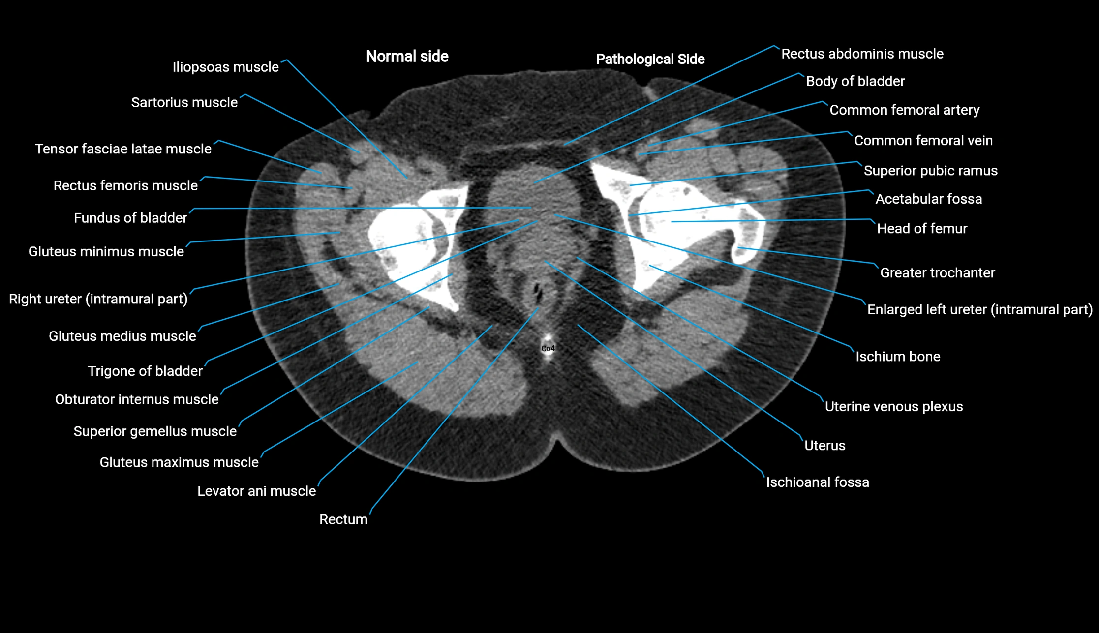
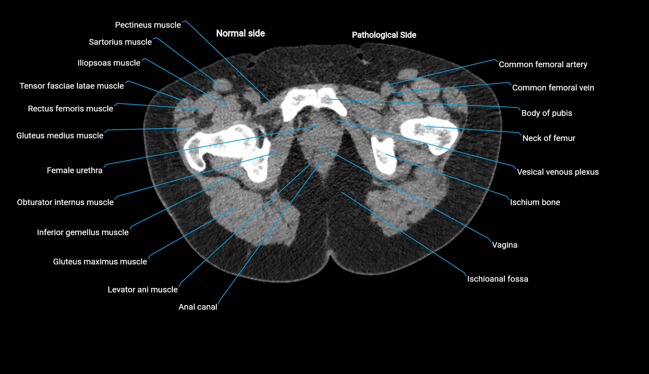
CT image

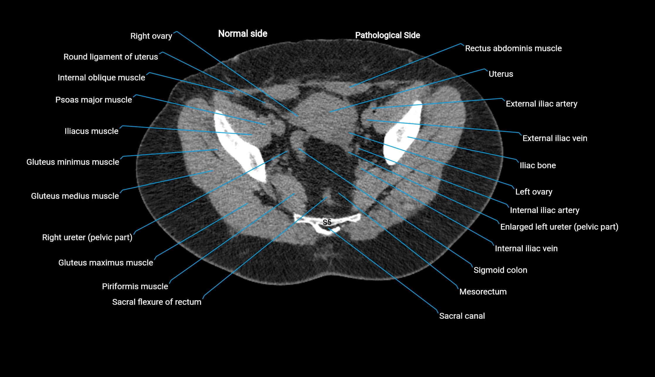
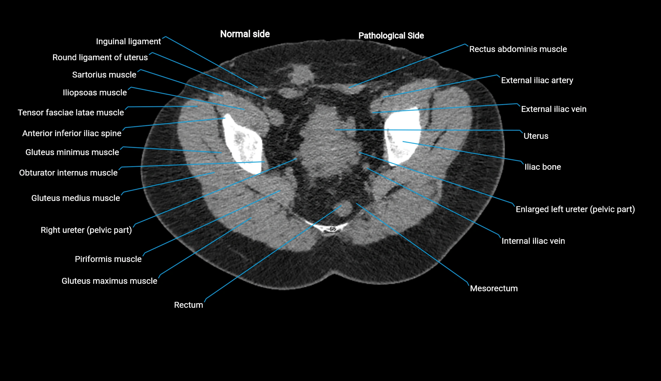
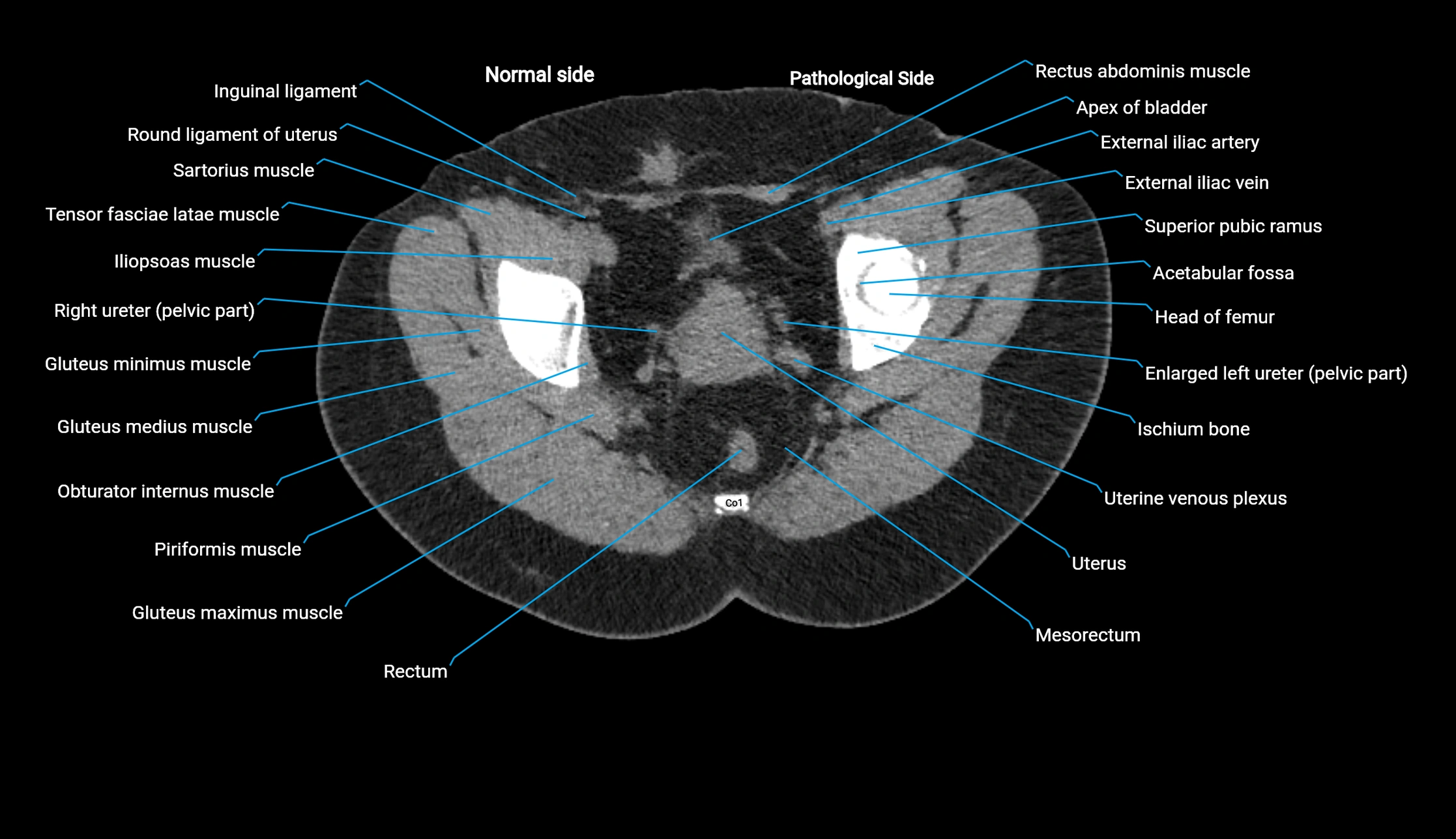
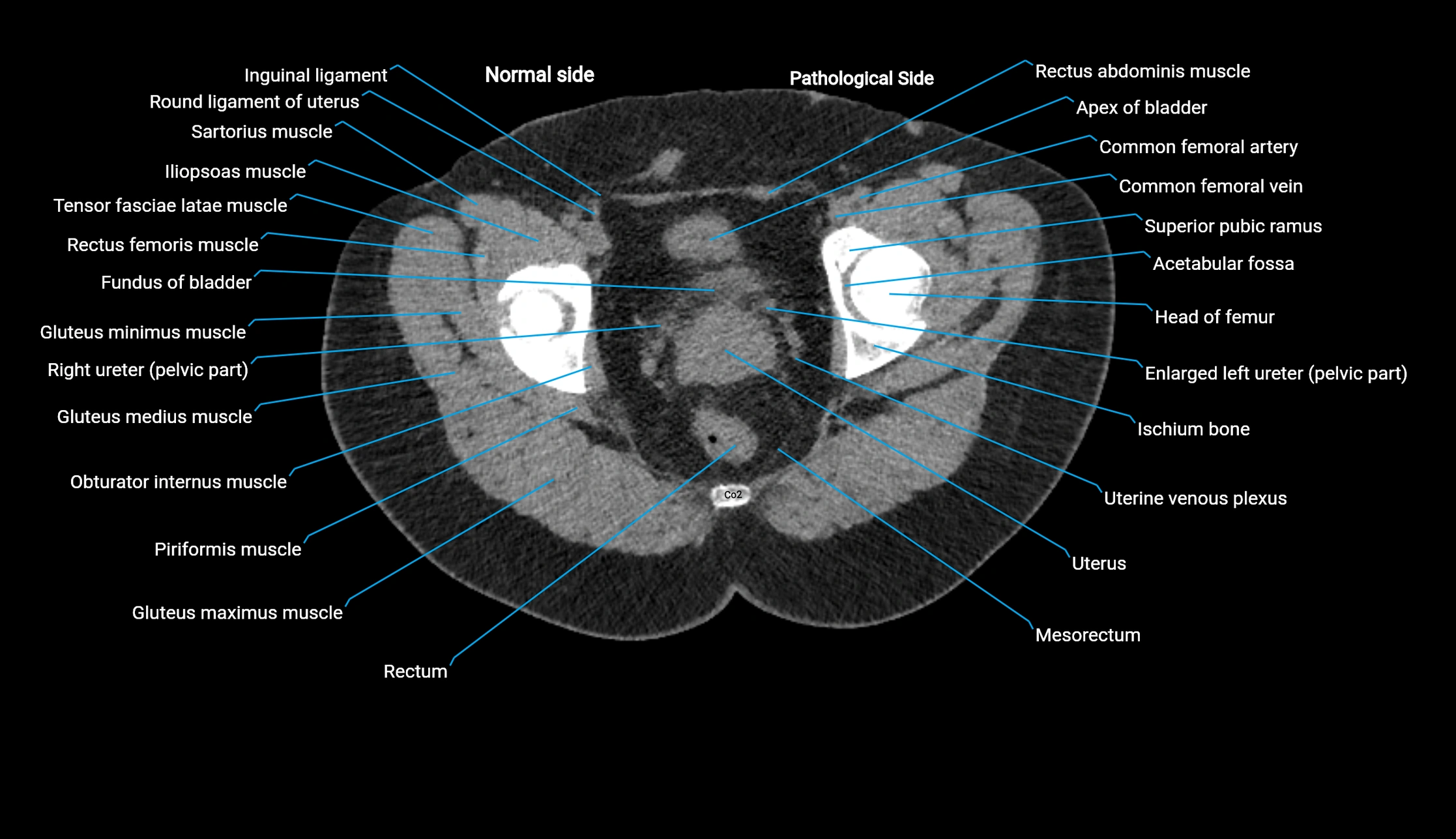
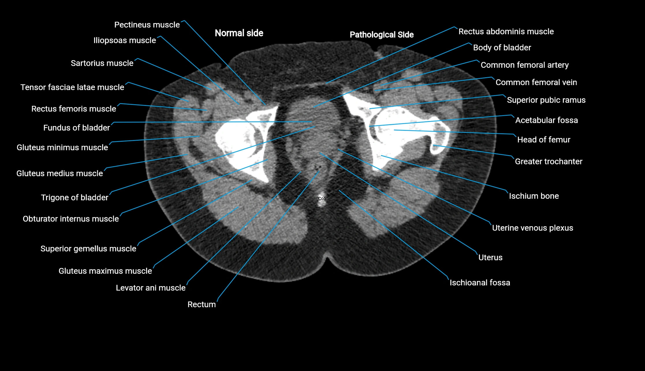
CT image

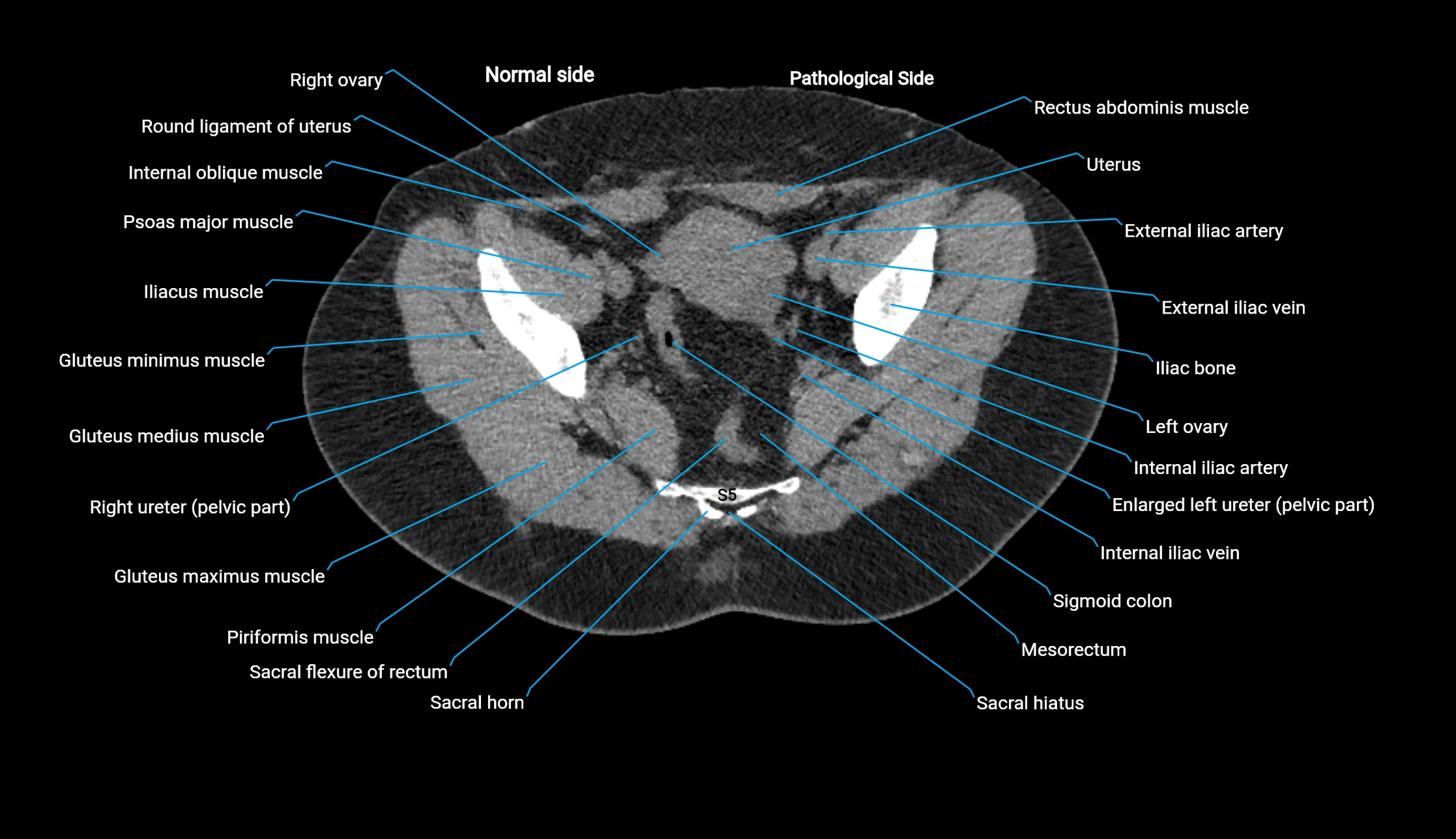
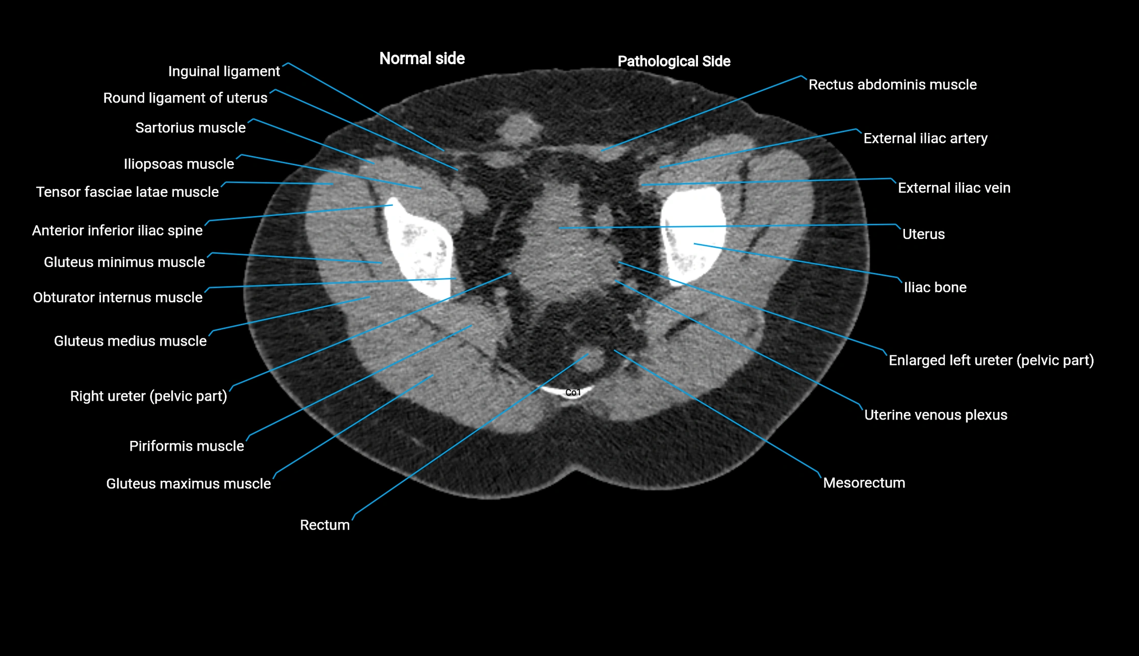
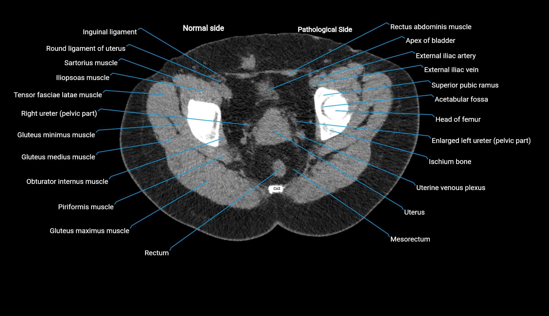
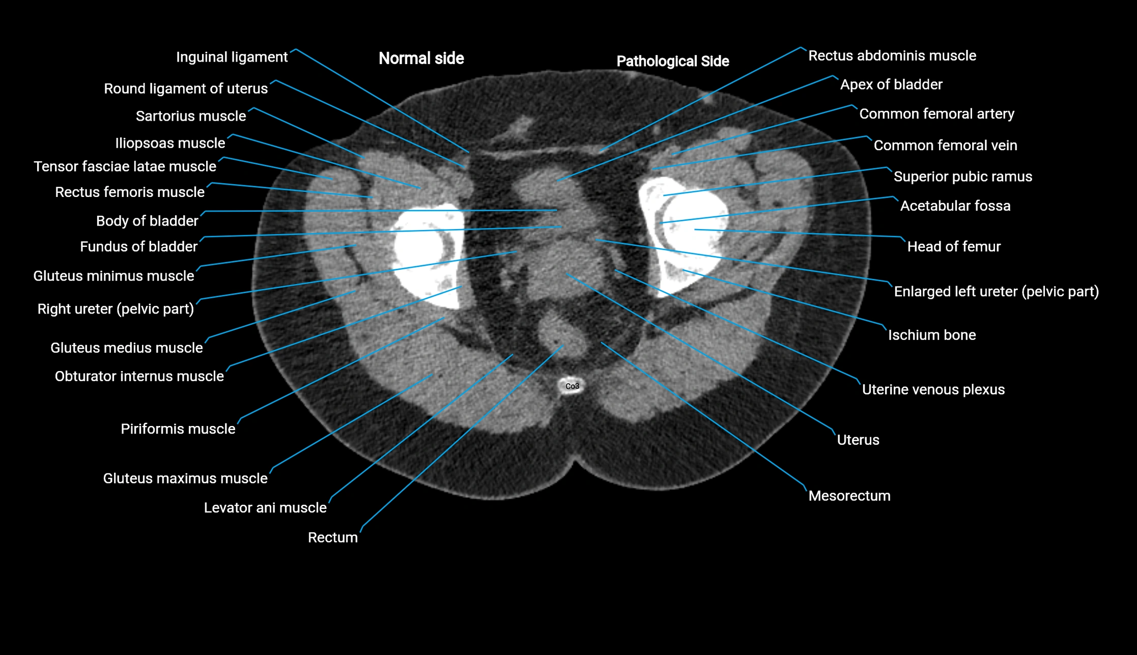
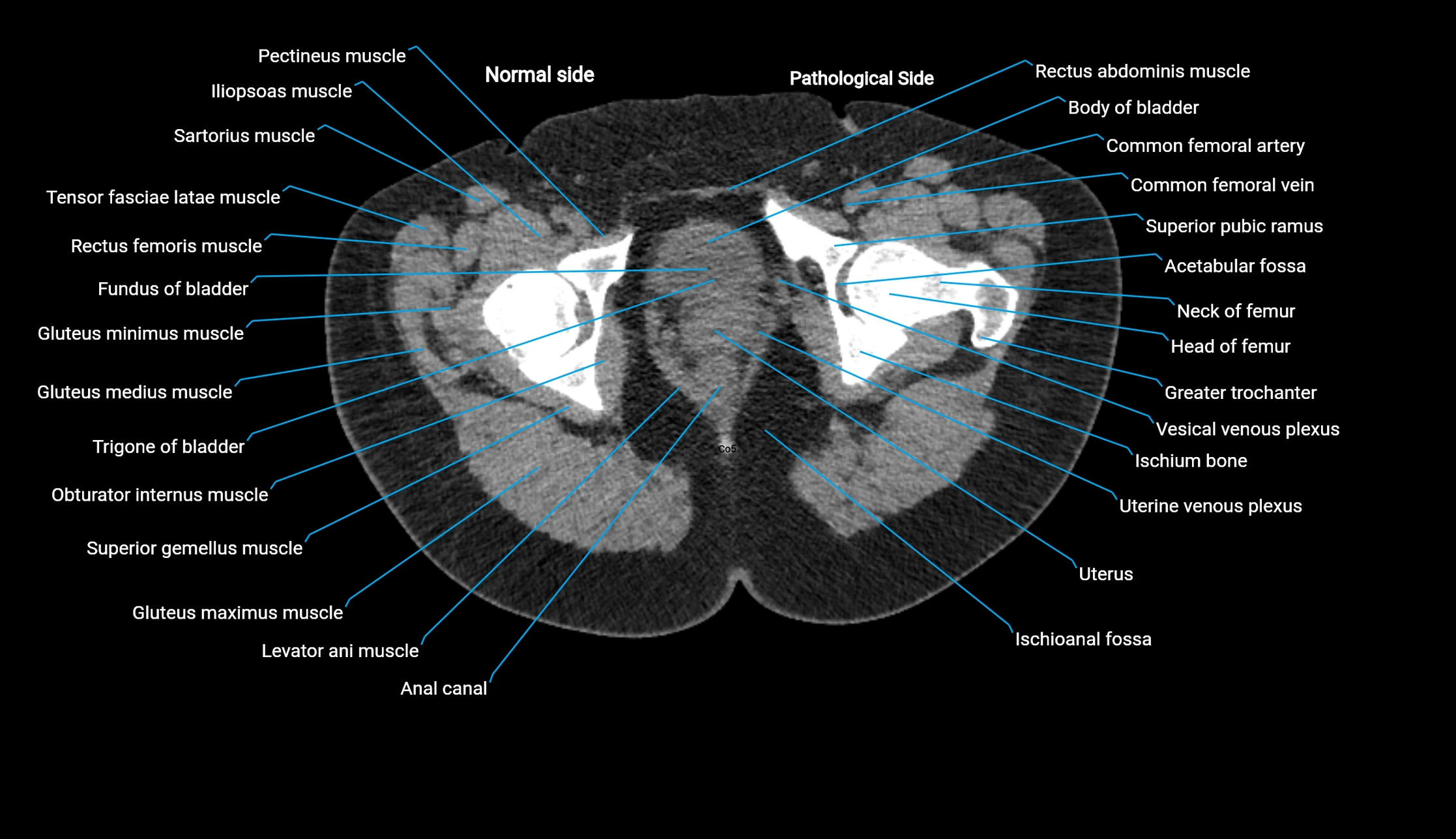
MRI image

MRI image
