


Topic
- Acetabular margin (Acetabular rim)
- Acetabular notch
- Acetabulum
- Ala of ilium (wing of ilium)
- Anterior Fibromuscular Stroma of prostate
- Anterior acetabular wall
- Anterior division of obturator nerve (Anterior branch of obturator nerve)
- Anterior inferior iliac spine
- Anterior lateral femoral cutaneous nerve
- Anterior rim of acetabulum
- Anterior sacral foramina
- Anterior sacroiliac ligament
- Anterior superior iliac spine
- Anterior wall of acetabulum
- Apex of urinary bladder
- Body of femur
- Body of ilium
- Body of ischium
- Body of pubis
- Body of urinary bladder
- Bulbospongiosus muscle (Female)
- Bulbospongiosus muscle (Male)
- Central zone of prostate
- Clitoris
- Coccygeal nerve
- Coccygeal plexus
- Coccyx
- Common iliac lymph nodes
- Common iliac vein
- Conjoint tendon of biceps femoris & semitendinosus
- Deep femoral vein (profunda femoris vein)
- Deep transverse perineal muscle
- Ejaculatory duct
- Endocervical canal
- Erector spinae muscles
- Exiting nerve root of spinal nerve S1
- Exiting nerve root of spinal nerve S2
- External iliac vein
- External os of the cervix
- External urethral orifice
- External urethral sphincter (female)
- External urethral sphincter (male)
- Fascia of pelvic diaphragm
- Femoral nerve
- Femoral shaft
- Femur
- Fovea for ligament of head of femur
- Fundus of urinary bladder
- Genitofemoral nerve
- Gluteal lymph nodes
- Gluteal tuberosity
- Gluteus medius muscle
- Gluteus medius tendon
- Gluteus minimus muscle
- Gluteus minimus tendon
- Gracilis muscle
- Greater sciatic notch
- Greater trochanter
- Hamstring muscles
- Head of femur
- Hip joint
- Iliac bone
- Iliococcygeus muscle
- Iliopsoas muscle
- Iliopubic eminence
- Ilium bone
- Inferior epigastric artery
- Inferior epigastric veins
- Inferior pubic ramus
- Inferior rectal nerve
- Inferior rim of acetabulum
- Inferior vesical artery
- Intermediate sacral crest
- Internal iliac lymph nodes
- Internal os of the cervix
- Internal urethral sphincter (female)
- Internal urethral sphincter (male)
- Interosseous sacroiliac ligament
- Intertrochanteric crest
- Ischial spine
- Ischial tuberosity
- Ischiocavernosus muscle (Female)
- Ischiocavernosus muscle (Male)
- Ischiococcygeus muscle
- Ischiopubic ramus
- Ischium bone
- Labia majora
- Labia minora
- Lateral femoral cutaneous nerve
- Lateral part of sacrum
- Lateral sacral artery
- Lateral sacral crest
- Lateral sacral vein
- Lesser trochanter
- Lumbosacral trunk
- Median sacral crest
- Median sacral vein
- Membranous urethra
- Mesorectal free fluid
- Mons pubis
- Muscular branches of femoral nerve
- Neck of femur
- Neck of urinary bladder
- Obturator externus tendon
- Obturator foramen
- Obturator internus tendon
- Obturator nerve
- Obturator veins
- Penile urethra
- Perineal nerves
- Peripheral zone of prostate
- Posterior acetabular wall
- Posterior division of obturator nerve (Posterior branch of obturator nerve)
- Posterior femoral cutaneous nerve
- Posterior inferior iliac spine
- Posterior lateral femoral cutaneous nerve
- Posterior rim of acetabulum
- Posterior sacral foramina
- Posterior wall of acetabulum
- Presacral fascia
- Prostatic urethra
- Psoas major muscle
- Pubic symphysis
- Pubic tubercle
- Puboanalis muscle
- Pubococcygeus muscle
- Puboprostatic ligament
- Puboprostaticus muscle
- Quadratus femoris muscle
- Ramus of ischium
- Rectal proper fascia (Fascia propria of the rectum)
- Rectococcygeal muscle
- Rectoprostatic fascia (Denonvilliers' fascia)
- Rectosacral fascia (Waldeyer's fascia)
- Rectouterine pouch (pouch of Douglas)
- Rectus femoris tendon (Proximal tendon of rectus femoris)
- Retropubic space
- Sacral cornu (sacral horn)
- Sacral hiatus
- Sacral lymph nodes
- Sacral plexus
- Sacroiliac joint
- Sacrum
- Saphenous nerve
- Sartorius Tendon (Proximal)
- Semimembranosus tendon (proximal)
- Seminal vesicle
- Sigmoid colon
- Skene’s gland (paraurethral glands)
- Spinal nerve Co1
- Spinal nerve L5
- Spinal nerve S1
- Spinal nerve S2
- Spinal nerve S3
- Spinal nerve S5
- Stroma of the cervix
- Superficial transverse perineal muscle
- Superior gluteal artery
- Superior pubic ramus
- Superior rectal artery
- Superior rim of acetabulum
- Superior vesical artery
- Tensor fasciae latae tendon
- Testicular artery
- Third trochanter
- Transitional zone of prostate
- Transverse ridges
- Traversing nerve root of spinal nerve
- Trigone of urinary bladder
- Trochanteric fossa
- Urethrovaginal space
- Urinary bladder
- Uterine horn
- Vas deferens
- Vastus intermedius muscle
- Vesical veins
- Vesical venous plexus
- Vesicouterine pouch
- Vestibular fossa
The acetabular margin, also called the acetabular rim, is the bony edge of the acetabulum, the cup-shaped cavity on the lateral aspect of the pelvis that articulates with the head of the femur to form the hip joint. The acetabular margin consists of the superior, anterior, and posterior borders of the acetabulum and is interrupted inferiorly by the acetabular notch.
The rim provides attachment for the acetabular labrum, a fibrocartilaginous structure that deepens the acetabulum, increasing hip joint stability. The transverse acetabular ligament bridges the acetabular notch, completing the bony ring. Superiorly, the margin bears the greatest load during standing and gait, making it the most common site of degenerative changes.
The acetabular margin is clinically significant in femoroacetabular impingement (FAI), acetabular fractures, hip dysplasia, and osteoarthritis. Its morphology (depth, coverage, and orientation) is a key factor in hip biomechanics and surgical planning, especially in arthroscopy and hip preservation surgery.
Synonyms
-
Acetabular rim
-
Acetabular border
-
Margin of acetabulum
Function
-
Forms the boundary of the acetabulum, contributing to the hip joint socket
-
Provides attachment for the acetabular labrum
-
Serves as a load-bearing structure during locomotion
-
Landmark for orthopedic surgery, hip arthroscopy, and imaging evaluation
MRI Appearance
T1-weighted images:
-
Acetabular bone cortex: low signal intensity
-
Marrow within acetabular margin: intermediate signal
-
Labrum appears hypointense, cartilage intermediate
T2-weighted images:
-
Subchondral bone: hypointense
-
Articular cartilage: bright hyperintense
-
Acetabular labrum: dark hypointense triangle at margin
-
Labral tears appear as hyperintense fluid clefts
PD Fat-Saturated (Proton Density with Fat Suppression):
-
Suppresses marrow fat, highlighting cartilage, labrum, and soft tissue pathology
-
Labral tears, cartilage defects, and periacetabular edema appear bright hyperintense
-
Highly sensitive for subtle labral pathology and chondral damage
STIR:
-
Fat suppression highlights bone marrow edema, fractures, or periacetabular inflammation
-
Useful in trauma and early arthritis detection
T1 Post-Gadolinium (MR Arthrography):
-
Contrast fills joint space and extends into labral or chondral tears
-
Enhances delineation of acetabular margin–labrum interface
-
Detects subtle labral detachments and capsular pathology
3D T2-weighted Imaging:
-
Provides isotropic voxels for multiplanar reconstructions of the acetabulum
-
Excellent for visualizing labrum, cartilage surface, and acetabular morphology
-
Essential for arthroscopic planning and evaluation of femoroacetabular impingement
CT Appearance
Non-contrast CT:
-
Demonstrates cortical bone of acetabular rim in excellent detail
-
Detects fractures, dysplasia, retroversion, or bony overcoverage (pincer impingement)
-
3D reconstructions used in preoperative hip surgery planning
CT Post-Contrast (CT Arthrography):
-
Joint contrast outlines the acetabular labrum, cartilage, and margin
-
Demonstrates labral tears, cartilage defects, and subtle bony abnormalities
-
Alternative to MR arthrography in patients with MRI contraindications
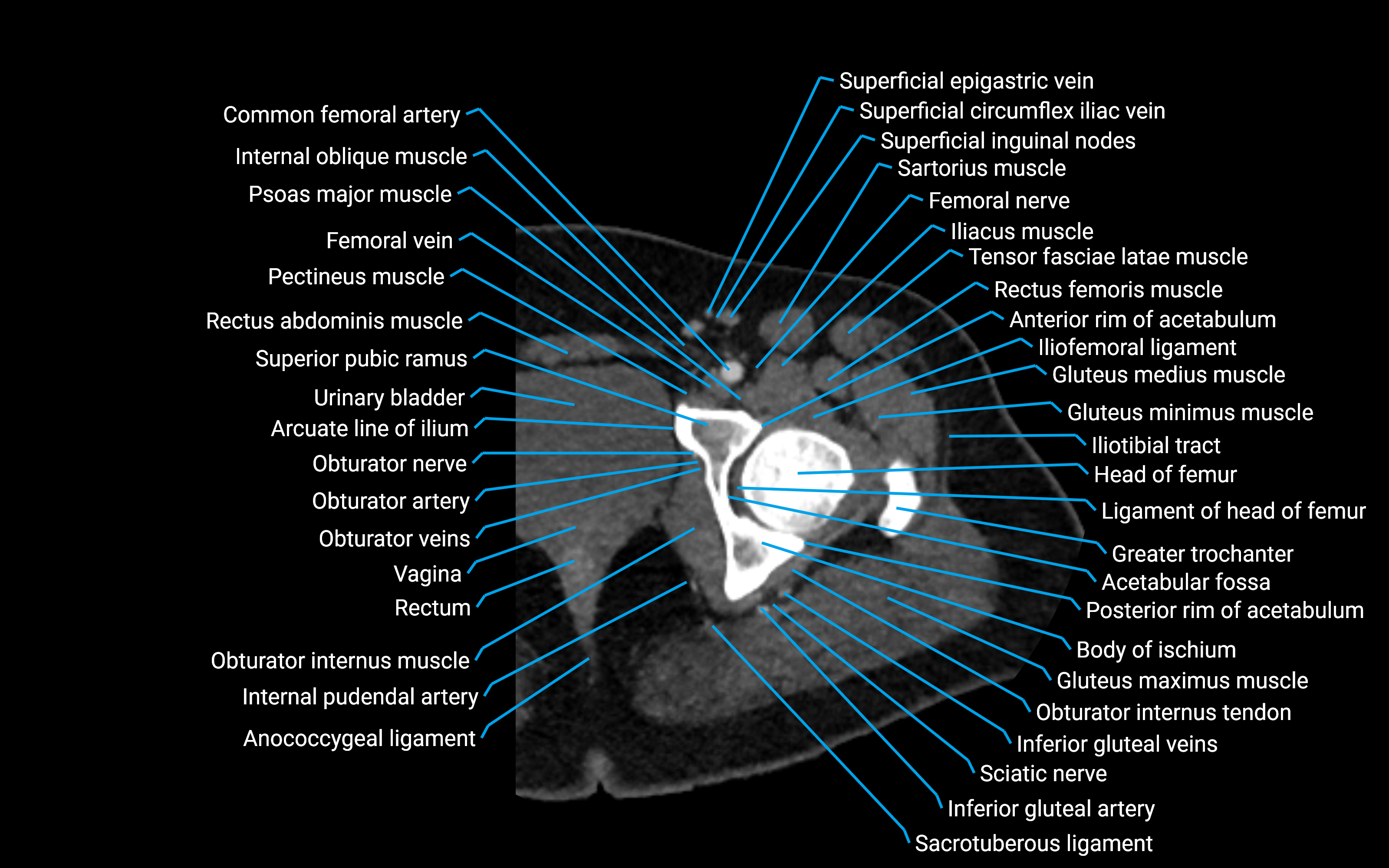
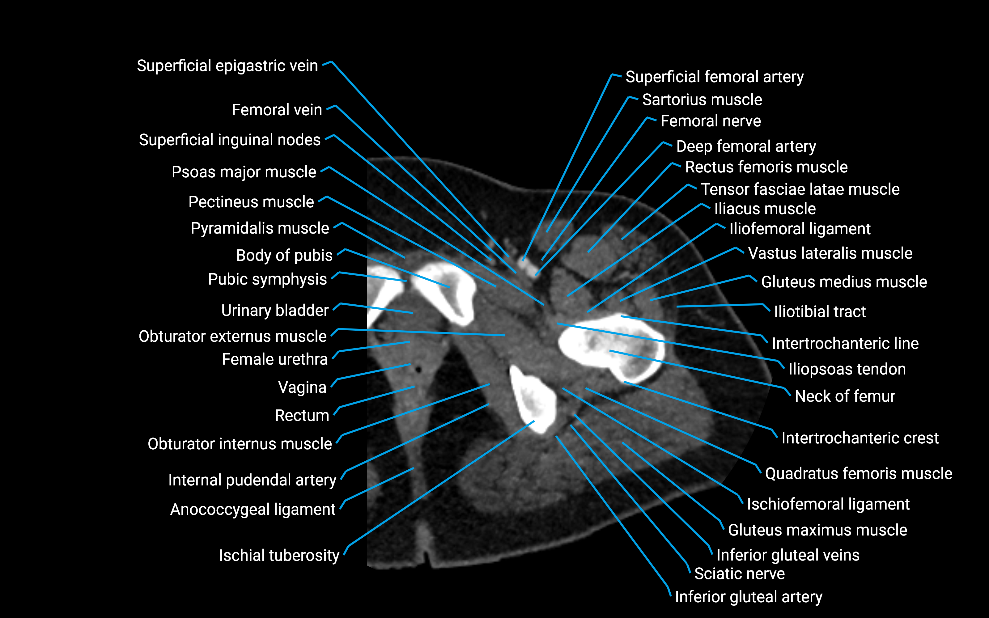
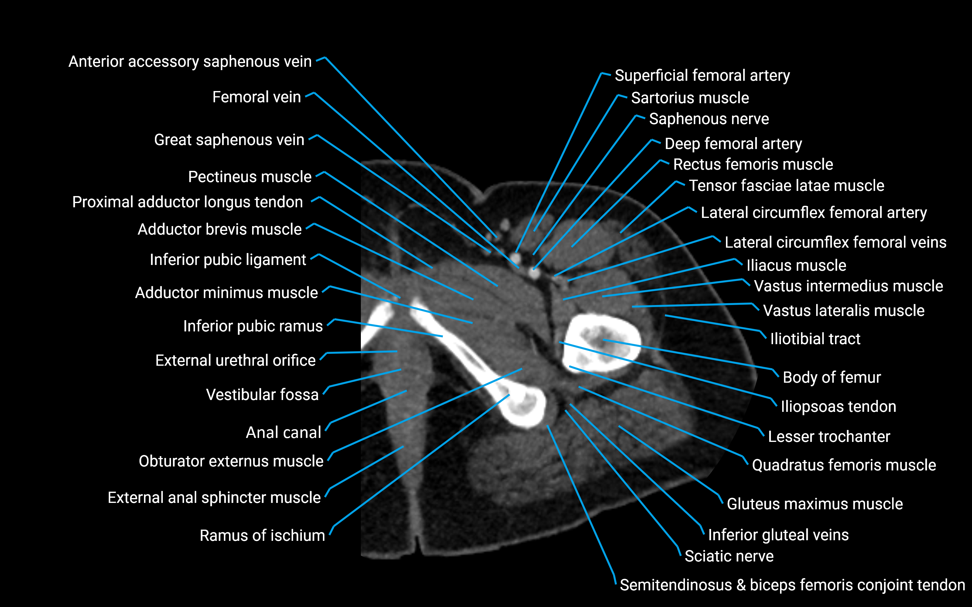
CT VRT 3D image

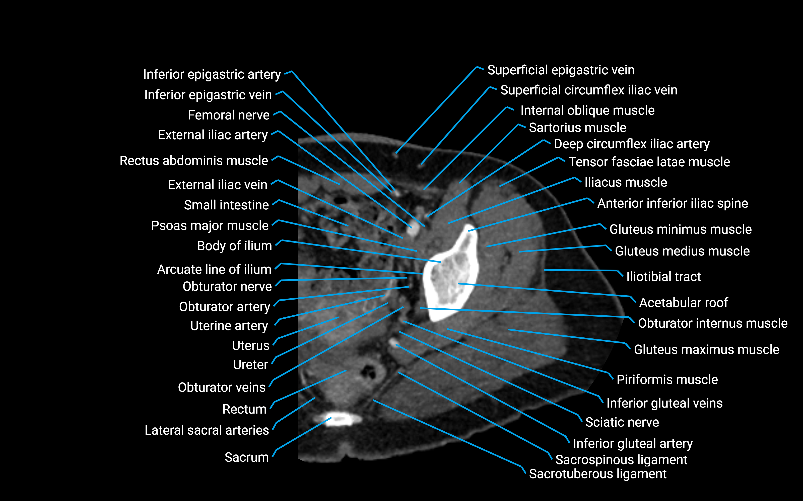
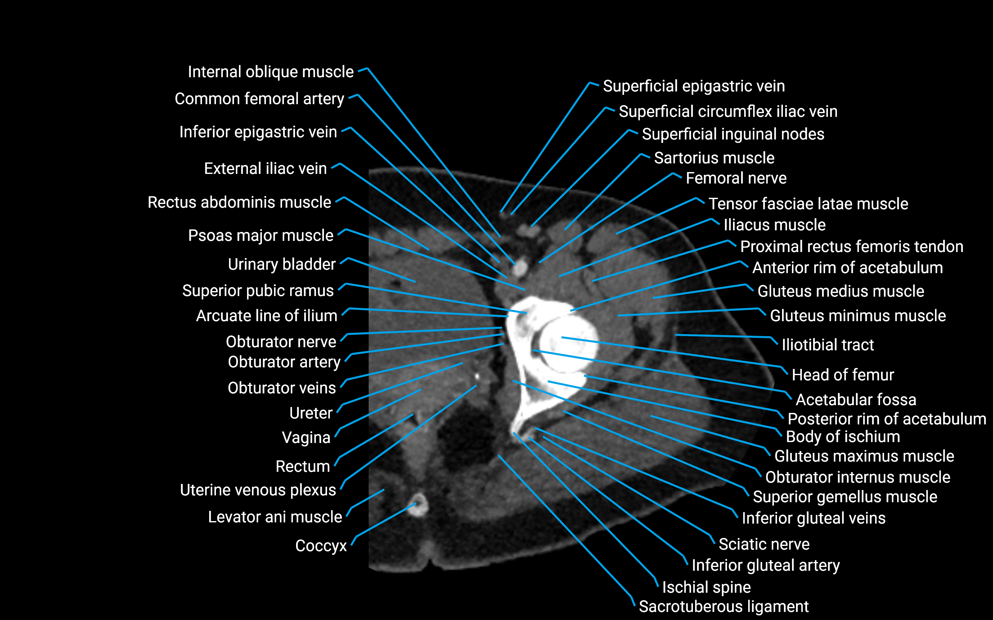
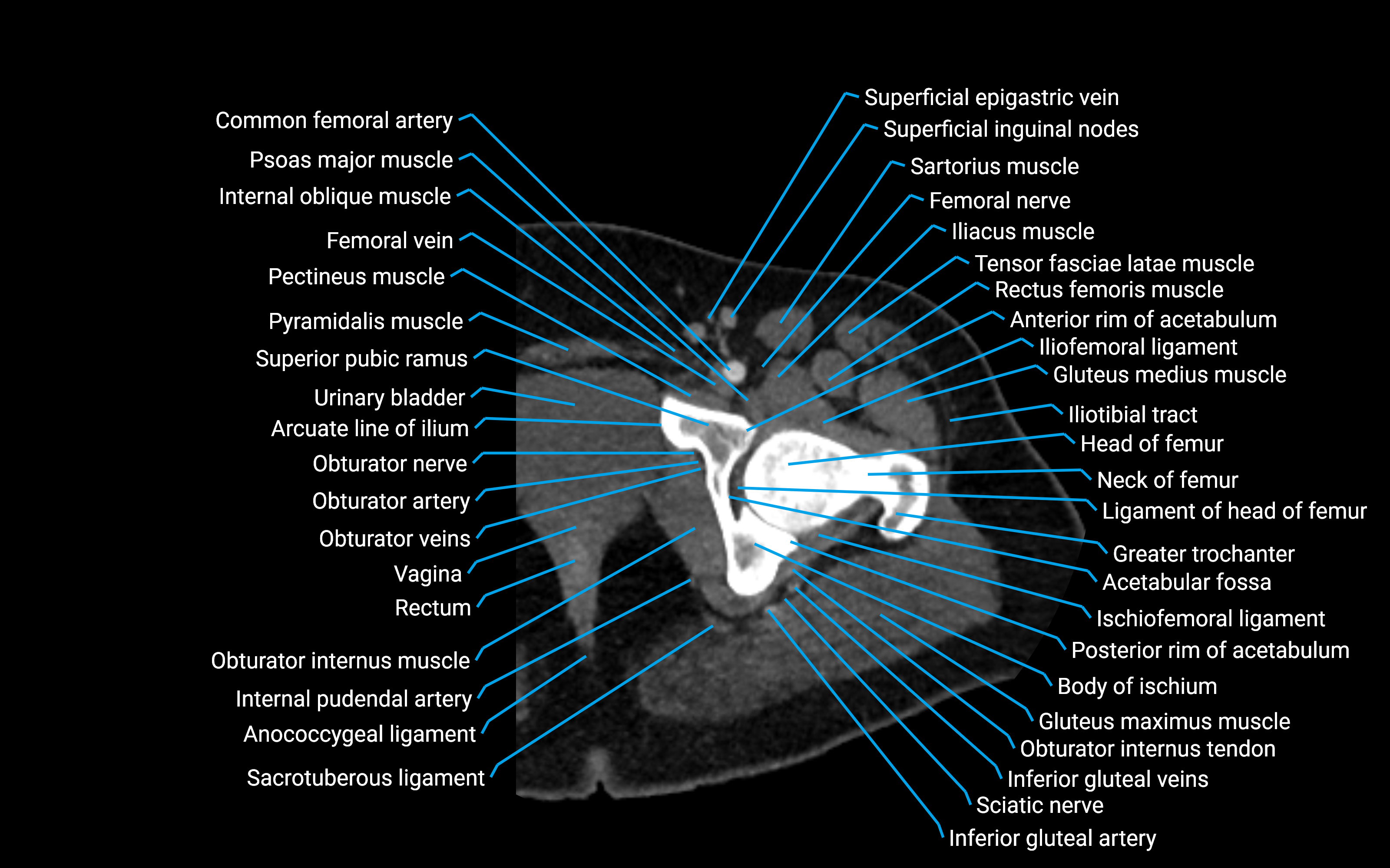
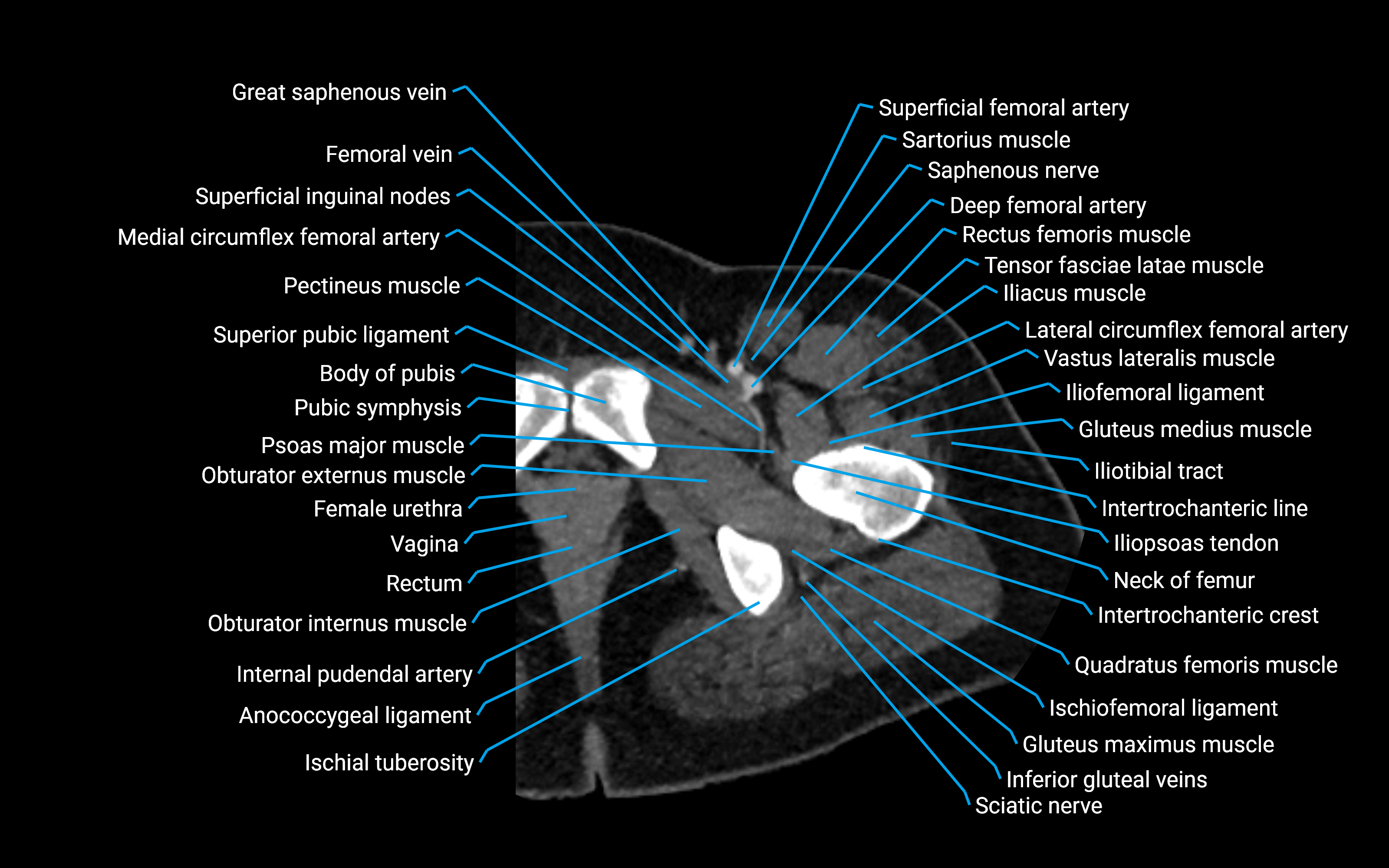
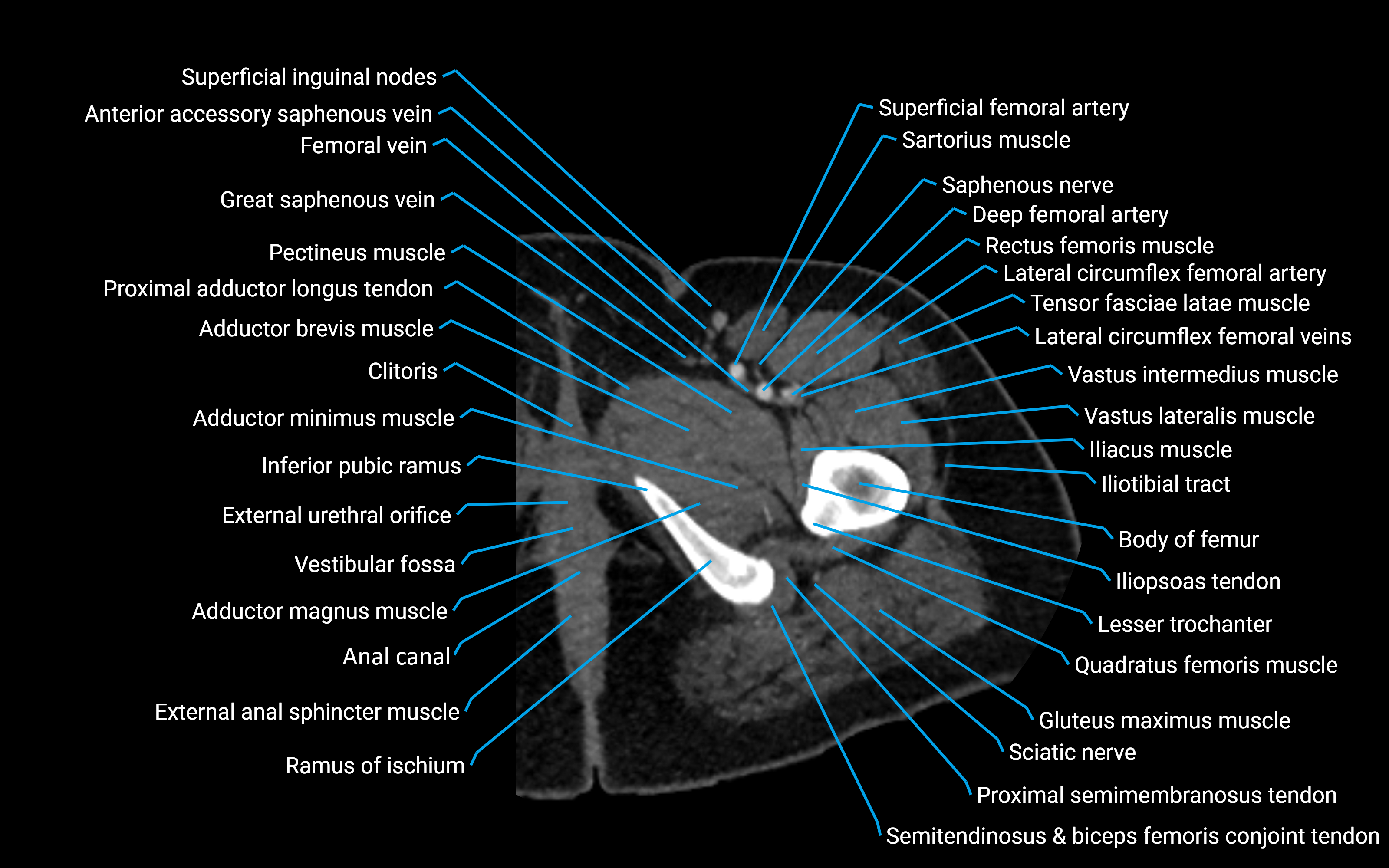
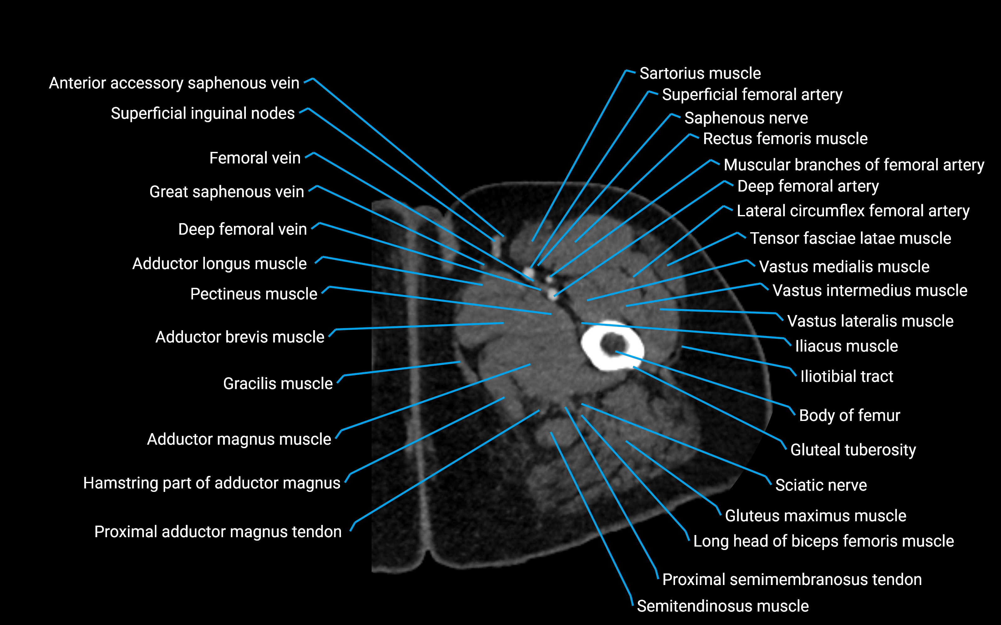
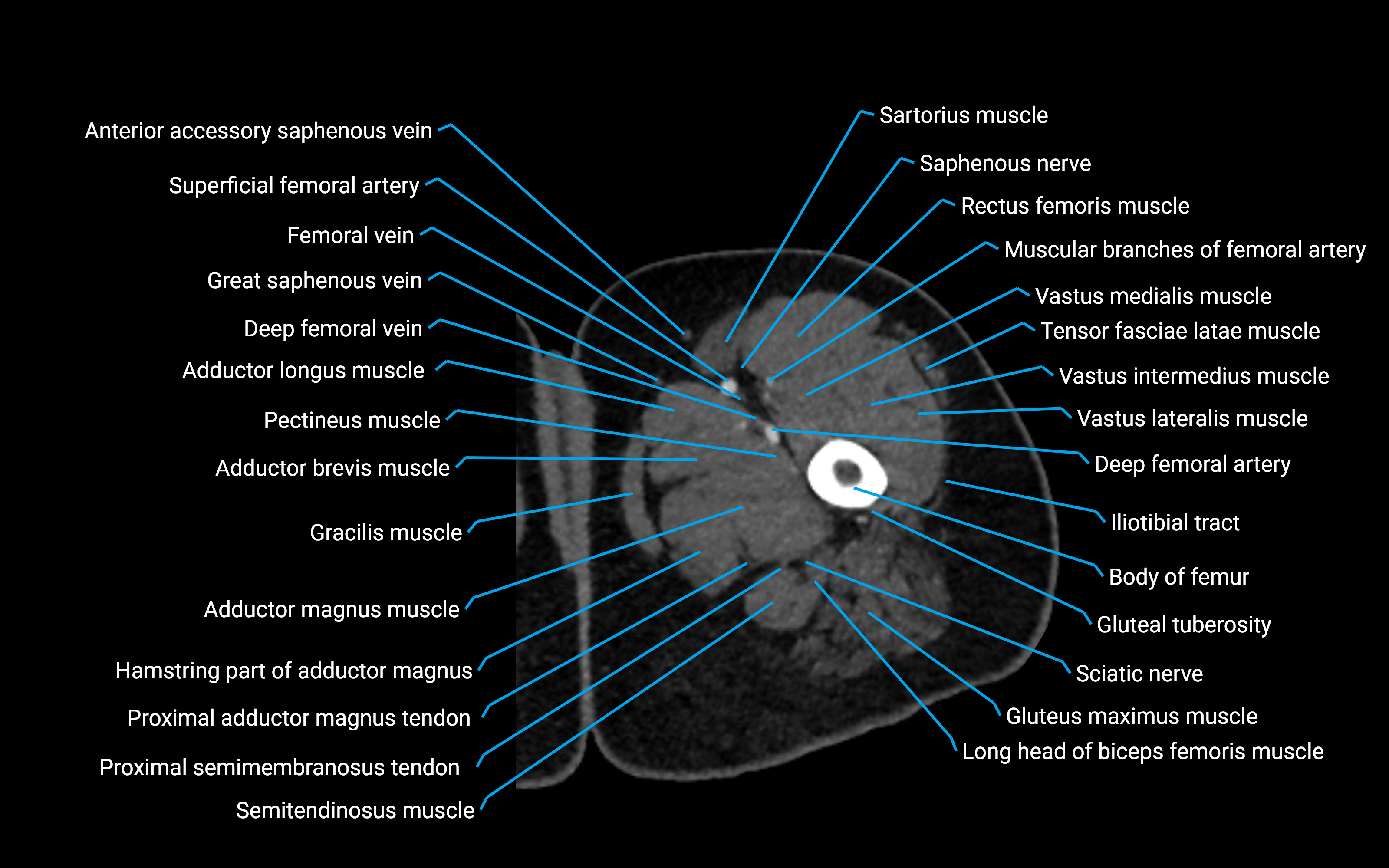
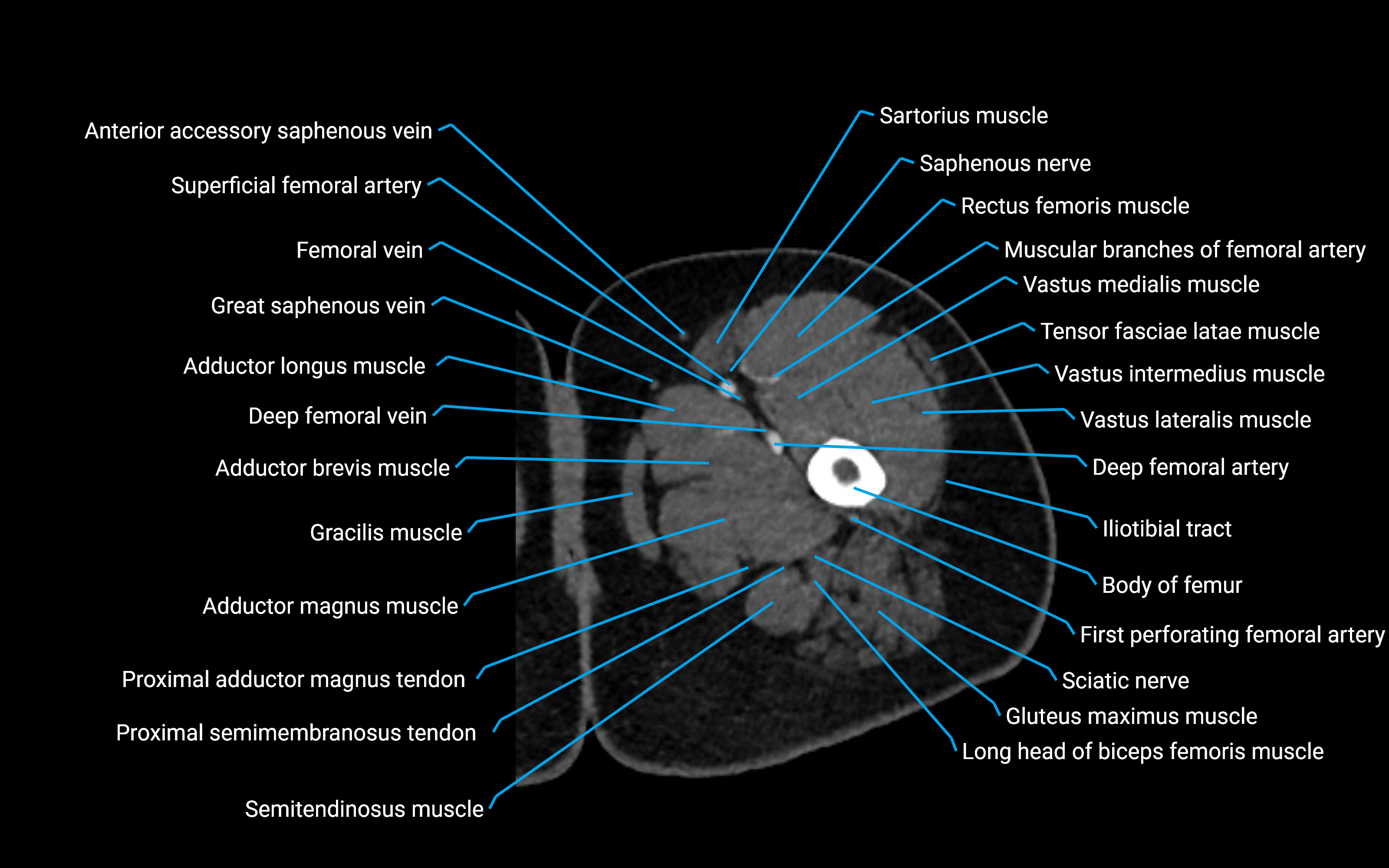
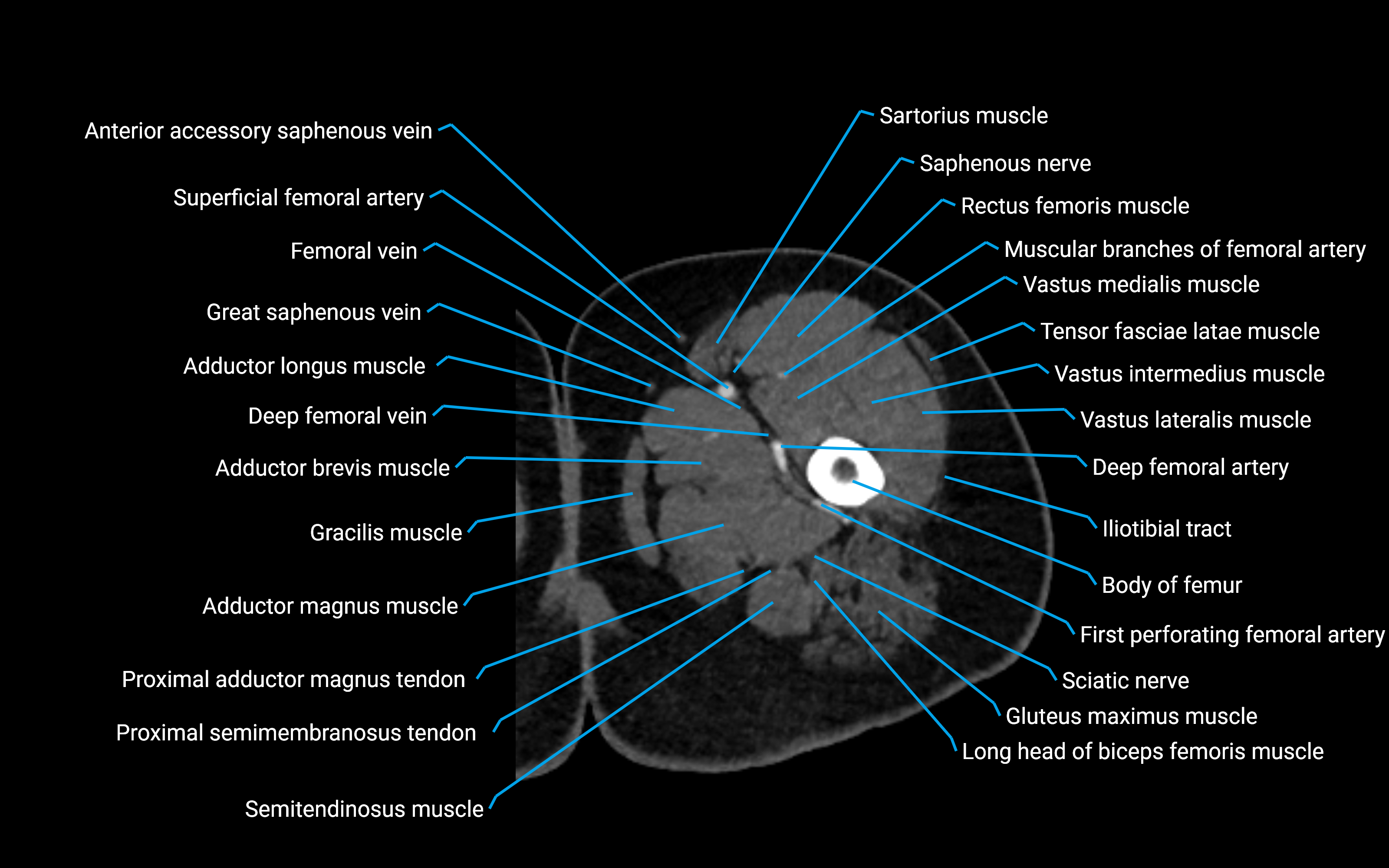
CT image

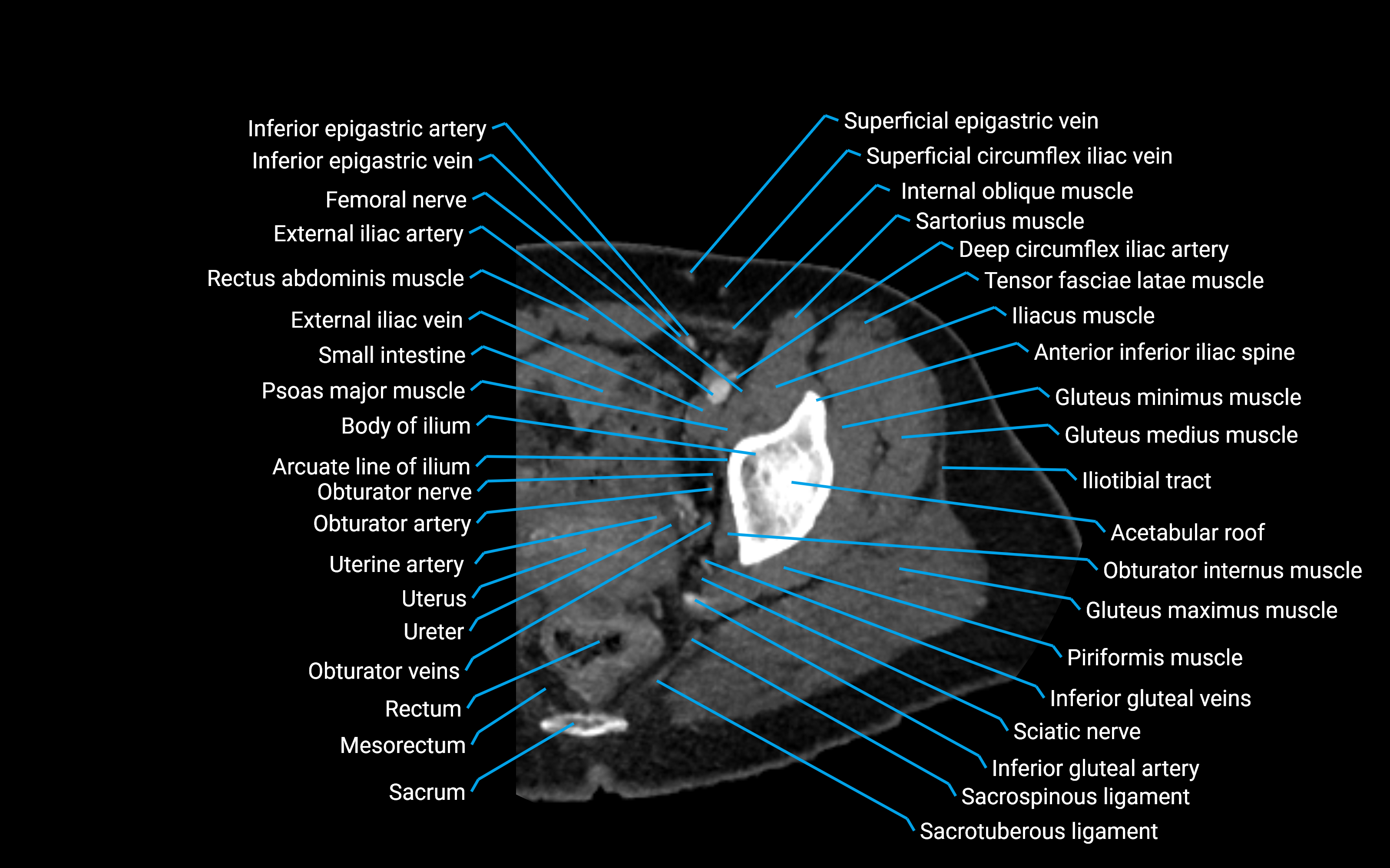
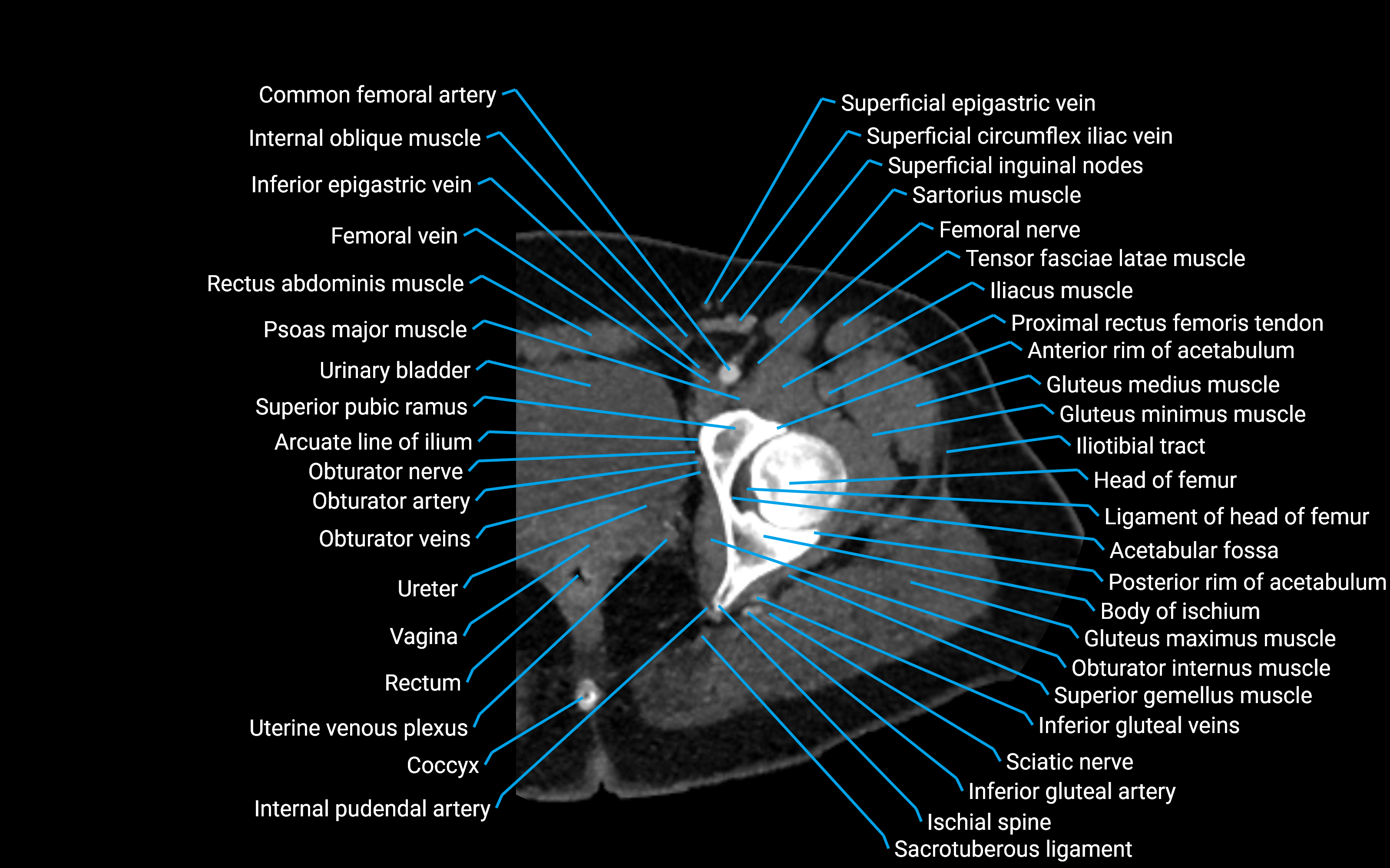
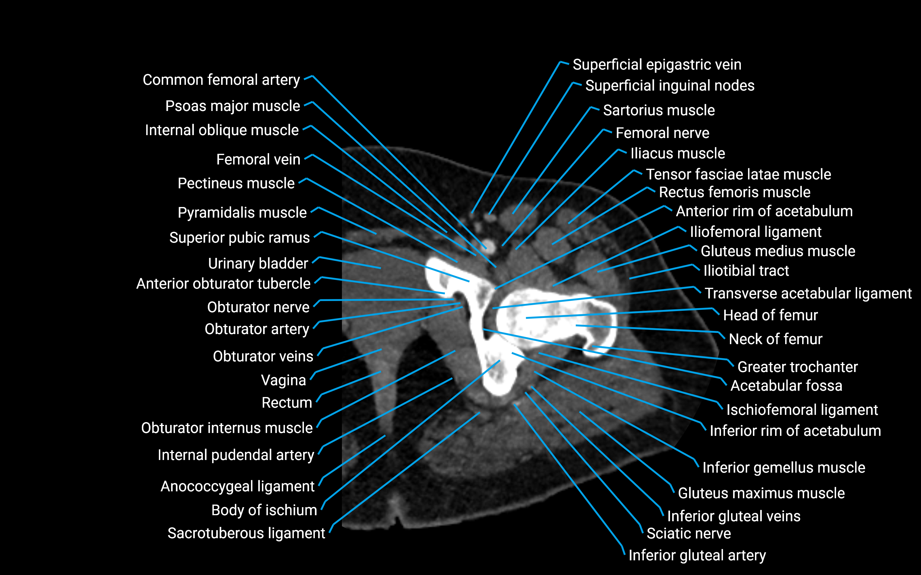
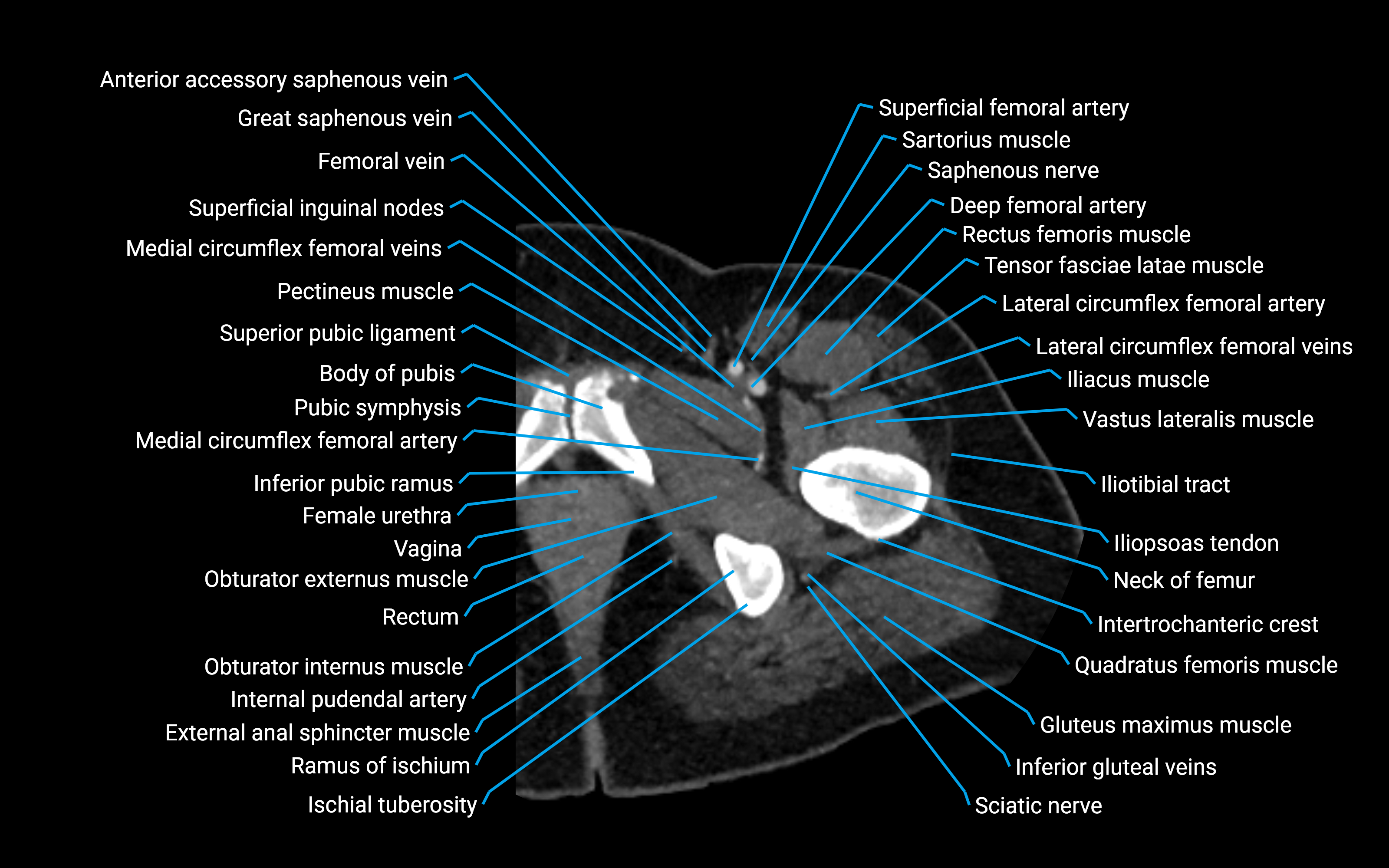
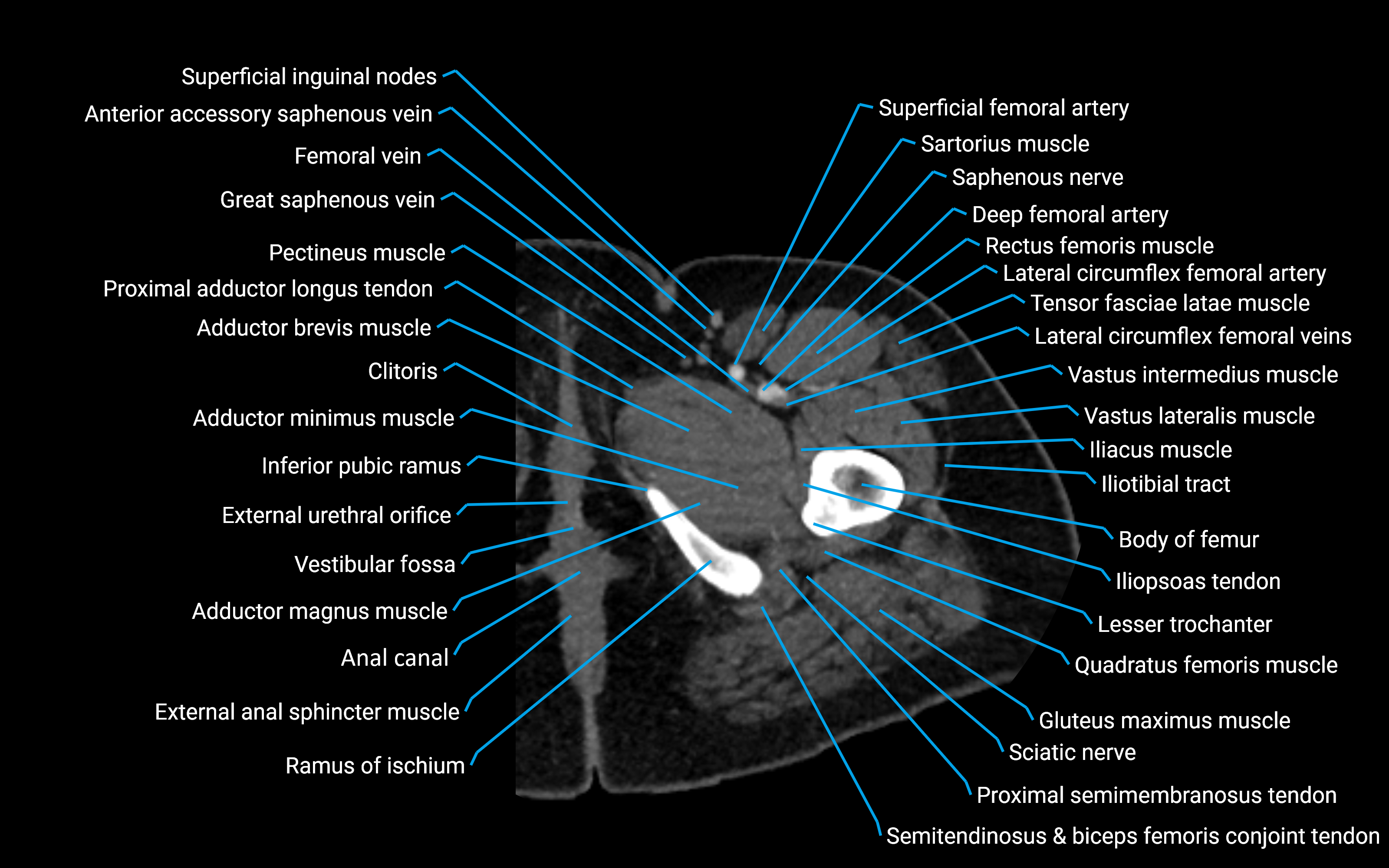
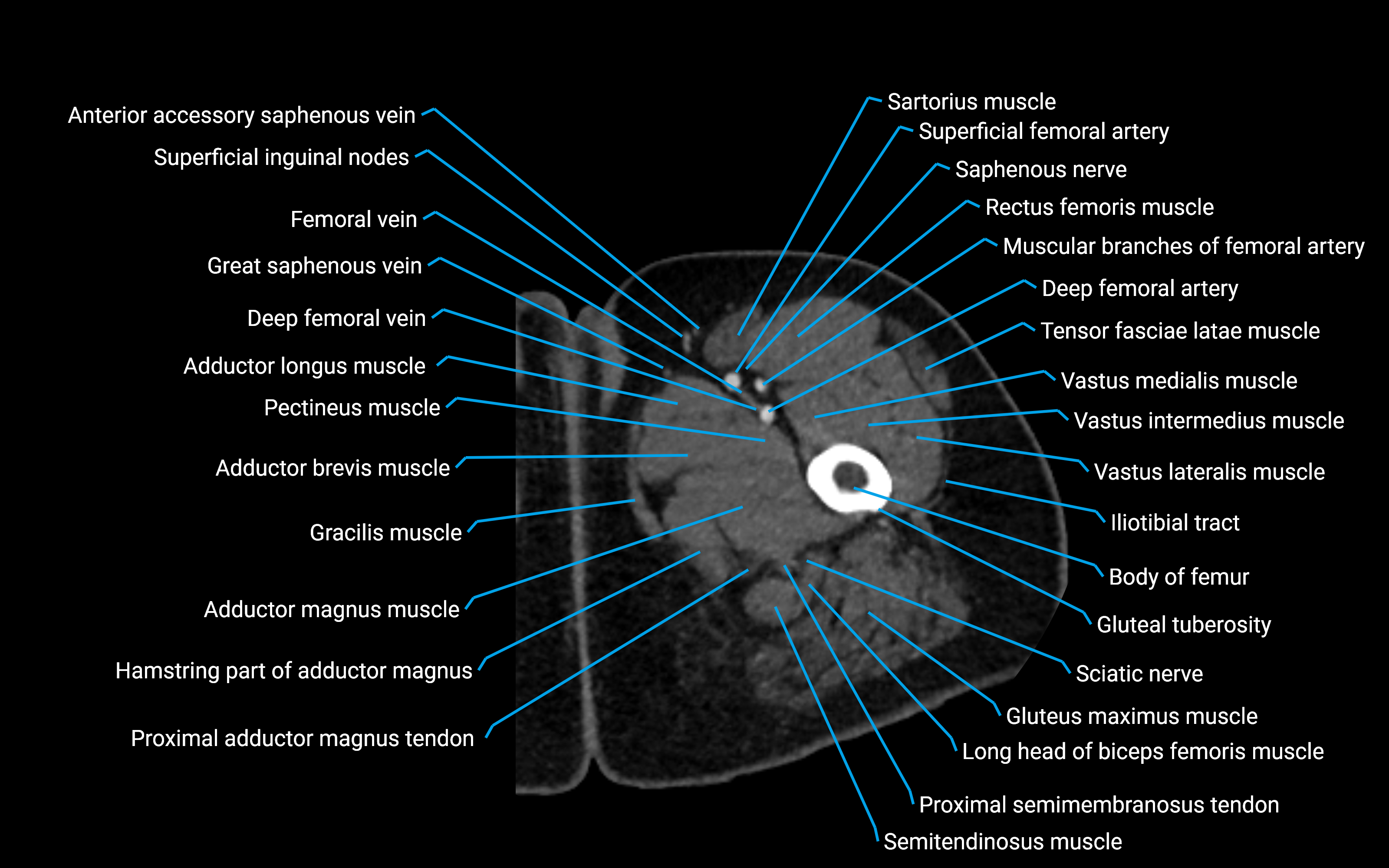
CT image

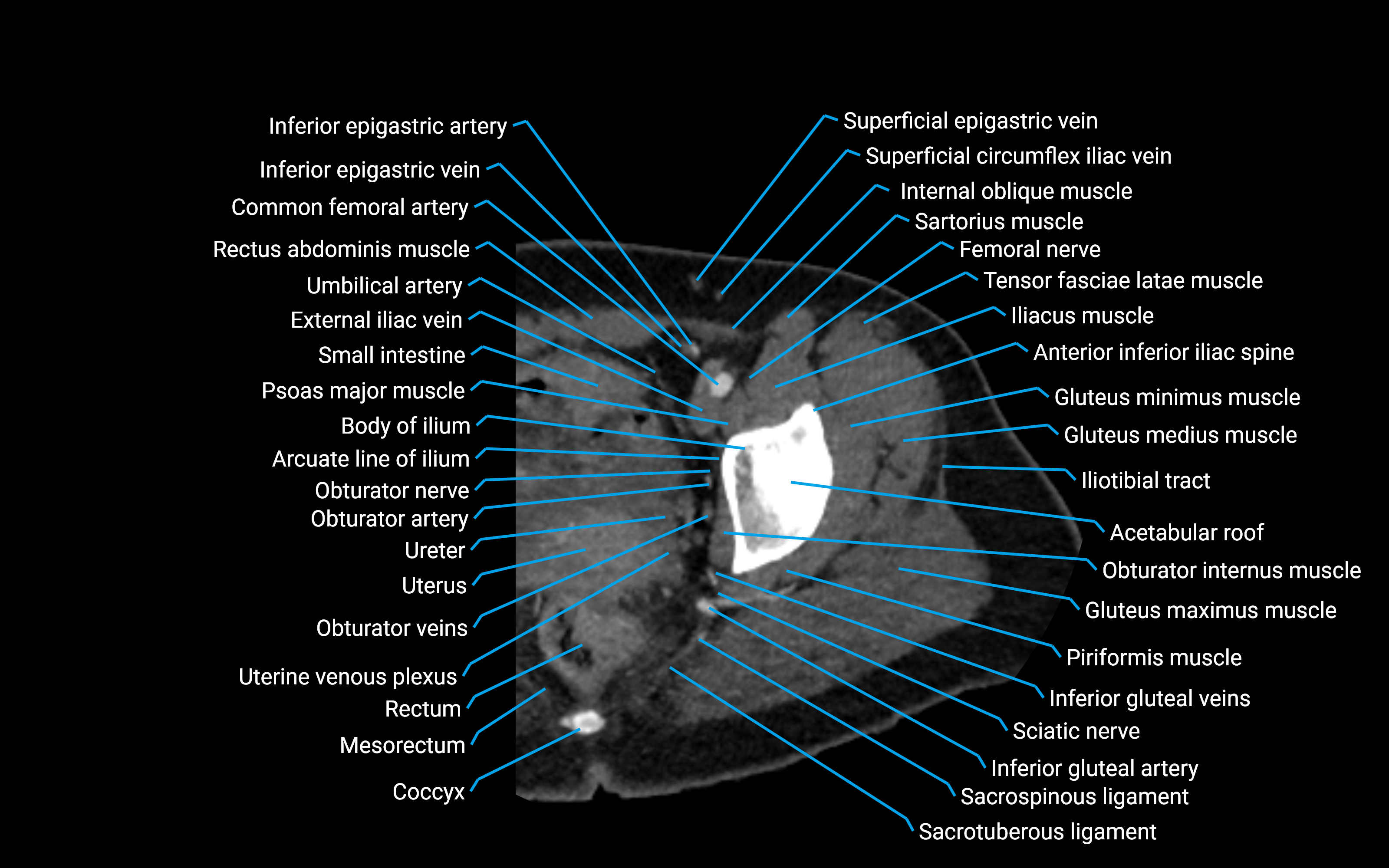
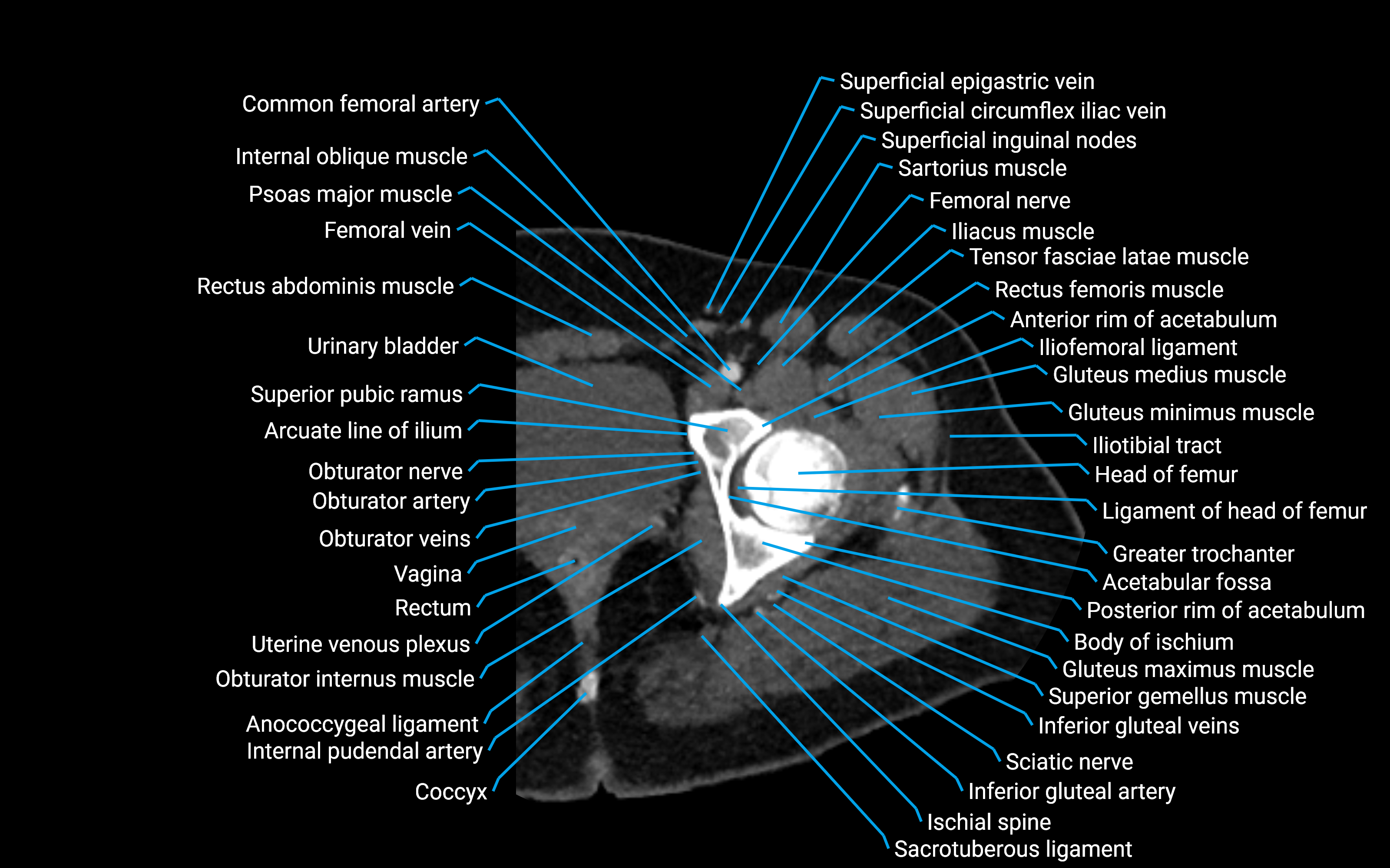
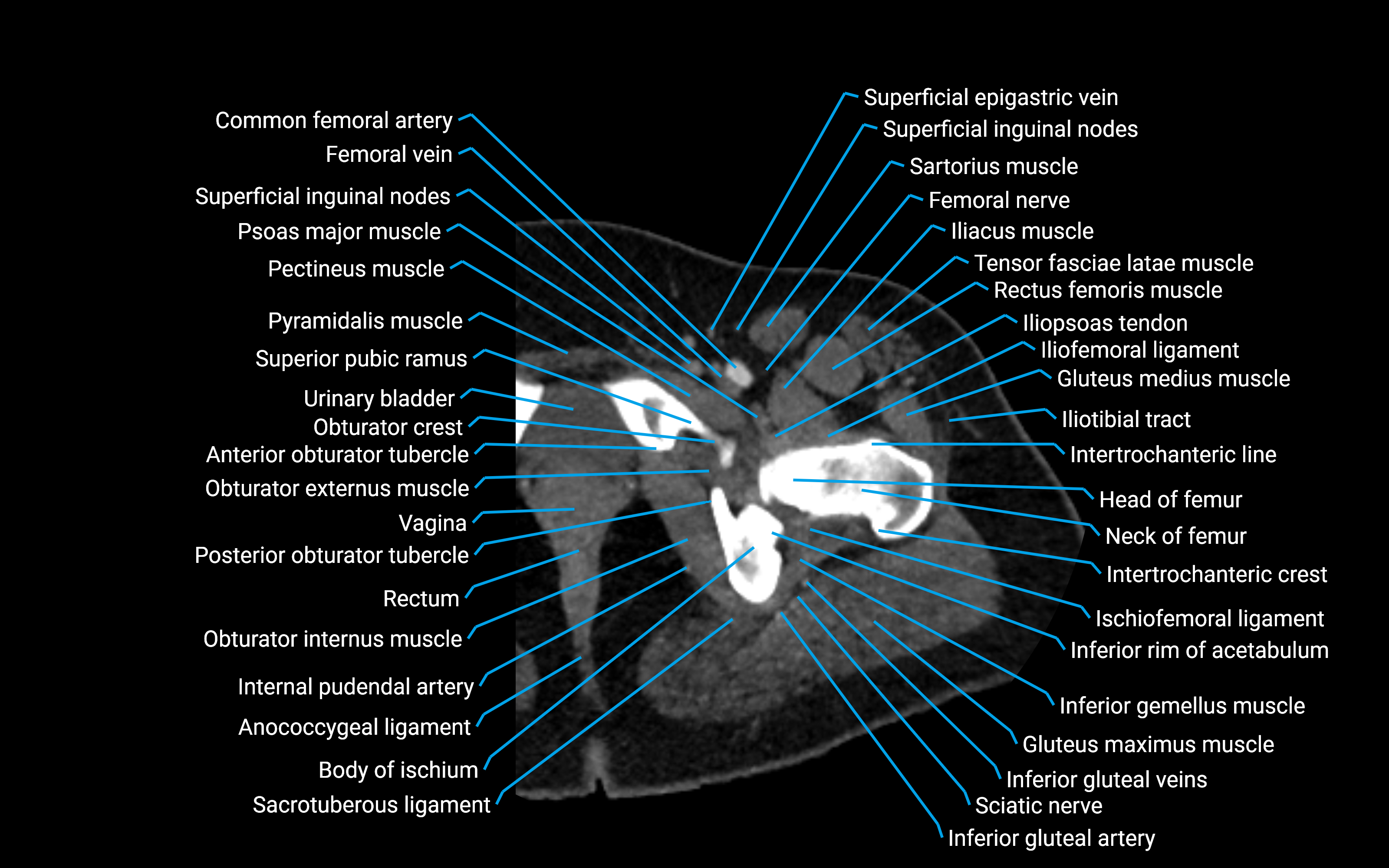
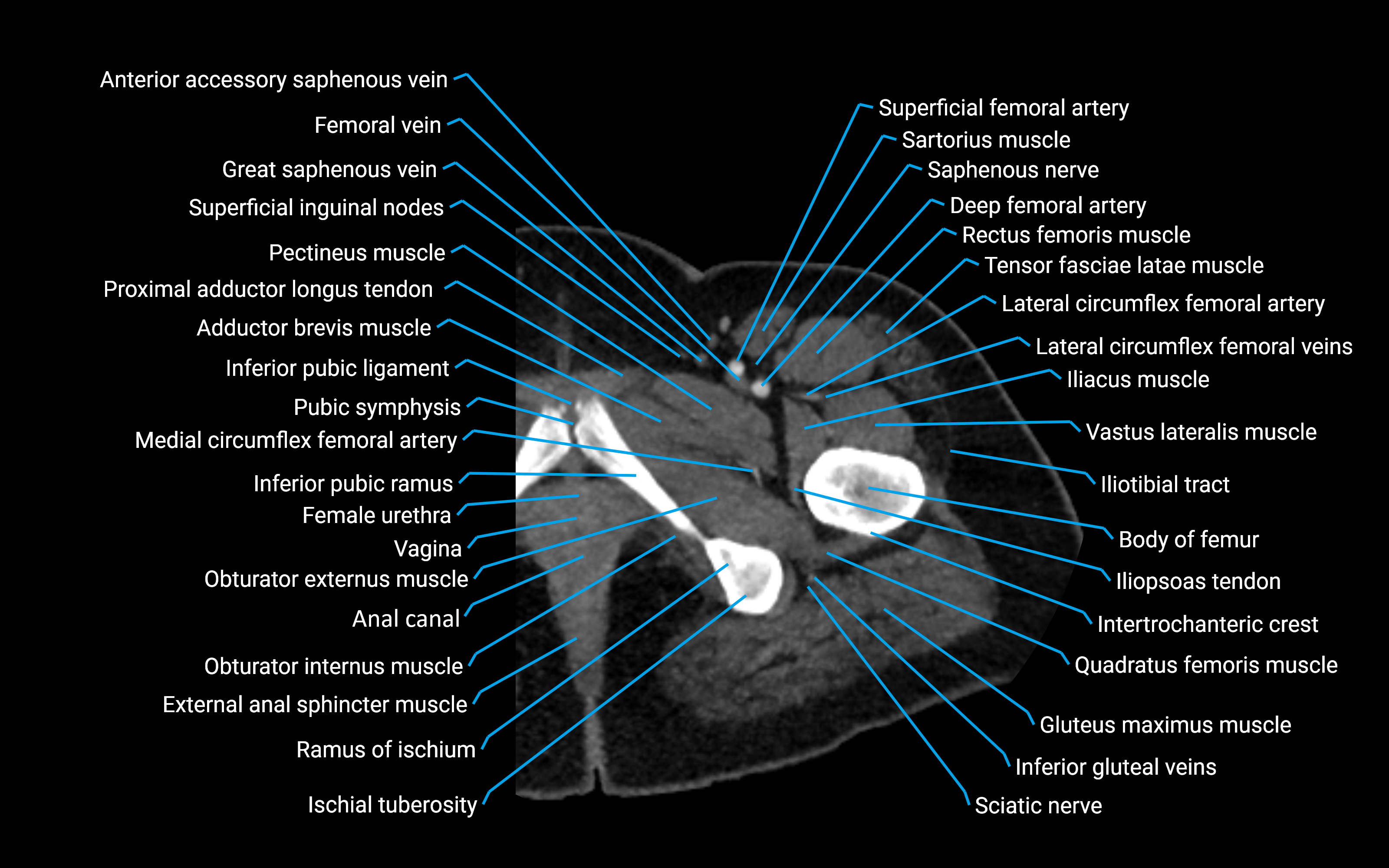
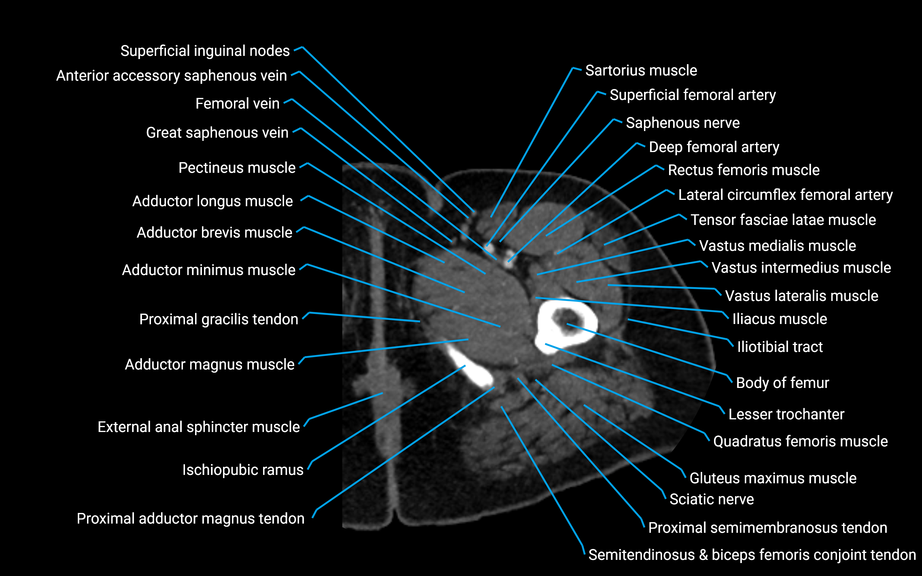
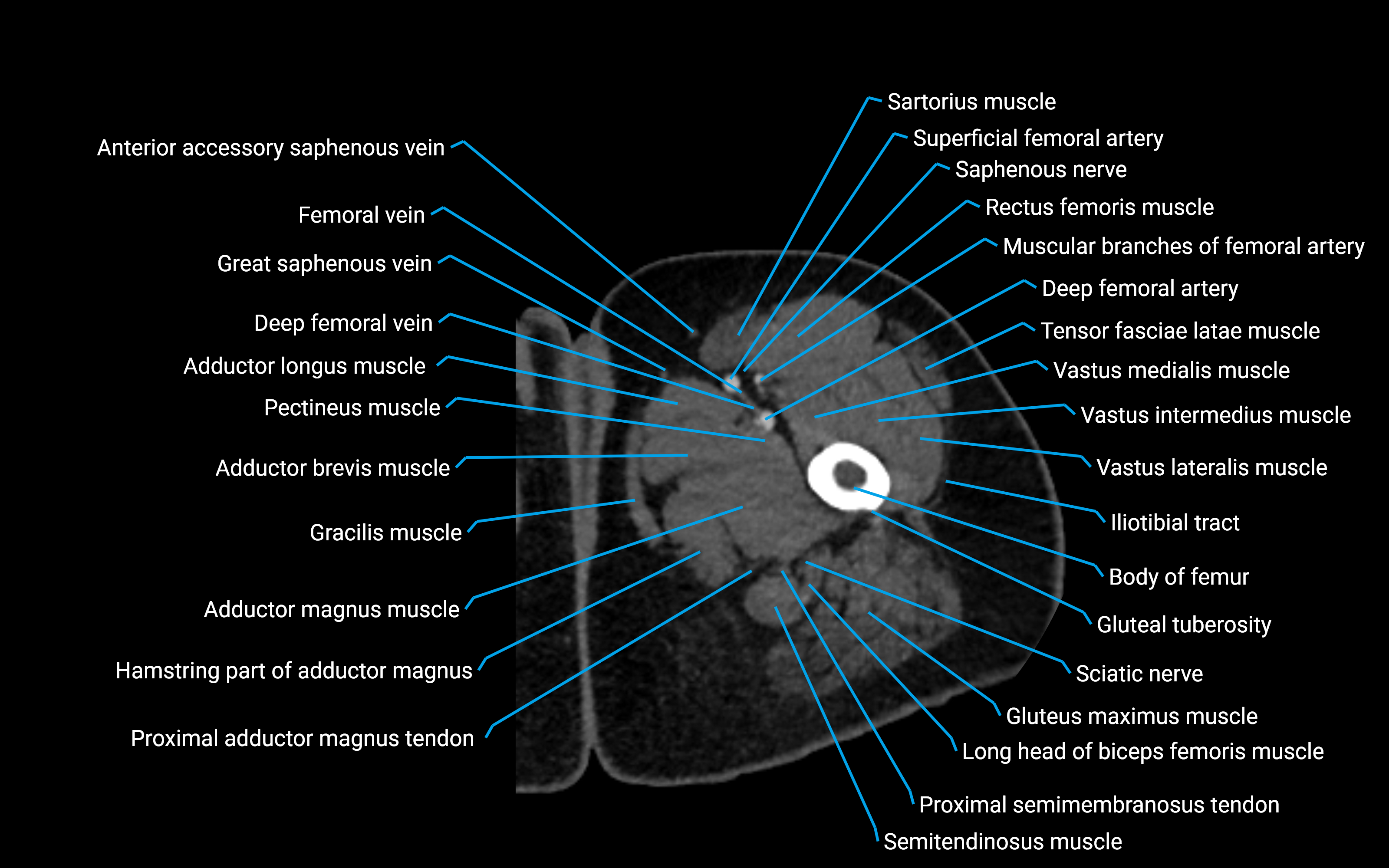
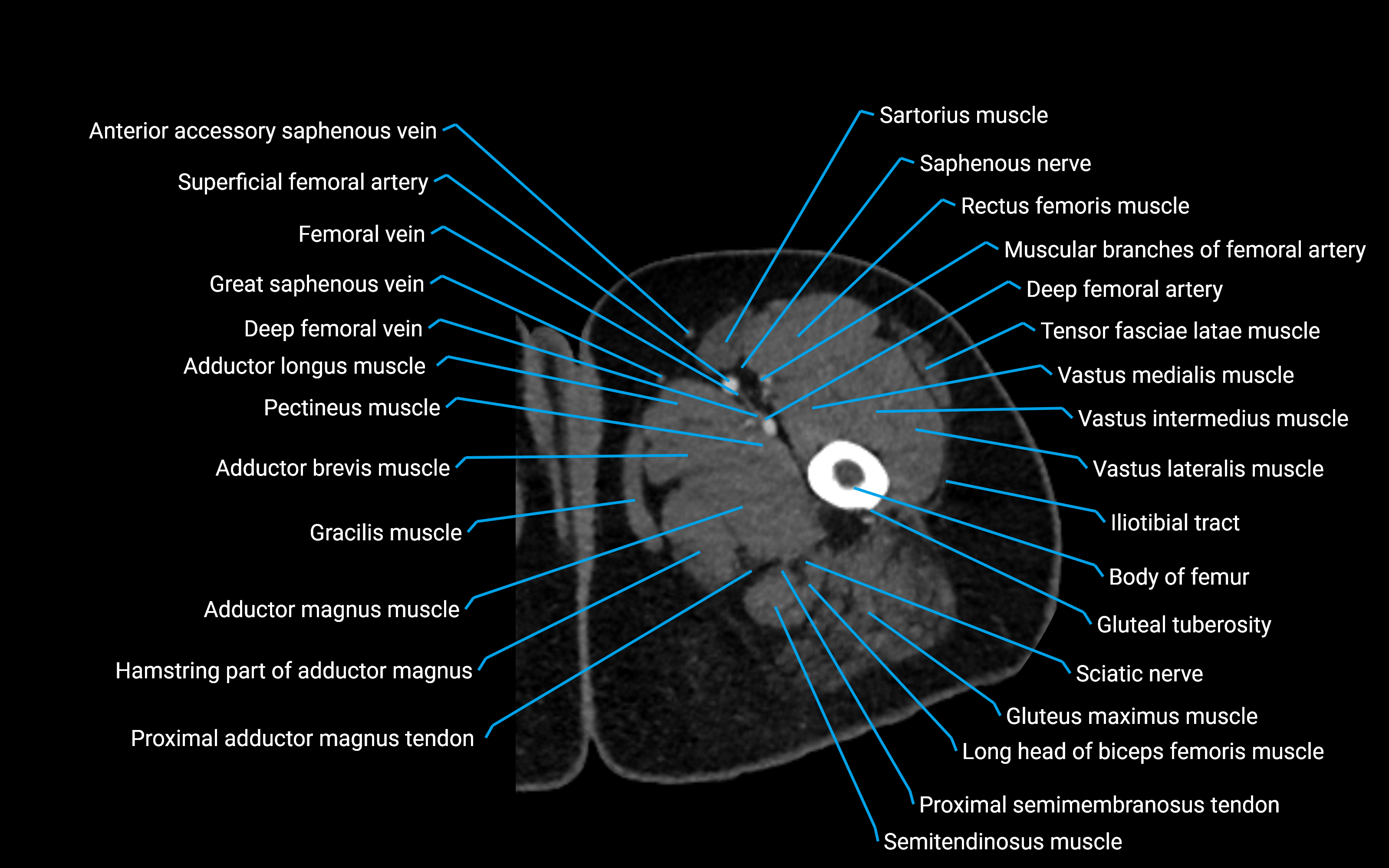
CT image

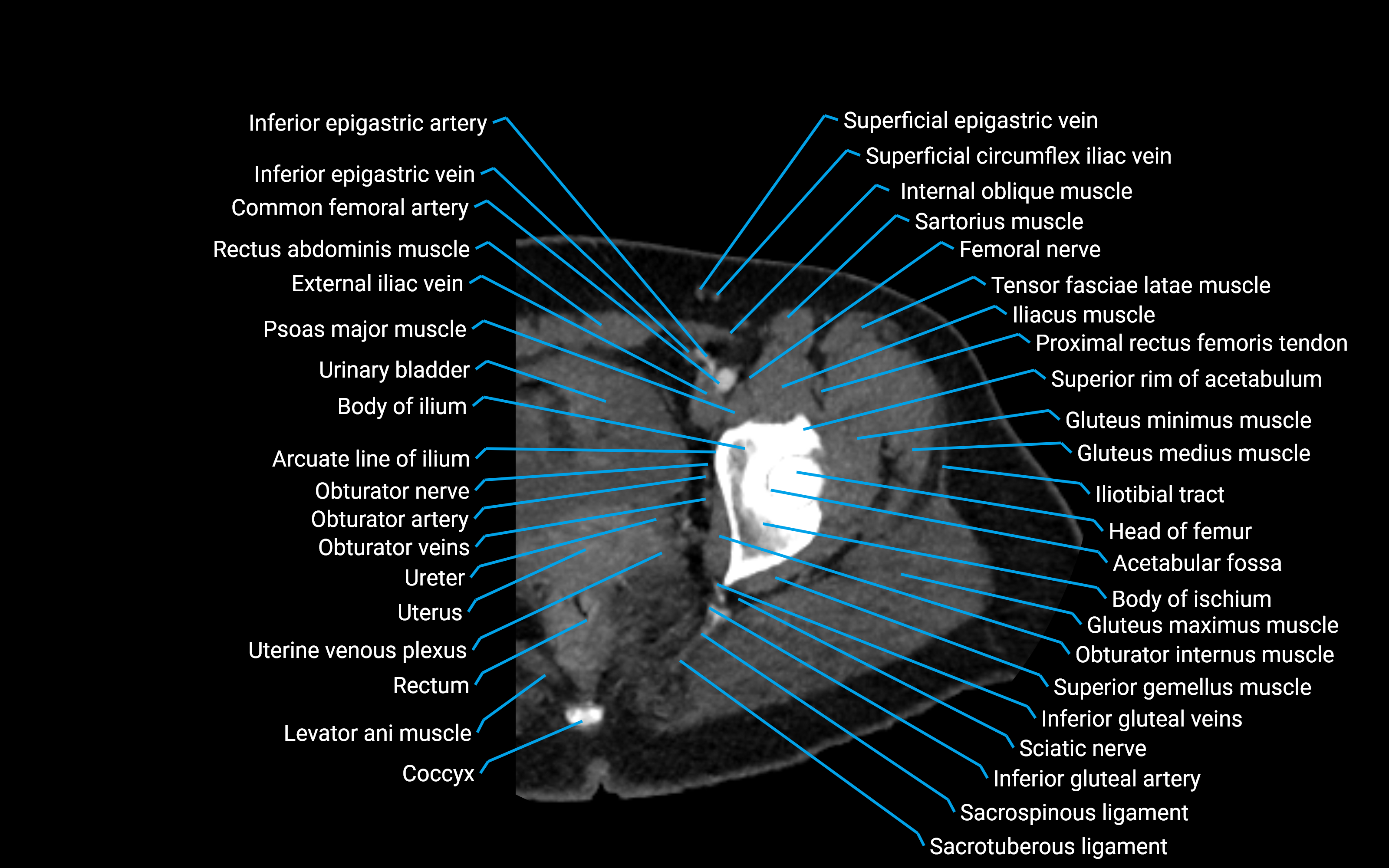
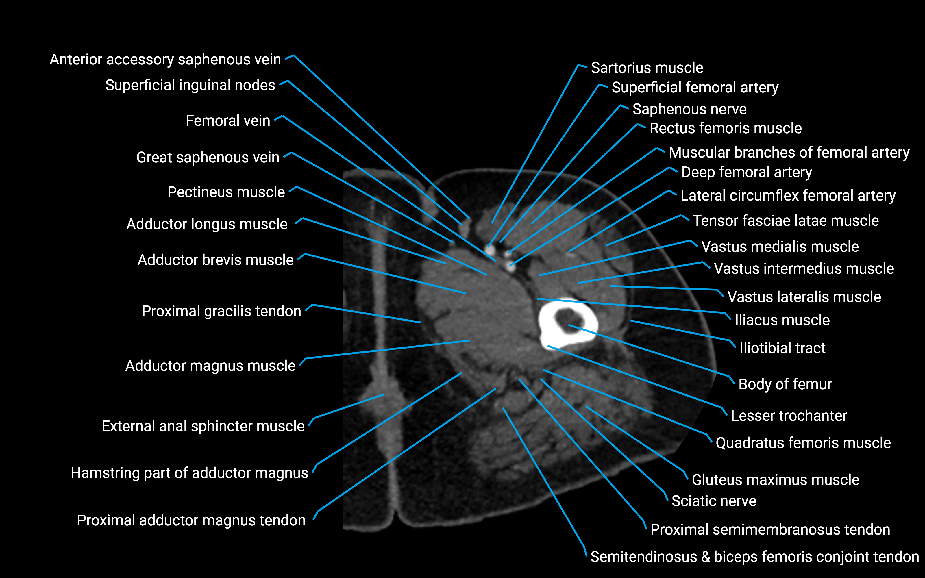
MRI image

MRI image
