


Topic
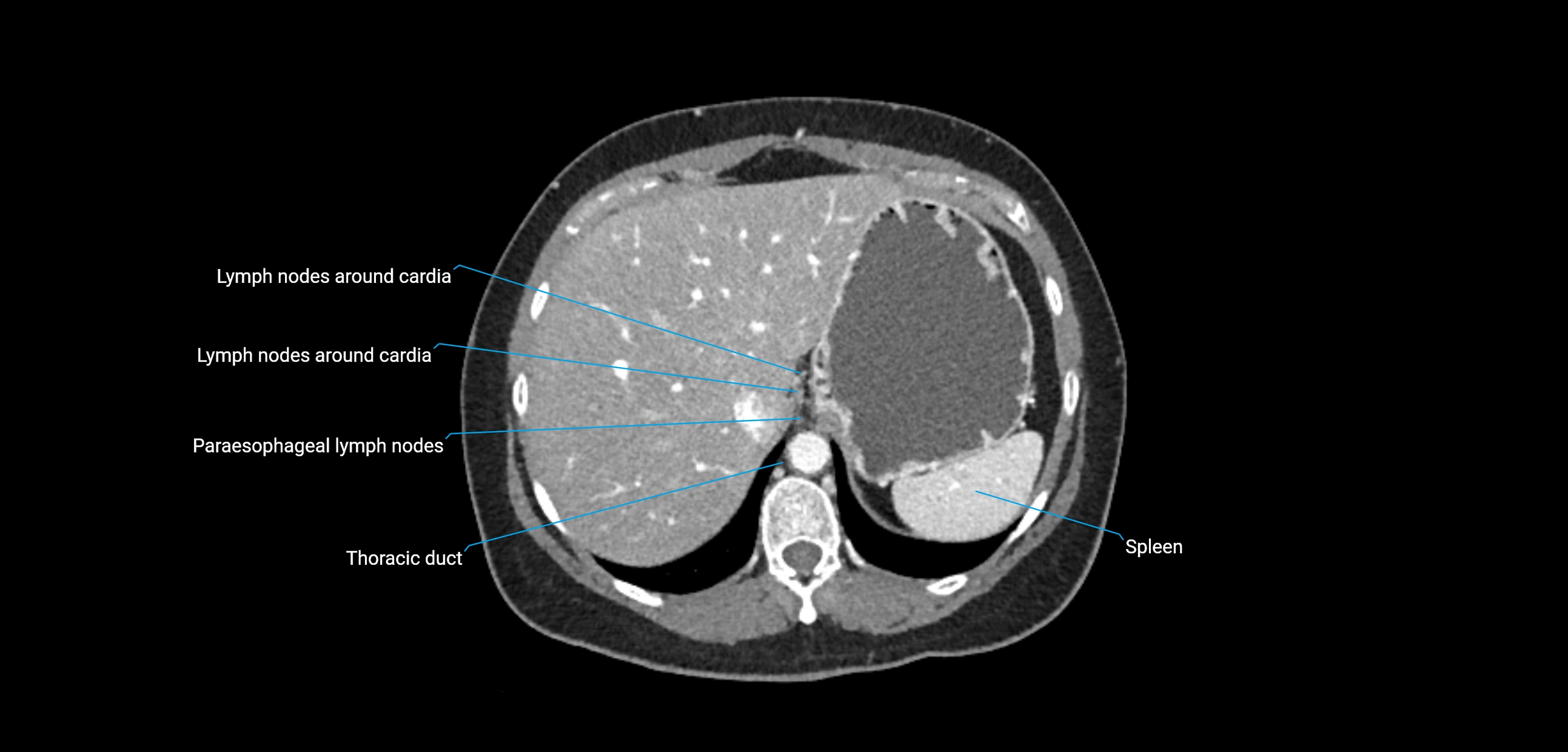
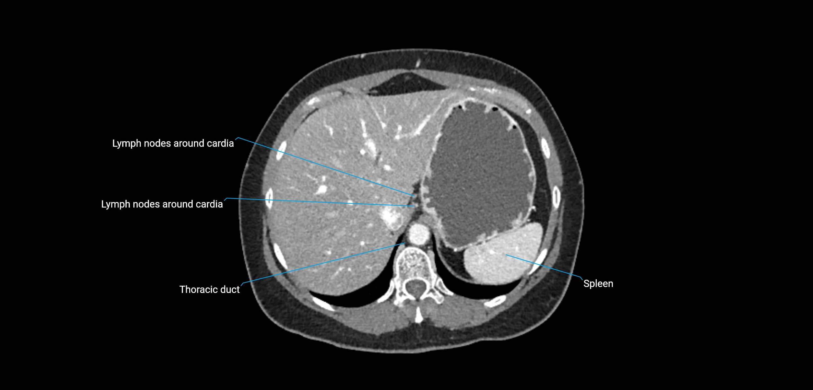
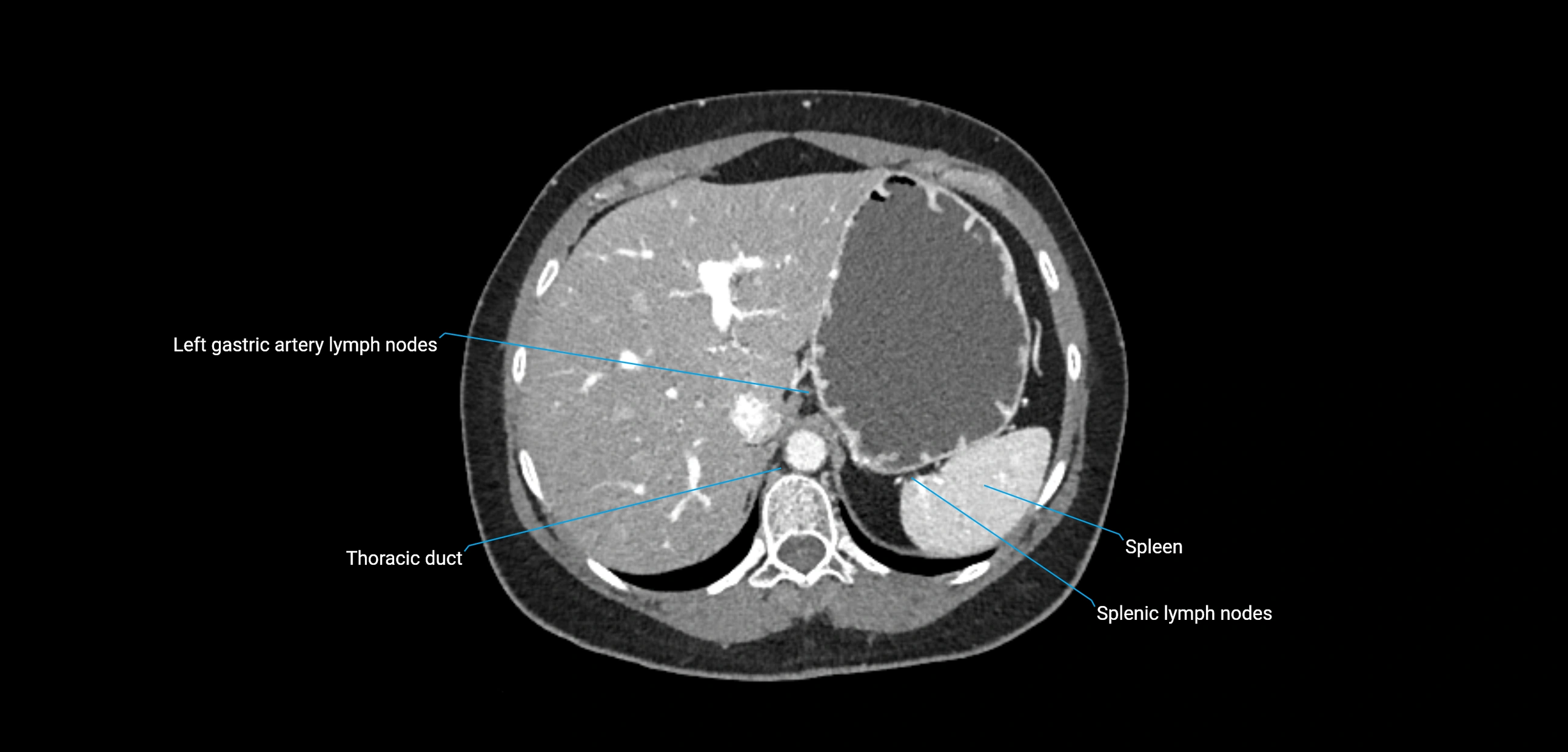
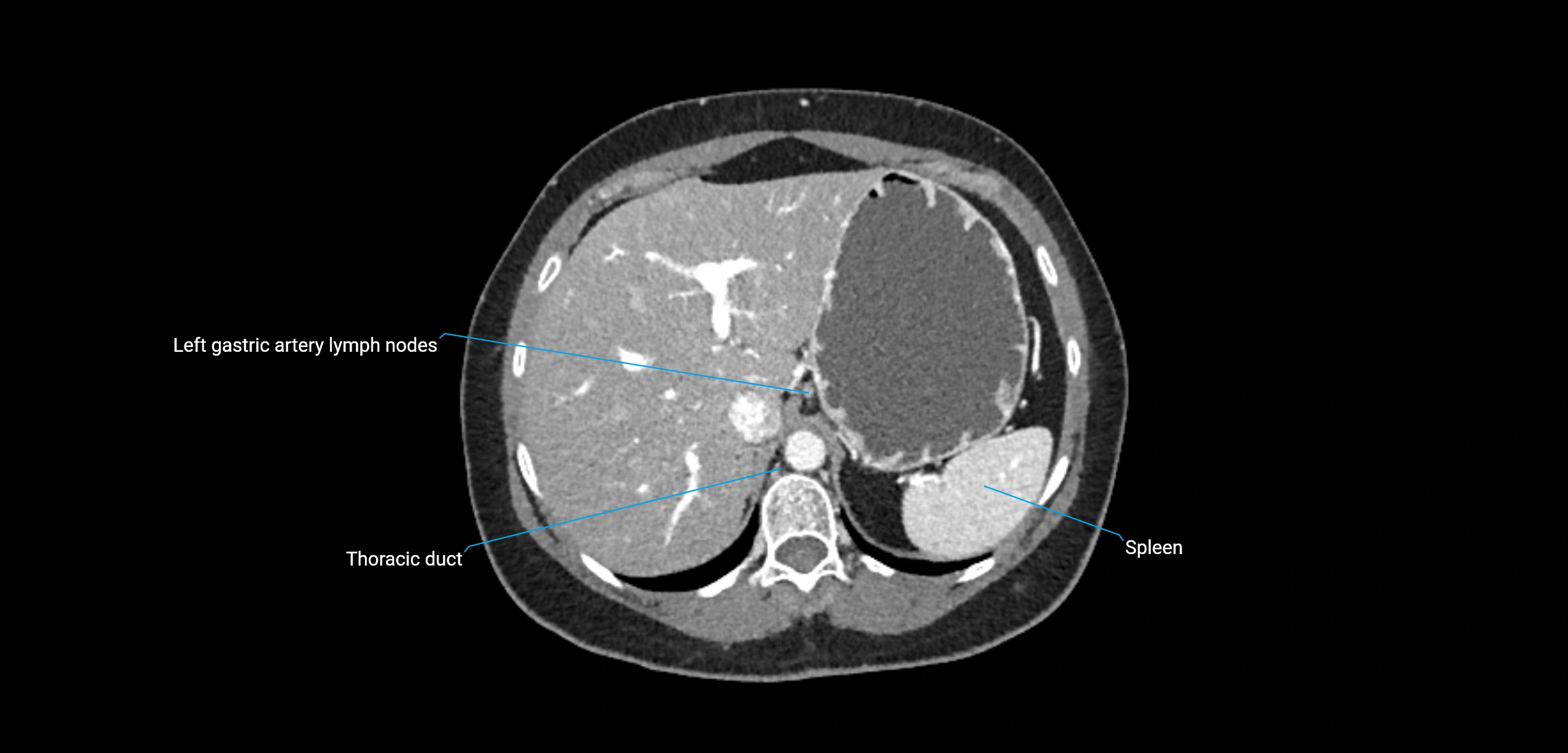
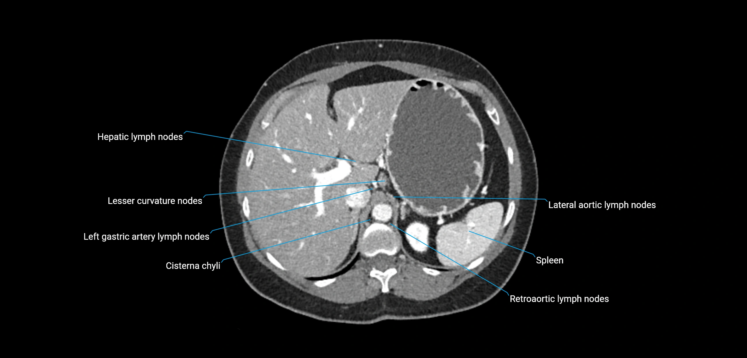
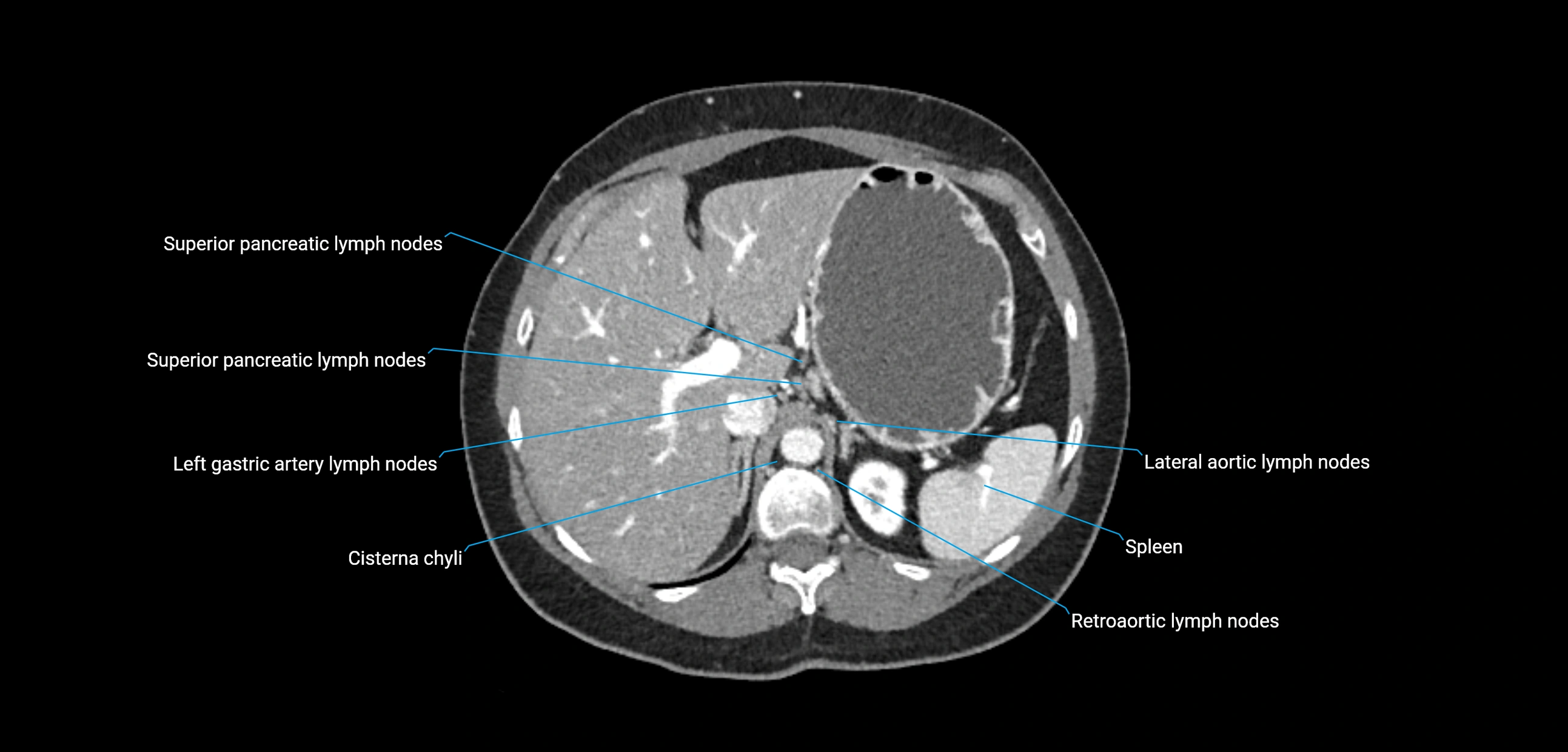
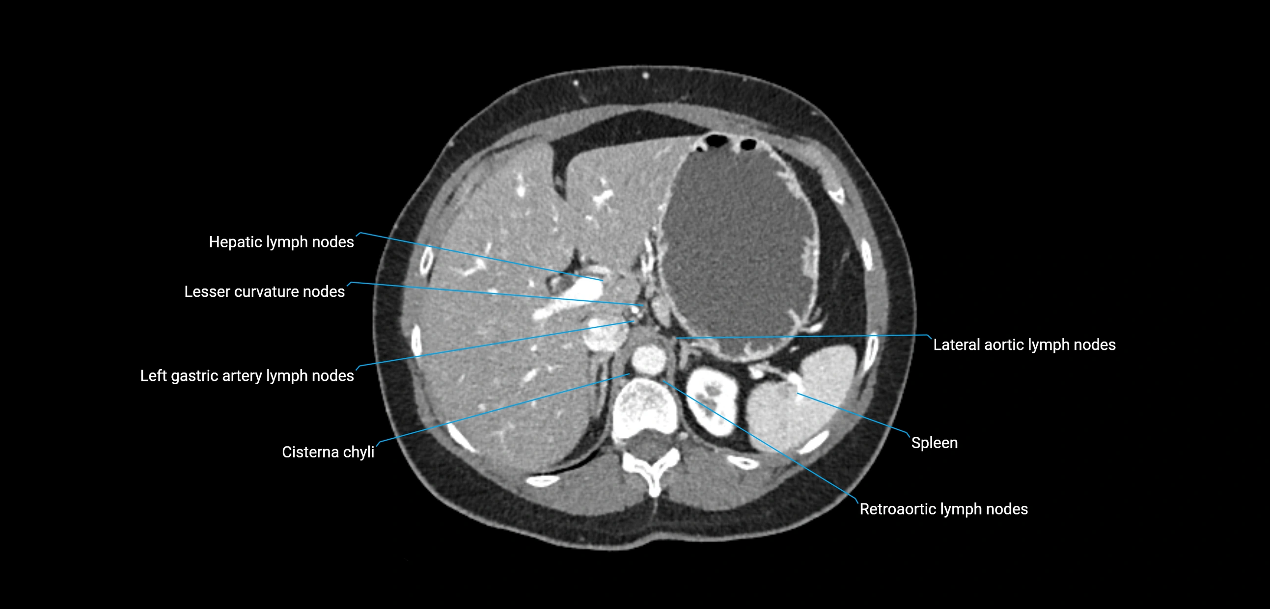
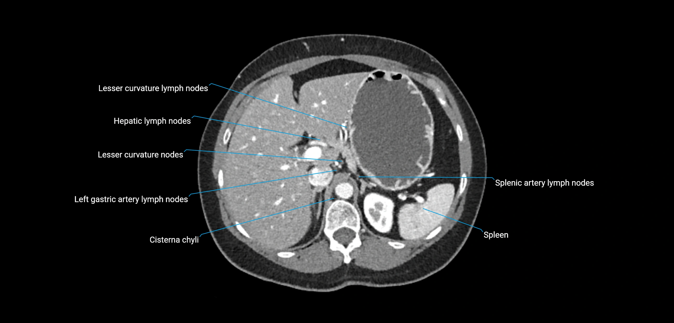
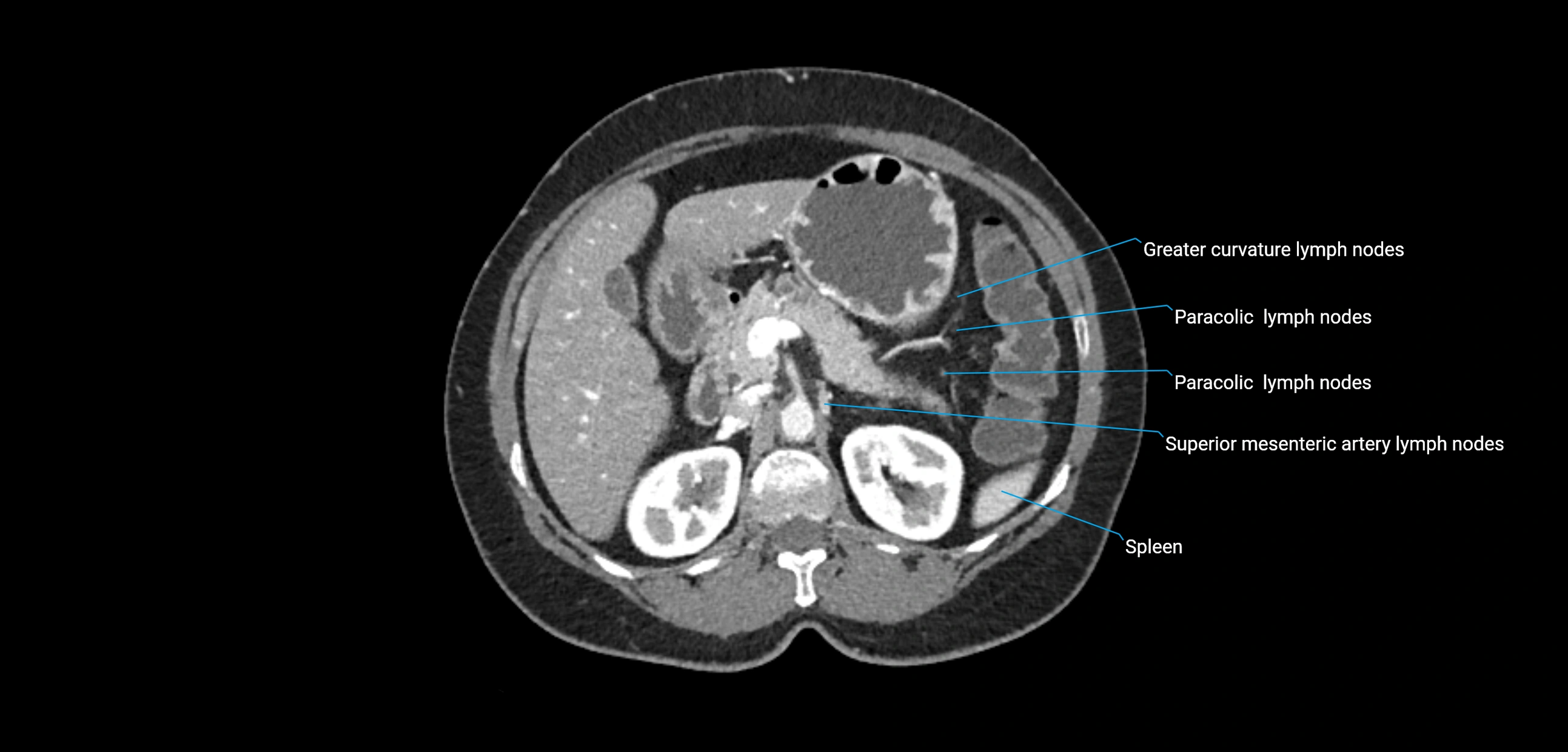
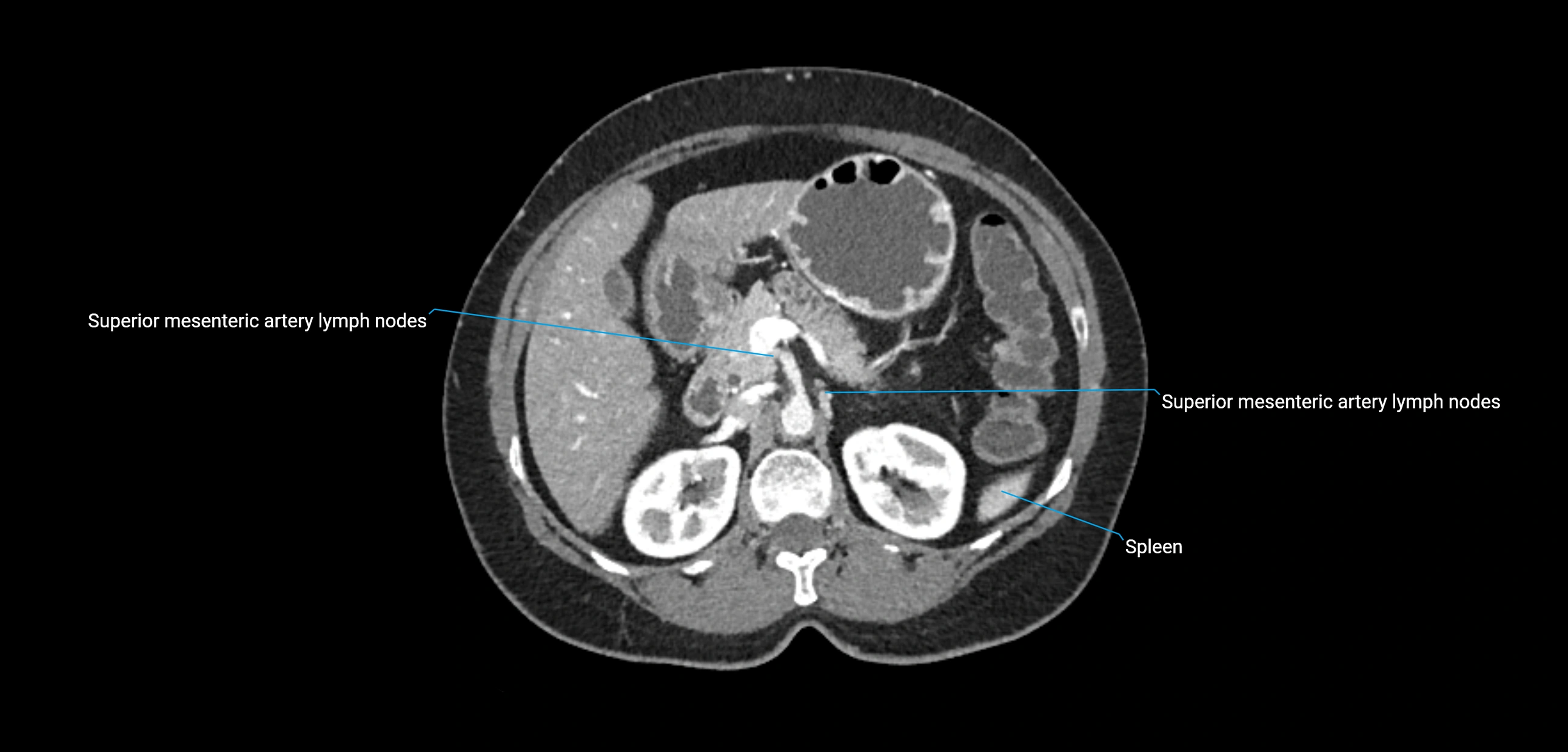
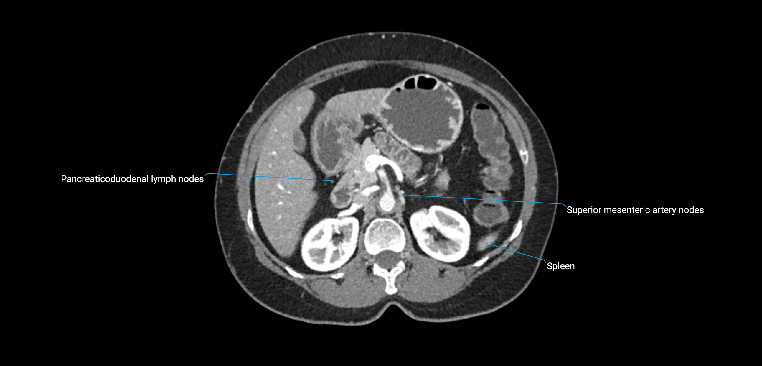
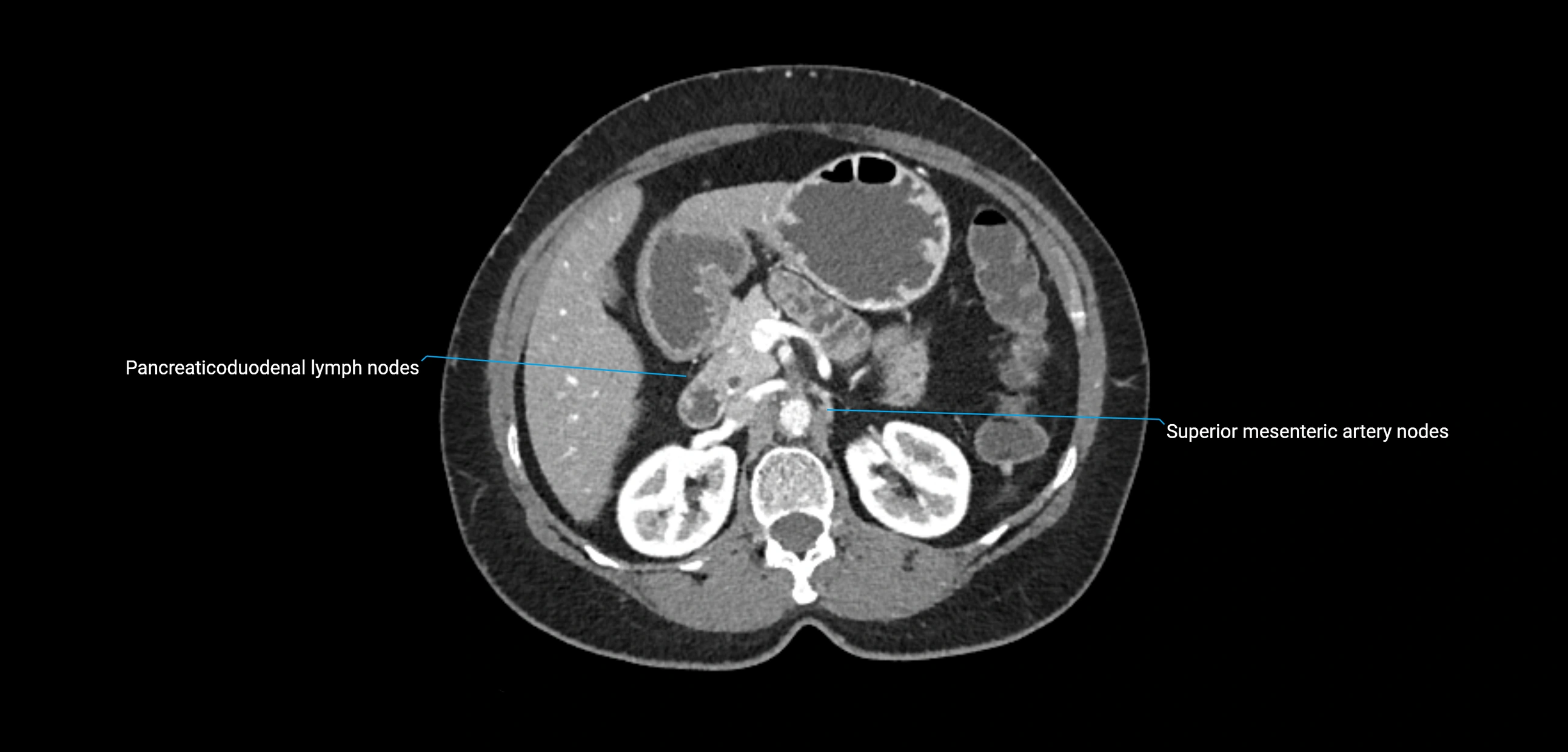
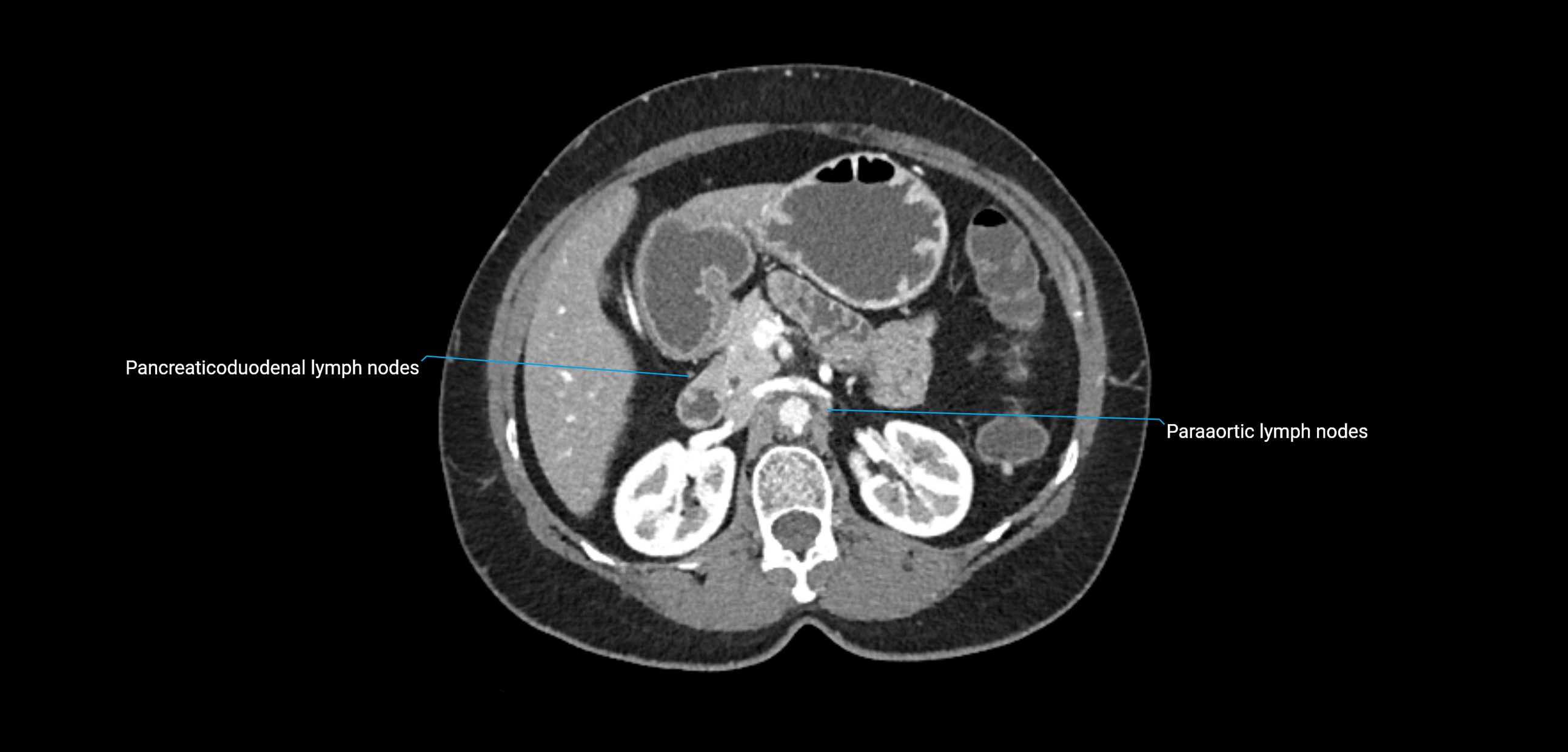
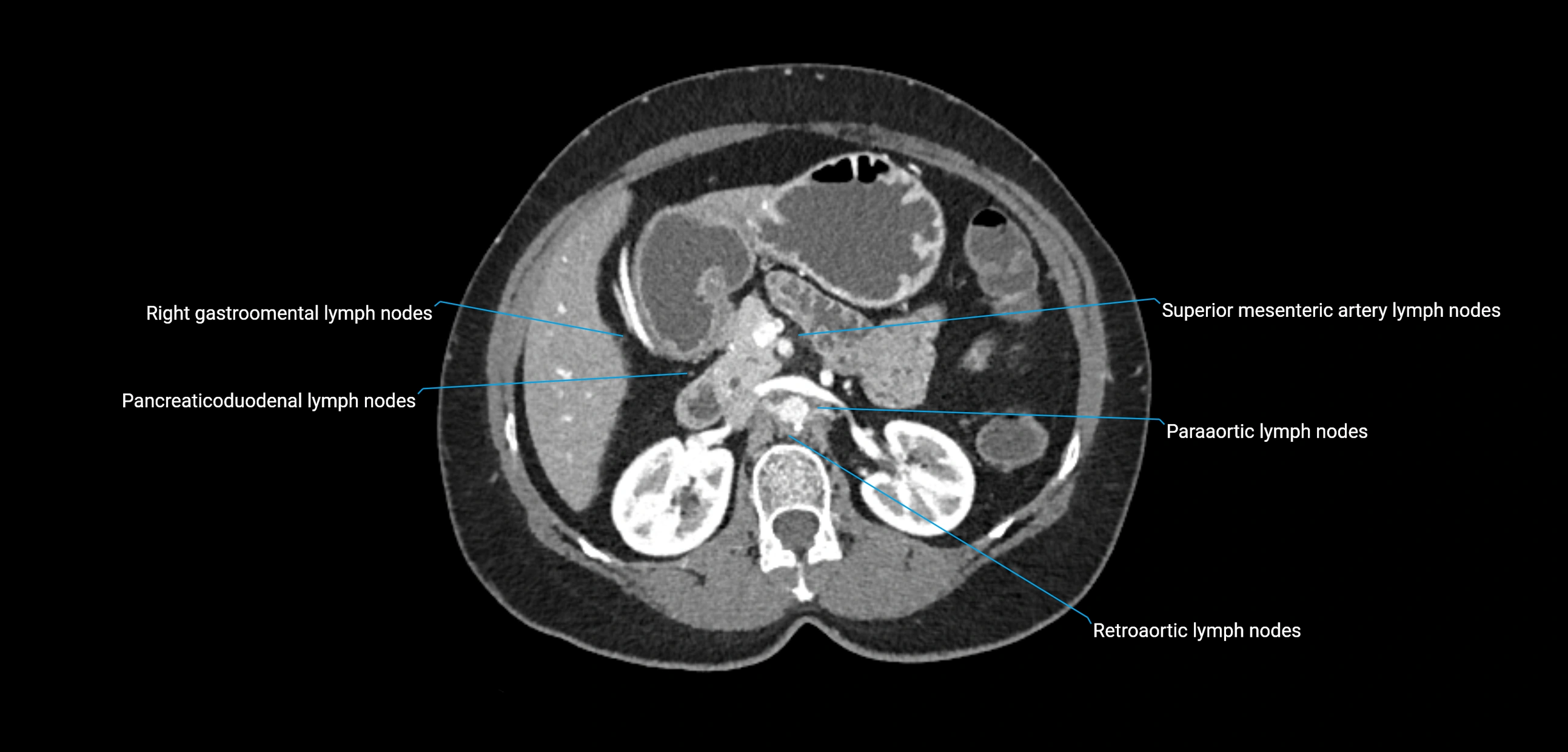
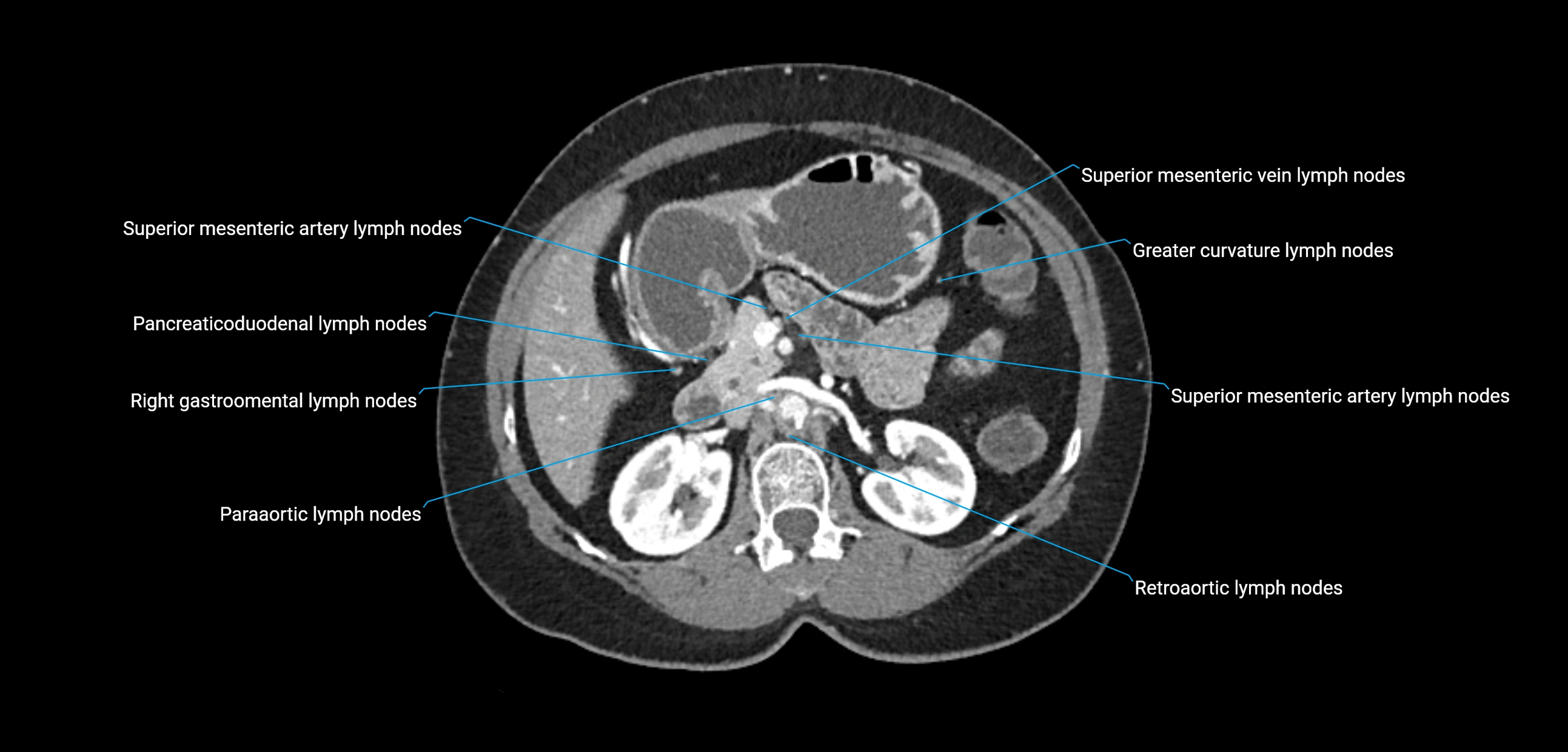
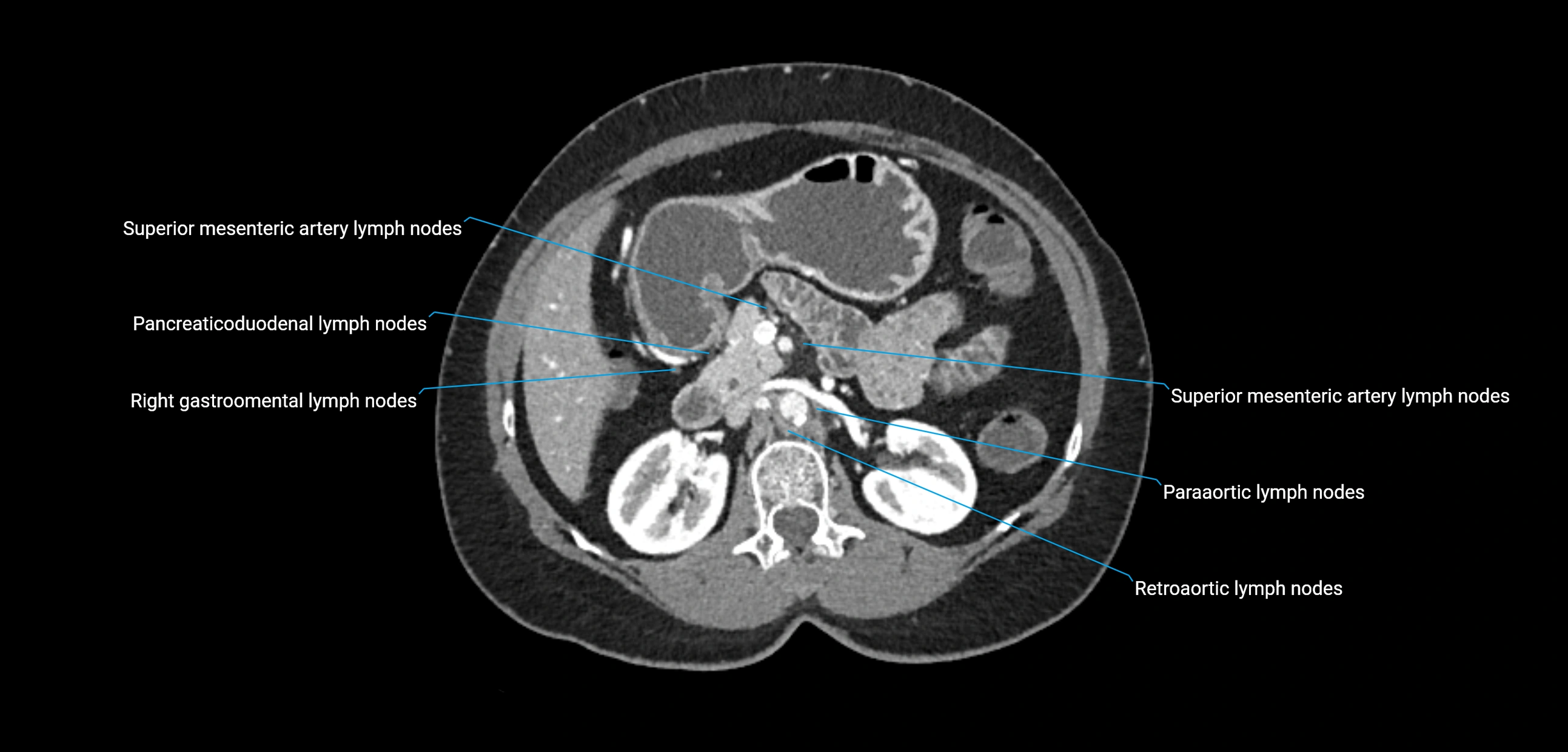
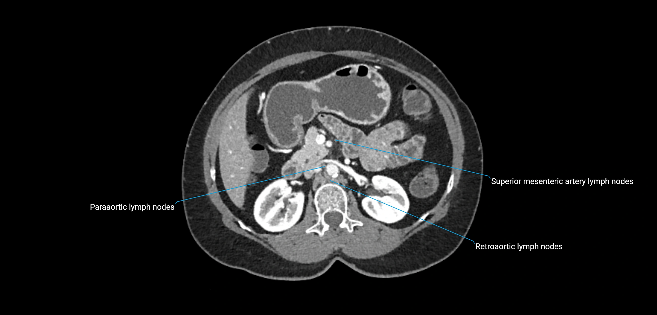
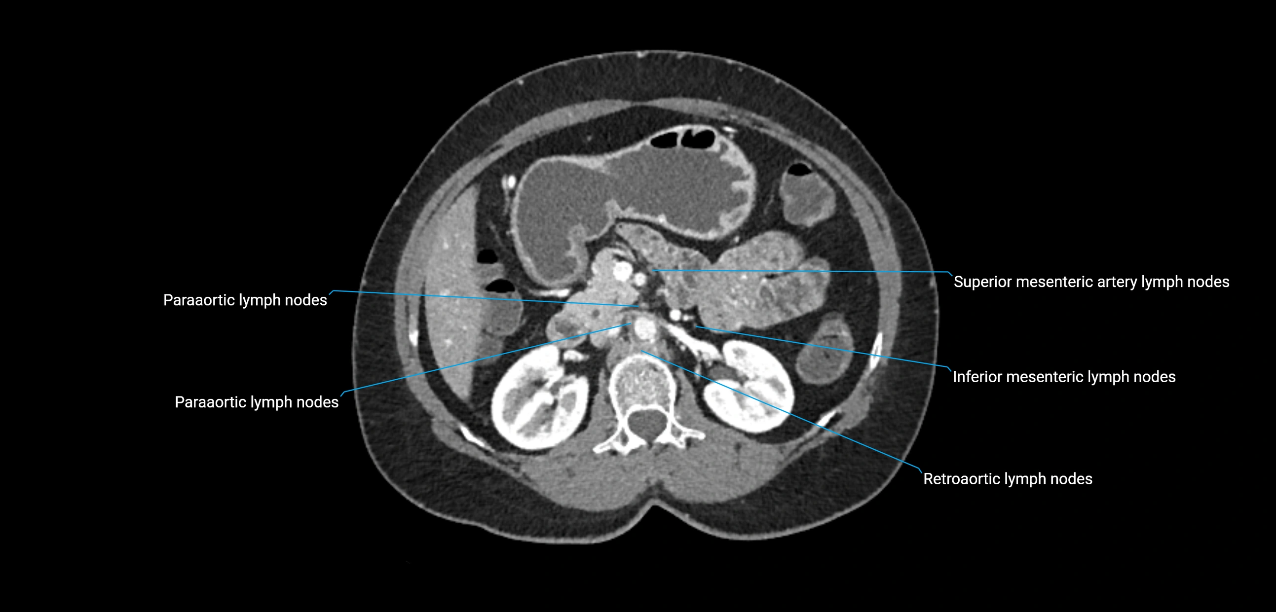
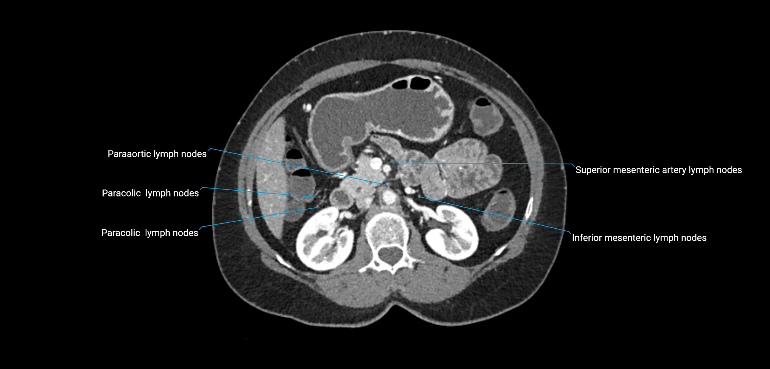
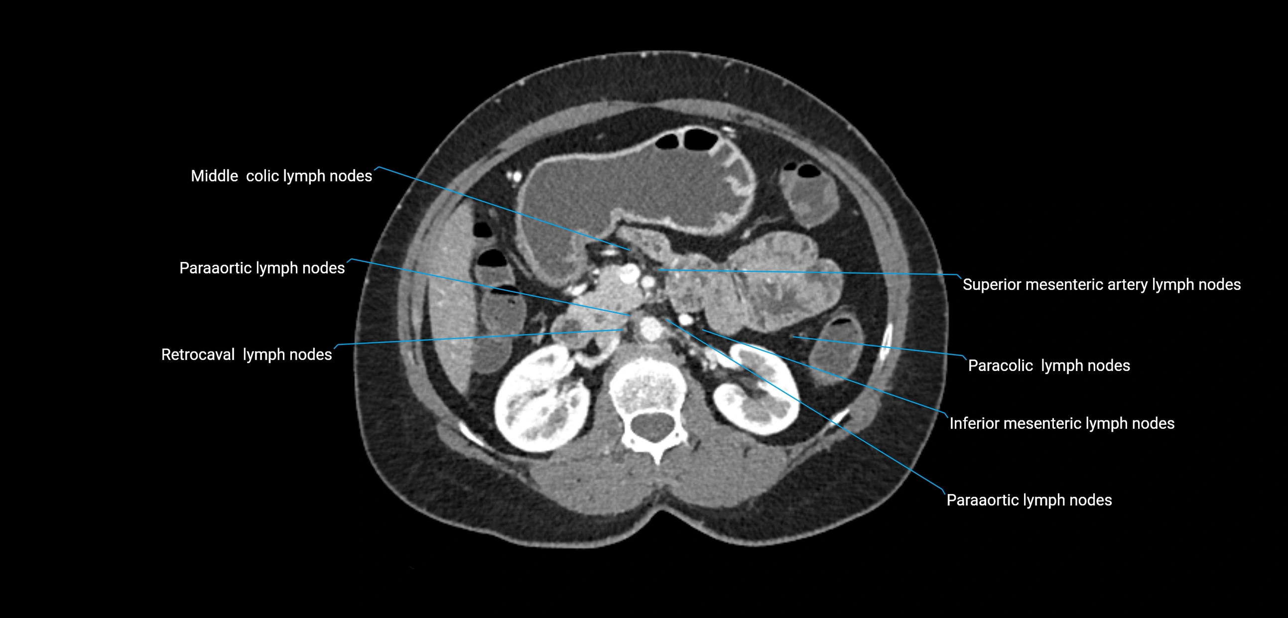
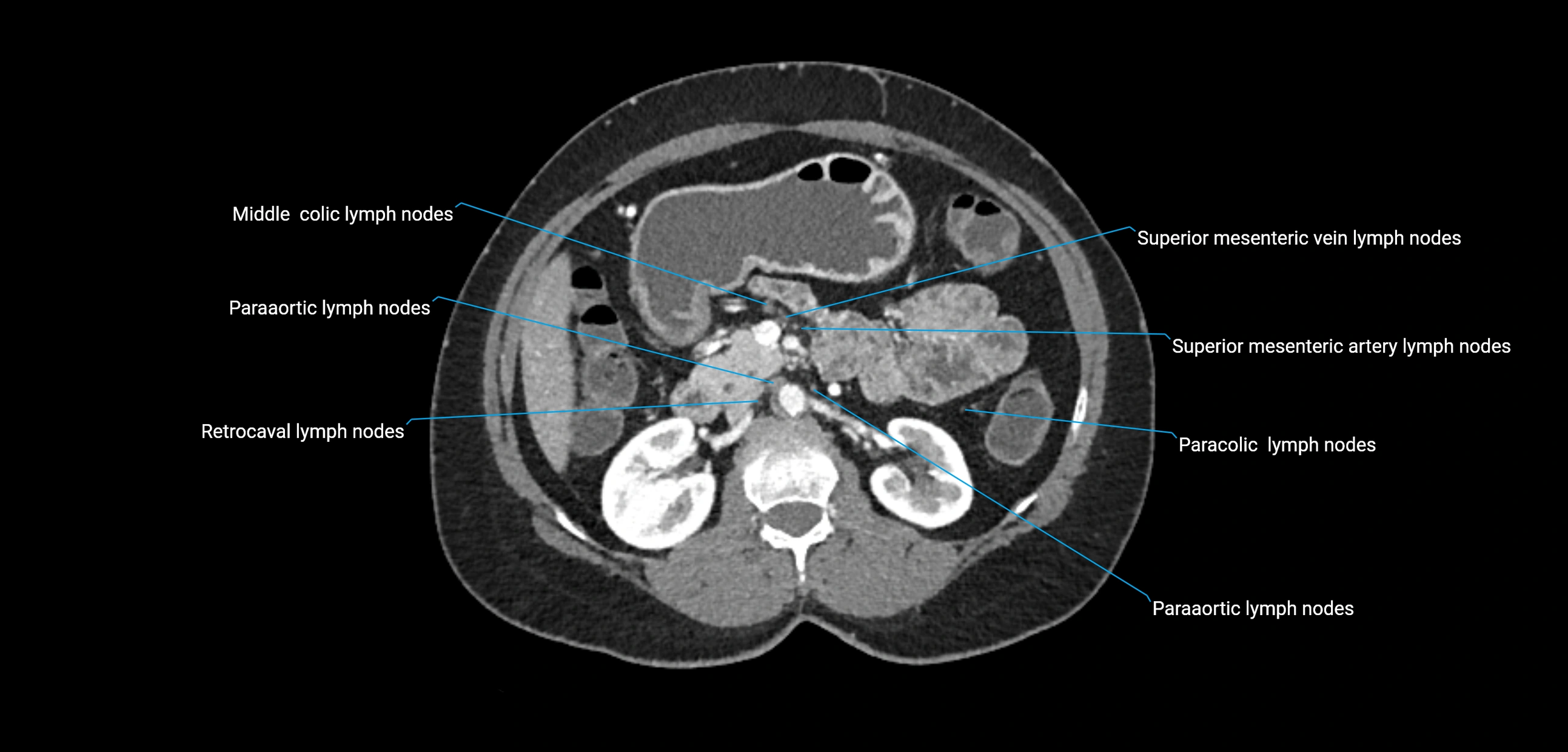
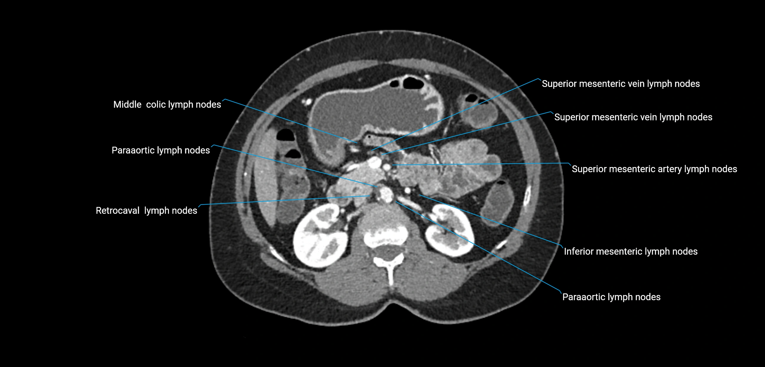
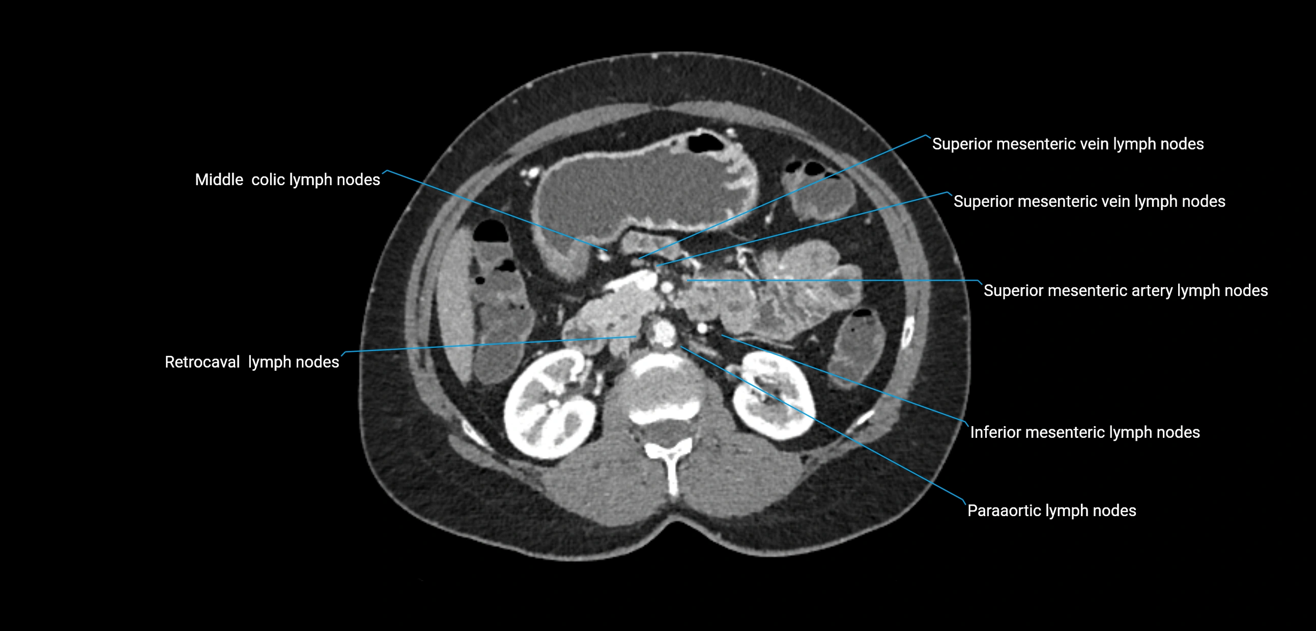
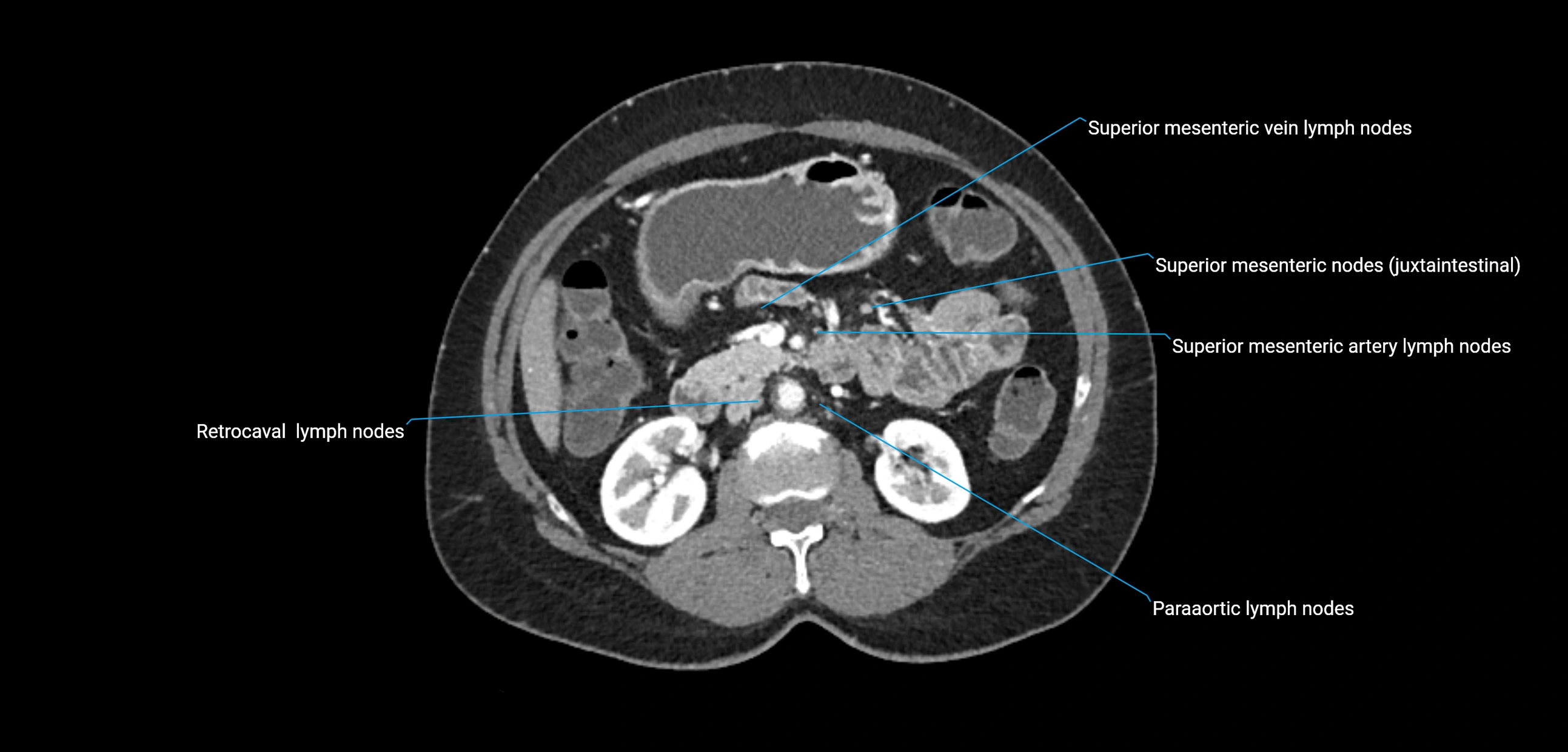
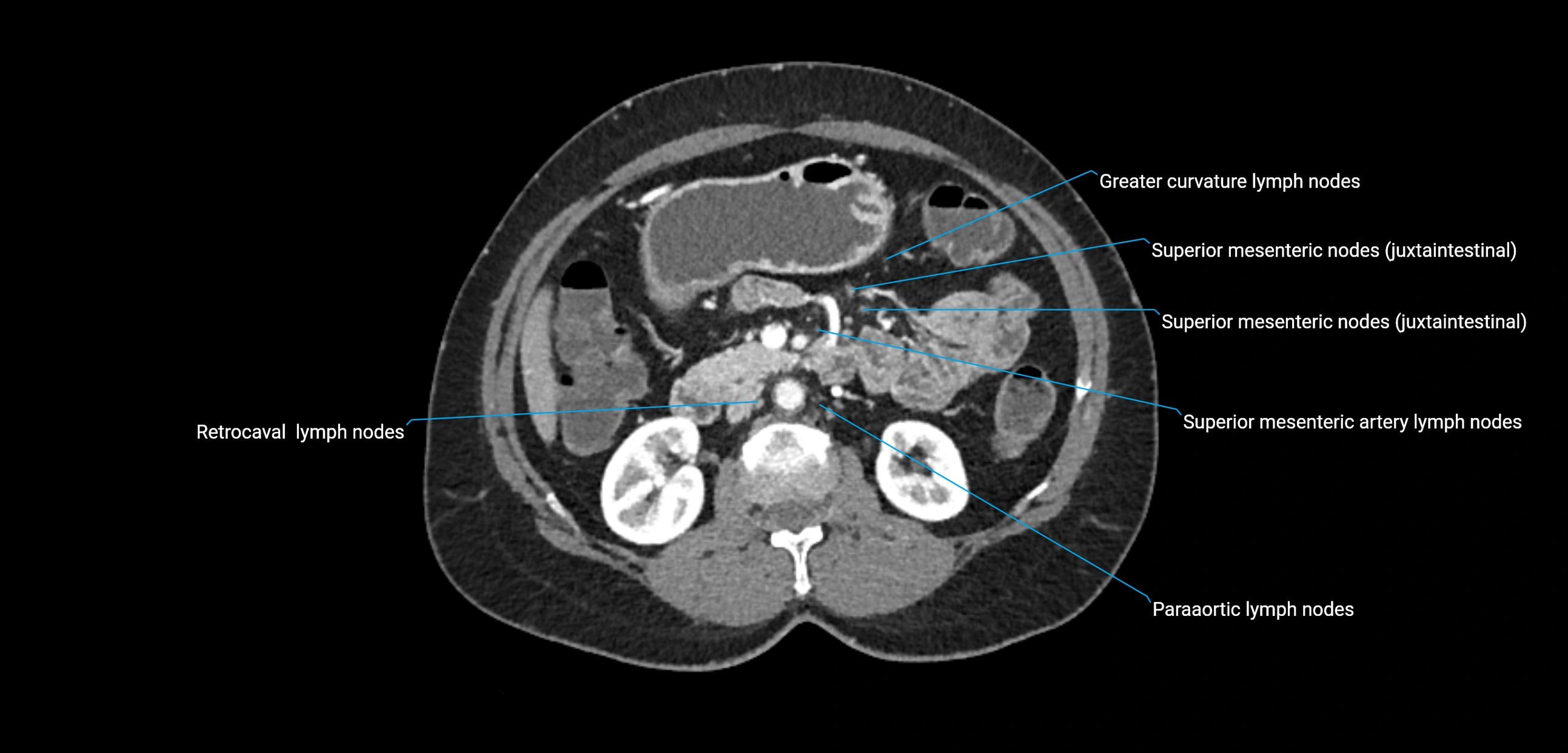
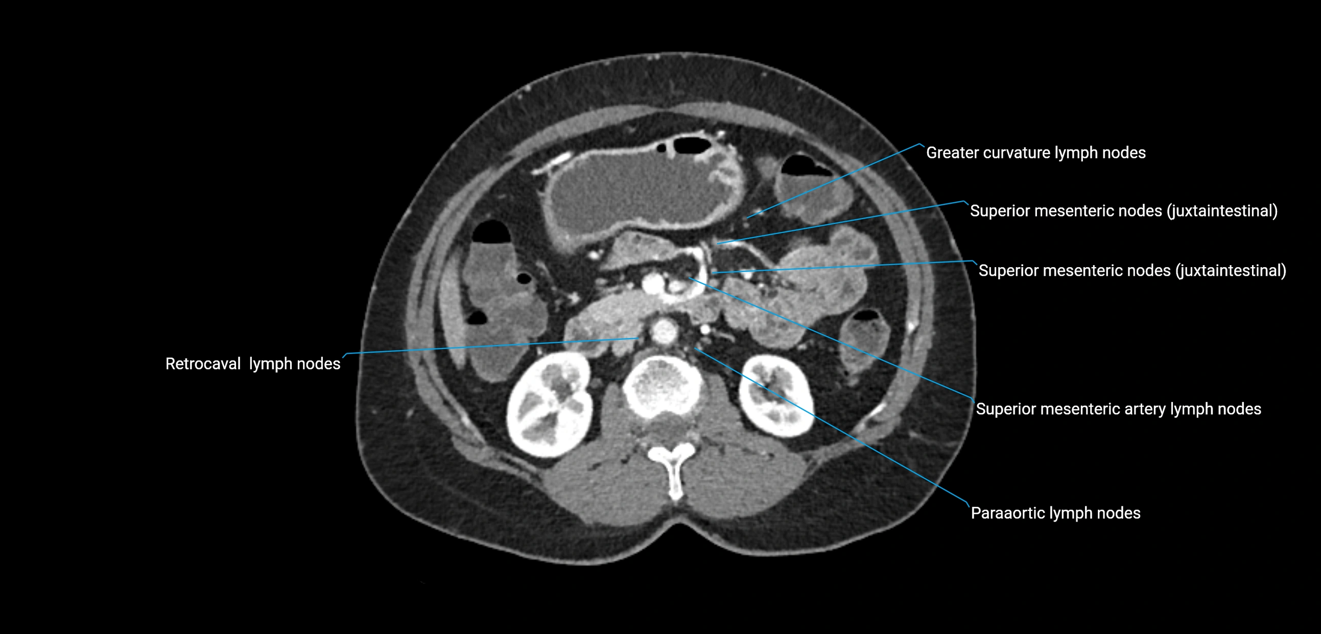
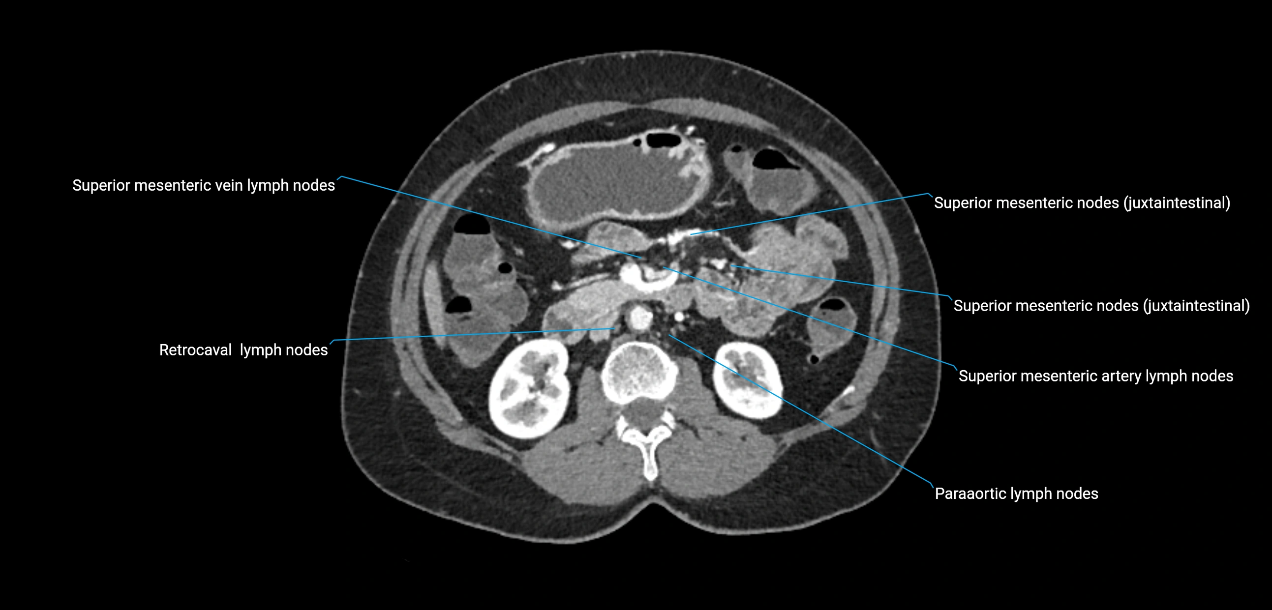
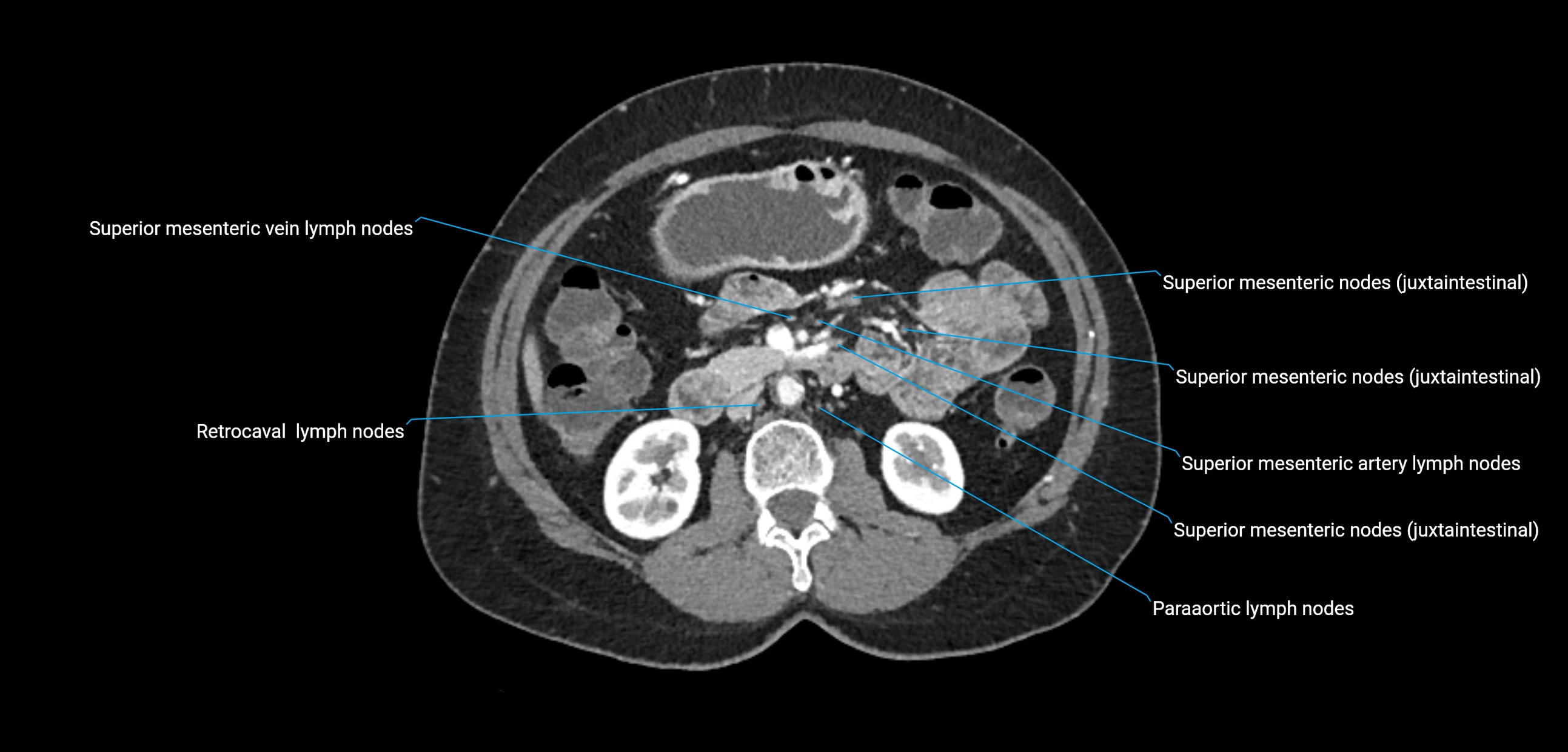
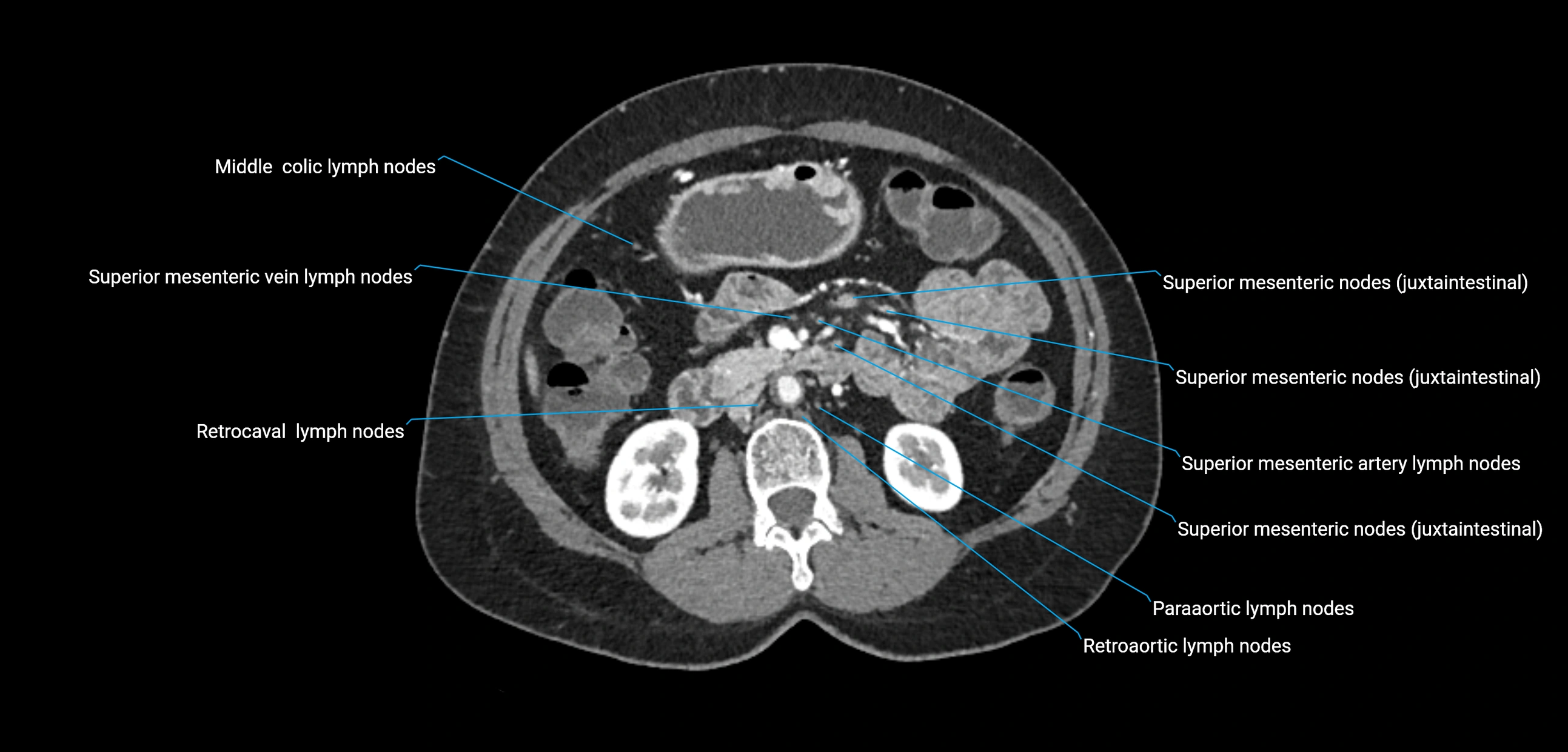
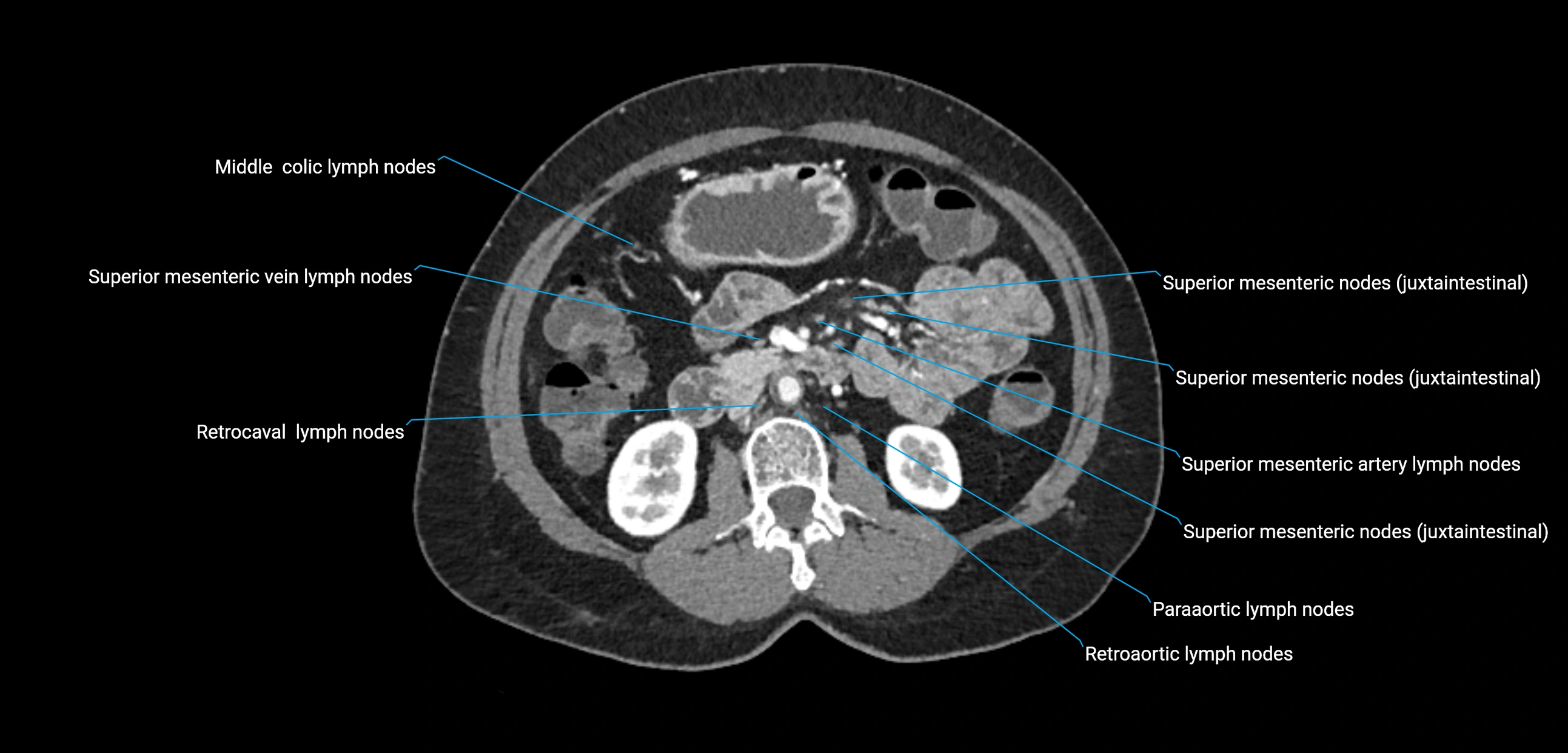
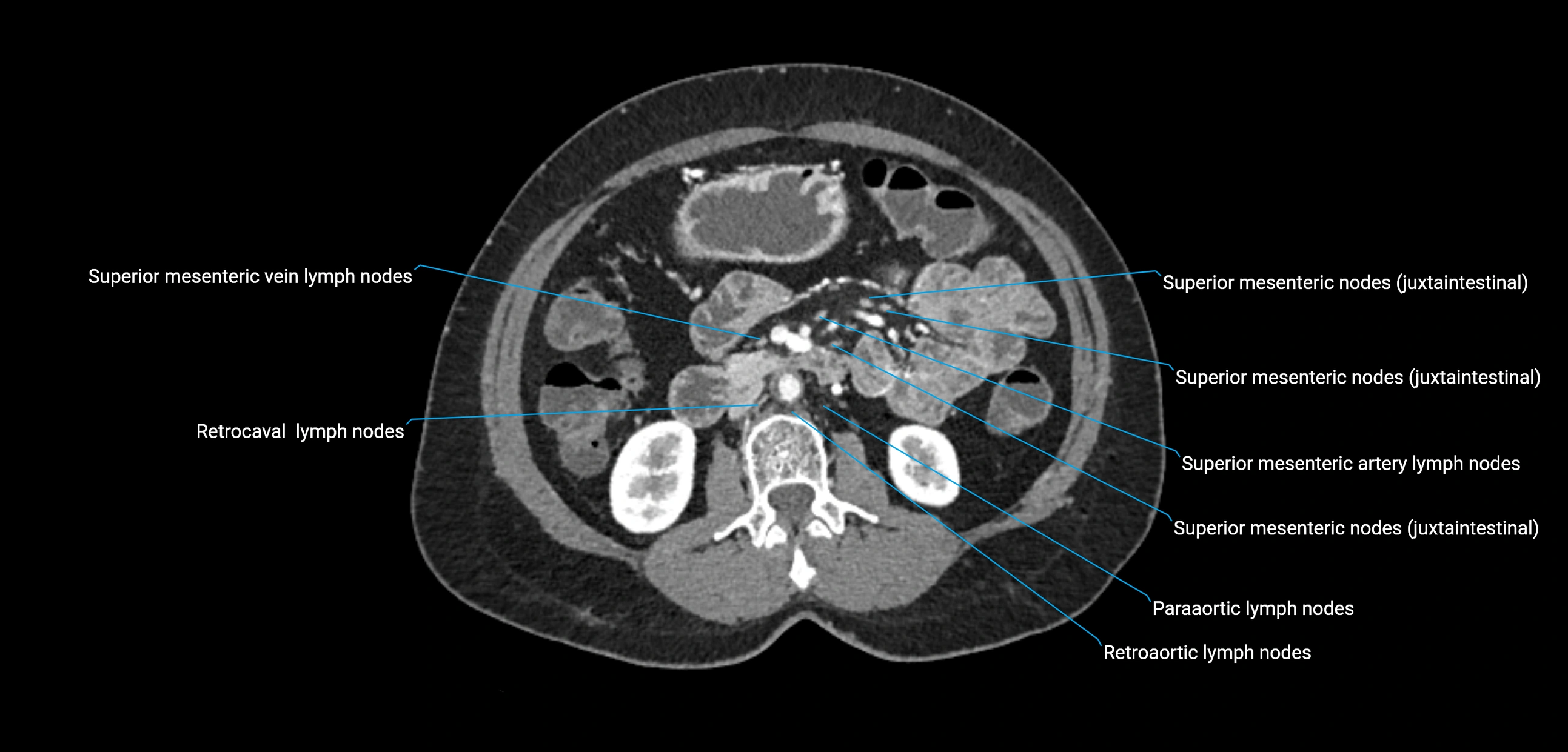
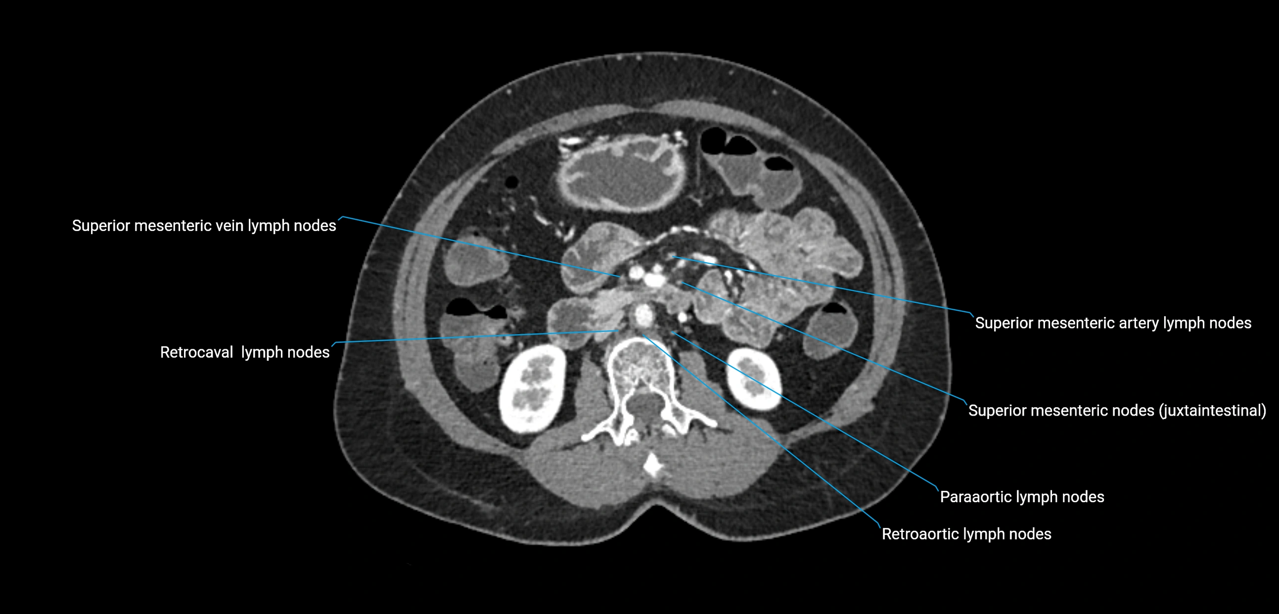
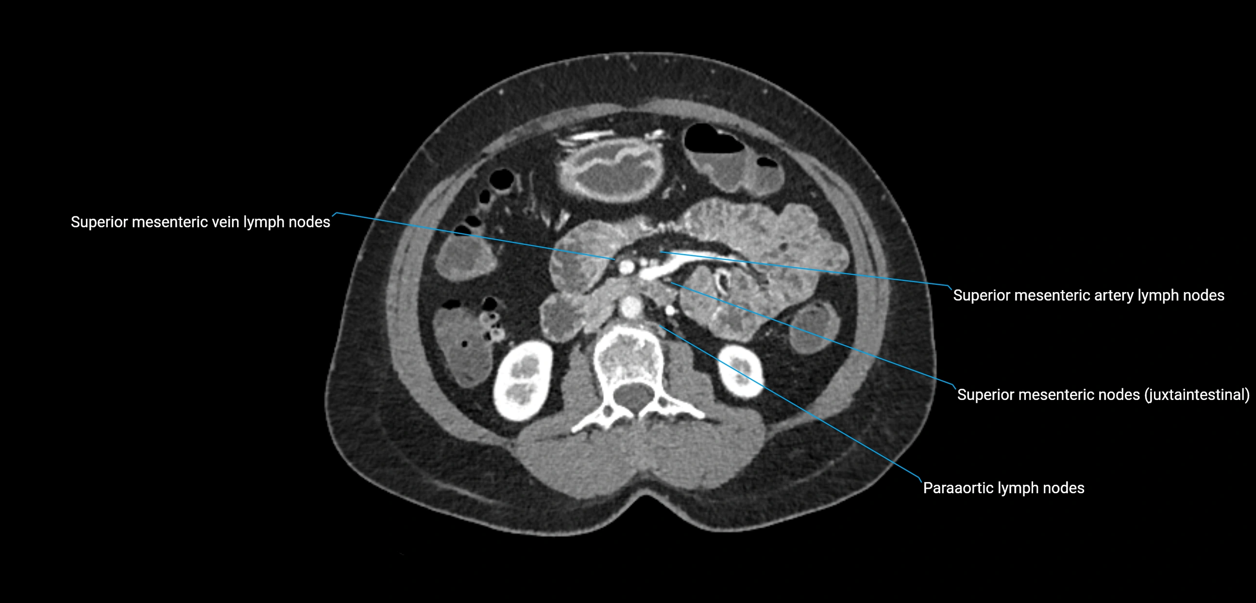
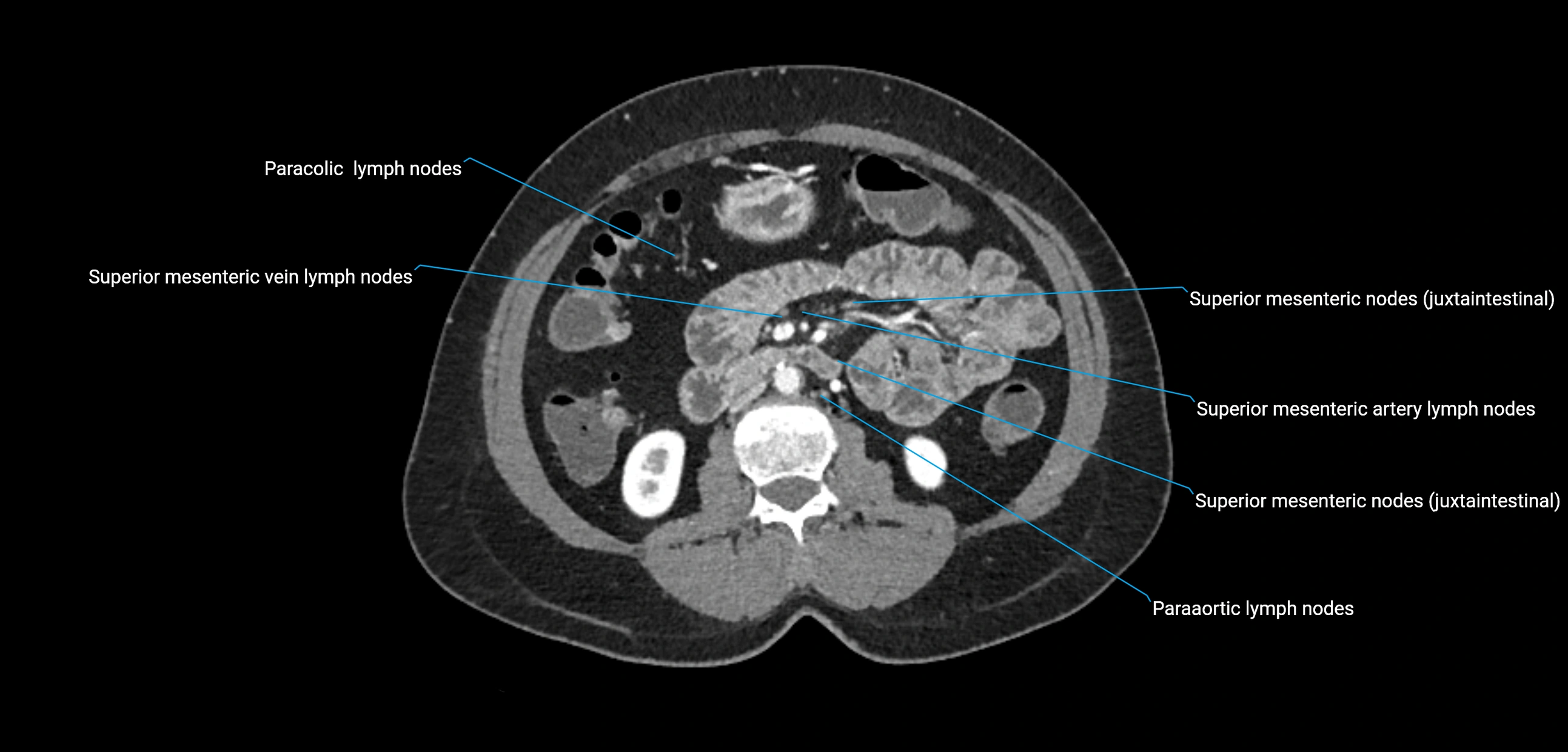
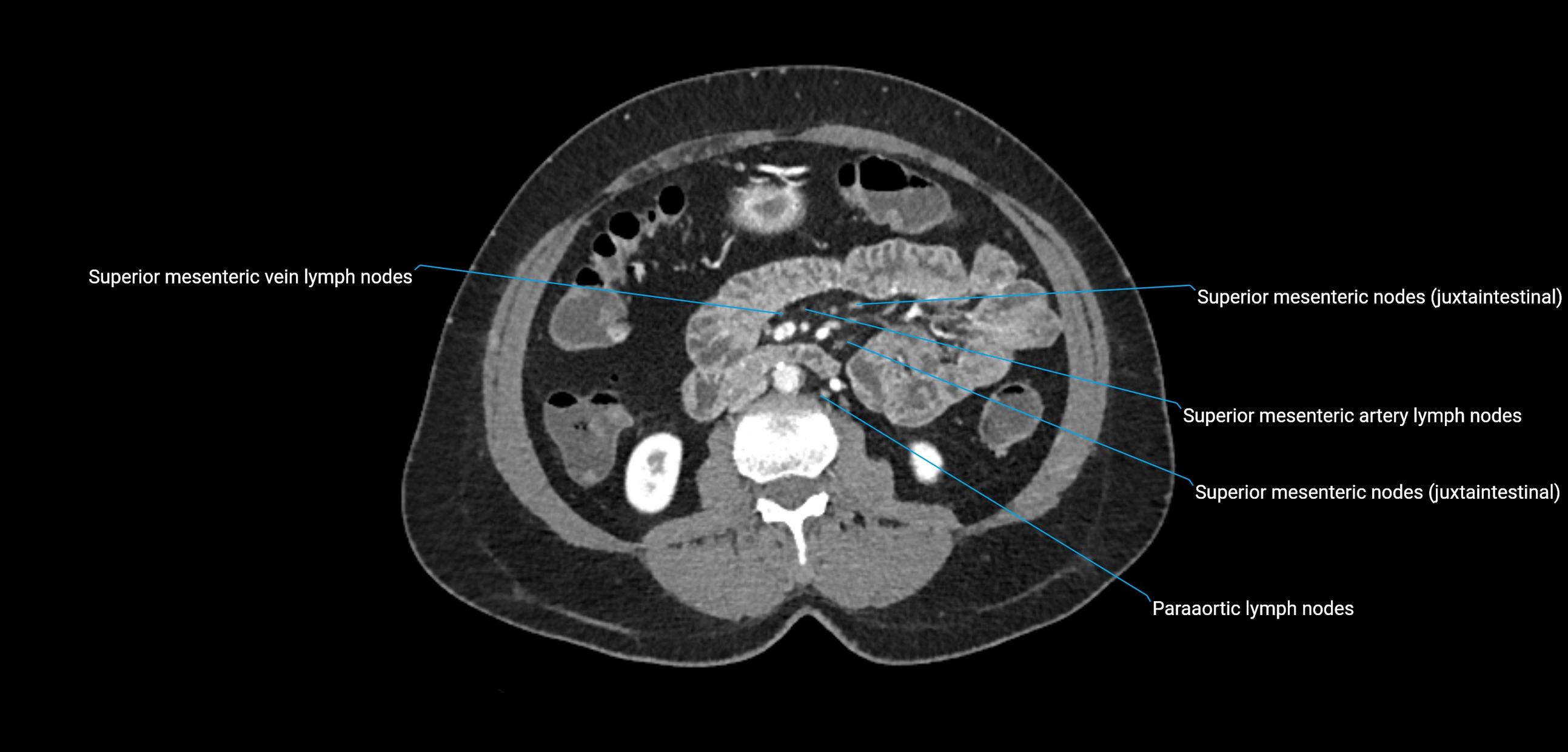
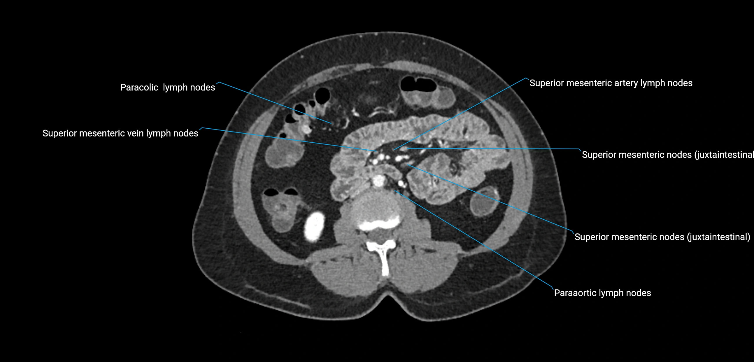
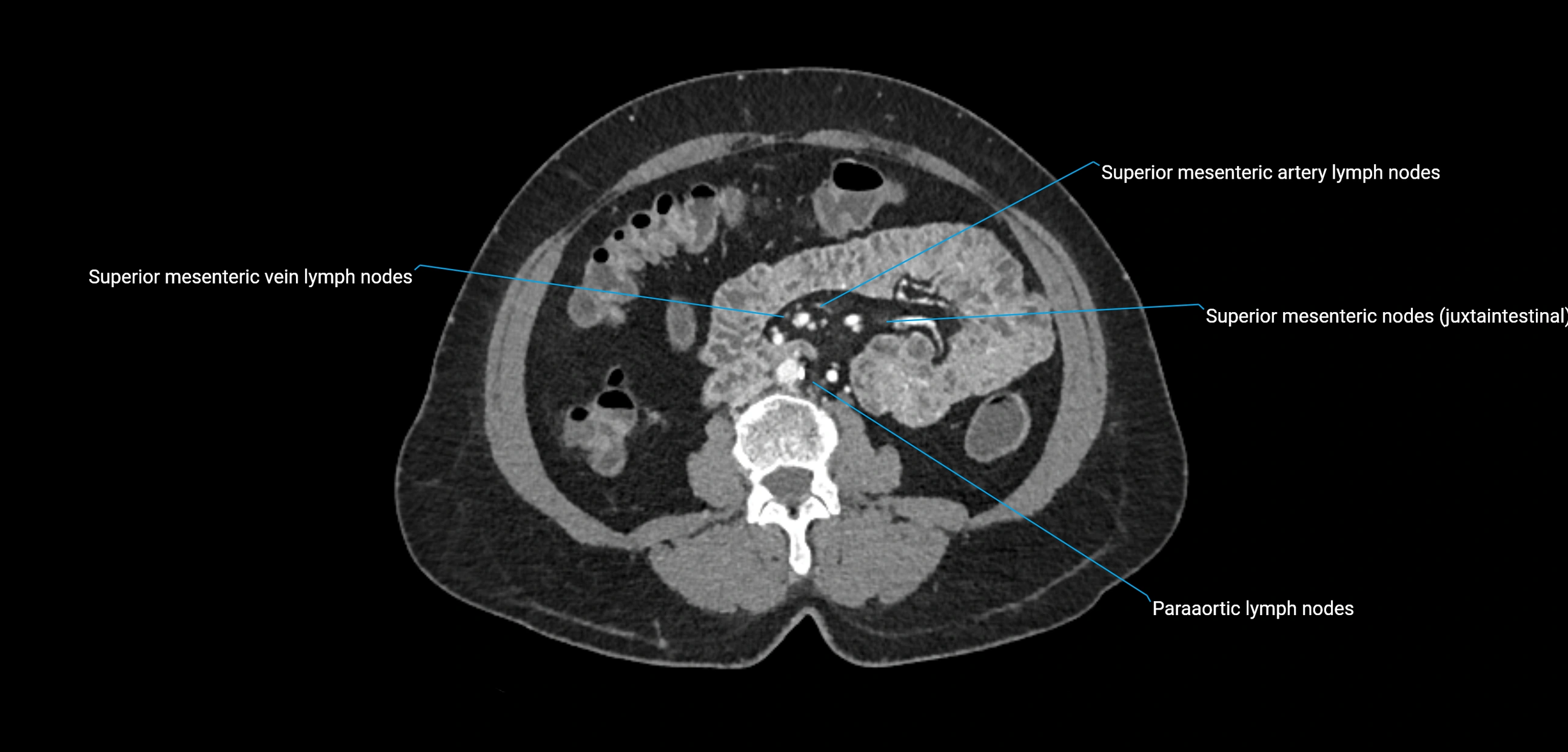
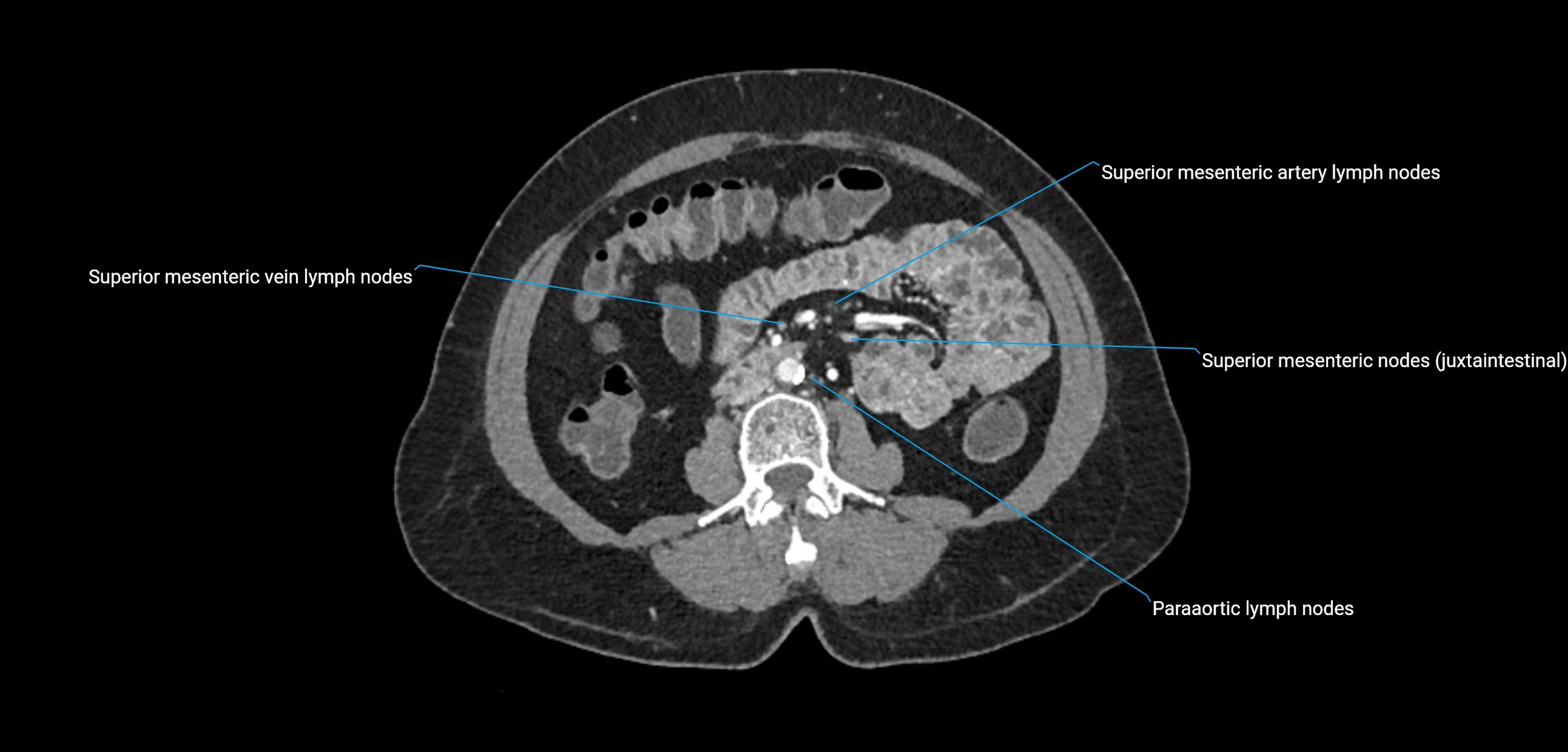
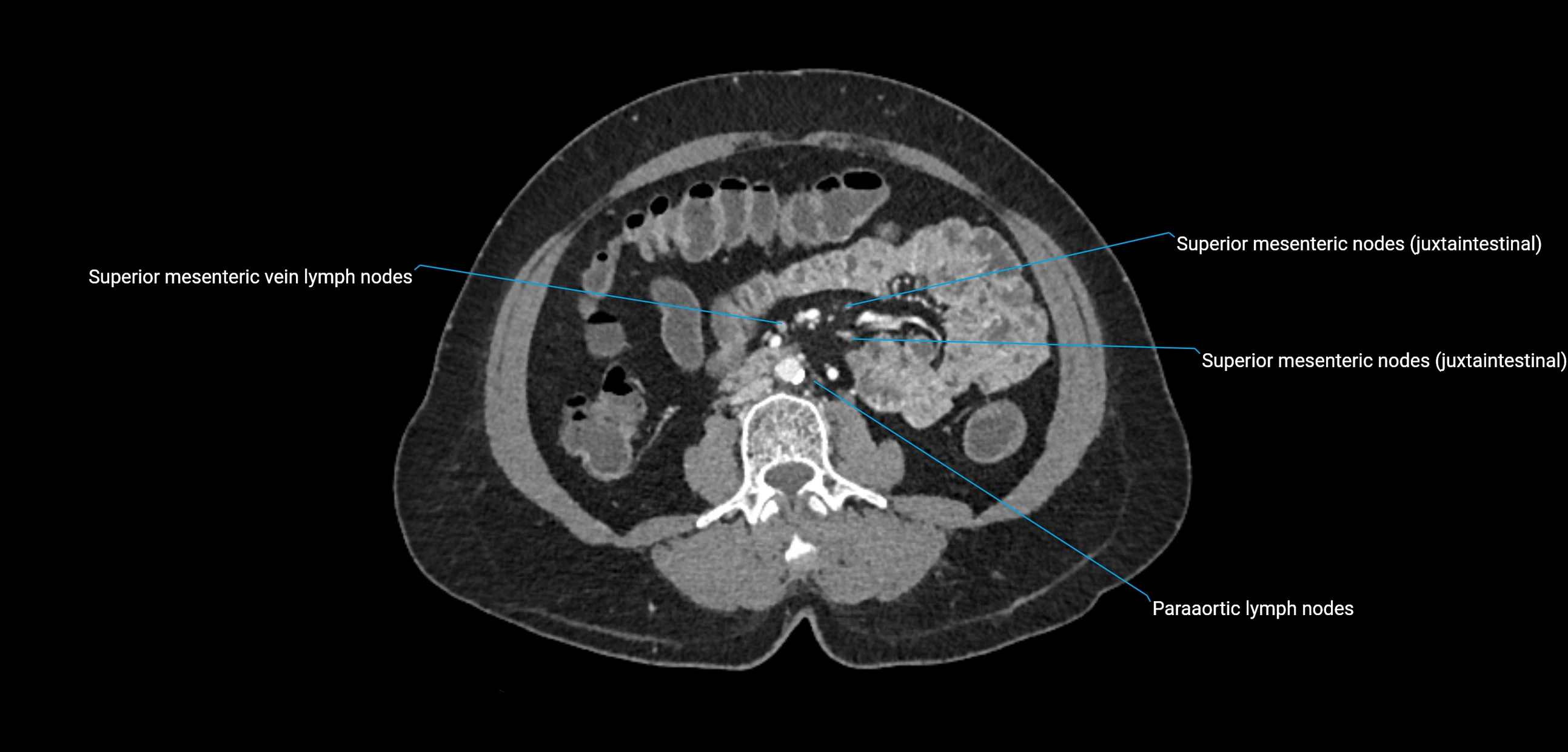
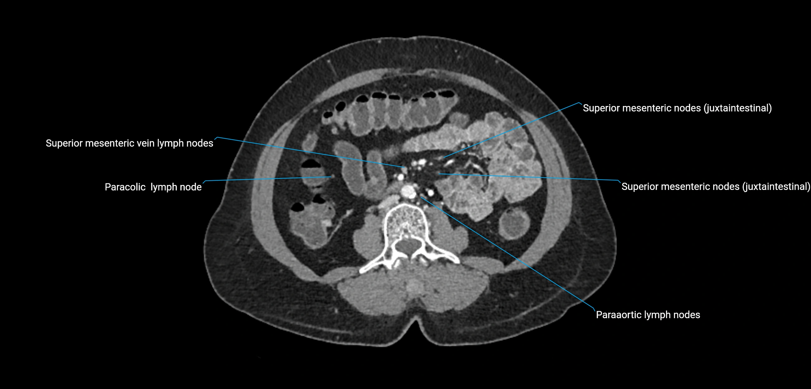
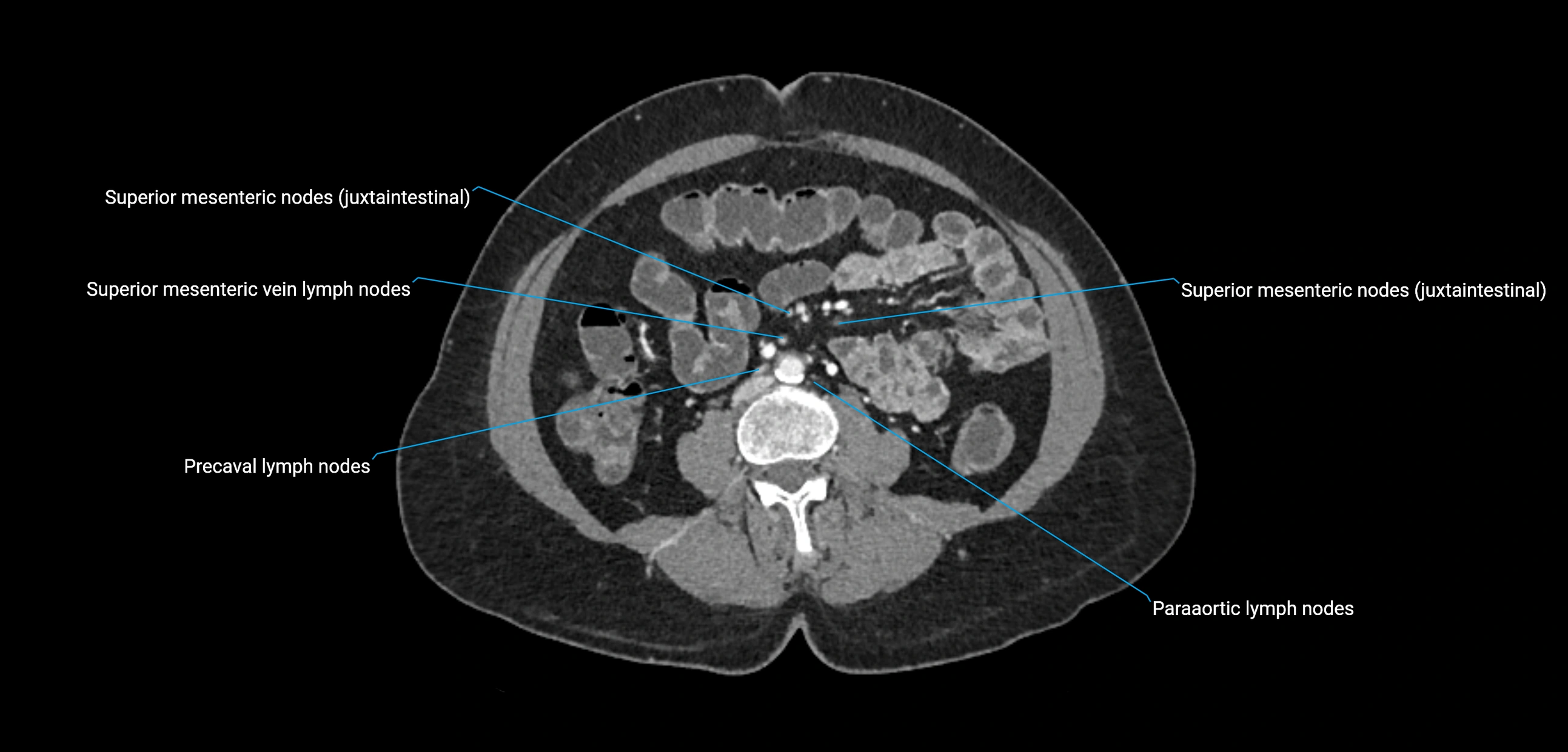
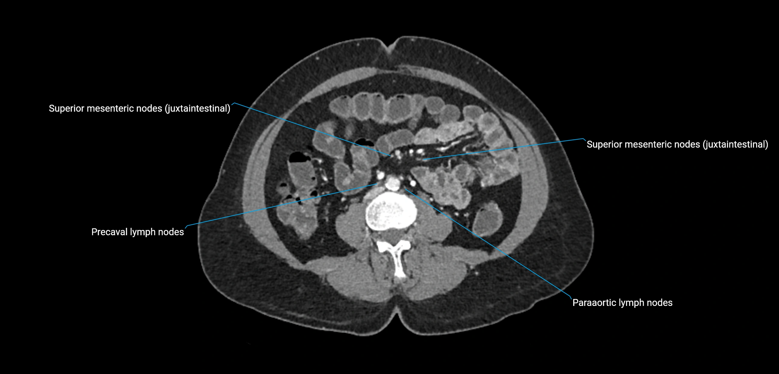
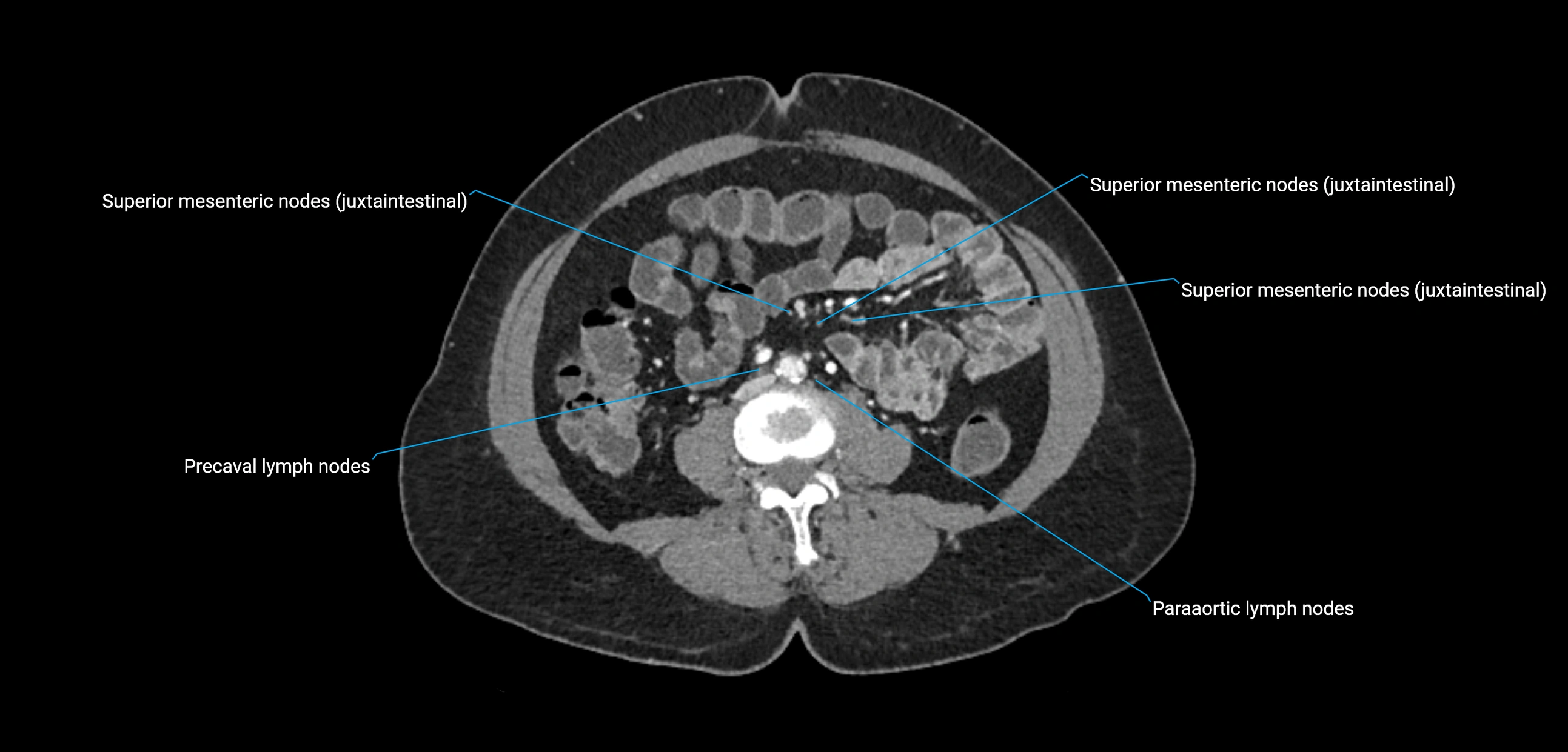
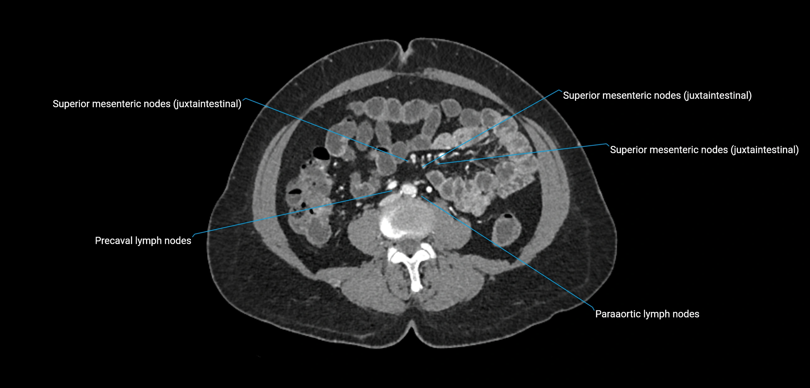
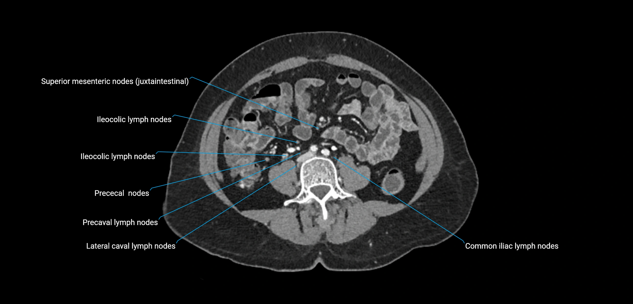
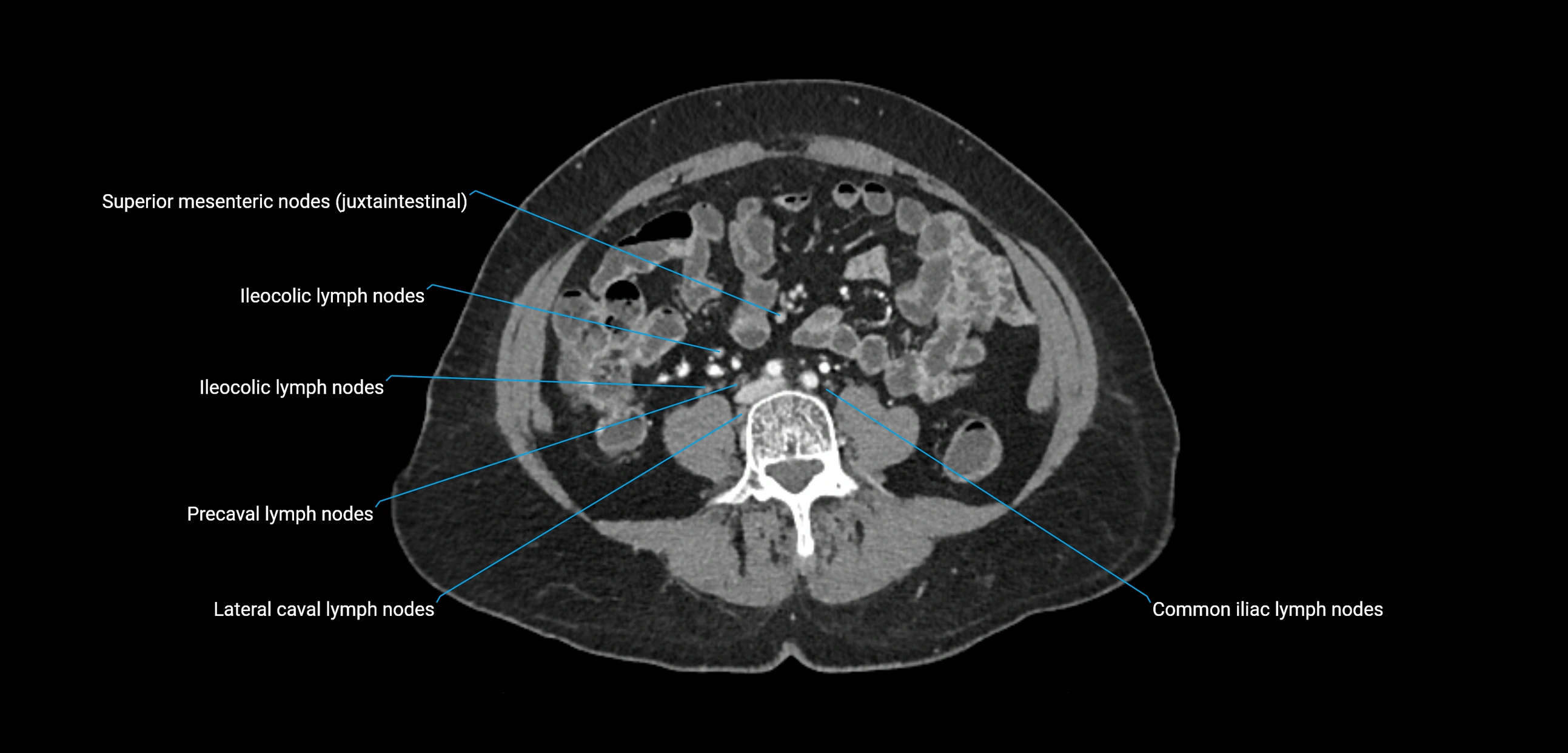
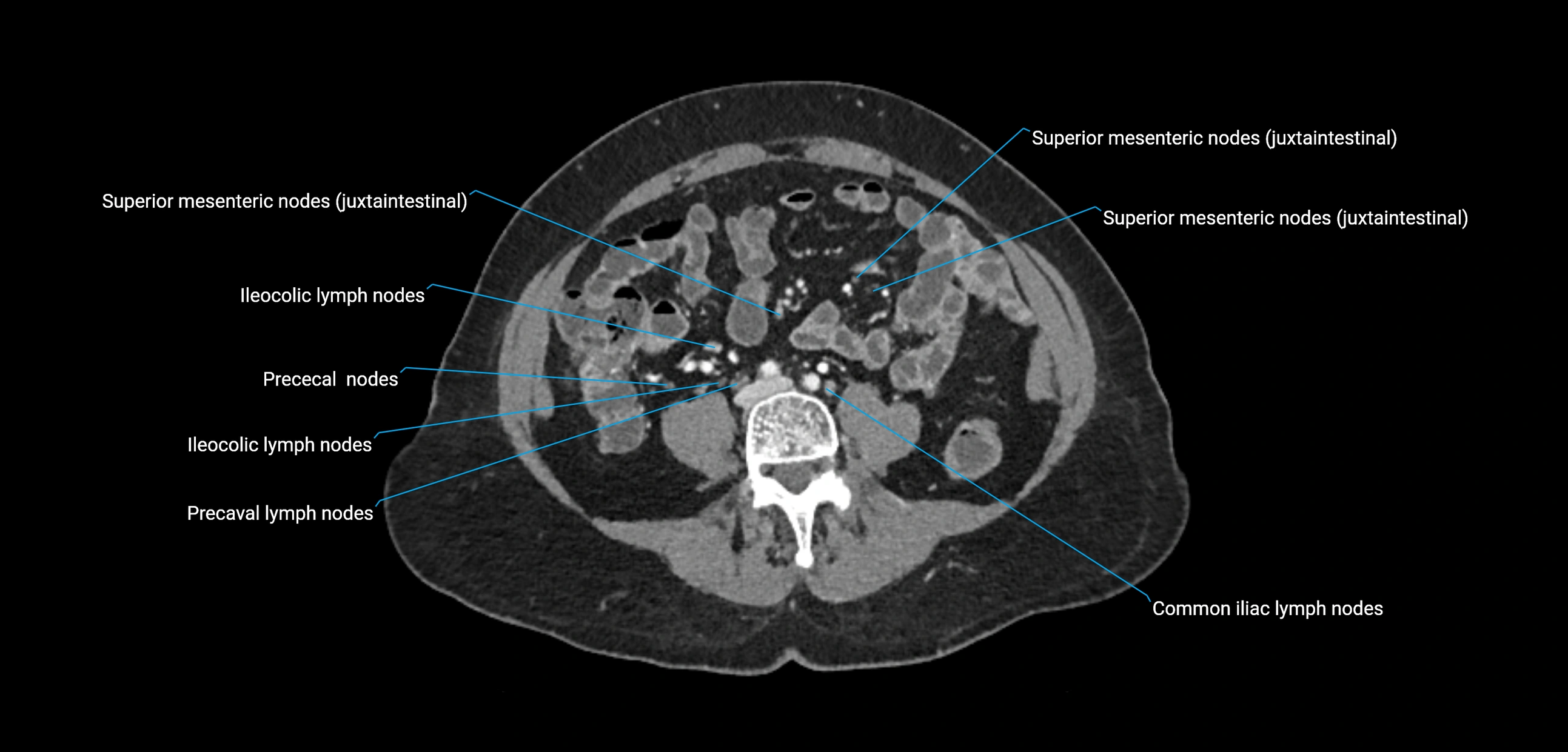
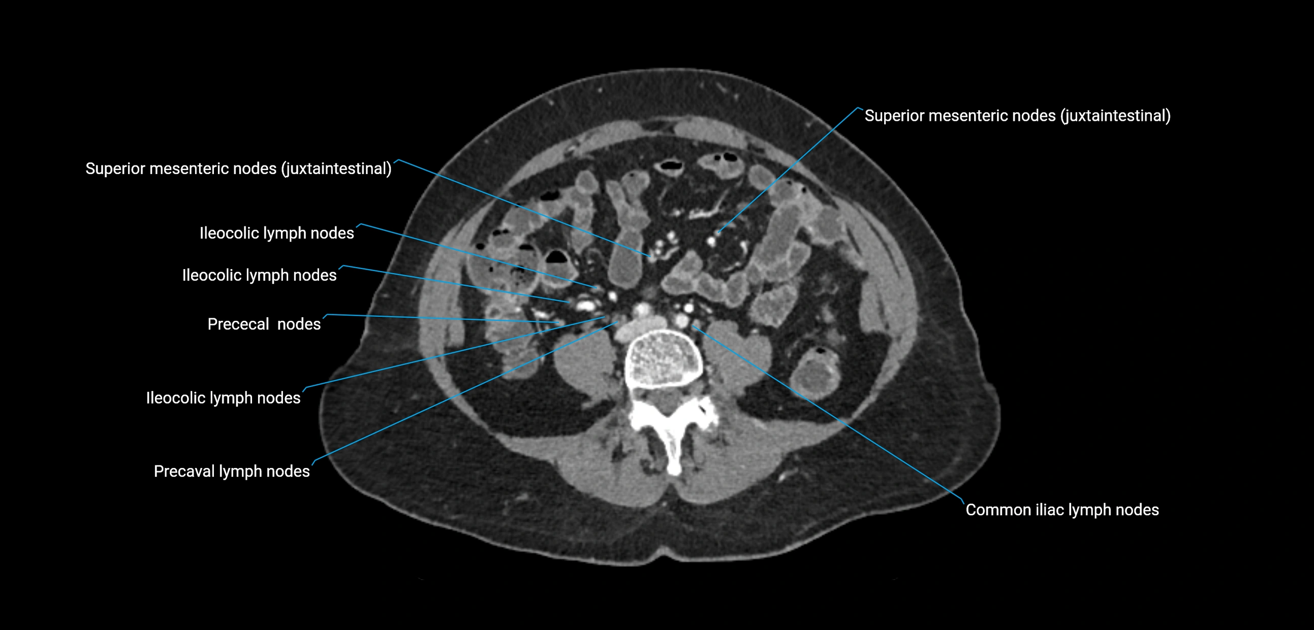
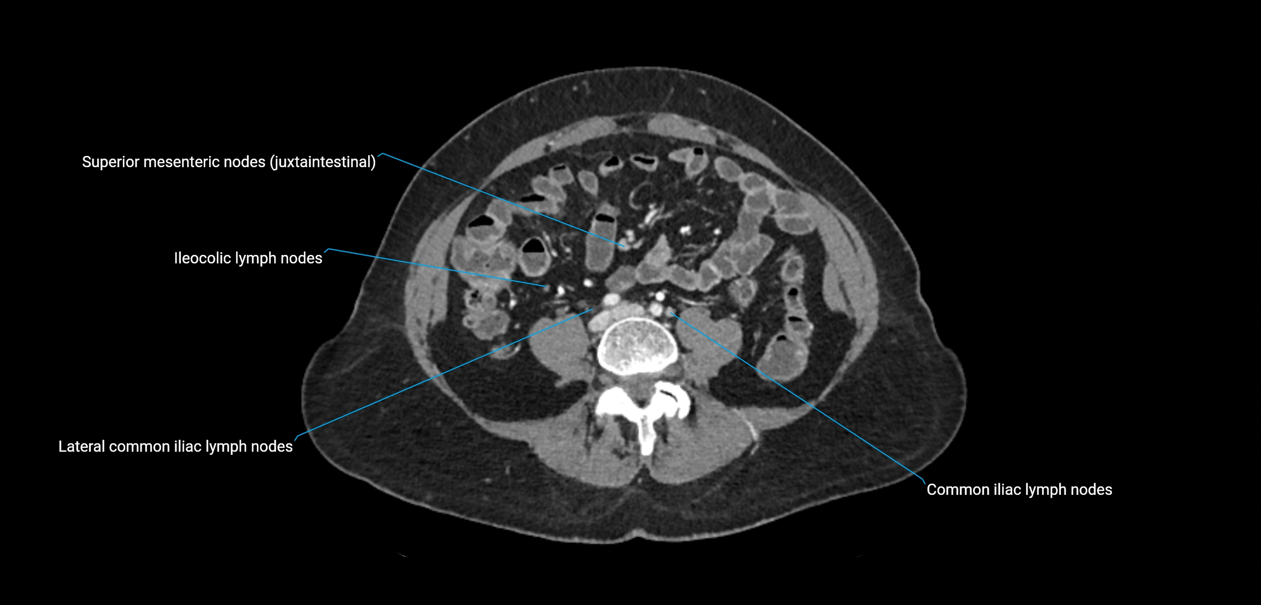
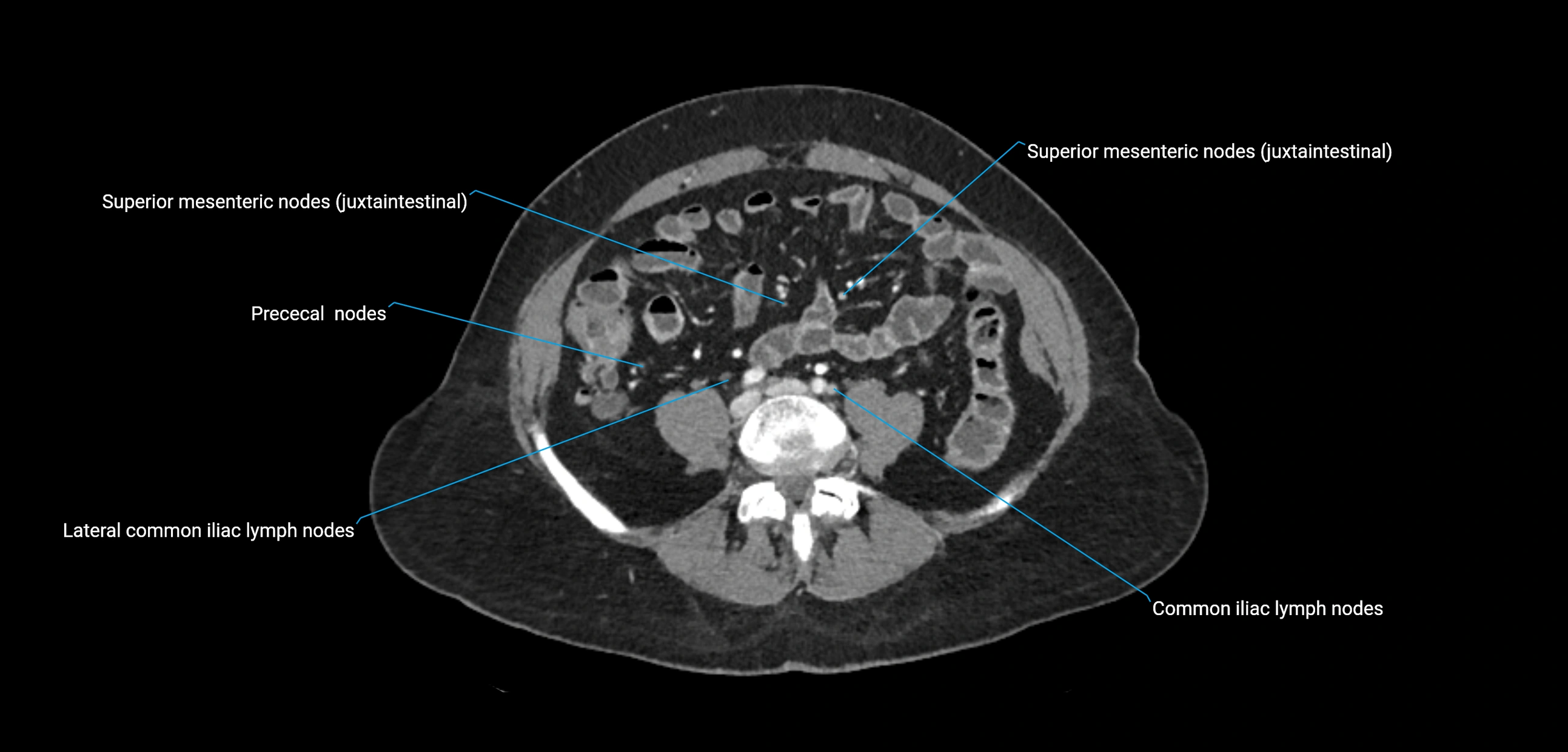
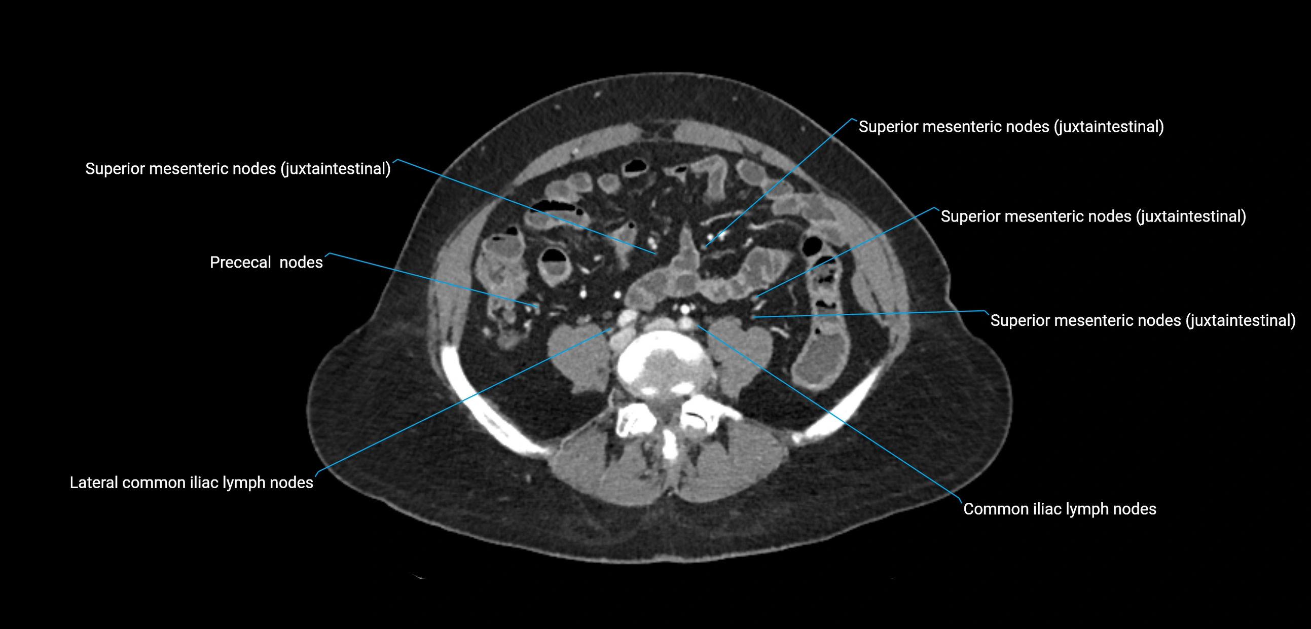
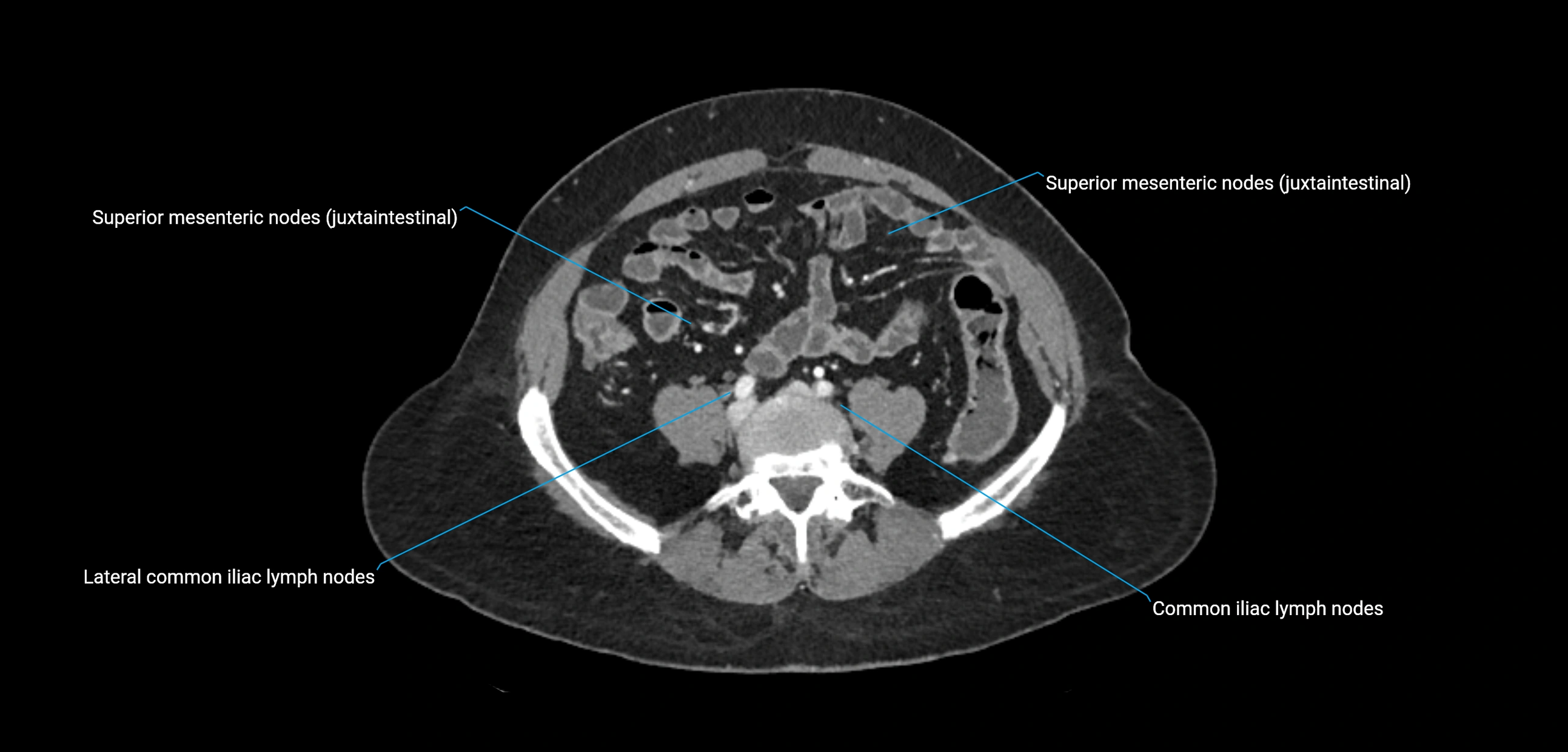
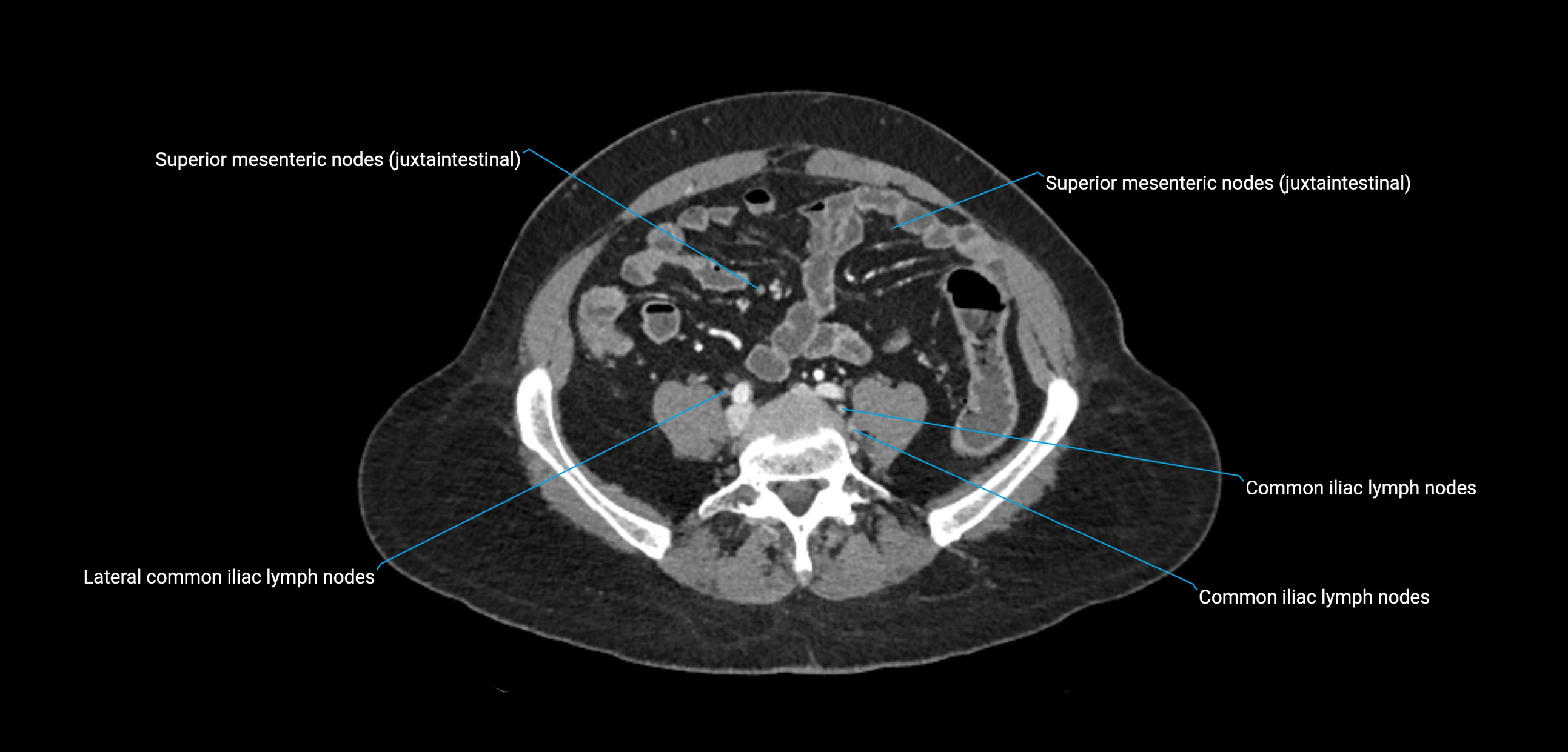
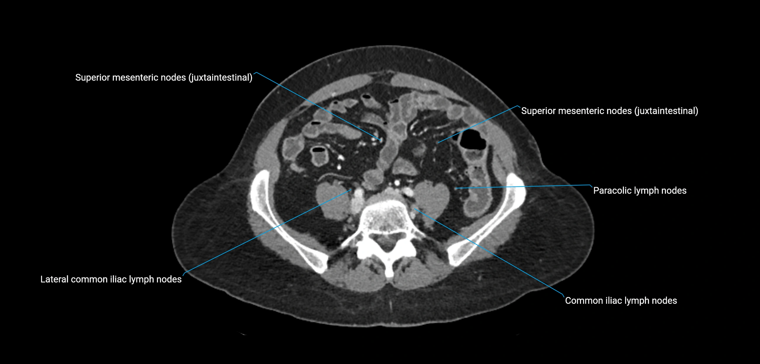
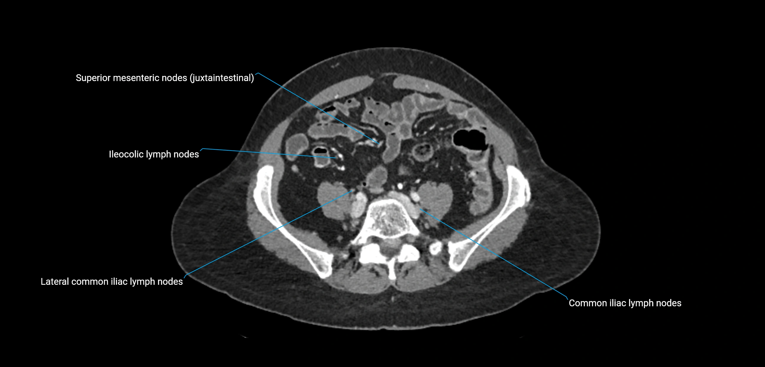
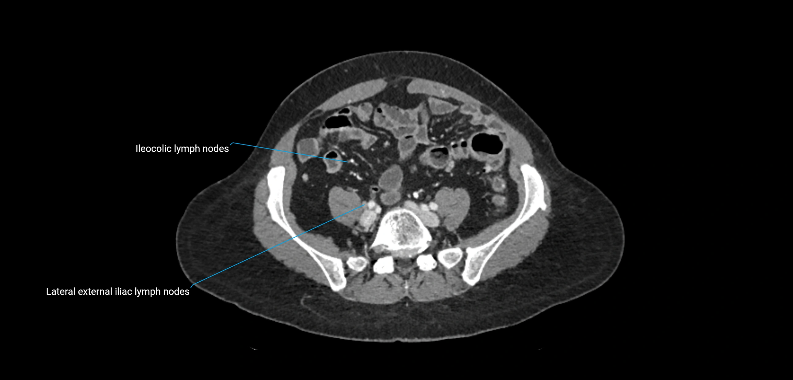
The lateral aortic lymph nodes (also called para-aortic lymph nodes) are a major group of retroperitoneal lymph nodes located along the abdominal aorta and its branches. They lie between the diaphragmatic crura superiorly and the bifurcation of the aorta at L4 inferiorly.
They are positioned on both sides of the abdominal aorta:
-
Right lateral aortic nodes: adjacent to the inferior vena cava (IVC)
-
Left lateral aortic nodes: lateral to the abdominal aorta
These nodes receive lymph from a wide range of abdominal and pelvic structures. Specifically, they drain lymph from the kidneys, suprarenal glands, gonads (testes/ovaries), uterus, uterine tubes, and pelvic organs, before converging into the lumbar lymphatic trunks, which terminate in the cisterna chyli → thoracic duct.
Clinically, the lateral aortic lymph nodes are critically important in oncology, being involved in the spread of testicular cancer, ovarian cancer, endometrial cancer, cervical cancer, renal malignancies, and retroperitoneal lymphomas. They are also key targets in retroperitoneal lymph node dissection (RPLND) for testicular tumors.
Synonyms
-
Para-aortic lymph nodes
-
Lumbar lymph nodes
-
Retroperitoneal aortic nodes
Function
-
Drain lymph from the kidneys, suprarenal glands, ureters, gonads, and pelvic organs
-
Provide a major pathway to the cisterna chyli and thoracic duct
-
Serve as sentinel nodes in urologic and gynecologic oncology
-
Act as barriers and filters for infections and malignancies of the retroperitoneum
MRI Appearance
T1-weighted images:
-
Normal nodes appear as low-to-intermediate signal ovoid structures in retroperitoneal fat
-
Surrounded by hyperintense fat planes for easy identification
T2-weighted images:
-
Nodes are intermediate to mildly hyperintense relative to muscle
-
Malignant nodes often appear larger, with heterogeneous signal intensity
STIR:
-
Fat suppression highlights nodes against retroperitoneal fat
-
Enlarged or inflamed nodes appear hyperintense
T1 Fat-Saturated Post-Contrast (Gadolinium):
-
Normal nodes show mild, homogeneous enhancement
-
Malignant nodes show heterogeneous or rim-like enhancement
-
Necrotic nodes may appear as non-enhancing central regions
MRI Non-Contrast 3D Imaging:
-
Provides volumetric mapping of para-aortic nodal stations
-
Helpful for oncology staging, follow-up, and surgical planning
CT Appearance
CT Pre-Contrast:
-
Nodes appear as soft-tissue density nodules adjacent to the aorta and IVC
-
Calcification may be seen in chronic infections (e.g., tuberculosis)
CT Post-Contrast:
-
Normal nodes enhance homogeneously
-
Malignant nodes may show heterogeneous enhancement, central necrosis, or conglomerate formation
-
Size >1 cm short axis is suspicious, though morphology and distribution are equally important
CT Venography (CTV):
-
Demonstrates nodal encasement or compression of adjacent vessels (aorta, IVC, renal veins)
-
Useful in staging testicular and ovarian malignancies
-
Provides 3D reconstructions for retroperitoneal lymph node dissection planning
MRI images

MRI images

MRI images

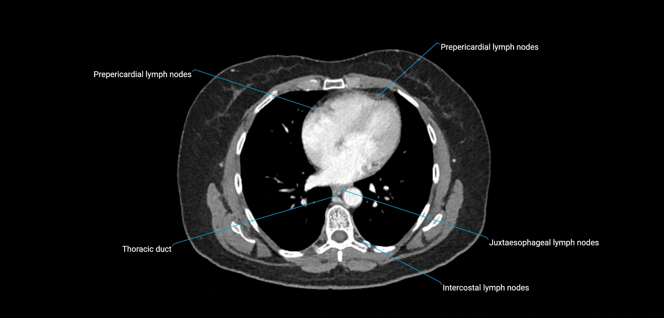
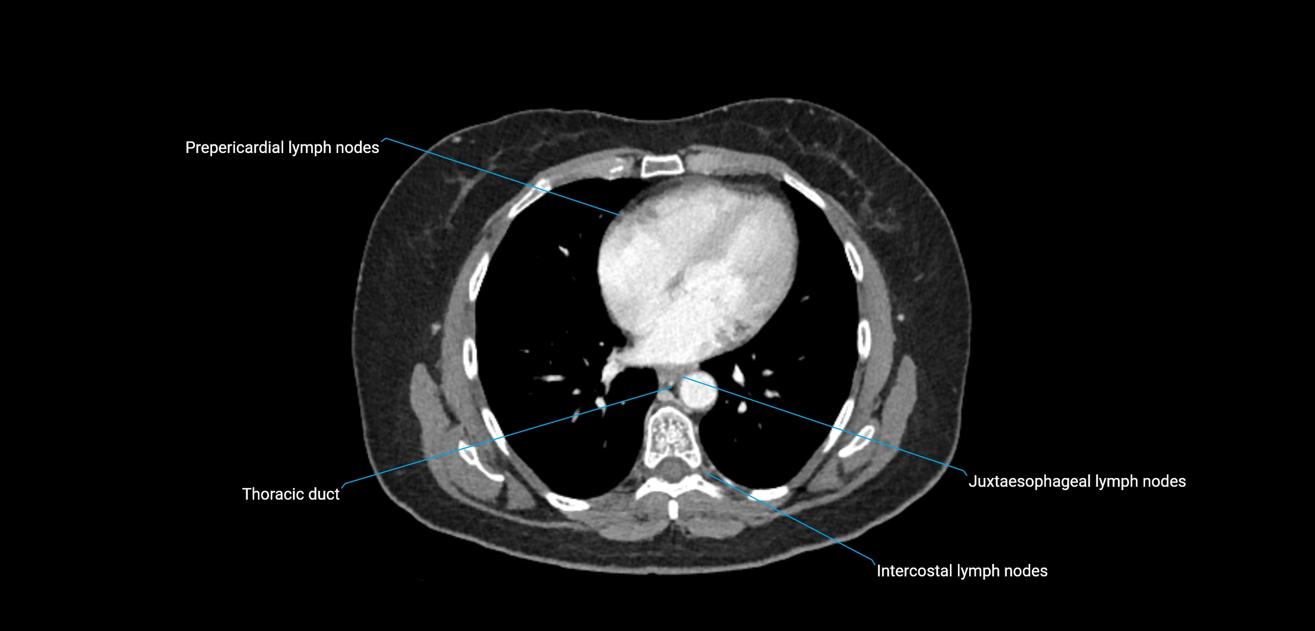
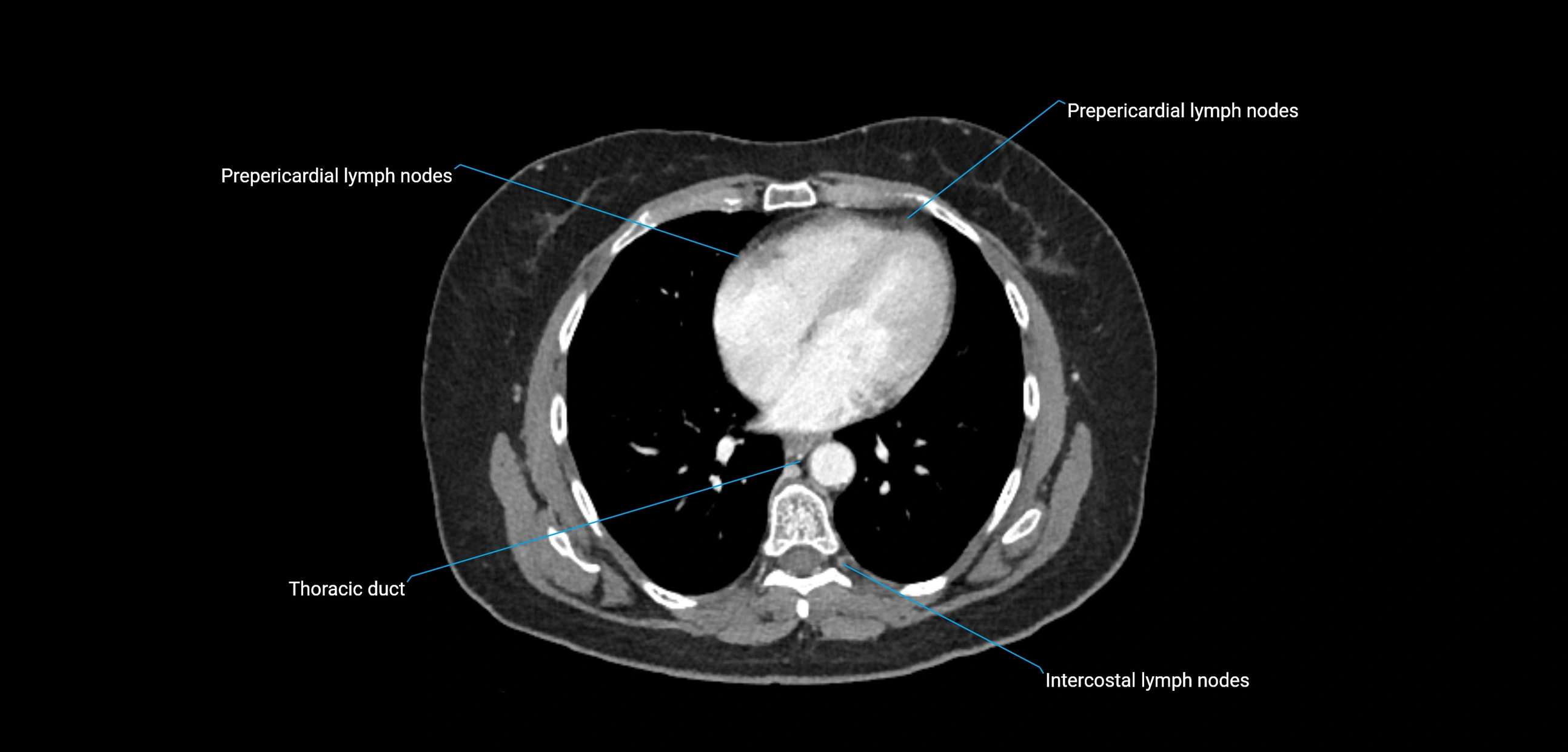
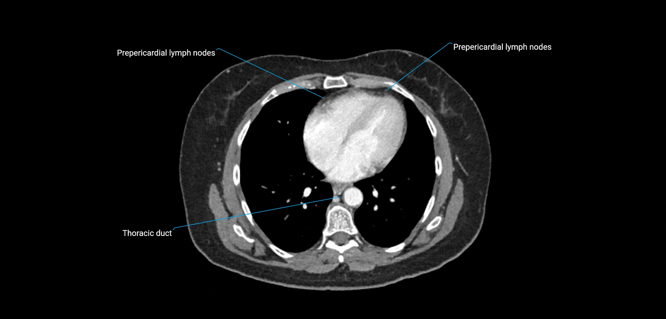
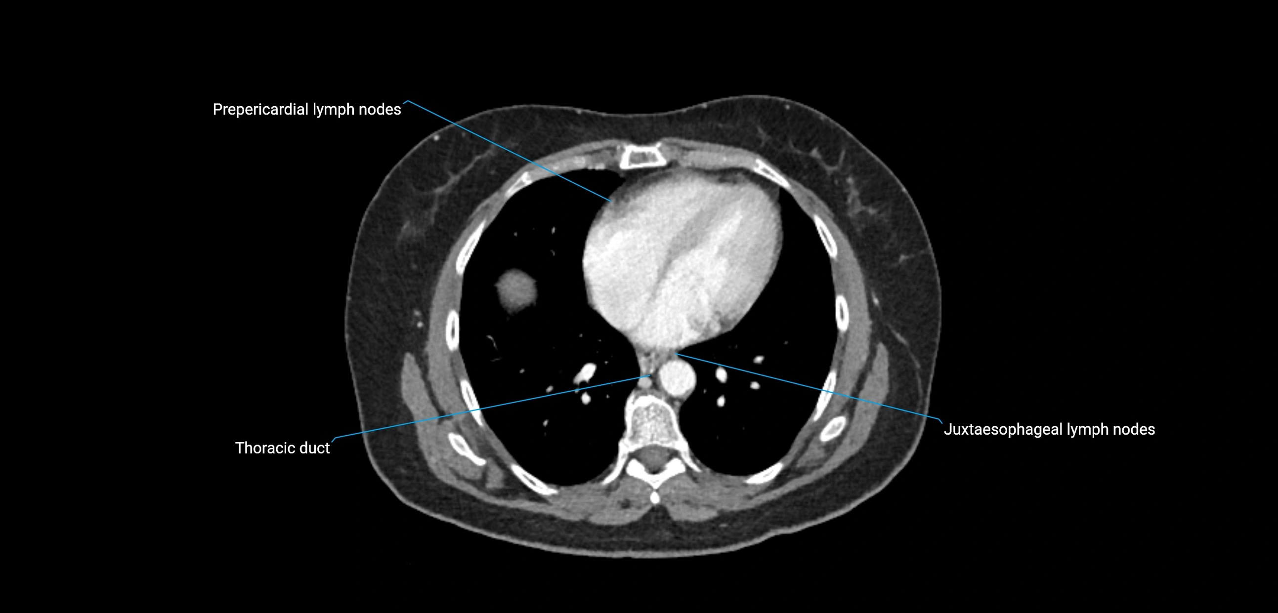
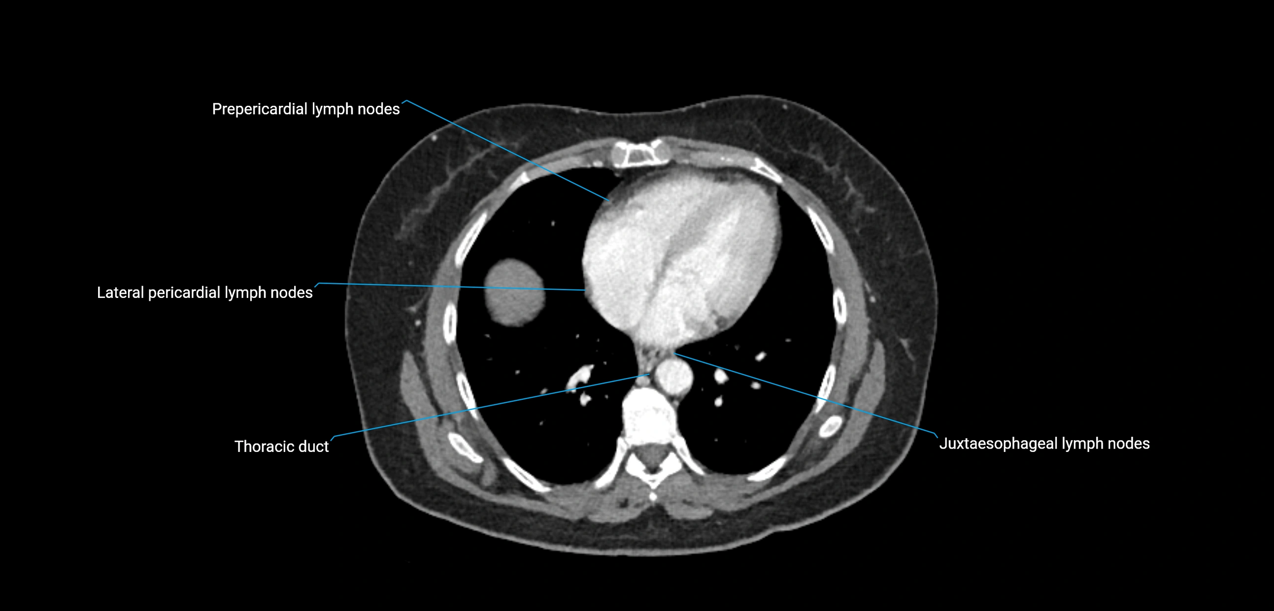
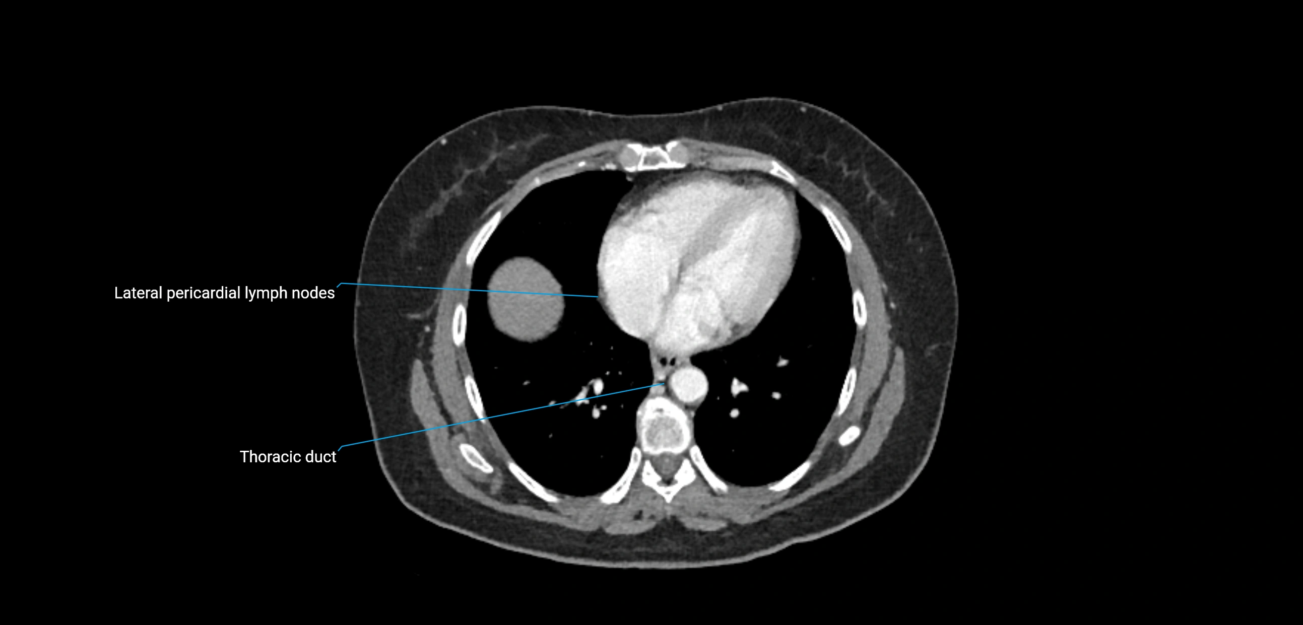
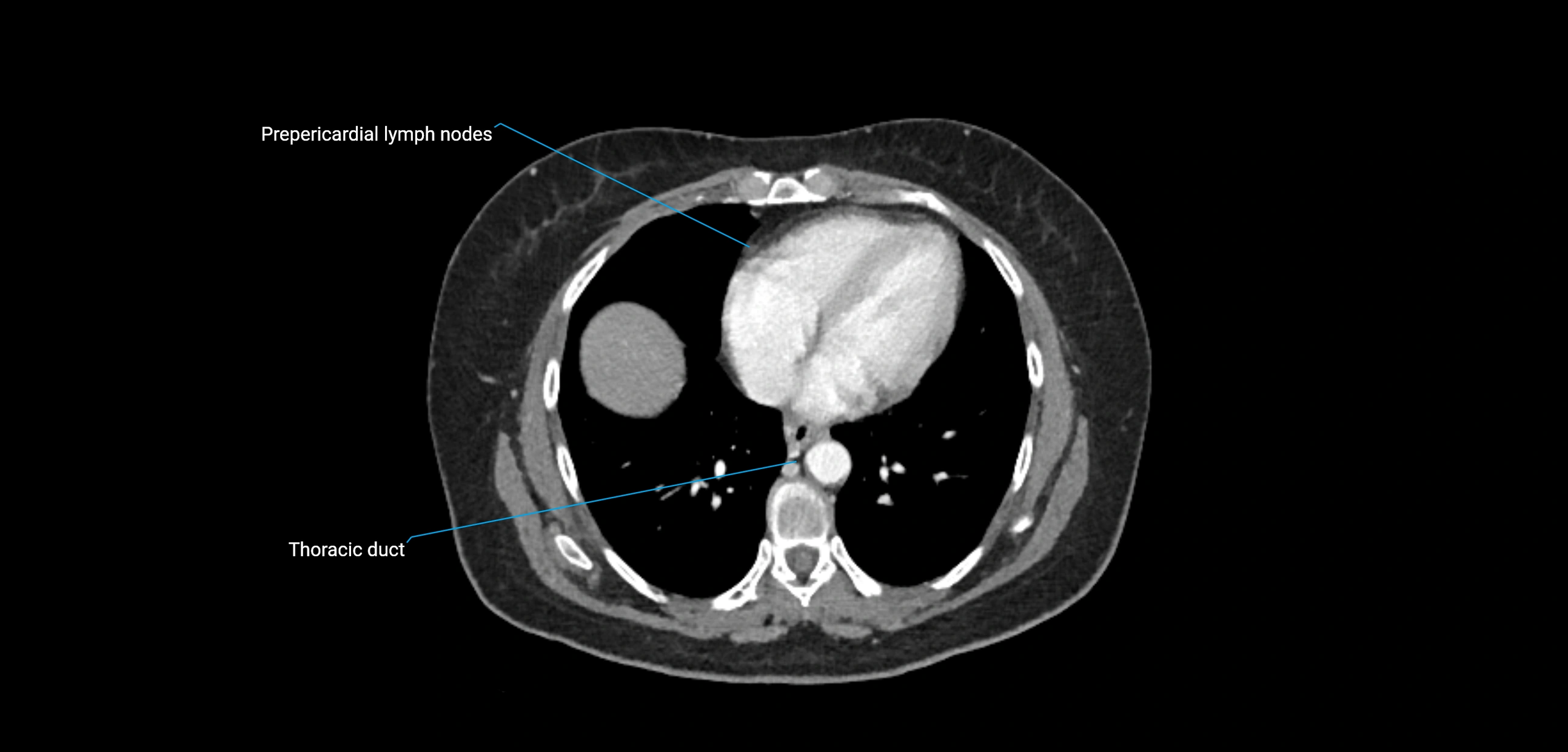
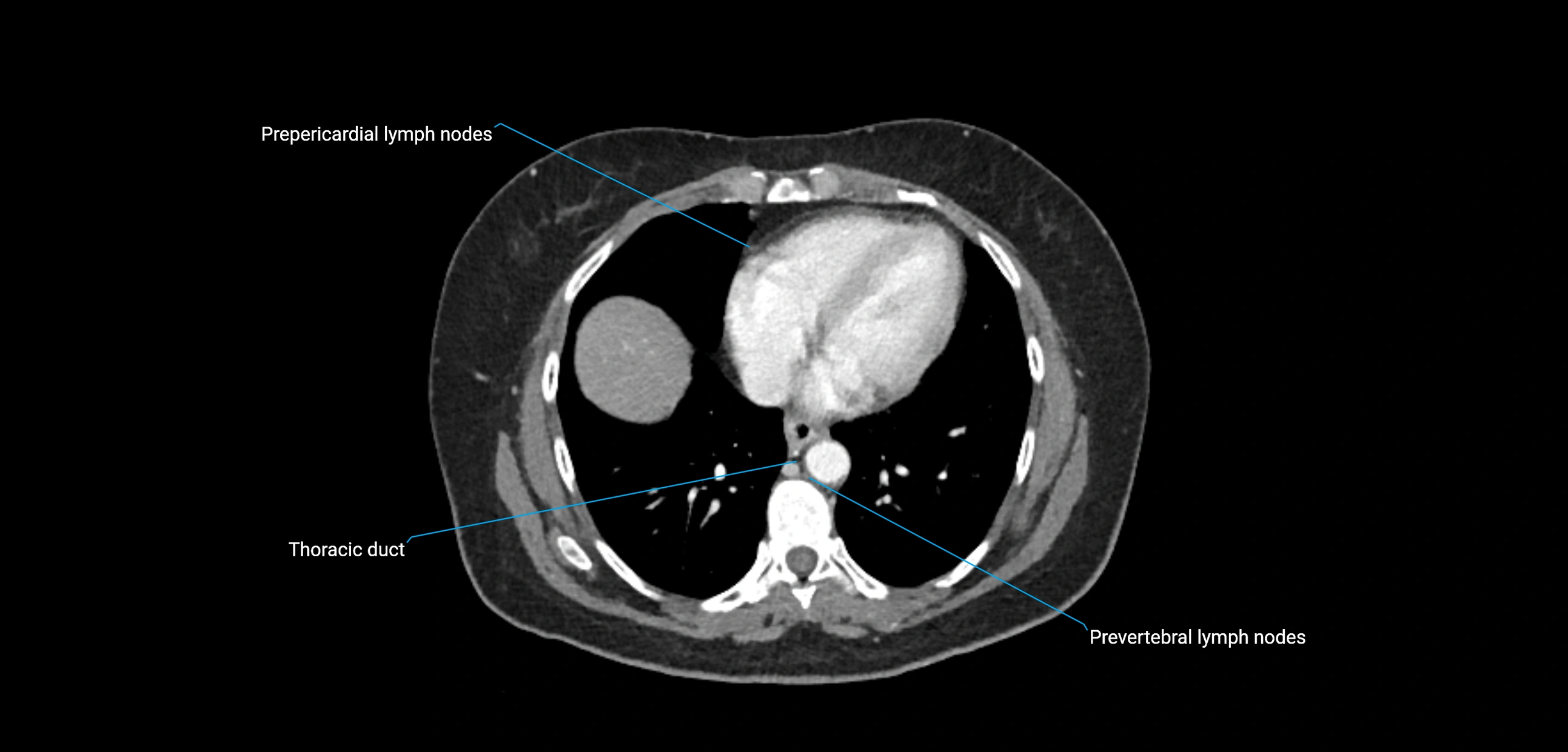
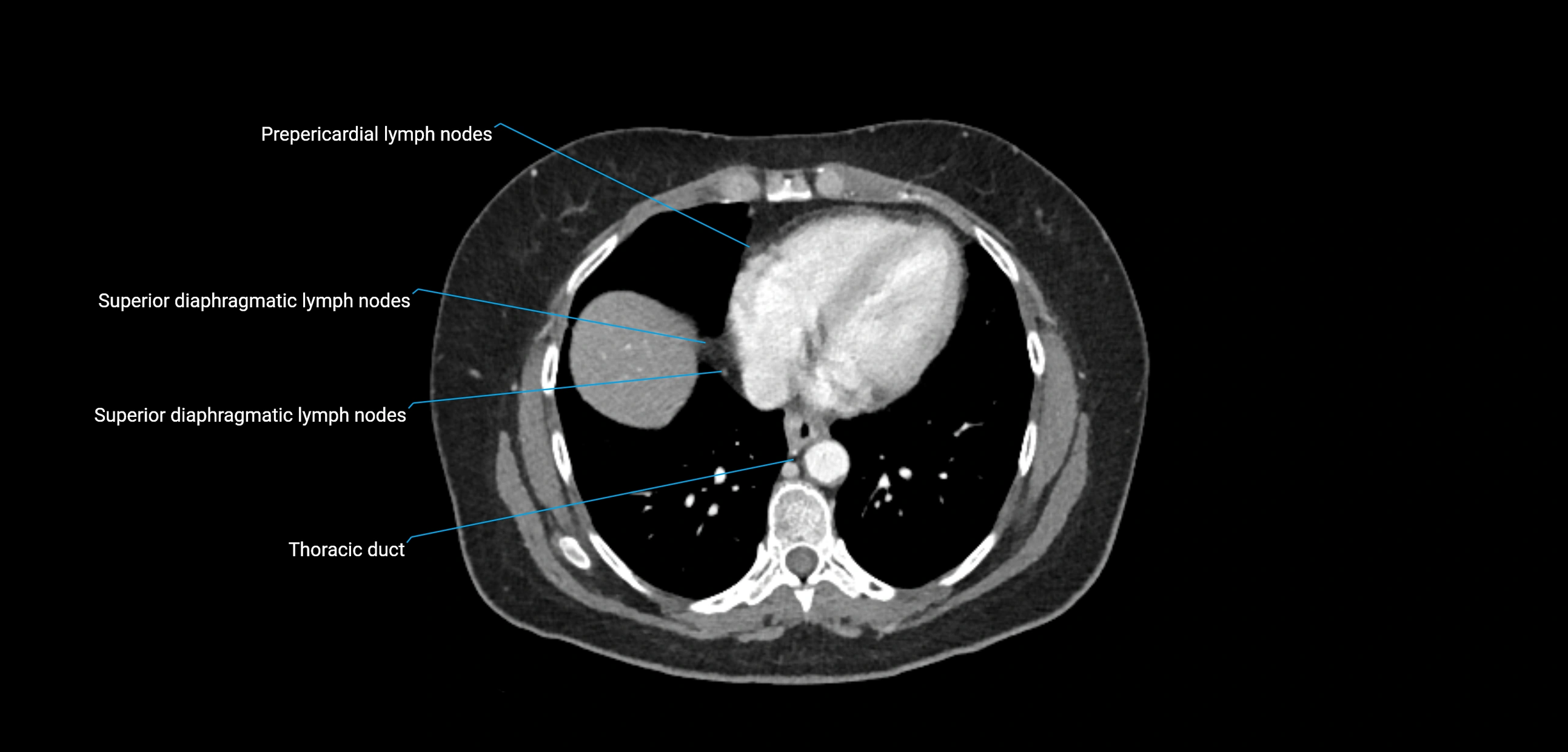
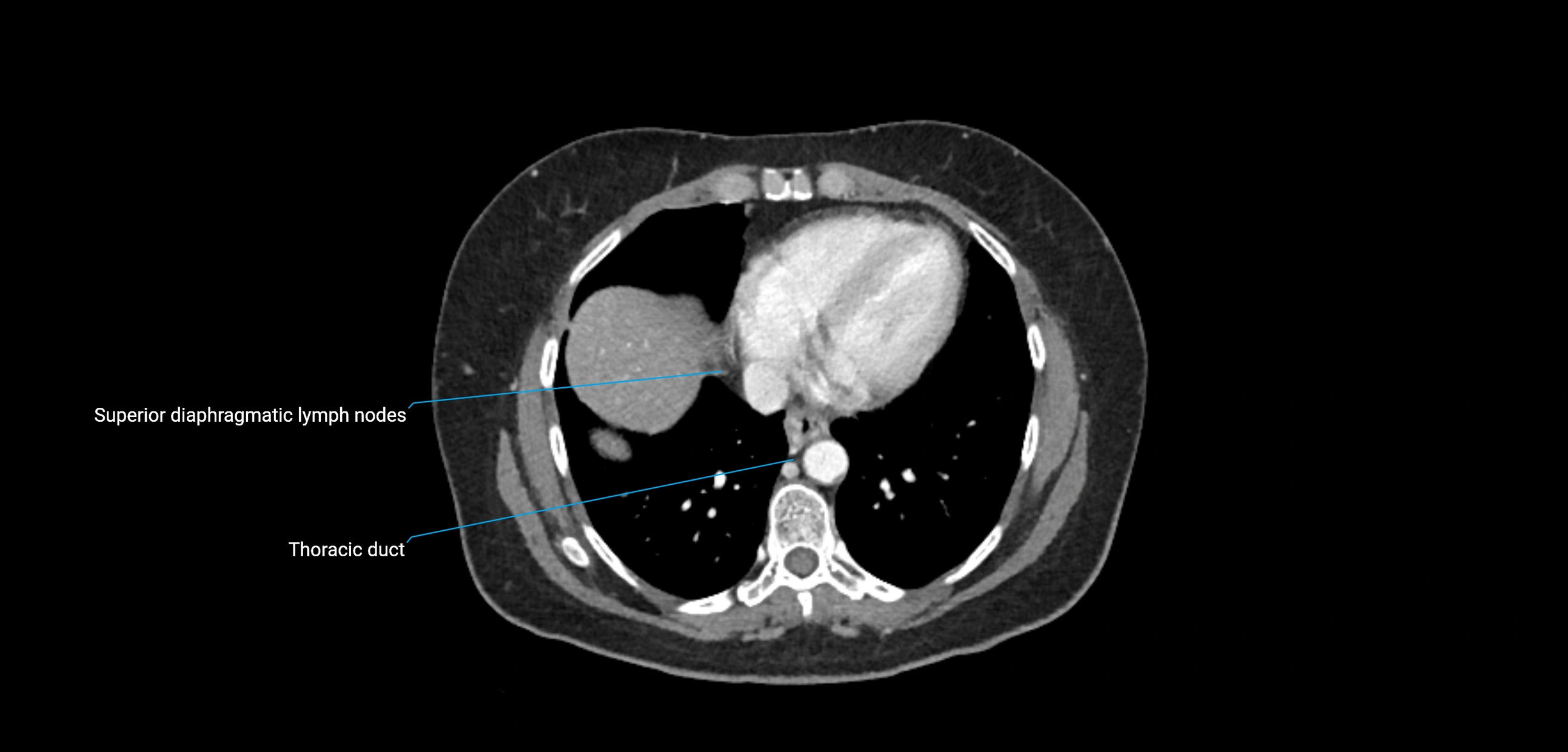
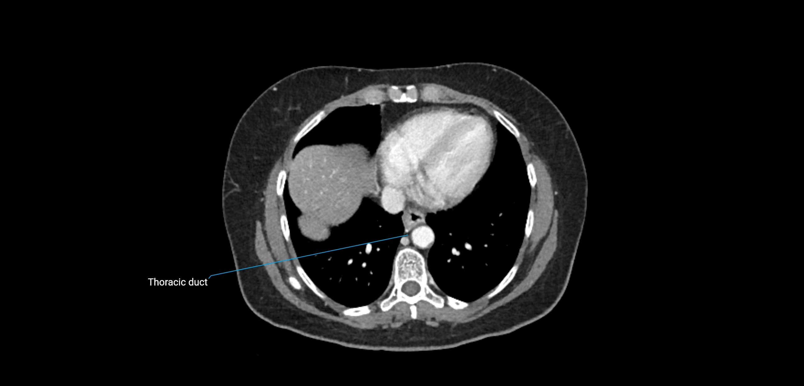
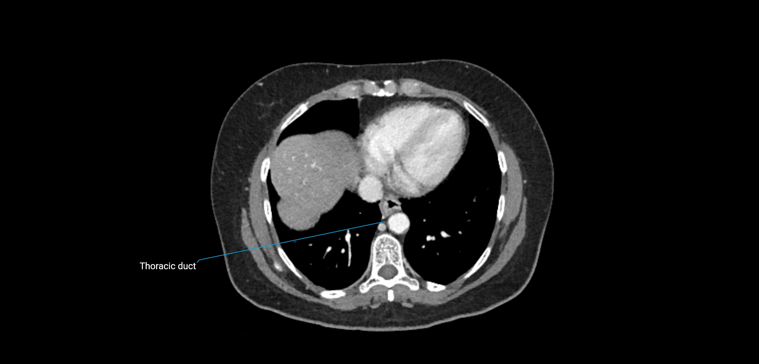
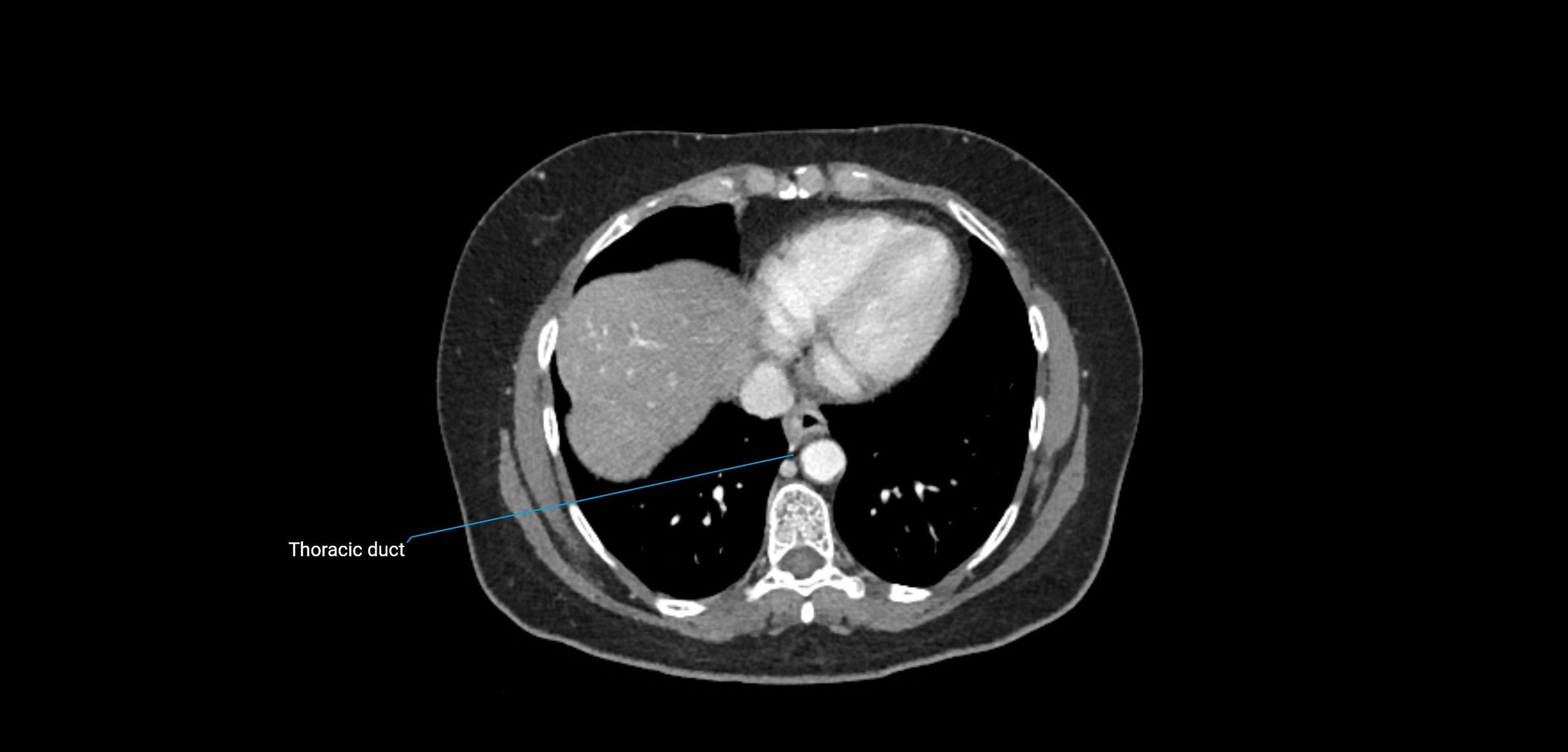
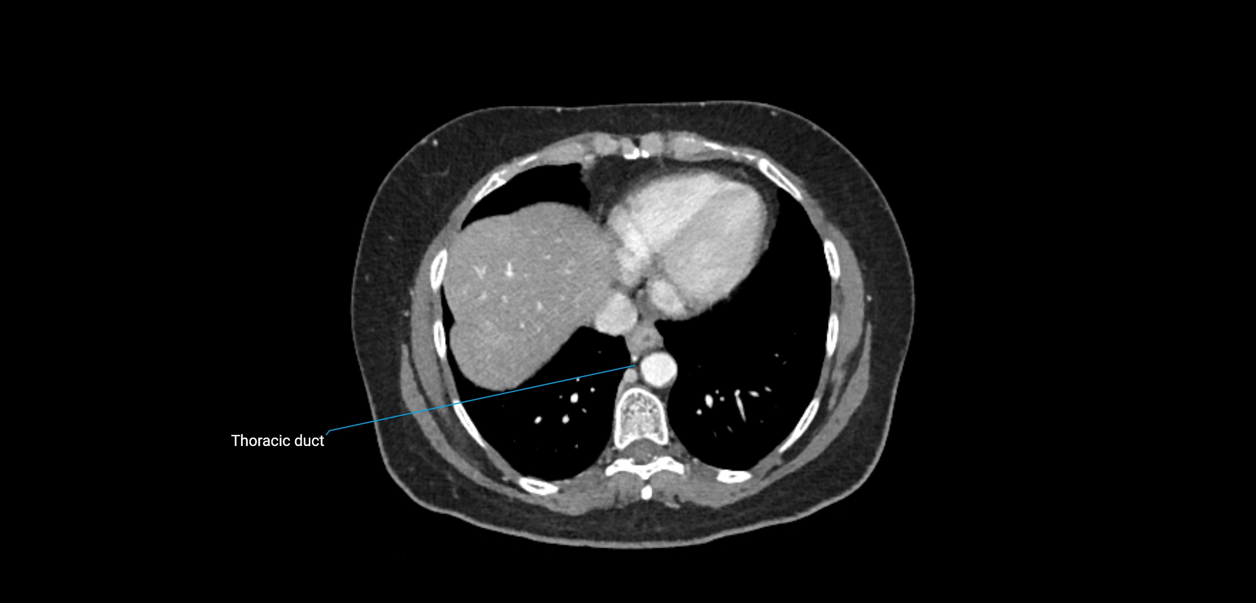
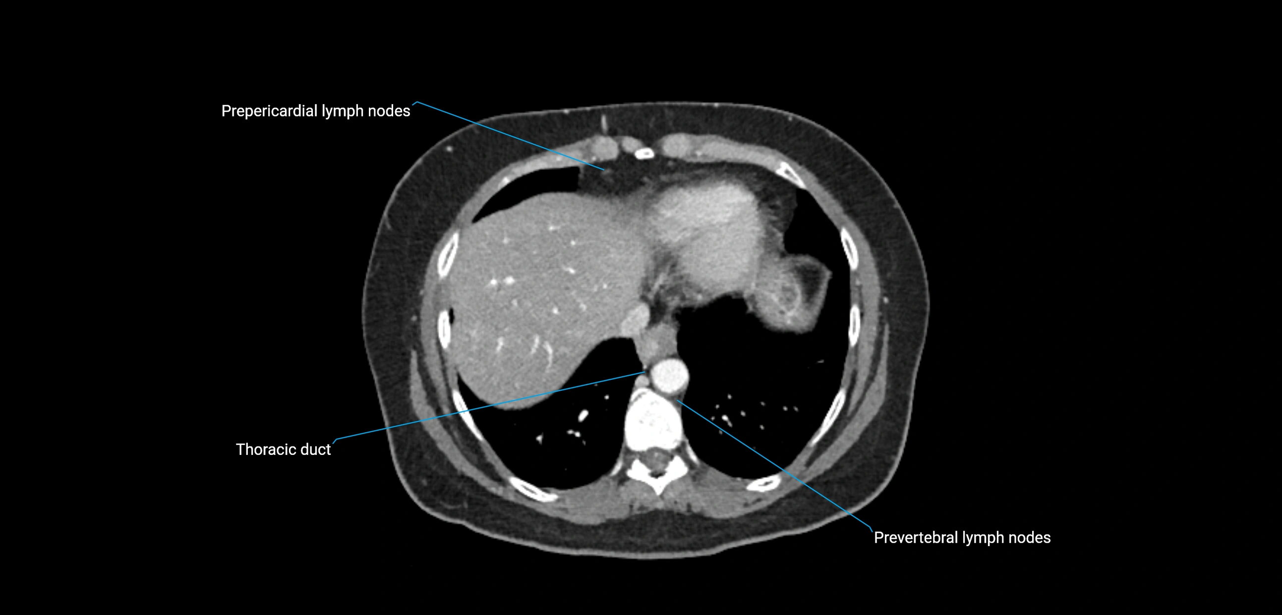
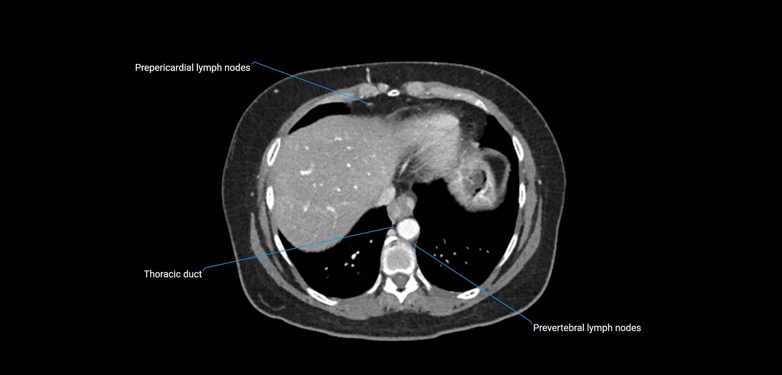
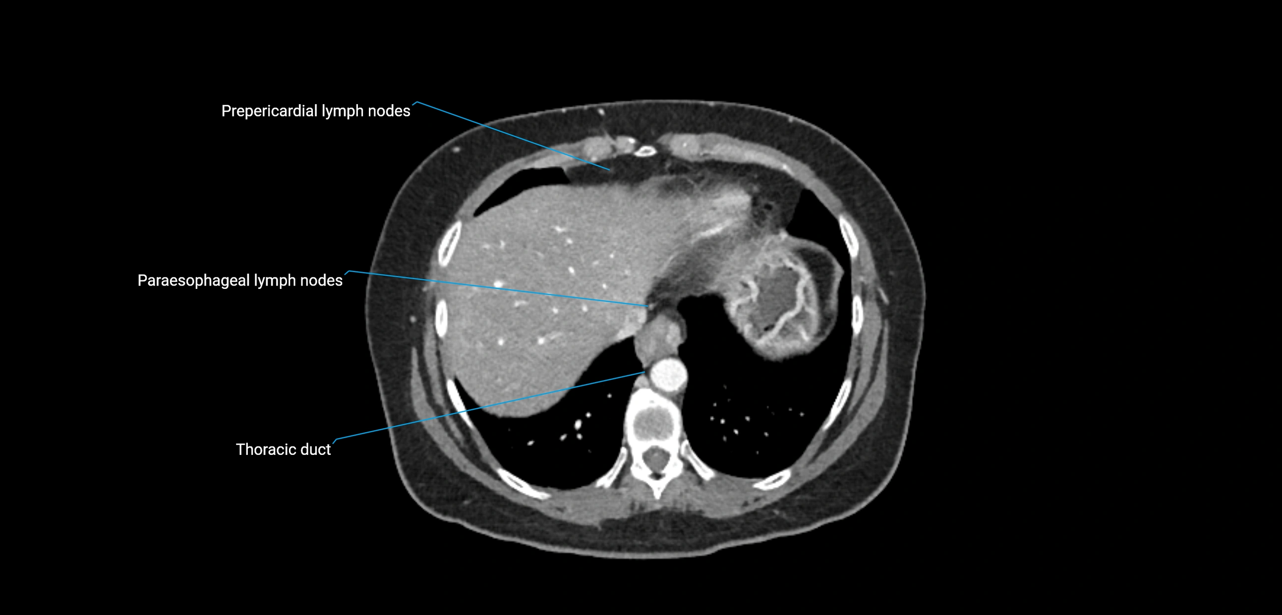
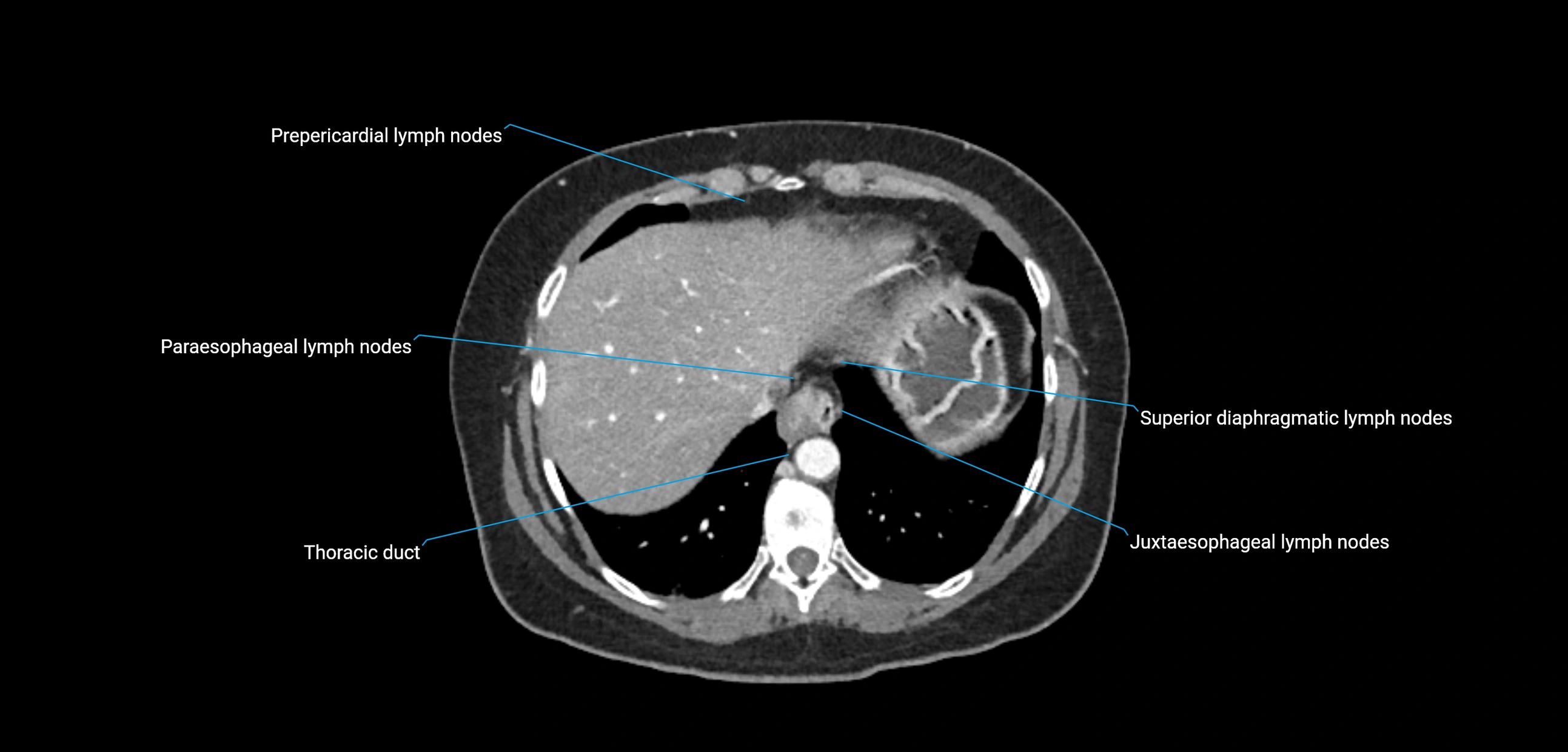
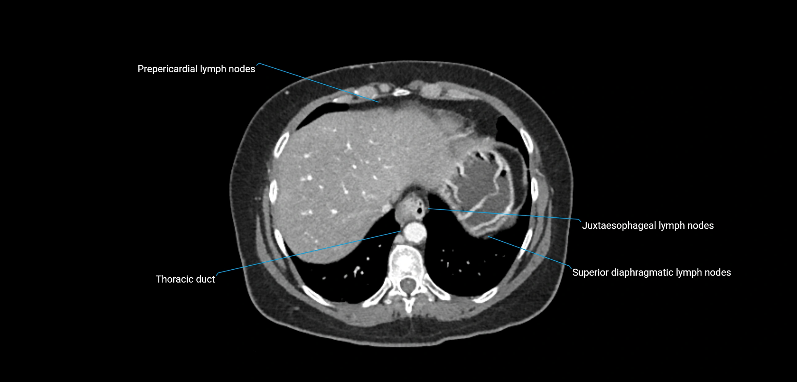
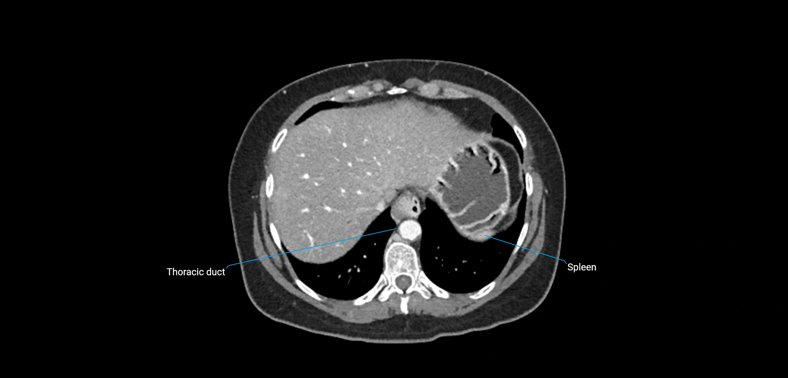
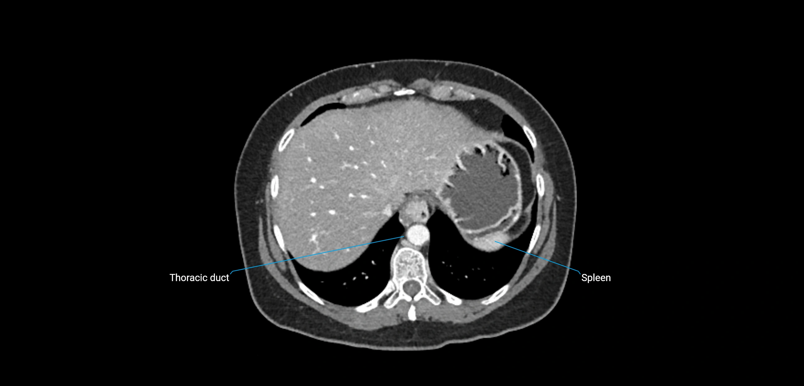
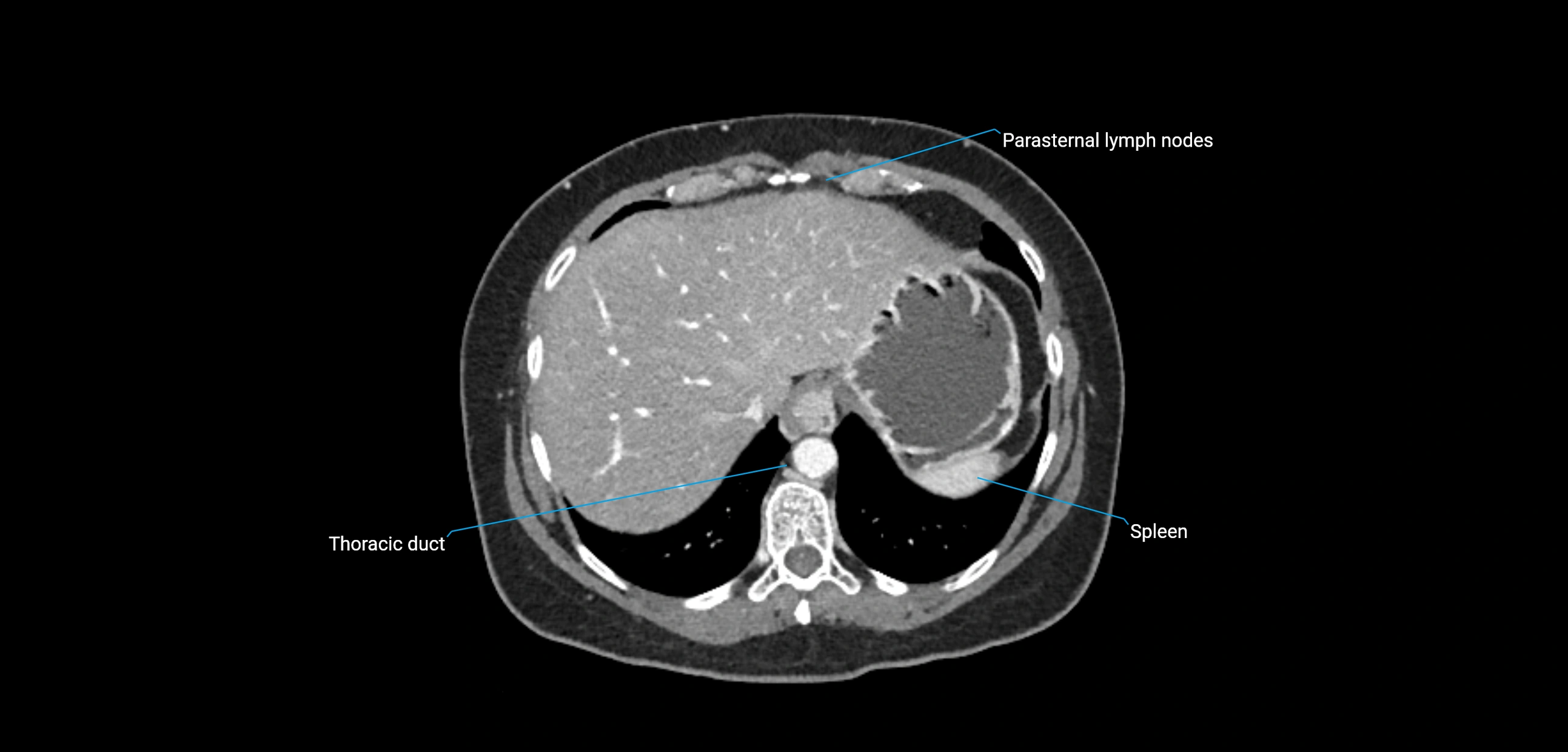
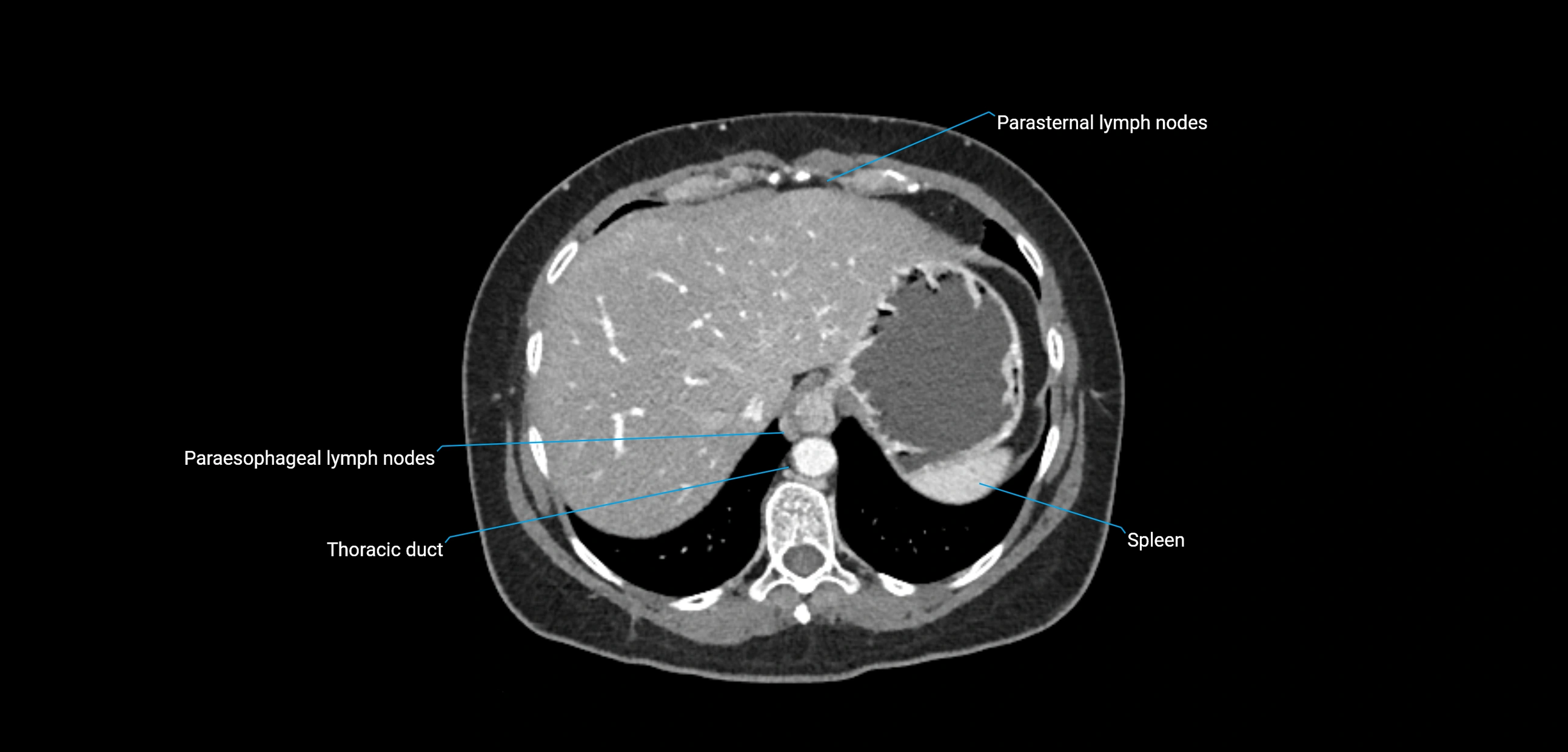
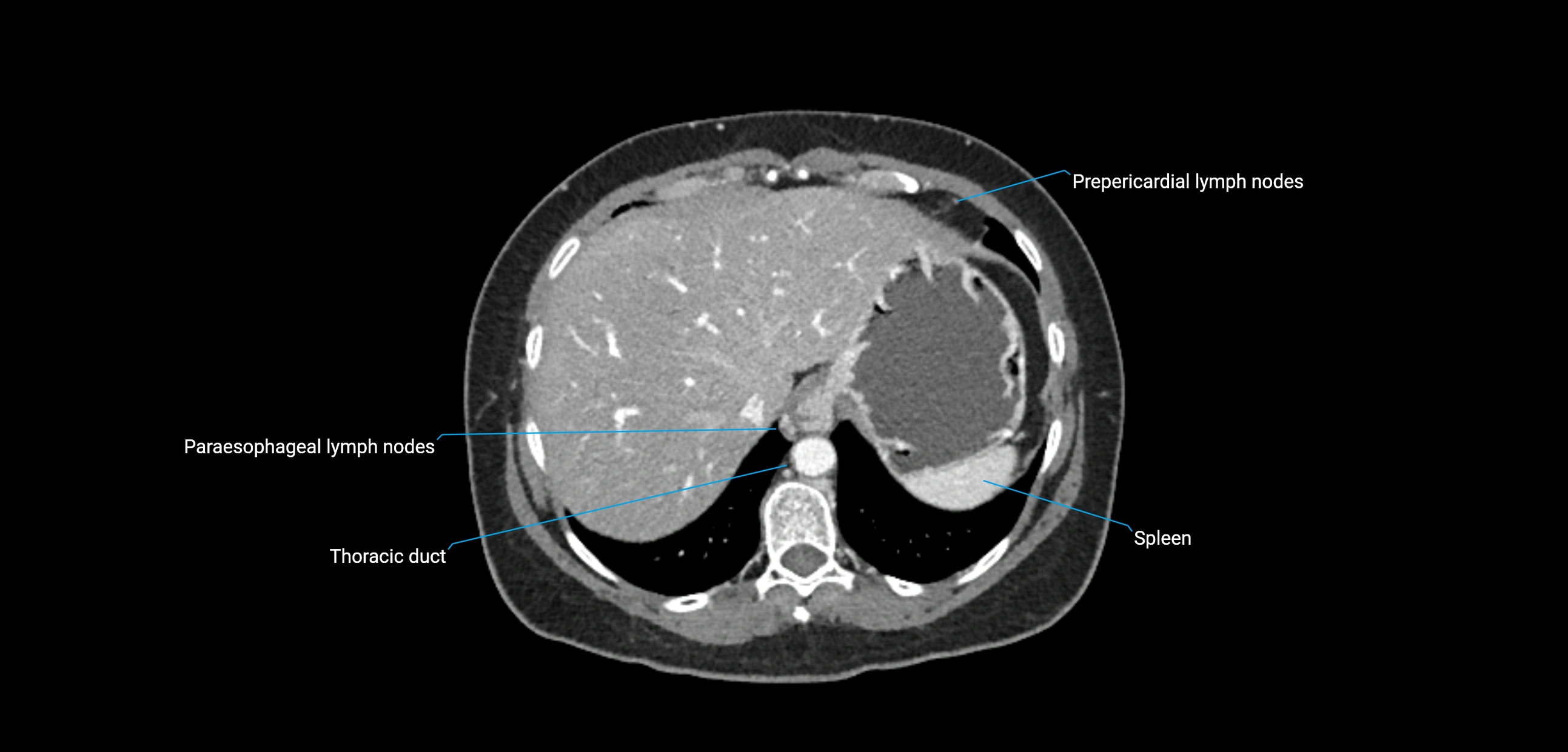
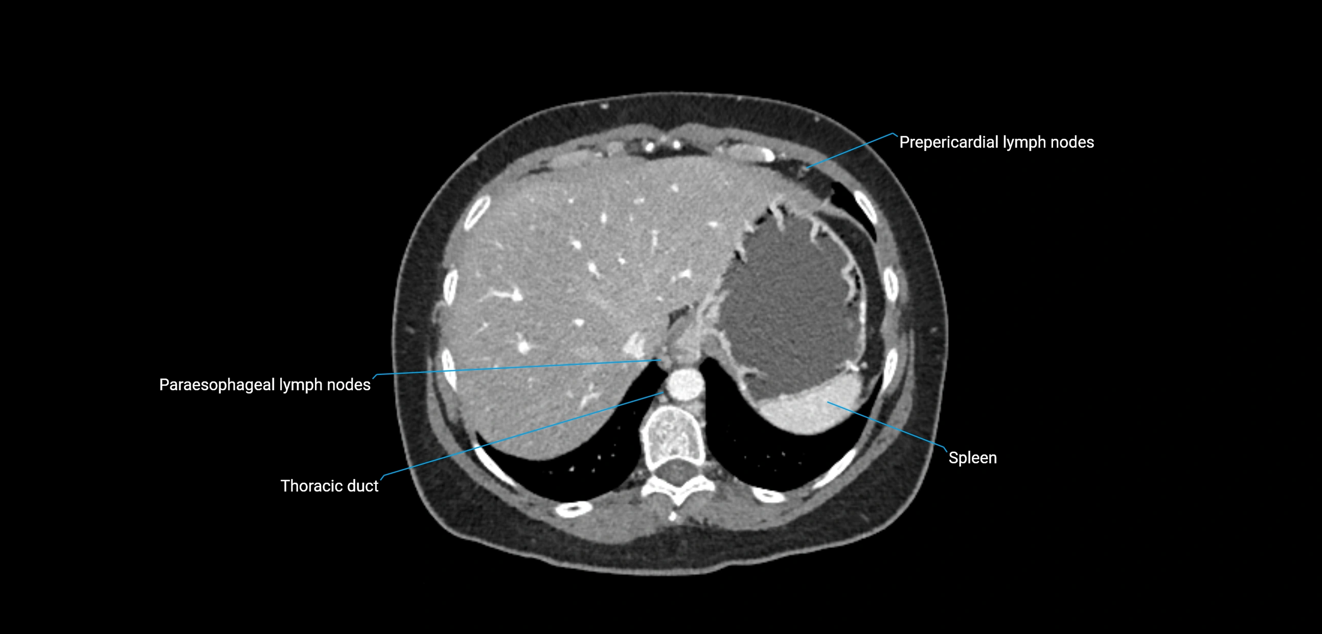
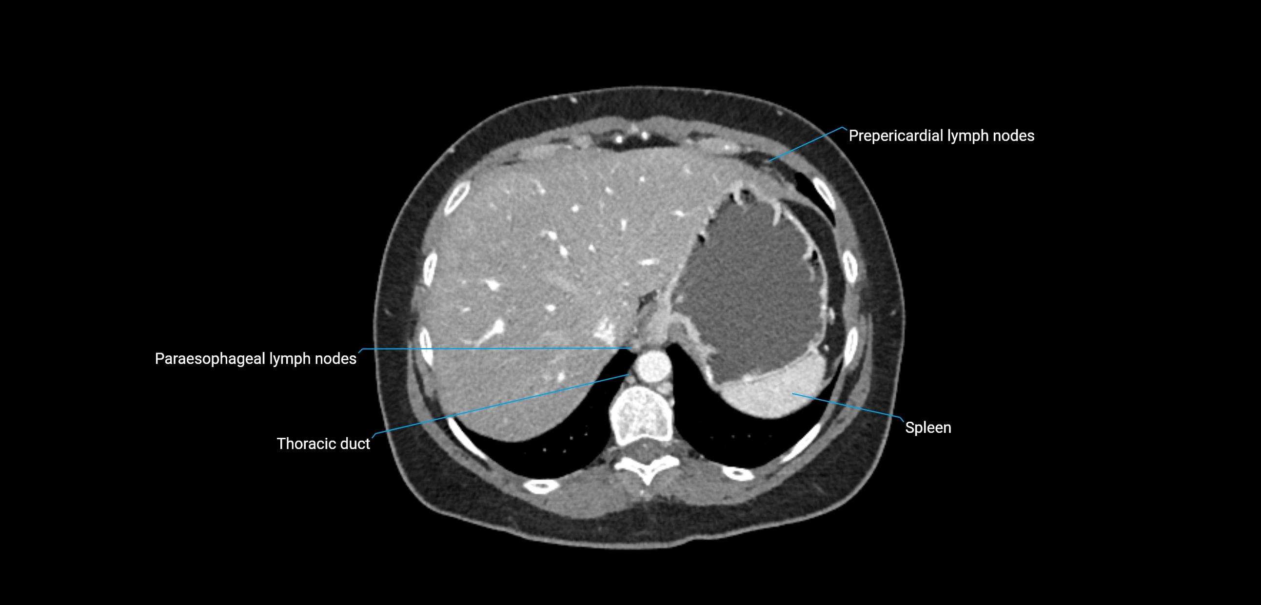
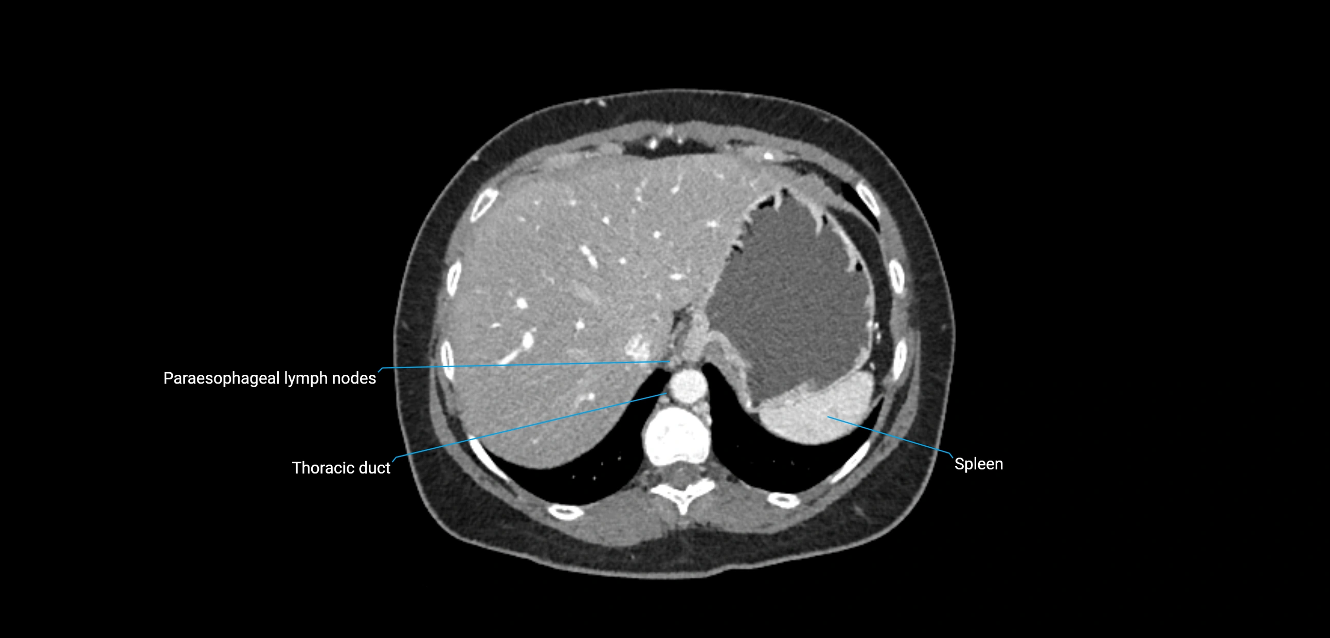
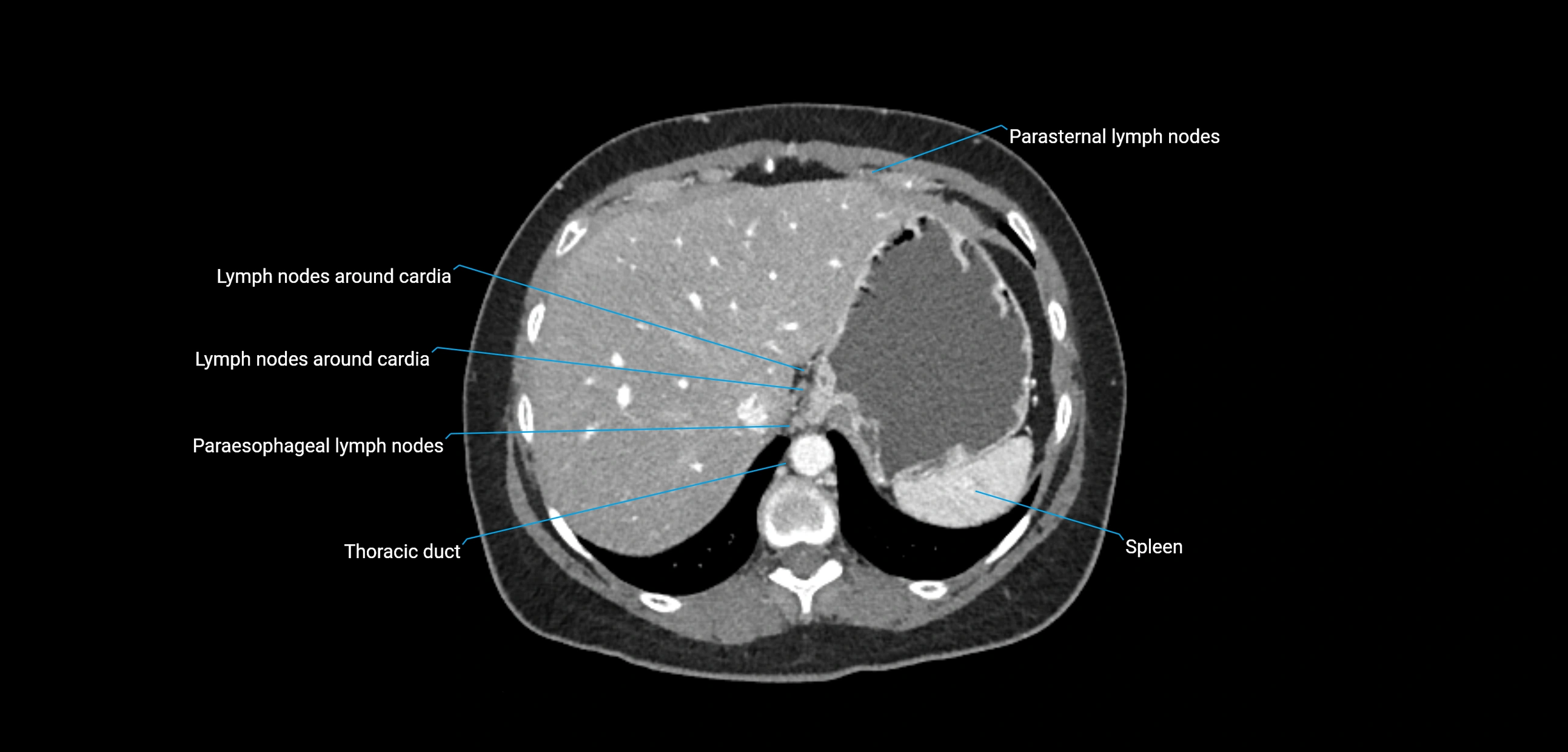
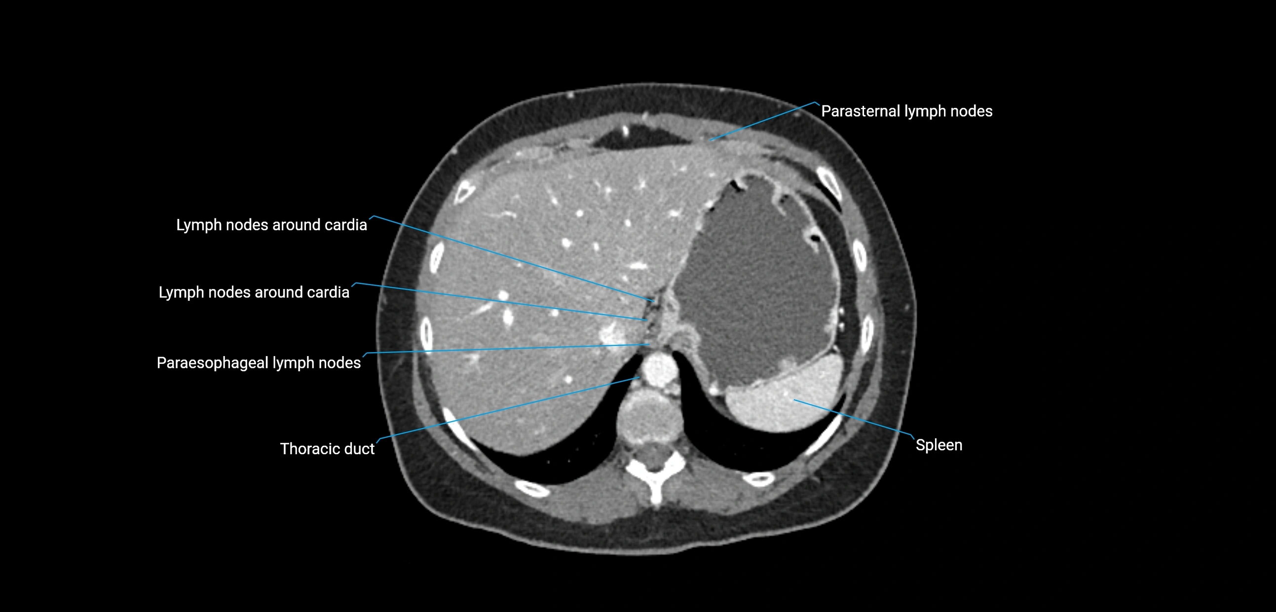
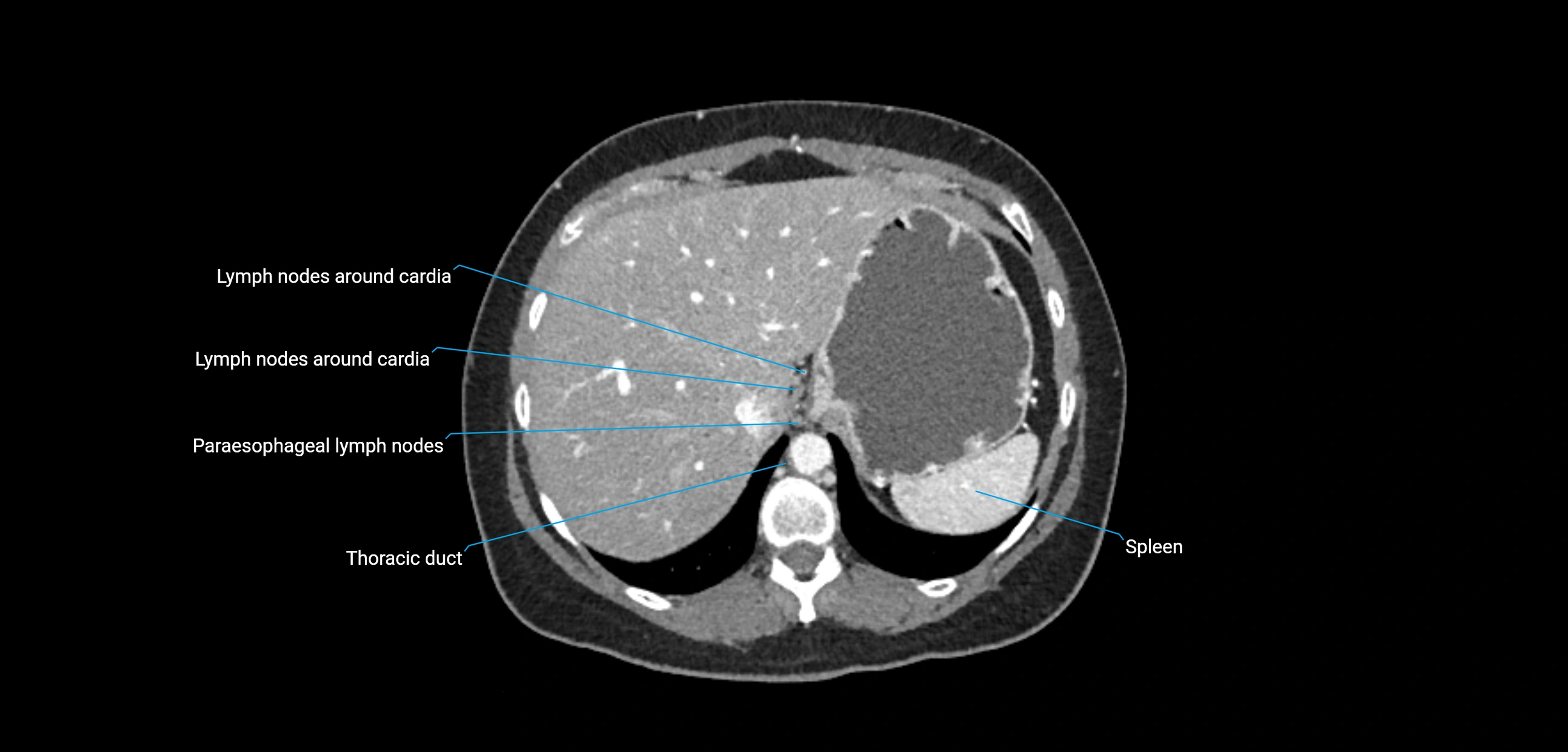
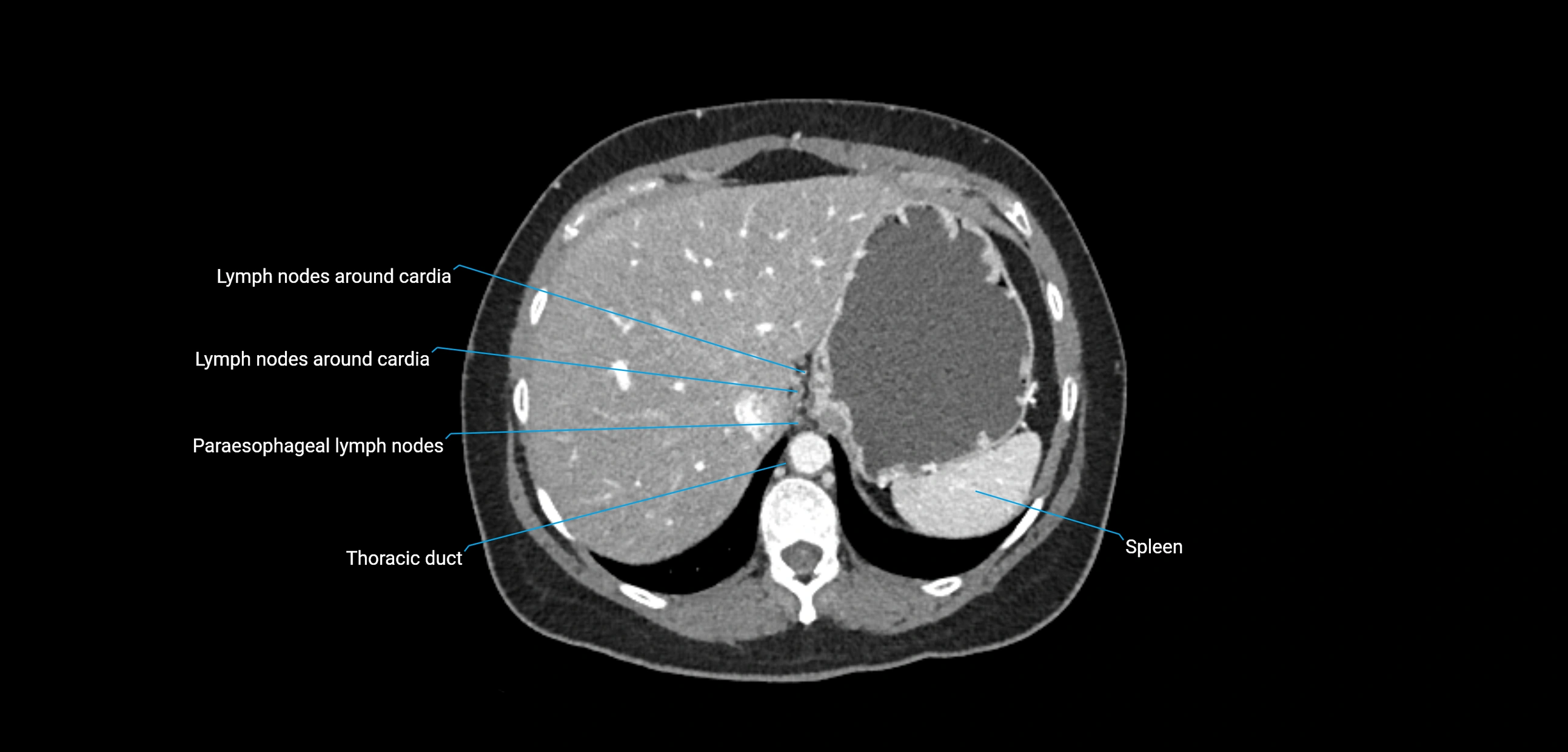
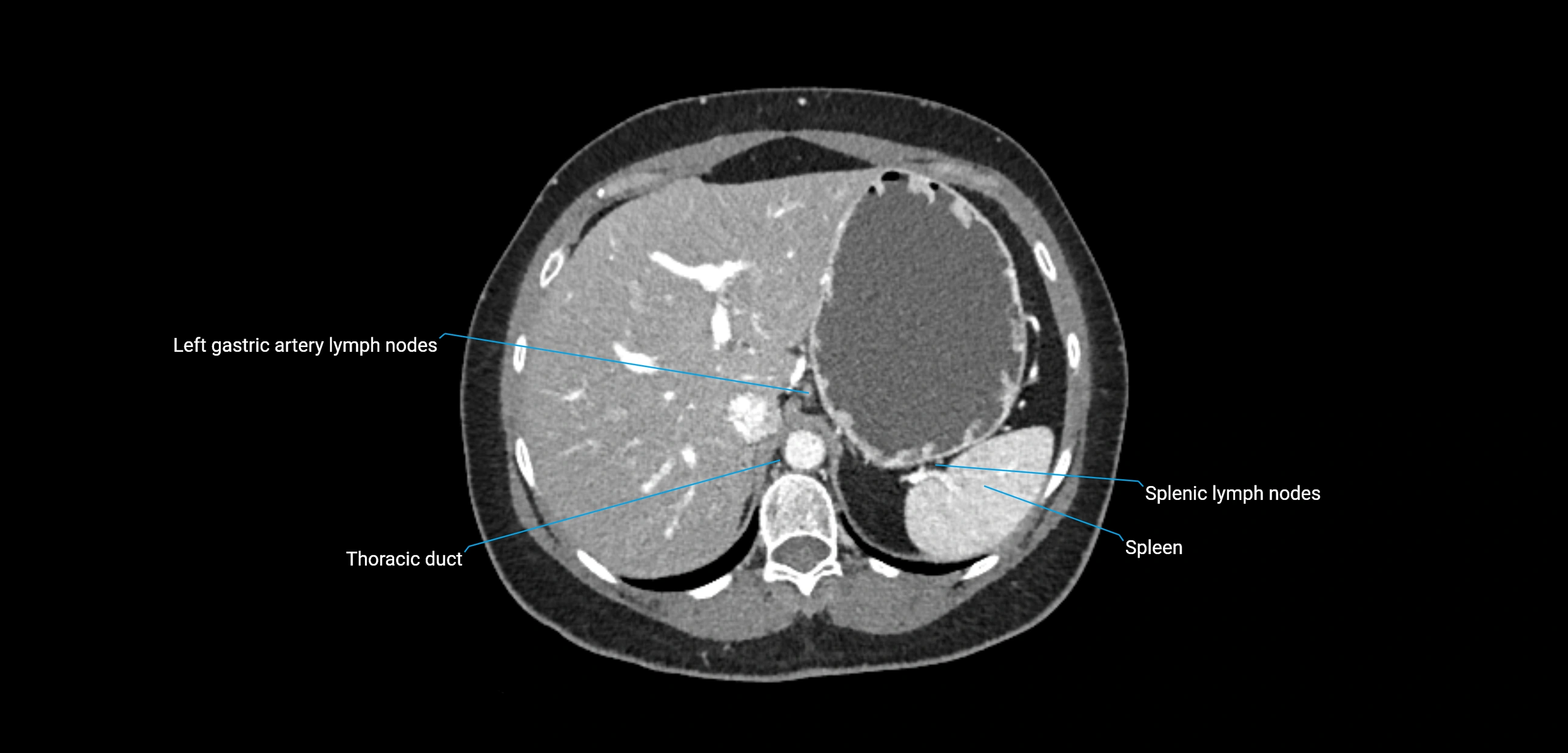
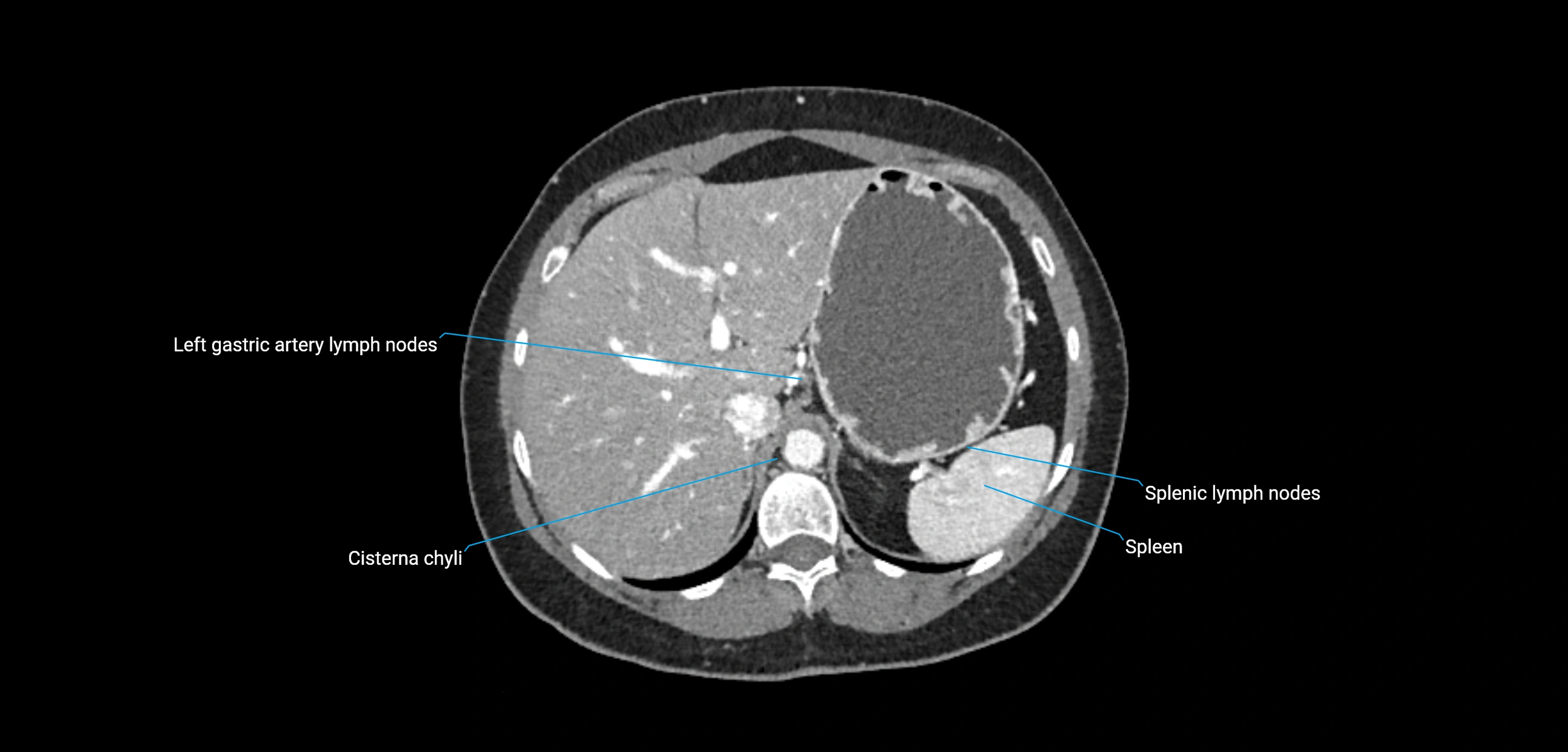
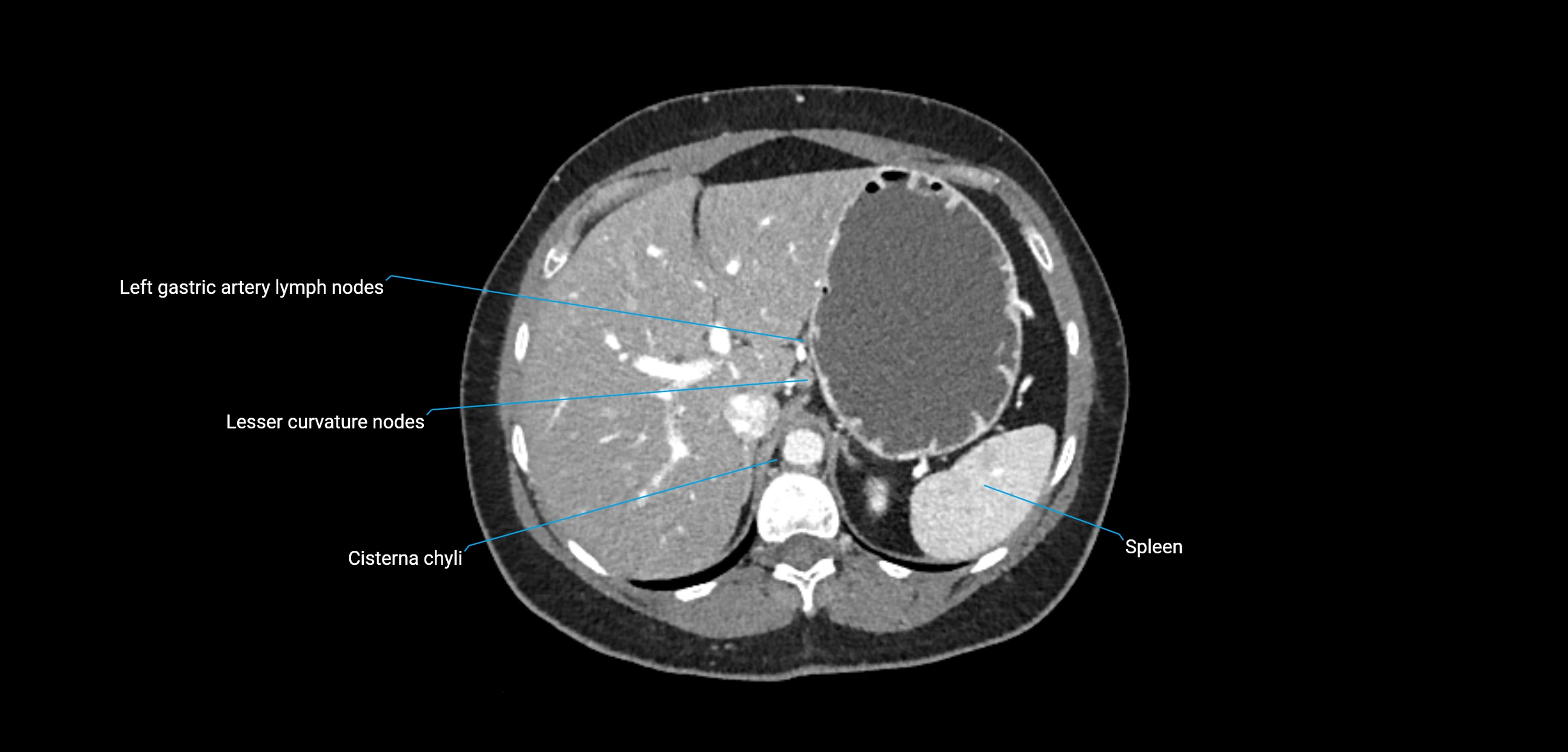
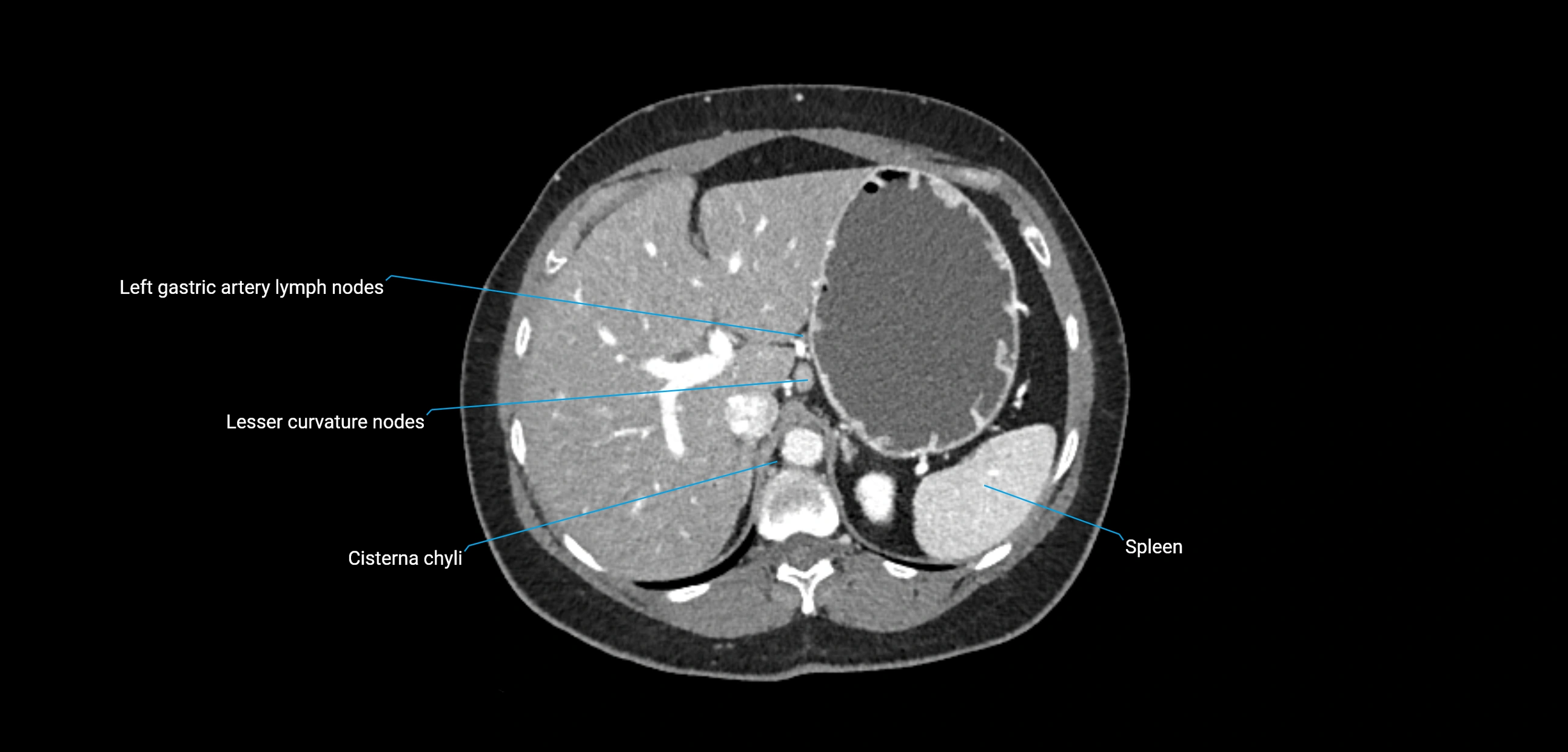
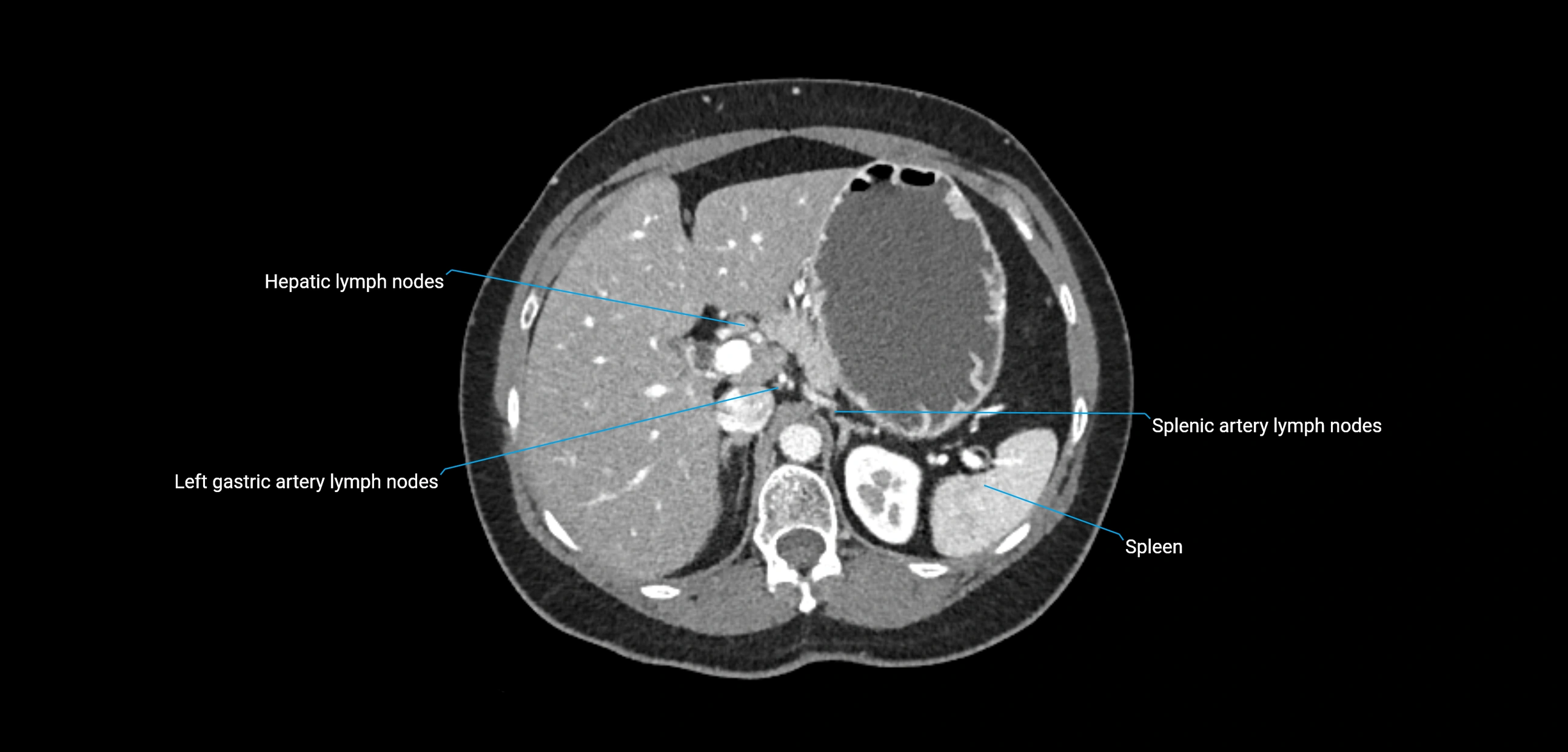
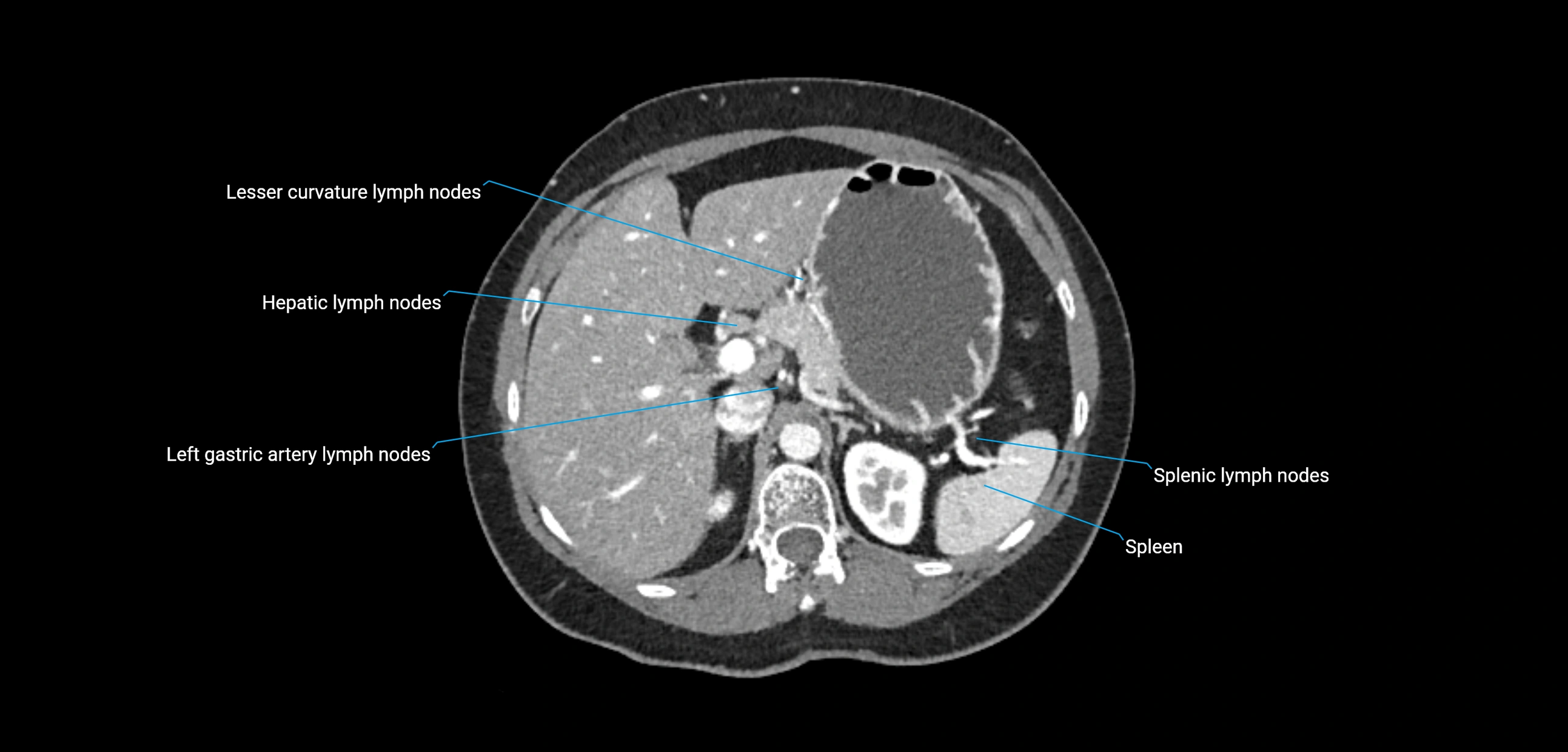
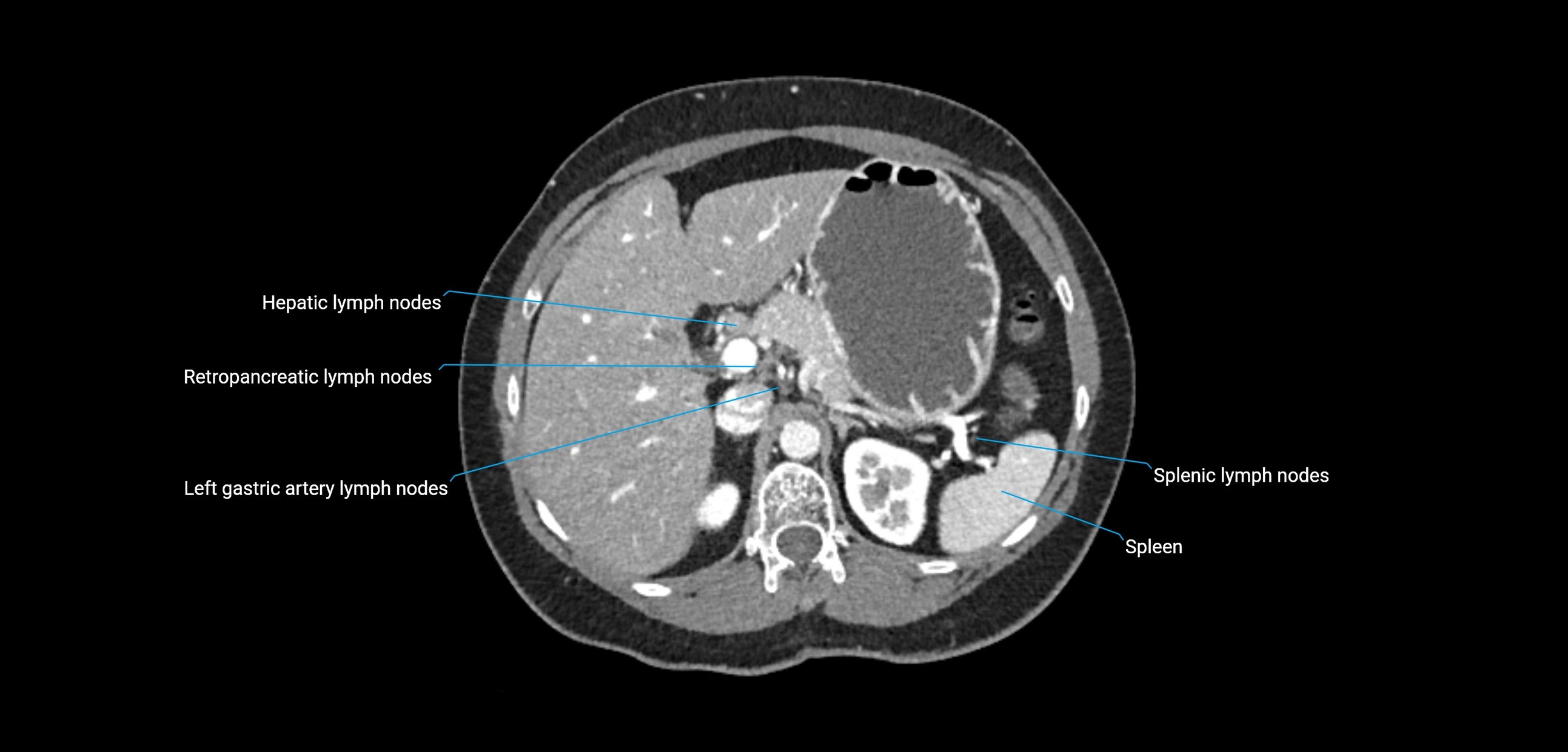
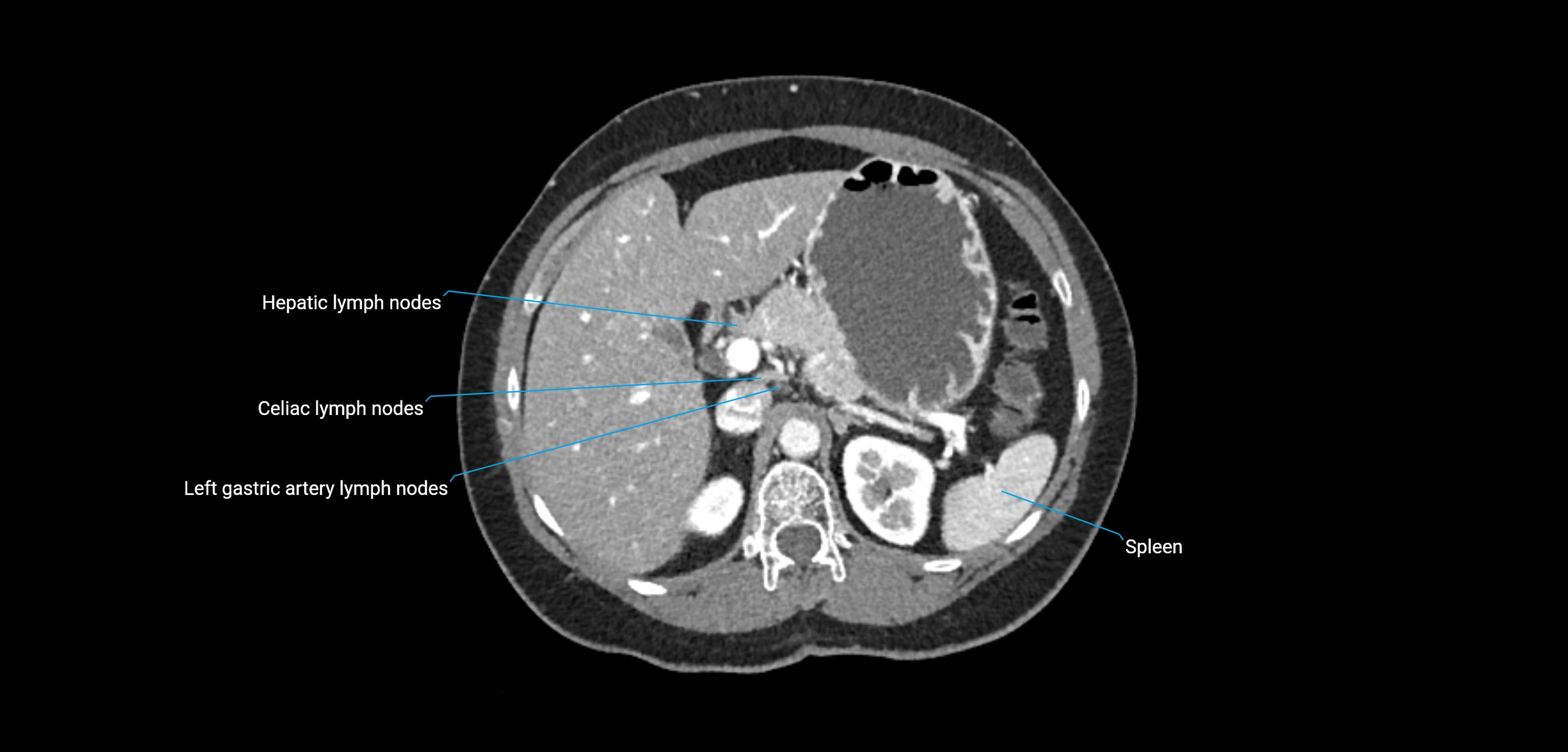
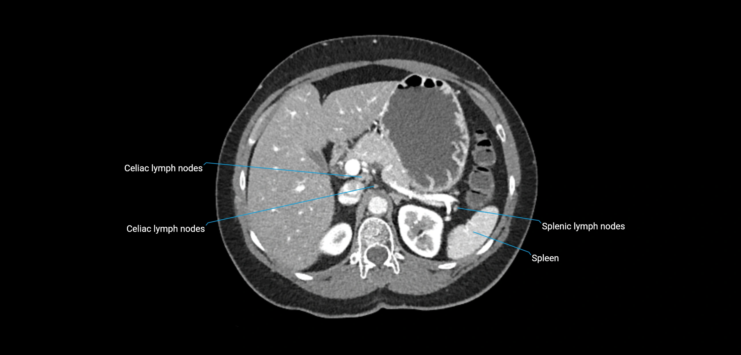
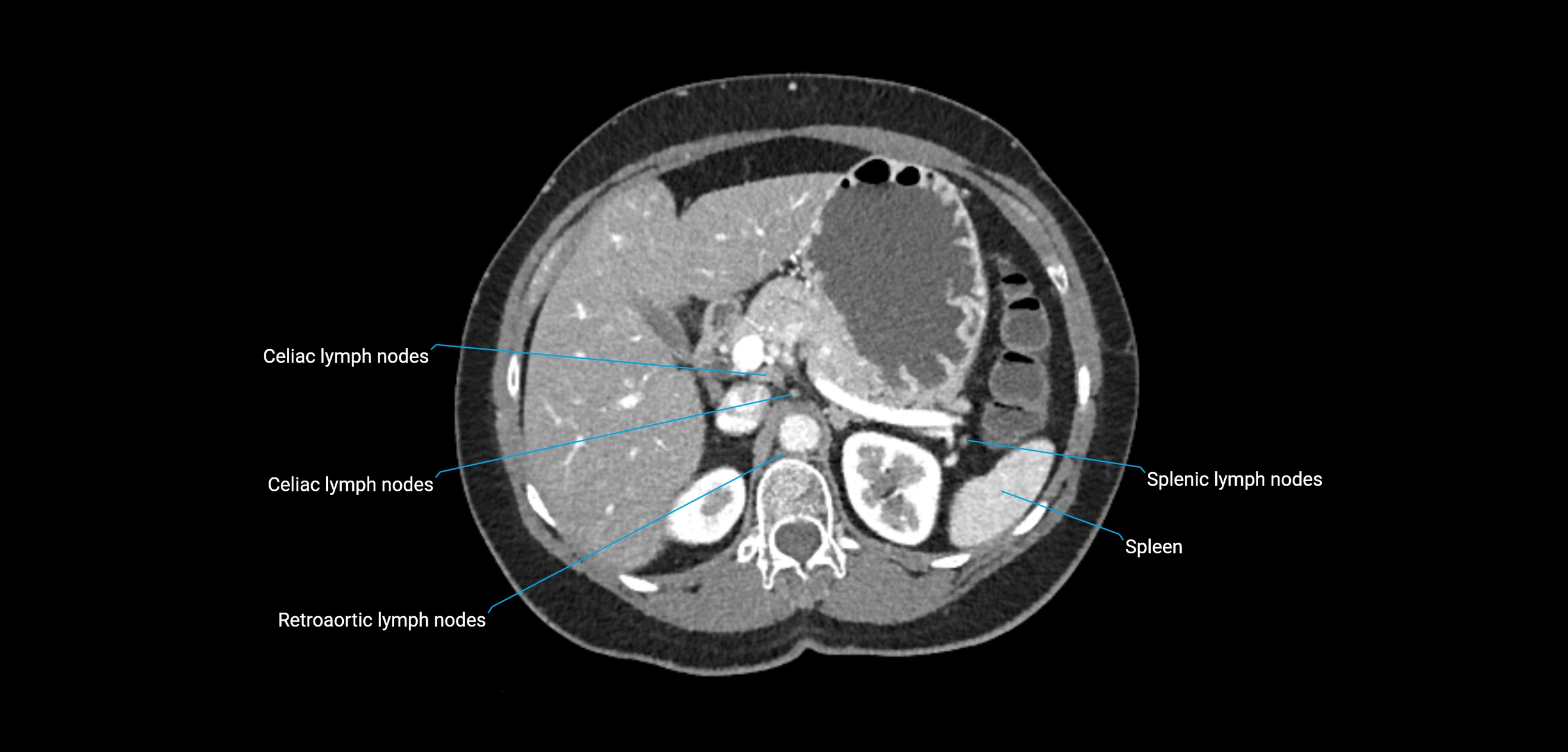
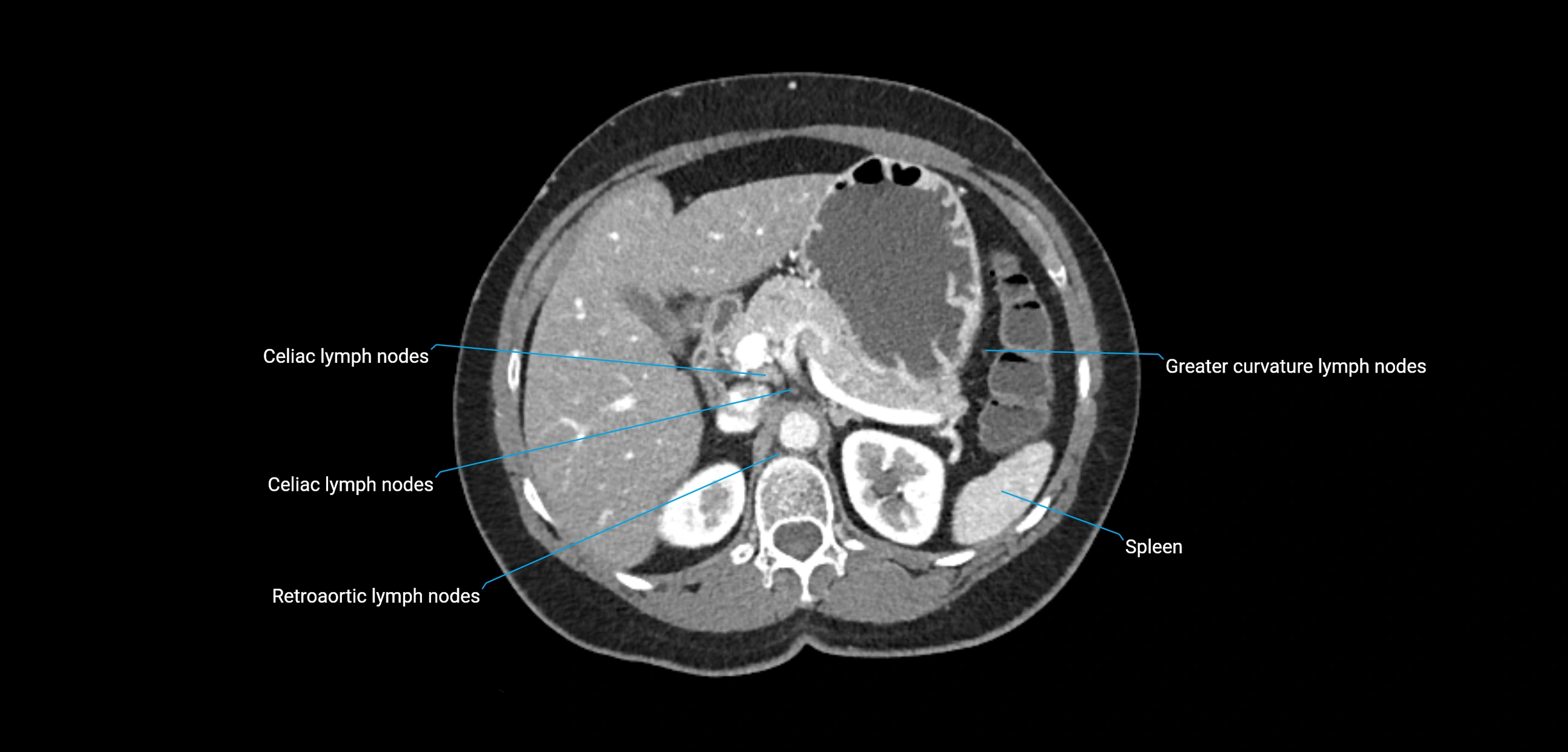
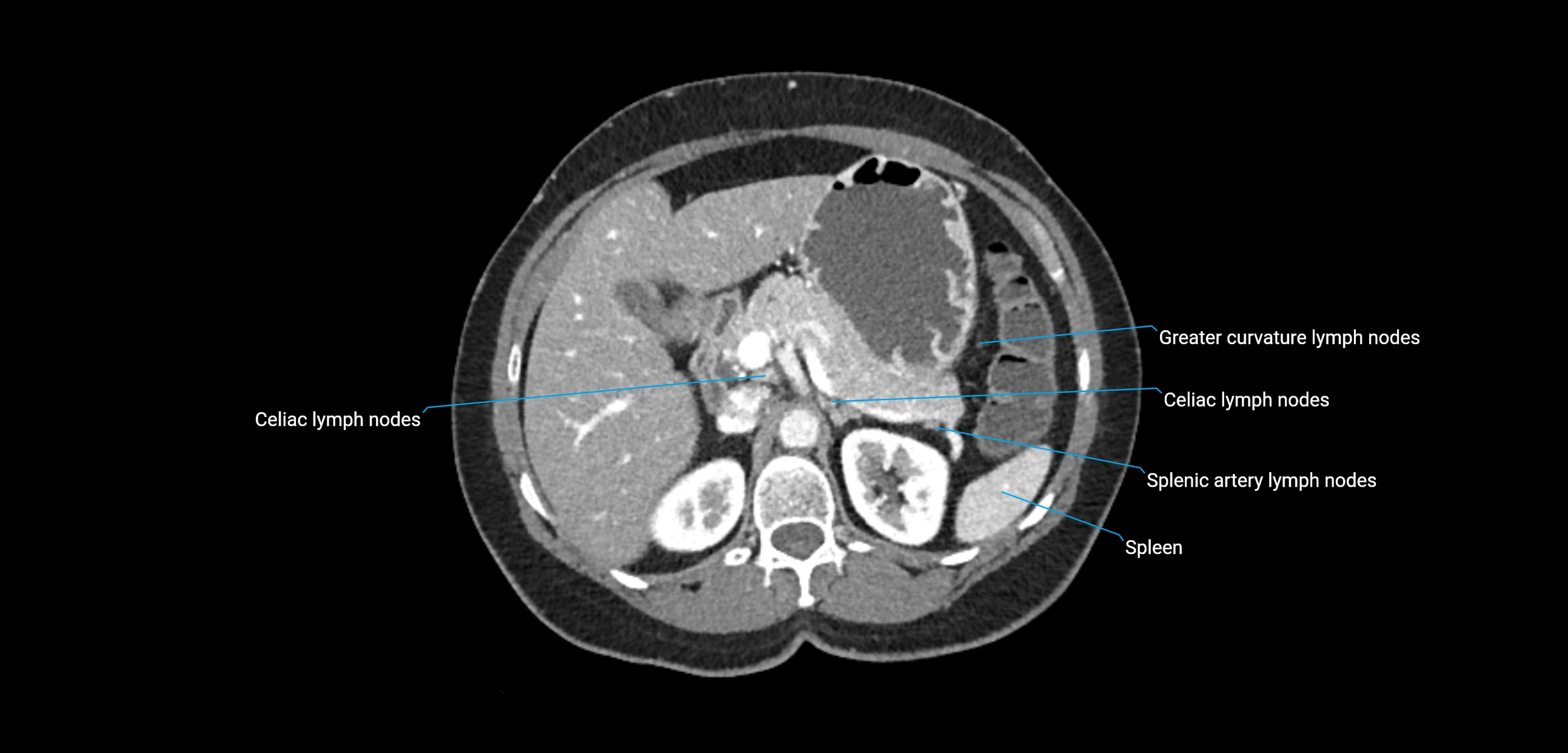
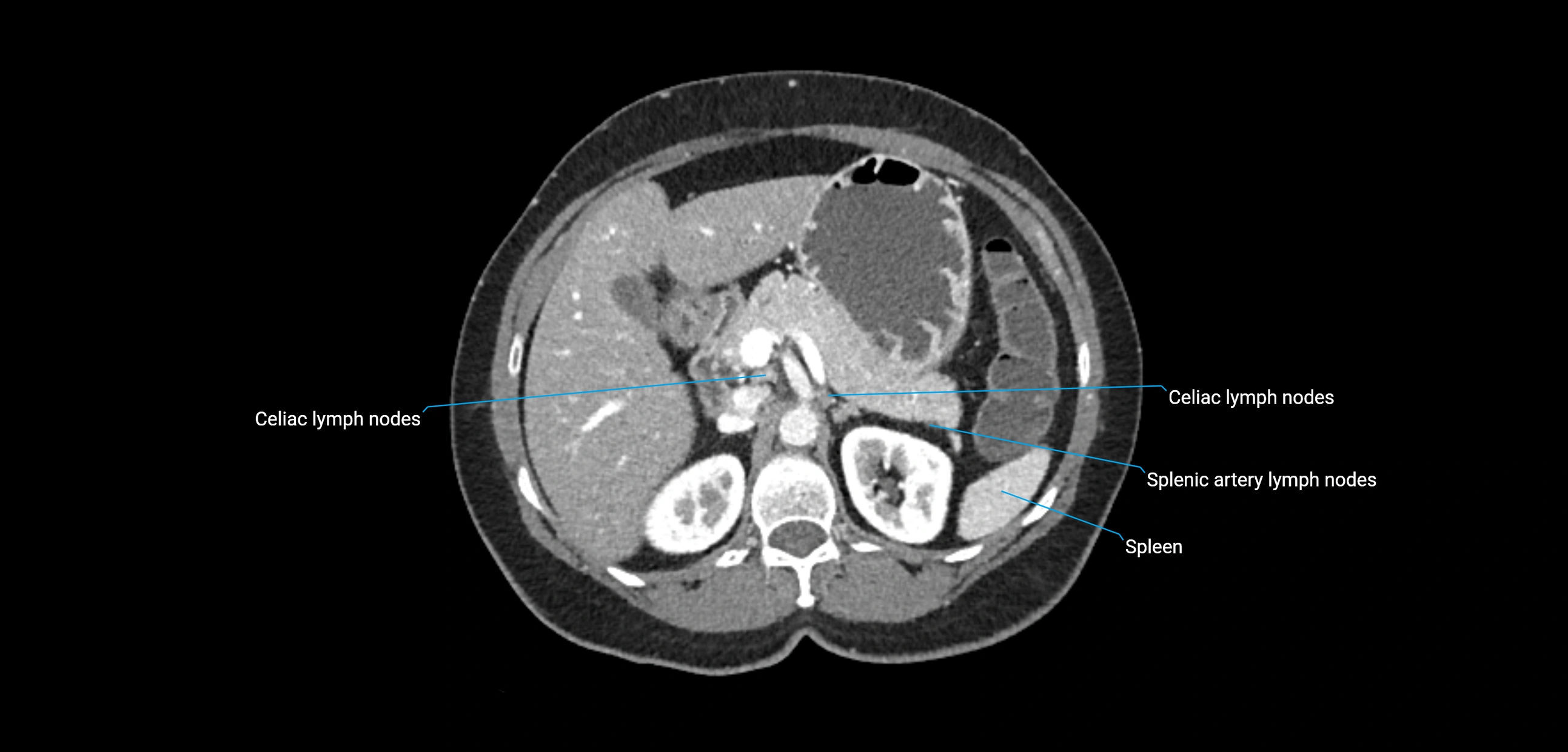
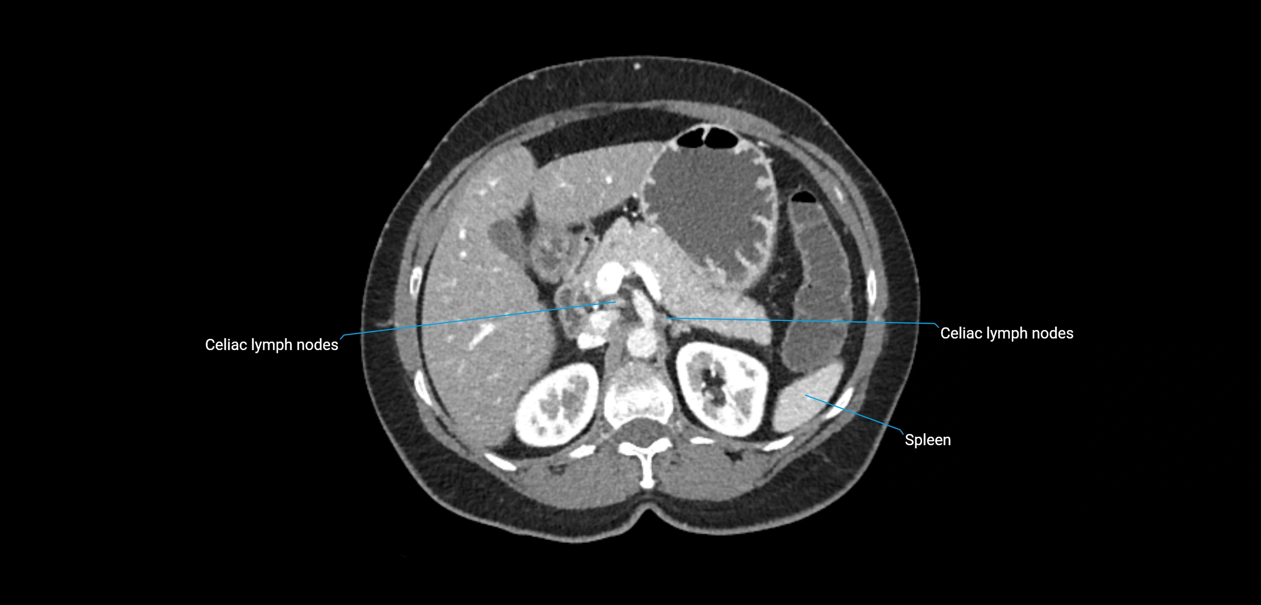
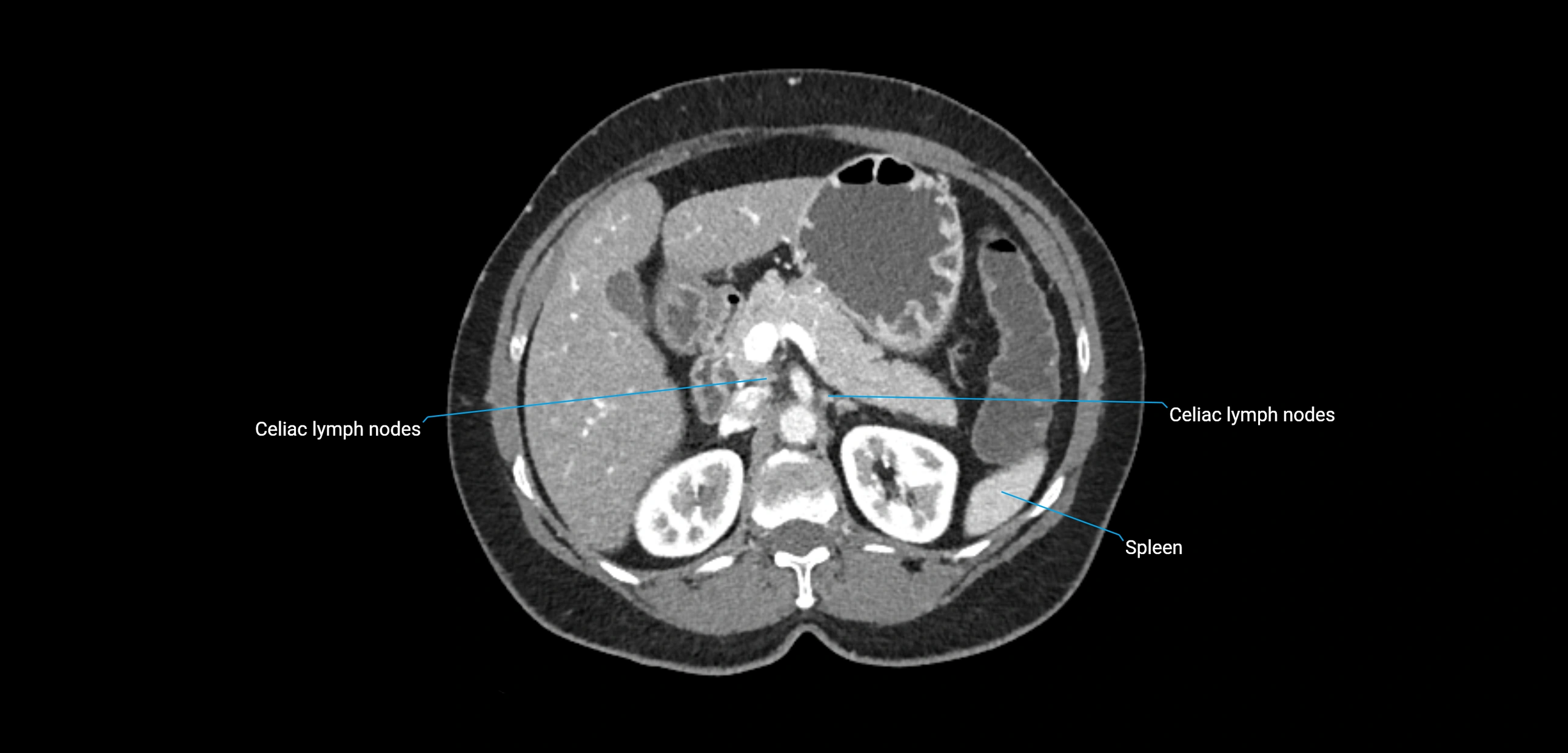
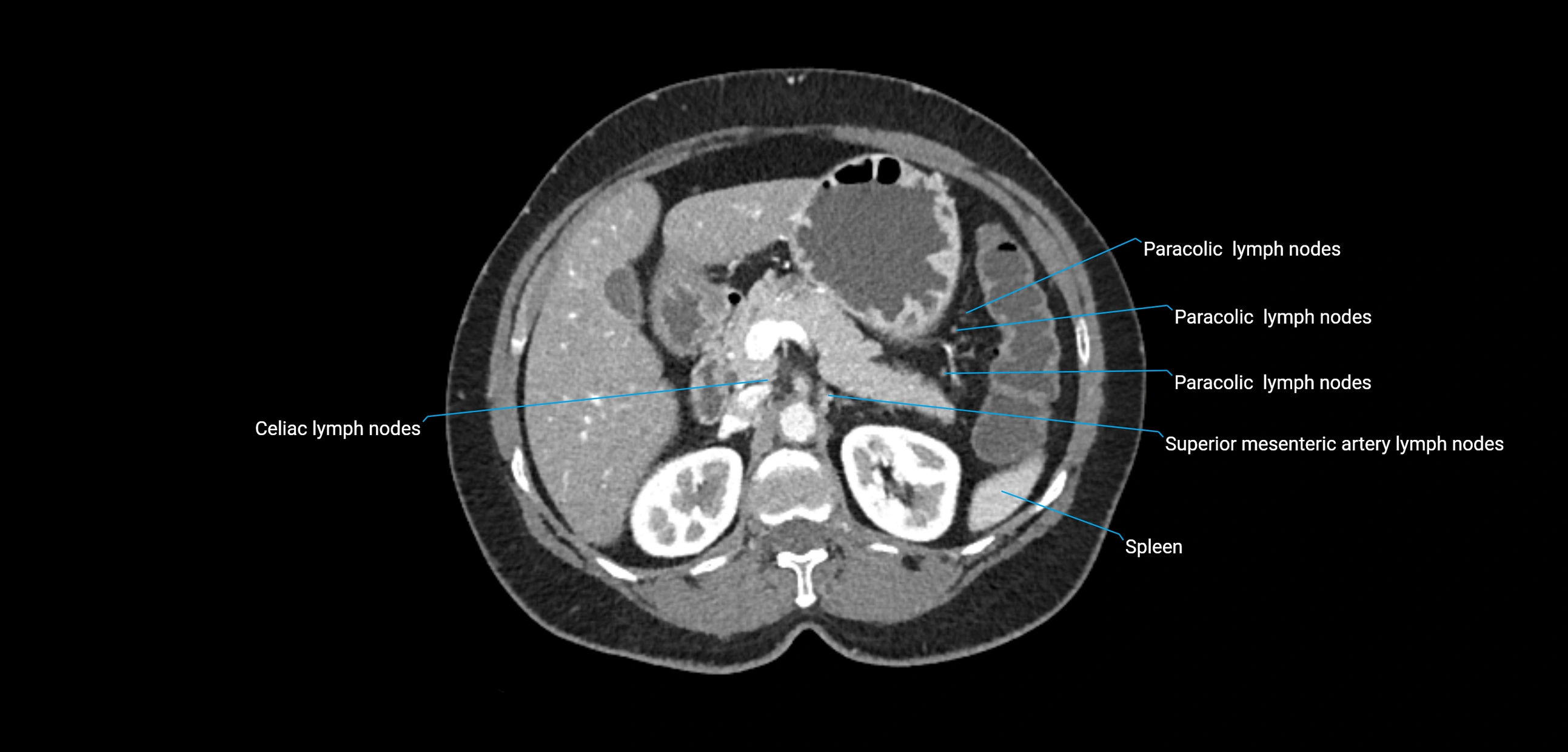
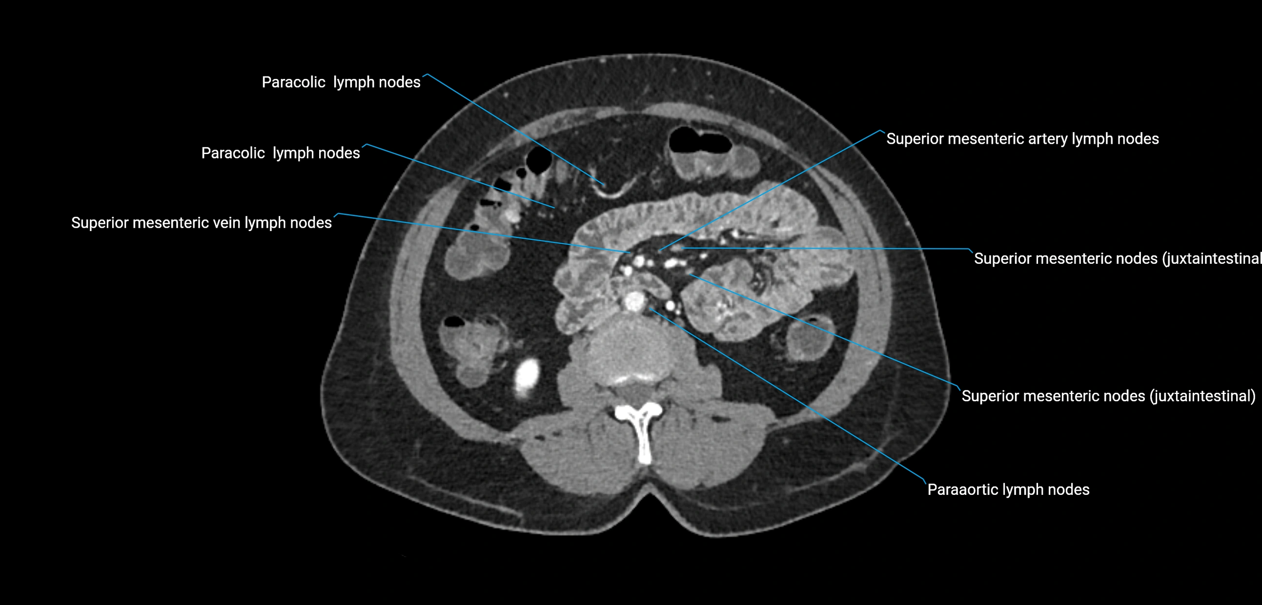
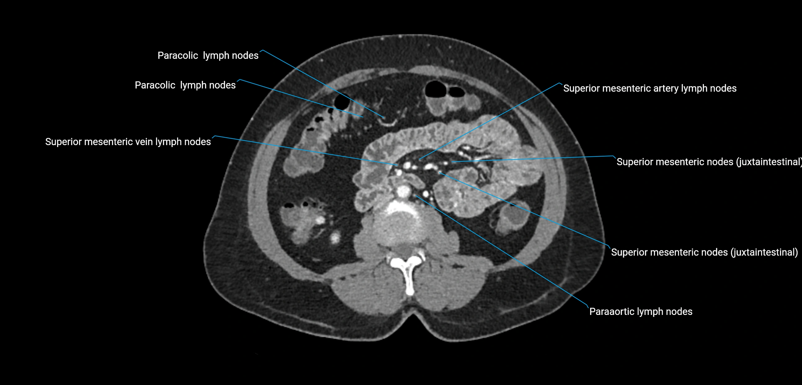
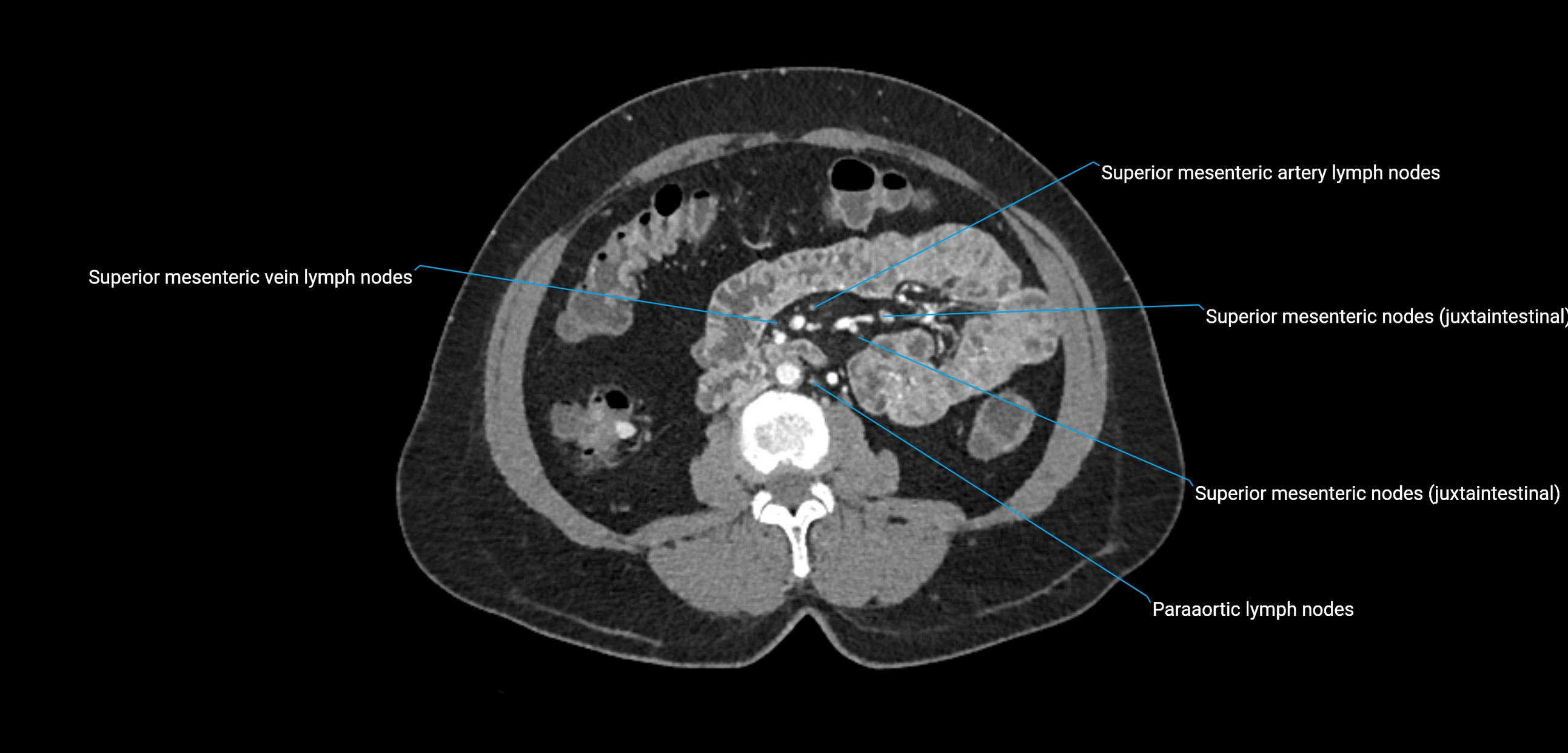
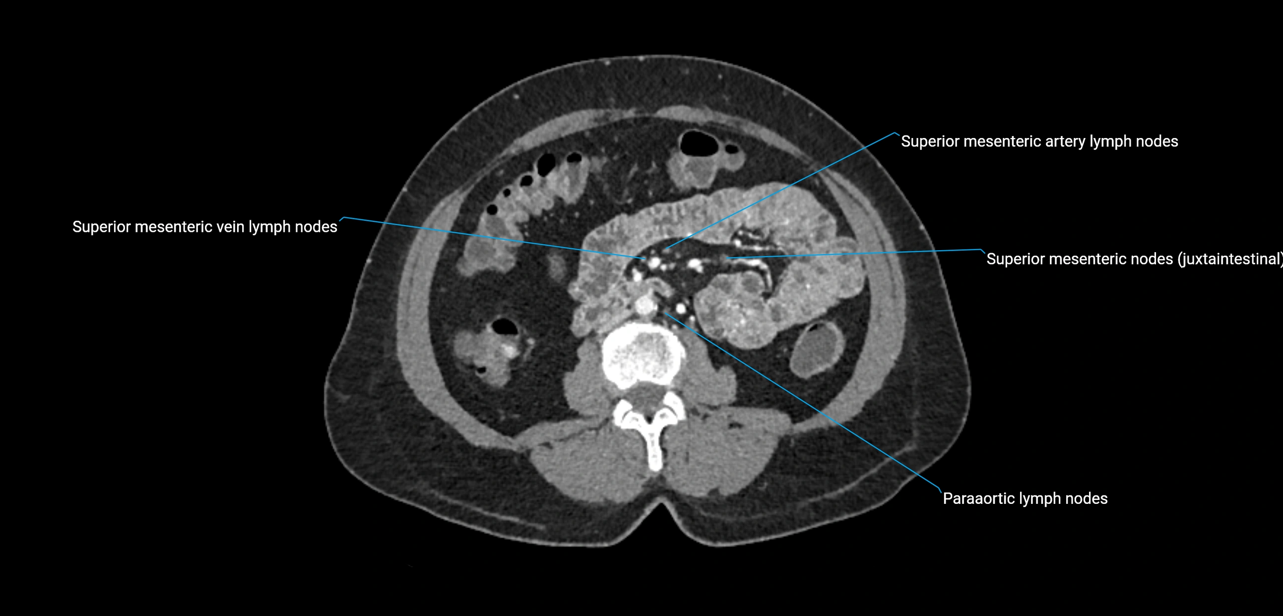
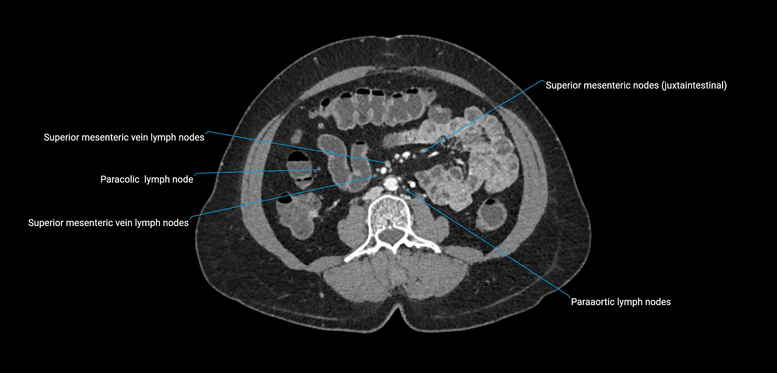
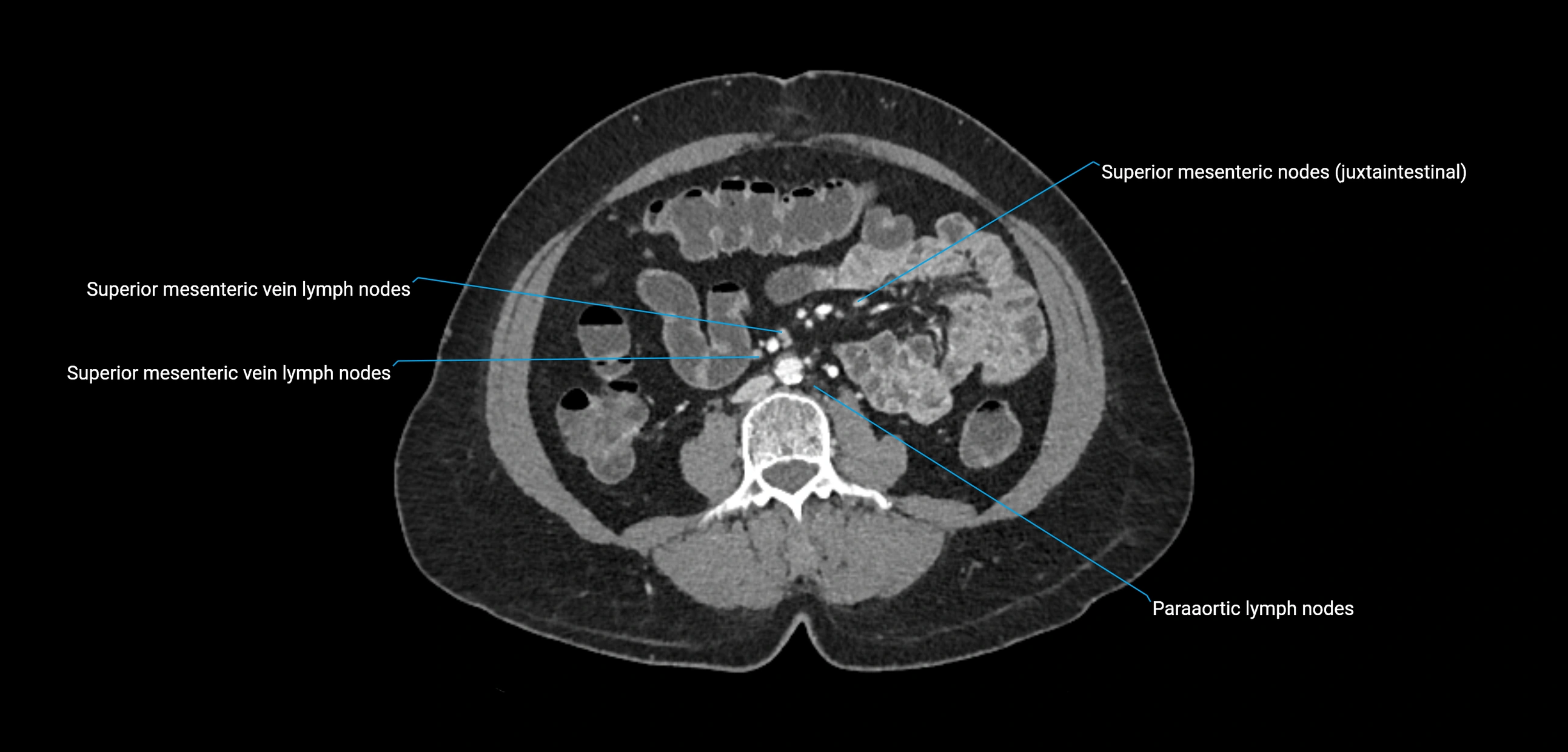
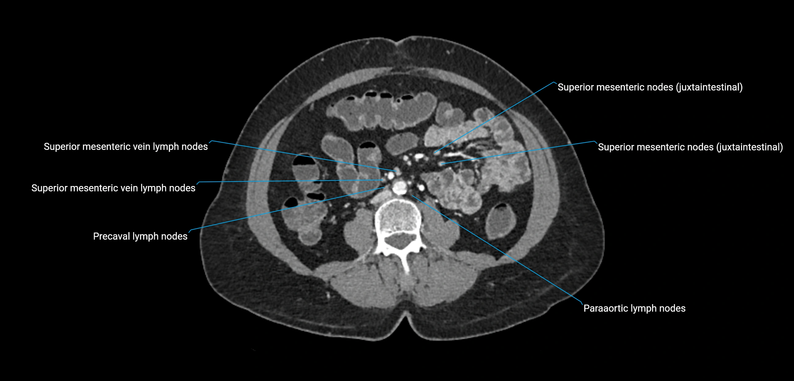
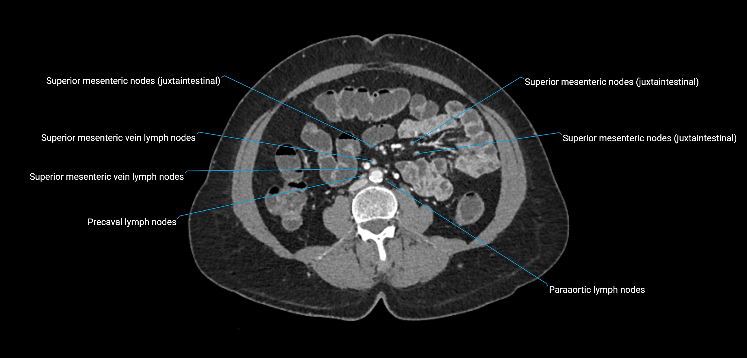
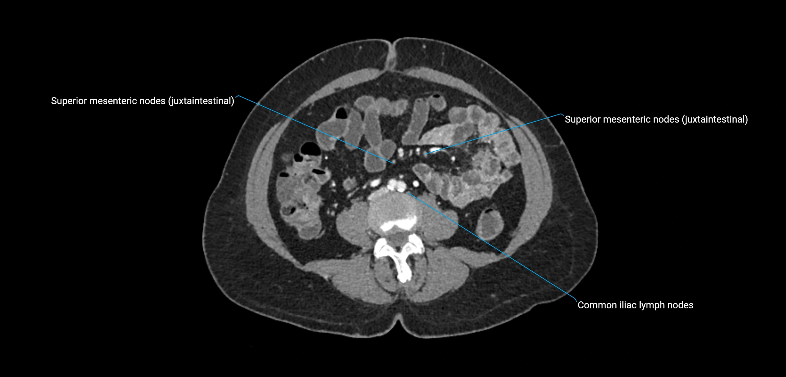
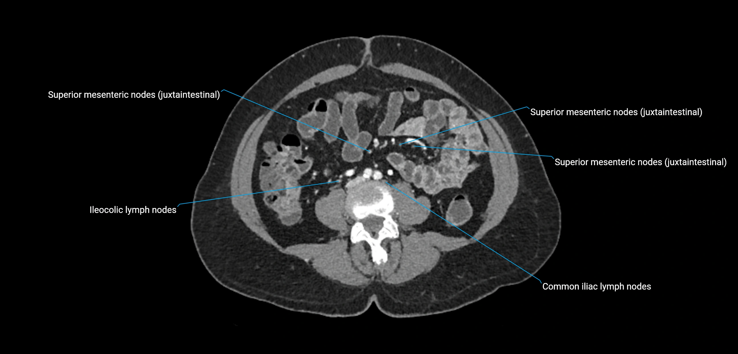
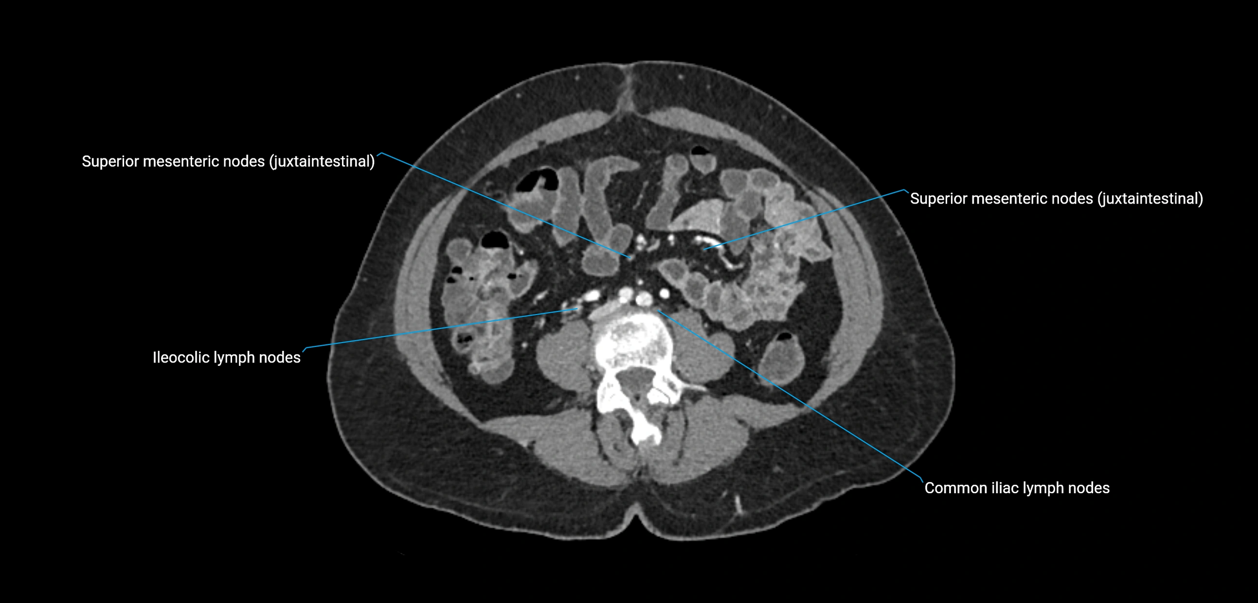
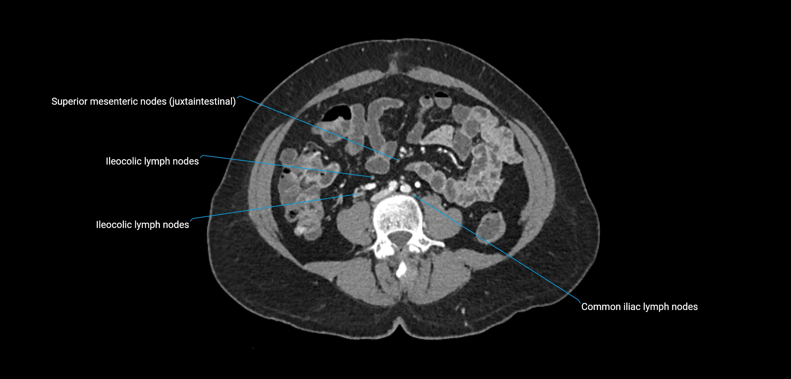
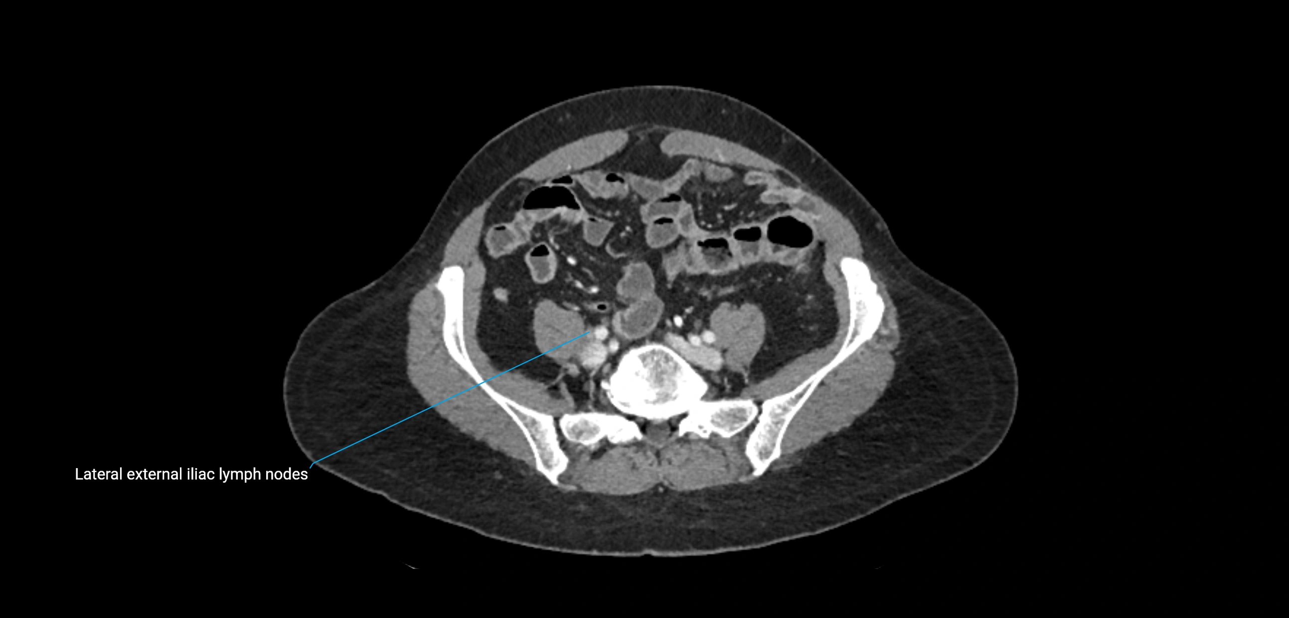
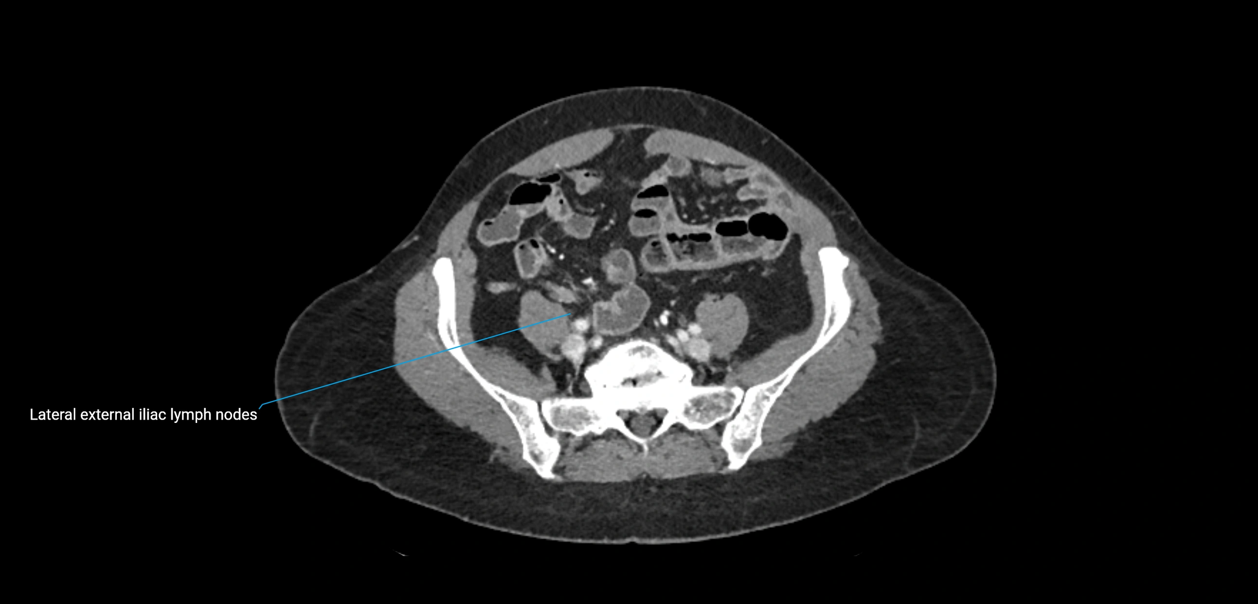
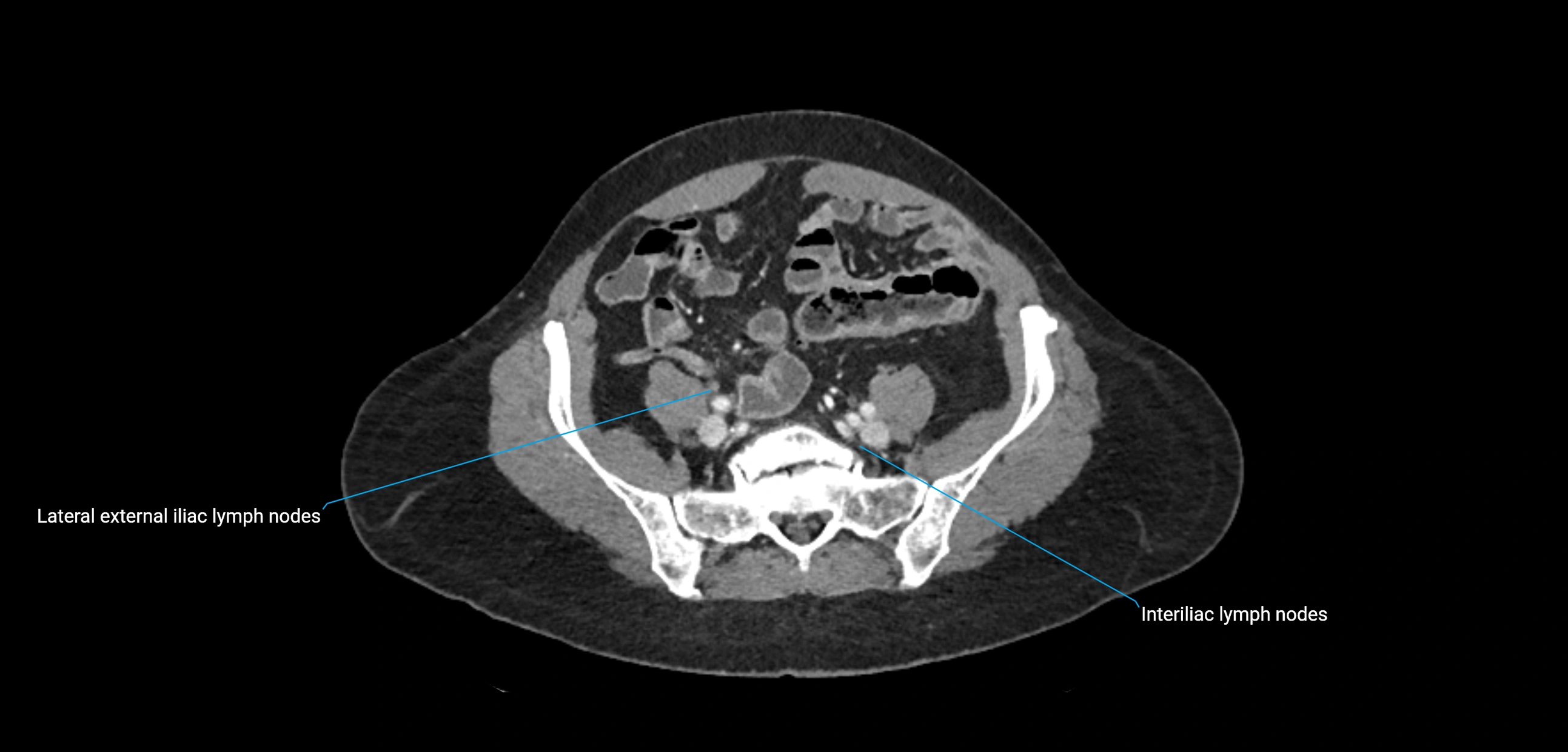
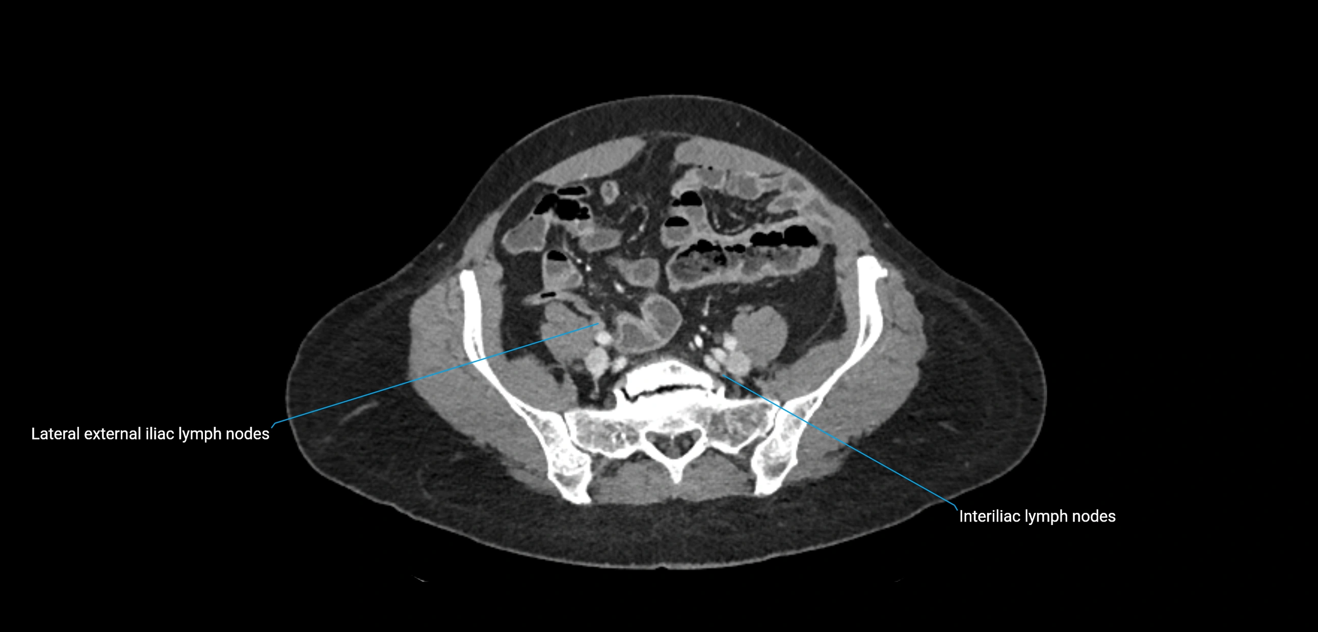
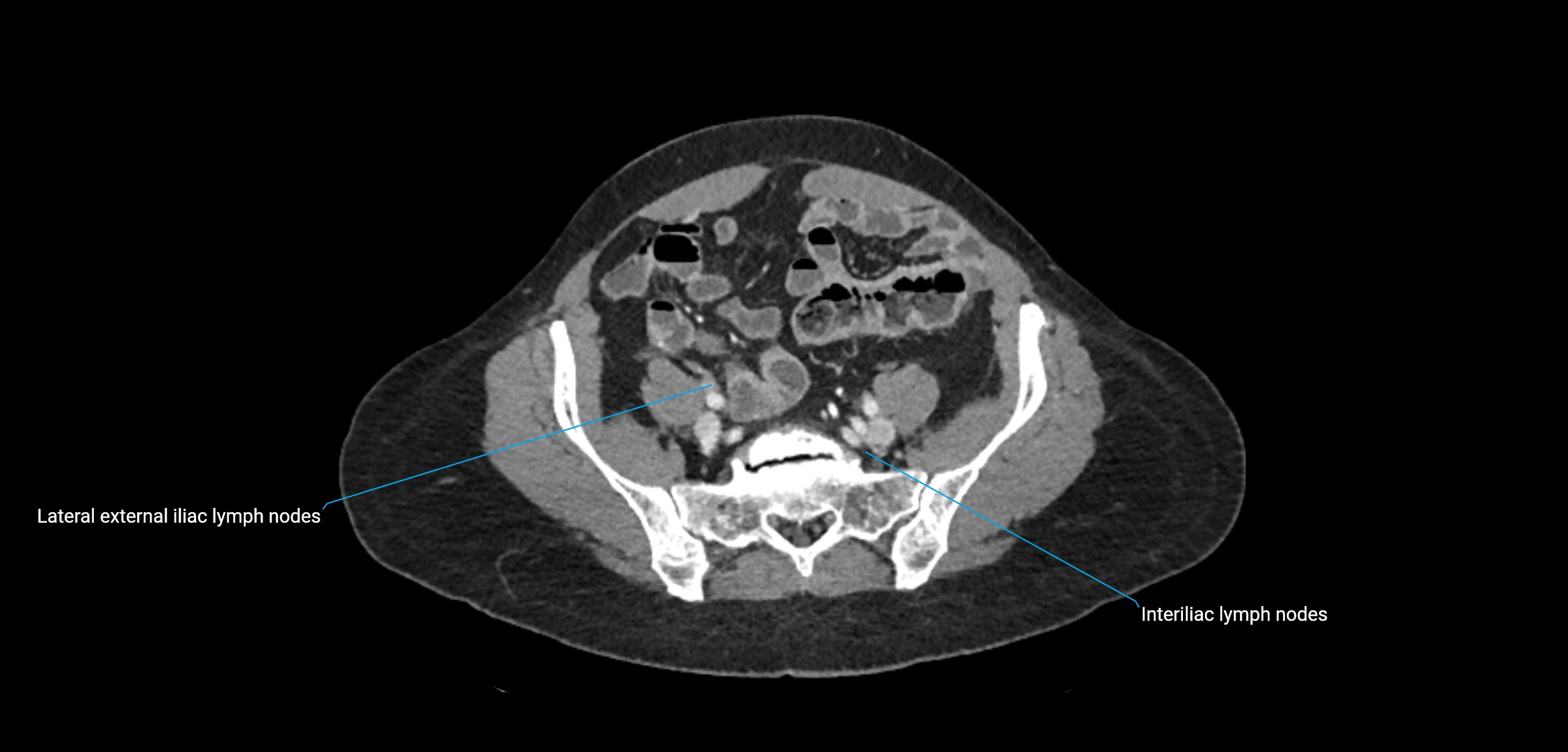
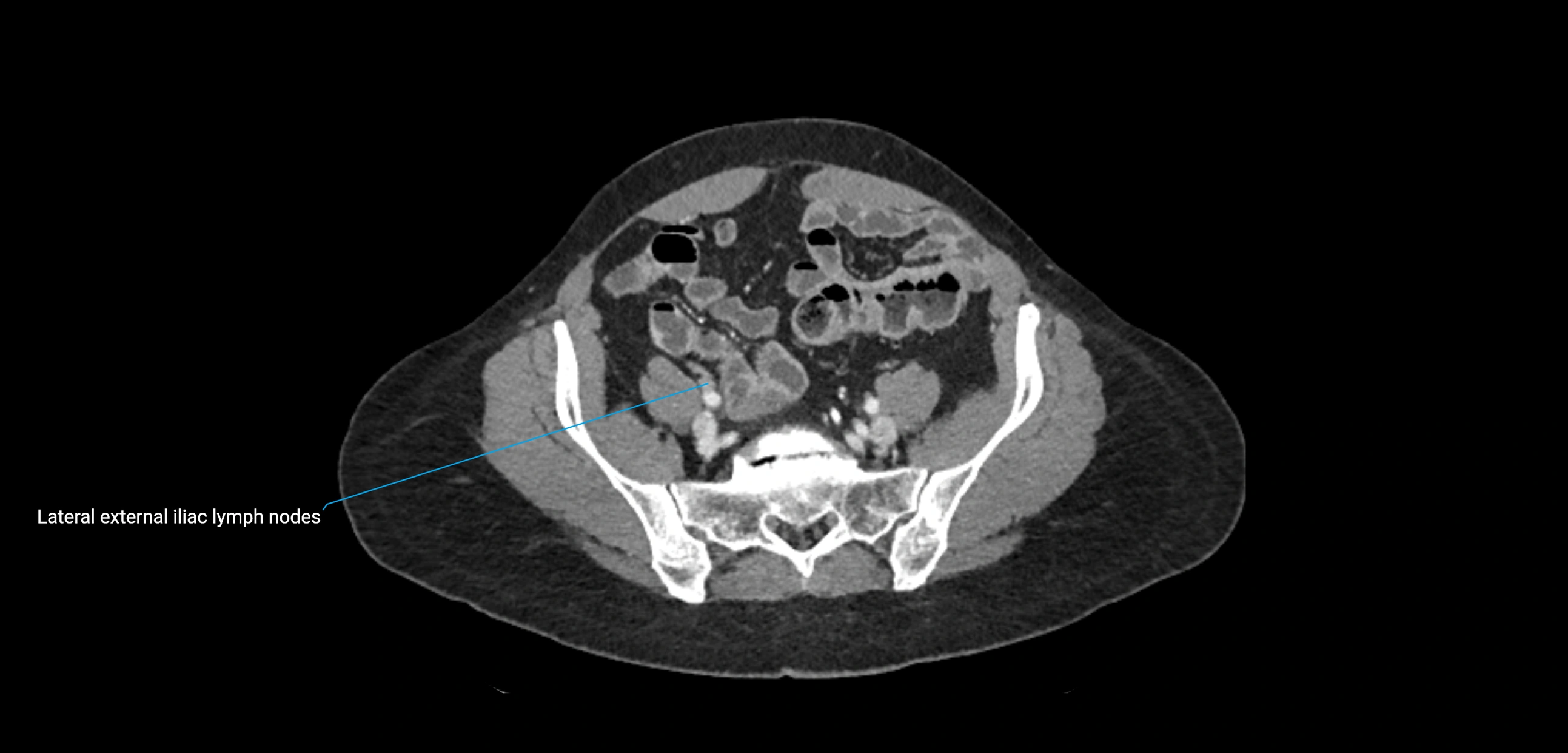
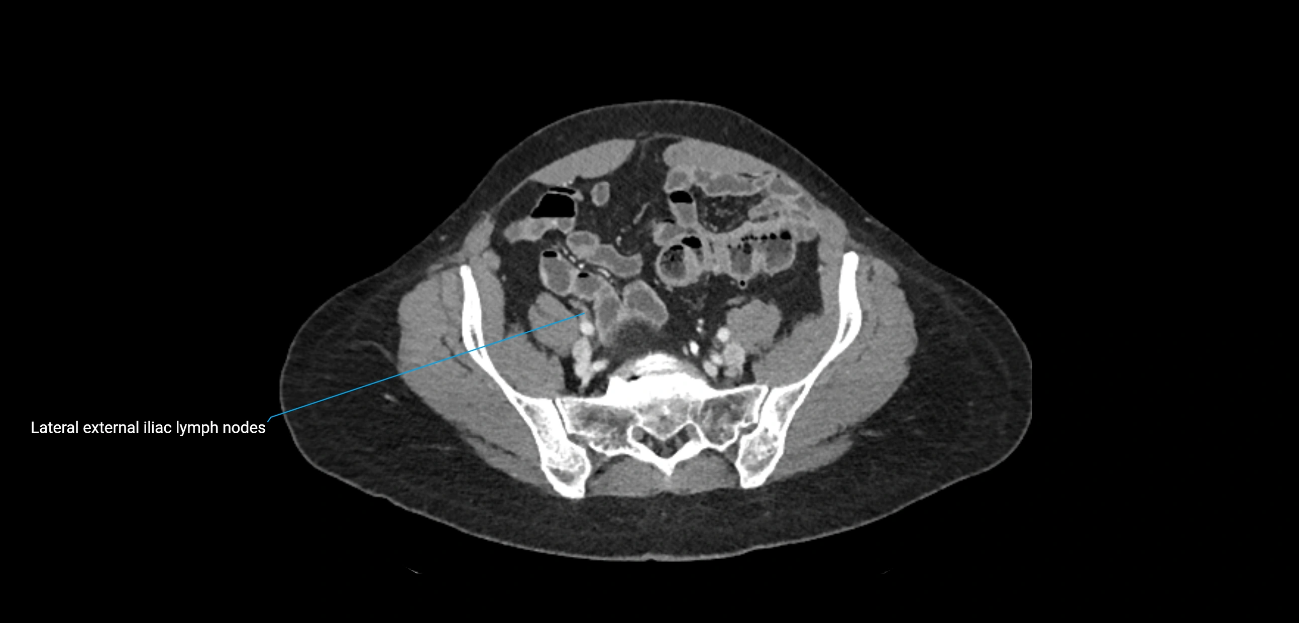
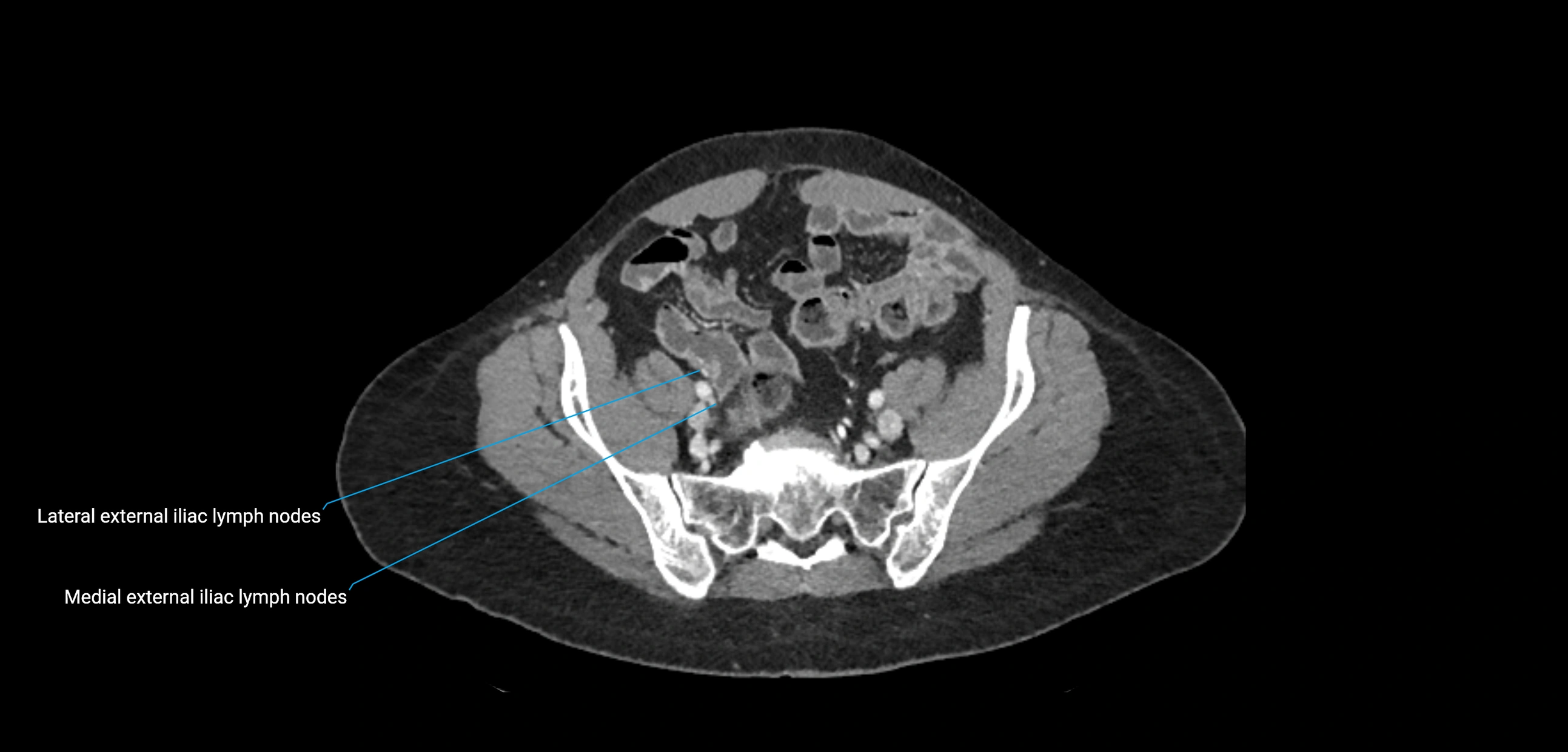
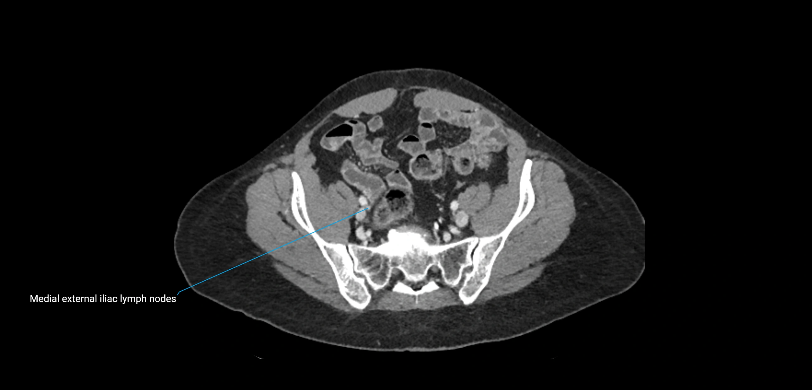
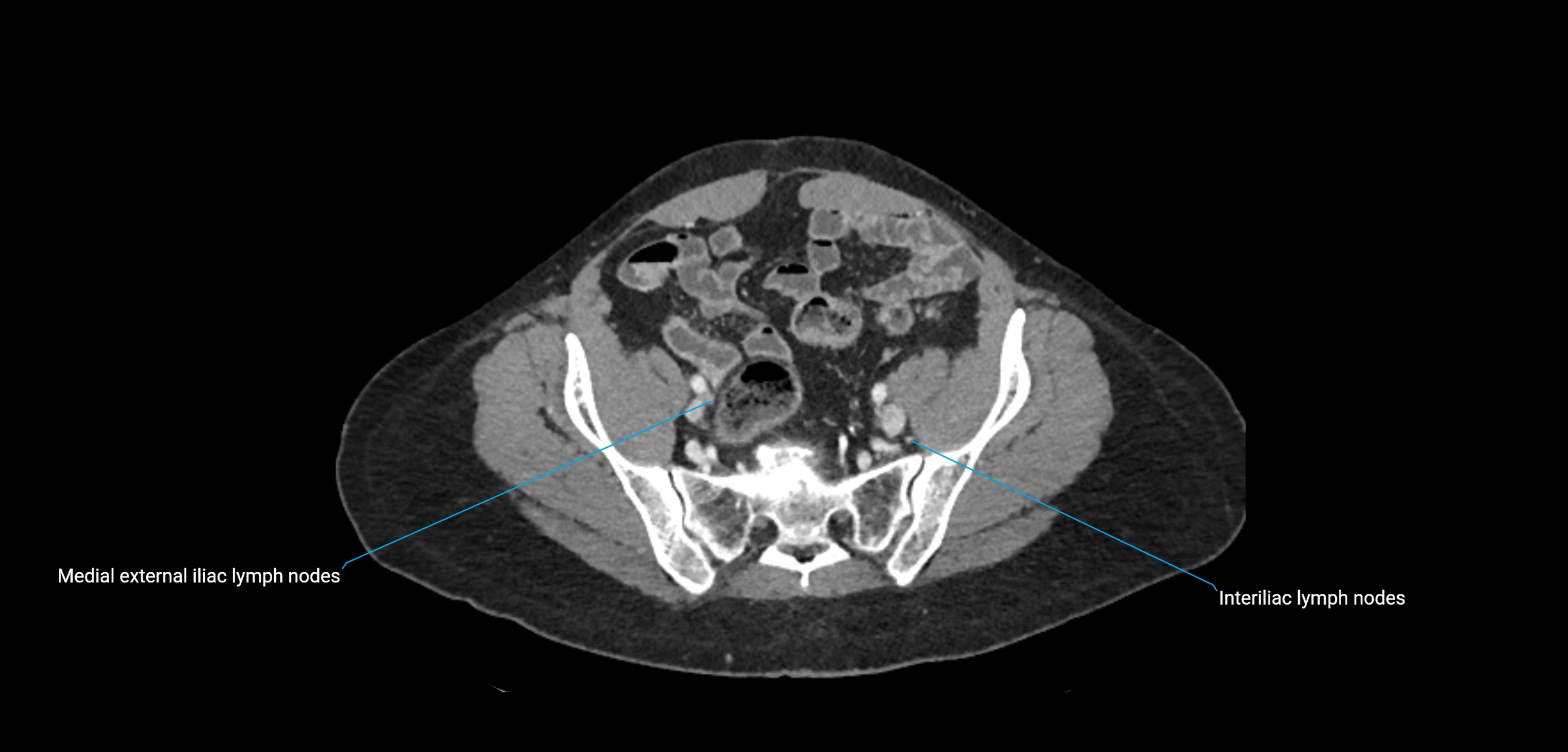
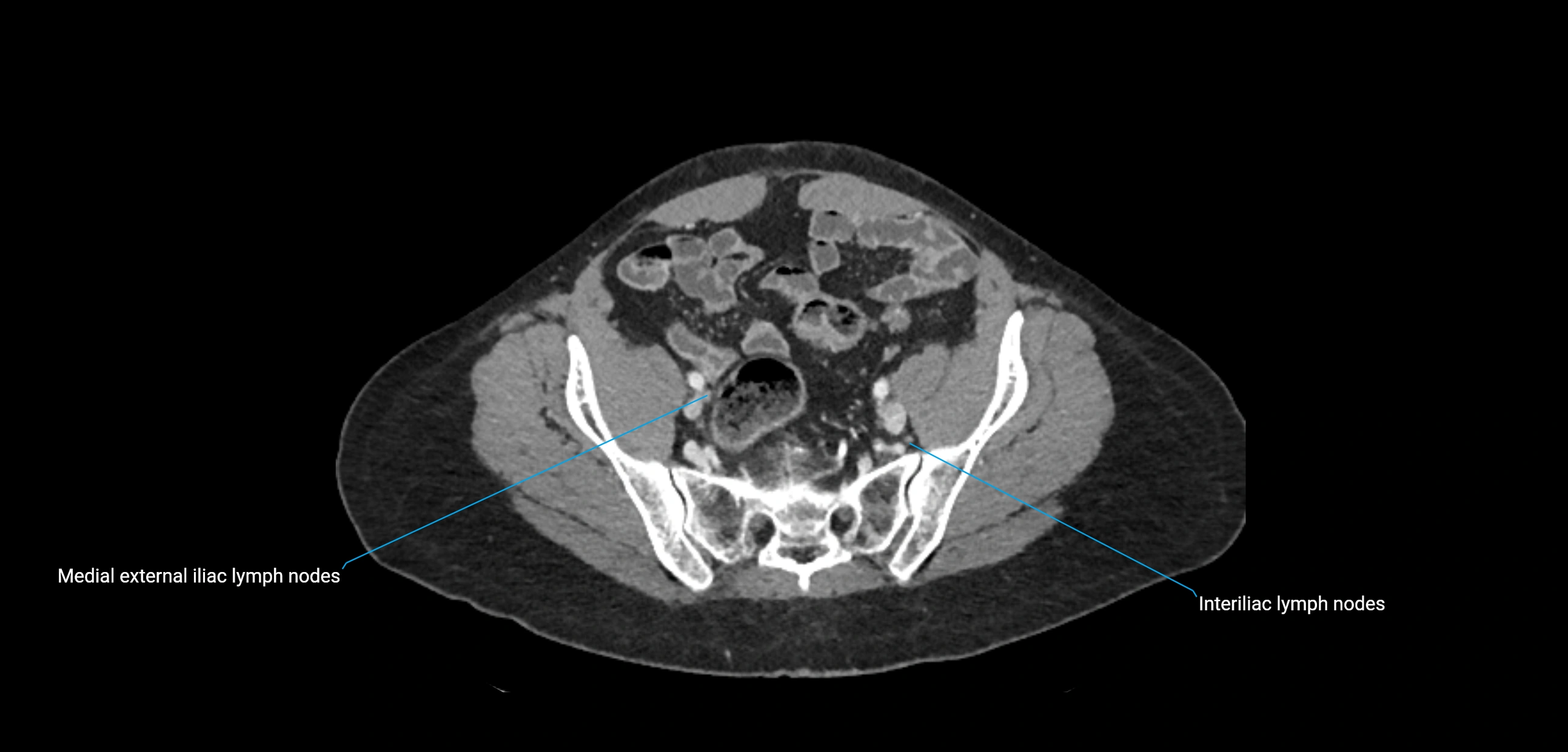
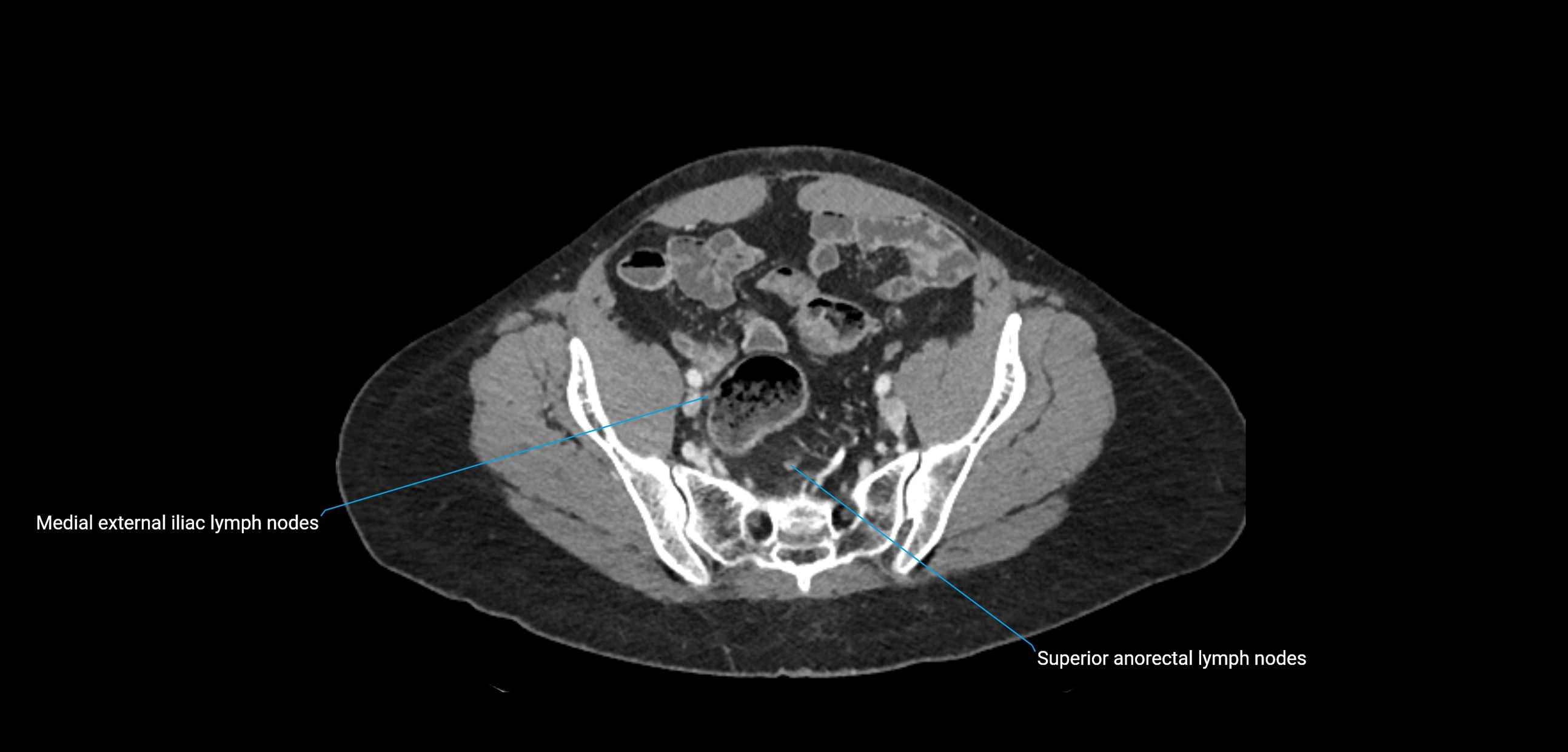
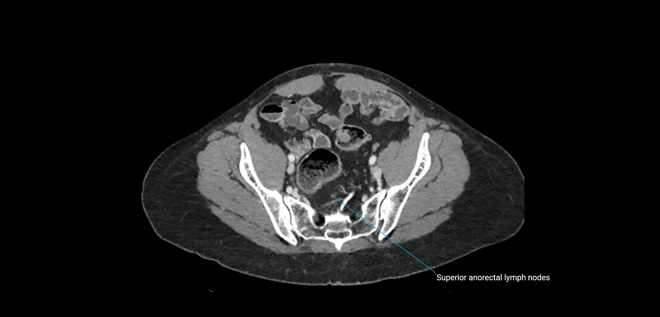
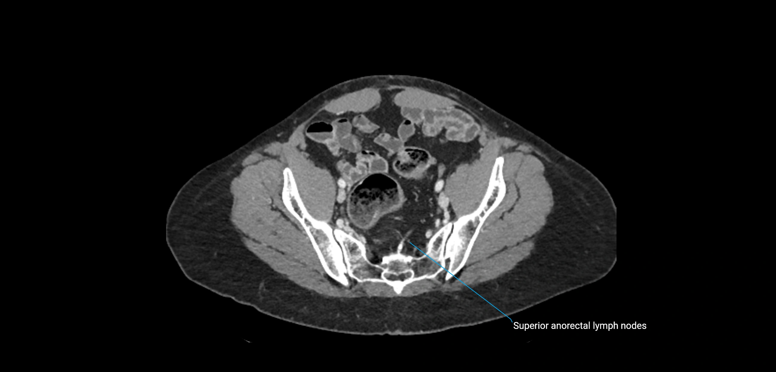
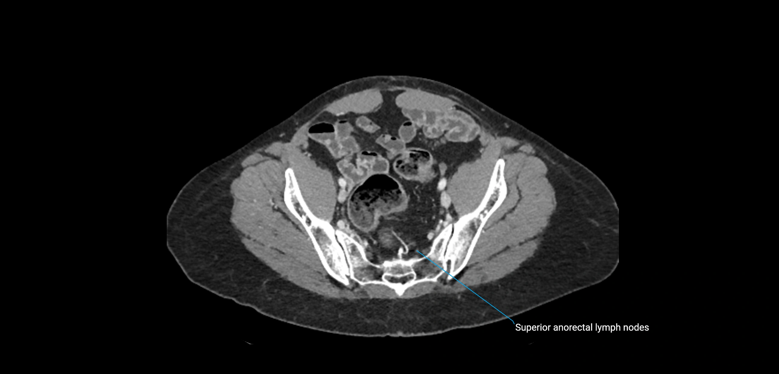
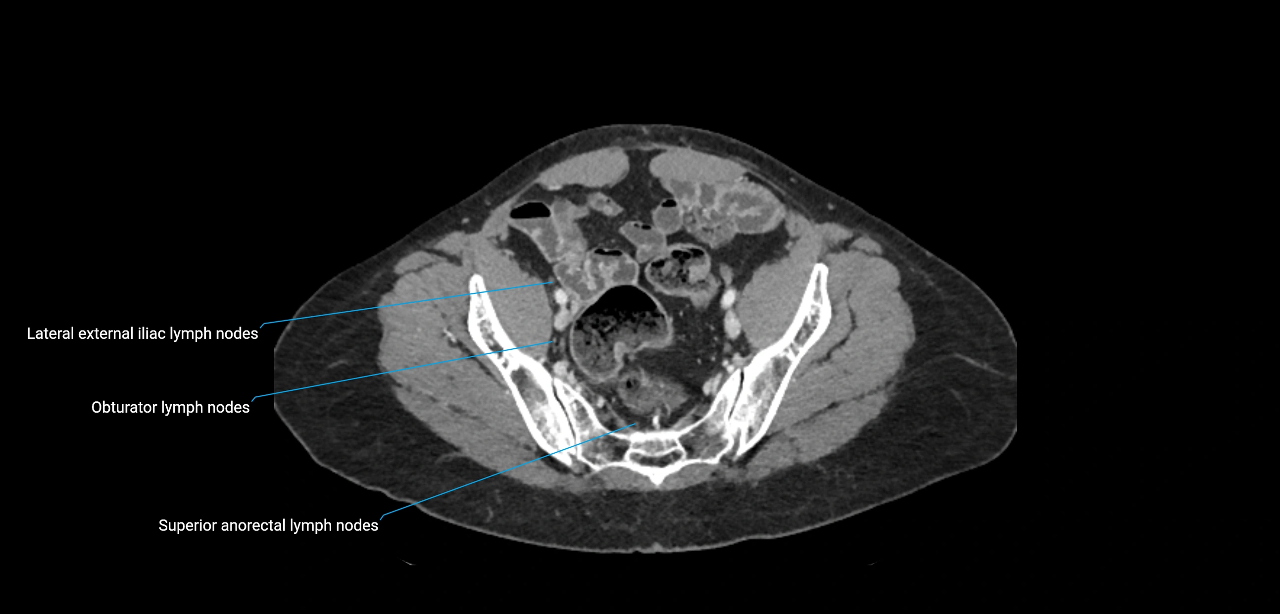
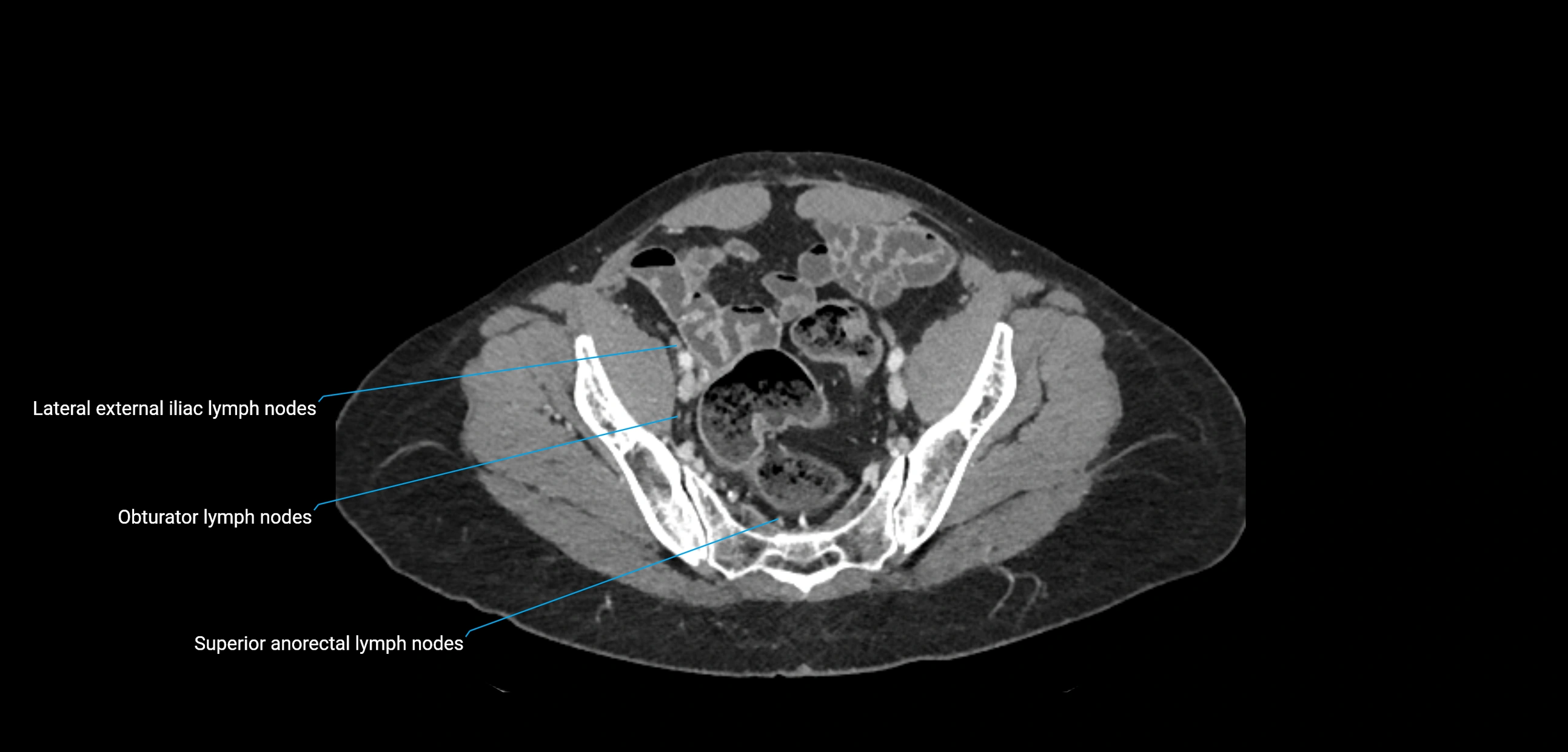
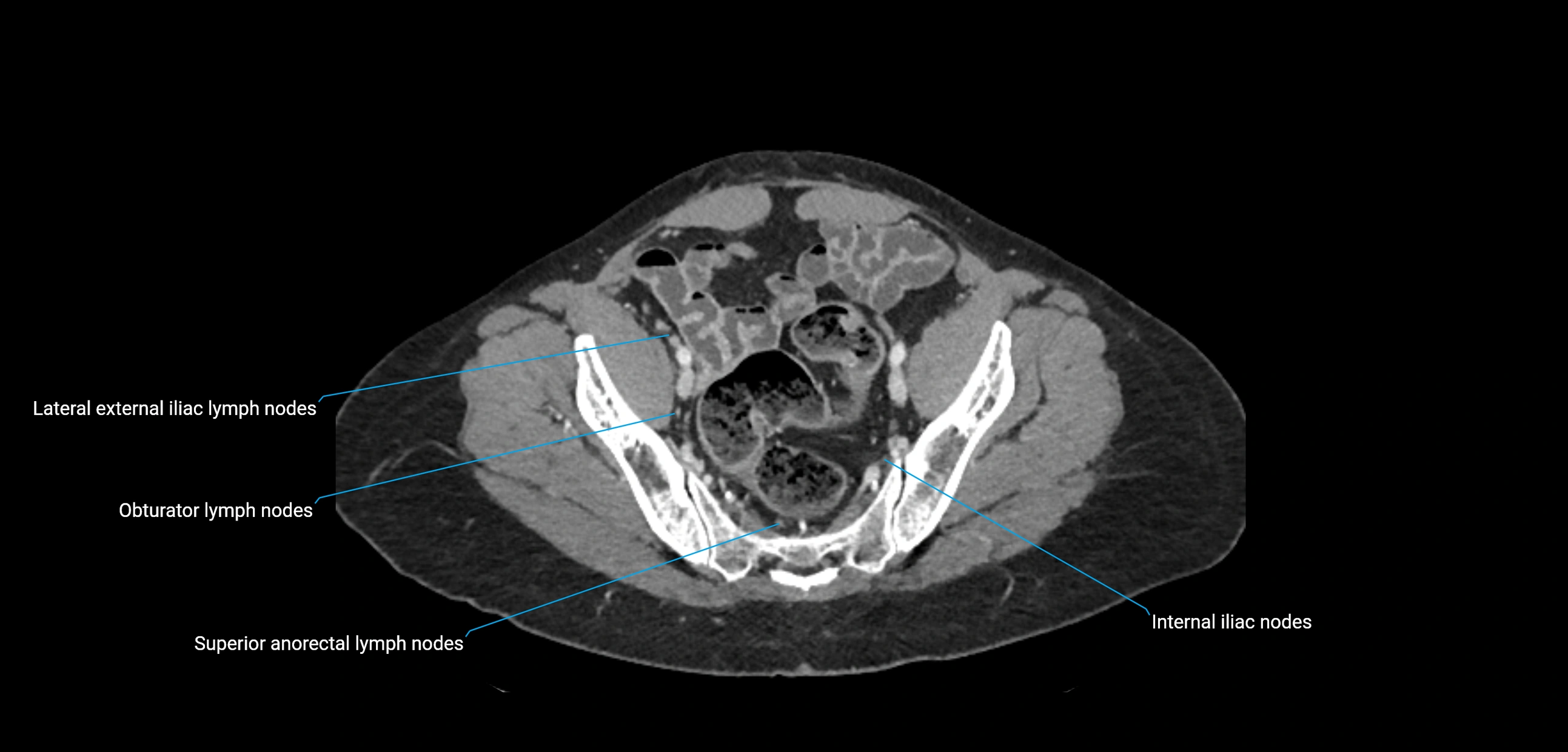
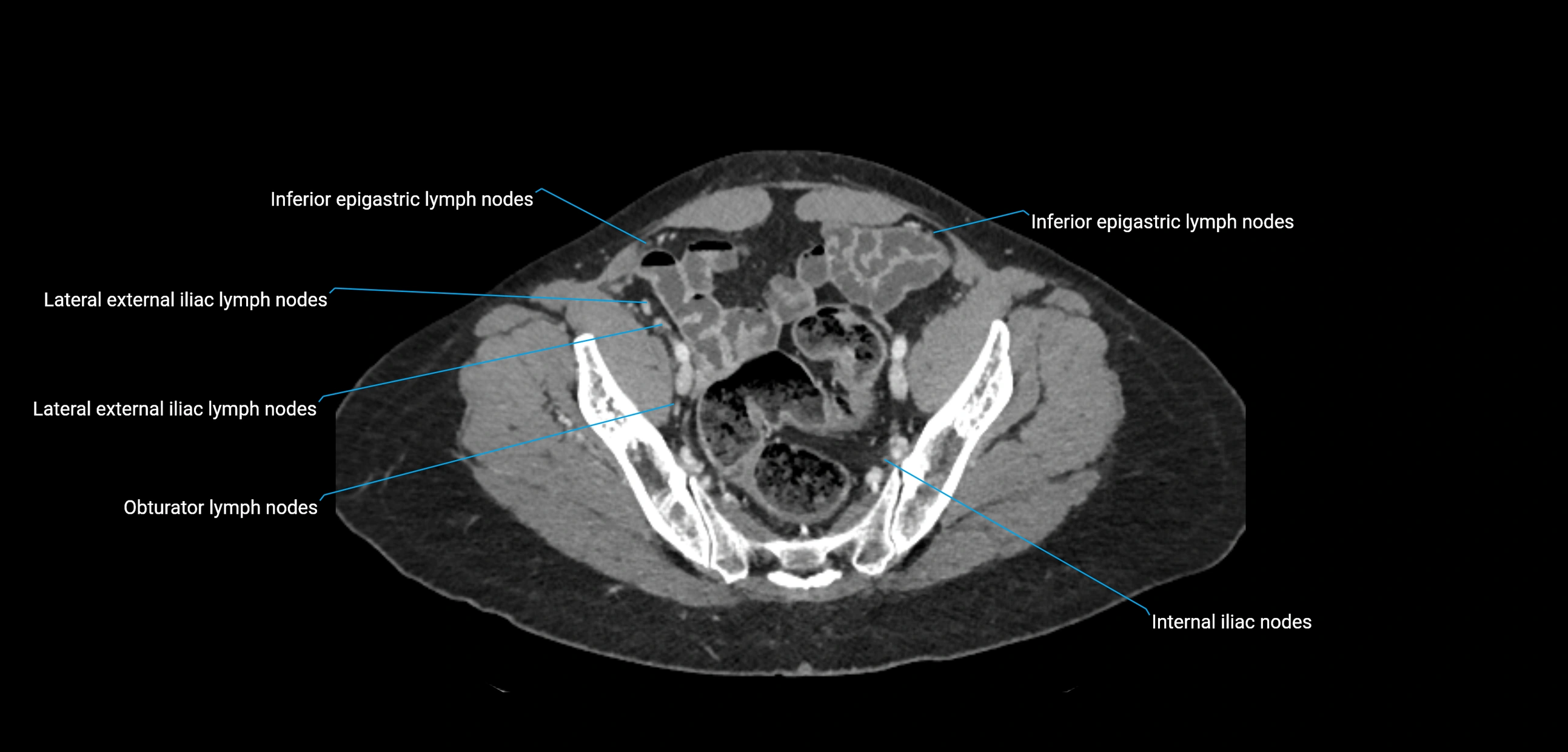
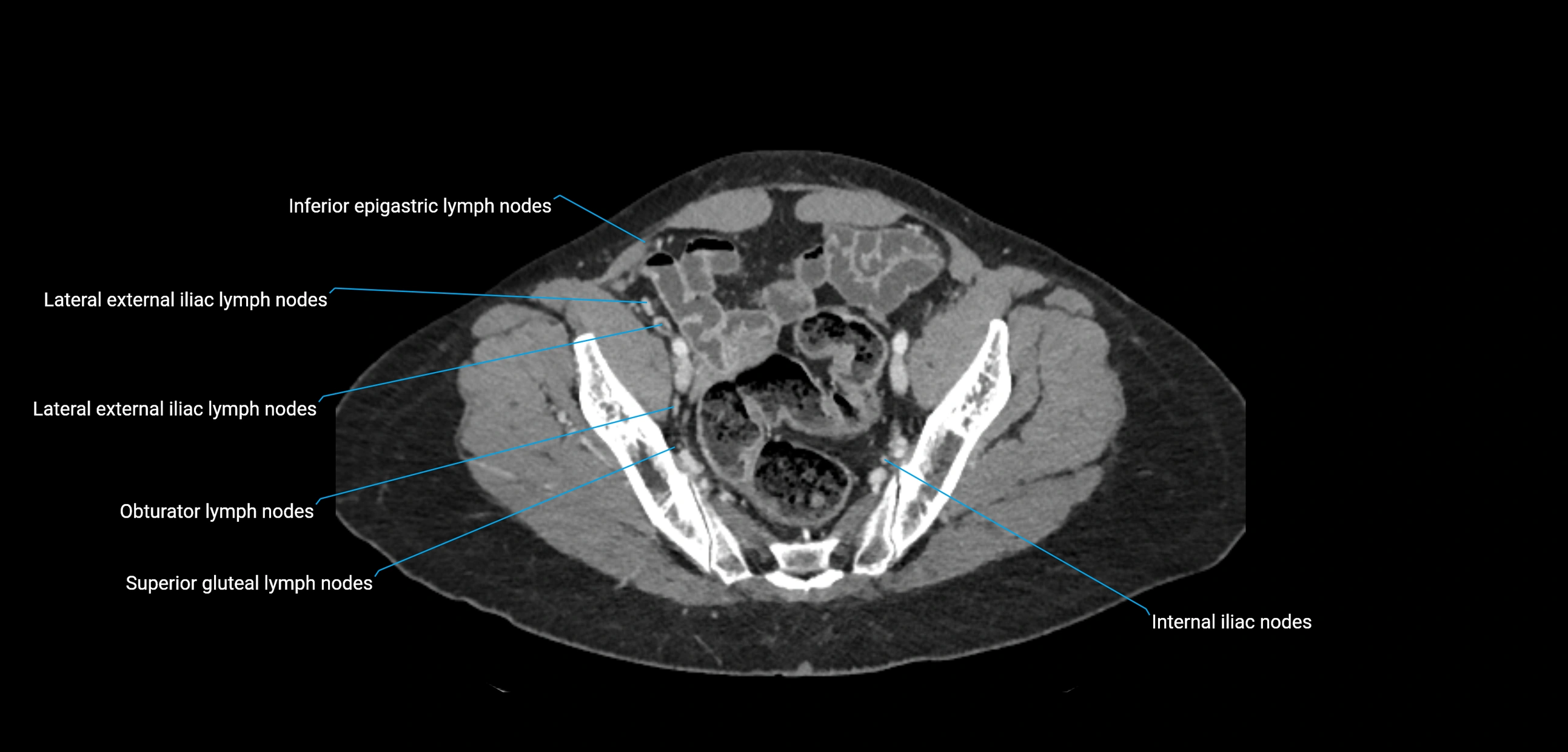
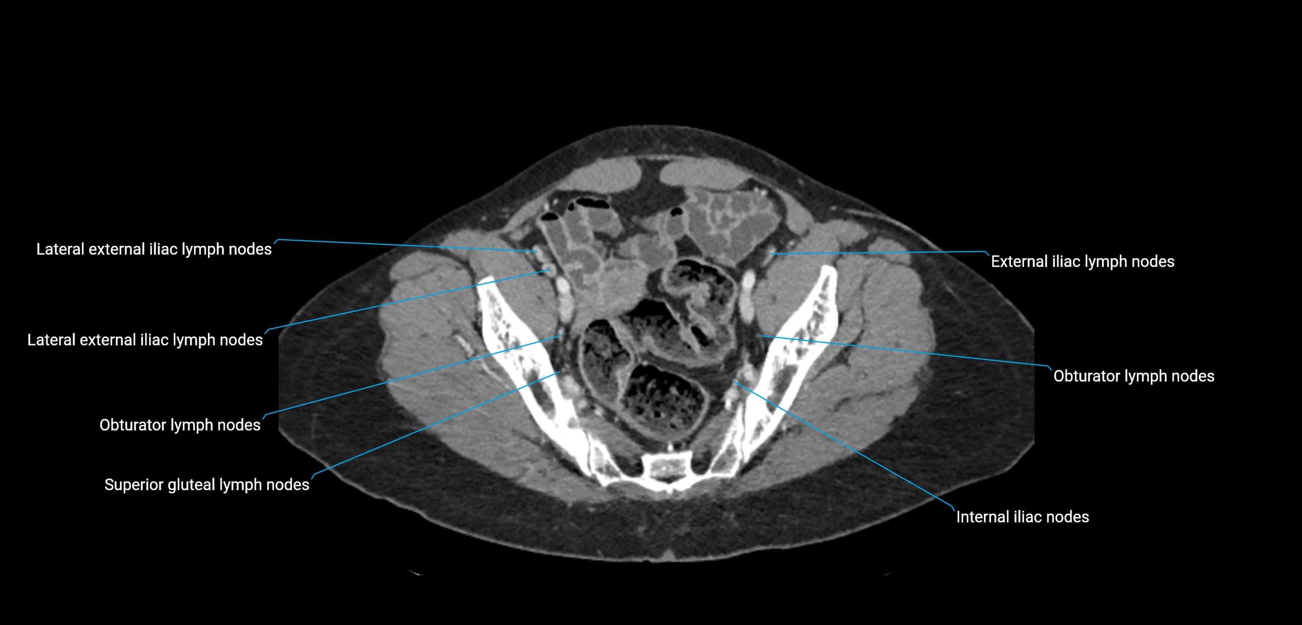
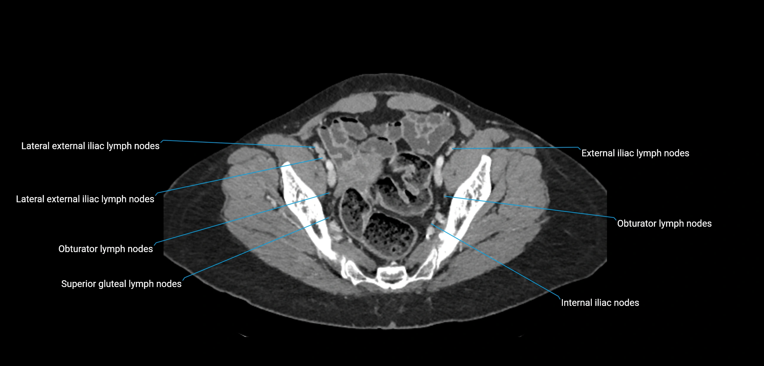
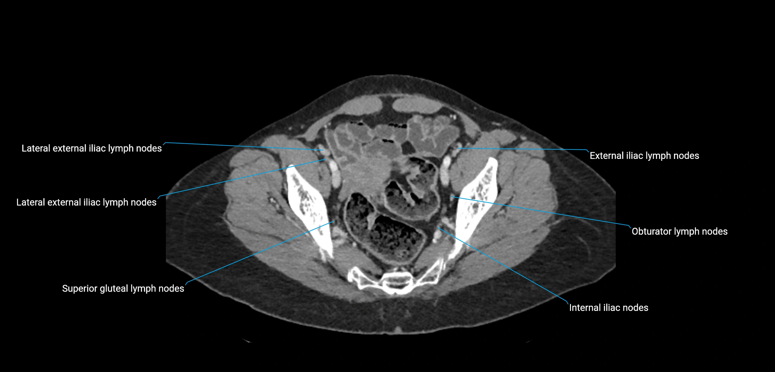
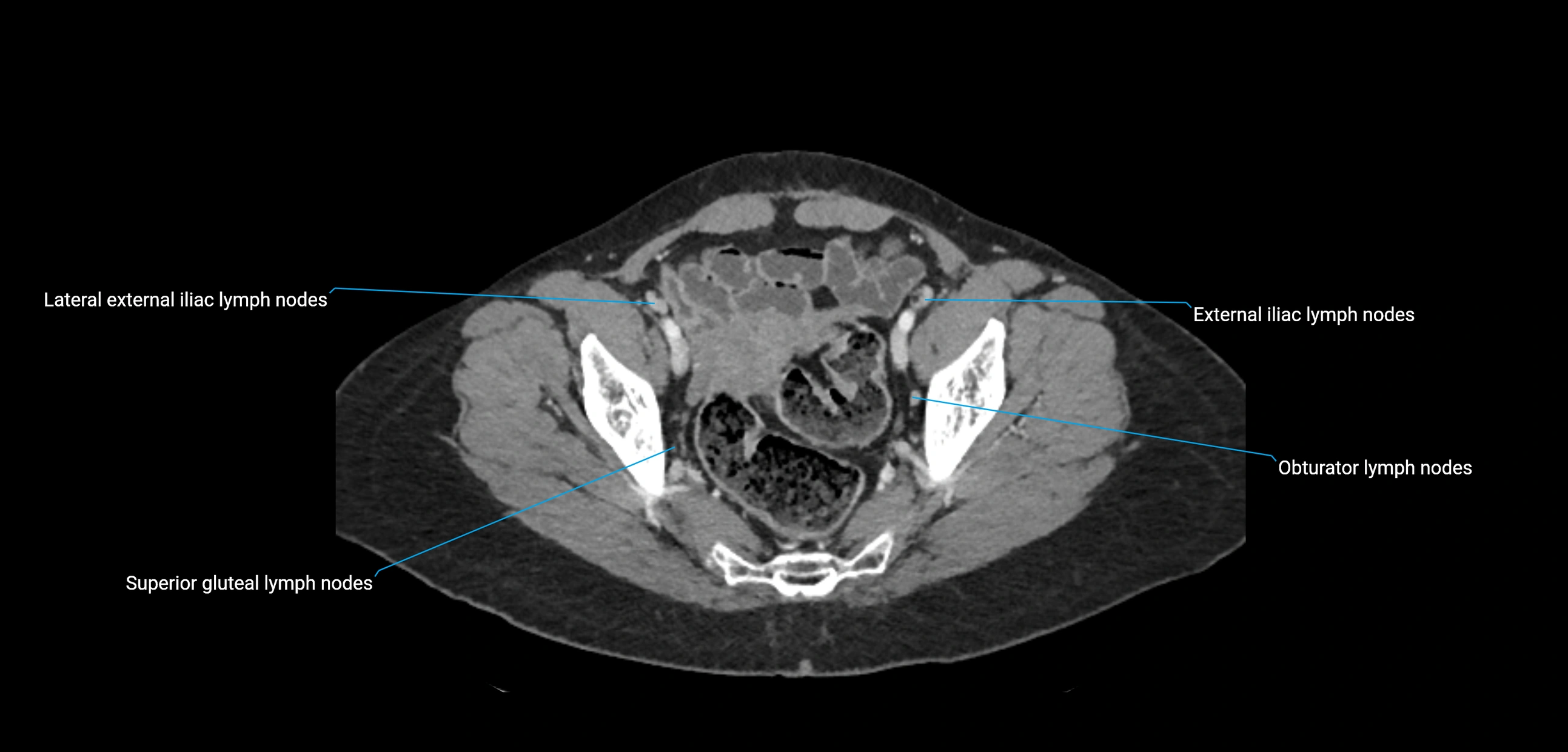
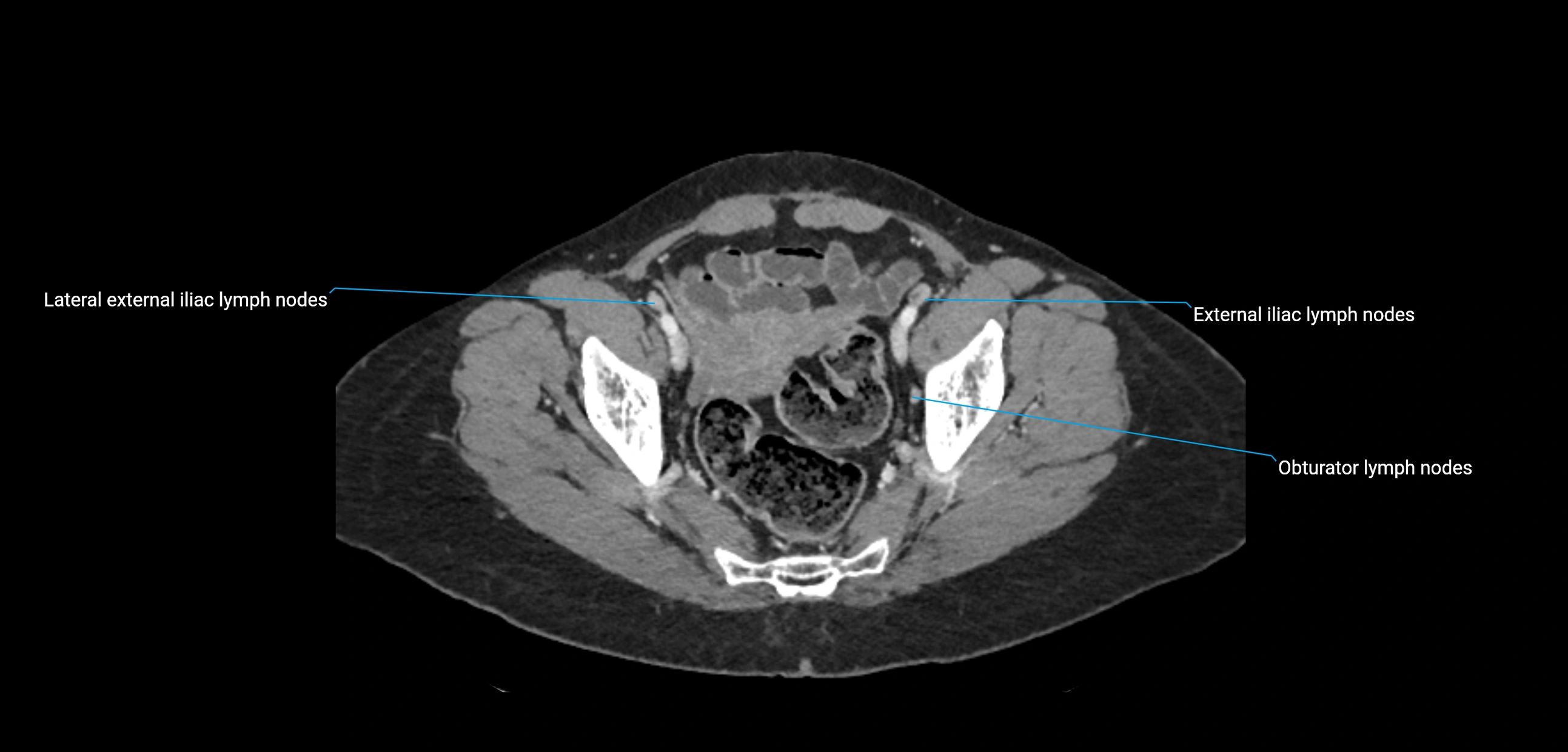
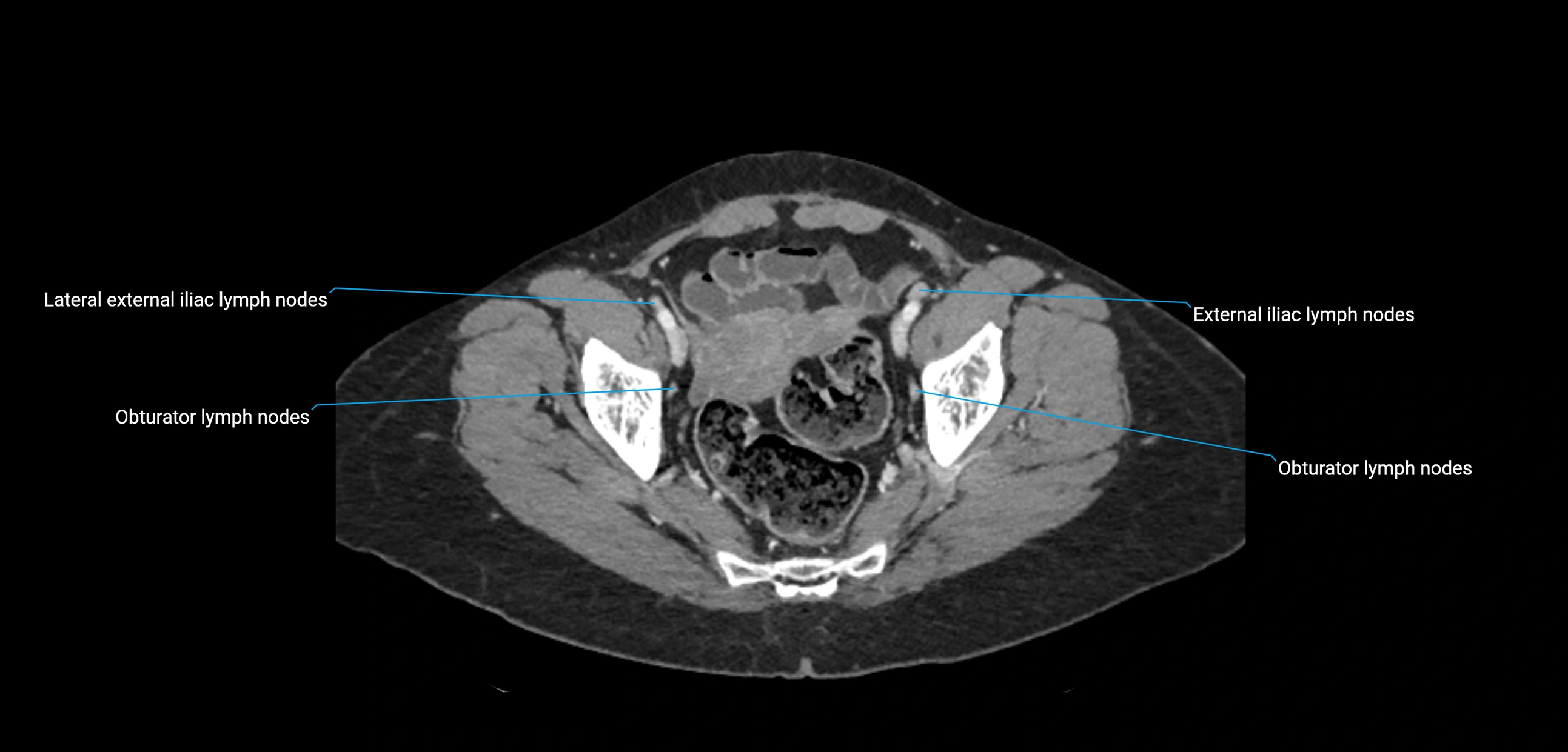
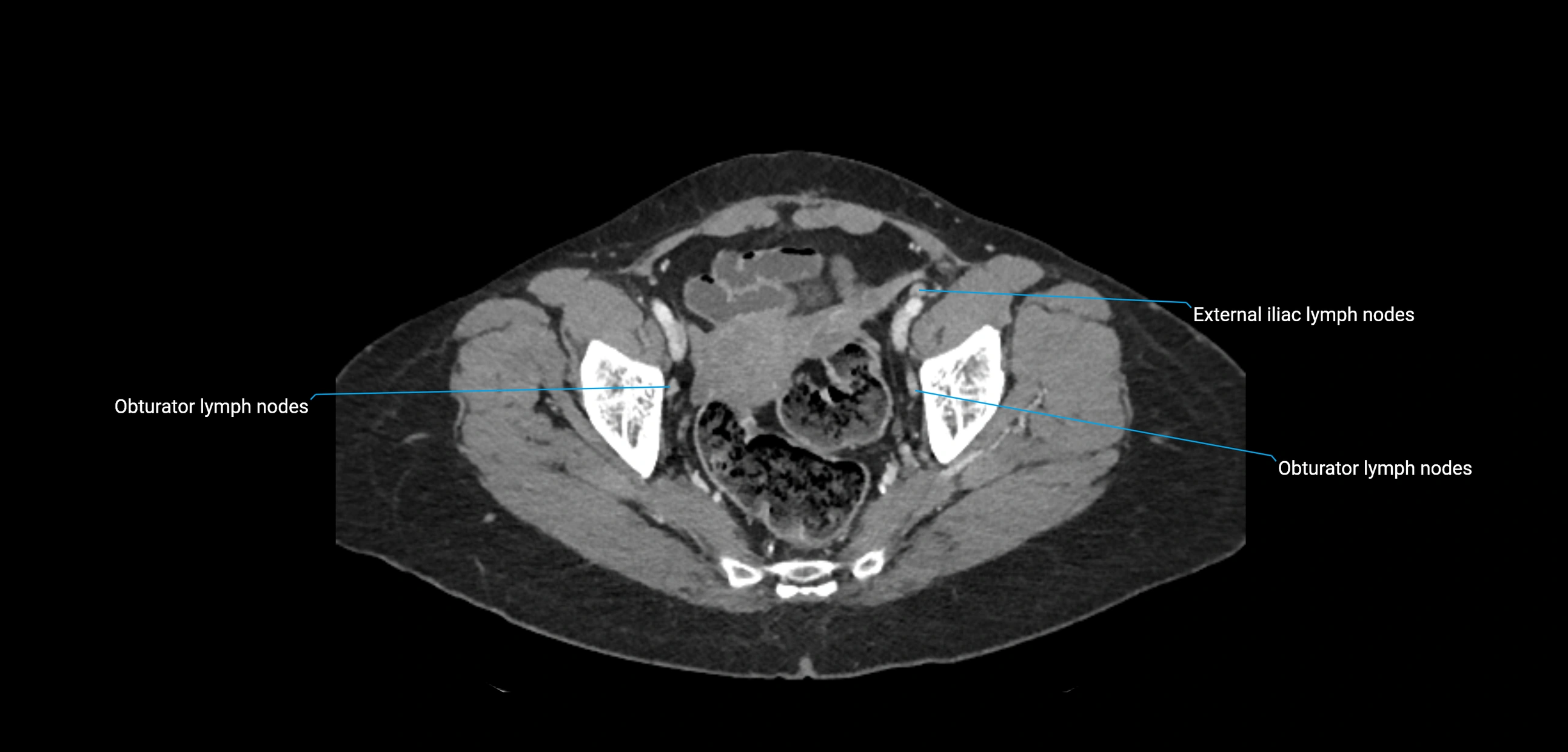
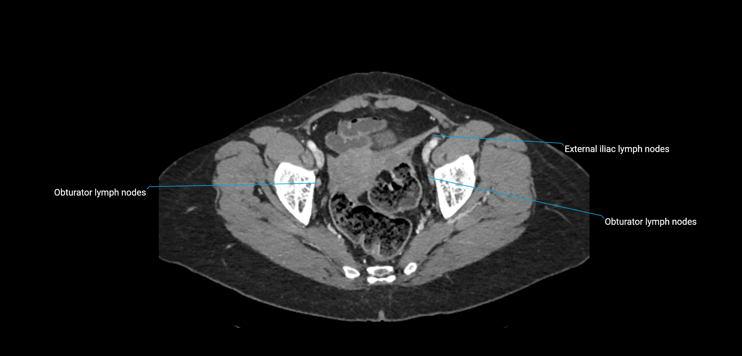
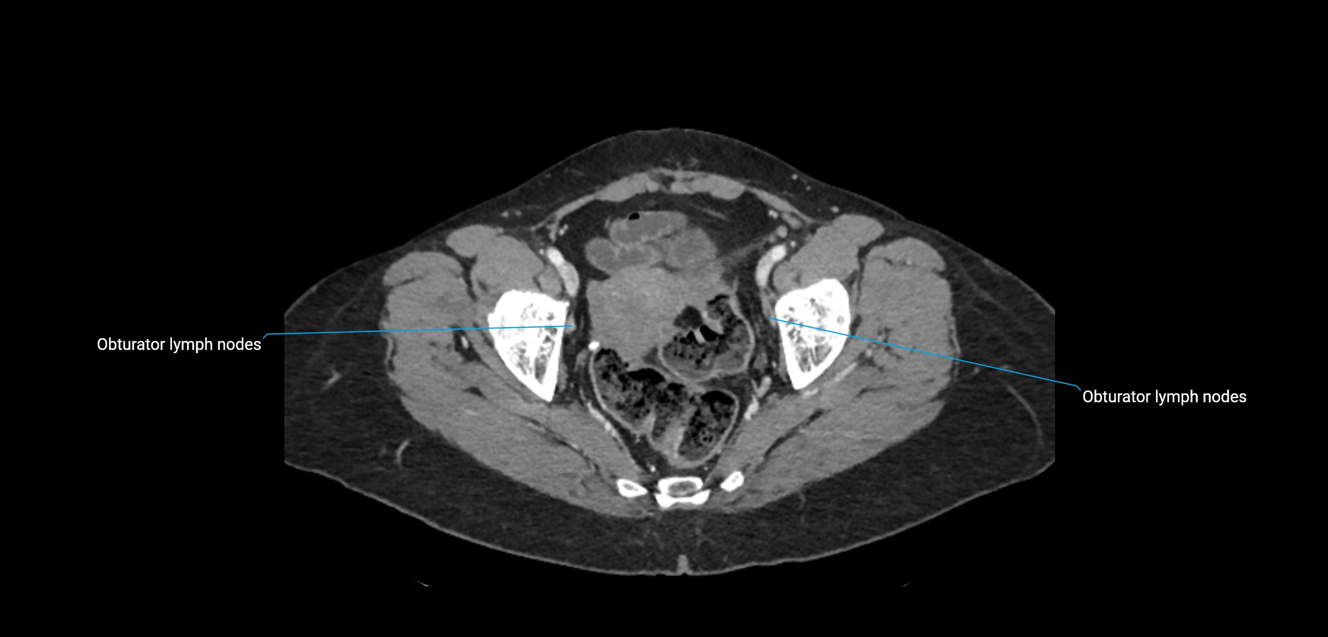
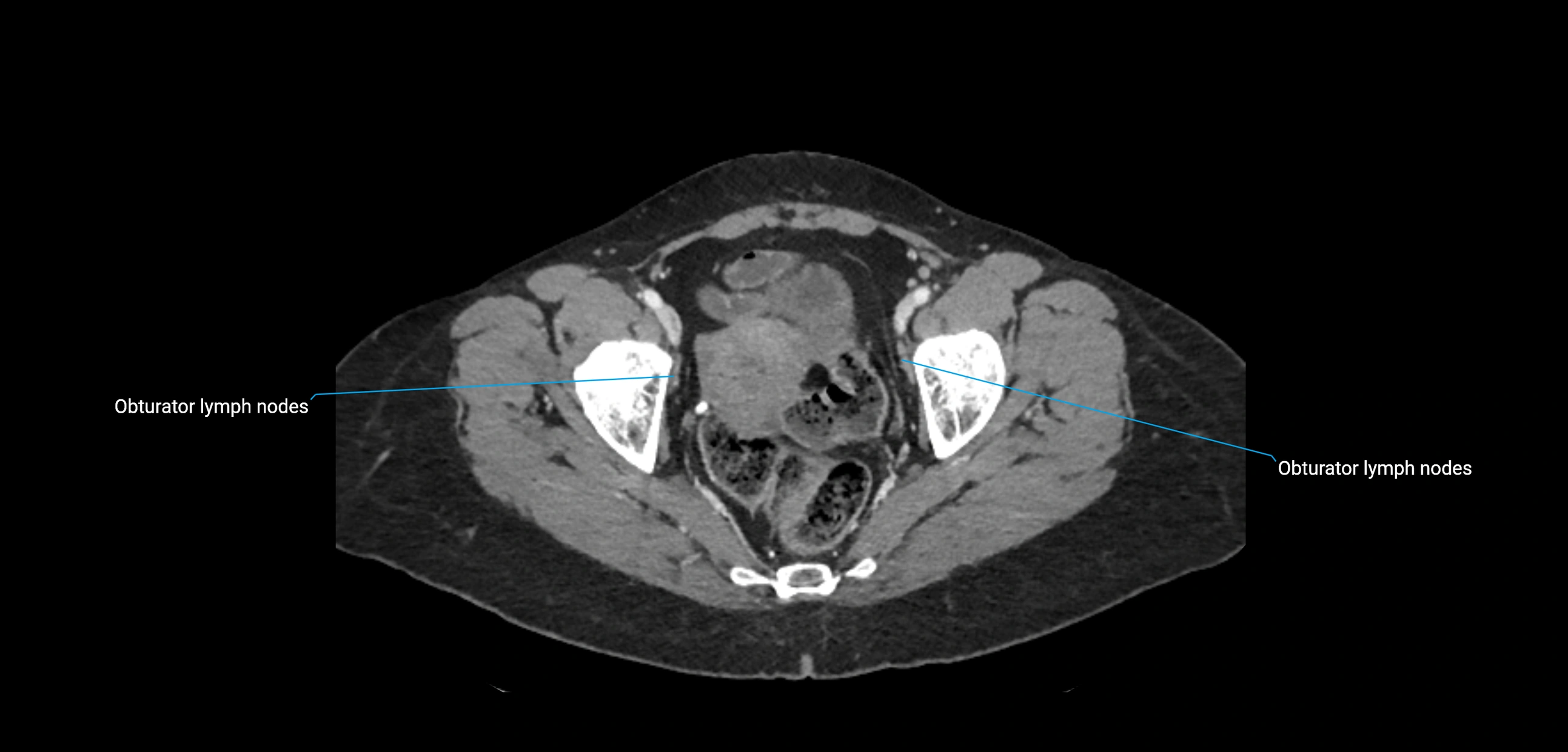
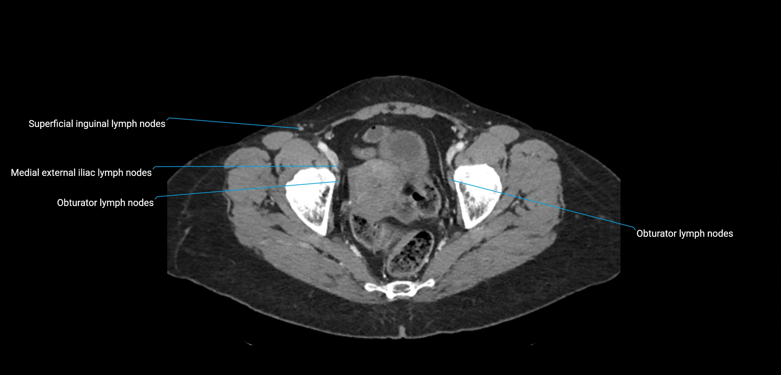
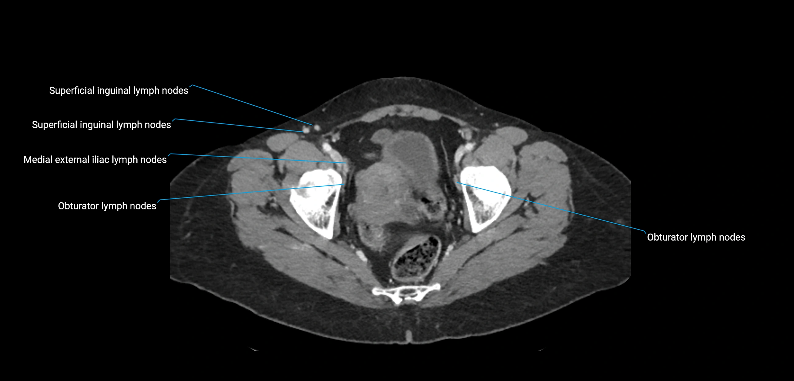
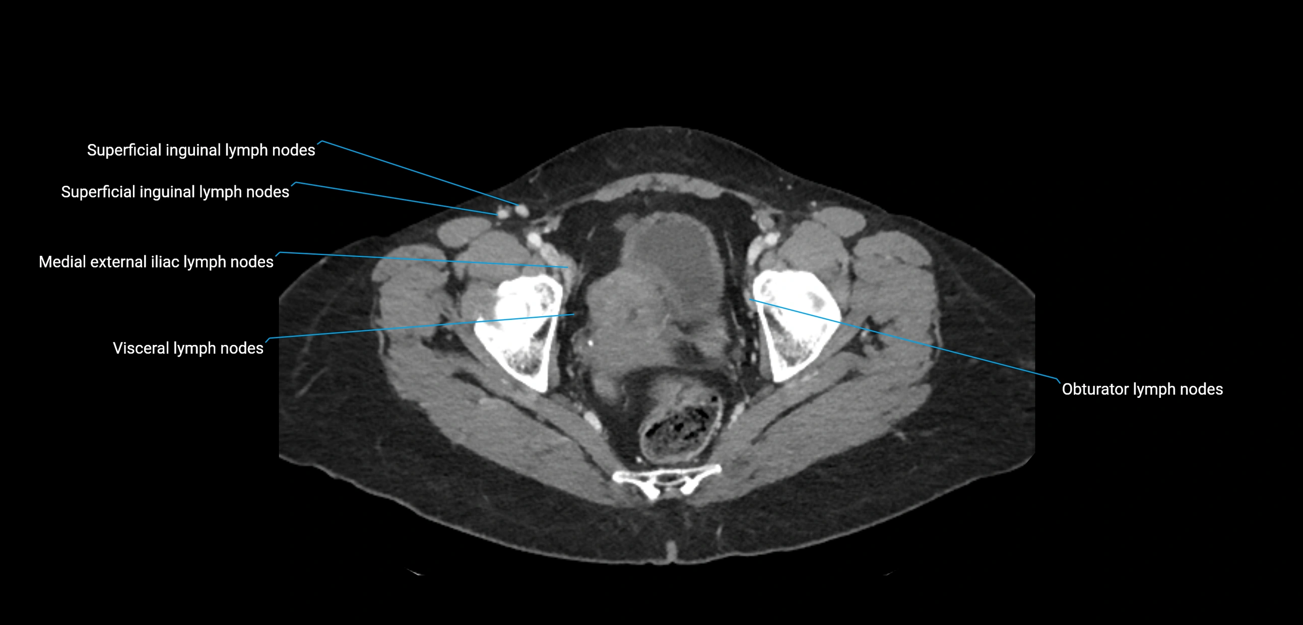
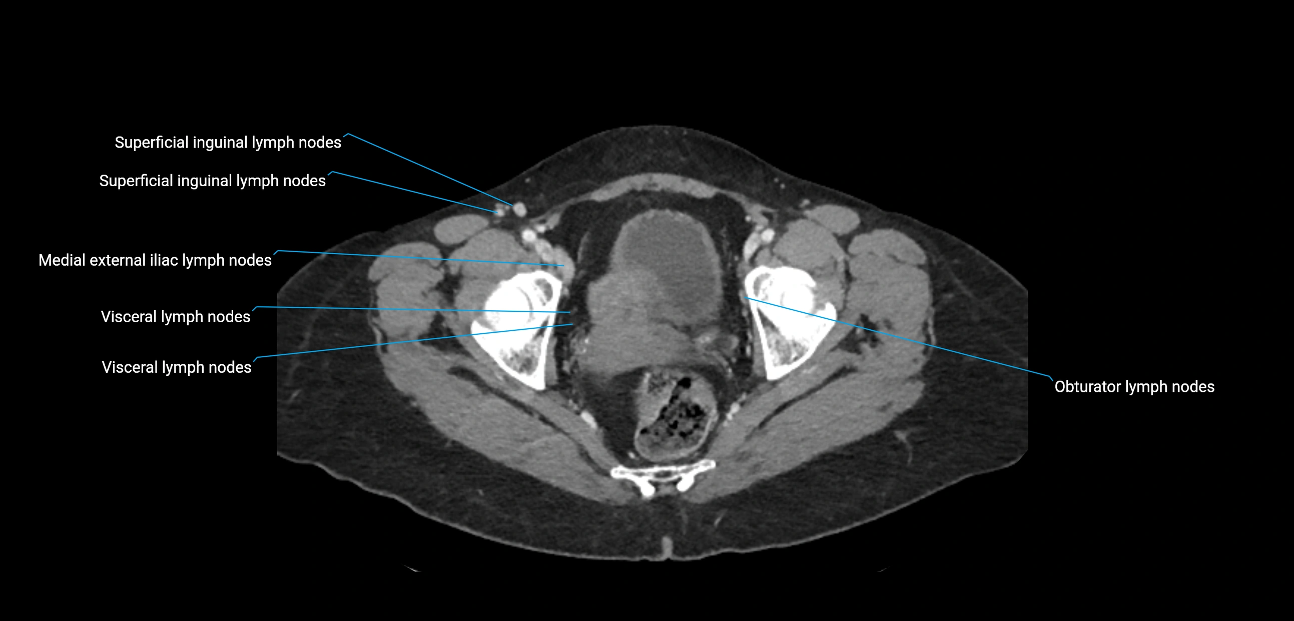
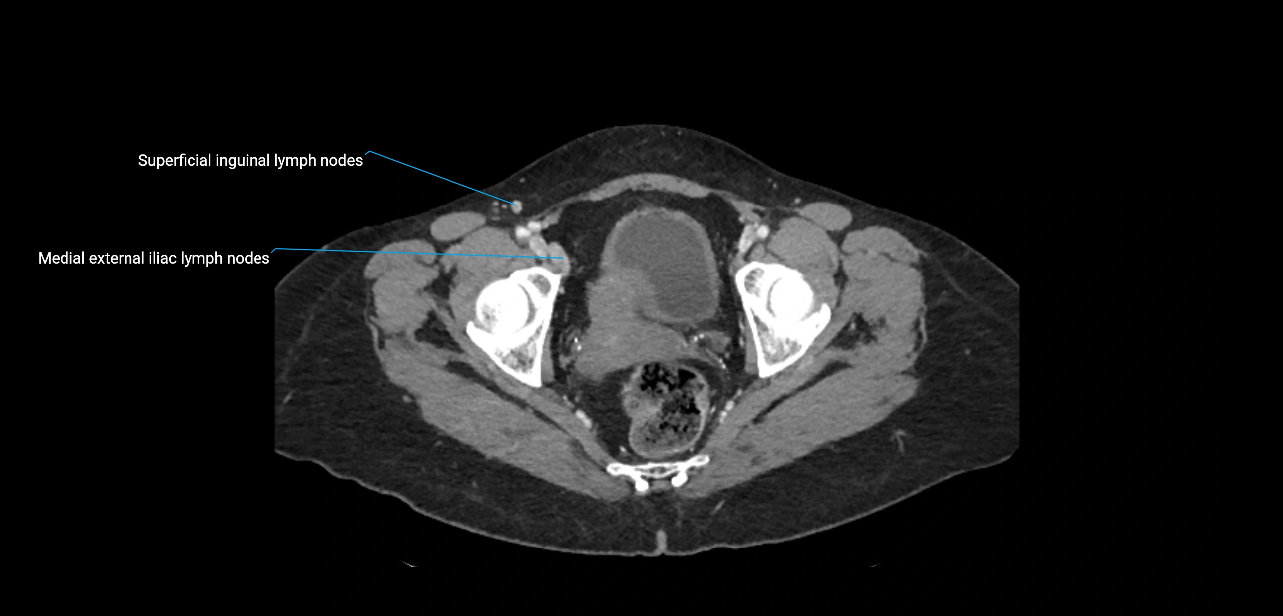
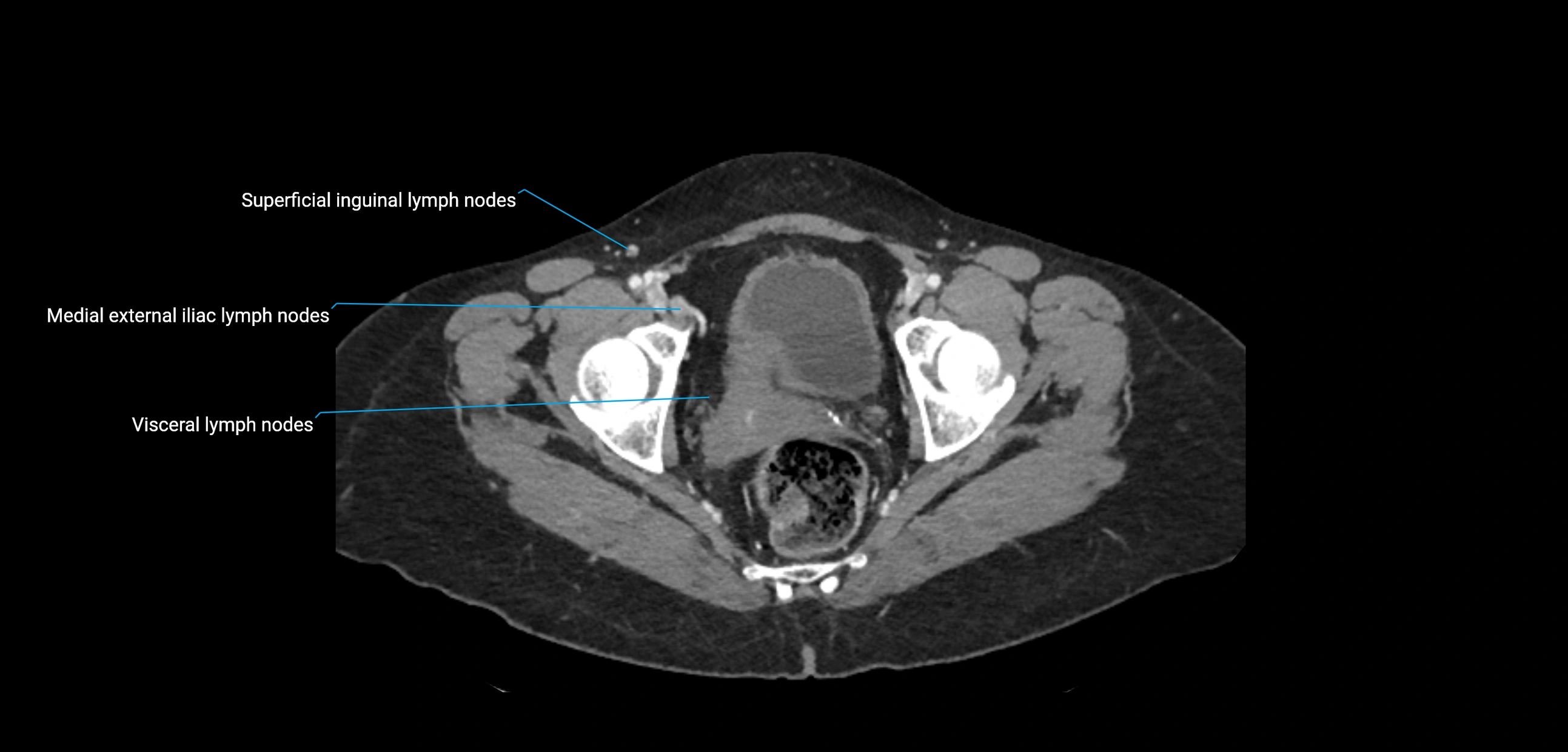
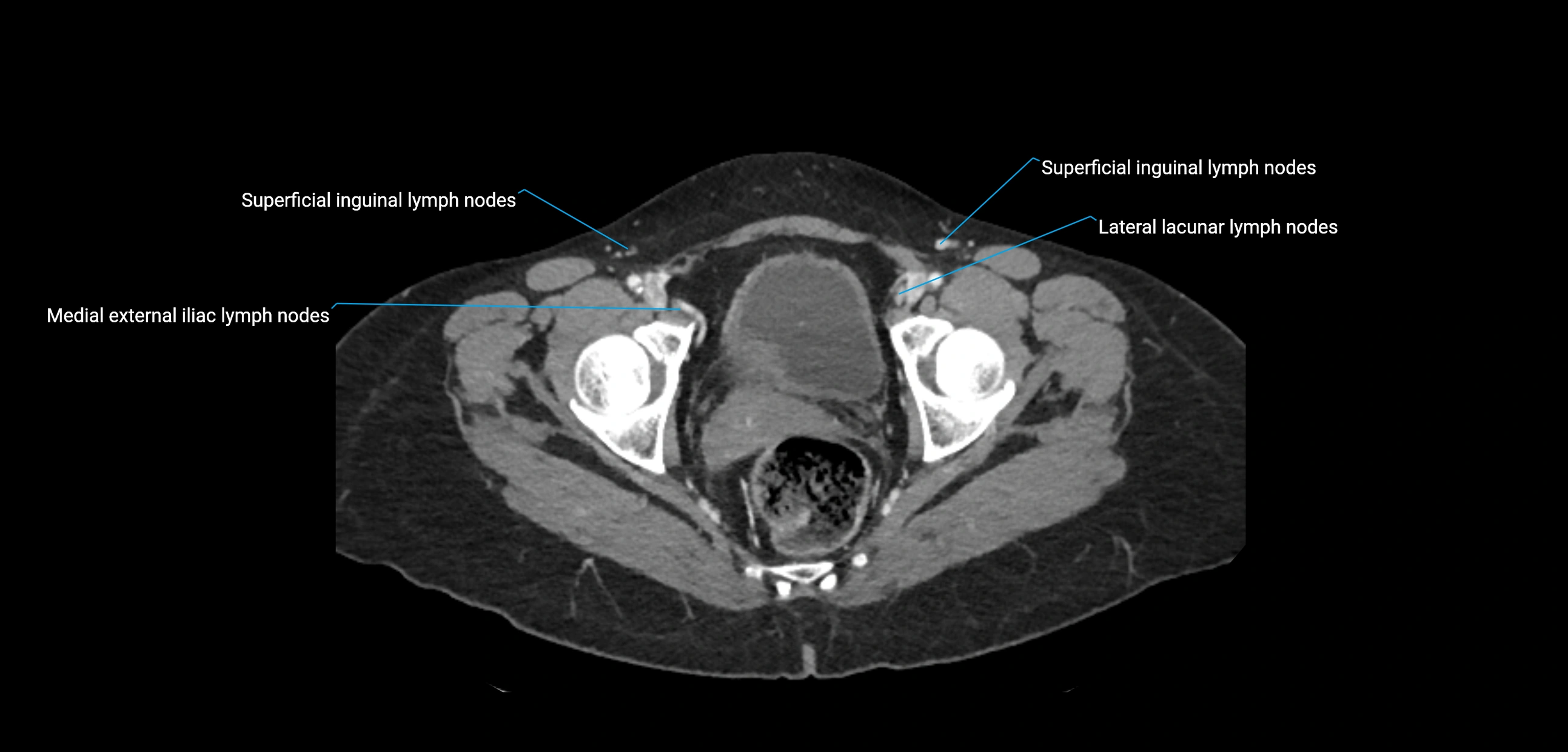
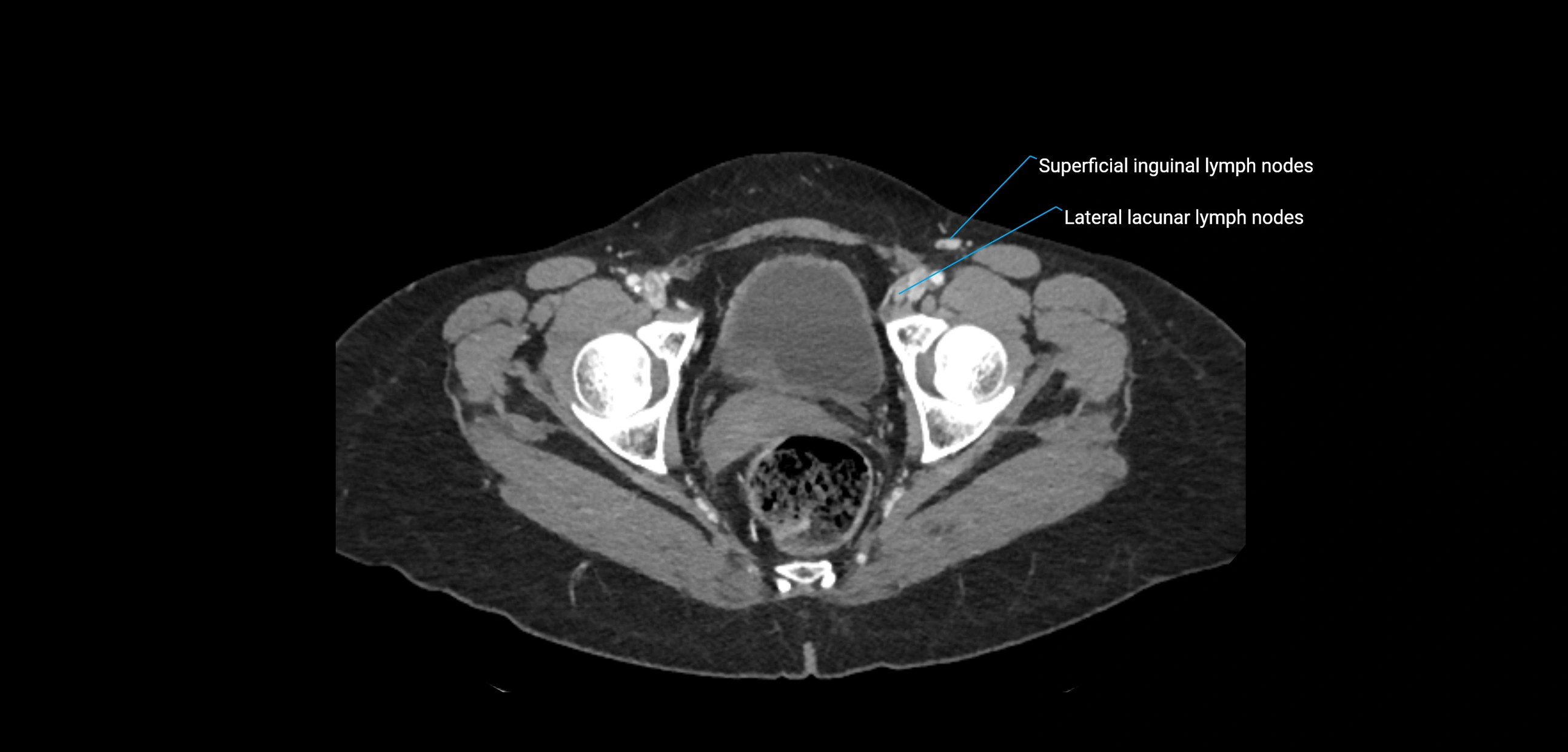
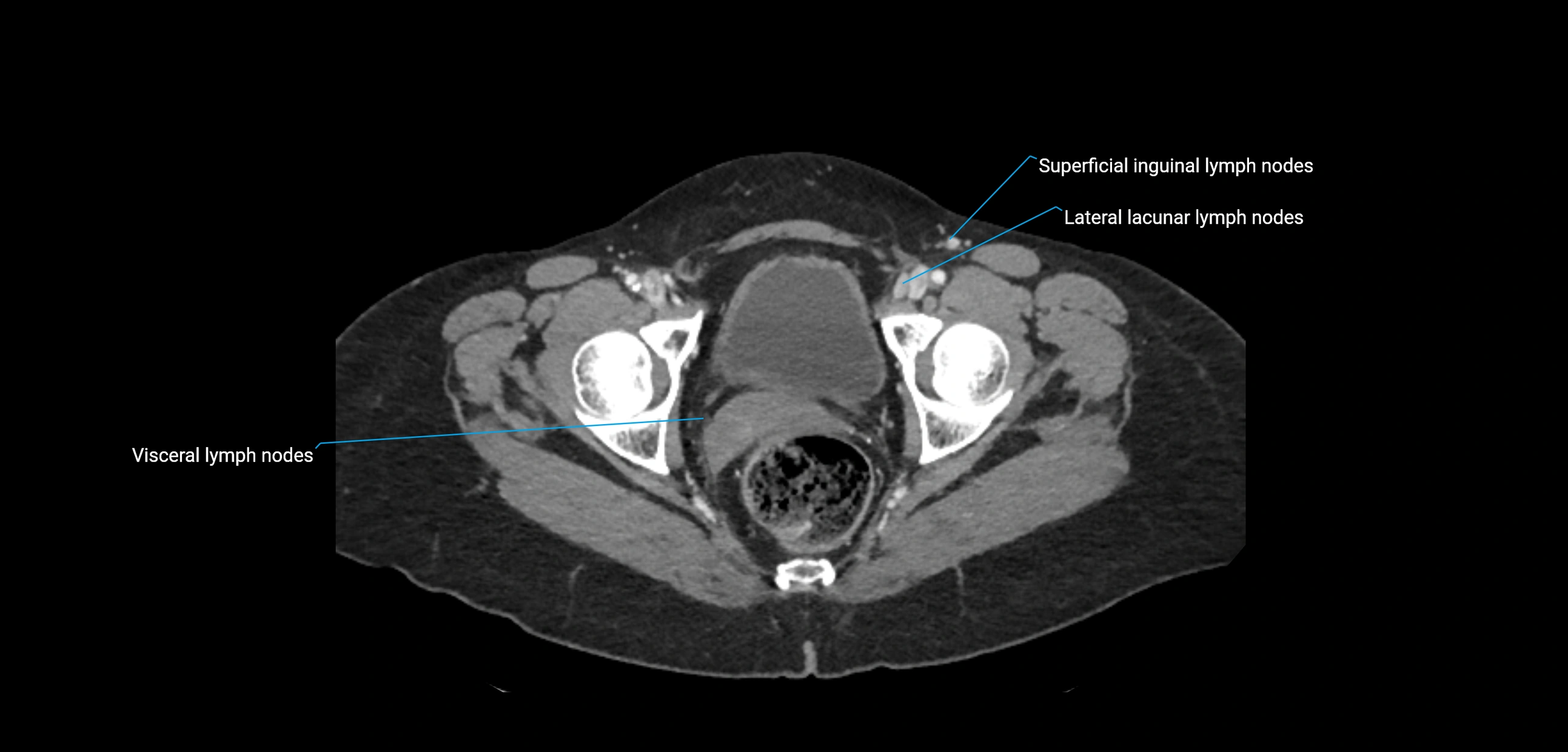
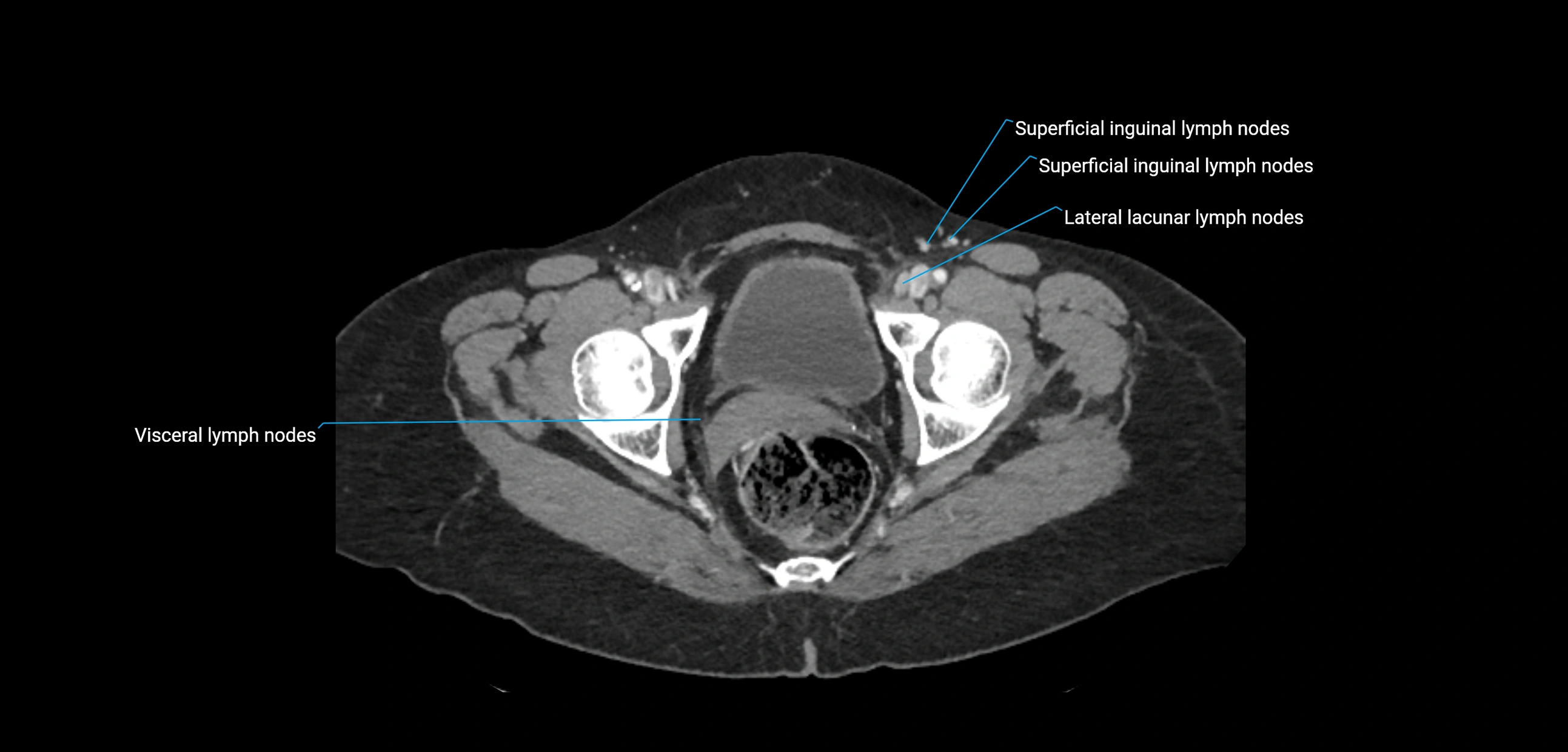
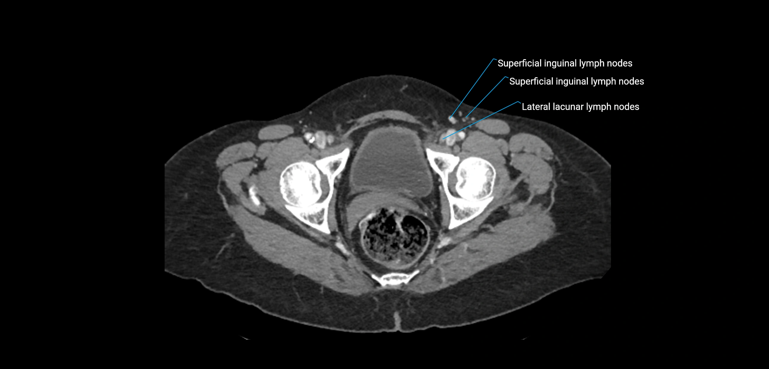
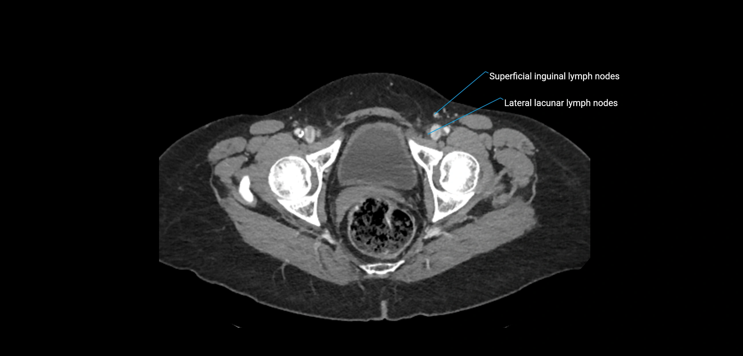
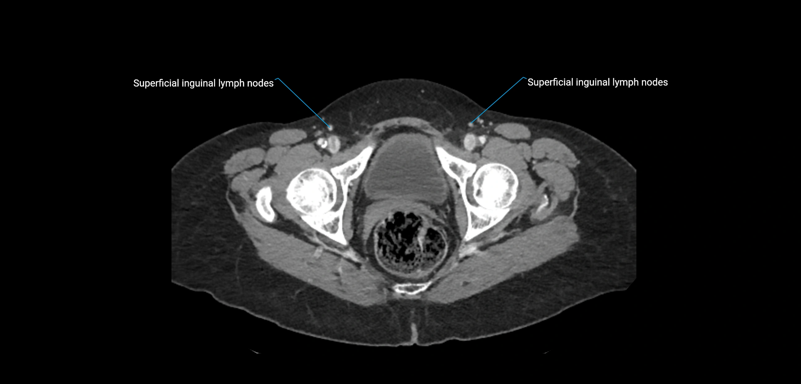
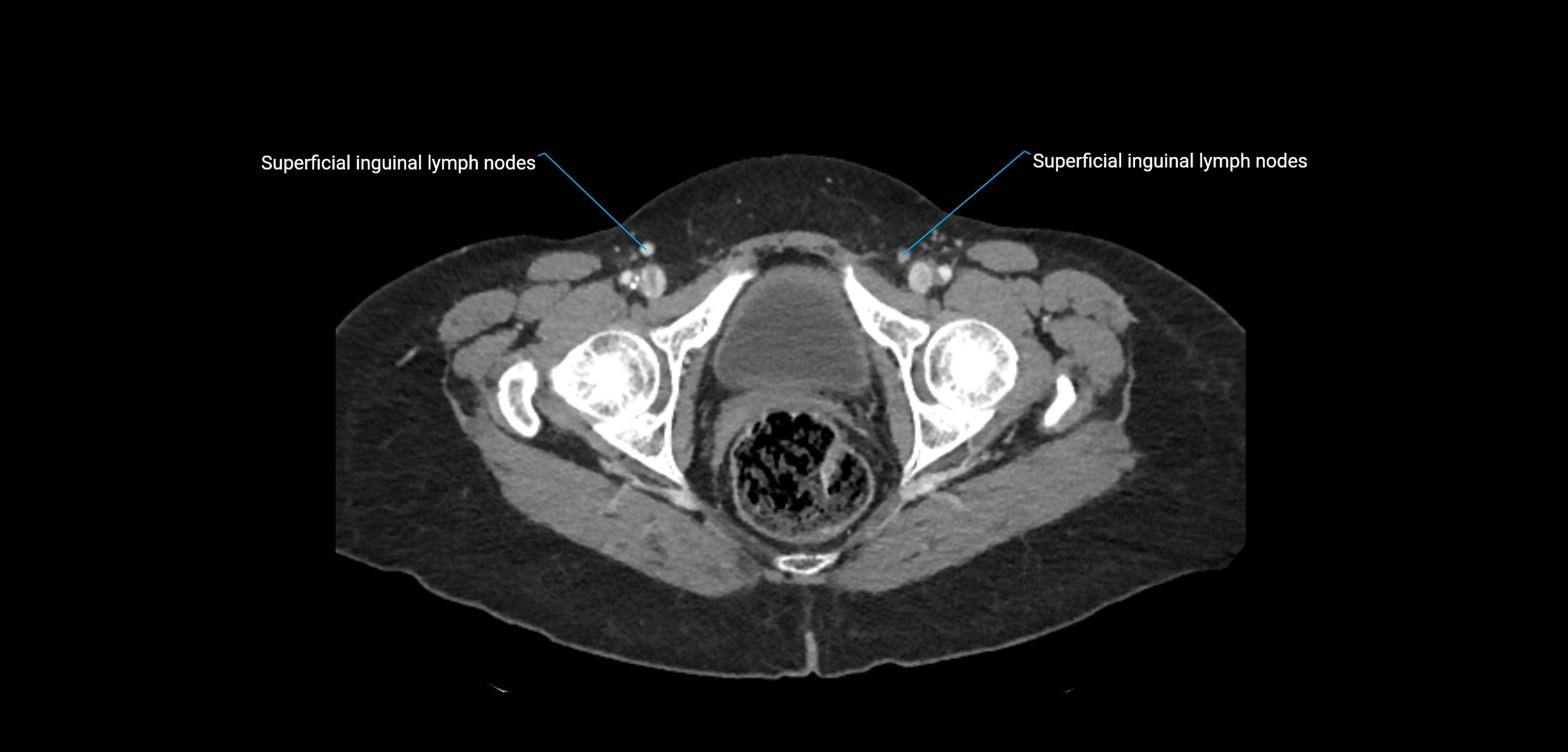
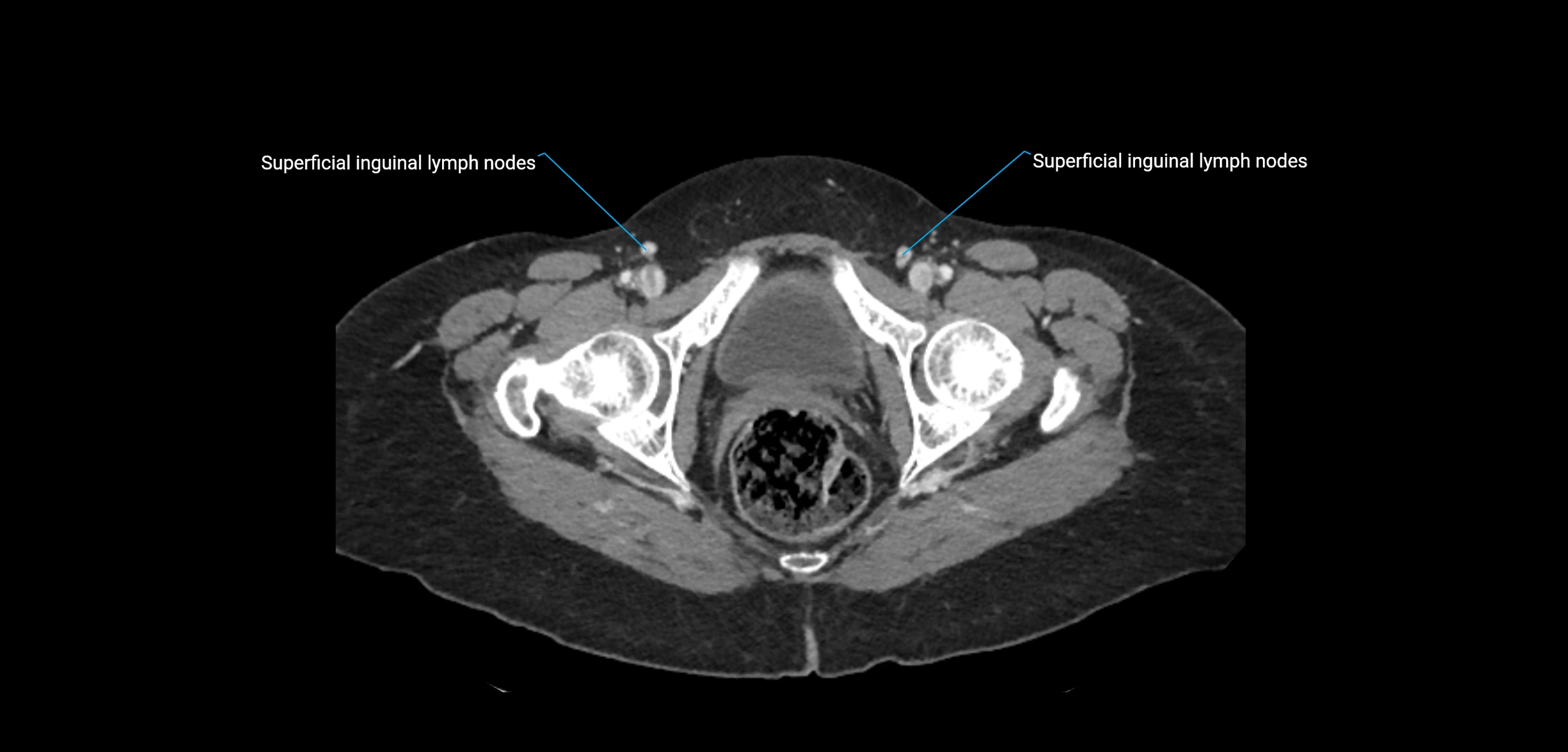
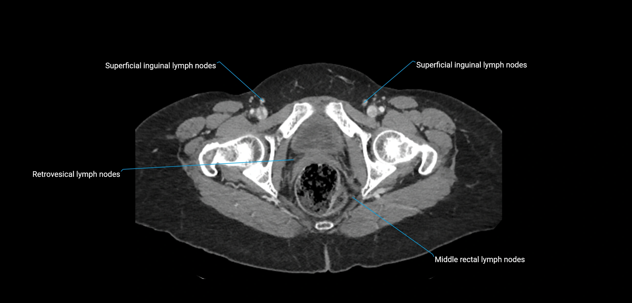
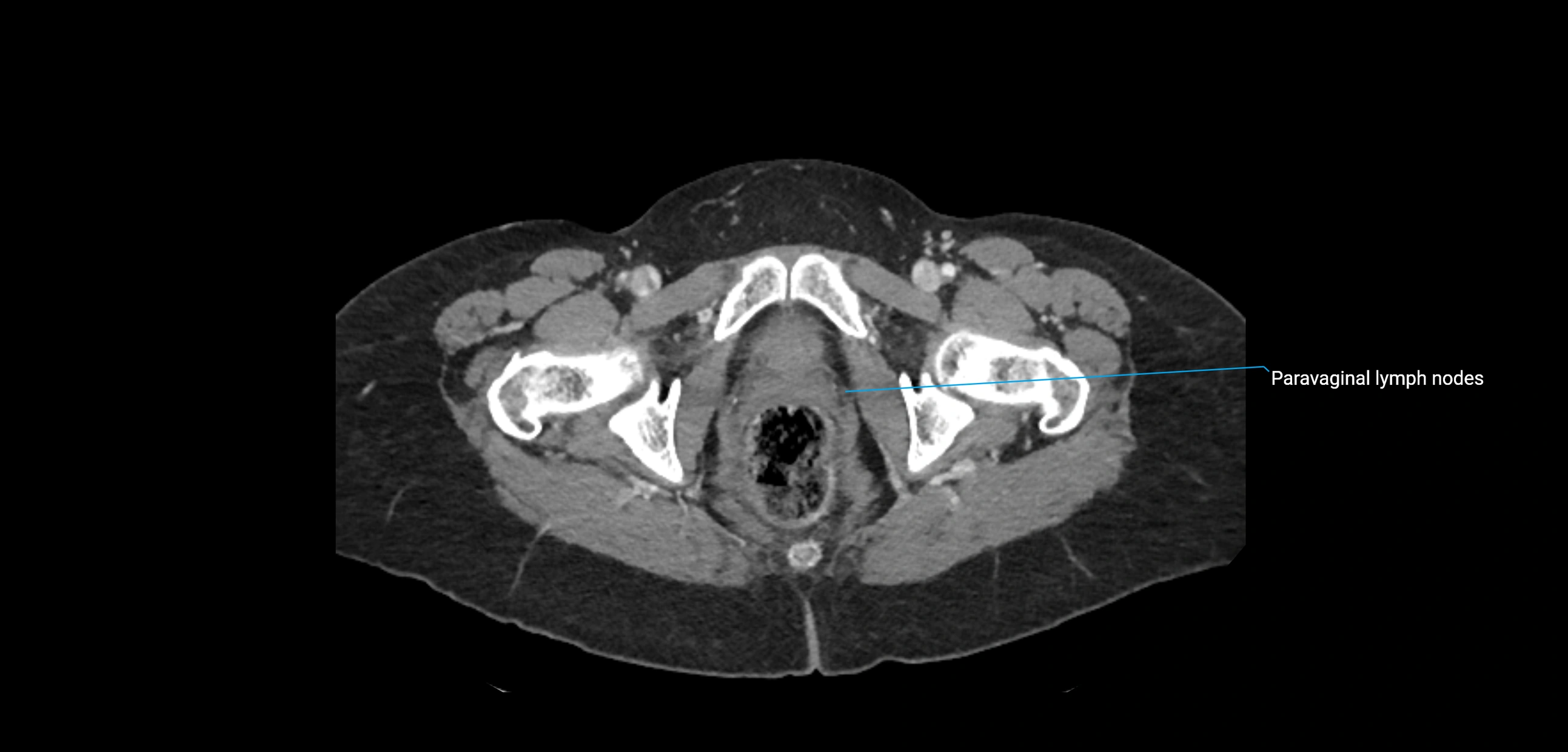
CT image
