


Topic
The annular epiphysis (vertebral ring apophysis) is a secondary ossification center forming a thin bony ring at the superior and inferior margins of the vertebral body endplates. It plays a key role in vertebral growth, endplate stability, and attachment of the outer fibers of the intervertebral disc.
It is an important structure in pediatric and adolescent spine anatomy and a common site involved in traction-related changes and ring apophyseal variants seen on imaging.
Synonyms
-
Vertebral ring apophysis
-
Ring epiphysis of the vertebral body
Location
-
Situated at the peripheral margins of the vertebral body endplates
-
Forms a circumferential ring along the superior and inferior endplates
-
Between the vertebral body and the outer annulus fibrosus
-
Present at each vertebral level from cervical to lumbar spine
-
Deep to the anterior and posterior longitudinal ligaments
Anatomical components
-
Thin circumferential bony rim
-
Superior annular epiphysis
-
Inferior annular epiphysis
-
Attachment site for:
-
Outer fibers of the annulus fibrosus
-
Sharpey-type collagen fibers
-
Relations
Superiorly:
-
Intervertebral disc endplate
Inferiorly:
-
Vertebral body cancellous bone
Anteriorly:
-
Anterior longitudinal ligament
-
Anterior annulus fibrosus
Posteriorly:
-
Posterior annulus fibrosus
-
Posterior longitudinal ligament (near midline)
Laterally:
-
Peripheral annulus fibrosus
Developmental anatomy
-
Not present at birth
-
Appears between 5–10 years of age as a secondary ossification center
-
Progressive ossification during adolescence
-
Fusion with vertebral body: Usually between 18–25 years
-
May remain partially visible in young adults as a normal variant
X-ray appearance
Spine radiographs (AP / lateral views):
-
Annular epiphysis: Thin, linear bony rim along vertebral endplate margins
-
Appearance: Smooth, well-corticated ring
-
Age dependence: More conspicuous in children and adolescents
-
Fusion: Disappears after complete fusion with vertebral body
CT appearance
Pre-contrast CT:
-
Annular epiphysis: Thin hyperdense bony ring at the vertebral endplate periphery
-
Margins: Well-defined cortical outline
-
Relationship: Clearly separated from central endplate in skeletally immature patients
-
After fusion: Appears continuous with the vertebral body cortex
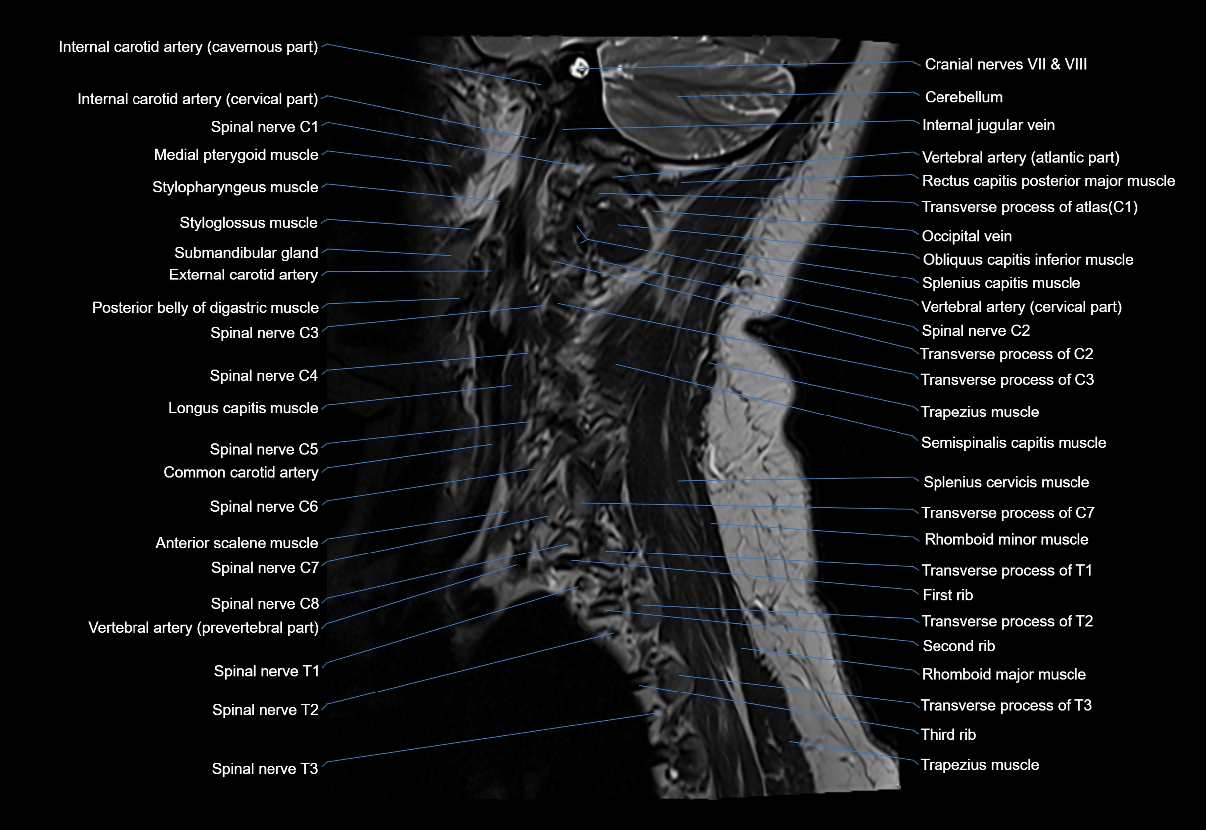
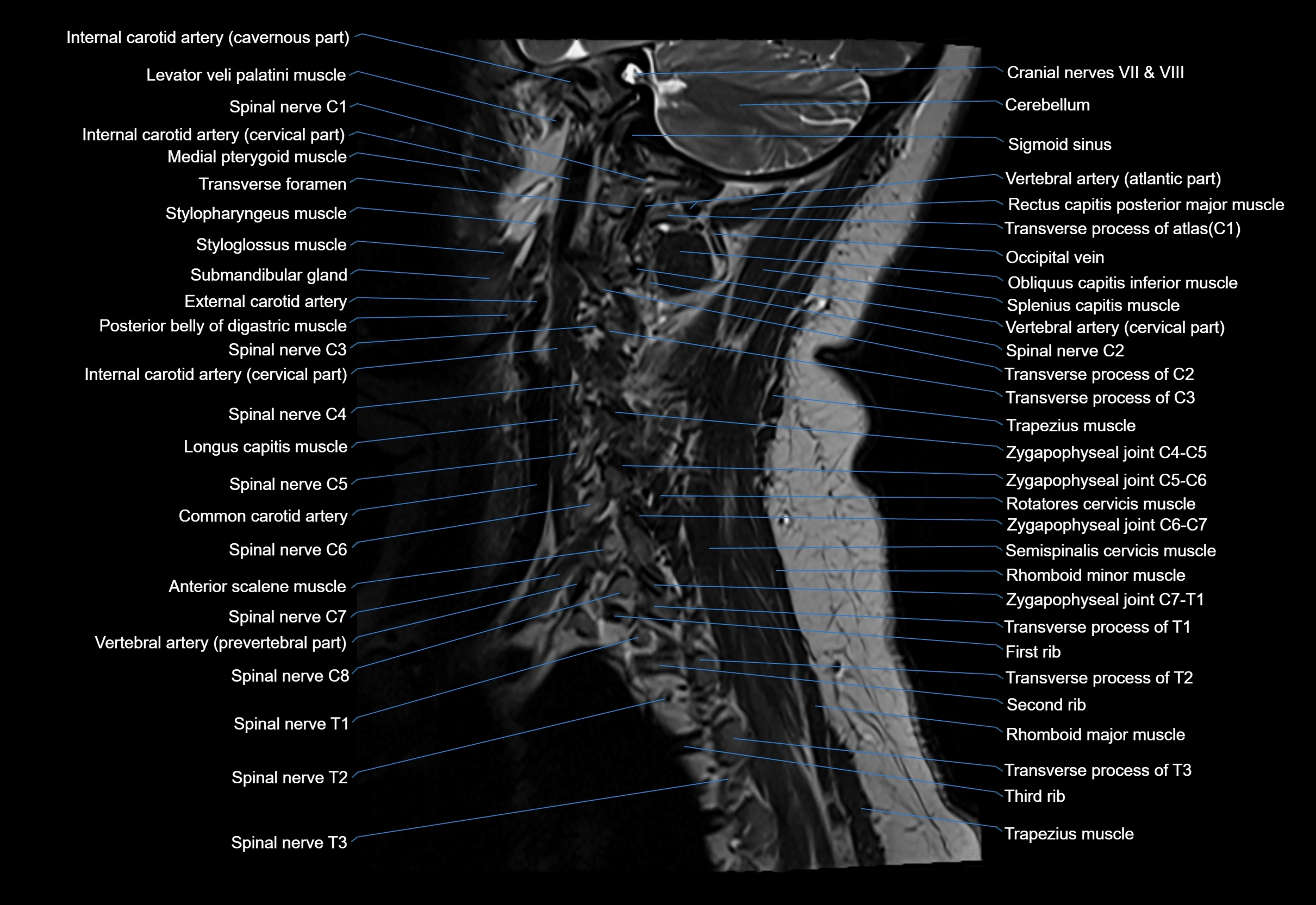
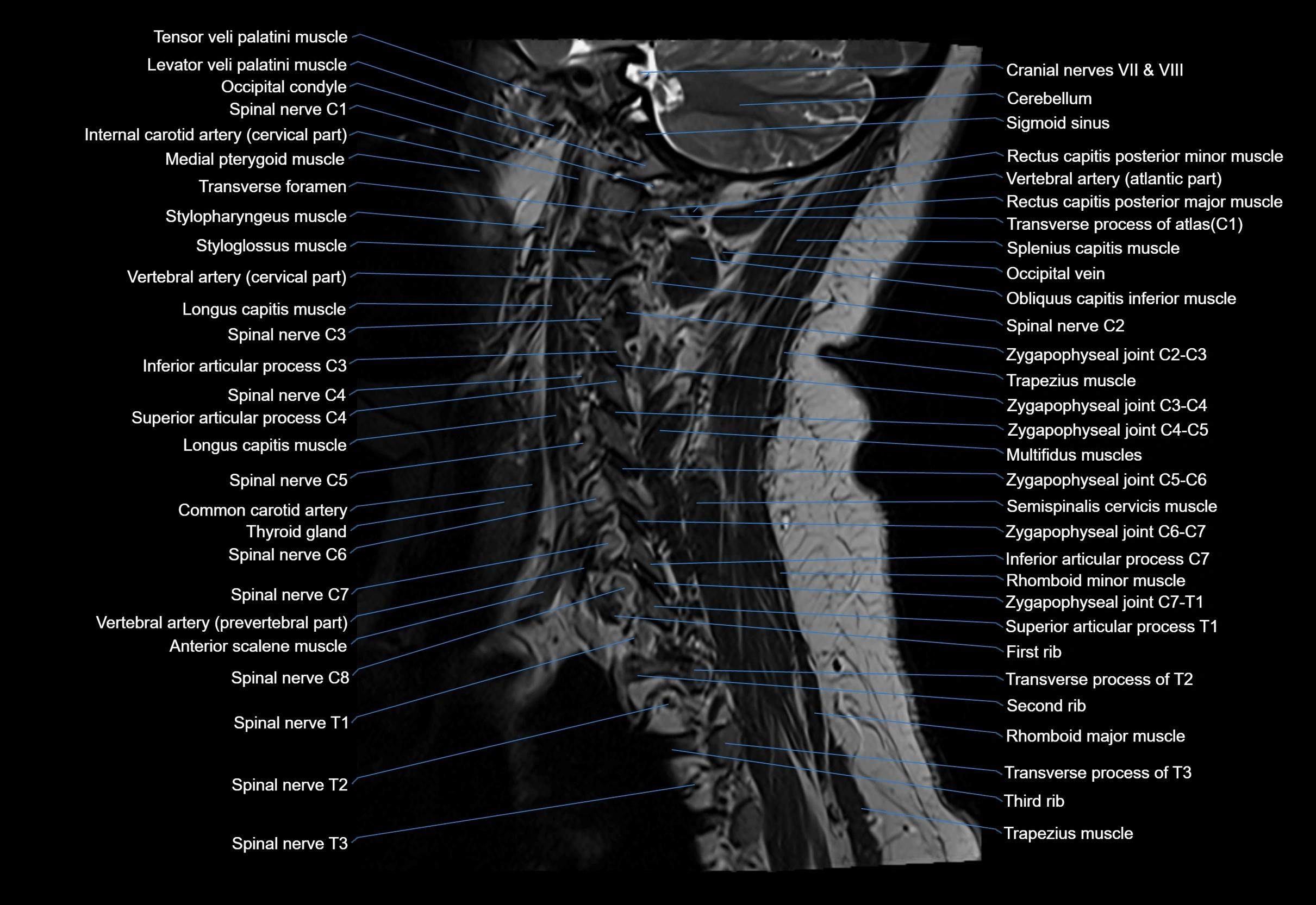
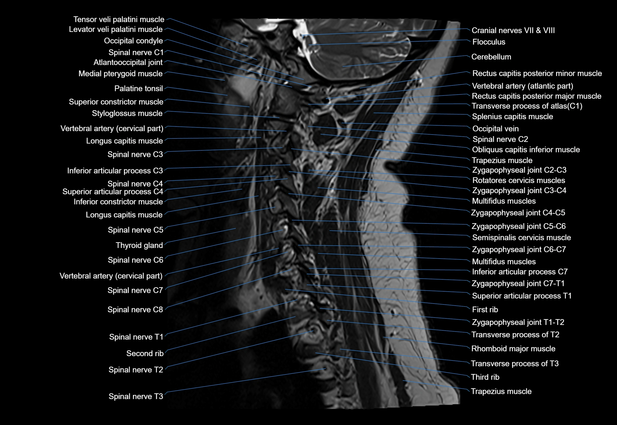
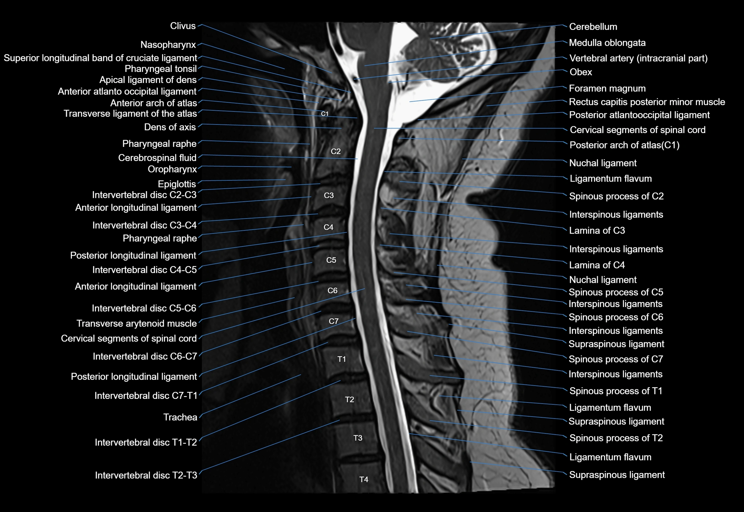
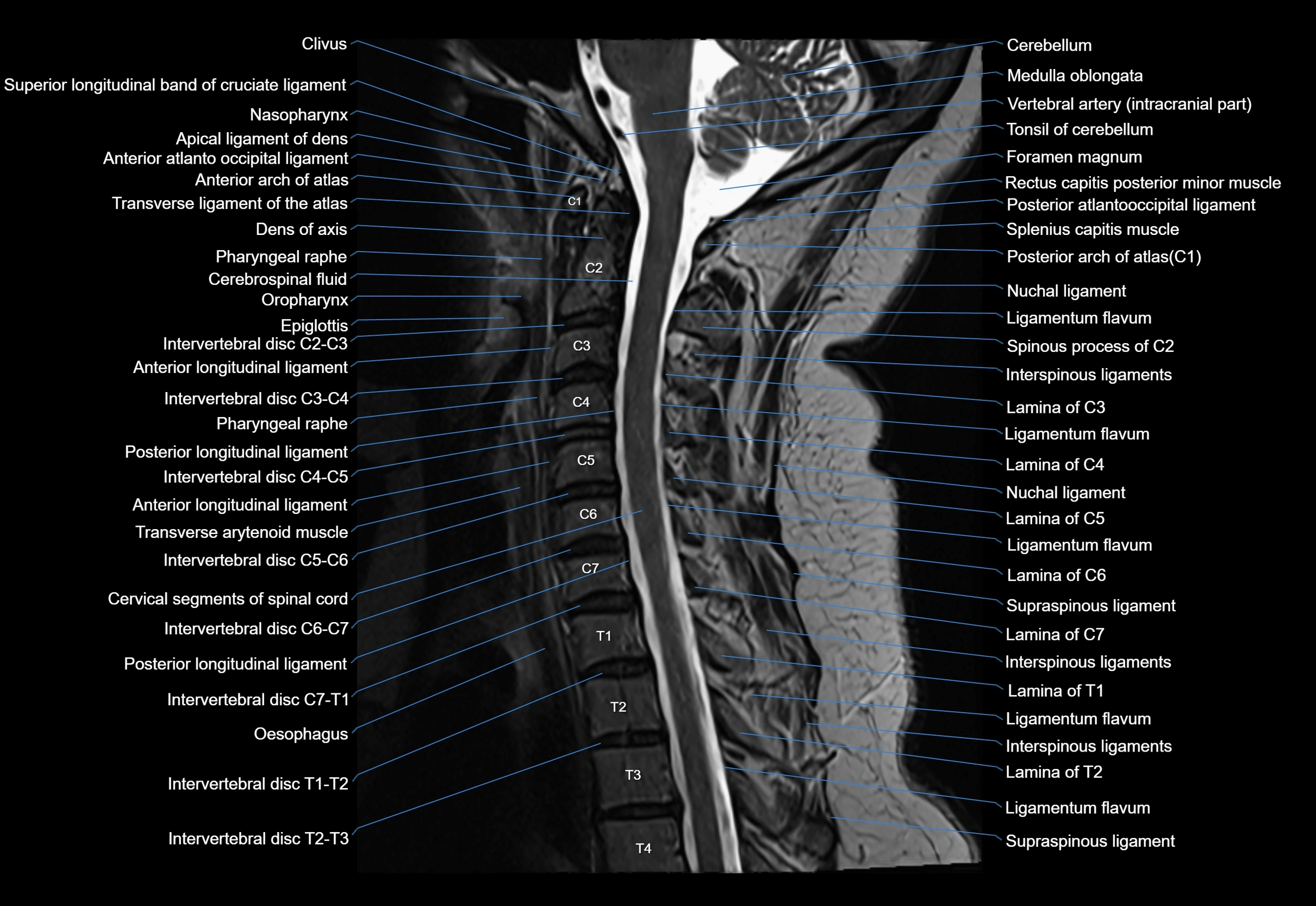
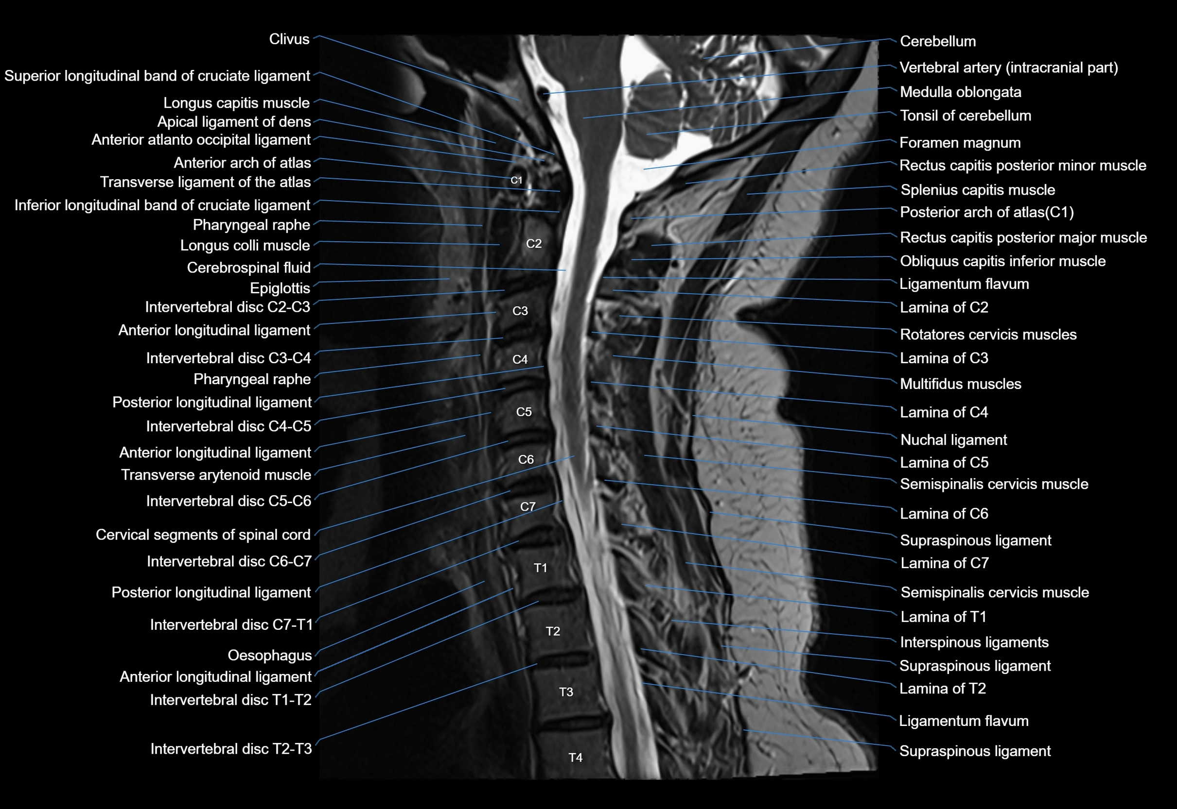
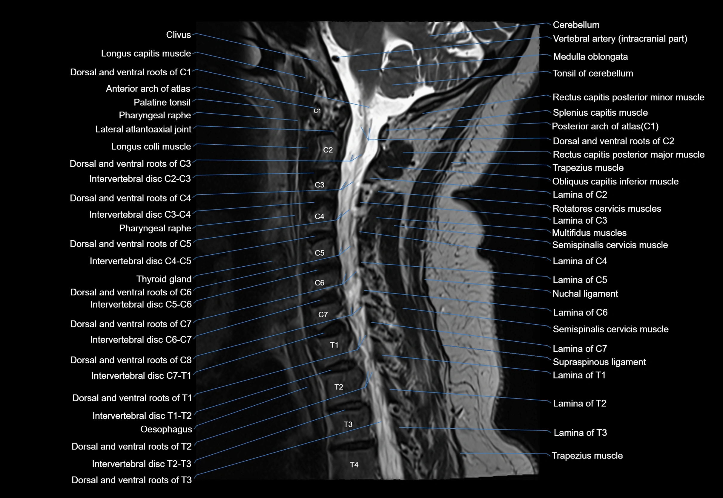
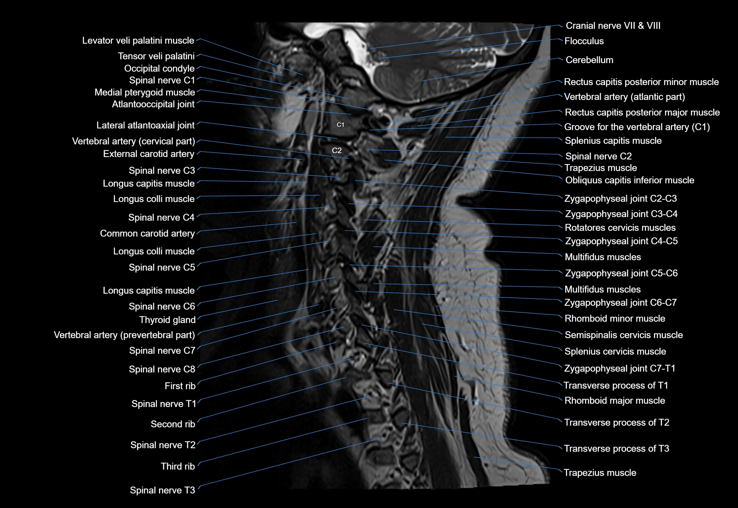
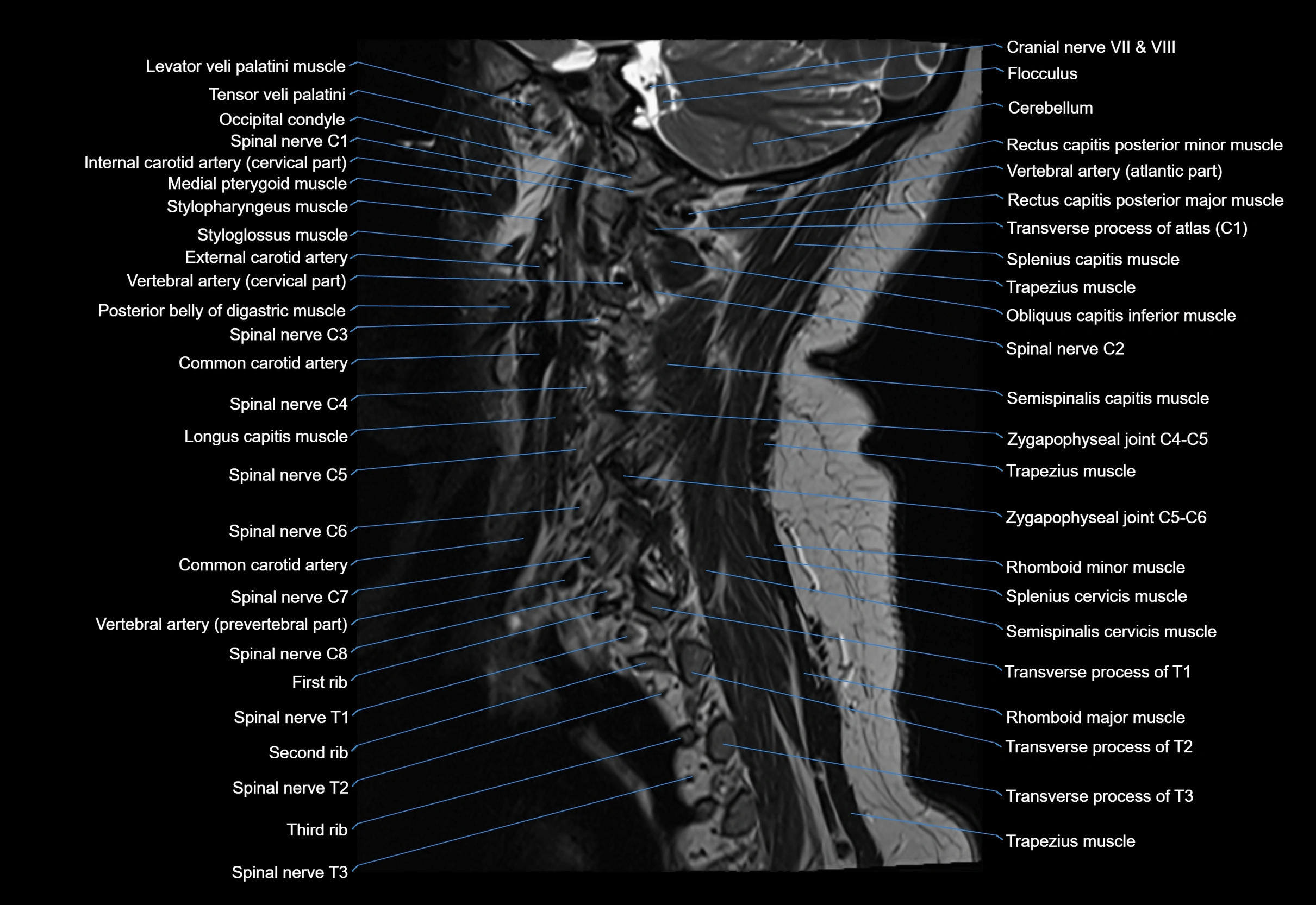
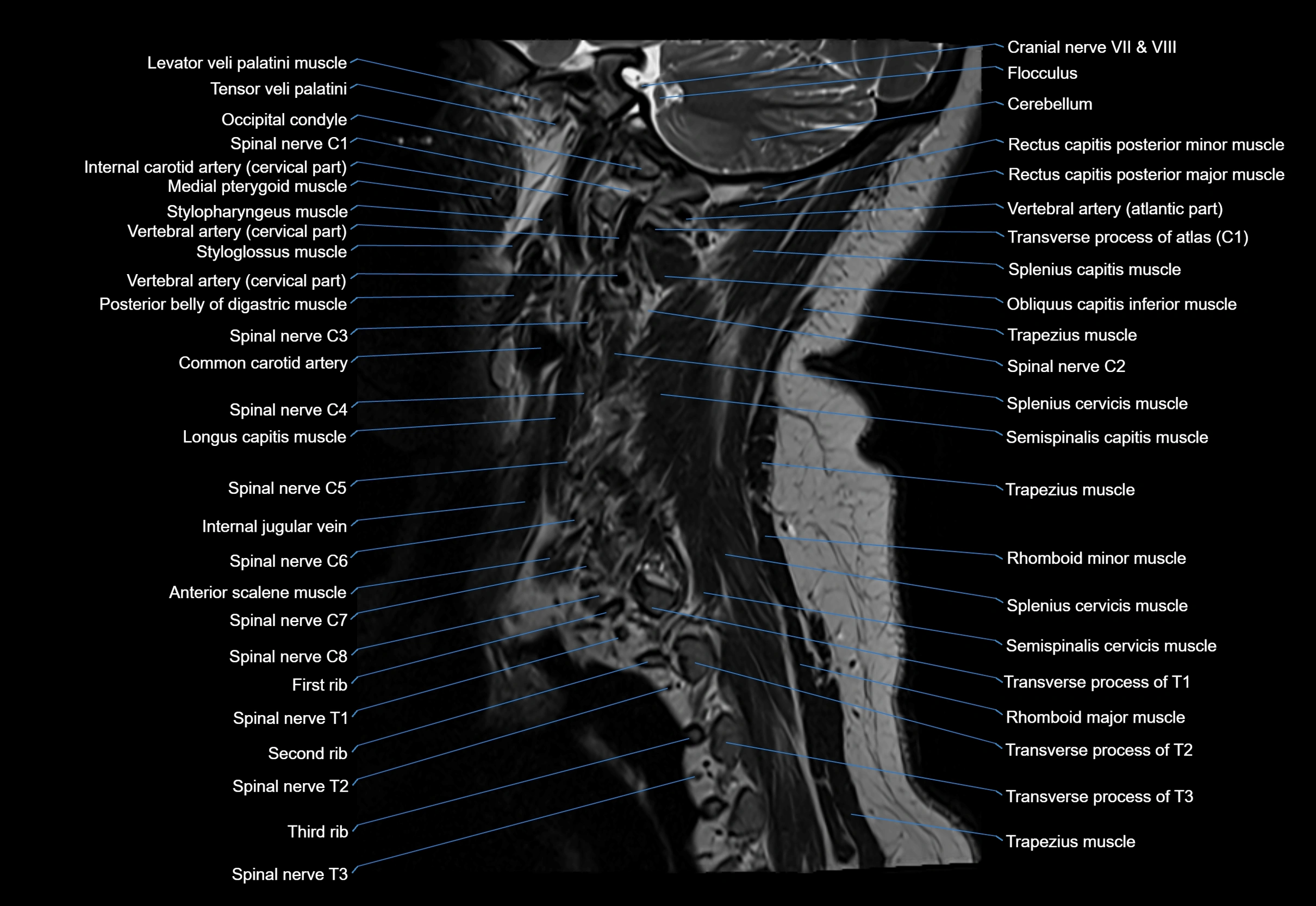
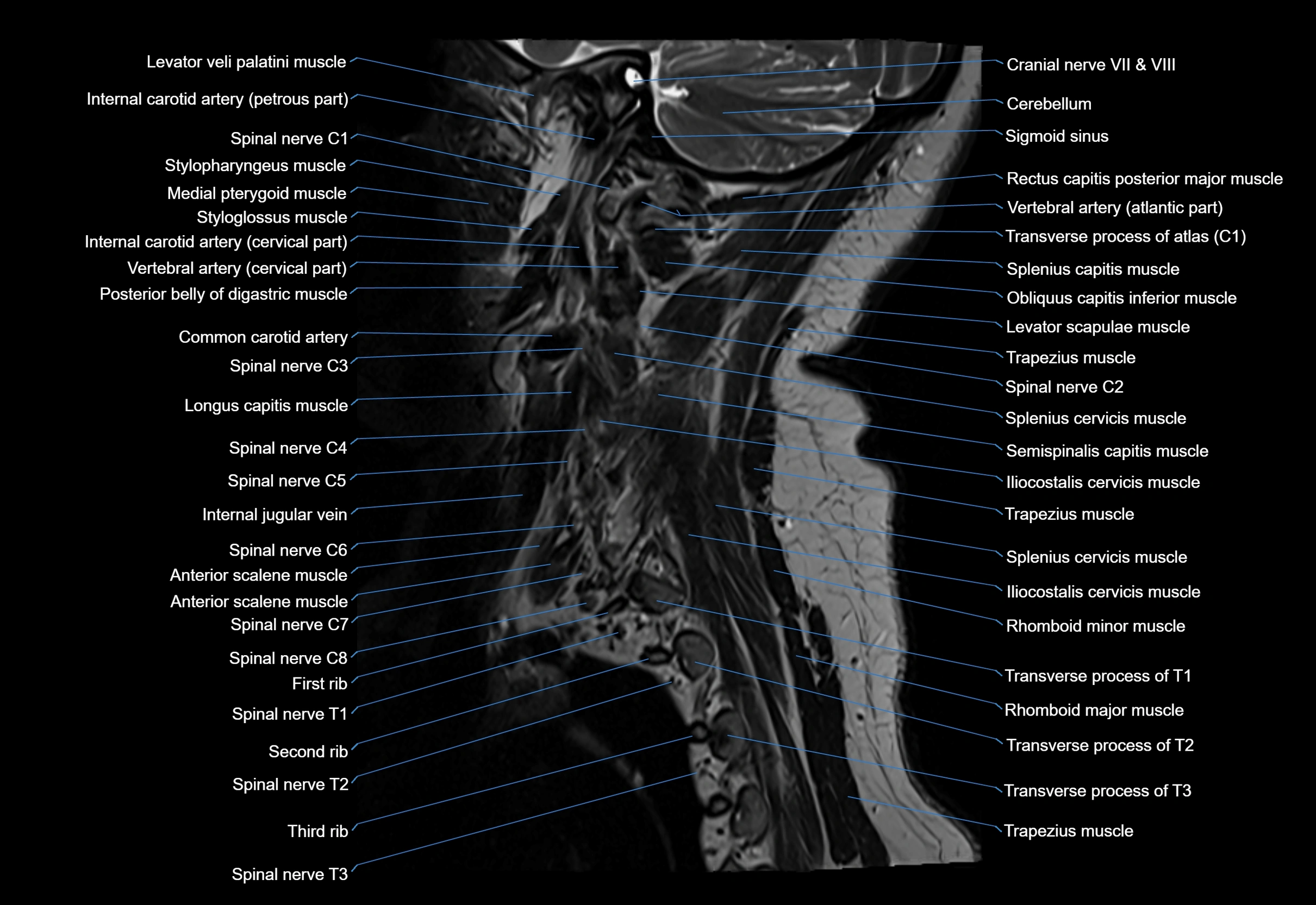
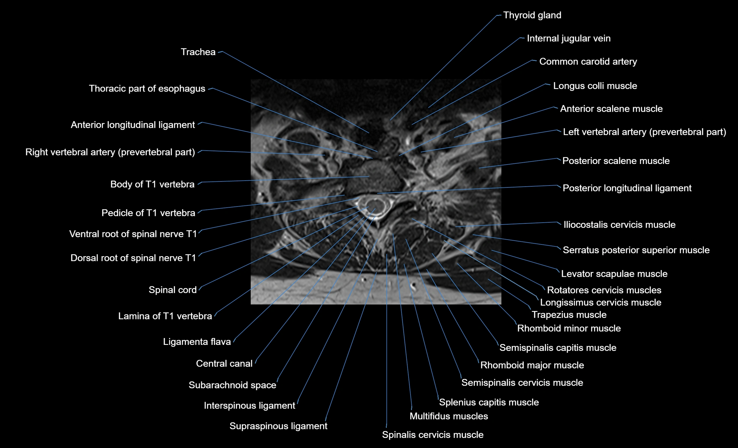
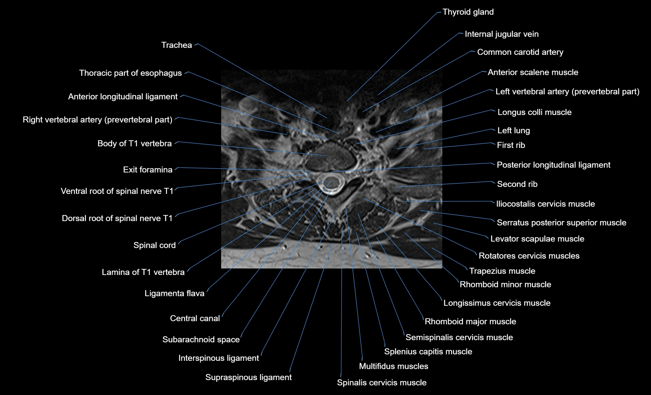
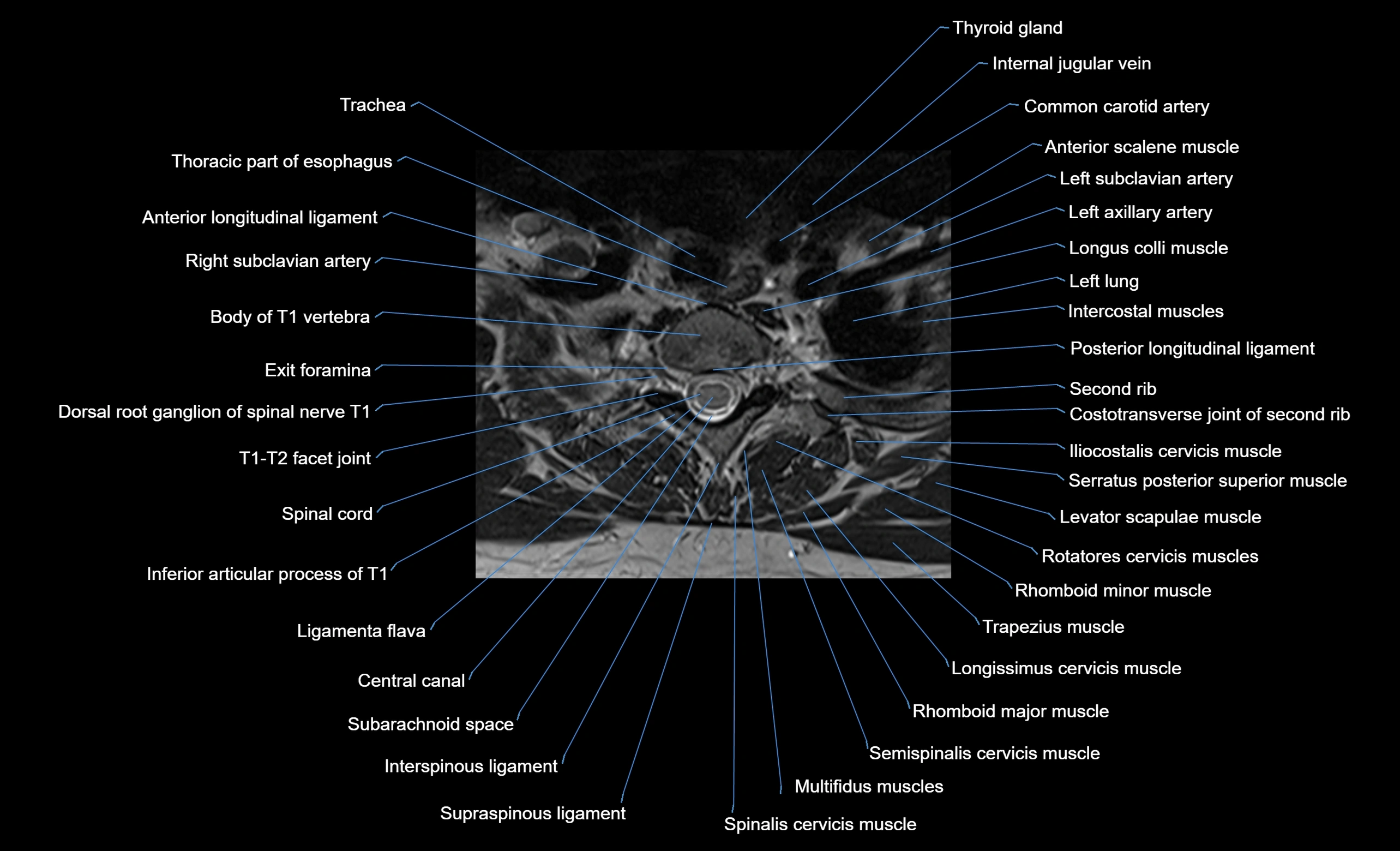
MRI appearance
T1-weighted images:
-
Annular epiphysis: Low signal cortical rim at the vertebral margin
-
Adjacent marrow: Intermediate-to-high signal in the vertebral body
-
Disc interface: Clear delineation between bone and annulus
T2-weighted images:
-
Annular epiphysis: Low signal intensity line
-
Endplate cartilage (in younger patients): Intermediate signal
-
Intervertebral disc: High signal nucleus pulposus
STIR:
-
Annular epiphysis: Low signal cortical rim
-
Adjacent marrow: Suppressed fat signal with preserved bony outline
-
Utility: Highlights marrow and endplate interface in developing spine
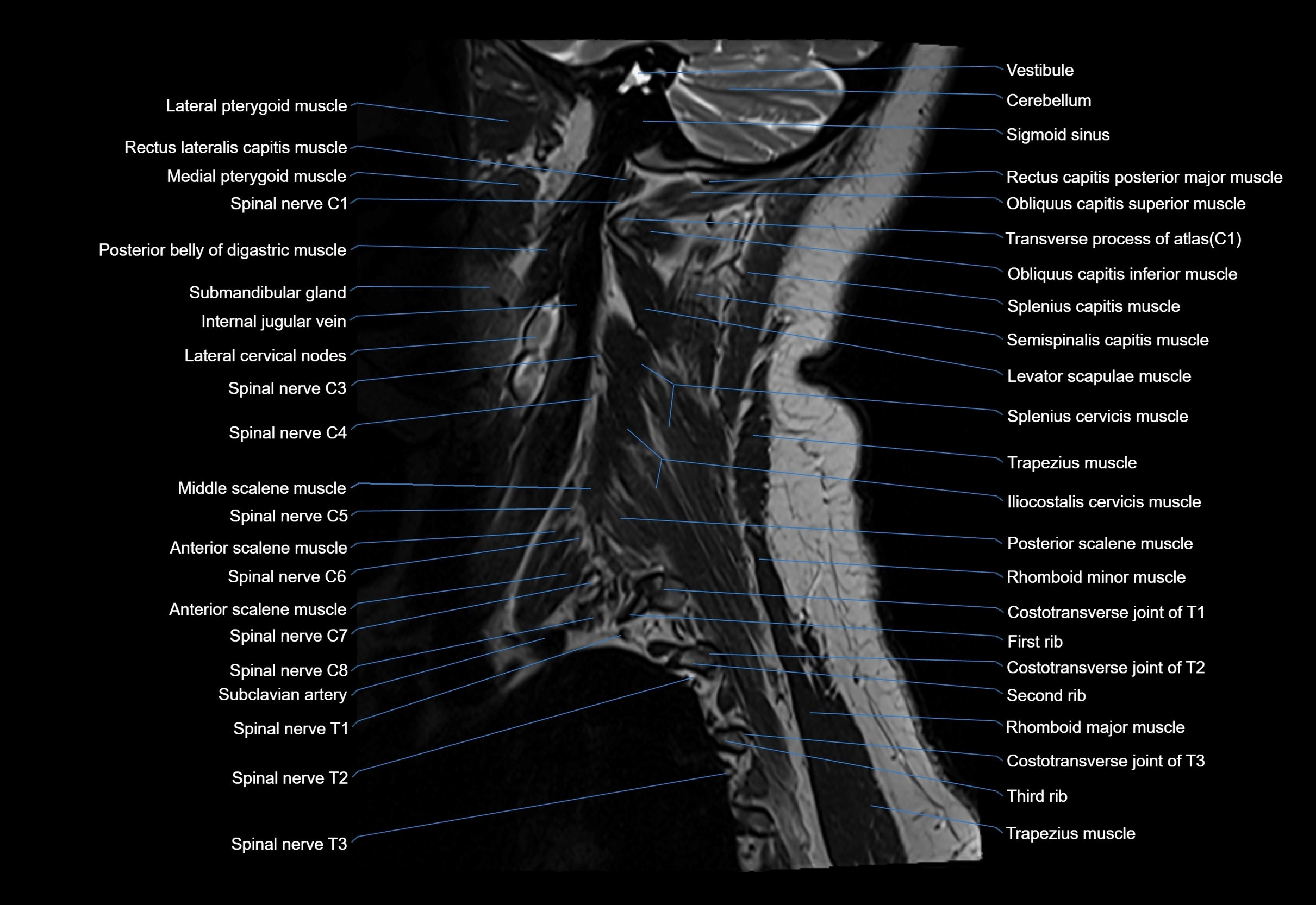
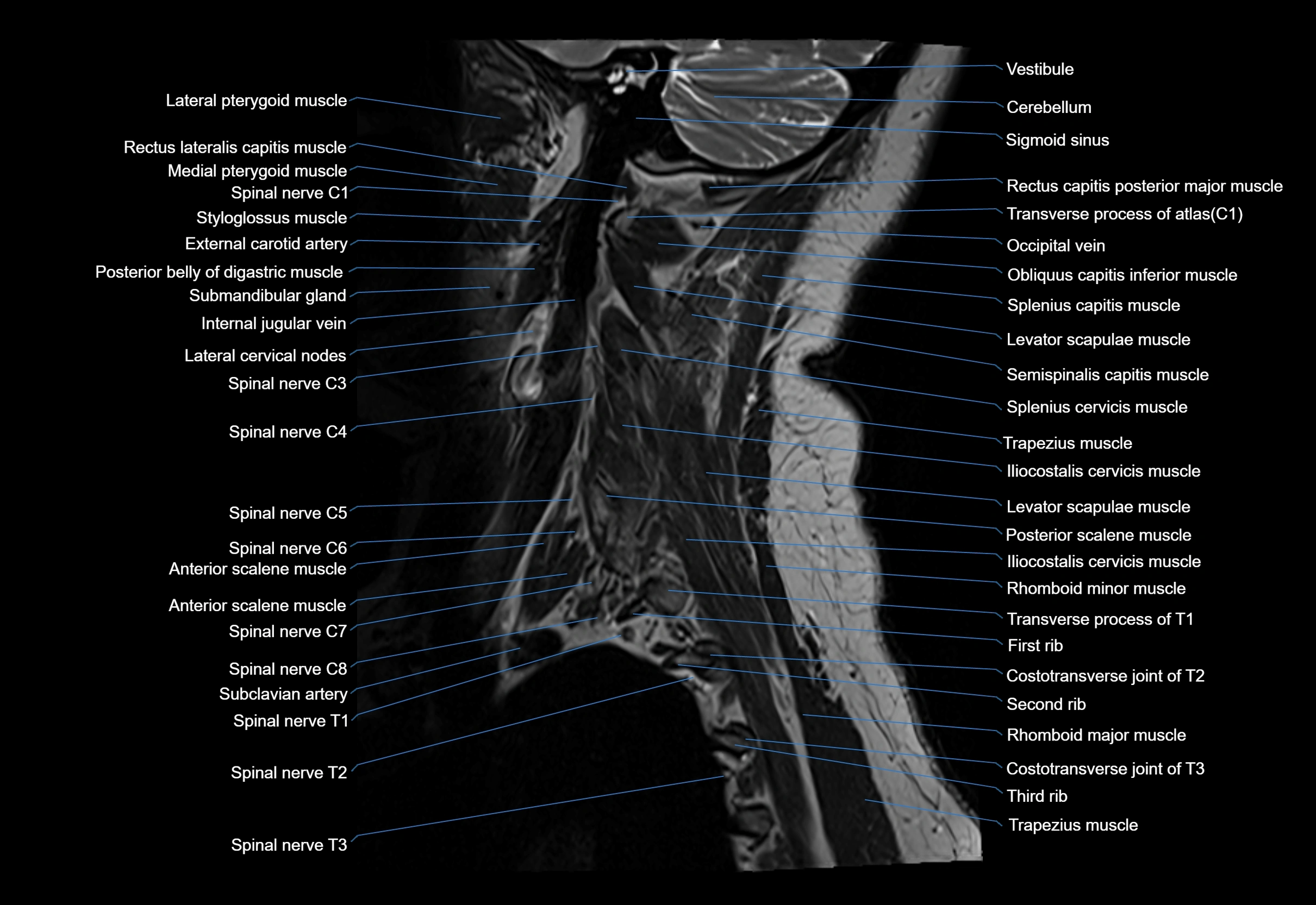
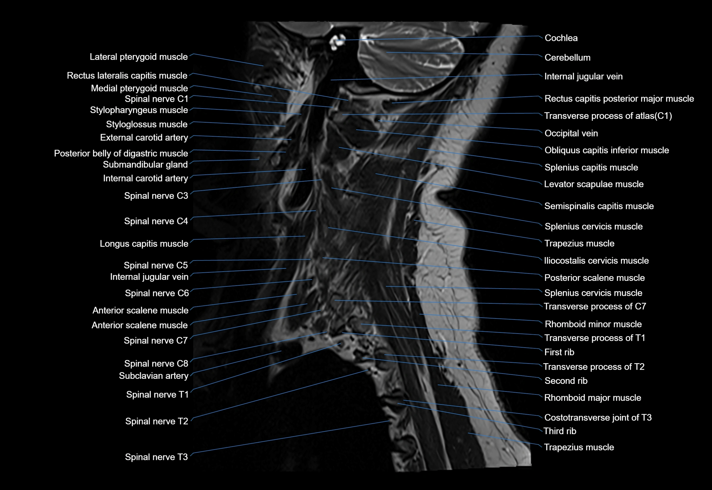
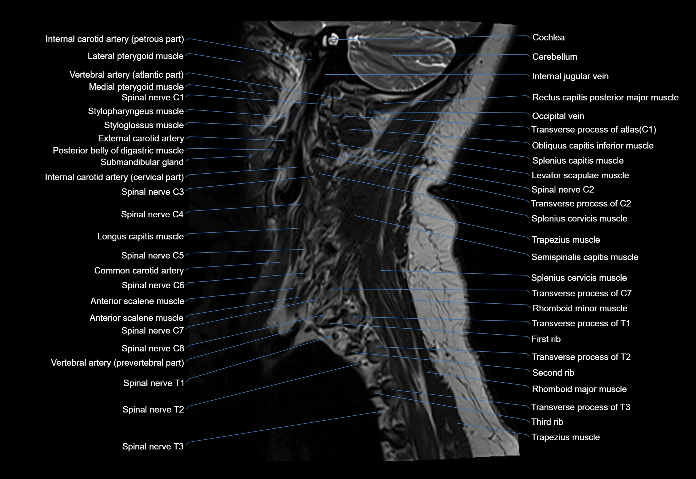
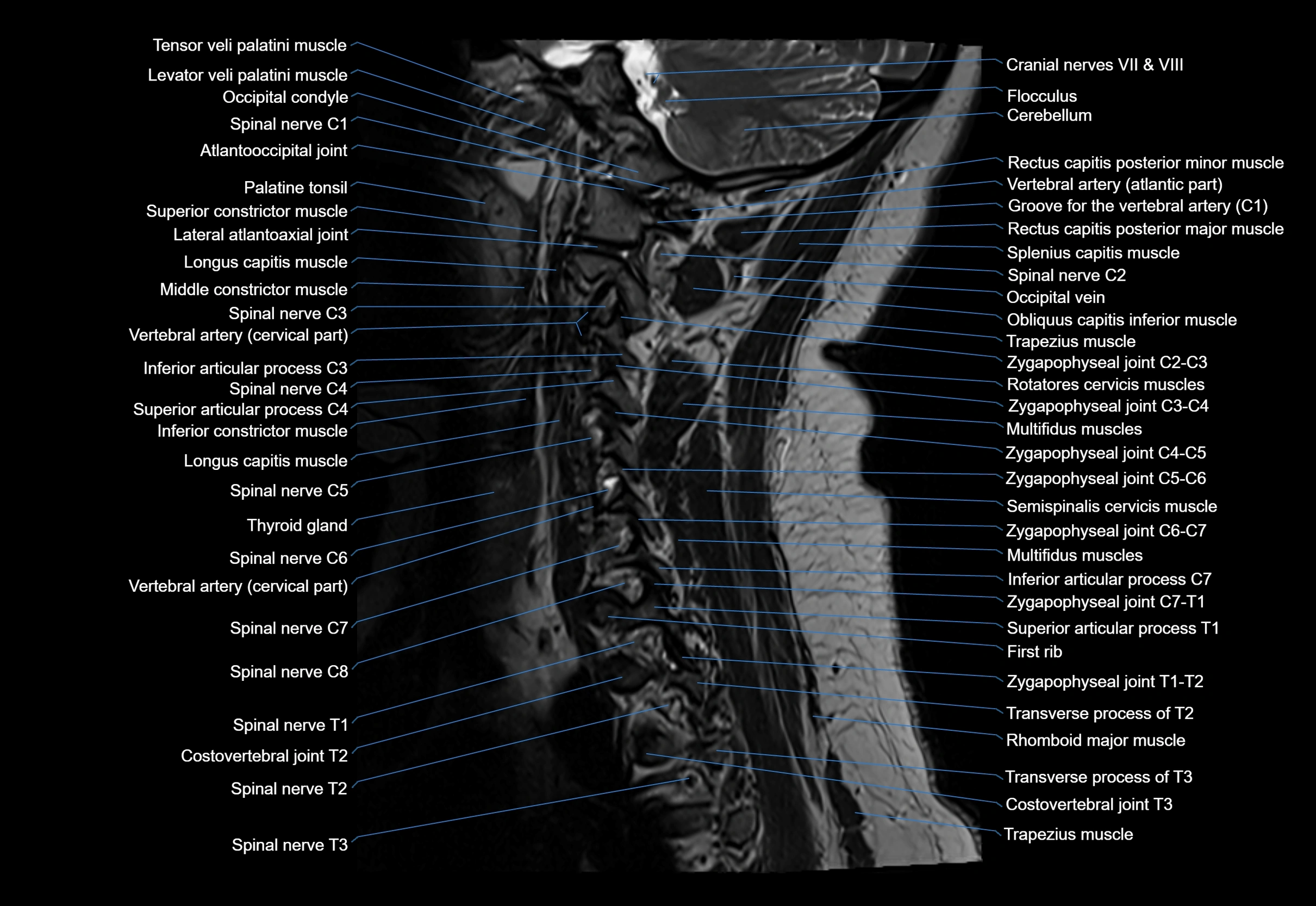
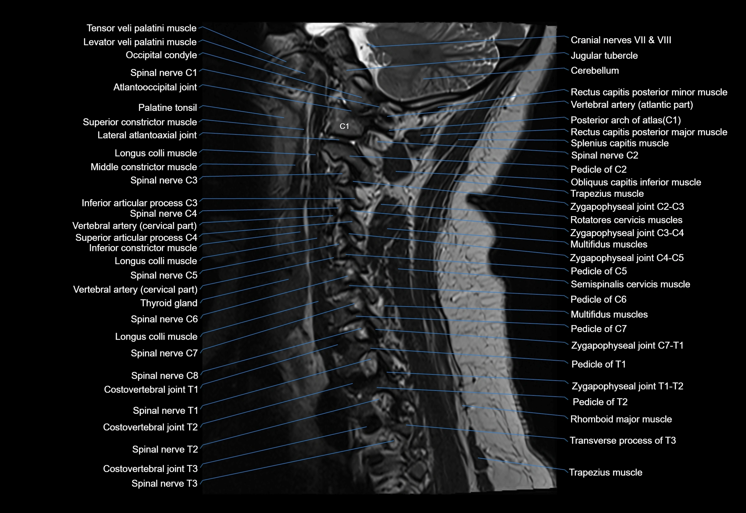
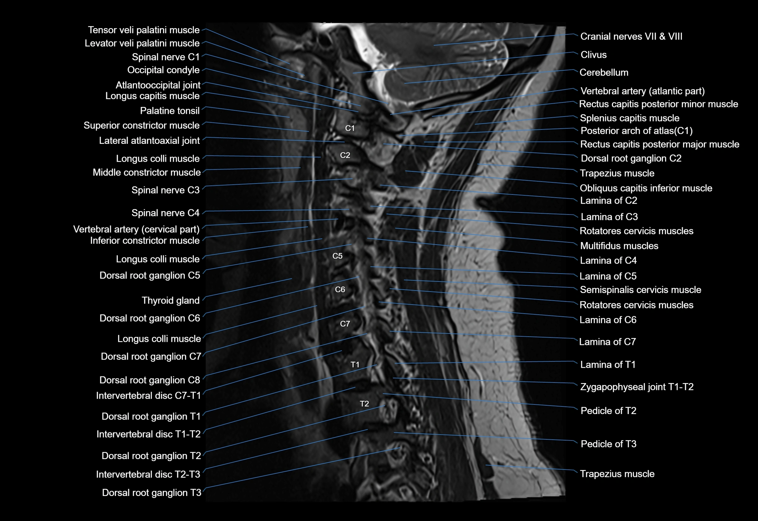
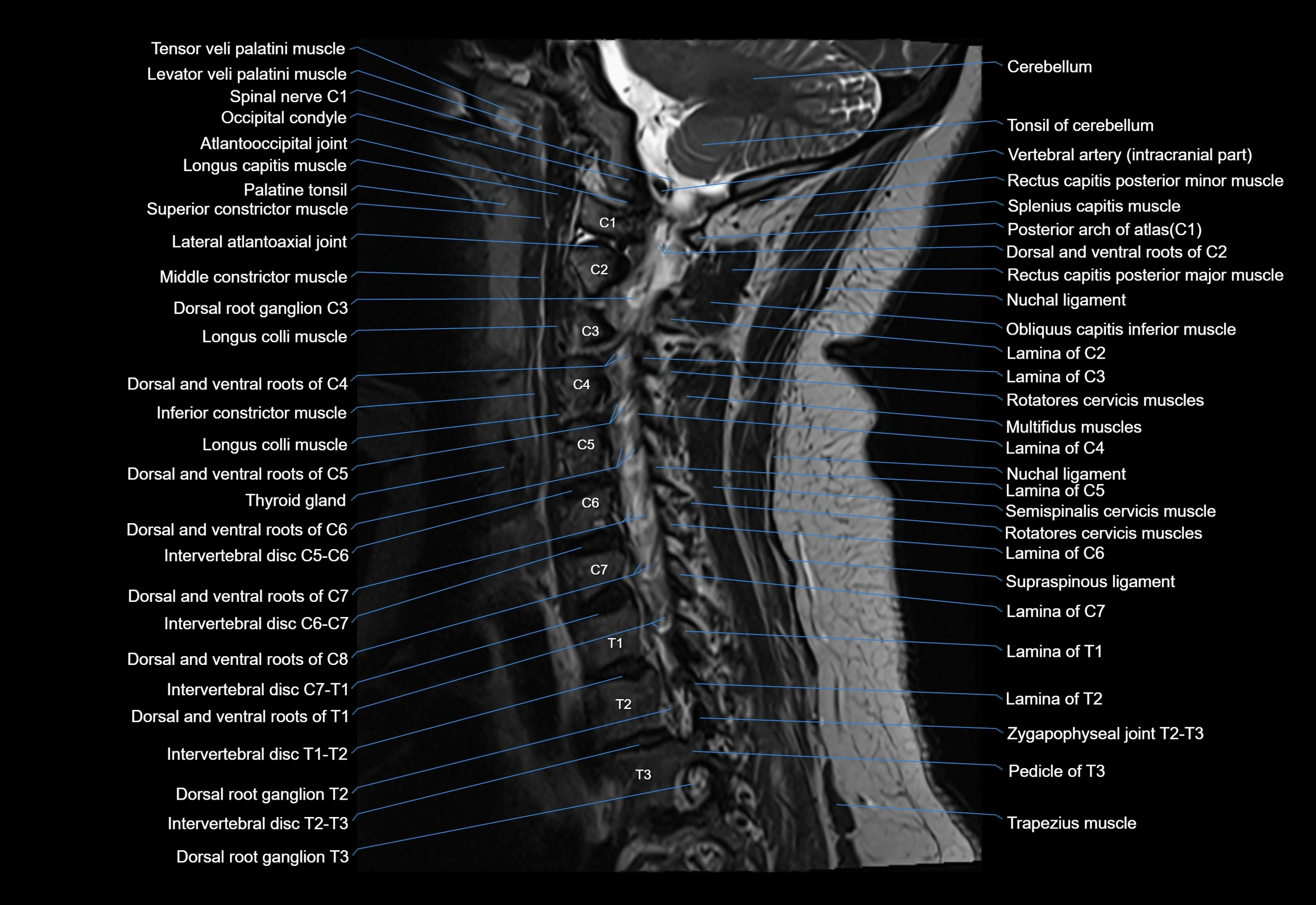
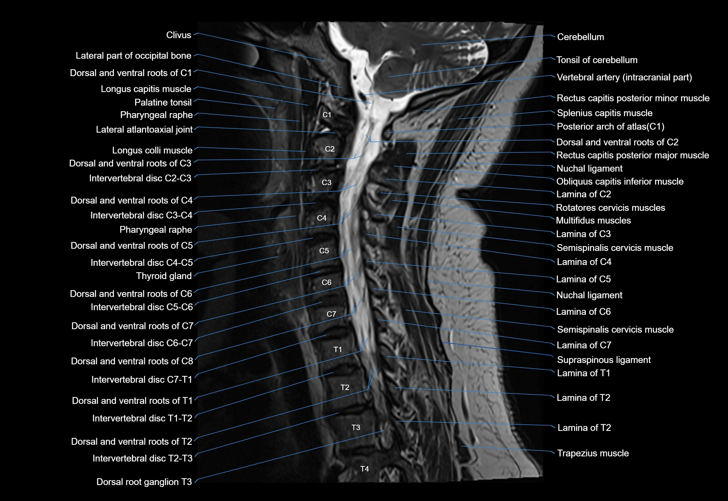
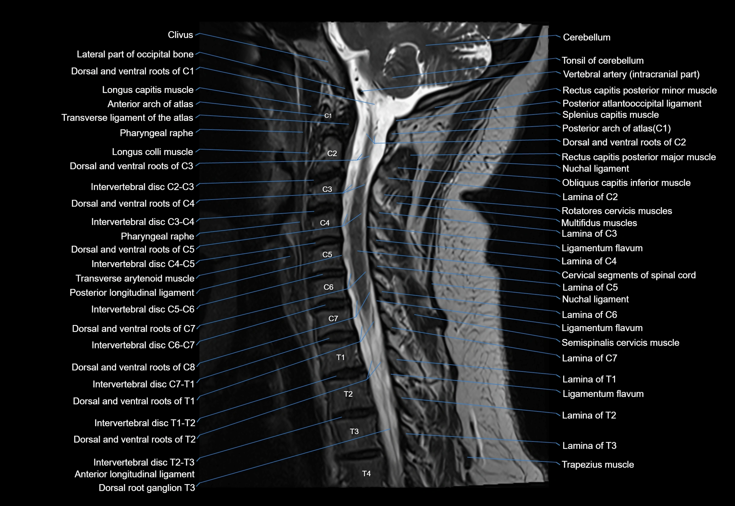
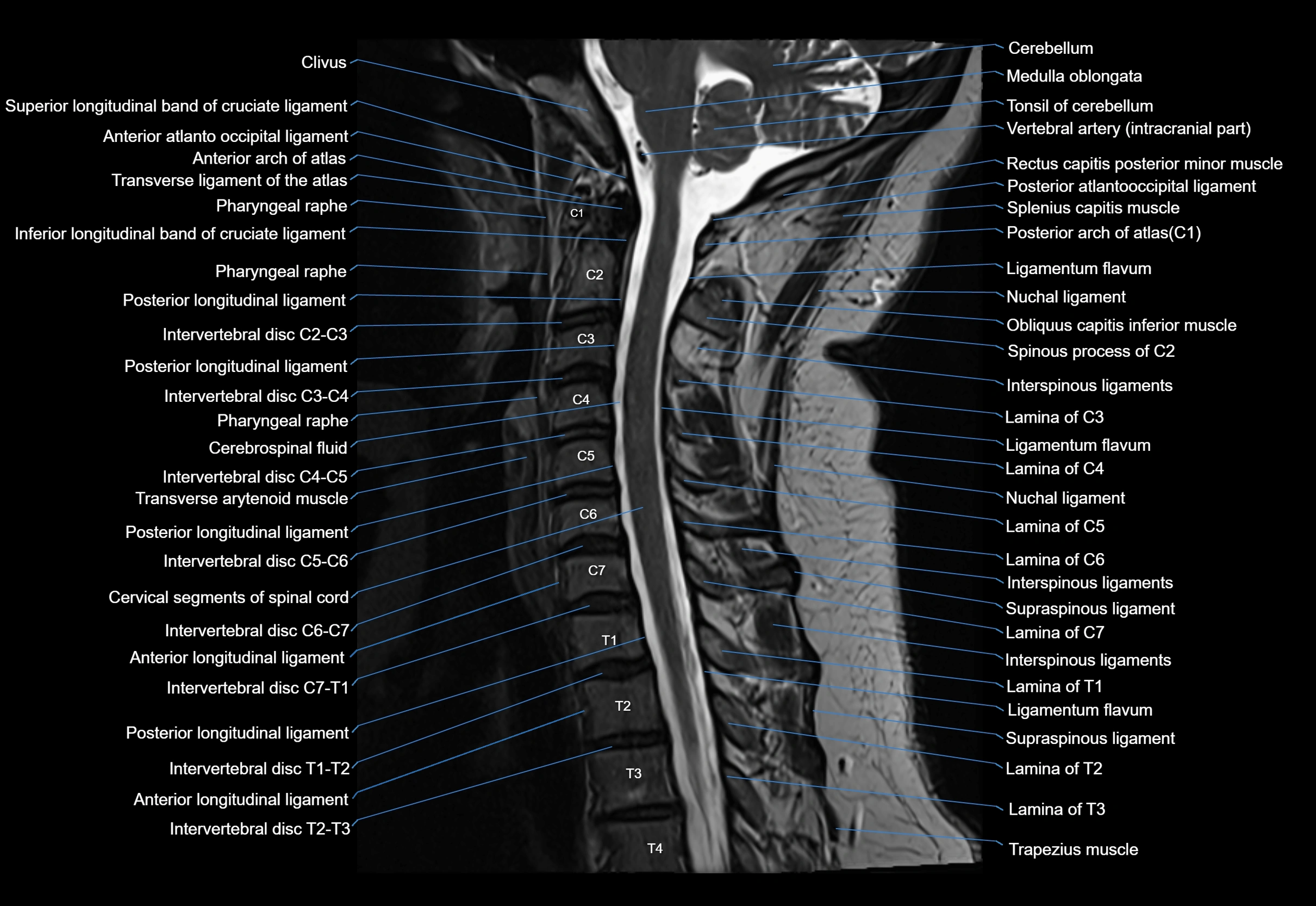
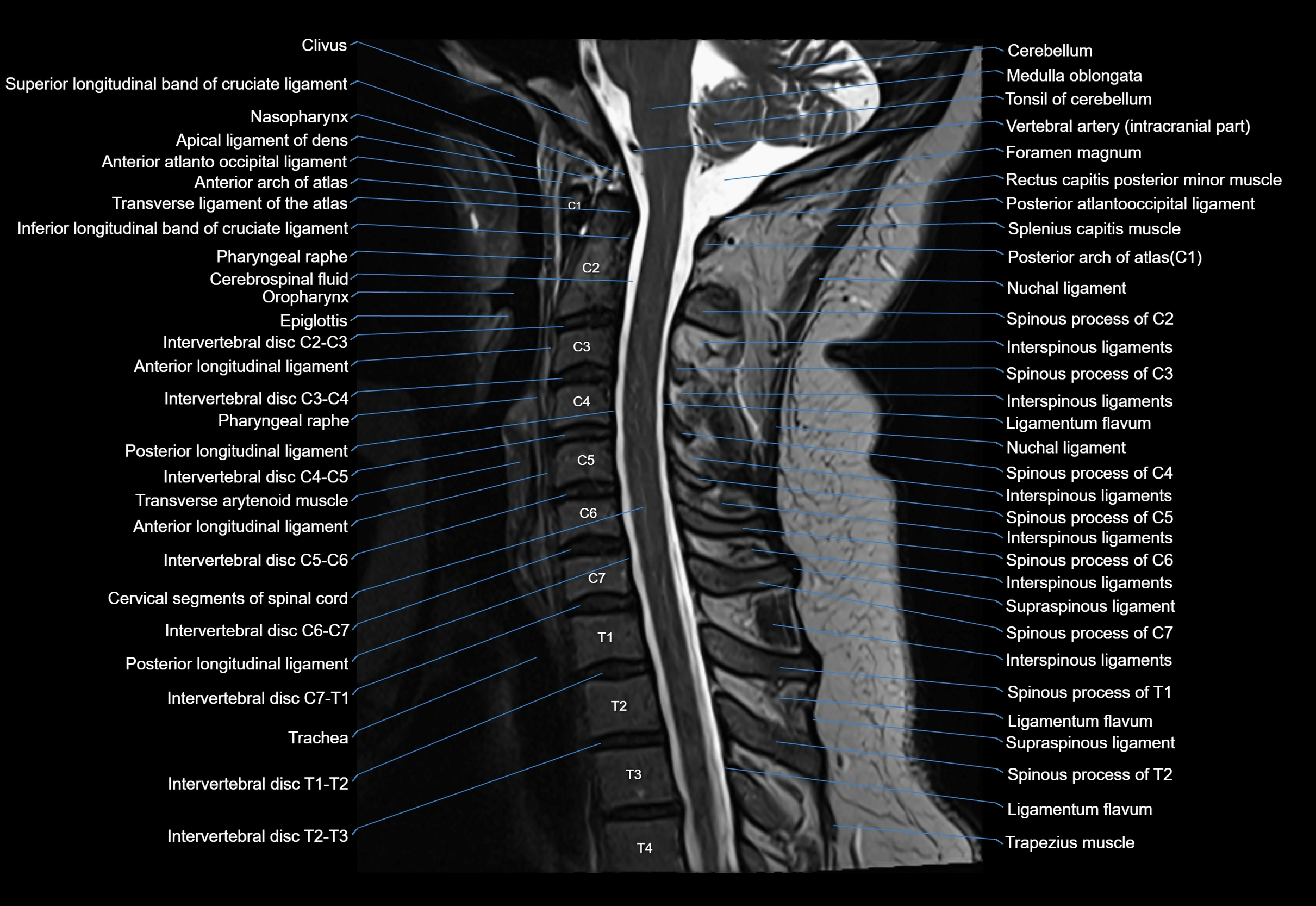
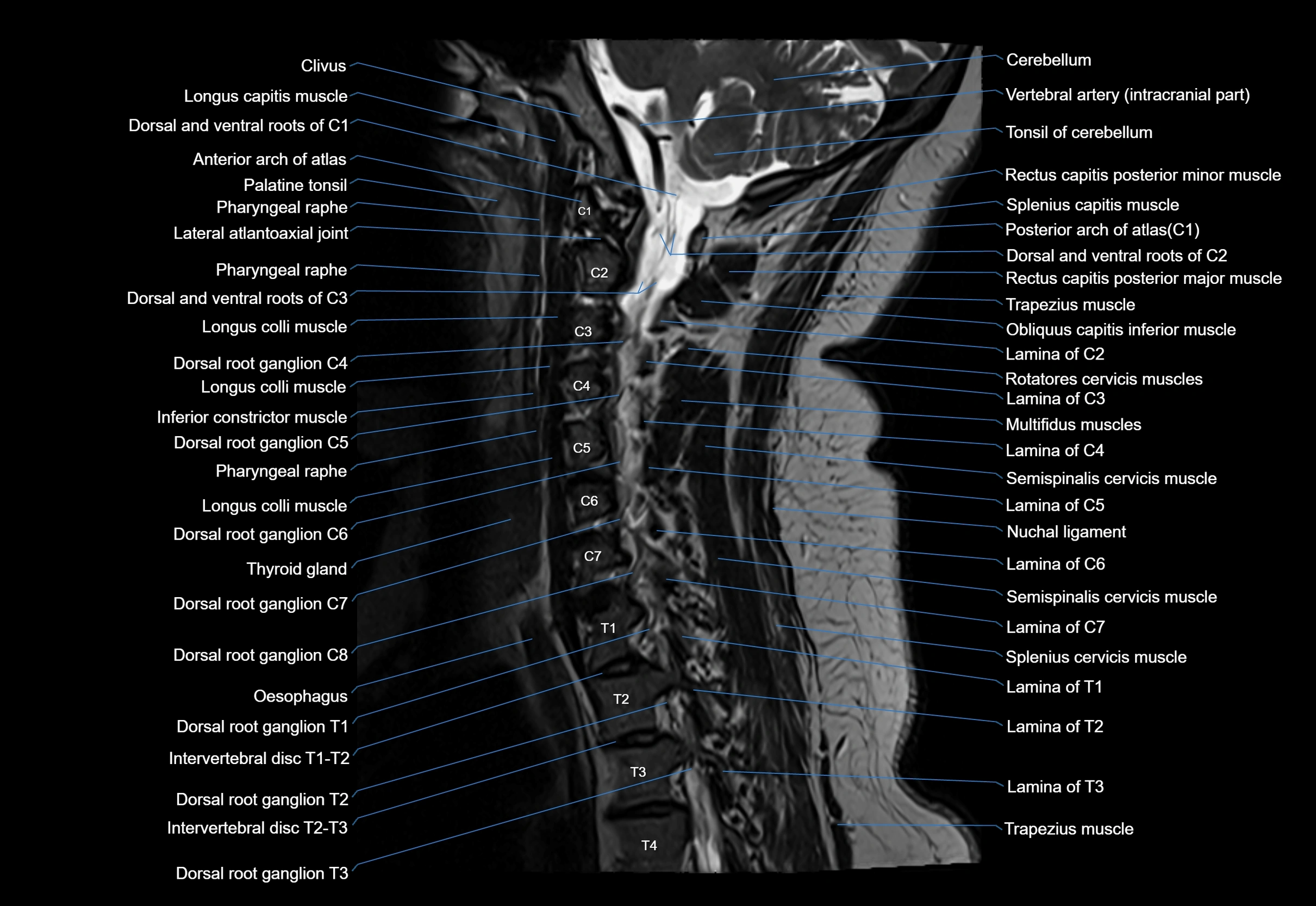
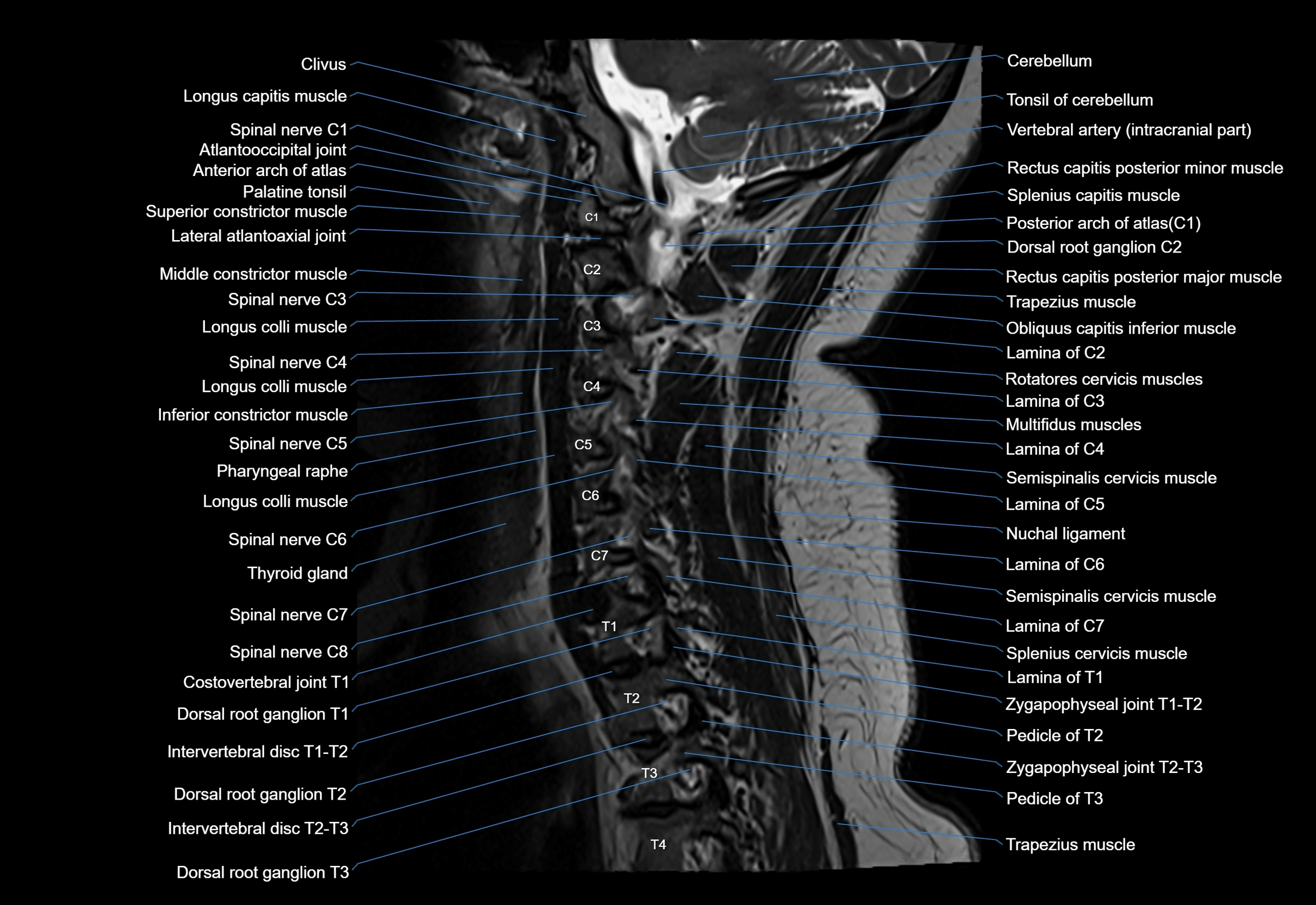
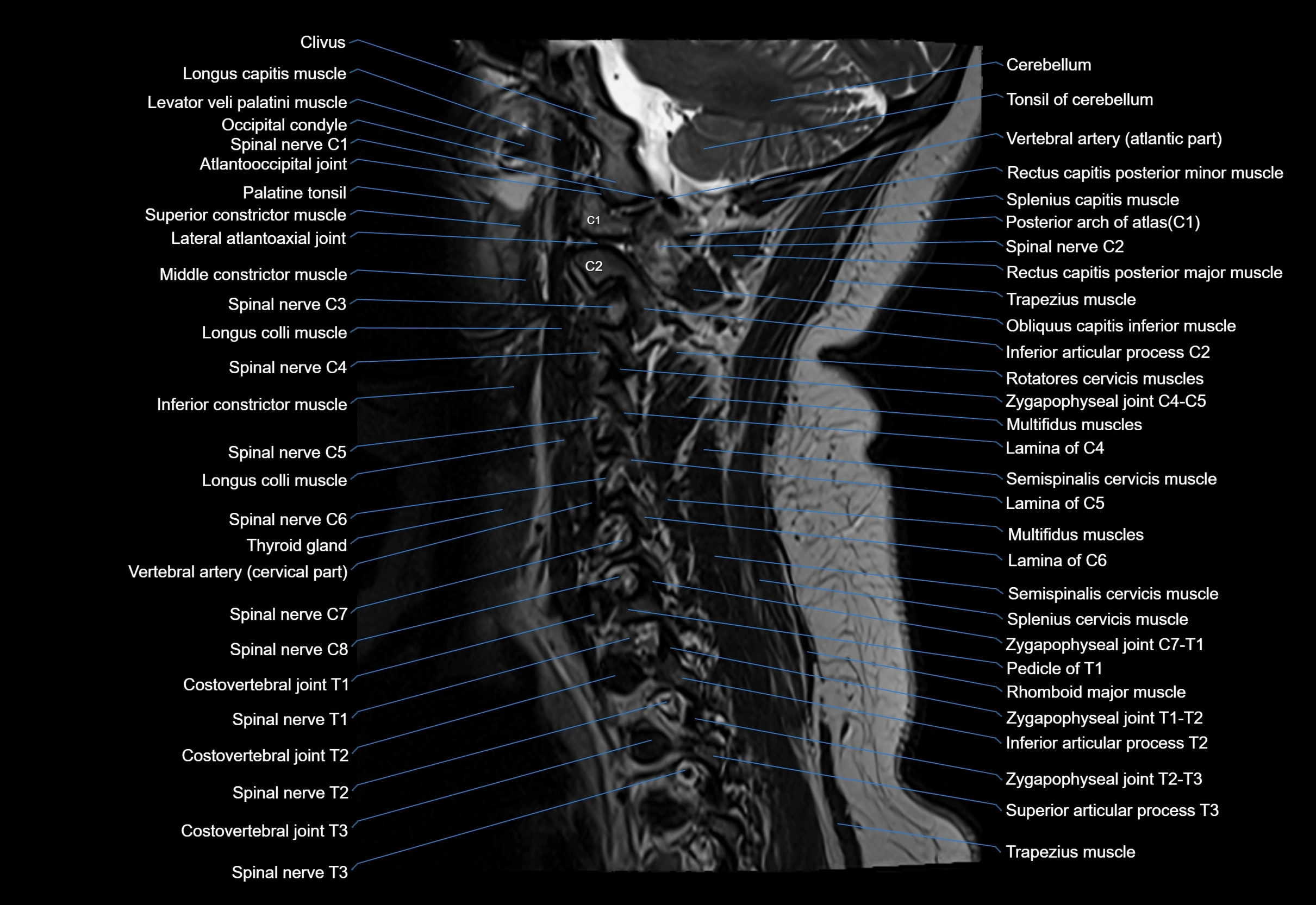
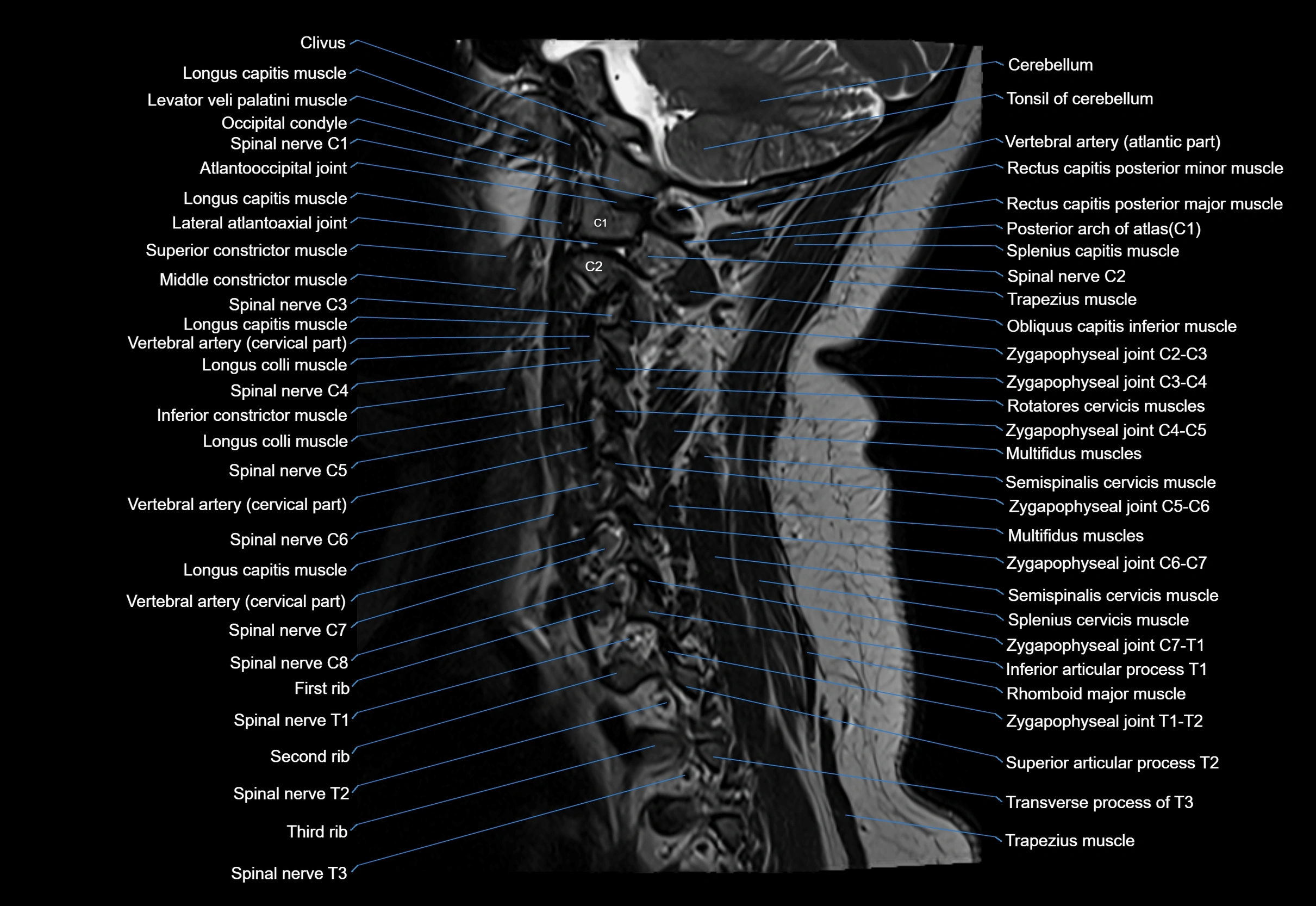
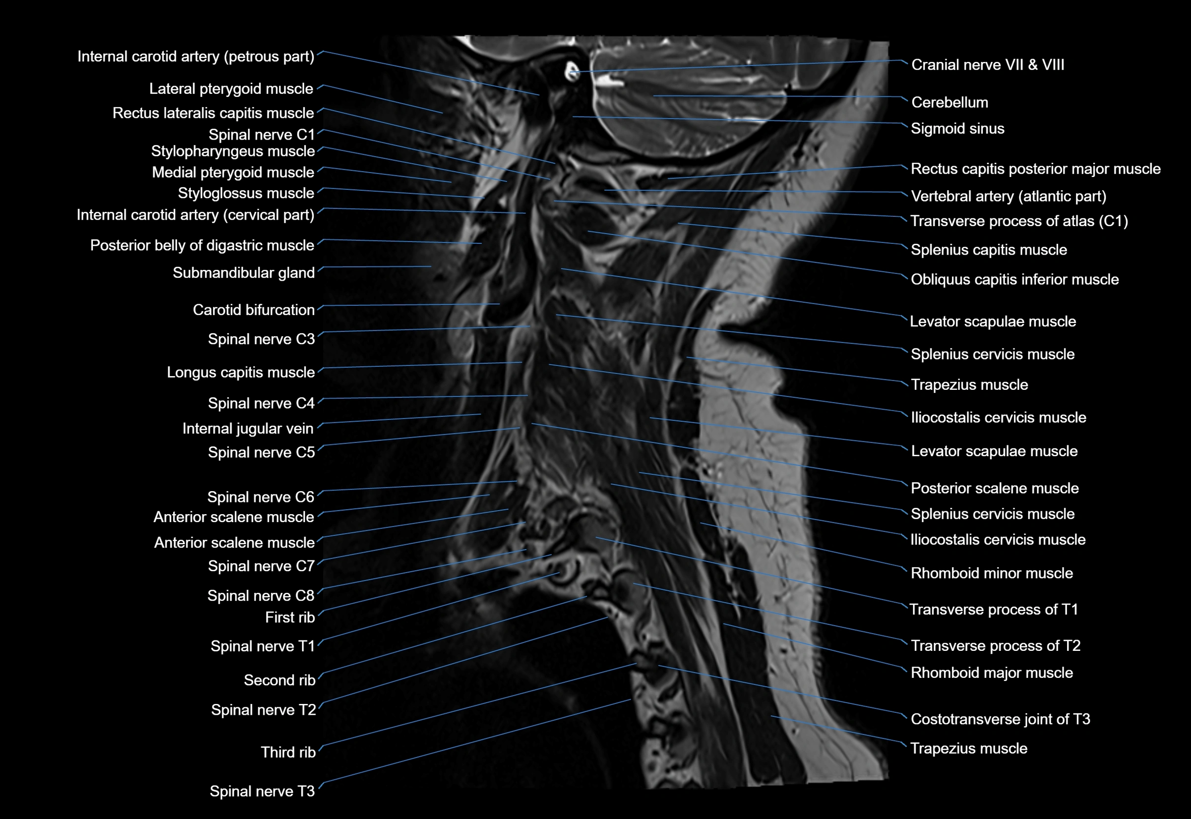
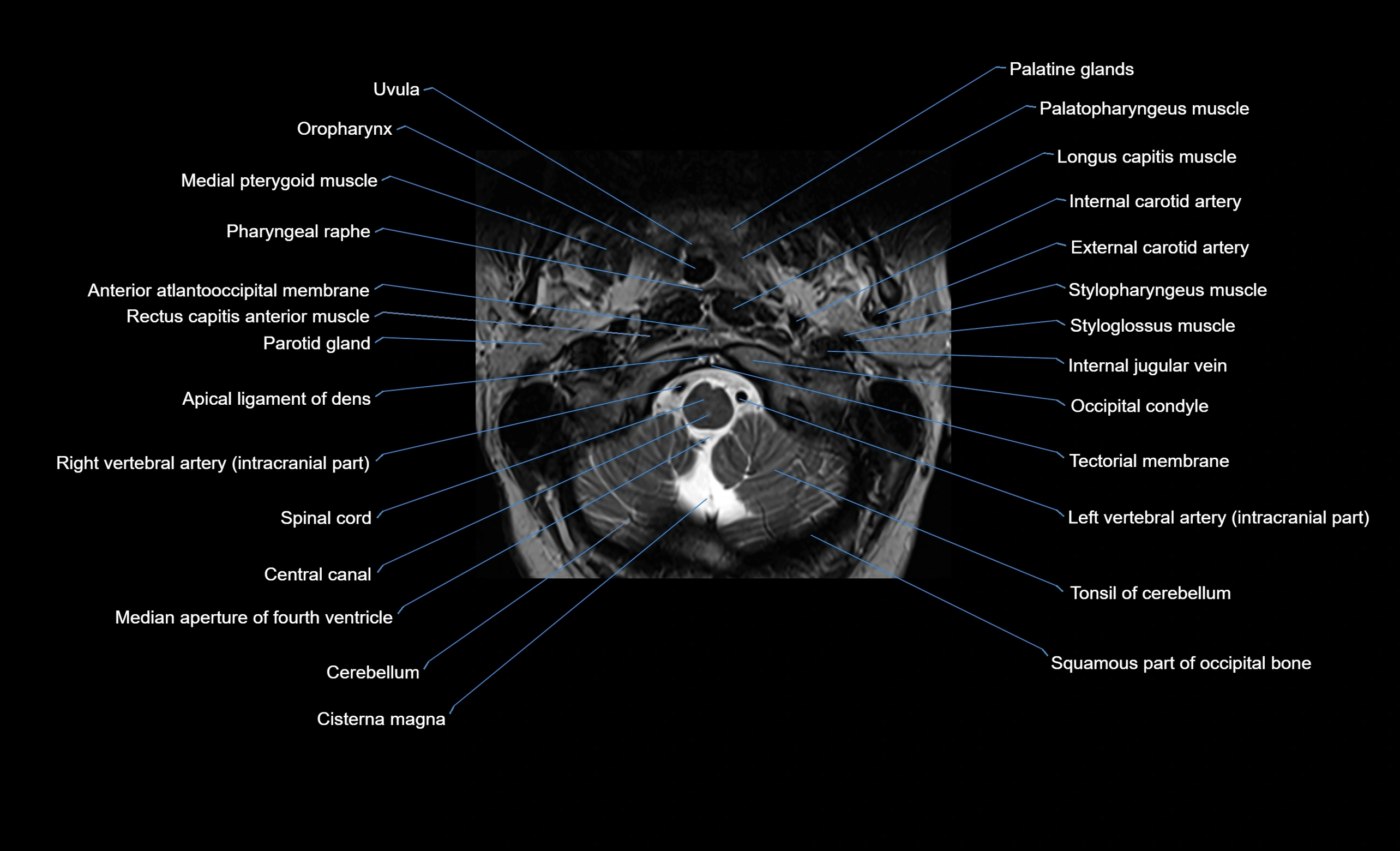
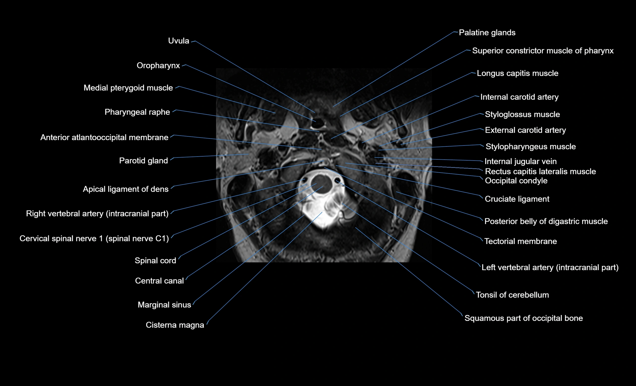
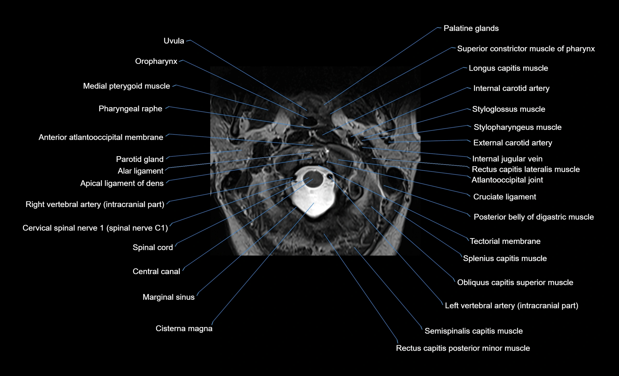
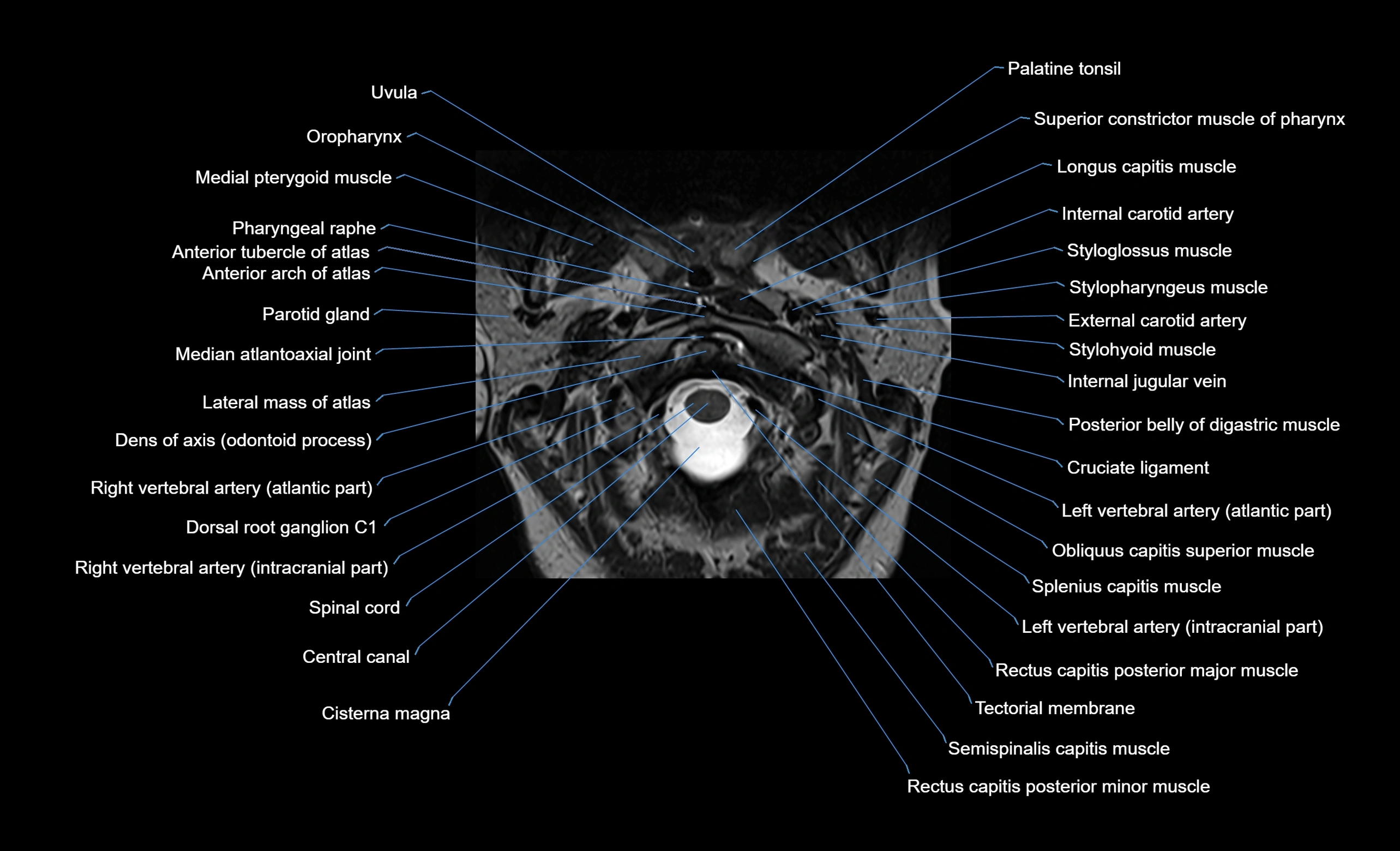
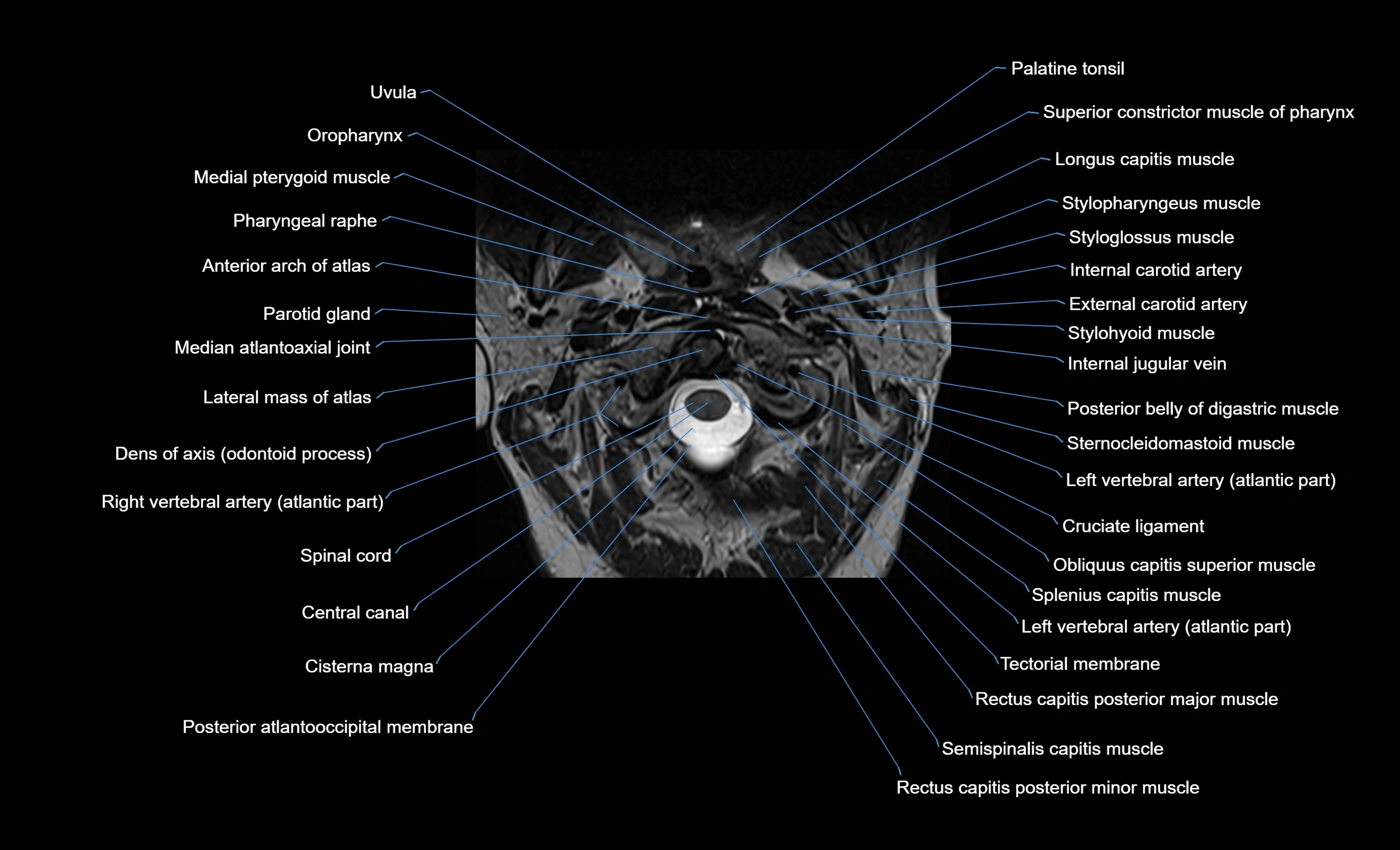
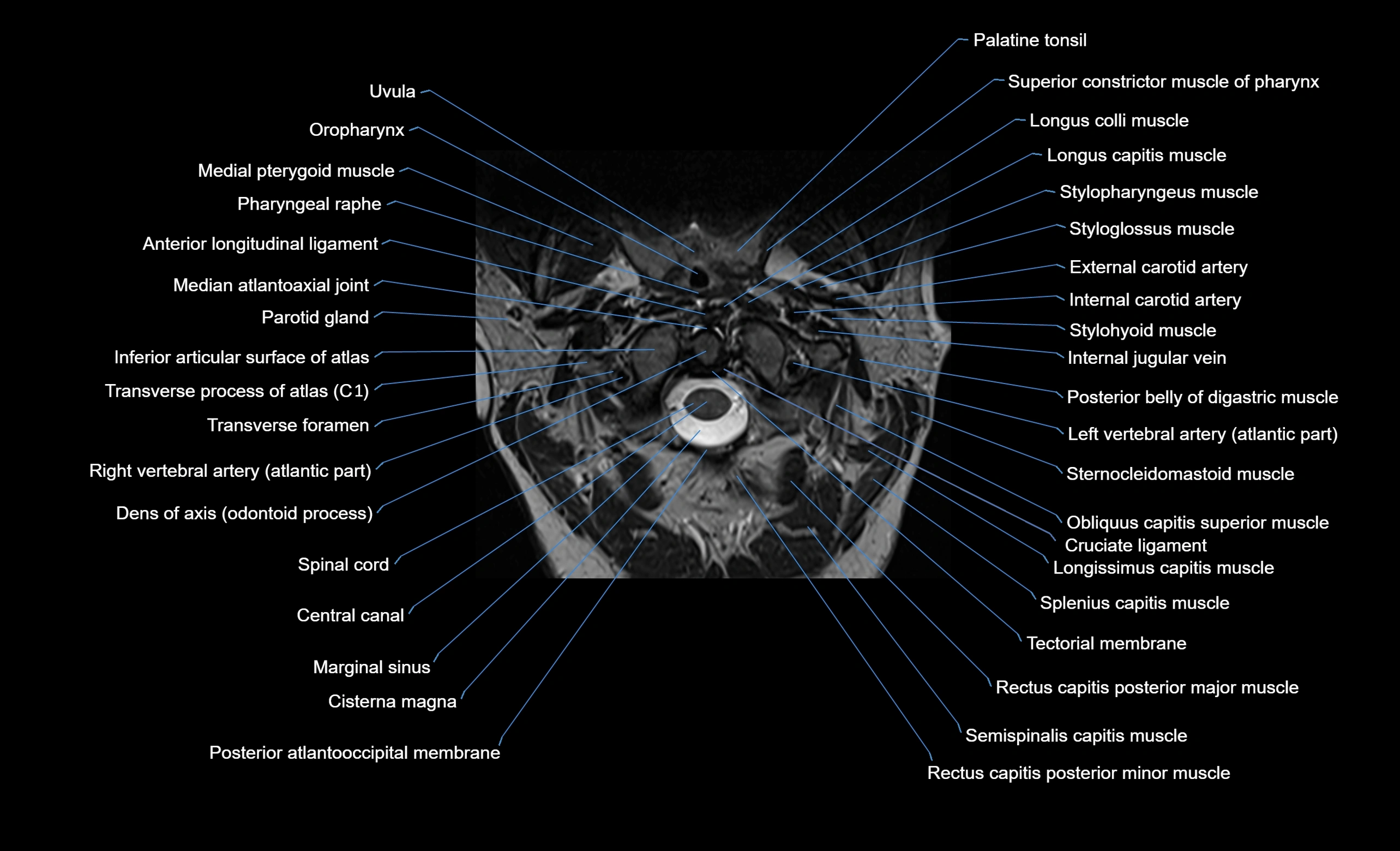
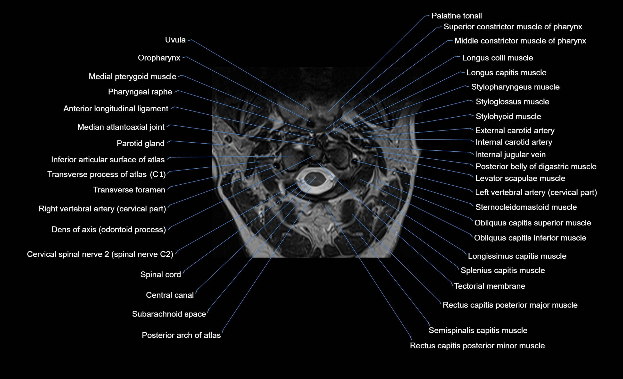
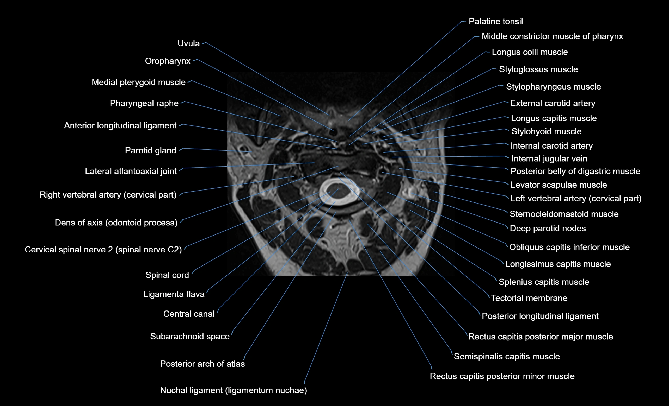
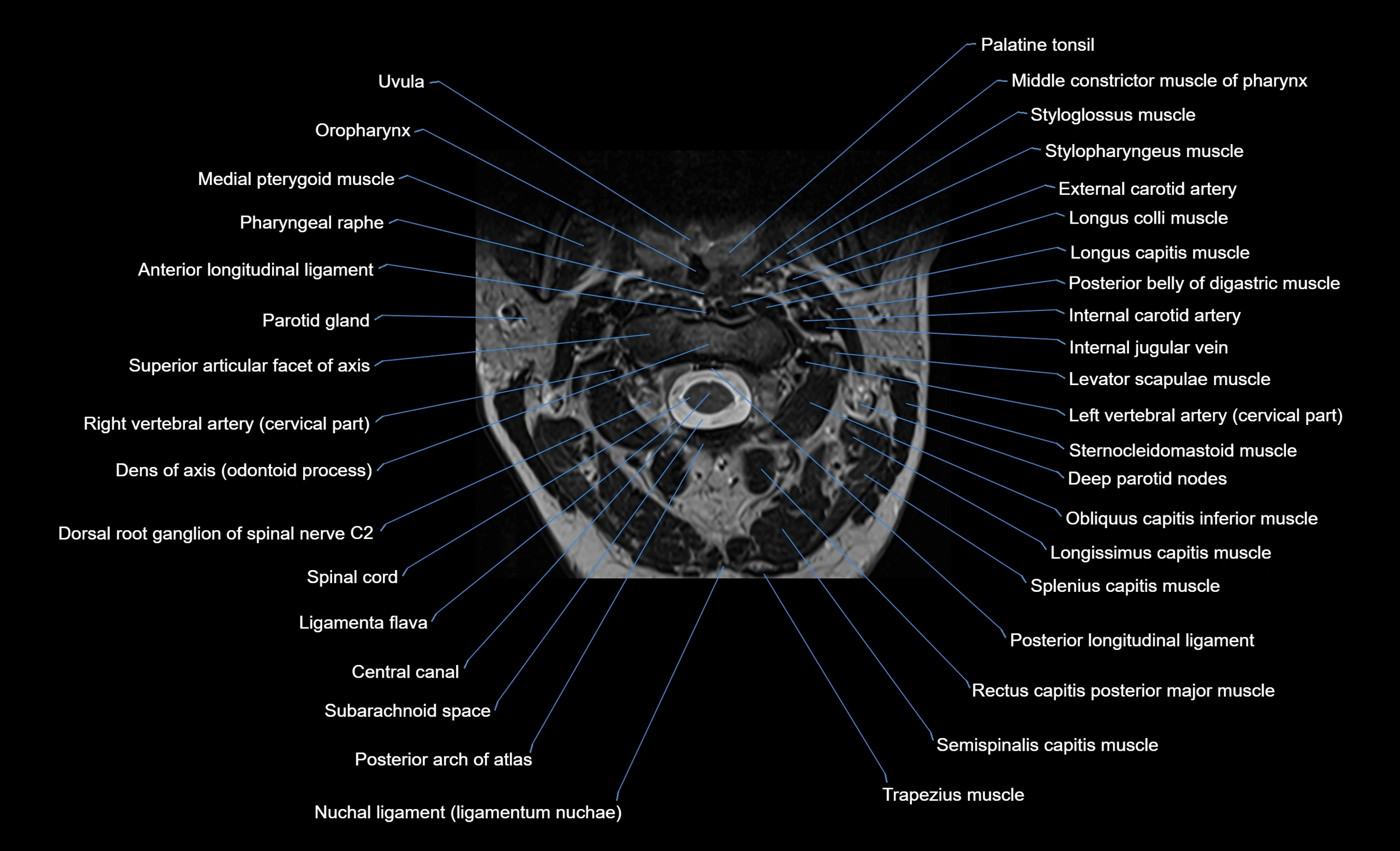
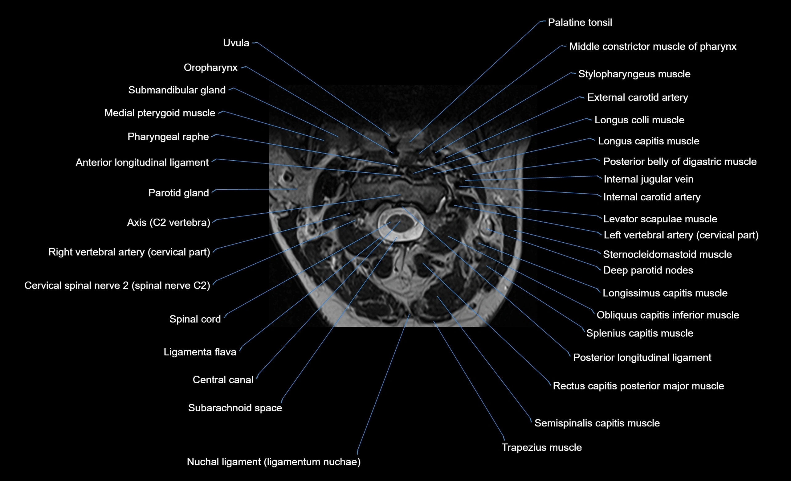
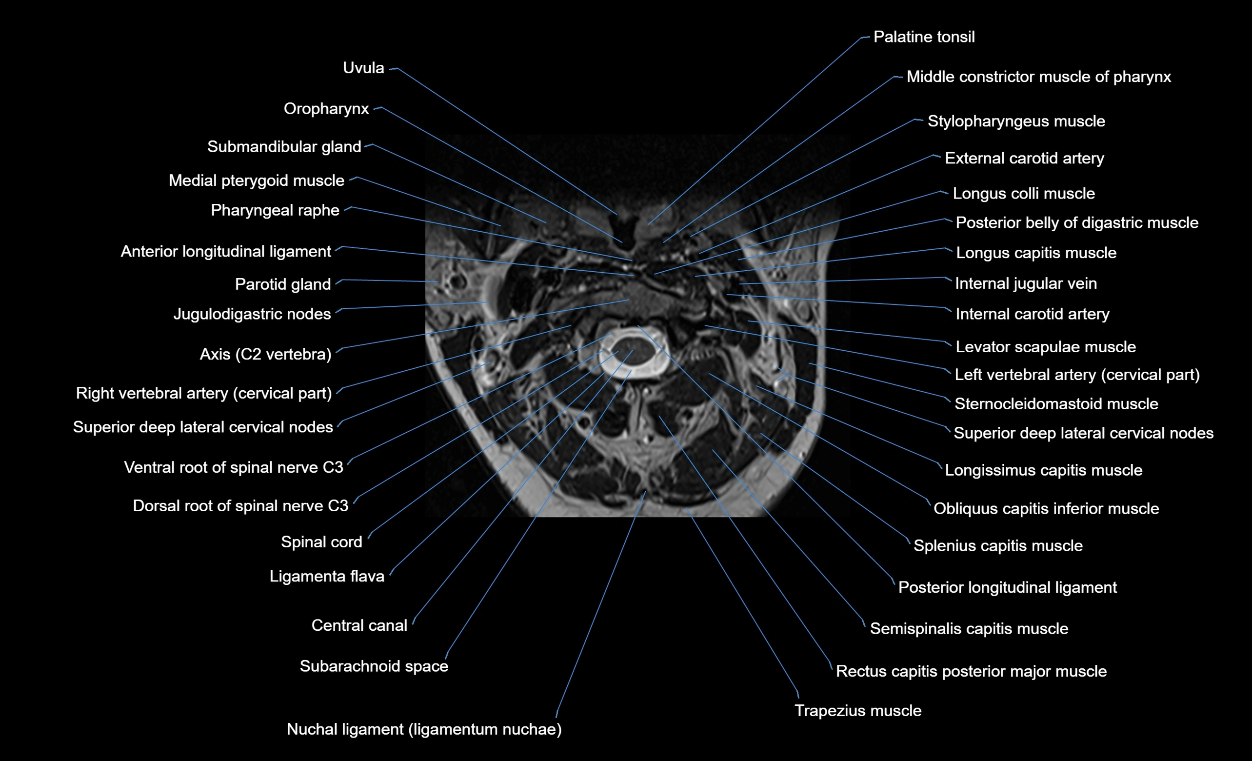
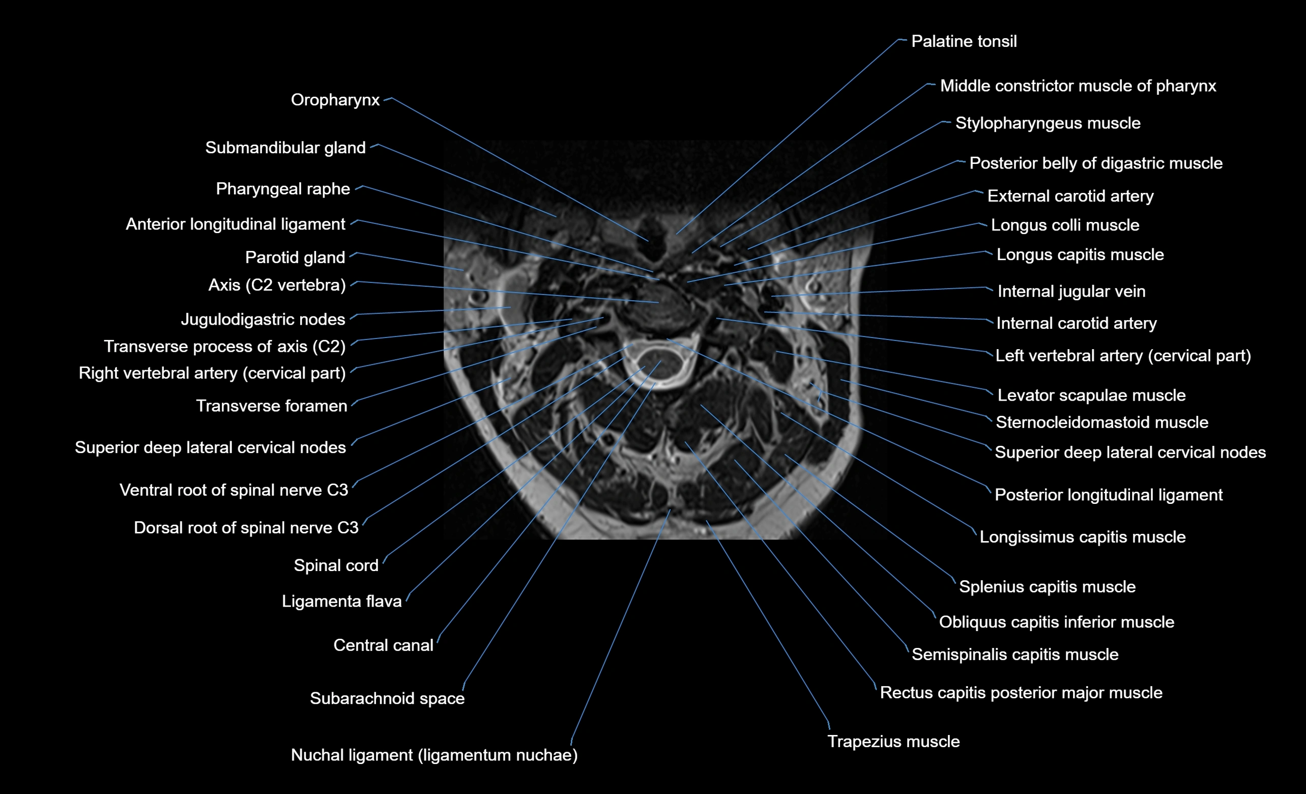
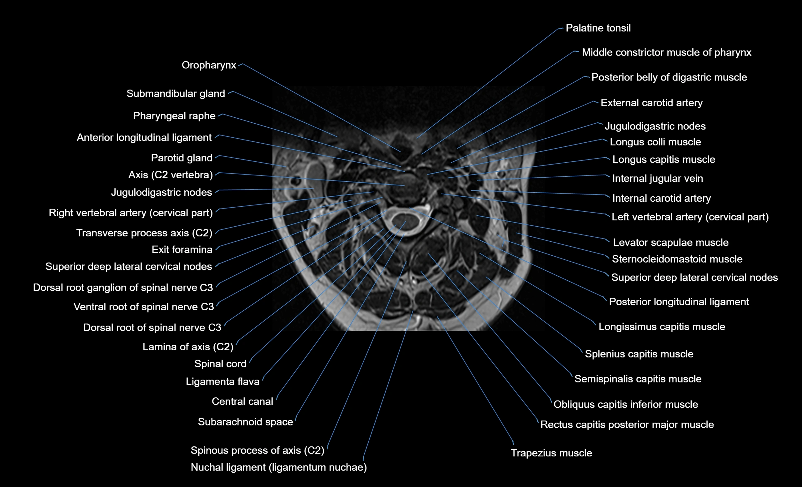
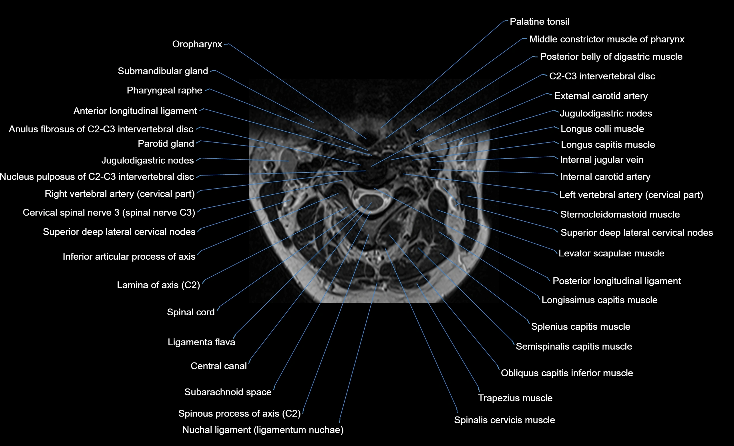
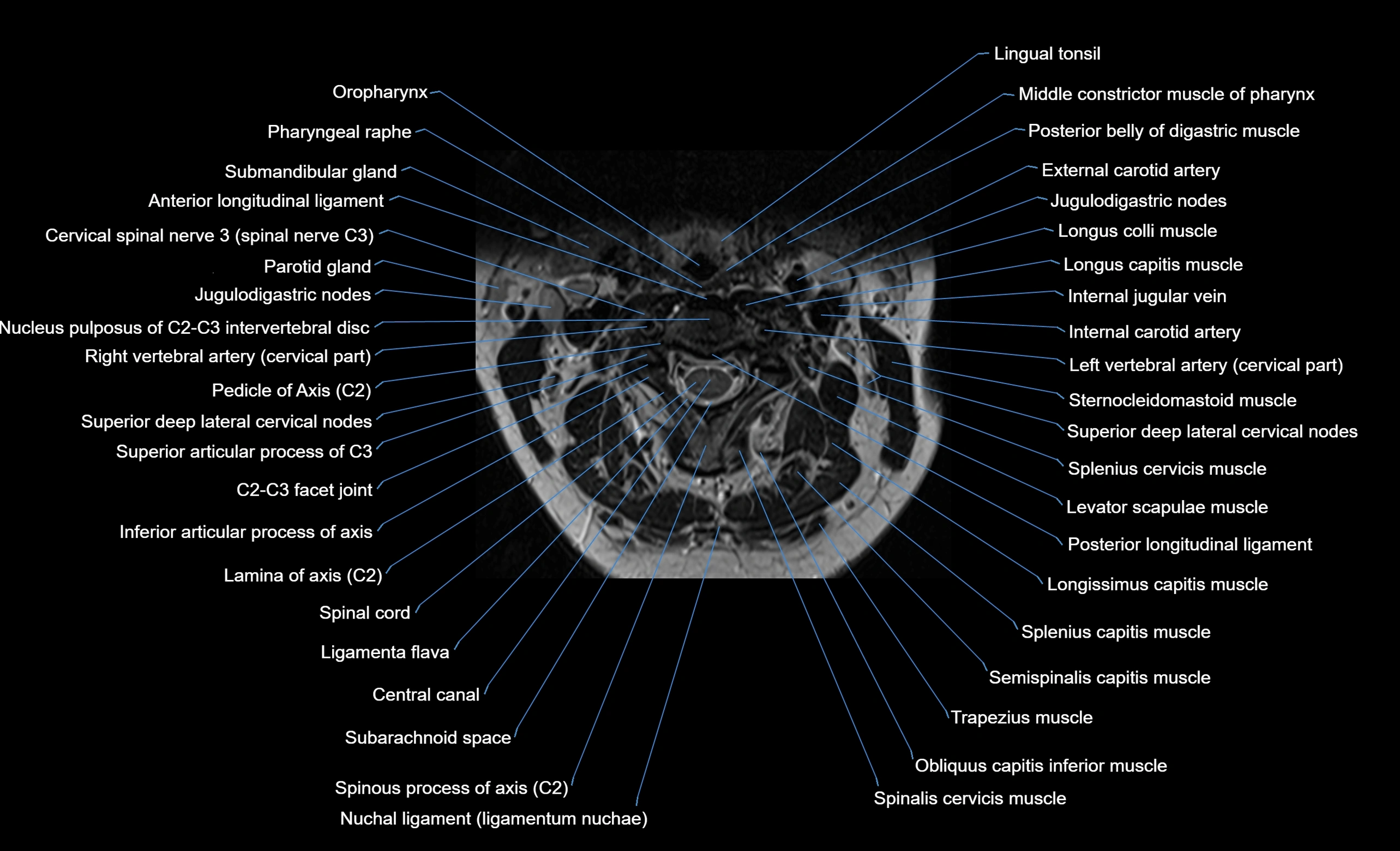
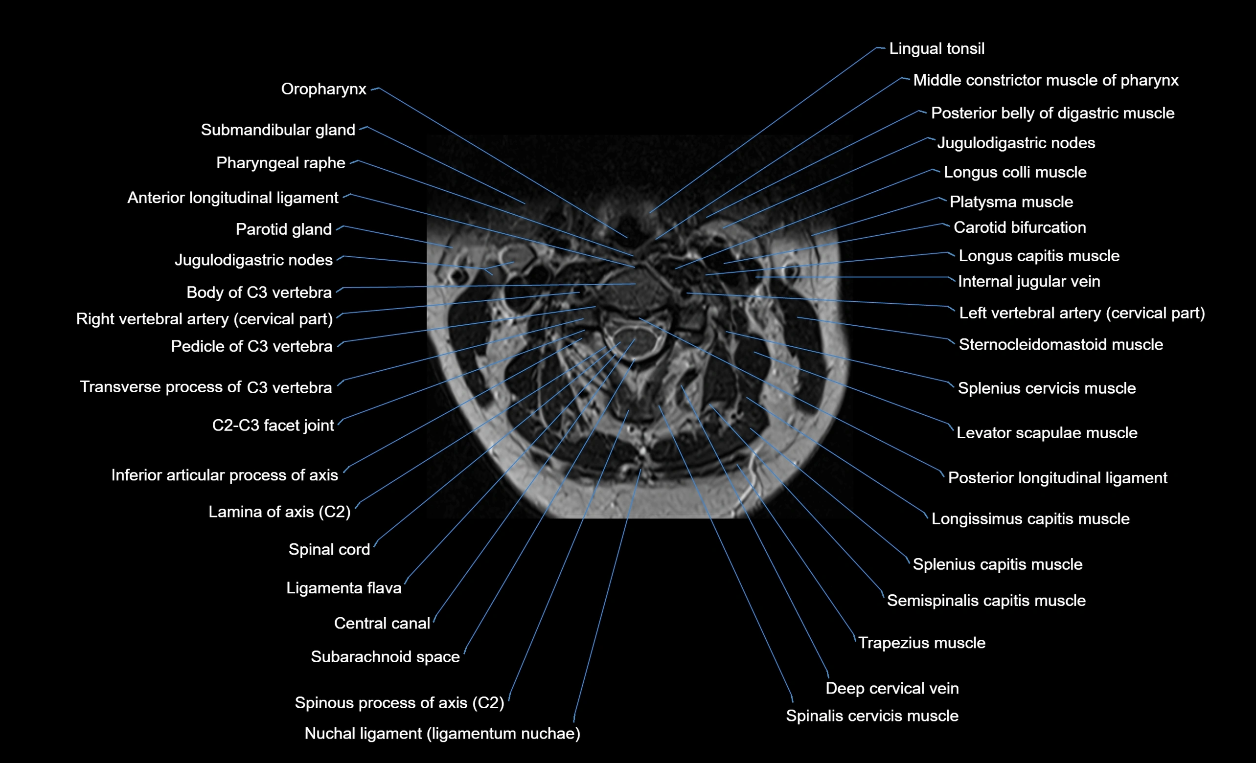
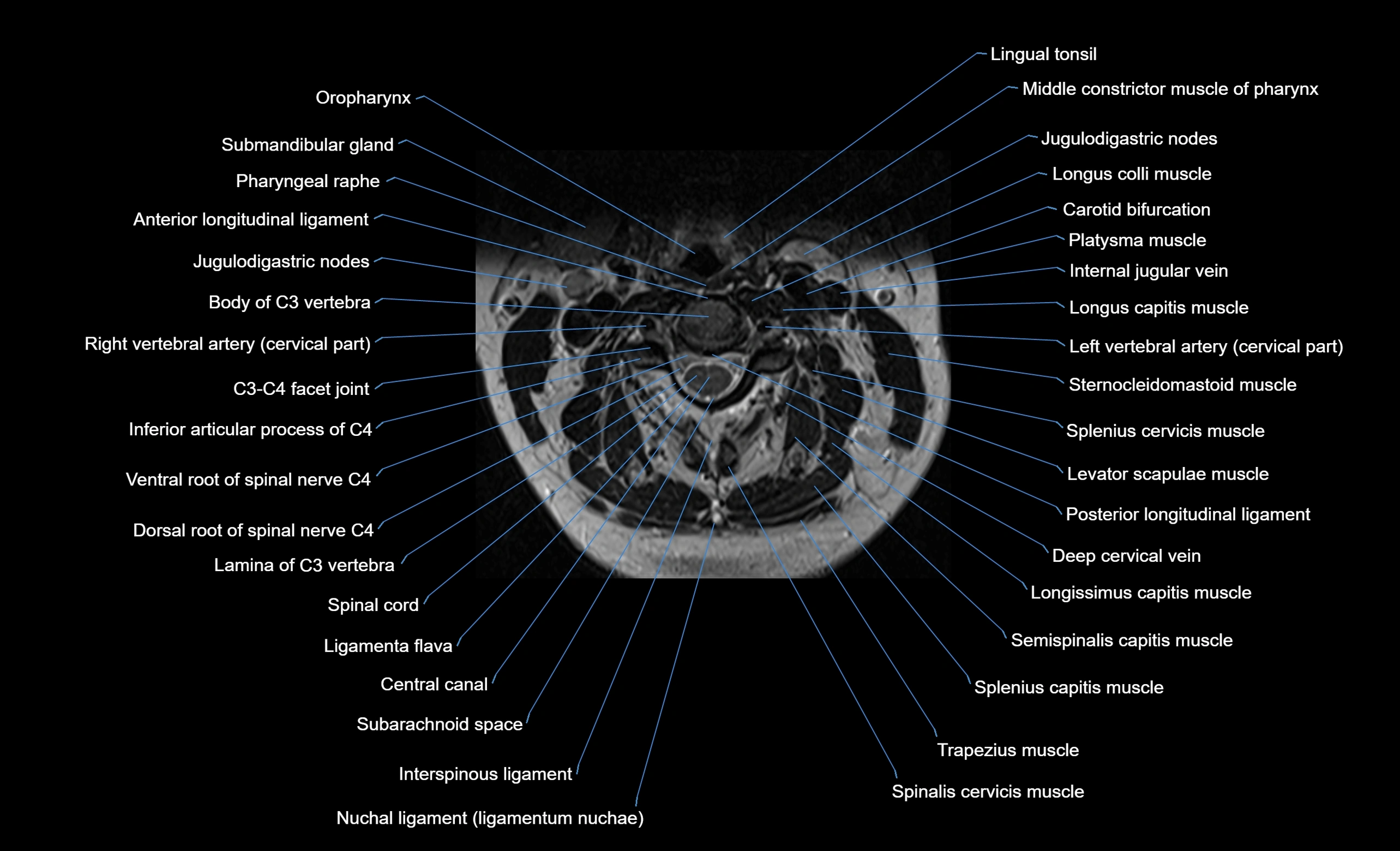
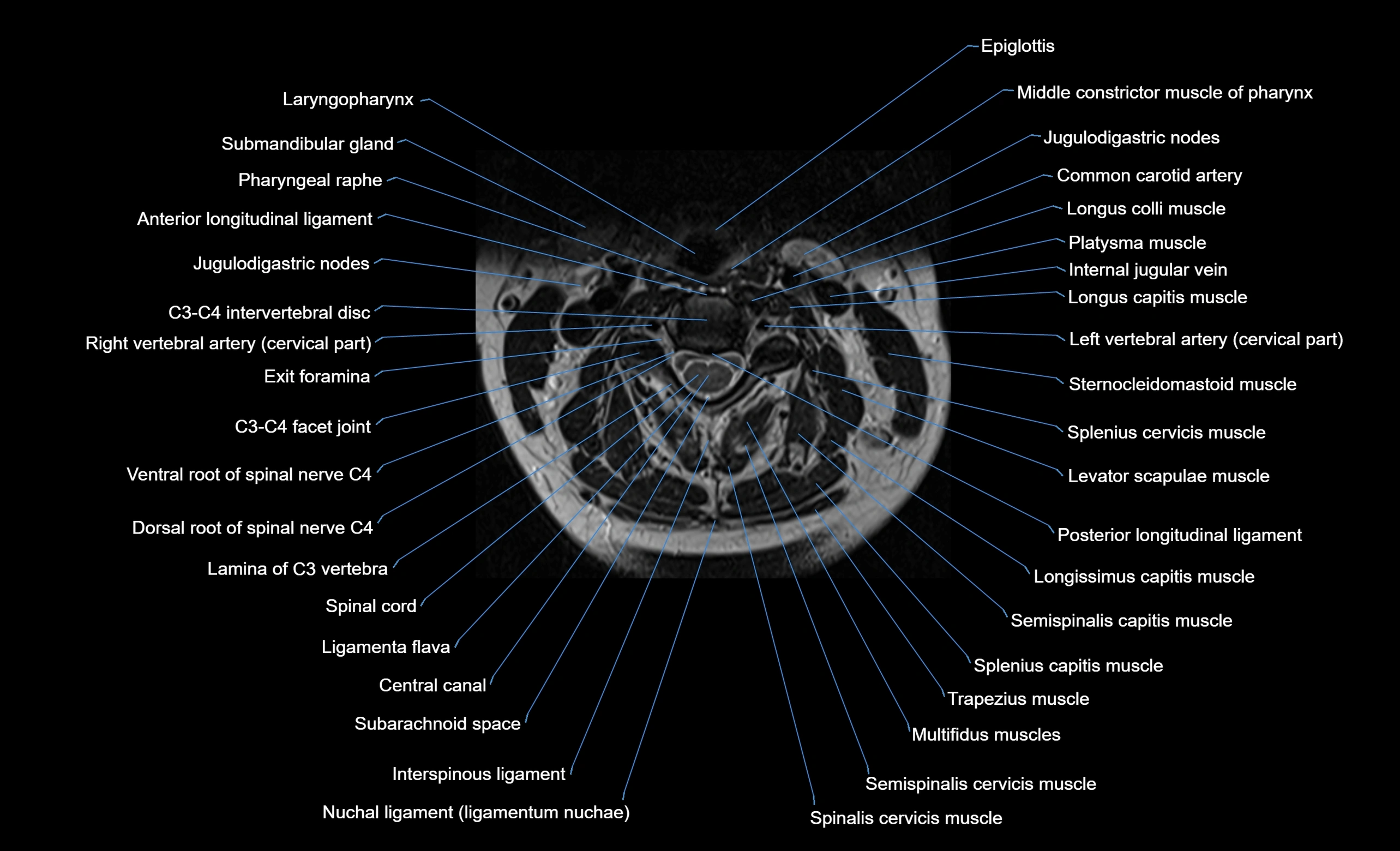
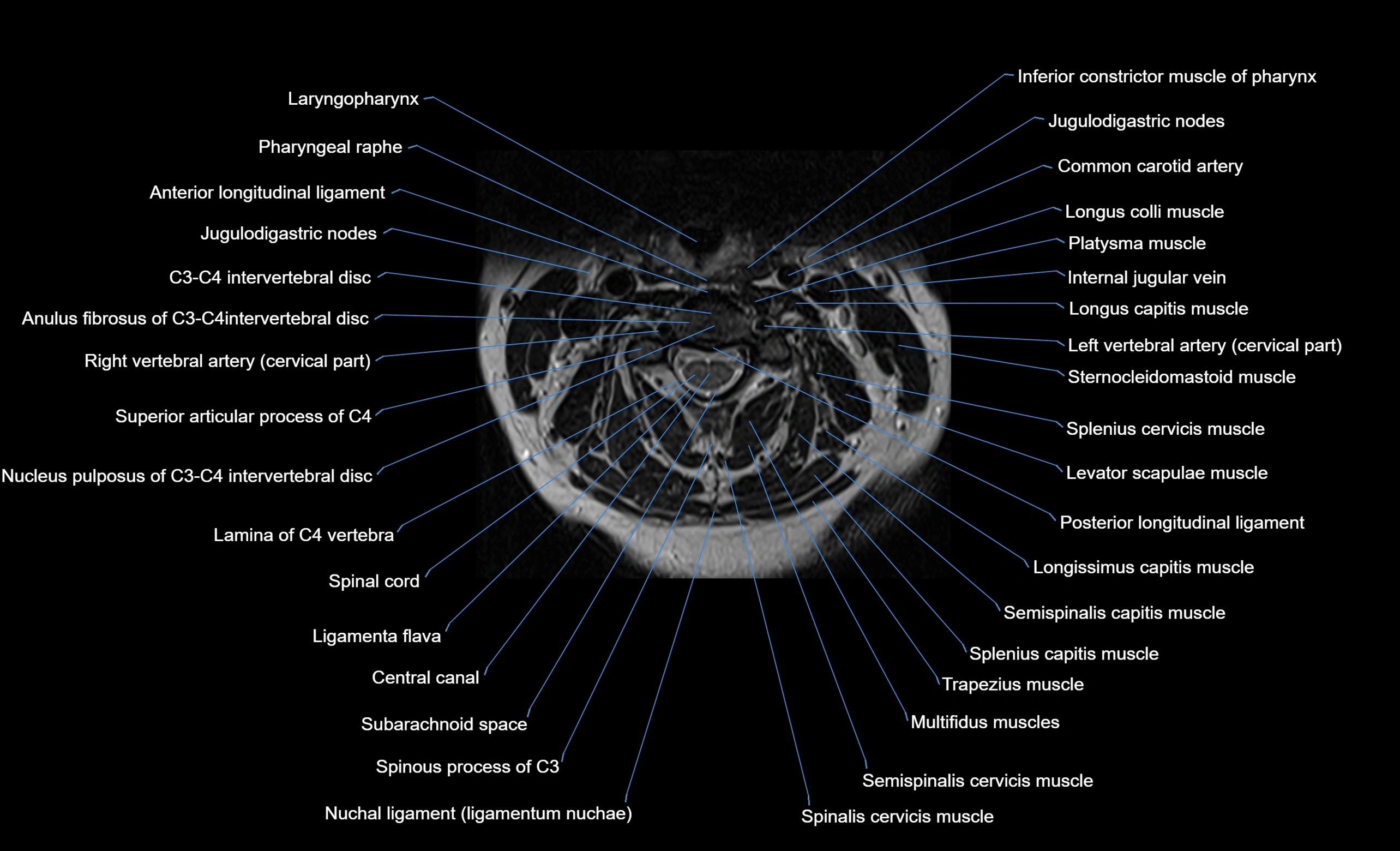
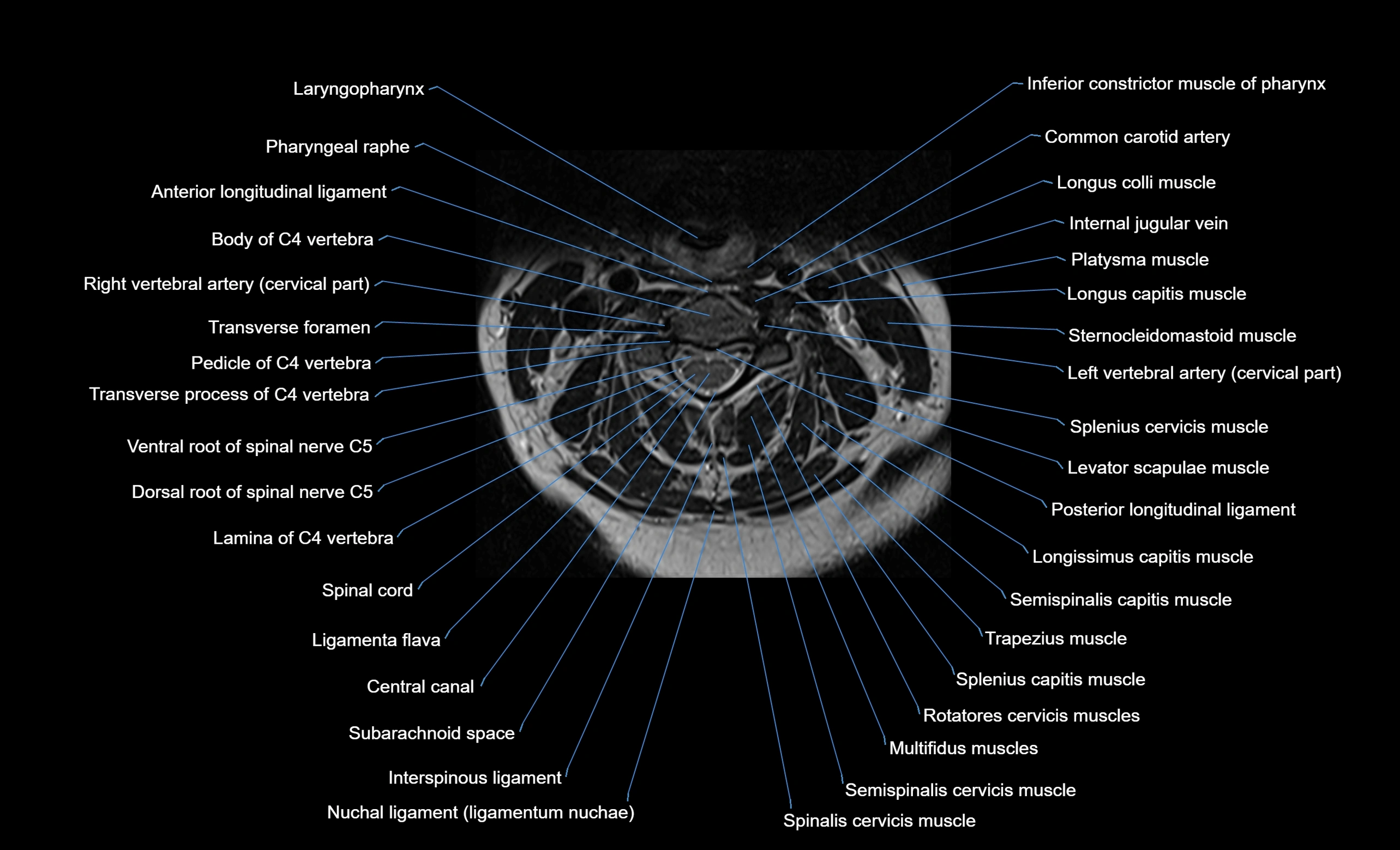
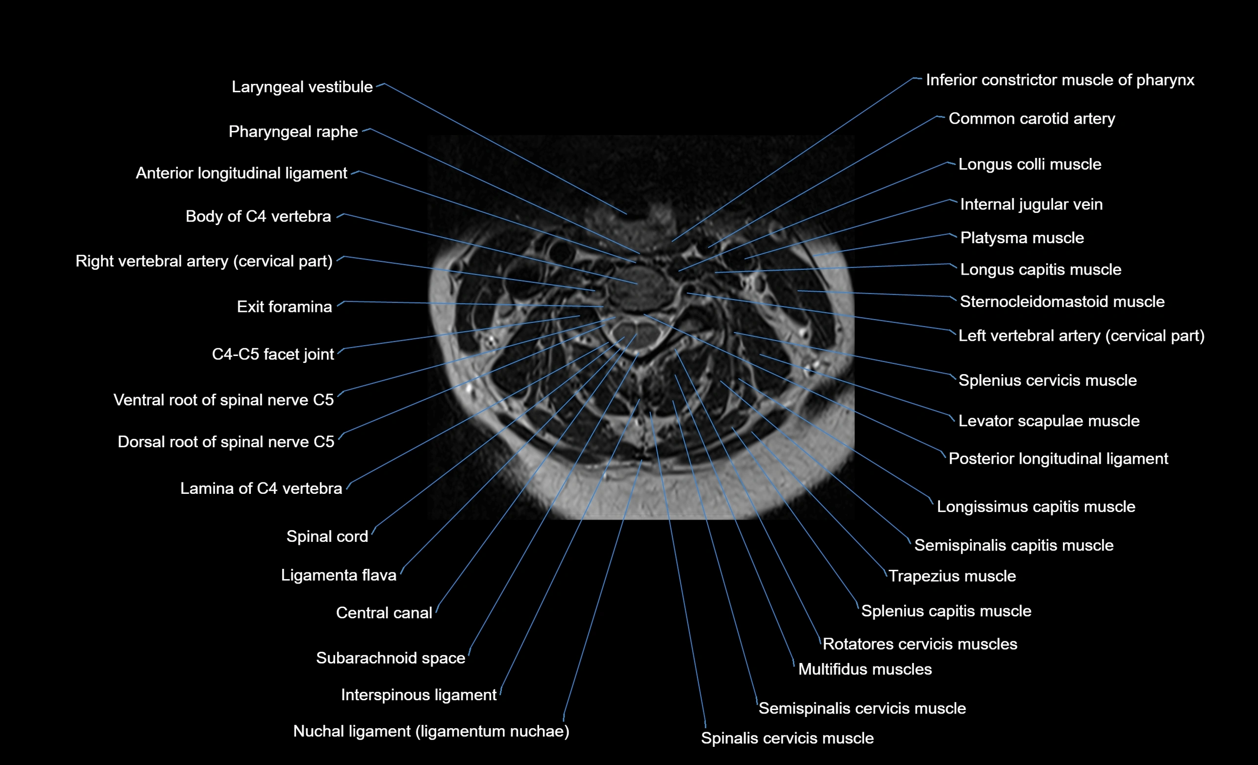
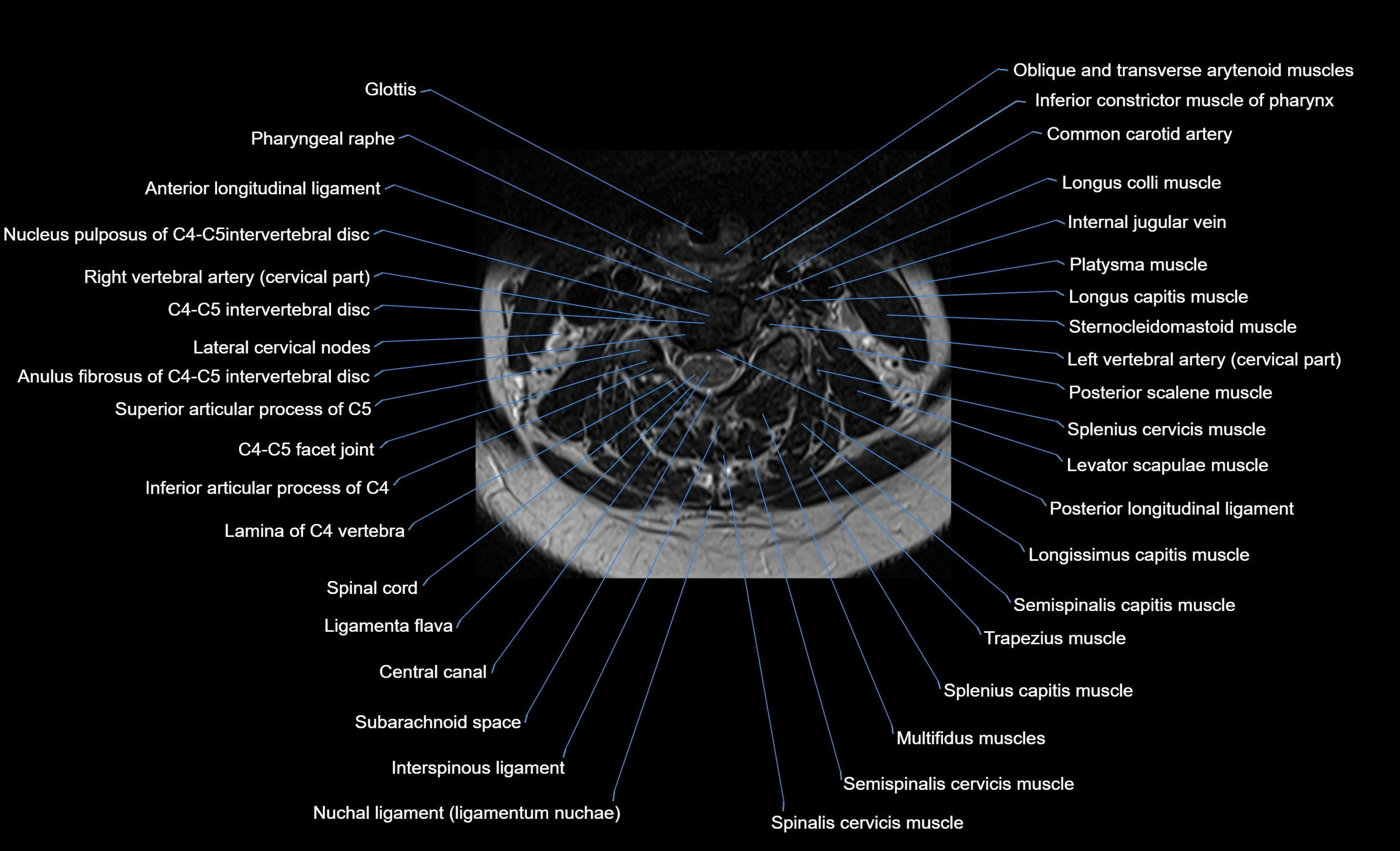
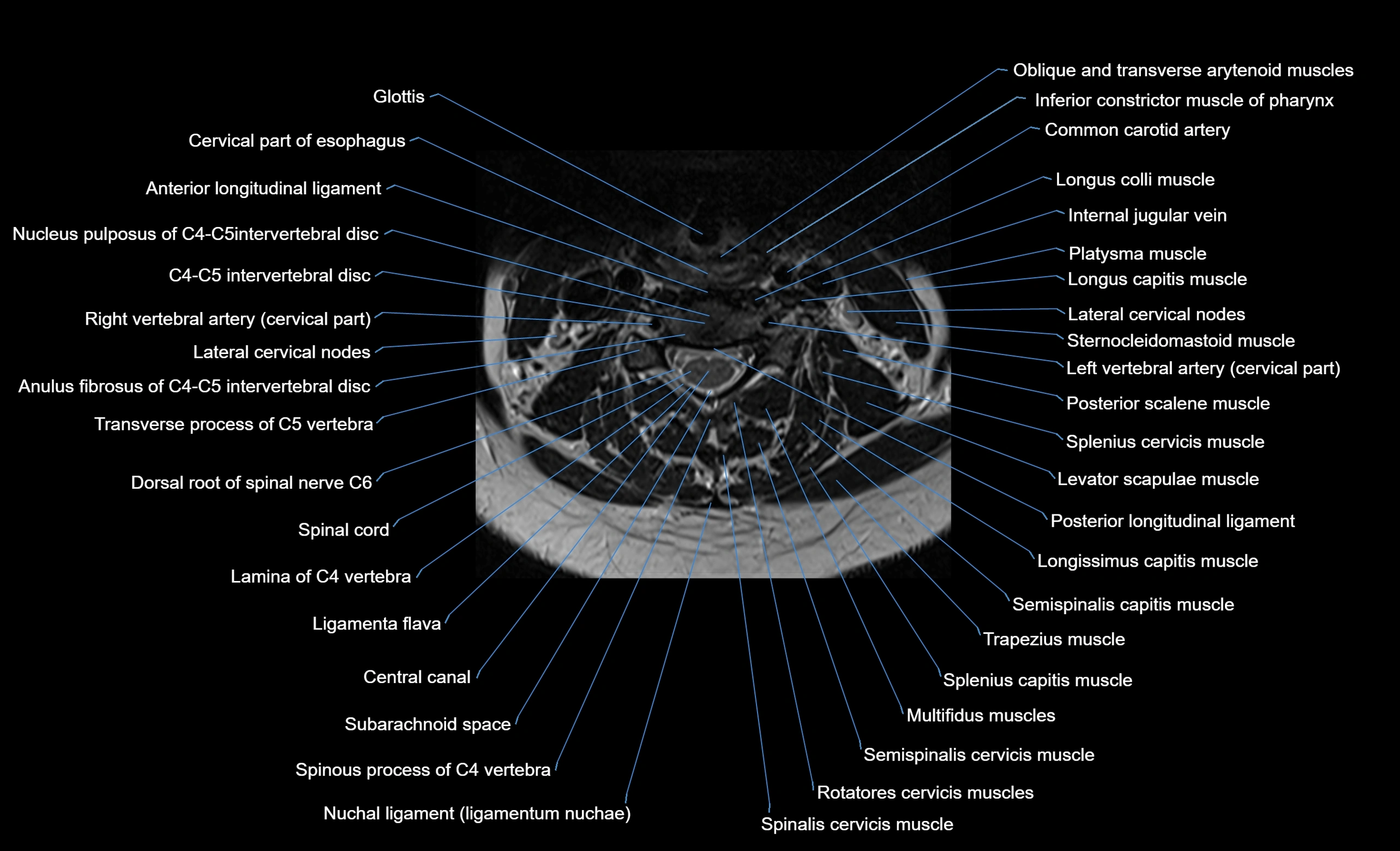
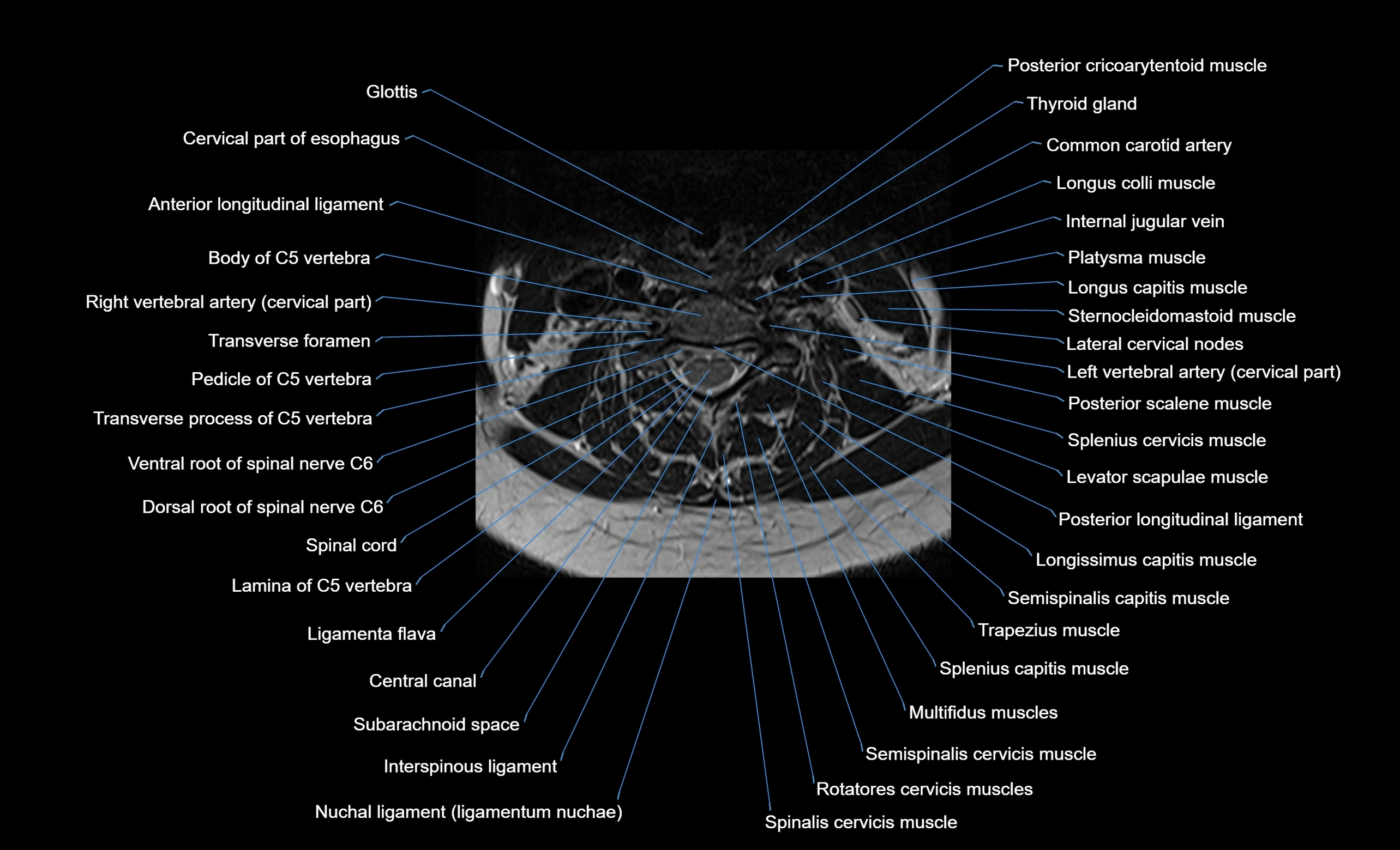
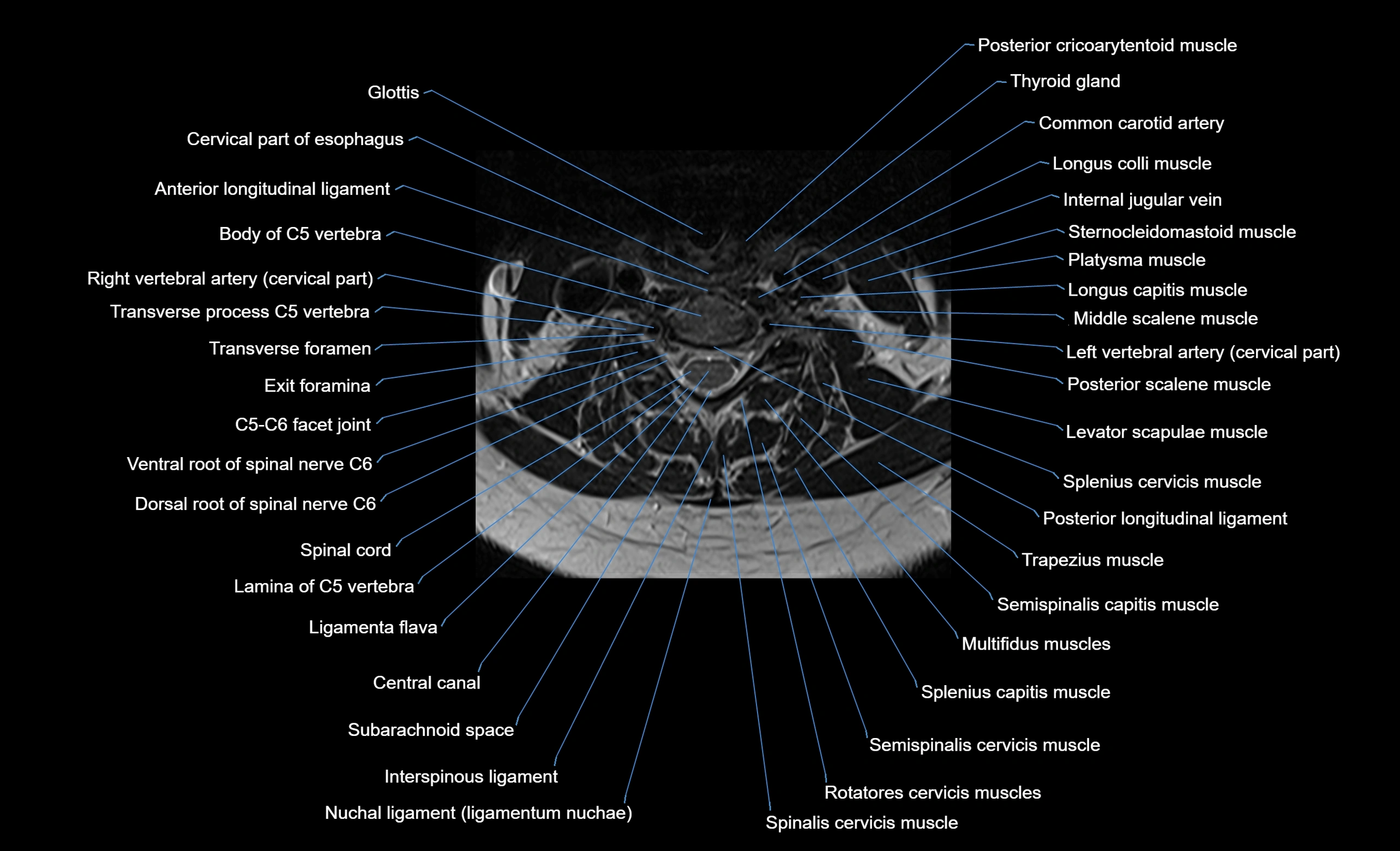
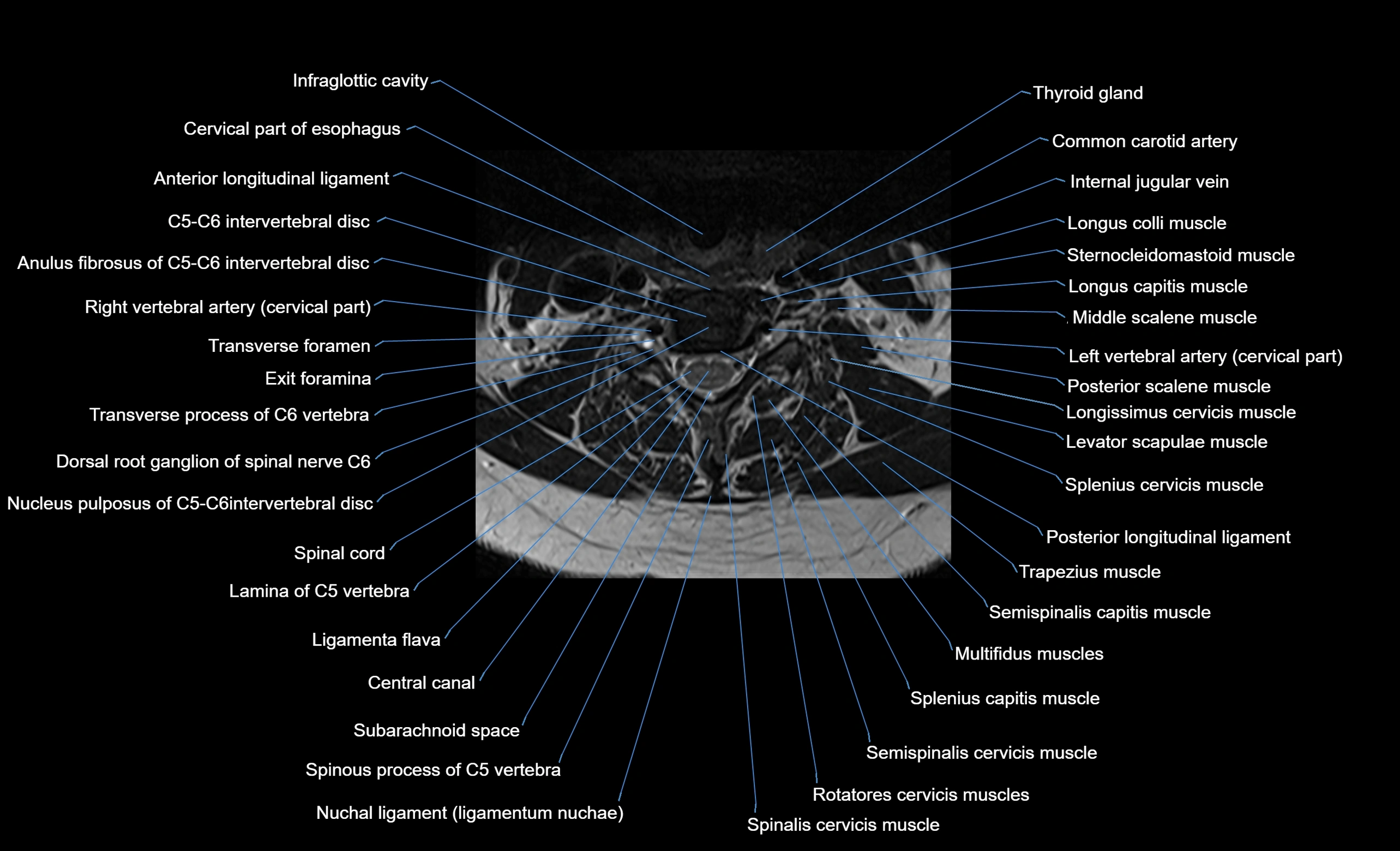
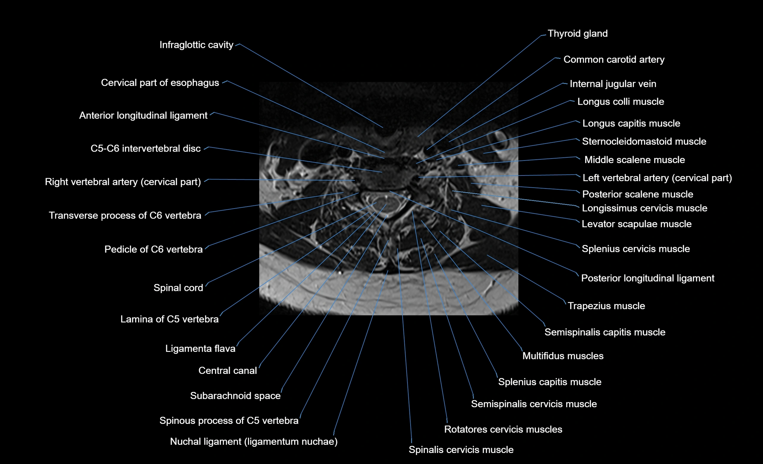
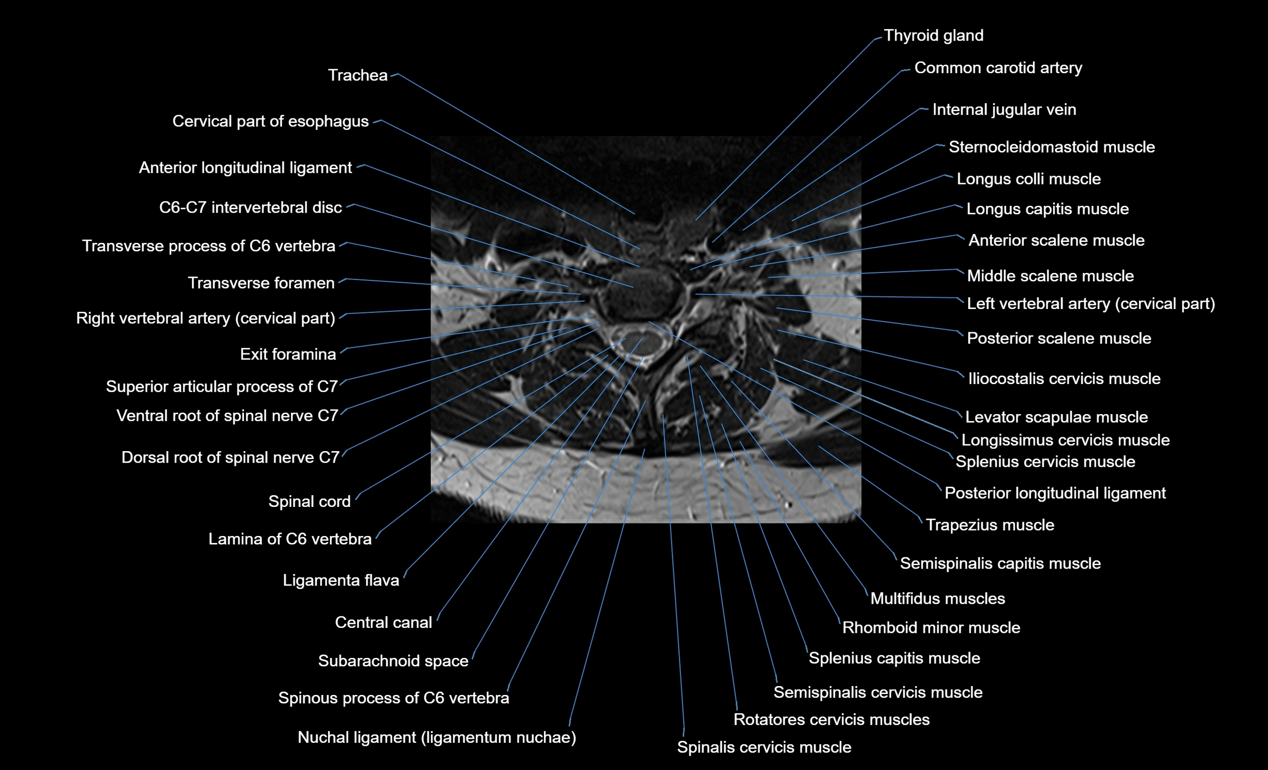
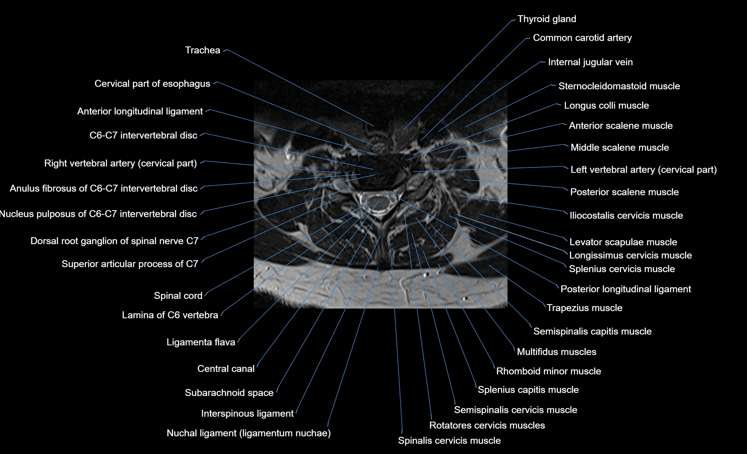
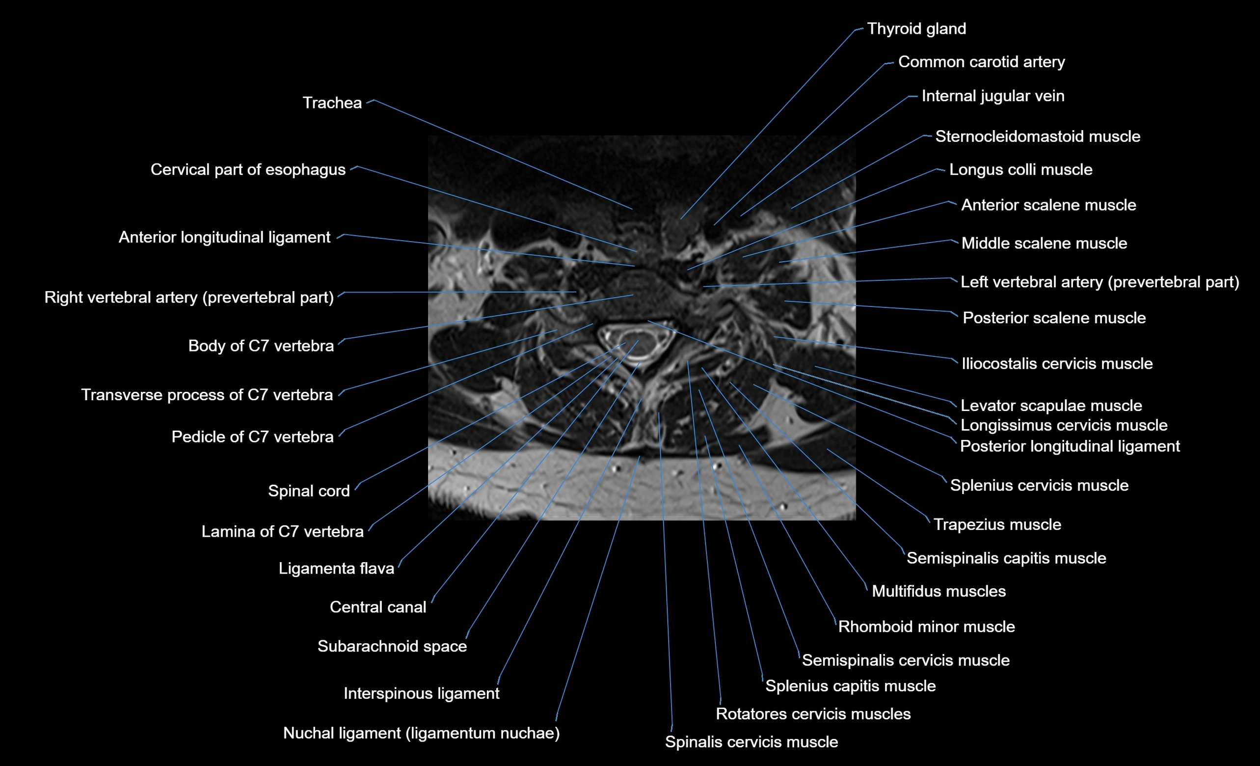
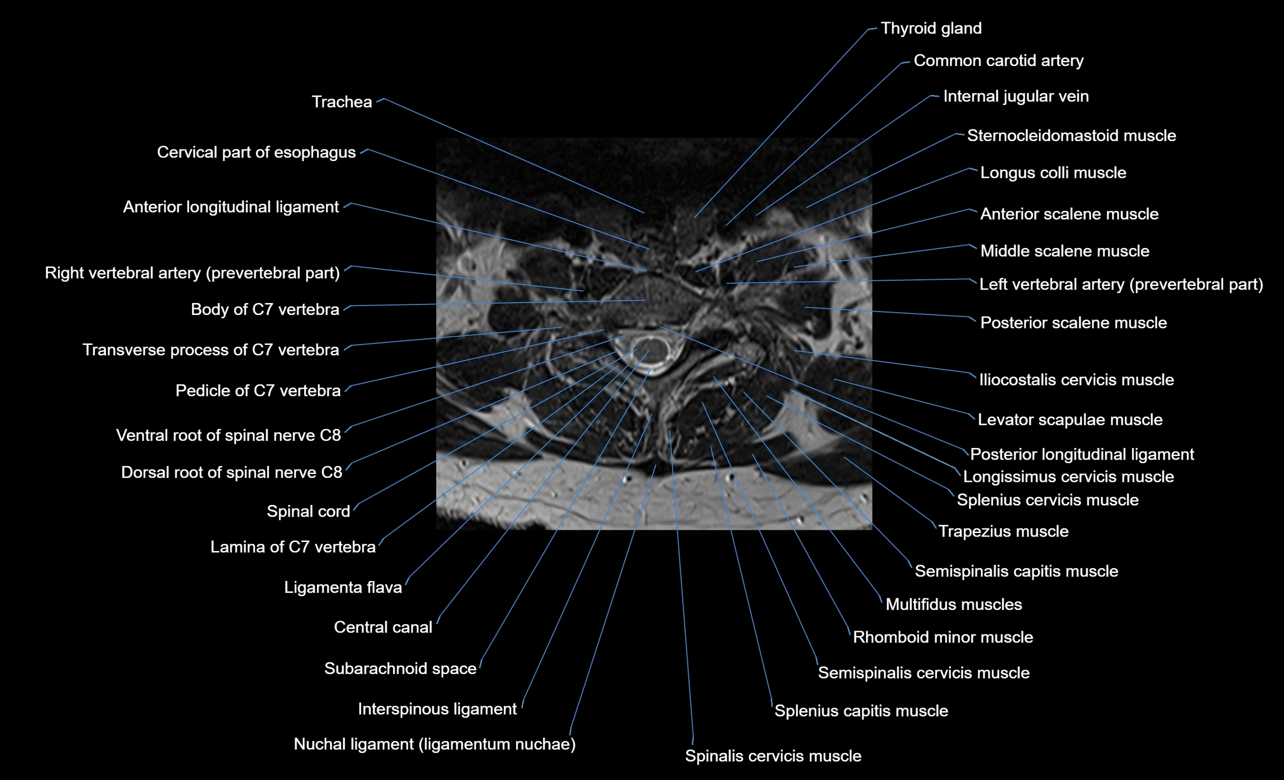
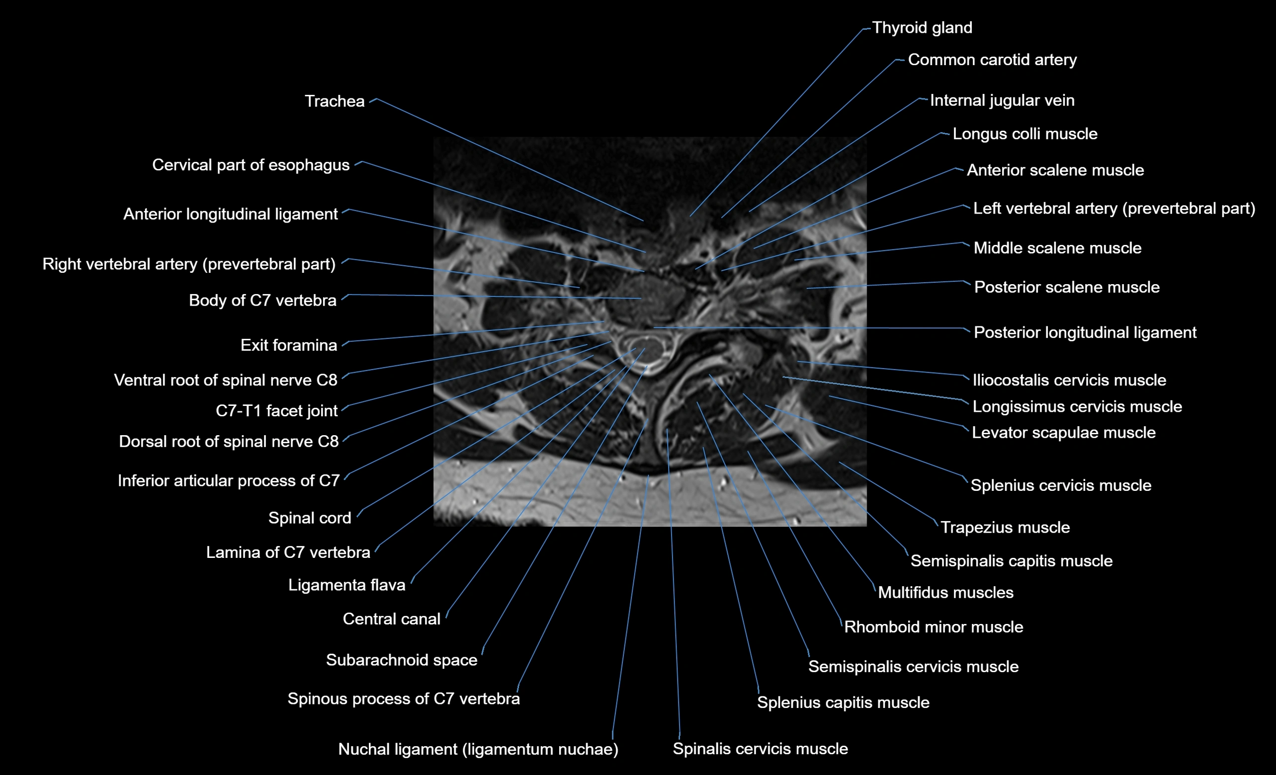
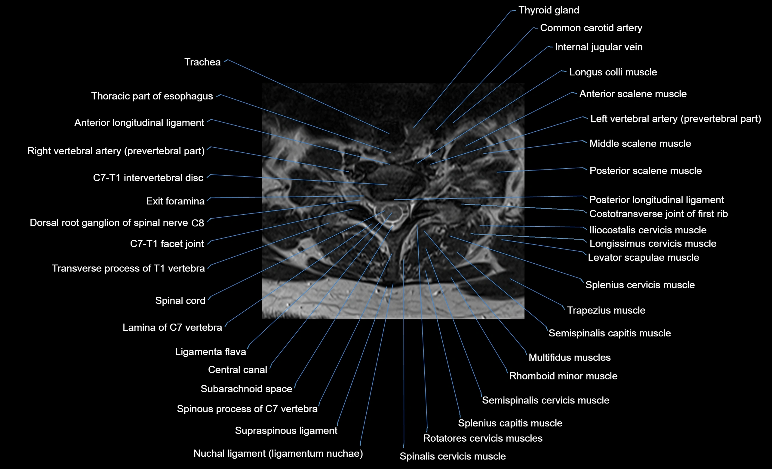
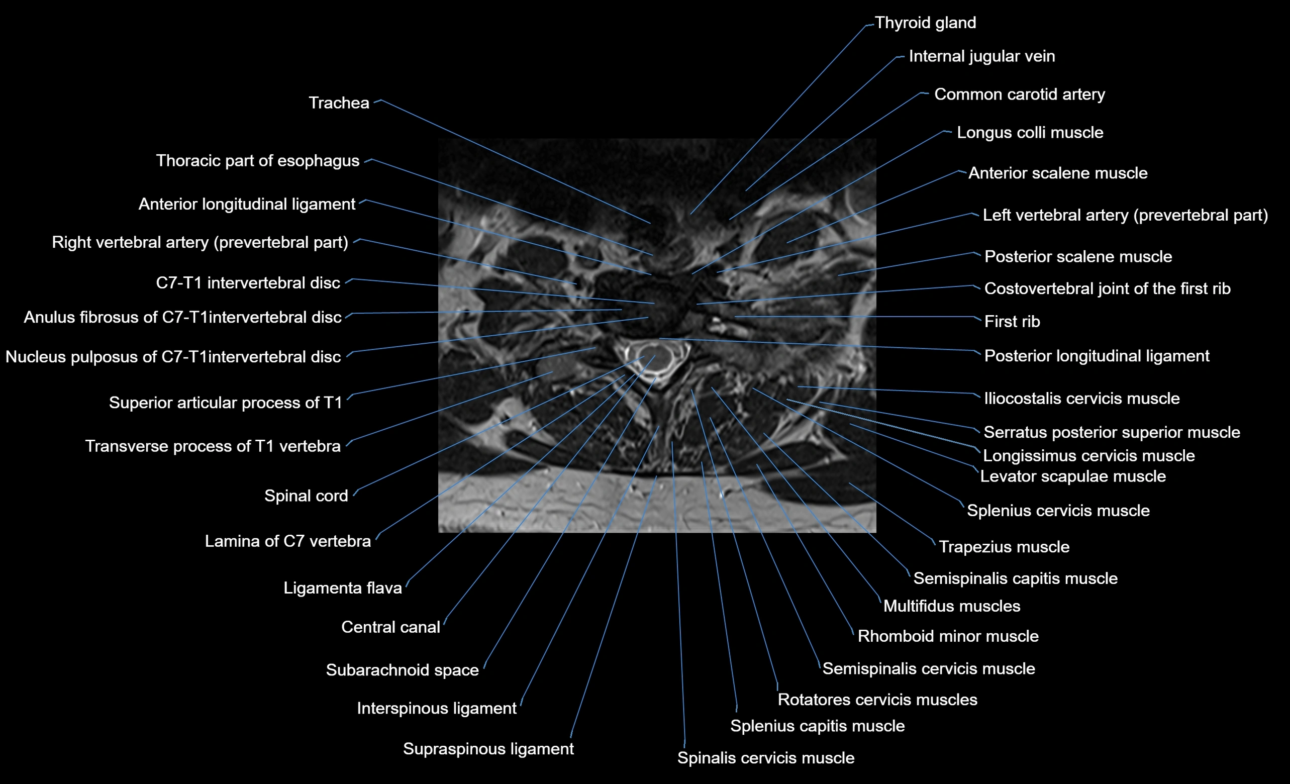
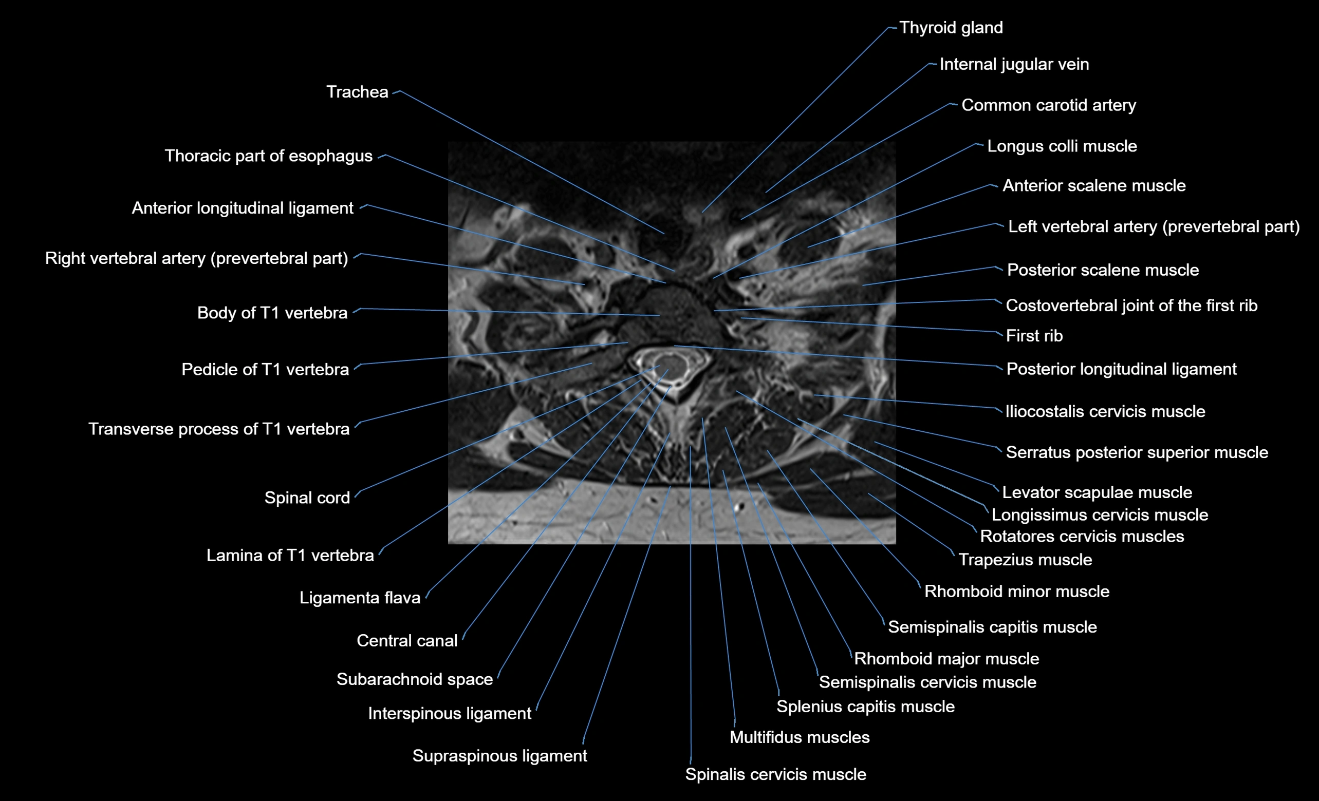
MRI image

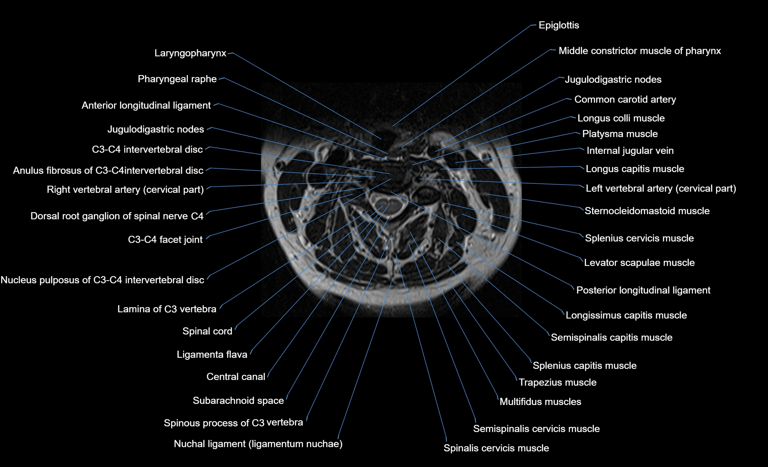
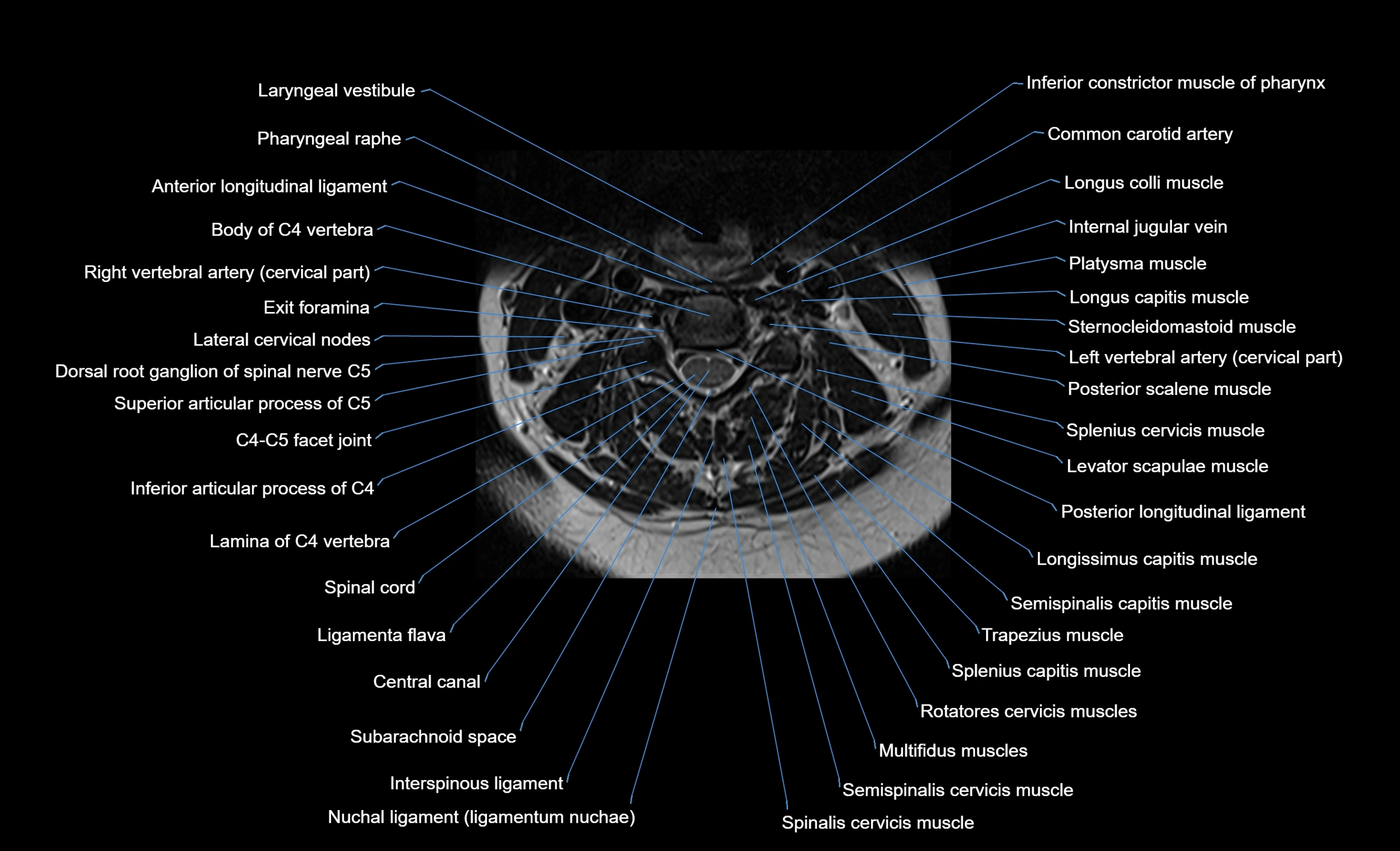
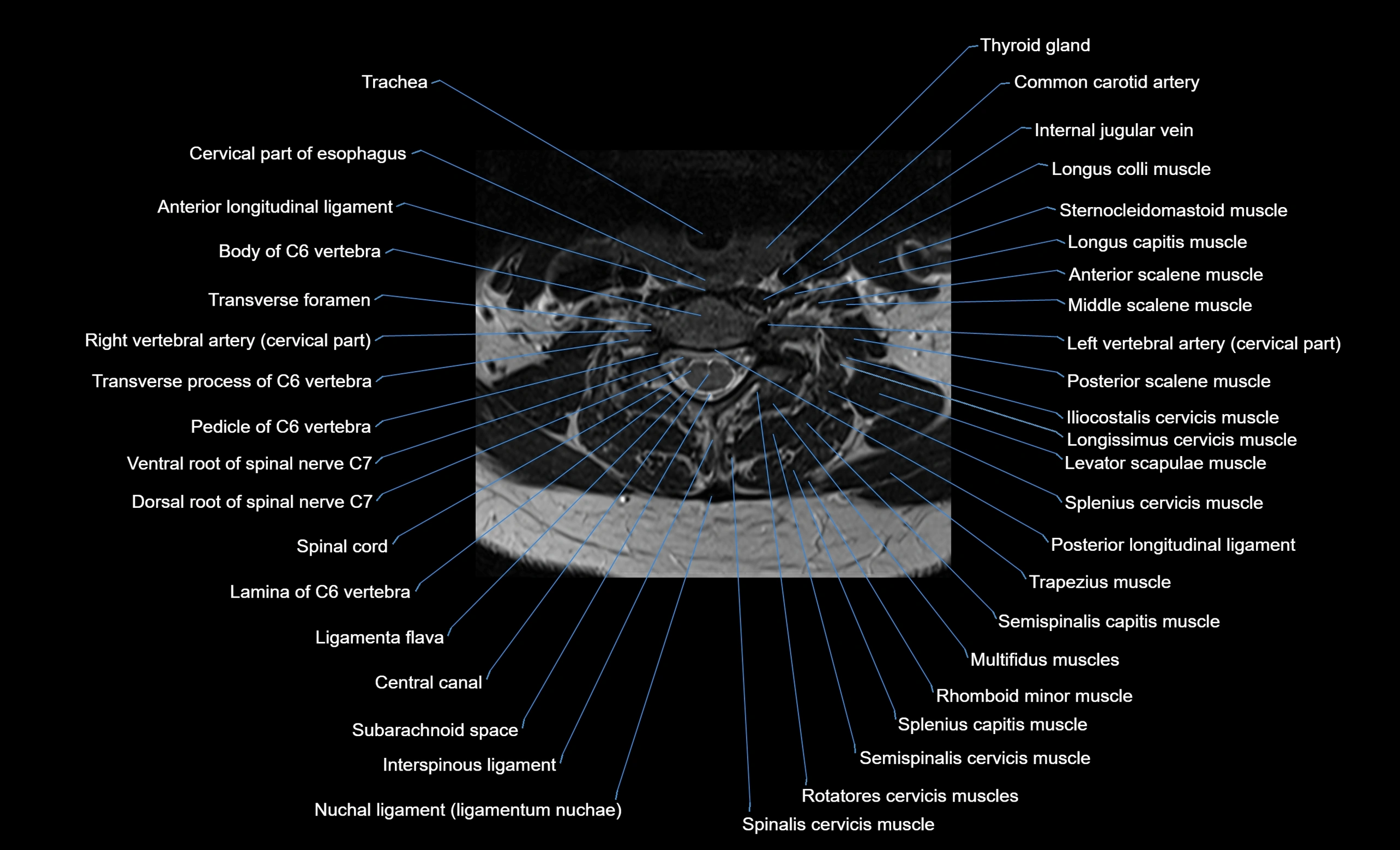
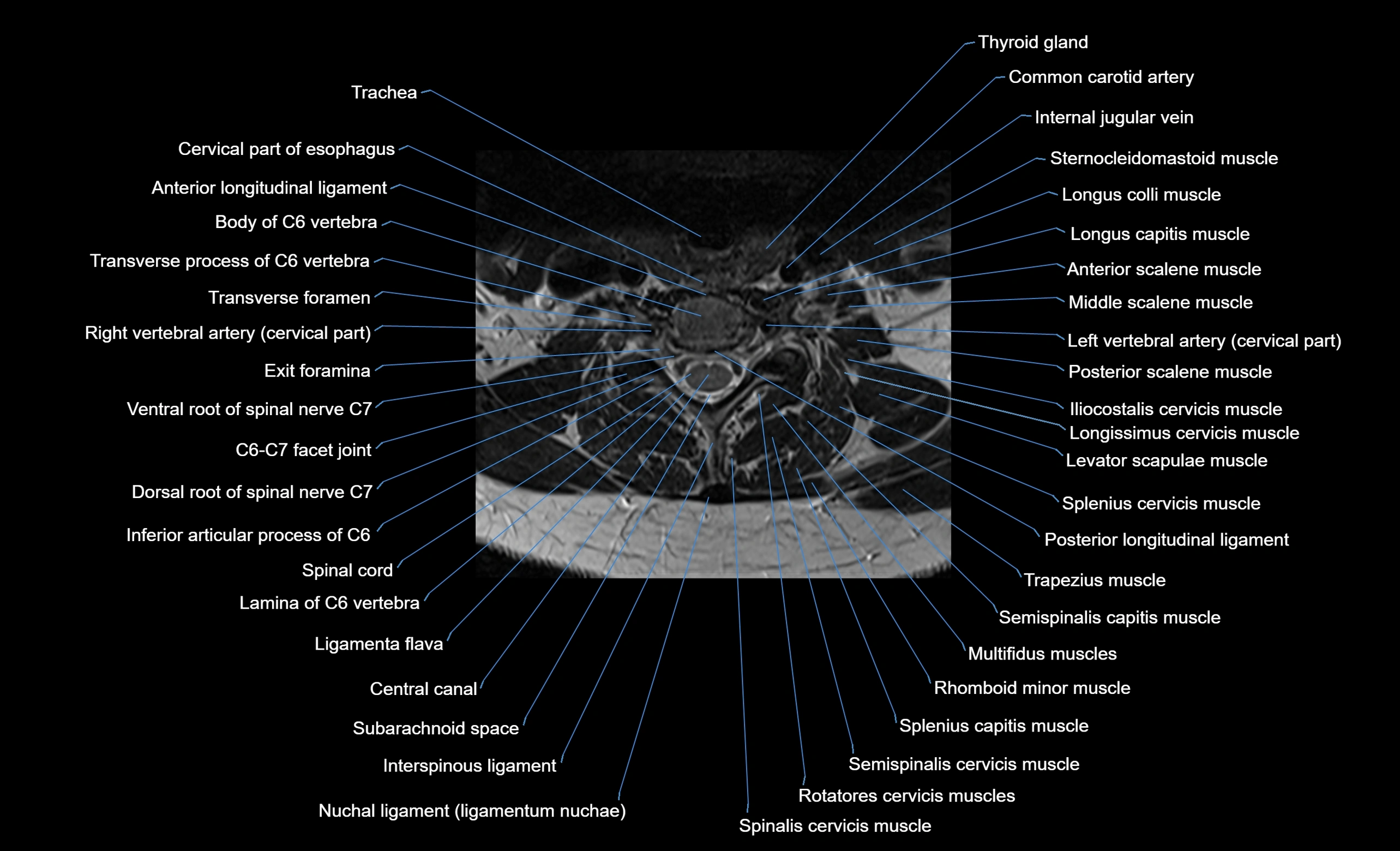
CT image


