


Topic
- Abducens nerve (Cranial nerve VI)
- Accessory Nerve (Cranial nerve XI)
- Ambient cistern
- Amygdalohippocampal area
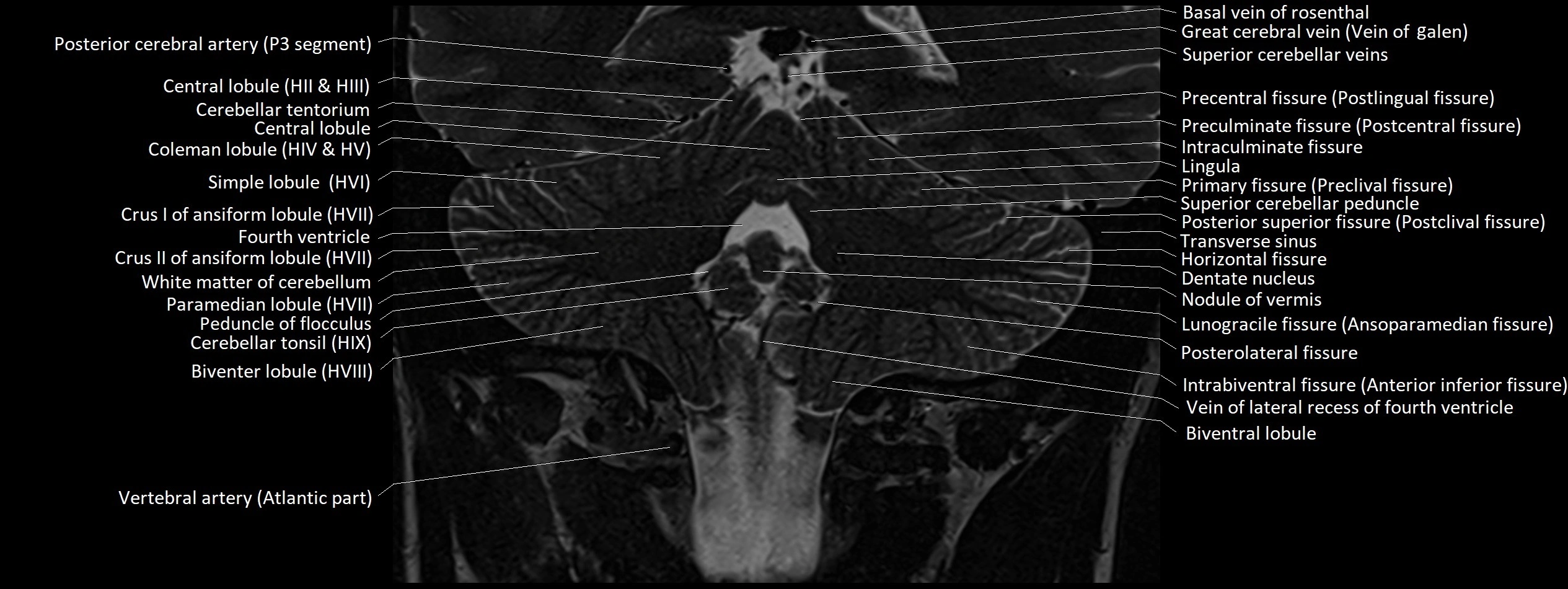
- Angular vein
- Anterior calcarine sulcus
- Anterior cerebral artery (A1 Segment)
- Anterior cerebral artery (A2 Segment)
- Anterior cerebral artery (A3 Segment)
- Anterior cochlear nucleus
- Anterior external vertebral venous plexuses
- Anterior internal vertebral venous plexus
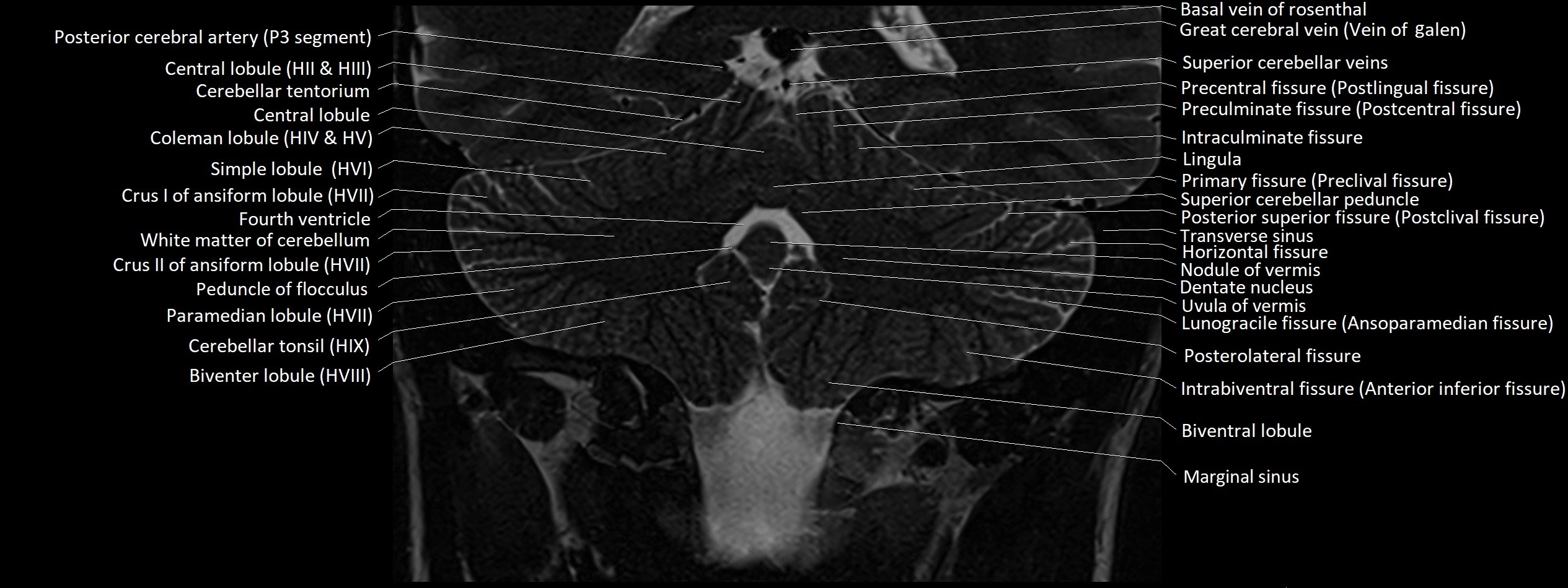
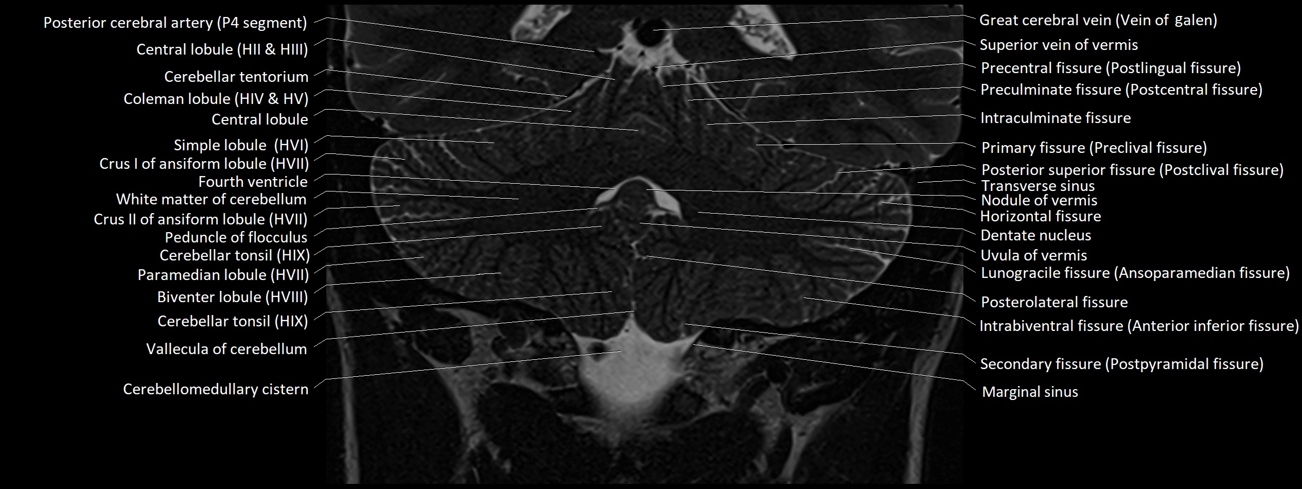
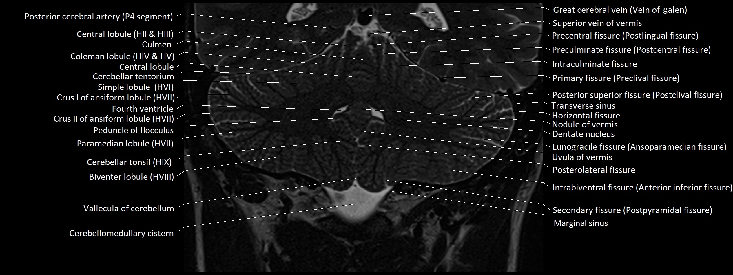
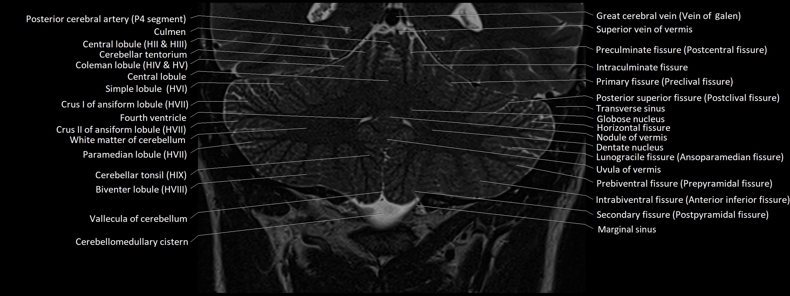
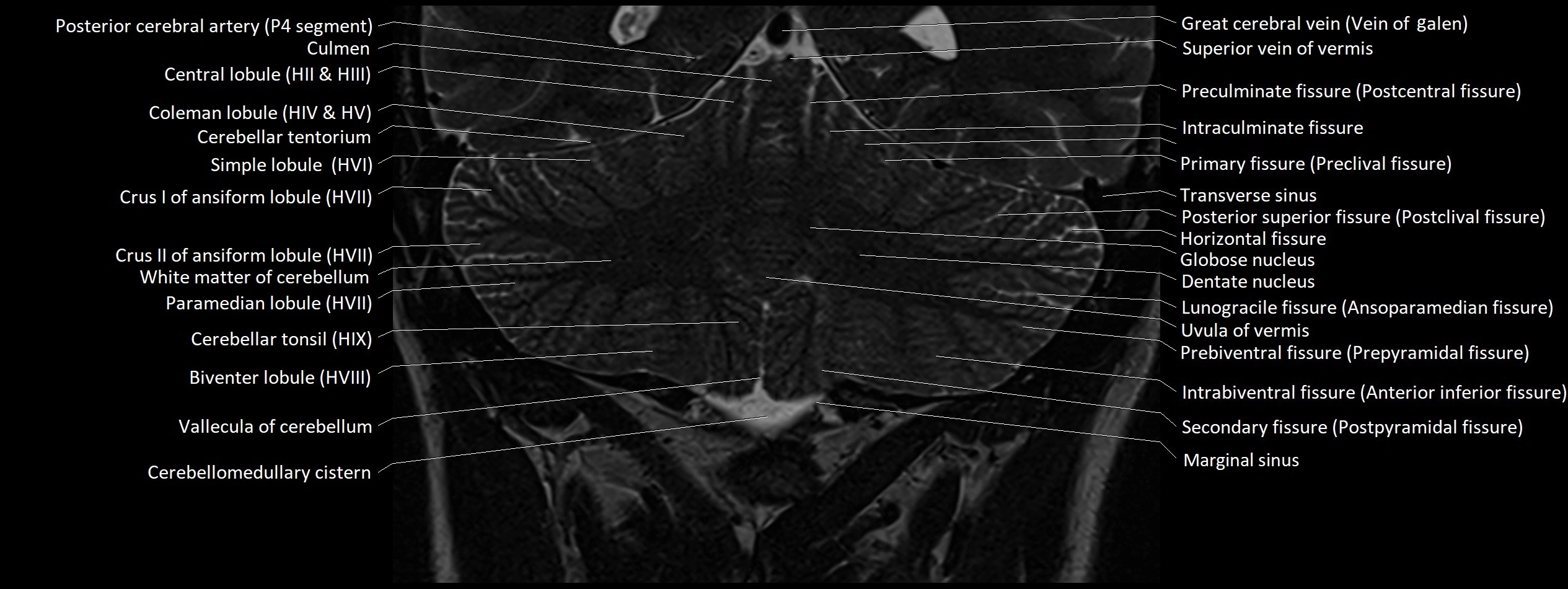
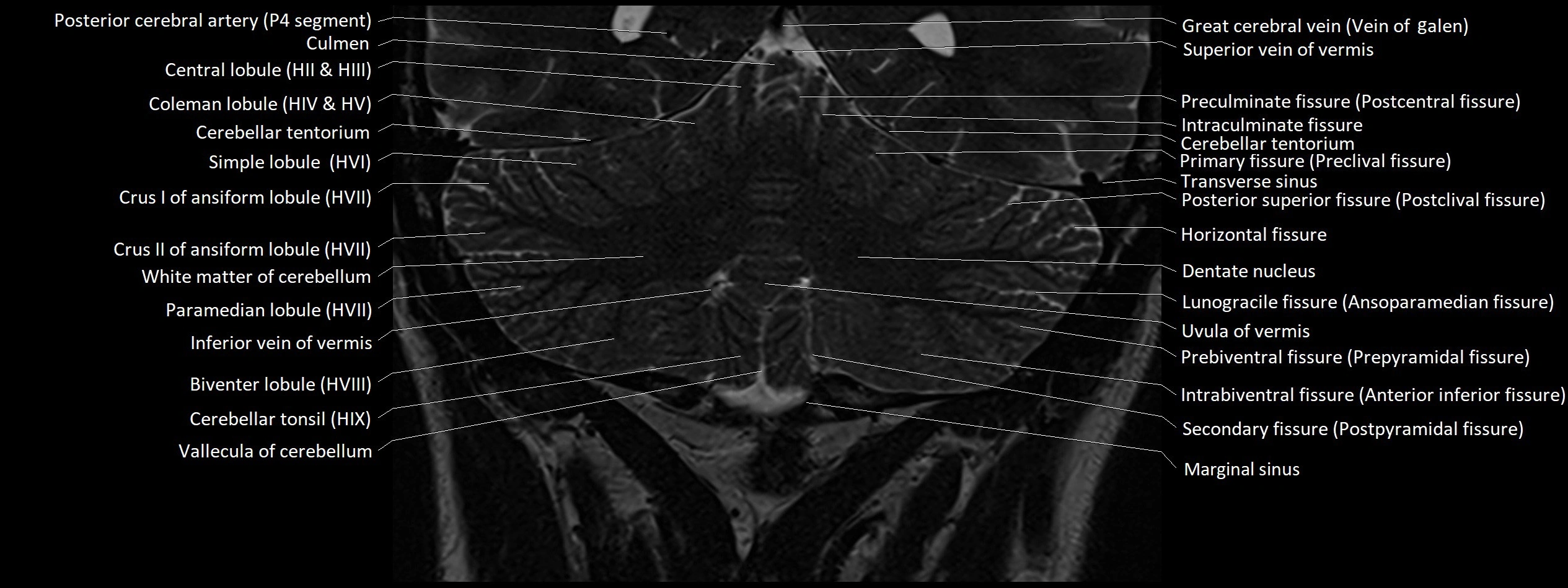
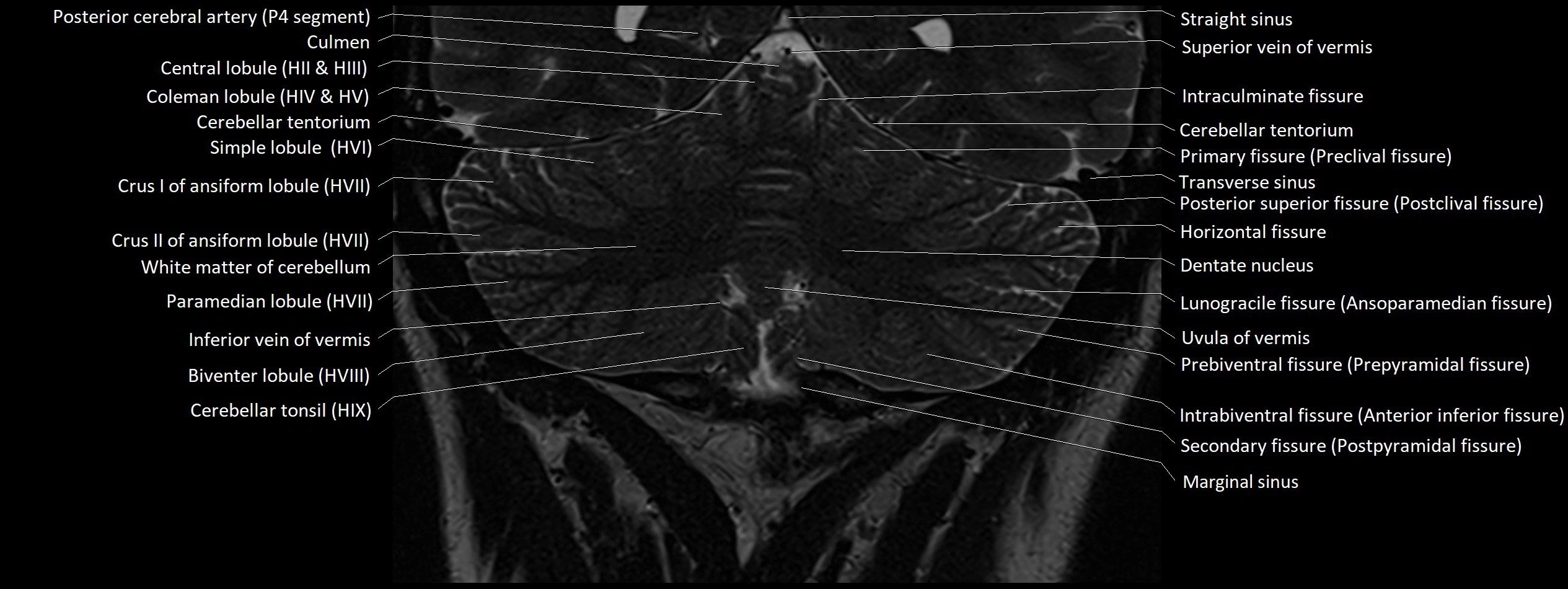
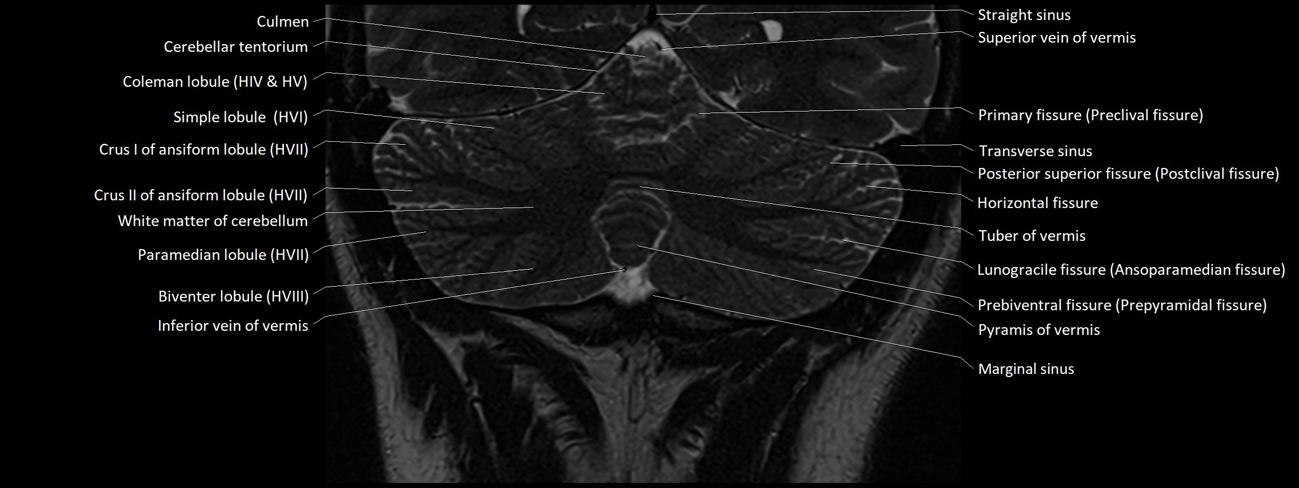
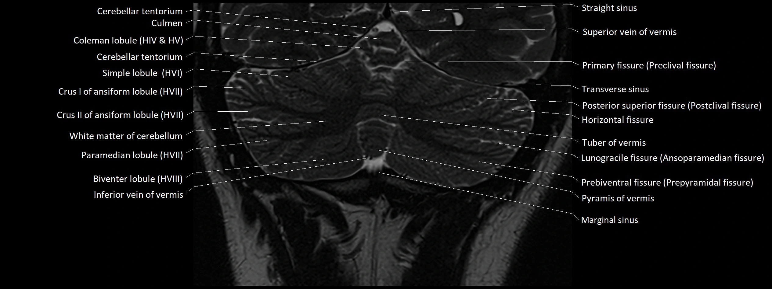
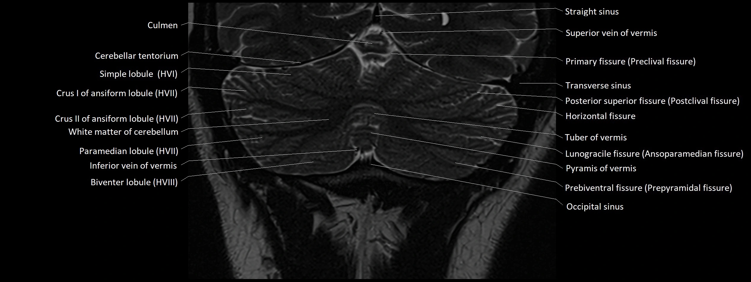
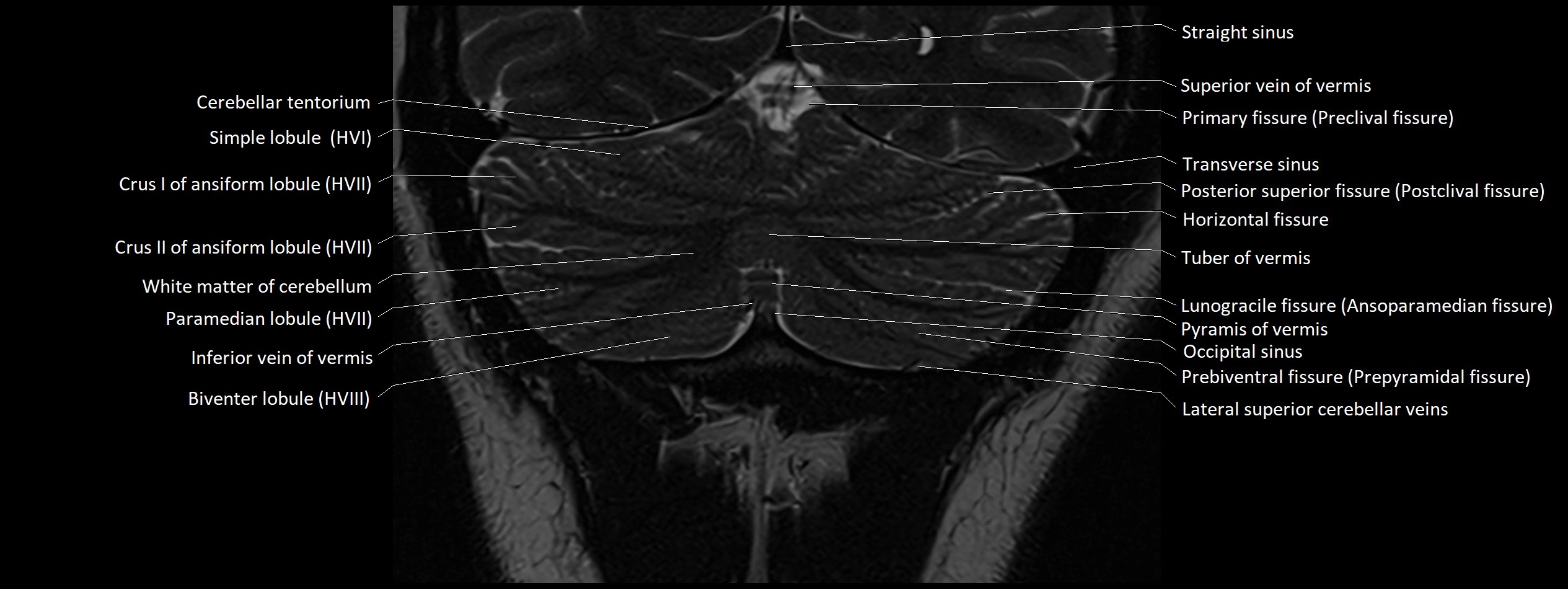
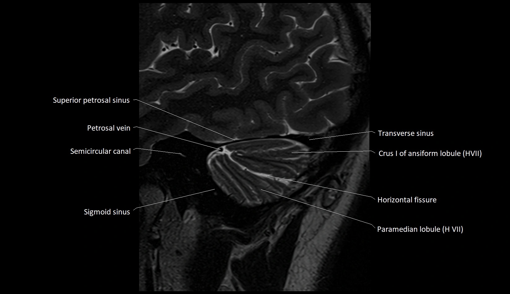
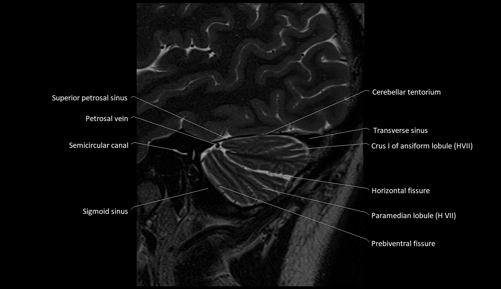
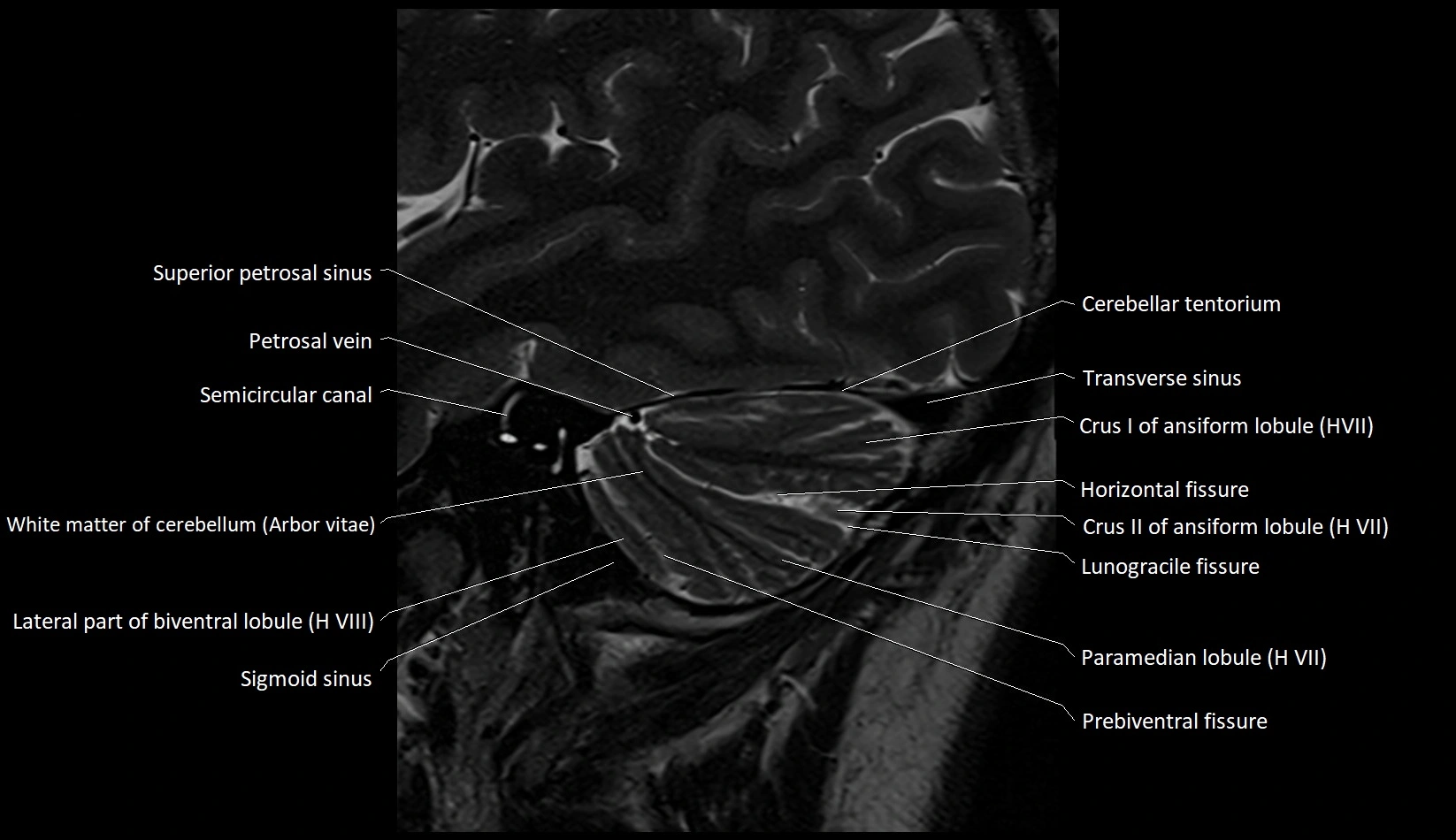
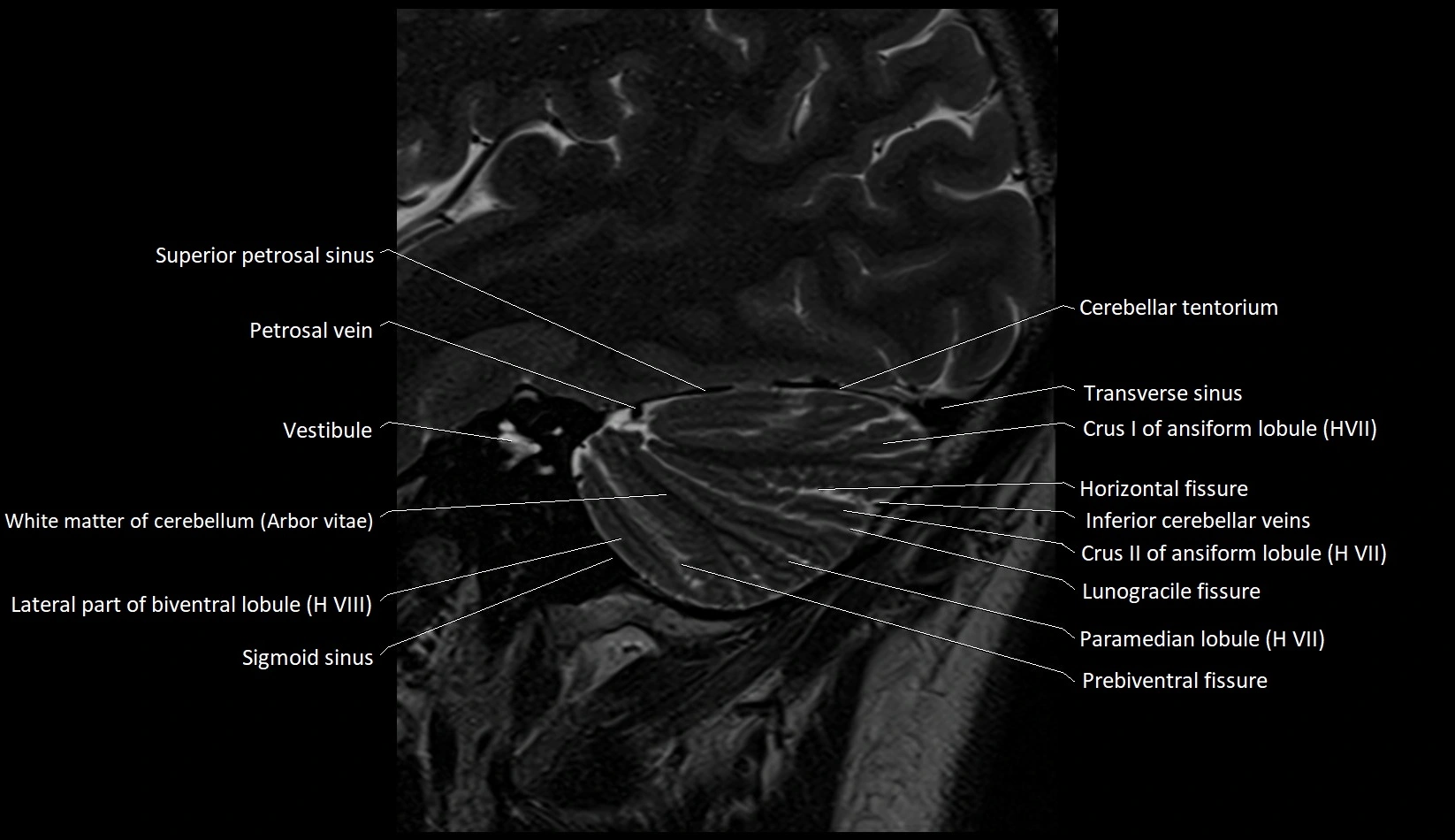
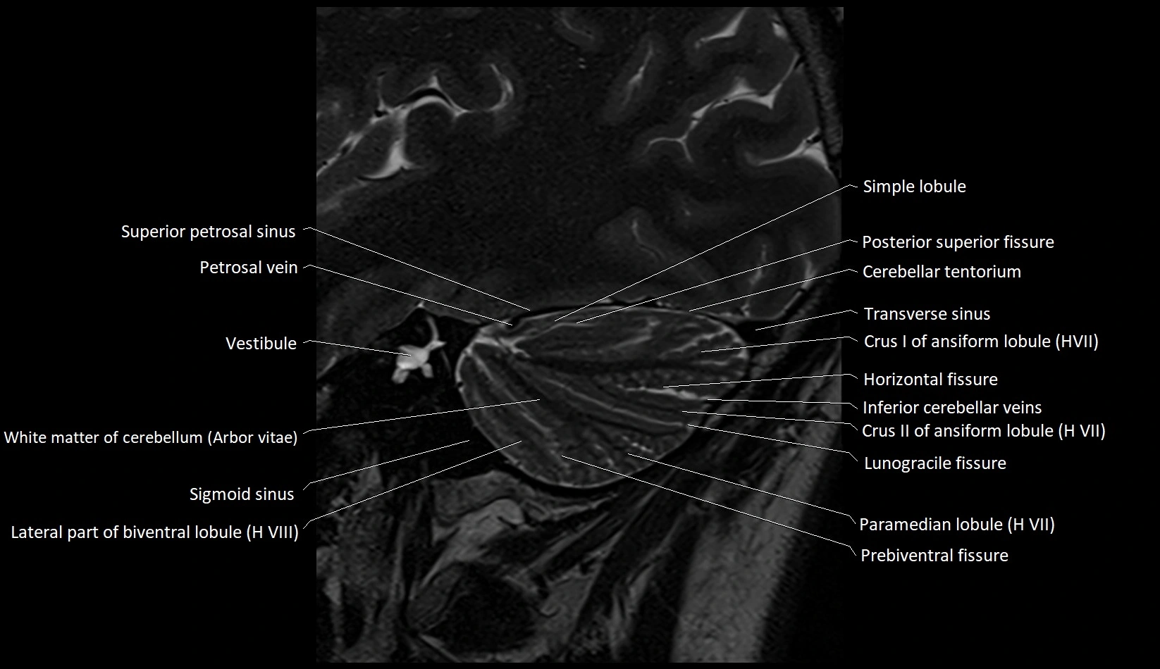
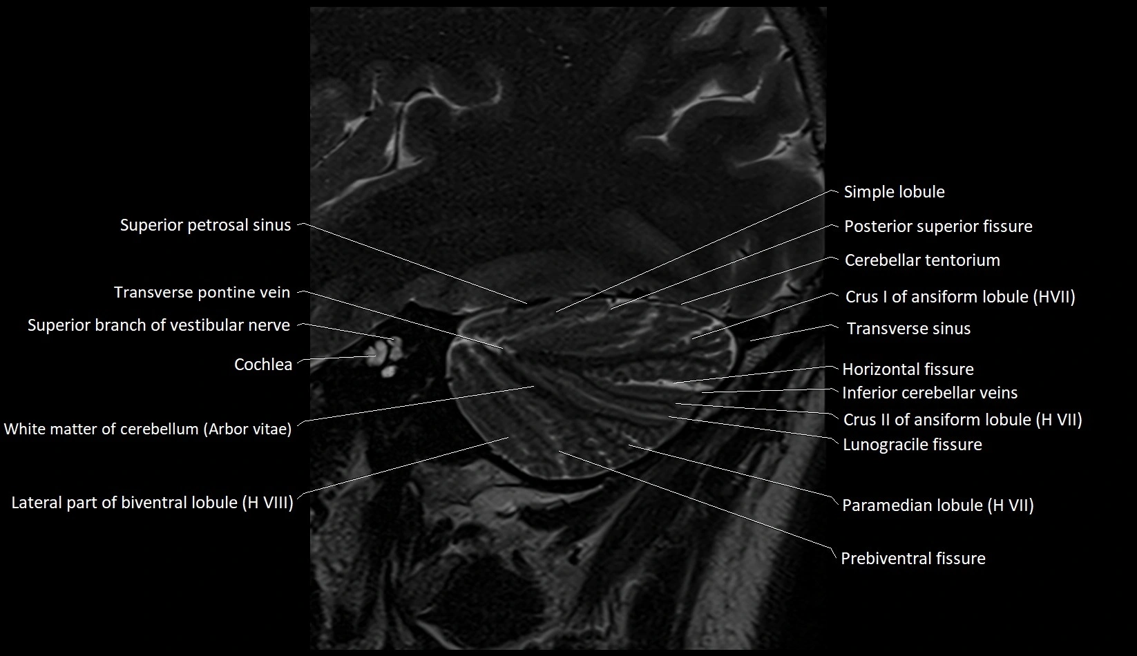
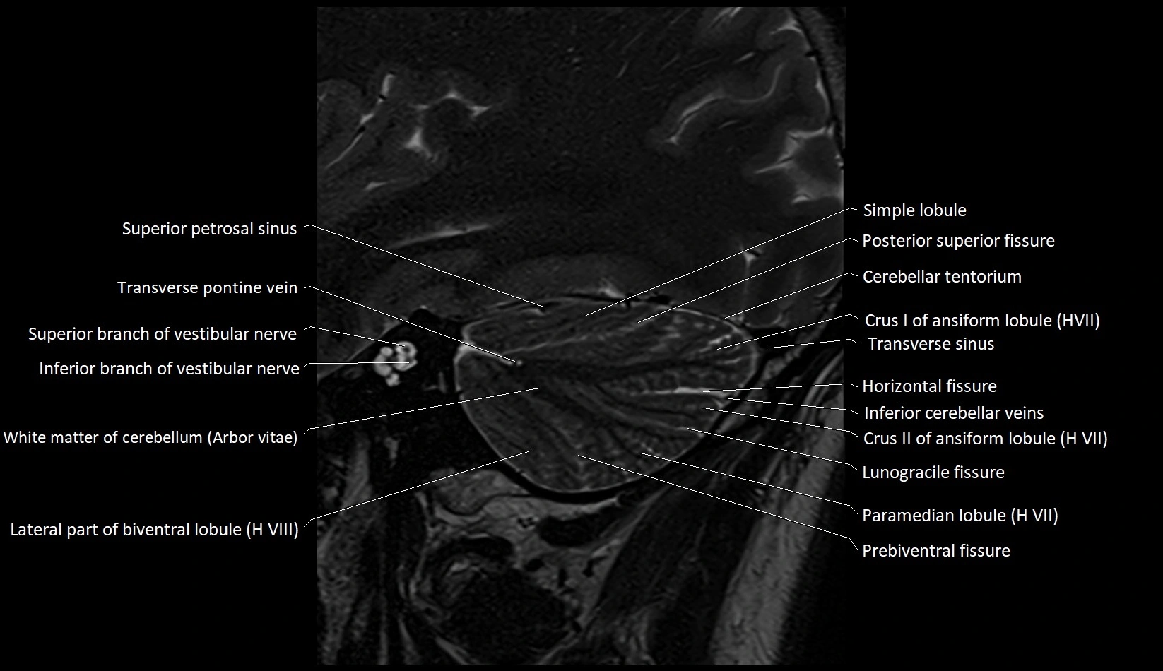
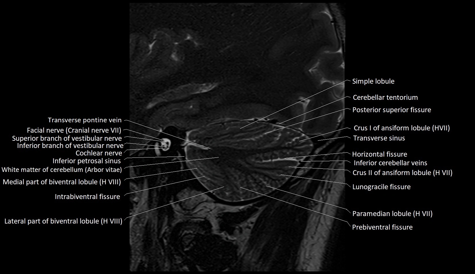
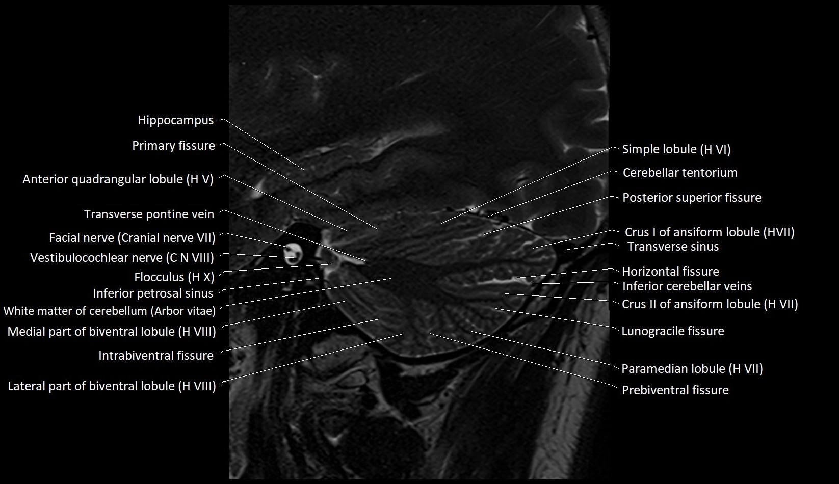
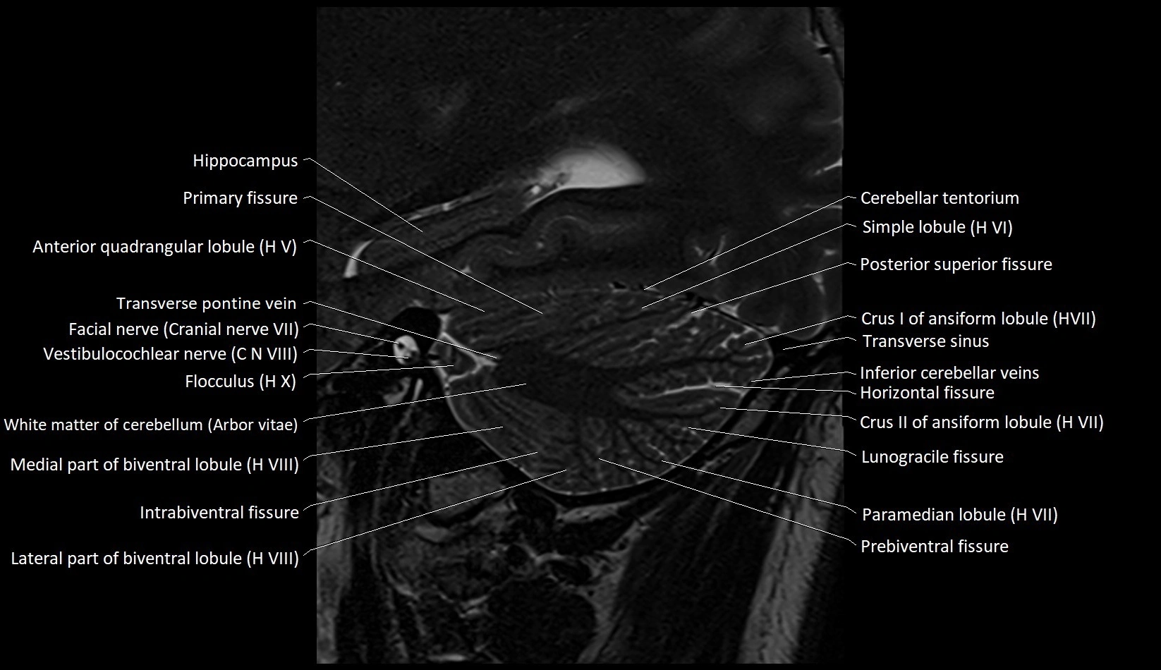
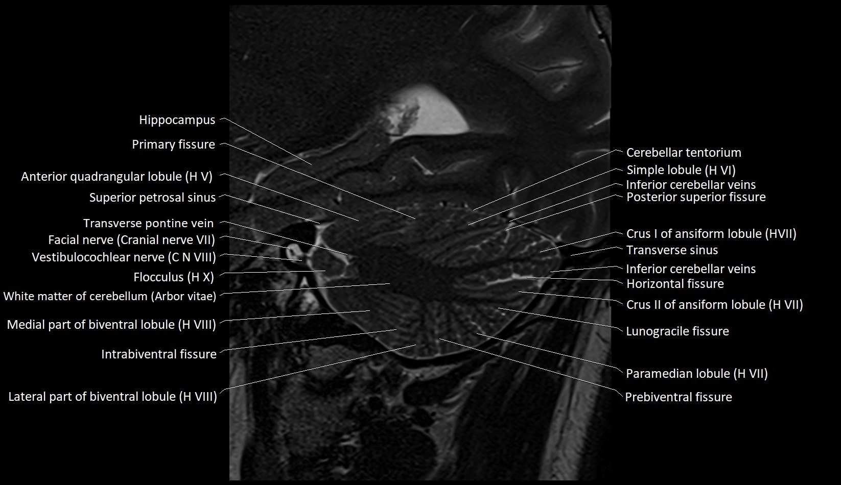
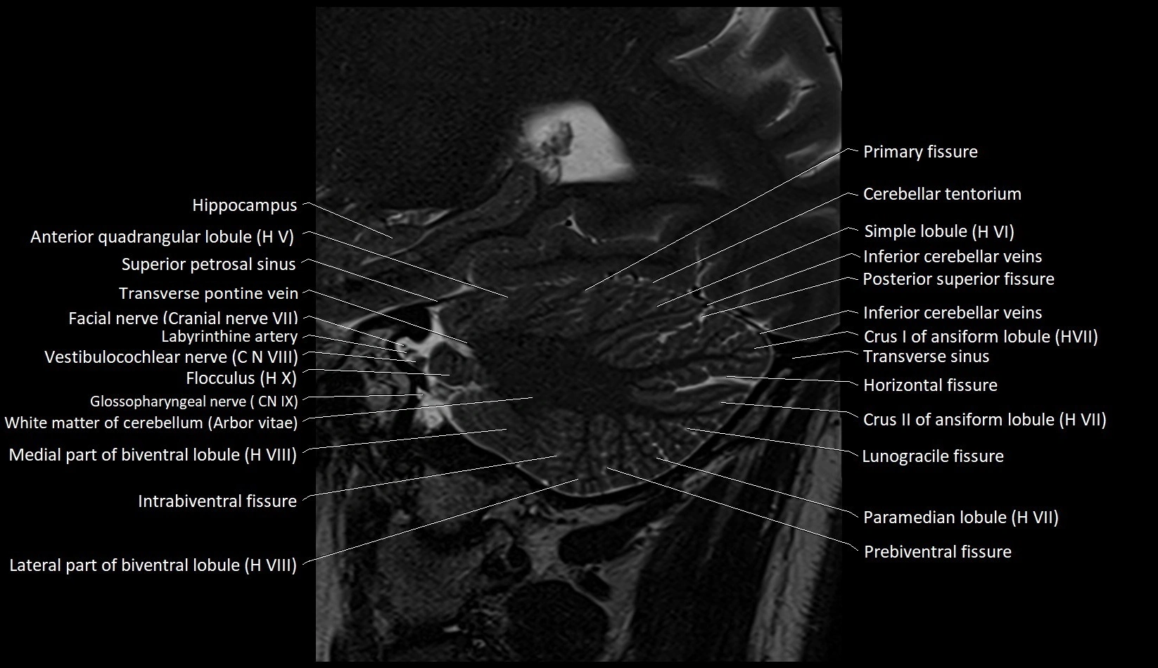
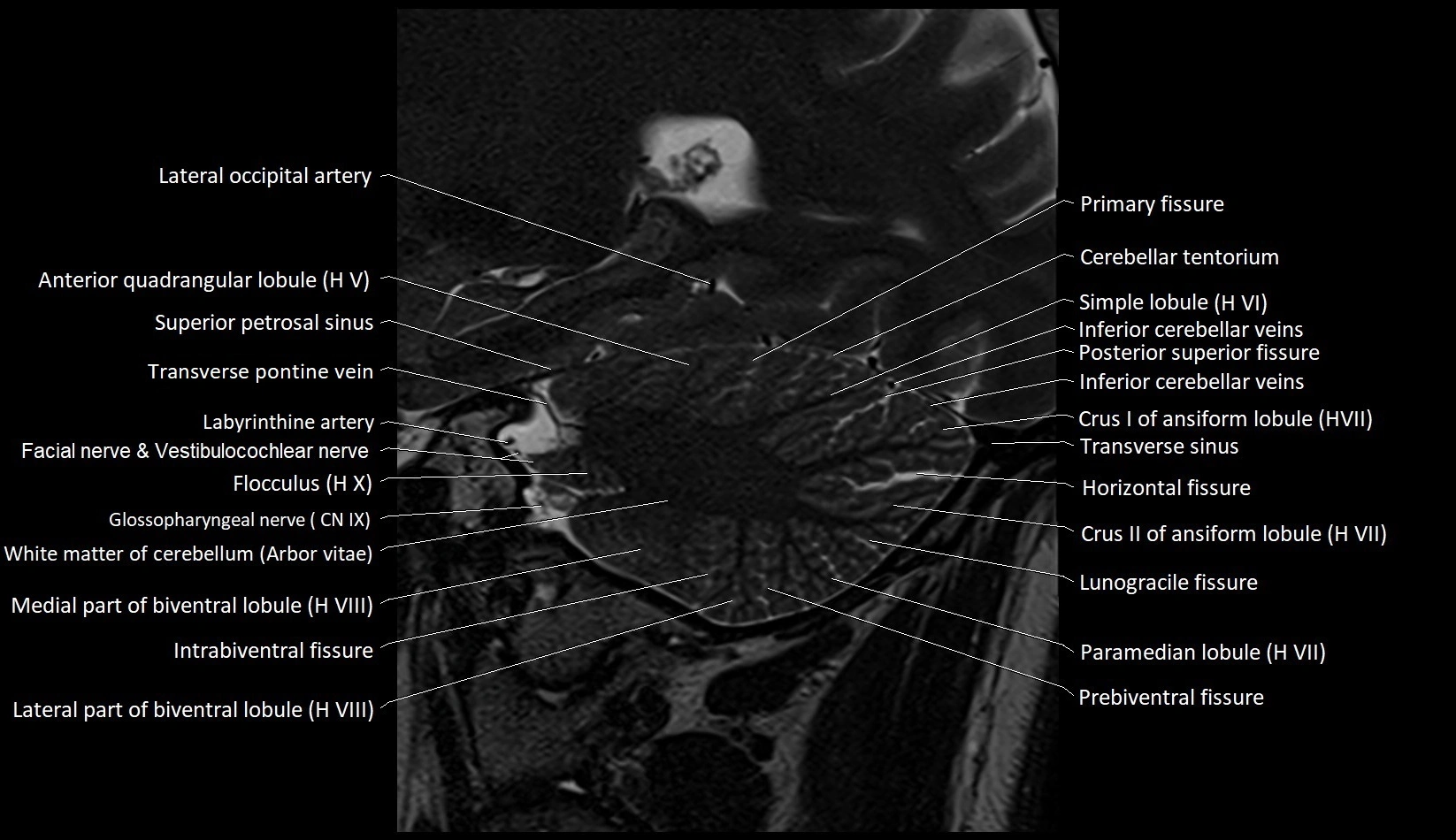
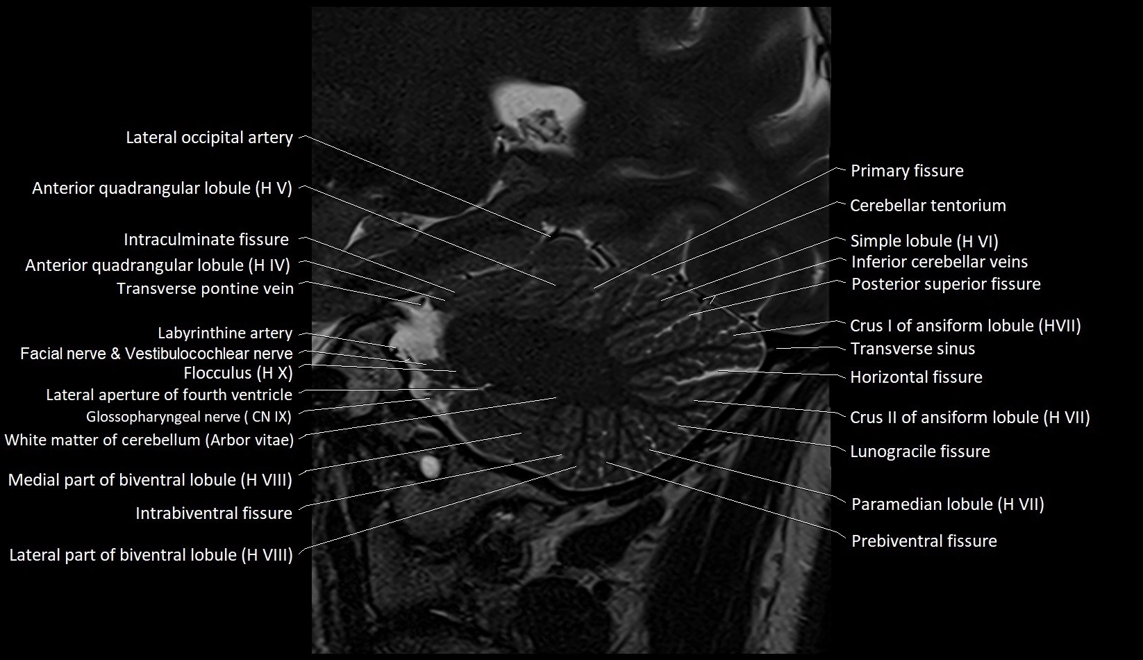
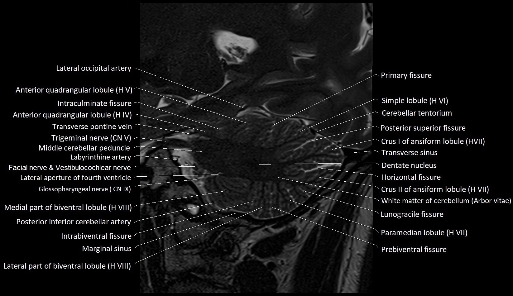
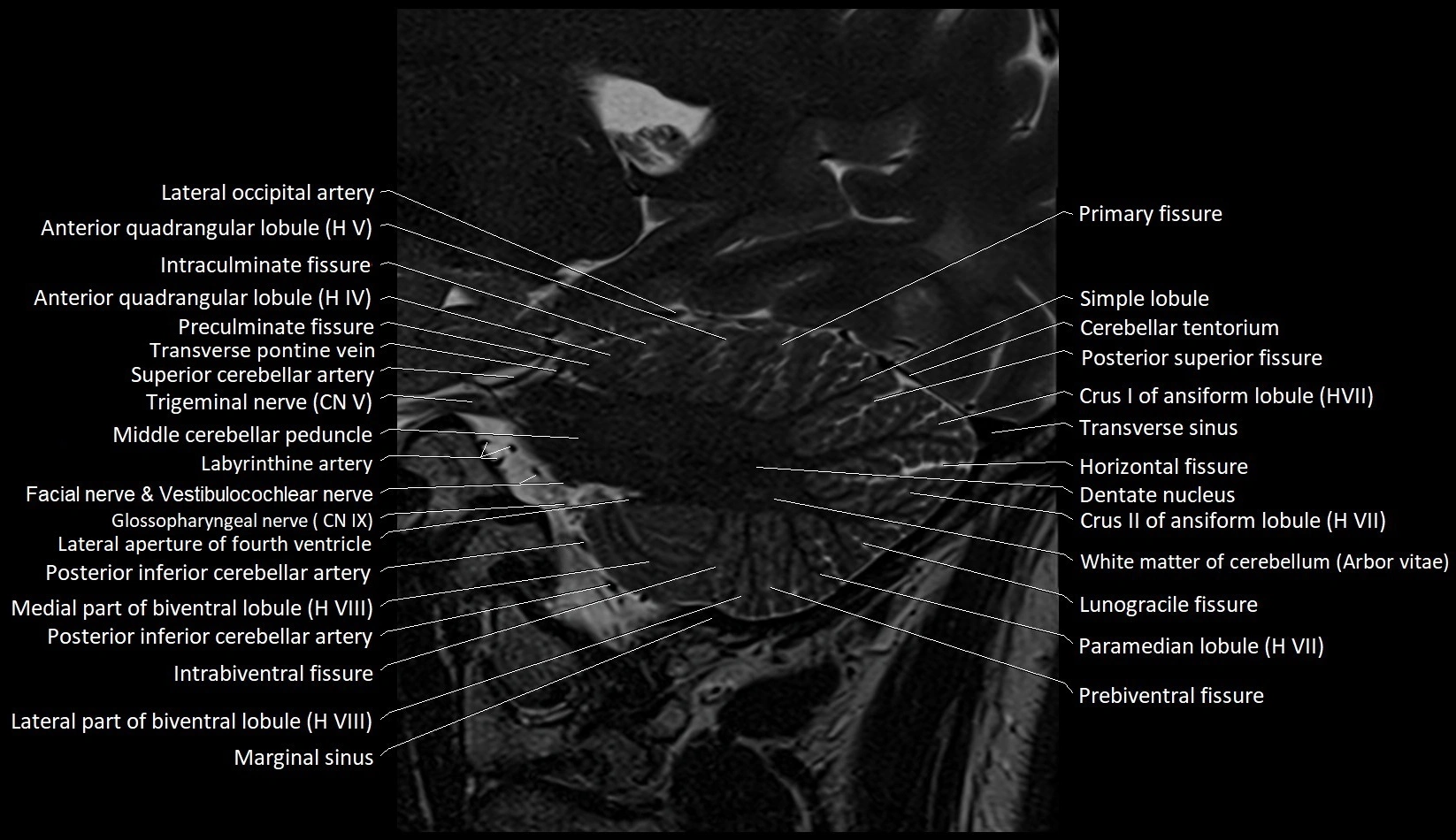
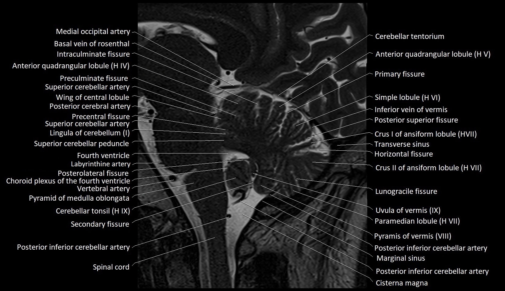
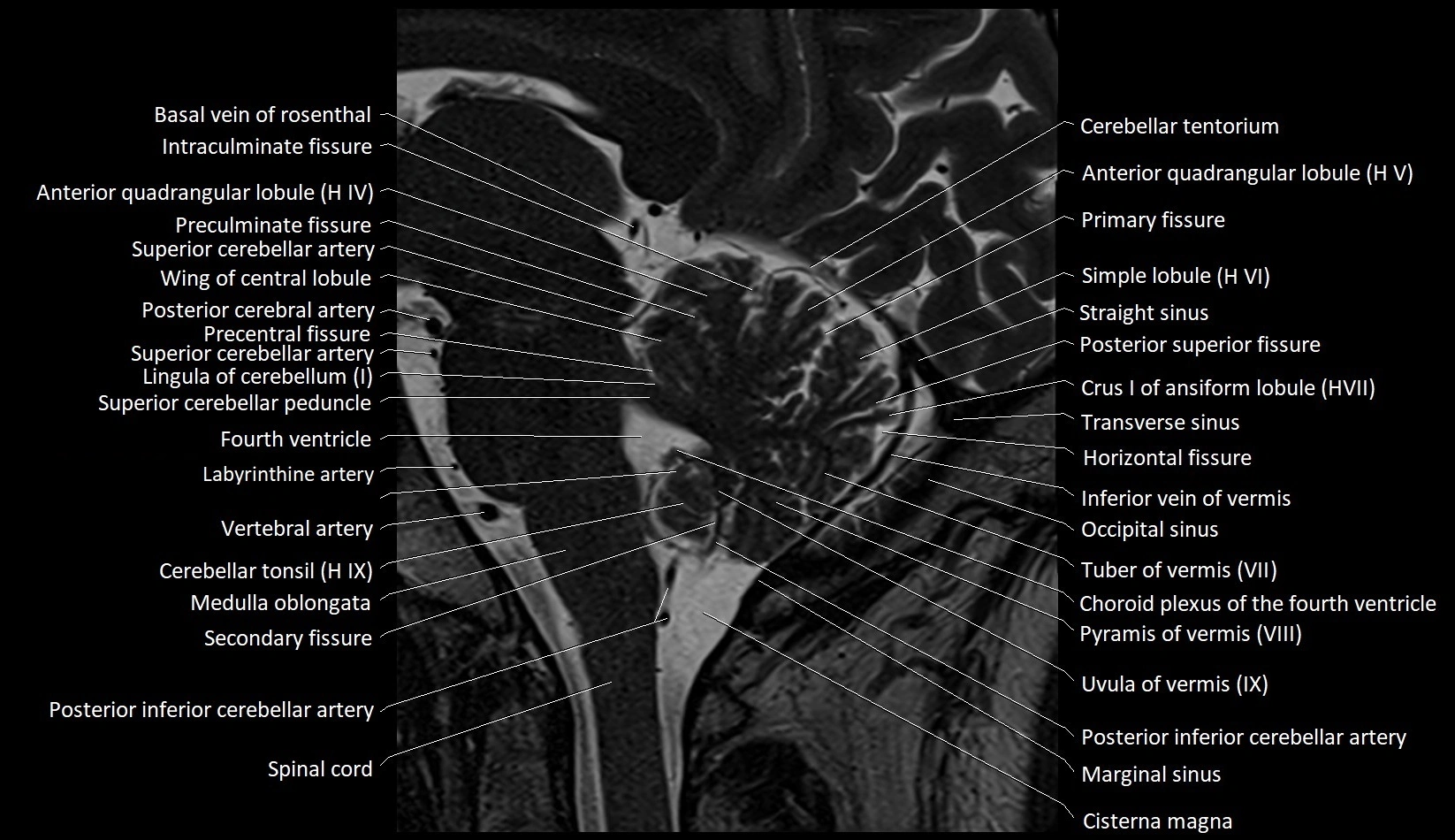
- Anterior lobe of cerebellum
- Anterior parietal artery
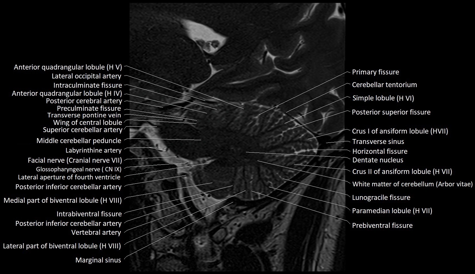
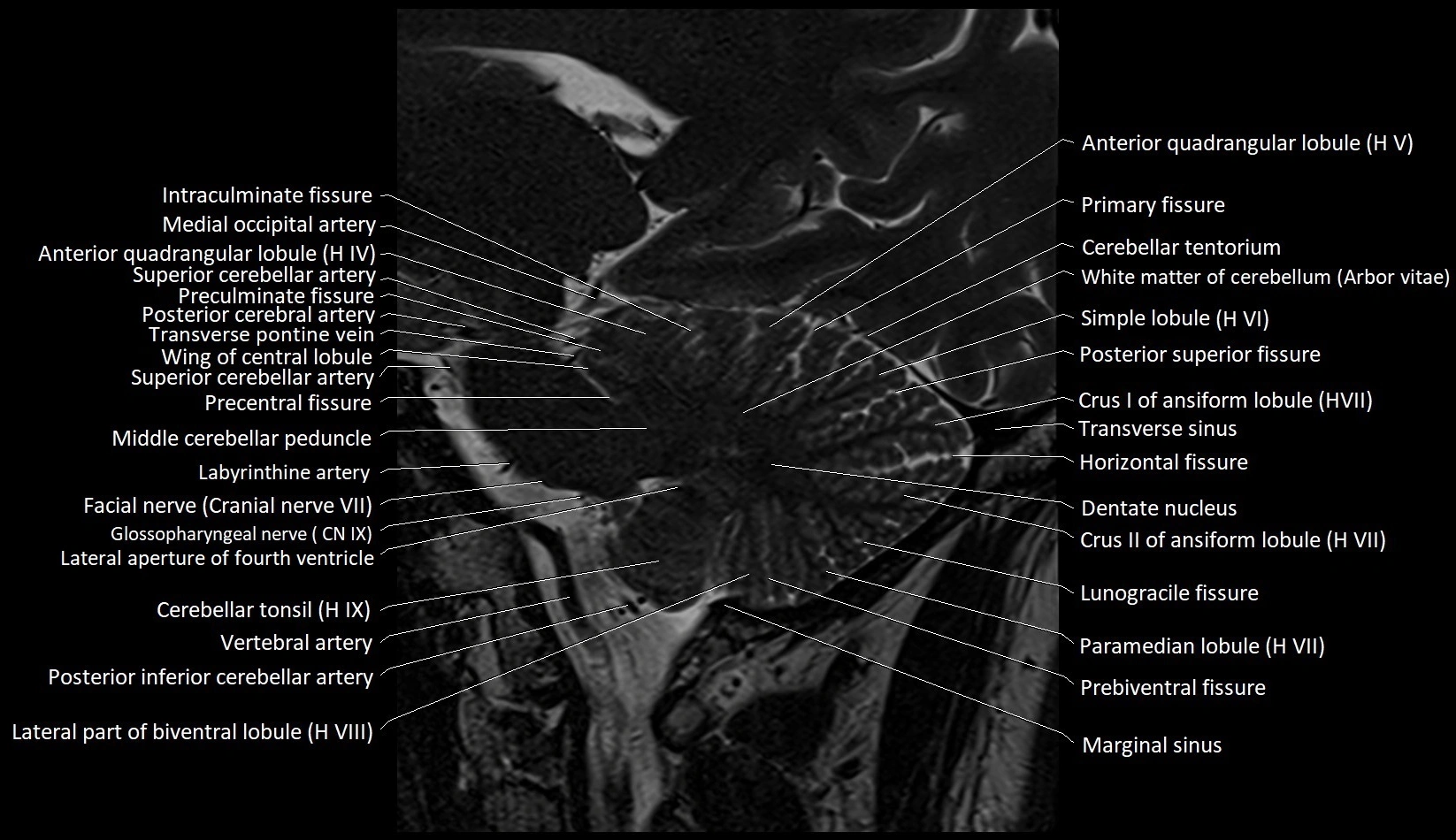
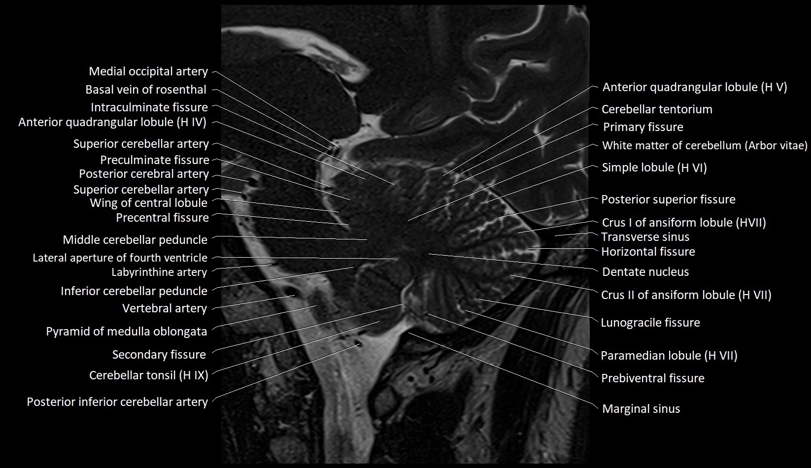
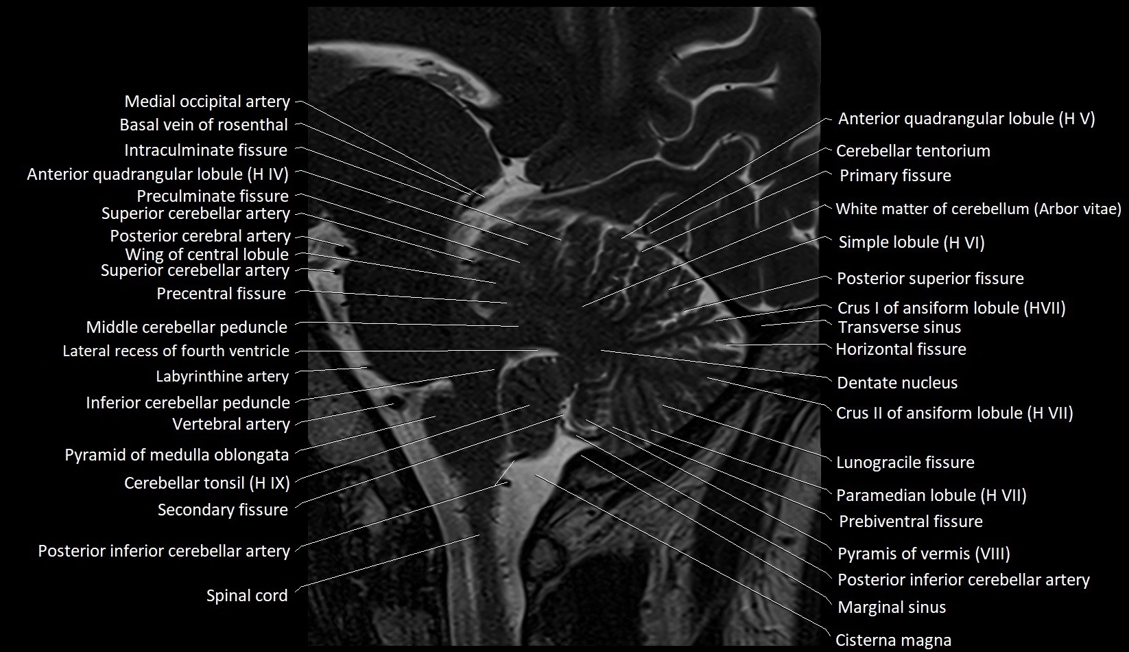
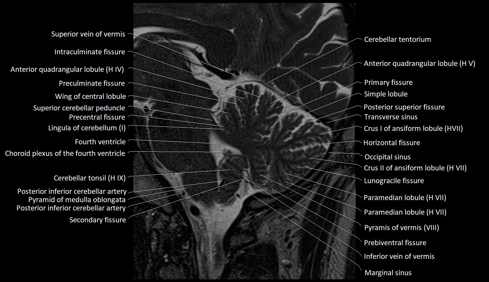
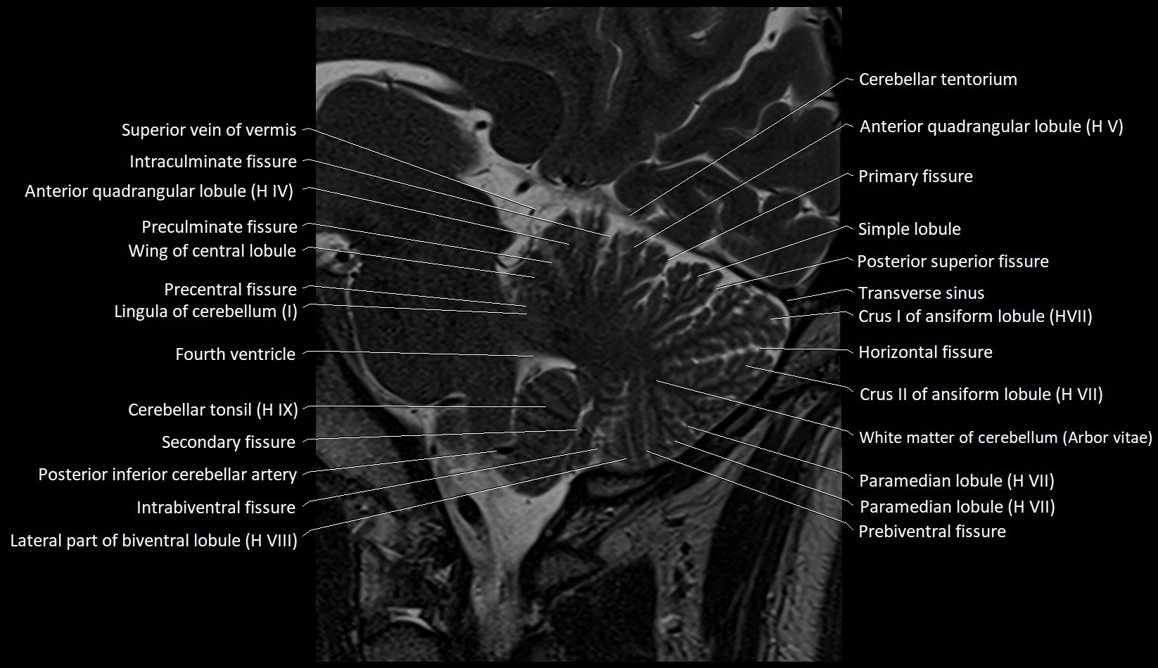
- Anterior quadrangular lobule
- Anterior quadrangular lobule (HV) of cerebellum
- Anterior quadrangular lobule (HlV) of cerebellum
- Anterolateral pontine vein
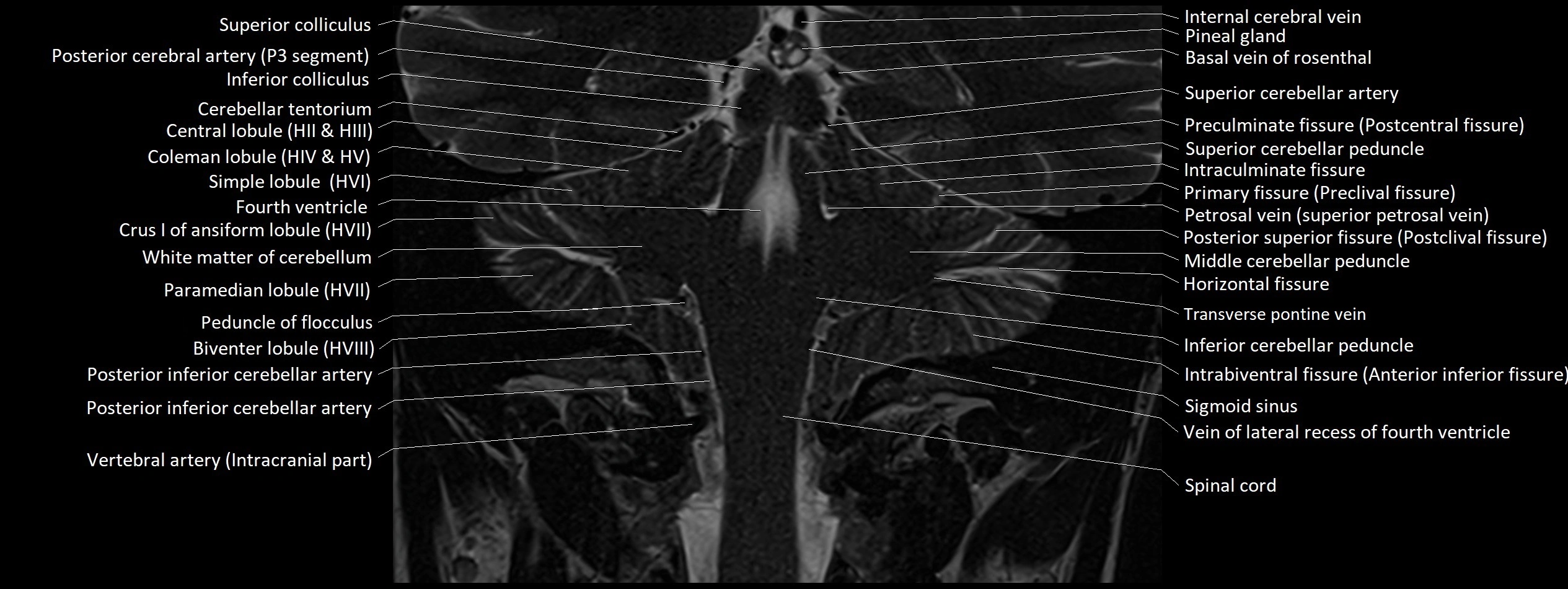
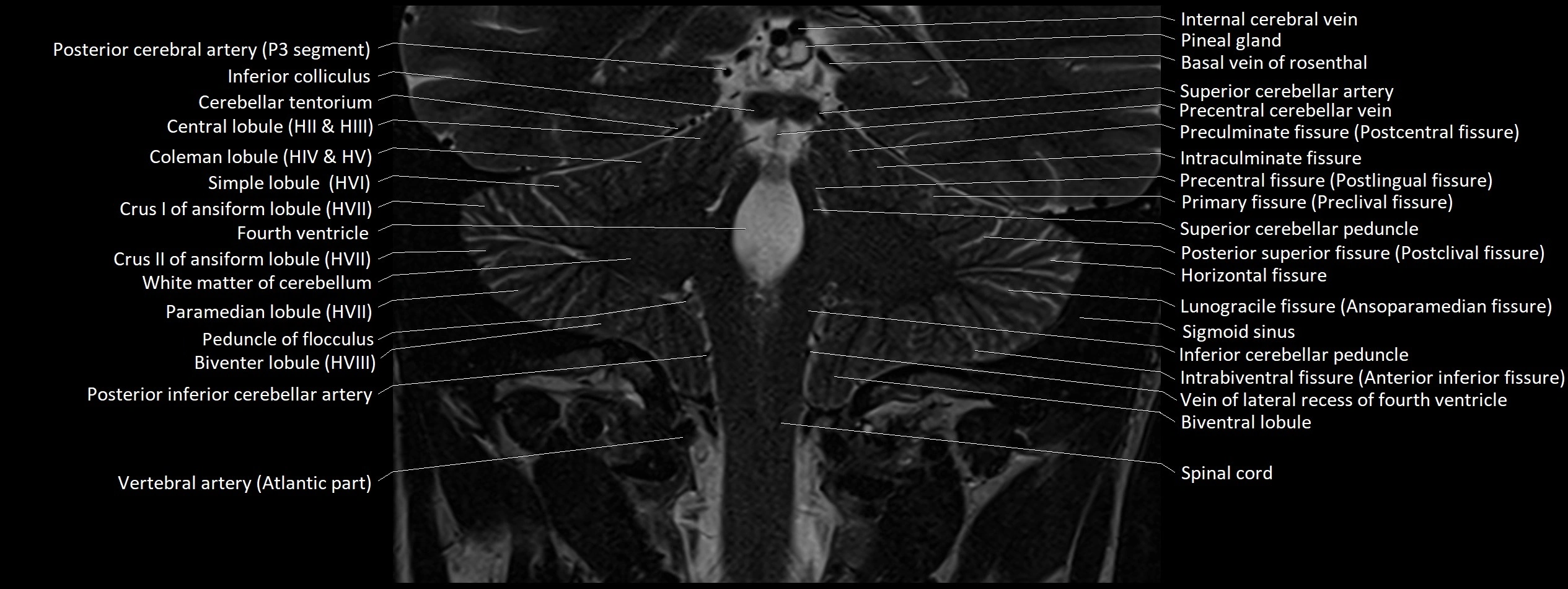
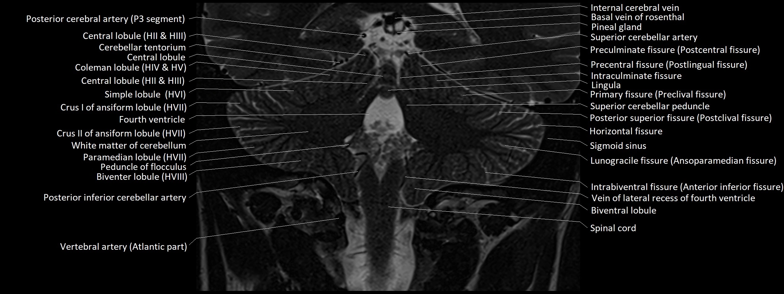
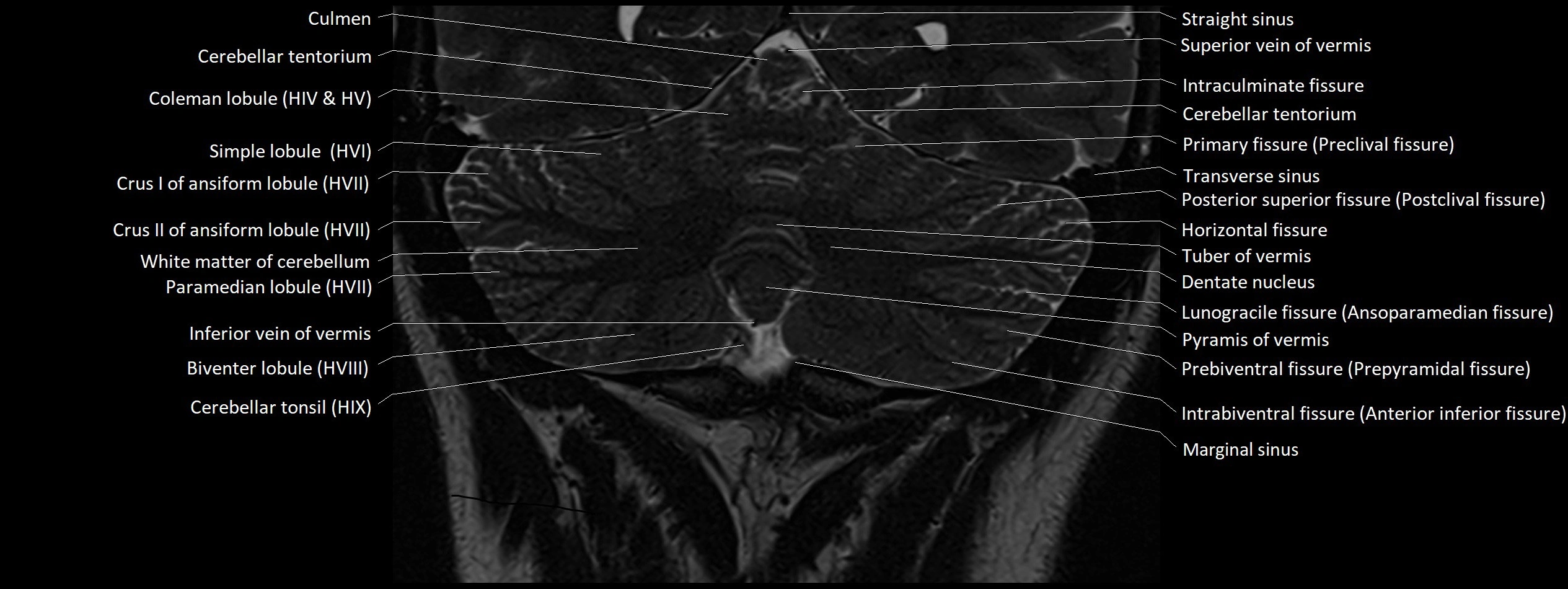
- Arbor Vitae (Cerebellar White Matter)
- Biventral lobule (HVIII) of cerebellum
- Calcarine artery
- Carotid cistern
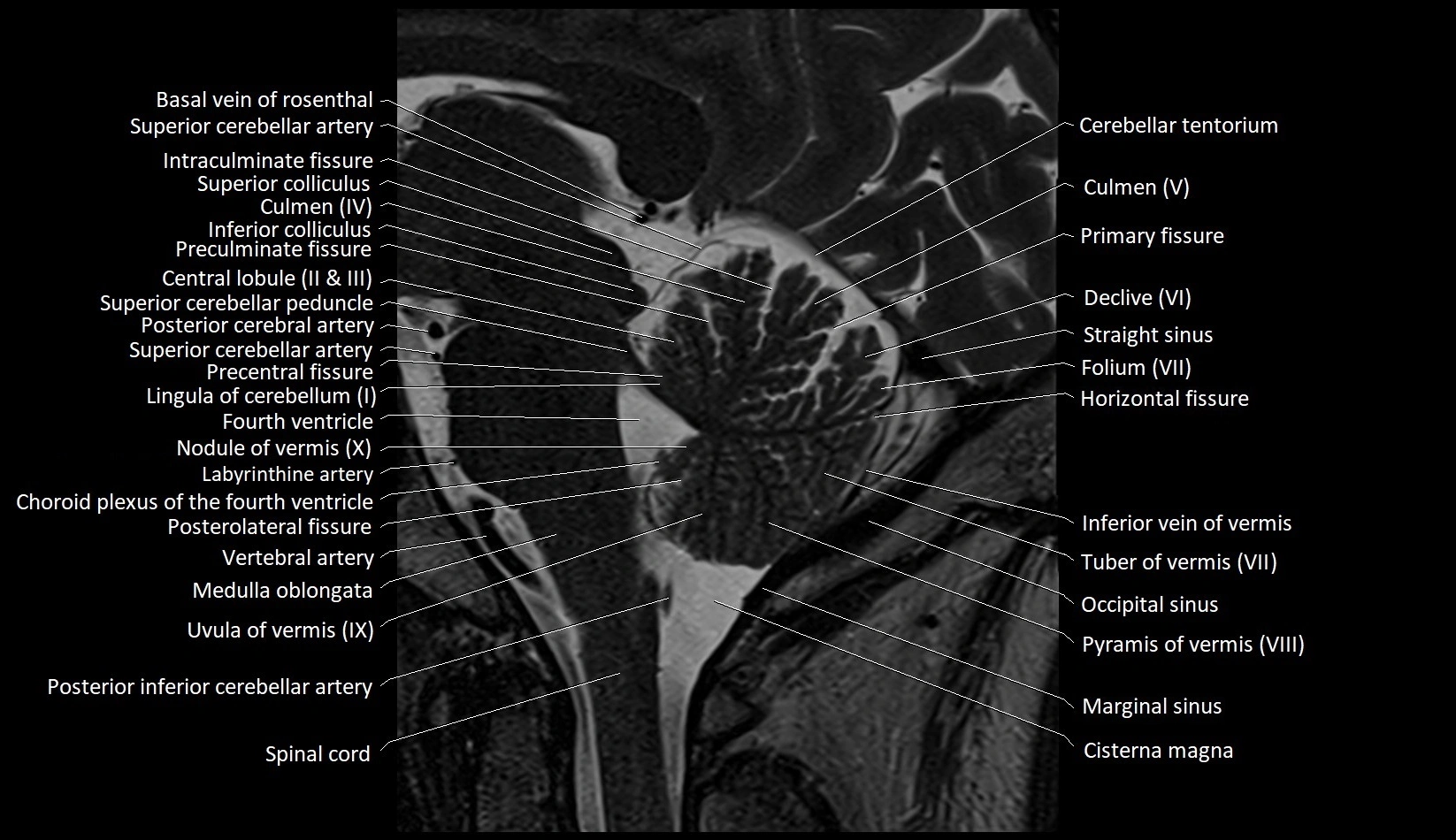
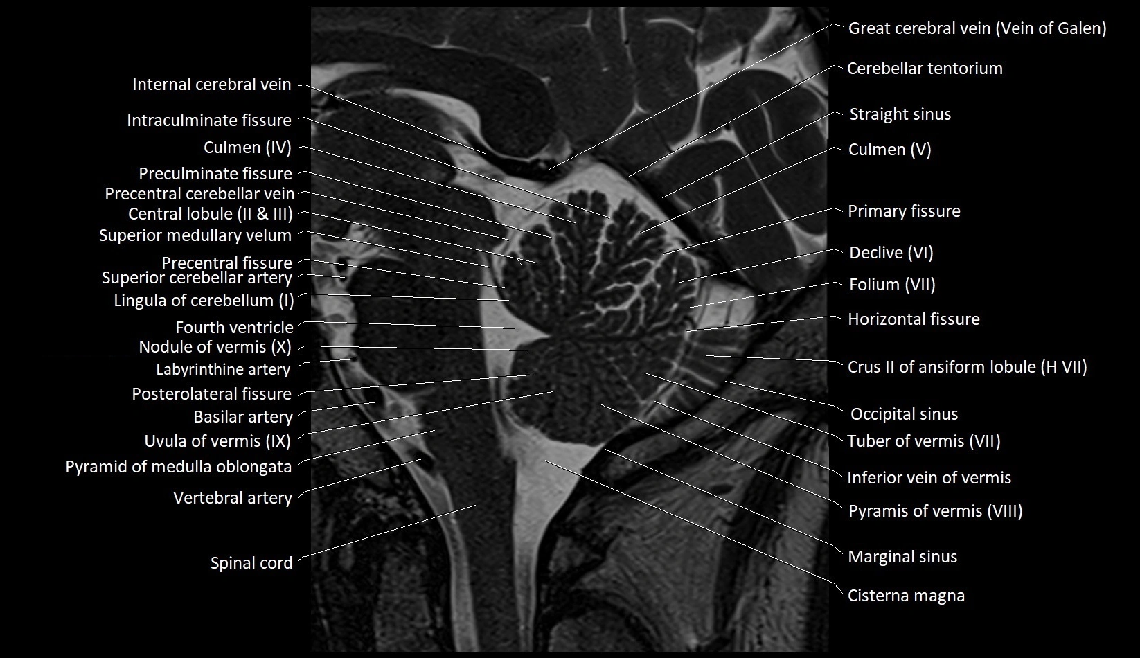
- Central lobule
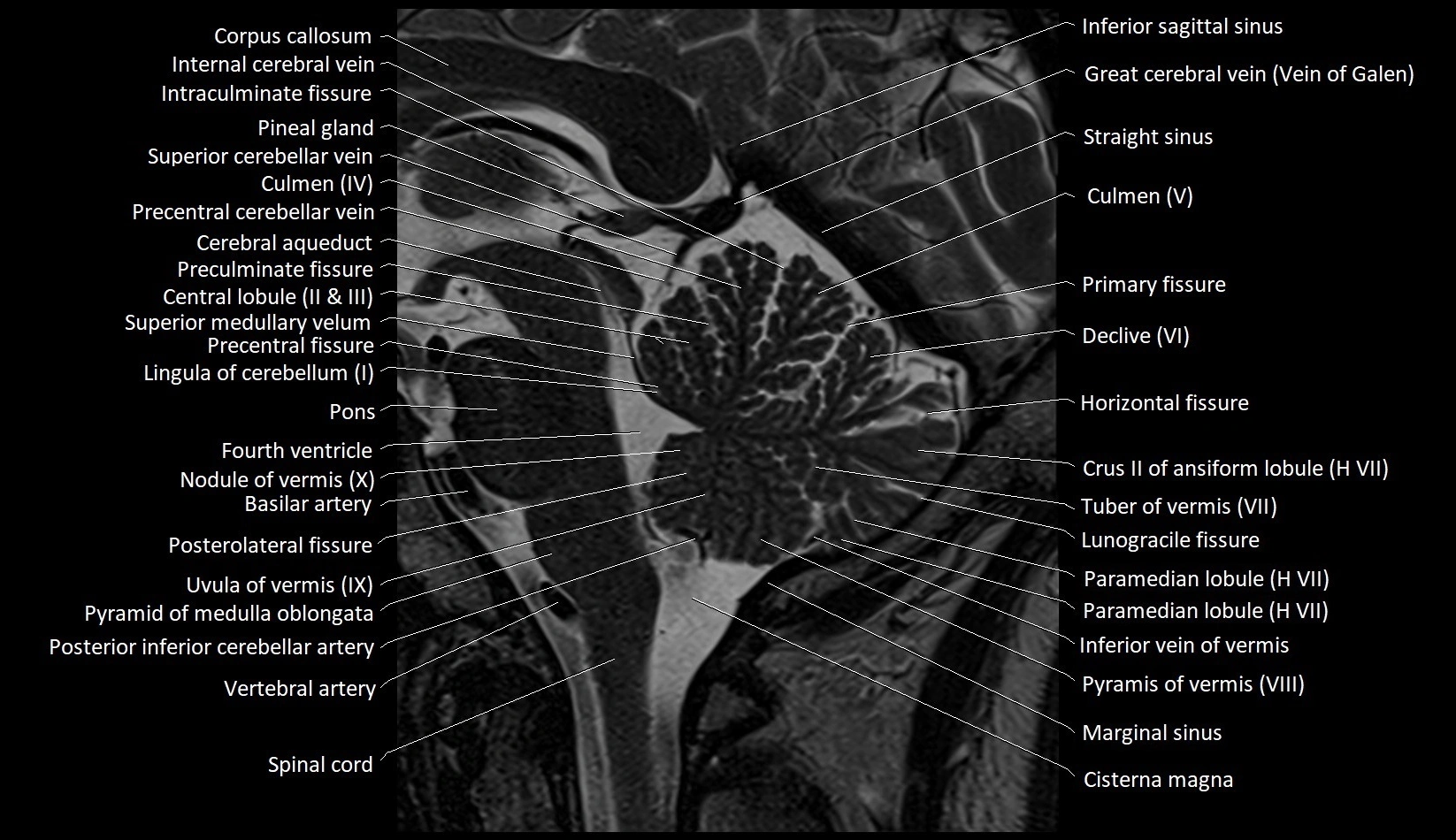
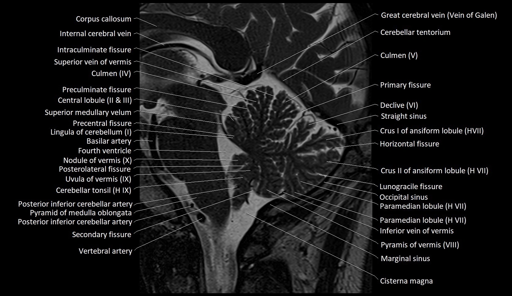
- Central lobule (II & III) of Cerebellum
- Cerebellar commissure
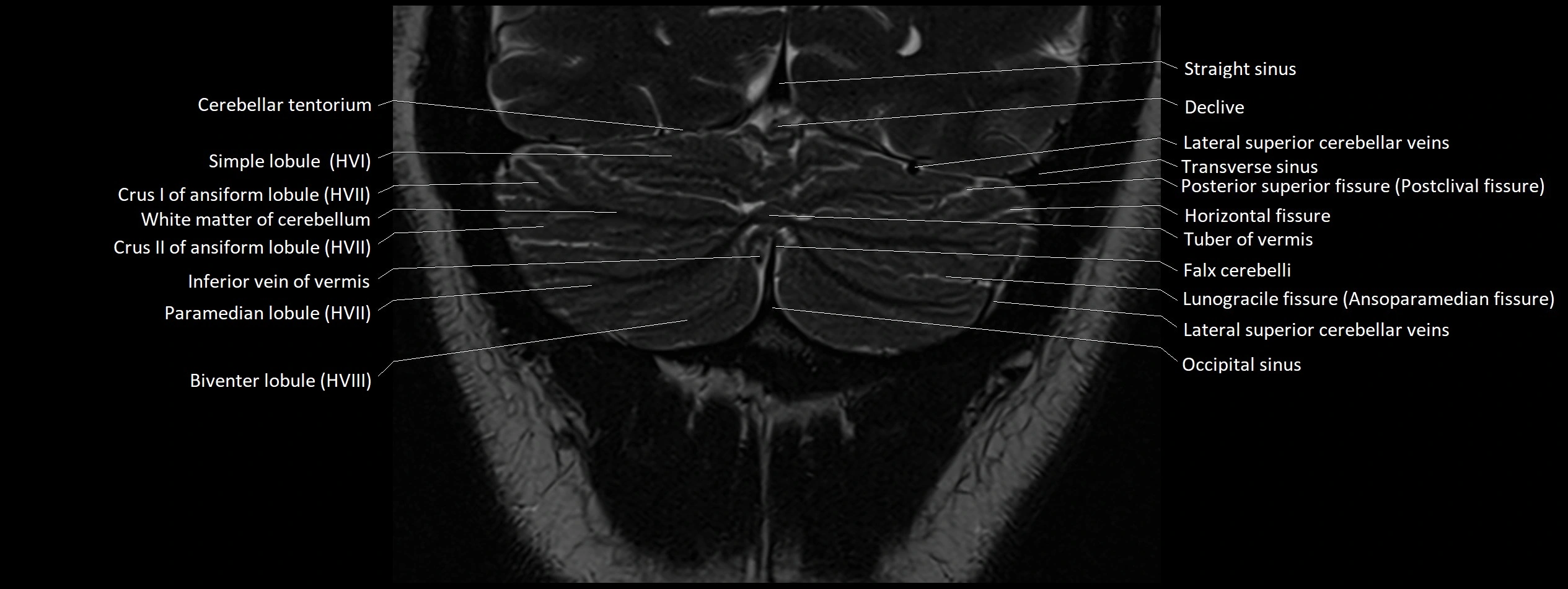
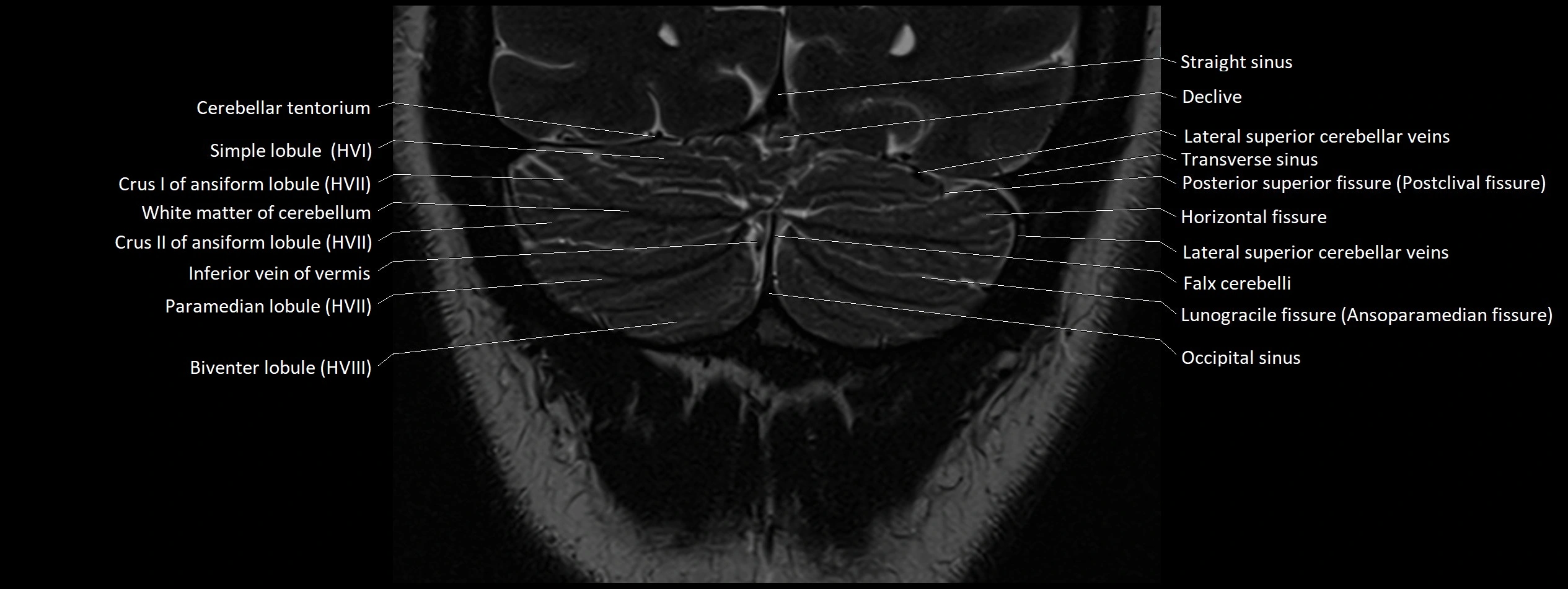
- Cerebellar falx
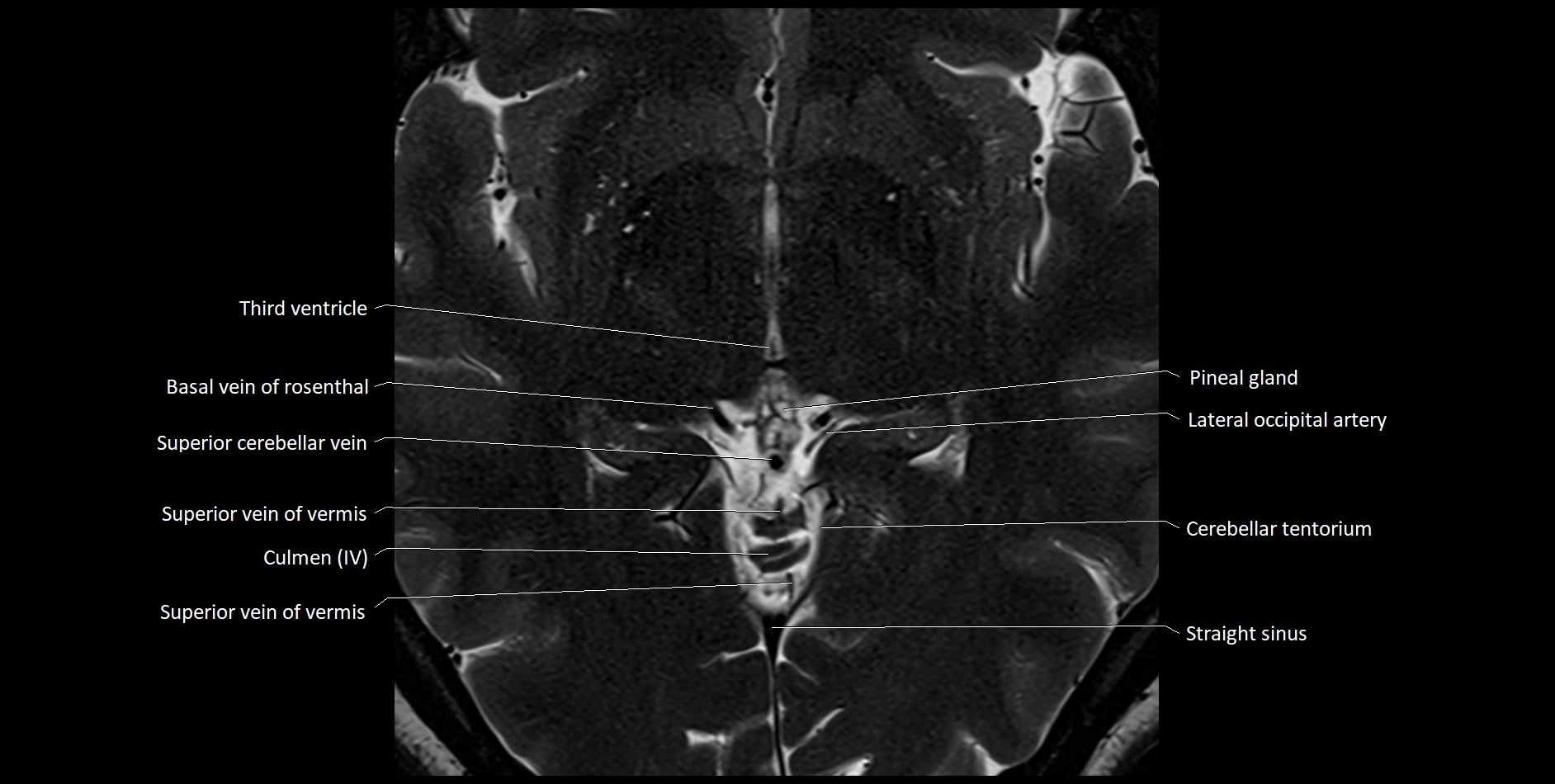
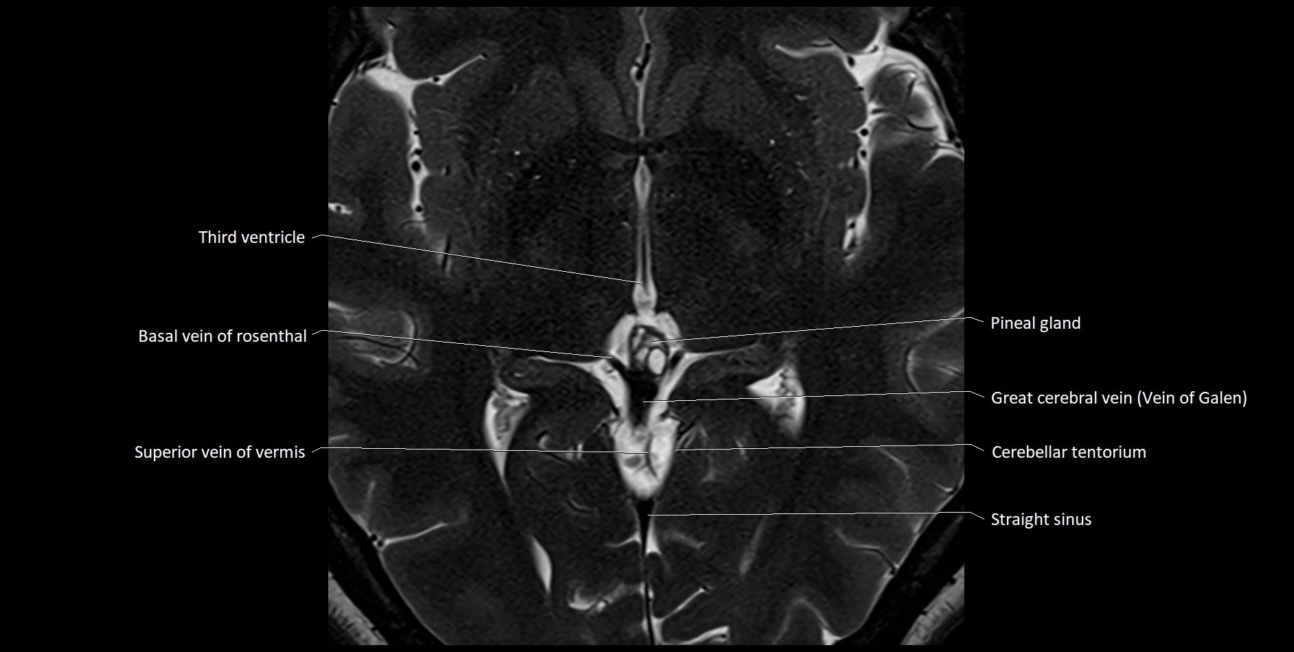
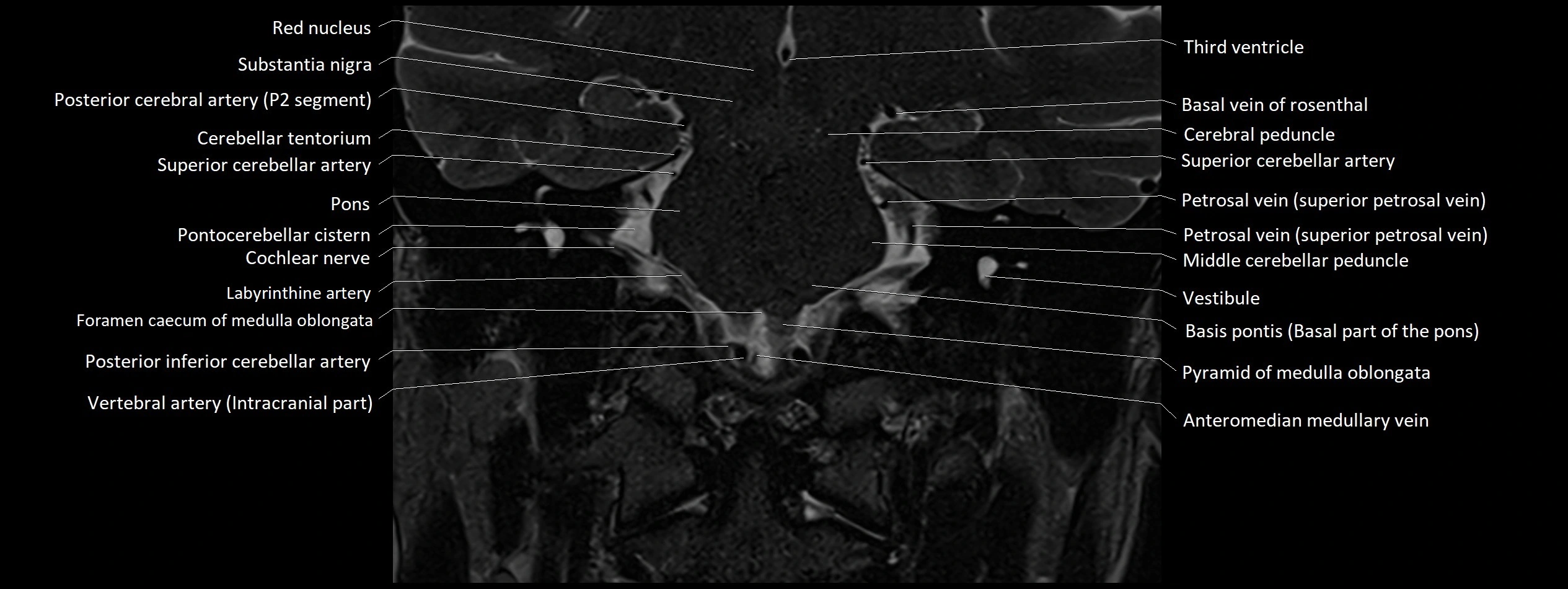
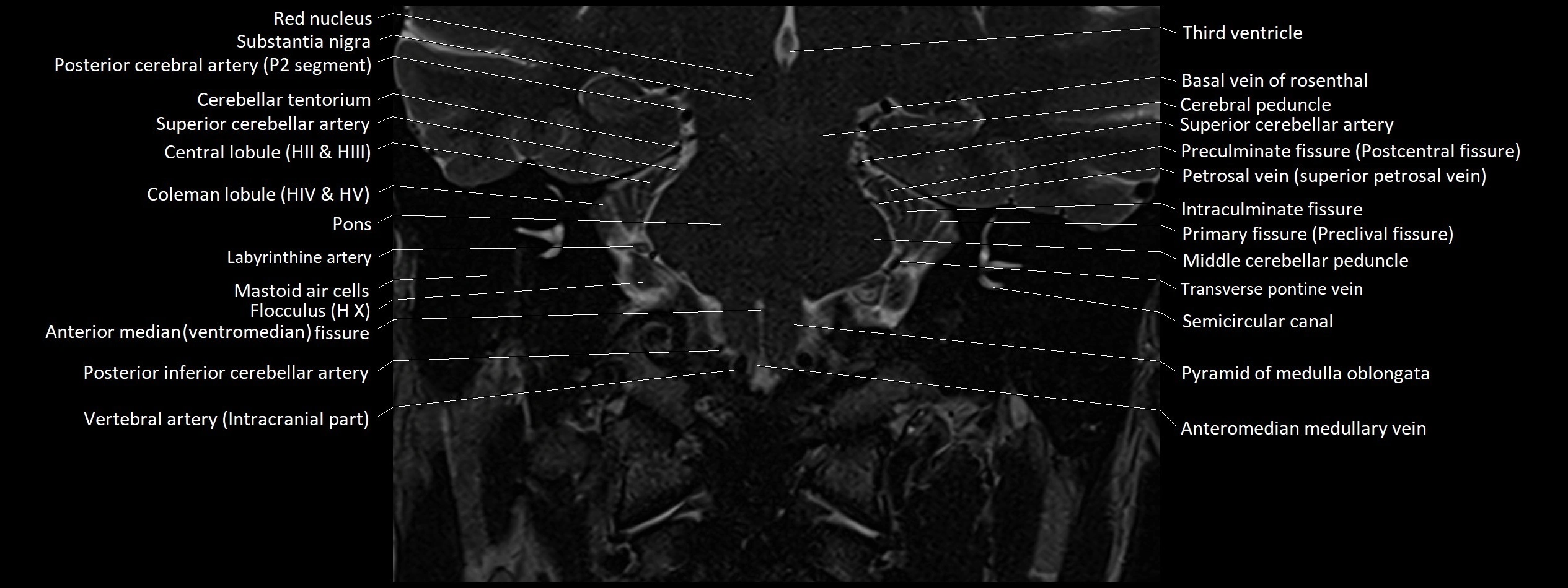
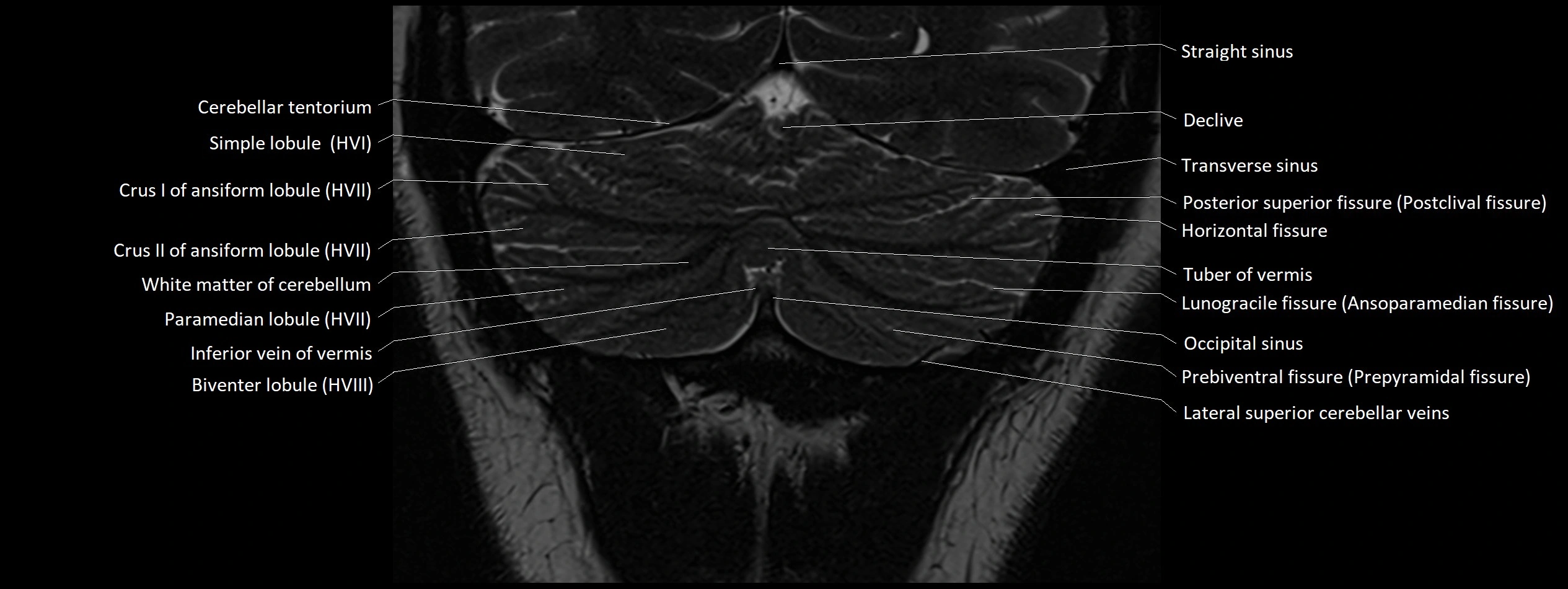
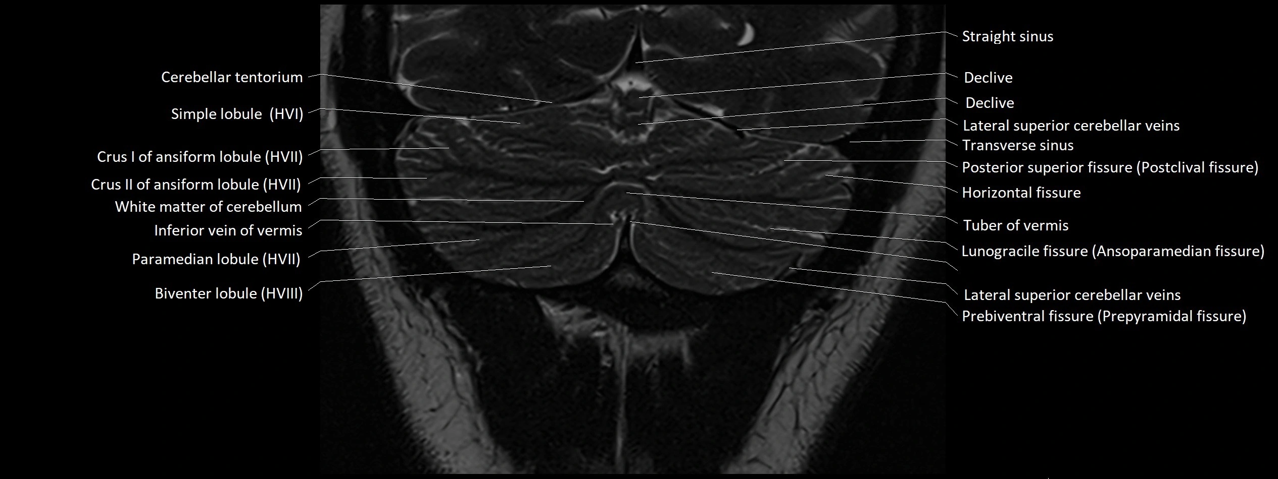
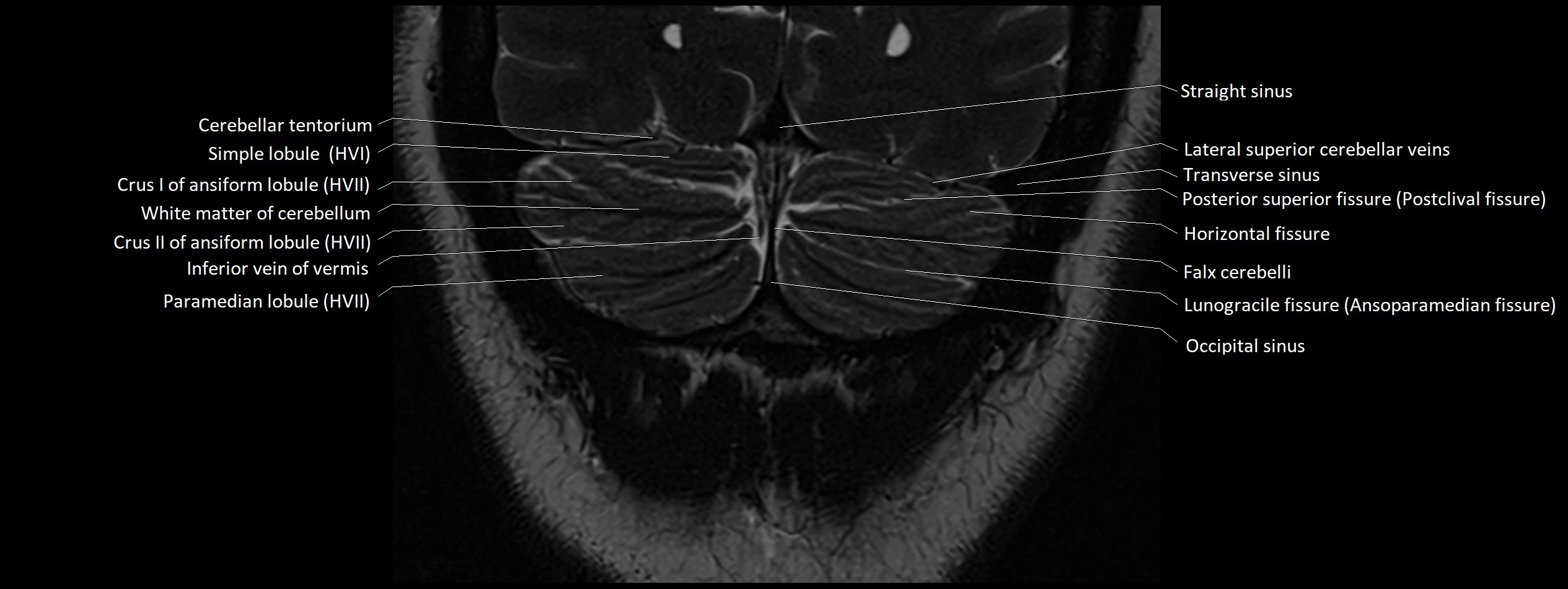
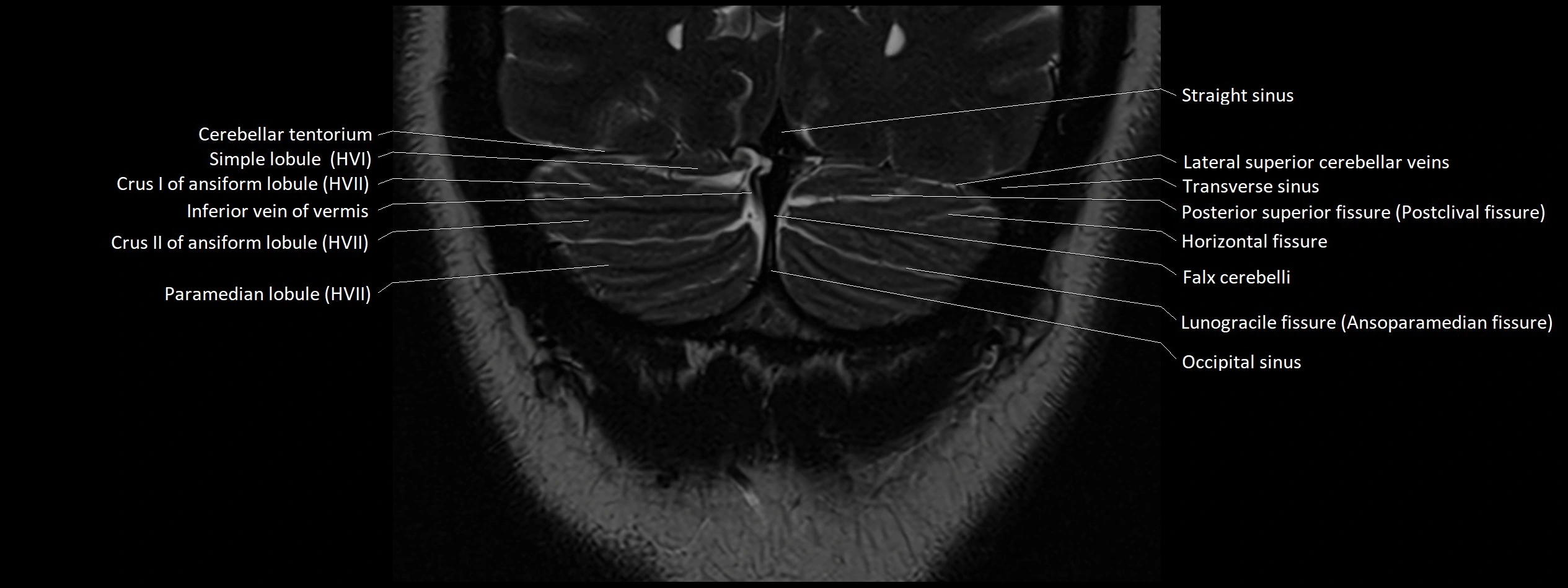
- Cerebellar tentorium
- Cerebellar tonsil (H IX)
- Cerebellopontine angle
- Cerebellum
- Chiasmatic cistern
- Choroid plexus
- Cistern of central sulcus
- Cistern of lamina terminalis
- Cistern of lateral cerebral fossa
- Cistern of transverse fissure
- Cochlea
- Cochlear nerve (Cranial nerve VIII)
- Common facial vein
- Crural cistern
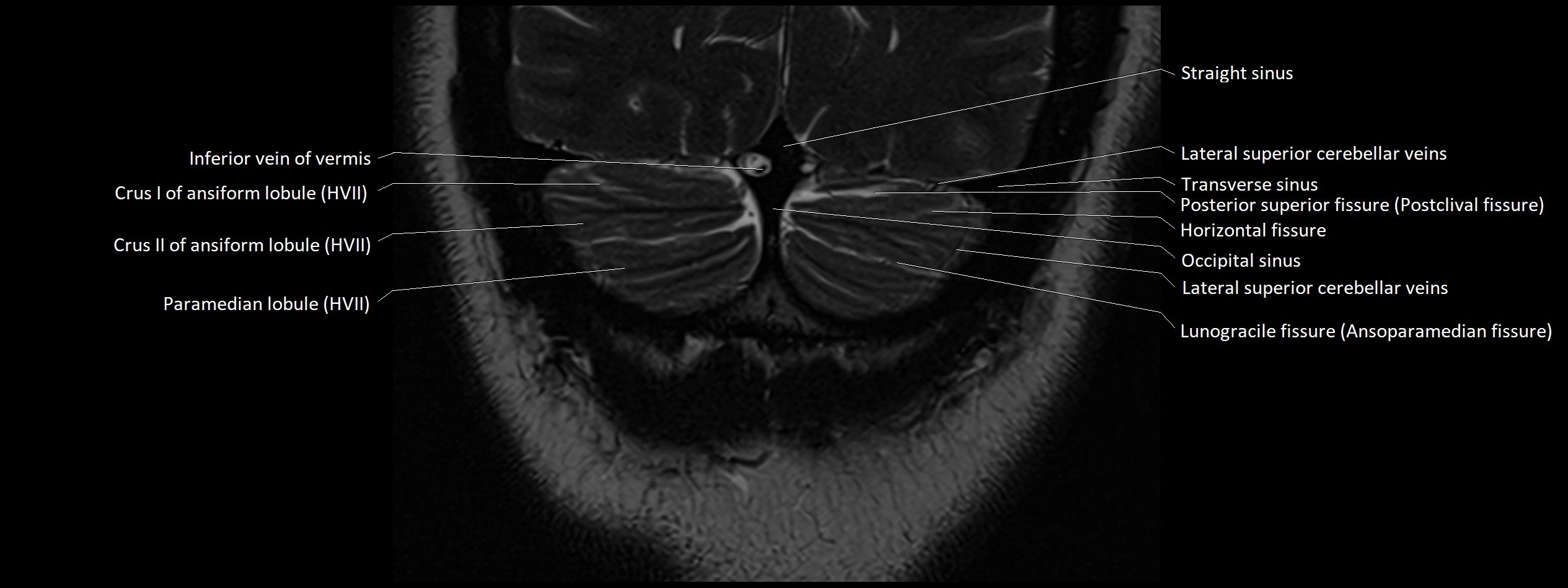
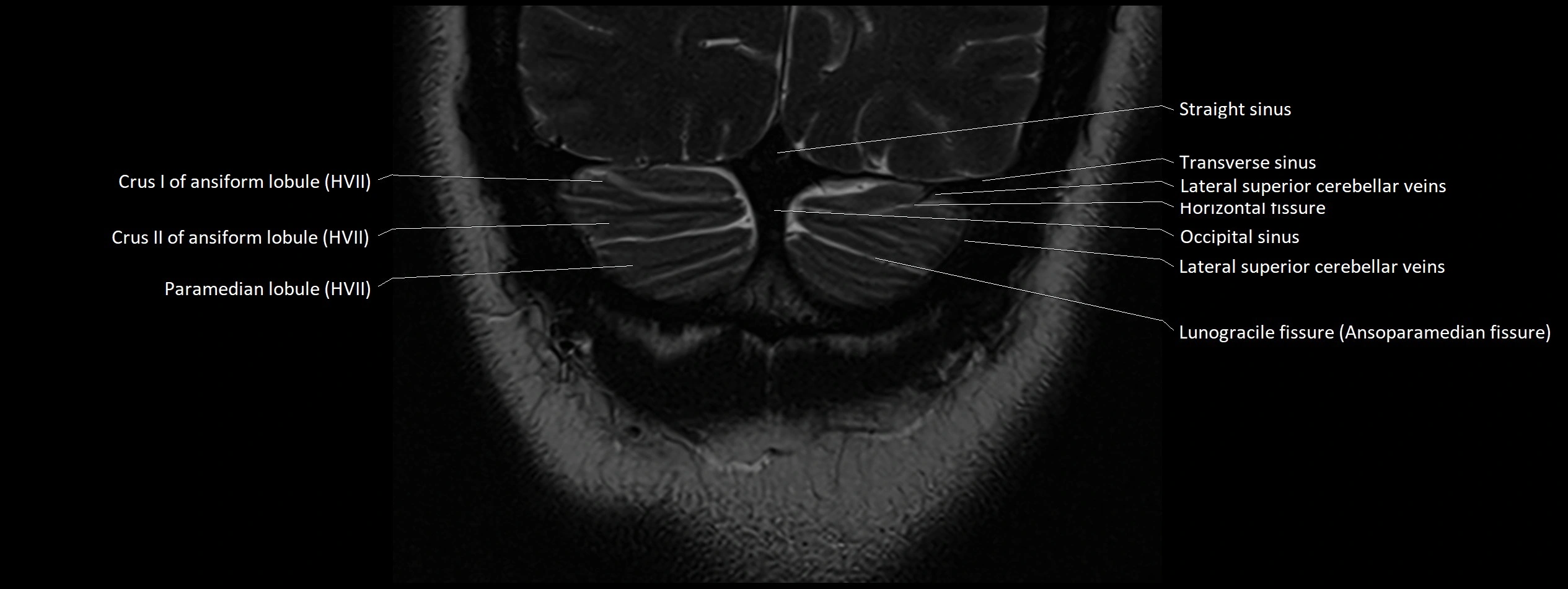
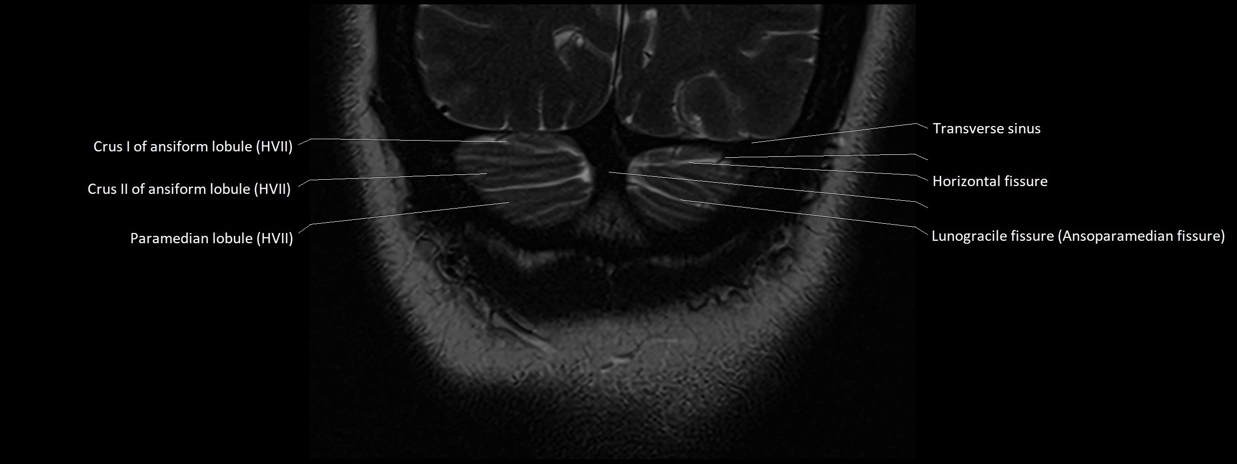
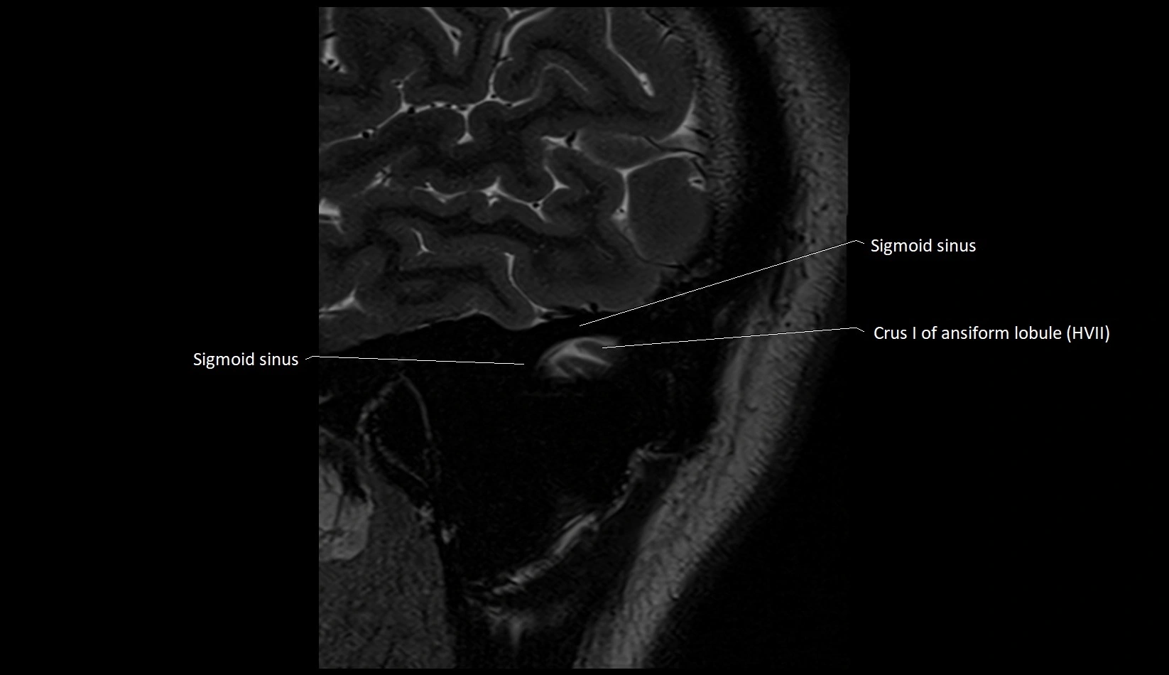
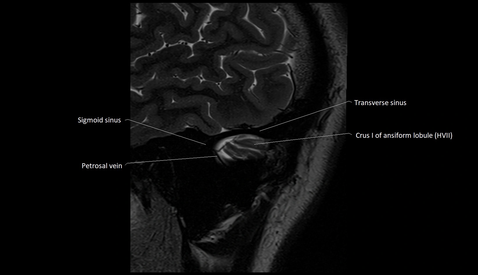
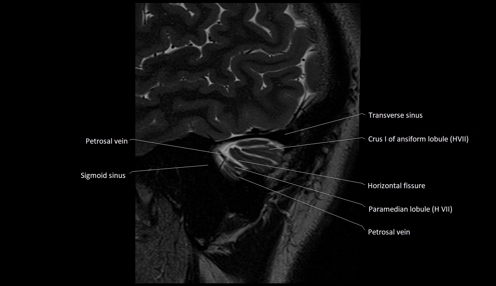
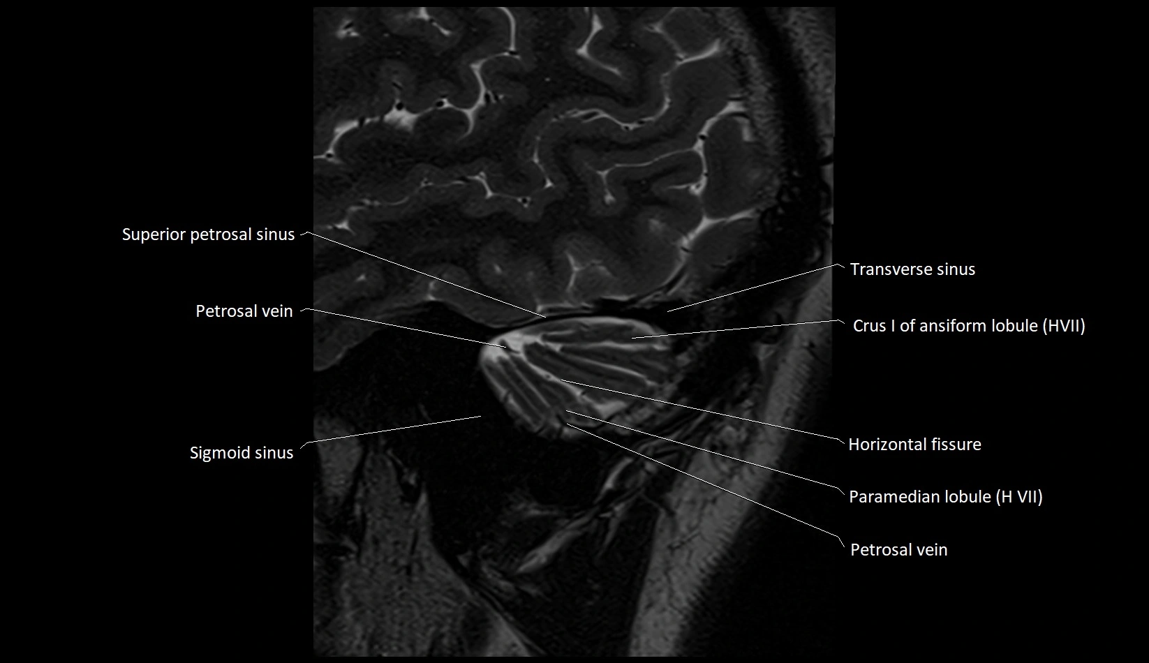
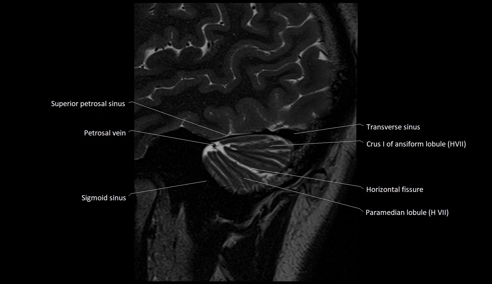
- Crus I of ansiform lobule of cerebellum
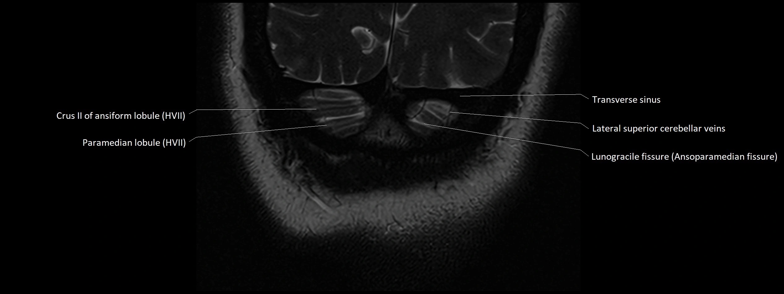
- Crus II of ansiform lobule of cerebellum
- Crus cerebri
- Culmen
- Culmen (IV, V) of Cerebellum
- Declive
- Declive (VI) of Cerebellum
- Dentate nucleus
- Facial Nerve (Cranial nerve VII)
- Flocculonodular lobe
- Flocculus
- Folium (VII) of Cerebellum
- Folium of Vermis
- Frontopolar artery
- Glossopharyngeal nerve (Cranial nerve IX)
- Horizontal fissure (cerebellum)
- Hypoglossal Nerve (Cranial nerve XII)
- Inferior branch vestibular nerve
- Inferior cerebellar peduncle
- Inferior cerebellar veins
- Inferior choroidal vein
- Inferior hemispheric veins of the cerebellum
- Inferior petrosal sinus
- Inferior salivatory nucleus
- Inferior semilunar lobule
- Inferior vein of vermis
- Inferior vermian vein
- Inferior vestibular nucleus
- Infundibulum
- Internal cerebral vein
- Internal jugular vein
- Interpeduncular Cistern
- Intrabiventral Fissure of Biventral Lobule
- Intraculminate fissure
- Lacrimal nucleus
- Lateral cerebellomedullary cistern
- Lateral occipital artery
- Lateral orbitofrontal artery
- Lateral part of biventeral lobule
- Lateral vestibular nucleus
- Lingual vein
- Lingula of cerebellum
- Lingula of cerebellum (I)
- Lunogracle fissure
- Lunogranicile fissure of cerebellum
- Marginal sinus
- Mastoid emissary vein
- Medial occipital artery
- Medial orbitofrontal artery
- Medial part of biventeral lobule
- Medial vestibular nucleus
- Mesencephalic nucleus of trigeminal nerve
- Middle cerebellar peduncle
- Middle cerebral artery cortical segment (M4)
- Middle cerebral artery horizontal segment (M1)
- Middle cerebral artery insular segment (M2)
- Middle cerebral artery opercular segment (M3)
- Middle meningeal artery
- Motor nucleus of facial nerve
- Motor nucleus of trigeminal nerve
- Nodule of vermis
- Nodule of vermis (X)
- Nucleus of abducens nerve
- Nucleus of hypoglossal nerve
- Nucleus of oculomotor nerve
- Nucleus of solitary tract
- Nucleus of trochlear nerve
- Occipital emissary vein
- Occipital sinus
- Oculomotor Nerve (Cranial Nerve III)
- Oculomotor cistern
- Olfactory Nerve (Cranial Nerve I)
- Olfactory bulb
- Olfactory cistern
- Olfactory tract
- Optic Nerve (Cranial Nerve II)
- Optic chiasm
- Paramedian lobule
- Paramedian lobule (HVII) of cerebellum
- Parieto-occipital artery
- Peduncle of flocculus
- Pericallosal cistern
- Petrosal vein
- Pineal gland
- Pontine artery
- Pontocerebellar cistern
- Pontomedullary junction
- Posterior cerebellomedullary cistern (cisterna magna)
- Posterior cerebral artery (P1 Segment)
- Posterior cerebral artery (P2 Segment)
- Posterior cerebral artery (P3 Segment)
- Posterior cerebral artery (P4 Segment)
- Posterior cochlear nucleus
- Posterior external vertebral venous plexus
- Posterior hippocampal artery
- Posterior inferior cerebellar artery
- Posterior lobe of cerebellum
- Posterior mesencephalic vein
- Posterior quadrangular lobule
- Posterior superior fissure
- Posterior vein of caudate nucleus
- Posterior veins of septum pellucidum
- Posterolateral fissure
- Posteromedian medullary vein
- Pre-Rolandic artery
- Prebiventral fissure
- Precentral cerebellar vein
- Precentral fissure
- Preculminate fissure
- Prepontine cistern
- Primary fissure
- Principal sensory nucleus of the trigeminal nerve
- Pyramid of vermis (VIII)
- Pyramis of vermis
- Quadrigeminal cistern
- Rolandiс artery
- Rostral gyrus
- Rostral sulcus
- Secondary fissure
- Semicircular Canals
- Sigmoid sinus
- Simple lobule
- Simple lobule (HVI) of cerebellum
- Spinal nucleus of trigeminal nerve
- Straight sinus
- Superficial cerebral veins
- Superficial middle cerebral vein
- Superior anastomotic vein
- Superior branch of vestibular nerve
- Superior cerebellar cistern
- Superior cerebellar peduncle
- Superior cerebellar vein
- Superior hemispheric cerebellar veins
- Superior hemispheric veins of the cerebellum
- Superior medullary velum
- Superior petrosal sinus
- Superior sagittal sinus
- Superior salivatory nucleus
- Superior semilunar lobule of cerebellum
- Superior thalamic veins
- Superior thalamostriate vein
- Superior vein of vermis
- Superior vermian vein
- Superior vestibular nucleus
- Supratrochlear veins
- Tegmentum of pons
- Temporopolar artery
- Tonsil of cerebellum
- Transverse pontine vein
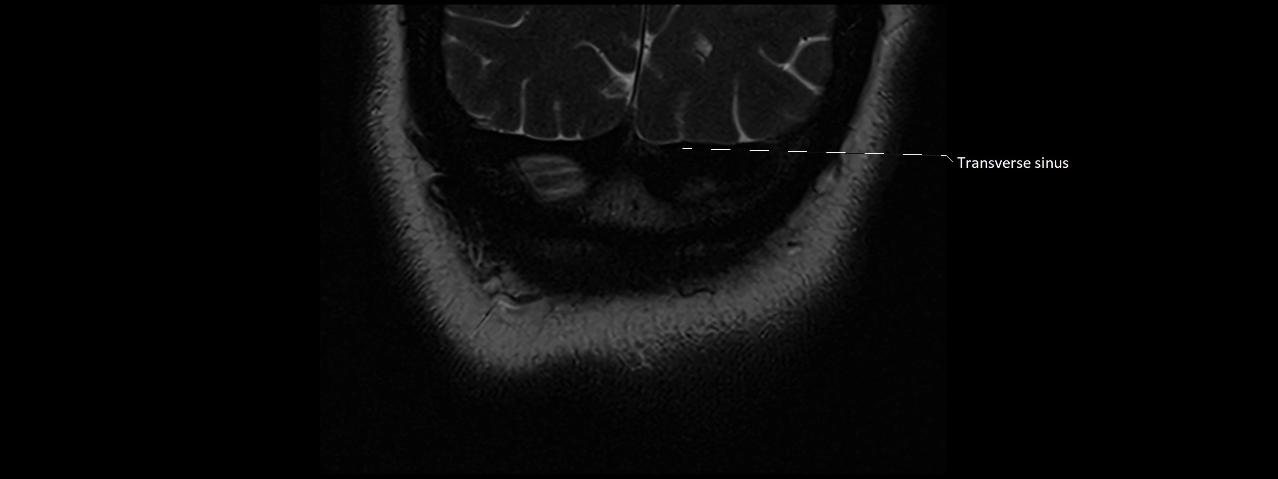
- Transverse sinus
- Trigeminal ganglion
- Trigeminal nerve (Cranial nerve V)
- Trochlear nerve (Cranial nerve IV)
- Tuber of vermis
- Tuber of vermis (VII)
- Uvula of vermis
- Uvula of vermis (IX)
- Vagus nerve (Cranial nerve X)
- Vein of lateral recess of fourth ventricle
- Vermis of cerebellum
- Vestibular ganglion
- Vestibule
- Vestibulocochlear nerve (Cranial nerve VIII)
- White matter of cerebellum (Arbor vitae)
- White substance of cerebellum
- Wing of central lobule
The Abducens nerve (Cranial nerve VI) is a purely motor cranial nerve responsible for innervating the lateral rectus muscle of the eye, which is crucial for lateral movement (abduction) of the eyeball. It arises from the abducens nucleus in the dorsal pons, emerges at the pontomedullary junction, and travels a long intracranial course before entering the orbit via the superior orbital fissure. Because of its long path and proximity to the clivus, it is particularly susceptible to injury from increased intracranial pressure or trauma.
Synonyms
-
Sixth cranial nerve
-
CN VI
-
N. abducens (Latin)
-
Nervus abducens
Function
-
Innervates the lateral rectus muscle of the eye
-
Responsible for abduction of the eyeball (moving the eye outward, away from the midline)
-
Is a purely motor nerve (no sensory or autonomic fibers)
-
Lesion results in inability to abduct the affected eye, leading to horizontal diplopia (double vision)
MRI Appearance
-
The abducens nerve is a small, thin, linear structure
-
Best visualized on high-resolution T2-weighted 3D MRI sequences (e.g., FIESTA or CISS)
-
Seen as a hypointense (dark) line running from the brainstem at the pontomedullary junction, traversing the prepontine cistern, and entering Dorello’s canal under the petrosphenoidal ligament, then into the cavernous sinus, and finally the orbit
-
May be challenging to visualize in standard MRI due to its small size
-
Pathology may be inferred by absence, displacement, or enhancement of the nerve
CT Appearance
-
The nerve itself is not directly visualized on conventional CT due to its small size and soft tissue density
-
Indirect signs: assessment of the bony course, such as the Dorello’s canal, superior orbital fissure, or adjacent pathologies (fractures, masses, or inflammation) that could impinge the nerve
-
CT is mainly used to exclude structural lesions or fractures that might affect the course of CN VI
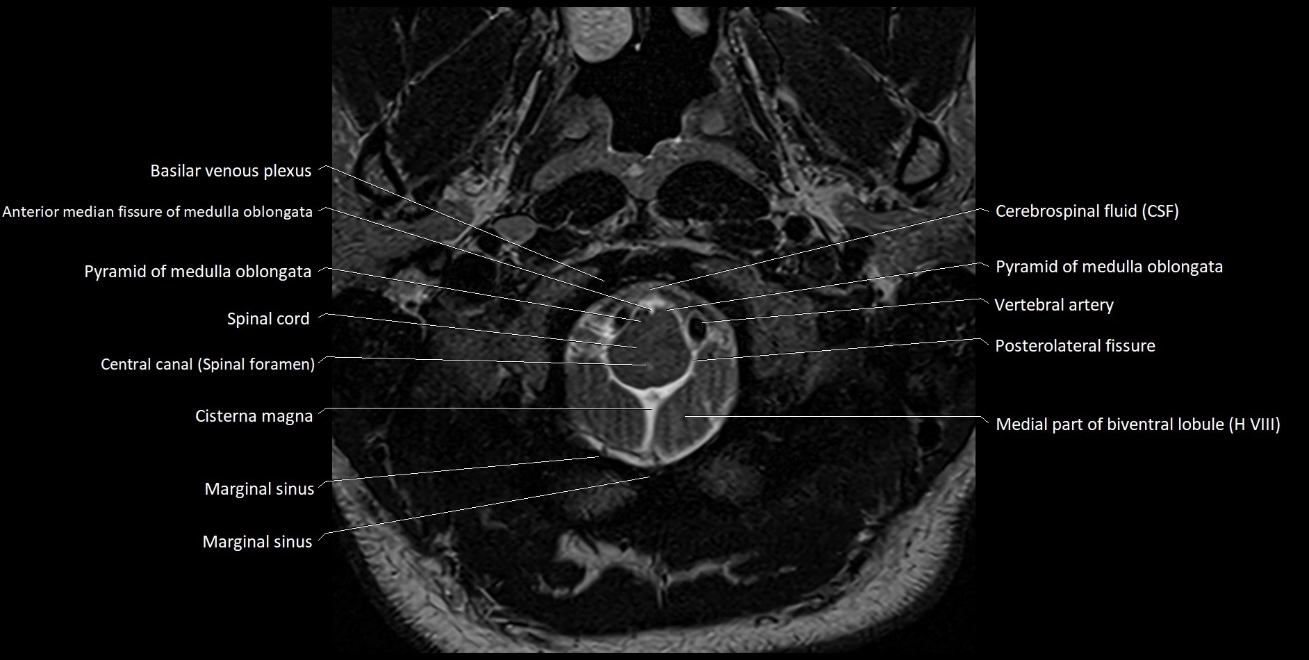
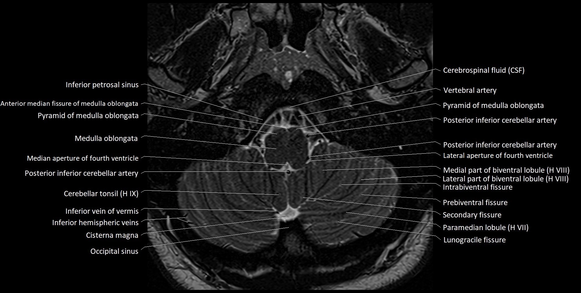
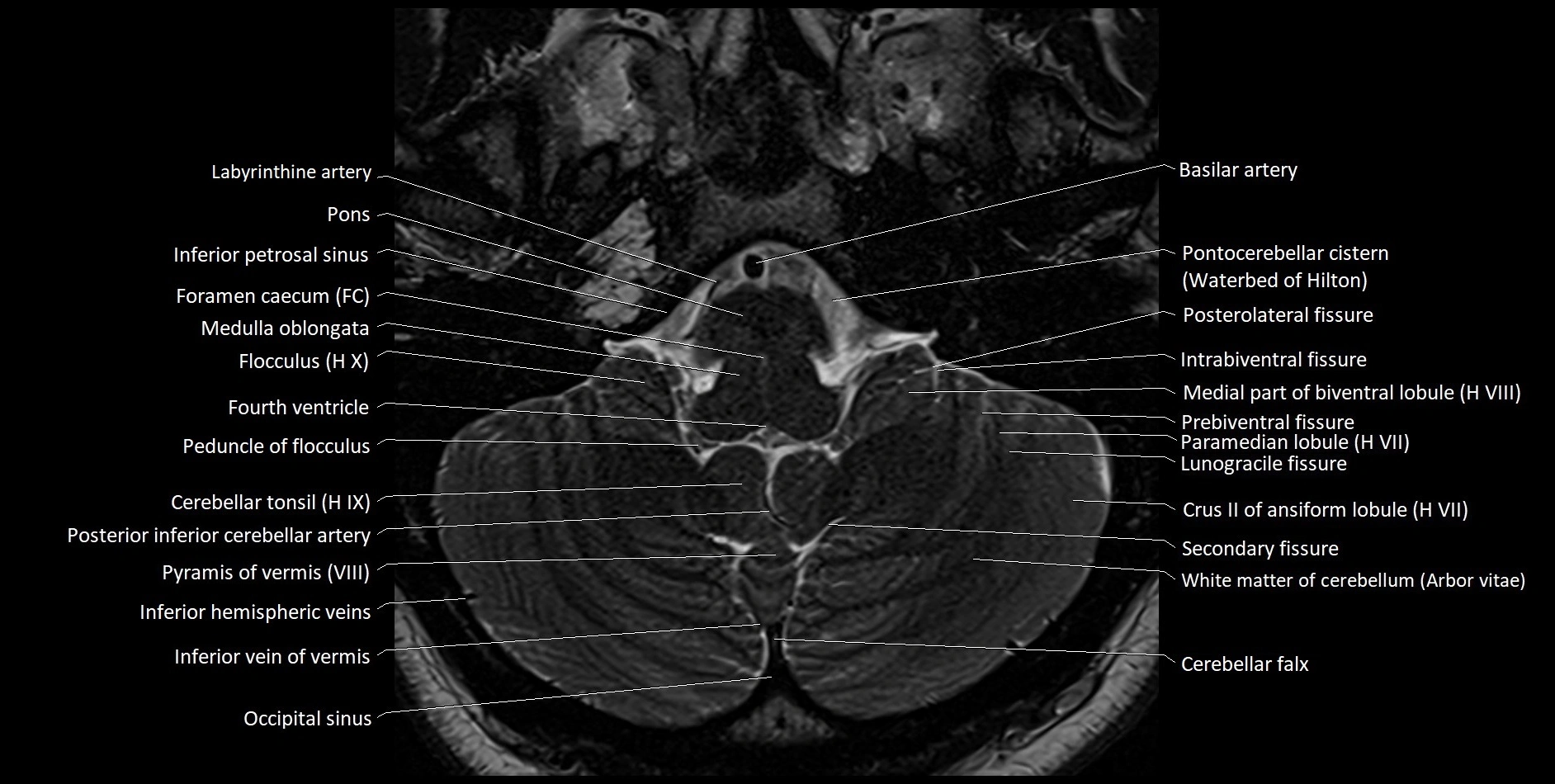
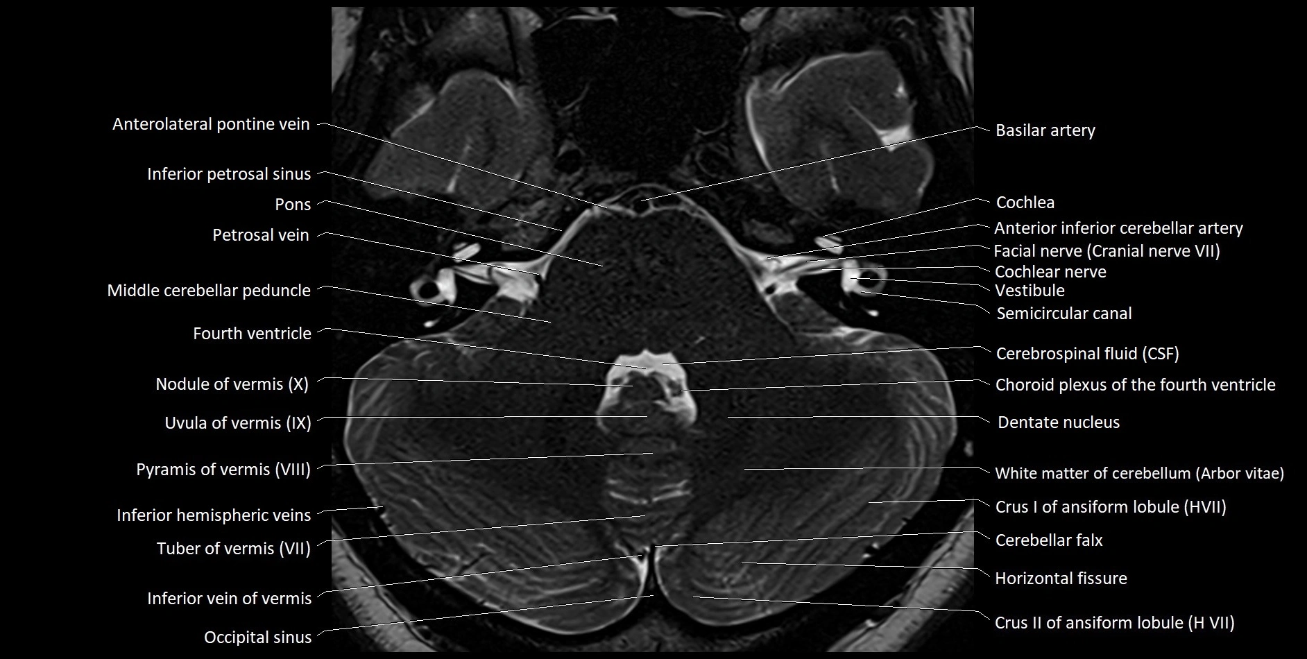
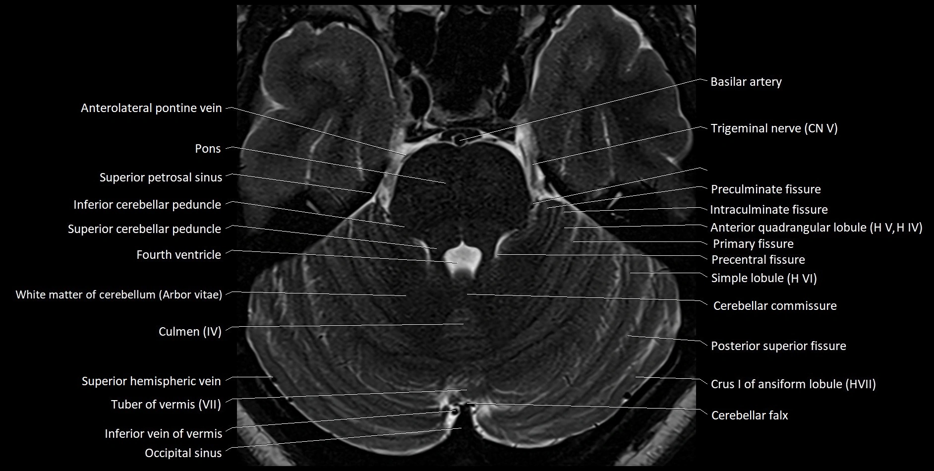
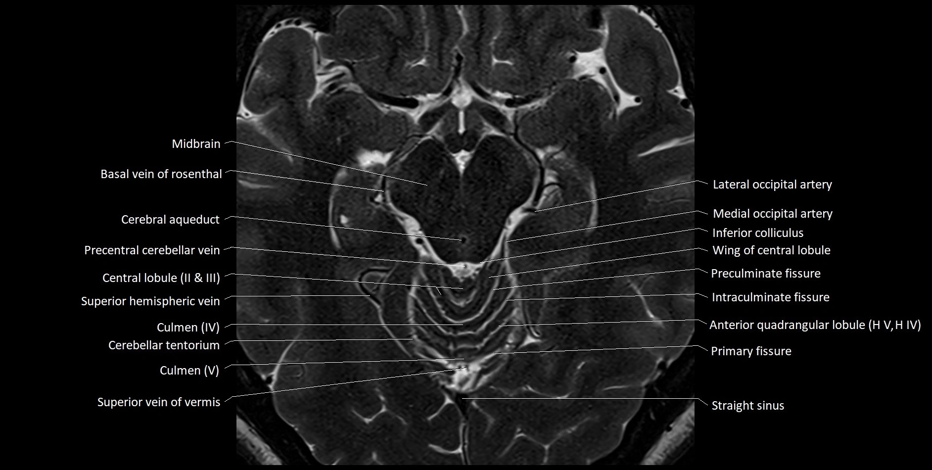
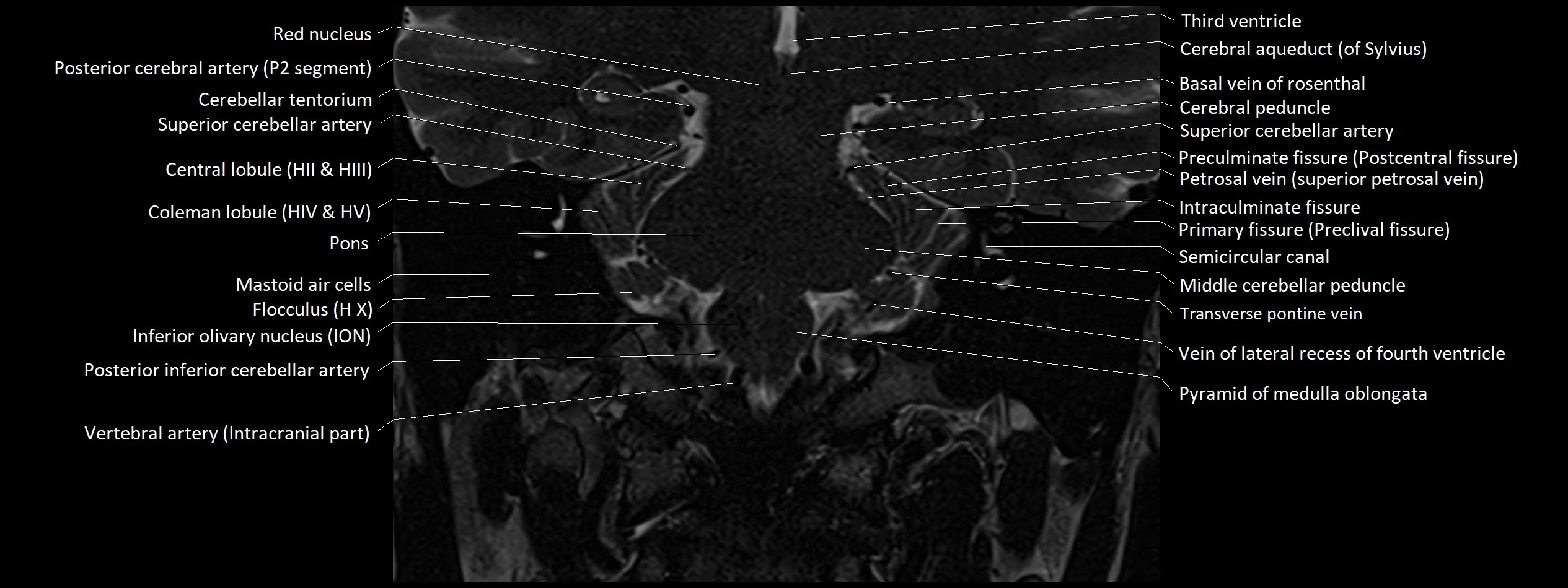
MRI images

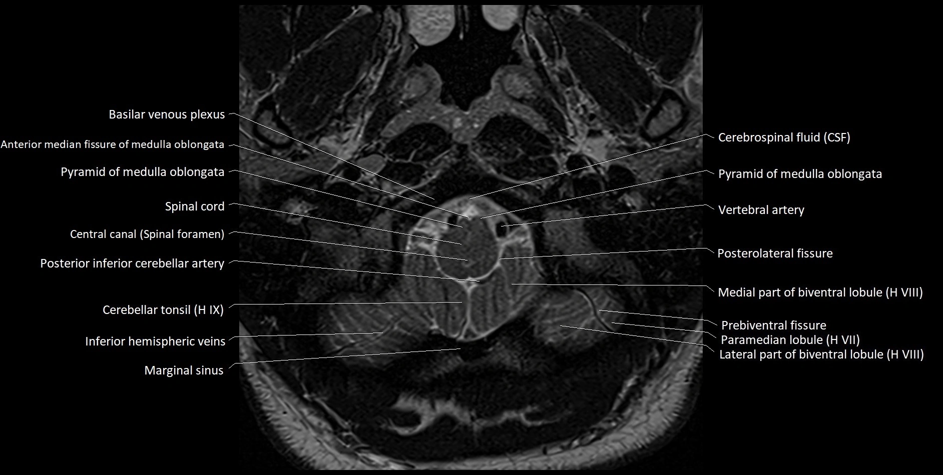
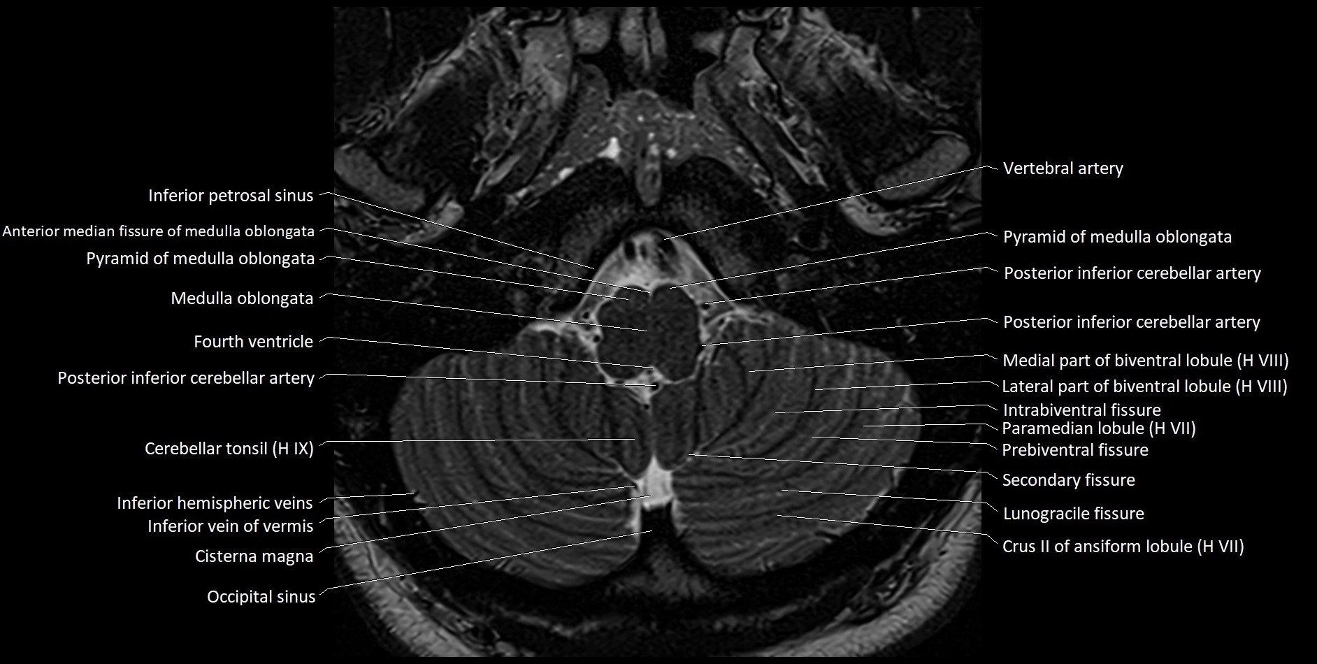
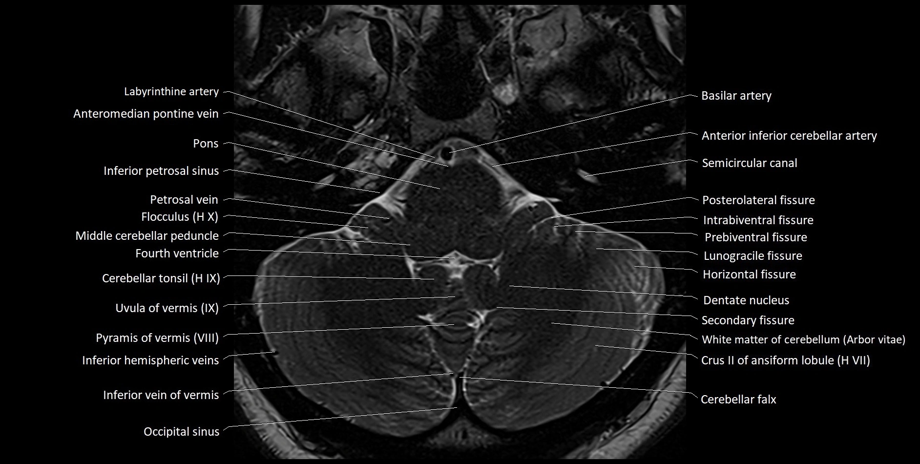
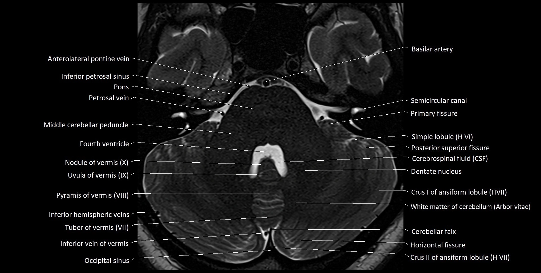
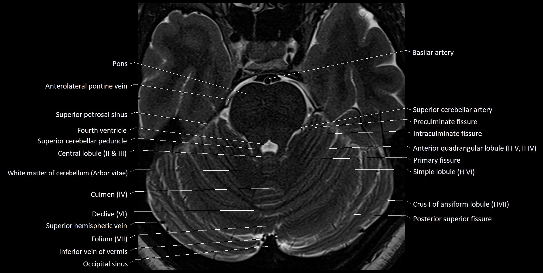
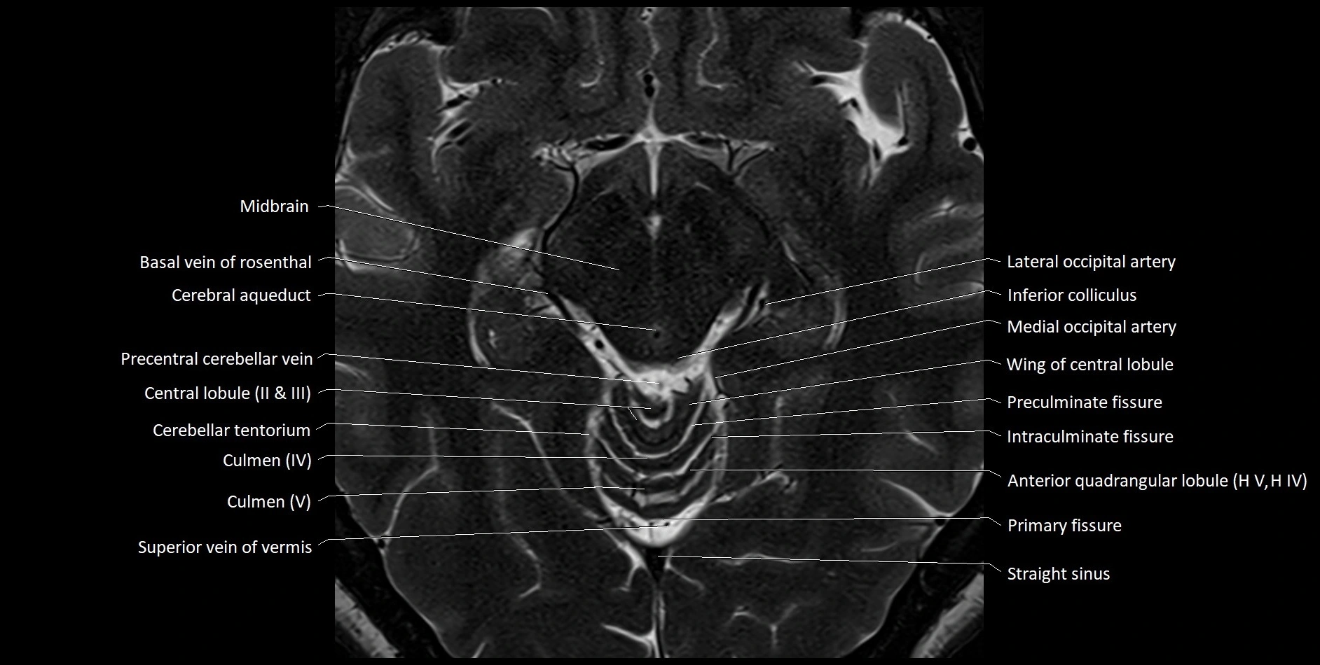
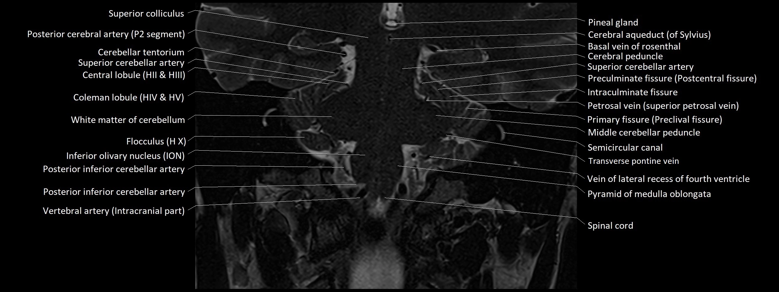
MRI images

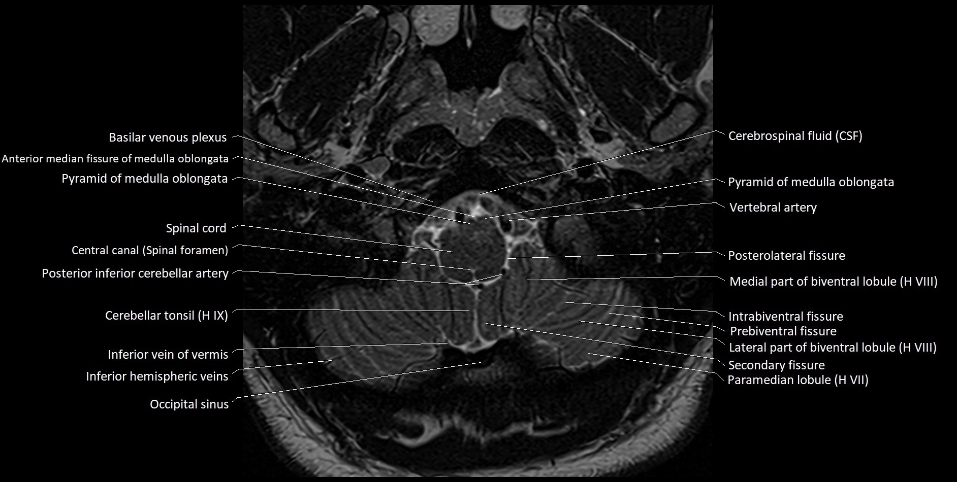
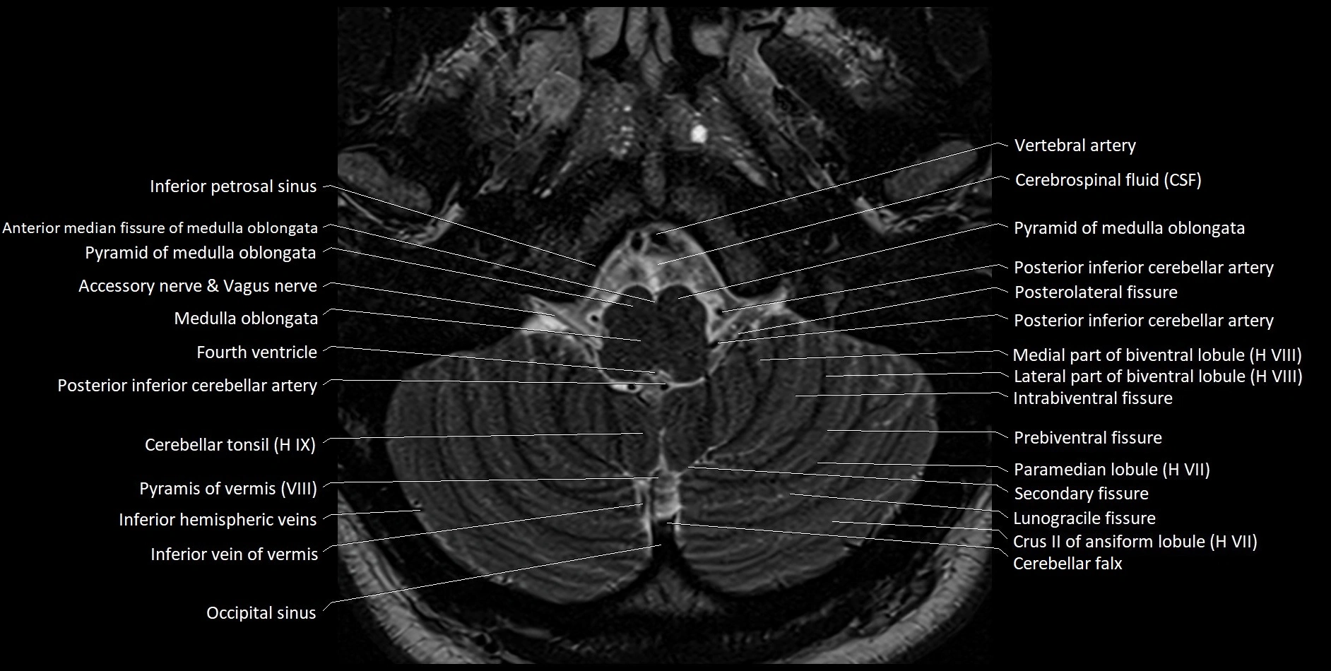
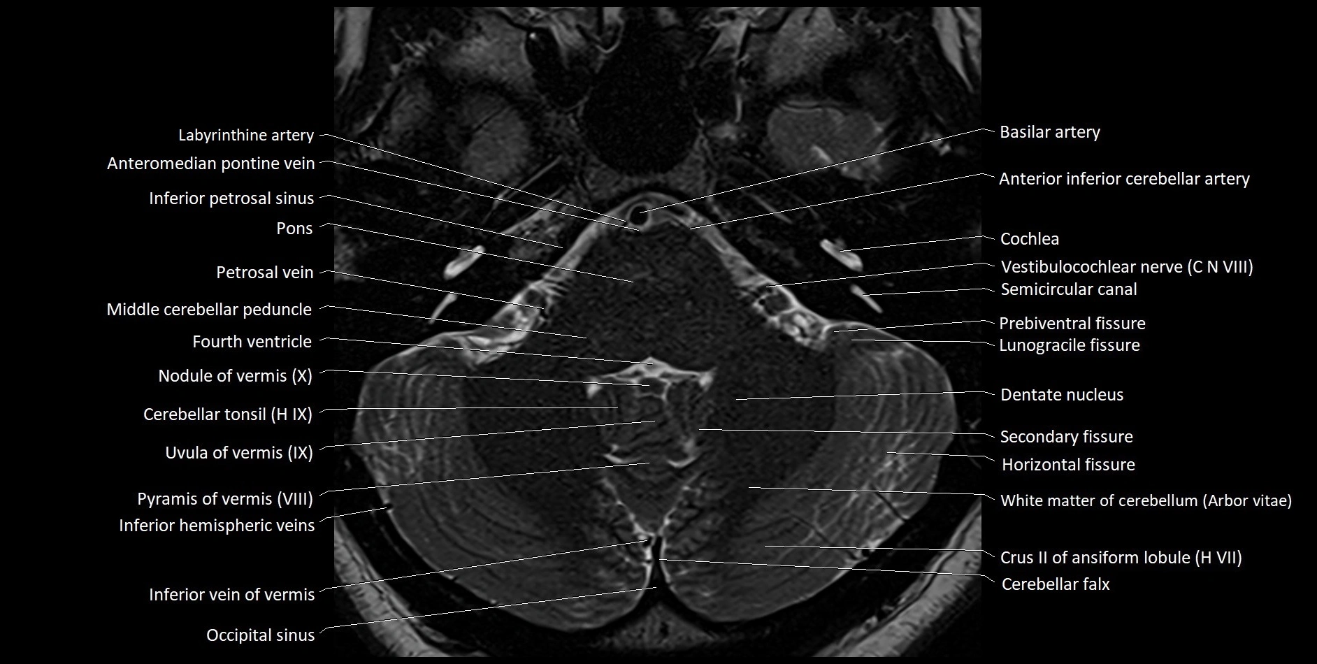
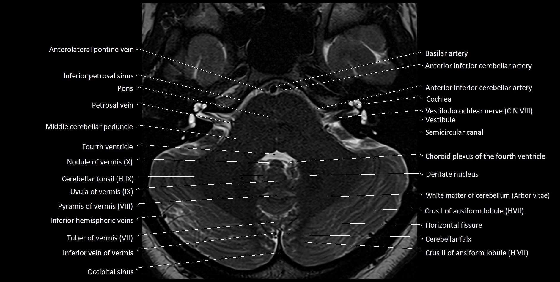
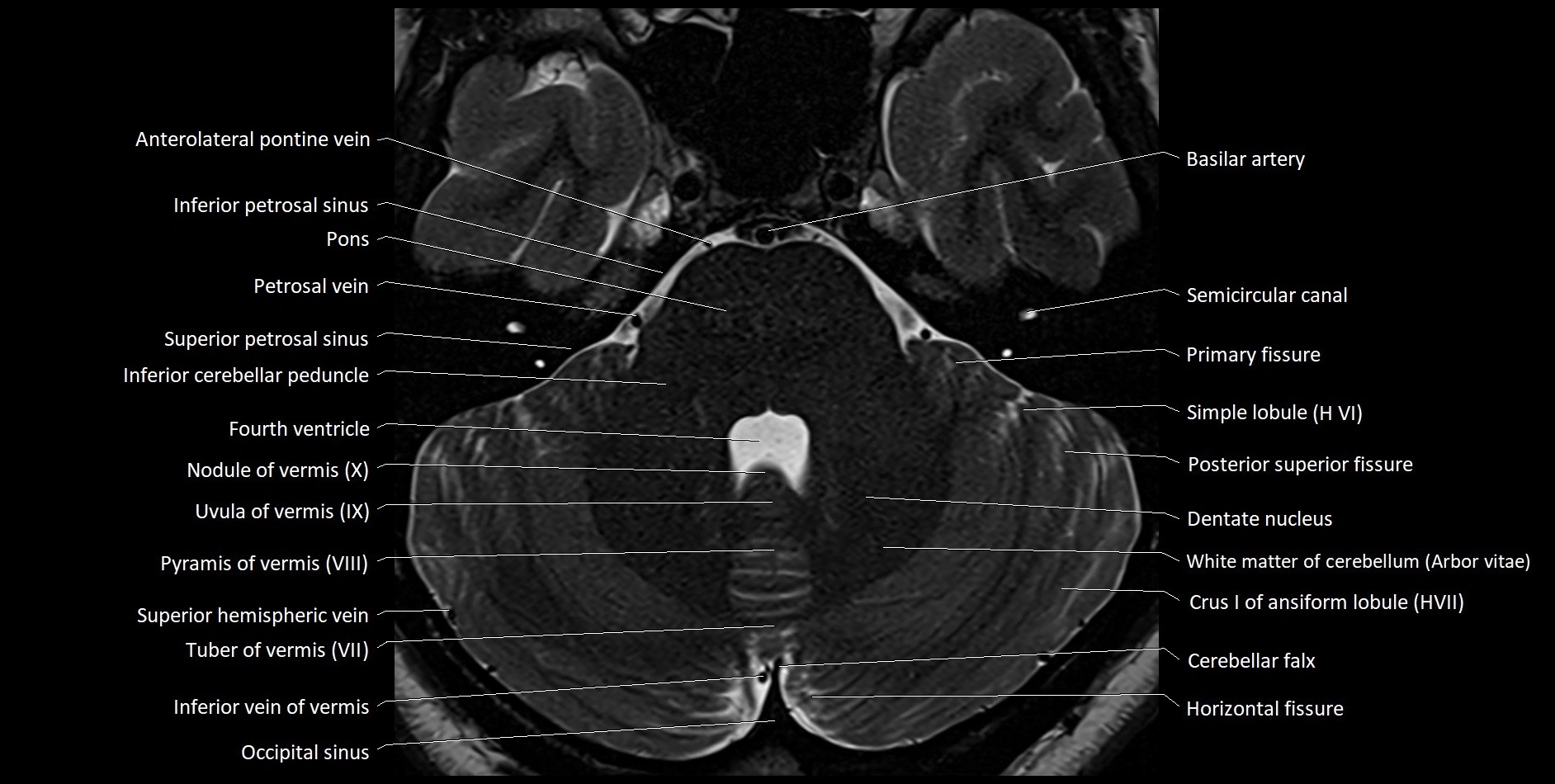
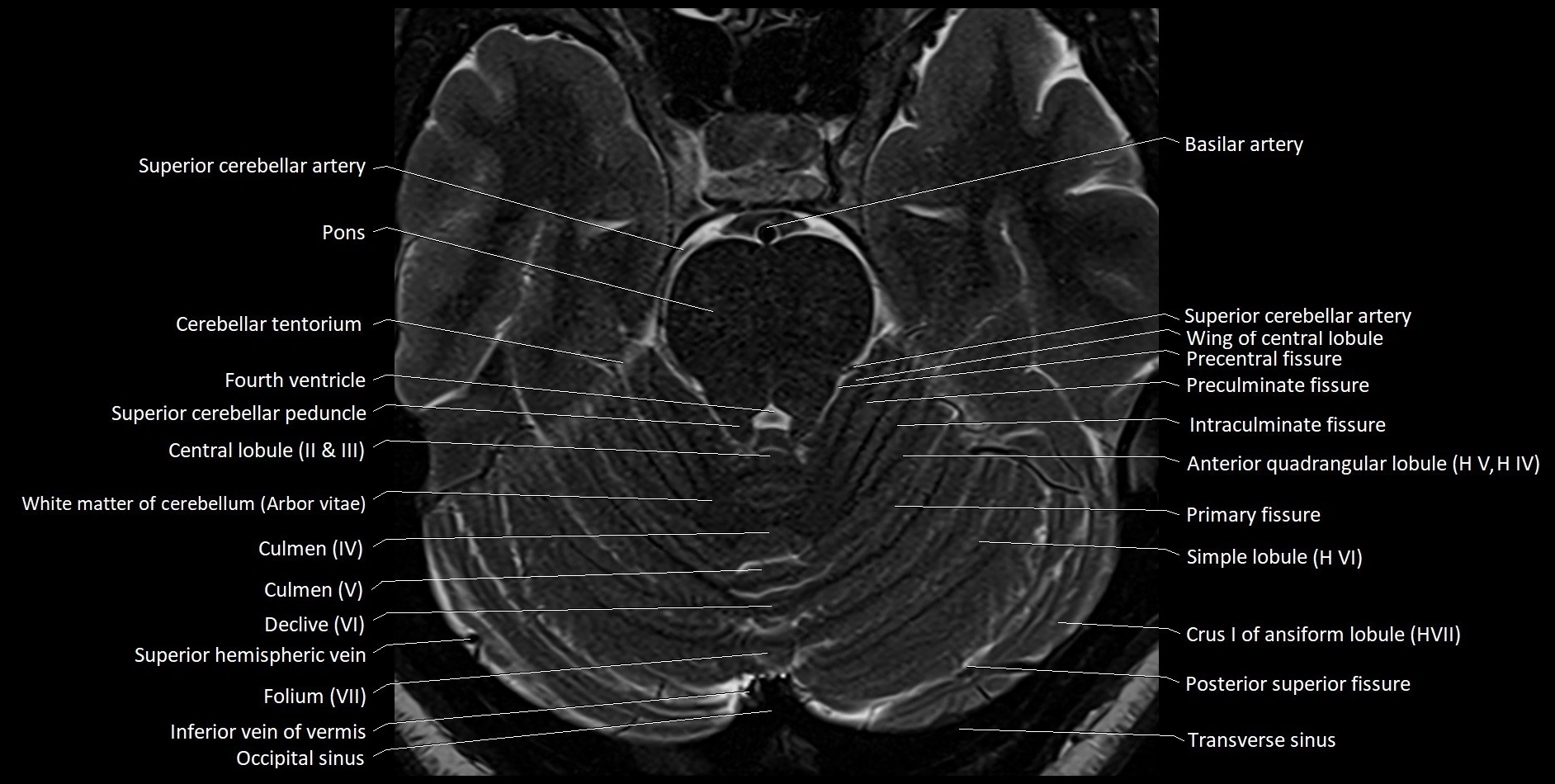
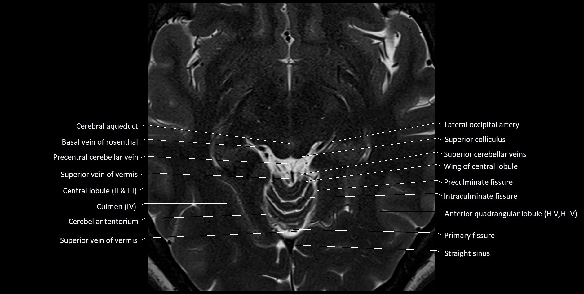
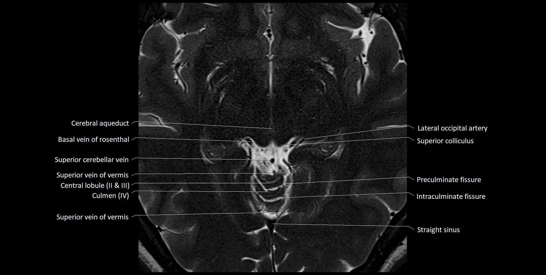
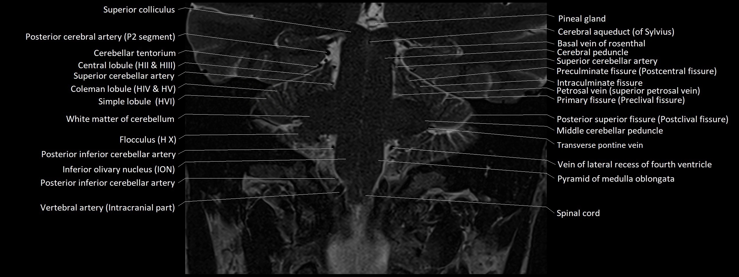
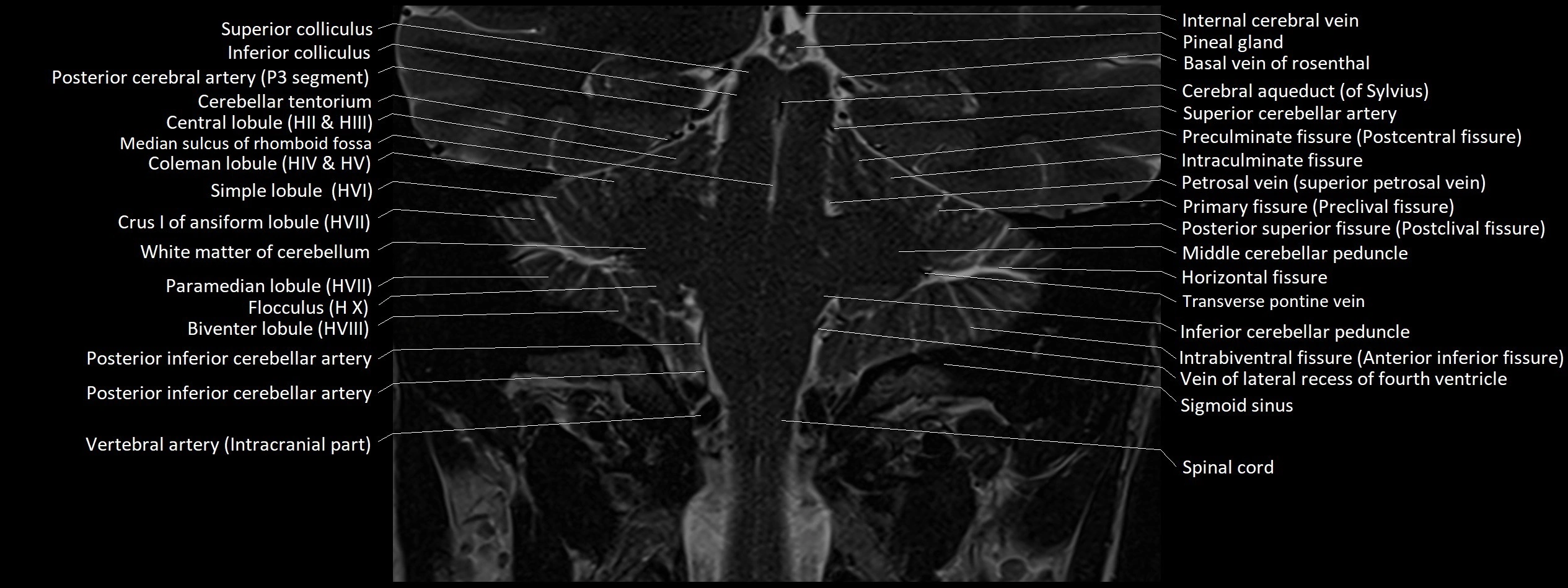
MRI images


