


Topic
- Abducens nerve (Cranial nerve VI)
- Accessory Nerve (Cranial nerve XI)
- Ambient cistern
- Anterior Choroidal Artery
- Anterior Communicating Artery
- Anterior calcarine sulcus
- Anterior cerebral artery
- Anterior clinoid process
- Anterior cochlear nucleus
- Anterior intercavernous sinus
- Anterior lobe of pituitary gland
- Anterior temporal artery anatomy
- Anterolateral central (lenticulostriate) arteries anatomy
- Anteromedial central (perforating) arteries anatomy
- Artery of central sulcus
- Artery of postcentral sulcus
- Artery of precentral sulcus
- Artery to angular gyrus anatomy
- Basilar artery
- Basilar part of pons
- Basilar sulcus
- Basilar venous plexus
- Body of lateral ventricle
- Callosomarginal artery
- Carotid cistern
- Carotid siphon
- Cerebral falx
- Chiasmatic cistern
- Cistern of central sulcus
- Cistern of lamina terminalis
- Cistern of lateral cerebral fossa
- Cistern of transverse fissure
- Cochlea
- Cochlear nerve (Cranial nerve VIII)
- Crista galli
- Crural cistern
- Extreme capsule
- Facial Nerve (Cranial nerve VII)
- Frontobasal artery
- Glossopharyngeal nerve (Cranial nerve IX)
- Greater wing of sphenoid bone
- Hypoglossal Nerve (Cranial nerve XII)
- Hypothalamus
- Inferior branch vestibular nerve
- Inferior hemispheric cerebellar veins
- Inferior hypophyseal artery anatomy
- Inferior parietal lobule
- Inferior petrosal sinus
- Inferior salivatory nucleus
- Inferior vestibular nucleus
- Infundibulum
- Internal carotid artery
- Internal carotid artery (cervical part)
- Internal carotid artery (petrous part)
- Interpeduncular Cistern
- Labyrinthine artery
- Lacrimal nucleus
- Lateral cerebellomedullary cistern
- Lateral frontobasal artery
- Lateral vestibular nucleus
- Lesser wing of sphenoid
- Long medial striate artery
- Marginal branch of cingulate sulcus
- Medial frontobasal artery
- Medial vestibular nucleus
- Meninges
- Mesencephalic nucleus of trigeminal nerve
- Middle cerebral artery
- Middle temporal artery
- Motor nucleus of facial nerve
- Motor nucleus of trigeminal nerve
- Nucleus of abducens nerve
- Nucleus of hypoglossal nerve
- Nucleus of oculomotor nerve
- Nucleus of solitary tract
- Nucleus of trochlear nerve
- Oculomotor Nerve (Cranial Nerve III)
- Oculomotor cistern
- Olfactory Nerve (Cranial Nerve I)
- Olfactory bulb
- Olfactory cistern
- Olfactory tract
- Olfactory trigone
- Olfactory tubercle
- Ophthalmic artery
- Ophthalmic nerve
- Optic Nerve (Cranial Nerve II)
- Optic chiasm
- Optic tract
- Orbital Sulci
- Orbital gyri
- Paracentral artery
- Paracentral gyrus
- Paracentral lobule
- Paraolfactory sulci
- Pericallosal artery
- Pericallosal cistern
- Perivascular spaces
- Pituitary gland
- Pituitary stalk
- Polar frontal artery
- Polar temporal artery
- Pontine arteries
- Pontocerebellar cistern
- Posterior cerebellomedullary cistern (cisterna magna)
- Posterior cerebral artery
- Posterior cochlear nucleus
- Posterior communicating artery
- Posterior intercavernous sinus
- Posterior lateral choroidal artery
- Posterior lobe pituitary gland
- Posterior orbital sulcus
- Posterior parietal artery
- Posteromedial central (perforating) arteries
- Preoptic area
- Prepontine cistern
- Principal sensory nucleus of the trigeminal nerve
- Quadrigeminal cistern
- Rostral gyrus
- Rostral sulcus
- Semicircular Canals
- Sphenoparietal sinus
- Spinal nucleus of trigeminal nerve
- Superior branch of vestibular nerve
- Superior cerebellar artery
- Superior cerebellar cistern
- Superior hypophyseal artery
- Superior salivatory nucleus
- Superior vestibular nucleus
- Temporal lobe
- Transverse occipital sulcus
- Transverse temporal sulcus
- Trigeminal cave
- Trigeminal ganglion
- Trigeminal nerve (Cranial nerve V)
- Trochlear nerve (Cranial nerve IV)
- Tuber cinereum
- Vagus nerve (Cranial nerve X)
- Vestibular ganglion
- Vestibule
- Vestibulocochlear nerve (Cranial nerve VIII)
- cavernous sinus
- foramen of Monro
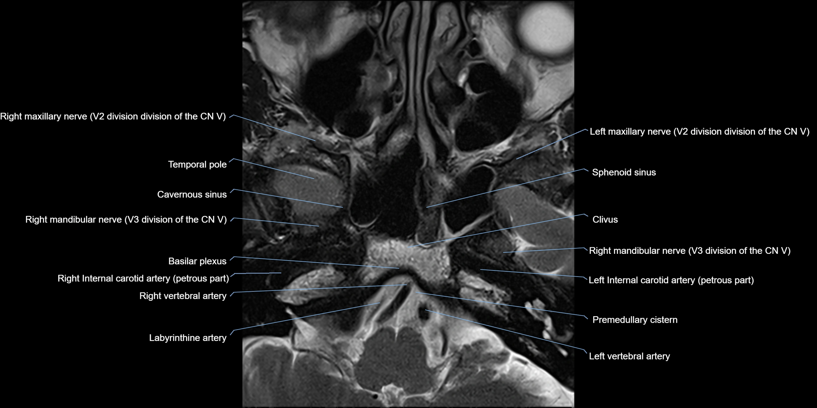
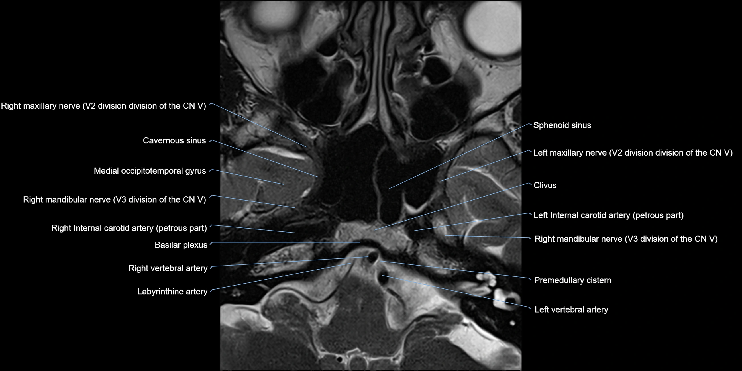
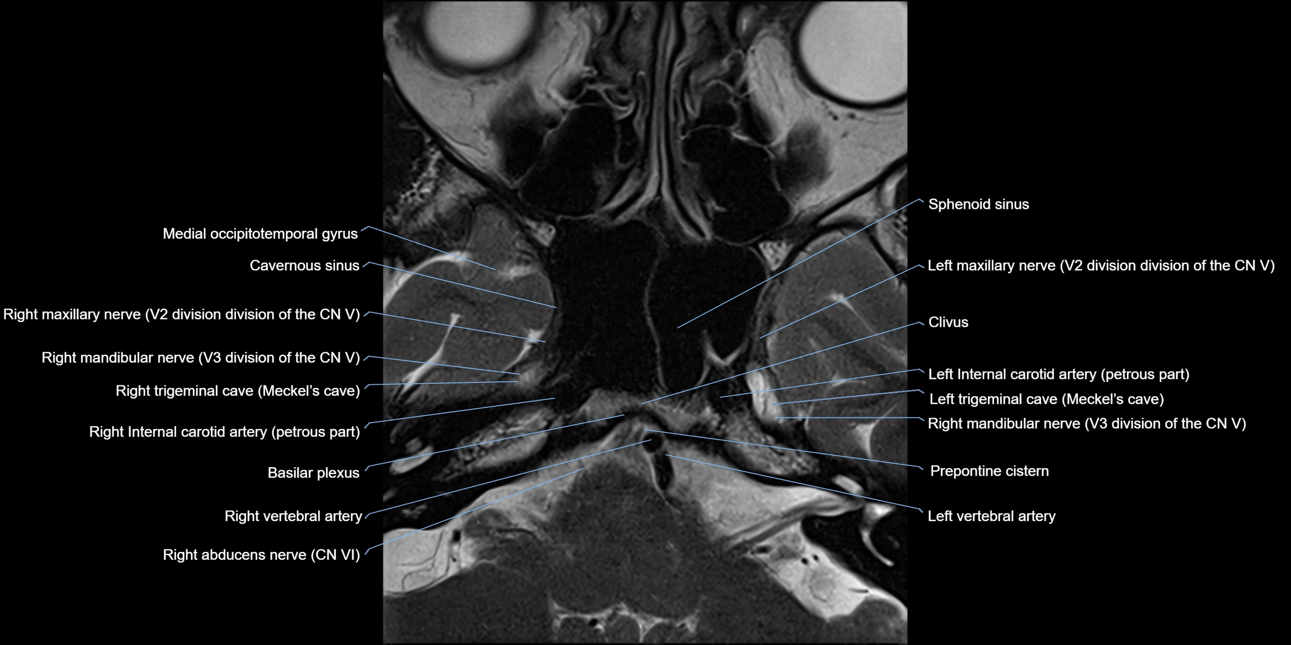
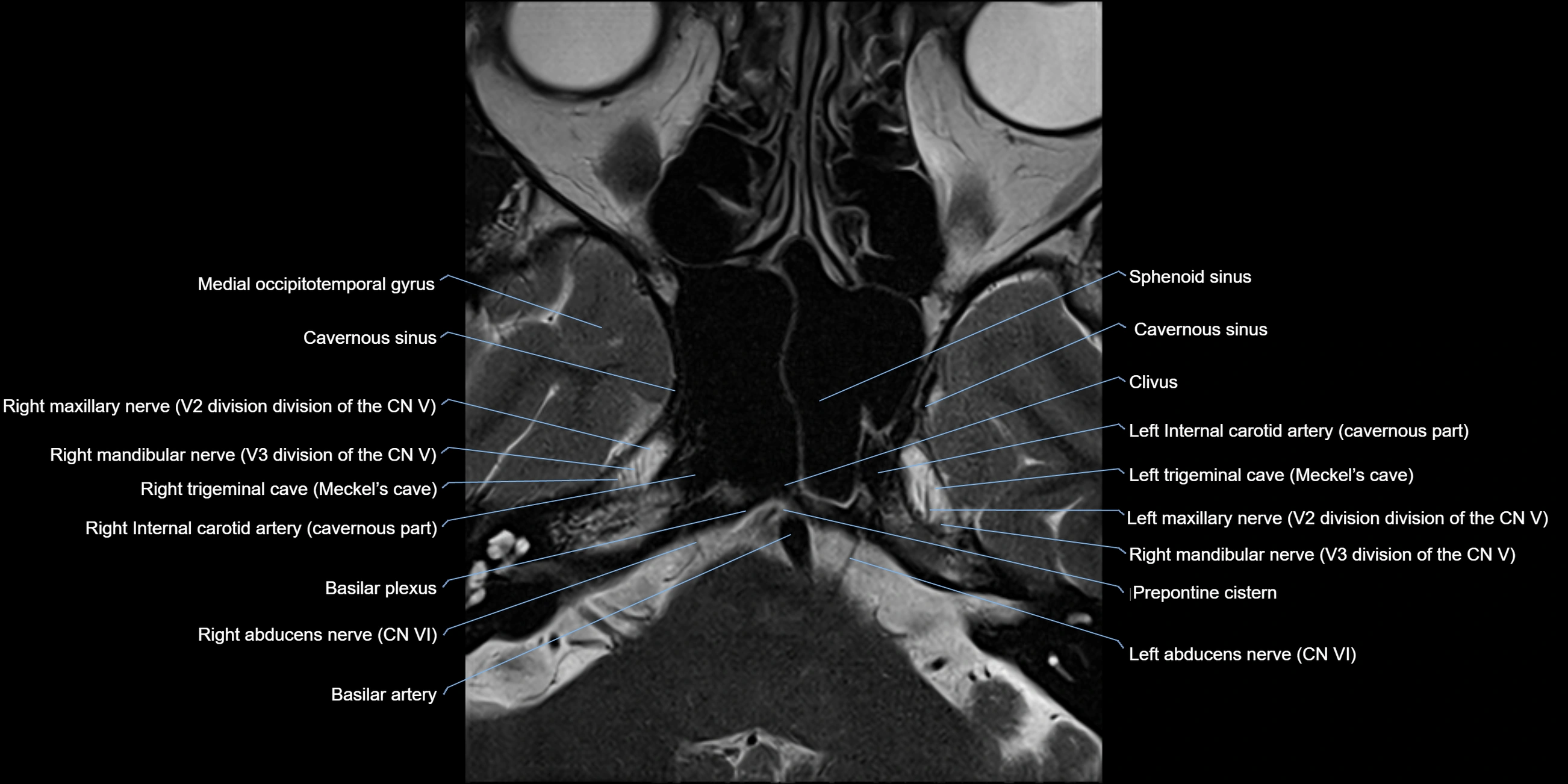
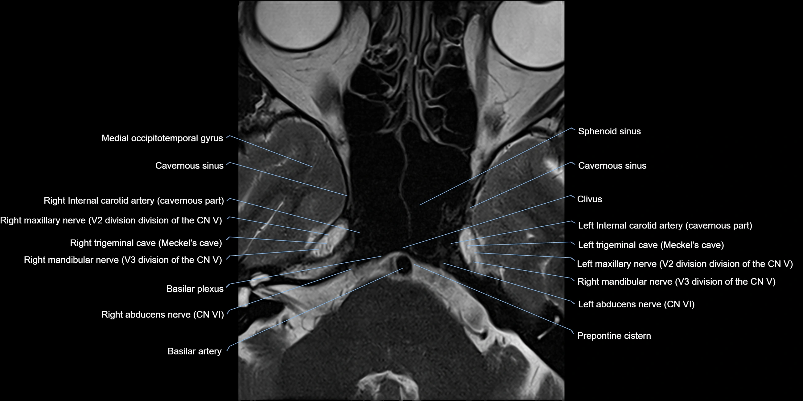
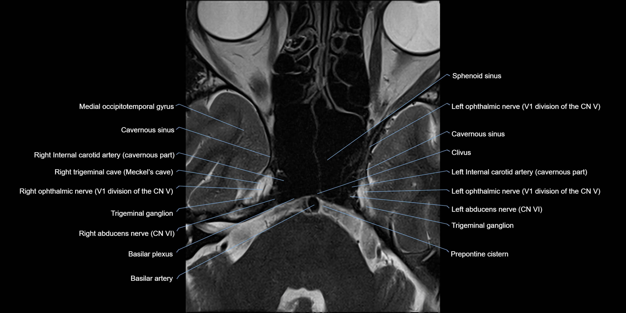
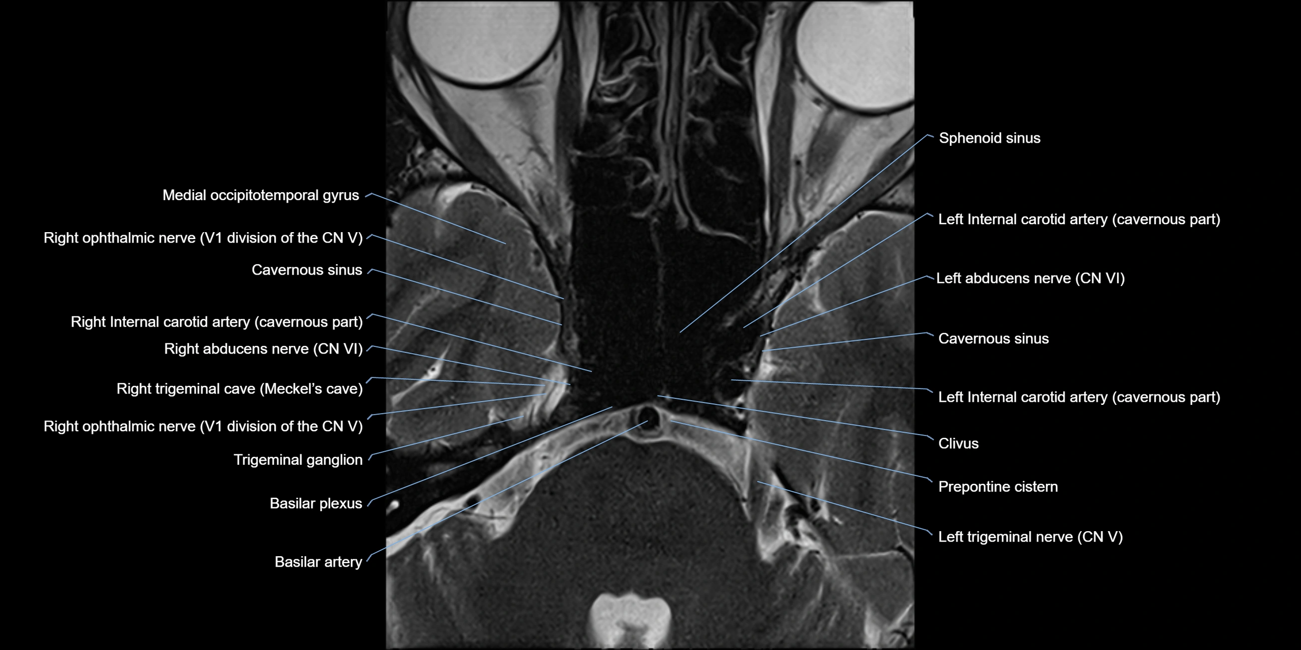
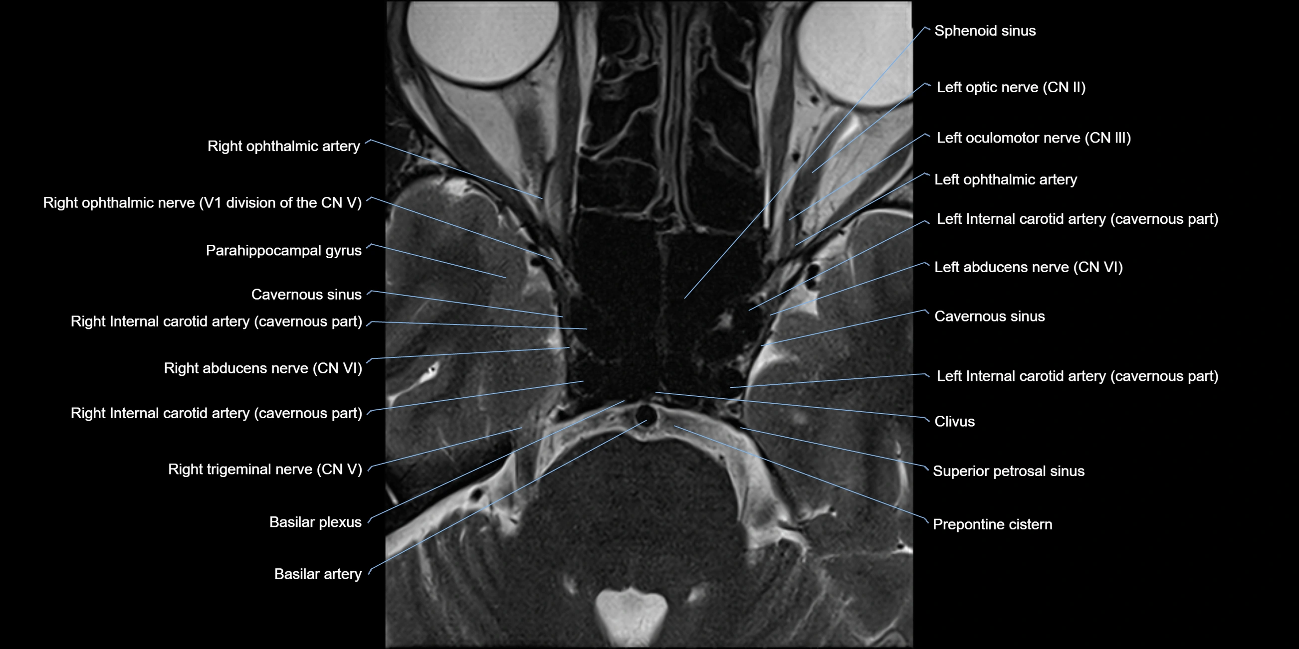
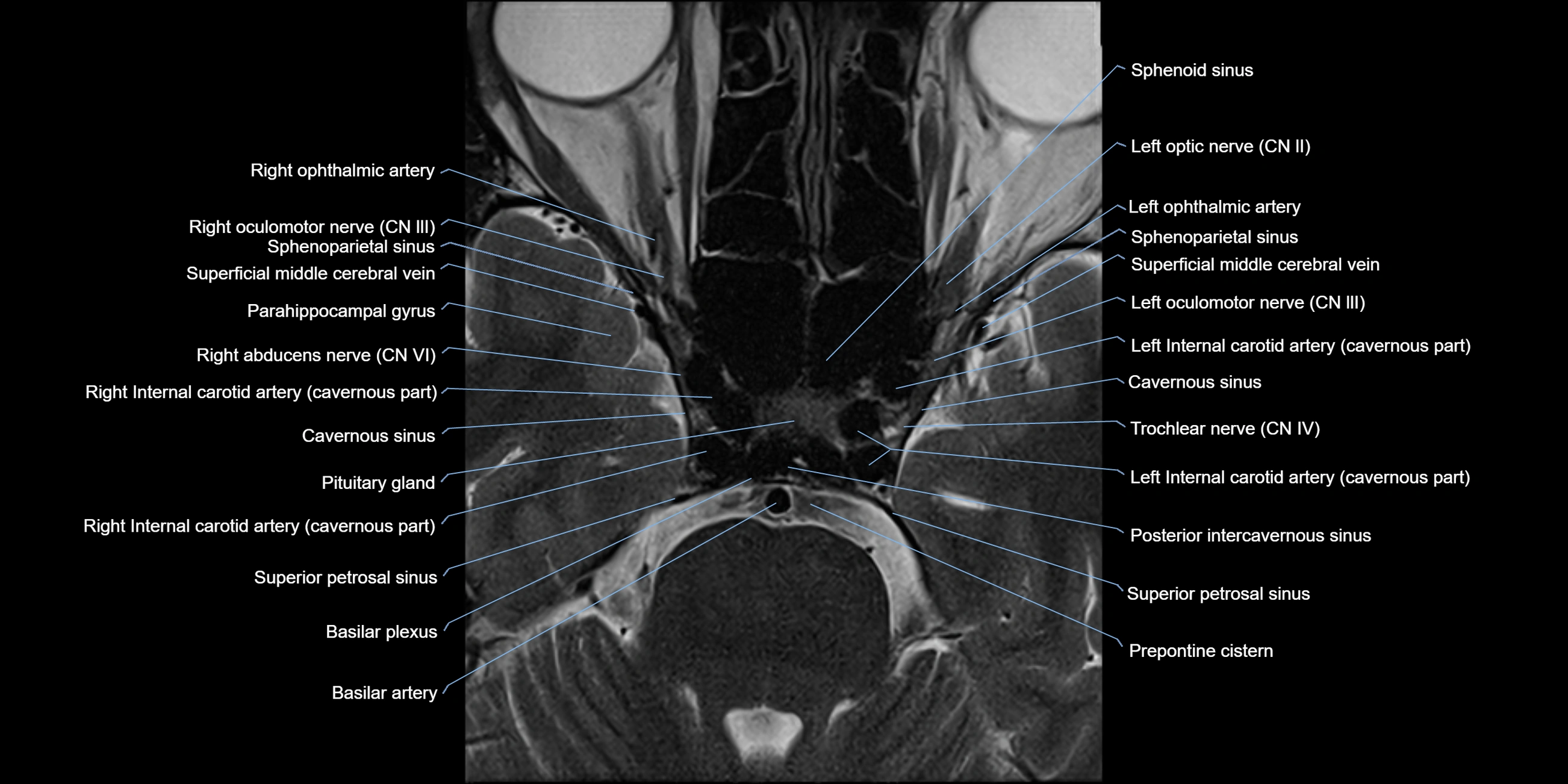
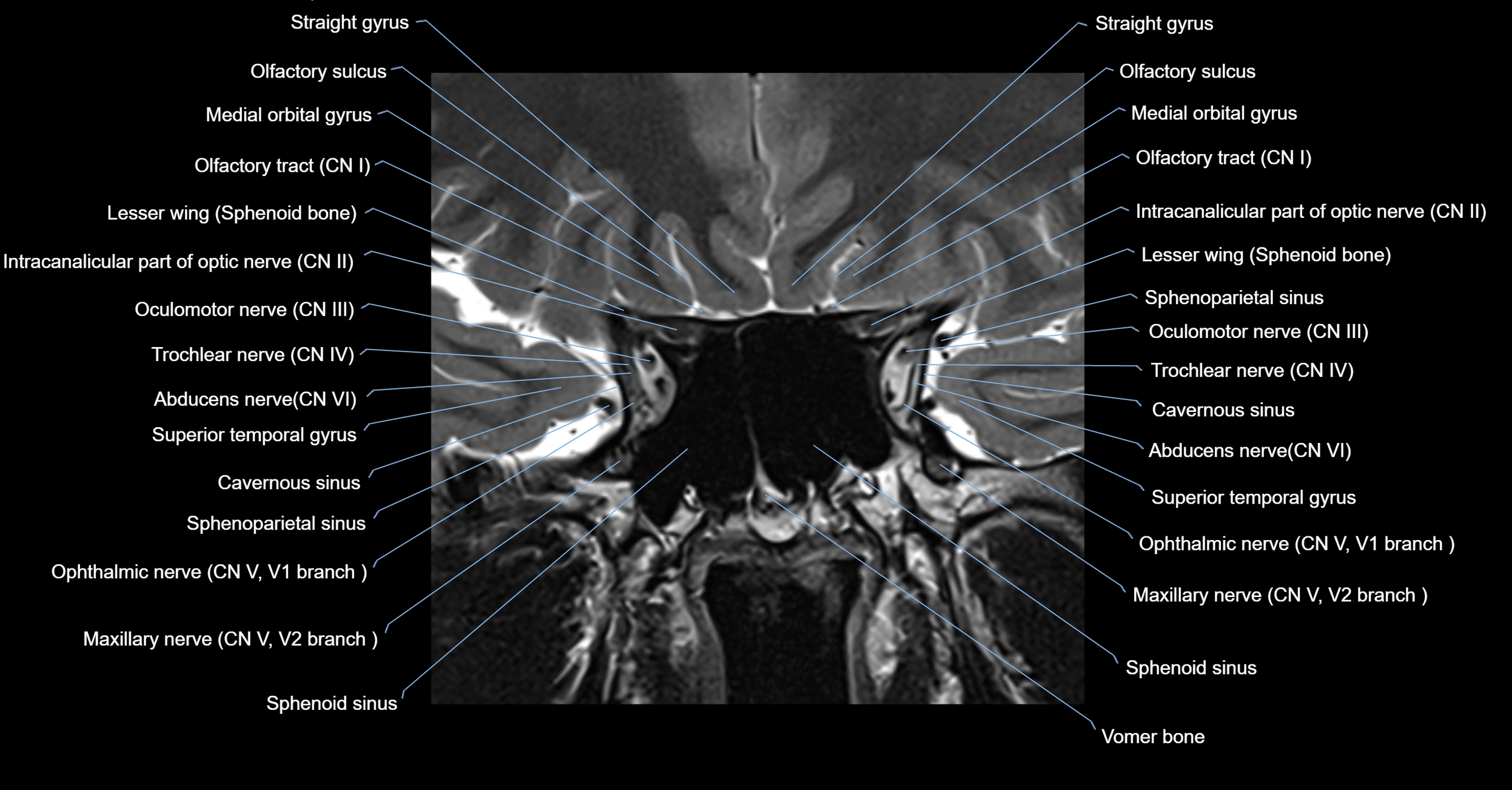
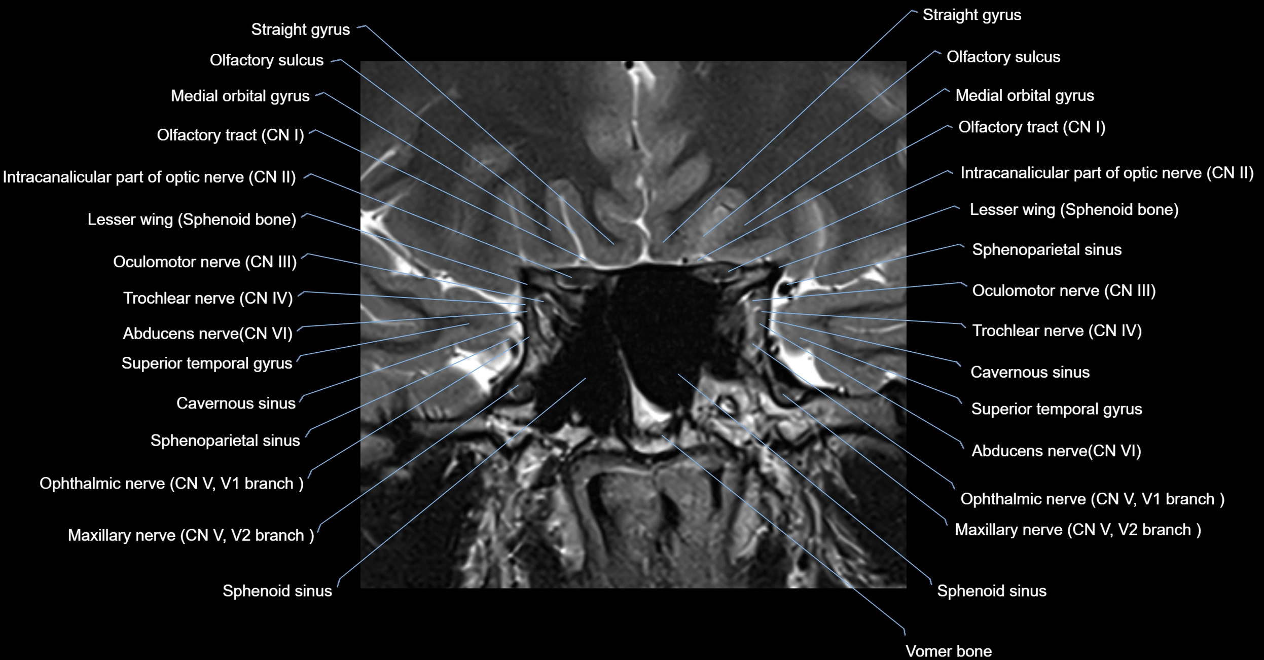
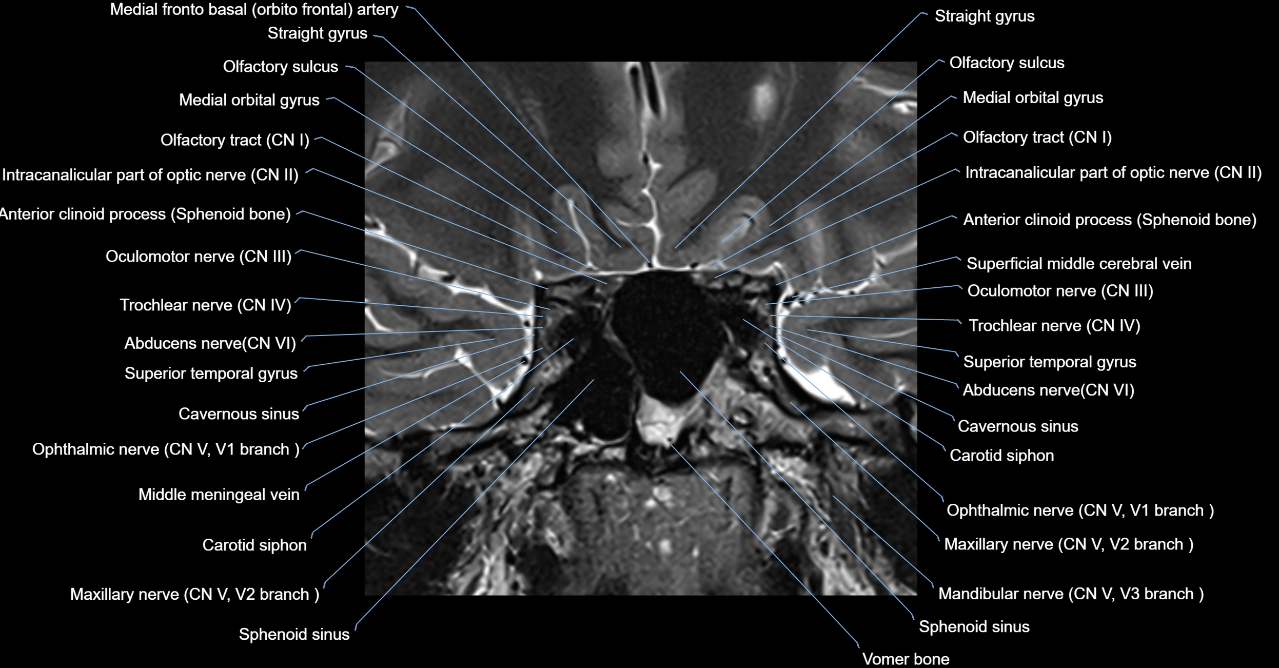
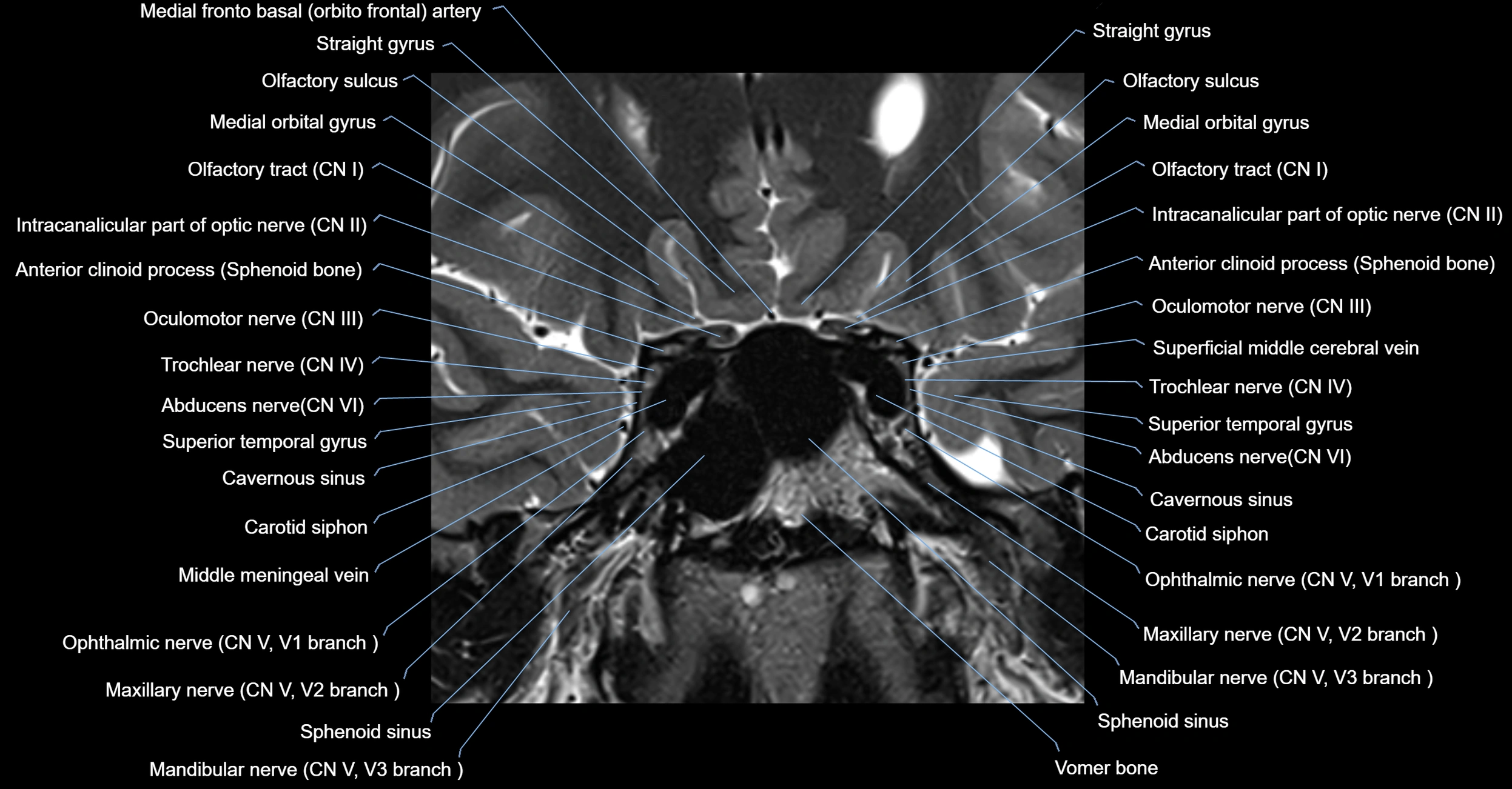
The Abducens nerve (Cranial nerve VI) is a purely motor cranial nerve responsible for innervating the lateral rectus muscle of the eye, which is crucial for lateral movement (abduction) of the eyeball. It arises from the abducens nucleus in the dorsal pons, emerges at the pontomedullary junction, and travels a long intracranial course before entering the orbit via the superior orbital fissure. Because of its long path and proximity to the clivus, it is particularly susceptible to injury from increased intracranial pressure or trauma.
Synonyms
-
Sixth cranial nerve
-
CN VI
-
N. abducens (Latin)
-
Nervus abducens
Function
-
Innervates the lateral rectus muscle of the eye
-
Responsible for abduction of the eyeball (moving the eye outward, away from the midline)
-
Is a purely motor nerve (no sensory or autonomic fibers)
-
Lesion results in inability to abduct the affected eye, leading to horizontal diplopia (double vision)
MRI Appearance
-
The abducens nerve is a small, thin, linear structure
-
Best visualized on high-resolution T2-weighted 3D MRI sequences (e.g., FIESTA or CISS)
-
Seen as a hypointense (dark) line running from the brainstem at the pontomedullary junction, traversing the prepontine cistern, and entering Dorello’s canal under the petrosphenoidal ligament, then into the cavernous sinus, and finally the orbit
-
May be challenging to visualize in standard MRI due to its small size
-
Pathology may be inferred by absence, displacement, or enhancement of the nerve
CT Appearance
-
The nerve itself is not directly visualized on conventional CT due to its small size and soft tissue density
-
Indirect signs: assessment of the bony course, such as the Dorello’s canal, superior orbital fissure, or adjacent pathologies (fractures, masses, or inflammation) that could impinge the nerve
-
CT is mainly used to exclude structural lesions or fractures that might affect the course of CN VI
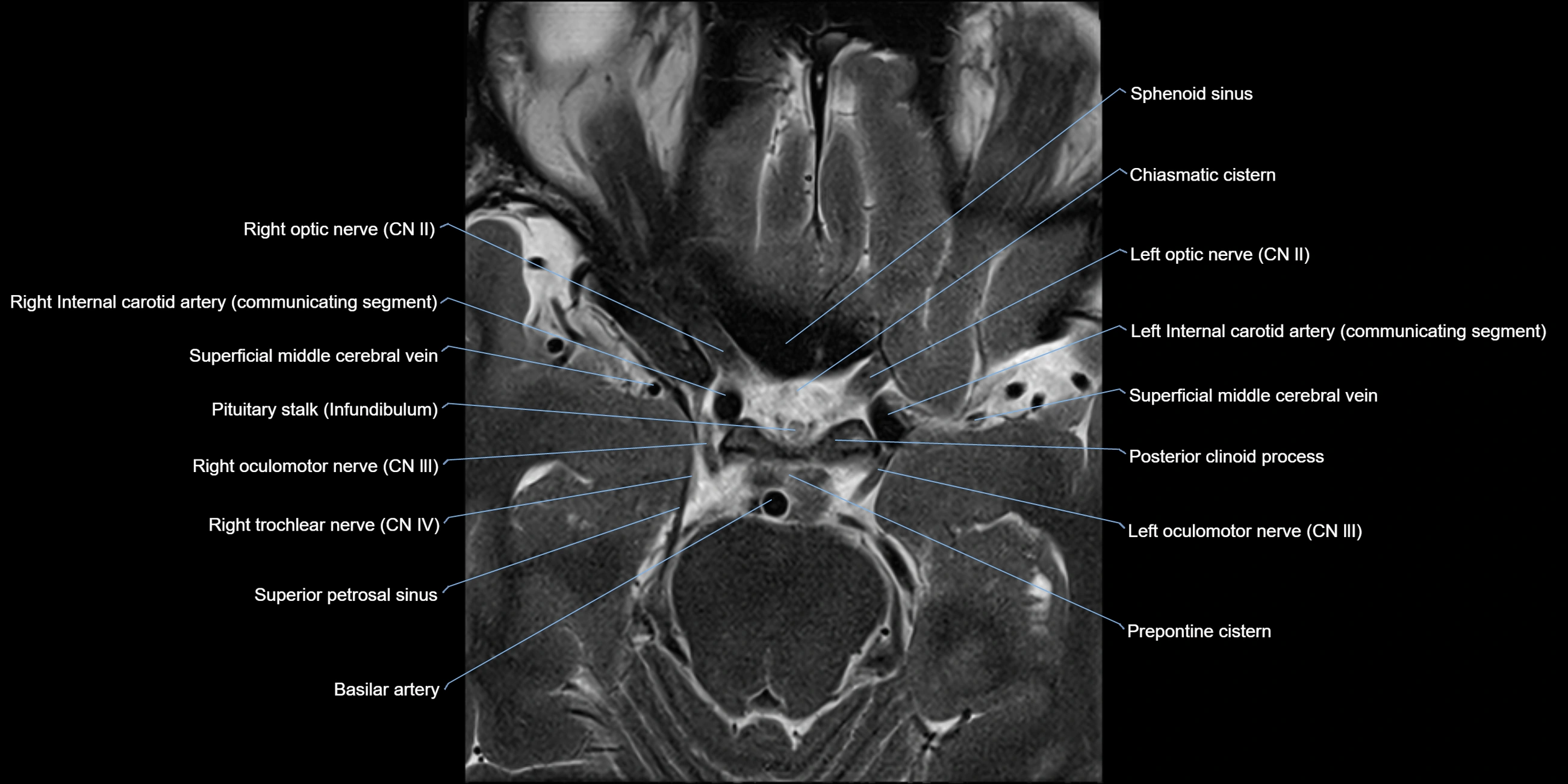
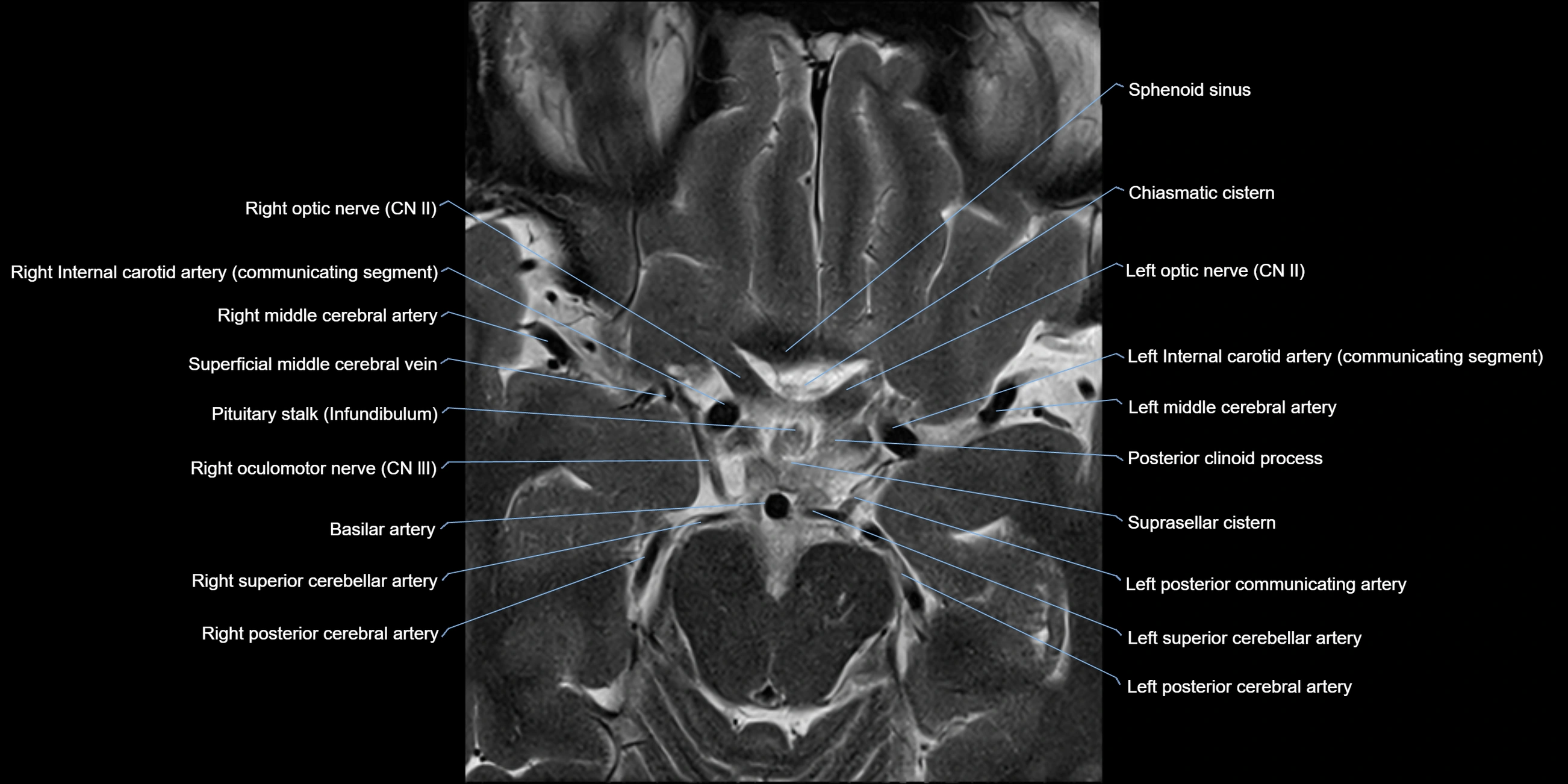
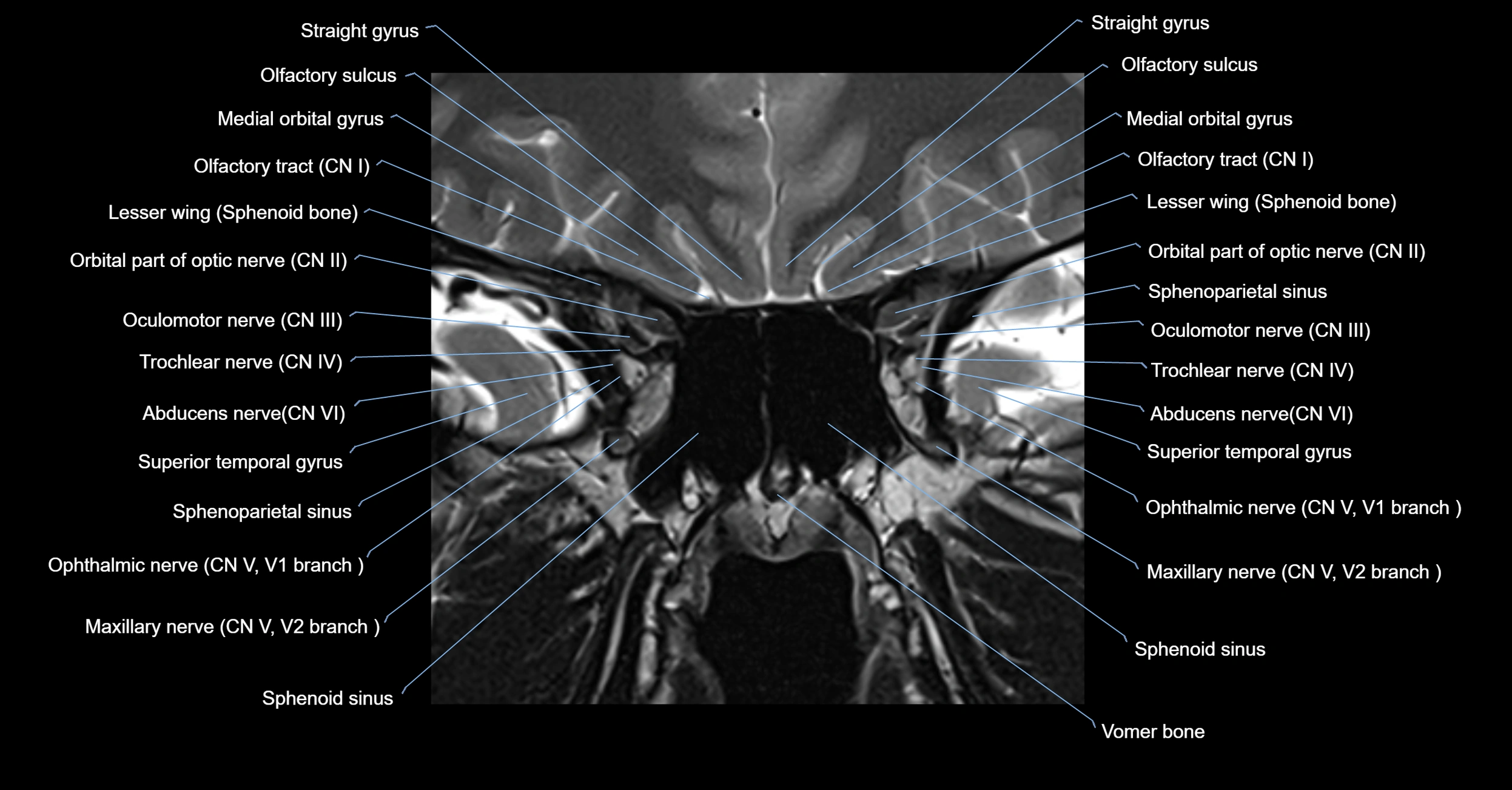
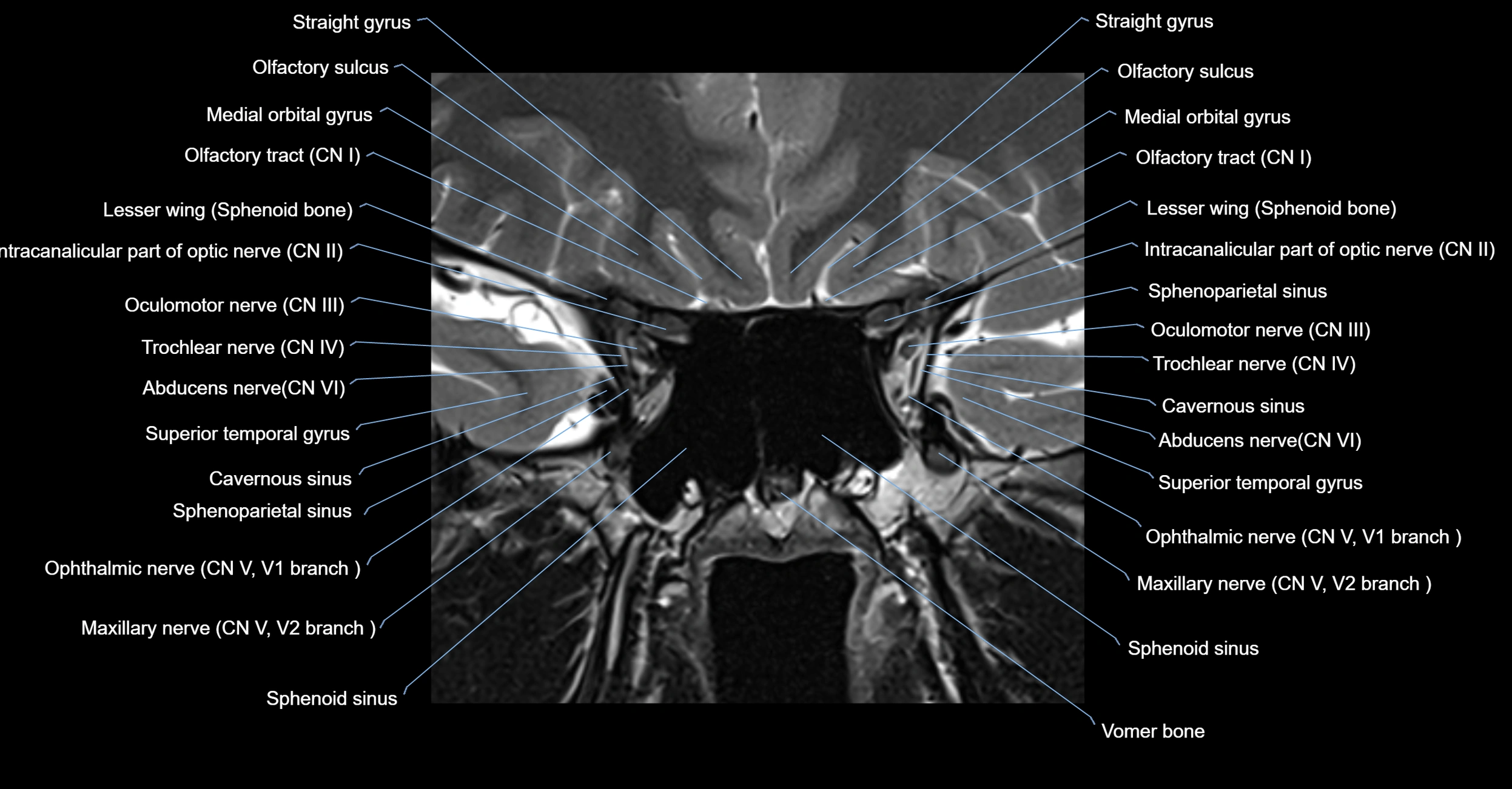
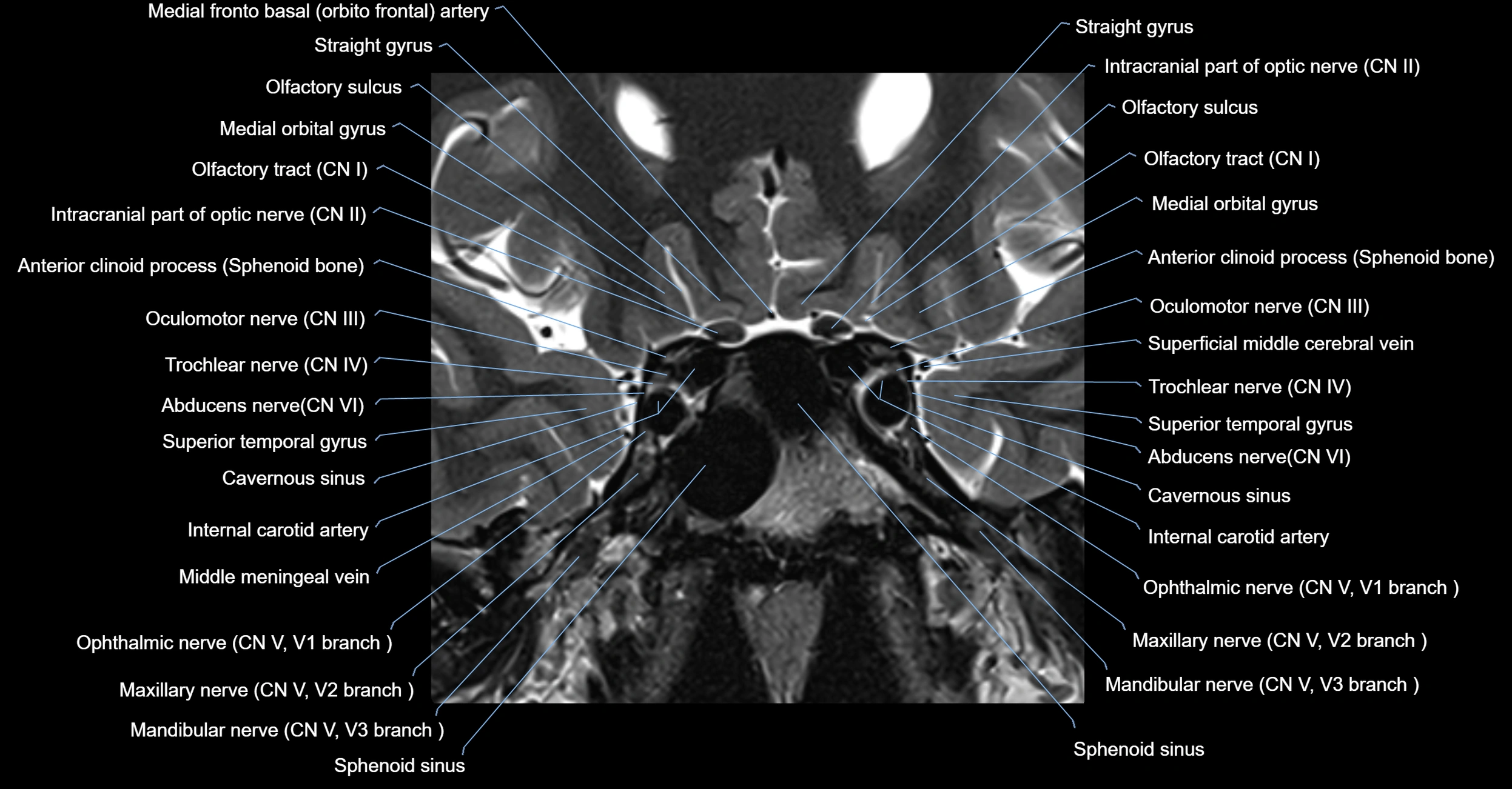
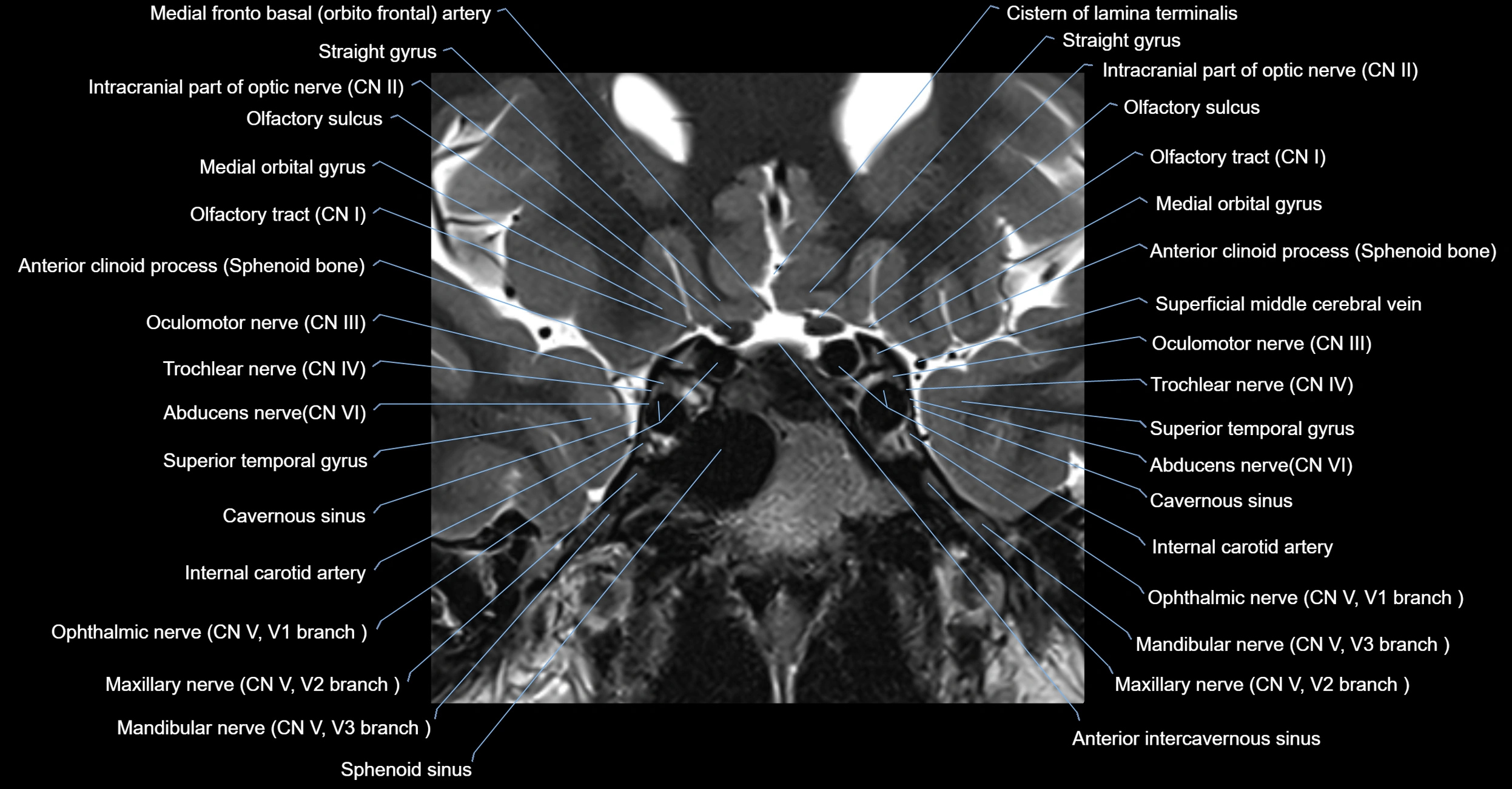
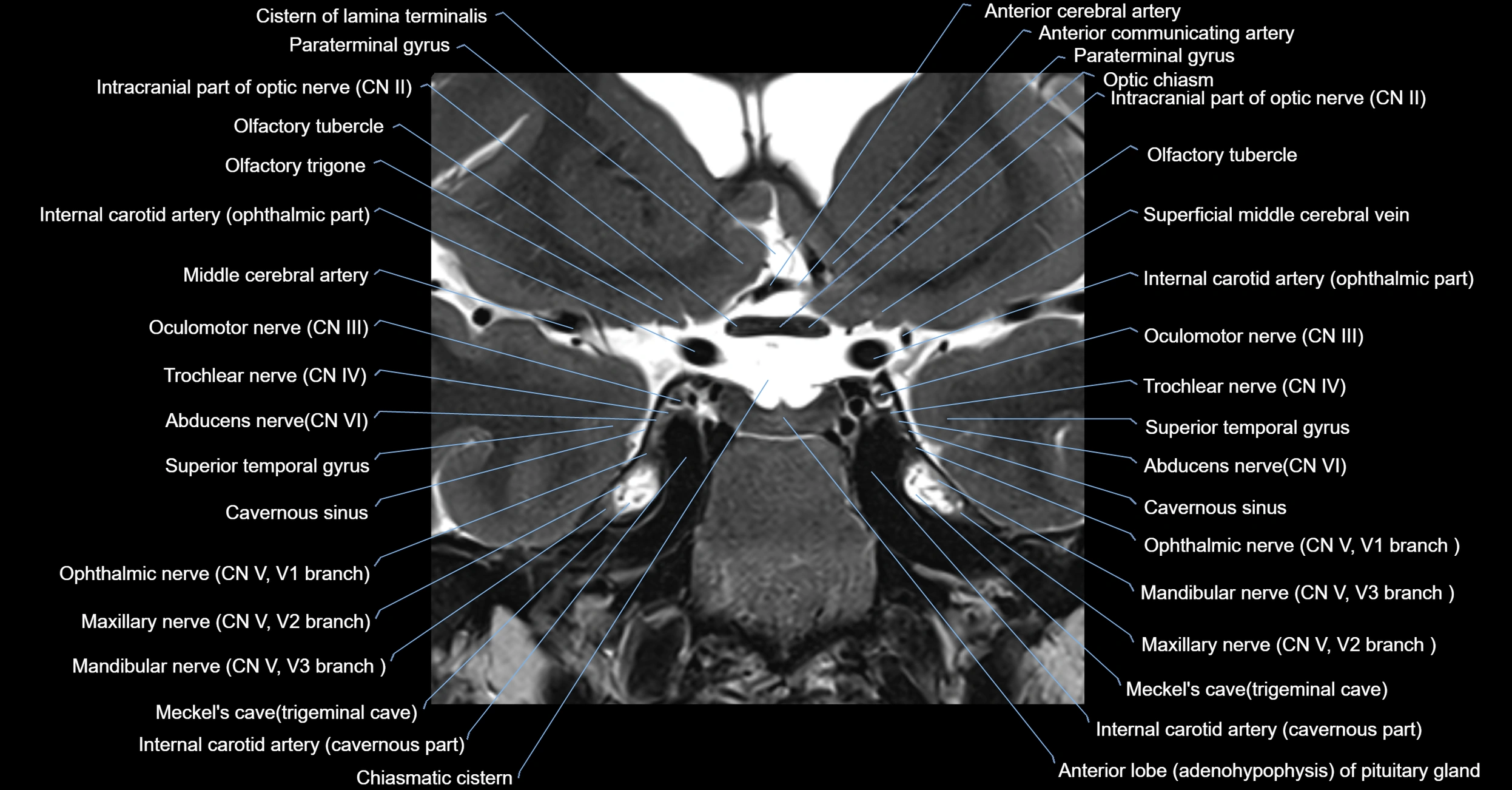
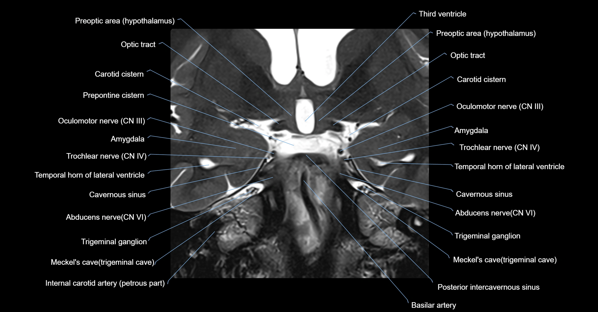
MRI images

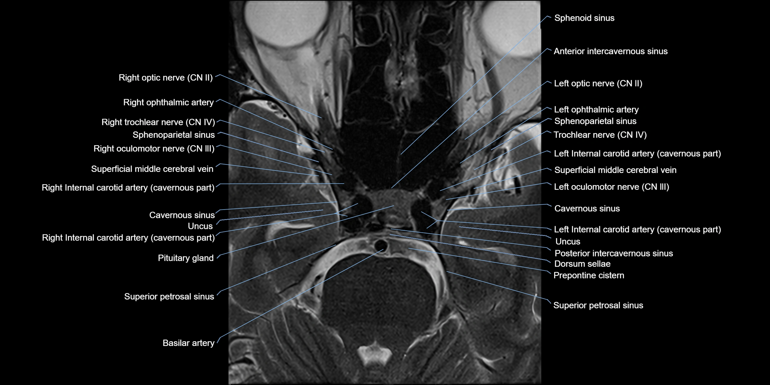
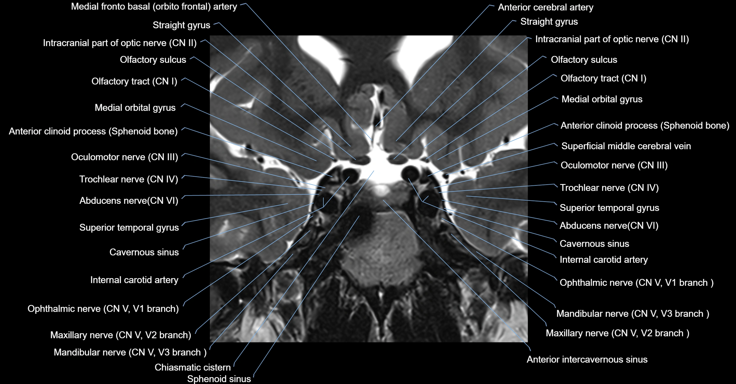
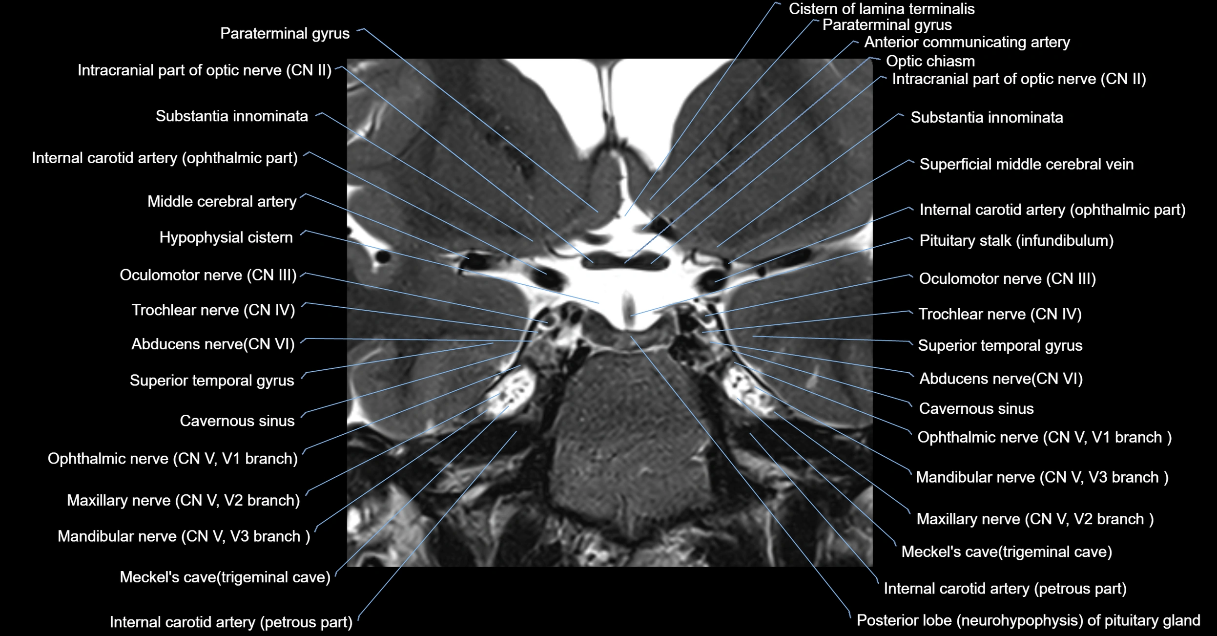
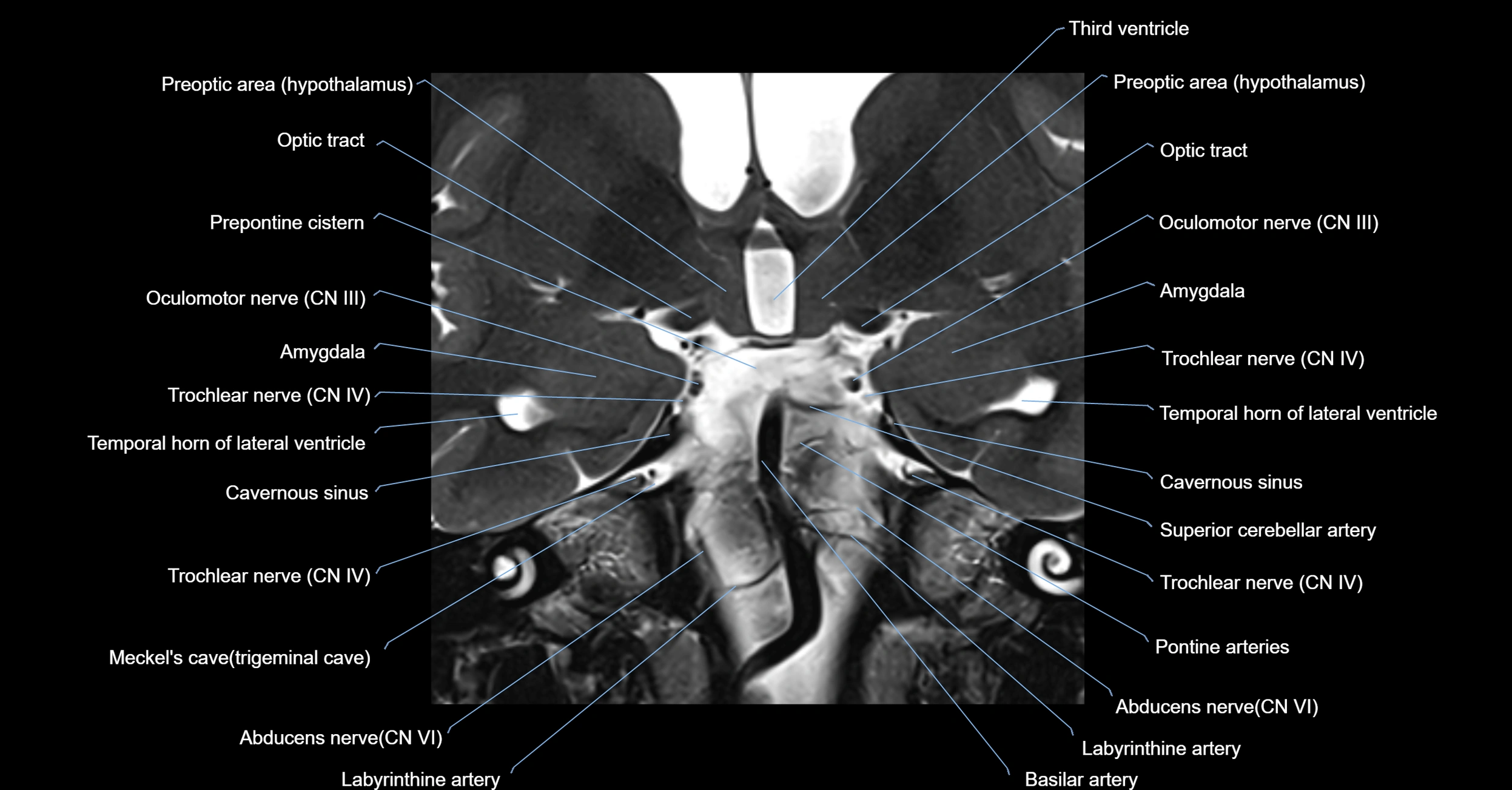
MRI images

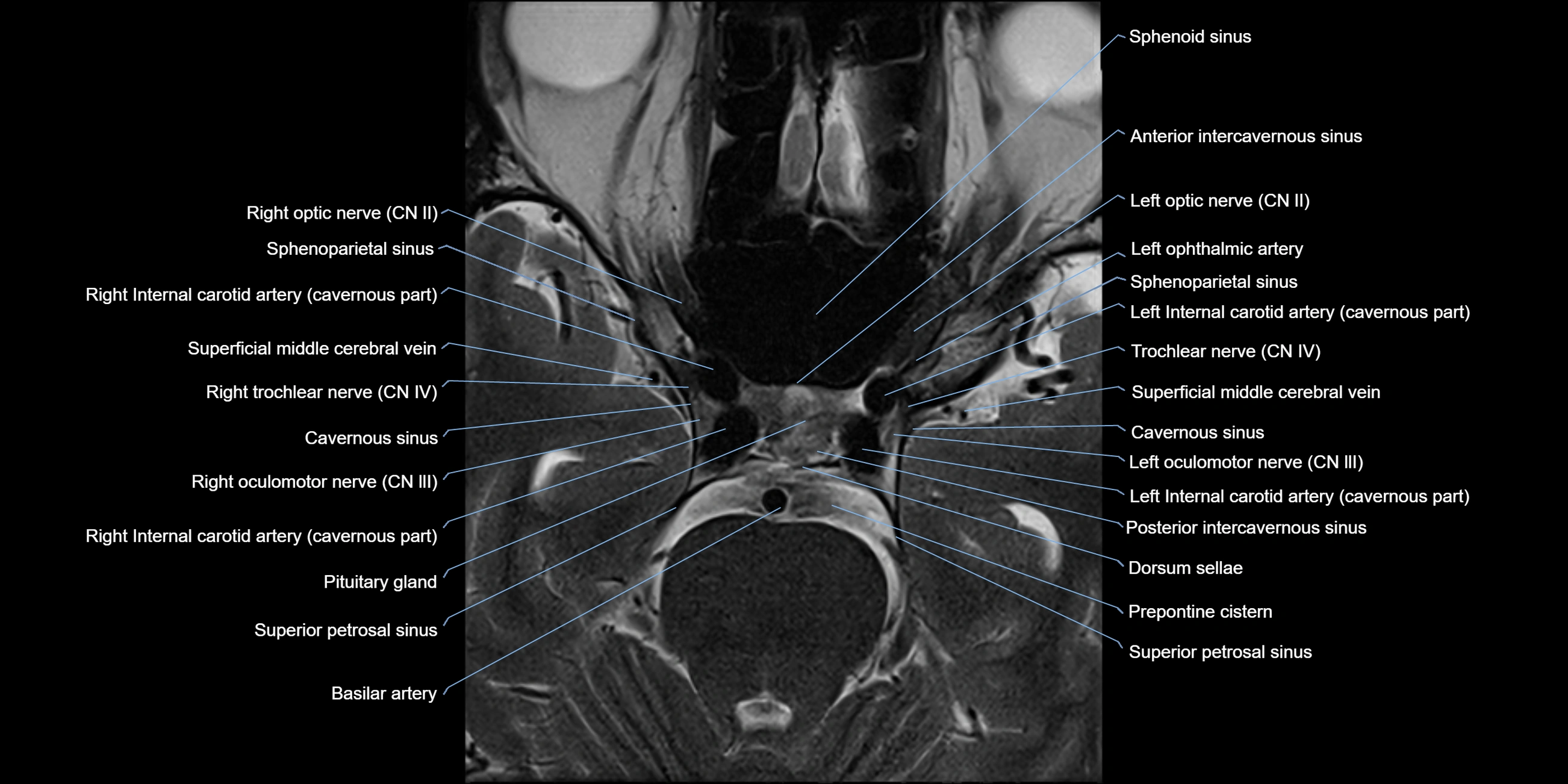
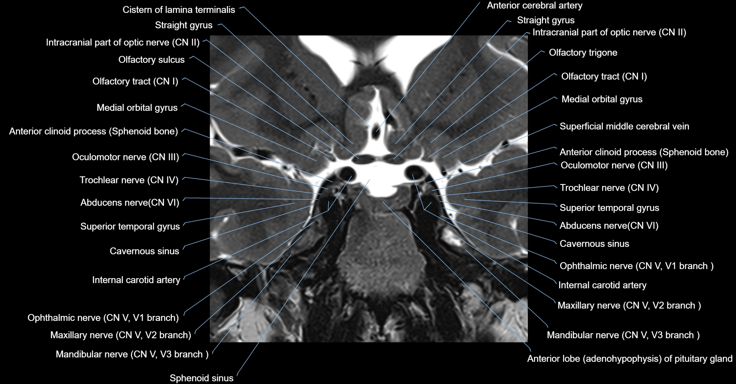
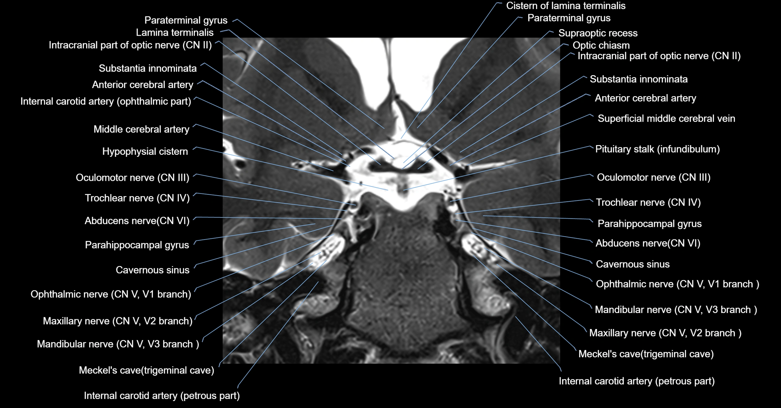
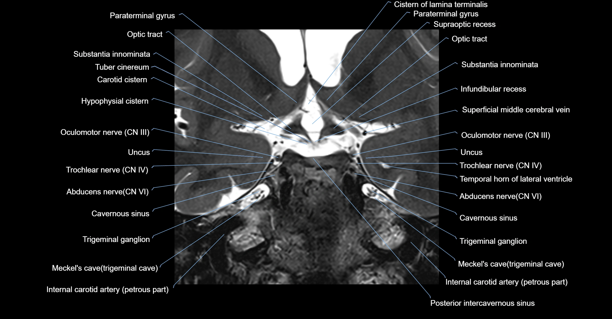
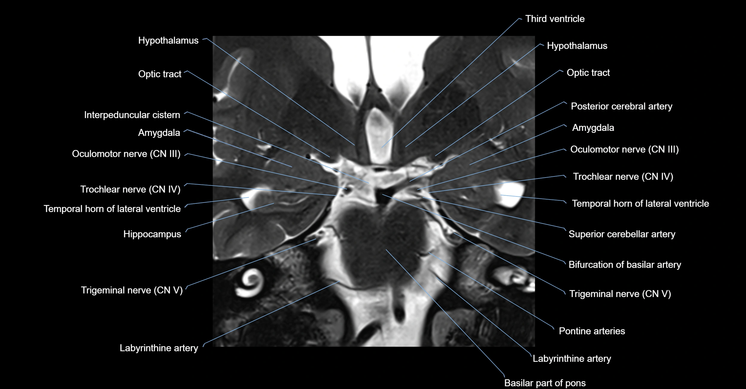
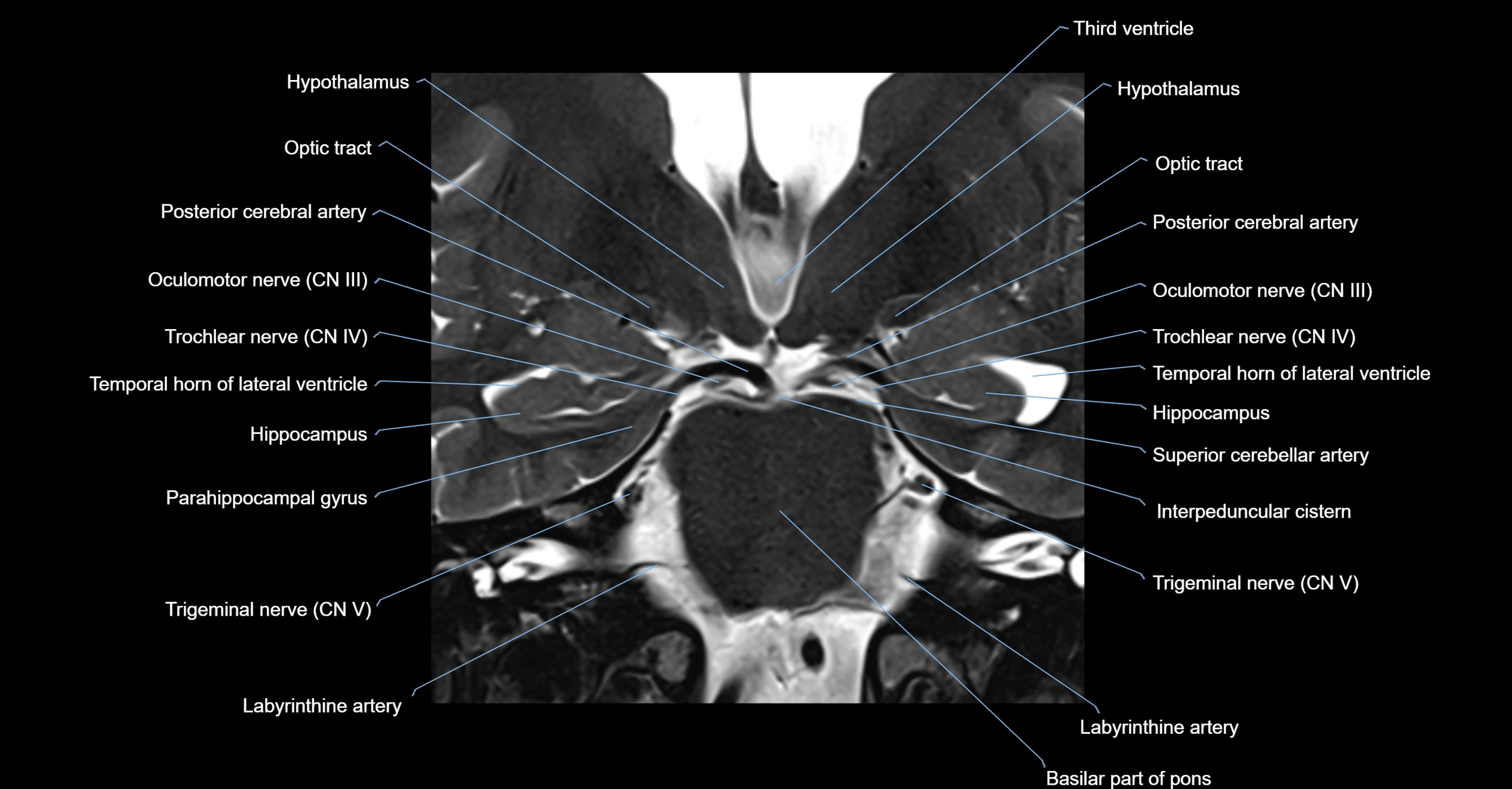
MRI images


