


Topic
- Abdominal aorta
- Abdominal part of esophagus
- Accessory hepatic vein
- Accessory pancreatic duct
- Accessory process of vertebrae
- Adrenal gland
- Adrenal glands
- Ala of ilium (wing of ilium)
- Ala of sacrum
- Annulus fibrosus of intervertebral disc
- Anterior branch of right hepatic duct
- Anterior cecal artery
- Anterior lateral femoral cutaneous nerve
- Anterior longitudinal ligament
- Anterior right branch of portal vein
- Anterior sacral foramina
- Anulus fibrosus of intervertebral disc
- Aortic bifurcation
- Apex of the heart
- Appendicular artery
- Ascending colon
- Ascending mesocolon
- Axillary artery
- Azygos vein
- Basivertebral veins
- Bile duct serving liver segment I
- Bile duct serving liver segment II
- Bile duct serving liver segment III
- Bile duct serving liver segment IVa
- Bile duct serving liver segment IVb
- Bile duct serving liver segment V
- Bile duct serving liver segment VI
- Bile duct serving liver segment VII
- Bile duct serving liver segment VIII
- Body of gallbladder
- Body of pancreas
- Body of vertebra
- Cardia of stomach
- Cauda equina
- Caudate lobe of liver
- Cecum
- Celiac trunk
- Coccyx
- Common bile duct
- Common hepatic artery
- Common hepatic duct
- Common iliac lymph nodes
- Common iliac vein
- Conus medullaris
- Costal part of diaphragm
- Costochondral joints
- Costotransverse joint
- Costotransverse joint of twelfth rib
- Costotransverse ligament
- Costovertebral joint
- Costovertebral joint of twelfth rib
- Crura of diaphragm
- Crural part of diaphragm
- Cystic artery
- Cystic duct
- Descending colon
- Descending mesocolon
- Descending thoracic aorta
- Diaphragm
- Dorsal exiting nerve root
- Dorsal root ganglion of spinal nerve
- Dorsal traversing nerve root
- Duodenal bulb
- Duodenum – Ascending part (D4)
- Duodenum – Descending part (D2)
- Duodenum – Horizontal part (D3)
- Duodenum – Superior part (D1)
- Erector spinae muscles
- External oblique muscle
- Facet joint of vertebra (Zygapophyseal joints)
- Falciform ligament (liver)
- Femoral nerve
- Fissure for ligamentum teres
- Fissure for ligamentum venosum
- Fundus of gallbladder
- Gallbladder
- Gastroduodenal artery
- Gluteus maximus muscle
- Gluteus medius muscle
- Gluteus minimus muscle
- Great pancreatic vein
- Head of pancreas
- Head of twelfth rib
- Heart
- Hemiazygos vein
- Hepatic portal vein
- Hepatopancreatic ampulla (ampulla of Vater)
- Ileal arteries
- Ileocaecal valve (ileocecal junction)
- Ileocolic artery
- Ileocolic artery colic branches
- Ileocolic artery ileal branches
- Ileum
- Iliac crest
- Iliac tubercle
- Iliocostalis lumborum muscle
- Iliohypogastric nerve
- Ilioinguinal nerve
- Inferior articular process of vertebra
- Inferior mesenteric artery (IMA)
- Inferior mesenteric vein
- Inferior vena cava
- Intercostal muscles
- Interlobar arteries of kidney
- Internal iliac vein
- Internal oblique muscle
- Internal thoracic artery
- Internal thoracic veins
- Interspinales lumborum muscle
- Interspinales muscles
- Interspinous ligament
- Intertransversarii muscle
- Intertransverse ligament
- Intervertebral foramen
- Intra-articular ligament of head of rib
- Jejunal arteries
- Jejunum
- L1–L2 Intervertebral Disc
- L2–L3 Intervertebral Disc
- L3–L4 Intervertebral Disc
- L4–L5 Intervertebral Disc
- L5–S1 Intervertebral disc
- Lamina of vertebra
- Lateral aortic lymph nodes
- Lateral branch of left hepatic duct
- Lateral intertransversarii lumborum muscle
- Latissimus dorsi muscle
- Left adrenal gland
- Left atrium
- Left auricle
- Left branch of hepatic portal vein
- Left branch of portal vein
- Left colic artery
- Left colic flexure (splenic flexure)
- Left common carotid artery
- Left crus of diaphragm
- Left gastric artery
- Left hemidiaphragm
- Left hepatic artery
- Left hepatic duct
- Left internal thoracic artery
- Left internal thoracic veins
- Left kidney
- Left lobe of liver
- Left lumbar part of diaphragm
- Left ovarian vein
- Left paracolic gutter
- Left phrenic nerve
- Left renal artery
- Left renal vein
- Left ureter
- Left ventricle
- Lesser curvature lymph nodes
- Ligamenta flava (Ligamentum flavum)
- Ligamentum teres (round ligament of the liver)
- Ligamentum venosum
- Linea alba
- Liver
- Liver Segment I – Caudate lobe
- Liver Segment II – Left lateral superior segment
- Liver Segment III – Left lateral inferior segment
- Liver Segment IVa – Left medial superior segment
- Liver Segment IVb – Left medial inferior segment
- Liver Segment V – Right anteroinferior segment
- Liver Segment VI – Right posteroinferior segment
- Liver Segment VII – Right posterosuperior segment
- Liver Segment VIII – Right anterosuperior segment
- Longissimus thoracis muscle
- Lumbar arteries
- Lumbar part of diaphragm
- Lumbar triangle
- Lumbar veins
- Mammillary process of vertebra
- Marginal artery of Drummond
- Medial intertransversarii lumborum
- Median arcuate ligament
- Median sacral crest
- Middle Colic Vein
- Middle colic artery
- Neck of gallbladder
- Neck of pancreas
- Nucleus pulposus of intervertebral disc
- Omental branches of gastro-omental (gastroepiploic) artery
- Pancreas
- Pancreatic duct
- Paraesophageal lymph nodes
- Parietal peritoneum
- Pedicle of vertebra
- Periaortic lymph nodes
- Pericardium
- Phrenicomediastinal recess
- Phrenoesophageal ligament
- Pleura
- Portal vein branch to liver segment I
- Portal vein branch to liver segment II
- Portal vein branch to liver segment III
- Portal vein branch to liver segment IV
- Portal vein branch to liver segment V
- Portal vein branch to liver segment VI
- Portal vein branch to liver segment VII
- Portal vein branch to liver segment VIII
- Posterior branch of right hepatic duct
- Posterior cecal artery
- Posterior inferior iliac spine
- Posterior intercostal arteries
- Posterior intercostal veins
- Posterior lateral femoral cutaneous nerve
- Posterior longitudinal ligament
- Posterior right branch of portal vein
- Posterior sacral foramina
- Posterior sternoclavicular ligament
- Posterior superior iliac spine
- Prepericardial lymph nodes
- Preperitoneal space
- Proper hepatic artery
- Psoas major muscle
- Pulmonary trunk
- Quadrate lobe of liver
- Quadratus lumborum muscle
- Rectus abdominis muscle
- Renal artery
- Renal capsule
- Renal fascia
- Renal medulla
- Renal pelvis
- Renal pyramids
- Renal vein
- Ribs
- Right adrenal gland
- Right atrium
- Right branch of hepatic portal vein
- Right branch of portal vein
- Right colic artery
- Right colic flexure (hepatic flexure)
- Right crus of diaphragm
- Right gastric artery
- Right gastric vein CT axial image
- Right hemidiaphragm
- Right hepatic artery
- Right hepatic duct
- Right internal thoracic artery
- Right internal thoracic veins
- Right kidney
- Right lobe of liver
- Right lumbar part of diaphragm
- Right ovarian vein
- Right paracolic gutter
- Right renal artery
- Right renal vein
- Right ureter
- Right ventricle
- Rotatores lumborum muscles
- Rotatores muscle
- Rotatores thoracis muscles
- Sacral canal
- Sacrum
- Sciatic nerve
- Serratus posterior inferior muscle
- Short gastric arteries
- Sigmoid veins
- Small intestine
- Spinal cord
- Spinal dura mater
- Spinal epidural space
- Spinal nerve L1
- Spinal nerve L2
- Spinal nerve L3
- Spinal nerve L4
- Spinal nerve L5
- Spinal nerve S1
- Spinal nerve S2
- Spinal nerve S3
- Spinal nerves
- Spinalis thoracis muscle
- Spinous process of vertebra
- Spleen
- Splenic artery
- Sternal end of the clavicle
- Sternal part of diaphragm
- Stomach
- Straight Arteries
- Superior articular process of vertebra
- Superior cluneal nerves
- Superior diaphragmatic lymph nodes
- Superior epigastric artery
- Superior epigastric veins
- Superior mesenteric artery (SMA)
- Superior mesenteric lymph nodes
- Superior mesenteric vein (SMV)
- Superior phrenic artery
- Superior rectal artery
- Superior rectal vein
- Supraspinous ligament
- T12–L1 Intervertebral Disc
- Tail of pancreas
- Terminal ileum
- Thoracic aorta
- Thoracic duct
- Thoracolumbar fascia (anterior layer)
- Thoracolumbar fascia (middle layer)
- Thoracolumbar fascia (posterior layer)
- Transverse colon
- Transverse mesocolon
- Transverse process of vertebra
- Transverse processes
- Transversus abdominis muscle
- Transversus thoracis muscle
- Umbilical vein
- Uncinate process of pancreas
- Ureteropelvic junction
- Ureters
- Vasa recta (kidney)
- Ventral exiting nerve root
- Ventral traversing nerve root
- Vertebrae
- Vertebral venous plexus
- Zygapophyseal joint
- kidney cortex (renal cortex)
- kidneys
- left gastro-omental artery (left gastroepiploic artery)
The abdominal aorta is the continuation of the thoracic aorta, beginning at the level of the aortic hiatus of the diaphragm (T12 vertebra) and terminating at the level of the L4 vertebra where it bifurcates into the right and left common iliac arteries. It lies slightly to the left of the midline and courses anterior to the vertebral bodies, surrounded by the retroperitoneal structures of the abdomen.
The abdominal aorta gives off numerous visceral and parietal branches, supplying the abdominal organs, pelvic structures, and lower limbs. It is the main conduit of oxygenated blood from the heart to the abdomen and lower body. The aorta is clinically significant as the common site of aneurysm, dissection, atherosclerosis, and traumatic injury.
Synonyms
-
Aorta abdominalis
-
Infradiaphragmatic aorta
-
Abdominal portion of aorta
Function
-
Conducts oxygenated blood from the thoracic aorta to abdominal, pelvic, and lower limb structures
-
Provides direct arterial supply to major abdominal organs (liver, spleen, kidneys, intestines)
-
Maintains systemic blood flow and hemodynamic regulation
-
Plays a central role in surgical and interventional procedures (aneurysm repair, stent grafts)
Branches
-
Unpaired visceral branches: celiac trunk, superior mesenteric artery (SMA), inferior mesenteric artery (IMA)
-
Paired visceral branches: middle suprarenal arteries, renal arteries, gonadal arteries (testicular or ovarian)
-
Parietal branches: inferior phrenic arteries, lumbar arteries, median sacral artery
-
Terminal branches: right and left common iliac arteries
MRI Appearance
T1-weighted images:
-
Flowing blood appears as a signal void (black lumen)
-
Vessel wall appears as a thin hypointense rim; retroperitoneal fat enhances contrast
T2-weighted images:
-
Lumen remains a signal void due to flow
-
Adjacent edema, hematoma, or aneurysm wall thrombus may appear hyperintense
STIR (Short Tau Inversion Recovery):
-
Fat suppression improves visualization of the aortic wall and periaortic tissues
-
Wall edema, inflammation, or periaortic hematoma appears hyperintense
-
Useful in vasculitis, dissection, or trauma
T1 Post-Contrast (Gadolinium-enhanced):
-
Aortic lumen enhances brightly and homogeneously
-
Clearly demonstrates aneurysm, stenosis, dissection, mural thrombus, or aortic wall enhancement in vasculitis
MRA (Magnetic Resonance Angiography):
-
Contrast-enhanced MRA provides high-resolution imaging of the aorta and its branches
-
Allows 3D reconstruction of visceral, parietal, and terminal branches
-
Excellent for evaluating aneurysm size, dissection flap, stenosis, or preoperative planning
-
Non-invasive alternative to conventional angiography
CT Appearance
Non-contrast CT:
-
Appears as a tubular soft tissue structure anterior to vertebral bodies
-
Calcified atherosclerotic plaques appear as hyperdense foci along the wall
-
Useful for screening abdominal aortic aneurysm (AAA) size and mural calcification
Contrast-enhanced CT (CTA):
-
Gold standard for abdominal aortic imaging
-
Provides excellent detail of lumen, wall, aneurysm, thrombus, and branch vessels
-
Multiplanar and 3D reconstructions help in aneurysm measurement, stent graft planning, and dissection evaluation
-
Detects acute rupture, traumatic injury, or occlusion with high sensitivity
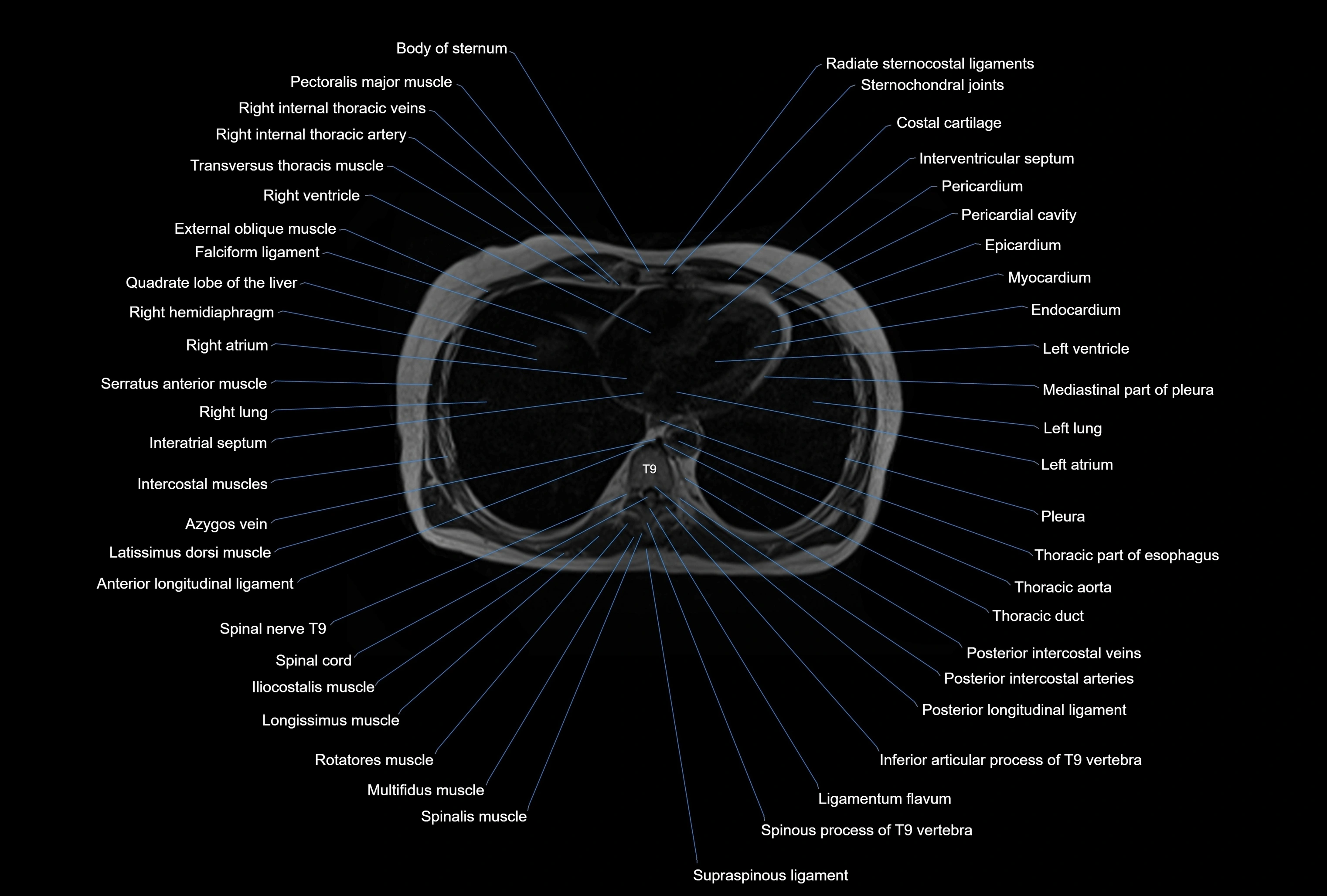
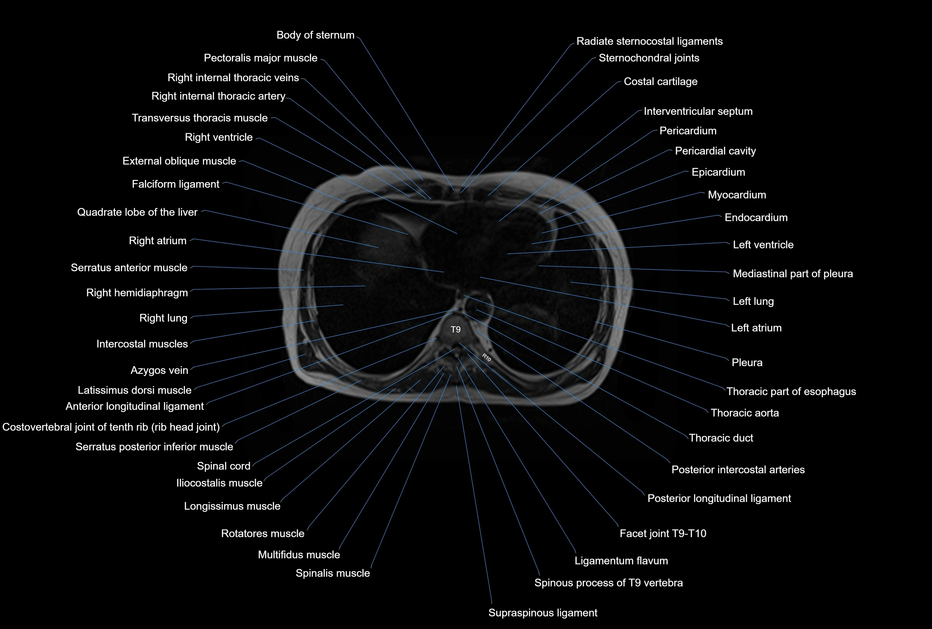
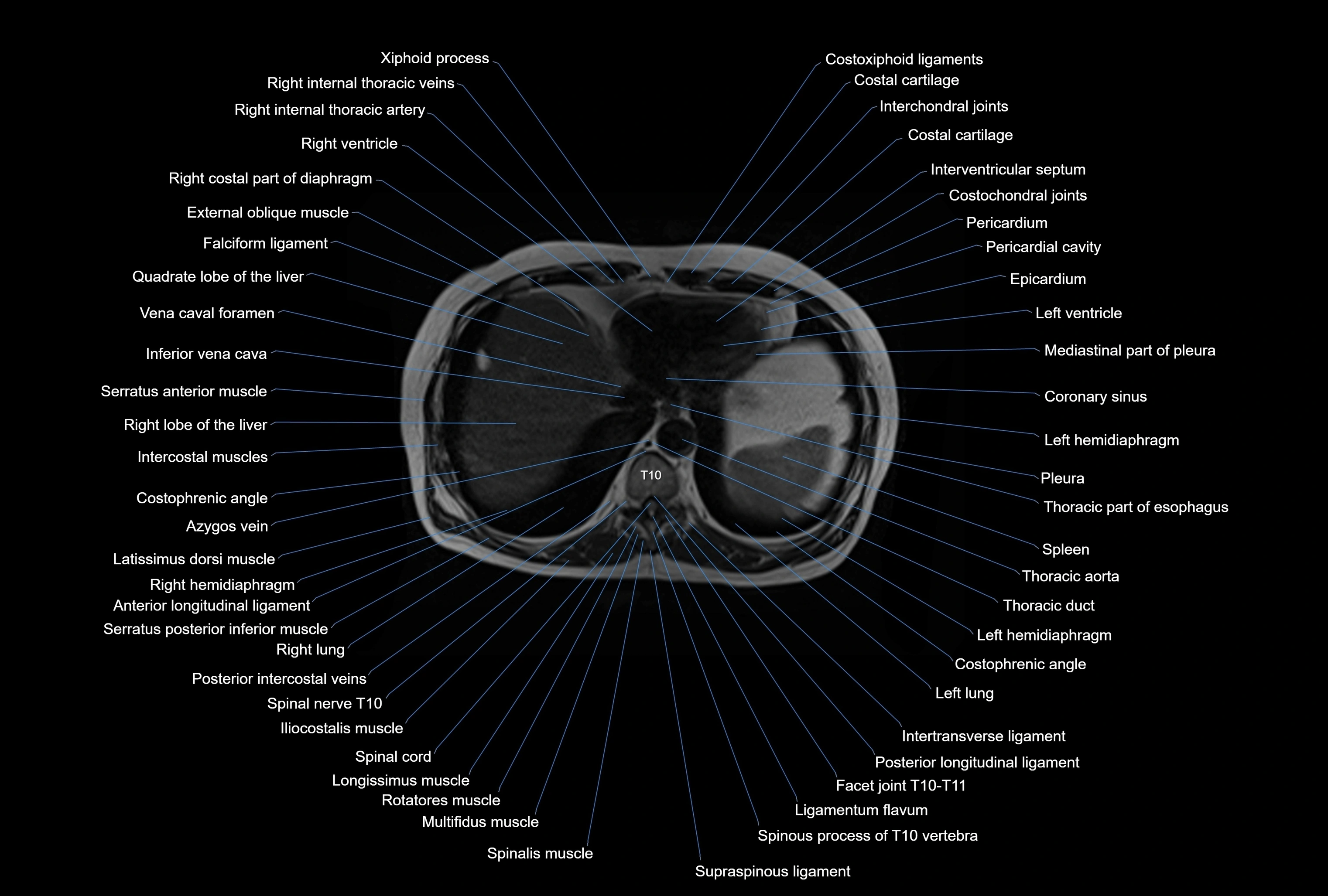
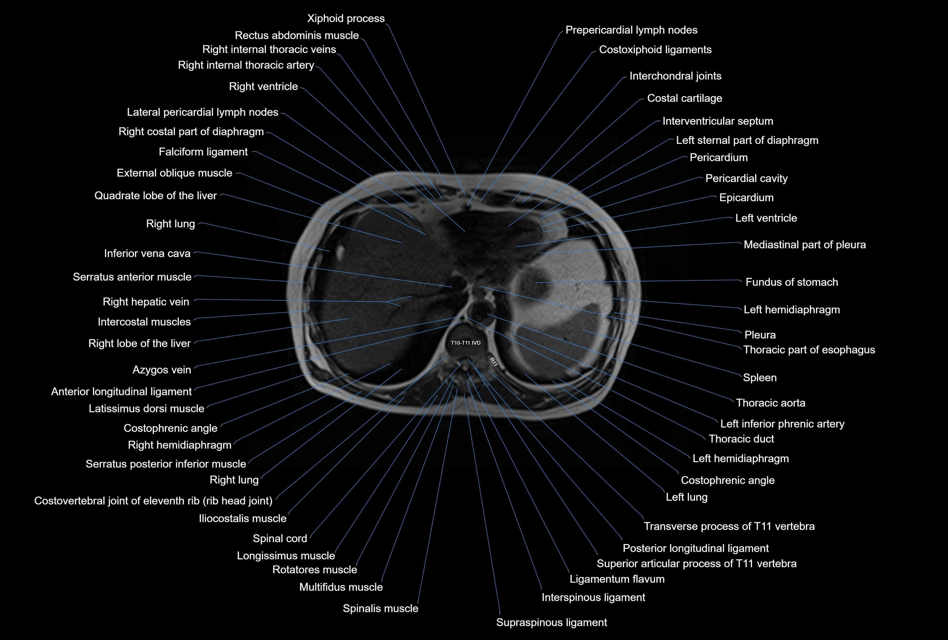
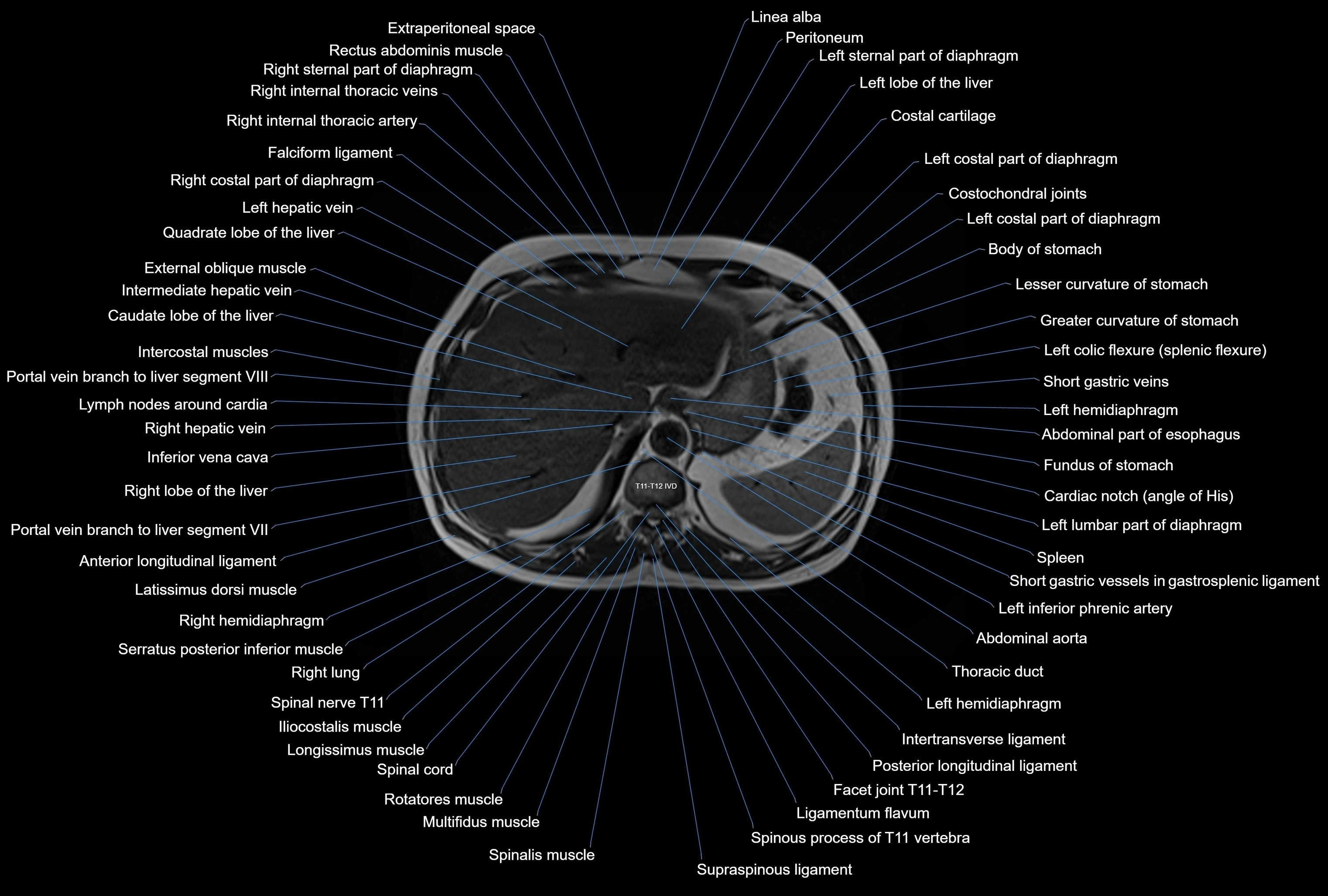
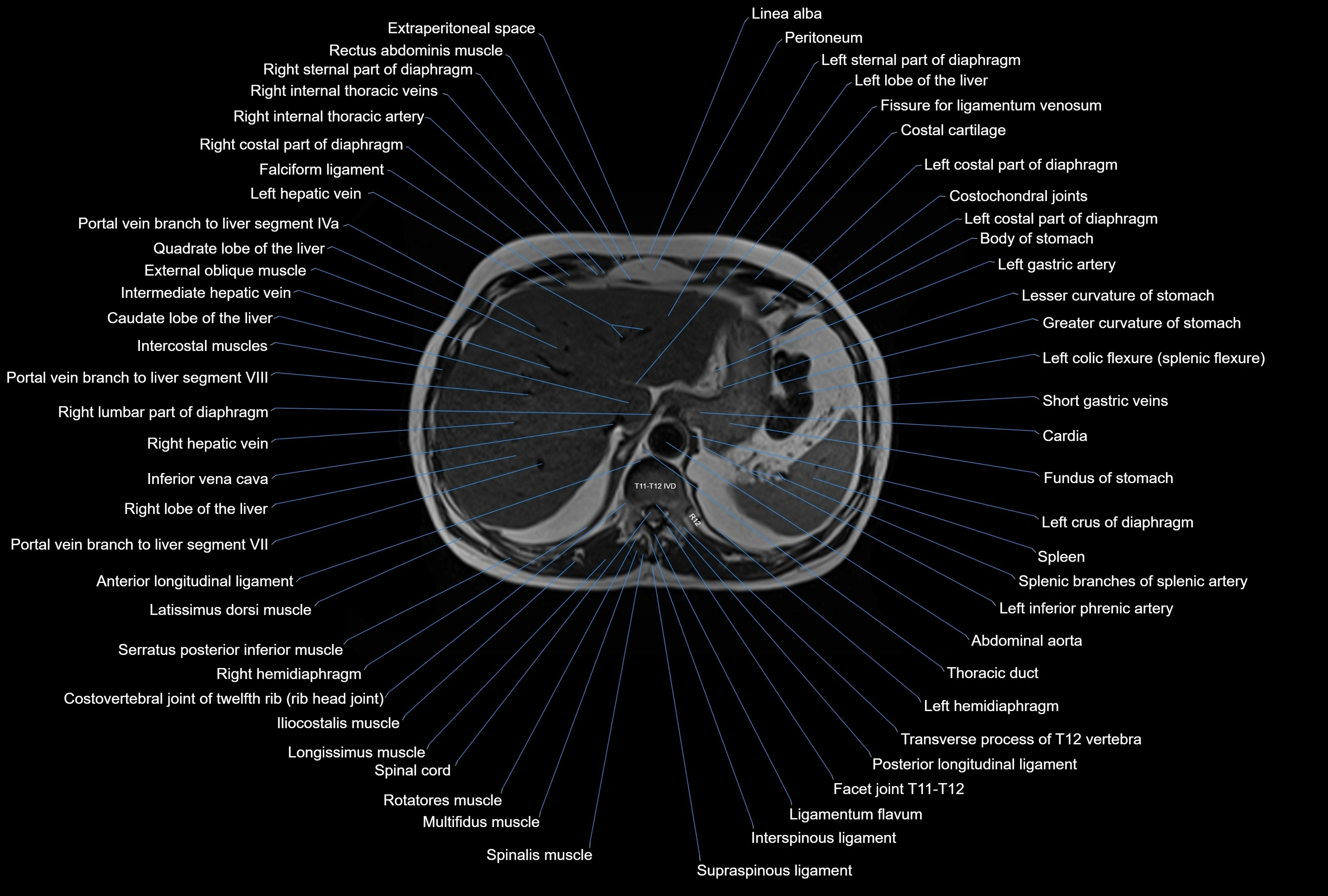
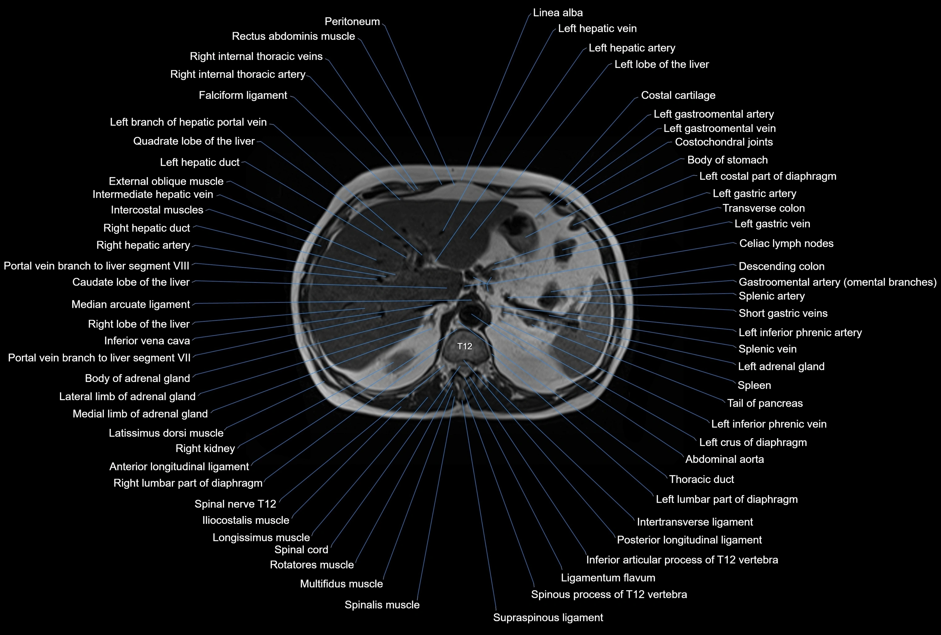
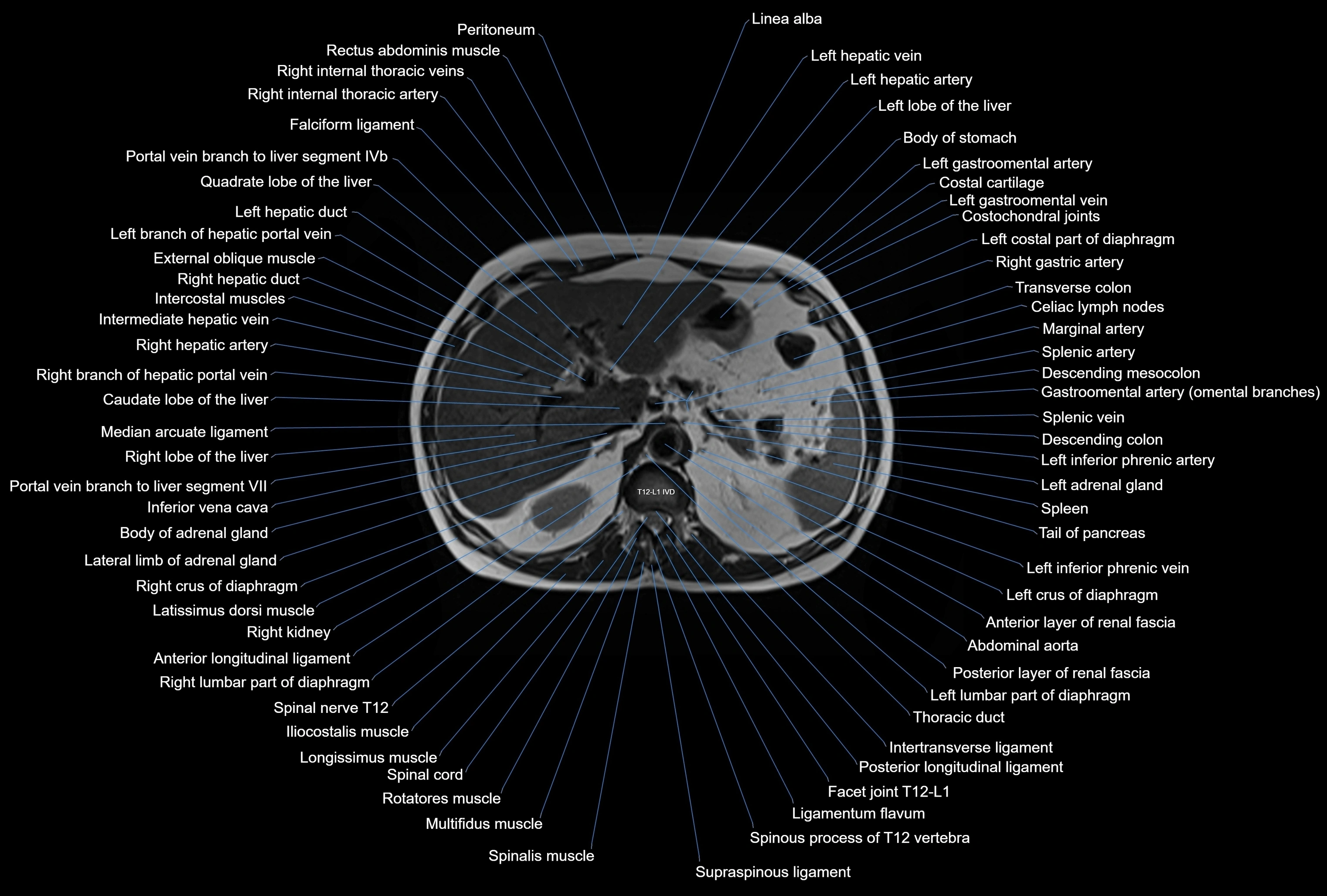
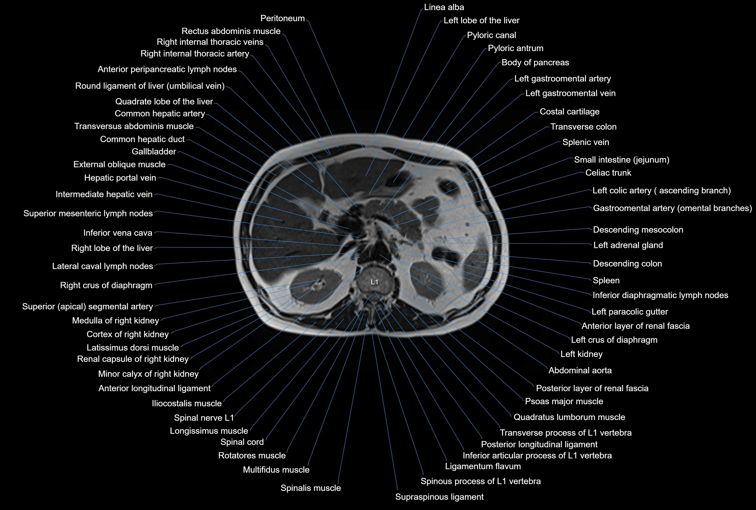
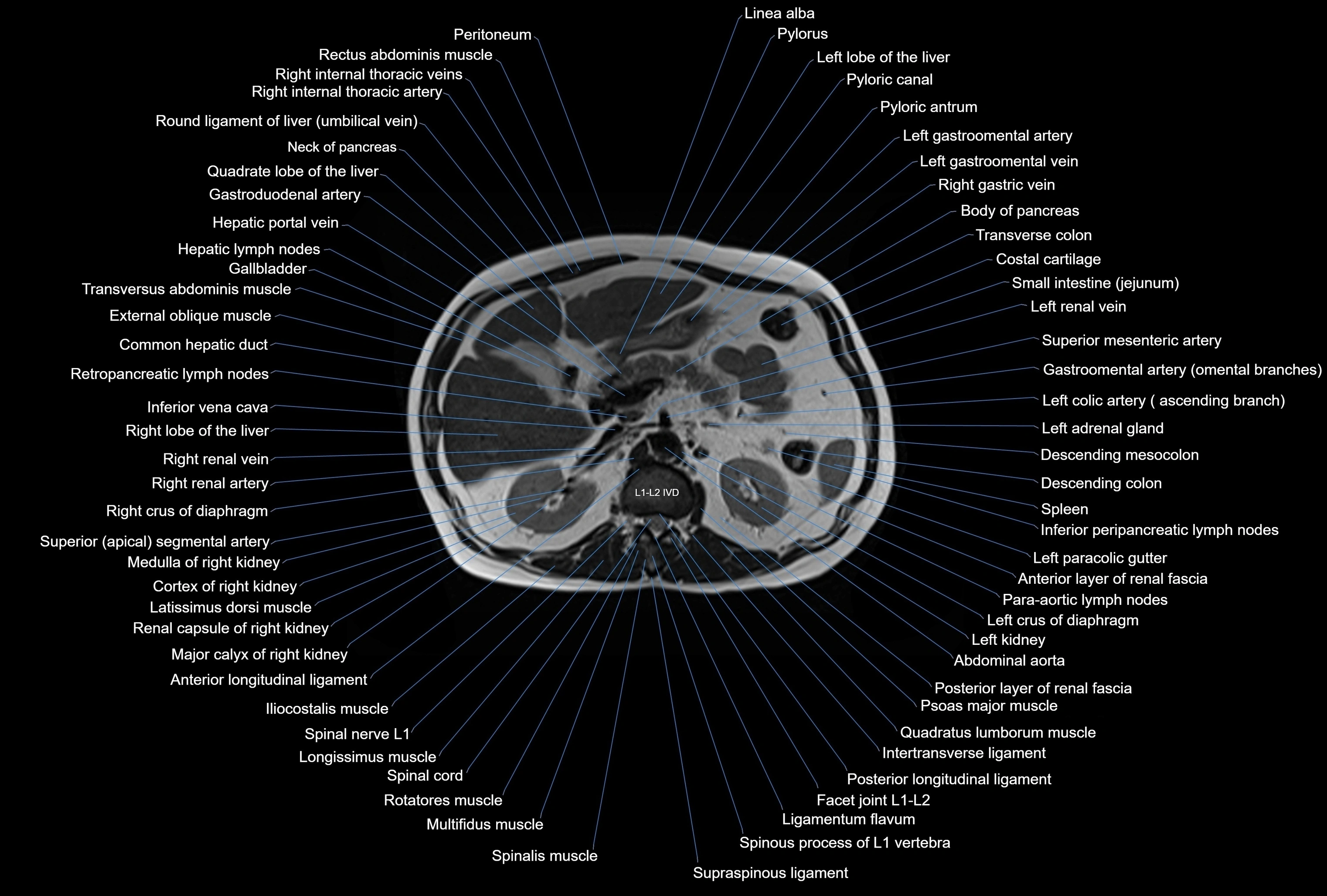
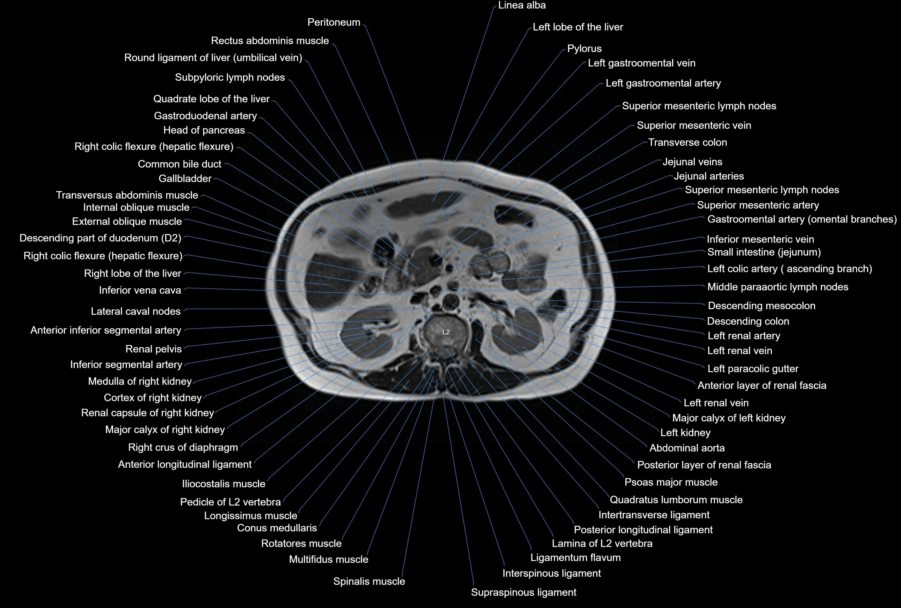
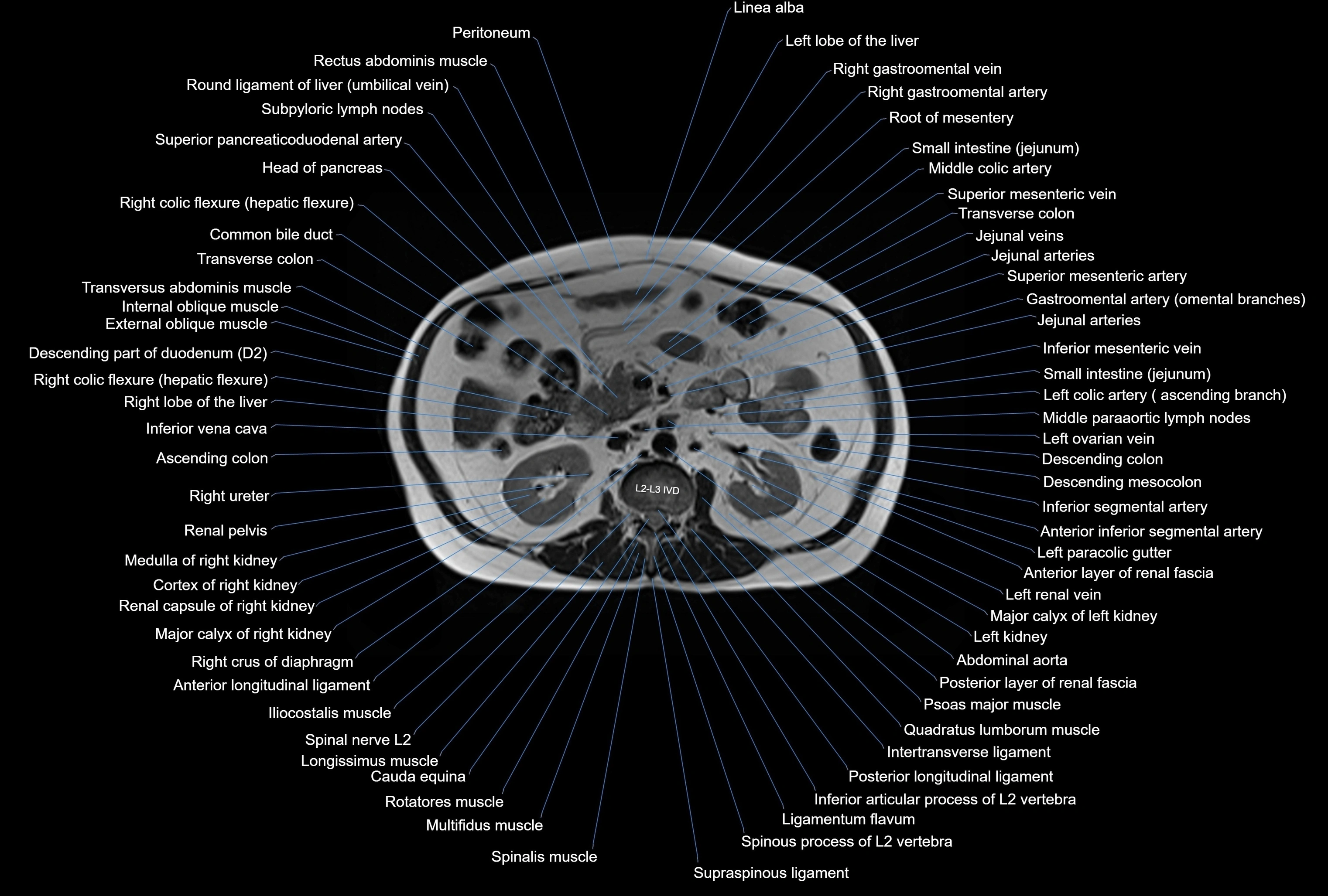
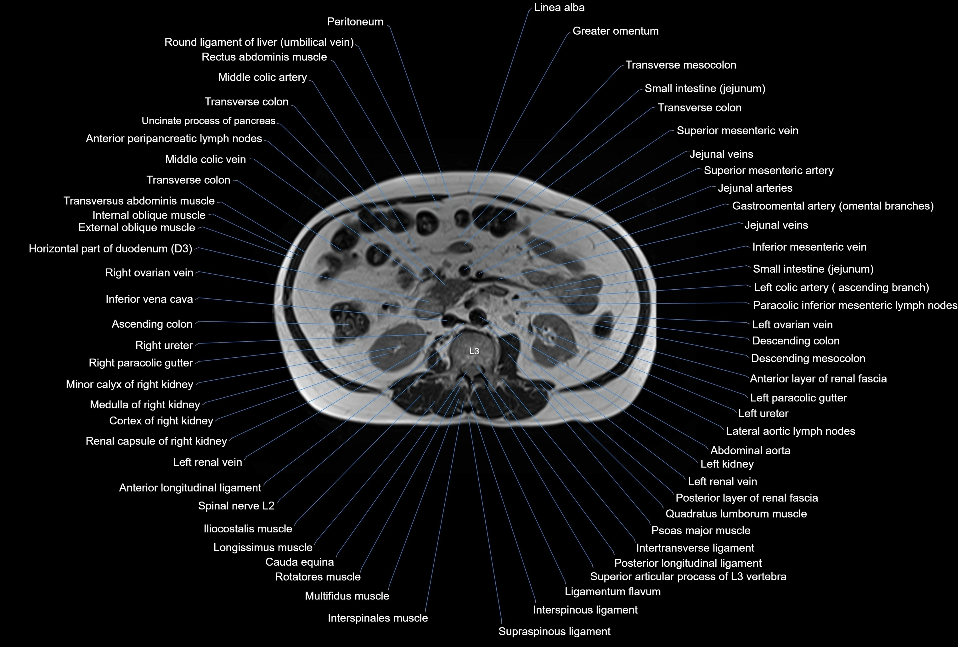
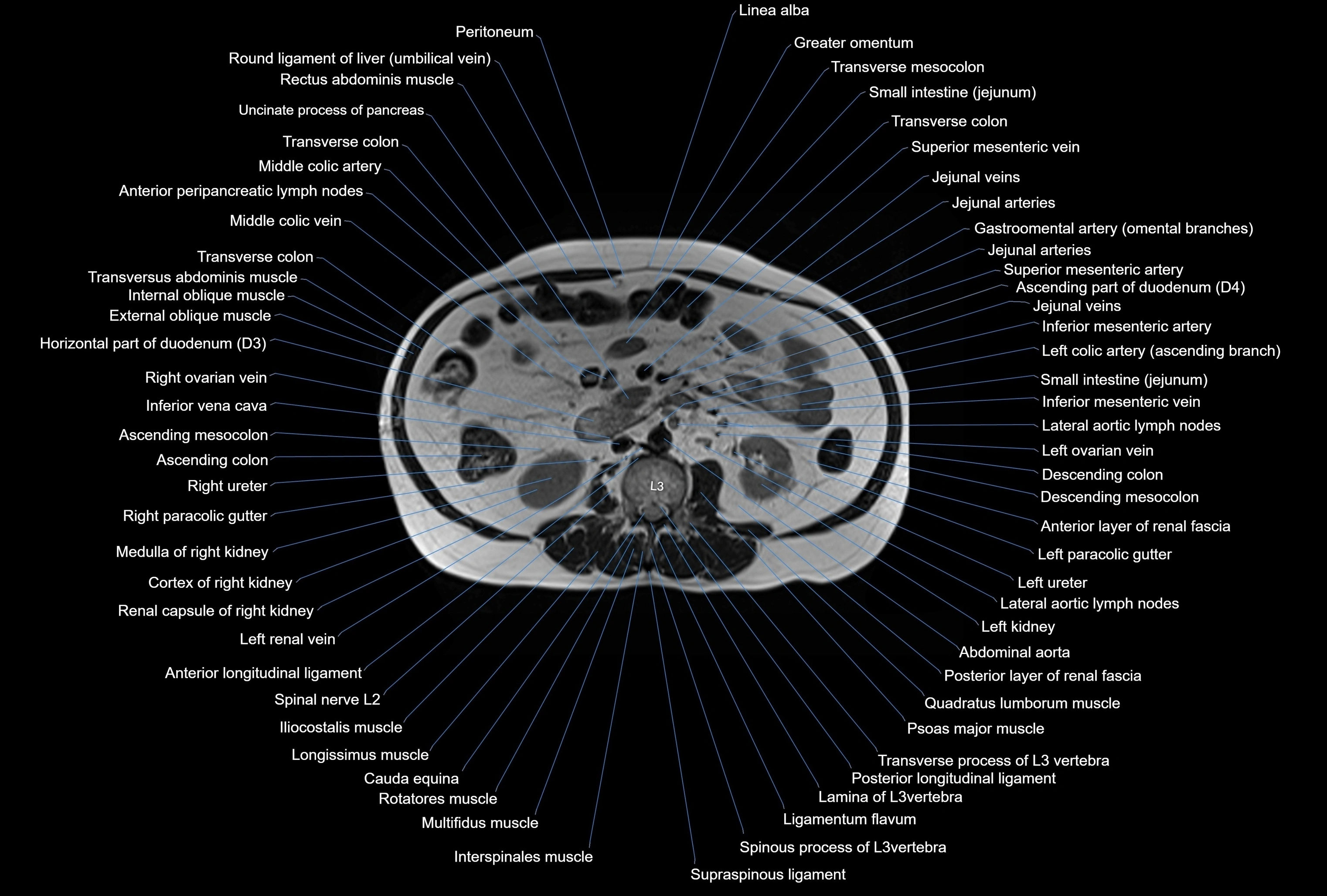
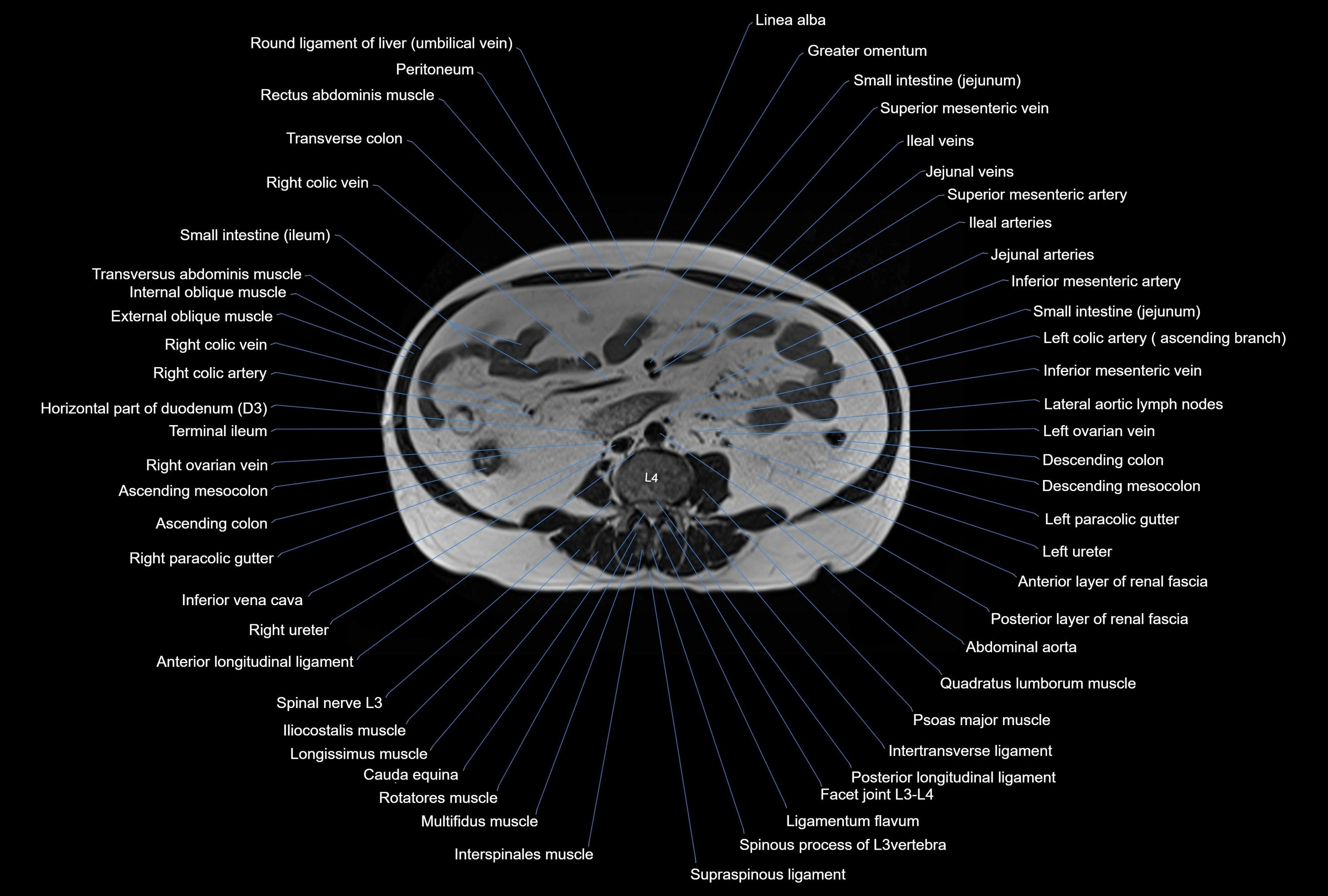
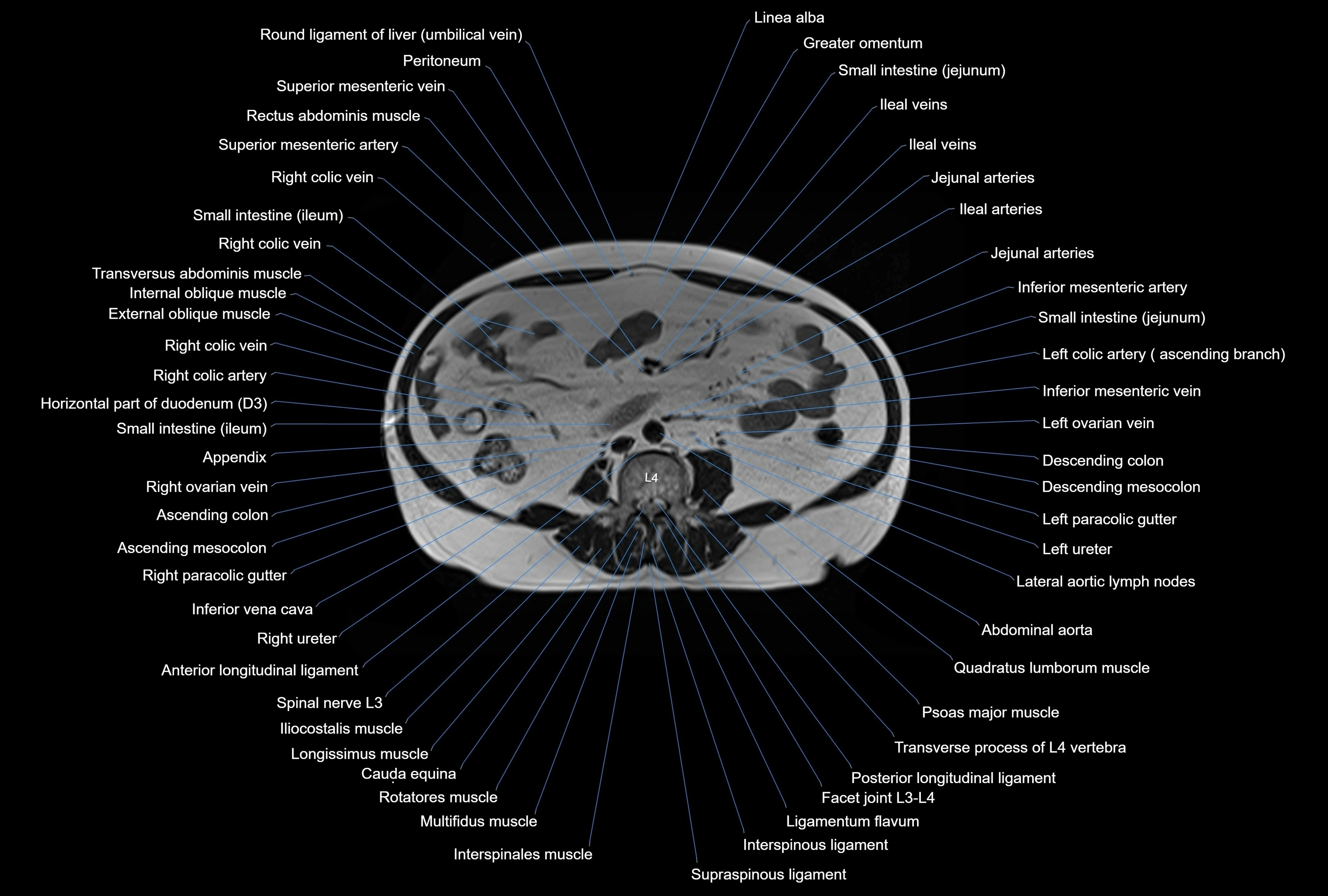
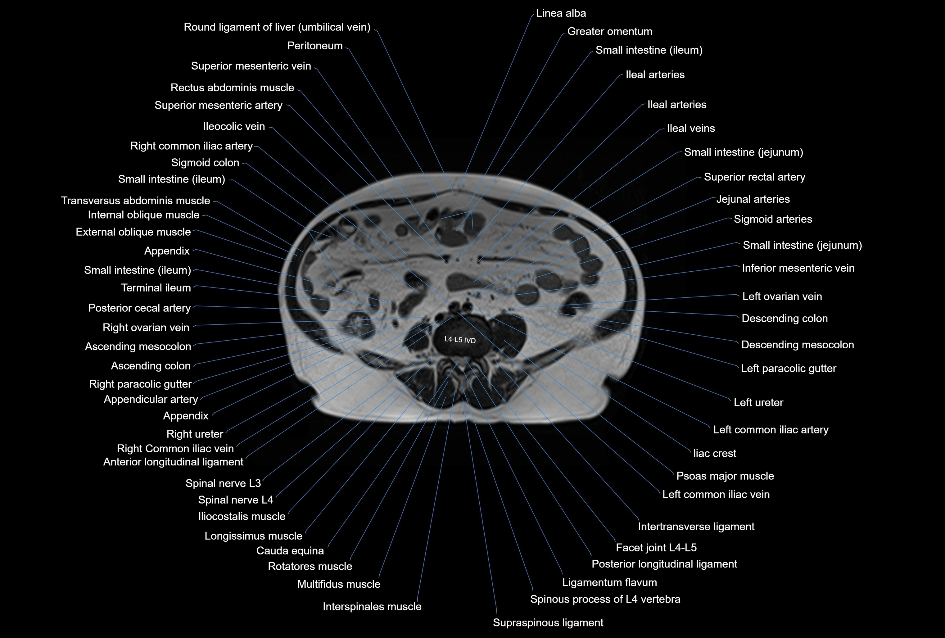
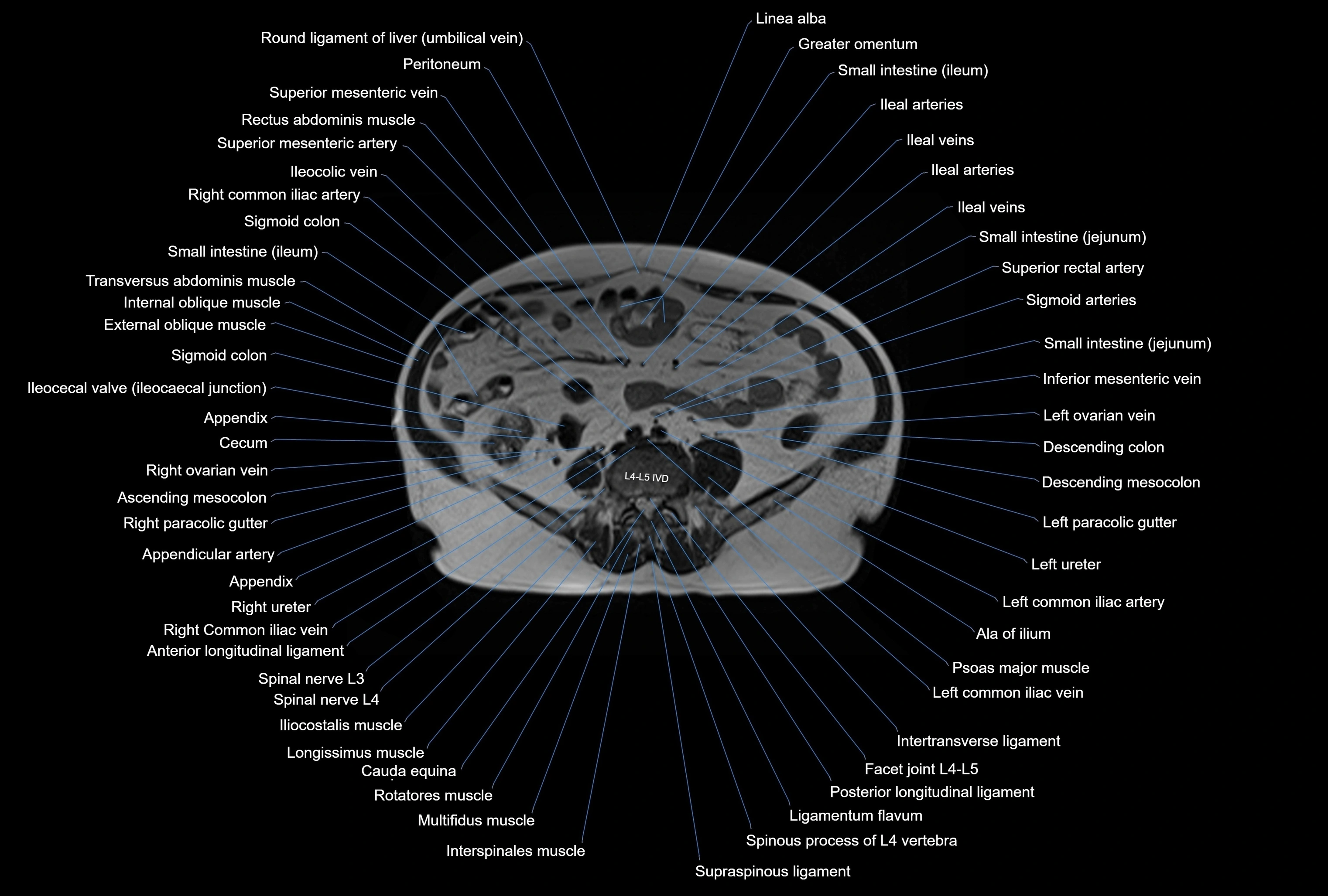
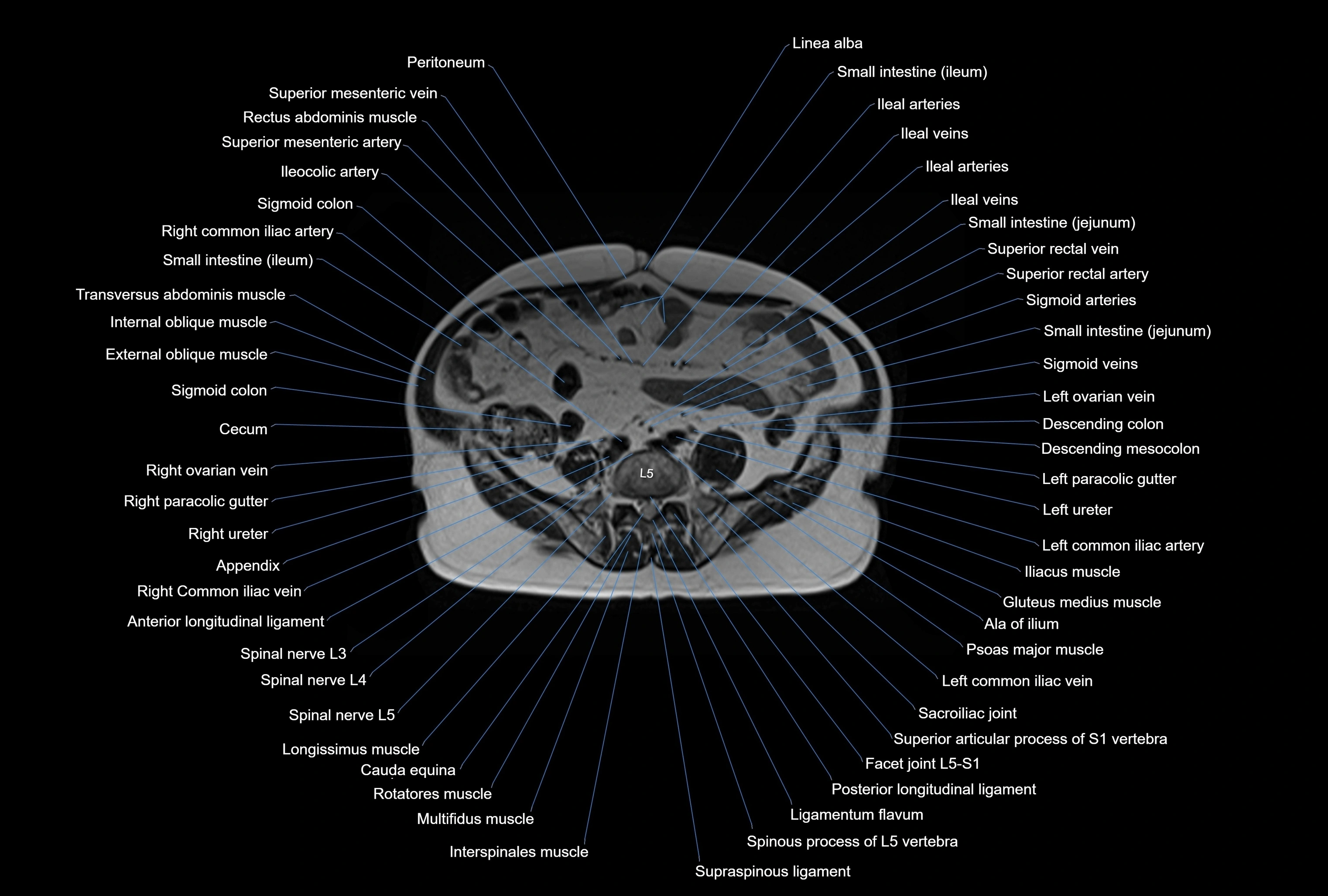
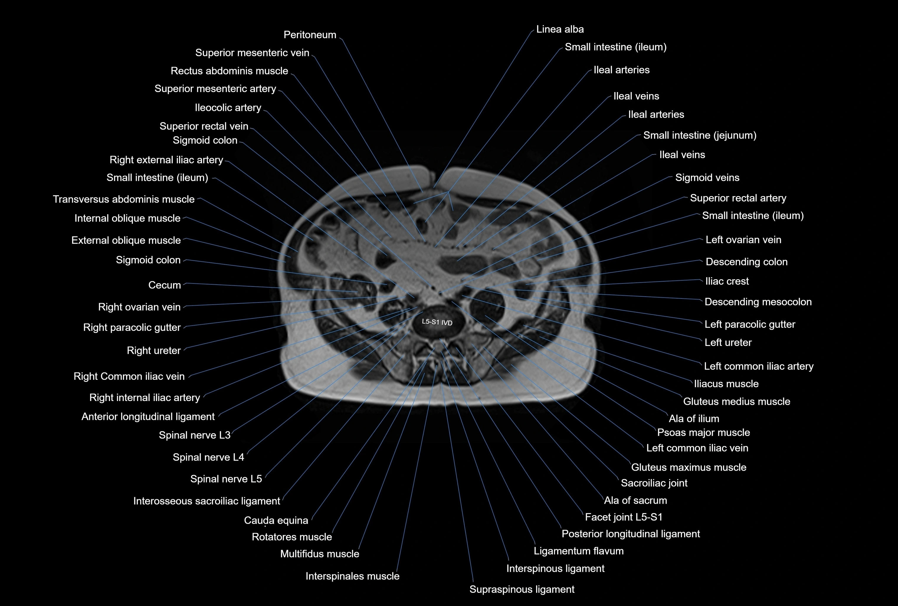
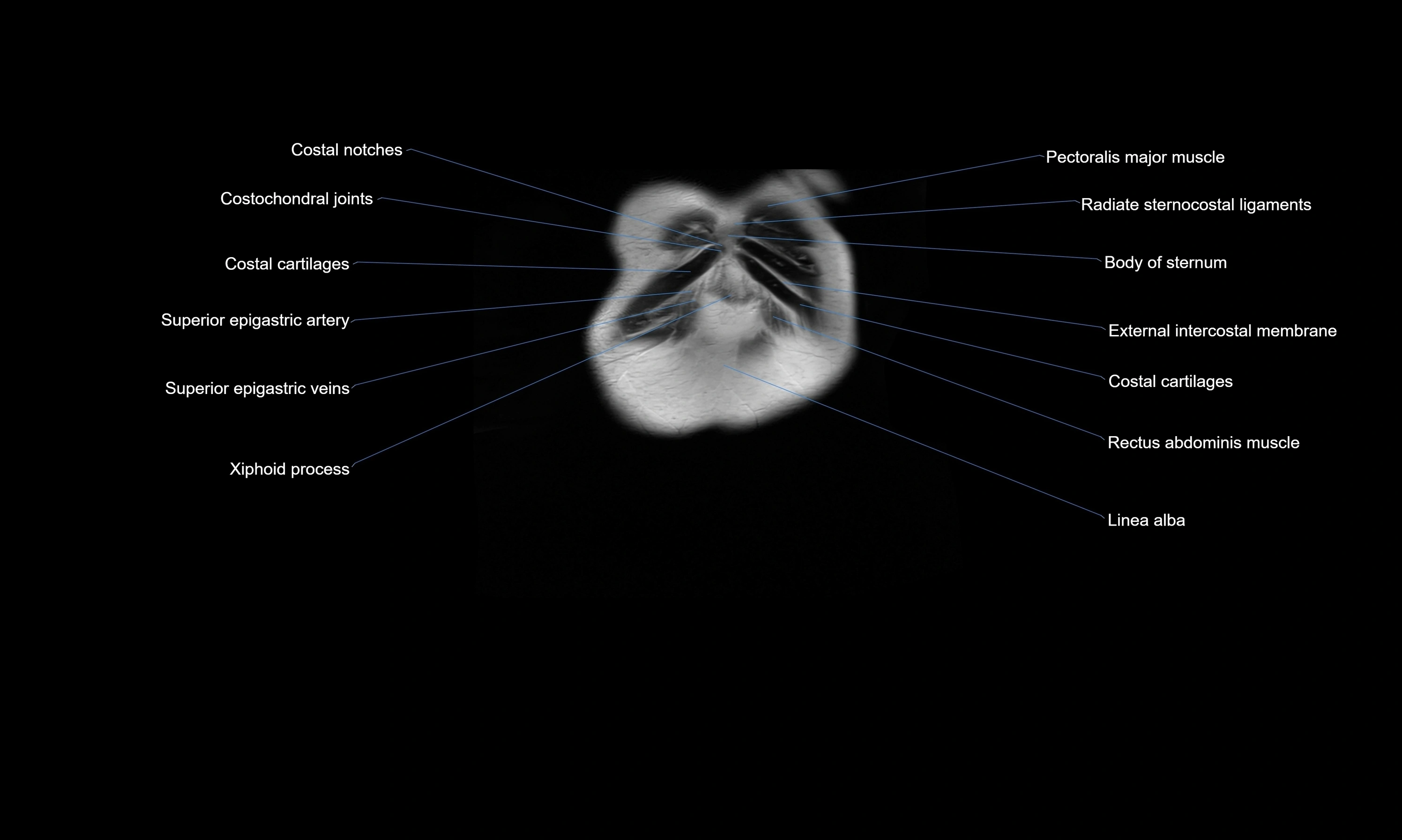
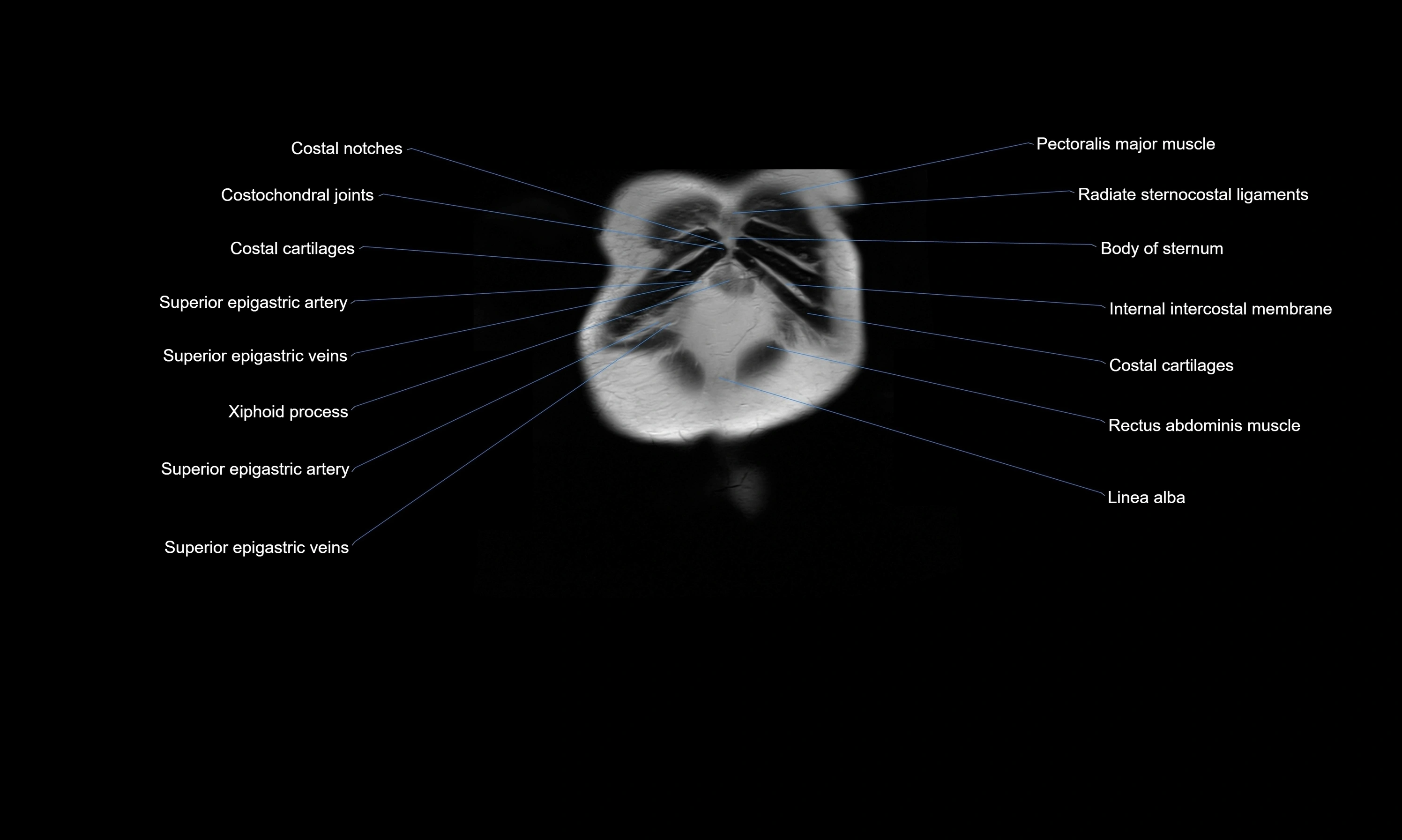
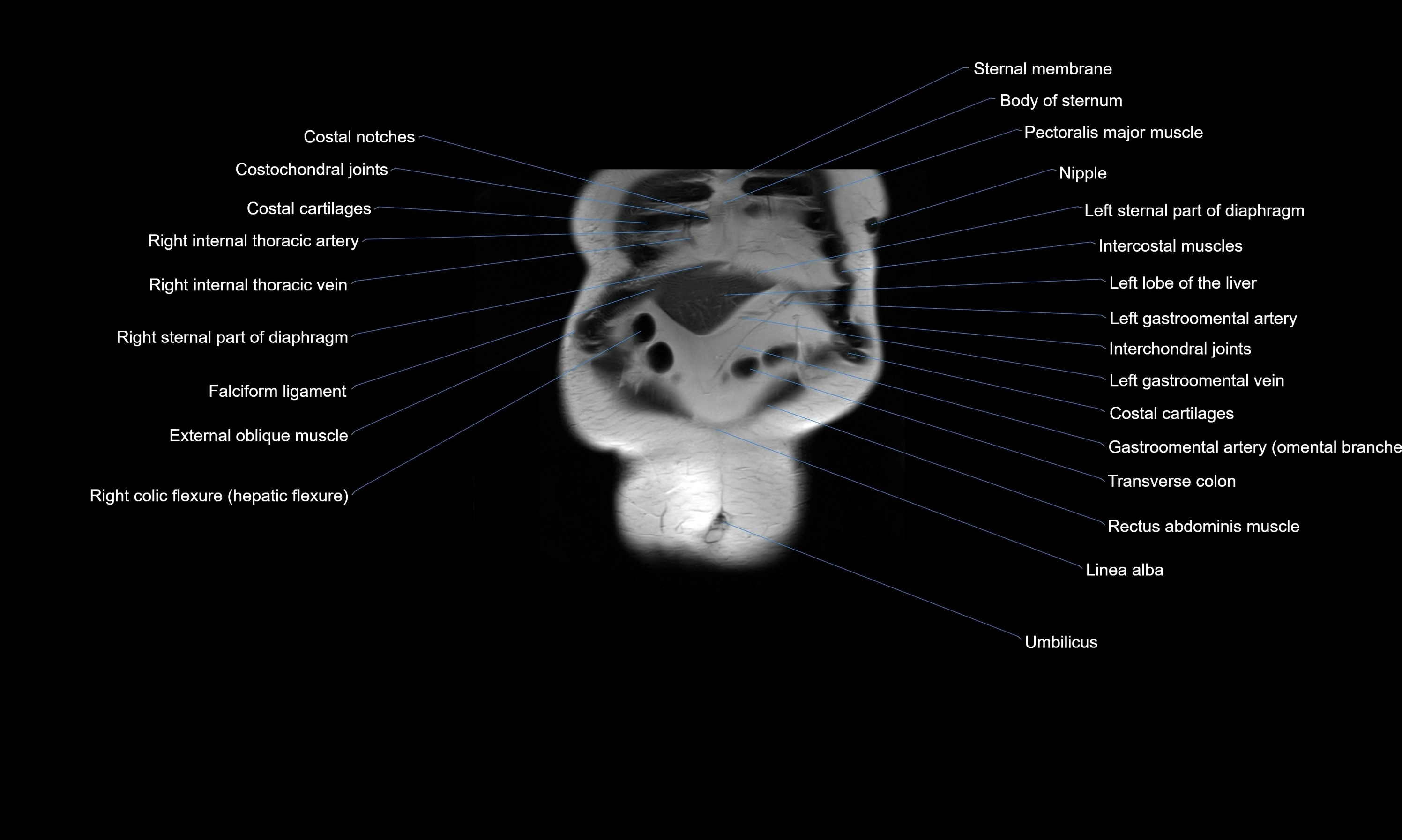
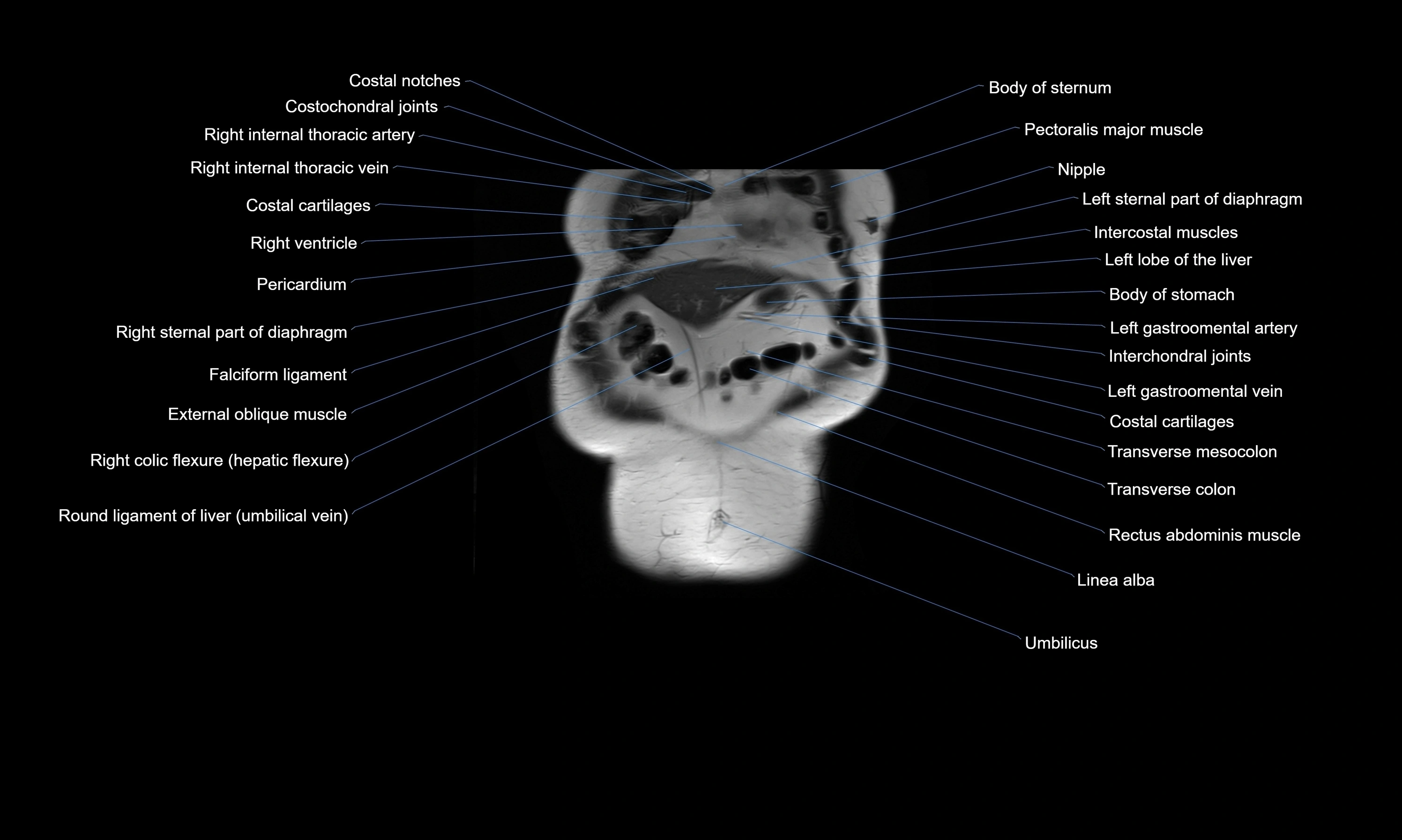
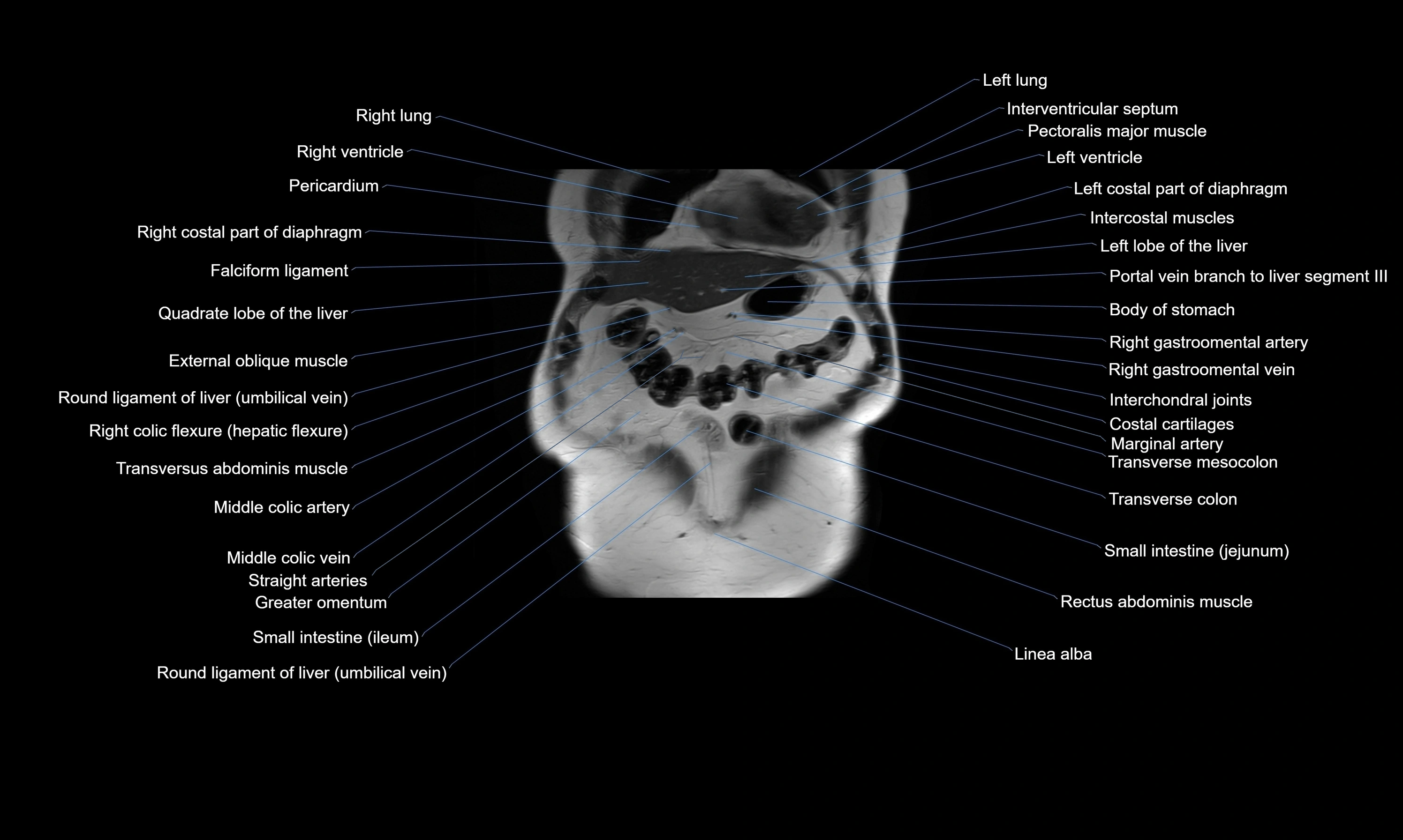
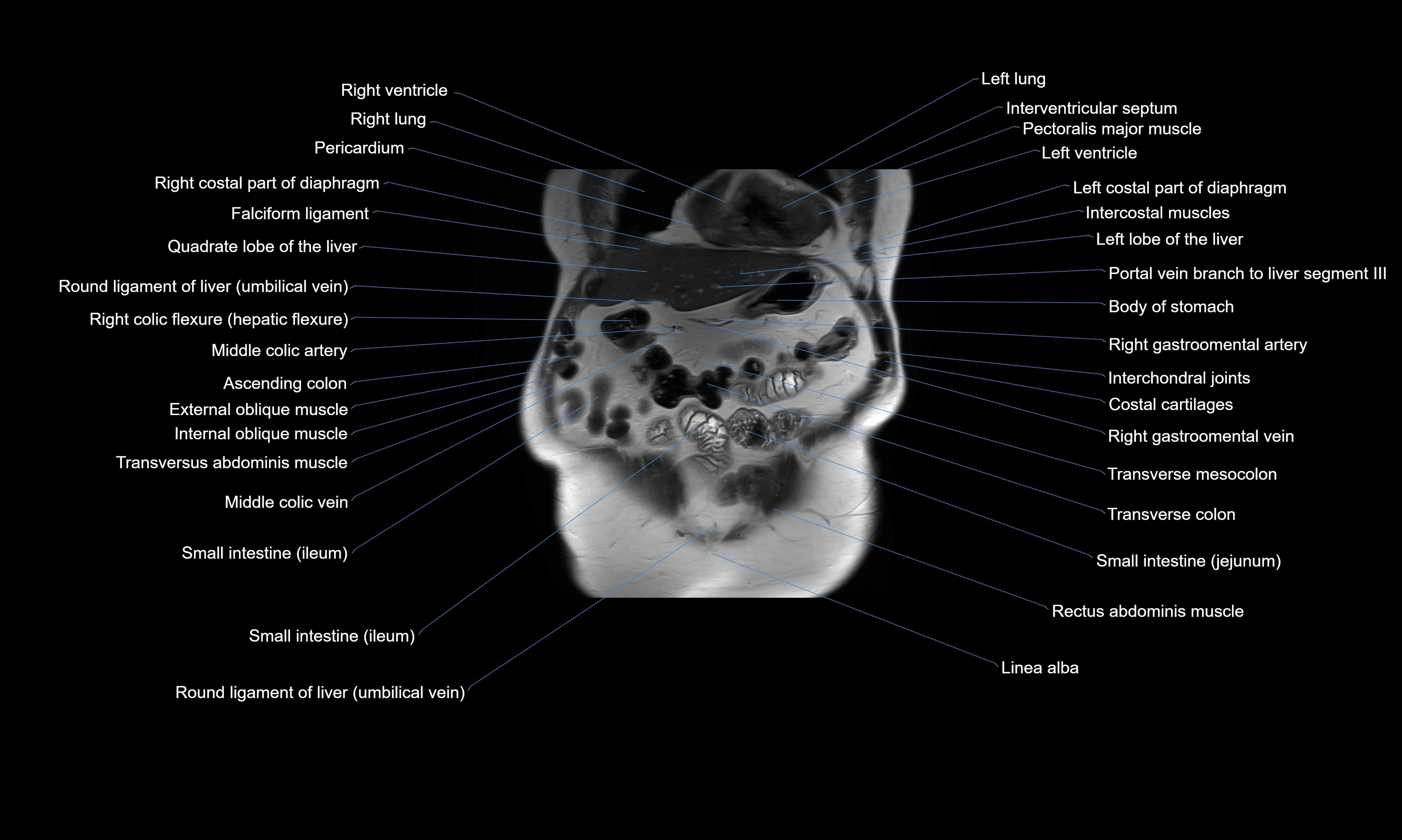
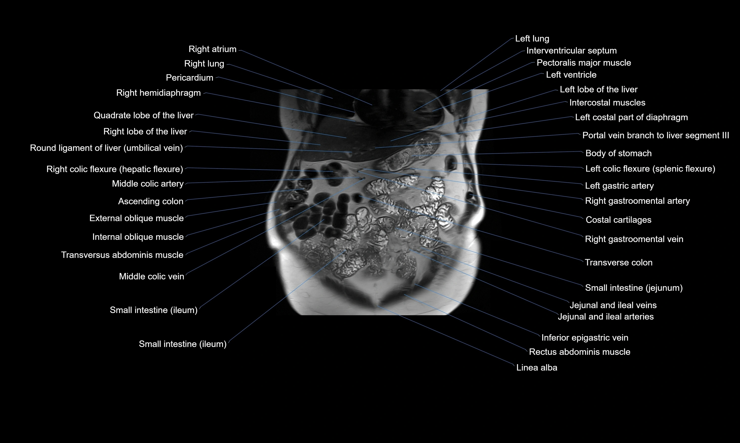
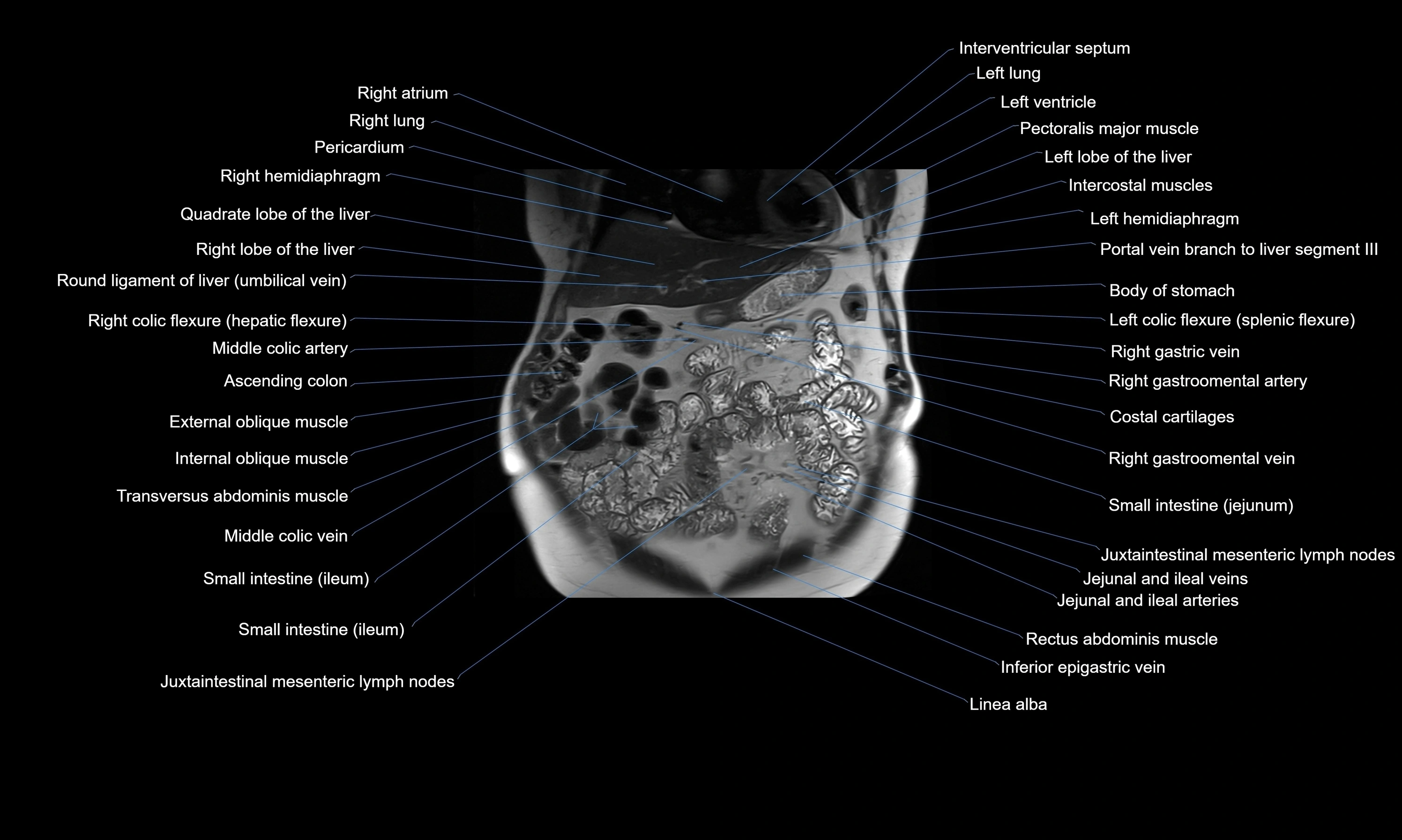
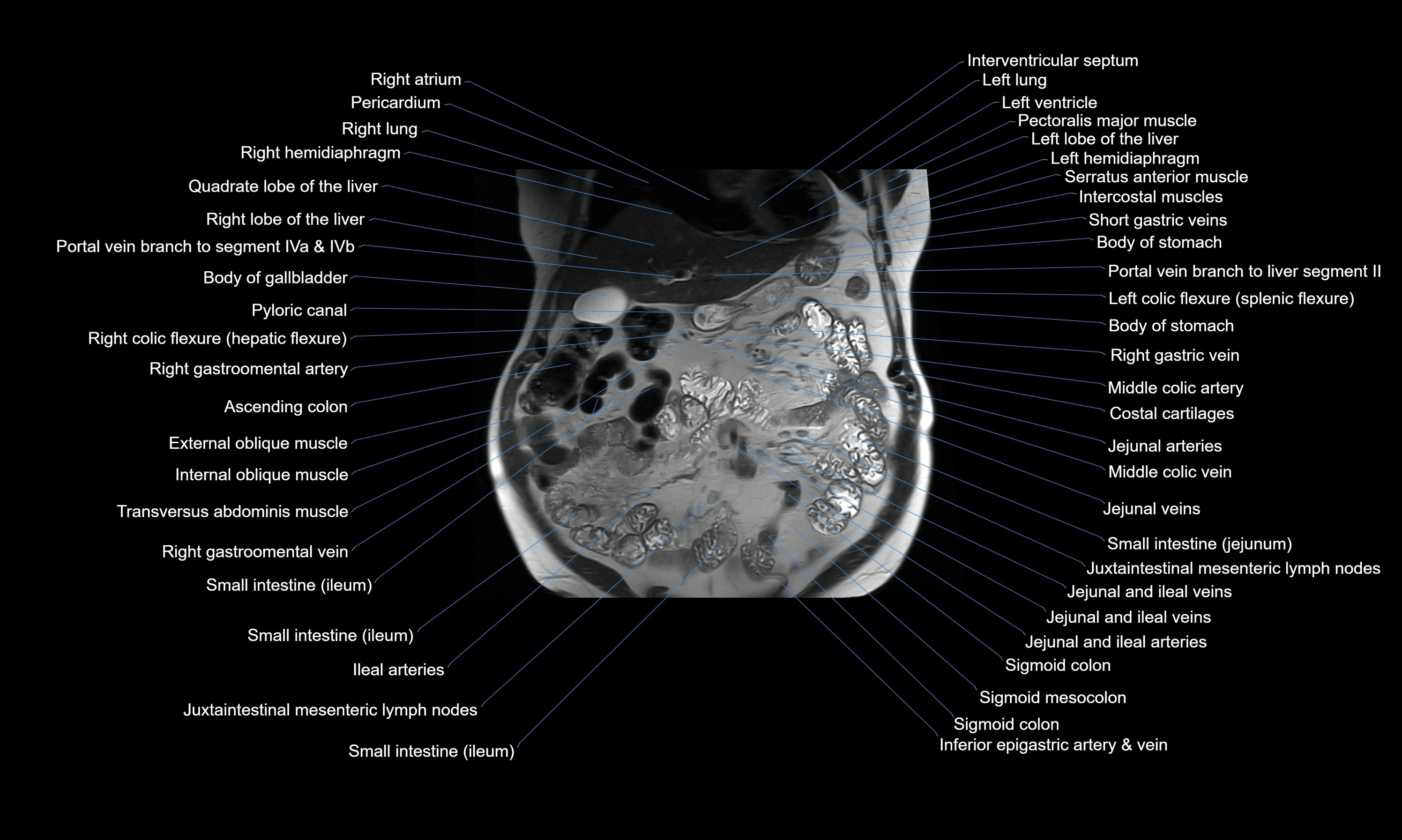
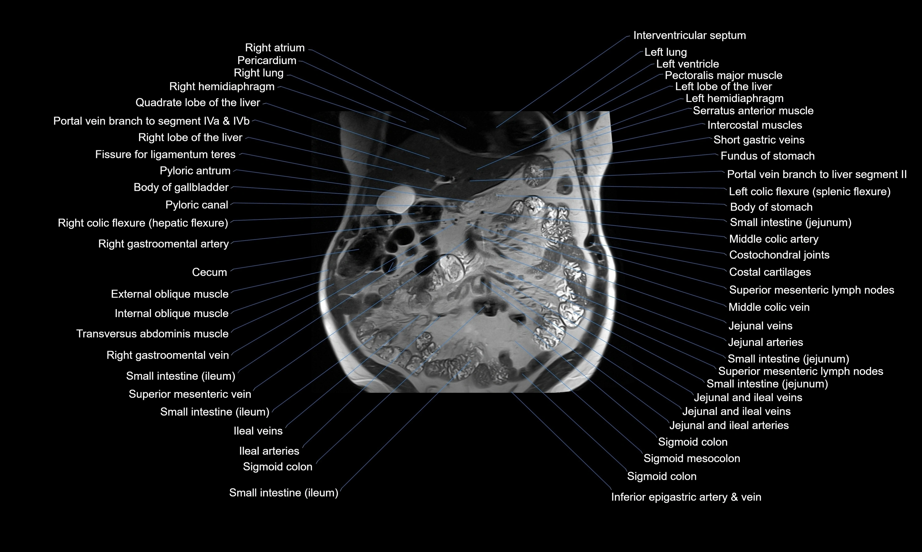
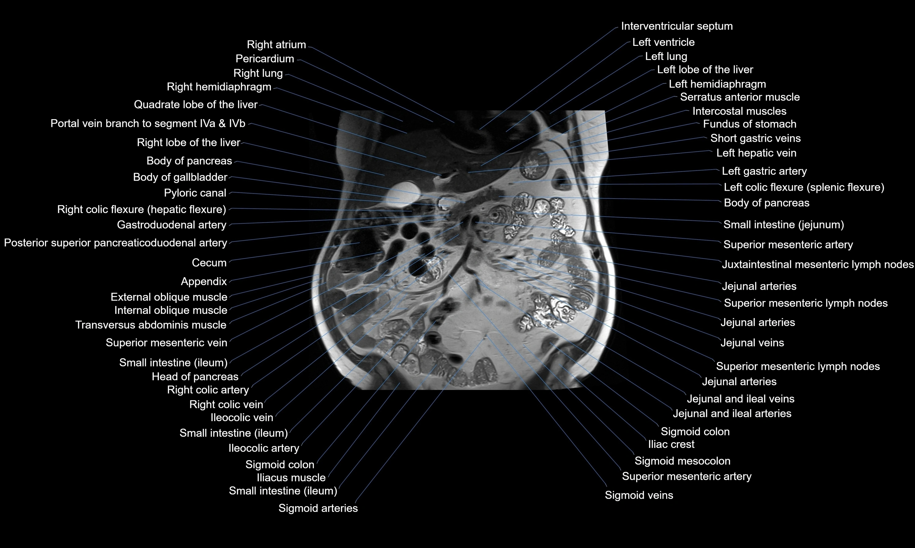
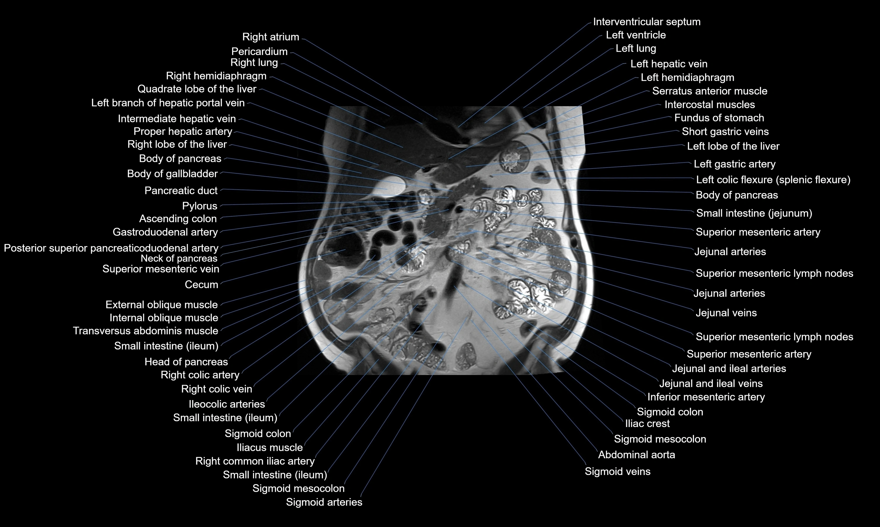
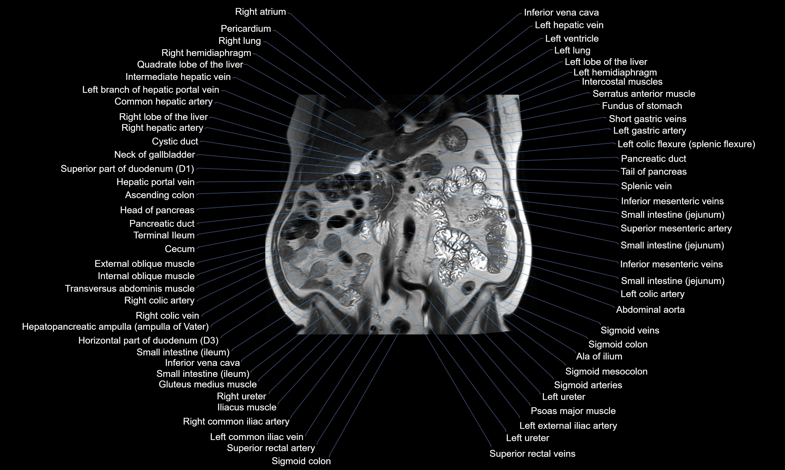
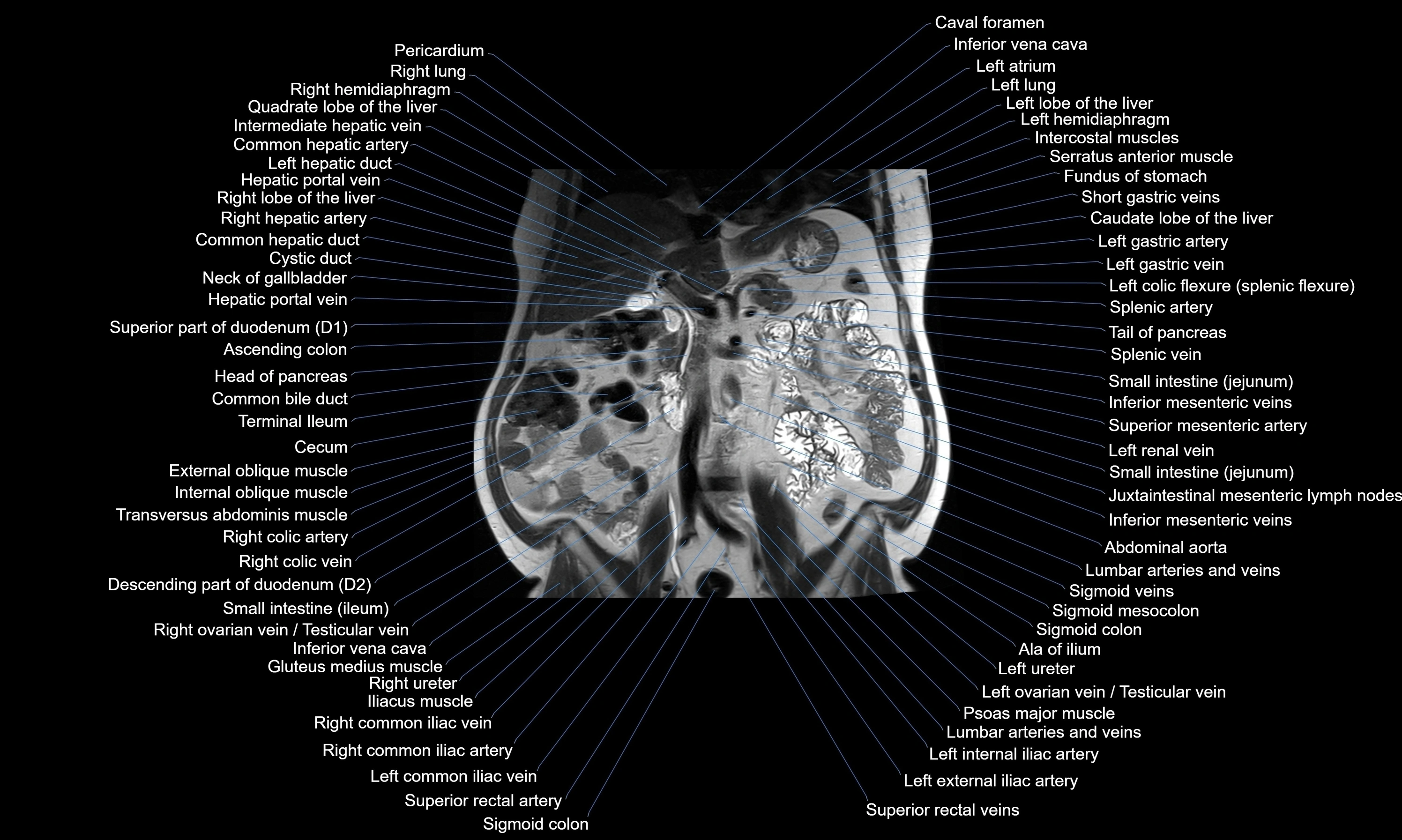
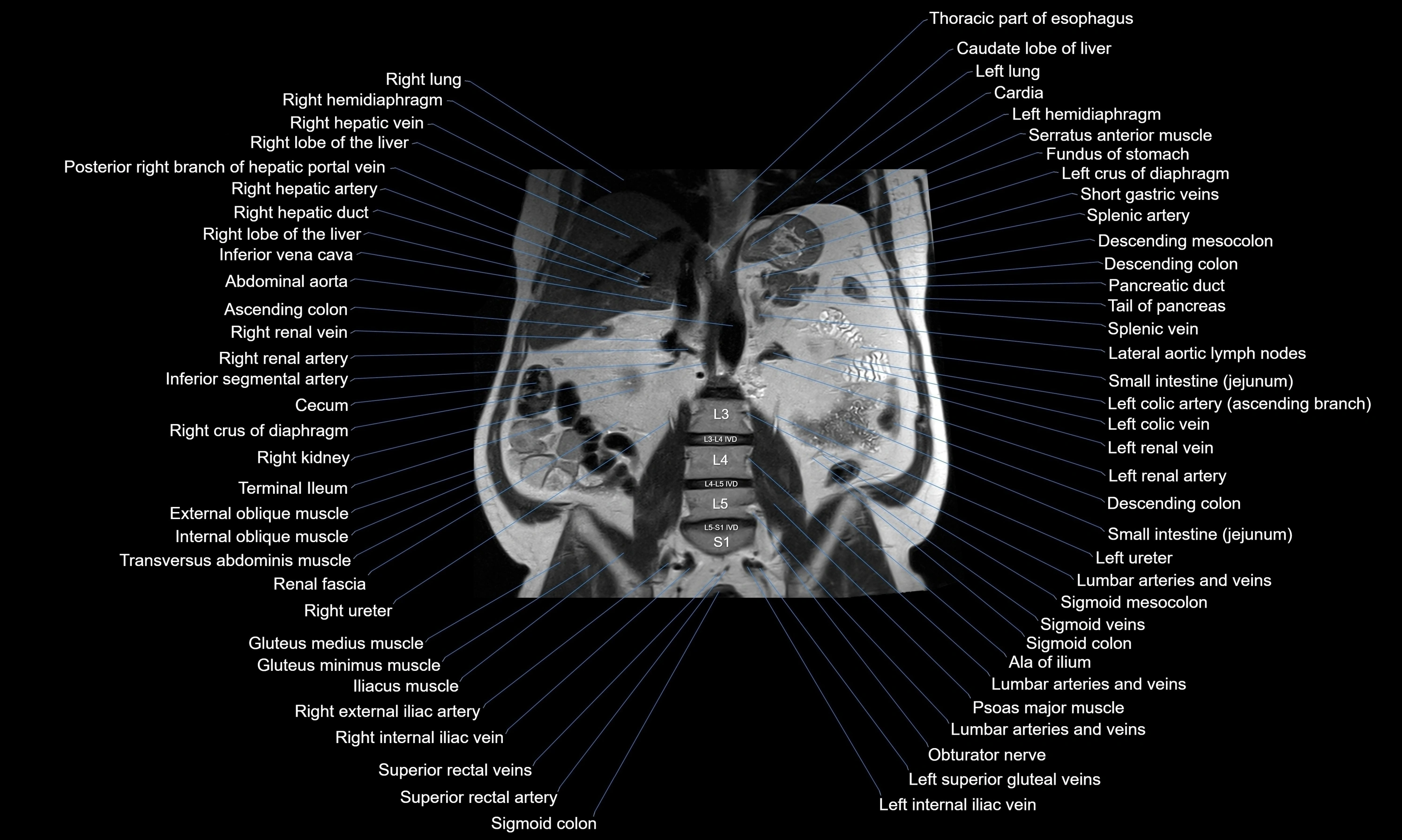
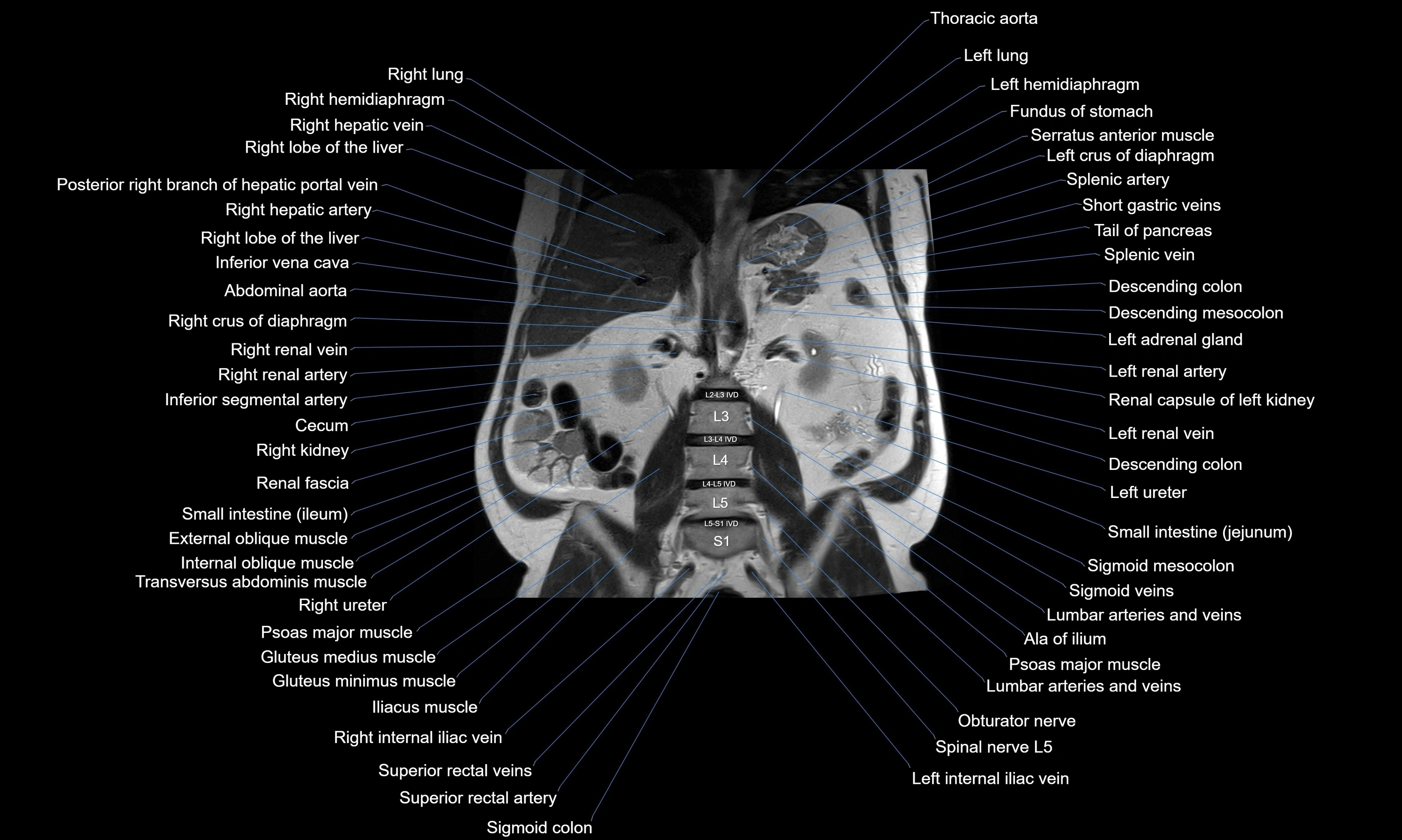
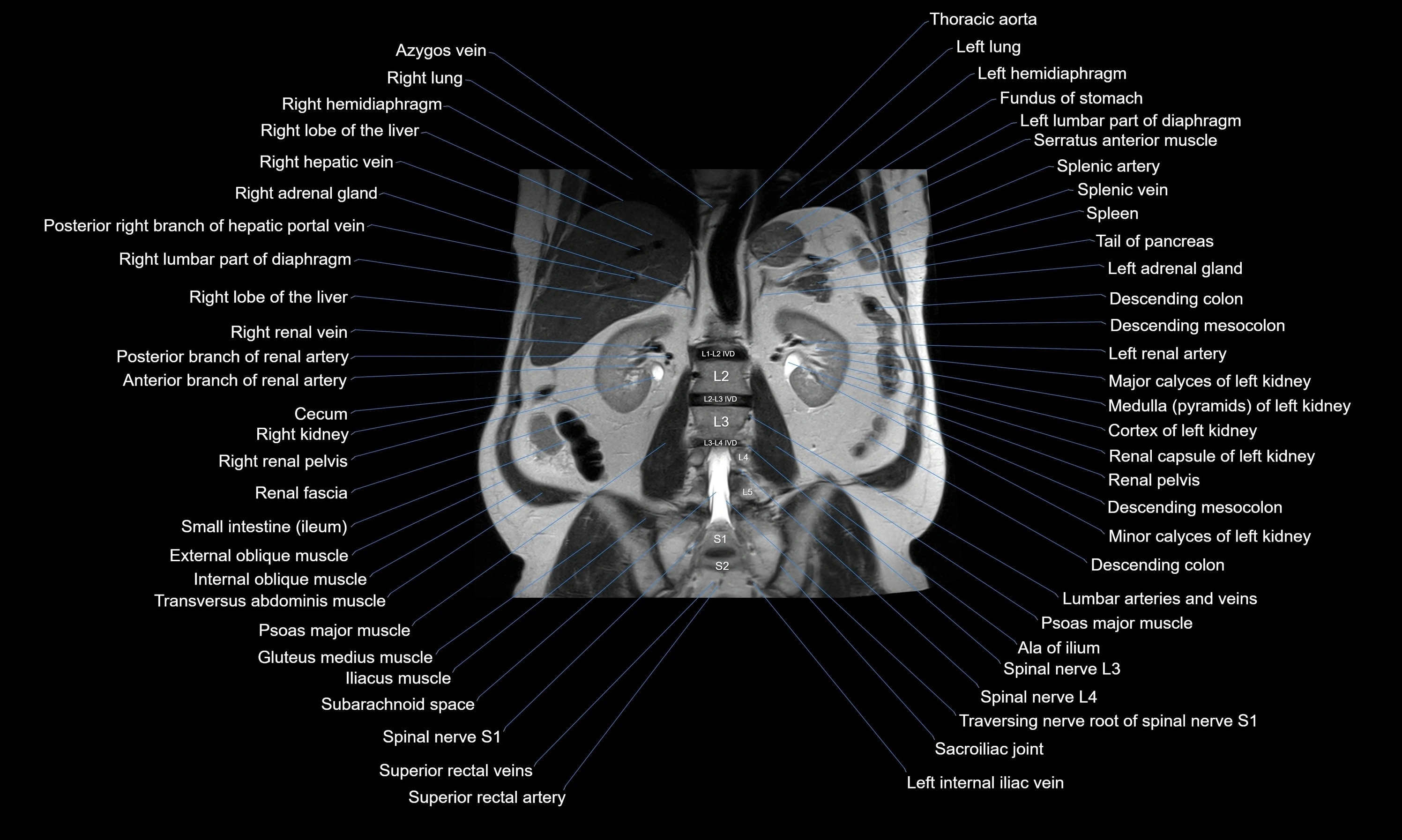
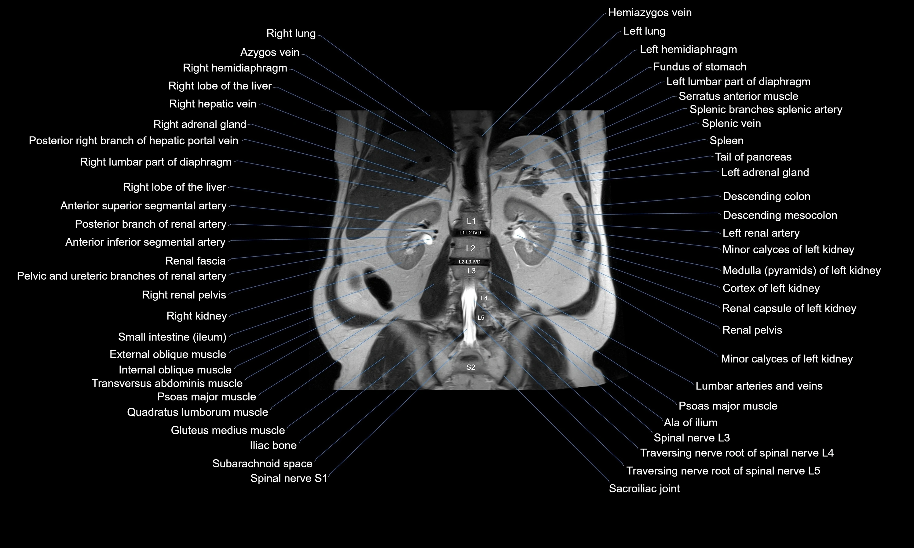
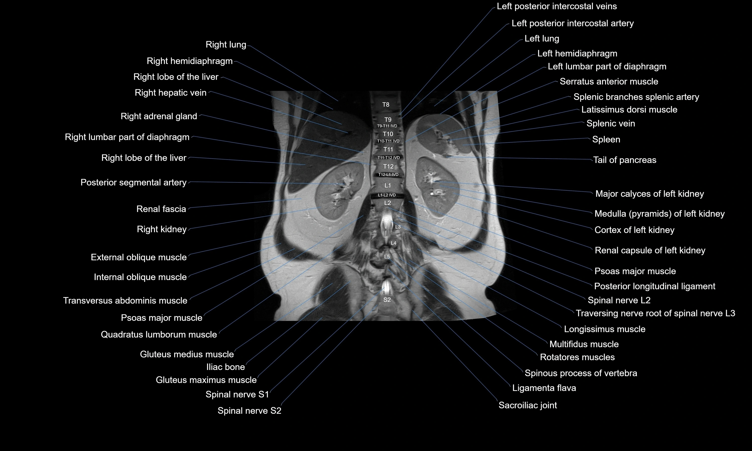
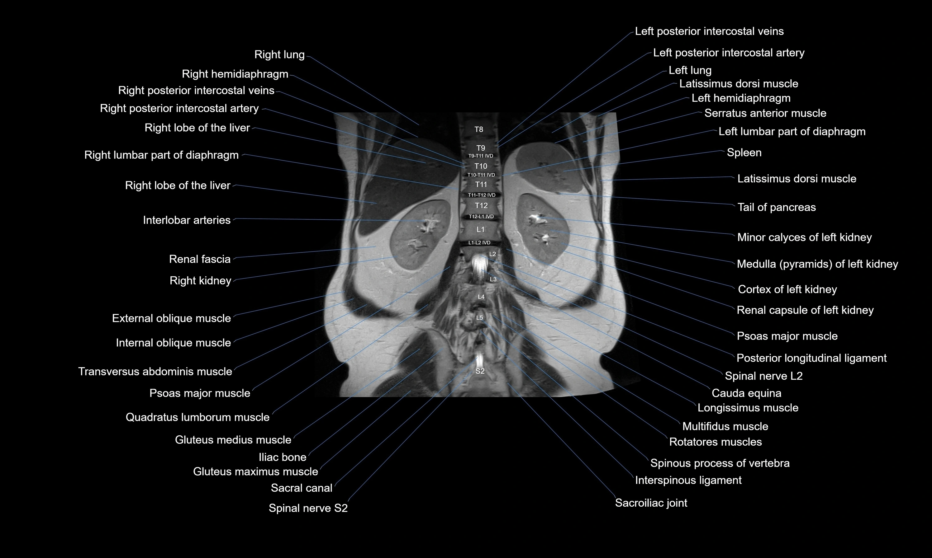
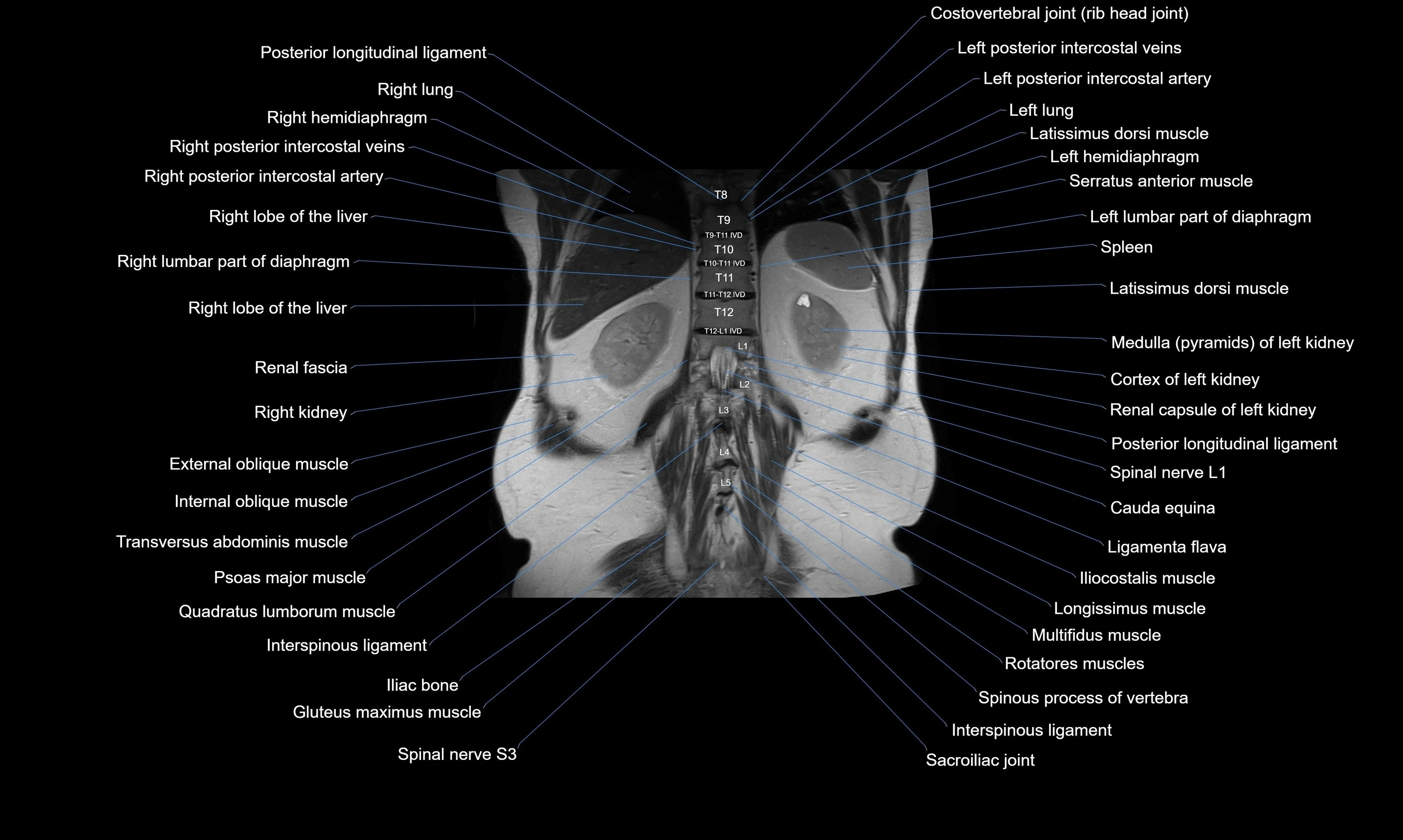
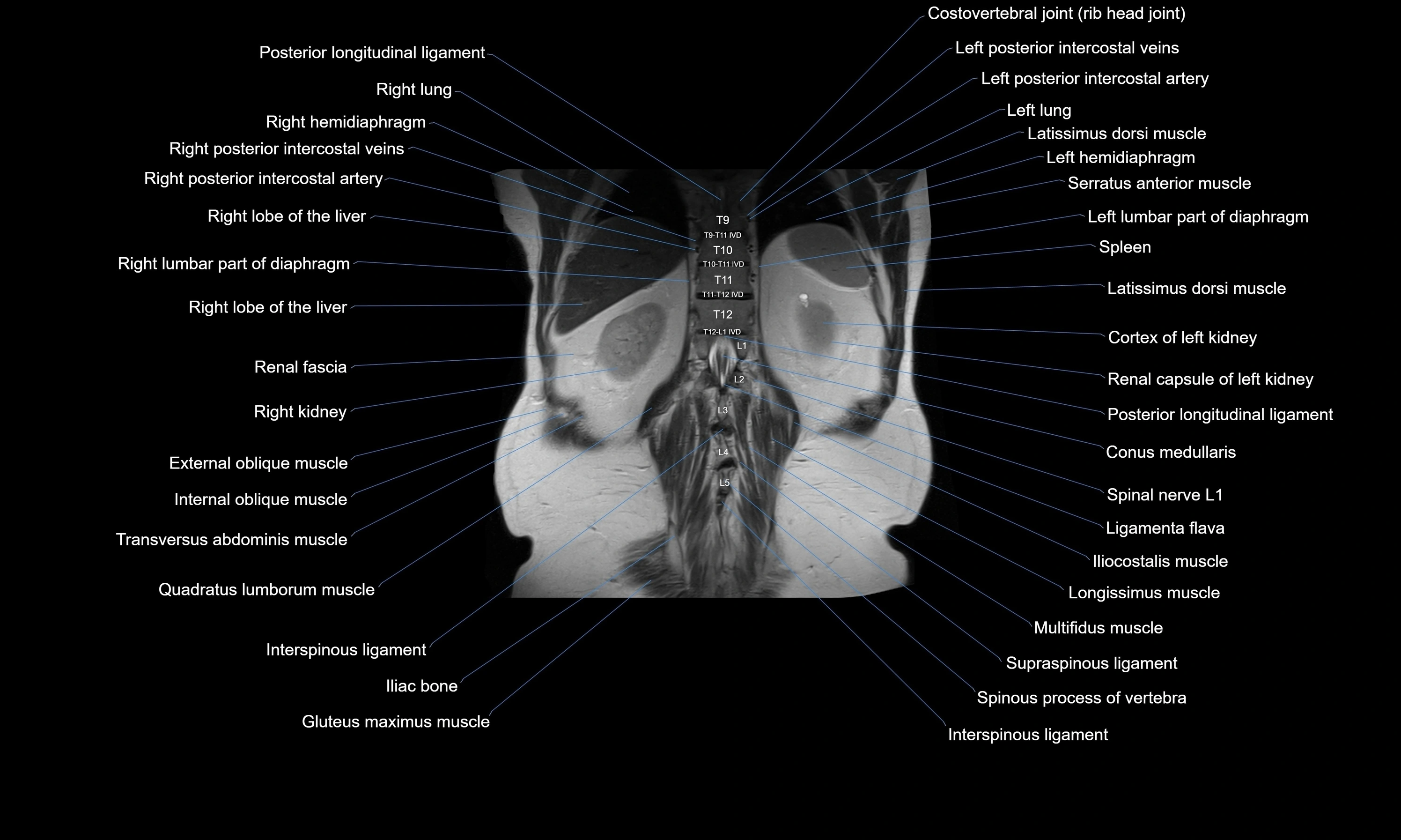
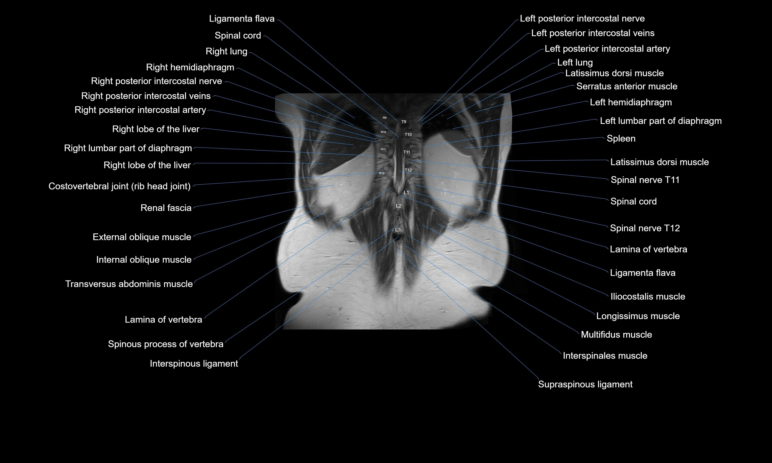
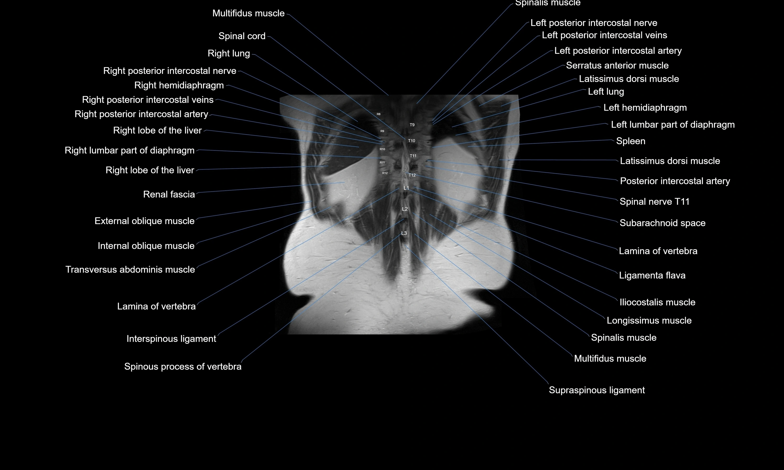
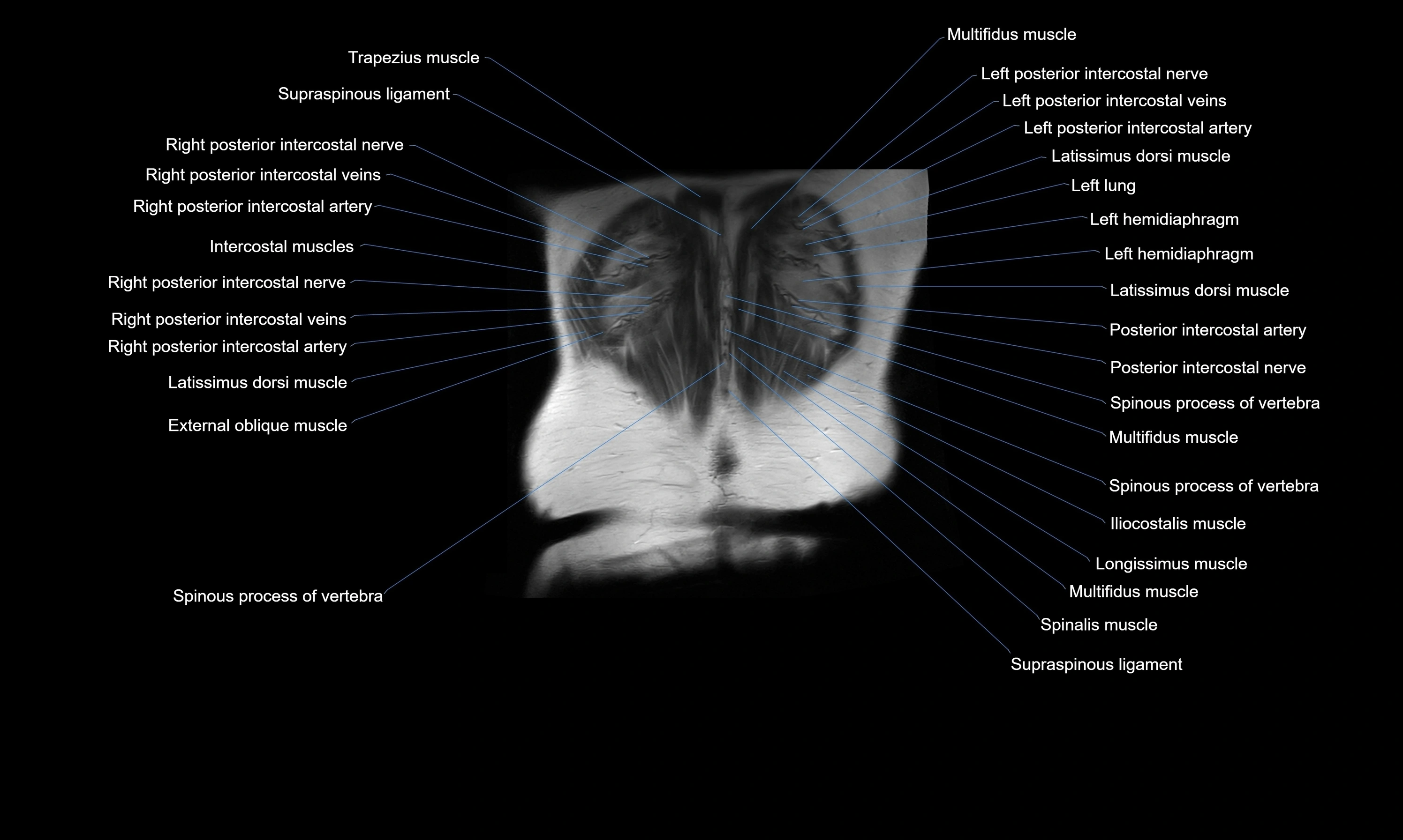
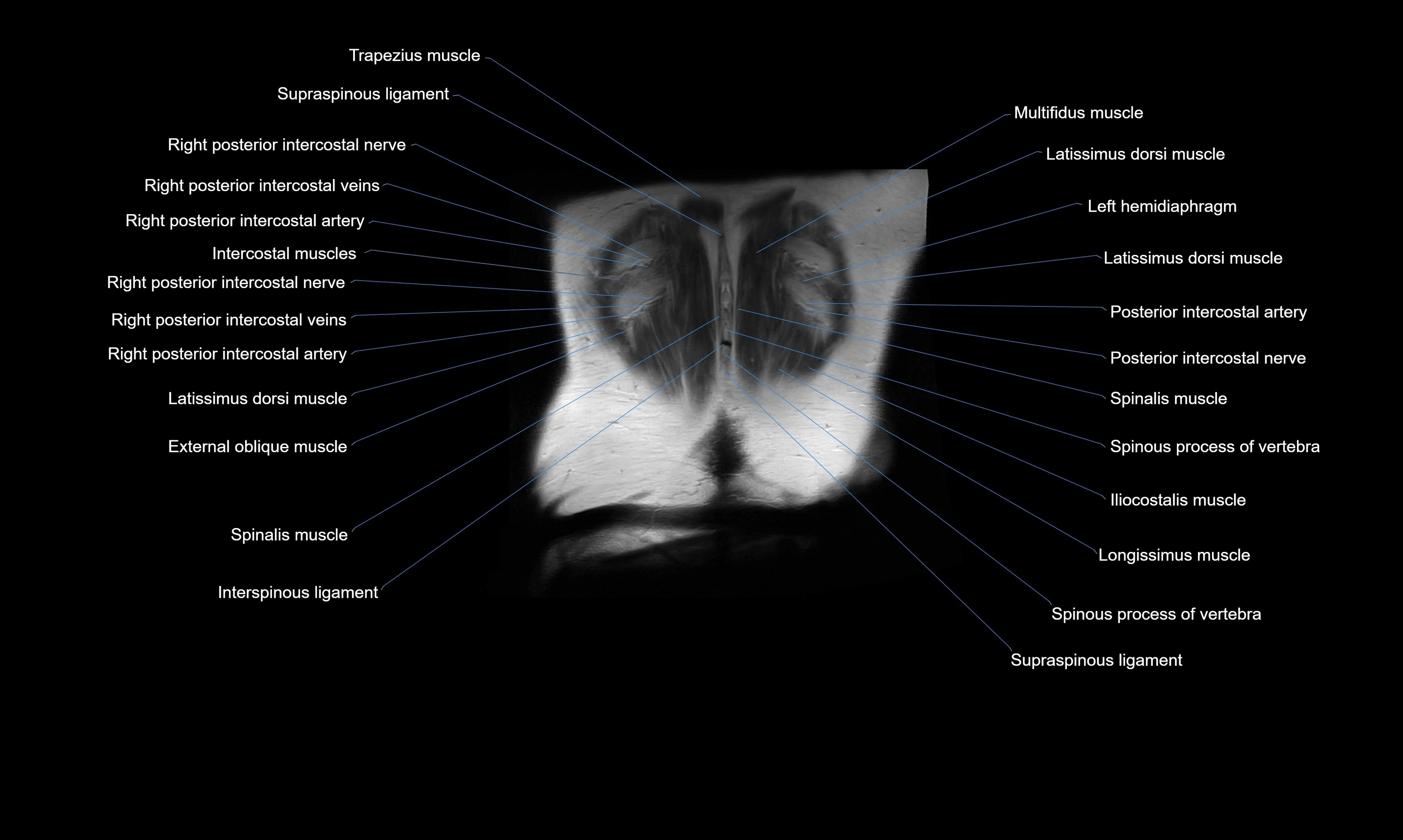
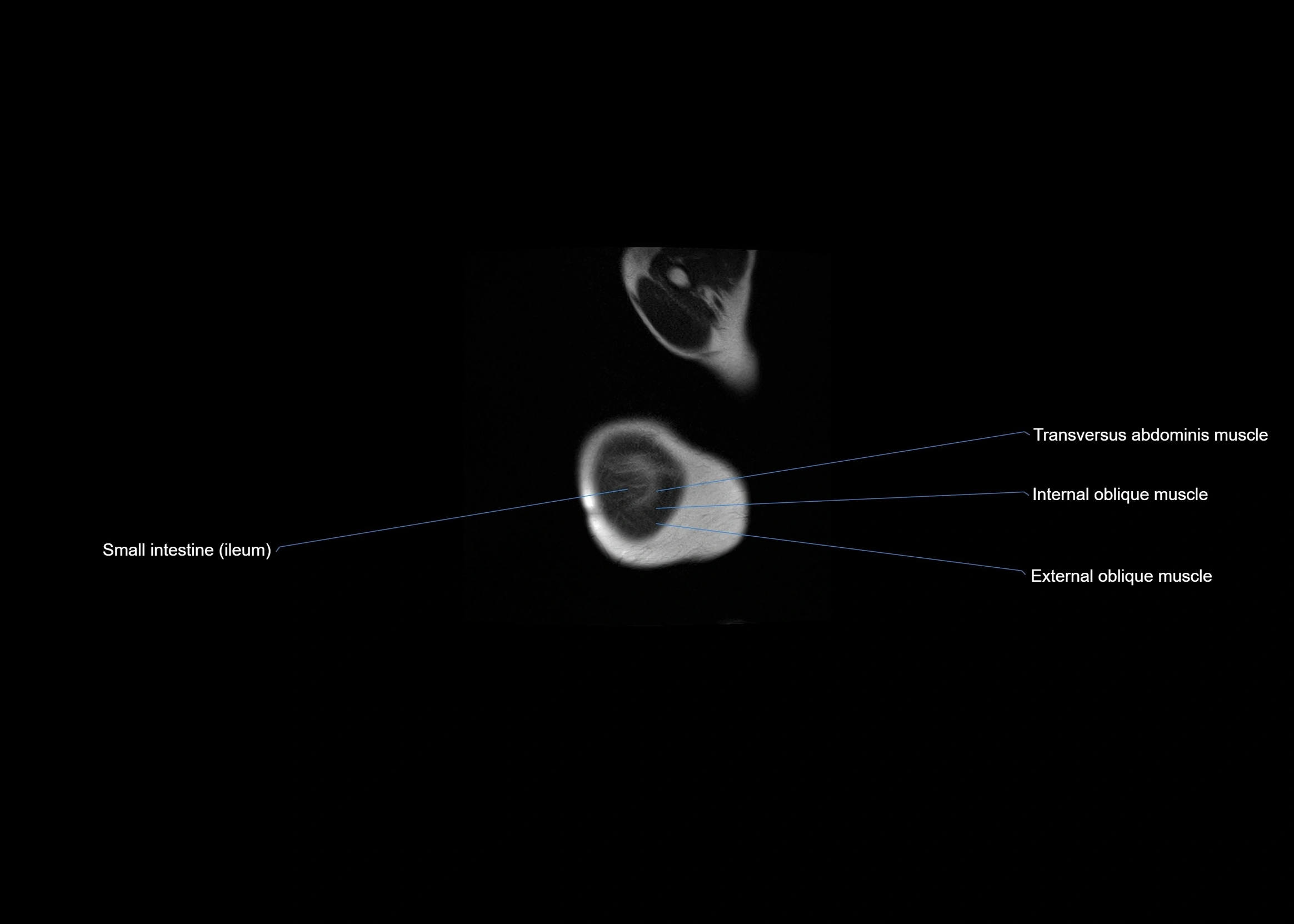
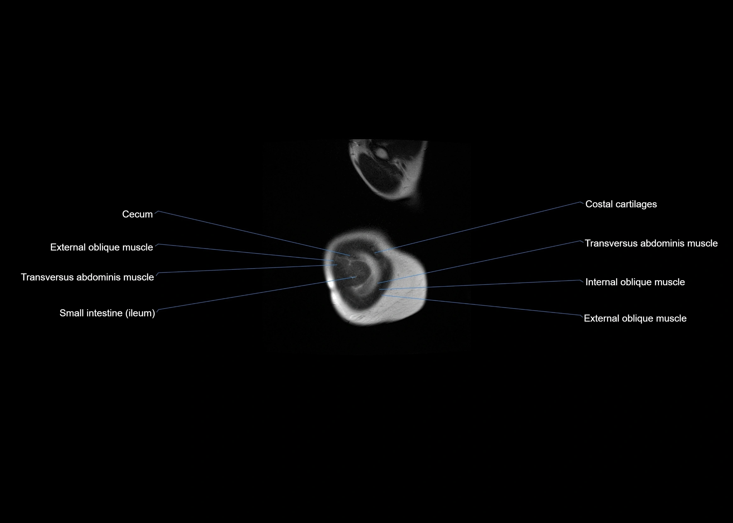
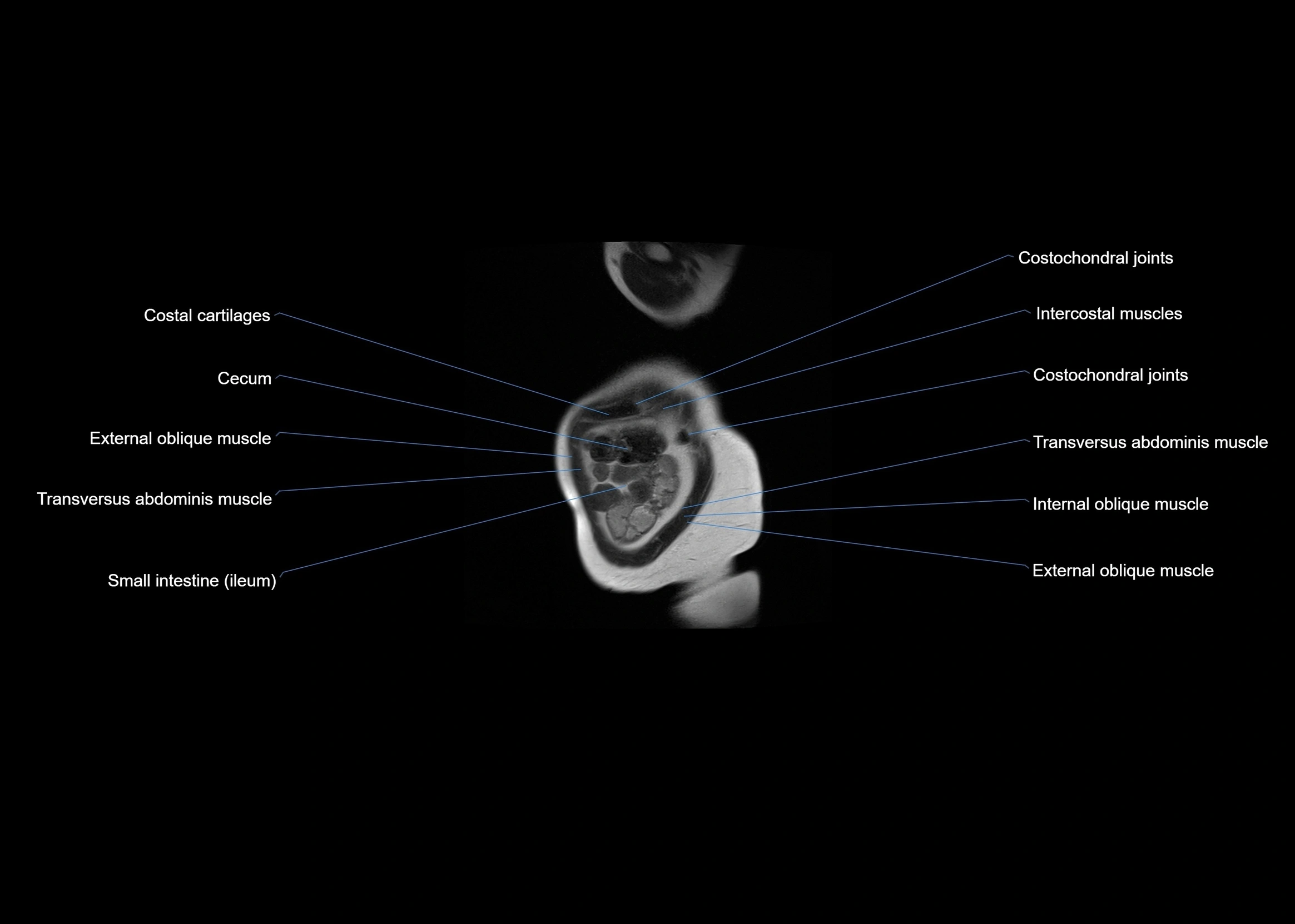
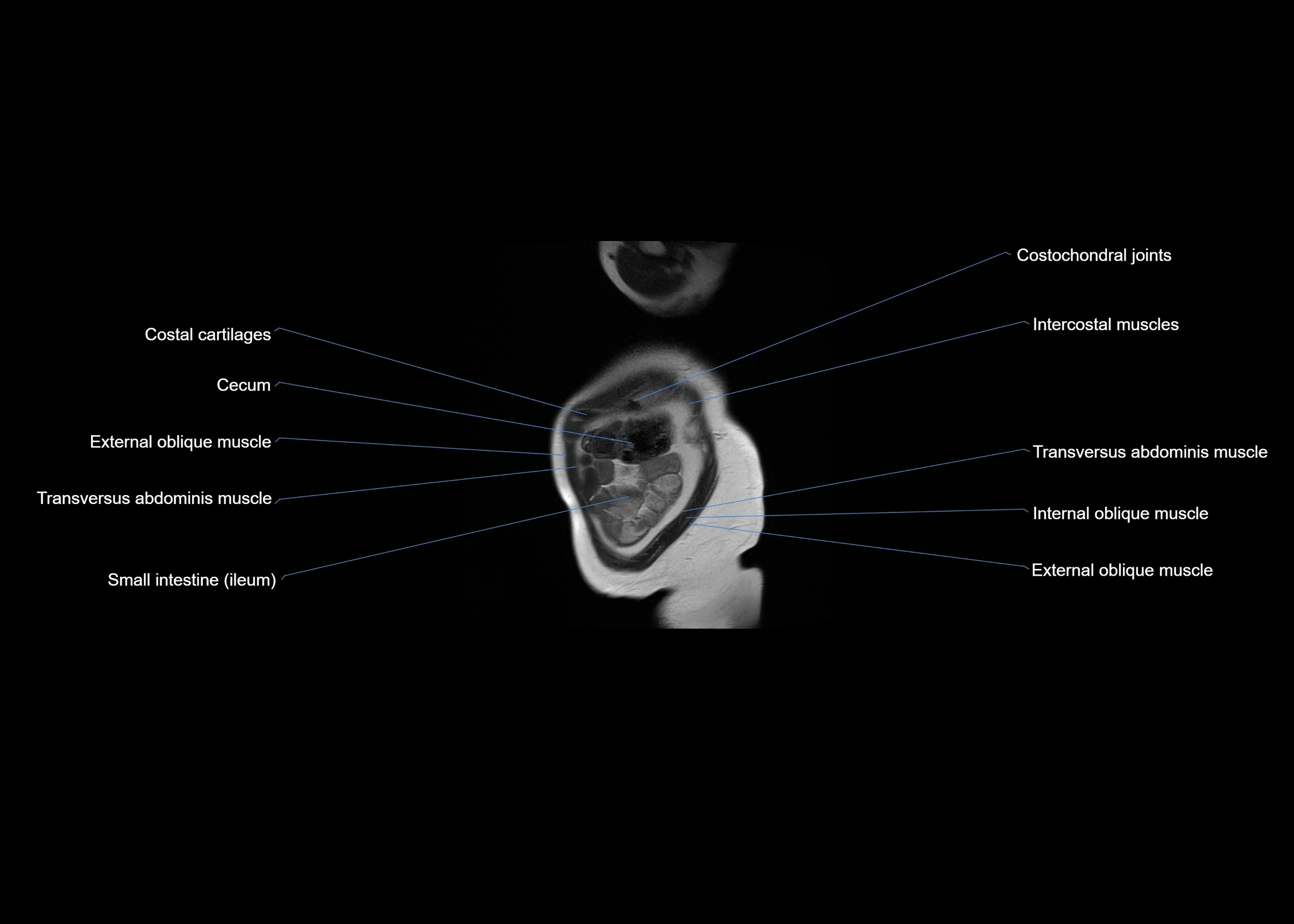
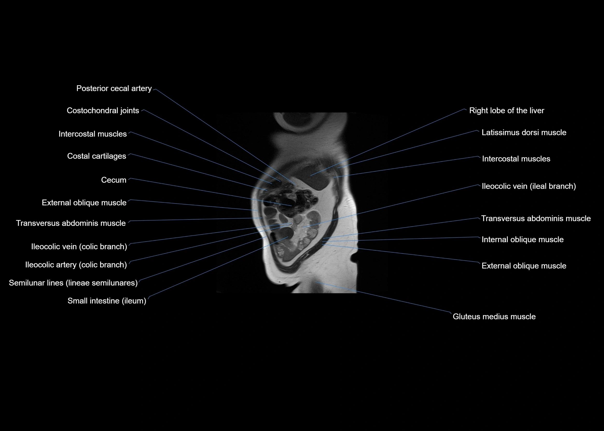
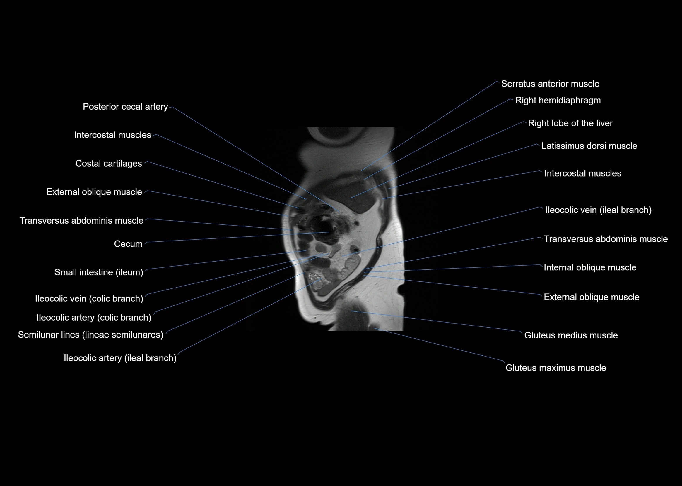
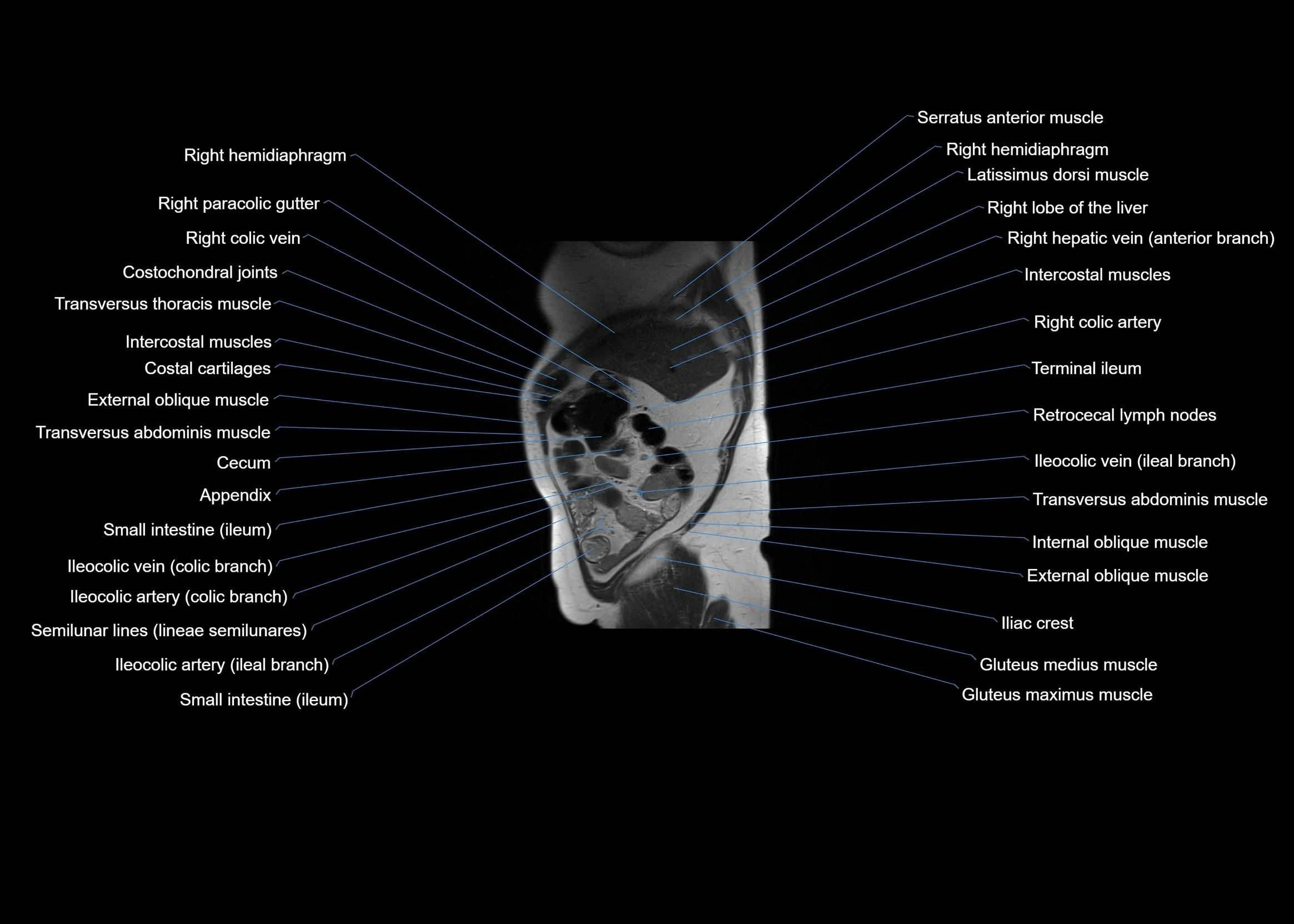
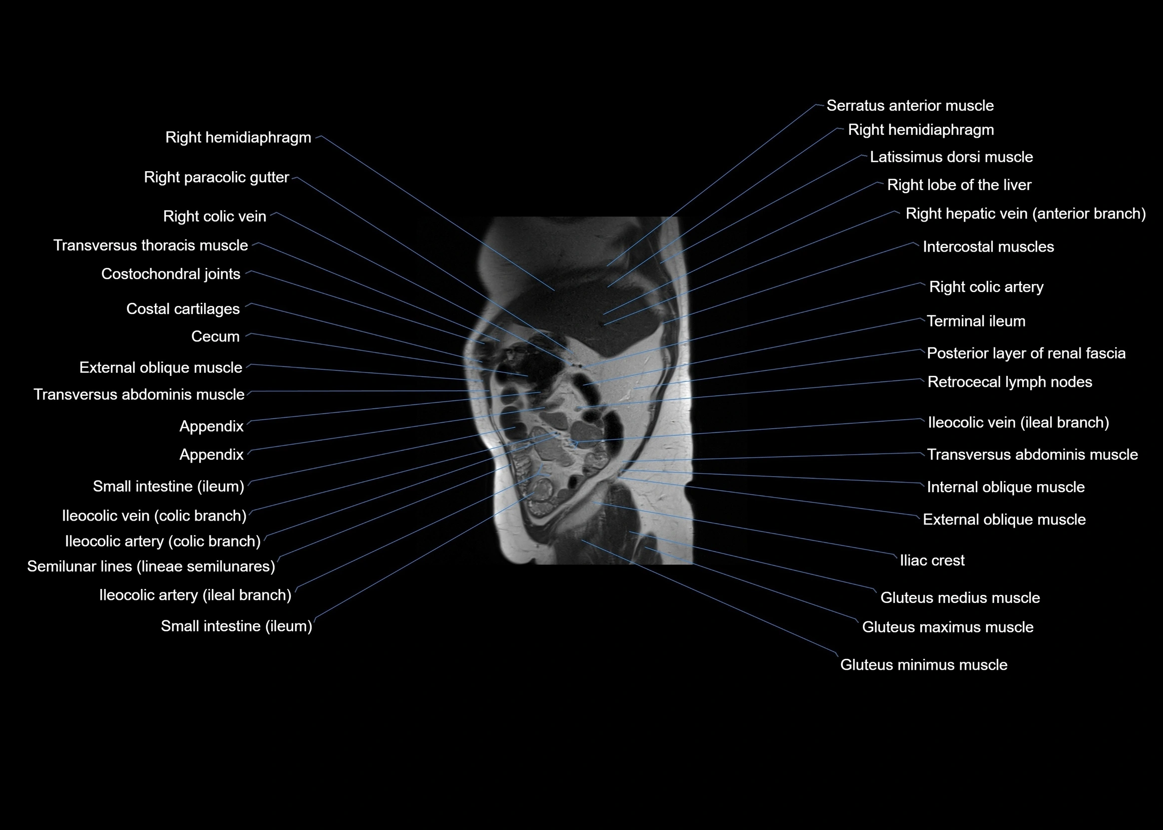
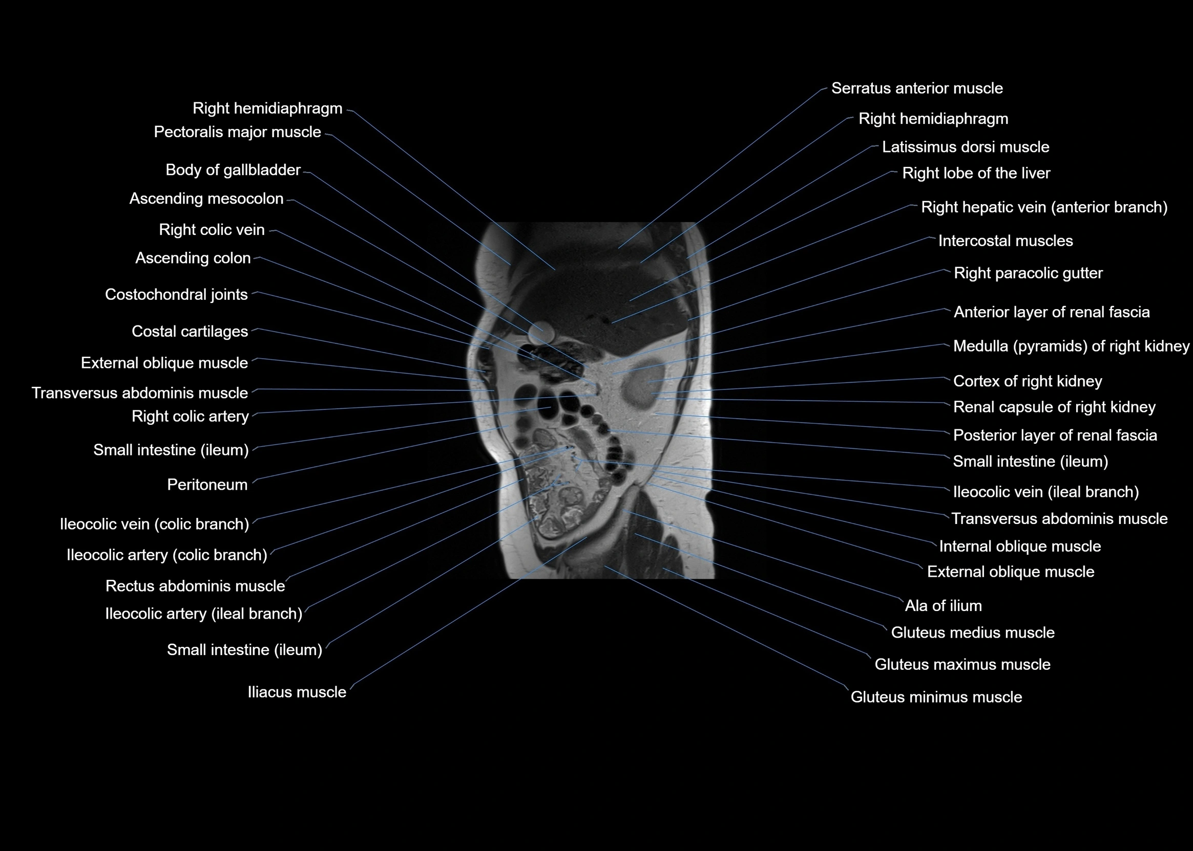
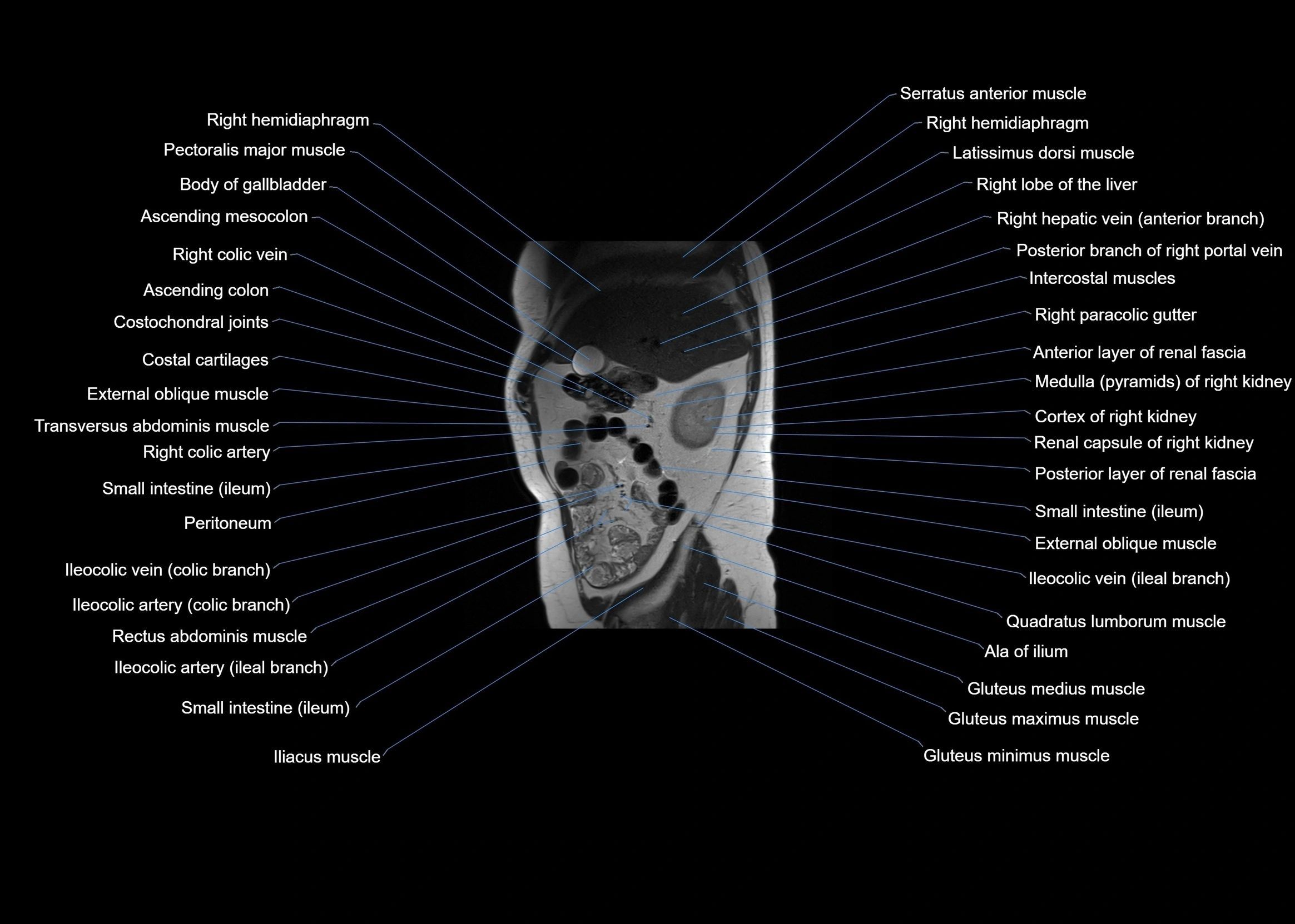
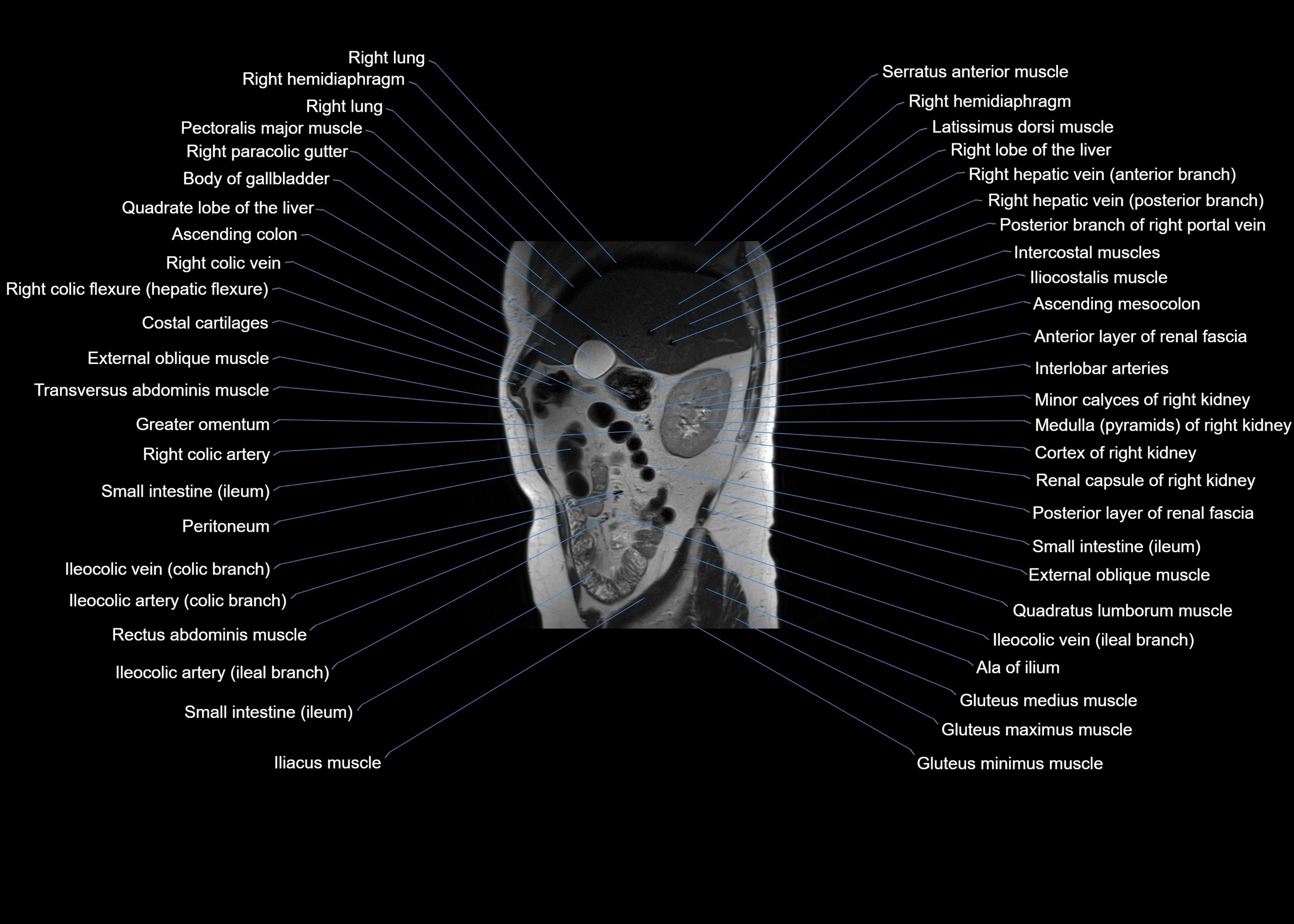
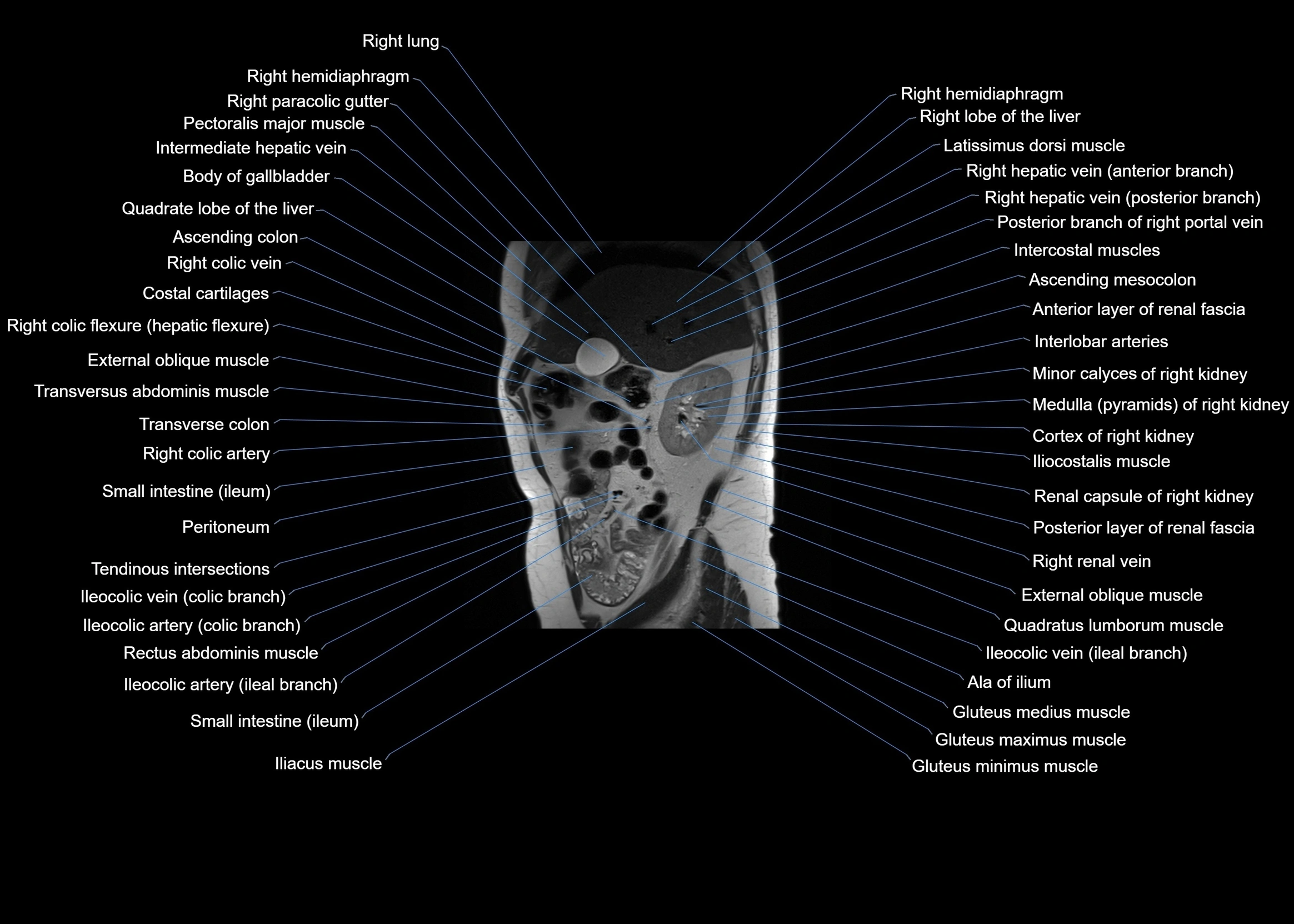
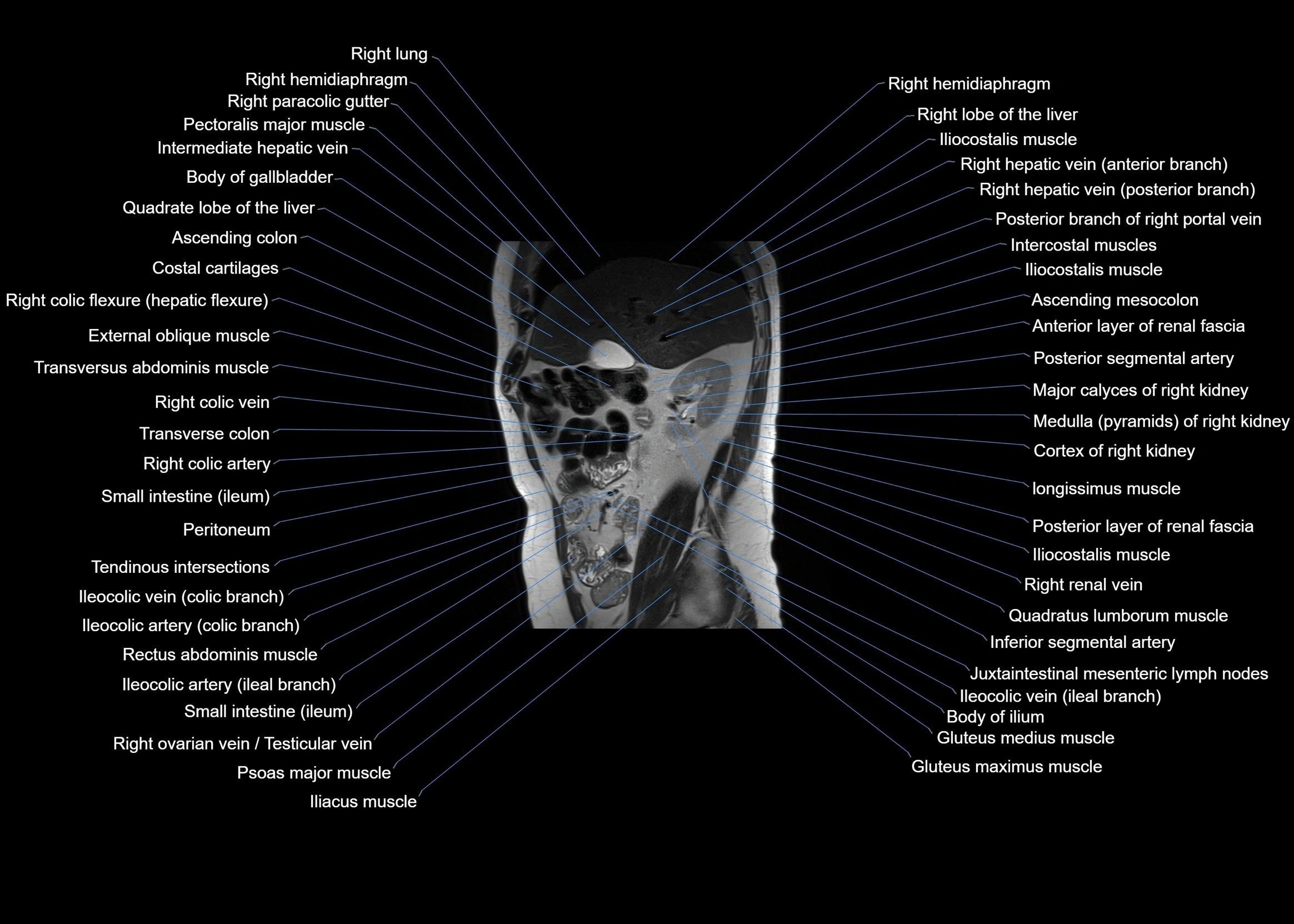
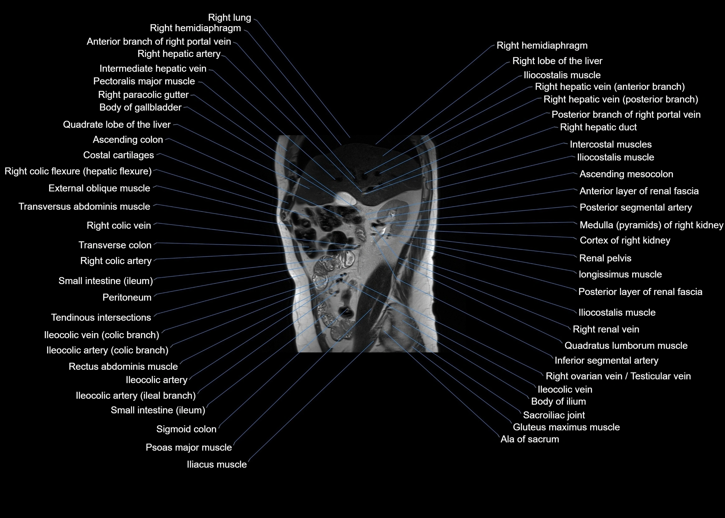
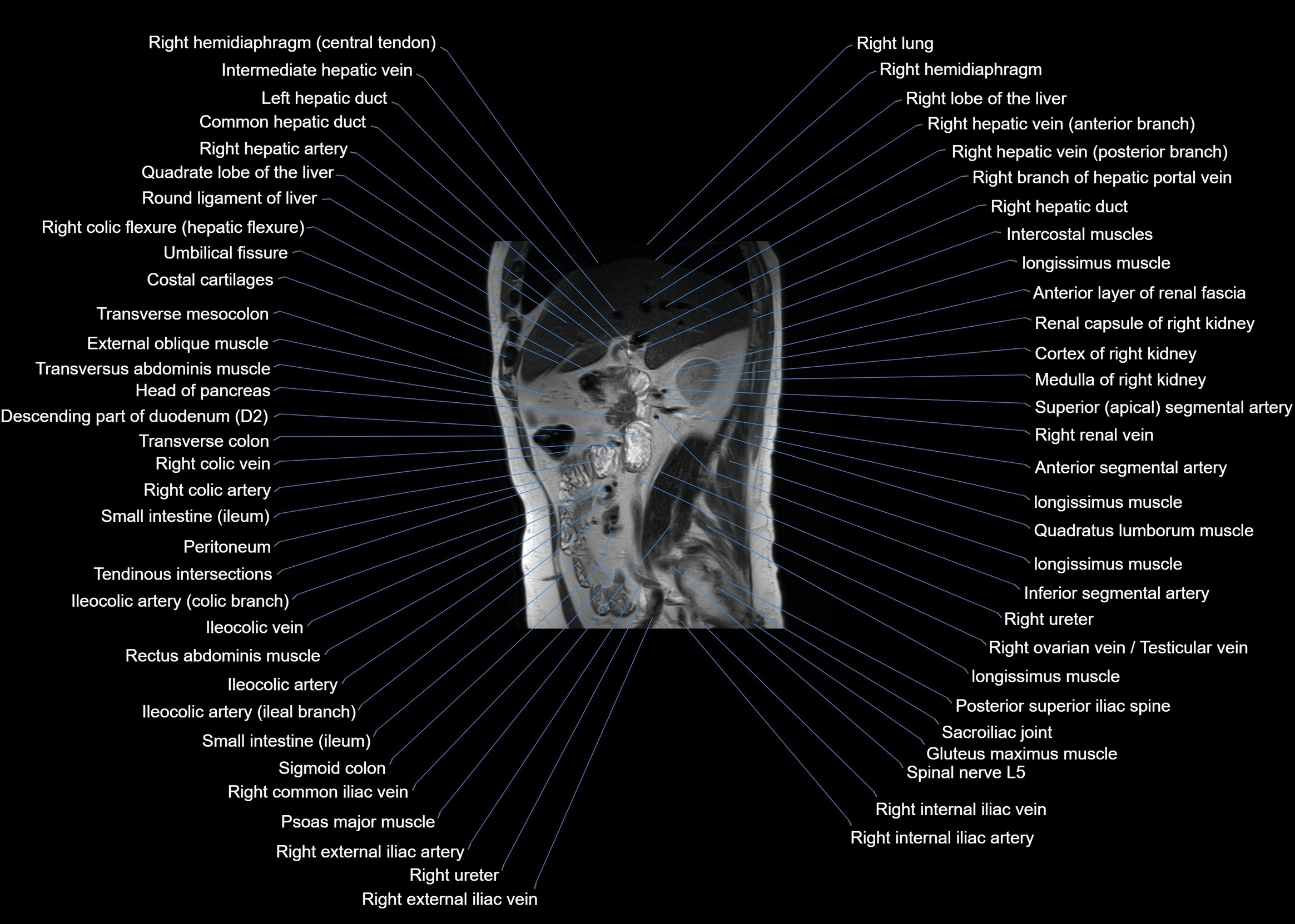
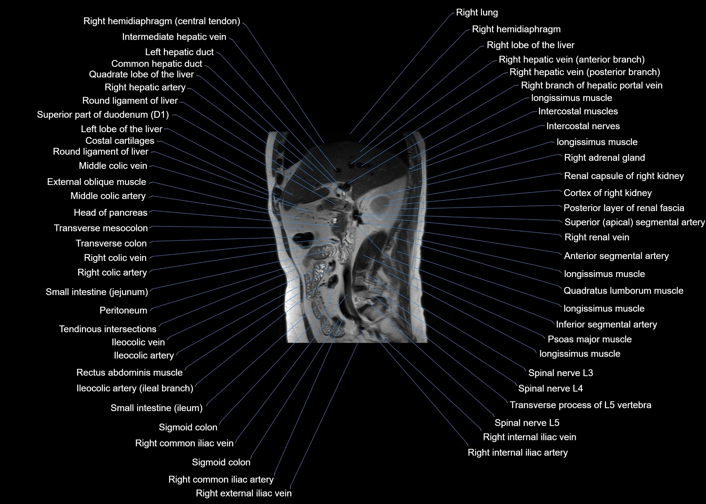
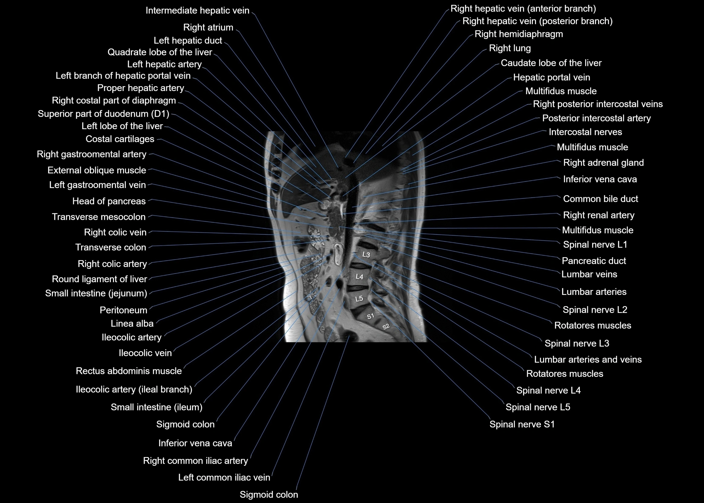
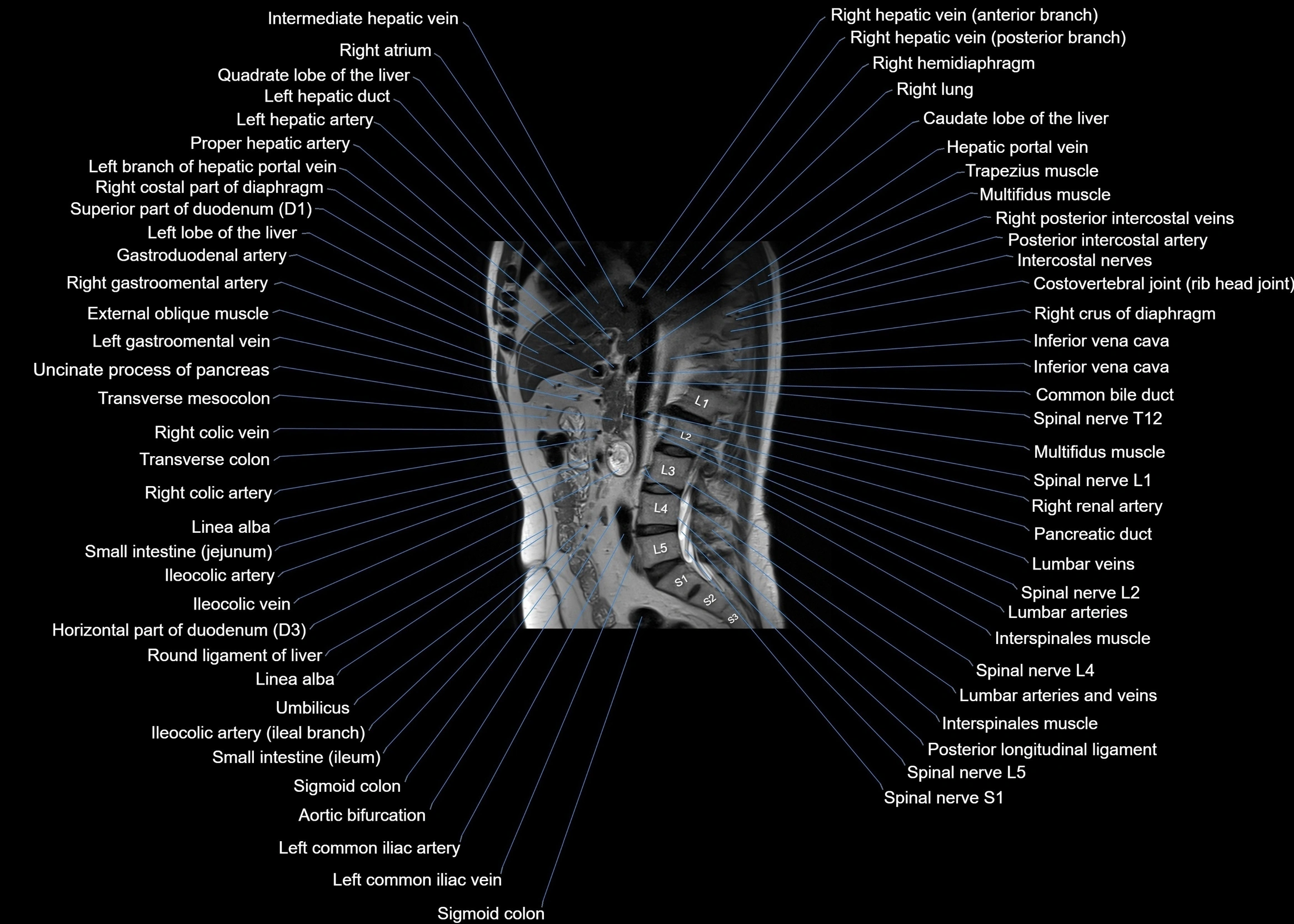
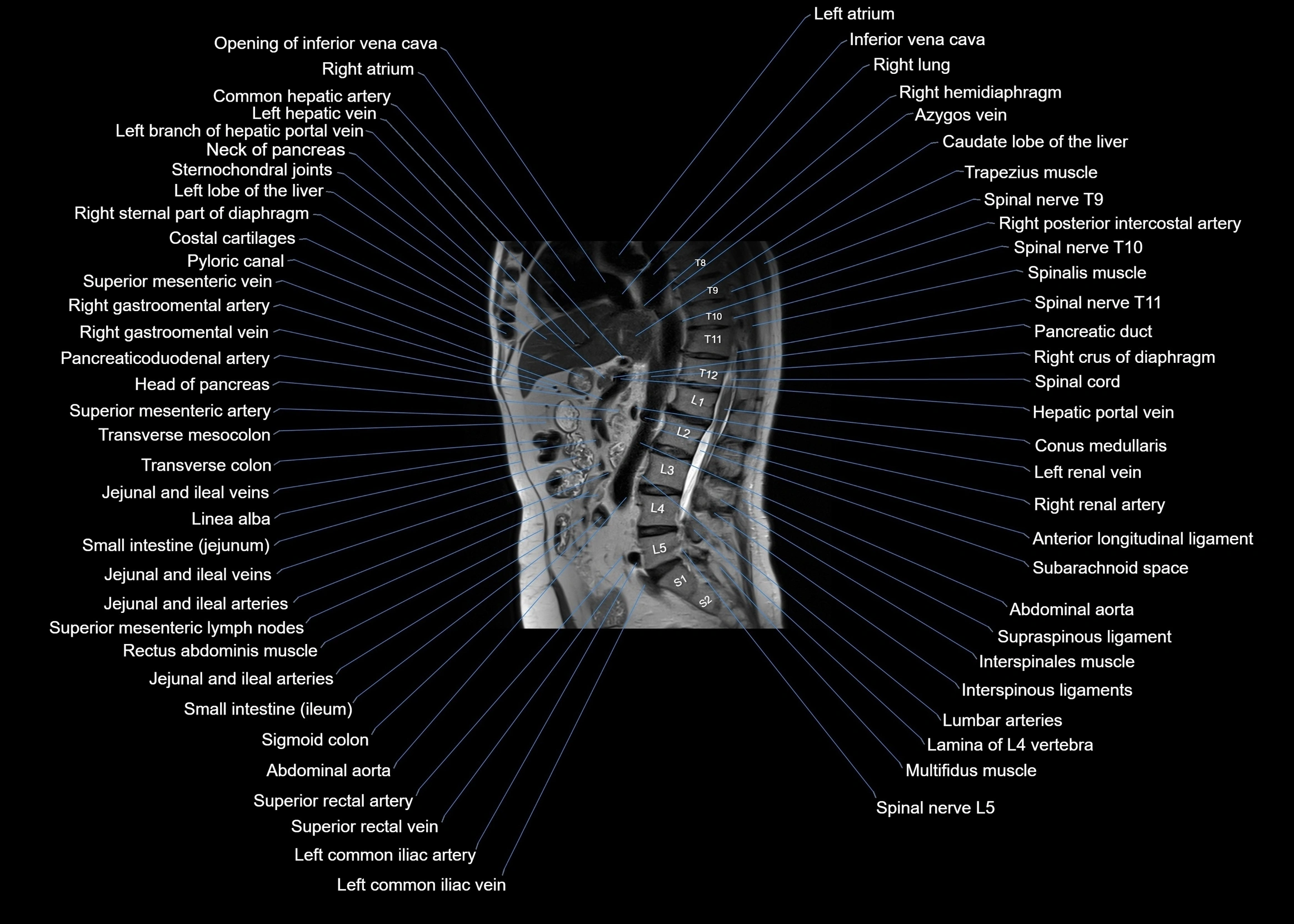
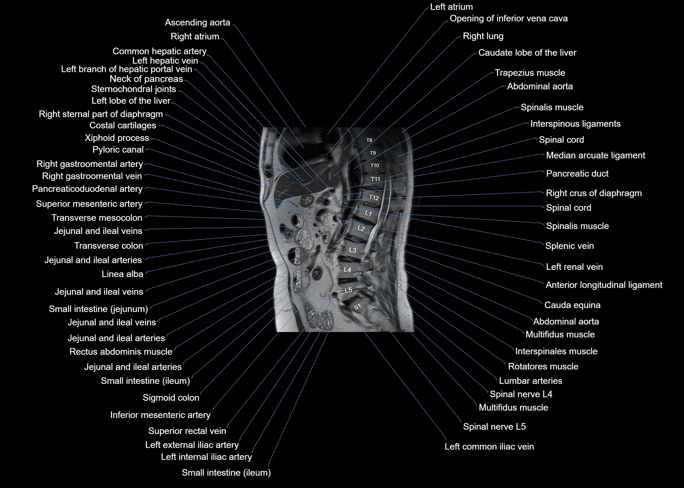
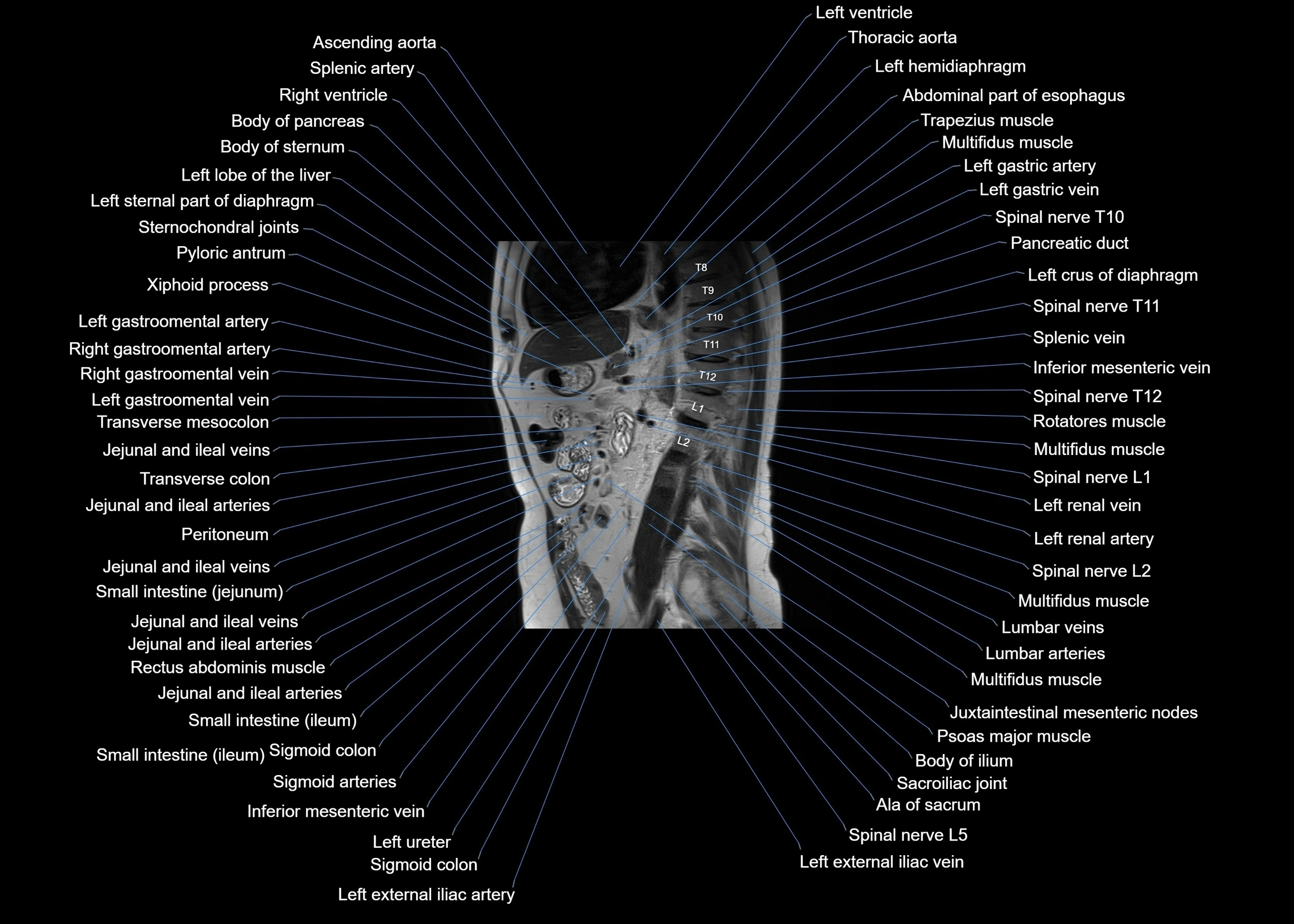
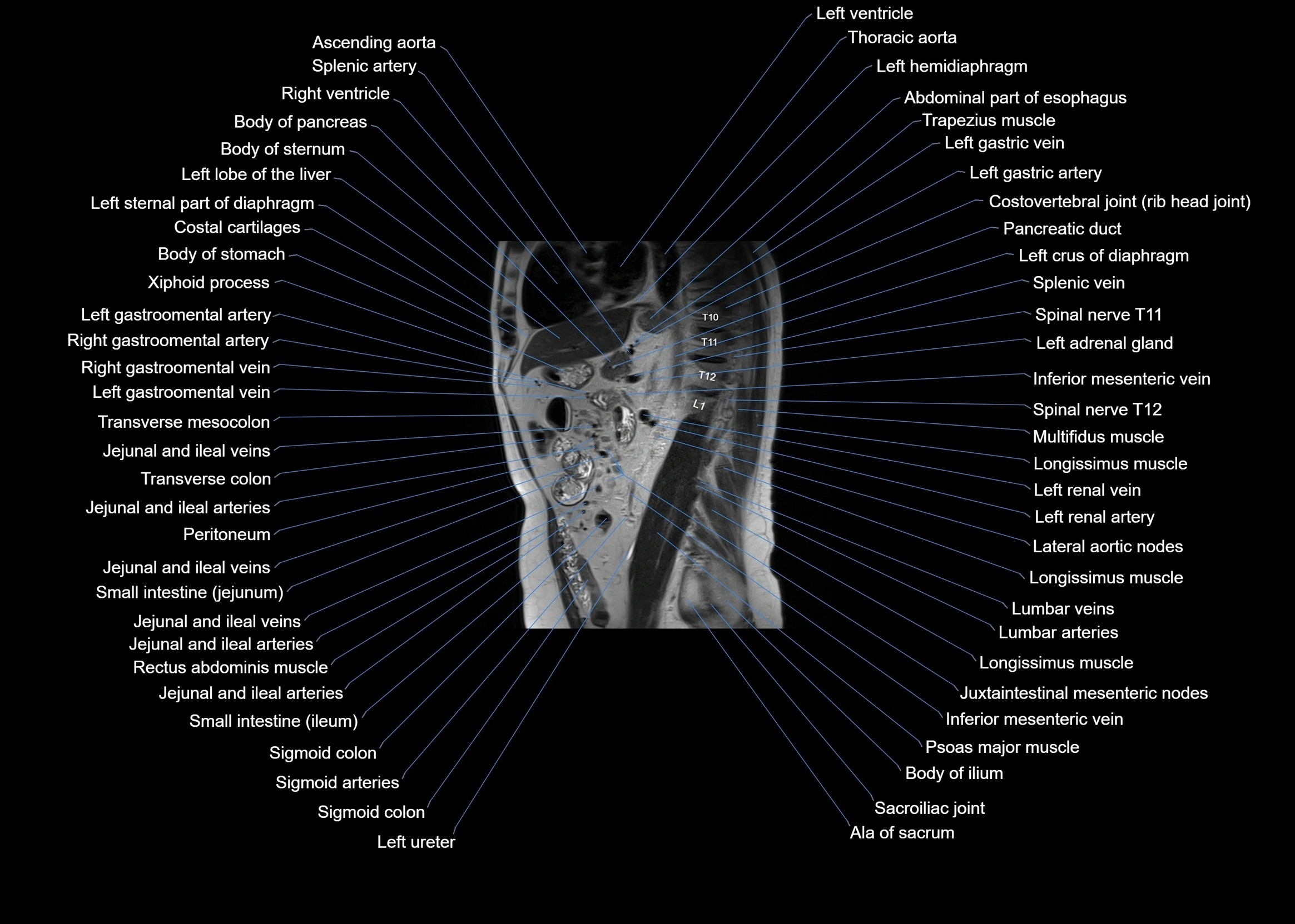
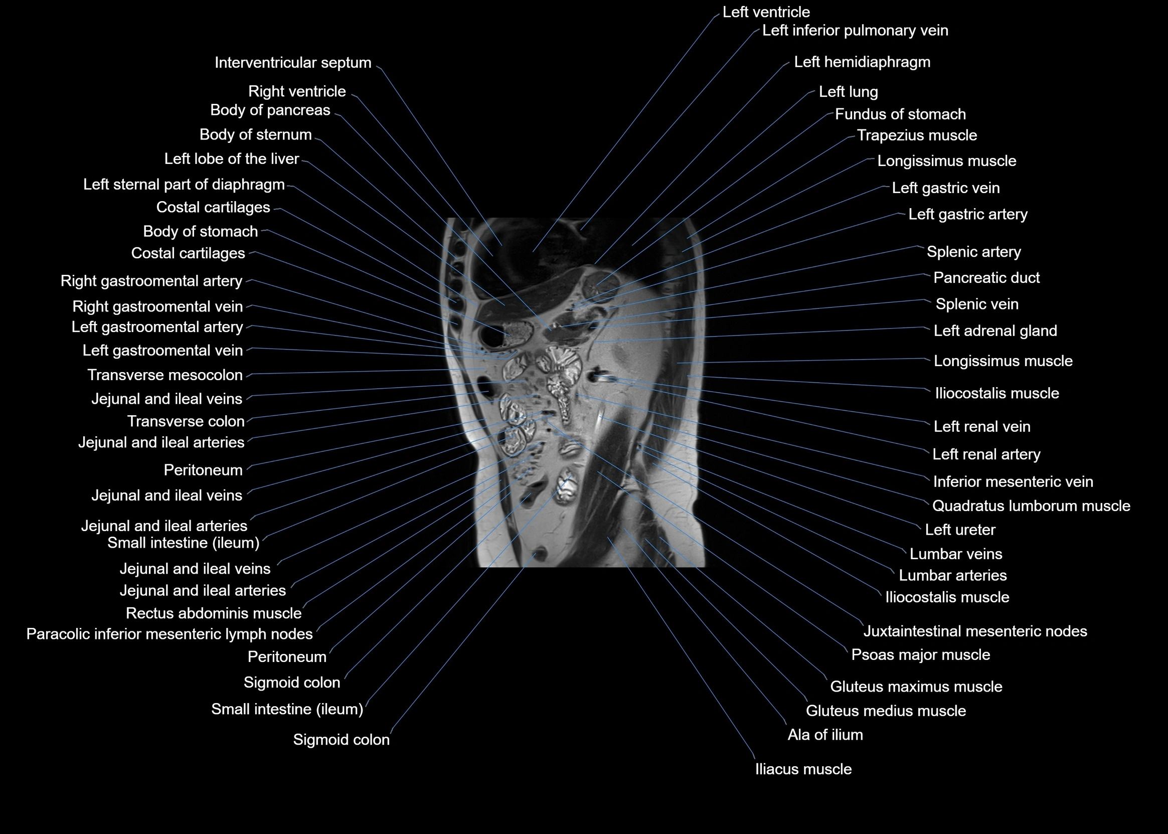
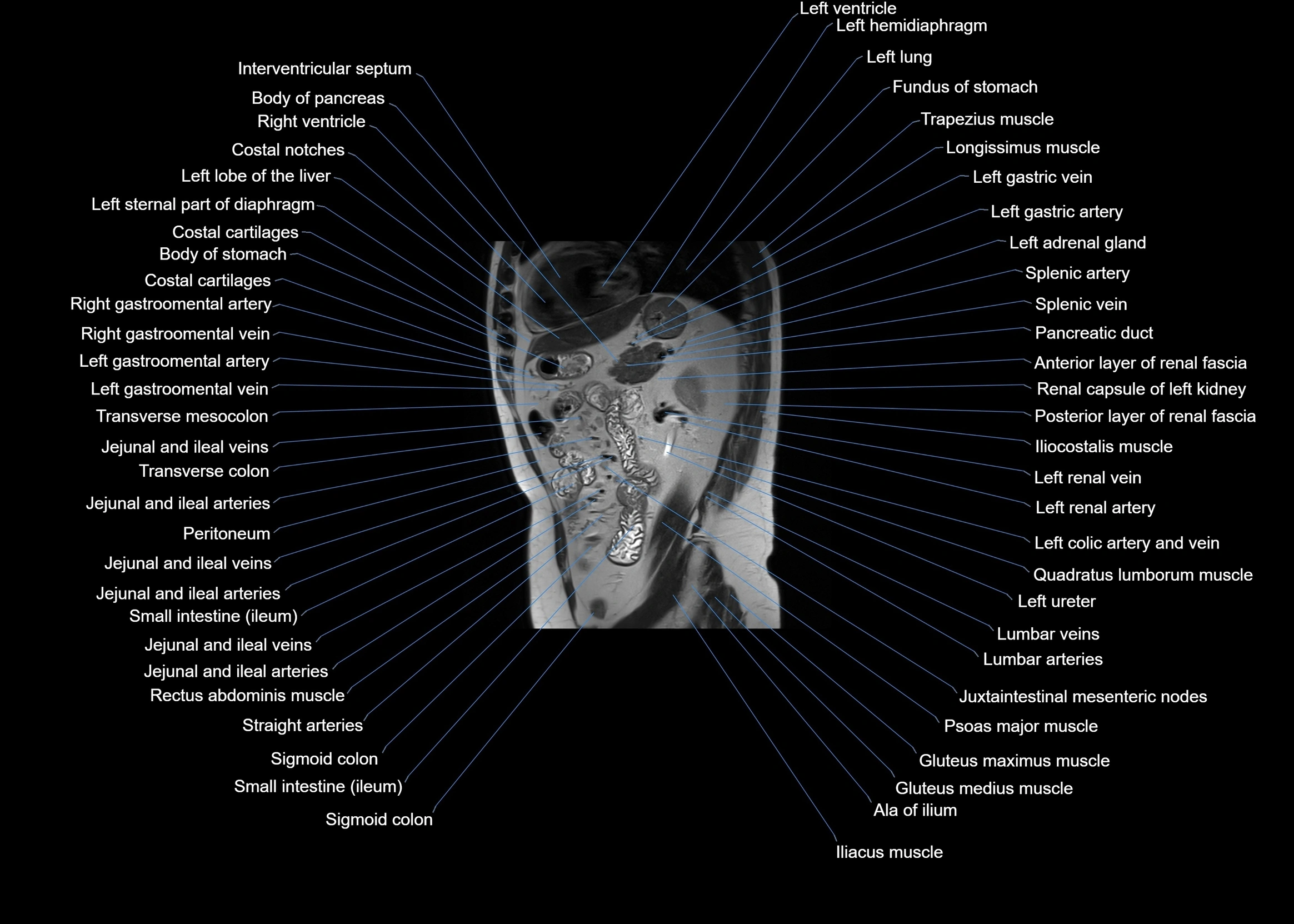
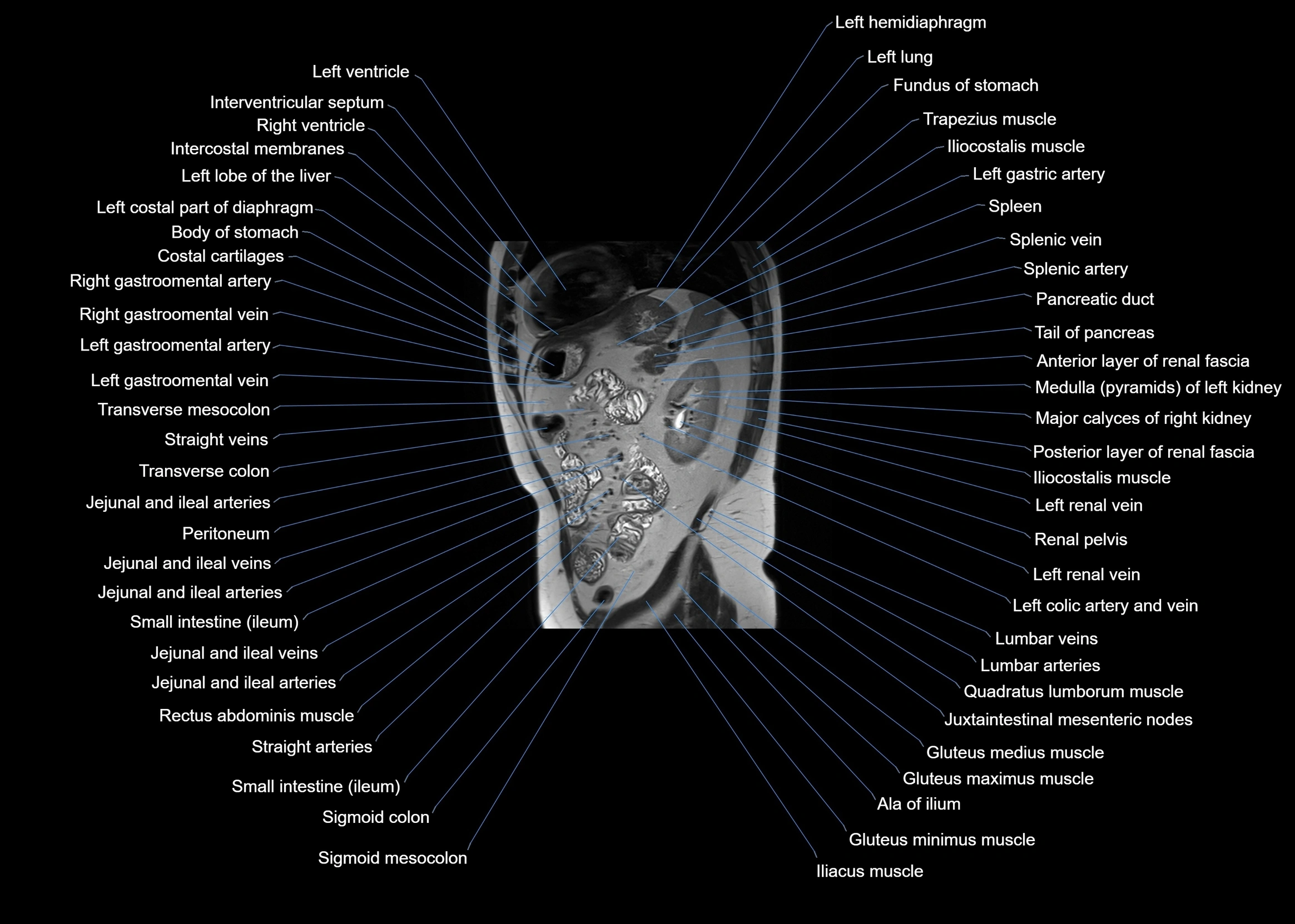
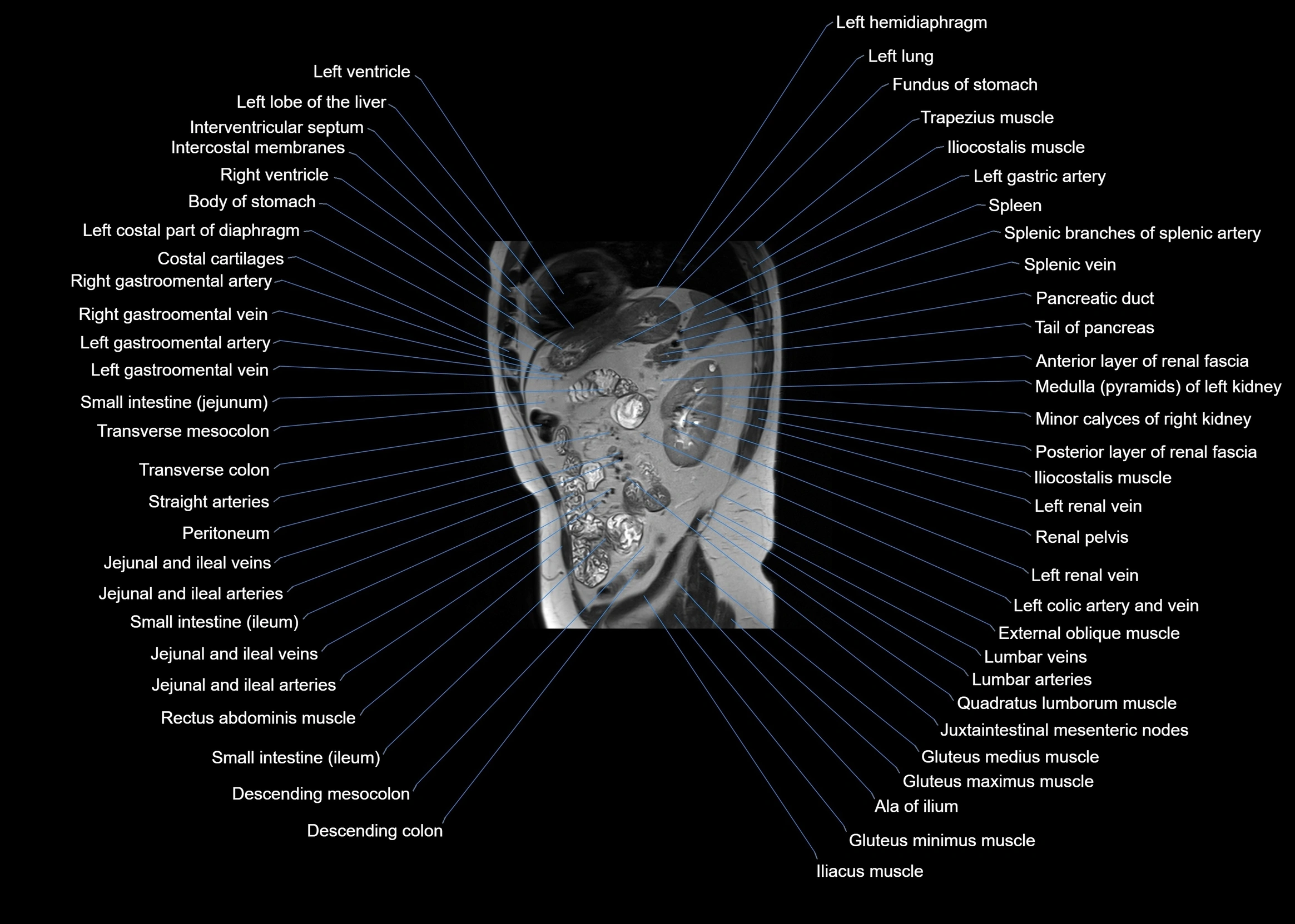
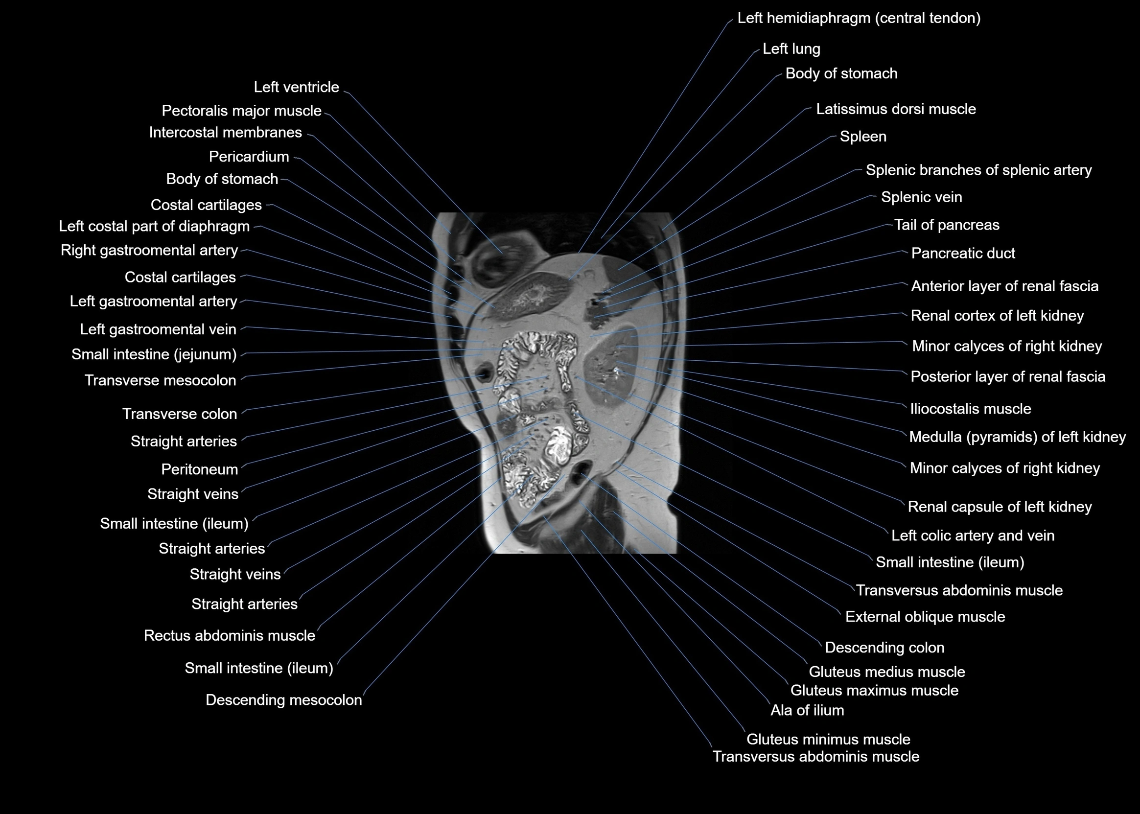
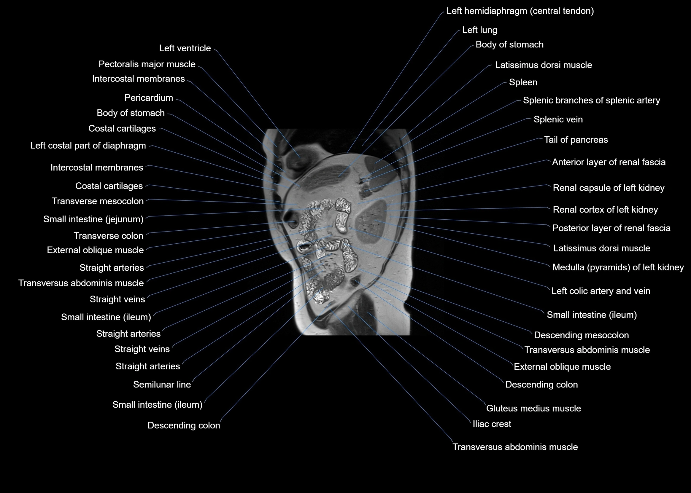
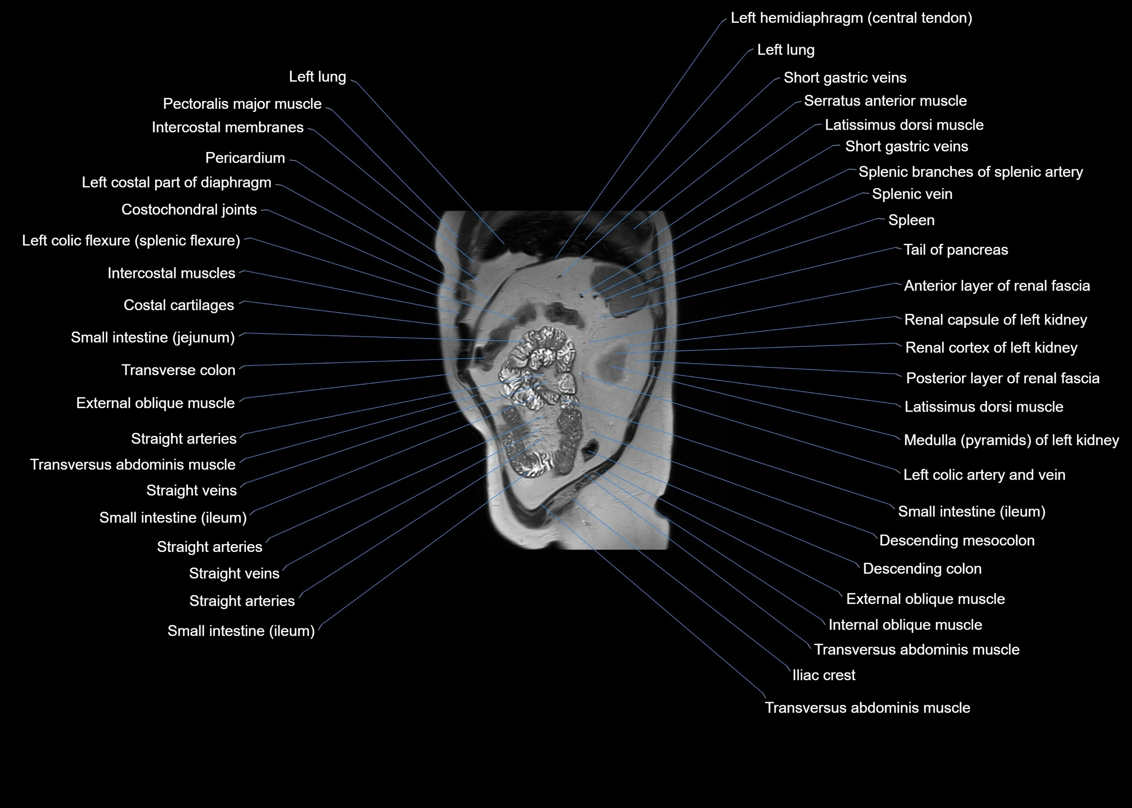
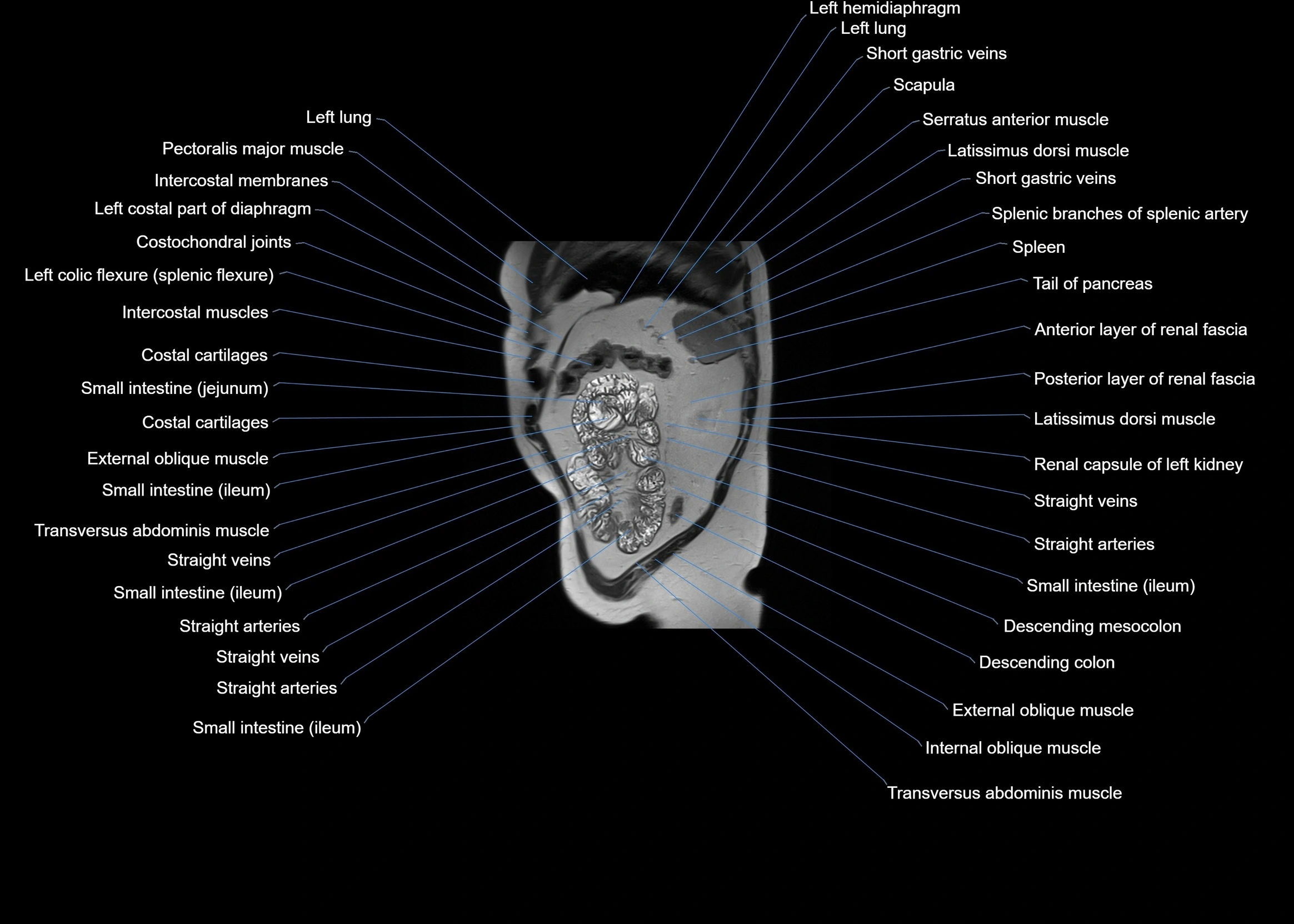
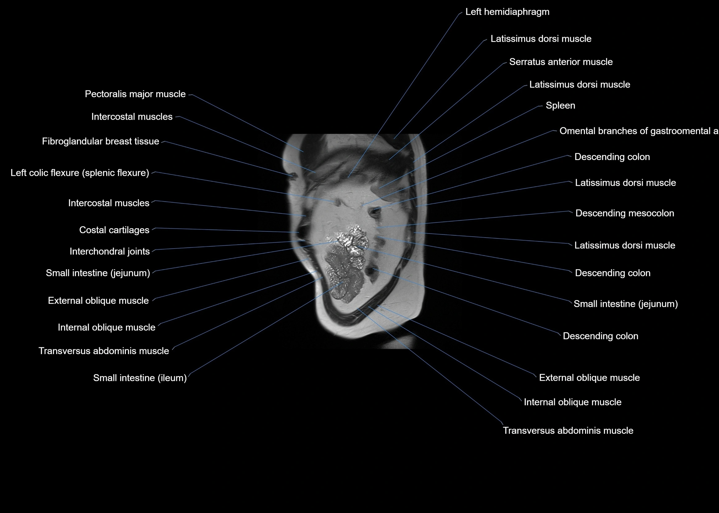
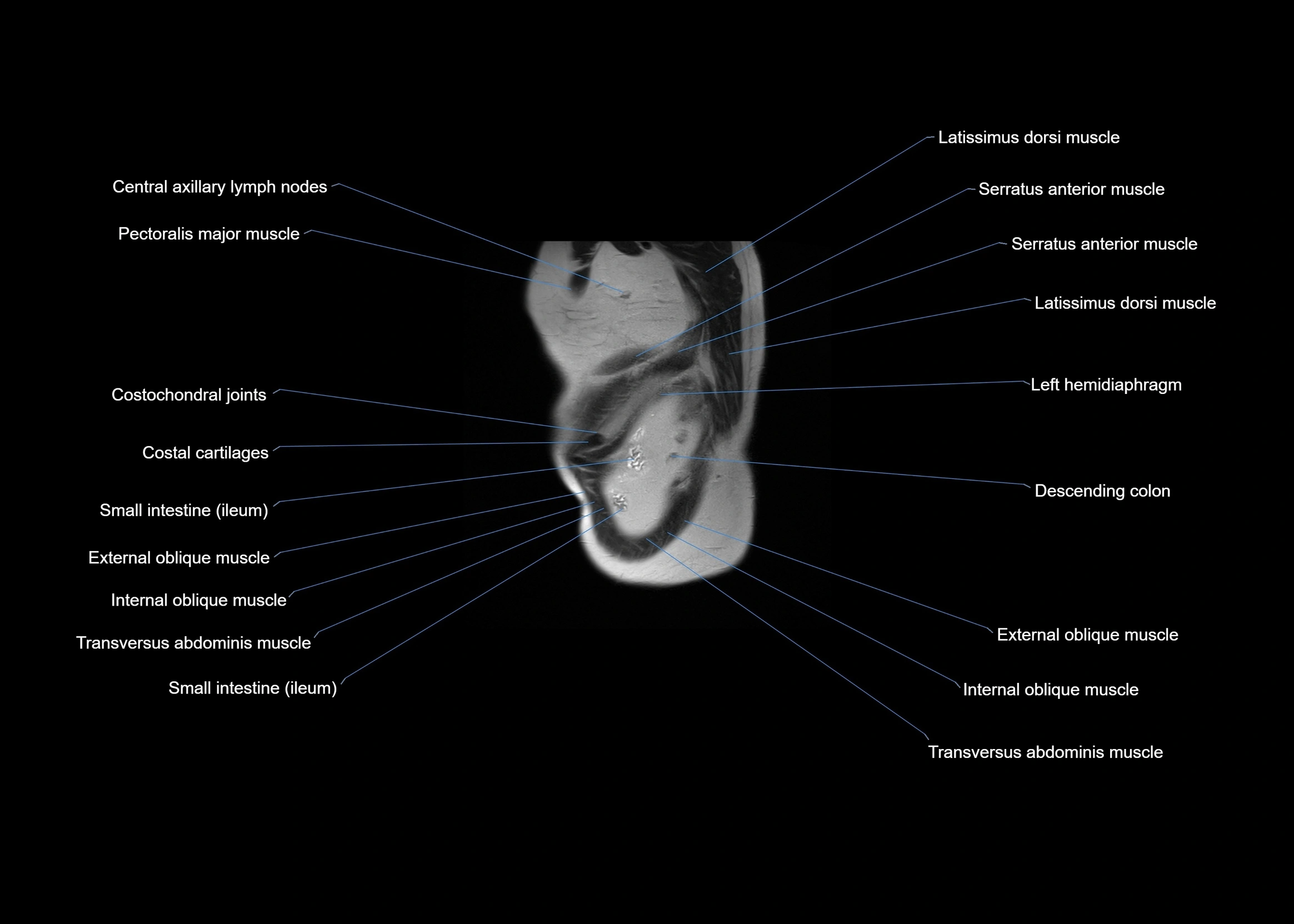
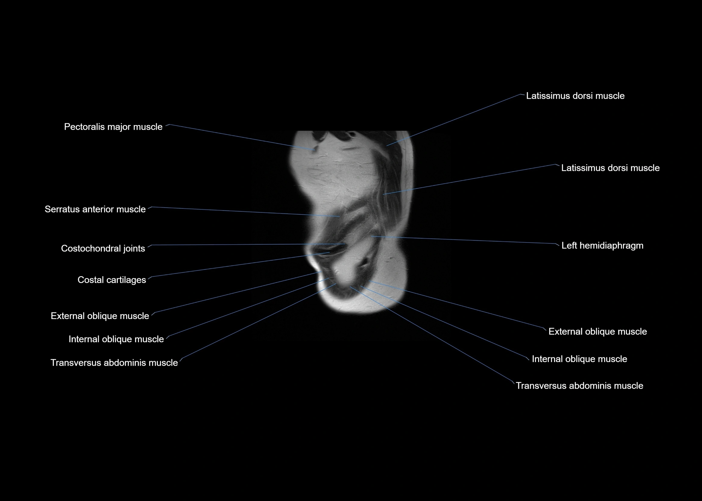
MRI images

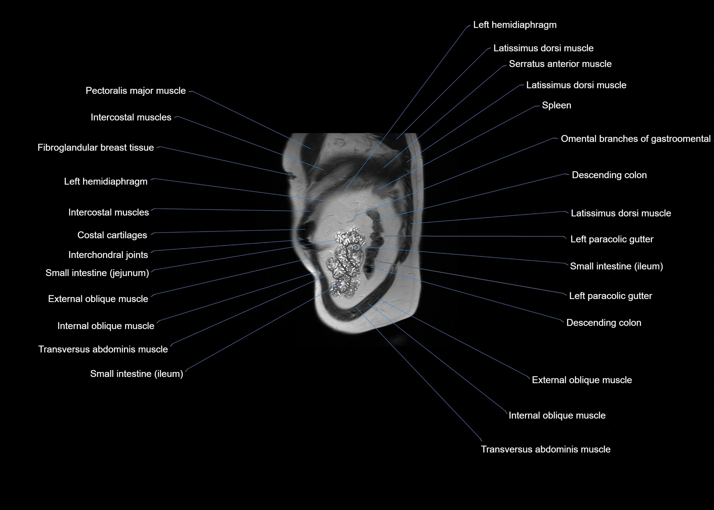
MRI images

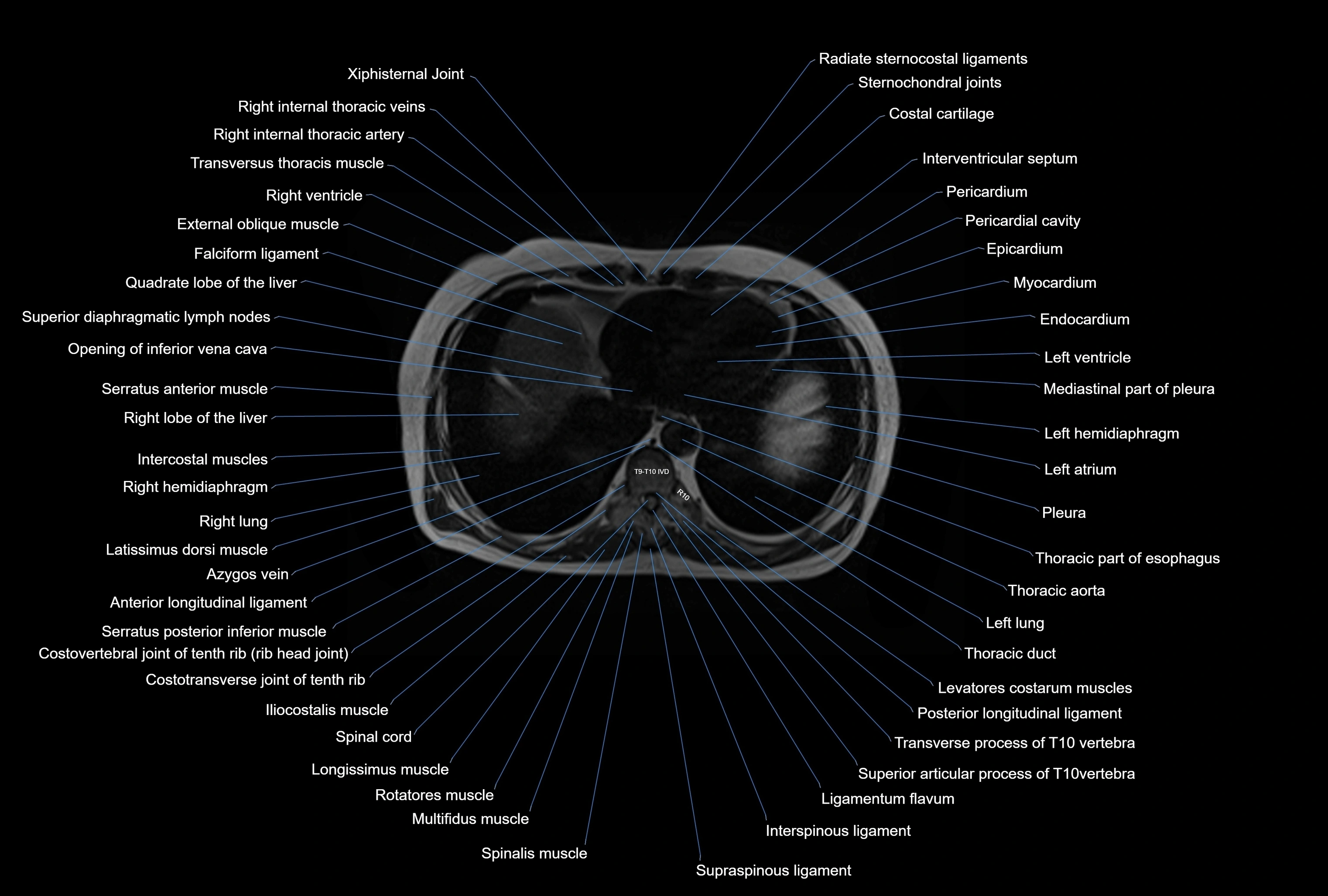
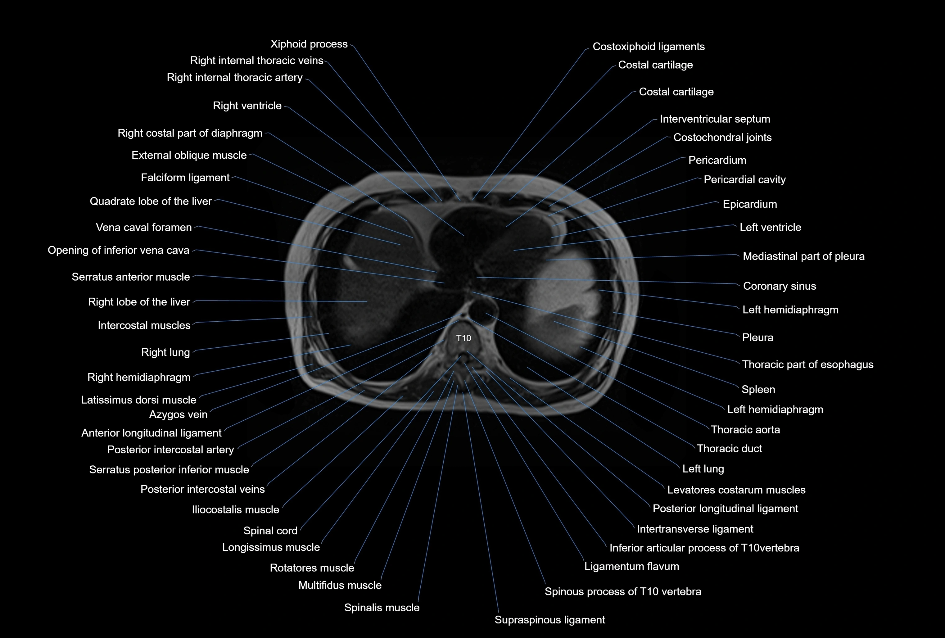
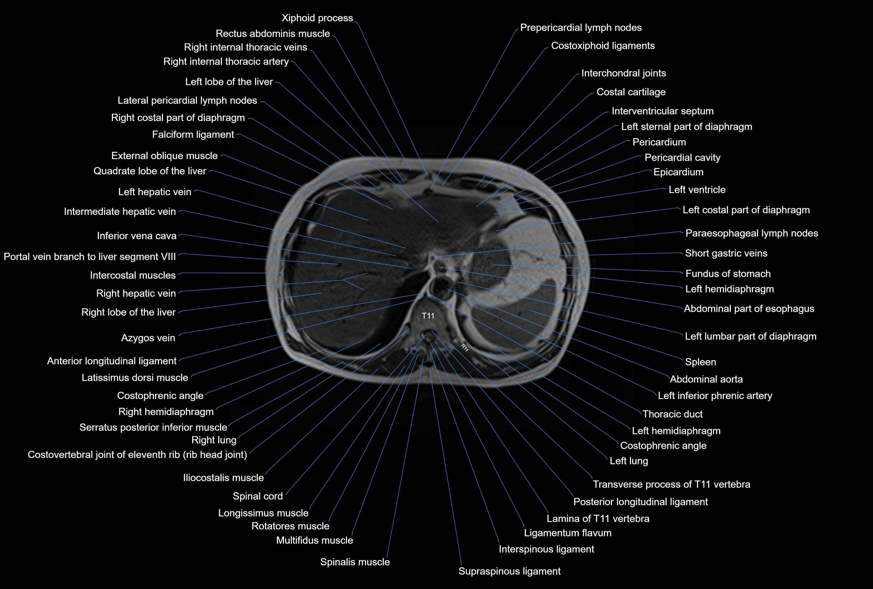
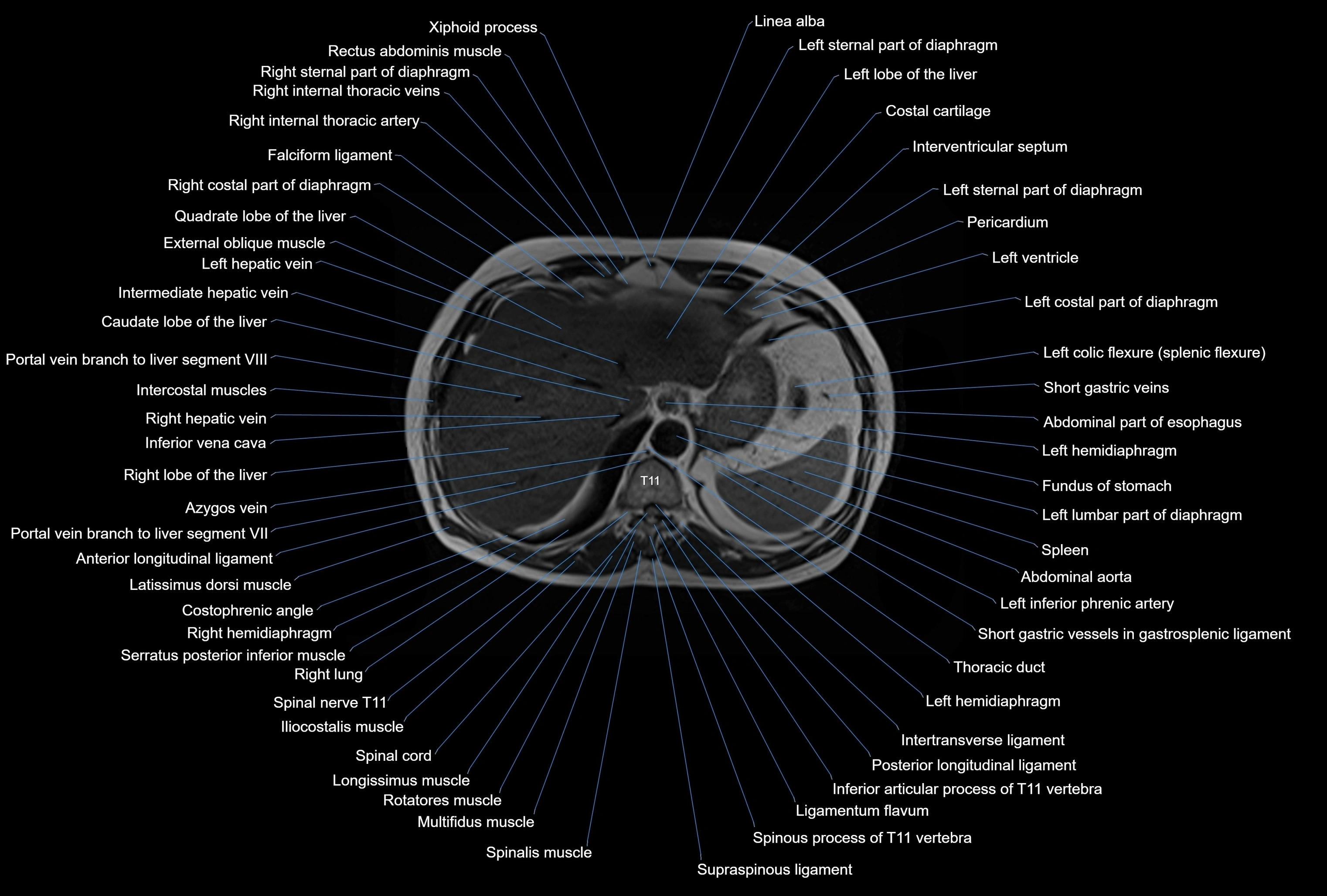
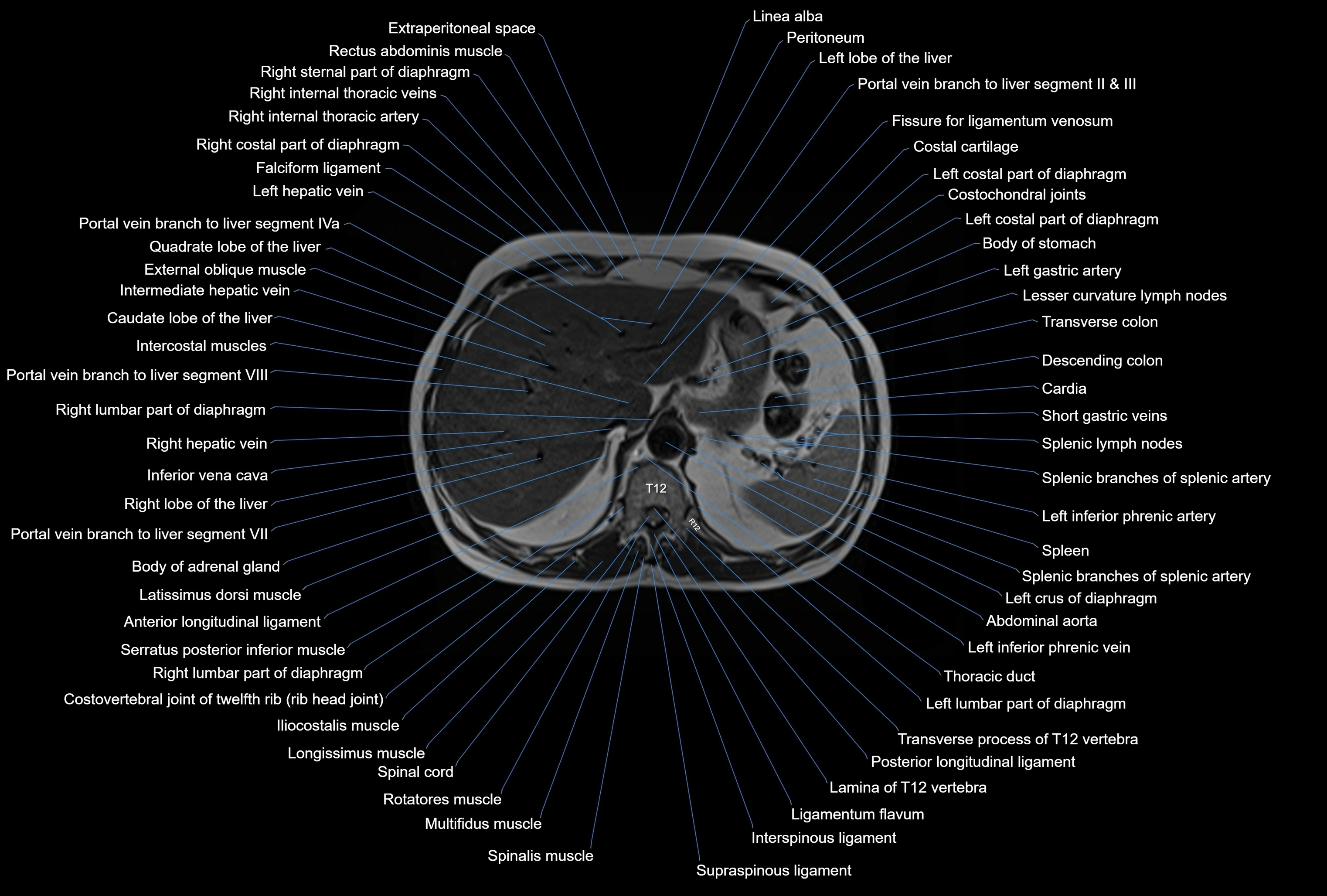
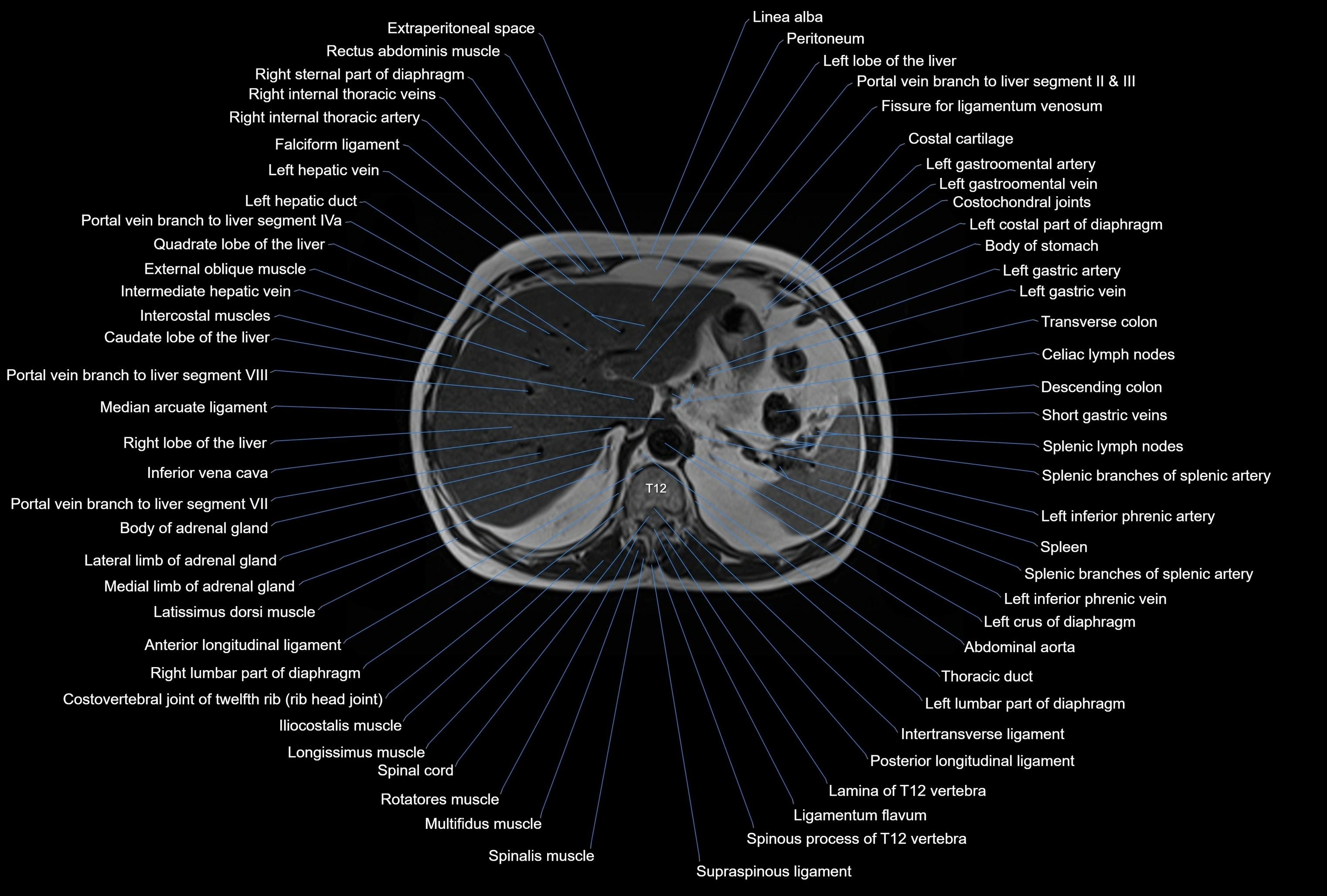
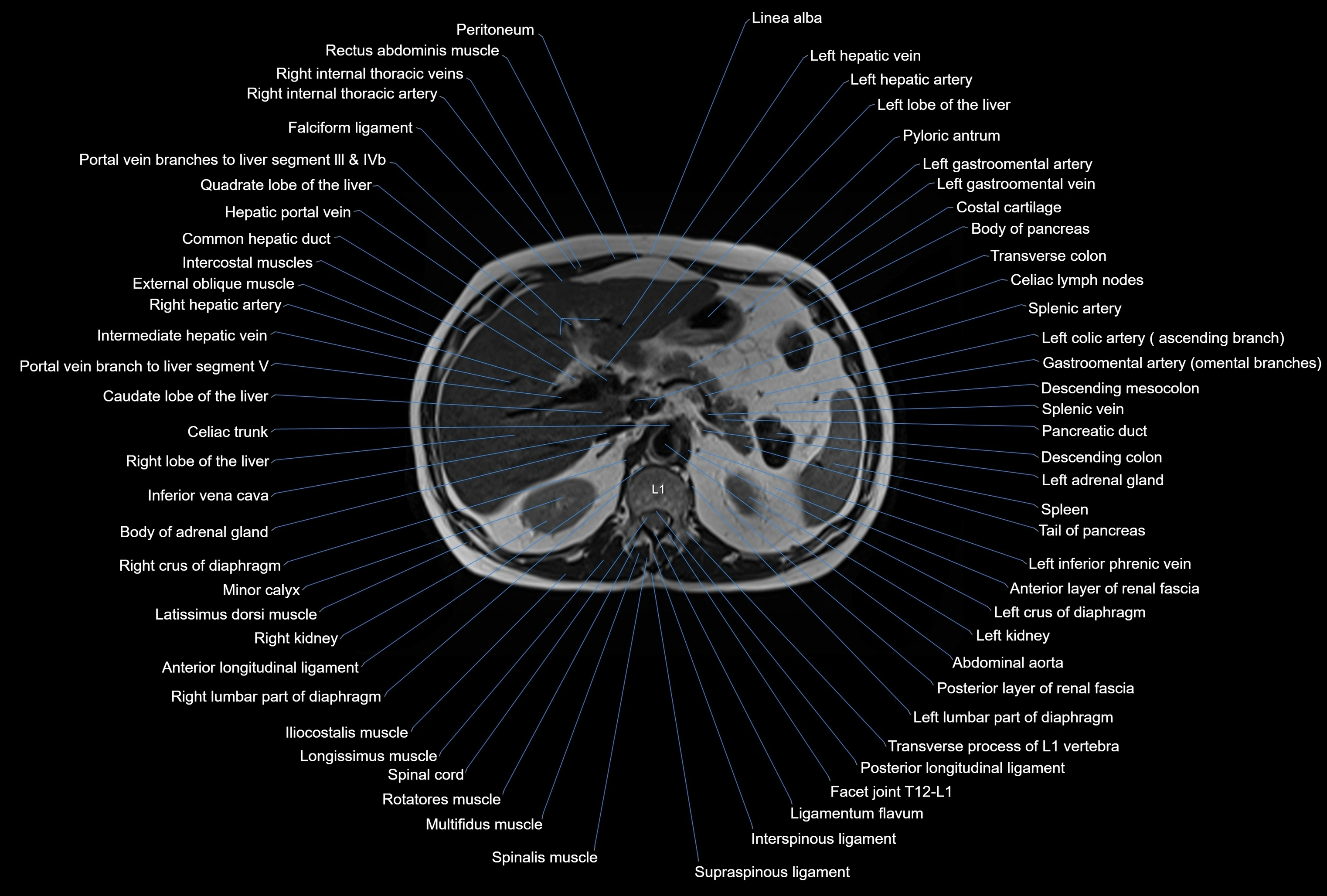
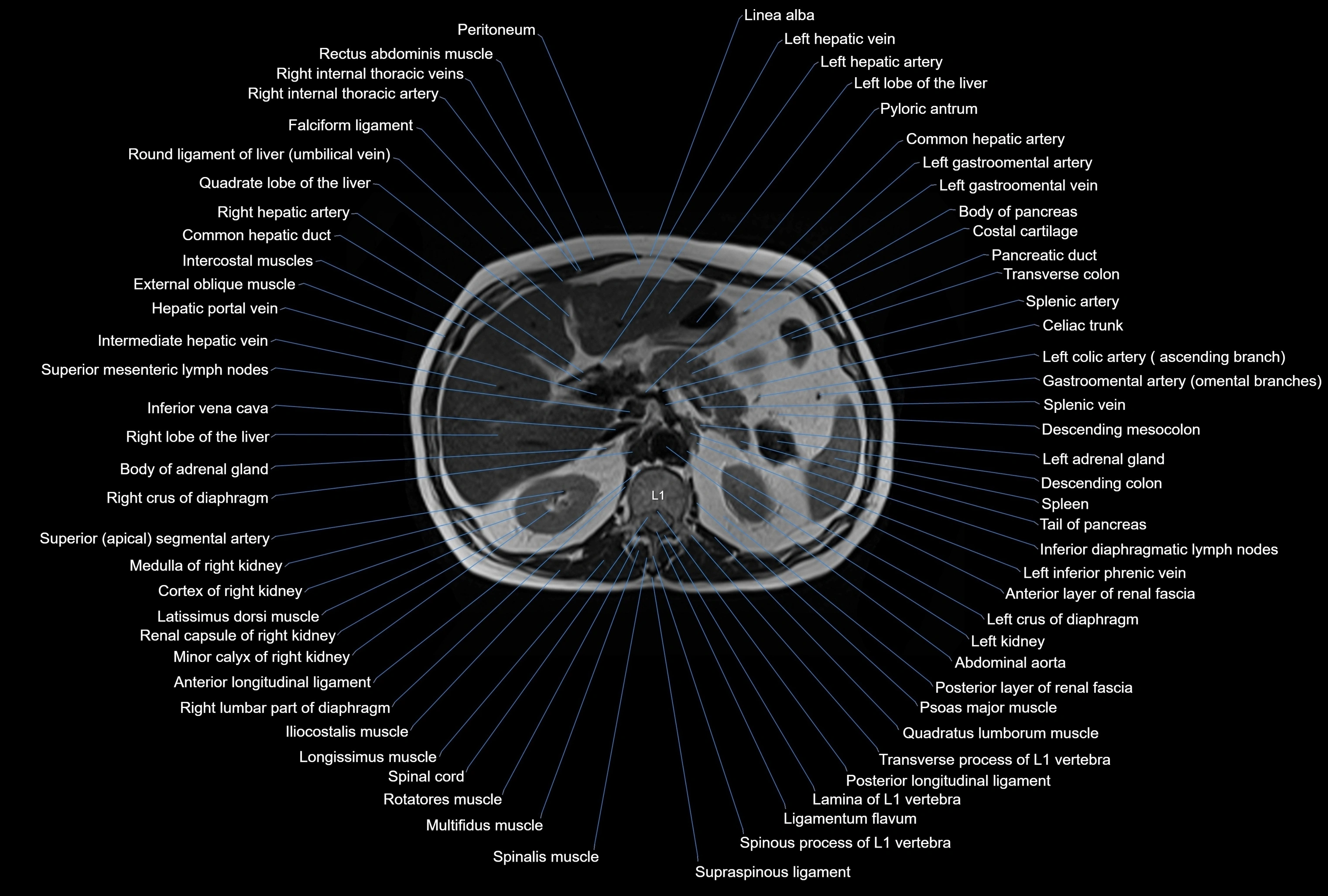
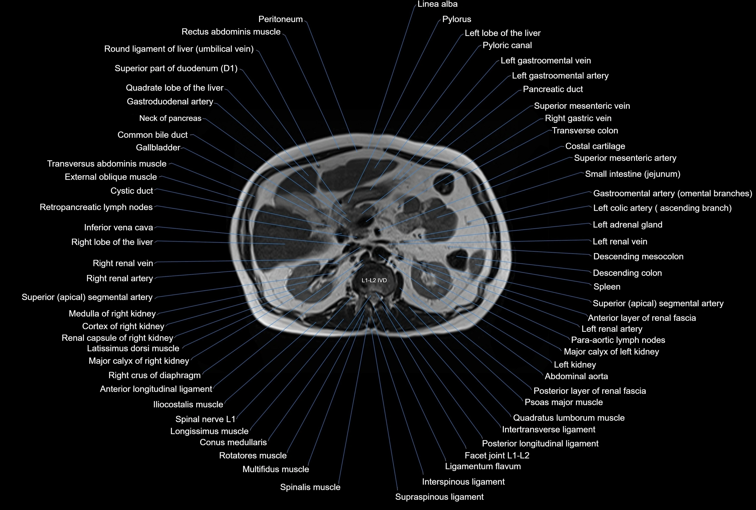
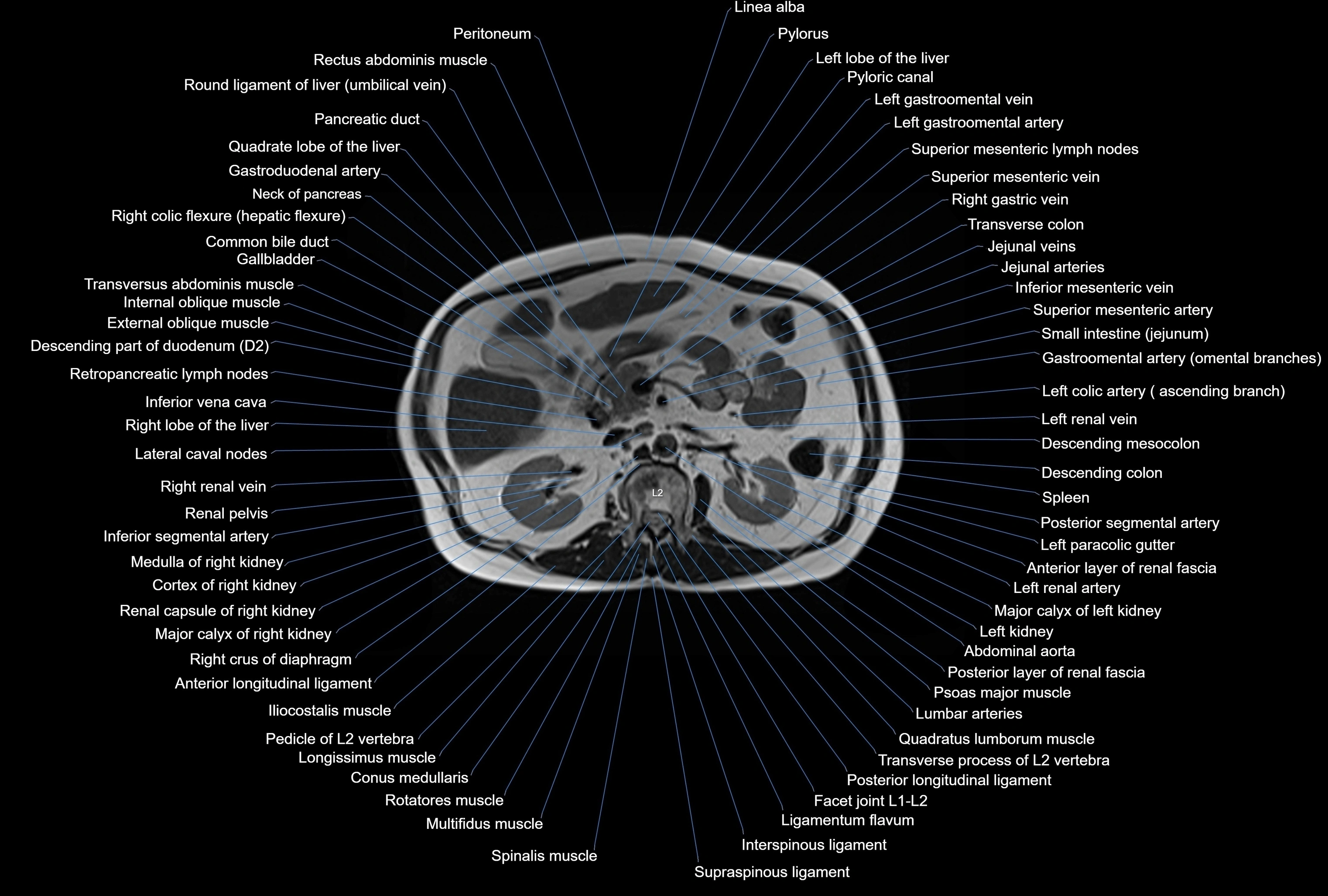
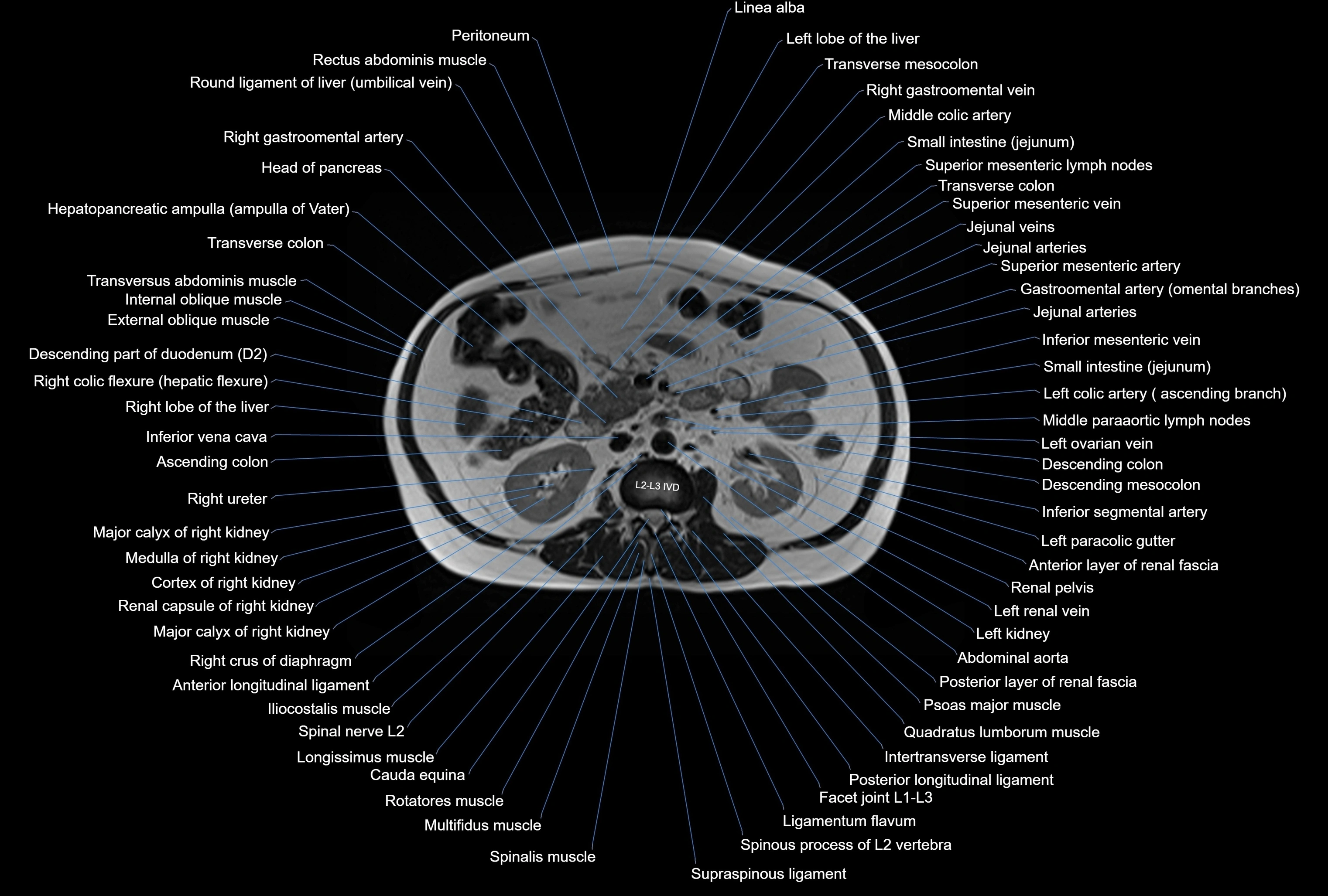
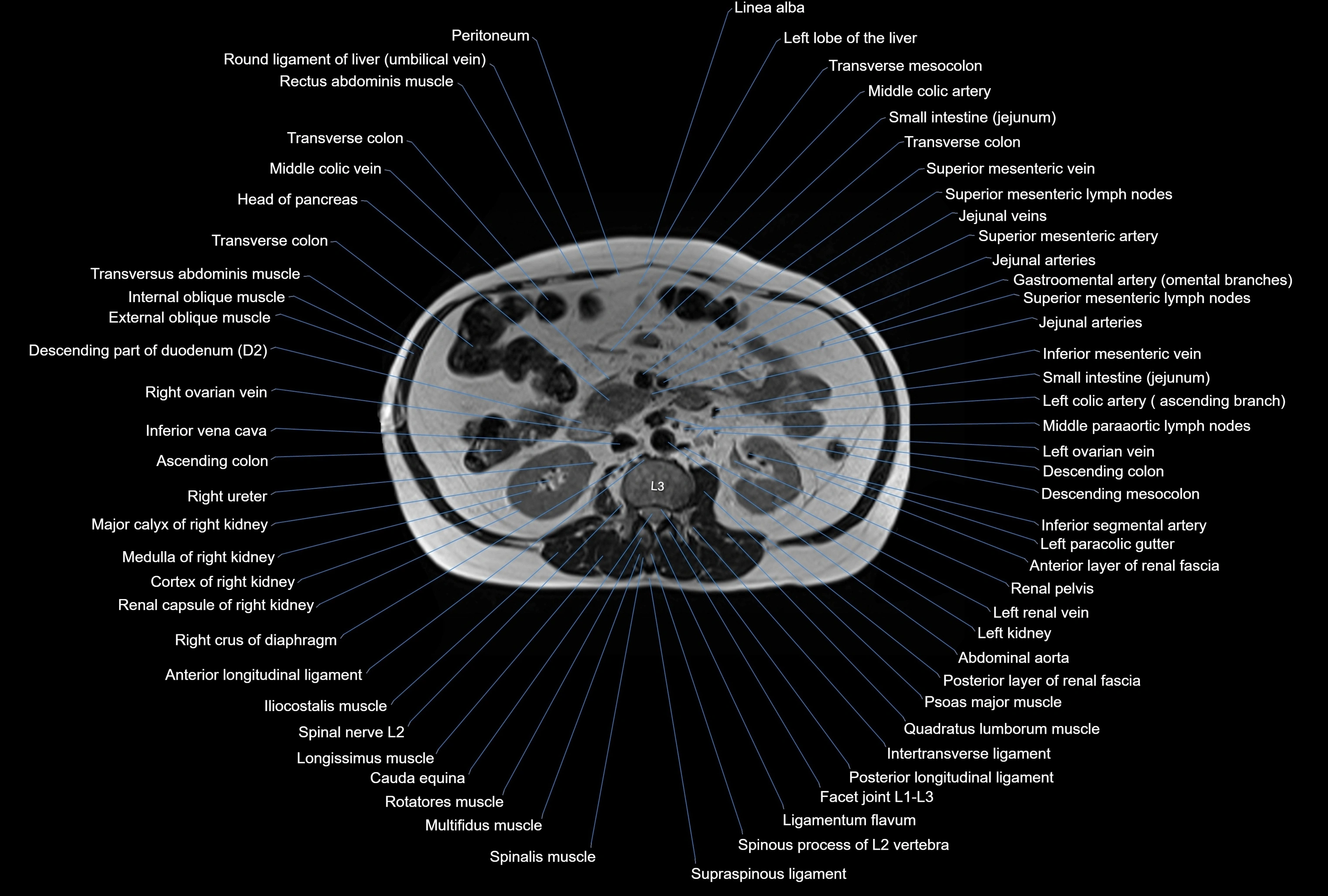
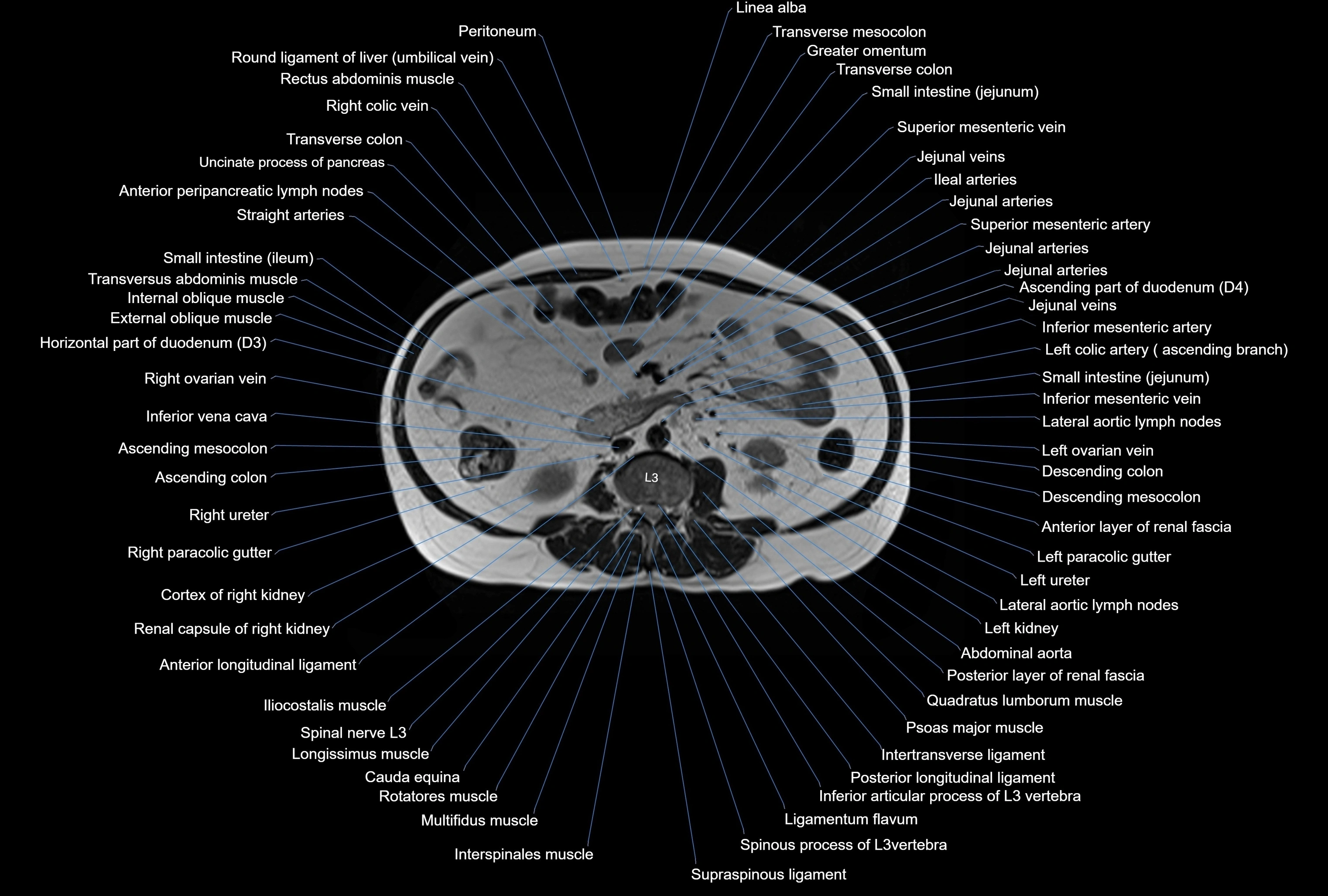
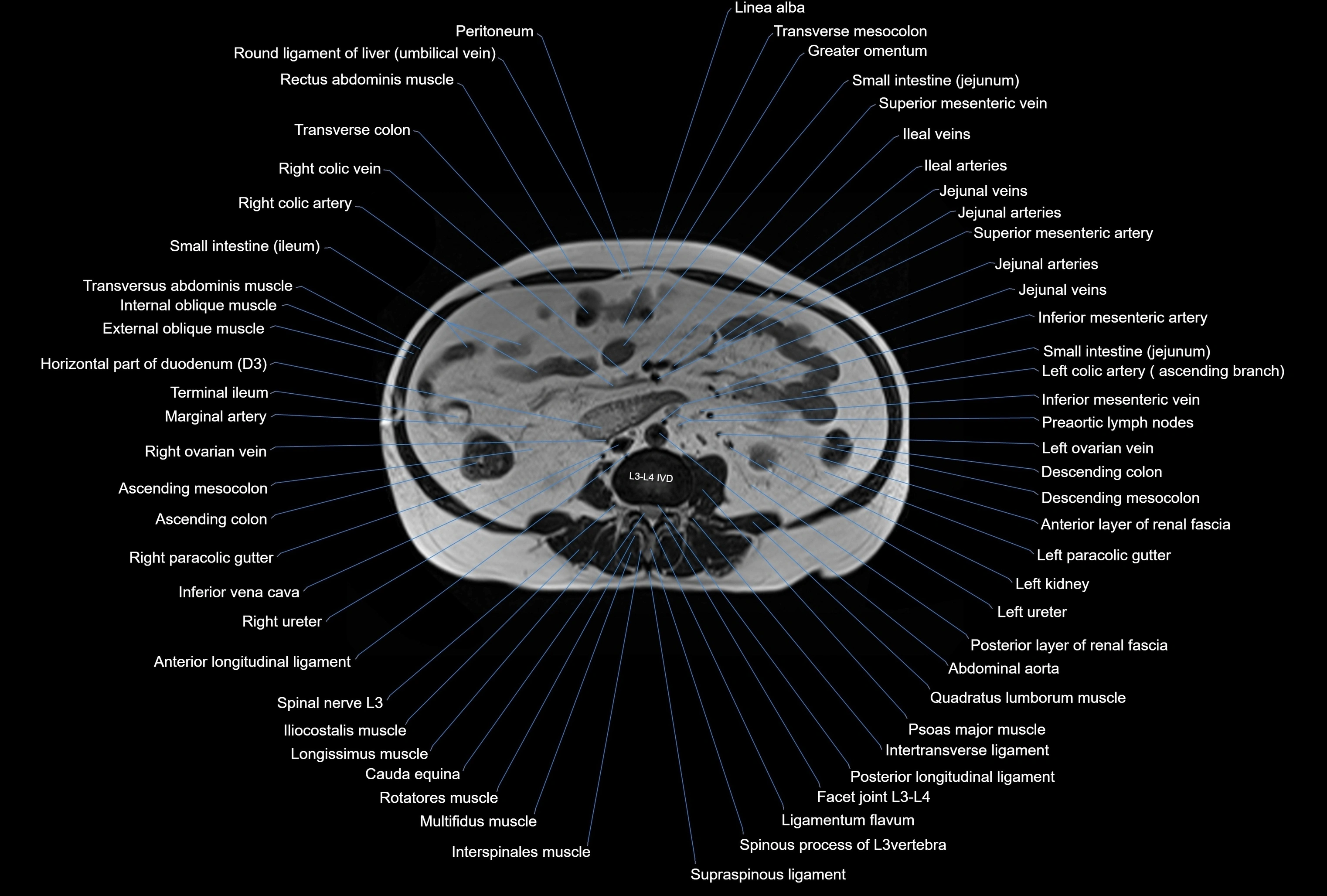
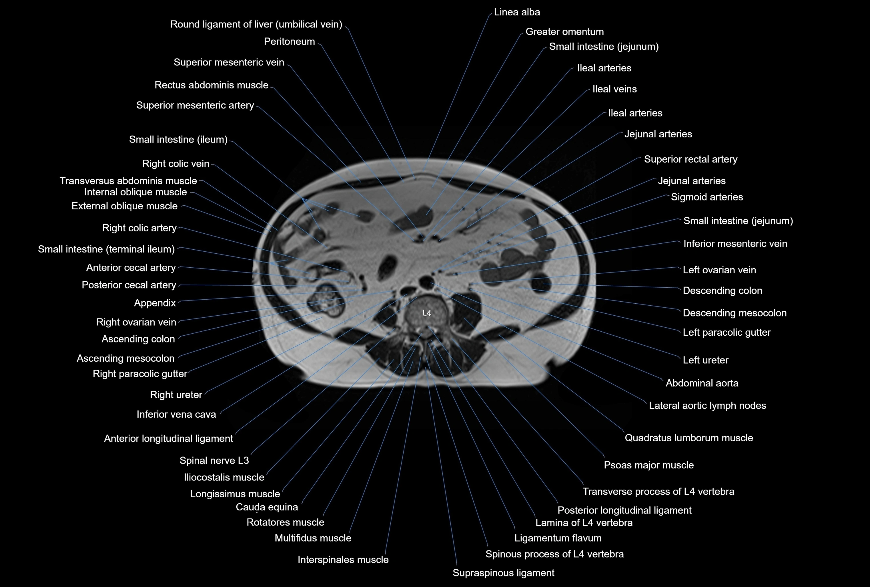
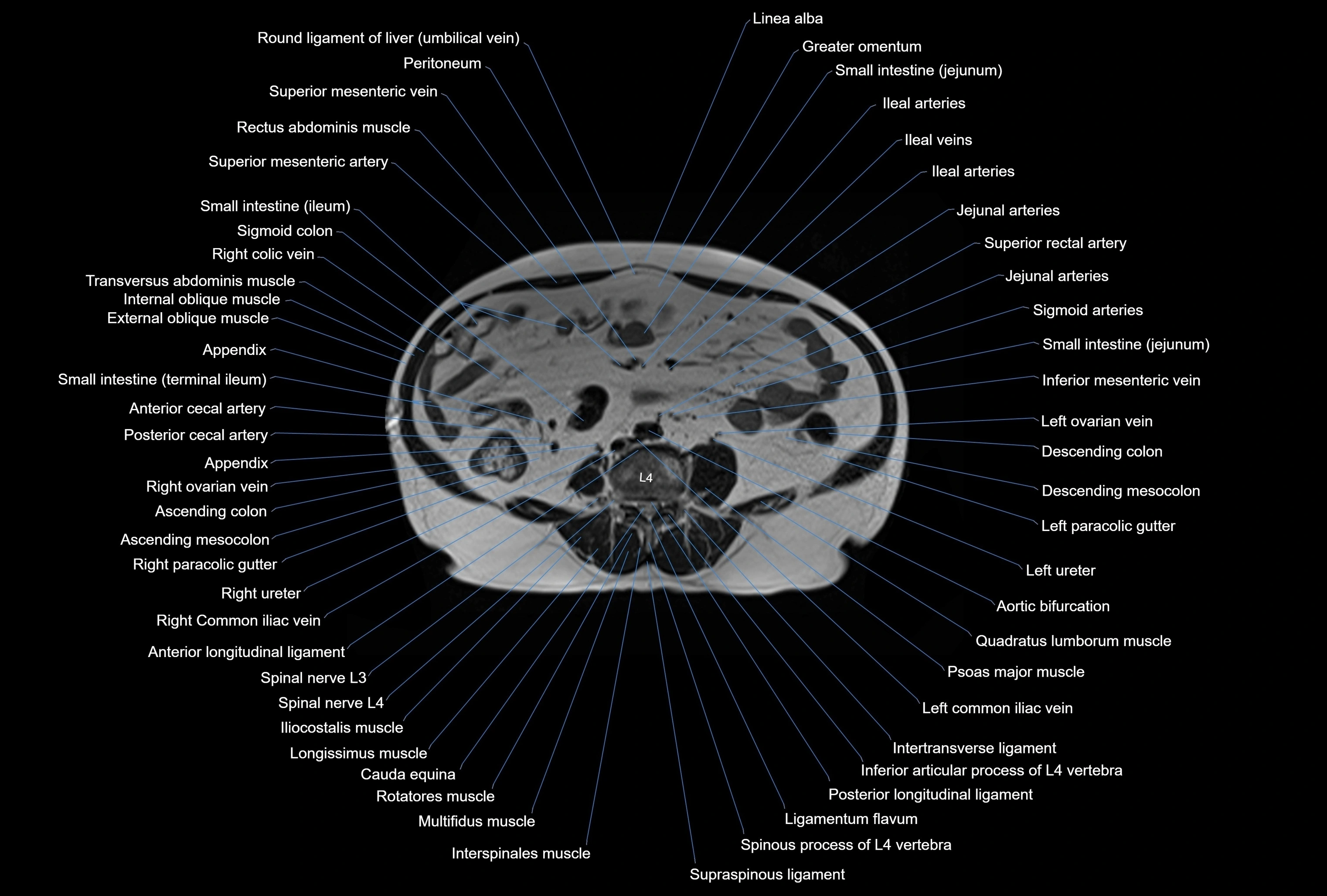
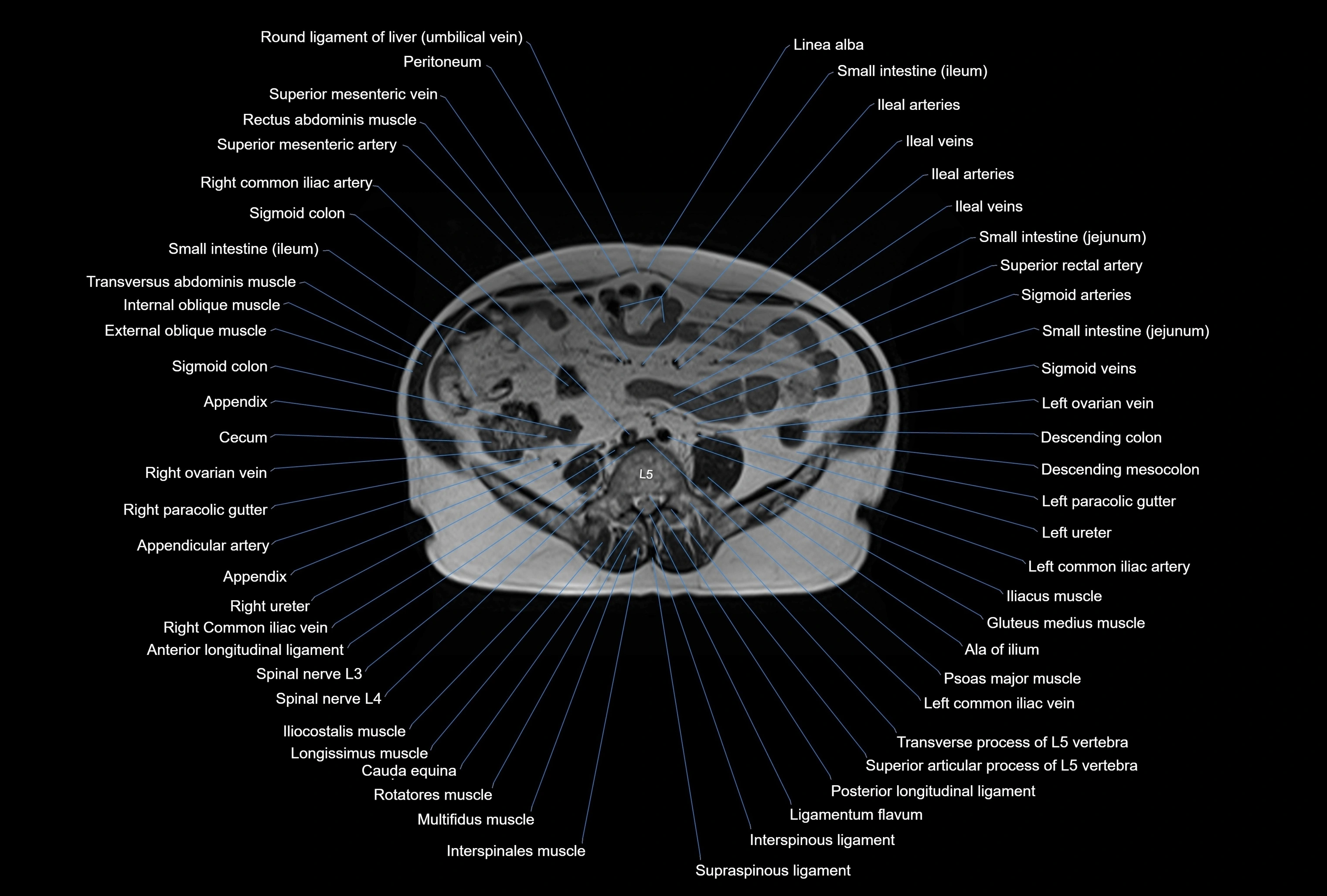
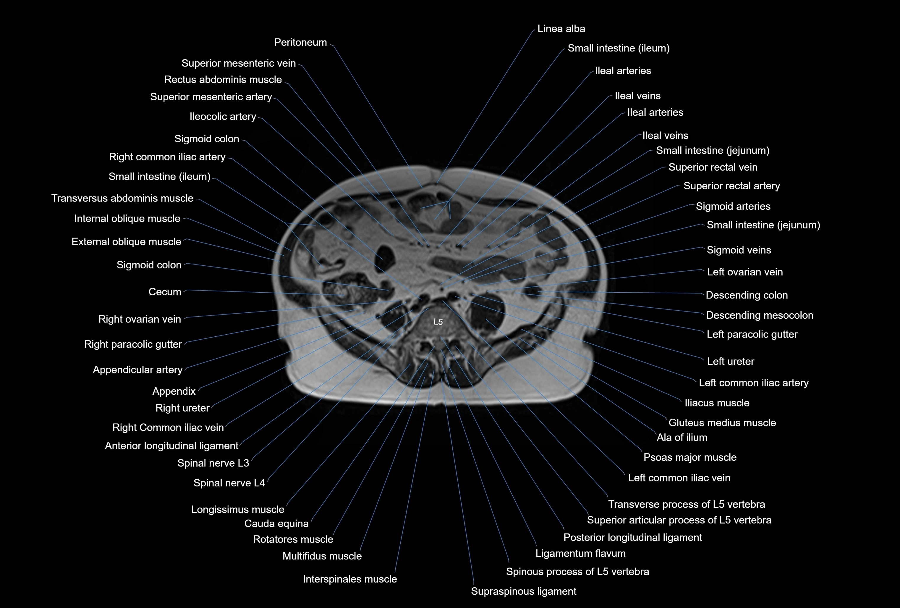
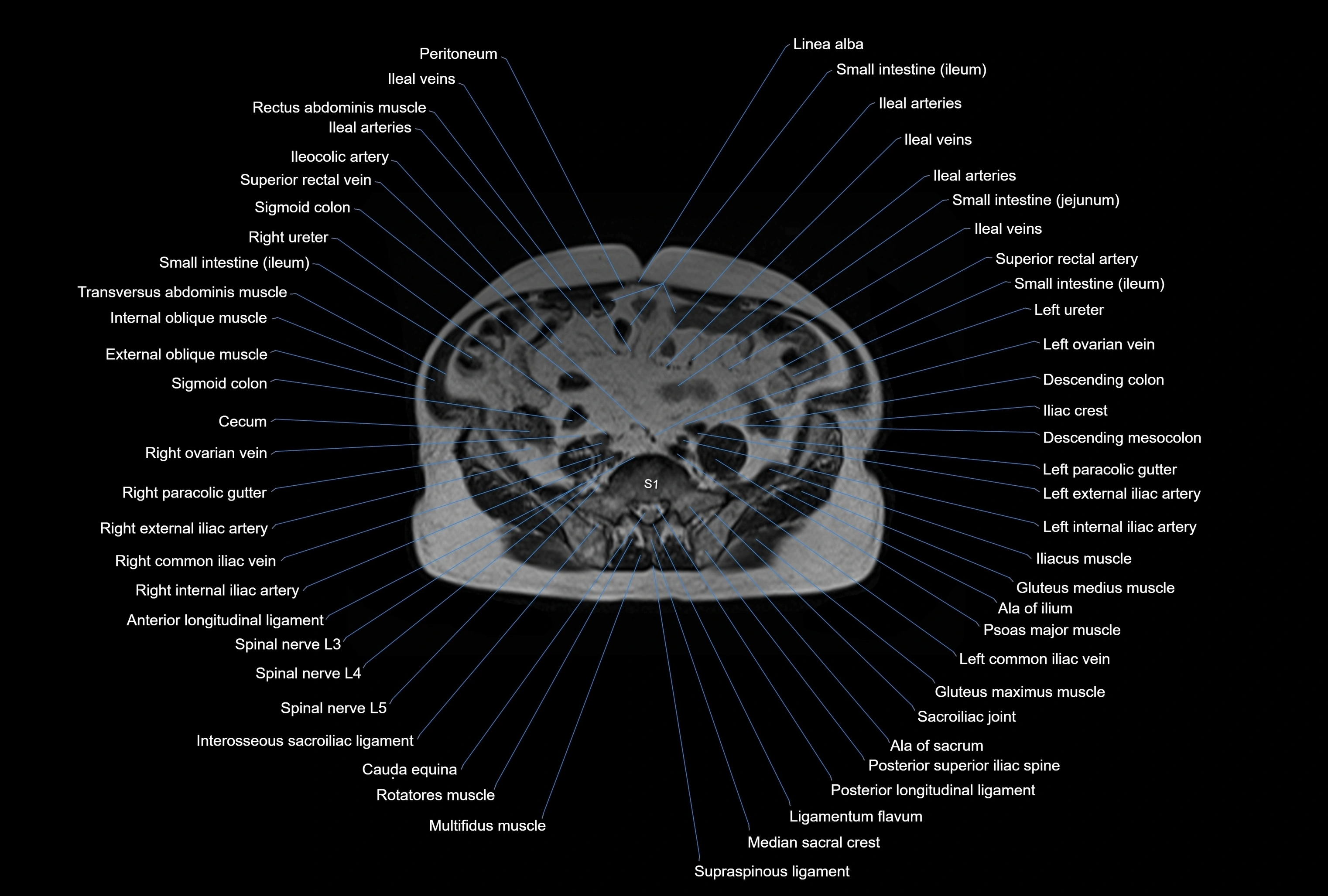
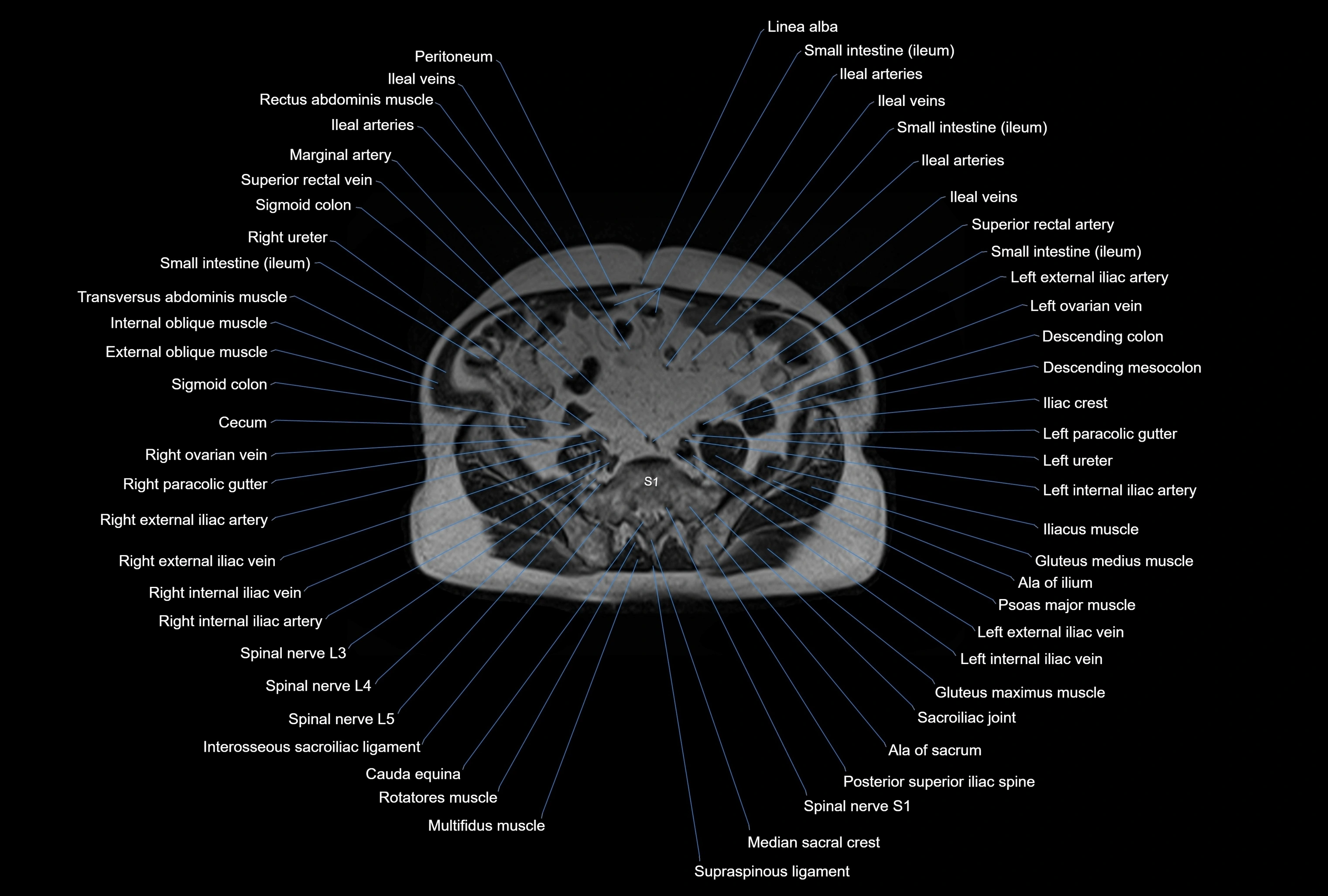
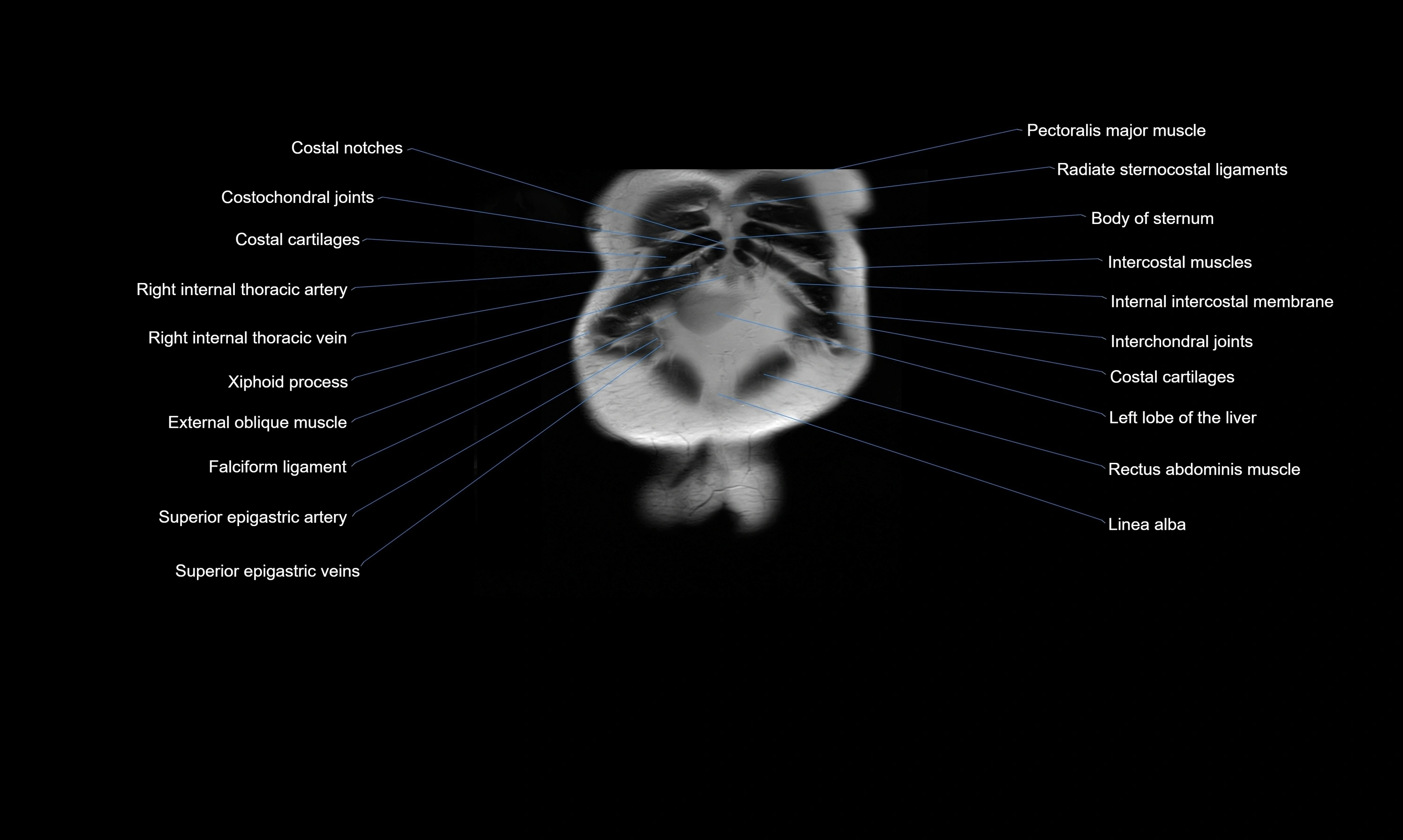
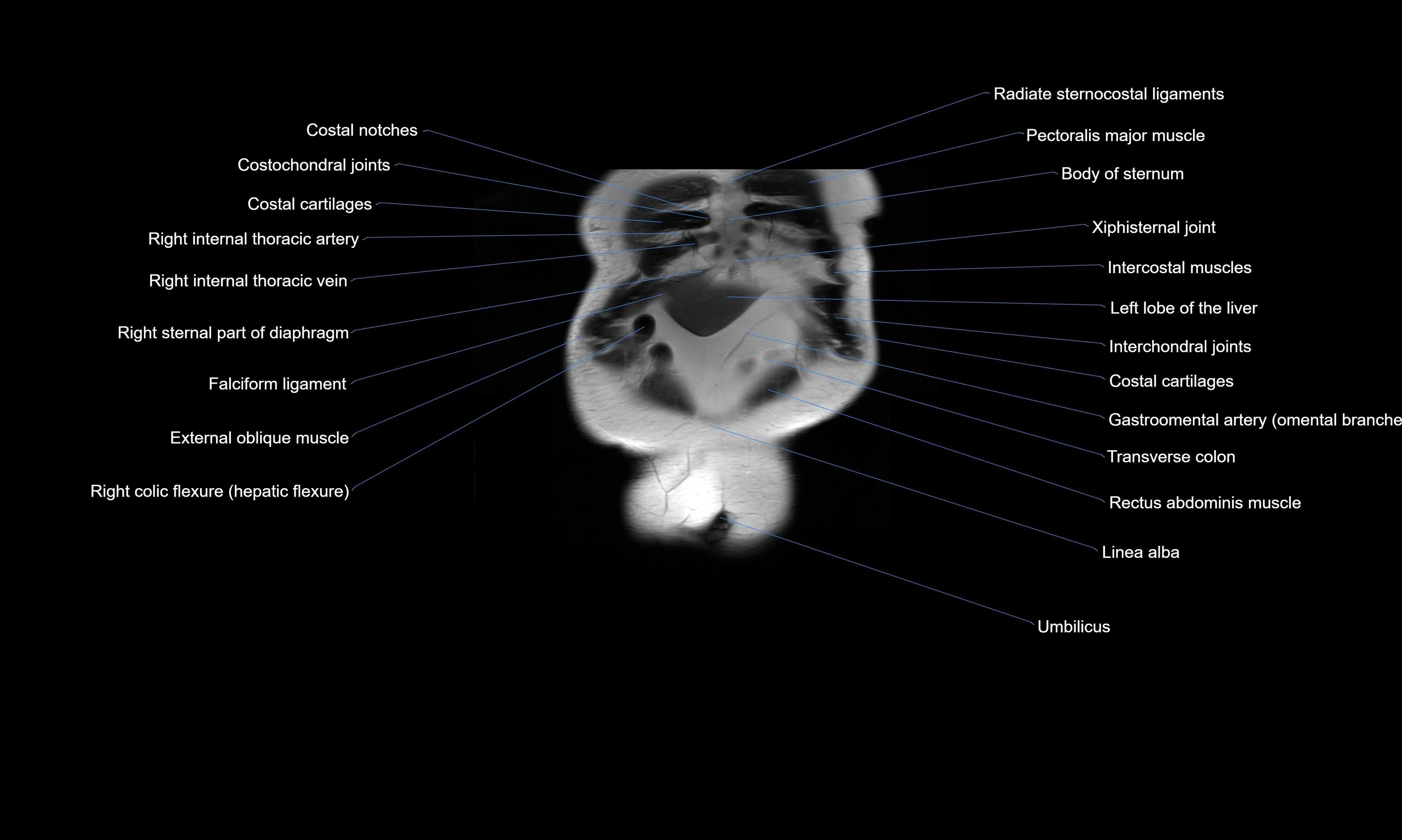
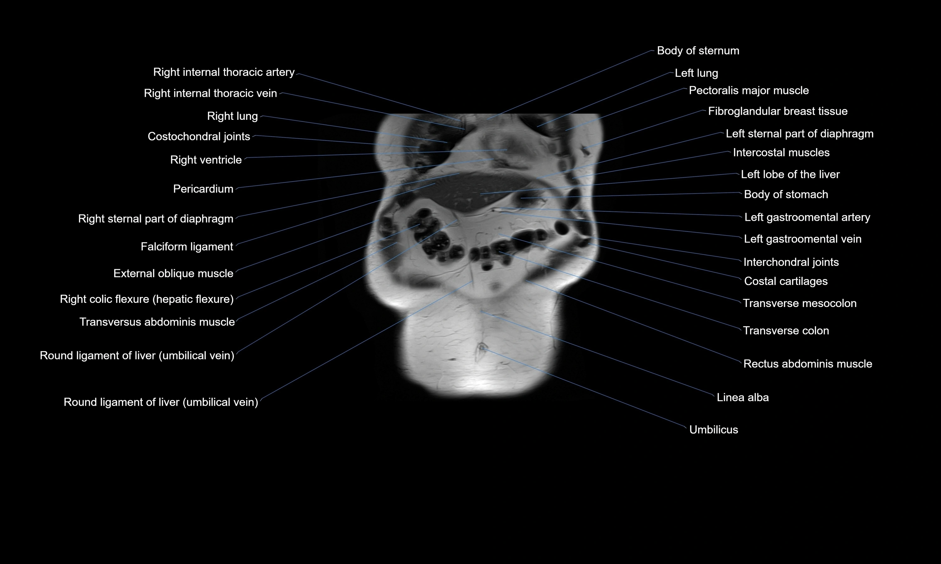
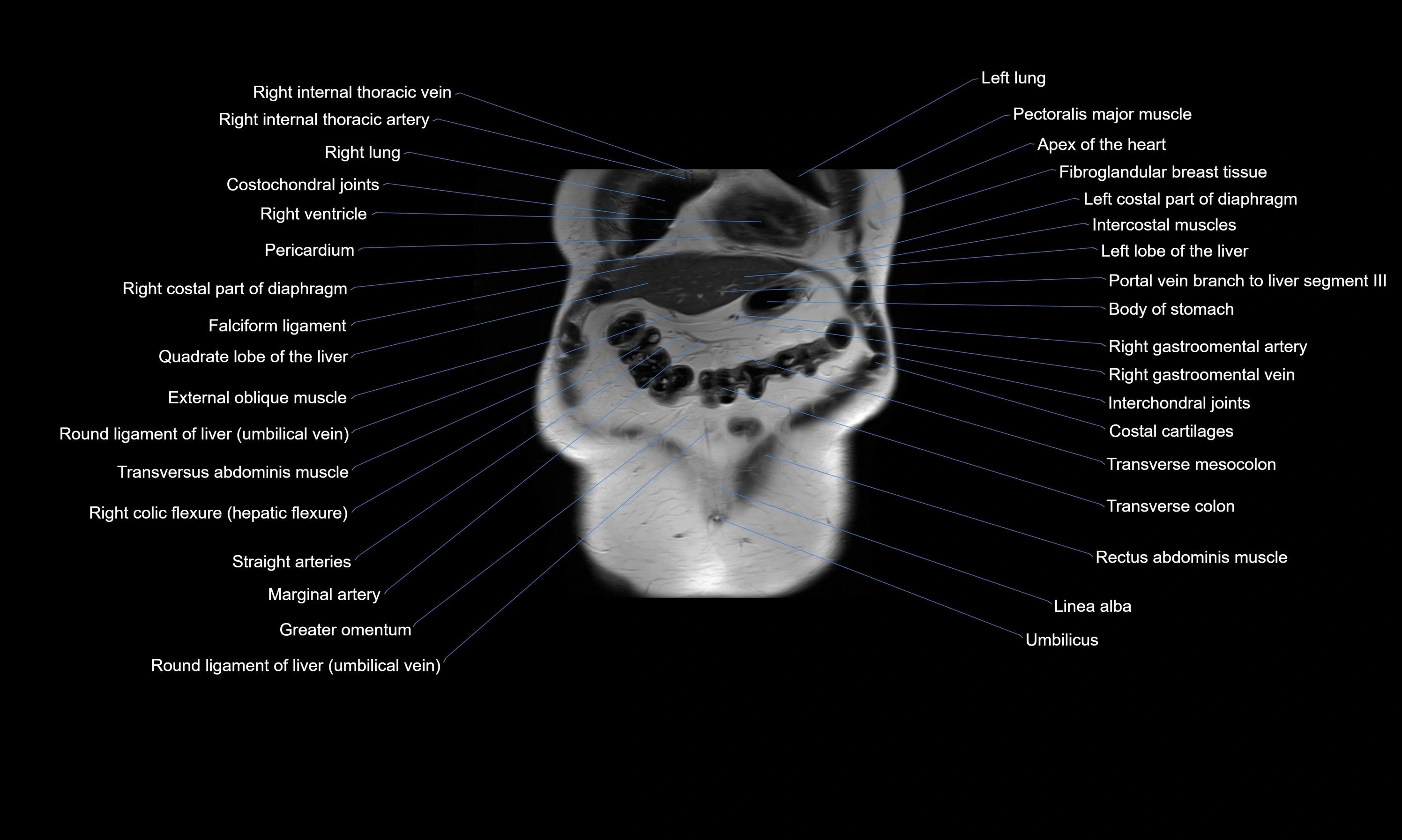
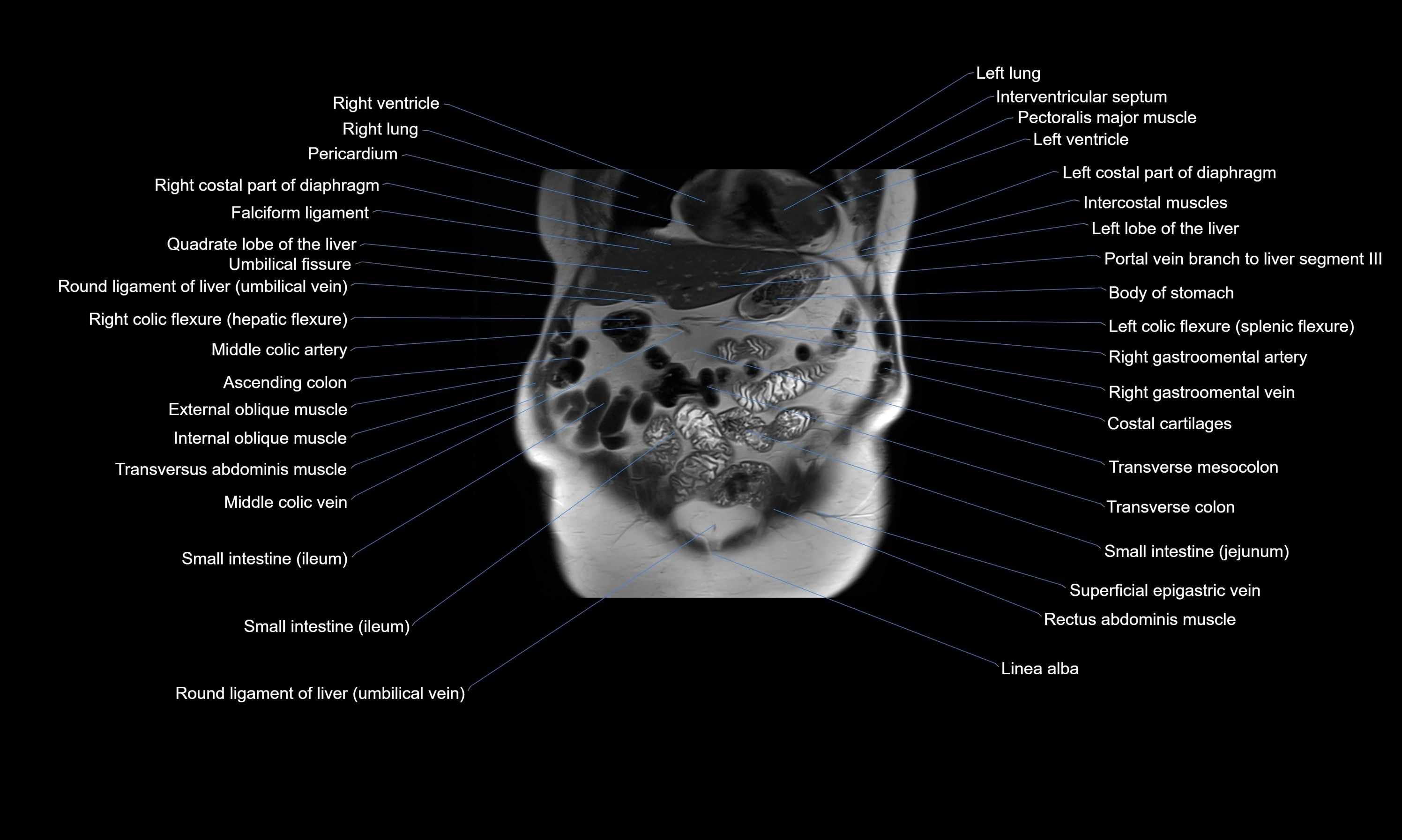
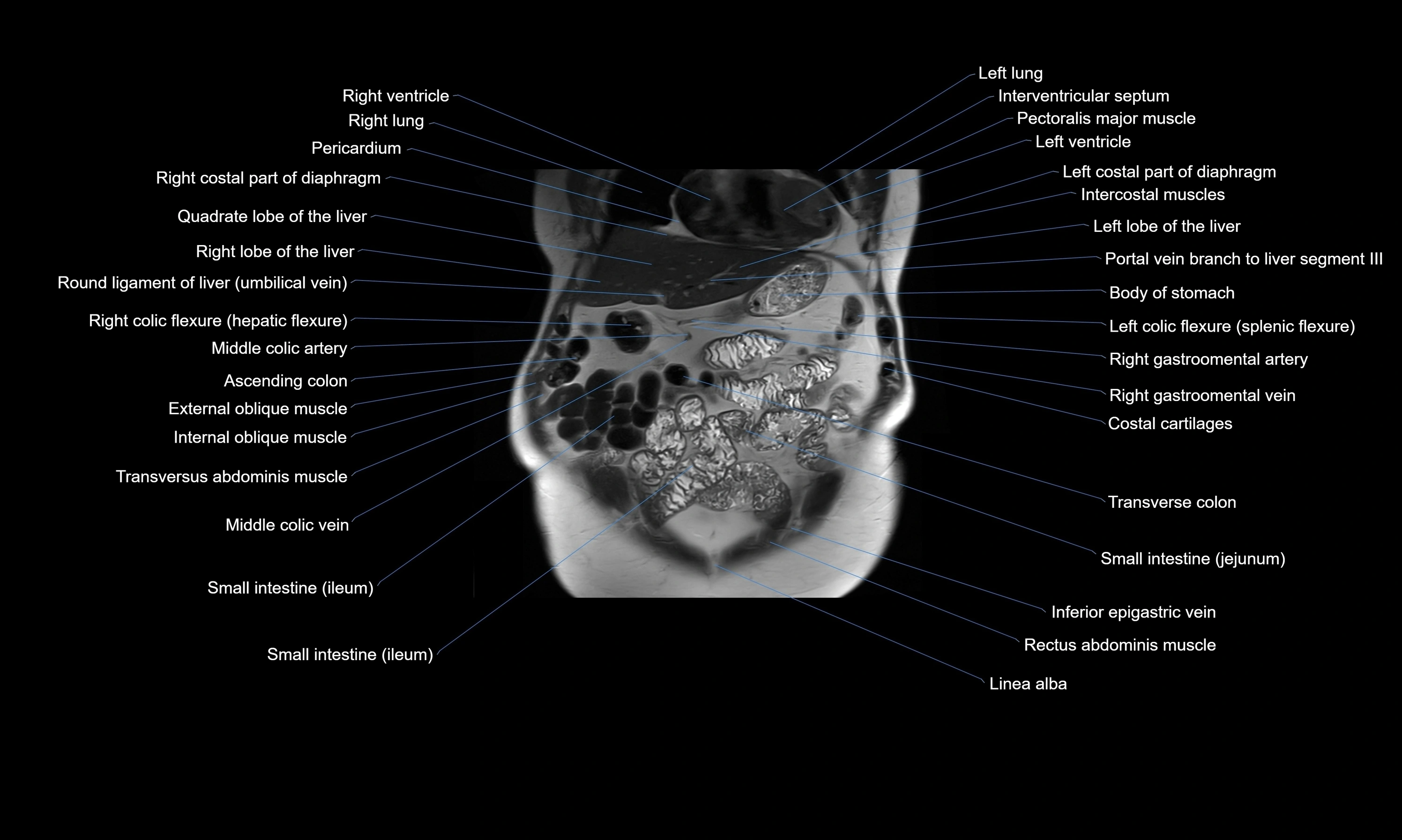
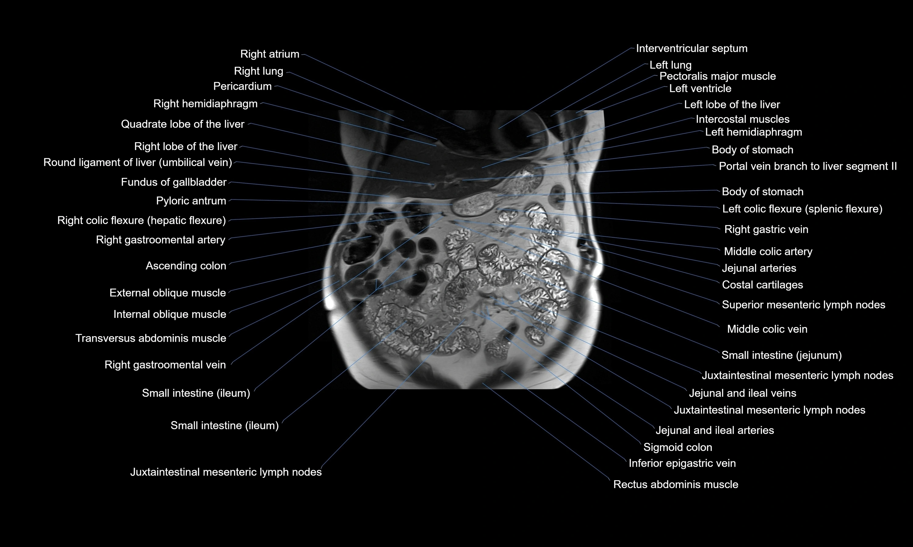
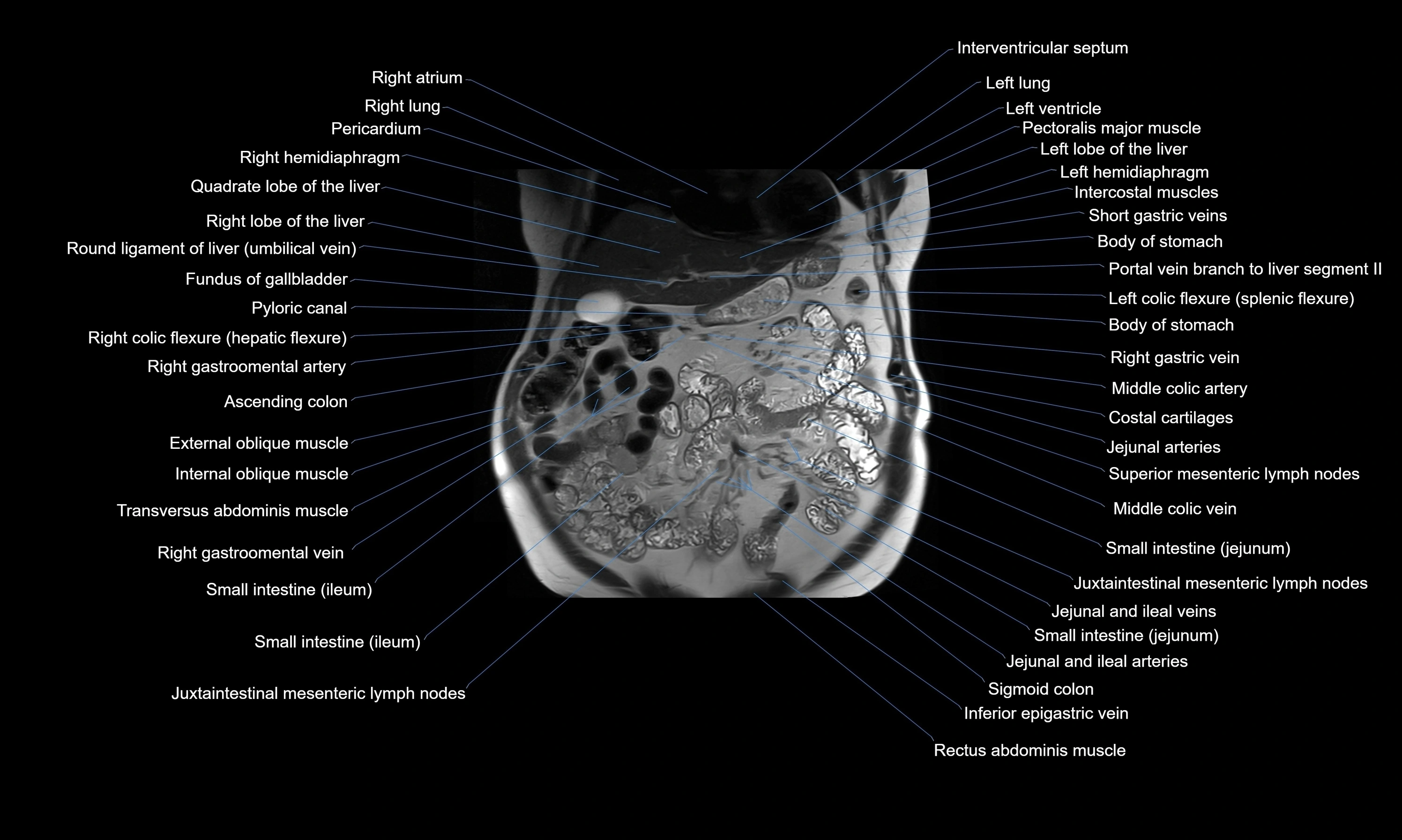
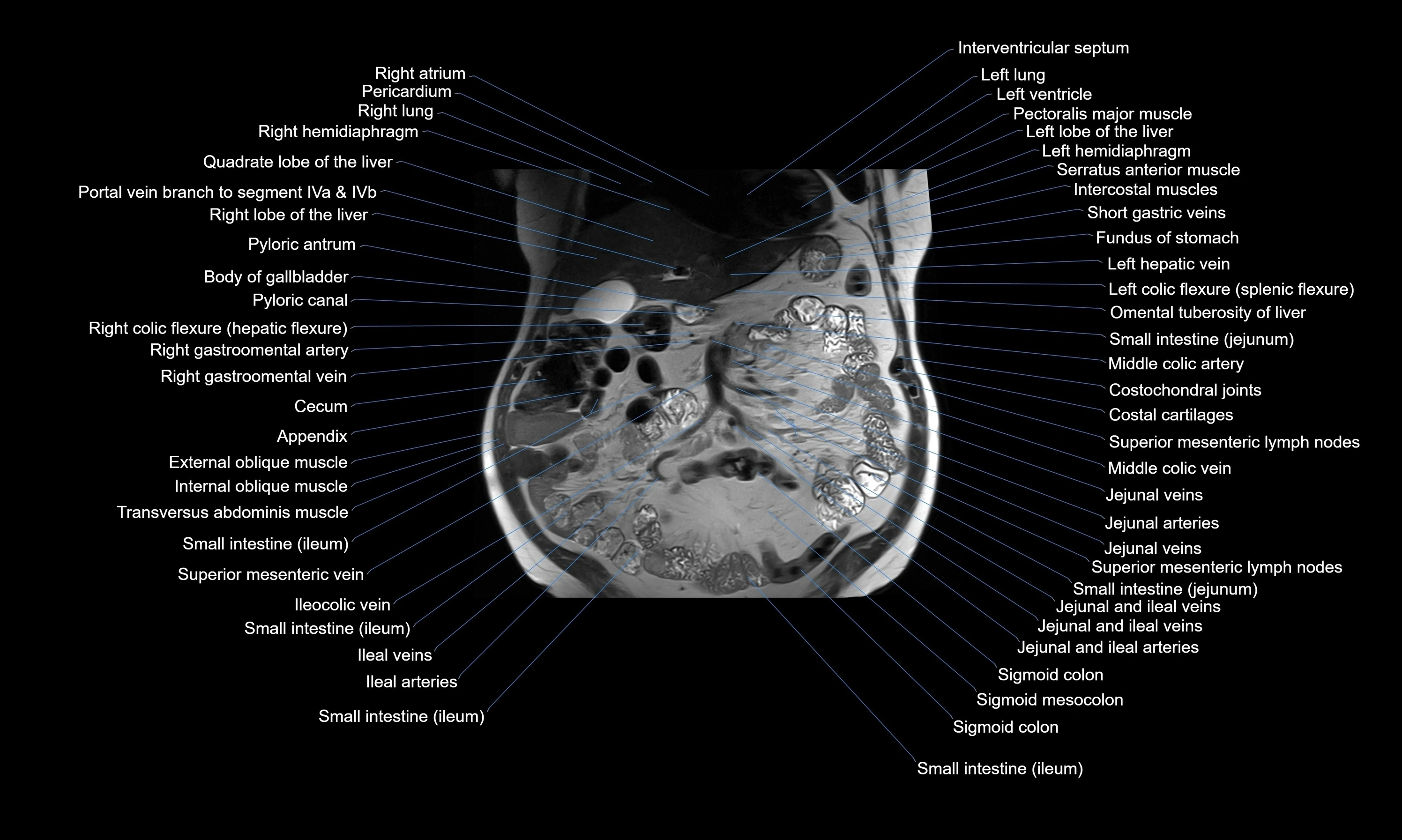
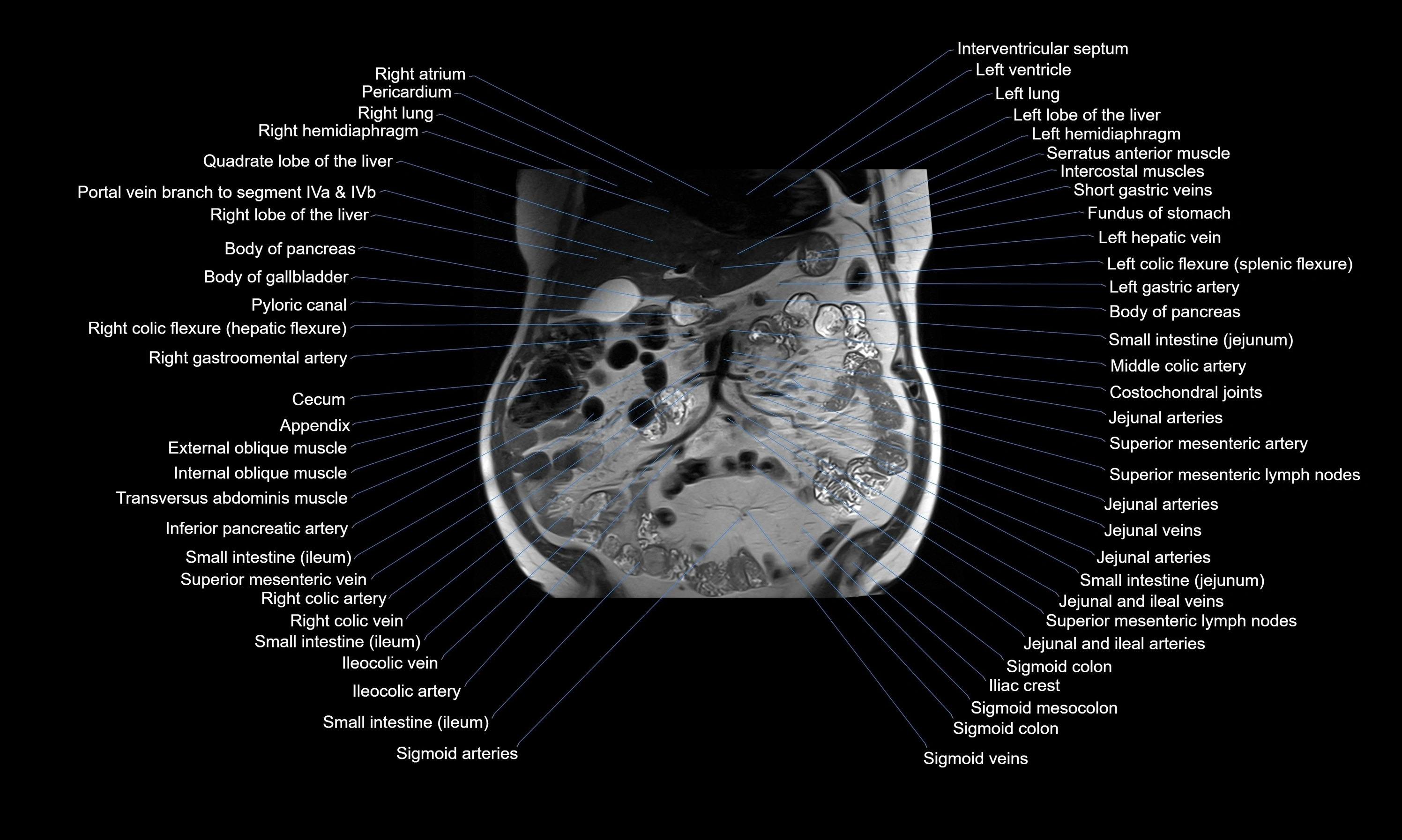
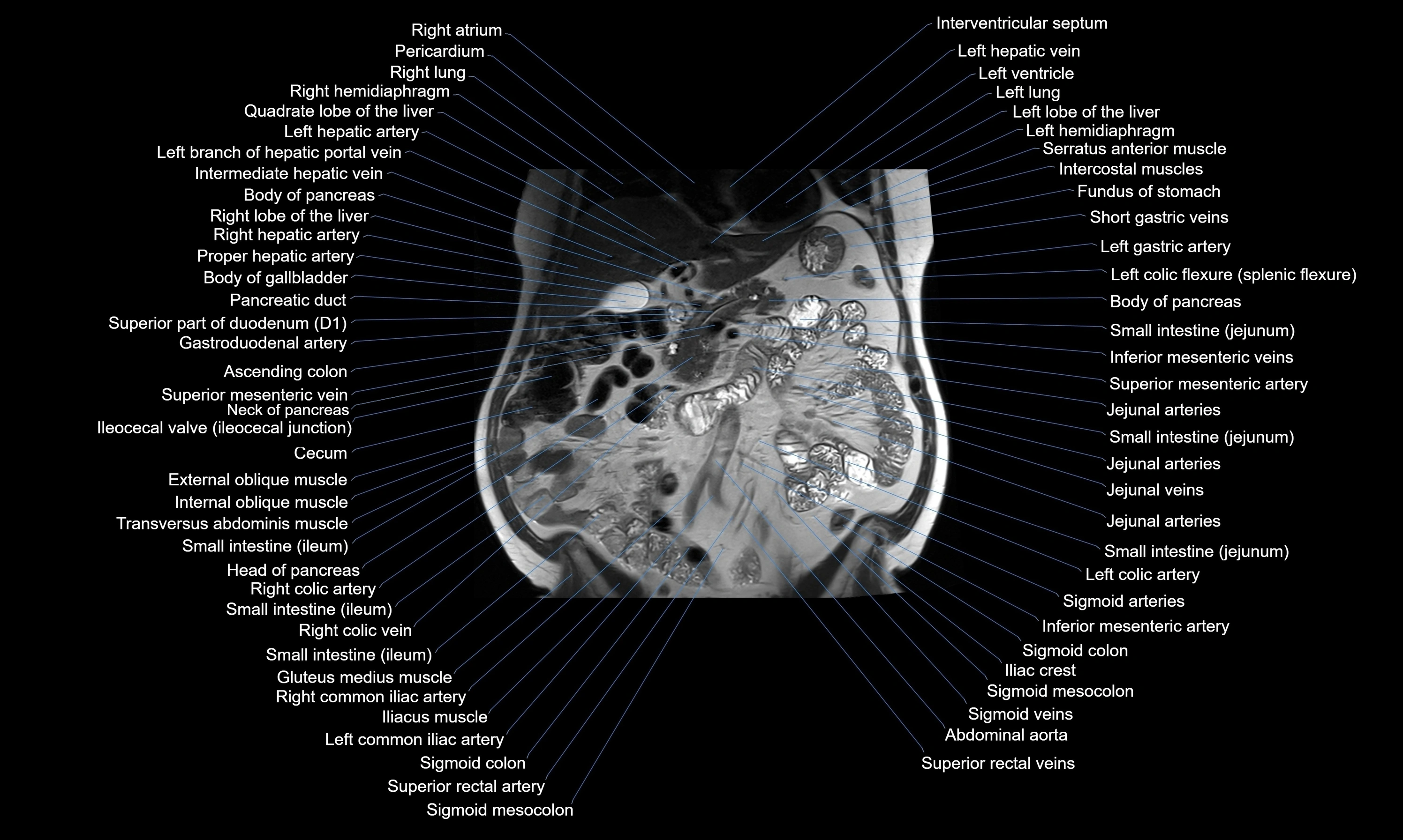
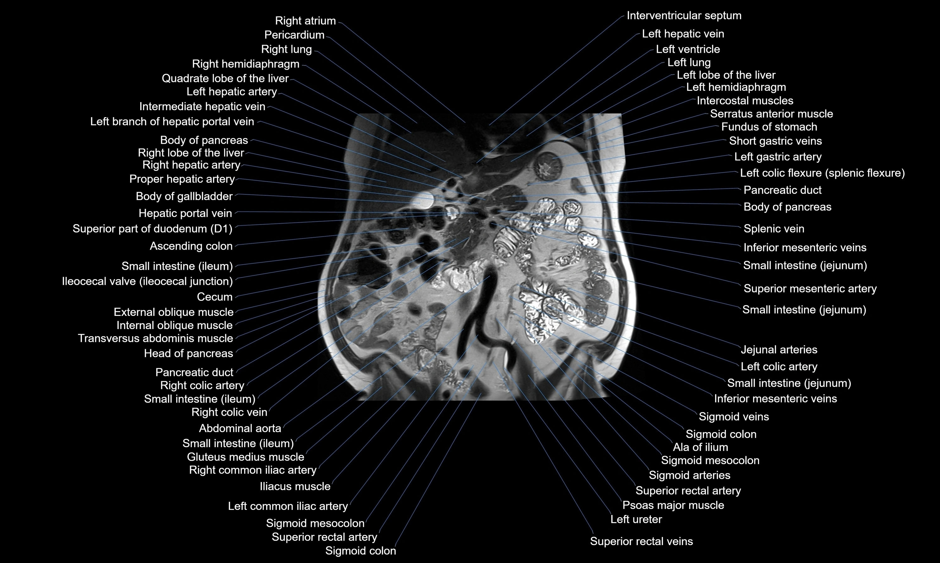
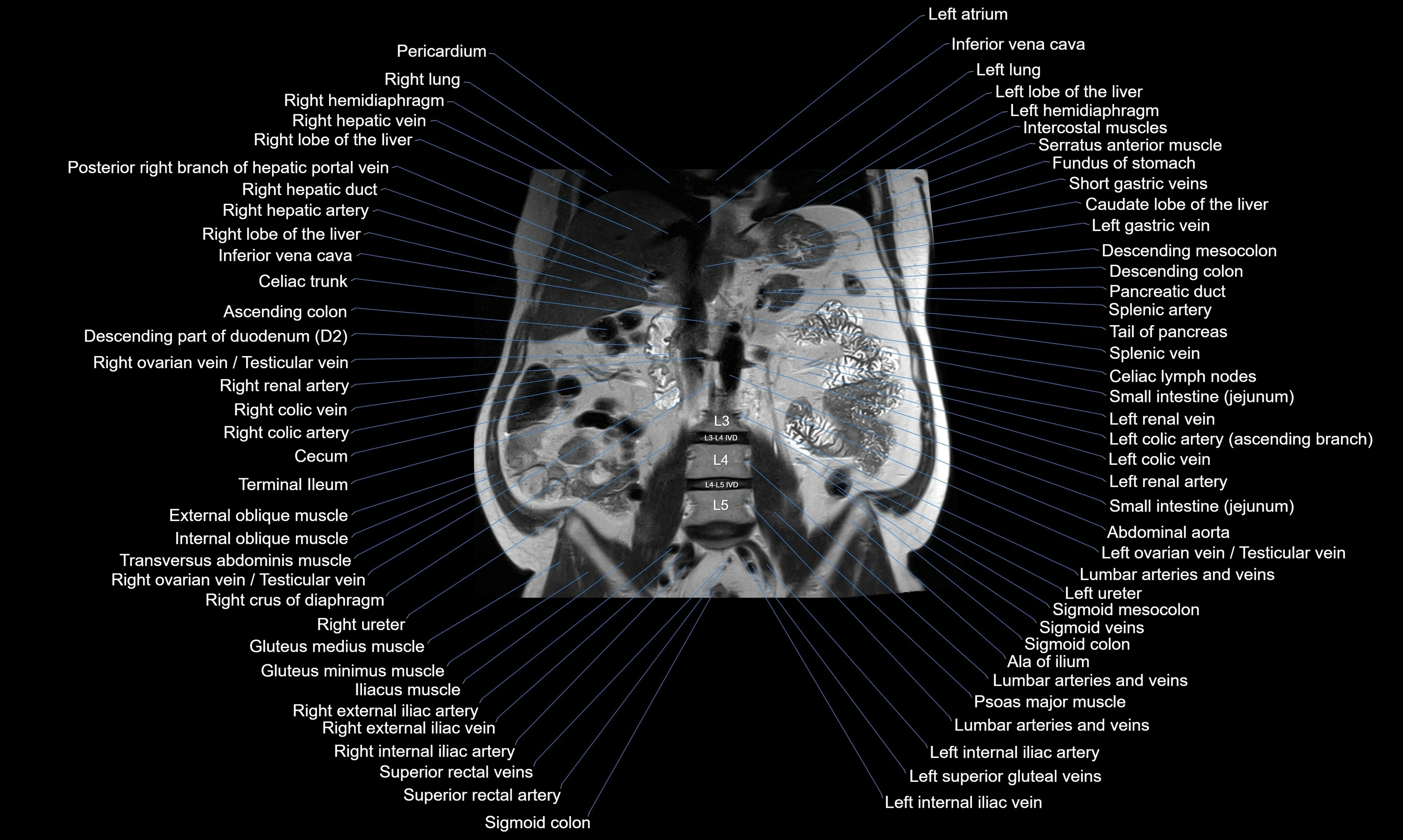
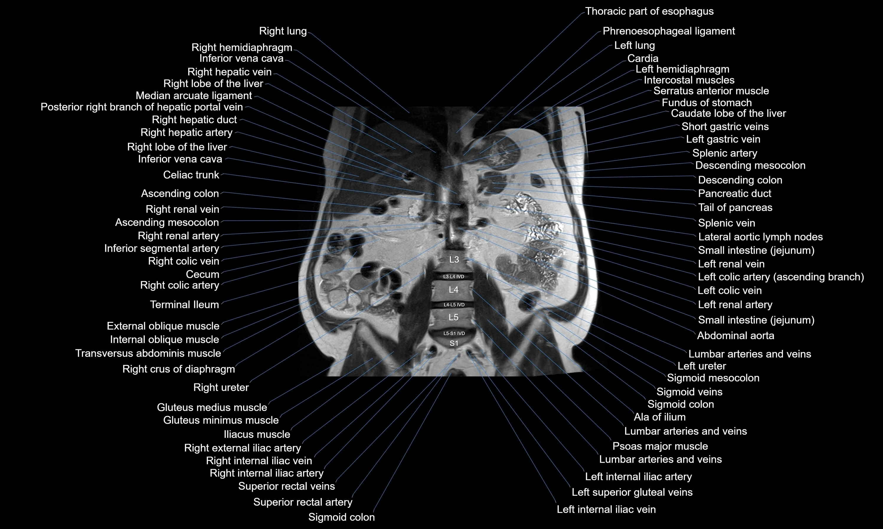
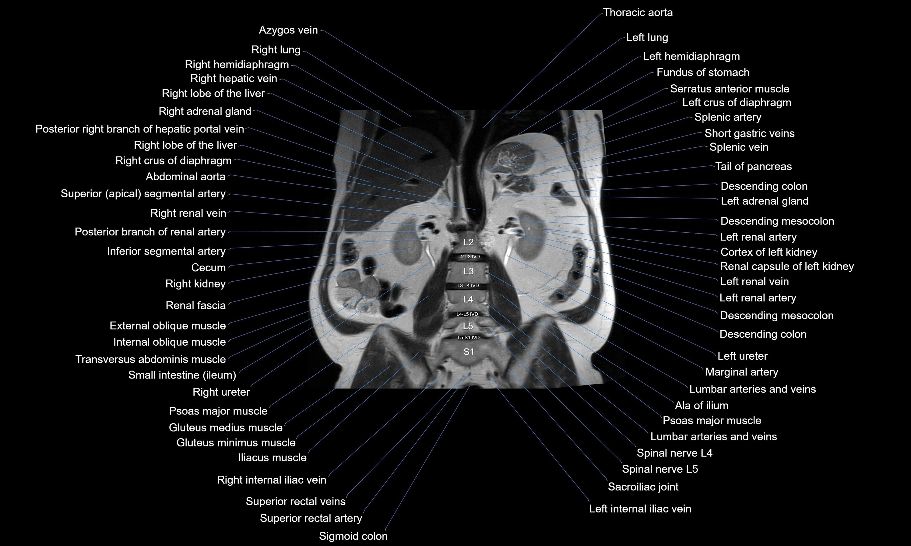
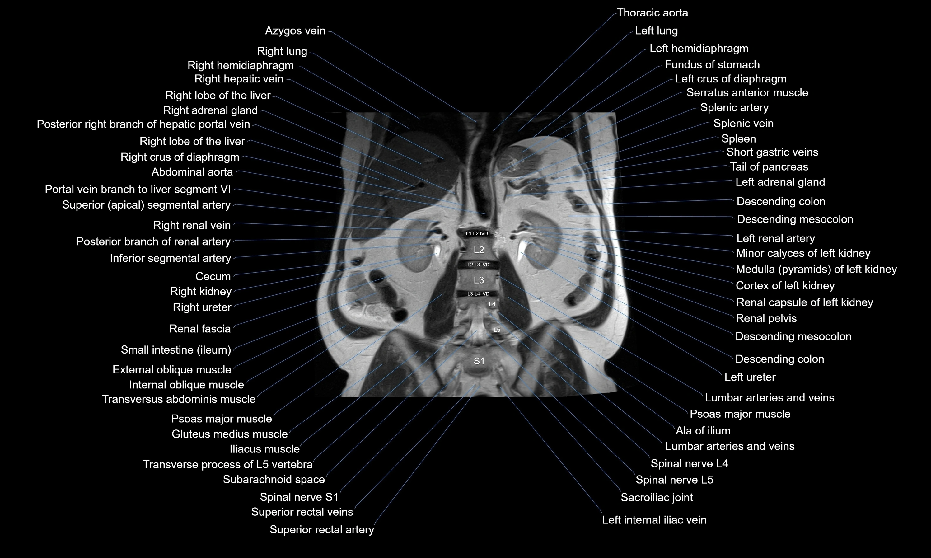
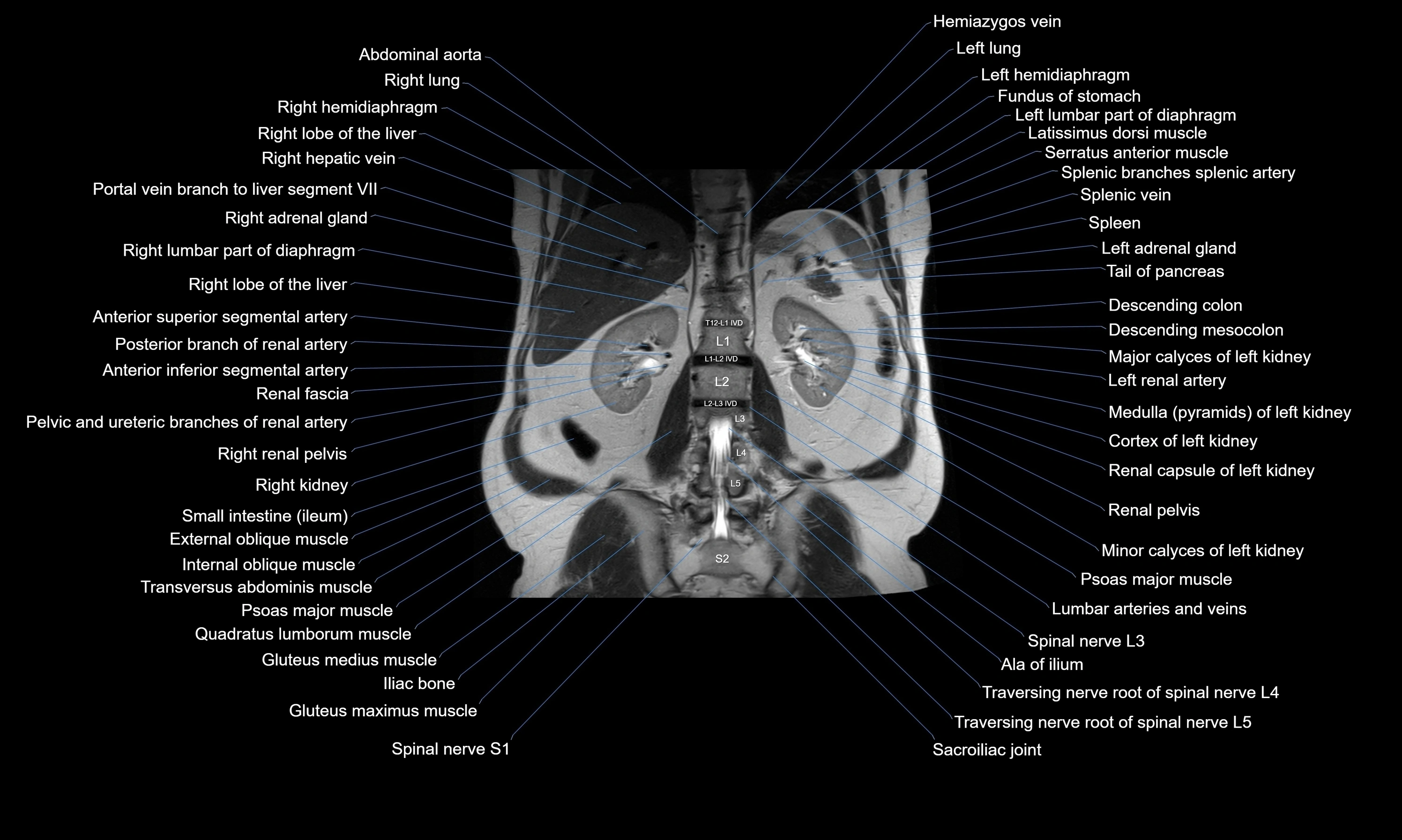
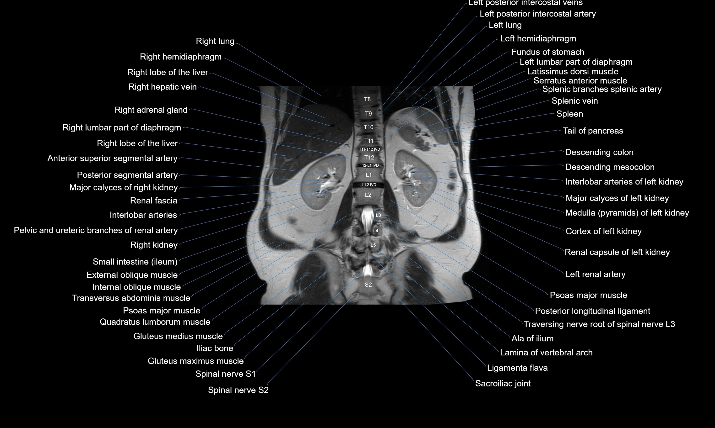
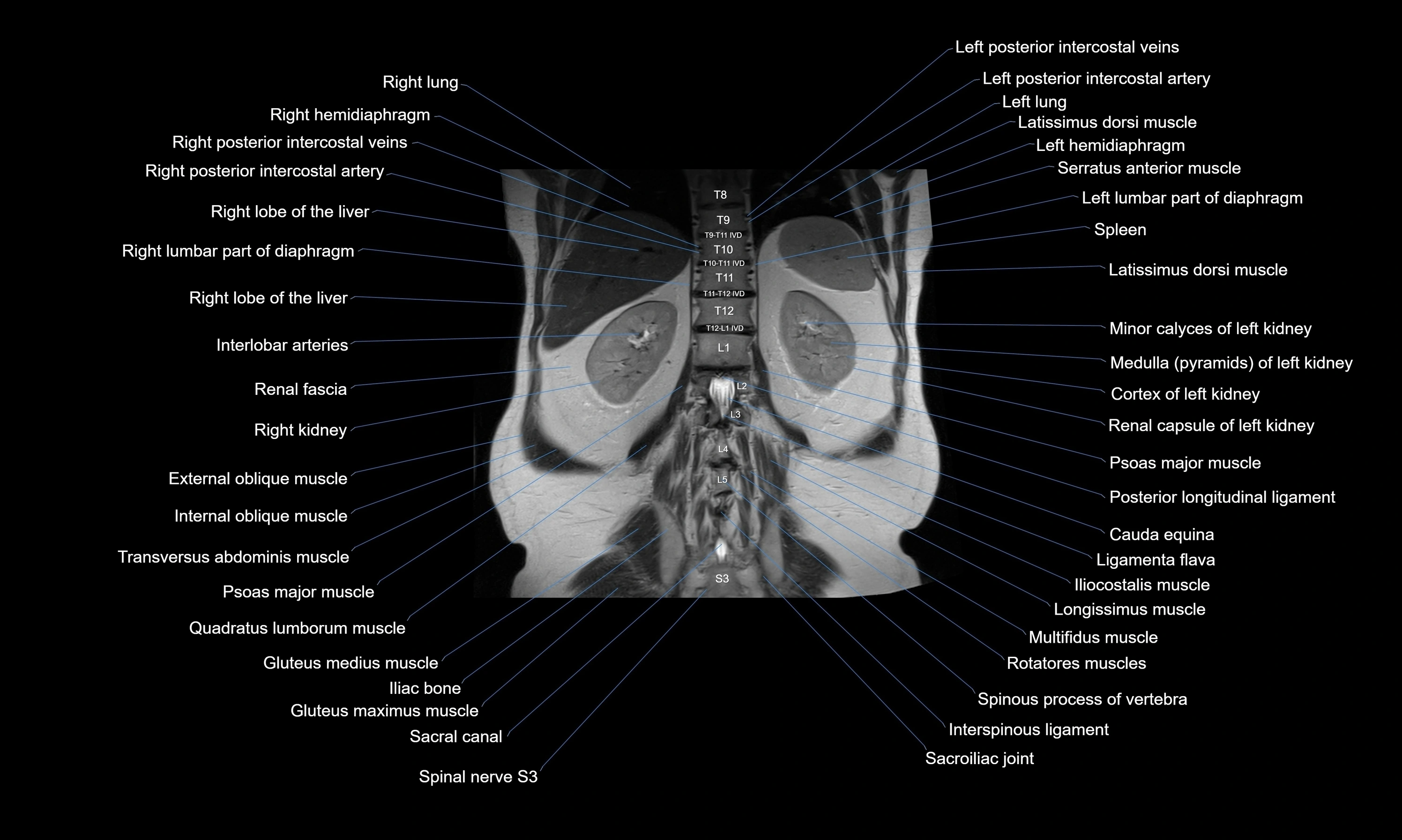
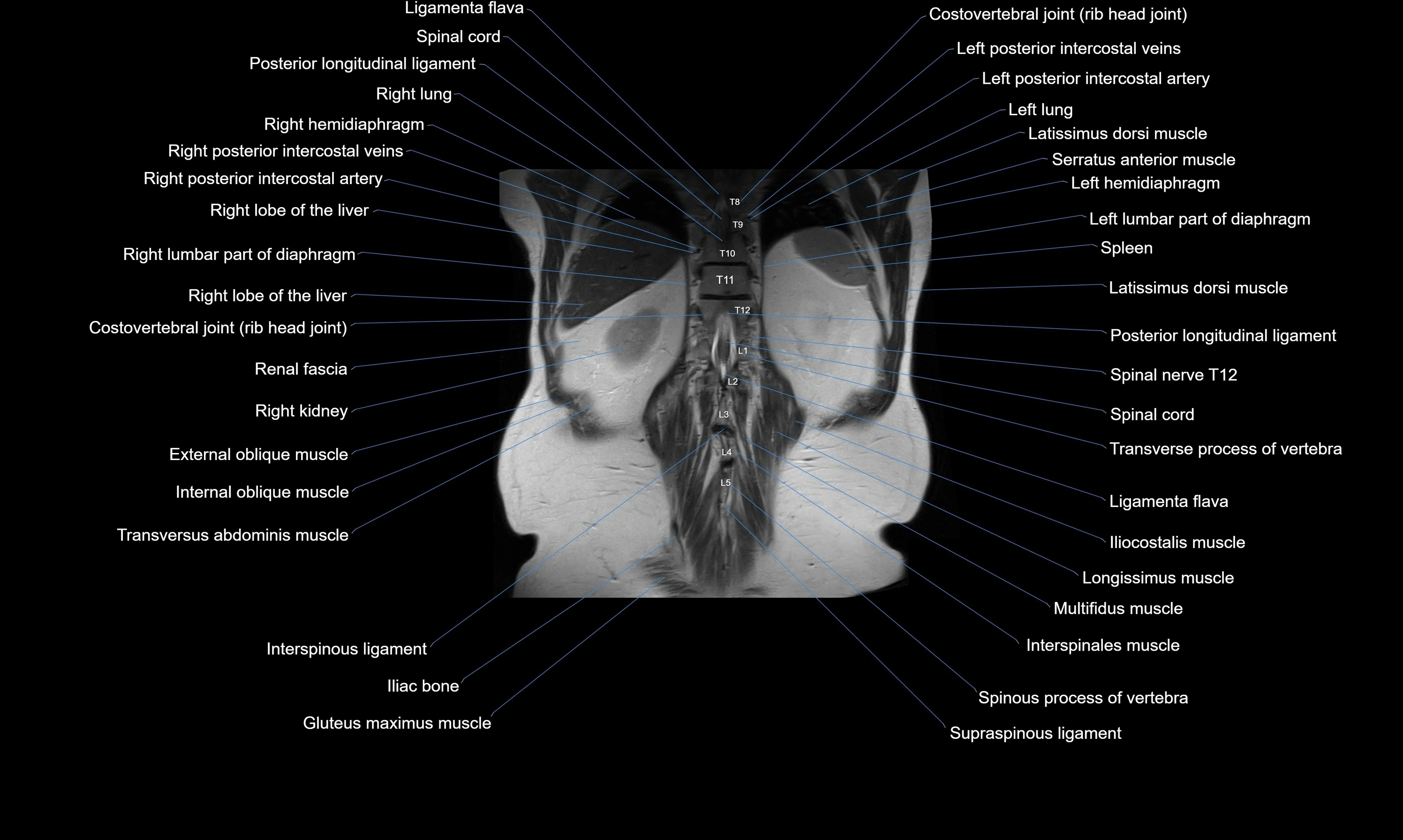
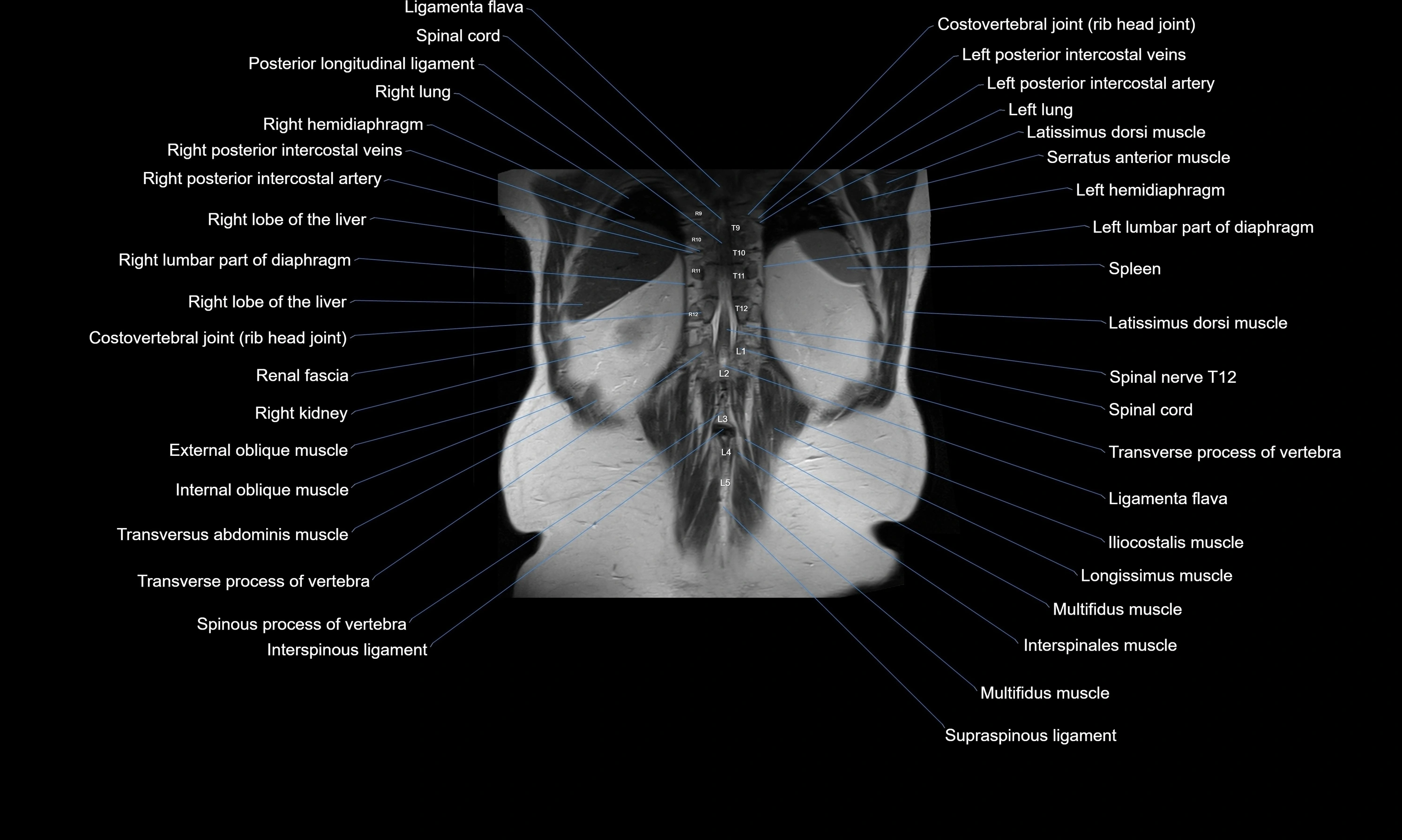
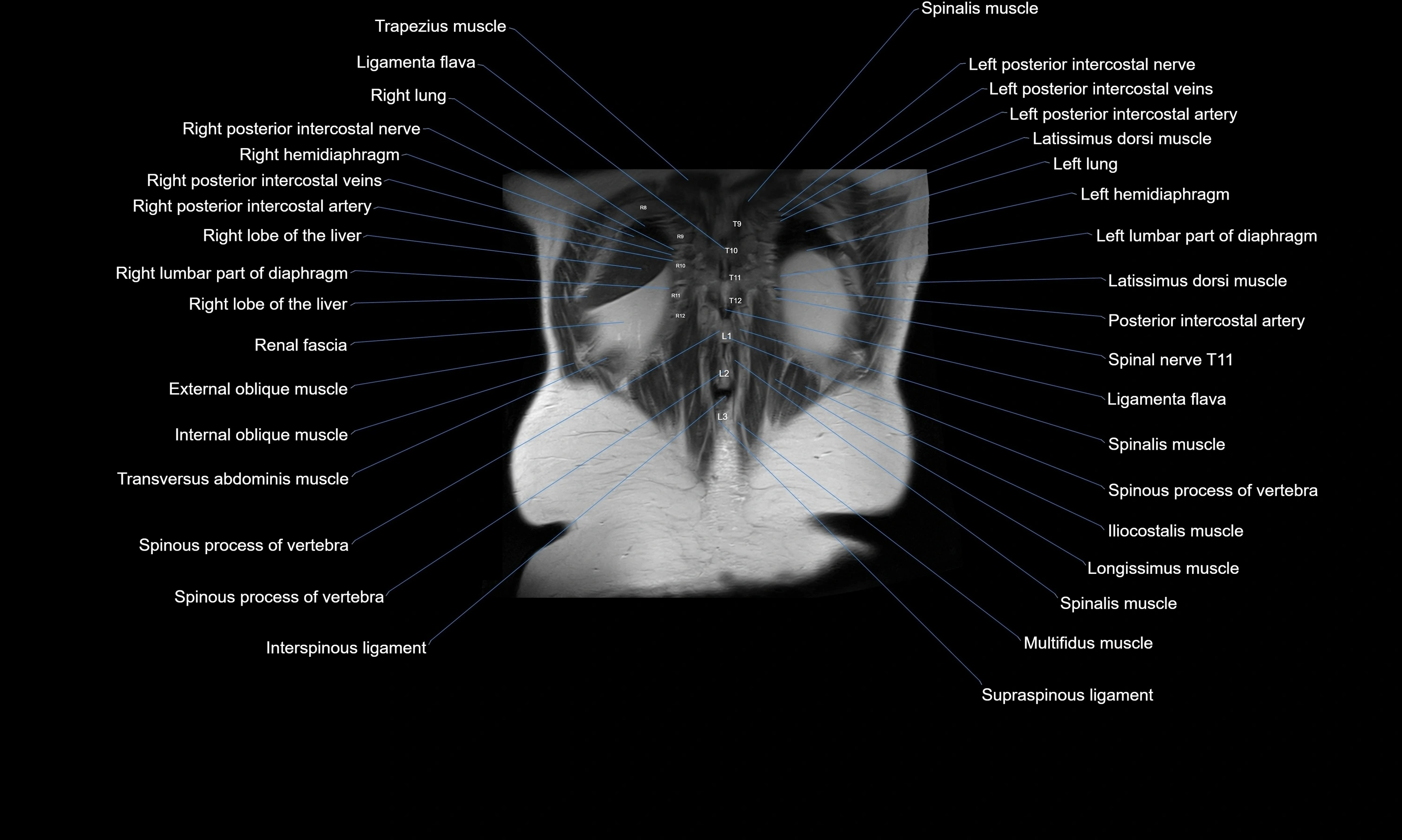
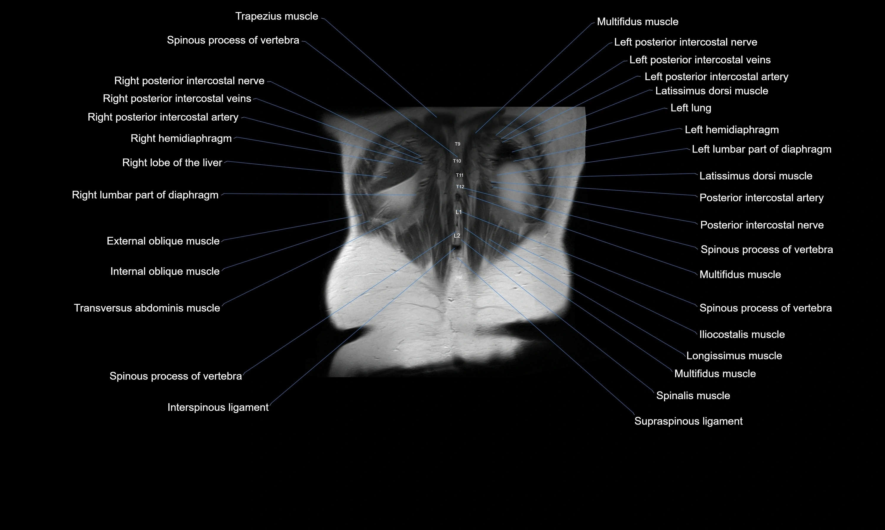
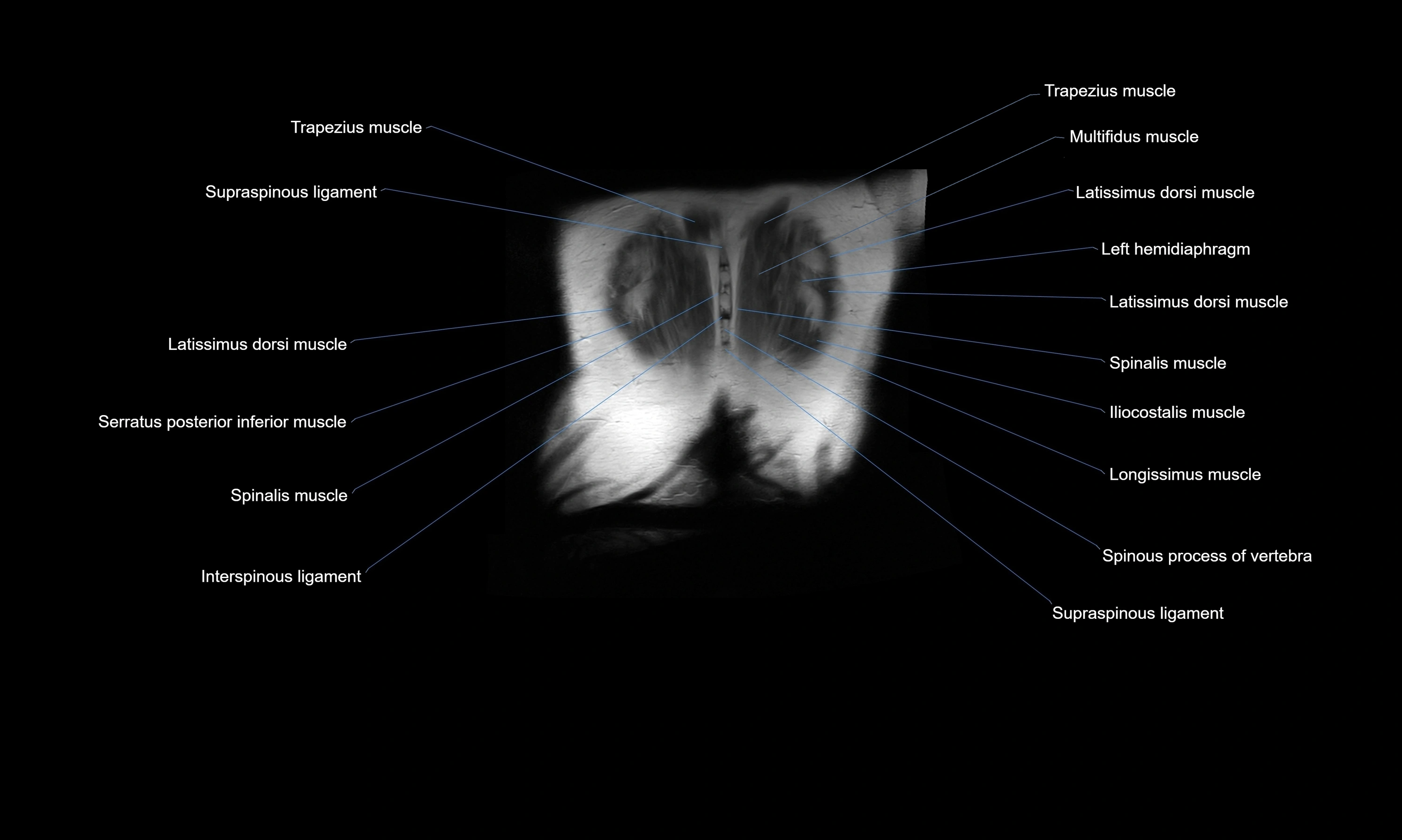
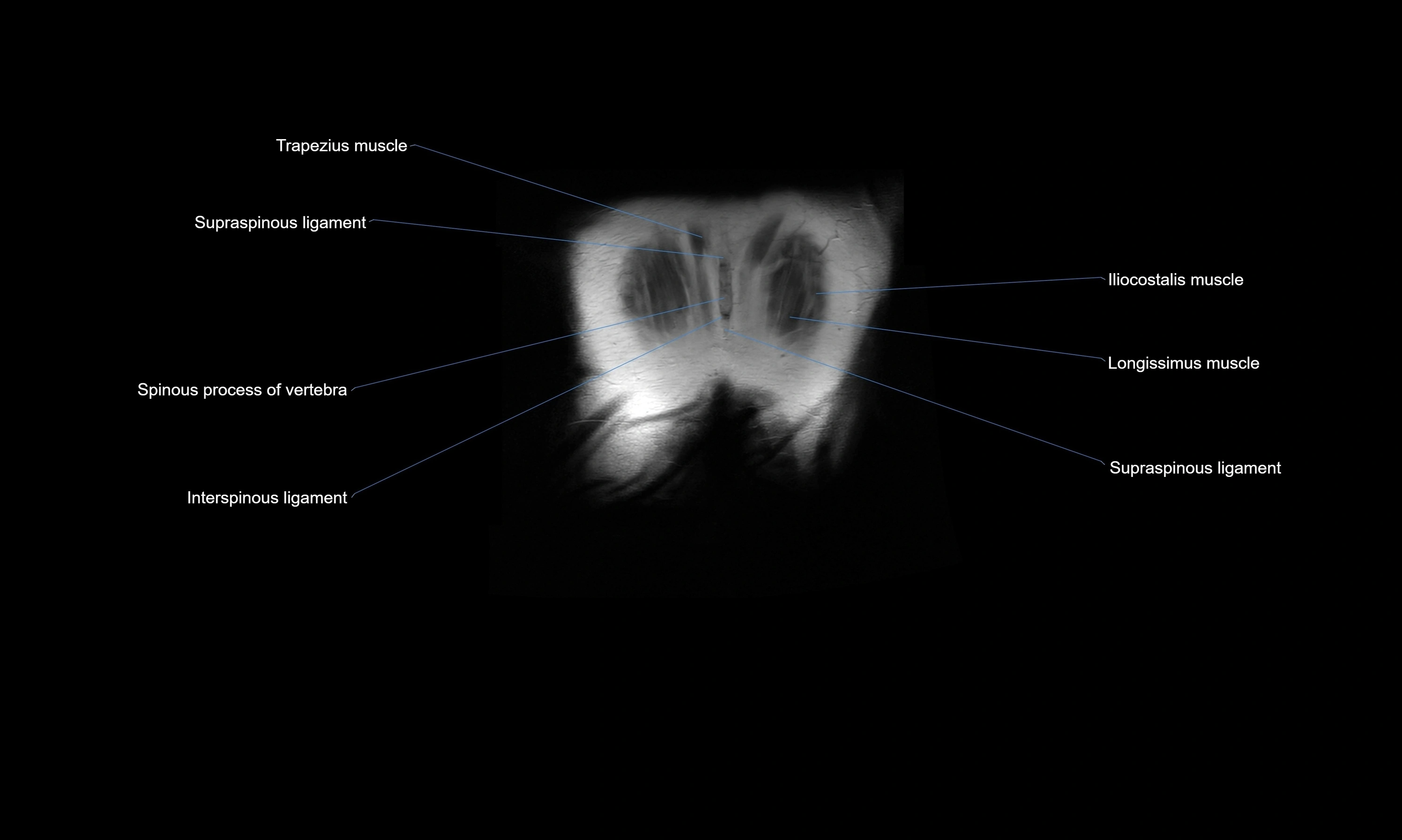
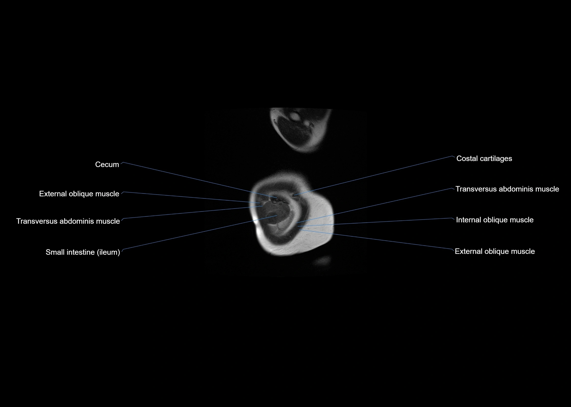
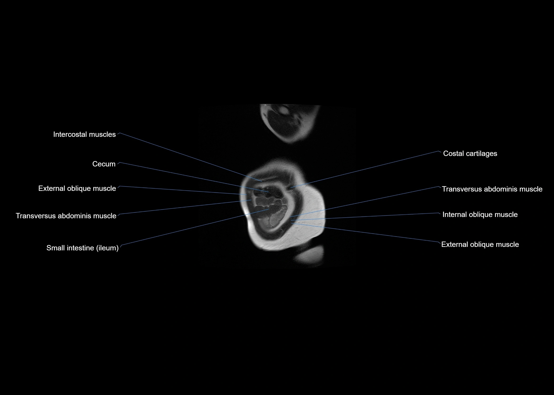
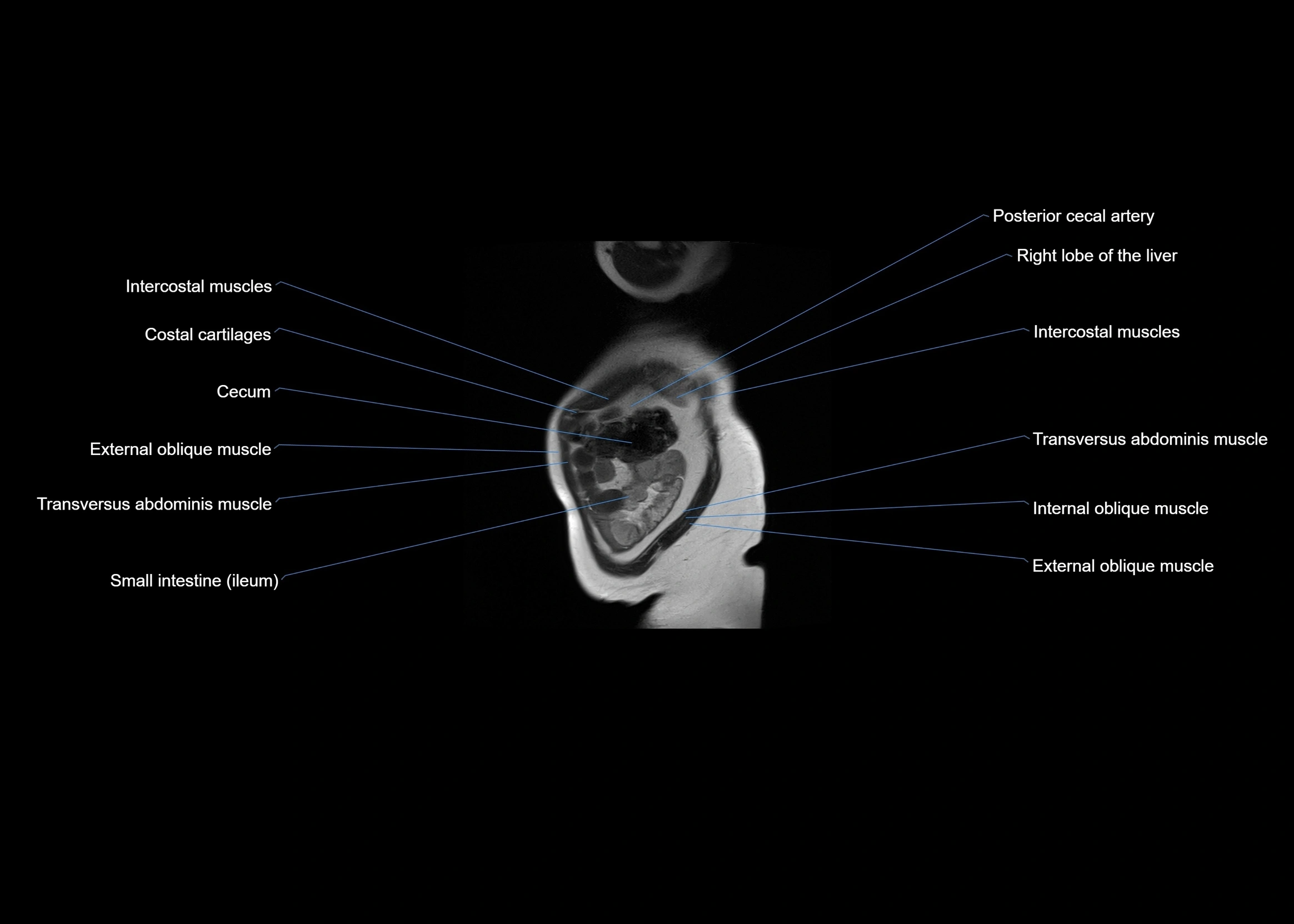
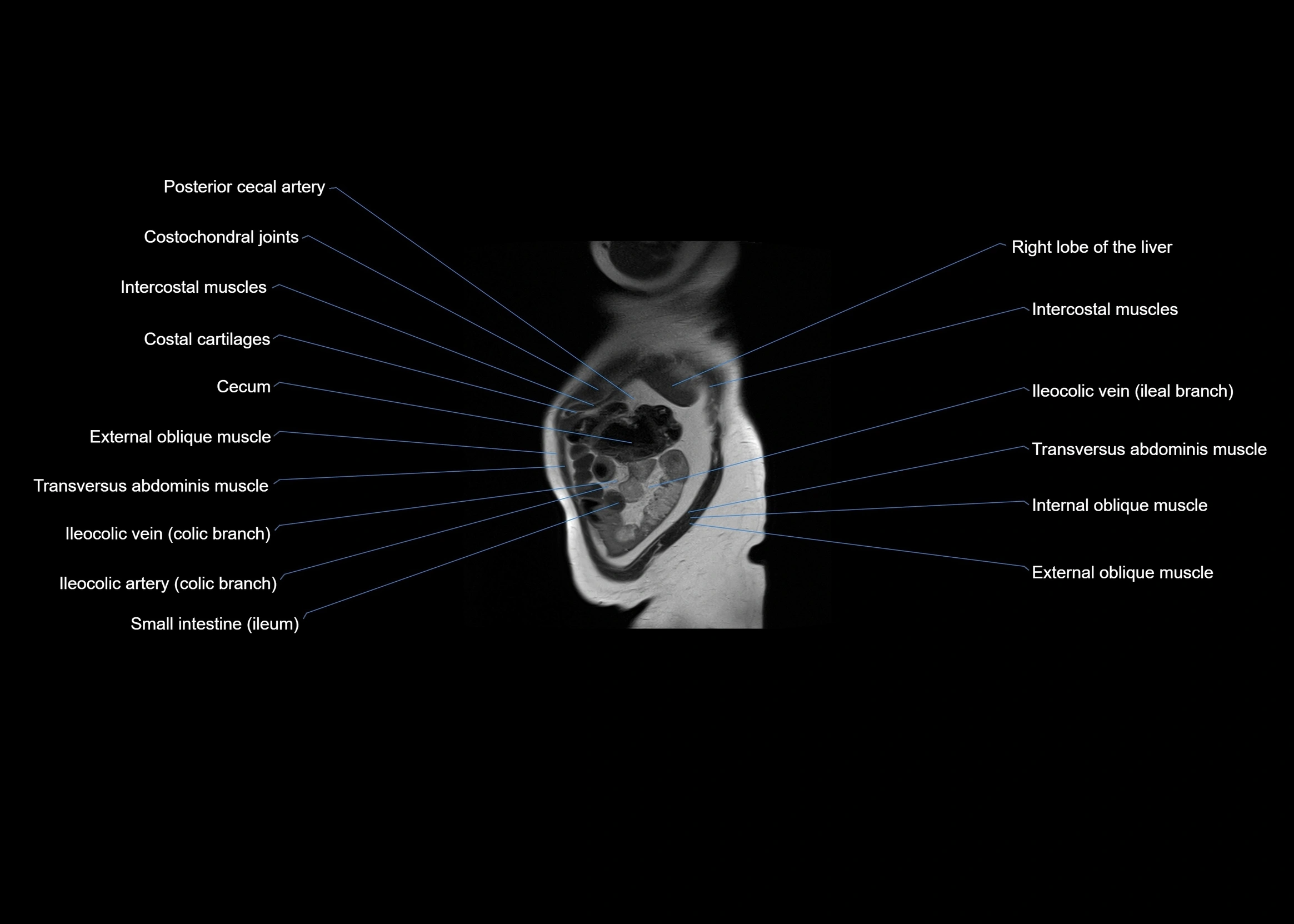
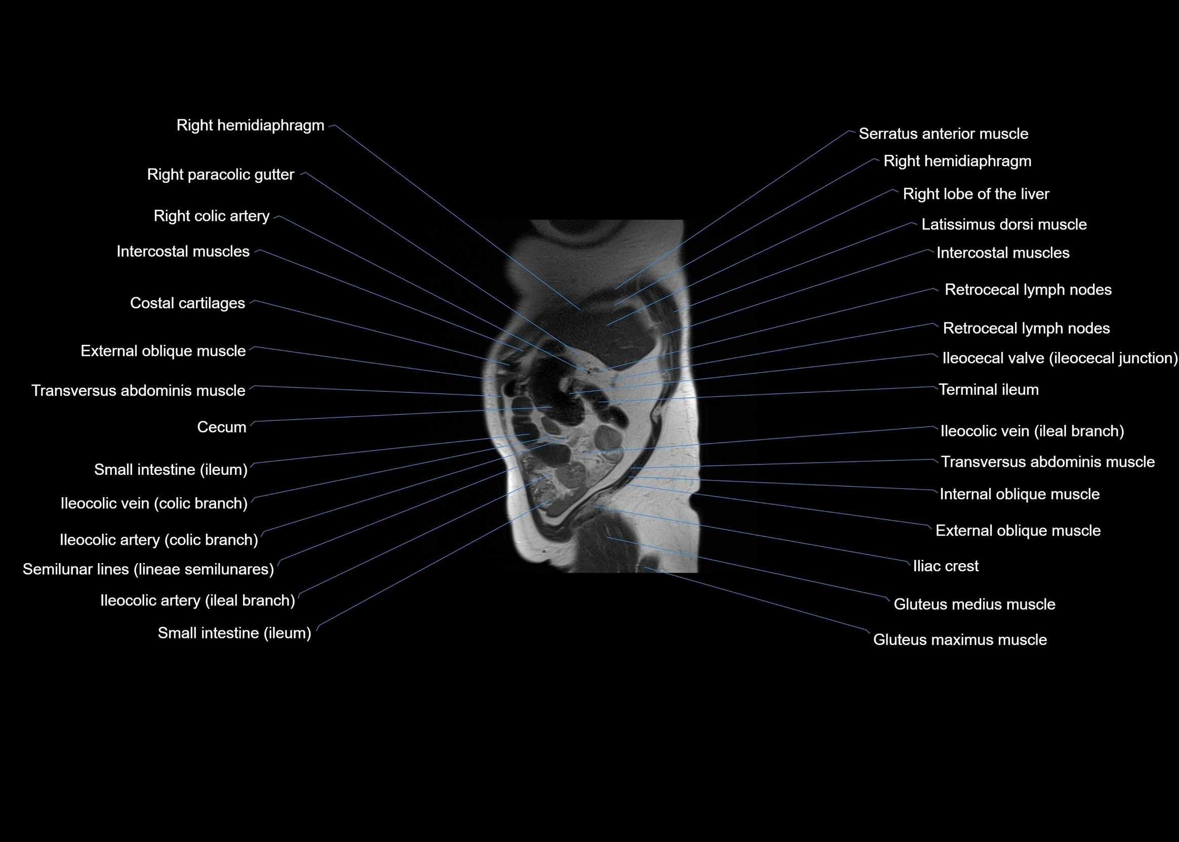
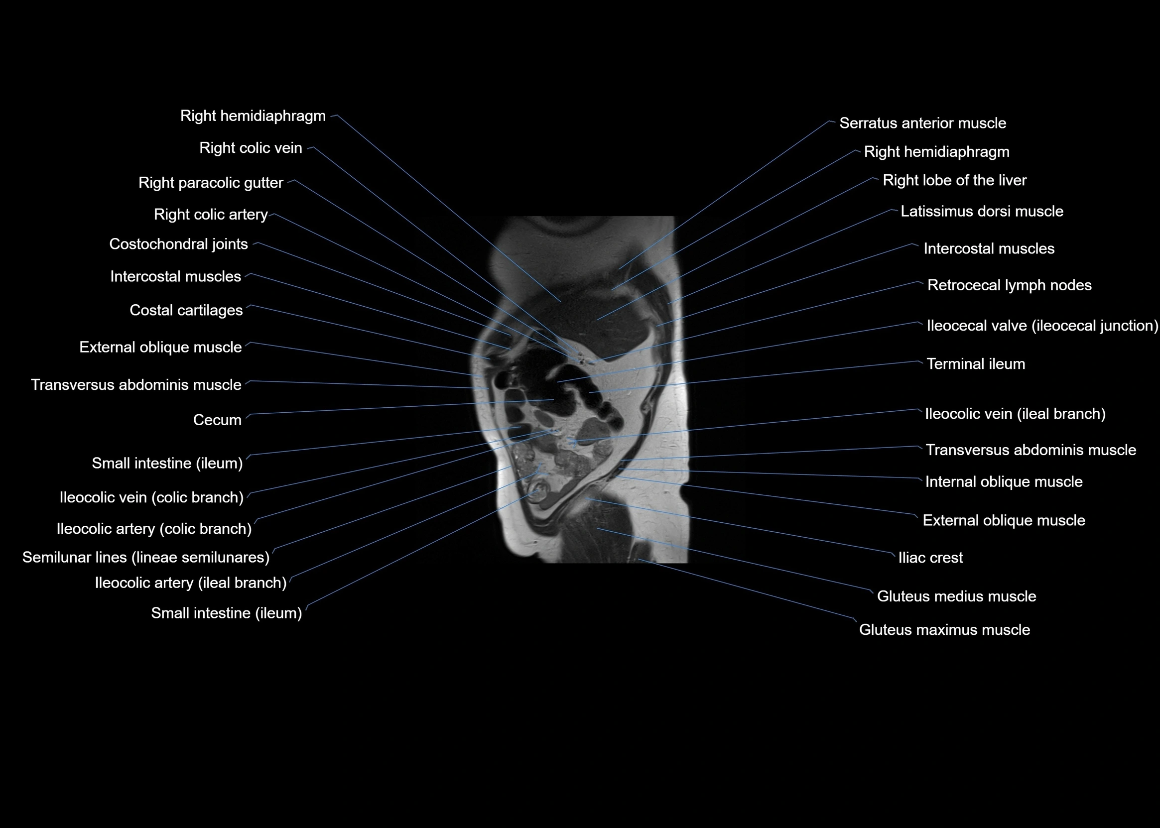
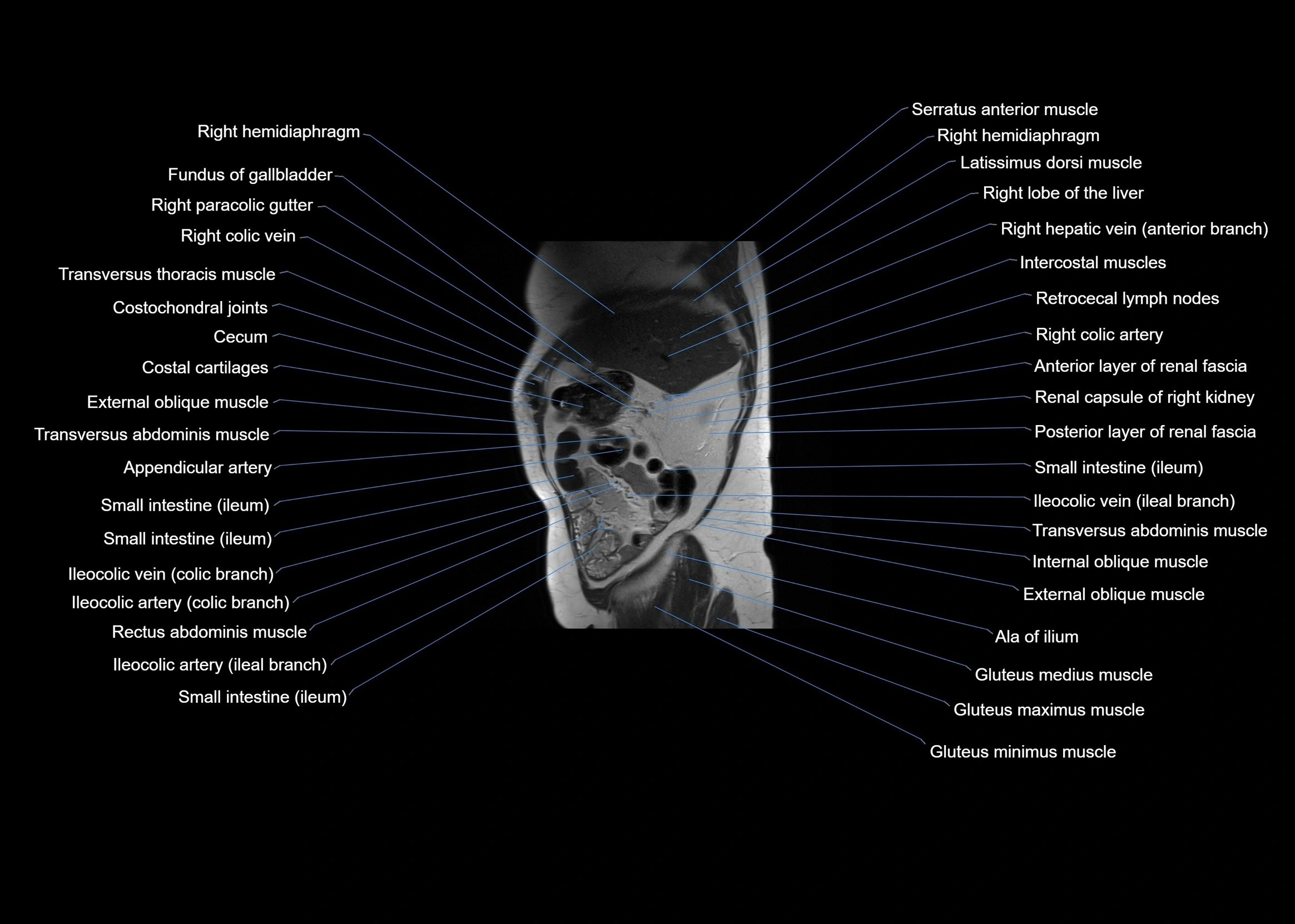
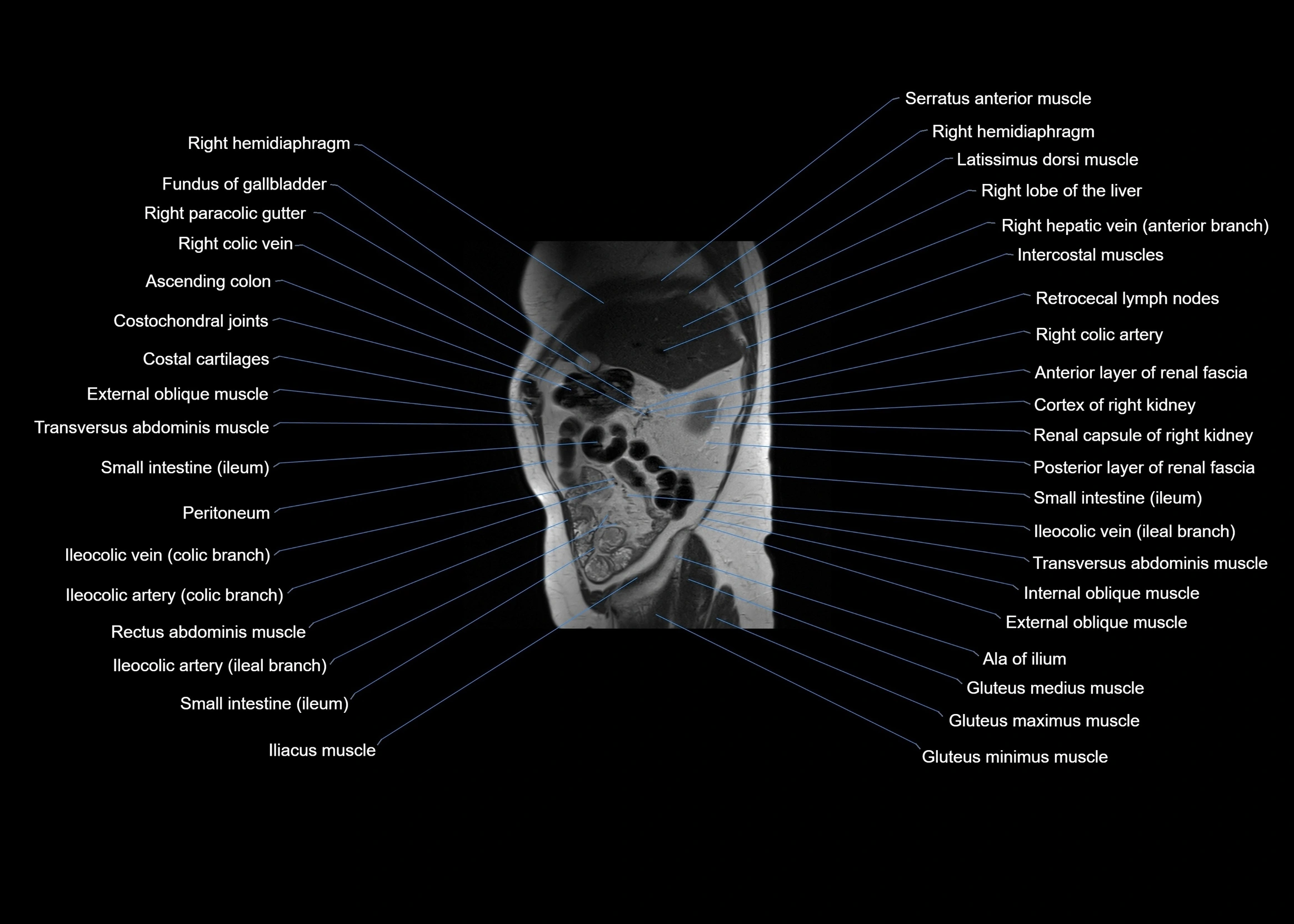
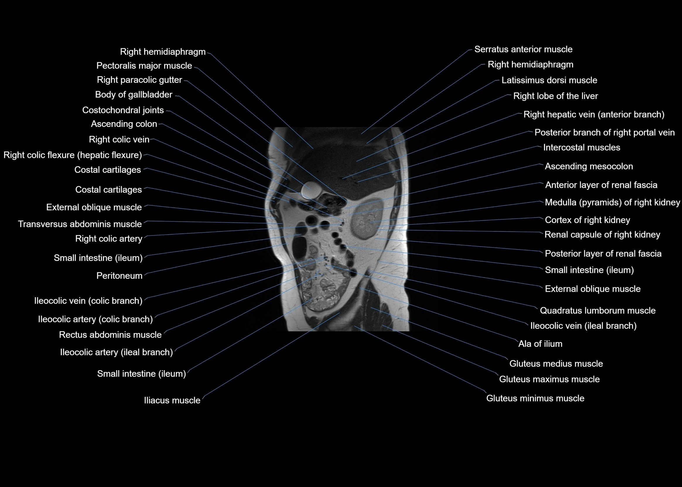
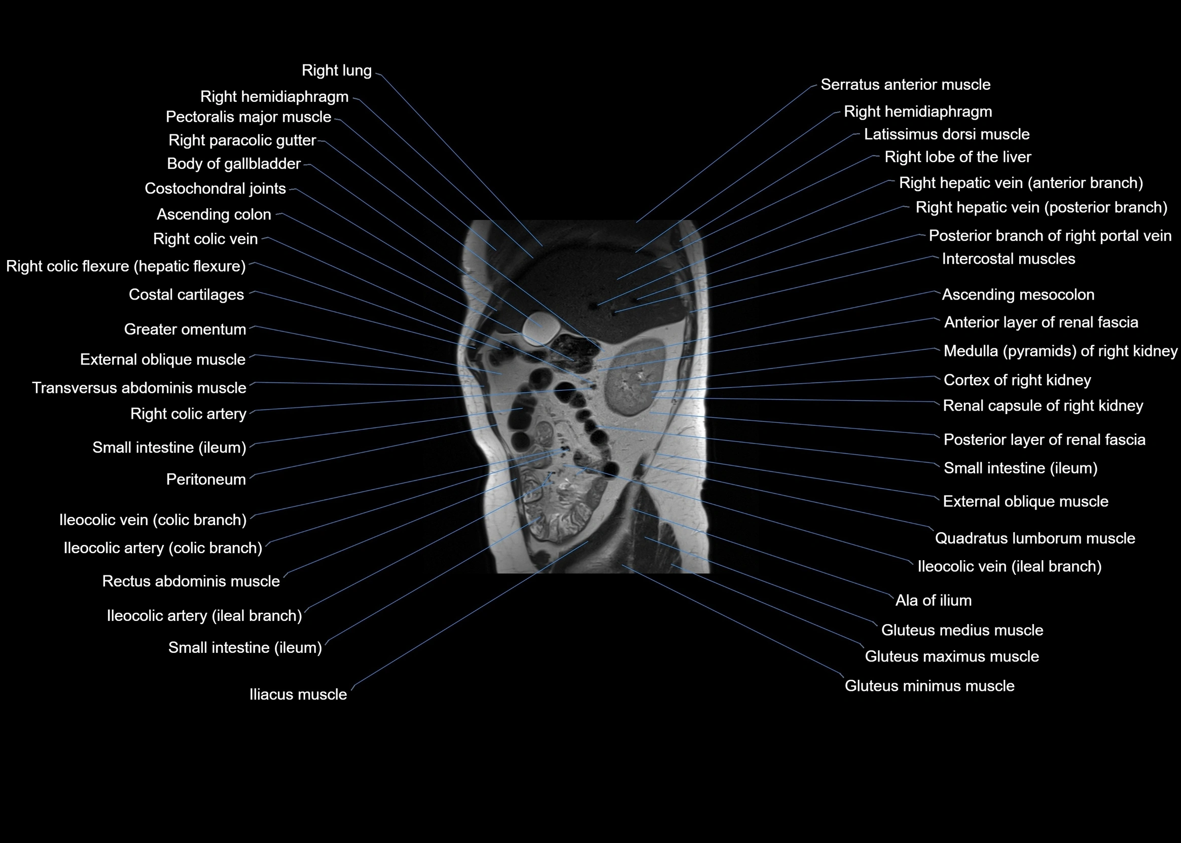
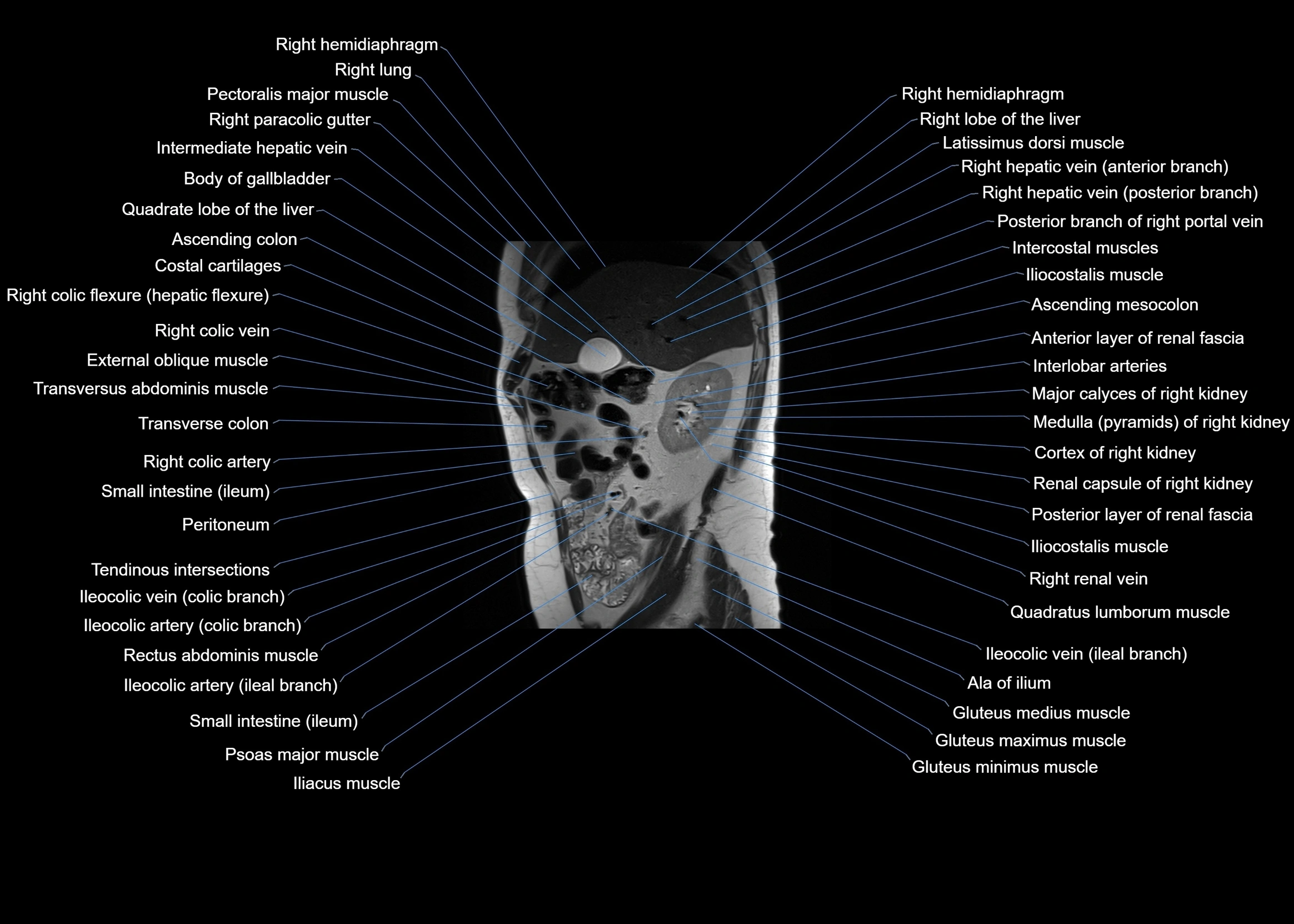
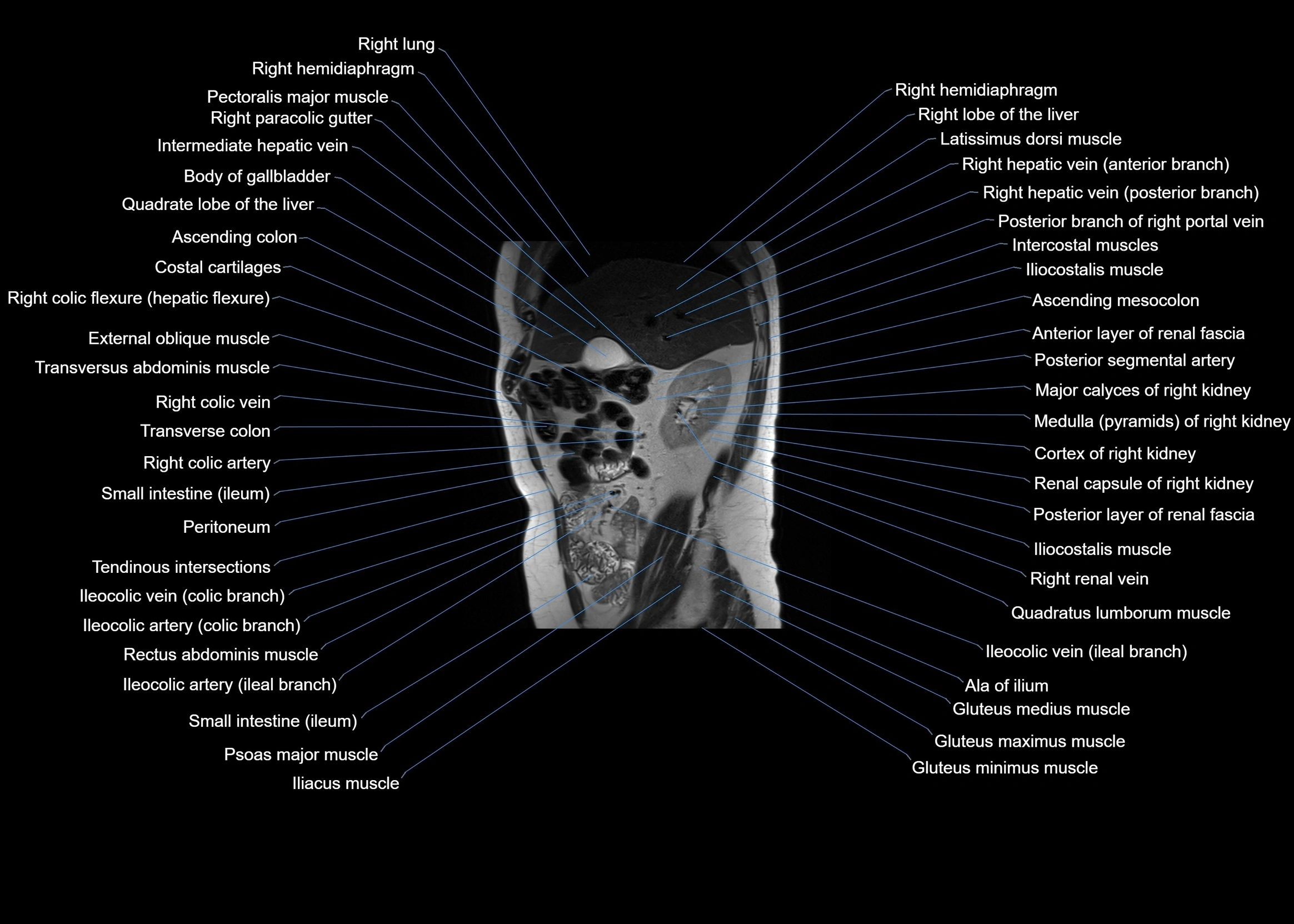
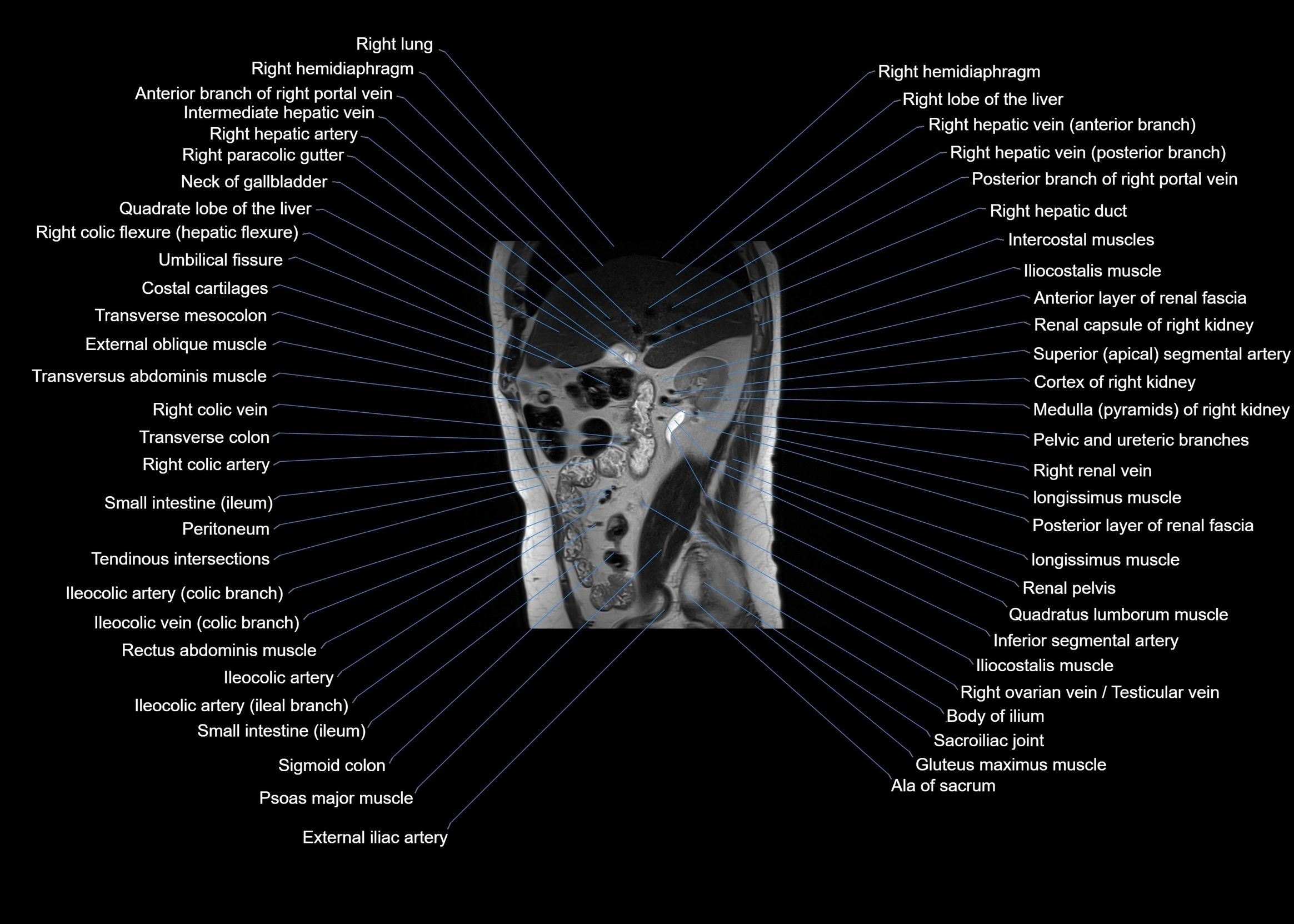
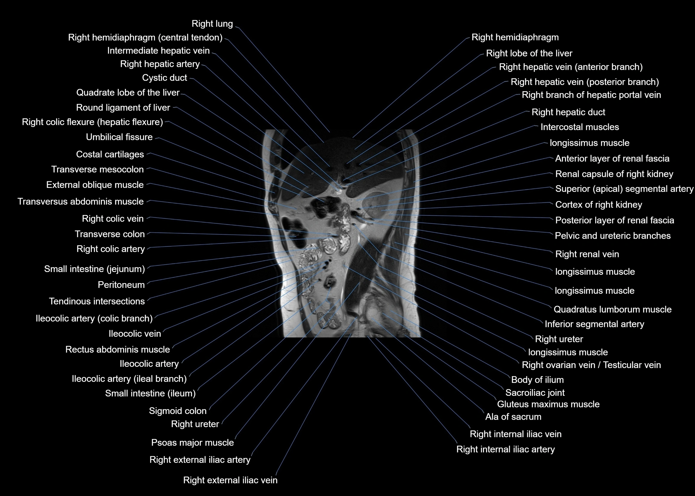
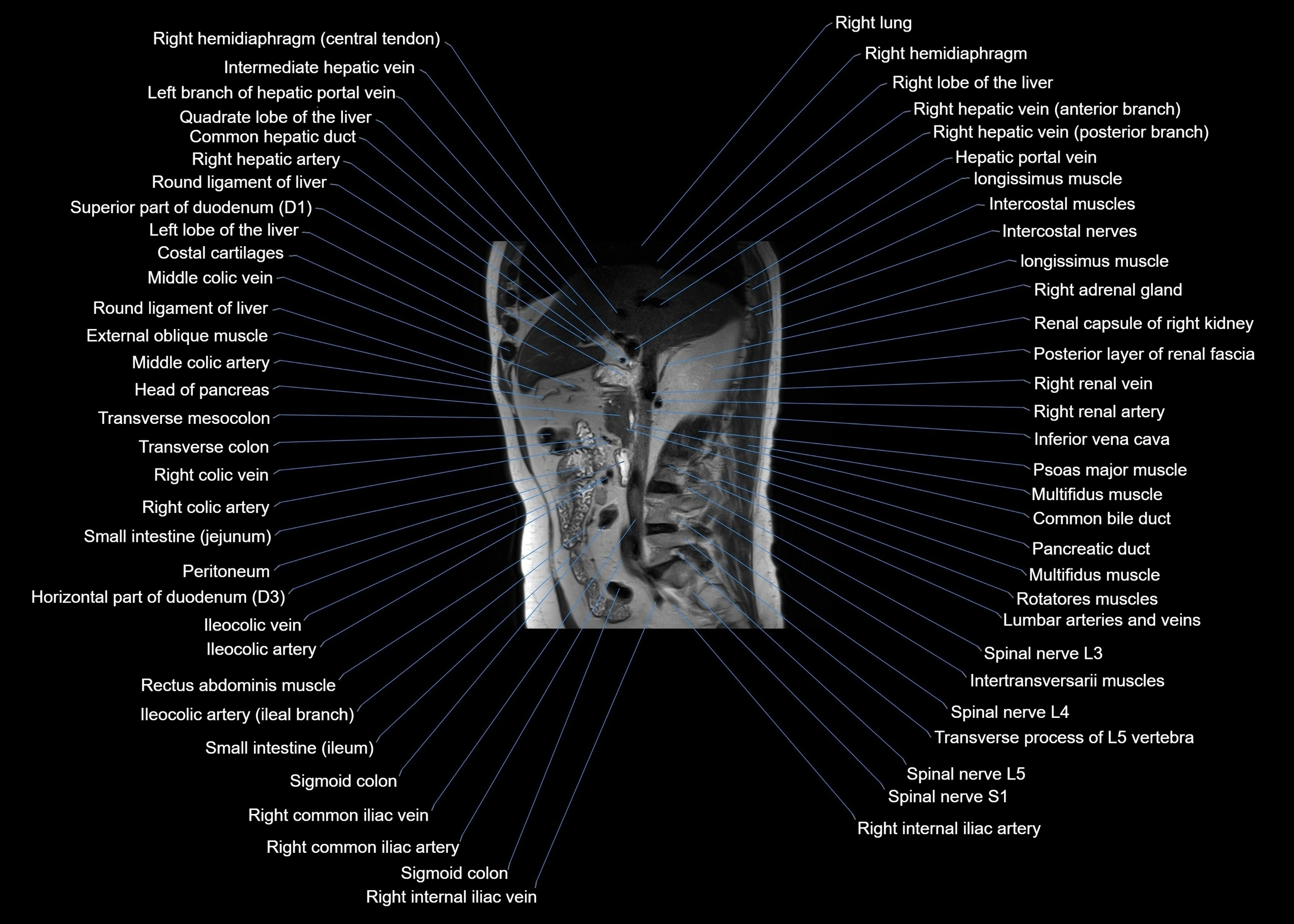
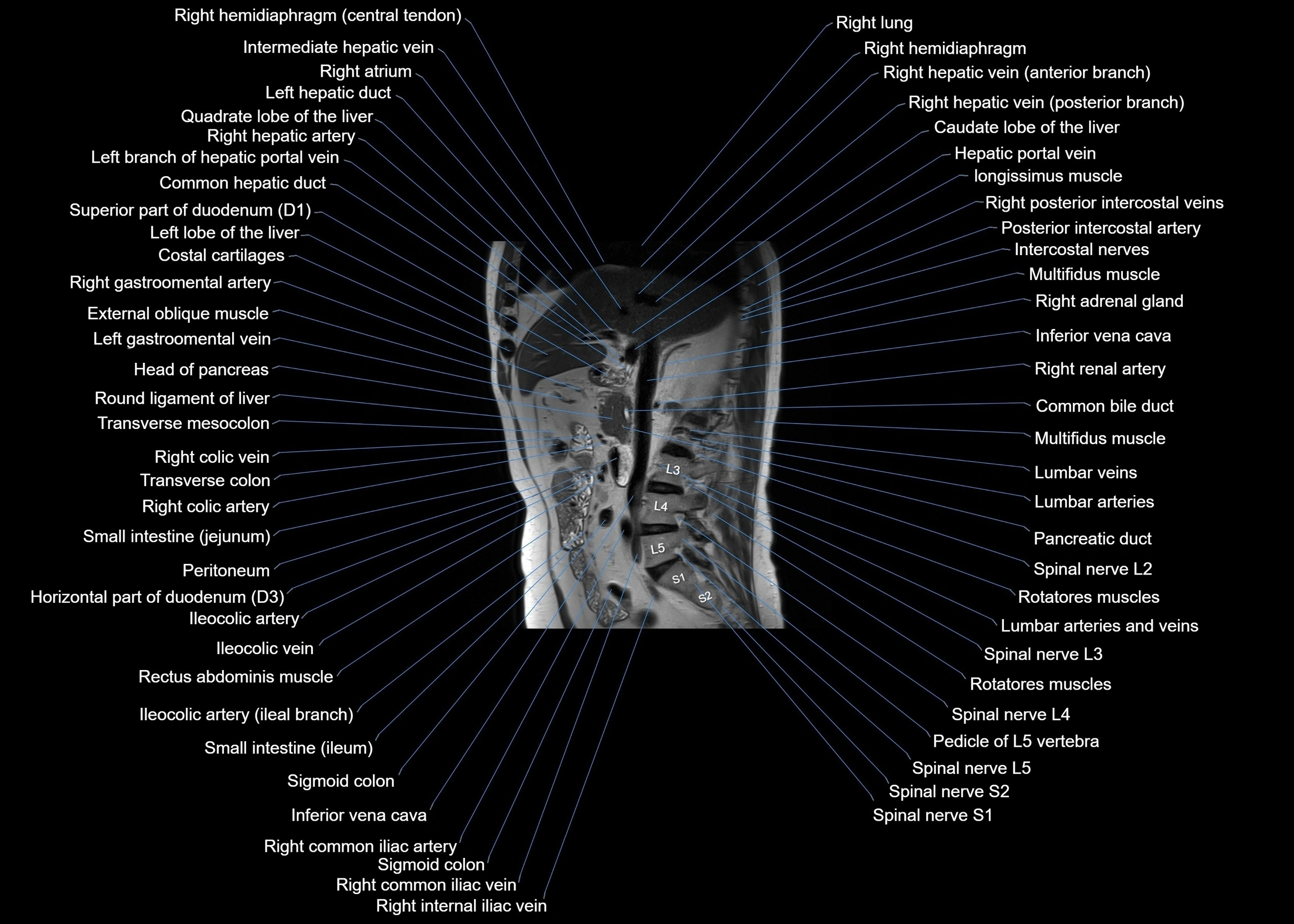
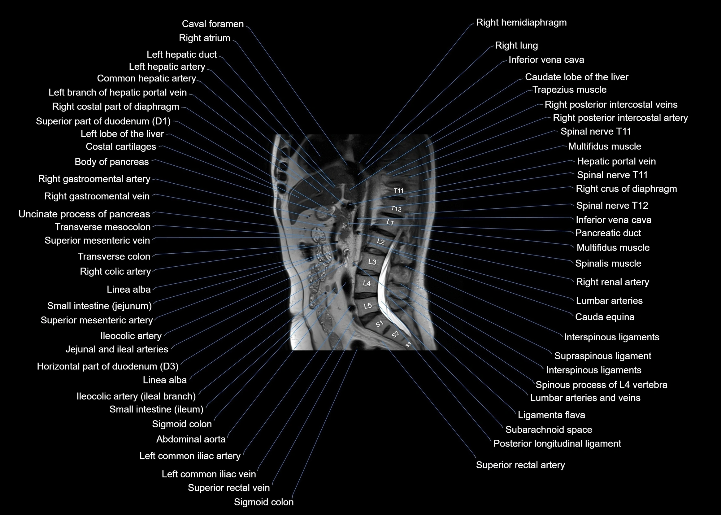
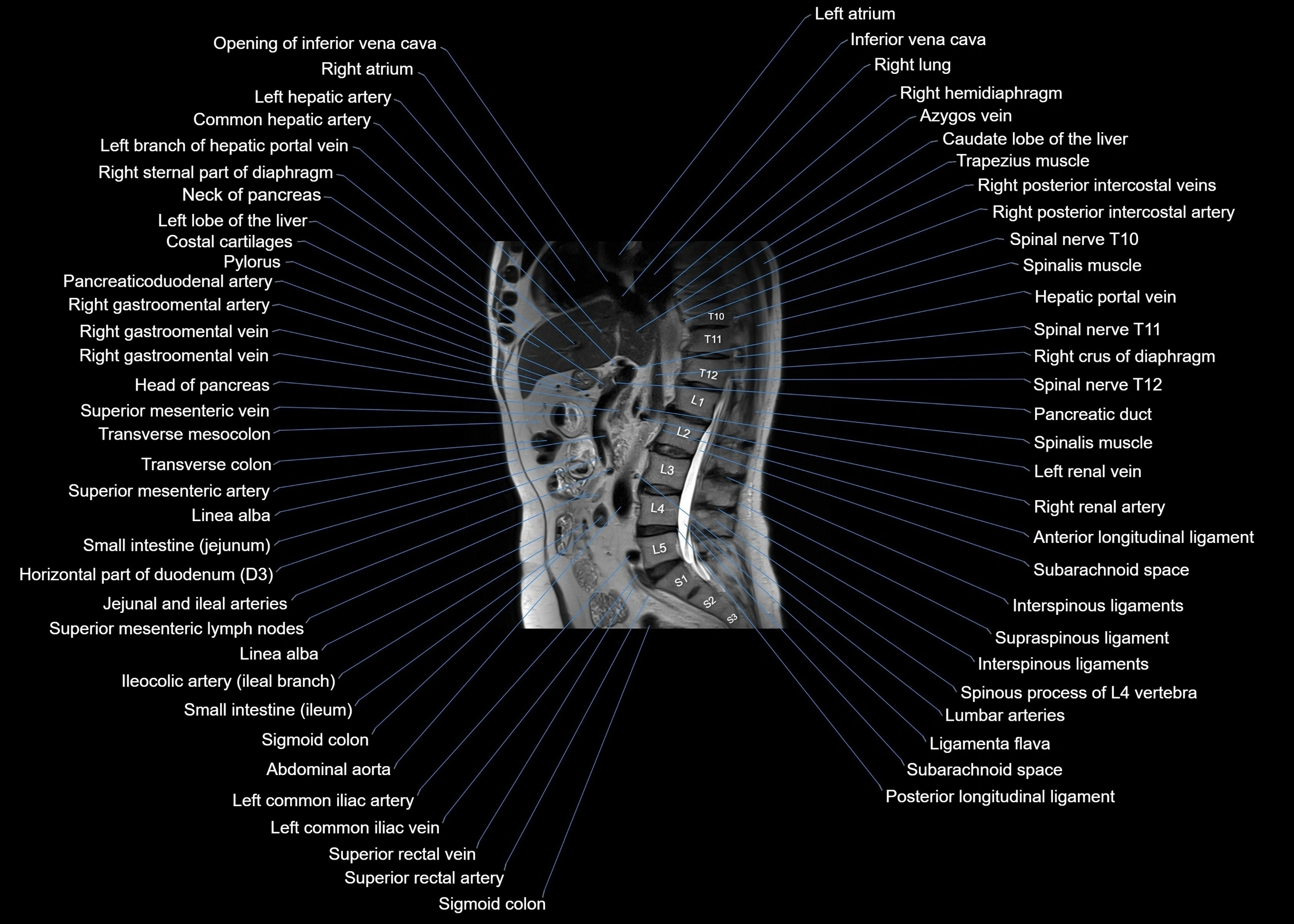
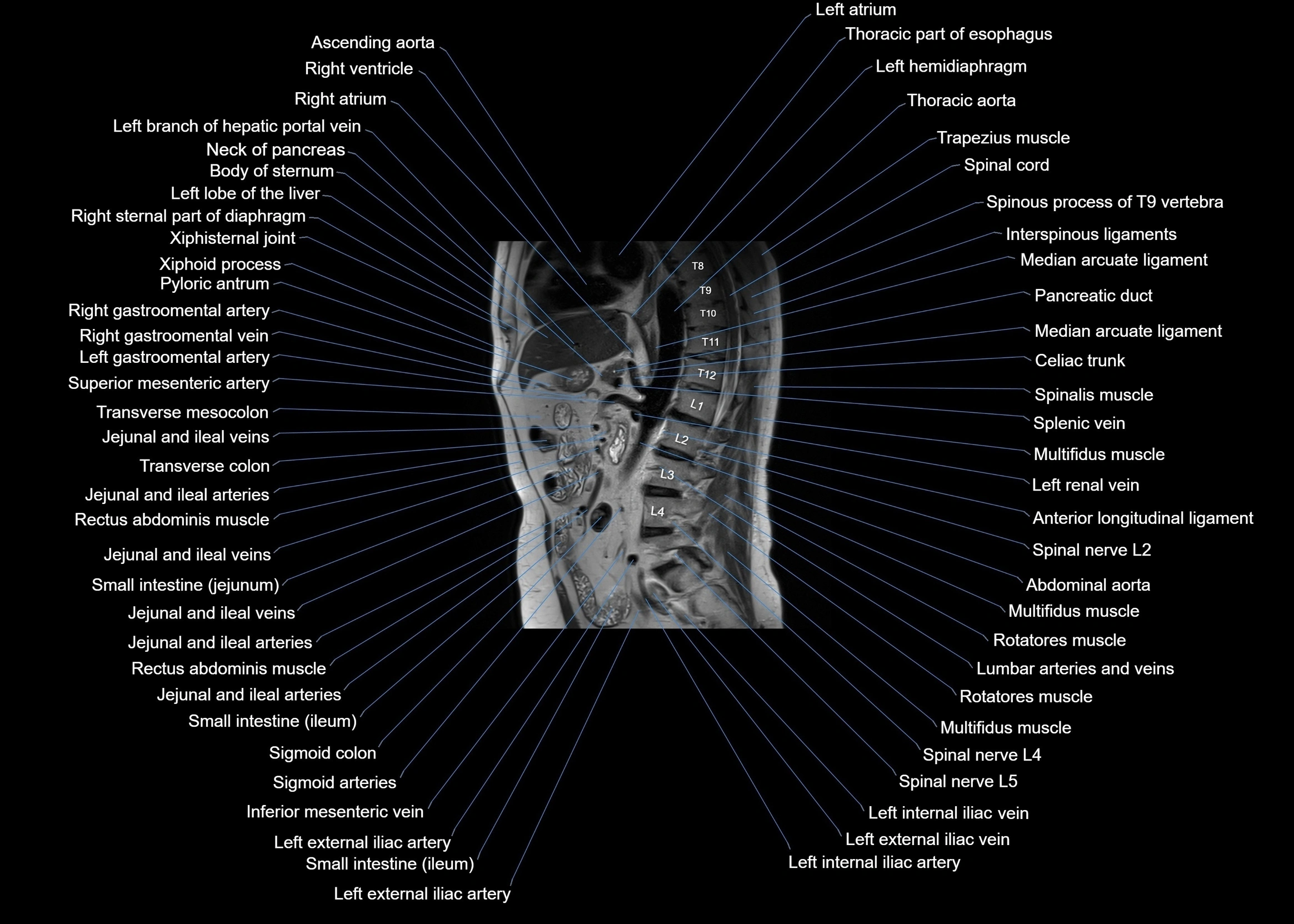
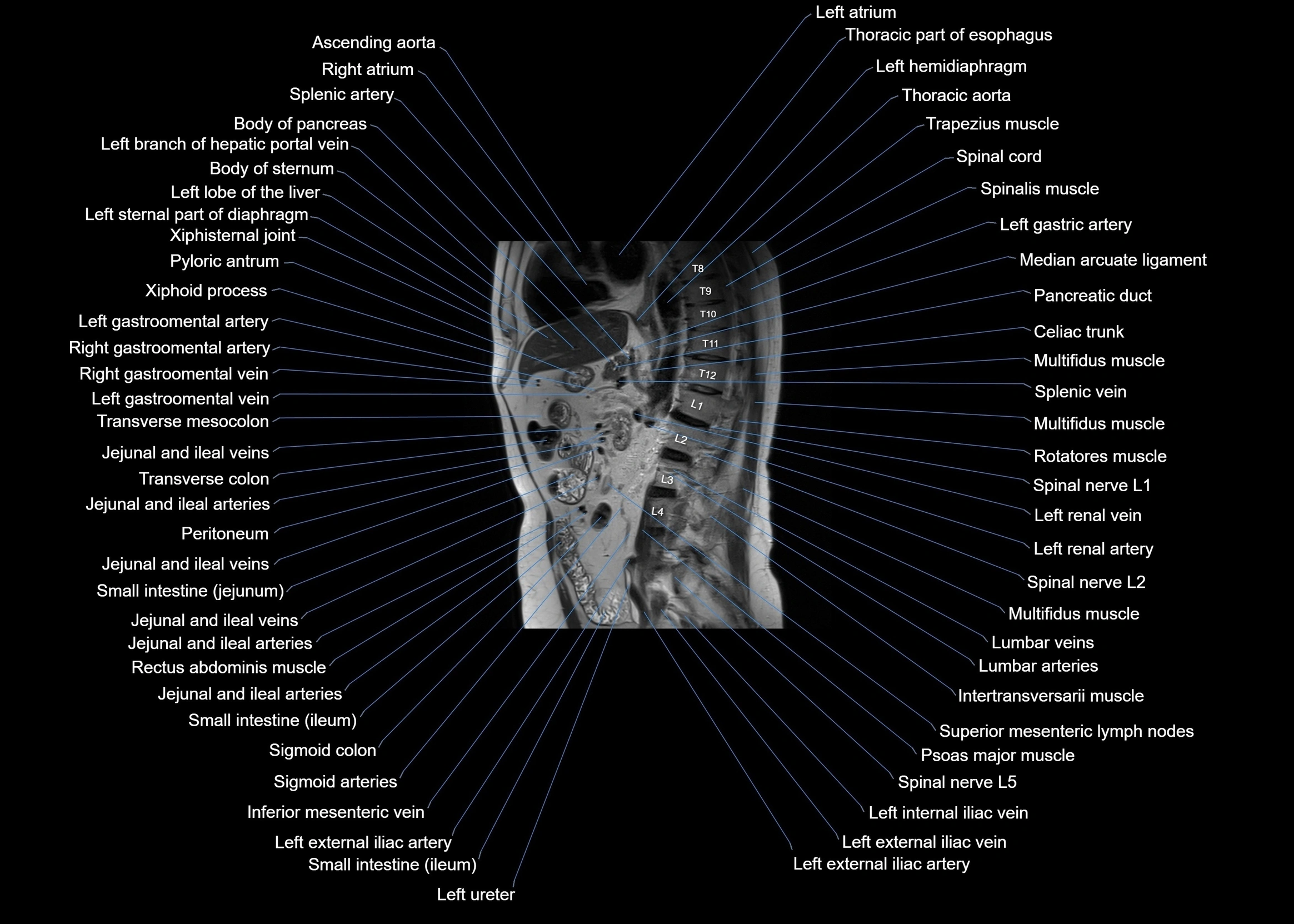
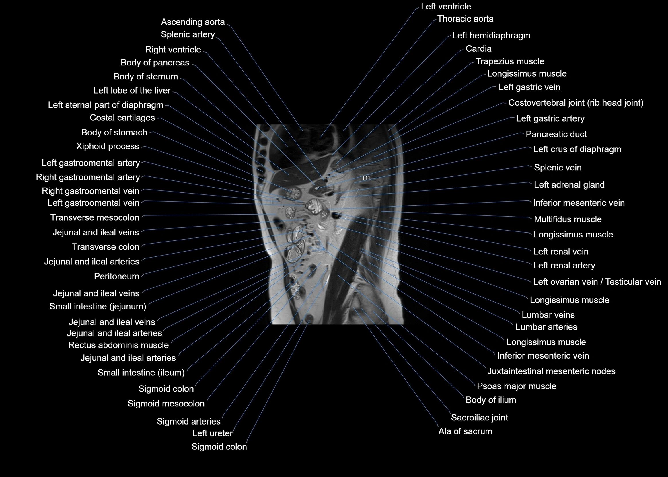
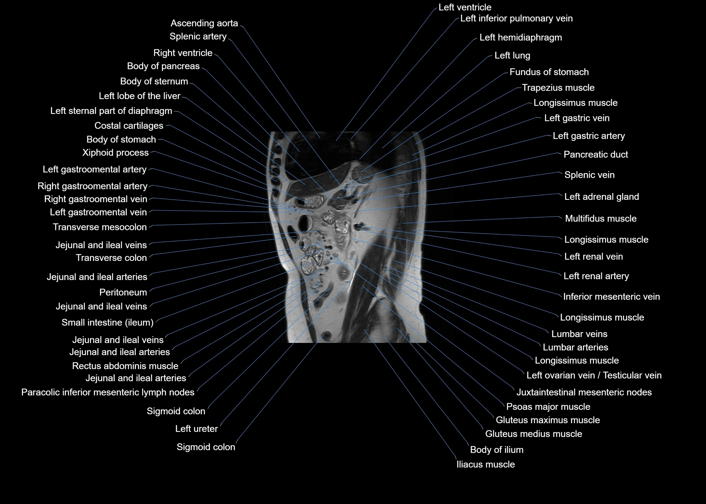
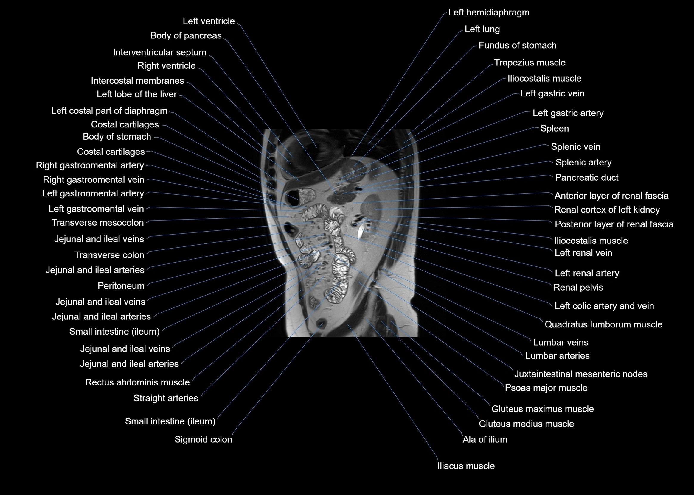
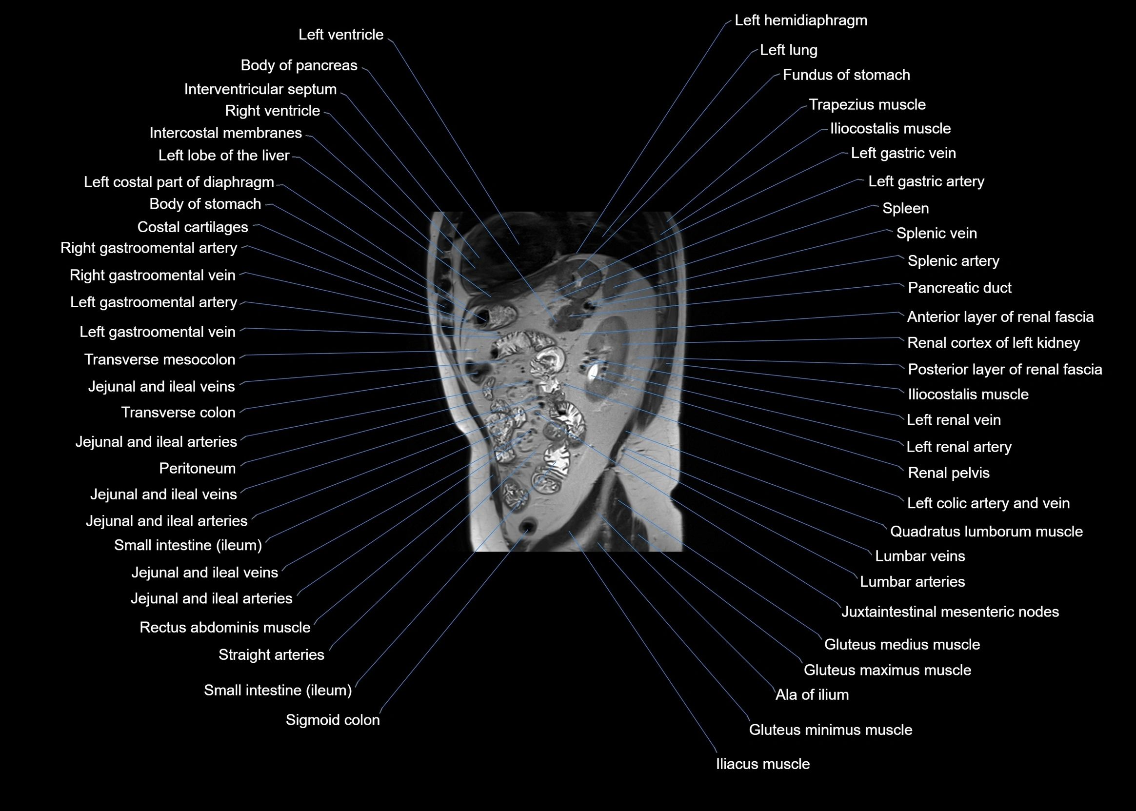
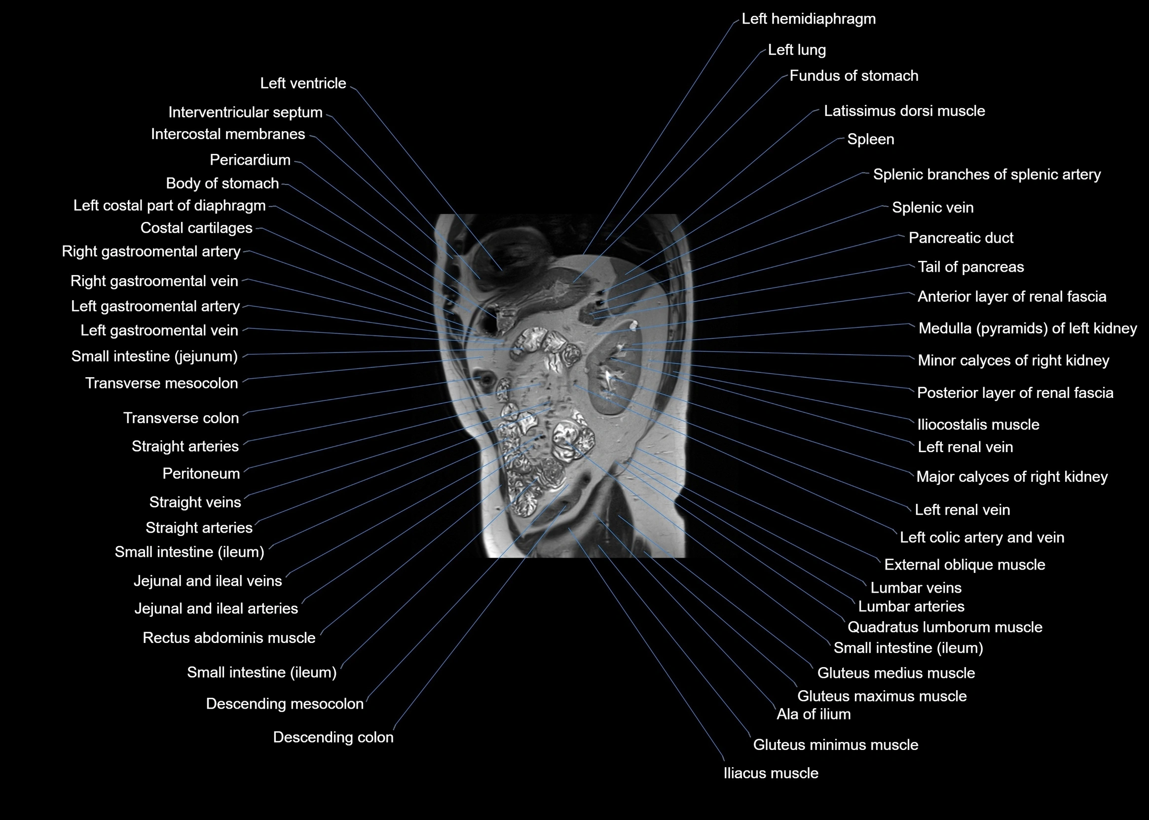
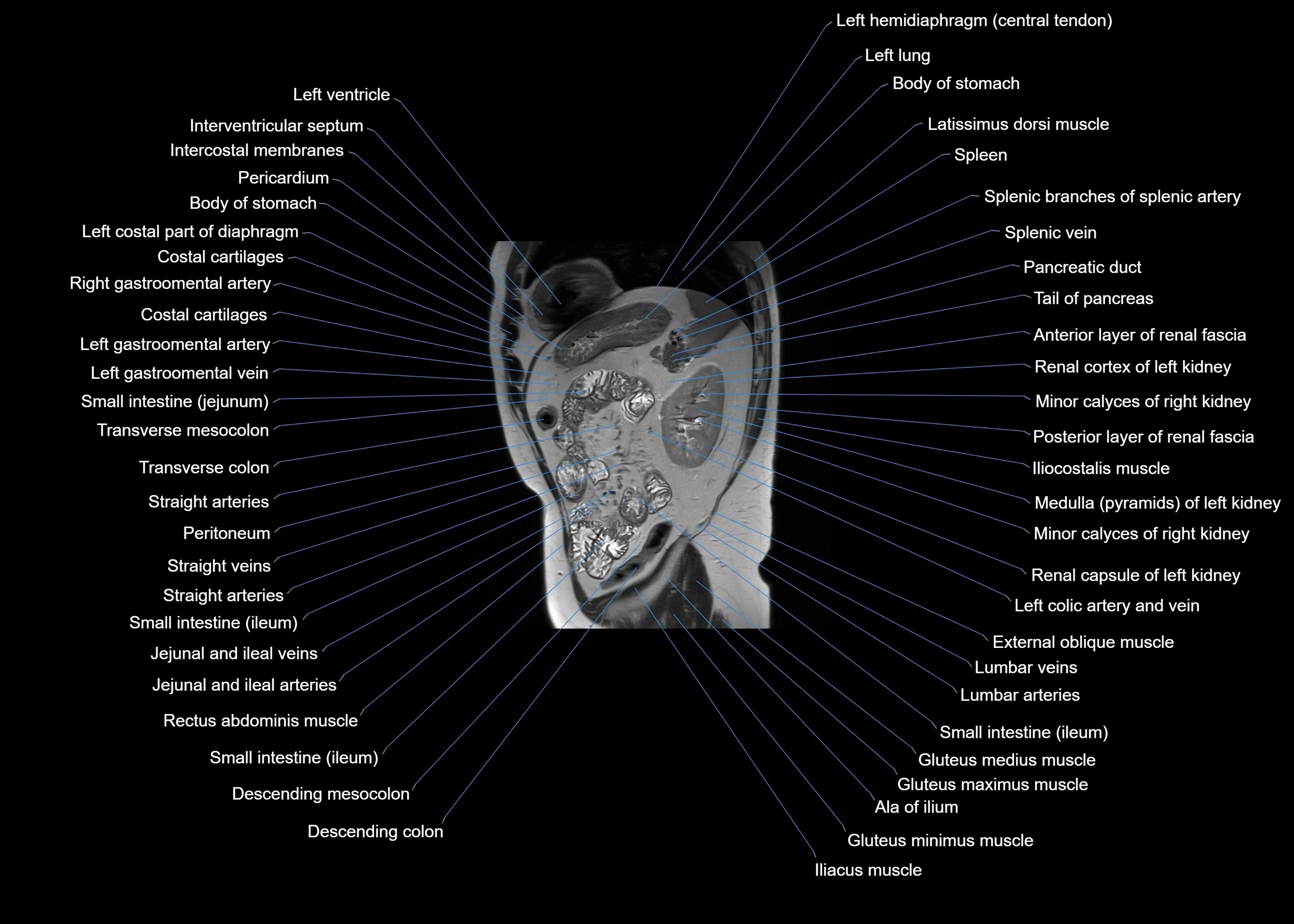
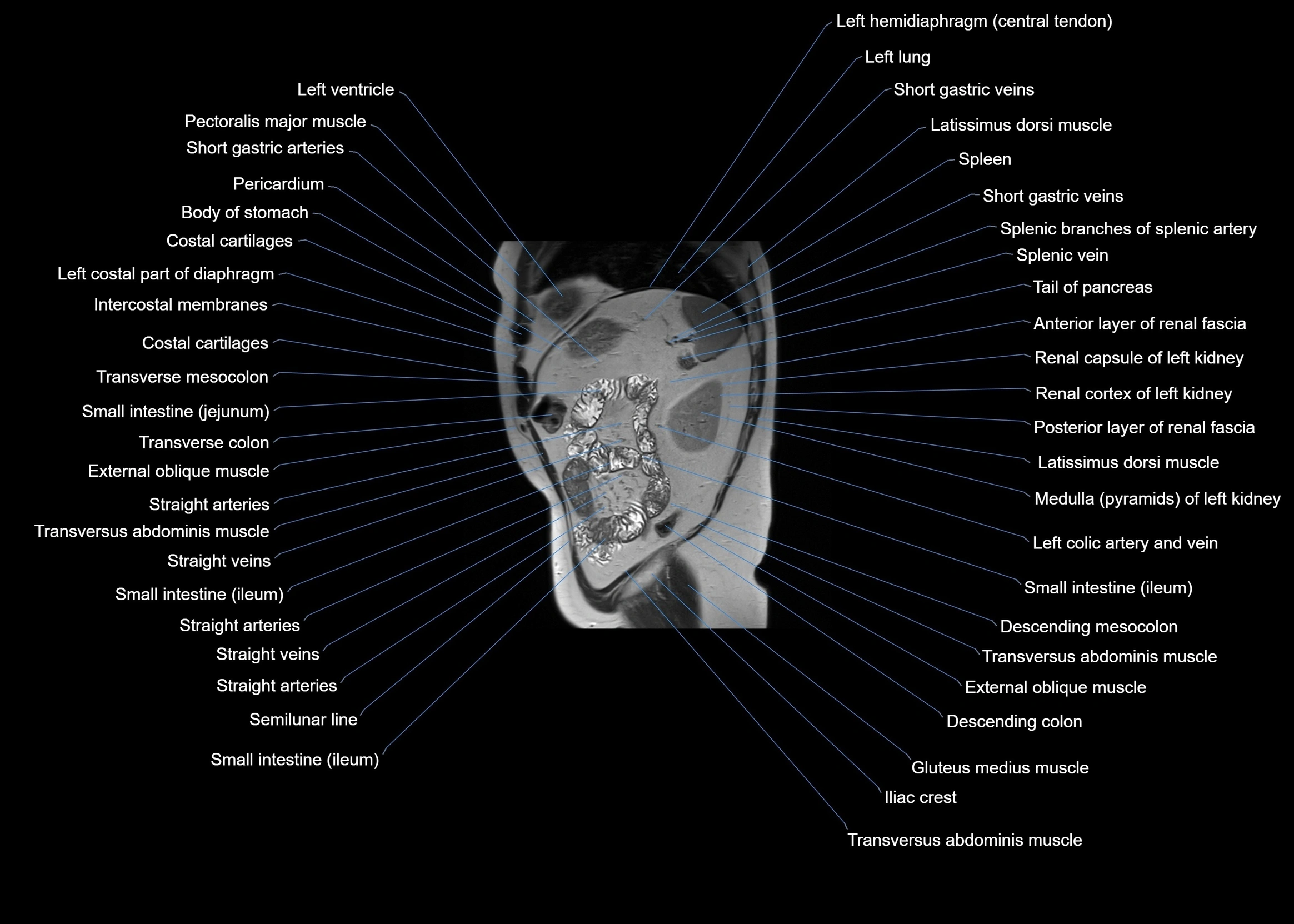
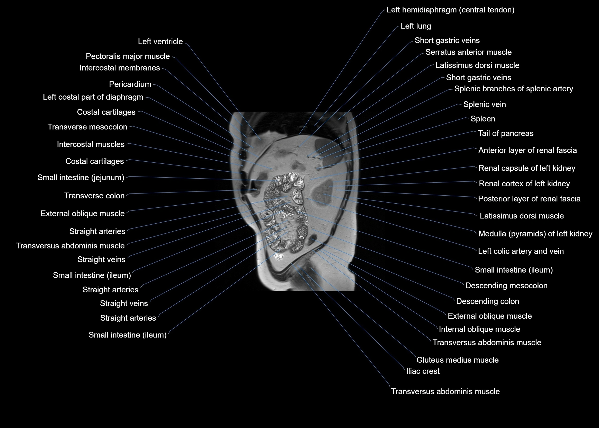
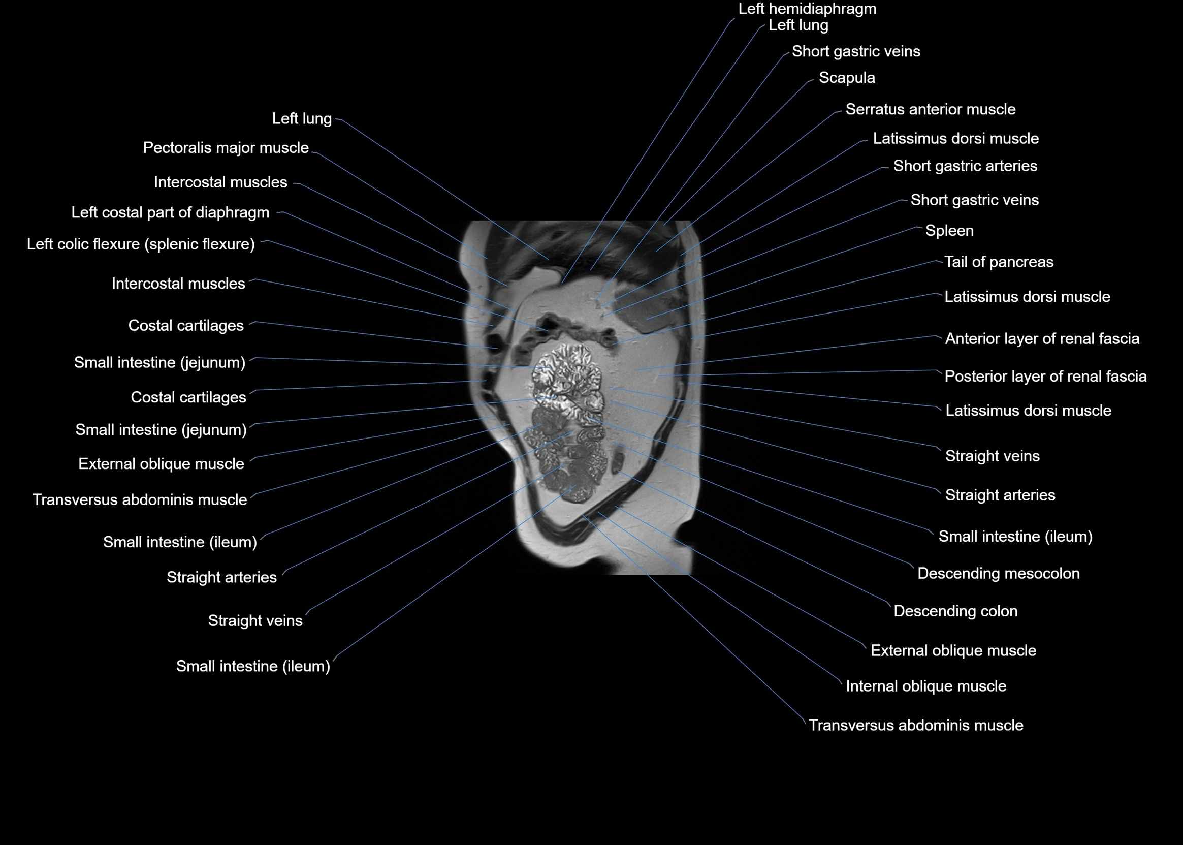
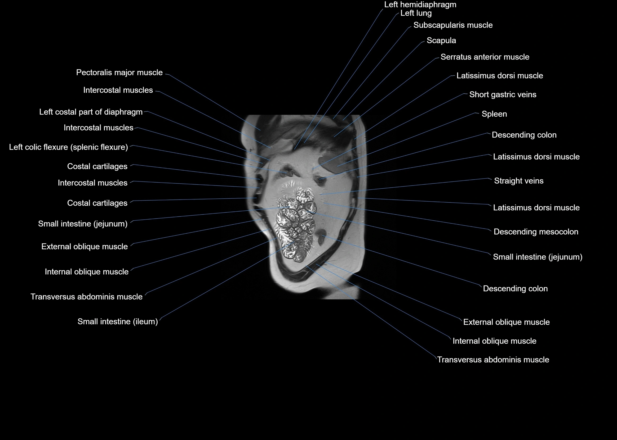
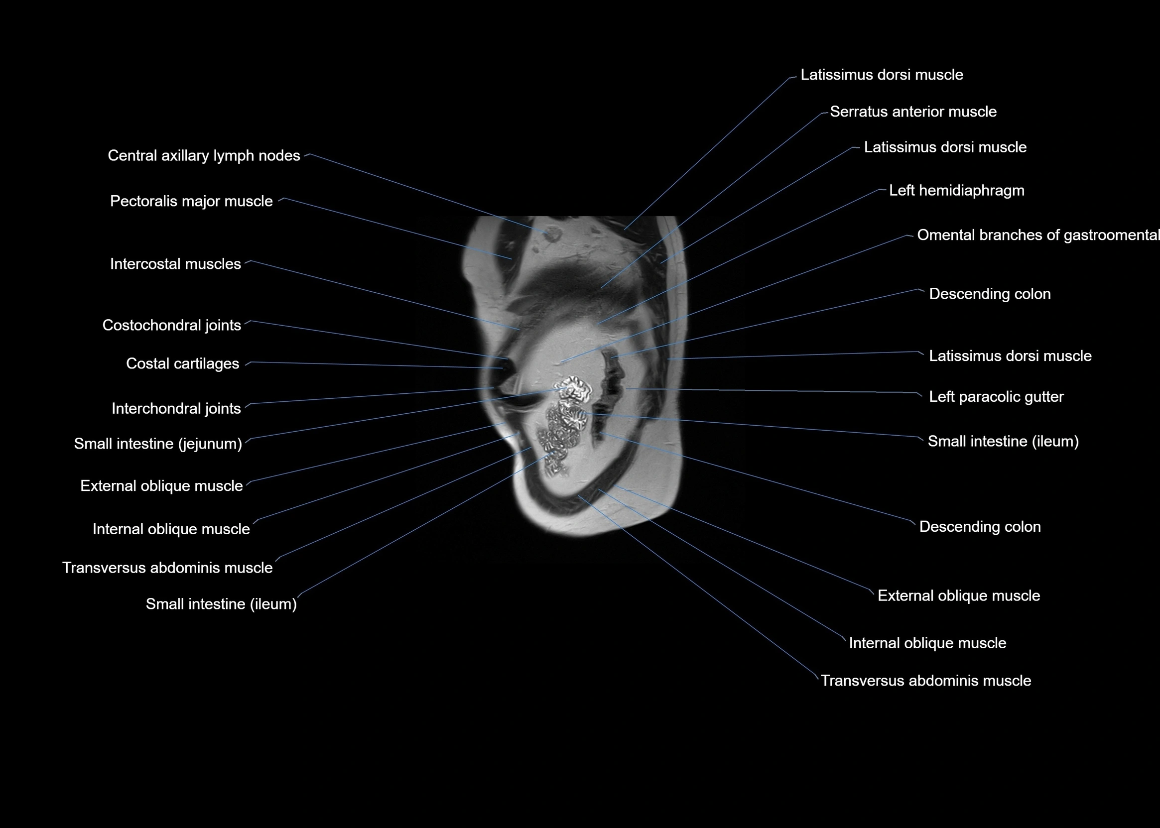
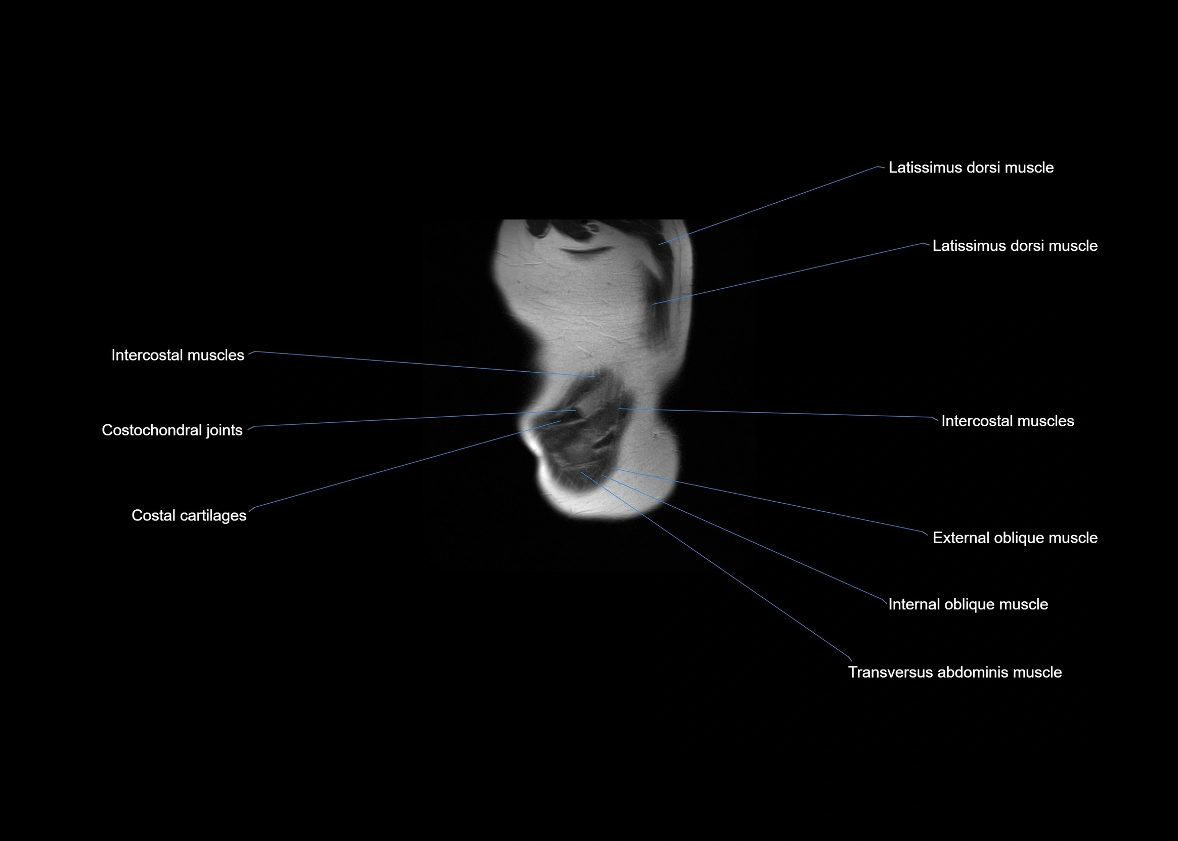
CT images

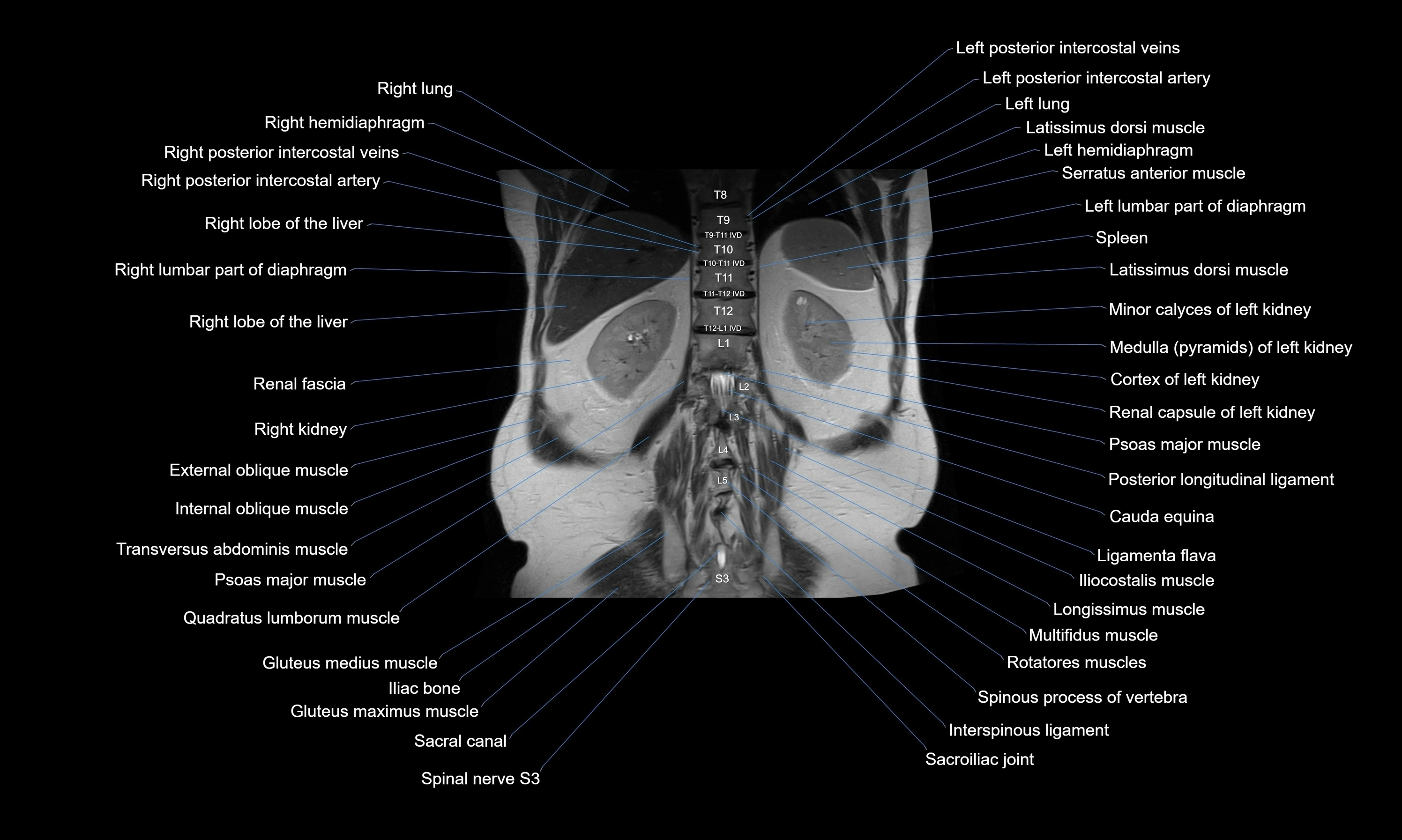
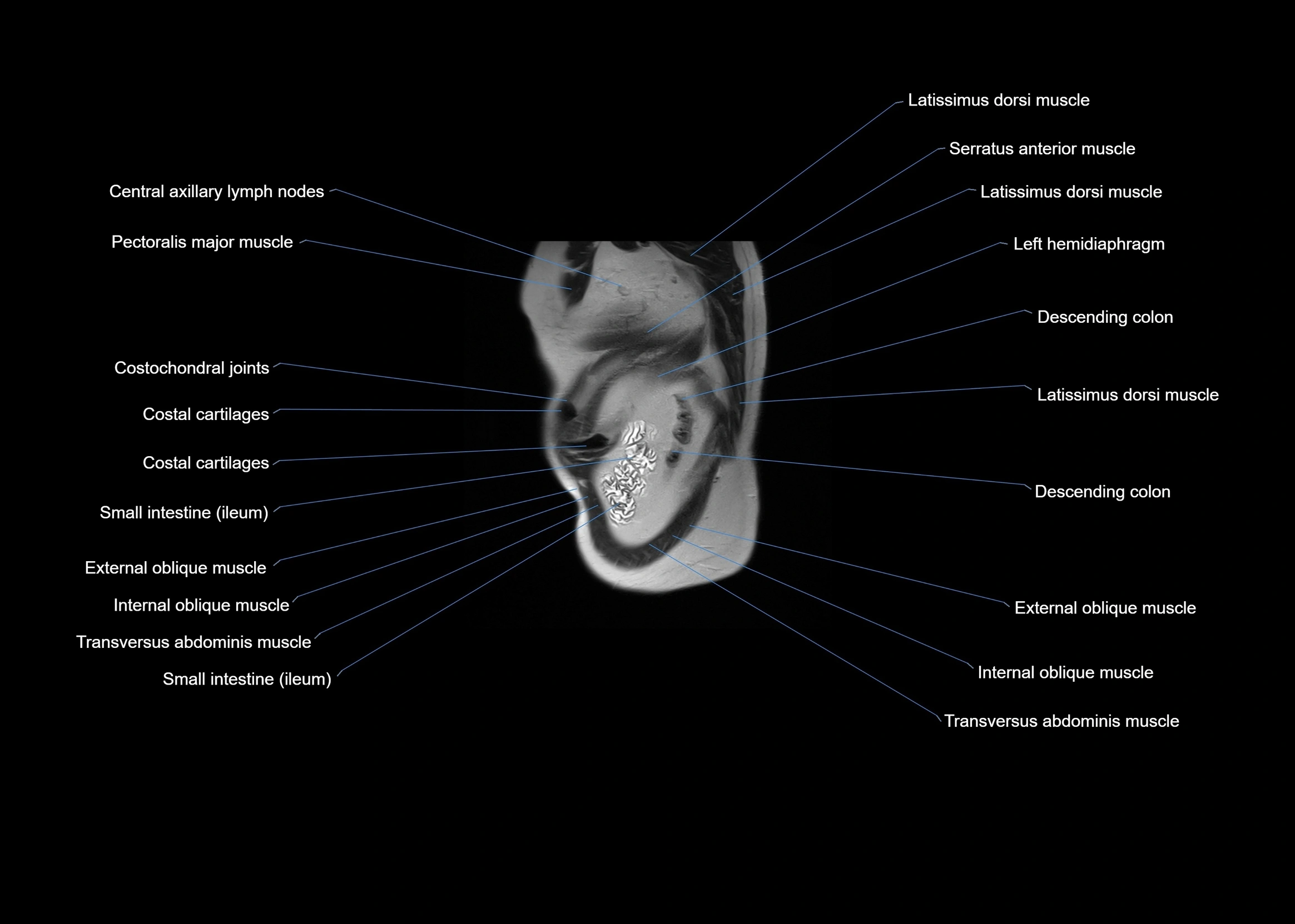
CT images


